US10983104B2 - Microfluidic chamber assembly for mastitis assay - Google Patents
Microfluidic chamber assembly for mastitis assayDownload PDFInfo
- Publication number
- US10983104B2 US10983104B2US14/105,662US201314105662AUS10983104B2US 10983104 B2US10983104 B2US 10983104B2US 201314105662 AUS201314105662 AUS 201314105662AUS 10983104 B2US10983104 B2US 10983104B2
- Authority
- US
- United States
- Prior art keywords
- wedge
- chamber
- milk
- base
- leukocyte
- Prior art date
- Legal status (The legal status is an assumption and is not a legal conclusion. Google has not performed a legal analysis and makes no representation as to the accuracy of the status listed.)
- Active, expires
Links
- 208000004396mastitisDiseases0.000titleclaimsabstractdescription28
- 238000003556assayMethods0.000titleclaimsdescription21
- 210000000265leukocyteAnatomy0.000claimsabstractdescription62
- 238000000034methodMethods0.000claimsabstractdescription41
- 210000004027cellAnatomy0.000claimsabstractdescription36
- 201000010099diseaseDiseases0.000claimsabstractdescription9
- 208000037265diseases, disorders, signs and symptomsDiseases0.000claimsabstractdescription9
- 239000012530fluidSubstances0.000claimsabstractdescription5
- 239000002356single layerSubstances0.000claimsabstractdescription4
- 235000013336milkNutrition0.000claimsdescription66
- 239000008267milkSubstances0.000claimsdescription66
- 210000004080milkAnatomy0.000claimsdescription66
- 239000003086colorantSubstances0.000claimsdescription14
- 239000007788liquidSubstances0.000claimsdescription13
- 239000000853adhesiveSubstances0.000claimsdescription7
- 230000001070adhesive effectEffects0.000claimsdescription7
- 210000004698lymphocyteAnatomy0.000claimsdescription6
- 210000002540macrophageAnatomy0.000claimsdescription6
- 239000000203mixtureSubstances0.000claimsdescription6
- 210000000440neutrophilAnatomy0.000claimsdescription6
- 238000003384imaging methodMethods0.000claimsdescription5
- 230000006651lactationEffects0.000claimsdescription5
- 230000009471actionEffects0.000claimsdescription3
- 230000003862health statusEffects0.000claimsdescription3
- 239000011800void materialSubstances0.000claims2
- 210000001124body fluidAnatomy0.000abstractdescription8
- 239000010839body fluidSubstances0.000abstractdescription6
- 241000124008MammaliaSpecies0.000abstractdescription3
- 238000001514detection methodMethods0.000abstractdescription3
- 235000020247cow milkNutrition0.000abstract1
- 241000283690Bos taurusSpecies0.000description22
- 238000012360testing methodMethods0.000description11
- 239000004033plasticSubstances0.000description9
- 229920003023plasticPolymers0.000description9
- 238000013461designMethods0.000description8
- 210000000481breastAnatomy0.000description7
- 239000011521glassSubstances0.000description7
- 208000015181infectious diseaseDiseases0.000description6
- 239000000463materialSubstances0.000description5
- 230000000007visual effectEffects0.000description5
- 206010061218InflammationDiseases0.000description4
- 210000004369bloodAnatomy0.000description4
- 239000008280bloodSubstances0.000description4
- 238000006243chemical reactionMethods0.000description4
- 235000013365dairy productNutrition0.000description4
- 230000004054inflammatory processEffects0.000description4
- 238000004519manufacturing processMethods0.000description4
- 244000005700microbiomeSpecies0.000description4
- 238000011160researchMethods0.000description4
- 241000894006BacteriaSpecies0.000description3
- 241001465754MetazoaSpecies0.000description3
- 238000010817Wright-Giemsa stainingMethods0.000description3
- 230000002159abnormal effectEffects0.000description3
- 231100000433cytotoxicToxicity0.000description3
- 230000001472cytotoxic effectEffects0.000description3
- 210000005075mammary glandAnatomy0.000description3
- 238000005259measurementMethods0.000description3
- 210000001082somatic cellAnatomy0.000description3
- 208000035143Bacterial infectionDiseases0.000description2
- 208000031462Bovine MastitisDiseases0.000description2
- 230000008901benefitEffects0.000description2
- 230000015556catabolic processEffects0.000description2
- 230000004069differentiationEffects0.000description2
- 210000004907glandAnatomy0.000description2
- 210000004969inflammatory cellAnatomy0.000description2
- 238000000386microscopyMethods0.000description2
- 230000003287optical effectEffects0.000description2
- 239000000123paperSubstances0.000description2
- 239000008188pelletSubstances0.000description2
- 230000000284resting effectEffects0.000description2
- 238000005070samplingMethods0.000description2
- 239000000758substrateSubstances0.000description2
- 238000010998test methodMethods0.000description2
- 210000001519tissueAnatomy0.000description2
- 239000003053toxinSubstances0.000description2
- 231100000765toxinToxicity0.000description2
- 108700012359toxinsProteins0.000description2
- XLYOFNOQVPJJNP-UHFFFAOYSA-NwaterSubstancesOXLYOFNOQVPJJNP-UHFFFAOYSA-N0.000description2
- ZOMLUNRKXJYKPD-UHFFFAOYSA-N1,3,3-trimethyl-2-[2-(2-methylindol-3-ylidene)ethylidene]indole;hydrochlorideChemical compound[Cl-].C1=CC=C2C(C)(C)C(/C=C/C=3C4=CC=CC=C4NC=3C)=[N+](C)C2=C1ZOMLUNRKXJYKPD-UHFFFAOYSA-N0.000description1
- PHWHSHNIIPVVHF-UHFFFAOYSA-N4-[(4-aminophenyl)-(4-ethyliminocyclohexa-2,5-dien-1-ylidene)methyl]-n,n-diethyl-2-methylaniline;hydrochlorideChemical compoundCl.C1=CC(=NCC)C=CC1=C(C=1C=C(C)C(N(CC)CC)=CC=1)C1=CC=C(N)C=C1PHWHSHNIIPVVHF-UHFFFAOYSA-N0.000description1
- 206010037660PyrexiaDiseases0.000description1
- UYQVPCFMVWEDCA-UHFFFAOYSA-M[6-(diethylamino)-9-[2-[[9-(diethylamino)benzo[a]phenoxazin-5-ylidene]carbamoyl]phenyl]xanthen-3-ylidene]-diethylazanium;chlorideChemical compound[Cl-].C12=CC=CC=C2C2=NC3=CC=C(N(CC)CC)C=C3OC2=CC1=NC(=O)C1=CC=CC=C1C1=C2C=CC(=[N+](CC)CC)C=C2OC2=CC(N(CC)CC)=CC=C21UYQVPCFMVWEDCA-UHFFFAOYSA-M0.000description1
- DPKHZNPWBDQZCN-UHFFFAOYSA-Nacridine orange free baseChemical compoundC1=CC(N(C)C)=CC2=NC3=CC(N(C)C)=CC=C3C=C21DPKHZNPWBDQZCN-UHFFFAOYSA-N0.000description1
- 239000002313adhesive filmSubstances0.000description1
- 238000004458analytical methodMethods0.000description1
- 239000000427antigenSubstances0.000description1
- 108091007433antigensProteins0.000description1
- 102000036639antigensHuman genes0.000description1
- 230000004596appetite lossEffects0.000description1
- 208000022362bacterial infectious diseaseDiseases0.000description1
- 230000000721bacterilogical effectEffects0.000description1
- 230000004888barrier functionEffects0.000description1
- DZBUGLKDJFMEHC-UHFFFAOYSA-NbenzoquinolinylideneNatural productsC1=CC=CC2=CC3=CC=CC=C3N=C21DZBUGLKDJFMEHC-UHFFFAOYSA-N0.000description1
- 230000003115biocidal effectEffects0.000description1
- 239000012503blood componentSubstances0.000description1
- 239000000298carbocyanineSubstances0.000description1
- 239000000306componentSubstances0.000description1
- 239000002131composite materialSubstances0.000description1
- 150000001875compoundsChemical class0.000description1
- 239000000470constituentSubstances0.000description1
- 238000012258culturingMethods0.000description1
- 208000013184decreased milk productionDiseases0.000description1
- 230000018044dehydrationEffects0.000description1
- 238000006297dehydration reactionMethods0.000description1
- 230000001419dependent effectEffects0.000description1
- 230000008021depositionEffects0.000description1
- 238000003745diagnosisMethods0.000description1
- 239000003814drugSubstances0.000description1
- 229940079593drugDrugs0.000description1
- 230000009977dual effectEffects0.000description1
- 210000002919epithelial cellAnatomy0.000description1
- ZMMJGEGLRURXTF-UHFFFAOYSA-Nethidium bromideChemical compound[Br-].C12=CC(N)=CC=C2C2=CC=C(N)C=C2[N+](CC)=C1C1=CC=CC=C1ZMMJGEGLRURXTF-UHFFFAOYSA-N0.000description1
- 229960005542ethidium bromideDrugs0.000description1
- 238000000684flow cytometryMethods0.000description1
- 238000002073fluorescence micrographMethods0.000description1
- 230000006870functionEffects0.000description1
- 230000036541healthEffects0.000description1
- 238000007654immersionMethods0.000description1
- 230000007124immune defenseEffects0.000description1
- 230000028993immune responseEffects0.000description1
- 210000000987immune systemAnatomy0.000description1
- 230000028709inflammatory responseEffects0.000description1
- 239000010410layerSubstances0.000description1
- 235000021266loss of appetiteNutrition0.000description1
- 208000019017loss of appetiteDiseases0.000description1
- 210000004880lymph fluidAnatomy0.000description1
- 230000003458metachromatic effectEffects0.000description1
- 239000002184metalSubstances0.000description1
- 230000000877morphologic effectEffects0.000description1
- PKALEBFTSZCHGU-UHFFFAOYSA-Mn,n-diethyl-4-[2-(1,3,3-trimethylindol-1-ium-2-yl)ethenyl]aniline;chlorideChemical compound[Cl-].C1=CC(N(CC)CC)=CC=C1\C=C\C1=[N+](C)C2=CC=CC=C2C1(C)CPKALEBFTSZCHGU-UHFFFAOYSA-M0.000description1
- ZTBANYZVKCGOKD-UHFFFAOYSA-Mn-(2-chloroethyl)-n-methyl-4-[2-(1,3,3-trimethylindol-1-ium-2-yl)ethenyl]aniline;chlorideChemical compound[Cl-].C1=CC(N(CCCl)C)=CC=C1C=CC1=[N+](C)C2=CC=CC=C2C1(C)CZTBANYZVKCGOKD-UHFFFAOYSA-M0.000description1
- 244000052769pathogenSpecies0.000description1
- QWYZFXLSWMXLDM-UHFFFAOYSA-Mpinacyanol iodideChemical compound[I-].C1=CC2=CC=CC=C2N(CC)C1=CC=CC1=CC=C(C=CC=C2)C2=[N+]1CCQWYZFXLSWMXLDM-UHFFFAOYSA-M0.000description1
- 229920000642polymerPolymers0.000description1
- 238000004094preconcentrationMethods0.000description1
- 238000002810primary assayMethods0.000description1
- 230000008569processEffects0.000description1
- 238000011084recoveryMethods0.000description1
- 230000004044responseEffects0.000description1
- 230000028327secretionEffects0.000description1
- 230000003595spectral effectEffects0.000description1
- 238000010561standard procedureMethods0.000description1
- 239000006228supernatantSubstances0.000description1
- 230000008961swellingEffects0.000description1
- 229950003937toloniumDrugs0.000description1
- HNONEKILPDHFOL-UHFFFAOYSA-Mtolonium chlorideChemical compound[Cl-].C1=C(C)C(N)=CC2=[S+]C3=CC(N(C)C)=CC=C3N=C21HNONEKILPDHFOL-UHFFFAOYSA-M0.000description1
- 238000012549trainingMethods0.000description1
- 239000012780transparent materialSubstances0.000description1
- 239000001974tryptic soy brothSubstances0.000description1
- 108010050327trypticase-soy brothProteins0.000description1
- 238000003260vortexingMethods0.000description1
Images
Classifications
- G—PHYSICS
- G01—MEASURING; TESTING
- G01N—INVESTIGATING OR ANALYSING MATERIALS BY DETERMINING THEIR CHEMICAL OR PHYSICAL PROPERTIES
- G01N15/00—Investigating characteristics of particles; Investigating permeability, pore-volume or surface-area of porous materials
- G01N15/10—Investigating individual particles
- G01N15/14—Optical investigation techniques, e.g. flow cytometry
- G01N15/1429—Signal processing
- G01N15/1433—Signal processing using image recognition
- G—PHYSICS
- G01—MEASURING; TESTING
- G01N—INVESTIGATING OR ANALYSING MATERIALS BY DETERMINING THEIR CHEMICAL OR PHYSICAL PROPERTIES
- G01N33/00—Investigating or analysing materials by specific methods not covered by groups G01N1/00 - G01N31/00
- G01N33/02—Food
- G01N33/04—Dairy products
- G01N15/1463—
- G—PHYSICS
- G01—MEASURING; TESTING
- G01N—INVESTIGATING OR ANALYSING MATERIALS BY DETERMINING THEIR CHEMICAL OR PHYSICAL PROPERTIES
- G01N15/00—Investigating characteristics of particles; Investigating permeability, pore-volume or surface-area of porous materials
- G01N15/10—Investigating individual particles
- G01N15/14—Optical investigation techniques, e.g. flow cytometry
- G01N15/1484—Optical investigation techniques, e.g. flow cytometry microstructural devices
- B—PERFORMING OPERATIONS; TRANSPORTING
- B01—PHYSICAL OR CHEMICAL PROCESSES OR APPARATUS IN GENERAL
- B01L—CHEMICAL OR PHYSICAL LABORATORY APPARATUS FOR GENERAL USE
- B01L3/00—Containers or dishes for laboratory use, e.g. laboratory glassware; Droppers
- B01L3/50—Containers for the purpose of retaining a material to be analysed, e.g. test tubes
- B01L3/502—Containers for the purpose of retaining a material to be analysed, e.g. test tubes with fluid transport, e.g. in multi-compartment structures
- B01L3/5027—Containers for the purpose of retaining a material to be analysed, e.g. test tubes with fluid transport, e.g. in multi-compartment structures by integrated microfluidic structures, i.e. dimensions of channels and chambers are such that surface tension forces are important, e.g. lab-on-a-chip
- G—PHYSICS
- G01—MEASURING; TESTING
- G01N—INVESTIGATING OR ANALYSING MATERIALS BY DETERMINING THEIR CHEMICAL OR PHYSICAL PROPERTIES
- G01N1/00—Sampling; Preparing specimens for investigation
- G01N1/28—Preparing specimens for investigation including physical details of (bio-)chemical methods covered elsewhere, e.g. G01N33/50, C12Q
- G01N1/2813—Producing thin layers of samples on a substrate, e.g. smearing, spinning-on
- G01N2015/008—
- G—PHYSICS
- G01—MEASURING; TESTING
- G01N—INVESTIGATING OR ANALYSING MATERIALS BY DETERMINING THEIR CHEMICAL OR PHYSICAL PROPERTIES
- G01N15/00—Investigating characteristics of particles; Investigating permeability, pore-volume or surface-area of porous materials
- G01N15/01—Investigating characteristics of particles; Investigating permeability, pore-volume or surface-area of porous materials specially adapted for biological cells, e.g. blood cells
- G01N2015/016—White blood cells
Definitions
- the present inventionrelates to microfluidic chamber for analyzing a subjects' body fluids, in particular milk, to determine if the subject has a bacterial infection such as bovine mastitis.
- the inventionrelates to a chamber assembly which can be used to detect mastitis or other bacterial disease in bovine or other mammal species.
- Mastitisis the inflammation of the mammary gland caused by microorganisms that invade one or more quadrants of the bovine udder, multiply, and produce toxins that are harmful to the mammary gland. Economic loss to mastitis in the United States is estimated to be approximately $185/cow annually. The total annual US cost of mastitis is over $2 billion. This is approximately 10% of the total value of farm milk sales, and about two-thirds of this loss is due to reduced milk production in subclinically infected cows.
- the average production loss per lactation for one infected udder quarteris about 1,600 pounds. Other losses are due to discarded abnormal milk and milk withheld from cows treated with antibiotic, costs of early replacement of affected cows, reduced sale value of culled cows, costs of drugs and veterinary services, and increased labor costs. Mastitis reduces milk yield and alters milk composition. The magnitude of these changes in individual cows varies with the severity and duration of the infection and the causative microorganisms. Mastitis is almost always caused by bacteria. These microorganisms produce toxins that can directly damage milk-producing tissue of the mammary gland, and the presence of bacteria initiates inflammation within the mammary tissue in an attempt to eliminate the invading microorganisms. The inflammation contributes to decreased milk production and is primarily responsible for the compositional changes observed in milk from infected quarters and cows. In general, compositional changes involve an increase in blood components present in milk and a decrease in normal milk constituents.
- Clinical Mastitisincludes visible signs of mastitis such as the “mild” signs, for example, flakes or clots in the milk, or slight swelling of an infected quarter. It also includes “severe” signs such as abnormal secretion; hot, swollen quarter or full udder; fever, rapid pulse, loss of appetite; dehydration and depression; and in some cases death may occur.
- Subclinical Mastitisthere are no visible signs of the disease. Diagnosis of subclinical mastitis is characterized by the Somatic Cell Count (SCC) of the milk being elevated and the bacteriological culturing of milk will detect bacteria in the milk. Subclinical mastitis causes the greatest financial loss to dairy farmers through lowered milk production. For every clinical case of clinical mastitis, there will be 15 to 40 sub-clinical cases.
- SCCSomatic Cell Count
- the SCCis the number of leukocytes or white blood cells per milliliter of milk.
- the SCChas become the standard procedure for diagnosing sub-clinical mastitis and is also used worldwide as the index of milk quality.
- the SCCenumerates the many cells that populate the milk-producing gland as part of the immune defense system, and then cross the blood/milk barrier, as well as the few epithelial cells that line the udder, and also get into the milk.
- the animals' immune systemsproduce an “inflammatory response” in the gland and more of the infection-fighting white cells (mostly neutrophils) find their way into the milk.
- the SCCis reported as “the sum”, the total of those cells. Normal milk will have less than 200,000 cells per milliliter.
- SCC200,000 and over
- SCCis the current measure for commercial acceptability for milk in many countries, for example milk with an SCC over 400,000 can not be sold in Europe. Other countries limits include Canada, 500,000 and the United States 750,000 (600,000 in California). Currently there is no SCC associated with Clinical Mastitis.
- the SCCis also used in bulk tank management, in determining the suitability for shipment, in making culling decisions and in the payment of quality bonuses.
- the SCCa single number, is everywhere in the dairy industry and it is used in almost every area of milk production.
- the SCCled to increases in productivity, losses due to clinical-stage mastitis being stabilized, and the generally accepted notion that the milk supply is safe.
- the SCCis not a number representing a single type of leukocyte, rather it is a mixture of multiple types of leukocytes, each with its own significance.
- the predominant cell typesare lymphocytes, followed by much lesser numbers of neutrophils and macrophages.
- the percentages of each kind of cellrise and fall as part of the immune response to infection. Those percentages, “the differential milk leukocyte count”, represent the unique immune status of an individual quarter udder, at a specific point in time.
- the literaturehas suggested that a better understanding of the dynamics of mastitis could be accomplished by specifically measuring the rise and fall in the types of leukocytes involved in the disease process and recovery.
- the literaturehas also specifically identified the normal differential pattern of uninfected cows at various stages of lactation, it has suggested algorithms to identify the various stages of infection by looking at changes in those patterns and it has suggested linkages of specific abnormal patterns to specific pathogens.
- Hammanused the differential pattern to show quarters were inter-dependent.
- the literaturesuggests that the breakdown of the SCC into its component parts, the “milk somatic cell differential”, should be a better, more accurate and specific indicator of udder health, and thus a better tool for the management of mastitis.
- the SCCcontinues to be reported as a total SCC, and the differential information remains a research tool. This is primarily been driven by the cost differential between the SCC and computations for a differential.
- milkis centrifuged at 2500 g for 10 min at 4 degrees Celsius, the fat layer and supernatant are removed, leaving 500 ⁇ l or less of milk with a pellet.
- the pelletis re-suspended via vortexing and pipetting.
- Microscope slidesare coated with Trypticase Soy Broth, air-dried and 10 ⁇ l of milk is placed on the slide for a 30-degree angle smear to be made.
- the slideis allowed to air dry and is then stained with Wright Giemsa Stain.
- the slideis placed in the stain for 15 seconds, then water for 30 seconds, then a 15 second dip in clean water and finally it is allowed to air dry.
- Slidesare examined on a transmitted light microscope with a 100 ⁇ oil immersion lens.
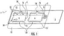
- FIG. 1is a wedge type slide comprising a microscope slide base, a slip cover wedge top and a rectilinear shim at the top part of the wedge.
- This designis described primarily for blood but is taught to be useful for any leukocyte containing liquid for example milk.
- the devicesuffers from some difficulties in use. First since the cover slip end opposite the shim is not fixed the end can leak, shift or get caught on other objects during use. Second since the design involves a shim the entire length of the cover slip there is no easy way to place the liquid in the chamber.
- FIG. 2 in the Wardlaw patentdescribes the appropriate angle for creating a wedge sampling configuration for us in differential testing. Accordingly, it would be very useful to have a different wedge chamber design that overcomes the limitations of the prior art teachings.
- a wedge forming devicewhich secures a first edge of the top to the base forms the bottom edge of the wedge chamber, elevates and secures the opposite second edge of the top from the base to form a wedge chamber and provides for a liquid entry well for liquid addition to the chamber;
- the assay itselfcomprises in one embodiment, method for performing a leukocyte differential assay comprising:
- FIG. 1shows an embodiment of the invention with a dual chamber mounted on a base.
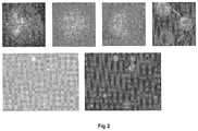
- FIG. 2displays enhanced images produced by an embodiment of the invention.
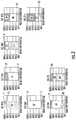
- FIG. 3displays graphs of the resultant cell count produced by an embodiment of the present invention.
- the present inventionrelates to a novel microfluidic wedge chamber and a leukocyte differential assay (LDA) for determination if a subject has mastitis in a given sample of milk.
- LDAleukocyte differential assay
- the general description of both the device and methodare stated in the Brief Summary above. This detailed description defines the meaning of the terms used herein and specifically describes embodiments in order for those skilled in the art to practice the invention.
- the assayis done exposing leukocyte containing body fluid such as bovine milk, whole blood or other leukocyte containing body fluid such as lymph fluid, spinal fluid or the like, to a predetermined cell count in the device of the invention.
- the testis compared for difference in leukocyte types and those differences determine if the subject has mastitis i.e.
- leukocyte SCC200,000 or greater and what the breakdown of different leukocytes within that population is.
- a distinct advantage of the assay of the inventionis that it is not only qualitative in nature, it is quantitative in nature and therefore leukocyte such measurements are more telling of the exact condition of the subject bovine patient.
- subjectis meant herein to be any animal especially bovine and especially bovine milk containing leukocytes.
- the primary assay of the inventionis use of the chamber to test for bovine mastitis but the chamber could simply be used to make a leukocyte determination for other bodily fluids for example as taught in the Wardlaw patent described above.
- leukocyteis meant any of the sub categories of leukocytes that are known to exist in milk or blood or other bodily fluid. These sub-categories can be identified and quantified with the device and assay of the invention especially for the detection of mastitis or other leukocyte based disease states.
- the baseis meant a substrate sufficient for performing a leukocyte differential determination.
- the basewould be a microscope slide made of glass or plastic and optionally optically transparent. It is clear however that where the determination is made with observation and lighting from the top of the assembly, that the base could be a non-transparent material such as paper or opaque plastic.
- Wild topis meant a substrate which forms the angle top wall of a chamber such as described in Wardlaw.
- top portionis a slide cover slip of glass or plastic which is positioned to form a wedge chamber for making cell count differential determinations. It is an embodiment that the wedge top be optically transparent but where a reading is taken from the bottom the top may be opaque and be of a material other than plastic or glass including paper cardboard, metal or the like.
- wedge forming deviceis meant a device which by its design accomplishes 4 important functions. First, it secures an edge of the wedge top to the base. Second, it secures the opposite edge of the top in an elevated configuration such that the wedge shaped chamber is formed sufficient for cell differential determination. One skilled in the art as taught above would easily be able to determine the proper angles for such chamber. Third, it secures the opposite side in such a manner that it may not move during normal use and fourthly, it provides a cut-out well so that liquid may be added to the top portion of the wedge instead of from the side as in previous prior art wedge chambers. This is accomplished by use of an adhesive backed film in one embodiment.
- the adhesive filmis cut out in such a manner that a flap for adhesion of one side of the top is formed.
- the filmalso has a cut out portion allowing for an unobstructed view of the top and base. Further it provides a means for securing the top edge of the top and it also provides a cut out portion for a liquid addition well.
- the wedge configuration of the chamberis then formed by one edge of the top resting against the base and the other opposite edge of the top resting on the film, the thickness of the film determines the shape and volume of the chamber. In general, where the base is a microscope slide and the top a slide slip cover the film would be chosen to be about 0.04 to 0.06 mm more or less in thickness. In one embodiment, the film is 0.05 mm in thickness.
- the adhesiveallows the film to be attached to the base and the top simultaneously because of the adhesive backing and the unique cut-out design.
- the filmis in an embodiment a polymer which gives it both flexibility and ease of putting an adhesive backing thereon.
- the adhesiveshould be such that it adheres to both the top and base and one skilled in the art would be able to make optimum choices depending on the materials chosen for the top and base.
- the liquid in the chamberis observed by methods taught for differentiation (see Example) and thus the chamber allows for differentiation of the leukocyte sub-population.
- deriving a differential countis meant that once the sub populations of the ample are determined that a count of each of the sub populations is made such that the sub population of the total SCC count can be determined.
- Leukocyte observation colorantsare compounds known to differentially color morphological factors, in a leukocyte and cause various colorations (spectral factors) at various wavelengths based on the leukocytes reaction or lack of reaction to the antigen.
- Examples of such colorantsinclude but are not limited to: Astrozone Orange, Also known as Basic Orange 21 which is 3-trimethyl-2(2-(2-methyl-1H-indol-3-yl)-vinyl)-3H-indolium chloride.
- colorantsinclude Acridine Orange, Ethidium Bromide, Griefswalder's Blue, Blue Borrel, Rhodanile Blue, Toluidine Blue, Night Blue, Prune Pure, Hofmann's Violet, Basic Red 13, Basic Violet 16, Carbocyanine K-5, and mixtures of above.
- Many of the colorantsare cytotoxic. When selecting a cytotoxic colorant it is preferable to allow it to be in contact with cells the minimum time. In the embodiment of the invention where the observation and reaction chambers are separate the minimum time in contact is achieved. Where the predetermined time is short enough or the colorant is not cytotoxic the embodiment where the reaction and observation microchambers of each test are the same microchamber can be used.
- the “chamber”is a chamber for which leukocytes measurement factors can easily be observed by optical scan.
- Theis designed to spread out the fluid sample in such a manner to make a field by field, YYZ scan possible. See, for example, U.S. Pat. No. 6,350,613 which describes such chamber and optical scan thereby.
- the deviceis made in a disposable format.
- This devicewould be made of plastic, glass or other inexpensive disposable material.
- the device of the invention containing the subject samplecan be discarded in an appropriate manner and the tester need never come in contact with the contents.
- the disposable microfluidic devicecan be constructed credit card size more or less similar to other microfluidic assays such that it fits in a reader portion of an image analyzer that can read the colorimetric data from the tests by either moving the test device around or moving a reader in the analyzer or both to take readings of the type in the above referenced patents and also described herein. A microscopic slide size will also be useful.
- FIG. 1is an embodiment of the invention wherein two wedge chambers are shown suitable for two assays.
- the microfluidic assembly 1consists of the wedge base 2 , which in this case is a microscopic glass slide. Other materials including plastics can be used for the wedge base 2 Wedge base 2 is shown as a rectangle but one skilled in the art could chose what ever shape necessary or desired to accommodate the assay or assay machine.
- the Wedge base 2is a microscope slide.
- a slideis a good embodiment since slides are readily available in both glass and plastic and normally come optically transparent.
- FIG. 1there are actually two Microfluidic chambers since based on the size of the chambers it is easy to place multiple chambers on the base 2 .
- the wedge top 6 in this embodimentis a microscope slide slip cover and has a first edge 15 and an opposite second edge 16 .
- Thin glassis used in this embodiment but plastic and other thicknesses as desired could be substituted as well.
- the wedge forming device 8is a piece of cut out plastic sheeting with an adhesive backing facing the wedge base 2 .
- the wedge forming device 8is placed against the base 2 so that it adheres.
- the functioning of the wedge forming device 8will be clear now upon looking at this embodiment.
- the scored hold down flap 20adheres to the top 6 and holds the first edge 15 securely against the base 2 forming the bottom point of a chamber 30 .
- the second opposite edge 16rests on the upper surface of the wedge forming device 8 such that it forms the high point under the top 6 of chamber 30 .
- the top 6is held down in place on the upper surface of wedge forming device 8 by two arms 35 which fold over the top and adhere to the tops upper surface.
- a last feature in the wedge forming device 8is a cut out for a liquid addition well 38 .
- a liquid for example bovine milkis placed in well 38 and spreads out evenly in chamber 30 .
- the design of the chamberis such that at the first end of the top the area under the top is such that a single layer of cells is created within chamber 30 .
- 80 ⁇ l of milkis mixed with 20 ⁇ l of a meta-chromatic stain, gently mixed, and a small drop of the mixture is placed in the deposition well of a slide of the invention.
- the wedge of the slide chamberfills automatically by capillary action, the cells in the milk are distributed evenly at optimum locations, and are ready for observation in seconds.
- a pre-concentration stepmay be required for very low SCC samples.
- the wedgecan be aptly described as a “self preparing wet smear.”
- the enhanced imagedis analyzed using mathematical features captured by software derived from face-recognition/machine-vision research, and a report of the percent of each of the three inflammatory cells is presented, as well as total SCC.
- the dark areasare % PMN, the light areas are % lymphocytes and the cross hatched areas are % macrophages.
- SCCis .times.1000 cells/ml for all examples. See FIG. 3 .
Landscapes
- Chemical & Material Sciences (AREA)
- Health & Medical Sciences (AREA)
- Life Sciences & Earth Sciences (AREA)
- General Physics & Mathematics (AREA)
- Pathology (AREA)
- Analytical Chemistry (AREA)
- Biochemistry (AREA)
- General Health & Medical Sciences (AREA)
- Physics & Mathematics (AREA)
- Immunology (AREA)
- Engineering & Computer Science (AREA)
- Dispersion Chemistry (AREA)
- Food Science & Technology (AREA)
- Signal Processing (AREA)
- Medicinal Chemistry (AREA)
- Investigating Or Analysing Biological Materials (AREA)
- Computer Vision & Pattern Recognition (AREA)
- Measuring Or Testing Involving Enzymes Or Micro-Organisms (AREA)
Abstract
Description
Principle of the Image Computer Enhancement
| Correlation of Visual ID vs. | ||
| One Hundred Cell Smear | ||
| Neutrophils (PMN) | R2= 0.763 | ||
| Lymphocytes | R2= 0.786 | ||
| Macrophages/Epithelial | R2= 0.713 | ||
Automatic Counts of the Inflammatory Cells by the Simple Imaging Instrument
| Correlation of Instrumented ID vs. | ||
| Two Hundred Cell Smear | ||
| Neutrophils (PMN) | R2= 0.794 | ||
| Lymphocytes | R2= 0.863 | ||
| Macrophages/Epithelial | R2= 0.724 | ||
- 1. Anderson K L, et al. PMN leukocyte function in clinical bovine patients and in cows with or withoutStaphylococcus aureusmastitis. Vet Res Com1992; 16(2):107-115
- 2. Doboo I R, et al. Use of total and differential somatic cell counts from composite milk samples to detect mastitis in individual cows. Can J Comp Med. 1981; 45 (1): 8-14
- 3. Dosogne H, et al. Differential leukocyte method for bovine low somatic cell count milk. J Dairy Sci. 2003; (3): 828-34
- 4. Dulin A M, et al. Cytospin centrifuge in differential counts of milk somatic cells. J Dairy Sci. 1982; 65: 1247-1251
- 5. Emanuelson U, et al. Potential of differential somatic cell counts as indicators of mastitis in quarter milk samples from dairy cows. Acta Vet Scand 1989; (4): 475-81
- 6. Hamman J, et al. Differential cell count and interdependence of udder quarters, Proceedings IDF Congress on Mastitis and Milk Quality, June 2005
- 7. Kelly M et al. Correlation between bovine somatic cell counts and PMN Leukocyte levels for samples of bulk milk and milk from individual cows, J Dairy Sci 2000; 83:77:619-627
- 8. Kitchen B J. Review of the progress of dairy science bovine mastitis, milk compositional changes and related diagnostic tests. J Dairy Sci 1981; 48:167-188
- 9. Koepke J A, et al. A critical evaluation of the manual/visual differential leukocyte counting method. Blood Cells 1985: 11:173-186
- 10. Leitner G, et al. Milk leukocyte population patterns in bovine udder infection of different aetiology. Journal Vet Med B 2000; 47, 581-589
- 11. Miller R H, et al. Flow cytometric analysis of neutrophils in cow's milk. Am Vet Res 1993; 54:1975-1979
- 12. Paape, M J, et al. Historical perspective on the evolution of the milk somatic cell count. Flem. Vet. J. Suppl., 66:93
- 13. Pillai, S R et al. Application of differential inflammatory cell count as a tool to monitor udder health. J Dairy Sci 2001; 84:1413-1420
- 14. Redelman D. A mastitis monitoring program using the differential inflammatory cell count (DICC) Pages 219-220, Proc 34.sup.th Annual Meeting, NMC, 1997
- 15. Rivas, A L, et al. Longitudinal evaluation of bovine mammary gland health status by somatic cell counting, flow cytometry and cytology. 2001; J Vet Diagn Invest 13:399-407
- 16. Rumke C L. Expected Variability in Differential Leukocyte Counting. In John A. Koepke (Ed.), Differential Leukocyte Counting (pp. 39-45). Aspen: CAP 1977
- 17. Schroder A C, et al. The influence of technical factors on differential cell count in milk. J Dairy Research 2005; 72: 153-158
Claims (23)
Priority Applications (1)
| Application Number | Priority Date | Filing Date | Title |
|---|---|---|---|
| US14/105,662US10983104B2 (en) | 2006-03-24 | 2013-12-13 | Microfluidic chamber assembly for mastitis assay |
Applications Claiming Priority (4)
| Application Number | Priority Date | Filing Date | Title |
|---|---|---|---|
| US78587706P | 2006-03-24 | 2006-03-24 | |
| PCT/US2007/064893WO2007112332A2 (en) | 2006-03-24 | 2007-03-26 | Microfluidic chamber assembly for mastitis assay |
| US29403708A | 2008-09-22 | 2008-09-22 | |
| US14/105,662US10983104B2 (en) | 2006-03-24 | 2013-12-13 | Microfluidic chamber assembly for mastitis assay |
Related Parent Applications (2)
| Application Number | Title | Priority Date | Filing Date |
|---|---|---|---|
| US12/294,037ContinuationUS20090233329A1 (en) | 2006-03-24 | 2007-03-26 | Microfluidic chamber assembly for mastitis assay |
| PCT/US2007/064893ContinuationWO2007112332A2 (en) | 2006-03-24 | 2007-03-26 | Microfluidic chamber assembly for mastitis assay |
Publications (2)
| Publication Number | Publication Date |
|---|---|
| US20140315242A1 US20140315242A1 (en) | 2014-10-23 |
| US10983104B2true US10983104B2 (en) | 2021-04-20 |
Family
ID=38541833
Family Applications (2)
| Application Number | Title | Priority Date | Filing Date |
|---|---|---|---|
| US12/294,037AbandonedUS20090233329A1 (en) | 2006-03-24 | 2007-03-26 | Microfluidic chamber assembly for mastitis assay |
| US14/105,662Active2027-10-03US10983104B2 (en) | 2006-03-24 | 2013-12-13 | Microfluidic chamber assembly for mastitis assay |
Family Applications Before (1)
| Application Number | Title | Priority Date | Filing Date |
|---|---|---|---|
| US12/294,037AbandonedUS20090233329A1 (en) | 2006-03-24 | 2007-03-26 | Microfluidic chamber assembly for mastitis assay |
Country Status (5)
| Country | Link |
|---|---|
| US (2) | US20090233329A1 (en) |
| EP (1) | EP2005155B1 (en) |
| IL (1) | IL194186A (en) |
| NZ (1) | NZ571425A (en) |
| WO (1) | WO2007112332A2 (en) |
Families Citing this family (50)
| Publication number | Priority date | Publication date | Assignee | Title |
|---|---|---|---|---|
| US7794741B2 (en)* | 2007-05-30 | 2010-09-14 | Conopco, Inc. | Enhanced delivery of certain fragrance components from personal care compositions |
| US20140152801A1 (en) | 2009-10-28 | 2014-06-05 | Alentic Microscience Inc. | Detecting and Using Light Representative of a Sample |
| US9075225B2 (en) | 2009-10-28 | 2015-07-07 | Alentic Microscience Inc. | Microscopy imaging |
| CA2778725C (en) | 2009-10-28 | 2019-04-30 | Alentic Microscience Inc. | Microscopy imaging |
| JP5709894B2 (en) | 2009-12-18 | 2015-04-30 | アボット ポイント オブ ケア インコーポレイテッド | Biological fluid analysis cartridge |
| US9199233B2 (en) | 2010-03-31 | 2015-12-01 | Abbott Point Of Care, Inc. | Biologic fluid analysis cartridge with deflecting top panel |
| WO2012052046A1 (en) | 2010-10-18 | 2012-04-26 | Foss Analytical A/S | Method for determining a degree of infection |
| EP2658653B1 (en) | 2010-12-30 | 2015-03-04 | Abbott Point Of Care, Inc. | Biologic fluid analysis cartridge with sample handling portion and analysis chamber portion |
| WO2013028980A1 (en) | 2011-08-24 | 2013-02-28 | Abbott Point Of Care, Inc. | Biologic fluid sample analysis cartridge |
| US9052315B2 (en) | 2012-05-09 | 2015-06-09 | Advanced Animal Diagnostics, Inc. | Rapid detection of analytes in liquid samples |
| EP2863733B1 (en) | 2012-06-20 | 2019-05-08 | Advanced Animal Diagnostics, Inc. | Sample collection and transfer assembly and related methods |
| US9816982B2 (en) | 2012-07-03 | 2017-11-14 | Advanced Animal Diagnostics, Inc. | Diagnostic apparatus |
| US10359614B2 (en) | 2012-07-03 | 2019-07-23 | Advanced Animal Diagnostics, Inc. | Diagnostic apparatus |
| US10502666B2 (en) | 2013-02-06 | 2019-12-10 | Alentic Microscience Inc. | Sample processing improvements for quantitative microscopy |
| US9797893B2 (en) | 2013-05-09 | 2017-10-24 | Advanced Animal Diagnostics, Inc. | Rapid detection of analytes in liquid samples |
| CA2953620C (en) | 2013-06-26 | 2020-08-25 | Alentic Microscience Inc. | Sample processing improvements for microscopy |
| US9536304B2 (en)* | 2013-08-30 | 2017-01-03 | Dairy Quality Inc. | Determining pathogens based on an image of somatic cells in a fluid sample |
| AU2016298095B2 (en) | 2015-07-29 | 2021-05-06 | Advanced Animal Diagnostics, Inc. | Apparatus for rapid collection of blood from livestock |
| WO2017027643A1 (en) | 2015-08-10 | 2017-02-16 | Essenlix Corp. | Bio/chemical assay devices and methods for simplified steps, small samples, accelerated speed, and ease-of-use |
| KR20190057445A (en) | 2015-09-14 | 2019-05-28 | 에센릭스 코프. | Device and system for analyzing a sample, particularly blood, as well as methods of using the same |
| HK1254027A1 (en) | 2015-09-14 | 2019-07-12 | Essenlix Corporation | Device and system for collecting and analyzing vapor condensate, particularly exhaled breath condensate, as well as method of using the same |
| CN105154315B (en)* | 2015-09-30 | 2017-12-01 | 重庆大学 | A kind of biofluid experiment flow cavity |
| EP3408281A4 (en) | 2016-01-29 | 2019-08-14 | Advanced Animal Diagnostics, Inc. | METHODS AND COMPOSITIONS FOR DETECTING MYCOPLASMA EXPOSURE |
| EP3558121B1 (en) | 2016-12-21 | 2022-06-08 | Essenlix Corporation | Devices and methods for authenticating a sample and use of the same |
| EP3579981A4 (en) | 2017-02-07 | 2021-03-31 | Essenlix Corporation | Compressed open flow assay and use |
| CN110741240A (en) | 2017-02-08 | 2020-01-31 | Essenlix公司 | Molecular manipulation and detection by temperature control |
| WO2018148469A1 (en) | 2017-02-08 | 2018-08-16 | Essenlix Corp. | Bio/chemical material extraction and assay |
| CA3053005A1 (en) | 2017-02-08 | 2018-08-16 | Essenlix Corporation | Sample collection and handling for delayed analysis |
| CA3052809A1 (en) | 2017-02-08 | 2018-08-23 | Essenlix Corporation | Qmax assays and applications |
| CA3053114A1 (en) | 2017-02-09 | 2018-08-16 | Essenlix Corporation | Assay using different spacing heights |
| CN110998325B (en) | 2017-02-09 | 2024-08-16 | 上海宜晟生物科技有限公司 | Amplification assay |
| JP2020507770A (en) | 2017-02-09 | 2020-03-12 | エッセンリックス コーポレーション | Colorimetric assay |
| EP3583423A4 (en) | 2017-02-15 | 2021-03-31 | Essenlix Corporation | FAST TEMPERATURE CHANGE TEST |
| CA3053301A1 (en) | 2017-02-16 | 2018-08-23 | Essenlix Corporation | Assay with textured surface |
| JP7339245B2 (en) | 2017-06-12 | 2023-09-05 | エッセンリックス コーポレーション | Homogeneous assay method |
| CN111492222A (en) | 2017-08-01 | 2020-08-04 | Essenlix公司 | Sample collection, retention and assay |
| US11280706B2 (en) | 2017-08-01 | 2022-03-22 | Essenlix Corporation | Dilution calibration |
| WO2019028133A1 (en) | 2017-08-01 | 2019-02-07 | Essenlix Corporation | DEVICES AND METHODS FOR EXAMINING THE EFFECTS OF MEDICINE ON MICROORGANISMS |
| US11067526B2 (en) | 2017-08-17 | 2021-07-20 | Abbott Point Of Care Inc. | Devices, systems, and methods for performing optical and electrochemical assays |
| WO2019075244A1 (en) | 2017-10-11 | 2019-04-18 | Essenlix Corporation | Containing a liquid sample |
| WO2019075415A1 (en) | 2017-10-13 | 2019-04-18 | Essenlix Corporation | Devices and methods for authenticating a medical test and use of the same |
| US11609224B2 (en) | 2017-10-26 | 2023-03-21 | Essenlix Corporation | Devices and methods for white blood cell analyses |
| US10807095B2 (en) | 2017-10-26 | 2020-10-20 | Essenlix Corporation | Making and tracking assay card |
| US11237113B2 (en) | 2017-10-26 | 2022-02-01 | Essenlix Corporation | Rapid pH measurement |
| WO2019118652A1 (en) | 2017-12-12 | 2019-06-20 | Essenlix Corporation | Sample manipulation and assay with rapid temperature change |
| US11510608B2 (en) | 2017-12-14 | 2022-11-29 | Essenlix Corporation | Devices, systems, and methods for monitoring hair |
| US11156606B2 (en) | 2018-01-11 | 2021-10-26 | Essenlix Corporation | Homogeneous assay (II) |
| US11885952B2 (en) | 2018-07-30 | 2024-01-30 | Essenlix Corporation | Optics, device, and system for assaying and imaging |
| CN111912769B (en)* | 2019-05-09 | 2025-08-05 | 苏州中科苏净生物技术有限公司 | A milk somatic cell counter and counting method thereof |
| US20220040687A1 (en)* | 2020-08-06 | 2022-02-10 | KovaDx, Inc. | Diagnostic Systems and Methods for Hemolytic Anemias and Other Conditions |
Citations (43)
| Publication number | Priority date | Publication date | Assignee | Title |
|---|---|---|---|---|
| US3963580A (en) | 1975-02-10 | 1976-06-15 | Microlife Technics, Inc. | Method for determining the suitability of milk for bacterial fermentation activity |
| US4190020A (en) | 1978-01-03 | 1980-02-26 | Mezogazdasagi Foiskola, Kaposvar | Process and equipment for machine milking to provide sterile milk free from blood and pus |
| US4385590A (en) | 1981-12-11 | 1983-05-31 | Bruce Mortensen | Apparatus for on-site detection of mastitis in milk animals |
| JPS6319532A (en) | 1986-07-11 | 1988-01-27 | Sekisui Chem Co Ltd | Plate for observation |
| US4790640A (en) | 1985-10-11 | 1988-12-13 | Nason Frederic L | Laboratory slide |
| US5116731A (en) | 1984-12-21 | 1992-05-26 | Boehringer Mannheim Gmbh | Process for the detection of an allergy or an anti-allergic substance |
| US5168044A (en) | 1988-12-22 | 1992-12-01 | University College Dublin | Immunodiagnostic assays for use in the detection and determination of mastitis |
| US5302903A (en) | 1989-05-10 | 1994-04-12 | N.V. Nederlandsche Apparatenfabriek Nedap | Mastitis detector for dairy cattle |
| US5306719A (en) | 1986-07-31 | 1994-04-26 | Otsuka Pharmaceutical Co., Ltd. | Carbostyril derivatives and salts thereof |
| US5434082A (en) | 1990-08-06 | 1995-07-18 | Sanwa Kagaku Kenkyusho Co., Ltd | Early diagnosis of mastitis or garget |
| US5480778A (en) | 1989-04-19 | 1996-01-02 | Levine; Robert A. | Determination of lymphocyte reactivity to specific antigens in blood |
| US5550148A (en) | 1995-06-07 | 1996-08-27 | Zymogenetics, Inc. | PAF synthesis modulators |
| US5628964A (en) | 1995-09-18 | 1997-05-13 | Tassitano; Henry | Mastitis detecting device |
| US5637469A (en) | 1992-05-01 | 1997-06-10 | Trustees Of The University Of Pennsylvania | Methods and apparatus for the detection of an analyte utilizing mesoscale flow systems |
| US5660993A (en) | 1993-02-18 | 1997-08-26 | Biocircuits Corporation | Disposable device in diagnostic assays |
| US5792964A (en) | 1995-01-06 | 1998-08-11 | Maasland N.V. A Dutch Limited Liability Company | Milking system including a milk quantity meter |
| US5807684A (en) | 1994-01-21 | 1998-09-15 | Simmons; Maxine Helen | Mastitis test |
| US5849488A (en) | 1996-02-27 | 1998-12-15 | Oulutech Ltd. | DNA-sequence-based diagnosis of mastitis from a milk sample |
| US5948686A (en) | 1998-03-07 | 1999-09-07 | Robert A. Leuine | Method for performing blood cell counts |
| WO1999045365A1 (en) | 1998-03-07 | 1999-09-10 | Wardlaw Partners Lp | Calibration of a whole blood sample analyser |
| US6004821A (en) | 1998-03-07 | 1999-12-21 | Levine; Robert A. | Method and apparatus for performing chemical, qualitative, quantitative, and semi-quantitative analyses of a urine sample |
| US6038030A (en) | 1997-01-13 | 2000-03-14 | Maasland N.V. | Method of establishing the presence of specific substances in milk as well as an implement for applying same |
| US6073580A (en) | 1996-07-25 | 2000-06-13 | Siliconform Gmbh & Co. Kg | Automatic milk sorting device |
| US6180314B1 (en) | 1998-05-27 | 2001-01-30 | Becton, Dickinson And Company | Method for preparing thin liquid samples for microscopic analysis |
| US6197538B1 (en) | 1998-11-12 | 2001-03-06 | Maasland N.V. A Dutch Company | Method of establishing the presence of specific substances in milk and an implement for applying same |
| US6235536B1 (en) | 1998-03-07 | 2001-05-22 | Robert A. Levine | Analysis of quiescent anticoagulated whole blood samples |
| US6287771B1 (en) | 1997-09-10 | 2001-09-11 | Snow Brand Milk Products Co., Ltd. | Method for determining mastitis using keratin and casein which are amplified by primers |
| JP2001248060A (en) | 2000-03-01 | 2001-09-14 | Uni Charm Corp | Stain preventing sheet formed by bonding a plurality of sheets |
| US6297045B1 (en) | 1998-08-04 | 2001-10-02 | Japan As Represented By National Institute Of Animal Health, Ministry Of Agriculture, Forestry And Fishers, Director General | Mastitis diagnosing apparatus |
| US6307362B1 (en) | 1998-02-17 | 2001-10-23 | Agricultural Instruments Canada Ltd. | Somatic cell analyser |
| US6330350B1 (en) | 1997-05-22 | 2001-12-11 | Korea Institute Of Science And Technology | Method and apparatus for automatically recognizing blood cells |
| US6350613B1 (en) | 1998-03-07 | 2002-02-26 | Belton Dickinson & Co. | Determination of white blood cell differential and reticulocyte counts |
| US20020028158A1 (en) | 1998-03-07 | 2002-03-07 | Wardlaw Stephen C. | Apparatus for analyzing biologic fluids |
| US20020054831A1 (en) | 2000-02-02 | 2002-05-09 | Lely Enterprises Ag,A Swiss Limited Liability Company | Implement for detecting physical abnormalities in milk |
| US6479017B2 (en) | 1996-04-24 | 2002-11-12 | Delaval International Ab | Device for measuring an electrical parameter in the milk |
| US20020183600A1 (en) | 2000-03-31 | 2002-12-05 | Roumiana Tsenkova | Method and apparatus for detecting mastitis by using visual light and/or near infrared lights |
| US20020198441A1 (en) | 2000-03-31 | 2002-12-26 | Roumiana Tsenkova | Method and apparatus for detecting mastitis by using visible light rays and/or near infrared light |
| US6658143B2 (en) | 2002-04-29 | 2003-12-02 | Amersham Biosciences Corp. | Ray-based image analysis for biological specimens |
| US20040019300A1 (en) | 2002-07-26 | 2004-01-29 | Leonard Leslie Anne | Microfluidic blood sample separations |
| US6723290B1 (en)* | 1998-03-07 | 2004-04-20 | Levine Robert A | Container for holding biologic fluid for analysis |
| EP1500935A1 (en) | 2002-04-30 | 2005-01-26 | ARKRAY, Inc. | Analytical instrument |
| US6979550B1 (en)* | 2002-09-05 | 2005-12-27 | Rivas Ariel L | Method for diagnosis of, and determination of susceptibility to bovine mastitis |
| US20060024756A1 (en) | 2002-02-14 | 2006-02-02 | Arjan Tibbe | Methods and algorithms for cell enumeration in low-cost cytometer |
Family Cites Families (3)
| Publication number | Priority date | Publication date | Assignee | Title |
|---|---|---|---|---|
| JPH0833392B2 (en)* | 1988-03-29 | 1996-03-29 | 積水化学工業株式会社 | Specimen preparation method and specimen plate |
| EP1211225B1 (en)* | 2000-11-29 | 2005-11-23 | Giuliano Onali | Apparatus for the biological purification of water containing organic materials and derived products thereof |
| US20020197441A1 (en)* | 2001-03-29 | 2002-12-26 | Ramesh Hariharan | Storage medium for data |
- 2007
- 2007-03-26WOPCT/US2007/064893patent/WO2007112332A2/enactiveApplication Filing
- 2007-03-26USUS12/294,037patent/US20090233329A1/ennot_activeAbandoned
- 2007-03-26NZNZ571425Apatent/NZ571425A/enunknown
- 2007-03-26EPEP07759349.9Apatent/EP2005155B1/enactiveActive
- 2008
- 2008-09-17ILIL194186Apatent/IL194186A/enactiveIP Right Grant
- 2013
- 2013-12-13USUS14/105,662patent/US10983104B2/enactiveActive
Patent Citations (46)
| Publication number | Priority date | Publication date | Assignee | Title |
|---|---|---|---|---|
| US3963580A (en) | 1975-02-10 | 1976-06-15 | Microlife Technics, Inc. | Method for determining the suitability of milk for bacterial fermentation activity |
| US4190020A (en) | 1978-01-03 | 1980-02-26 | Mezogazdasagi Foiskola, Kaposvar | Process and equipment for machine milking to provide sterile milk free from blood and pus |
| US4385590A (en) | 1981-12-11 | 1983-05-31 | Bruce Mortensen | Apparatus for on-site detection of mastitis in milk animals |
| US5116731A (en) | 1984-12-21 | 1992-05-26 | Boehringer Mannheim Gmbh | Process for the detection of an allergy or an anti-allergic substance |
| US4790640A (en) | 1985-10-11 | 1988-12-13 | Nason Frederic L | Laboratory slide |
| JPS6319532A (en) | 1986-07-11 | 1988-01-27 | Sekisui Chem Co Ltd | Plate for observation |
| US5306719A (en) | 1986-07-31 | 1994-04-26 | Otsuka Pharmaceutical Co., Ltd. | Carbostyril derivatives and salts thereof |
| US5168044A (en) | 1988-12-22 | 1992-12-01 | University College Dublin | Immunodiagnostic assays for use in the detection and determination of mastitis |
| US5480778A (en) | 1989-04-19 | 1996-01-02 | Levine; Robert A. | Determination of lymphocyte reactivity to specific antigens in blood |
| US5302903A (en) | 1989-05-10 | 1994-04-12 | N.V. Nederlandsche Apparatenfabriek Nedap | Mastitis detector for dairy cattle |
| US5434082A (en) | 1990-08-06 | 1995-07-18 | Sanwa Kagaku Kenkyusho Co., Ltd | Early diagnosis of mastitis or garget |
| US5637469A (en) | 1992-05-01 | 1997-06-10 | Trustees Of The University Of Pennsylvania | Methods and apparatus for the detection of an analyte utilizing mesoscale flow systems |
| US5660993A (en) | 1993-02-18 | 1997-08-26 | Biocircuits Corporation | Disposable device in diagnostic assays |
| US5807684A (en) | 1994-01-21 | 1998-09-15 | Simmons; Maxine Helen | Mastitis test |
| US5792964A (en) | 1995-01-06 | 1998-08-11 | Maasland N.V. A Dutch Limited Liability Company | Milking system including a milk quantity meter |
| US5550148A (en) | 1995-06-07 | 1996-08-27 | Zymogenetics, Inc. | PAF synthesis modulators |
| US5628964A (en) | 1995-09-18 | 1997-05-13 | Tassitano; Henry | Mastitis detecting device |
| US5849488A (en) | 1996-02-27 | 1998-12-15 | Oulutech Ltd. | DNA-sequence-based diagnosis of mastitis from a milk sample |
| US6479017B2 (en) | 1996-04-24 | 2002-11-12 | Delaval International Ab | Device for measuring an electrical parameter in the milk |
| US6073580A (en) | 1996-07-25 | 2000-06-13 | Siliconform Gmbh & Co. Kg | Automatic milk sorting device |
| US6038030A (en) | 1997-01-13 | 2000-03-14 | Maasland N.V. | Method of establishing the presence of specific substances in milk as well as an implement for applying same |
| US6330350B1 (en) | 1997-05-22 | 2001-12-11 | Korea Institute Of Science And Technology | Method and apparatus for automatically recognizing blood cells |
| US6287771B1 (en) | 1997-09-10 | 2001-09-11 | Snow Brand Milk Products Co., Ltd. | Method for determining mastitis using keratin and casein which are amplified by primers |
| US6307362B1 (en) | 1998-02-17 | 2001-10-23 | Agricultural Instruments Canada Ltd. | Somatic cell analyser |
| US6350613B1 (en) | 1998-03-07 | 2002-02-26 | Belton Dickinson & Co. | Determination of white blood cell differential and reticulocyte counts |
| US20020055178A1 (en) | 1998-03-07 | 2002-05-09 | Wardlaw Stephen C. | Apparatus and method for analyzing biologic fluids |
| US6235536B1 (en) | 1998-03-07 | 2001-05-22 | Robert A. Levine | Analysis of quiescent anticoagulated whole blood samples |
| US6723290B1 (en)* | 1998-03-07 | 2004-04-20 | Levine Robert A | Container for holding biologic fluid for analysis |
| US5948686A (en) | 1998-03-07 | 1999-09-07 | Robert A. Leuine | Method for performing blood cell counts |
| US20020028158A1 (en) | 1998-03-07 | 2002-03-07 | Wardlaw Stephen C. | Apparatus for analyzing biologic fluids |
| US6127184A (en) | 1998-03-07 | 2000-10-03 | Robert A. Levine | Calibration of a whole blood sample analyzer |
| WO1999045365A1 (en) | 1998-03-07 | 1999-09-10 | Wardlaw Partners Lp | Calibration of a whole blood sample analyser |
| US6004821A (en) | 1998-03-07 | 1999-12-21 | Levine; Robert A. | Method and apparatus for performing chemical, qualitative, quantitative, and semi-quantitative analyses of a urine sample |
| US6180314B1 (en) | 1998-05-27 | 2001-01-30 | Becton, Dickinson And Company | Method for preparing thin liquid samples for microscopic analysis |
| US6297045B1 (en) | 1998-08-04 | 2001-10-02 | Japan As Represented By National Institute Of Animal Health, Ministry Of Agriculture, Forestry And Fishers, Director General | Mastitis diagnosing apparatus |
| US6197538B1 (en) | 1998-11-12 | 2001-03-06 | Maasland N.V. A Dutch Company | Method of establishing the presence of specific substances in milk and an implement for applying same |
| US6493071B2 (en) | 2000-02-02 | 2002-12-10 | Lely Enerprises A.G. | Implement for detecting physical abnormalities in milk |
| US20020054831A1 (en) | 2000-02-02 | 2002-05-09 | Lely Enterprises Ag,A Swiss Limited Liability Company | Implement for detecting physical abnormalities in milk |
| JP2001248060A (en) | 2000-03-01 | 2001-09-14 | Uni Charm Corp | Stain preventing sheet formed by bonding a plurality of sheets |
| US20020198441A1 (en) | 2000-03-31 | 2002-12-26 | Roumiana Tsenkova | Method and apparatus for detecting mastitis by using visible light rays and/or near infrared light |
| US20020183600A1 (en) | 2000-03-31 | 2002-12-05 | Roumiana Tsenkova | Method and apparatus for detecting mastitis by using visual light and/or near infrared lights |
| US20060024756A1 (en) | 2002-02-14 | 2006-02-02 | Arjan Tibbe | Methods and algorithms for cell enumeration in low-cost cytometer |
| US6658143B2 (en) | 2002-04-29 | 2003-12-02 | Amersham Biosciences Corp. | Ray-based image analysis for biological specimens |
| EP1500935A1 (en) | 2002-04-30 | 2005-01-26 | ARKRAY, Inc. | Analytical instrument |
| US20040019300A1 (en) | 2002-07-26 | 2004-01-29 | Leonard Leslie Anne | Microfluidic blood sample separations |
| US6979550B1 (en)* | 2002-09-05 | 2005-12-27 | Rivas Ariel L | Method for diagnosis of, and determination of susceptibility to bovine mastitis |
Non-Patent Citations (28)
| Title |
|---|
| Andrew Je Seely et al. Science review: Cell-membrane expression (connectivity) regulates neutrophil delivery, function and clearance; Critical Care 2003; Jan. 9, 2003; 7:291-307; doi:10.1186/cc1853. |
| Dennis D. Taub et al. T Lymphocyte Recruitment by Interleukin-8(IL-8) IL-8-induced Degranulation of Neutrophils Releases Potent Chemoattractants fro Human T Lymphocytes Both In Vitro and In Vivo; J. Clin. Invest.; Apr. 1996; 1931-1941; vol. 97, No. 8, Baltimore, MD. |
| European Search Report Corresponding to European Application No. 07759349.9; dated Feb. 17, 2014; 8 Pages. |
| Fatima, Fluorescence-Assisted Transmigration Invasion and Motility Assay; Tecan. |
| Feng et al., European Food Research and Technology, vol. 220, pp. 653-657 (electronically available Nov. 30, 2004).* |
| Feng et al., European Food Research and Technology, vol. 220, pp. 653-657; electronically available Nov. 30, 2004 (of record).* |
| Fernandez-Segura E. et al. Shape, F-actin, and surface morphology changes during chemotactic peptide-induced polarity in human neutrophils; PMID: 7604967 [PubMed—indexed for MEDLINE]; Anat Rec. Apr. 1995; 241(4):519-28; Departmento de Biologia Celular e Histologia, Facultad de Medicina, Universidad de Granada, Spain. |
| Fluorescence-Assisted Transmigration Invasion and Motility Assay; date unknown but believed earlier than (Mar. 24, 2006). |
| Gavin P. Sandilands et al. Cross-linking of neutrophil CD11b results in rapid cell surface expression of molecules, required for antigen presentation and T-cell activation; Immunology; vol. 114, Issue 3, p. 354, Mar. 2005; doi:10.1111/j.1365-2567.2004.02114.x. |
| Jasper et al. "Acridine Orange Staining for Diagnosis of Mycoplasma bovis infection in Cow Milk" Journal of Clinical Microbiology, vol. 20, No. 4, pp. 624-625 (1984). |
| K. Petrovski and E. Stefanov. Milk composition changes during mastitis. Milkproduction.com. (2006) 11 pages. Downloaded Jul. 1, 2016. |
| Kokura S, Wolf RE, Yoshikawa T, Granger DN, AW TY; T-lymphocyte-derived tumor necrosis factor exacerbates anoxia-reoxygenation-induced neutrophil-endothelial cell adhesion: Circ Res.; Feb. 4, 2000; 86(2):205-13 PubMed. |
| Larry A. Harshyne et al. Dendritic Cells Acquire Antigens from Live Cells for Cross-Presentation to CTL; The Journal of Immunology, 2001, 166:3717-3723; The American Association of Immunologists: University of Pittsburgh, PA. |
| Leitner et al. "Milk Leucocyte Population Patterns in Bovine Udder Infection of Different Aetiology", J. Vet. Med. B, vol. 47, pp. 581-589 (2000). |
| Maurizio Provenzano, Simone Mocellin, Paola Bonginelli, Dirk Nagorsen, Seog-Woon Kwon and David Stroncek; Ex vivo screening for immunodominant viral epotopes by quantitative real time polymerase chain reaction (qRT-PCR); Journal of Translational Medicine, Dec. 15, 2003, 1:12: Journal of Translational Medicine. |
| Nicholas S. Potter and Clifford V. Harding; Neutrophils Process Exogenous Bacteria Via an Alternate Class I MHC Processing Pathway for Presentation of Peptides to T Lymphocytes: Department of Pathology, Case Western Reserve University, Cleveland, OH. |
| Oleg Chertov et al. Identification of Human Neutrophil-derived Cathepsin G and Azurocidin/CAP37 as Chemoattractants for Mononuclear Cells and Neutrophils; J. Exp. Med.; Aug. 29, 1997; 739-747; vol. 186, No. 5; Frederick, MD. |
| Perihan Nalbant, Louis Hodgson, Vadim Kraynov, Alexei Toutchkine, Klaus M. Hahn; Activitation of Endogenous Cdc42 Visualized in Living Cells; Science: Sep. 10, 2004; 1615-1619; vol. 305, Science Magazine; Chapel Hill, NC. |
| Radsak M et al. Polymorphonuclear neutrophils as accessory cells for T-cell activation: major histocompatibility complex class II restricted antigen-dependent induction of T-cell proliferation; 1: Immunology Dec. 2000; 101(4):521-30; Institut for Immunologie and Medizinische Klinik der Universitat Heidelberg, Heidelberg, Germany. |
| Ron L. Bardell et al: "Microfluidic Disposables for Cellular and Chemical Detection-CFD Model Results and Fluidic Verification Experiments"; Proc. SPIE 4265, 1 (2001); doi: 10.1117/12.427961. |
| S.J. Molesworth-Kenyon et al. A novel role for neutrophils as a source of T cell-recruiting chemokines IP-10 and Mig during the DTH response to HSV-1 antigen; J Virol. Aug. 2002:76(16):8050-7; Department of Microbiology and Immunology, University of South Alabama; Mobile, AL. |
| S.J. Molesworth-Kenyon et al. A novel role for neutrophils as a source of T cell-recruiting chemokines IP-10 and Mig during the DTH response to HSV-1 antigen; Journal of Leukocyte Biology: 2005:77:552-559; Society for Leukocyte Biology; Department of Microbiology and Immunology, University of South Alabama; Moibile, AL. |
| Shigeo Yamashiro et al. Phenotypic and functional change of cytokine-activated neutrophils: inflammatory neutrophils are heterogenous and enhance adaptive immune responses: Journal of Leukocyte Biology; 2001; 69:698-704; Society for Leukocyte Biology, Laboratory of Molecular Immunoregulation, National Cancer Institute at Frederick, Frederick, MD. |
| Taras A. Lyubchenko, Georjeana A. Wurth, and Adam Zweifach; Role of Calcium Influx in Cytotoxic T Lymphocyte Lytic Granule Exocytosis During Target Cell Killing; Immunity; Nov. 2001; vol. 15, 1-20; Cell Press Denver, CO. |
| Terrence M. Tumpey et al; "Role for Macrophage Inflammatory Protein 2 (MIP-2) MIP-1α, and Interleukin-1α in the Delayed-Type Hypersensitivity Response to Viral Antigen"; J. Virol. Aug. 2002, 76(16): pp. 8050-8057. |
| Tilo Biedermann et al. Mast Cells Control Neutrophil Recruitment during T Cell-mediated Delayed-type Hypersensitivity Reactions through Tumor Necrosis Factor and Macrophage Inflammatory Protein 2; Journal of Experimental Medicine; Nov. 13, 2000; 1441-1452; vol. 192, No. 10; Rockefeller University Press. |
| Tumpey, TM et al. Role for macrophage inflammatory protein 2(MIP-2), MIP-1alpha, and interdeukin-1alpha in the delayed-type hypersensitivity response to viral antigen, PMID: 12134010 [PubMed—indexed for MEDLINE]; Southeast Poultry Research Laboratory, U.S. Department of Agriculture, Agriculture Research Service, South Atlantic Area, Athens, GA. |
| Tvinnereim, AR et al. Neutrophil involvement in cross-priming CD8+ T cell responses to bacterial antigens; PMID: 15265934 [PubMed—indexed for MEDLINE]; J. Immunol. Aug. 1, 2004; 173(3):1994-2002; Department of Microbiology, University of Iowa, Iowa City, IA. |
Also Published As
| Publication number | Publication date |
|---|---|
| US20090233329A1 (en) | 2009-09-17 |
| US20140315242A1 (en) | 2014-10-23 |
| EP2005155A2 (en) | 2008-12-24 |
| WO2007112332A3 (en) | 2007-11-15 |
| EP2005155B1 (en) | 2021-06-30 |
| IL194186A (en) | 2012-10-31 |
| WO2007112332A2 (en) | 2007-10-04 |
| EP2005155A4 (en) | 2014-03-19 |
| NZ571425A (en) | 2011-09-30 |
Similar Documents
| Publication | Publication Date | Title |
|---|---|---|
| US10983104B2 (en) | Microfluidic chamber assembly for mastitis assay | |
| Chien et al. | Urine sediment examination: a comparison of automated urinalysis systems and manual microscopy | |
| Lamchiagdhase et al. | Urine sediment examination: a comparison between the manual method and the iQ200 automated urine microscopy analyzer | |
| JP2022078344A (en) | Image analysis and measurement of biological samples | |
| Delanghe et al. | The role of automated urine particle flow cytometry in clinical practice | |
| US6852527B2 (en) | Apparatus and method for the measurement of cells in biological samples | |
| JP5188001B2 (en) | Semen analysis | |
| JP6453310B2 (en) | Equipment for cell motility analysis | |
| Bellwood et al. | Veterinary technician's handbook of laboratory procedures | |
| JPS60162955A (en) | Blood cell automatic analyzer | |
| US5496704A (en) | Method for in vitro detection of formed elements in biological samples | |
| US8008029B2 (en) | Method and device for characterizing cellular components of a biological fluid | |
| Dimech et al. | Evaluation of an automated urinalysis system for testing urine chemistry, microscopy and culture | |
| Knoll et al. | Clinical hematology: in-clinic analysis, quality control, reference values, and system selection | |
| Nakayama et al. | Outline and features of UF-5000, fully automated urine particle analyzer | |
| Sun et al. | A smartphone-based diagnostic analyzer for point-of-care milk somatic cell counting | |
| NO20130484A1 (en) | PROCEDURE FOR DETERMINING THE DEGREE OF INFECTION | |
| Santoro et al. | Platelet concentrations and platelet‐associated IgG in greyhounds | |
| CENARIU et al. | Advanced Techniques of Bovine Semen Analysis. | |
| Papakonstantinou et al. | Rapid, effective and user-friendly immunophenotyping of canine lymphoma using a personal flow cytometer | |
| US10718757B2 (en) | Method for the rapid and convenient detection and enumeration of neutrophils in biological samples | |
| Feng et al. | Comparing techniques for detecting the number of somatic cells in raw milk | |
| Randall | “This stud’sa dud!”-Canine semen evaluation protocols and pitfalls | |
| US20210356487A1 (en) | Multisystem for simultaneously performing biochemical examination and blood test, and multi-disc used therein | |
| US20230173482A1 (en) | Urine analysis systems and methods |
Legal Events
| Date | Code | Title | Description |
|---|---|---|---|
| AS | Assignment | Owner name:CULTIVAN SANDBOX FOOD & AGRICULTURE FUND II, L.P., ILLINOIS Free format text:SECURITY INTEREST;ASSIGNOR:ADVANCED ANIMAL DIAGNOSTICS, INC.;REEL/FRAME:040079/0926 Effective date:20161020 Owner name:LABORATORY CORPORATION OF AMERICA HOLDINGS, NORTH CAROLINA Free format text:SECURITY INTEREST;ASSIGNOR:ADVANCED ANIMAL DIAGNOSTICS, INC.;REEL/FRAME:040079/0926 Effective date:20161020 Owner name:SILICON VALLEY BANK, CALIFORNIA Free format text:SECURITY INTEREST;ASSIGNOR:ADVANCED ANIMAL DIAGNOSTICS, INC.;REEL/FRAME:040074/0453 Effective date:20161020 Owner name:CULTIVAN SANDBOX FOOD & AGRICULTURE FUND II, L.P., Free format text:SECURITY INTEREST;ASSIGNOR:ADVANCED ANIMAL DIAGNOSTICS, INC.;REEL/FRAME:040079/0926 Effective date:20161020 Owner name:SANDBOX ADVANTAGE FUND, L.P., ILLINOIS Free format text:SECURITY INTEREST;ASSIGNOR:ADVANCED ANIMAL DIAGNOSTICS, INC.;REEL/FRAME:040079/0926 Effective date:20161020 Owner name:INTERSOUTH PARTNERS VII, L.P., NORTH CAROLINA Free format text:SECURITY INTEREST;ASSIGNOR:ADVANCED ANIMAL DIAGNOSTICS, INC.;REEL/FRAME:040079/0926 Effective date:20161020 Owner name:LABORATORY CORPORATION OF AMERICA HOLDINGS, NORTH Free format text:SECURITY INTEREST;ASSIGNOR:ADVANCED ANIMAL DIAGNOSTICS, INC.;REEL/FRAME:040079/0926 Effective date:20161020 | |
| AS | Assignment | Owner name:HANNAN, JTWROS, KAREN LEE AND ROBERT E., SOUTH CAROLINA Free format text:SECURITY INTEREST;ASSIGNOR:ADVANCED ANIMAL DIAGNOSTICS, INC.;REEL/FRAME:041834/0684 Effective date:20170123 Owner name:CULTIVIAN SANDBOX FOOD & AGRICULTURE FUND II, L.P., ILLINOIS Free format text:SECURITY INTEREST;ASSIGNOR:ADVANCED ANIMAL DIAGNOSTICS, INC.;REEL/FRAME:041834/0684 Effective date:20170123 Owner name:HANNAN, JTWROS, KAREN LEE AND ROBERT E., SOUTH CAR Free format text:SECURITY INTEREST;ASSIGNOR:ADVANCED ANIMAL DIAGNOSTICS, INC.;REEL/FRAME:041834/0684 Effective date:20170123 Owner name:GREENBAUM, GARY R., ARIZONA Free format text:SECURITY INTEREST;ASSIGNOR:ADVANCED ANIMAL DIAGNOSTICS, INC.;REEL/FRAME:041834/0684 Effective date:20170123 Owner name:SANDBOX ADVANTAGE FUND, L.P., ILLINOIS Free format text:SECURITY INTEREST;ASSIGNOR:ADVANCED ANIMAL DIAGNOSTICS, INC.;REEL/FRAME:041834/0684 Effective date:20170123 Owner name:INTERSOUTH PARTNERS VII, L.P., NORTH CAROLINA Free format text:SECURITY INTEREST;ASSIGNOR:ADVANCED ANIMAL DIAGNOSTICS, INC.;REEL/FRAME:041834/0684 Effective date:20170123 Owner name:ORIGAMI CAPITAL PARTNERS, LLC, ILLINOIS Free format text:SECURITY INTEREST;ASSIGNOR:ADVANCED ANIMAL DIAGNOSTICS, INC.;REEL/FRAME:041834/0684 Effective date:20170123 Owner name:CULTIVIAN SANDBOX FOOD & AGRICULTURE FUND II, L.P. Free format text:SECURITY INTEREST;ASSIGNOR:ADVANCED ANIMAL DIAGNOSTICS, INC.;REEL/FRAME:041834/0684 Effective date:20170123 | |
| AS | Assignment | Owner name:MIDDLELAND AG FUND II, L.P., DISTRICT OF COLUMBIA Free format text:SECURITY INTEREST;ASSIGNOR:ADVANCED ANIMAL DIAGNOSTICS, INC.;REEL/FRAME:042204/0515 Effective date:20170501 Owner name:CULTIVIAN SANDBOX FOOD & AGRICULTURE FUND II, L.P. Free format text:SECURITY INTEREST;ASSIGNOR:ADVANCED ANIMAL DIAGNOSTICS, INC.;REEL/FRAME:042204/0515 Effective date:20170501 Owner name:INTERSOUTH PARTNERS VII, L.P., NORTH CAROLINA Free format text:SECURITY INTEREST;ASSIGNOR:ADVANCED ANIMAL DIAGNOSTICS, INC.;REEL/FRAME:042204/0515 Effective date:20170501 Owner name:ORIGAMI CAPITAL PARTNERS, LLC, ILLINOIS Free format text:SECURITY INTEREST;ASSIGNOR:ADVANCED ANIMAL DIAGNOSTICS, INC.;REEL/FRAME:042204/0515 Effective date:20170501 Owner name:SANDBOX ADVANTAGE FUND, L.P., ILLINOIS Free format text:SECURITY INTEREST;ASSIGNOR:ADVANCED ANIMAL DIAGNOSTICS, INC.;REEL/FRAME:042204/0515 Effective date:20170501 Owner name:CULTIVIAN SANDBOX FOOD & AGRICULTURE FUND II, L.P., ILLINOIS Free format text:SECURITY INTEREST;ASSIGNOR:ADVANCED ANIMAL DIAGNOSTICS, INC.;REEL/FRAME:042204/0515 Effective date:20170501 | |
| STPP | Information on status: patent application and granting procedure in general | Free format text:NON FINAL ACTION MAILED | |
| STCV | Information on status: appeal procedure | Free format text:NOTICE OF APPEAL FILED | |
| STCV | Information on status: appeal procedure | Free format text:NOTICE OF APPEAL FILED | |
| STCV | Information on status: appeal procedure | Free format text:APPEAL BRIEF (OR SUPPLEMENTAL BRIEF) ENTERED AND FORWARDED TO EXAMINER | |
| STCV | Information on status: appeal procedure | Free format text:EXAMINER'S ANSWER TO APPEAL BRIEF MAILED | |
| STCV | Information on status: appeal procedure | Free format text:ON APPEAL -- AWAITING DECISION BY THE BOARD OF APPEALS | |
| STPP | Information on status: patent application and granting procedure in general | Free format text:NOTICE OF ALLOWANCE MAILED -- APPLICATION RECEIVED IN OFFICE OF PUBLICATIONS | |
| STPP | Information on status: patent application and granting procedure in general | Free format text:PUBLICATIONS -- ISSUE FEE PAYMENT RECEIVED | |
| STPP | Information on status: patent application and granting procedure in general | Free format text:PUBLICATIONS -- ISSUE FEE PAYMENT VERIFIED | |
| STCF | Information on status: patent grant | Free format text:PATENTED CASE | |
| MAFP | Maintenance fee payment | Free format text:PAYMENT OF MAINTENANCE FEE, 4TH YR, SMALL ENTITY (ORIGINAL EVENT CODE: M2551); ENTITY STATUS OF PATENT OWNER: SMALL ENTITY Year of fee payment:4 |