US10980529B2 - Devices and methods of visualizing and determining depth of penetration in cardiac tissue - Google Patents
Devices and methods of visualizing and determining depth of penetration in cardiac tissueDownload PDFInfo
- Publication number
- US10980529B2 US10980529B2US16/058,847US201816058847AUS10980529B2US 10980529 B2US10980529 B2US 10980529B2US 201816058847 AUS201816058847 AUS 201816058847AUS 10980529 B2US10980529 B2US 10980529B2
- Authority
- US
- United States
- Prior art keywords
- tissue
- distal
- anchor
- depth
- elongate body
- Prior art date
- Legal status (The legal status is an assumption and is not a legal conclusion. Google has not performed a legal analysis and makes no representation as to the accuracy of the status listed.)
- Active, expires
Links
Images


Classifications
- A—HUMAN NECESSITIES
- A61—MEDICAL OR VETERINARY SCIENCE; HYGIENE
- A61B—DIAGNOSIS; SURGERY; IDENTIFICATION
- A61B17/00—Surgical instruments, devices or methods
- A61B17/04—Surgical instruments, devices or methods for suturing wounds; Holders or packages for needles or suture materials
- A61B17/0401—Suture anchors, buttons or pledgets, i.e. means for attaching sutures to bone, cartilage or soft tissue; Instruments for applying or removing suture anchors
- A—HUMAN NECESSITIES
- A61—MEDICAL OR VETERINARY SCIENCE; HYGIENE
- A61B—DIAGNOSIS; SURGERY; IDENTIFICATION
- A61B17/00—Surgical instruments, devices or methods
- A61B17/04—Surgical instruments, devices or methods for suturing wounds; Holders or packages for needles or suture materials
- A61B17/0469—Suturing instruments for use in minimally invasive surgery, e.g. endoscopic surgery
- A—HUMAN NECESSITIES
- A61—MEDICAL OR VETERINARY SCIENCE; HYGIENE
- A61B—DIAGNOSIS; SURGERY; IDENTIFICATION
- A61B17/00—Surgical instruments, devices or methods
- A61B17/04—Surgical instruments, devices or methods for suturing wounds; Holders or packages for needles or suture materials
- A61B17/0493—Protective devices for suturing, i.e. for protecting the patient's organs or the operator
- A—HUMAN NECESSITIES
- A61—MEDICAL OR VETERINARY SCIENCE; HYGIENE
- A61B—DIAGNOSIS; SURGERY; IDENTIFICATION
- A61B90/00—Instruments, implements or accessories specially adapted for surgery or diagnosis and not covered by any of the groups A61B1/00 - A61B50/00, e.g. for luxation treatment or for protecting wound edges
- A61B90/06—Measuring instruments not otherwise provided for
- A—HUMAN NECESSITIES
- A61—MEDICAL OR VETERINARY SCIENCE; HYGIENE
- A61B—DIAGNOSIS; SURGERY; IDENTIFICATION
- A61B17/00—Surgical instruments, devices or methods
- A61B17/00234—Surgical instruments, devices or methods for minimally invasive surgery
- A61B2017/00238—Type of minimally invasive operation
- A61B2017/00243—Type of minimally invasive operation cardiac
- A61B2017/00247—Making holes in the wall of the heart, e.g. laser Myocardial revascularization
- A—HUMAN NECESSITIES
- A61—MEDICAL OR VETERINARY SCIENCE; HYGIENE
- A61B—DIAGNOSIS; SURGERY; IDENTIFICATION
- A61B17/00—Surgical instruments, devices or methods
- A61B17/00234—Surgical instruments, devices or methods for minimally invasive surgery
- A61B2017/00292—Surgical instruments, devices or methods for minimally invasive surgery mounted on or guided by flexible, e.g. catheter-like, means
- A61B2017/00336—Surgical instruments, devices or methods for minimally invasive surgery mounted on or guided by flexible, e.g. catheter-like, means with a protective sleeve, e.g. retractable or slidable
- A—HUMAN NECESSITIES
- A61—MEDICAL OR VETERINARY SCIENCE; HYGIENE
- A61B—DIAGNOSIS; SURGERY; IDENTIFICATION
- A61B17/00—Surgical instruments, devices or methods
- A61B17/04—Surgical instruments, devices or methods for suturing wounds; Holders or packages for needles or suture materials
- A61B17/0401—Suture anchors, buttons or pledgets, i.e. means for attaching sutures to bone, cartilage or soft tissue; Instruments for applying or removing suture anchors
- A61B2017/0409—Instruments for applying suture anchors
- A—HUMAN NECESSITIES
- A61—MEDICAL OR VETERINARY SCIENCE; HYGIENE
- A61B—DIAGNOSIS; SURGERY; IDENTIFICATION
- A61B17/00—Surgical instruments, devices or methods
- A61B17/04—Surgical instruments, devices or methods for suturing wounds; Holders or packages for needles or suture materials
- A61B17/0401—Suture anchors, buttons or pledgets, i.e. means for attaching sutures to bone, cartilage or soft tissue; Instruments for applying or removing suture anchors
- A61B2017/044—Suture anchors, buttons or pledgets, i.e. means for attaching sutures to bone, cartilage or soft tissue; Instruments for applying or removing suture anchors with a threaded shaft, e.g. screws
- A61B2017/0441—Suture anchors, buttons or pledgets, i.e. means for attaching sutures to bone, cartilage or soft tissue; Instruments for applying or removing suture anchors with a threaded shaft, e.g. screws the shaft being a rigid coil or spiral
- A—HUMAN NECESSITIES
- A61—MEDICAL OR VETERINARY SCIENCE; HYGIENE
- A61B—DIAGNOSIS; SURGERY; IDENTIFICATION
- A61B17/00—Surgical instruments, devices or methods
- A61B17/04—Surgical instruments, devices or methods for suturing wounds; Holders or packages for needles or suture materials
- A61B17/0401—Suture anchors, buttons or pledgets, i.e. means for attaching sutures to bone, cartilage or soft tissue; Instruments for applying or removing suture anchors
- A61B2017/0464—Suture anchors, buttons or pledgets, i.e. means for attaching sutures to bone, cartilage or soft tissue; Instruments for applying or removing suture anchors for soft tissue
- A—HUMAN NECESSITIES
- A61—MEDICAL OR VETERINARY SCIENCE; HYGIENE
- A61B—DIAGNOSIS; SURGERY; IDENTIFICATION
- A61B90/00—Instruments, implements or accessories specially adapted for surgery or diagnosis and not covered by any of the groups A61B1/00 - A61B50/00, e.g. for luxation treatment or for protecting wound edges
- A61B90/06—Measuring instruments not otherwise provided for
- A61B2090/062—Measuring instruments not otherwise provided for penetration depth
- A—HUMAN NECESSITIES
- A61—MEDICAL OR VETERINARY SCIENCE; HYGIENE
- A61B—DIAGNOSIS; SURGERY; IDENTIFICATION
- A61B90/00—Instruments, implements or accessories specially adapted for surgery or diagnosis and not covered by any of the groups A61B1/00 - A61B50/00, e.g. for luxation treatment or for protecting wound edges
- A61B90/08—Accessories or related features not otherwise provided for
- A61B2090/0807—Indication means
- A61B2090/0811—Indication means for the position of a particular part of an instrument with respect to the rest of the instrument, e.g. position of the anvil of a stapling instrument
- A—HUMAN NECESSITIES
- A61—MEDICAL OR VETERINARY SCIENCE; HYGIENE
- A61B—DIAGNOSIS; SURGERY; IDENTIFICATION
- A61B90/00—Instruments, implements or accessories specially adapted for surgery or diagnosis and not covered by any of the groups A61B1/00 - A61B50/00, e.g. for luxation treatment or for protecting wound edges
- A61B90/39—Markers, e.g. radio-opaque or breast lesions markers
- A61B2090/3966—Radiopaque markers visible in an X-ray image
Definitions
- Each of these technologiesis has its limitations, including resolution, contrast-induced nephropathy (CIN), and/or fluid overload, among others.
- Resolution with transesophageal echo (TEE)can be insufficient, while at the same time it is difficult to obtain an absolute orientation, given the degrees of freedom of the probe.
- Electrical sensorscan be effective to signal contact with tissue, but are prone to error when used to determine depth of penetration below the surface of the tissue.
- fluoroscopic interpretationis made more difficult by the transient nature of the contrast injection, and can be exacerbated further by the shape of the heart chamber.
- fluoroscopic short axis views of the left ventriclecan be difficult to interpret for patients with conditions such as heart failure or mitral valve regurgitation. These conditions can necessitate the use of a larger volume of contrast to obtain an adequate image.
- the inability to precisely assess the cardiac tissue in a beating heartrenders it difficult to perform procedures with the precision needed to adequately treat these patients.
- a delivery devicemay comprise an elongate body, a tissue anchor disposed within a first longitudinal lumen of the elongate body, and a tissue depth indicator slidable within a second longitudinal lumen of the elongate body.
- the tissue depth indicatormay have a first configuration where the tissue depth indicator extends tangentially toward and/or past the distal tip/end of the elongate body and a second configuration where the tissue depth indicator points or extends sharply away from the distal tip of the elongate body.
- the tissue depth indicatormay be capable of delineating the boundary and/or surface structures of the target tissue.
- the tissue depth indicatormay transition to the second configuration when the distal tip of the elongate body has been advanced to a preselected depth into the target tissue.
- some delivery cathetersmay further comprise a penetration depth limiter that resists or limits penetration of the delivery catheter into the tissue after a preselected depth has been reached.
- a tissue depth indicatormay also be configured to resist or limit the penetration of the delivery catheter into tissue.
- tissue anchor delivery devicemay comprise an elongate body comprising a proximal end, a distal end, a first longitudinal lumen that terminates at a first distal opening located at the distal end of the elongate body, and a second longitudinal lumen that terminates at a second distal opening located proximal to the distal end of the elongate body, a tissue anchor disposed in the first longitudinal lumen and configured to exit the first distal opening when deployed into tissue, and a tissue depth indicator.
- the anchor delivery cathetermay comprise a push member slidably disposed within the first longitudinal lumen and configured to contact and distally advance the tissue anchor, and a stop structure located within the first longitudinal lumen and configured to restrict sliding the push tube past a selected location along the first longitudinal lumen.
- the tissue depth indicatormay be slidable within the second longitudinal lumen such that a distal portion of the tissue depth indicator exits the second distal opening.
- the distal portion of the tissue depth indicatormay comprise a first configuration where the distal portion extends toward the distal end of the elongate body and a second configuration where the distal portion extends away from the distal end of the elongate body, and where the tissue depth indicator is configured to transition from the first configuration to the second configuration after the distal end of the elongate body has penetrated a tissue surface at a pre-selected depth.
- the distal portion of the indicator wiremay form an obtuse angle with respect to the second longitudinal lumen
- the distal portion of the indicator wiremay form an acute angle with respect to the second longitudinal lumen.
- the obtuse anglemay be from about 90 degrees to about 180 degrees (e.g., about 120 degrees), and the acute angle may be from about 0 degrees to about 89 degrees (e.g., about 80 degrees).
- sliding the tissue depth indicator within the second longitudinal lumenmay vary the length of the distal portion of the indicator that exits the second longitudinal lumen. At least the distal portion of the indicator wire may be radiopaque.
- the tissue depth indicatormay comprise a radiopaque indicator wire having a proximal portion, and the distal portion of the indicator wire may be more compliant or flexible (e.g., less stiff) than the proximal portion.
- the proximal portion of the indicator wiremay have a first stiffness and the distal portion of the indicator wire may have a second stiffness, and the second stiffness may be about 5% to about 50% of the first stiffness.
- the distal portionmay have a length from about 1 cm to about 5 cm, e.g., about 3 cm.
- the distal portion of the tissue depth indicatormay extend beyond the distal end of the elongate body.
- the first longitudinal lumenmay be distinct from the second longitudinal lumen.
- the first longitudinal lumen and the second longitudinal lumenmay be separated by a wall.
- tissue anchor delivery devicemay comprise an elongate body comprising a proximal end, a distal end, a first longitudinal lumen that terminates at a first distal opening located at the distal end of the elongate body, a second longitudinal lumen that terminates at a second distal opening located proximal to the distal end of the elongate body, and a tissue depth limiter located within the second longitudinal lumen such that a distal portion of the tissue depth limiter exits the second distal opening and the distal end of the limiter is rotatably attached along an outer wall of the elongate body at a location proximal to the first distal opening, and a tissue anchor disposed in the first longitudinal lumen and configured to exit the first distal opening when deployed into tissue.
- the distal portion of the tissue depth limitermay comprise a first configuration where at least a length of the distal portion is substantially straight, and a second configuration wherein the distal portion has a preformed curve. Contacting the preformed curve with tissue may cause the preformed curve to rotate with respect to the elongate body and may help to prevent the distal end of the elongate body from penetrating a tissue surface beyond a pre-selected depth.
- the distal end of the tissue depth limitermay be radiopaque and/or may be rotatably attached along the outer wall of the elongate body at a hinge.
- the hingemay comprise a wire pin.
- the distal end of the tissue depth limitermay comprise a loop, and the wire pin may be disposed through the loop such that in the second configuration, the tissue depth limiter rotates (e.g., with respect to the elongate body) by translating along the pin.
- a distance between the attachment location of the depth limiter to the elongate body and the distal end of the elongate bodycorresponds to the pre-selected penetration depth.
- a method of deploying a tissue anchormay comprise advancing an anchor delivery device to a surface of a target tissue, where the anchor delivery device may comprise an elongate body comprising a proximal end, a distal end, a first longitudinal lumen that terminates at a first distal opening located at the distal end of the elongate body, and a second longitudinal lumen that terminates at a second distal opening located at a distance proximal to the distal end of the elongate body, a tissue anchor disposed in the first longitudinal lumen, and a tissue depth indicator located within the second longitudinal lumen.
- the methodmay further comprise sliding the tissue depth indicator within the second longitudinal lumen such that a distal portion of the tissue depth indicator exits the second distal opening to contact the surface of the target tissue, urging a distal portion of the tissue depth indicator along a length of the target tissue surface to delineate the tissue surface (e.g., the edge), advancing the distal end of the anchor delivery device into the target tissue until the distal portion of the tissue depth indicator deflects away from the tissue surface, and deploying the tissue anchor from the first distal opening into the target tissue.
- Urging the distal portion of the tissue depth indicatormay comprise sliding the tissue depth indicator within the second longitudinal lumen.
- the distal portion of the tissue depth indicatorWhen the distal portion of the tissue depth indicator is urged along the target tissue surface, the distal portion may form an obtuse angle with respect to the second longitudinal lumen and when the distal portion of the tissue depth indicator deflects away from the tissue surface, the distal portion may form an acute angle with respect to the second longitudinal lumen.
- the obtuse anglemay be from about 90 degrees to about 180 degrees (e.g., about 120 degrees), and the acute angle may be from about 0 degrees to about 89 degrees (e.g., about 80 degrees).
- Deploying the tissue anchormay comprise advancing a push member to contact the anchor such that it exits the first distal opening.
- the methodmay further comprise advancing a tunnel catheter to the surface of the target tissue before advancing the anchor delivery device.
- the tunnel cathetermay comprise one or more side apertures along a distal length of the tunnel catheter, and advancing the anchor delivery device may comprise advancing the anchor delivery device through a first side aperture of the tunnel catheter.
- the methodmay further comprise withdrawing the anchor delivery device after deploying the tissue anchor and advancing a second anchor delivery device within the tunnel catheter through a second side aperture of the tunnel catheter.
- the target tissuemay be cardiac tissue, such as ventricular tissue and/or endocardium of the left ventricle. Fluoroscopy may be used to visualize the steps of advancing the anchor delivery device, sliding the tissue depth indicator, urging the distal portion of the tissue depth indicator and deploying the tissue anchor.
- Similar devices and methodsmay be used for the percutaneous delivery of a tissue anchor to any region of the body, including, but not limited to, blood vessels (e.g., arteries, veins), heart valves.
- blood vesselse.g., arteries, veins
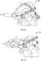
- FIG. 1Adepicts a side view of one variation of an anchor delivery catheter.
- FIG. 1Bdepicts a top view of the anchor delivery catheter of FIG. 1B .
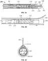
- FIG. 1Cdepicts a cross-sectional view of one region of an anchor delivery catheter.
- FIG. 1Ddepicts a cross-sectional view of another region of an anchor delivery catheter.
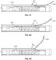
- FIGS. 1E and 1Fdepict the operation of a proximal portion of the anchor delivery catheter of FIG. 1A .
- FIG. 2Ais a top view of a distal portion of one variation of an anchor delivery catheter comprising a tissue depth indicator.
- FIG. 2Bis a side view of the distal portion of the anchor delivery catheter of FIG. 2A .
- FIG. 2Cis an end view of the anchor delivery catheter of FIG. 2A .
- FIG. 2Ddepicts the anchor delivery catheter of FIGS. 2A-2C with the depth indicator in a first configuration.
- FIG. 2Edepicts the anchor delivery catheter of FIGS. 2A-2C with the depth indicator in a second configuration.
- FIGS. 2F-2Hdepicts angular variations of the depth indicator in the first configuration.
- FIG. 3Ais a side view of a distal portion of another variation of an anchor delivery catheter comprising a tissue depth limiter in an expanded configuration.
- FIG. 3Bis a top view of the distal portion of the anchor delivery catheter of FIG. 3A .
- FIG. 3Cis an end view of the anchor delivery catheter of FIG. 3A .
- FIG. 3Ddepicts a close-up view of one variation of a rotatable attachment mechanism between the depth limiter and the elongate body of the delivery catheter.
- FIG. 3Eis a side view of a distal portion of another variation of the anchor delivery catheter of FIG. 3A , where the tissue depth limiter is in a compressed configuration.
- FIG. 4Ais a flowchart depiction of one variation of a method for delivering anchors using a delivery catheter comprising a depth indicator.
- FIG. 4Bis a flowchart depiction of another variation of method for delivering anchors using a delivery catheter comprising a depth indicator.
- FIGS. 5A-5Kdepict a short axis view of the left ventricle (LV), showing the aortic outflow tract and the LV chamber and the steps of a method for delivering tissue anchors using a delivery catheter comprising a depth indicator in accordance with the method depicted in FIG. 4A .
- LVleft ventricle
- FIGS. 6A and 6Bare schematic fluoroscopic depictions of a tissue depth indicator in a first configuration and a second configuration, respectively.
- Visualization of cardiac tissueis often complicated by the presence of blood and moving tissue, and as such, it can also be challenging to effectively deliver tissue anchors to a desired depth of into tissue.
- Penetration depth itselfmay be difficult to interpret, especially in diseased hearts or myocardium, which may have additional anatomical irregularities.
- the actual depth achieved for a given displacement of the delivery catheteris a function of apposition between a reference starting point and the endocardium, any tissue tenting, and surface topology or trabeculations.
- Each of these variablescan contribute significant challenges to accurately determining the actual penetration depth of the delivery catheter (and therefore, the actual delivery location of the anchor).
- the devices and methods described hereinallow the boundary of the cardiac surface to be visualized in a beating heart, and facilitate providing an indication as to the depth of penetration into that tissue by, for example, an anchor delivery catheter.
- the devices herein belowmay also limit the penetration depth of a delivery catheter tip beyond a preselected depth.
- an anchor delivery devicemay comprise an elongate body having a proximal end, a distal end, a first longitudinal lumen that terminates at a first distal opening located at the distal end and a second longitudinal lumen that terminates at a second distal opening.
- a tissue depth indicatormay be provided within the second longitudinal lumen. The tissue depth indicator may be inserted into the second longitudinal lumen during manufacturing or may be inserted by a practitioner just prior to inserting the delivery catheter into a patient.
- a tissue anchormay be disposed within the first longitudinal lumen, and may, for example, be located near a distal segment of the first longitudinal lumen.
- the anchormay be preloaded during manufacturing of the delivery catheter, or may be loaded by a practitioner just prior to inserting the delivery catheter into a patient.
- the anchormay be located just proximal to the first distal opening so that distally translating a pushing member within the first longitudinal lumen and contacting the anchor may push or advance the anchor out from the delivery catheter through the first distal opening.
- the tissue depth indicatormay be an elongate element, such as a wire or guidewire, that has a proximal portion and a distal portion, configured to be slidably disposed within the second longitudinal lumen of the elongate body.
- a tissue depth indicatormay be disposed within the first longitudinal lumen of the elongate body (i.e., in the same longitudinal lumen as the anchor).
- the proximal portionmay be stiffer than the distal portion, which may provide sufficient column strength so that the tissue depth indicator can be pushed from its proximal end and advanced through the second longitudinal lumen without looping or twisting.
- the distal portionmay be less stiff and/or more compliant than the proximal portion.
- the distal portion of the depth indicatormay be deflected, bent, angled, curved, bowed, and/or turned when it encounters a tissue surface. The compliant distal portion of the depth indicator may exit the second distal opening.
- the tissue depth indicatormay have two configurations.
- the distal portionmay form an obtuse angle with respect to the second longitudinal lumen and/or may extend along and/or toward the distal end of the elongate body.
- the distal tip of the tissue depth indicatormay point towards, or in the same direction as, the distal tip of the elongate body.
- the distal-most length of the depth indicatormay extend or track along the surface of a target tissue.
- the tissue depth indicatormay form an acute angle with respect to the second longitudinal lumen and/or may deflect backward away from the distal end of the elongate body and/or the surface of the target tissue.
- the distal portion of the depth indicator in the first configurationmay have a relatively smooth or gradual curve (i.e., a relatively large radius of curvature), without any acute or sharp curves or angles.
- a relatively smooth or gradual curvei.e., a relatively large radius of curvature
- the distal portion of the depth indicator in the second configurationmay comprise an inflection or discontinuity, such as a sharp curve, bend or angle, with a relatively abrupt change in curvature (i.e., a relatively small radius of curvature).
- This sharp bendmay be readily visible by various imaging techniques, including fluoroscopy or transesophageal echocardiogram (TEE), which may provide a visual signal or indication of when the depth indicator has transitioned from the first configuration to the second configuration.
- TEEtransesophageal echocardiogram
- the distance between the second distal opening (where the depth indicator exits the second longitudinal lumen) and the first distal opening may (where the anchor exits the first longitudinal lumen)may correspond to a preselected depth in tissue where the anchor is desired to be delivered.
- the length of the distal portion of the depth indicatormay be longer than the distance between the first and second distal openings, such that if desired, the tissue depth indicator may be advanced such it extends beyond the distal end of the elongate body.
- a tissue depth indicatormay comprise a radiopaque material.
- at least the distal portion of a depth indicator wiremay be radiopaque, while the proximal portion of the depth indicator may or may not be radiopaque.
- a depth indicator having at least a radiopaque distal portionmay allow the conformational changes of the distal portion to be visualized using fluoroscopy techniques. Changes in the geometry or orientation of the depth indicator (e.g., changes in the curves of a depth indicator wire) as it interacts with myocardial tissue may help a practitioner to identify the location of the tissue surface with respect to the delivery catheter. For example, as the distal tip of the delivery catheter is advanced towards and into the target tissue, the tissue depth indicator may be in the first configuration.
- the tissueWhen the distal tip of the delivery catheter reaches a desired, preselected tissue depth, the tissue may press against the tissue depth indicator, thereby deflecting it away from the tissue surface and transitioning it to the second configuration. In the second configuration, the depth indicator may deflect away from the tissue at a sharp curve or discontinuity, as previously described.
- a tissue depth indicatormay be made of one or more materials.
- a tissue depth indicatormay comprise a wire or guidewire where the proximal portion is made of small diameter Nitinol or stainless steel, and the distal, tissue-contacting portion is made of a coil of platinum, platinum-iridium, tungsten or gold wound around a core wire of Nitinol or stainless steel.
- the proximal and distal portions of the indicator wiremay be made of the same material(s), such as nickel titanium alloy, stainless steel, etc.
- a depth indicator wiremay have a radiopaque core (e.g., a nickel titanium alloy core, stainless steel core, or scitanium core) and a polymeric exterior.
- a depth indicator wiremay have a proximal portion having a stainless steel core and a distal portion having a nickel titanium alloy core, with either the same or different polymeric exterior.
- a depth indicator wiremay have a nickel titanium alloy core throughout its entire length, but the proximal portion may have a PTFE exterior while the distal portion may have a polymeric hydrophilic exterior.
- the overall length of the indicator wiremay be from about 120 cm to about 600 cm, e.g., from about 130 cm to about 300 cm, about 180 cm, about 195 cm, about 200 cm, about 300 cm, about 450 cm.
- the proximal portionmay have a length from about 115 cm to about 595 cm, e.g., about 145 cm.
- the distal portionmay have a length from about 1 cm to about 8 cm, e.g., about 2.5 cm, about 3 cm, about 3.5 cm, about 5 cm.
- the distal, tissue-contacting portionmay be relatively softer or more flexible, having, for example, a coil construction using a lower modulus material such as titanium, and stiffness only 5% to 50% as great as the proximal portion.
- the distal portionmay be about 5%, about 10%, about 25%, about 40%, about 50%, as stiff as the proximal portion.
- the diameter of the depth indicator wiremay be from about 0.005 in to about 0.050 in, e.g., about 0.008 in, about 0.010 in, about 0.012 in, about 0.014 in, about 0.018 in, about 0.035 in, etc.
- the distal portion of the depth indicator wiremay have a preformed curve (e.g., a J curve) while in other variations, the distal portion may not have a preformed curve.
- some anchor delivery cathetersmay comprise a tissue depth limiter, which may help to resist or prevent advancing a delivery catheter beyond a certain tissue depth. This may be a safety feature to help ensure that the delivery catheter does not puncture or cut through the target tissue.
- tissue depth limitermay help to resist or prevent advancing a delivery catheter beyond a certain tissue depth.
- Thismay be a safety feature to help ensure that the delivery catheter does not puncture or cut through the target tissue.
- theremay be a structure separate from the tissue depth indicator that resists or stops further advancement of the delivery catheter past a certain tissue depth while in other variations, the tissue depth indicator itself may resist advancement of the delivery catheter past a certain tissue depth.
- an anchor delivery cathetermay comprise an elongate body having a proximal end, a distal end, and a first longitudinal lumen that terminates at a first distal opening located at the distal end and a second longitudinal lumen that terminates at a second distal opening located proximal to the distal end.
- the tissue delivery cathetermay further comprise a tissue depth limiter disposed within the second longitudinal lumen.
- the tissue depth limitermay have a protrusion, such as a shoulder or curved surface, that may abut against tissue and resist distal travel of the delivery catheter into tissue.
- a tissue anchormay be disposed within the first longitudinal lumen, and may, for example, be located near a distal segment of the first longitudinal lumen.
- the anchormay be preloaded during manufacturing of the delivery catheter, or may be loaded by a practitioner just prior to inserting the delivery catheter into a patient.
- the anchormay be located just proximal to the first distal opening so that distally translating a pushing member and contacting the anchor may push or advance the anchor out from the delivery catheter through the first distal opening.
- a tissue depth limitermay comprise a first, low-profile configuration and a second, expanded configuration.
- a tissue depth limitermay comprise an elongate member, such as a wire (e.g., a flat wire), disposed within the second longitudinal lumen of the elongate body, where a distal portion of the elongate member exits the second distal opening and the distal tip of the elongate member is attached along an outer surface of the elongate body.
- the attachment location of the depth limitermay be at a preselected distance proximal to the distal end of the elongate body. In some variations, the preselected distance may correspond to the maximum tissue depth at which the anchor delivery catheter tip may be advanced.
- the distal-most end of a depth limitermay be rotatably attached to the elongate body.
- the distal-most end of a depth limitermay be coupled to an attachment member that is attached to the elongate body such that there is a rotational degree of freedom between the distal-most end of the depth limiter and the attachment member.
- the depth limitermay be movably coupled to the attachment member such that movement along the attachment member would cause the depth limiter to rotate around the elongate body.
- the distal-most end of the depth limiter and the attachment membermay be coupled as a ball-and-socket arrangement, which may allow the depth limiter to pivot around the attachment member.
- the depth limitermay be flush against the outer surface of the elongate body. This may be a desired configuration for navigating the anchor delivery catheter through the vasculature (and/or within the lumen of an outer catheter) before it reaches the mydocardium. Before the delivery catheter contacts the surface of the myocardium, the depth limiter may be transitioned to the second expanded configuration. The depth limiter may be expanded into the second configuration by distally advancing the depth limiter within the second longitudinal lumen. The distally-directed pushing force on the depth limiter wire may cause the distal portion of the wire to curve or rotate away from the outer surface of the elongate body, thereby having a profile and stiffness that may act as a shoulder or protrusion to abut against tissue.
- the distal portion of the depth limiter wiremay have a preformed or preshaped curve such that when the depth limiter wire is pushed distally, the distal portion is biased toward having the preformed or preshaped curve.
- at least the distal portion of the depth limiter wiremay be made of a shape memory and/or elastic material such that the natural or low-energy state is the expanded or curved shape, and withdrawing the depth limiter wire within a lumen constrains the wire in a straightened, high-energy state. Once the distal portion of depth limiter wire is advanced distally through the second longitudinal lumen and the second distal aperture, the depth limiter wire automatically transitions to the second, expanded configuration.
- At least a portion of the depth limiter in the expanded configurationis substantially perpendicular to the longitudinal axis of the delivery catheter and/or the surface of the target tissue.
- a shoulder of the depth limiterextends away from the longitudinal axis of the delivery catheter.
- the distal-most end of a tissue depth limitermay be rotatably or pivotably attached to the elongate body (e.g., along the outer surface of the elongate body).
- the distal portion of a tissue depth limitermay be more flexible than a proximal portion of the limiter to facilitate rotational or pivotal motion with respect to the elongate body. This may allow the depth limiter to rotate, pivot, or twist when it is in the expanded configuration.
- the degree and/or orientation direction of the rotationmay depend on, for example, the amount of force exerted on the limiter by the tissue as the practitioner advances the delivery catheter into the tissue. That is, the deeper the penetration, the more the depth limiter may rotate.
- the depth limitermay rotate anywhere from about 1 degree to about 180 degrees, e.g., about 45 degrees, about 90 degrees, etc.
- the rotation of the depth limitermay provide a visual signal (in addition or alternatively to a tactile signal) that a preselected depth into tissue has been attained.
- rotation of the depth limiter by about 90 degreesmay provide a distinct visual cue (e.g., the limiter sweeping out to have a larger cross-sectional area or sweeping inward to have a smaller cross-sectional area) that the delivery catheter tip is at the preselected depth and/or that the depth limiter is pressed against the tissue surface.
- the conformational, rotational and/or orientation changes of the limitercan be visualized using fluoroscopy and/or transesophageal echocardiogram techniques.
- the distal-most end of a tissue depth limitermay be fixedly attached along the length of the elongate body.
- the distal-most endmay be attached to the elongate body by welding, soldering, and the like, and/or one or more adhesives.
- the distal-most end of the limitermay be attached to the elongate body by any suitable rotational mechanisms, including, but not limited to, hinges, pivots, ball-and-socket joints, ball bearings, and the like.
- the distal-most end of the depth limitermay comprise a loop and the rotatable attachment mechanism may comprise a curved wireform shaft or pin attached along the outer surface of the elongate body.
- the curved wireform shaft or pinmay be in the form of a ring, or a partial ring (e.g., a U-shaped curve where the two ends are attached to the elongate body).
- the ring and the loopmay mutually engage, thereby allowing the depth limiter to rotatably slide along the curve of the ring.
- the depth limitermay sweep about 90 degrees counterclockwise with respect to a vertical axis perpendicular to the longitudinal axis of the elongate body and/or about 90 degrees clockwise with response to the vertical axis, depending on the direction and magnitude of force applied on the depth limiter by the surface of the target tissue.
- the distance of the attachment mechanism from the distal end of the elongate bodymay correspond to a preselected tissue penetration depth. For example, if it is desired that the tip of the anchor delivery catheter is not to be advanced past a tissue depth of about 6 mm, then the attachment mechanism may be located at about 6 mm away from the distal end of the elongate body.
- the tissue depth limitermay be made of any of the materials described above for the tissue depth indicator.
- the stiffness of the tissue depth limiter constructed from a Nitinol flat wire of width of about 0.006 in and thickness of about 0.011 inchesmay be about 0.04 lbf*in 2 (275 Nmm 2 ).
- the tissue depth limitermay comprise a flattened nickel titanium alloy wire, while in other variations, the tissue depth limiter may comprise a hypodermic tube.
- a depth limiter comprising a flattened wiremay have a width from about 0.010 in to about 0.04 in, e.g., about 0.015 in, about 0.025 in, about 0.0.030 in, about 0.035 in, etc., and a thickness from about 0.005 in to about 0.015 in.
- the second longitudinal lumen of the elongate body within which the depth limiter is disposedmay have a width of about 0.0.018 in to about 0.043 in, e.g., about 0.02 in, about 0.025 in, about 0.04 in, etc.
- the overall height of a distal section of the anchor delivery catheter(i.e., the sum of the diameter of the elongate body and the second longitudinal lumen) may be from about 0.06 in to about 0.10 in for example, about 0.09 in to about 0.11 in, e.g., about 0.098 in, about 0.1 in, etc.
- the overall height of the distal section of the anchor delivery catheter(i.e., the sum of the diameter of the elongate body and the height of the expanded depth limiter) may be from about 0.15 in to about 0.35 in for example, about 0.2 in to about 0.5 in, e.g., about 0.283 in, about 0.3 in, about 0.38 in, about 0.45 in, etc.
- any of the anchor delivery catheters comprising a tissue depth indicator and/or a tissue depth limiter described hereinmay further comprise a push member, such as a push tube, within the first longitudinal lumen of the elongate body to deploy an anchor disposed within that lumen.
- the elongate bodymay optionally have one or more curves, where the one or more curves define one or more distinct planes that may be located at one or more angles with respect to each other.
- the elongate body of the anchor delivery cathetermay be steerable. The actuation of the push member, along with the control of the tissue depth indicator and/or tissue depth limiter, and/or any steering mechanisms of the delivery catheter, may be controlled at a proximal handle of the delivery catheter.
- one or more tethers or suturesmay be threaded through the anchor to be delivered (e.g., where the implanted device comprises a series of tethered anchors), and the proximal end of the one or more tethers or sutures may be coupled to the proximal handle of the delivery catheter.
- the proximal handlemay comprise a suture holder that is configured to releasably retain a suture, a push tube actuator, and a tissue depth indicator port.
- the proximal handlemay comprise a tissue depth limiter port.
- a practitionermay control the length of the depth limiter or indicator that exits the distal opening of the second longitudinal lumen by advancing or retracting/withdrawing the proximal portion of the limiter and/or indicator at these proximal ports.
- the location of the depth indicator and/or limitermay be locked at a proximal portion.
- a tissue depth indicator wiremay be withdrawn entirely from the delivery catheter.
- the push tube actuatormay comprise a locking mechanism so that the position of the push tube may be secured once it has been advanced to the desired location. Any portion of these components may be radiopaque, as may be desirable for fluoroscopic monitoring of the progress of the procedure.
- the distal tip of the elongate body and/or a distal length of the tissue depth indicator and/or limitermay be radiopaque.
- any of the anchor delivery catheters described hereinmay also comprise a push tube stop structure within the first longitudinal lumen of the elongate body.
- the push tube stop structuremay prevent the push tube from being over-advanced, e.g., advanced out of the elongate body.
- an anchor delivery cathetermay be one catheter in a system of catheters used in a multi-step intravascular procedure. In these procedures, an anchor delivery catheter may be advanced within the lumen of one or more other catheters, and some anchor delivery catheters may comprise features to limit relative motion between nested catheter elements, as well as to help direct orientation of the anchor delivery catheter with respect to outer catheter elements.
- an anchor delivery cathetermay optionally comprise stop elements that limit its travel within a guide catheter and/or a multi-window catheter so that the length of the delivery catheter that extends out from these catheters is restricted.
- These stop elementsmay be external to the anchor delivery catheter, but internal to the outer guide catheter and/or multi-window guide catheter (e.g., the stop elements do not contact the target tissue).
- a stop elementmay be a flat ribbon, wire loop, spring, protrusion, wing or petal. Additional details regarding anchor delivery catheters with stop elements that limit its travel within a guide catheter and/or multi-window catheter without contacting tissue are provided in co-pending U.S. Pat. Appln. Pub. No. 2014/0142619, filed Oct. 11, 2013, which is hereby incorporated by reference in its entirety.
- Anchor delivery catheter 100may comprise a proximal handle 102 , an elongate body 104 , and a tissue depth indicator 106 .
- the distal section 108 of the elongate body 104may comprise one or more curves.
- the proximal handle 102may comprise a deployment handle 110 , a separable suture holder 112 configured to retain a suture 113 , a push tube 114 , a safety retainer clip 117 , and a tissue depth indicator port 116 .
- the proximal handle 102may also comprise a touhy borst valve 118 with a flush port.
- the elongate body 104comprises a first longitudinal lumen 120 and a second longitudinal lumen 122 .
- the first longitudinal lumen 120may extend from the proximal handle (e.g., in communication with a port 115 through which the push tube 114 is inserted) and terminate at a distal opening 105 .
- the second longitudinal lumenmay extend from the proximal handle (e.g., in communication with depth indicator port 116 ) and terminate at a second distal opening 125 ( FIG. 1A ).
- the push tube 114is disposed within the first longitudinal lumen 120 and is configured to contact an anchor (not shown) in order to deploy it from a distal opening 105 of the elongate body 104 .
- the push tube 114may be coupled to the deployment handle 110 such that manual actuation of the handle 110 distally translates the push tube 114 to deploy the anchor, as depicted in FIGS. 1E and 1F .
- FIG. 1Edepicts the deployment handle 110 prior to anchor deployment (e.g., the anchor-stowed configuration).
- FIG. 1Fdepicts the deployment handle 110 as it is depressed by a practitioner to deploy the anchor (e.g., the anchor-deployed configuration).
- the second longitudinal lumen 112has a second longitudinal lumen 112 that is located within the boundaries of an outer wall 107 of the elongate body 104 , the second longitudinal lumen may be located outside of the outer wall of the elongate body.
- the variation depicted in FIG. 1Dhas a second longitudinal lumen 122 ′ that is located external to the outer wall 107 ′ of the elongate body 104 ′.
- the arrangement of the first and second longitudinal lumens depicted in FIG. 1Dmay be used at a distal-most section of the elongate body (e.g., the section of the elongate body that may contact and/or penetrate tissue) while the arrangement of the first and second longitudinal lumens depicted in FIG. 1C may be used at a more proximal section of the elongate body.
- FIGS. 2A-2Hdepict one variation of an anchor delivery catheter comprising a tissue depth indicator.
- Anchor delivery catheter 200comprises an elongate body 204 having a first longitudinal lumen 206 that terminates at a distal opening 208 .
- An anchor 207may be disposed within the first longitudinal lumen 206 such that it exits the distal opening 208 when deployed.
- a push tube 210may located proximal to the anchor 207 and may be distally advanced to contact and deploy the anchor.
- a push tube stop member 211may be disposed within the first longitudinal lumen 206 to limit the distal travel of the push tube 210 .
- the push tube 210may comprise a radiopaque marker band (not shown) that circumscribes the tube.
- the outer diameter of the markermay be larger than the inner diameter of stop member 211 such that when the push tube has been advanced to a selected distal location, the marker band abuts the stop member 211 .
- the marker bandmay be located about 13 mm proximally from the distal end of the push tube 210 .
- the elongate body 204may optionally comprise a side slot 212 in communication with the first longitudinal lumen 206 and distal opening 208 .
- the anchor 207may comprise a loop or eyelet 213 through which a tether or suture may be disposed.
- the anchormay be one of a plurality of tethered anchors in a tethered anchor assembly, and tensioning the tether across the anchors may cause the length of the tissue to which these anchors are attached to shorten.
- the anchor 207may be oriented within the first longitudinal lumen 206 such that the anchor loop 213 is aligned with the opening of the slot 212 , which may help to facilitate threading of a tether (not shown) therethrough.
- the elongate body 204may also comprise a second longitudinal lumen 214 , which may extend from the proximal handle and terminate at a second distal opening 216 . In some variations, the edge of the distal opening 216 may be beveled at an angle.
- a beveled angle at any lumen openingmay help to provide an atraumatic leading edge to the second longitudinal lumen.
- a tissue depth indicator 230may be disposed within the second longitudinal lumen 214 and a distal portion of the depth indicator may exit the second distal opening 216 .
- the length of the depth indicator that exits and/or extends from the second distal openingmay be varied by advancing or retracting the depth indicator at the proximal handle.
- the second longitudinal lumen 214may have a diameter from about 0.012 in to about 0.02 in, e.g., about 0.018 in, which may be selected based on the diameter of the tissue depth indicator.
- the location of the second distal opening 216may be determined at least in part by the desired penetration depth of the delivery catheter tip (i.e., the desired anchor deployment depth into tissue).
- the distance D 1 between the first distal opening 208 and second distal opening 216may correspond to the desired penetration depth and/or anchor delivery depth.
- the desired penetration depth and/or anchor delivery depthmay vary according to a number of factors, include the targeted tissue region, the anchor depth required to withstand pull-out forces to which the anchor may be subjected during/after implantation, toughness of the tissue, thickness of the tissue, etc.
- the desired penetration and/or anchor delivery depthmay be from about 1 mm to about 15 mm, e.g., about 3 mm, about 4 mm, about 5.5 mm, about 6 mm, about 8 mm, about 10 mm, about 12.5 mm, etc.
- distance D 1may vary from about 1 mm to about 15 mm, e.g., about 3 mm, about 4 mm, about 5.5 mm, about 6 mm, about 8 mm, about 10 mm, about 12.5 mm, etc.
- FIGS. 2D and 2Edepict one variation of a tissue depth indicator 230 having a distal portion 234 that is more compliant (e.g., softer, less stiff) than a proximal portion (view blocked by the wall of the second longitudinal lumen) of the indicator.
- the tissue depth indicator 230may comprise an indicator wire with a distal portion 234 that is more compliant than its proximal portion.
- the indicator wire 230may have a first configuration ( FIG. 2D ) and a second configuration ( FIG. 2E ).
- the angle A 1 between the indicator wire 230 and the second longitudinal lumen 214may vary between the two configurations.
- the vertex 232 of the angle A 1may be co-localized with the second distal opening 216 (e.g., the location where the indicator wire exits the second distal opening).
- the angle A 1may be obtuse, and in the second configuration, the angle A 1 may be acute.
- the angle A 1 in the first configurationmay be from about 120 degrees to about 180 degrees (see FIGS. 2D, 2F-2H ), while the angle A 1 in the second configuration may be from about 80 degrees to about 120 degrees (see FIG. 2E ).
- the curvature of the indicator wire in the second configurationhas a discontinuity or inflection at the point of exit that is not present in the first configuration.
- the indicator wire 230may be in the first configuration when the distal tip (i.e., the distal opening 208 ) of anchor delivery catheter is not located at the desired tissue depth.
- the distal portion 234 of the indicator wire 230may extend past the distal end of the elongate body, may contact a length of the distal section of the elongate body, and/or may generally have smoother curves (i.e., the radius of the curvature does not change abruptly).
- the flexibility of the distal portion 234allows the indicator wire to interrogate, track along, delineate, or otherwise be urged along the surface of the target tissue without puncturing or abrading the tissue.
- the curvature of a distal portion that comprises a radiopaque materialmay be visualized using fluoroscopic techniques, and may allow a practitioner to identify the boundary of the tissue surface, along with the topology of the tissue surface. For example, curvature of the distal portion along the surface of the tissue may also delineate any irregularities, protrusions, trabeculae, etc. on the surface of the myocardium. Advancing the distal portion 234 of the indicator wire 230 ahead of the delivery catheter may facilitate the navigation of the delivery catheter towards the tissue surface. It may also help facilitate the proper orientation of the delivery catheter with respect to the tissue surface.
- the delivery cathetermay be navigated so that the direction of its travel is substantially oriented at an angle of 20 to 80 degrees, e.g., about 30 degrees to about 45 degrees with respect to the surface of the target tissue as delineated by the distal portion of the indicator wire.
- the tip of the delivery cathetermay penetrate more deeply into the tissue, and when the penetration depth is approximately the same as the distance D 1 , the tissue surface may press against the compliant distal portion of the indicator, thereby causing it to deflect away from the tissue surface and/or the distal end of the elongate body (i.e., the depth indicator transitions from the first configuration to the second configuration).
- This deflectionresults in an acute change or discontinuity in the curvature of the distal portion, it is readily visualized using fluoroscopic techniques.
- This conformational changeprovides a visual indication to the practitioner that the distal tip of the delivery catheter has penetrated the tissue at a depth that is approximately the same as the distance D 1 .
- the anchormay then be deployed from the delivery catheter.
- an anchor delivery cathetermay alternatively or additionally comprise a tissue depth limiter, which may resist penetration of the distal tip of the delivery catheter past a preselected depth. This may provide a tactile signal to a practitioner that a desired (or maximum) penetration depth has been attained.
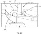
- FIGS. 3A-3Ddepict one variation of an anchor delivery catheter 300 comprising an elongate body 304 and a tissue depth limiter 330 .
- the elongate body 304may have a first longitudinal lumen 306 that extends from the proximal handle and terminates at a first distal opening 308 and a second longitudinal lumen 314 that extends from the proximal handle and terminates at a second distal opening 316 .
- a tissue depth limiter 330may be disposed within the second longitudinal lumen 314 and a distal portion 331 of the depth limiter 330 may exit the second distal opening 316 .
- the distal end 333 of the depth limiter 330may be attached to the elongate body 304 .
- the distance D 2 between the attachment location of the depth limiter 330 and the distal tip/opening of the elongate bodymay correspond to the desired or maximum tissue penetration depth.
- Distance D 1may be, for example, from about 1 mm to about 15 mm, e.g., about 3 mm, about 4 mm, about 5.5 mm, about 6 mm, about 8 mm, about 10 mm, about 12.5 mm, etc.
- the distal portion 331 of the depth limitermay comprise a preformed curve, while in other variations, the distal portion does not have a preformed curve.
- the depth limiter 330may have a first collapsed configuration ( FIG. 3E ) and a second expanded configuration ( FIG. 3A ). In the first configuration, the depth limiter may be retracted proximally such that the distal portion is substantially flush with (e.g., extends along) the outer surface 305 of the elongate body 304 . In the second configuration, the depth limiter may be advanced distally such that a longer length of the distal portion exits the second distal opening and allows for the distal portion to form a curve or loop, as shown in FIG. 3A .
- the distal portion 331may have a preformed curve that automatically expands when a sufficient length of the depth limiter has exited the second longitudinal lumen.
- the depth limiter 330may comprise a shape-memory material that is preformed with a distal curve. In the first configuration, the depth limiter is compressed to a straightened form within the second longitudinal lumen. In the second configuration, the distal portion with the preformed curve automatically assumes its expanded shape when it exits the second distal opening and is released from the second longitudinal lumen.
- the shape of the atraumatic curvemay be selected such that a larger surface of the curve is located generally perpendicularly to the longitudinal axis of the elongate body.
- a depth limitermay comprise a flat wire that may help to provide resistance to forward travel while reducing the likelihood of cutting through the tissue surface.
- the distal end of the depth limitermay be attached to the elongate body of the delivery catheter by any suitable method.
- the distal endmay be attached to the elongate body (e.g., the outer surface of the elongate body) by welding, soldering, and the like, and/or using one or more adhesives.
- These types of attachment mechanismsmay allow the distal end of the depth limiter to be rigidly attached to the elongate body.
- other types of attachment mechanismsmay allow the distal end of the depth limiter to pivot, rotate, slide and/or otherwise deflect with respect to the elongate body.
- a rotatable attachment mechanism 332is depicted in FIG.
- the attachment mechanism 332may comprise a curved pin/shaft or ringed structure 336 disposed within a groove 305 of the elongate body 304 .
- the ringed structure 336may be a closed ring or an open ring constructed from a single component (e.g., a unibody component having a C-shape or a U-shape) or two components (e.g., two L-shaped components that are joined together).
- the distal end 333 of the depth limiter 330may comprise a loop 334 , which may be slidably or rotatably coupled with the ringed structure 336 .
- the loopmay be integrally formed with the proximal portion of the depth limiter 330 , while in other variations, the loop may be a hypodermic tubing that may be attached to the proximal portion of the depth limiter.
- the loop of a depth limitermay be formed from a hypodermic tube may have an inner diameter of about 0.008 in, an outer diameter of about 0.016 in, and a wall thickness of about 0.004 in that is soldered or welded to a proximal portion of the depth limiter.
- the loop 334 of the depth limitermay track around the circumference of the ringed structure 336 counterclockwise or clockwise (depicted in FIG. 3C along arrows 340 L and 340 R, respectively).
- the plane defined by the curve of the distal portion 331may sweep between about 1 degree to about 90 degrees counterclockwise, and/or may sweep between 1 degree to about 90 degrees clockwise.
- the rotation or pivoting of the curved distal portionmay provide a visual indication (in addition to a tactile indication) that a preselected tissue depth has been attained.
- these devices and methodsmay be used to deliver anchors to a particular depth in the endocardium of the left ventricle during a beating heart procedure.
- One example of a method for visualizing and determining the depth of delivery catheter penetration into tissueis outlined in the flow diagram of FIG. 4A and depicted in FIGS. 5A-5K .
- the method 400is one in which tissue anchors are delivered to endocardium of the left ventricle (LV), though it should be appreciated that this method may be employed in many other procedures as well.
- Such methodmay comprise using fluoroscopy imaging to locate the myocardium, position the catheters, and deliver one or more tethered anchors. Fluoroscopic images or video may be taken from a short axis view of the LV or other views of the LV.
- FIG. 5Ashows the short axis of the left side of the heart 10 with the surrounding myocardium 11 , endocardium 12 , LV chamber 13 and aortic outflow tract and aortic valve 14 .
- the method 400may comprise advancing a guide catheter (step 402 ) to subvalvular tissue in the LV.
- FIG. 5Bdepicts a guide catheter 20 that may extend across the aortic valve (AV) and tangent to the LV wall, with a distal opening 21 that may be inserted across the aortic valve 14 and placed tangent to the endocardium 12 .
- the method 400may then comprise advancing a tunnel catheter through the guide catheter (step 404 ) such that a length of the tunnel catheter is positioned against or near the endocardium.
- FIG. 5Cdepicts placing a tunnel catheter 30 deployed from the guide catheter against or near the endocardium 12 .
- the tunnel cathetermay extend around and alongside the LV wall.
- Radiopaque markers and windowsmay be disposed along the outer radius of the tunnel catheter wall, through which delivery catheters can be deployed.
- the tunnel cathetermay be a template device, using windows 31 and radiopaque markers 32 to direct the placement of devices, such as anchors, into the myocardium of the LV.
- the method 400may comprise advancing a delivery catheter through the tunnel catheter (step 406 ) to a selected window of the tunnel catheter.
- FIGS. 5D-5Gthe method would comprise the steps depicted in FIGS. 5D-5G .
- a delivery catheter 40may be advanced through the tunnel catheter 30 such that the distal tip 41 exits a preferred window 31 and contacts endocardium 12 .
- Advancing delivery catheter 40further causes it to penetrate endocardium 12 to a desired depth.
- FIG. 5Eschematically depicts a fluoroscopic image of the guide catheter, tunnel catheter and delivery catheter positioned along endocardium of the LV.
- the information revealed/depicted in a fluoroscopic imageis relatively sparse, especially with respect to tissue location (e.g., boundary of the myocardium surface) and depth of penetration.
- the next stepmay comprise advancing the tissue depth indicator out of a lumen of the delivery catheter (step 408 ) ahead of the distal tip of the delivery catheter, as depicted in FIG. 5H .
- FIG. 5Hdepicts a delivery catheter comprising a radiopaque depth indicator wire 70 .
- Depth indicator wire 70may be translatable to extend distally or proximally with respect to the distal end 41 of delivery catheter 40 , though in some variations the depth indicator wire may be fixed and not translatable along the longitudinal axis of the delivery catheter.
- the method 400may comprise delineating the boundary of the ventricular wall or myocardium surface using the depth indicator wire (step 410 ).
- the depth indicator wiremay be in the first configuration, as described previously and also depicted in FIG. 6A .
- the depth indicator wiremay be used to interrogate the surface of endocardium 12 and may provide a durable localization and visualization of endocardium 12 where distal tip 41 penetrates.
- the delineation of the location and/or surface textures or structures by the deflections and curves of the depth indicator wiremay be evident in the resulting fluoroscopic image ( FIG. 5I ). This may facilitate the identification of the location of endocardium 12 .
- the method 400may then comprise advancing the delivery catheter such that the distal tip of the delivery catheter penetrates through the endocardium and into the myocardium (step 412 ).
- the depth indicator wiremay transition to the second configuration. That is, an inflection or discontinuity may be introduced in the depth indicator wire resulting from the tissue pushing on the depth indicator wire, thereby deflecting the distal portion of the wire away from the tissue surface.
- FIGS. 5J and 6Bschematically depict the distal segment 81 of the lumen in which depth indicator wire 70 , and its location relative to distal tip 41 of the delivery catheter 40 , as well as the inflection or discontinuity 71 in the curvature of the distal portion of the indicator wire.
- FIG. 5Kis a depiction of the fluoroscopic image of the arrangement in FIG. 5J .
- the practitionermay stop advancing the delivery catheter into the endocardium (step 414 ) and deliver the anchor into the endocardium (step 416 ).
- all of the anchorsmay be delivered with the guidance provided by a tissue depth indicator.
- the first and second anchors depicted in FIGS. 5D-5Gmay be deployed into tissue using method 400 .
- fluoroscopic imagesmay be acquired at any point during method 400 , from any view (e.g., short axis view (SAX), long axis view (LAX), A/P view, oblique views, etc.).
- the method 400uses the anchor delivery catheter with additional catheters (e.g., a guide catheter and a tunnel catheter), it should be understood that the anchor delivery catheter may be used to deliver anchors without other catheters, with fewer catheter, or with more catheters.
- One example of a method for delivering anchorsis depicted in FIG. 4B .
- the method 420may comprise advancing a delivery catheter to the surface of the target tissue (step 422 ), advancing the depth indicator out of a lumen of the delivery catheter ahead of the distal tip of the delivery catheter (step 424 ), delineating the boundary of the surface of the target tissue using the depth indicator (step 426 ), and advancing the delivery catheter to penetrate the target tissue (step 428 ) until a desired penetration depth is reached.
- the method 420may then comprise stopping the advancement of the delivery catheter when the distal portion of the depth indicator deflects away from the surface of the target tissue (i.e., transitions from the first configuration to the second configuration, which has an inflection or discontinuity) and delivering the anchor into the tissue at the preselected depth.
- fluoroscopic imagesmay be acquired at any point during method 420 , from any view.
- An anchor delivery cathetercomprising a tissue depth limiter may also be used to perform the methods of FIGS. 4A and 4B .
- the tissue depth limitermay not be capable of delineating the boundary of the surface of the target tissue. Instead, prior to contacting the tissue, the depth limiter may be transitioned from the first collapsed configuration to the second expanded configuration. In the expanded configuration, the depth limiter may provide a tactile signal that the tip of the delivery catheter has attained a preselected or maximum penetration depth.
- an anchor delivery cathetercomprising a tissue depth limiter that has a distal end rotatably attached to the elongate body of the delivery catheter may provide a visual cue that the preselected penetration depth has been attained.
- the rotation of the depth limiter when it abuts the target tissue surfacemay provide a visual change in a fluoroscopic image that indicates a desired or maximum penetration depth has been attained.
- fluoroscopic imagesmay be acquired at any point during these procedures, from any view.
- kitscomprising an anchor delivery catheter and a tissue depth indicator and/or tissue depth limiter.
- a kitmay comprise an anchor delivery catheter comprising an elongate body with a first longitudinal lumen terminating at a first distal opening and a second longitudinal lumen terminating at a second distal opening.
- the kitmay further comprise a depth indicator wire configured to be disposed within the second longitudinal lumen of the elongate body, where the depth indicator wire may have a proximal portion and a distal portion that is relatively more compliant or flexible than the proximal portion.
- the depth indicator wiremay be pre-assembled during manufacturing so that it is disposed within the second longitudinal lumen, or may be kept separate from the elongate body and inserted by the practitioner just prior to use.
- the kitmay comprise an anchor disposed within the first longitudinal lumen.
- a kitmay comprise an anchor delivery catheter comprising an elongate body with a first longitudinal lumen terminating at a first distal opening and a second longitudinal lumen terminating at a second distal opening, and an anchor disposed within the first longitudinal lumen.
- the tissue depth indicatormay be in a separate kit.
- a kitmay comprise an anchor delivery catheter comprising an elongate body with a first longitudinal lumen terminating at a first distal opening and a second longitudinal lumen terminating at a second distal opening and a tissue depth limiter disposed within the second longitudinal lumen and attached at a distal end of the elongate body.
- the kitmay or may not include an anchor disposed within the first longitudinal lumen.
Landscapes
- Health & Medical Sciences (AREA)
- Surgery (AREA)
- Life Sciences & Earth Sciences (AREA)
- Medical Informatics (AREA)
- Animal Behavior & Ethology (AREA)
- Engineering & Computer Science (AREA)
- Biomedical Technology (AREA)
- Heart & Thoracic Surgery (AREA)
- Veterinary Medicine (AREA)
- Molecular Biology (AREA)
- Nuclear Medicine, Radiotherapy & Molecular Imaging (AREA)
- General Health & Medical Sciences (AREA)
- Public Health (AREA)
- Rheumatology (AREA)
- Oral & Maxillofacial Surgery (AREA)
- Pathology (AREA)
- Media Introduction/Drainage Providing Device (AREA)
- Apparatus For Radiation Diagnosis (AREA)
Abstract
Description
Claims (27)
Priority Applications (3)
| Application Number | Priority Date | Filing Date | Title |
|---|---|---|---|
| US16/058,847US10980529B2 (en) | 2015-03-05 | 2018-08-08 | Devices and methods of visualizing and determining depth of penetration in cardiac tissue |
| US17/221,294US12102316B2 (en) | 2015-03-05 | 2021-04-02 | Devices and methods of visualizing and determining depth of penetration in cardiac tissue |
| US18/823,227US20250134514A1 (en) | 2015-03-05 | 2024-09-03 | Devices and methods of visualizing and determining depth of penetration in cardiac tissue |
Applications Claiming Priority (3)
| Application Number | Priority Date | Filing Date | Title |
|---|---|---|---|
| US201562128628P | 2015-03-05 | 2015-03-05 | |
| US15/061,748US10058321B2 (en) | 2015-03-05 | 2016-03-04 | Devices and methods of visualizing and determining depth of penetration in cardiac tissue |
| US16/058,847US10980529B2 (en) | 2015-03-05 | 2018-08-08 | Devices and methods of visualizing and determining depth of penetration in cardiac tissue |
Related Parent Applications (1)
| Application Number | Title | Priority Date | Filing Date |
|---|---|---|---|
| US15/061,748ContinuationUS10058321B2 (en) | 2015-03-05 | 2016-03-04 | Devices and methods of visualizing and determining depth of penetration in cardiac tissue |
Related Child Applications (1)
| Application Number | Title | Priority Date | Filing Date |
|---|---|---|---|
| US17/221,294ContinuationUS12102316B2 (en) | 2015-03-05 | 2021-04-02 | Devices and methods of visualizing and determining depth of penetration in cardiac tissue |
Publications (2)
| Publication Number | Publication Date |
|---|---|
| US20190008505A1 US20190008505A1 (en) | 2019-01-10 |
| US10980529B2true US10980529B2 (en) | 2021-04-20 |
Family
ID=56848322
Family Applications (4)
| Application Number | Title | Priority Date | Filing Date |
|---|---|---|---|
| US15/061,748ActiveUS10058321B2 (en) | 2015-03-05 | 2016-03-04 | Devices and methods of visualizing and determining depth of penetration in cardiac tissue |
| US16/058,847Active2036-12-31US10980529B2 (en) | 2015-03-05 | 2018-08-08 | Devices and methods of visualizing and determining depth of penetration in cardiac tissue |
| US17/221,294Active2037-04-20US12102316B2 (en) | 2015-03-05 | 2021-04-02 | Devices and methods of visualizing and determining depth of penetration in cardiac tissue |
| US18/823,227PendingUS20250134514A1 (en) | 2015-03-05 | 2024-09-03 | Devices and methods of visualizing and determining depth of penetration in cardiac tissue |
Family Applications Before (1)
| Application Number | Title | Priority Date | Filing Date |
|---|---|---|---|
| US15/061,748ActiveUS10058321B2 (en) | 2015-03-05 | 2016-03-04 | Devices and methods of visualizing and determining depth of penetration in cardiac tissue |
Family Applications After (2)
| Application Number | Title | Priority Date | Filing Date |
|---|---|---|---|
| US17/221,294Active2037-04-20US12102316B2 (en) | 2015-03-05 | 2021-04-02 | Devices and methods of visualizing and determining depth of penetration in cardiac tissue |
| US18/823,227PendingUS20250134514A1 (en) | 2015-03-05 | 2024-09-03 | Devices and methods of visualizing and determining depth of penetration in cardiac tissue |
Country Status (5)
| Country | Link |
|---|---|
| US (4) | US10058321B2 (en) |
| EP (2) | EP4292551B1 (en) |
| AU (2) | AU2016226023B2 (en) |
| CA (1) | CA2978599C (en) |
| WO (1) | WO2016141358A1 (en) |
Cited By (1)
| Publication number | Priority date | Publication date | Assignee | Title |
|---|---|---|---|---|
| US20210361278A1 (en)* | 2015-03-05 | 2021-11-25 | Ancora Heart, Inc. | Devices and methods of visualizing and determining depth of penetration in cardiac tissue |
Families Citing this family (42)
| Publication number | Priority date | Publication date | Assignee | Title |
|---|---|---|---|---|
| US8911494B2 (en) | 2009-05-04 | 2014-12-16 | Valtech Cardio, Ltd. | Deployment techniques for annuloplasty ring |
| US10517719B2 (en) | 2008-12-22 | 2019-12-31 | Valtech Cardio, Ltd. | Implantation of repair devices in the heart |
| WO2010073246A2 (en) | 2008-12-22 | 2010-07-01 | Valtech Cardio, Ltd. | Adjustable annuloplasty devices and adjustment mechanisms therefor |
| WO2010085456A1 (en) | 2009-01-20 | 2010-07-29 | Guided Delivery Systems Inc. | Anchor deployment devices and related methods |
| US9968452B2 (en) | 2009-05-04 | 2018-05-15 | Valtech Cardio, Ltd. | Annuloplasty ring delivery cathethers |
| US10098737B2 (en) | 2009-10-29 | 2018-10-16 | Valtech Cardio, Ltd. | Tissue anchor for annuloplasty device |
| US9307980B2 (en)* | 2010-01-22 | 2016-04-12 | 4Tech Inc. | Tricuspid valve repair using tension |
| US10792152B2 (en) | 2011-06-23 | 2020-10-06 | Valtech Cardio, Ltd. | Closed band for percutaneous annuloplasty |
| EP3345573B1 (en) | 2011-06-23 | 2020-01-29 | Valtech Cardio, Ltd. | Closure element for use with annuloplasty structure |
| US8858623B2 (en) | 2011-11-04 | 2014-10-14 | Valtech Cardio, Ltd. | Implant having multiple rotational assemblies |
| EP2881083B1 (en) | 2011-12-12 | 2017-03-22 | David Alon | Heart valve repair device |
| EP2911593B1 (en) | 2012-10-23 | 2020-03-25 | Valtech Cardio, Ltd. | Percutaneous tissue anchor techniques |
| WO2014064694A2 (en) | 2012-10-23 | 2014-05-01 | Valtech Cardio, Ltd. | Controlled steering functionality for implant-delivery tool |
| EP2961351B1 (en) | 2013-02-26 | 2018-11-28 | Mitralign, Inc. | Devices for percutaneous tricuspid valve repair |
| US10449333B2 (en) | 2013-03-14 | 2019-10-22 | Valtech Cardio, Ltd. | Guidewire feeder |
| CN105283214B (en) | 2013-03-15 | 2018-10-16 | 北京泰德制药股份有限公司 | Translate conduit, system and its application method |
| US10070857B2 (en) | 2013-08-31 | 2018-09-11 | Mitralign, Inc. | Devices and methods for locating and implanting tissue anchors at mitral valve commissure |
| WO2015059699A2 (en) | 2013-10-23 | 2015-04-30 | Valtech Cardio, Ltd. | Anchor magazine |
| CN107847320B (en) | 2015-04-30 | 2020-03-17 | 瓦尔泰克卡迪欧有限公司 | Valvuloplasty techniques |
| US10828160B2 (en) | 2015-12-30 | 2020-11-10 | Edwards Lifesciences Corporation | System and method for reducing tricuspid regurgitation |
| US11833034B2 (en) | 2016-01-13 | 2023-12-05 | Shifamed Holdings, Llc | Prosthetic cardiac valve devices, systems, and methods |
| GB201611910D0 (en) | 2016-07-08 | 2016-08-24 | Valtech Cardio Ltd | Adjustable annuloplasty device with alternating peaks and troughs |
| WO2018208827A1 (en)* | 2017-05-08 | 2018-11-15 | Crossfire Medical Llc | Catheter systems and methods for ablating varicose veins |
| US11135062B2 (en) | 2017-11-20 | 2021-10-05 | Valtech Cardio Ltd. | Cinching of dilated heart muscle |
| US11173030B2 (en) | 2018-05-09 | 2021-11-16 | Neochord, Inc. | Suture length adjustment for minimally invasive heart valve repair |
| EP3820406B1 (en) | 2018-07-12 | 2023-12-20 | Edwards Lifesciences Innovation (Israel) Ltd. | Annuloplasty systems and locking tools therefor |
| AU2019325548B2 (en) | 2018-08-21 | 2025-06-26 | Shifamed Holdings, Llc | Prosthetic cardiac valve devices, systems, and methods |
| CN113260337A (en) | 2018-10-05 | 2021-08-13 | 施菲姆德控股有限责任公司 | Prosthetic heart valve devices, systems, and methods |
| CN113056302B (en) | 2018-10-19 | 2023-03-28 | 施菲姆德控股有限责任公司 | Adjustable medical device |
| EP3941391B1 (en) | 2019-03-19 | 2024-12-04 | Shifamed Holdings, LLC | Prosthetic cardiac valve devices, systems |
| WO2020214818A1 (en)* | 2019-04-16 | 2020-10-22 | Neochord, Inc. | Transverse helical cardiac anchor for minimally invasive heart valve repair |
| SG11202112651QA (en) | 2019-05-29 | 2021-12-30 | Valtech Cardio Ltd | Tissue anchor handling systems and methods |
| US11672524B2 (en) | 2019-07-15 | 2023-06-13 | Ancora Heart, Inc. | Devices and methods for tether cutting |
| JP2022546160A (en) | 2019-08-30 | 2022-11-04 | エドワーズ ライフサイエンシーズ イノベーション (イスラエル) リミテッド | Anchor channel tip |
| EP4034042A1 (en) | 2019-09-25 | 2022-08-03 | Cardiac Implants LLC | Cardiac valve annulus reduction system |
| CA3155927A1 (en)* | 2019-09-26 | 2021-04-01 | Ancora Heart, Inc. | Systems and methods for reshaping a heart ventricle |
| WO2021146757A2 (en) | 2020-01-16 | 2021-07-22 | Neochord, Inc. | Helical cardiac anchors for minimally invasive heart valve repair |
| US12023247B2 (en) | 2020-05-20 | 2024-07-02 | Edwards Lifesciences Corporation | Reducing the diameter of a cardiac valve annulus with independent control over each of the anchors that are launched into the annulus |
| CN116456937A (en) | 2020-08-31 | 2023-07-18 | 施菲姆德控股有限责任公司 | Prosthetic Valve Delivery System |
| US12329635B2 (en) | 2020-12-04 | 2025-06-17 | Shifamed Holdings, Llc | Flared prosthetic cardiac valve delivery devices and systems |
| US12201521B2 (en) | 2021-03-22 | 2025-01-21 | Shifamed Holdings, Llc | Anchor position verification for prosthetic cardiac valve devices |
| WO2023196369A1 (en)* | 2022-04-05 | 2023-10-12 | Carpenter Jeffrey P | Antegrade femoral artery entry device and sheath |
Citations (429)
| Publication number | Priority date | Publication date | Assignee | Title |
|---|---|---|---|---|
| US3656185A (en) | 1969-02-04 | 1972-04-18 | Rhone Poulenc Sa | Cardiac valvular support prosthesis |
| US3773034A (en) | 1971-11-24 | 1973-11-20 | Itt Research Institute | Steerable catheter |
| US3961419A (en) | 1975-06-30 | 1976-06-08 | Boris Schwartz | Method of cutting and removing sutures |
| US3976079A (en) | 1974-08-01 | 1976-08-24 | Samuels Peter B | Securing devices for sutures |
| US4014492A (en) | 1975-06-11 | 1977-03-29 | Senco Products, Inc. | Surgical staple |
| US4034473A (en) | 1975-12-23 | 1977-07-12 | International Paper Company | Suture cutter |
| US4042979A (en) | 1976-07-12 | 1977-08-23 | Angell William W | Valvuloplasty ring and prosthetic method |
| US4043504A (en) | 1976-03-09 | 1977-08-23 | Senco Products, Inc. | Staple cartridge and feed means for use with a surgical stapling instrument |
| US4044765A (en) | 1975-12-17 | 1977-08-30 | Medical Evaluation Devices And Instruments Corporation | Flexible tube for intra-venous feeding |
| US4053979A (en) | 1975-12-23 | 1977-10-18 | International Paper Company | Suture cutter |
| US4055861A (en) | 1975-04-11 | 1977-11-01 | Rhone-Poulenc Industries | Support for a natural human heart valve |
| US4069825A (en) | 1976-01-28 | 1978-01-24 | Taichiro Akiyama | Surgical thread and cutting apparatus for the same |
| US4273127A (en) | 1978-10-12 | 1981-06-16 | Research Corporation | Method for cutting and coagulating tissue |
| US4290151A (en) | 1979-07-31 | 1981-09-22 | Massana Miguel P | Adjustable annular prosthesis for cardiac surgery |
| US4384406A (en) | 1981-03-05 | 1983-05-24 | Cordis Corporation | Combination suture cutter and remover |
| US4445509A (en) | 1982-02-04 | 1984-05-01 | Auth David C | Method and apparatus for removal of enclosed abnormal deposits |
| US4445892A (en) | 1982-05-06 | 1984-05-01 | Laserscope, Inc. | Dual balloon catheter device |
| US4489446A (en) | 1982-07-14 | 1984-12-25 | Reed Charles C | Heart valve prosthesis |
| US4494542A (en) | 1982-03-31 | 1985-01-22 | Lee Mary K | Suture cutter, extractor and method to cut and remove sutures |
| US4576772A (en) | 1984-07-20 | 1986-03-18 | Warner-Lambert Technologies, Inc. | Catheter with optimum resistance to bending and method of manufacture |
| US4619247A (en) | 1983-03-31 | 1986-10-28 | Sumitomo Electric Industries, Ltd. | Catheter |
| US4681093A (en) | 1982-12-13 | 1987-07-21 | Sumitomo Electric Industries, Ltd. | Endoscope |
| US4700250A (en) | 1984-11-16 | 1987-10-13 | Alps Electric Co., Ltd. | Structure for fixing floating type magnetic head to gimbal spring |
| US4726371A (en) | 1982-02-09 | 1988-02-23 | Gibbens Everett N | Surgical cutting instrument |
| US4758221A (en) | 1986-02-18 | 1988-07-19 | St. Louis University | Catheter with a tip manipulation feature |
| US4784133A (en) | 1987-01-28 | 1988-11-15 | Mackin Robert A | Working well balloon angioscope and method |
| US4817613A (en) | 1987-07-13 | 1989-04-04 | Devices For Vascular Intervention, Inc. | Guiding catheter |
| US4845851A (en) | 1987-12-09 | 1989-07-11 | Milliken Research Corporation | Synthetic suture cutting device |
| US4848341A (en) | 1986-11-25 | 1989-07-18 | Riyaz Ahmad | Suture cutter |
| US4850354A (en) | 1987-08-13 | 1989-07-25 | Baxter Travenol Laboratories, Inc. | Surgical cutting instrument |
| US4898577A (en) | 1988-09-28 | 1990-02-06 | Advanced Cardiovascular Systems, Inc. | Guiding cathether with controllable distal tip |
| US4898591A (en) | 1988-08-09 | 1990-02-06 | Mallinckrodt, Inc. | Nylon-PEBA copolymer catheter |
| US4906230A (en) | 1987-06-30 | 1990-03-06 | Baxter Travenol Laboratories, Inc. | Steerable catheter tip |
| US4961738A (en) | 1987-01-28 | 1990-10-09 | Mackin Robert A | Angioplasty catheter with illumination and visualization within angioplasty balloon |
| US4969893A (en) | 1989-06-16 | 1990-11-13 | Swor G Michael | Disposable suture cutter and needle holder |
| US4976710A (en) | 1987-01-28 | 1990-12-11 | Mackin Robert A | Working well balloon method |
| US5037404A (en) | 1988-11-14 | 1991-08-06 | Cordis Corporation | Catheter having sections of variable torsion characteristics |
| US5053047A (en) | 1989-05-16 | 1991-10-01 | Inbae Yoon | Suture devices particularly useful in endoscopic surgery and methods of suturing |
| US5057092A (en) | 1990-04-04 | 1991-10-15 | Webster Wilton W Jr | Braided catheter with low modulus warp |
| US5064431A (en) | 1991-01-16 | 1991-11-12 | St. Jude Medical Incorporated | Annuloplasty ring |
| US5084058A (en) | 1990-04-25 | 1992-01-28 | Mitek Surgical Products, Inc. | Suture rundown tool and cutter system |
| US5103804A (en) | 1990-07-03 | 1992-04-14 | Boston Scientific Corporation | Expandable tip hemostatic probes and the like |
| US5125909A (en) | 1989-06-14 | 1992-06-30 | Richard Wolf Gmbh | Flexible tubular channel with external supporting ridges |
| US5133723A (en) | 1990-04-25 | 1992-07-28 | Mitek Surgical Products, Inc. | Suture rundown tool and cutter system |
| US5152744A (en) | 1990-02-07 | 1992-10-06 | Smith & Nephew Dyonics | Surgical instrument |
| US5195990A (en) | 1991-09-11 | 1993-03-23 | Novoste Corporation | Coronary catheter |
| US5221255A (en) | 1990-01-10 | 1993-06-22 | Mahurkar Sakharam D | Reinforced multiple lumen catheter |
| US5242457A (en) | 1992-05-08 | 1993-09-07 | Ethicon, Inc. | Surgical instrument and staples for applying purse string sutures |
| US5242456A (en) | 1991-11-21 | 1993-09-07 | Kensey Nash Corporation | Apparatus and methods for clamping tissue and reflecting the same |
| US5254107A (en) | 1991-03-06 | 1993-10-19 | Cordis Corporation | Catheter having extended braid reinforced transitional tip |
| US5257975A (en) | 1992-08-14 | 1993-11-02 | Edward Weck Incorporated | Cannula retention device |
| WO1994003227A1 (en) | 1992-07-31 | 1994-02-17 | Christodoulos Stefanadis | Steerable cardiac catheter |
| USD345419S (en) | 1991-12-30 | 1994-03-22 | Medtronic, Inc. | Guide catheter torque handle |
| US5312341A (en) | 1992-08-14 | 1994-05-17 | Wayne State University | Retaining apparatus and procedure for transseptal catheterization |
| US5314407A (en) | 1986-11-14 | 1994-05-24 | Heart Technology, Inc. | Clinically practical rotational angioplasty system |
| US5324298A (en) | 1992-11-03 | 1994-06-28 | Edward H. Phillips | Endoscopic knot pushing instrument |
| JPH06237939A (en) | 1993-02-17 | 1994-08-30 | Olympus Optical Co Ltd | Clip device |
| US5344439A (en) | 1992-10-30 | 1994-09-06 | Medtronic, Inc. | Catheter with retractable anchor mechanism |
| US5346500A (en) | 1993-02-16 | 1994-09-13 | Sood Suchart | Suture cutting scissor apparatus |
| US5358479A (en) | 1993-12-06 | 1994-10-25 | Electro-Catheter Corporation | Multiform twistable tip deflectable catheter |
| US5364407A (en) | 1994-03-21 | 1994-11-15 | Poll Wayne L | Laparoscopic suturing system |
| US5366479A (en) | 1991-10-18 | 1994-11-22 | United States Surgical Corporation | Surgical staple for attaching an object to body tissue |
| US5368564A (en) | 1992-12-23 | 1994-11-29 | Angeion Corporation | Steerable catheter |
| US5368591A (en) | 1988-10-28 | 1994-11-29 | Prutech Research And Development Partnership Ii | Heated balloon catheters |
| US5381782A (en) | 1992-01-09 | 1995-01-17 | Spectrum Medsystems Corporation | Bi-directional and multi-directional miniscopes |
| US5383905A (en) | 1992-10-09 | 1995-01-24 | United States Surgical Corporation | Suture loop locking device |
| EP0637431A1 (en) | 1993-08-05 | 1995-02-08 | VODA, Jan | Suture device |
| US5399164A (en) | 1992-11-02 | 1995-03-21 | Catheter Imaging Systems | Catheter having a multiple durometer |
| US5403348A (en) | 1993-05-14 | 1995-04-04 | Bonutti; Peter M. | Suture anchor |
| US5409483A (en) | 1993-01-22 | 1995-04-25 | Jeffrey H. Reese | Direct visualization surgical probe |
| US5415666A (en) | 1992-03-23 | 1995-05-16 | Advanced Surgical, Inc. | Tethered clamp retractor |
| US5417700A (en) | 1992-03-30 | 1995-05-23 | Thomas D. Egan | Automatic suturing and ligating device |
| US5423837A (en) | 1993-12-14 | 1995-06-13 | Advanced Surgical, Inc. | Surgical knot pusher |
| WO1995015715A1 (en) | 1993-12-06 | 1995-06-15 | Heartport, Inc. | Devices and methods for intracardiac procedures |
| US5437680A (en) | 1987-05-14 | 1995-08-01 | Yoon; Inbae | Suturing method, apparatus and system for use in endoscopic procedures |
| US5439470A (en) | 1992-10-09 | 1995-08-08 | Li Medical Technologies, Inc. | Suture throw rundown tool |
| US5445625A (en) | 1991-01-23 | 1995-08-29 | Voda; Jan | Angioplasty guide catheter |
| EP0669101A1 (en) | 1994-02-24 | 1995-08-30 | United States Surgical Corporation | Method and apparatus for applying a cinch member to the ends of a suture |
| US5450860A (en) | 1993-08-31 | 1995-09-19 | W. L. Gore & Associates, Inc. | Device for tissue repair and method for employing same |
| US5452513A (en) | 1994-06-29 | 1995-09-26 | Eric Hulsman | Suture cutter |
| US5474572A (en) | 1993-01-07 | 1995-12-12 | Hayhurst; John O. | Clip for suture |
| WO1996008208A1 (en) | 1994-09-16 | 1996-03-21 | Biopsys Medical, Inc. | Methods and devices for defining and marking tissue |
| WO1996010365A1 (en) | 1994-10-03 | 1996-04-11 | Apollo Camera, L.L.C. | Low profile tool for applying spring action ligation clips |
| US5522873A (en) | 1991-12-26 | 1996-06-04 | Webster Laboratories, Inc. | Catheter having electrode with annular recess and method of using same |
| US5524630A (en) | 1988-03-21 | 1996-06-11 | Crowley; Robert J. | Acoustic imaging catheter and the like |
| US5527323A (en) | 1993-06-02 | 1996-06-18 | General Surgical Innovations, Inc. | Surgical instrument for tying a knot in a length of suture at a remote location |
| US5545134A (en) | 1994-04-15 | 1996-08-13 | Laboratoire Nycomed Sa | Rapid-exchange dilatation catheter |
| US5545168A (en) | 1994-03-11 | 1996-08-13 | Burke; Dennis W. | Apparatus for both tensioning and crimping a surgical wire |
| US5569218A (en) | 1994-02-14 | 1996-10-29 | Scimed Life Systems, Inc. | Elastic guide catheter transition element |
| WO1996039942A1 (en) | 1995-06-07 | 1996-12-19 | Heartport, Inc. | Less invasive devices and methods for treatment of cardiac valves |
| US5591194A (en) | 1994-02-18 | 1997-01-07 | C. R. Bard, Inc. | Telescoping balloon catheter and method of use |
| US5626614A (en) | 1995-12-22 | 1997-05-06 | Applied Medical Resources Corporation | T-anchor suturing device and method for using same |
| US5630824A (en) | 1994-06-01 | 1997-05-20 | Innovasive Devices, Inc. | Suture attachment device |
| US5640955A (en) | 1995-02-14 | 1997-06-24 | Daig Corporation | Guiding introducers for use in the treatment of accessory pathways around the mitral valve using a retrograde approach |
| US5643289A (en) | 1994-02-24 | 1997-07-01 | Lasersurge, Inc. | Surgical crimping device and method of use |
| WO1997027807A1 (en) | 1996-01-31 | 1997-08-07 | Heartport, Inc. | Endoscopic suturing devices and methods |
| WO1997027799A1 (en) | 1996-01-31 | 1997-08-07 | Heartport, Inc. | Less-invasive devices and methods for cardiac valve surgery |
| WO1997029709A1 (en) | 1996-02-15 | 1997-08-21 | Biosense, Inc. | Medical procedures and apparatus using intrabody probes |
| WO1997030638A1 (en) | 1996-02-23 | 1997-08-28 | Influence, Inc. | Non-linear anchor inserter device and bone anchors |
| US5674197A (en) | 1994-07-01 | 1997-10-07 | Cordis Corporation | Controlled flexible catheter |
| US5674279A (en) | 1992-01-27 | 1997-10-07 | Medtronic, Inc. | Annuloplasty and suture rings |
| JPH09289989A (en) | 1996-02-29 | 1997-11-11 | Olympus Optical Co Ltd | Clip device |
| US5690655A (en) | 1994-09-28 | 1997-11-25 | Innovasive Devices, Inc. | Suture tensioning device |
| US5695505A (en) | 1995-03-09 | 1997-12-09 | Yoon; Inbae | Multifunctional spring clips and cartridges and applicators therefor |
| US5701905A (en) | 1995-11-13 | 1997-12-30 | Localmed, Inc. | Guide catheter with sensing element |
| US5709695A (en) | 1994-08-10 | 1998-01-20 | Segmed, Inc. | Apparatus for reducing the circumference of a vascular structure |
| US5713950A (en) | 1993-11-01 | 1998-02-03 | Cox; James L. | Method of replacing heart valves using flexible tubes |
| US5716370A (en) | 1996-02-23 | 1998-02-10 | Williamson, Iv; Warren | Means for replacing a heart valve in a minimally invasive manner |
| US5718725A (en) | 1992-12-03 | 1998-02-17 | Heartport, Inc. | Devices and methods for intracardiac procedures |
| WO1998007375A1 (en) | 1996-08-22 | 1998-02-26 | The Trustees Of Columbia University | Endovascular flexible stapling device |
| US5735290A (en) | 1993-02-22 | 1998-04-07 | Heartport, Inc. | Methods and systems for performing thoracoscopic coronary bypass and other procedures |
| US5741260A (en) | 1994-02-24 | 1998-04-21 | Pioneer Laboratories, Inc. | Cable system for bone securance |
| US5741301A (en) | 1996-02-28 | 1998-04-21 | Pagedas; Anthony C. | Self locking suture lock |
| US5752966A (en) | 1997-03-07 | 1998-05-19 | Chang; David W. | Exovascular anastomotic device |
| US5752964A (en) | 1996-04-16 | 1998-05-19 | Mericle; Robert W. | Surgical knot pusher with flattened spatulated tip |
| US5752518A (en) | 1996-10-28 | 1998-05-19 | Ep Technologies, Inc. | Systems and methods for visualizing interior regions of the body |
| US5755730A (en) | 1994-03-23 | 1998-05-26 | University College London | Device for use in cutting threads |
| US5766240A (en) | 1996-10-28 | 1998-06-16 | Medtronic, Inc. | Rotatable suturing ring for prosthetic heart valve |
| US5769812A (en) | 1991-07-16 | 1998-06-23 | Heartport, Inc. | System for cardiac procedures |
| US5769830A (en) | 1991-06-28 | 1998-06-23 | Cook Incorporated | Soft tip guiding catheter |
| US5779673A (en) | 1995-06-26 | 1998-07-14 | Focal, Inc. | Devices and methods for application of intraluminal photopolymerized gels |
| US5779718A (en) | 1992-10-09 | 1998-07-14 | United States Surgical Corporation | Method of anastomosing a vessel using a surgical clip applier |
| US5807403A (en) | 1992-11-13 | 1998-09-15 | Technion Research And Development Foundation, Ltd. | Medical anchor device with suture thread and method for implantation into bone |
| US5810848A (en) | 1996-08-21 | 1998-09-22 | Hayhurst; John O. | Suturing system |
| US5810853A (en) | 1996-01-16 | 1998-09-22 | Yoon; Inbae | Knotting element for use in suturing anatomical tissue and methods therefor |
| US5817107A (en) | 1995-12-28 | 1998-10-06 | Schaller; Guenter | Grasping instrument with a guided-on, attachable modified knot pusher |
| WO1998046142A1 (en) | 1997-04-14 | 1998-10-22 | Ethicon, Inc. | Needle assembly and method for passing suture |
| US5827171A (en) | 1996-10-31 | 1998-10-27 | Momentum Medical, Inc. | Intravascular circulatory assist device |
| US5833632A (en) | 1995-12-07 | 1998-11-10 | Sarcos, Inc. | Hollow guide wire apparatus catheters |
| US5843169A (en) | 1997-04-08 | 1998-12-01 | Taheri; Syde A. | Apparatus and method for stapling graft material to a blood vessel wall while preserving the patency of orifices |
| US5849035A (en) | 1993-04-28 | 1998-12-15 | Focal, Inc. | Methods for intraluminal photothermoforming |
| US5848969A (en) | 1996-10-28 | 1998-12-15 | Ep Technologies, Inc. | Systems and methods for visualizing interior tissue regions using expandable imaging structures |
| US5861003A (en) | 1996-10-23 | 1999-01-19 | The Cleveland Clinic Foundation | Apparatus and method for occluding a defect or aperture within body surface |
| US5860993A (en) | 1996-09-25 | 1999-01-19 | Medworks Corp. | Suture cutter |
| US5879371A (en) | 1997-01-09 | 1999-03-09 | Elective Vascular Interventions, Inc. | Ferruled loop surgical fasteners, instruments, and methods for minimally invasive vascular and endoscopic surgery |
| US5885238A (en) | 1991-07-16 | 1999-03-23 | Heartport, Inc. | System for cardiac procedures |
| US5888240A (en) | 1994-07-29 | 1999-03-30 | Baxter International Inc. | Distensible annuloplasty ring for surgical remodelling of an atrioventricular valve and nonsurgical method for post-implantation distension thereof to accomodate patient growth |
| US5902321A (en) | 1997-07-25 | 1999-05-11 | Innovasive Devices, Inc. | Device and method for delivering a connector for surgically joining and securing flexible tissue repair members |
| US5904651A (en) | 1996-10-28 | 1999-05-18 | Ep Technologies, Inc. | Systems and methods for visualizing tissue during diagnostic or therapeutic procedures |
| US5904657A (en) | 1997-02-26 | 1999-05-18 | Unsworth; John D. | System for guiding devices in body lumens |
| US5906579A (en) | 1996-08-16 | 1999-05-25 | Smith & Nephew Endoscopy, Inc. | Through-wall catheter steering and positioning |
| US5911715A (en) | 1994-02-14 | 1999-06-15 | Scimed Life Systems, Inc. | Guide catheter having selected flexural modulus segments |
| US5916147A (en) | 1997-09-22 | 1999-06-29 | Boury; Harb N. | Selectively manipulable catheter |
| US5919208A (en) | 1996-06-27 | 1999-07-06 | Valenti; Gabriele | Suture block for surgical sutures |
| US5935149A (en) | 1995-06-07 | 1999-08-10 | Smith & Nephew Inc. | Suturing tissue |
| US5947983A (en) | 1998-03-16 | 1999-09-07 | Boston Scientific Corporation | Tissue cutting and stitching device and method |
| US5961539A (en) | 1997-01-17 | 1999-10-05 | Segmed, Inc. | Method and apparatus for sizing, stabilizing and/or reducing the circumference of an anatomical structure |
| US5961440A (en) | 1997-01-02 | 1999-10-05 | Myocor, Inc. | Heart wall tension reduction apparatus and method |
| US5971975A (en) | 1996-10-09 | 1999-10-26 | Target Therapeutics, Inc. | Guide catheter with enhanced guidewire tracking |
| US5972004A (en) | 1996-02-23 | 1999-10-26 | Cardiovascular Technologies, Llc. | Wire fasteners for use in minimally invasive surgery and apparatus and methods for handling those fasteners |
| US5976120A (en) | 1997-05-05 | 1999-11-02 | Micro Therapeutics, Inc. | Single segment microcatheter |
| US5984933A (en) | 1994-12-29 | 1999-11-16 | Yoon; Inbae | Apparatus for suturing tissue |
| US5991650A (en) | 1993-10-15 | 1999-11-23 | Ep Technologies, Inc. | Surface coatings for catheters, direct contacting diagnostic and therapeutic devices |
| US5989284A (en) | 1997-02-18 | 1999-11-23 | Hearten Medical, Inc. | Method and device for soft tissue modification |
| JPH11318910A (en) | 1997-11-25 | 1999-11-24 | Boston Scient Corp | Atraumatic anchoring and disengagement mechanism for permanent implanting device |
| WO1999059477A1 (en) | 1998-05-21 | 1999-11-25 | Walshe Christopher J | A tissue anchor system |
| US6013083A (en) | 1997-05-02 | 2000-01-11 | Bennett; William F. | Arthroscopic rotator cuff repair apparatus and method |
| US6015428A (en) | 1997-06-03 | 2000-01-18 | Anthony C. Pagedas | Integrally formed suture and suture lock |
| US6029671A (en) | 1991-07-16 | 2000-02-29 | Heartport, Inc. | System and methods for performing endovascular procedures |
| US6050936A (en) | 1997-01-02 | 2000-04-18 | Myocor, Inc. | Heart wall tension reduction apparatus |
| US6056743A (en) | 1997-11-04 | 2000-05-02 | Scimed Life Systems, Inc. | Percutaneous myocardial revascularization device and method |
| US6066160A (en) | 1998-11-23 | 2000-05-23 | Quickie Llc | Passive knotless suture terminator for use in minimally invasive surgery and to facilitate standard tissue securing |
| US6074401A (en) | 1997-01-09 | 2000-06-13 | Coalescent Surgical, Inc. | Pinned retainer surgical fasteners, instruments and methods for minimally invasive vascular and endoscopic surgery |
| US6077214A (en) | 1998-07-29 | 2000-06-20 | Myocor, Inc. | Stress reduction apparatus and method |
| US6077989A (en) | 1996-05-28 | 2000-06-20 | Kandel; Rita | Resorbable implant biomaterial made of condensed calcium phosphate particles |
| US6080182A (en) | 1996-12-20 | 2000-06-27 | Gore Enterprise Holdings, Inc. | Self-expanding defect closure device and method of making and using |
| US6099553A (en) | 1998-05-21 | 2000-08-08 | Applied Medical Resources Corporation | Suture clinch |
| US6110184A (en) | 1999-08-04 | 2000-08-29 | Weadock; Kevin S. | Introducer with vascular sealing mechanism |
| US6125852A (en) | 1993-02-22 | 2000-10-03 | Heartport, Inc. | Minimally-invasive devices and methods for treatment of congestive heart failure |
| WO2000060995A2 (en) | 1999-04-09 | 2000-10-19 | Evalve, Inc. | Methods and apparatus for cardiac valve repair |
| WO2000067640A2 (en) | 1999-05-10 | 2000-11-16 | Brock Rogers Surgical Inc. | Surgical instrument |
| US6149658A (en) | 1997-01-09 | 2000-11-21 | Coalescent Surgical, Inc. | Sutured staple surgical fasteners, instruments and methods for minimally invasive vascular and endoscopic surgery |
| WO2000071195A1 (en) | 1998-09-10 | 2000-11-30 | Percardia, Inc. | Tunneling catheter system for artificial ventricular wall conduit |
| US6156044A (en) | 1996-12-11 | 2000-12-05 | Ethicon, Inc. | Meniscal repair device |
| US6159187A (en) | 1996-12-06 | 2000-12-12 | Target Therapeutics, Inc. | Reinforced catheter with a formable distal tip |
| US6165183A (en) | 1998-07-15 | 2000-12-26 | St. Jude Medical, Inc. | Mitral and tricuspid valve repair |
| US6168588B1 (en) | 1995-12-12 | 2001-01-02 | Medi-Dyne Inc. | Overlapping welds for catheter constructions |
| US6183469B1 (en) | 1997-08-27 | 2001-02-06 | Arthrocare Corporation | Electrosurgical systems and methods for the removal of pacemaker leads |
| US6197017B1 (en) | 1998-02-24 | 2001-03-06 | Brock Rogers Surgical, Inc. | Articulated apparatus for telemanipulator system |
| US6199262B1 (en) | 1997-08-20 | 2001-03-13 | Medtronic, Inc. | Method of making a guiding catheter |
| US6203531B1 (en) | 1993-11-03 | 2001-03-20 | Daig Corporation | Guiding introducers for use in the treatment of accessory pathways around the mitral valve using a retrograde approach |
| WO2001019256A1 (en) | 1999-09-13 | 2001-03-22 | Rex Medical, Lp | Vascular closure |
| US6210432B1 (en) | 1999-06-29 | 2001-04-03 | Jan Otto Solem | Device and method for treatment of mitral insufficiency |
| WO2001026586A1 (en) | 1999-10-12 | 2001-04-19 | Jacques Seguin | Annuloplasty device for use in minimally invasive procedure |
| US6228096B1 (en) | 1999-03-31 | 2001-05-08 | Sam R. Marchand | Instrument and method for manipulating an operating member coupled to suture material while maintaining tension on the suture material |
| WO2001037742A2 (en) | 1999-11-29 | 2001-05-31 | General Surgical Innovations, Inc. | Blood vessel clip and applicator |
| US6250308B1 (en) | 1998-06-16 | 2001-06-26 | Cardiac Concepts, Inc. | Mitral valve annuloplasty ring and method of implanting |
| US20010005787A1 (en) | 1997-06-27 | 2001-06-28 | Oz Mehmet C. | Method and apparatus for circulatory valve repair |
| US6254620B1 (en) | 1997-02-07 | 2001-07-03 | Karl Storz Gmbh & Co. Kg | Surgical thread cutter |
| US6258118B1 (en) | 1998-11-25 | 2001-07-10 | Israel Aircraft Industries Ltd. | Removable support device |
| US6260552B1 (en) | 1998-07-29 | 2001-07-17 | Myocor, Inc. | Transventricular implant tools and devices |
| WO2001054618A1 (en) | 2000-01-31 | 2001-08-02 | Mitralife | Percutaneous mitral annuloplasty and cardiac reinforcement |
| US20010014800A1 (en) | 1999-09-20 | 2001-08-16 | Frazier Andrew G.C. | Method of reducing the volume of the heart |
| US6283993B1 (en) | 1989-07-31 | 2001-09-04 | Edwards Lifesciences Corporation | Annuloplasty ring delivery system |
| US20010023332A1 (en) | 1998-08-07 | 2001-09-20 | Kevin Hahnen | Inflatable cannula and method of using same |
| US20010031979A1 (en) | 2000-02-11 | 2001-10-18 | Ricci Donald R. | Stent delivery system and method of use |
| US6306149B1 (en) | 2000-02-15 | 2001-10-23 | Microline, Inc. | Medical clip device with cyclical pusher mechanism |
| US6312447B1 (en) | 1999-10-13 | 2001-11-06 | The General Hospital Corporation | Devices and methods for percutaneous mitral valve repair |
| US20010041821A1 (en) | 1993-06-17 | 2001-11-15 | Wilk Peter J. | Intrapericardial assist method |
| US20010047165A1 (en) | 1995-10-13 | 2001-11-29 | Transvascular, Inc. | Catheters and related devices for forming passageways between blood vessels or other anatomical structures |
| US6332893B1 (en) | 1997-12-17 | 2001-12-25 | Myocor, Inc. | Valve to myocardium tension members device and method |
| WO2002000099A2 (en) | 2000-06-23 | 2002-01-03 | Viacor Incorporated | Automated annular plication for mitral valve repair |
| WO2002003892A1 (en) | 2000-07-06 | 2002-01-17 | Medtentia Ab | Annuloplasty devices |
| US20020007190A1 (en) | 2000-04-05 | 2002-01-17 | Wulfman Edward I. | Intralumenal material removal systems and methods |
| US20020013621A1 (en) | 2000-07-27 | 2002-01-31 | Robert Stobie | Heart valve holder for constricting the valve commissures and methods of use |
| US20020013571A1 (en) | 1999-04-09 | 2002-01-31 | Evalve, Inc. | Methods and devices for capturing and fixing leaflets in valve repair |
| US6355030B1 (en) | 1998-09-25 | 2002-03-12 | Cardiothoracic Systems, Inc. | Instruments and methods employing thermal energy for the repair and replacement of cardiac valves |
| US20020035361A1 (en) | 1999-06-25 | 2002-03-21 | Houser Russell A. | Apparatus and methods for treating tissue |
| US20020035393A1 (en) | 1999-04-07 | 2002-03-21 | Lashinski Robert D. | Endolumenal prosthesis delivery assembly and method of use |
| WO2002030310A1 (en) | 2000-10-10 | 2002-04-18 | Medtronic, Inc. | Heart wall ablation/mapping catheter and method |
| US6378289B1 (en) | 1999-11-19 | 2002-04-30 | Pioneer Surgical Technology | Methods and apparatus for clamping surgical wires or cables |
| US6391048B1 (en) | 2000-01-05 | 2002-05-21 | Integrated Vascular Systems, Inc. | Integrated vascular device with puncture site closure component and sealant and methods of use |
| US20020065536A1 (en) | 1999-08-30 | 2002-05-30 | Applied Medical Resources Corporation | Surgical clip |
| US20020072757A1 (en) | 2000-12-06 | 2002-06-13 | Wilson-Cook Medical Inc. | Ligating band delivery apparatus |
| US6406420B1 (en) | 1997-01-02 | 2002-06-18 | Myocor, Inc. | Methods and devices for improving cardiac function in hearts |
| US6409743B1 (en) | 1998-07-08 | 2002-06-25 | Axya Medical, Inc. | Devices and methods for securing sutures and ligatures without knots |
| US20020087148A1 (en) | 1998-02-24 | 2002-07-04 | Brock David L. | Flexible instrument |
| WO2002051329A1 (en) | 2000-12-21 | 2002-07-04 | Endovia Medical Inc | Tendon actuated articulated members for a telemanipulator system |
| US20020087048A1 (en) | 1998-02-24 | 2002-07-04 | Brock David L. | Flexible instrument |
| US20020087049A1 (en) | 1998-02-24 | 2002-07-04 | Brock David L. | Flexible instrument |
| WO2002053011A2 (en) | 2000-12-19 | 2002-07-11 | Surgtek Inc. | Suturing system |
| US20020095167A1 (en) | 2000-10-23 | 2002-07-18 | Liddicoat John R. | Automated annular plication for mitral valve repair |
| US6423088B1 (en) | 1998-07-08 | 2002-07-23 | Axya Medical, Inc. | Sharp edged device for closing wounds without knots |
| US6428562B2 (en) | 1996-11-15 | 2002-08-06 | Peter M. Bonutti | Apparatus and method for use in positioning an anchor |
| US6432123B2 (en) | 1998-12-30 | 2002-08-13 | Ethicon, Inc. | Suture locking device |
| US20020133092A1 (en) | 2001-03-16 | 2002-09-19 | Microvena Corporation | Wire convertible from over-the-wire length to rapid exchange length |
| US20020138044A1 (en) | 2000-10-27 | 2002-09-26 | Streeter Richard B. | Intracardiovascular access (ICVATM) system |
| US6461327B1 (en) | 1998-08-07 | 2002-10-08 | Embol-X, Inc. | Atrial isolator and method of use |
| US20020156526A1 (en) | 2001-04-24 | 2002-10-24 | Hlavka Edwin J. | Method and apparatus for catheter-based annuloplasty |
| US20020161378A1 (en) | 2000-06-20 | 2002-10-31 | Downing Stephen W. | Apparatuses and methods for performing minimally invasive diagnostic and surgical procedures inside of a beating heart |
| WO2002085252A1 (en) | 2001-04-24 | 2002-10-31 | Dhc Systems, Inc. | Method and apparatus for catheter-based annuloplasty using local plications |
| US20020165486A1 (en) | 1996-04-10 | 2002-11-07 | Arthur A. Bertolero | Multichannel catheter |
| US20020183787A1 (en) | 2001-06-01 | 2002-12-05 | Velocimed, L.L.C. | Closure devices, related delivery methods and tools, and related methods of use |
| US20020183835A1 (en) | 2001-02-05 | 2002-12-05 | Taylor Daniel C. | Method and apparatus for improving mitral valve function |
| US20020198536A1 (en) | 2001-06-20 | 2002-12-26 | Trout Hugh H. | Fastener remover and method of use |
| JP2003500121A (en) | 1998-09-10 | 2003-01-07 | パーカーディア,インコーポレイティド | Ventricular wall conduction catheter for artificial conduit |
| US20030009196A1 (en) | 2001-06-08 | 2003-01-09 | James Peterson | Suture lock having non-through bore capture zone |
| US20030014060A1 (en) | 2001-07-13 | 2003-01-16 | Don Wilson | Endoscopic clip applier and method |
| US6514265B2 (en) | 1999-03-01 | 2003-02-04 | Coalescent Surgical, Inc. | Tissue connector apparatus with cable release |
| US20030033006A1 (en) | 2000-02-09 | 2003-02-13 | Peter Phillips | Device for the repair of arteries |
| US6524338B1 (en) | 2000-08-25 | 2003-02-25 | Steven R. Gundry | Method and apparatus for stapling an annuloplasty band in-situ |
| US20030045893A1 (en) | 2001-09-06 | 2003-03-06 | Integrated Vascular Systems, Inc. | Clip apparatus for closing septal defects and methods of use |
| US6533753B1 (en) | 2000-04-07 | 2003-03-18 | Philip Haarstad | Apparatus and method for the treatment of an occluded lumen |
| US20030060813A1 (en) | 2001-09-22 | 2003-03-27 | Loeb Marvin P. | Devices and methods for safely shrinking tissues surrounding a duct, hollow organ or body cavity |
| US6540755B2 (en) | 1995-02-14 | 2003-04-01 | Daig Corporation | Guiding introducers for use in the treatment of accessory pathways around the mitral valve using a retrograde approach |
| US20030069593A1 (en) | 2001-08-31 | 2003-04-10 | Tremulis William S. | Method and apparatus for valve repair |
| US20030069577A1 (en)* | 1996-10-22 | 2003-04-10 | Epicor, Inc. | Apparatus and method for diagnosis and therapy of electrophysiological disease |
| US20030074012A1 (en) | 2000-10-10 | 2003-04-17 | Coalescent Surgical, Inc. | Minimally invasive annuloplasty procedure and apparatus |
| US6551332B1 (en) | 2000-03-31 | 2003-04-22 | Coalescent Surgical, Inc. | Multiple bias surgical fastener |
| US20030078465A1 (en) | 2001-10-16 | 2003-04-24 | Suresh Pai | Systems for heart treatment |
| US20030078603A1 (en) | 1999-03-01 | 2003-04-24 | Coalescent Surgical, Inc. | Tissue connector apparatus and methods |
| US20030078601A1 (en) | 2001-10-22 | 2003-04-24 | Oleg Shikhman | Crimping and cutting device |
| US20030105520A1 (en) | 2001-12-05 | 2003-06-05 | Cardiac Dimensions, Inc. | Anchor and pull mitral valve device and method |
| US6575971B2 (en) | 2001-11-15 | 2003-06-10 | Quantum Cor, Inc. | Cardiac valve leaflet stapler device and methods thereof |
| US20030125767A1 (en) | 2001-12-28 | 2003-07-03 | Ethicon, Inc. | Suture anchoring and tensioning device |
| US20030125739A1 (en) | 2001-12-12 | 2003-07-03 | Bagga Charanpreet S. | Bioactive spinal implants and method of manufacture thereof |
| US20030130731A1 (en) | 2002-01-09 | 2003-07-10 | Myocor, Inc. | Devices and methods for heart valve treatment |
| US6591472B1 (en) | 1998-12-08 | 2003-07-15 | Medtronic, Inc. | Multiple segment catheter and method of fabrication |
| US6595983B2 (en) | 2000-12-07 | 2003-07-22 | Jan K. Voda | Guide or diagnostic catheter for right coronary artery |
| US20030144697A1 (en) | 2002-01-30 | 2003-07-31 | Cardiac Dimensions, Inc. | Fixed length anchor and pull mitral valve device and method |
| US6602289B1 (en) | 1999-06-08 | 2003-08-05 | S&A Rings, Llc | Annuloplasty rings of particular use in surgery for the mitral valve |
| US6602288B1 (en) | 2000-10-05 | 2003-08-05 | Edwards Lifesciences Corporation | Minimally-invasive annuloplasty repair segment delivery template, system and method of use |
| US6607541B1 (en) | 1998-06-03 | 2003-08-19 | Coalescent Surgical, Inc. | Tissue connector apparatus and methods |
| US20030158581A1 (en) | 2001-04-12 | 2003-08-21 | Scion International, Inc. | Suture lock, lock applicator and method therefor |
| US20030158464A1 (en) | 2001-12-04 | 2003-08-21 | Estech, Inc. (Endoscopic Technologies, Inc.) | Methods & devices for minimally invasive cardiac surgery for atrial fibrillation |
| US6610087B1 (en) | 1999-11-16 | 2003-08-26 | Scimed Life Systems, Inc. | Endoluminal stent having a matched stiffness region and/or a stiffness gradient and methods for providing stent kink resistance |
| US20030167071A1 (en) | 2002-03-01 | 2003-09-04 | Evalve, Inc. | Suture fasteners and methods of use |
| US20030167062A1 (en) | 2003-03-13 | 2003-09-04 | Gambale Richard A | Suture clips,delivery devices and methods |
| US20030171736A1 (en) | 2002-03-07 | 2003-09-11 | Edwin Bon | Catheter shaft with coextruded stiffener |
| US20030171776A1 (en) | 2002-03-06 | 2003-09-11 | Cardiac Dimensions, Inc. | Transvenous staples, assembly and method for mitral valve repair |
| US20030199974A1 (en) | 2002-04-18 | 2003-10-23 | Coalescent Surgical, Inc. | Annuloplasty apparatus and methods |
| US6641593B1 (en) | 1998-06-03 | 2003-11-04 | Coalescent Surgical, Inc. | Tissue connector apparatus and methods |
| US6645160B1 (en) | 2000-03-17 | 2003-11-11 | Christian M. Heesch | Guide support catheter |
| US6648903B1 (en) | 1998-09-08 | 2003-11-18 | Pierson, Iii Raymond H. | Medical tensioning system |
| US6648874B2 (en) | 2000-02-28 | 2003-11-18 | Scimed Life Systems, Inc. | Guide catheter with lubricious inner liner |
| US6655386B1 (en) | 1995-10-13 | 2003-12-02 | Transvascular, Inc. | Transluminal method for bypassing arterial obstructions |
| US20030225420A1 (en) | 2002-03-11 | 2003-12-04 | Wardle John L. | Surgical coils and methods of deploying |
| US20030233142A1 (en) | 2002-06-13 | 2003-12-18 | Guided Delivery Systems, Inc. | Devices and methods for heart valve repair |
| US20030233105A1 (en) | 2002-05-31 | 2003-12-18 | Gayton John F. | Method for applying tissue fastener |
| WO2003105667A2 (en) | 2002-06-12 | 2003-12-24 | Mitral Interventions, Inc. | Method and apparatus for tissue connection |
| US20030236535A1 (en) | 2002-05-08 | 2003-12-25 | Olympus Optical Co., Ltd. | Apparatus for ligating/suturing living tissues and system for resecting/suturing living tissues |
| US6669687B1 (en) | 1999-06-25 | 2003-12-30 | Vahid Saadat | Apparatus and methods for treating tissue |
| US6676702B2 (en) | 2001-05-14 | 2004-01-13 | Cardiac Dimensions, Inc. | Mitral valve therapy assembly and method |
| US6695793B2 (en) | 2001-07-31 | 2004-02-24 | Cardiac Pacemakers, Inc. | Guide catheter for placing cardiac lead |
| US6699263B2 (en) | 2002-04-05 | 2004-03-02 | Cook Incorporated | Sliding suture anchor |
| US6716243B1 (en) | 2000-09-13 | 2004-04-06 | Quickie, Inc. | Concentric passive knotless suture terminator |
| WO2004082538A3 (en) | 2003-03-18 | 2004-04-14 | St Jude Medical | Body tissue remodeling apparatus |
| US6723107B1 (en) | 1999-04-19 | 2004-04-20 | Orthopaedic Biosystems Ltd. | Method and apparatus for suturing |
| US6723038B1 (en) | 2000-10-06 | 2004-04-20 | Myocor, Inc. | Methods and devices for improving mitral valve function |
| WO2004037317A2 (en) | 2002-10-21 | 2004-05-06 | Mitralign Incorporated | Method and apparatus for performing catheter-based annuloplasty using local plications |
| US6733509B2 (en) | 2000-08-25 | 2004-05-11 | Sutura, Inc. | Suture cutter |
| US20040093024A1 (en) | 2000-09-01 | 2004-05-13 | James Lousararian | Advanced wound site management systems and methods |
| US20040092962A1 (en) | 1999-04-09 | 2004-05-13 | Evalve, Inc., A Delaware Corporation | Multi-catheter steerable guiding system and methods of use |
| US20040093044A1 (en) | 2002-08-05 | 2004-05-13 | Rychnovsky Steven J. | Light delivery catheter |
| US20040097788A1 (en) | 2002-05-30 | 2004-05-20 | Mourlas Nicholas J. | Apparatus and methods for coronary sinus access |
| US6749560B1 (en) | 1999-10-26 | 2004-06-15 | Circon Corporation | Endoscope shaft with slotted tube |
| US20040122450A1 (en) | 2002-12-20 | 2004-06-24 | Ran Oren | Suture manipulating and/or cutting implement |
| US20040138683A1 (en) | 2003-01-09 | 2004-07-15 | Walter Shelton | Suture arrow device and method of using |
| US20040148020A1 (en) | 2002-11-12 | 2004-07-29 | Vidlund Robert M. | Devices and methods for heart valve treatment |
| US6790231B2 (en) | 2001-02-05 | 2004-09-14 | Viacor, Inc. | Apparatus and method for reducing mitral regurgitation |
| US20040181238A1 (en) | 2003-03-14 | 2004-09-16 | David Zarbatany | Mitral valve repair system and method for use |
| US20040186378A1 (en) | 2003-03-21 | 2004-09-23 | Douglas Gesswein | Articulating guide catheter |
| US20040193191A1 (en) | 2003-02-06 | 2004-09-30 | Guided Delivery Systems, Inc. | Devices and methods for heart valve repair |
| US20040204724A1 (en) | 2002-12-19 | 2004-10-14 | Christian Kissel | Medical clip and apparatus for the application of such a clip |
| JP2004321343A (en) | 2003-04-23 | 2004-11-18 | Pentax Corp | Endoscope clip device |
| US20040236372A1 (en) | 2003-05-20 | 2004-11-25 | Anspach William E. | Suture clamp |
| US20040236419A1 (en) | 2001-12-21 | 2004-11-25 | Simcha Milo | Implantation system for annuloplasty rings |
| US20040243227A1 (en) | 2002-06-13 | 2004-12-02 | Guided Delivery Systems, Inc. | Delivery devices and methods for heart valve repair |
| US20040243102A1 (en) | 1994-02-14 | 2004-12-02 | Scimed Life Systems, Inc. | Guide catheter having selected flexural modulus segments |
| US20040260317A1 (en) | 2003-06-20 | 2004-12-23 | Elliot Bloom | Tensioning device, system, and method for treating mitral valve regurgitation |
| JP2005021576A (en) | 2003-07-02 | 2005-01-27 | Pentax Corp | Endoscopic magnetic anchor remote guidance system and endoscopic treatment method using magnetic anchor remote guidance system |
| US20050055087A1 (en) | 2003-09-04 | 2005-03-10 | Guided Delivery Systems, Inc. | Devices and methods for cardiac annulus stabilization and treatment |
| US20050054919A1 (en) | 2003-09-05 | 2005-03-10 | Medtronic, Inc. | Deflectable medical therapy delivery device having common lumen profile |
| US20050055052A1 (en) | 2003-09-10 | 2005-03-10 | Linvatec Corporation | Knotless suture anchor |
| WO2005025644A2 (en) | 2003-09-04 | 2005-03-24 | Guided Delivery Systems, Inc. | Delivery devices and mehods for heart valve repair |
| US20050065550A1 (en) | 2003-02-06 | 2005-03-24 | Guided Delivery Systems, Inc. | Delivery devices and methods for heart valve repair |
| US20050075654A1 (en) | 2003-10-06 | 2005-04-07 | Brian Kelleher | Methods and devices for soft tissue securement |
| US20050080454A1 (en) | 2003-10-08 | 2005-04-14 | Drews Michael J. | Attachment device and methods of using the same |
| US20050119523A1 (en) | 2003-09-03 | 2005-06-02 | Guided Delivery Systems, Inc. | Cardiac visualization devices and methods |
| US20050119673A1 (en) | 2002-12-05 | 2005-06-02 | Gordon Lucas S. | Percutaneous mitral valve annuloplasty device delivery method |
| US20050119734A1 (en) | 2002-10-21 | 2005-06-02 | Spence Paul A. | Tissue fastening systems and methods utilizing magnetic guidance |
| US20050137689A1 (en) | 2003-12-23 | 2005-06-23 | Sadra Medical, A Delware Corporation | Retrievable heart valve anchor and method |
| WO2005062931A2 (en) | 2003-12-23 | 2005-07-14 | Mitralign, Inc. | Tissue fastening systems and methods utilizing magnetic guidance |
| US6923818B2 (en) | 2001-03-26 | 2005-08-02 | Olympus Corporation | Apparatus for ligating living tissues |
| US6932792B1 (en) | 1997-04-23 | 2005-08-23 | Frederick G. St. Goar | Antegrade cardioplegia catheter and method |
| US20050192629A1 (en) | 1999-06-25 | 2005-09-01 | Usgi Medical Inc. | Methods and apparatus for creating and regulating a gastric stoma |
| US20050209690A1 (en) | 2002-01-30 | 2005-09-22 | Mathis Mark L | Body lumen shaping device with cardiac leads |
| US20050216078A1 (en) | 2002-06-13 | 2005-09-29 | Guided Delivery Systems, Inc. | Delivery devices and methods for heart valve repair |
| US20050228452A1 (en) | 2004-02-11 | 2005-10-13 | Mourlas Nicholas J | Steerable catheters and methods for using them |
| WO2005102181A1 (en) | 2004-03-24 | 2005-11-03 | Aptus Endosystems, Inc. | Devices, systems, and methods for supporting tissue and/or structures within a hollow body organ |
| US20050251166A1 (en) | 2004-05-07 | 2005-11-10 | Usgi Medical Inc. | Tissue manipulation and securement system |
| US20050251207A1 (en) | 2004-05-07 | 2005-11-10 | Usgi Medical Inc. | Apparatus and methods for positioning and securing anchors |
| US20050251177A1 (en) | 2004-05-07 | 2005-11-10 | Usgi Medical Inc. | Apparatus and methods for rapid deployment of tissue anchors |
| US20050251208A1 (en) | 2004-05-07 | 2005-11-10 | Usgi Medical Inc. | Linear anchors for anchoring to tissue |
| US20050251159A1 (en) | 2004-05-07 | 2005-11-10 | Usgi Medical Inc. | Methods and apparatus for grasping and cinching tissue anchors |
| US20050251209A1 (en) | 2004-05-07 | 2005-11-10 | Usgi Medical Inc. | Apparatus and methods for positioning and securing anchors |
| US20050251210A1 (en) | 2004-05-07 | 2005-11-10 | Usgi Medical Inc. | Methods and apparatus for grasping and cinching tissue anchors |
| US20050273128A1 (en) | 2004-06-08 | 2005-12-08 | Vladimir Reil | Body piercing instrument |
| US20050273138A1 (en) | 2003-12-19 | 2005-12-08 | Guided Delivery Systems, Inc. | Devices and methods for anchoring tissue |
| US20050277981A1 (en) | 2004-06-09 | 2005-12-15 | Usgi Medical Inc. | Apparatus and methods for optimizing anchoring force |
| US20050277983A1 (en) | 2004-06-09 | 2005-12-15 | Usgi Medical Inc. | System for optimizing anchoring force |
| US20050277966A1 (en) | 2004-06-09 | 2005-12-15 | Usgi Medical Inc. | Compressible tissue anchor assemblies |
| US6989025B2 (en) | 2002-10-04 | 2006-01-24 | Boston Scientific Scimed, Inc. | Extruded tubing with discontinuous striping |
| US6991643B2 (en) | 2000-12-20 | 2006-01-31 | Usgi Medical Inc. | Multi-barbed device for retaining tissue in apposition and methods of use |
| US20060025750A1 (en) | 2002-06-13 | 2006-02-02 | Guided Delivery Systems, Inc. | Delivery devices and methods for heart valve repair |
| US6997931B2 (en) | 2001-02-02 | 2006-02-14 | Lsi Solutions, Inc. | System for endoscopic suturing |
| US20060058817A1 (en) | 2002-06-13 | 2006-03-16 | Guided Delivery Systems, Inc. | Delivery devices and methods for heart valve repair |
| WO2006034243A2 (en) | 2004-09-17 | 2006-03-30 | 0783963 Bc Ltd. | Hydrocarbon processing devices and systems for engines and combustion equipment |
| WO2006037073A2 (en) | 2004-09-27 | 2006-04-06 | Evalve, Inc. | Methods and devices for tissue grasping and assessment |
| US20060089618A1 (en) | 2004-10-21 | 2006-04-27 | Mcferran Sean | Catheter with a pre-shaped distal tip |
| US7037334B1 (en) | 2001-04-24 | 2006-05-02 | Mitralign, Inc. | Method and apparatus for catheter-based annuloplasty using local plications |
| US20060122633A1 (en) | 2002-06-13 | 2006-06-08 | John To | Methods and devices for termination |
| US20060129188A1 (en) | 2002-06-13 | 2006-06-15 | Starksen Niel F | Remodeling a cardiac annulus |
| US20060155363A1 (en) | 2005-01-10 | 2006-07-13 | Laduca Robert | Apparatus and method for deploying an implantable device within the body |
| US20060161177A1 (en) | 2005-01-07 | 2006-07-20 | Seth Worley | Cardiac instrument for coronary sinus access through the right subclavian veins and method of using the same |
| US20060178682A1 (en) | 2005-02-04 | 2006-08-10 | Raimar Boehlke | Percutaneous suture path tracking device with cutting blade |
| US7090683B2 (en) | 1998-02-24 | 2006-08-15 | Hansen Medical, Inc. | Flexible instrument |
| US20060190030A1 (en) | 2002-06-13 | 2006-08-24 | John To | Methods and devices for termination |
| WO2006097931A2 (en) | 2005-03-17 | 2006-09-21 | Valtech Cardio, Ltd. | Mitral valve treatment techniques |
| US20060229697A1 (en) | 2005-03-30 | 2006-10-12 | Michael Gerdts | Catheter |
| US20060229708A1 (en) | 2005-02-07 | 2006-10-12 | Powell Ferolyn T | Methods, systems and devices for cardiac valve repair |
| US20060241656A1 (en) | 2002-06-13 | 2006-10-26 | Starksen Niel F | Delivery devices and methods for heart valve repair |
| WO2006116558A2 (en) | 1999-04-09 | 2006-11-02 | Evalve, Inc. | Device and methods for endoscopic annuloplasty |
| US20060264975A1 (en) | 2005-05-17 | 2006-11-23 | Pipenhagen Catherine A | Suture loop closure device |
| US20060270976A1 (en) | 2005-05-31 | 2006-11-30 | Prorhythm, Inc. | Steerable catheter |
| US20060271101A1 (en) | 2005-05-26 | 2006-11-30 | Usgi Medical Inc. | Methods and apparatus for securing and deploying tissue anchors |
| US20070005394A1 (en) | 2005-06-07 | 2007-01-04 | Up Todate Inc. | Method and apparatus for managing medical order sets |
| US20070005081A1 (en) | 2005-06-30 | 2007-01-04 | Findlay Thomas R Iii | System, apparatus, and method for fastening tissue |
| US20070010852A1 (en) | 2003-04-11 | 2007-01-11 | St. Jude Medical, Cardiology Division, Inc. | Closure devices, related delivery methods, and related methods of use |
| US20070010857A1 (en) | 2005-07-05 | 2007-01-11 | Mitralign, Inc. | Tissue anchor, anchoring system and methods of using the same |
| US20070016287A1 (en) | 2005-03-25 | 2007-01-18 | Cartledge Richard G | Methods and apparatus for controlling the internal circumference of an anatomic orifice or lumen |
| US7166127B2 (en) | 2003-12-23 | 2007-01-23 | Mitralign, Inc. | Tissue fastening systems and methods utilizing magnetic guidance |
| US20070032820A1 (en) | 2005-06-02 | 2007-02-08 | Chen Chao-Chin | Patent foramen ovale closure device |
| US20070038293A1 (en) | 1999-04-09 | 2007-02-15 | St Goar Frederick G | Device and methods for endoscopic annuloplasty |
| WO2007021564A1 (en) | 2005-08-10 | 2007-02-22 | Guided Delivery Systems, Inc. | Methods and devices for deployment of tissue anchors |
| US20070049942A1 (en) | 2005-08-30 | 2007-03-01 | Hindrichs Paul J | Soft body tissue remodeling methods and apparatus |
| US20070051377A1 (en) | 2003-11-12 | 2007-03-08 | Medtronic Vascular, Inc. | Cardiac valve annulus reduction system |
| US20070060880A1 (en) | 2005-09-02 | 2007-03-15 | Gregorich Daniel J | Adjustable stiffness catheter |
| US20070093805A1 (en) | 2005-10-17 | 2007-04-26 | Coaptus Medical Corporation | Systems and methods for securing cardiovascular tissue, including via asymmetric electrodes |
| US20070112422A1 (en) | 2005-11-16 | 2007-05-17 | Mark Dehdashtian | Transapical heart valve delivery system and method |
| US20070112425A1 (en) | 2005-04-22 | 2007-05-17 | Laurent Schaller | Catheter-based tissue remodeling devices and methods |
| WO2007059233A2 (en) | 2005-11-15 | 2007-05-24 | Johns Hopkins University | An active cannula for bio-sensing and surgical intervention |
| US20070135830A1 (en) | 2004-10-06 | 2007-06-14 | Cook Incorporated | Flexible tip |
| US7232422B2 (en) | 1999-07-08 | 2007-06-19 | C.R. Bard, Inc | Steerable catheter |
| US7235086B2 (en) | 2001-02-02 | 2007-06-26 | Lsi Solutions, Inc. | Crimping instrument with motion limiting feature |
| US7241310B2 (en) | 2002-01-14 | 2007-07-10 | Taylor Daniel C | Method and apparatus for reducing mitral regurgitation |
| US7257450B2 (en) | 2003-02-13 | 2007-08-14 | Coaptus Medical Corporation | Systems and methods for securing cardiovascular tissue |
| WO2007100409A2 (en) | 2005-12-15 | 2007-09-07 | Georgia Tech Research Corporation | Systems and methods to control the dimension of a heart valve |
| US7276062B2 (en) | 2003-03-12 | 2007-10-02 | Biosence Webster, Inc. | Deflectable catheter with hinge |
| US20070250042A1 (en) | 1994-12-15 | 2007-10-25 | Schneider (Europe) A.G. | Catheter for percutaneous transradial approach |
| US7306585B2 (en) | 2004-09-30 | 2007-12-11 | Engineering Resources Group, Inc. | Guide catheter |
| US7331972B1 (en) | 2002-11-15 | 2008-02-19 | Abbott Cardiovascular Systems Inc. | Heart valve chord cutter |
| US20080058765A1 (en) | 2006-08-31 | 2008-03-06 | Pierri Jais | Catheter for linear and circular mapping |
| US20080065156A1 (en) | 2006-09-08 | 2008-03-13 | Hauser David L | Expandable clip for tissue repair |
| US7344544B2 (en) | 2005-03-28 | 2008-03-18 | Cardica, Inc. | Vascular closure system |
| WO2008042987A2 (en) | 2006-10-04 | 2008-04-10 | Pathway Medical Technologies, Inc. | Interventional catheters |
| WO2008048626A2 (en) | 2006-10-18 | 2008-04-24 | Guided Delivery Systems, Inc. | Methods and devices for catheter advancement and delivery of substances therethrough |
| EP1920795A1 (en) | 2005-08-29 | 2008-05-14 | Japan Lifeline Co., Ltd | Medical device inserted in body cavity |
| US20080114364A1 (en) | 2006-11-15 | 2008-05-15 | Aoi Medical, Inc. | Tissue cavitation device and method |
| US20080177380A1 (en) | 2007-01-19 | 2008-07-24 | Starksen Niel F | Methods and devices for heart tissue repair |
| US20080234728A1 (en) | 2002-06-13 | 2008-09-25 | Guided Delivery Systems, Inc. | Devices and methods for heart valve repair |
| US7452325B2 (en) | 2004-11-15 | 2008-11-18 | Benvenue Medical Inc. | Catheter-based tissue remodeling devices and methods |
| US20080287828A1 (en) | 1998-04-08 | 2008-11-20 | Fred Burbank | Biopsy anchor device with cutter |
| US20090012541A1 (en) | 2007-06-11 | 2009-01-08 | Valentx, Inc. | Expandable fastener system with flower petal-shaped retention elements |
| US20090198216A1 (en) | 2004-04-21 | 2009-08-06 | Acclarent, Inc. | Frontal sinus spacer |
| US20090222083A1 (en) | 2008-02-06 | 2009-09-03 | Guided Delivery Systems Inc. | Multi-window guide tunnel |
| US20090234318A1 (en) | 2007-10-19 | 2009-09-17 | Guided Delivery Systems, Inc. | Systems and methods for cardiac remodeling |
| WO2009137712A1 (en) | 2008-05-07 | 2009-11-12 | Guided Delivery Systems Inc. | Deflectable guide |
| US7632308B2 (en) | 2005-11-23 | 2009-12-15 | Didier Loulmet | Methods, devices, and kits for treating mitral valve prolapse |
| US20100023056A1 (en) | 2008-07-23 | 2010-01-28 | Guided Delivery Systems Inc. | Tether-anchor assemblies |
| US20100049213A1 (en) | 2007-10-19 | 2010-02-25 | Guided Delivery Systems Inc. | Devices and methods for termination |
| US20100076408A1 (en) | 2007-10-19 | 2010-03-25 | Matthew Krever | Deflecting guide catheter for use in a minimally invasive medical procedure for the treatment of mitral valve regurgitation |
| US20100076548A1 (en) | 2008-09-19 | 2010-03-25 | Edwards Lifesciences Corporation | Prosthetic Heart Valve Configured to Receive a Percutaneous Prosthetic Heart Valve Implantation |
| US20100094213A1 (en) | 2004-07-07 | 2010-04-15 | Boston Scientific Scimed, Inc. | High performance balloon catheter/component |
| US20100094314A1 (en) | 2008-10-10 | 2010-04-15 | Hernlund Jonathan D | Tether tensioning devices and related methods |
| US20100121349A1 (en) | 2008-10-10 | 2010-05-13 | Meier Stephen C | Termination devices and related methods |
| US20100145267A1 (en) | 2008-11-10 | 2010-06-10 | Onset Medical Corporation | Expandable spinal sheath and method of use |
| US7740638B2 (en) | 2002-10-15 | 2010-06-22 | Abbott Cardiovascular Systems Inc. | Apparatuses and methods for heart valve repair |
| US20100185172A1 (en) | 2009-01-20 | 2010-07-22 | Mariel Fabro | Diagnostic catheters, guide catheters, visualization devices and chord manipulation devices, and related kits and methods |
| WO2010085456A1 (en) | 2009-01-20 | 2010-07-29 | Guided Delivery Systems Inc. | Anchor deployment devices and related methods |
| US7776812B2 (en) | 2003-04-16 | 2010-08-17 | Reckitt Benckiser (Uk) Limited | Multiple emulsion hard surface cleaning compositions |
| US7850600B1 (en) | 2003-09-23 | 2010-12-14 | Tyco Healthcare Group Lp | Laparoscopic instrument and trocar system and related surgical method |
| US7918787B2 (en) | 2005-02-02 | 2011-04-05 | Voyage Medical, Inc. | Tissue visualization and manipulation systems |
| US20120029535A1 (en) | 2002-11-01 | 2012-02-02 | Paul Swain | Devices and methods for transmural anchor delivery via a tubular body |
| EP2433571A2 (en) | 2010-09-23 | 2012-03-28 | Tornier, Inc. | System for bone anchor inserter depth indication |
| US20120232565A1 (en) | 2006-07-21 | 2012-09-13 | Kveen Graig L | Delivery of cardiac stimulation devices |
| US20130030178A1 (en) | 2011-01-31 | 2013-01-31 | Bristol-Myers Squibb Company | Methods of making hiv attachment inhibitor prodrug compound and intermediates |
| US8882713B1 (en) | 2013-10-17 | 2014-11-11 | Arizona Medical Systems, LLC | Over-the-needle guidewire vascular access system |
| US20160256149A1 (en) | 2015-03-05 | 2016-09-08 | Guided Delivery Systems Inc. | Devices and methods of visualizing and determining depth of penetration in cardiac tissue |
Family Cites Families (2)
| Publication number | Priority date | Publication date | Assignee | Title |
|---|---|---|---|---|
| US8864822B2 (en)* | 2003-12-23 | 2014-10-21 | Mitralign, Inc. | Devices and methods for introducing elements into tissue |
| US20130030478A1 (en) | 2011-07-26 | 2013-01-31 | Rodriguez Jose A | Anchor wire system and method |
- 2016
- 2016-03-04USUS15/061,748patent/US10058321B2/enactiveActive
- 2016-03-04WOPCT/US2016/021065patent/WO2016141358A1/ennot_activeCeased
- 2016-03-04EPEP23181882.4Apatent/EP4292551B1/enactiveActive
- 2016-03-04AUAU2016226023Apatent/AU2016226023B2/enactiveActive
- 2016-03-04EPEP16759631.1Apatent/EP3265004B1/enactiveActive
- 2016-03-04CACA2978599Apatent/CA2978599C/enactiveActive
- 2018
- 2018-08-08USUS16/058,847patent/US10980529B2/enactiveActive
- 2018-11-27AUAU2018271261Apatent/AU2018271261B2/enactiveActive
- 2021
- 2021-04-02USUS17/221,294patent/US12102316B2/enactiveActive
- 2024
- 2024-09-03USUS18/823,227patent/US20250134514A1/enactivePending
Patent Citations (599)
| Publication number | Priority date | Publication date | Assignee | Title |
|---|---|---|---|---|
| US3656185A (en) | 1969-02-04 | 1972-04-18 | Rhone Poulenc Sa | Cardiac valvular support prosthesis |
| US3773034A (en) | 1971-11-24 | 1973-11-20 | Itt Research Institute | Steerable catheter |
| US3976079A (en) | 1974-08-01 | 1976-08-24 | Samuels Peter B | Securing devices for sutures |
| US4055861A (en) | 1975-04-11 | 1977-11-01 | Rhone-Poulenc Industries | Support for a natural human heart valve |
| US4014492A (en) | 1975-06-11 | 1977-03-29 | Senco Products, Inc. | Surgical staple |
| US3961419A (en) | 1975-06-30 | 1976-06-08 | Boris Schwartz | Method of cutting and removing sutures |
| US4044765A (en) | 1975-12-17 | 1977-08-30 | Medical Evaluation Devices And Instruments Corporation | Flexible tube for intra-venous feeding |
| US4053979A (en) | 1975-12-23 | 1977-10-18 | International Paper Company | Suture cutter |
| US4034473A (en) | 1975-12-23 | 1977-07-12 | International Paper Company | Suture cutter |
| US4069825A (en) | 1976-01-28 | 1978-01-24 | Taichiro Akiyama | Surgical thread and cutting apparatus for the same |
| US4043504A (en) | 1976-03-09 | 1977-08-23 | Senco Products, Inc. | Staple cartridge and feed means for use with a surgical stapling instrument |
| US4042979A (en) | 1976-07-12 | 1977-08-23 | Angell William W | Valvuloplasty ring and prosthetic method |
| US4273127A (en) | 1978-10-12 | 1981-06-16 | Research Corporation | Method for cutting and coagulating tissue |
| US4290151A (en) | 1979-07-31 | 1981-09-22 | Massana Miguel P | Adjustable annular prosthesis for cardiac surgery |
| US4384406A (en) | 1981-03-05 | 1983-05-24 | Cordis Corporation | Combination suture cutter and remover |
| US4445509A (en) | 1982-02-04 | 1984-05-01 | Auth David C | Method and apparatus for removal of enclosed abnormal deposits |
| US4726371A (en) | 1982-02-09 | 1988-02-23 | Gibbens Everett N | Surgical cutting instrument |
| US4494542A (en) | 1982-03-31 | 1985-01-22 | Lee Mary K | Suture cutter, extractor and method to cut and remove sutures |
| US4445892A (en) | 1982-05-06 | 1984-05-01 | Laserscope, Inc. | Dual balloon catheter device |
| US4489446A (en) | 1982-07-14 | 1984-12-25 | Reed Charles C | Heart valve prosthesis |
| US4681093A (en) | 1982-12-13 | 1987-07-21 | Sumitomo Electric Industries, Ltd. | Endoscope |
| US4619247A (en) | 1983-03-31 | 1986-10-28 | Sumitomo Electric Industries, Ltd. | Catheter |
| US4576772A (en) | 1984-07-20 | 1986-03-18 | Warner-Lambert Technologies, Inc. | Catheter with optimum resistance to bending and method of manufacture |
| US4700250A (en) | 1984-11-16 | 1987-10-13 | Alps Electric Co., Ltd. | Structure for fixing floating type magnetic head to gimbal spring |
| US4758221A (en) | 1986-02-18 | 1988-07-19 | St. Louis University | Catheter with a tip manipulation feature |
| US5314407A (en) | 1986-11-14 | 1994-05-24 | Heart Technology, Inc. | Clinically practical rotational angioplasty system |
| US4848341A (en) | 1986-11-25 | 1989-07-18 | Riyaz Ahmad | Suture cutter |
| US4784133A (en) | 1987-01-28 | 1988-11-15 | Mackin Robert A | Working well balloon angioscope and method |
| US4976710A (en) | 1987-01-28 | 1990-12-11 | Mackin Robert A | Working well balloon method |
| US4961738A (en) | 1987-01-28 | 1990-10-09 | Mackin Robert A | Angioplasty catheter with illumination and visualization within angioplasty balloon |
| US5437680A (en) | 1987-05-14 | 1995-08-01 | Yoon; Inbae | Suturing method, apparatus and system for use in endoscopic procedures |
| US4906230A (en) | 1987-06-30 | 1990-03-06 | Baxter Travenol Laboratories, Inc. | Steerable catheter tip |
| US4817613A (en) | 1987-07-13 | 1989-04-04 | Devices For Vascular Intervention, Inc. | Guiding catheter |
| US4850354A (en) | 1987-08-13 | 1989-07-25 | Baxter Travenol Laboratories, Inc. | Surgical cutting instrument |
| US4845851A (en) | 1987-12-09 | 1989-07-11 | Milliken Research Corporation | Synthetic suture cutting device |
| US5524630A (en) | 1988-03-21 | 1996-06-11 | Crowley; Robert J. | Acoustic imaging catheter and the like |
| US4898591A (en) | 1988-08-09 | 1990-02-06 | Mallinckrodt, Inc. | Nylon-PEBA copolymer catheter |
| US4898577A (en) | 1988-09-28 | 1990-02-06 | Advanced Cardiovascular Systems, Inc. | Guiding cathether with controllable distal tip |
| US5368591A (en) | 1988-10-28 | 1994-11-29 | Prutech Research And Development Partnership Ii | Heated balloon catheters |
| US5037404A (en) | 1988-11-14 | 1991-08-06 | Cordis Corporation | Catheter having sections of variable torsion characteristics |
| US5053047A (en) | 1989-05-16 | 1991-10-01 | Inbae Yoon | Suture devices particularly useful in endoscopic surgery and methods of suturing |
| US5125909A (en) | 1989-06-14 | 1992-06-30 | Richard Wolf Gmbh | Flexible tubular channel with external supporting ridges |
| US4969893A (en) | 1989-06-16 | 1990-11-13 | Swor G Michael | Disposable suture cutter and needle holder |
| US6283993B1 (en) | 1989-07-31 | 2001-09-04 | Edwards Lifesciences Corporation | Annuloplasty ring delivery system |
| US5221255A (en) | 1990-01-10 | 1993-06-22 | Mahurkar Sakharam D | Reinforced multiple lumen catheter |
| US5152744A (en) | 1990-02-07 | 1992-10-06 | Smith & Nephew Dyonics | Surgical instrument |
| US5057092A (en) | 1990-04-04 | 1991-10-15 | Webster Wilton W Jr | Braided catheter with low modulus warp |
| US5133723A (en) | 1990-04-25 | 1992-07-28 | Mitek Surgical Products, Inc. | Suture rundown tool and cutter system |
| US5084058A (en) | 1990-04-25 | 1992-01-28 | Mitek Surgical Products, Inc. | Suture rundown tool and cutter system |
| US5103804A (en) | 1990-07-03 | 1992-04-14 | Boston Scientific Corporation | Expandable tip hemostatic probes and the like |
| US5064431A (en) | 1991-01-16 | 1991-11-12 | St. Jude Medical Incorporated | Annuloplasty ring |
| US5445625A (en) | 1991-01-23 | 1995-08-29 | Voda; Jan | Angioplasty guide catheter |
| US5254107A (en) | 1991-03-06 | 1993-10-19 | Cordis Corporation | Catheter having extended braid reinforced transitional tip |
| US5769830A (en) | 1991-06-28 | 1998-06-23 | Cook Incorporated | Soft tip guiding catheter |
| US6029671A (en) | 1991-07-16 | 2000-02-29 | Heartport, Inc. | System and methods for performing endovascular procedures |
| US5885238A (en) | 1991-07-16 | 1999-03-23 | Heartport, Inc. | System for cardiac procedures |
| US5769812A (en) | 1991-07-16 | 1998-06-23 | Heartport, Inc. | System for cardiac procedures |
| US5195990A (en) | 1991-09-11 | 1993-03-23 | Novoste Corporation | Coronary catheter |
| US5366479A (en) | 1991-10-18 | 1994-11-22 | United States Surgical Corporation | Surgical staple for attaching an object to body tissue |
| US5242456A (en) | 1991-11-21 | 1993-09-07 | Kensey Nash Corporation | Apparatus and methods for clamping tissue and reflecting the same |
| US5522873A (en) | 1991-12-26 | 1996-06-04 | Webster Laboratories, Inc. | Catheter having electrode with annular recess and method of using same |
| USD345419S (en) | 1991-12-30 | 1994-03-22 | Medtronic, Inc. | Guide catheter torque handle |
| US5381782A (en) | 1992-01-09 | 1995-01-17 | Spectrum Medsystems Corporation | Bi-directional and multi-directional miniscopes |
| US5674279A (en) | 1992-01-27 | 1997-10-07 | Medtronic, Inc. | Annuloplasty and suture rings |
| US5415666A (en) | 1992-03-23 | 1995-05-16 | Advanced Surgical, Inc. | Tethered clamp retractor |
| US5417700A (en) | 1992-03-30 | 1995-05-23 | Thomas D. Egan | Automatic suturing and ligating device |
| US5242457A (en) | 1992-05-08 | 1993-09-07 | Ethicon, Inc. | Surgical instrument and staples for applying purse string sutures |
| WO1994003227A1 (en) | 1992-07-31 | 1994-02-17 | Christodoulos Stefanadis | Steerable cardiac catheter |
| US5257975A (en) | 1992-08-14 | 1993-11-02 | Edward Weck Incorporated | Cannula retention device |
| US5312341A (en) | 1992-08-14 | 1994-05-17 | Wayne State University | Retaining apparatus and procedure for transseptal catheterization |
| US5439470A (en) | 1992-10-09 | 1995-08-08 | Li Medical Technologies, Inc. | Suture throw rundown tool |
| US5779718A (en) | 1992-10-09 | 1998-07-14 | United States Surgical Corporation | Method of anastomosing a vessel using a surgical clip applier |
| US5383905A (en) | 1992-10-09 | 1995-01-24 | United States Surgical Corporation | Suture loop locking device |
| US5344439A (en) | 1992-10-30 | 1994-09-06 | Medtronic, Inc. | Catheter with retractable anchor mechanism |
| US5624397A (en) | 1992-11-02 | 1997-04-29 | Snoke; Phillip J. | Catheter having a multiple durometer |
| US5542924A (en) | 1992-11-02 | 1996-08-06 | Catheter Imaging Systems | Method of forming a catheter having a multiple durometer |
| US5399164A (en) | 1992-11-02 | 1995-03-21 | Catheter Imaging Systems | Catheter having a multiple durometer |
| US5324298A (en) | 1992-11-03 | 1994-06-28 | Edward H. Phillips | Endoscopic knot pushing instrument |
| US5807403A (en) | 1992-11-13 | 1998-09-15 | Technion Research And Development Foundation, Ltd. | Medical anchor device with suture thread and method for implantation into bone |
| US5718725A (en) | 1992-12-03 | 1998-02-17 | Heartport, Inc. | Devices and methods for intracardiac procedures |
| US5368564A (en) | 1992-12-23 | 1994-11-29 | Angeion Corporation | Steerable catheter |
| US5507725A (en) | 1992-12-23 | 1996-04-16 | Angeion Corporation | Steerable catheter |
| US5474572A (en) | 1993-01-07 | 1995-12-12 | Hayhurst; John O. | Clip for suture |
| US5409483A (en) | 1993-01-22 | 1995-04-25 | Jeffrey H. Reese | Direct visualization surgical probe |
| US5346500A (en) | 1993-02-16 | 1994-09-13 | Sood Suchart | Suture cutting scissor apparatus |
| JPH06237939A (en) | 1993-02-17 | 1994-08-30 | Olympus Optical Co Ltd | Clip device |
| US5735290A (en) | 1993-02-22 | 1998-04-07 | Heartport, Inc. | Methods and systems for performing thoracoscopic coronary bypass and other procedures |
| US6010531A (en) | 1993-02-22 | 2000-01-04 | Heartport, Inc. | Less-invasive devices and methods for cardiac valve surgery |
| US6125852A (en) | 1993-02-22 | 2000-10-03 | Heartport, Inc. | Minimally-invasive devices and methods for treatment of congestive heart failure |
| US5571215A (en) | 1993-02-22 | 1996-11-05 | Heartport, Inc. | Devices and methods for intracardiac procedures |
| US6651671B1 (en) | 1993-02-22 | 2003-11-25 | Heartport, Inc. | Lens-invasive devices and methods for cardiac valve surgery |
| US5849035A (en) | 1993-04-28 | 1998-12-15 | Focal, Inc. | Methods for intraluminal photothermoforming |
| US5403348A (en) | 1993-05-14 | 1995-04-04 | Bonutti; Peter M. | Suture anchor |
| US5527323A (en) | 1993-06-02 | 1996-06-18 | General Surgical Innovations, Inc. | Surgical instrument for tying a knot in a length of suture at a remote location |
| US20010041821A1 (en) | 1993-06-17 | 2001-11-15 | Wilk Peter J. | Intrapericardial assist method |
| EP0637431A1 (en) | 1993-08-05 | 1995-02-08 | VODA, Jan | Suture device |
| US5450860A (en) | 1993-08-31 | 1995-09-19 | W. L. Gore & Associates, Inc. | Device for tissue repair and method for employing same |
| US5991650A (en) | 1993-10-15 | 1999-11-23 | Ep Technologies, Inc. | Surface coatings for catheters, direct contacting diagnostic and therapeutic devices |
| US5713950A (en) | 1993-11-01 | 1998-02-03 | Cox; James L. | Method of replacing heart valves using flexible tubes |
| US6203531B1 (en) | 1993-11-03 | 2001-03-20 | Daig Corporation | Guiding introducers for use in the treatment of accessory pathways around the mitral valve using a retrograde approach |
| WO1995015715A1 (en) | 1993-12-06 | 1995-06-15 | Heartport, Inc. | Devices and methods for intracardiac procedures |
| US5358479A (en) | 1993-12-06 | 1994-10-25 | Electro-Catheter Corporation | Multiform twistable tip deflectable catheter |
| US5423837A (en) | 1993-12-14 | 1995-06-13 | Advanced Surgical, Inc. | Surgical knot pusher |
| US5897537A (en) | 1994-02-14 | 1999-04-27 | Scimed Life Systems, Inc. | Guide catheter having a plurality of filled distal grooves |
| US20040243102A1 (en) | 1994-02-14 | 2004-12-02 | Scimed Life Systems, Inc. | Guide catheter having selected flexural modulus segments |
| US5911715A (en) | 1994-02-14 | 1999-06-15 | Scimed Life Systems, Inc. | Guide catheter having selected flexural modulus segments |
| US5569218A (en) | 1994-02-14 | 1996-10-29 | Scimed Life Systems, Inc. | Elastic guide catheter transition element |
| US5591194A (en) | 1994-02-18 | 1997-01-07 | C. R. Bard, Inc. | Telescoping balloon catheter and method of use |
| US5669917A (en) | 1994-02-24 | 1997-09-23 | Lasersurge, Inc. | Surgical crimping device and method of use |
| EP0669101A1 (en) | 1994-02-24 | 1995-08-30 | United States Surgical Corporation | Method and apparatus for applying a cinch member to the ends of a suture |
| US5741260A (en) | 1994-02-24 | 1998-04-21 | Pioneer Laboratories, Inc. | Cable system for bone securance |
| US5643289A (en) | 1994-02-24 | 1997-07-01 | Lasersurge, Inc. | Surgical crimping device and method of use |
| US5520702A (en) | 1994-02-24 | 1996-05-28 | United States Surgical Corporation | Method and apparatus for applying a cinch member to the ends of a suture |
| US5545168A (en) | 1994-03-11 | 1996-08-13 | Burke; Dennis W. | Apparatus for both tensioning and crimping a surgical wire |
| US5364407A (en) | 1994-03-21 | 1994-11-15 | Poll Wayne L | Laparoscopic suturing system |
| US5755730A (en) | 1994-03-23 | 1998-05-26 | University College London | Device for use in cutting threads |
| US5545134A (en) | 1994-04-15 | 1996-08-13 | Laboratoire Nycomed Sa | Rapid-exchange dilatation catheter |
| US5630824A (en) | 1994-06-01 | 1997-05-20 | Innovasive Devices, Inc. | Suture attachment device |
| US5452513A (en) | 1994-06-29 | 1995-09-26 | Eric Hulsman | Suture cutter |
| US5565122A (en) | 1994-06-29 | 1996-10-15 | Eric Hulsman | Suture cutting and cauterizing method using radiant heat |
| US5674197A (en) | 1994-07-01 | 1997-10-07 | Cordis Corporation | Controlled flexible catheter |
| US5888240A (en) | 1994-07-29 | 1999-03-30 | Baxter International Inc. | Distensible annuloplasty ring for surgical remodelling of an atrioventricular valve and nonsurgical method for post-implantation distension thereof to accomodate patient growth |
| US5709695A (en) | 1994-08-10 | 1998-01-20 | Segmed, Inc. | Apparatus for reducing the circumference of a vascular structure |
| US20020026201A1 (en) | 1994-09-16 | 2002-02-28 | Foerster Seth A. | Methods for defining and marking tissue |
| US20020193815A1 (en) | 1994-09-16 | 2002-12-19 | Foerster Seth A. | Methods and devices for defining and marking tissue |
| WO1996008208A1 (en) | 1994-09-16 | 1996-03-21 | Biopsys Medical, Inc. | Methods and devices for defining and marking tissue |
| US5690655A (en) | 1994-09-28 | 1997-11-25 | Innovasive Devices, Inc. | Suture tensioning device |
| WO1996010365A1 (en) | 1994-10-03 | 1996-04-11 | Apollo Camera, L.L.C. | Low profile tool for applying spring action ligation clips |
| JPH10506812A (en) | 1994-10-03 | 1998-07-07 | アポロ キャメラ,リミテッド ライアビリティー カンパニー | A small tool for driving a ligating clip with a spring action |
| US20070250042A1 (en) | 1994-12-15 | 2007-10-25 | Schneider (Europe) A.G. | Catheter for percutaneous transradial approach |
| US5984933A (en) | 1994-12-29 | 1999-11-16 | Yoon; Inbae | Apparatus for suturing tissue |
| US5640955A (en) | 1995-02-14 | 1997-06-24 | Daig Corporation | Guiding introducers for use in the treatment of accessory pathways around the mitral valve using a retrograde approach |
| US6540755B2 (en) | 1995-02-14 | 2003-04-01 | Daig Corporation | Guiding introducers for use in the treatment of accessory pathways around the mitral valve using a retrograde approach |
| US5868733A (en) | 1995-02-14 | 1999-02-09 | Daig Corporation | Guiding introducer system for use in the treatment of accessory pathways around the mitral valve using a retrograde approach |
| US5725542A (en) | 1995-03-09 | 1998-03-10 | Yoon; Inbae | Multifunctional spring clips and cartridges and applicators therefor |
| US5695505A (en) | 1995-03-09 | 1997-12-09 | Yoon; Inbae | Multifunctional spring clips and cartridges and applicators therefor |
| WO1996039942A1 (en) | 1995-06-07 | 1996-12-19 | Heartport, Inc. | Less invasive devices and methods for treatment of cardiac valves |
| US5935149A (en) | 1995-06-07 | 1999-08-10 | Smith & Nephew Inc. | Suturing tissue |
| US5779673A (en) | 1995-06-26 | 1998-07-14 | Focal, Inc. | Devices and methods for application of intraluminal photopolymerized gels |
| US6655386B1 (en) | 1995-10-13 | 2003-12-02 | Transvascular, Inc. | Transluminal method for bypassing arterial obstructions |
| US20010047165A1 (en) | 1995-10-13 | 2001-11-29 | Transvascular, Inc. | Catheters and related devices for forming passageways between blood vessels or other anatomical structures |
| US5701905A (en) | 1995-11-13 | 1997-12-30 | Localmed, Inc. | Guide catheter with sensing element |
| US5833632A (en) | 1995-12-07 | 1998-11-10 | Sarcos, Inc. | Hollow guide wire apparatus catheters |
| US6168588B1 (en) | 1995-12-12 | 2001-01-02 | Medi-Dyne Inc. | Overlapping welds for catheter constructions |
| US5626614A (en) | 1995-12-22 | 1997-05-06 | Applied Medical Resources Corporation | T-anchor suturing device and method for using same |
| US5817107A (en) | 1995-12-28 | 1998-10-06 | Schaller; Guenter | Grasping instrument with a guided-on, attachable modified knot pusher |
| US5810853A (en) | 1996-01-16 | 1998-09-22 | Yoon; Inbae | Knotting element for use in suturing anatomical tissue and methods therefor |
| WO1997027807A1 (en) | 1996-01-31 | 1997-08-07 | Heartport, Inc. | Endoscopic suturing devices and methods |
| US5860992A (en) | 1996-01-31 | 1999-01-19 | Heartport, Inc. | Endoscopic suturing devices and methods |
| WO1997027799A1 (en) | 1996-01-31 | 1997-08-07 | Heartport, Inc. | Less-invasive devices and methods for cardiac valve surgery |
| WO1997029709A1 (en) | 1996-02-15 | 1997-08-21 | Biosense, Inc. | Medical procedures and apparatus using intrabody probes |
| JP2001500749A (en) | 1996-02-15 | 2001-01-23 | バイオセンス・インコーポレイテッド | Medical method and apparatus using in-vivo probe |
| WO1997030638A1 (en) | 1996-02-23 | 1997-08-28 | Influence, Inc. | Non-linear anchor inserter device and bone anchors |
| JP2000505336A (en) | 1996-02-23 | 2000-05-09 | インフルエンス,インコーポレーテッド | Non-linear anchor insertion device and bone anchor |
| US5972004A (en) | 1996-02-23 | 1999-10-26 | Cardiovascular Technologies, Llc. | Wire fasteners for use in minimally invasive surgery and apparatus and methods for handling those fasteners |
| US5716370A (en) | 1996-02-23 | 1998-02-10 | Williamson, Iv; Warren | Means for replacing a heart valve in a minimally invasive manner |
| US5741301A (en) | 1996-02-28 | 1998-04-21 | Pagedas; Anthony C. | Self locking suture lock |
| US5766189A (en) | 1996-02-29 | 1998-06-16 | Olympus Optical Co., Ltd. | Clip device |
| JPH09289989A (en) | 1996-02-29 | 1997-11-11 | Olympus Optical Co Ltd | Clip device |
| US20020165486A1 (en) | 1996-04-10 | 2002-11-07 | Arthur A. Bertolero | Multichannel catheter |
| US5752964A (en) | 1996-04-16 | 1998-05-19 | Mericle; Robert W. | Surgical knot pusher with flattened spatulated tip |
| US6077989A (en) | 1996-05-28 | 2000-06-20 | Kandel; Rita | Resorbable implant biomaterial made of condensed calcium phosphate particles |
| US5919208A (en) | 1996-06-27 | 1999-07-06 | Valenti; Gabriele | Suture block for surgical sutures |
| US5906579A (en) | 1996-08-16 | 1999-05-25 | Smith & Nephew Endoscopy, Inc. | Through-wall catheter steering and positioning |
| US5810848A (en) | 1996-08-21 | 1998-09-22 | Hayhurst; John O. | Suturing system |
| WO1998007375A1 (en) | 1996-08-22 | 1998-02-26 | The Trustees Of Columbia University | Endovascular flexible stapling device |
| US5860993A (en) | 1996-09-25 | 1999-01-19 | Medworks Corp. | Suture cutter |
| US5971975A (en) | 1996-10-09 | 1999-10-26 | Target Therapeutics, Inc. | Guide catheter with enhanced guidewire tracking |
| US20030069577A1 (en)* | 1996-10-22 | 2003-04-10 | Epicor, Inc. | Apparatus and method for diagnosis and therapy of electrophysiological disease |
| US5861003A (en) | 1996-10-23 | 1999-01-19 | The Cleveland Clinic Foundation | Apparatus and method for occluding a defect or aperture within body surface |
| US5904651A (en) | 1996-10-28 | 1999-05-18 | Ep Technologies, Inc. | Systems and methods for visualizing tissue during diagnostic or therapeutic procedures |
| US5766240A (en) | 1996-10-28 | 1998-06-16 | Medtronic, Inc. | Rotatable suturing ring for prosthetic heart valve |
| US5848969A (en) | 1996-10-28 | 1998-12-15 | Ep Technologies, Inc. | Systems and methods for visualizing interior tissue regions using expandable imaging structures |
| US5752518A (en) | 1996-10-28 | 1998-05-19 | Ep Technologies, Inc. | Systems and methods for visualizing interior regions of the body |
| US5827171A (en) | 1996-10-31 | 1998-10-27 | Momentum Medical, Inc. | Intravascular circulatory assist device |
| US6428562B2 (en) | 1996-11-15 | 2002-08-06 | Peter M. Bonutti | Apparatus and method for use in positioning an anchor |
| US6159187A (en) | 1996-12-06 | 2000-12-12 | Target Therapeutics, Inc. | Reinforced catheter with a formable distal tip |
| US6156044A (en) | 1996-12-11 | 2000-12-05 | Ethicon, Inc. | Meniscal repair device |
| US6080182A (en) | 1996-12-20 | 2000-06-27 | Gore Enterprise Holdings, Inc. | Self-expanding defect closure device and method of making and using |
| US6589160B2 (en) | 1997-01-02 | 2003-07-08 | Myocor Inc | Heart wall tension reduction apparatus |
| US6059715A (en) | 1997-01-02 | 2000-05-09 | Myocor, Inc. | Heart wall tension reduction apparatus |
| US7189199B2 (en) | 1997-01-02 | 2007-03-13 | Myocor, Inc. | Methods and devices for improving cardiac function in hearts |
| US6050936A (en) | 1997-01-02 | 2000-04-18 | Myocor, Inc. | Heart wall tension reduction apparatus |
| US6406420B1 (en) | 1997-01-02 | 2002-06-18 | Myocor, Inc. | Methods and devices for improving cardiac function in hearts |
| US5961440A (en) | 1997-01-02 | 1999-10-05 | Myocor, Inc. | Heart wall tension reduction apparatus and method |
| US20070112244A1 (en) | 1997-01-02 | 2007-05-17 | Myocor, Inc. | Methods and devices for improving cardiac function in hearts |
| US20060161040A1 (en) | 1997-01-02 | 2006-07-20 | Myocor, Inc. | Methods and devices for improving cardiac function in hearts |
| US6793618B2 (en) | 1997-01-02 | 2004-09-21 | Myocor, Inc. | Heart wall tension reduction apparatus |
| US6162168A (en) | 1997-01-02 | 2000-12-19 | Myocor, Inc. | Heart wall tension reduction apparatus |
| US20020077524A1 (en) | 1997-01-02 | 2002-06-20 | Myocor, Inc. | Heart wall tension reduction apparatus |
| US6074401A (en) | 1997-01-09 | 2000-06-13 | Coalescent Surgical, Inc. | Pinned retainer surgical fasteners, instruments and methods for minimally invasive vascular and endoscopic surgery |
| US6149658A (en) | 1997-01-09 | 2000-11-21 | Coalescent Surgical, Inc. | Sutured staple surgical fasteners, instruments and methods for minimally invasive vascular and endoscopic surgery |
| US5879371A (en) | 1997-01-09 | 1999-03-09 | Elective Vascular Interventions, Inc. | Ferruled loop surgical fasteners, instruments, and methods for minimally invasive vascular and endoscopic surgery |
| US5961539A (en) | 1997-01-17 | 1999-10-05 | Segmed, Inc. | Method and apparatus for sizing, stabilizing and/or reducing the circumference of an anatomical structure |
| US6254620B1 (en) | 1997-02-07 | 2001-07-03 | Karl Storz Gmbh & Co. Kg | Surgical thread cutter |
| US5989284A (en) | 1997-02-18 | 1999-11-23 | Hearten Medical, Inc. | Method and device for soft tissue modification |
| US5904657A (en) | 1997-02-26 | 1999-05-18 | Unsworth; John D. | System for guiding devices in body lumens |
| US5752966A (en) | 1997-03-07 | 1998-05-19 | Chang; David W. | Exovascular anastomotic device |
| US5843169A (en) | 1997-04-08 | 1998-12-01 | Taheri; Syde A. | Apparatus and method for stapling graft material to a blood vessel wall while preserving the patency of orifices |
| WO1998046142A1 (en) | 1997-04-14 | 1998-10-22 | Ethicon, Inc. | Needle assembly and method for passing suture |
| US6932792B1 (en) | 1997-04-23 | 2005-08-23 | Frederick G. St. Goar | Antegrade cardioplegia catheter and method |
| US6013083A (en) | 1997-05-02 | 2000-01-11 | Bennett; William F. | Arthroscopic rotator cuff repair apparatus and method |
| US5976120A (en) | 1997-05-05 | 1999-11-02 | Micro Therapeutics, Inc. | Single segment microcatheter |
| US6015428A (en) | 1997-06-03 | 2000-01-18 | Anthony C. Pagedas | Integrally formed suture and suture lock |
| US6269819B1 (en) | 1997-06-27 | 2001-08-07 | The Trustees Of Columbia University In The City Of New York | Method and apparatus for circulatory valve repair |
| US20010005787A1 (en) | 1997-06-27 | 2001-06-28 | Oz Mehmet C. | Method and apparatus for circulatory valve repair |
| US5902321A (en) | 1997-07-25 | 1999-05-11 | Innovasive Devices, Inc. | Device and method for delivering a connector for surgically joining and securing flexible tissue repair members |
| US6199262B1 (en) | 1997-08-20 | 2001-03-13 | Medtronic, Inc. | Method of making a guiding catheter |
| US6183469B1 (en) | 1997-08-27 | 2001-02-06 | Arthrocare Corporation | Electrosurgical systems and methods for the removal of pacemaker leads |
| US5916147A (en) | 1997-09-22 | 1999-06-29 | Boury; Harb N. | Selectively manipulable catheter |
| US7832406B2 (en) | 1997-11-04 | 2010-11-16 | Boston Scientific Scimed, Inc. | Percutaneous myocardial revascularization |
| US6951557B2 (en) | 1997-11-04 | 2005-10-04 | Boston Scientific Scimed, Inc. | Percutaneous myocardial revascularization device and method |
| US20080314394A9 (en) | 1997-11-04 | 2008-12-25 | Boston Scientific Scimed, Inc. | Percutaneous myocardial revascularization |
| US6056743A (en) | 1997-11-04 | 2000-05-02 | Scimed Life Systems, Inc. | Percutaneous myocardial revascularization device and method |
| US6491689B1 (en) | 1997-11-04 | 2002-12-10 | Scimed Life Systems, Inc. | Percutaneous myocardial revascularization device and method |
| JPH11318910A (en) | 1997-11-25 | 1999-11-24 | Boston Scient Corp | Atraumatic anchoring and disengagement mechanism for permanent implanting device |
| US20020029080A1 (en) | 1997-12-17 | 2002-03-07 | Myocor, Inc. | Valve to myocardium tension members device and method |
| US6332893B1 (en) | 1997-12-17 | 2001-12-25 | Myocor, Inc. | Valve to myocardium tension members device and method |
| US20020087049A1 (en) | 1998-02-24 | 2002-07-04 | Brock David L. | Flexible instrument |
| US20020087169A1 (en) | 1998-02-24 | 2002-07-04 | Brock David L. | Flexible instrument |
| US20020087048A1 (en) | 1998-02-24 | 2002-07-04 | Brock David L. | Flexible instrument |
| US7090683B2 (en) | 1998-02-24 | 2006-08-15 | Hansen Medical, Inc. | Flexible instrument |
| US6197017B1 (en) | 1998-02-24 | 2001-03-06 | Brock Rogers Surgical, Inc. | Articulated apparatus for telemanipulator system |
| US7214230B2 (en) | 1998-02-24 | 2007-05-08 | Hansen Medical, Inc. | Flexible instrument |
| US20020095175A1 (en) | 1998-02-24 | 2002-07-18 | Brock David L. | Flexible instrument |
| US20020087148A1 (en) | 1998-02-24 | 2002-07-04 | Brock David L. | Flexible instrument |
| US5947983A (en) | 1998-03-16 | 1999-09-07 | Boston Scientific Corporation | Tissue cutting and stitching device and method |
| US20080287828A1 (en) | 1998-04-08 | 2008-11-20 | Fred Burbank | Biopsy anchor device with cutter |
| US6099553A (en) | 1998-05-21 | 2000-08-08 | Applied Medical Resources Corporation | Suture clinch |
| WO1999059477A1 (en) | 1998-05-21 | 1999-11-25 | Walshe Christopher J | A tissue anchor system |
| US6607541B1 (en) | 1998-06-03 | 2003-08-19 | Coalescent Surgical, Inc. | Tissue connector apparatus and methods |
| US6641593B1 (en) | 1998-06-03 | 2003-11-04 | Coalescent Surgical, Inc. | Tissue connector apparatus and methods |
| US6250308B1 (en) | 1998-06-16 | 2001-06-26 | Cardiac Concepts, Inc. | Mitral valve annuloplasty ring and method of implanting |
| US6409743B1 (en) | 1998-07-08 | 2002-06-25 | Axya Medical, Inc. | Devices and methods for securing sutures and ligatures without knots |
| US6423088B1 (en) | 1998-07-08 | 2002-07-23 | Axya Medical, Inc. | Sharp edged device for closing wounds without knots |
| US6165183A (en) | 1998-07-15 | 2000-12-26 | St. Jude Medical, Inc. | Mitral and tricuspid valve repair |
| US6077214A (en) | 1998-07-29 | 2000-06-20 | Myocor, Inc. | Stress reduction apparatus and method |
| US6746471B2 (en) | 1998-07-29 | 2004-06-08 | Myocor, Inc. | Transventricular implant tools and devices |
| US7722523B2 (en) | 1998-07-29 | 2010-05-25 | Edwards Lifesciences Llc | Transventricular implant tools and devices |
| US6260552B1 (en) | 1998-07-29 | 2001-07-17 | Myocor, Inc. | Transventricular implant tools and devices |
| US6908424B2 (en) | 1998-07-29 | 2005-06-21 | Myocor, Inc. | Stress reduction apparatus and method |
| US20030032979A1 (en) | 1998-07-29 | 2003-02-13 | Myocor, Inc. | Transventricular implant tools and devices |
| US6461327B1 (en) | 1998-08-07 | 2002-10-08 | Embol-X, Inc. | Atrial isolator and method of use |
| US20010023332A1 (en) | 1998-08-07 | 2001-09-20 | Kevin Hahnen | Inflatable cannula and method of using same |
| US6648903B1 (en) | 1998-09-08 | 2003-11-18 | Pierson, Iii Raymond H. | Medical tensioning system |
| JP2003500121A (en) | 1998-09-10 | 2003-01-07 | パーカーディア,インコーポレイティド | Ventricular wall conduction catheter for artificial conduit |
| WO2000071195A1 (en) | 1998-09-10 | 2000-11-30 | Percardia, Inc. | Tunneling catheter system for artificial ventricular wall conduit |
| US20040118415A1 (en) | 1998-09-10 | 2004-06-24 | Hall Todd A. | Delivery methods for left ventricular conduit |
| US6355030B1 (en) | 1998-09-25 | 2002-03-12 | Cardiothoracic Systems, Inc. | Instruments and methods employing thermal energy for the repair and replacement of cardiac valves |
| US6066160A (en) | 1998-11-23 | 2000-05-23 | Quickie Llc | Passive knotless suture terminator for use in minimally invasive surgery and to facilitate standard tissue securing |
| US6258118B1 (en) | 1998-11-25 | 2001-07-10 | Israel Aircraft Industries Ltd. | Removable support device |
| US6591472B1 (en) | 1998-12-08 | 2003-07-15 | Medtronic, Inc. | Multiple segment catheter and method of fabrication |
| US6432123B2 (en) | 1998-12-30 | 2002-08-13 | Ethicon, Inc. | Suture locking device |
| US20030093118A1 (en) | 1999-03-01 | 2003-05-15 | Coalescent Surgical, Inc. | Tissue connector apparatus with cable release |
| US20030078603A1 (en) | 1999-03-01 | 2003-04-24 | Coalescent Surgical, Inc. | Tissue connector apparatus and methods |
| US6514265B2 (en) | 1999-03-01 | 2003-02-04 | Coalescent Surgical, Inc. | Tissue connector apparatus with cable release |
| US6613059B2 (en) | 1999-03-01 | 2003-09-02 | Coalescent Surgical, Inc. | Tissue connector apparatus and methods |
| US6228096B1 (en) | 1999-03-31 | 2001-05-08 | Sam R. Marchand | Instrument and method for manipulating an operating member coupled to suture material while maintaining tension on the suture material |
| US20020035393A1 (en) | 1999-04-07 | 2002-03-21 | Lashinski Robert D. | Endolumenal prosthesis delivery assembly and method of use |
| US6629534B1 (en) | 1999-04-09 | 2003-10-07 | Evalve, Inc. | Methods and apparatus for cardiac valve repair |
| US20070038293A1 (en) | 1999-04-09 | 2007-02-15 | St Goar Frederick G | Device and methods for endoscopic annuloplasty |
| US20080097489A1 (en) | 1999-04-09 | 2008-04-24 | Evalve, Inc. | Fixation devices, systems and methods for engaging tissue |
| US20020013571A1 (en) | 1999-04-09 | 2002-01-31 | Evalve, Inc. | Methods and devices for capturing and fixing leaflets in valve repair |
| US20040092962A1 (en) | 1999-04-09 | 2004-05-13 | Evalve, Inc., A Delaware Corporation | Multi-catheter steerable guiding system and methods of use |
| US7226467B2 (en) | 1999-04-09 | 2007-06-05 | Evalve, Inc. | Fixation device delivery catheter, systems and methods of use |
| WO2006116558A3 (en) | 1999-04-09 | 2007-09-27 | Evalve Inc | Device and methods for endoscopic annuloplasty |
| WO2000060995A3 (en) | 1999-04-09 | 2001-04-12 | Evalve Inc | Methods and apparatus for cardiac valve repair |
| WO2000060995A2 (en) | 1999-04-09 | 2000-10-19 | Evalve, Inc. | Methods and apparatus for cardiac valve repair |
| US20080051703A1 (en) | 1999-04-09 | 2008-02-28 | Evalve, Inc. | Multi-catheter steerable guiding system and methods of use |
| US20040003819A1 (en) | 1999-04-09 | 2004-01-08 | Evalve, Inc. | Methods and apparatus for cardiac valve repair |
| US20070100356A1 (en) | 1999-04-09 | 2007-05-03 | Evalve, Inc. | Delivery device, systems and methods of use |
| US20040039442A1 (en) | 1999-04-09 | 2004-02-26 | Evalve, Inc. | Methods and apparatus for cardiac valve repair |
| WO2006116558A2 (en) | 1999-04-09 | 2006-11-02 | Evalve, Inc. | Device and methods for endoscopic annuloplasty |
| US6752813B2 (en) | 1999-04-09 | 2004-06-22 | Evalve, Inc. | Methods and devices for capturing and fixing leaflets in valve repair |
| US20040030382A1 (en) | 1999-04-09 | 2004-02-12 | Evalve, Inc. | Methods and apparatus for cardiac valve repair |
| US6723107B1 (en) | 1999-04-19 | 2004-04-20 | Orthopaedic Biosystems Ltd. | Method and apparatus for suturing |
| WO2000067640A2 (en) | 1999-05-10 | 2000-11-16 | Brock Rogers Surgical Inc. | Surgical instrument |
| WO2000067640A3 (en) | 1999-05-10 | 2001-04-19 | Brock Rogers Surgical Inc | Surgical instrument |
| US6602289B1 (en) | 1999-06-08 | 2003-08-05 | S&A Rings, Llc | Annuloplasty rings of particular use in surgery for the mitral valve |
| US6626899B2 (en) | 1999-06-25 | 2003-09-30 | Nidus Medical, Llc | Apparatus and methods for treating tissue |
| US20030018358A1 (en) | 1999-06-25 | 2003-01-23 | Vahid Saadat | Apparatus and methods for treating tissue |
| US7186262B2 (en) | 1999-06-25 | 2007-03-06 | Vahid Saadat | Apparatus and methods for treating tissue |
| US20020035361A1 (en) | 1999-06-25 | 2002-03-21 | Houser Russell A. | Apparatus and methods for treating tissue |
| US6669687B1 (en) | 1999-06-25 | 2003-12-30 | Vahid Saadat | Apparatus and methods for treating tissue |
| US20050192629A1 (en) | 1999-06-25 | 2005-09-01 | Usgi Medical Inc. | Methods and apparatus for creating and regulating a gastric stoma |
| US6210432B1 (en) | 1999-06-29 | 2001-04-03 | Jan Otto Solem | Device and method for treatment of mitral insufficiency |
| US7232422B2 (en) | 1999-07-08 | 2007-06-19 | C.R. Bard, Inc | Steerable catheter |
| US6110184A (en) | 1999-08-04 | 2000-08-29 | Weadock; Kevin S. | Introducer with vascular sealing mechanism |
| US20020065536A1 (en) | 1999-08-30 | 2002-05-30 | Applied Medical Resources Corporation | Surgical clip |
| WO2001019256A1 (en) | 1999-09-13 | 2001-03-22 | Rex Medical, Lp | Vascular closure |
| US20010014800A1 (en) | 1999-09-20 | 2001-08-16 | Frazier Andrew G.C. | Method of reducing the volume of the heart |
| US6328727B1 (en) | 1999-09-20 | 2001-12-11 | Appriva Medical, Inc. | Transluminal anastomosis method and apparatus |
| US20040186486A1 (en) | 1999-09-20 | 2004-09-23 | Ev3 Sunnyvale, Inc. | Method of closing an opening in a wall of the heart |
| US6689164B1 (en) | 1999-10-12 | 2004-02-10 | Jacques Seguin | Annuloplasty device for use in minimally invasive procedure |
| WO2001026586A1 (en) | 1999-10-12 | 2001-04-19 | Jacques Seguin | Annuloplasty device for use in minimally invasive procedure |
| US6312447B1 (en) | 1999-10-13 | 2001-11-06 | The General Hospital Corporation | Devices and methods for percutaneous mitral valve repair |
| US6749560B1 (en) | 1999-10-26 | 2004-06-15 | Circon Corporation | Endoscope shaft with slotted tube |
| US6610087B1 (en) | 1999-11-16 | 2003-08-26 | Scimed Life Systems, Inc. | Endoluminal stent having a matched stiffness region and/or a stiffness gradient and methods for providing stent kink resistance |
| US6378289B1 (en) | 1999-11-19 | 2002-04-30 | Pioneer Surgical Technology | Methods and apparatus for clamping surgical wires or cables |
| WO2001037742A3 (en) | 1999-11-29 | 2002-01-10 | Gen Surgical Innovations Inc | Blood vessel clip and applicator |
| JP2004500170A (en) | 1999-11-29 | 2004-01-08 | ジェネラル サージカル イノヴェイションズ インコーポレイテッド | Vascular clips and applicators |
| WO2001037742A2 (en) | 1999-11-29 | 2001-05-31 | General Surgical Innovations, Inc. | Blood vessel clip and applicator |
| US6391048B1 (en) | 2000-01-05 | 2002-05-21 | Integrated Vascular Systems, Inc. | Integrated vascular device with puncture site closure component and sealant and methods of use |
| WO2001054618A1 (en) | 2000-01-31 | 2001-08-02 | Mitralife | Percutaneous mitral annuloplasty and cardiac reinforcement |
| US20030033006A1 (en) | 2000-02-09 | 2003-02-13 | Peter Phillips | Device for the repair of arteries |
| US7326231B2 (en) | 2000-02-09 | 2008-02-05 | Anson Medical Limited | Device for the repair of arteries |
| US20010031979A1 (en) | 2000-02-11 | 2001-10-18 | Ricci Donald R. | Stent delivery system and method of use |
| US6849077B2 (en) | 2000-02-11 | 2005-02-01 | Evysio Medical Devices Ulc | Stent delivery system and method of use |
| US6306149B1 (en) | 2000-02-15 | 2001-10-23 | Microline, Inc. | Medical clip device with cyclical pusher mechanism |
| US6648874B2 (en) | 2000-02-28 | 2003-11-18 | Scimed Life Systems, Inc. | Guide catheter with lubricious inner liner |
| US6645160B1 (en) | 2000-03-17 | 2003-11-11 | Christian M. Heesch | Guide support catheter |
| US6551332B1 (en) | 2000-03-31 | 2003-04-22 | Coalescent Surgical, Inc. | Multiple bias surgical fastener |
| US6818001B2 (en) | 2000-04-05 | 2004-11-16 | Pathway Medical Technologies, Inc. | Intralumenal material removal systems and methods |
| US20020007190A1 (en) | 2000-04-05 | 2002-01-17 | Wulfman Edward I. | Intralumenal material removal systems and methods |
| US6533753B1 (en) | 2000-04-07 | 2003-03-18 | Philip Haarstad | Apparatus and method for the treatment of an occluded lumen |
| US20040024414A1 (en) | 2000-06-20 | 2004-02-05 | Downing Stephen W. | Apparatuses and methods for performing minimally invasive diagnostic and surgical procedures inside of a beating heart |
| US20020161378A1 (en) | 2000-06-20 | 2002-10-31 | Downing Stephen W. | Apparatuses and methods for performing minimally invasive diagnostic and surgical procedures inside of a beating heart |
| US6702826B2 (en) | 2000-06-23 | 2004-03-09 | Viacor, Inc. | Automated annular plication for mitral valve repair |
| WO2002000099A2 (en) | 2000-06-23 | 2002-01-03 | Viacor Incorporated | Automated annular plication for mitral valve repair |
| US20020042621A1 (en) | 2000-06-23 | 2002-04-11 | Liddicoat John R. | Automated annular plication for mitral valve repair |
| WO2002000099A3 (en) | 2000-06-23 | 2002-08-01 | Viacor Inc | Automated annular plication for mitral valve repair |
| WO2002003892A1 (en) | 2000-07-06 | 2002-01-17 | Medtentia Ab | Annuloplasty devices |
| US20020173841A1 (en) | 2000-07-06 | 2002-11-21 | Paul A. Spence | Annuloplasty devices and related heart valve repair methods |
| US20020013621A1 (en) | 2000-07-27 | 2002-01-31 | Robert Stobie | Heart valve holder for constricting the valve commissures and methods of use |
| US6733509B2 (en) | 2000-08-25 | 2004-05-11 | Sutura, Inc. | Suture cutter |
| US6524338B1 (en) | 2000-08-25 | 2003-02-25 | Steven R. Gundry | Method and apparatus for stapling an annuloplasty band in-situ |
| US20040210238A1 (en) | 2000-08-25 | 2004-10-21 | Nobles Anthony A | Suture cutter |
| US20040093024A1 (en) | 2000-09-01 | 2004-05-13 | James Lousararian | Advanced wound site management systems and methods |
| US6716243B1 (en) | 2000-09-13 | 2004-04-06 | Quickie, Inc. | Concentric passive knotless suture terminator |
| US6602288B1 (en) | 2000-10-05 | 2003-08-05 | Edwards Lifesciences Corporation | Minimally-invasive annuloplasty repair segment delivery template, system and method of use |
| US7766812B2 (en) | 2000-10-06 | 2010-08-03 | Edwards Lifesciences Llc | Methods and devices for improving mitral valve function |
| US20050075723A1 (en) | 2000-10-06 | 2005-04-07 | Myocor, Inc. | Methods and devices for improving mitral valve function |
| US20060241340A1 (en) | 2000-10-06 | 2006-10-26 | Myocor, Inc. | Methods and devices for improving mitral valve function |
| US6723038B1 (en) | 2000-10-06 | 2004-04-20 | Myocor, Inc. | Methods and devices for improving mitral valve function |
| US20040152947A1 (en) | 2000-10-06 | 2004-08-05 | Schroeder Richard F. | Methods and devices for improving mitral valve function |
| WO2002030310A1 (en) | 2000-10-10 | 2002-04-18 | Medtronic, Inc. | Heart wall ablation/mapping catheter and method |
| US20030074012A1 (en) | 2000-10-10 | 2003-04-17 | Coalescent Surgical, Inc. | Minimally invasive annuloplasty procedure and apparatus |
| US20020095167A1 (en) | 2000-10-23 | 2002-07-18 | Liddicoat John R. | Automated annular plication for mitral valve repair |
| US20020138044A1 (en) | 2000-10-27 | 2002-09-26 | Streeter Richard B. | Intracardiovascular access (ICVATM) system |
| US20020072757A1 (en) | 2000-12-06 | 2002-06-13 | Wilson-Cook Medical Inc. | Ligating band delivery apparatus |
| US6595983B2 (en) | 2000-12-07 | 2003-07-22 | Jan K. Voda | Guide or diagnostic catheter for right coronary artery |
| WO2002053011A2 (en) | 2000-12-19 | 2002-07-11 | Surgtek Inc. | Suturing system |
| WO2002053011A3 (en) | 2000-12-19 | 2004-02-19 | Surgtek Inc | Suturing system |
| US6991643B2 (en) | 2000-12-20 | 2006-01-31 | Usgi Medical Inc. | Multi-barbed device for retaining tissue in apposition and methods of use |
| WO2002051329A1 (en) | 2000-12-21 | 2002-07-04 | Endovia Medical Inc | Tendon actuated articulated members for a telemanipulator system |
| US6997931B2 (en) | 2001-02-02 | 2006-02-14 | Lsi Solutions, Inc. | System for endoscopic suturing |
| US7235086B2 (en) | 2001-02-02 | 2007-06-26 | Lsi Solutions, Inc. | Crimping instrument with motion limiting feature |
| US20020183835A1 (en) | 2001-02-05 | 2002-12-05 | Taylor Daniel C. | Method and apparatus for improving mitral valve function |
| US6790231B2 (en) | 2001-02-05 | 2004-09-14 | Viacor, Inc. | Apparatus and method for reducing mitral regurgitation |
| US20020133092A1 (en) | 2001-03-16 | 2002-09-19 | Microvena Corporation | Wire convertible from over-the-wire length to rapid exchange length |
| US6923818B2 (en) | 2001-03-26 | 2005-08-02 | Olympus Corporation | Apparatus for ligating living tissues |
| US20030158581A1 (en) | 2001-04-12 | 2003-08-21 | Scion International, Inc. | Suture lock, lock applicator and method therefor |
| US20030220685A1 (en) | 2001-04-24 | 2003-11-27 | Hlavka Edwin J. | Method and apparatus for catheter-based annuloplasty using local plications |
| WO2002085251A1 (en) | 2001-04-24 | 2002-10-31 | Dhc Systems, Inc. | Method and apparatus for performing catheter-based annuloplasty |
| US20020156526A1 (en) | 2001-04-24 | 2002-10-24 | Hlavka Edwin J. | Method and apparatus for catheter-based annuloplasty |
| US7037334B1 (en) | 2001-04-24 | 2006-05-02 | Mitralign, Inc. | Method and apparatus for catheter-based annuloplasty using local plications |
| US20040019378A1 (en) | 2001-04-24 | 2004-01-29 | Hlavka Edwin J. | Method and apparatus for performing catheter-based annuloplasty |
| US20060069429A1 (en) | 2001-04-24 | 2006-03-30 | Spence Paul A | Tissue fastening systems and methods utilizing magnetic guidance |
| US6619291B2 (en) | 2001-04-24 | 2003-09-16 | Edwin J. Hlavka | Method and apparatus for catheter-based annuloplasty |
| WO2002085252A1 (en) | 2001-04-24 | 2002-10-31 | Dhc Systems, Inc. | Method and apparatus for catheter-based annuloplasty using local plications |
| US6718985B2 (en) | 2001-04-24 | 2004-04-13 | Edwin J. Hlavka | Method and apparatus for catheter-based annuloplasty using local plications |
| US6676702B2 (en) | 2001-05-14 | 2004-01-13 | Cardiac Dimensions, Inc. | Mitral valve therapy assembly and method |
| US20020183787A1 (en) | 2001-06-01 | 2002-12-05 | Velocimed, L.L.C. | Closure devices, related delivery methods and tools, and related methods of use |
| US20030009196A1 (en) | 2001-06-08 | 2003-01-09 | James Peterson | Suture lock having non-through bore capture zone |
| US20020198536A1 (en) | 2001-06-20 | 2002-12-26 | Trout Hugh H. | Fastener remover and method of use |
| US20030014060A1 (en) | 2001-07-13 | 2003-01-16 | Don Wilson | Endoscopic clip applier and method |
| US6695793B2 (en) | 2001-07-31 | 2004-02-24 | Cardiac Pacemakers, Inc. | Guide catheter for placing cardiac lead |
| US20030069593A1 (en) | 2001-08-31 | 2003-04-10 | Tremulis William S. | Method and apparatus for valve repair |
| US7125421B2 (en) | 2001-08-31 | 2006-10-24 | Mitral Interventions, Inc. | Method and apparatus for valve repair |
| US20030045893A1 (en) | 2001-09-06 | 2003-03-06 | Integrated Vascular Systems, Inc. | Clip apparatus for closing septal defects and methods of use |
| US20030060813A1 (en) | 2001-09-22 | 2003-03-27 | Loeb Marvin P. | Devices and methods for safely shrinking tissues surrounding a duct, hollow organ or body cavity |
| US20050197694A1 (en) | 2001-10-16 | 2005-09-08 | Extensia Medical, Inc. | Systems for heart treatment |
| US20030078465A1 (en) | 2001-10-16 | 2003-04-24 | Suresh Pai | Systems for heart treatment |
| US20030078601A1 (en) | 2001-10-22 | 2003-04-24 | Oleg Shikhman | Crimping and cutting device |
| US6575971B2 (en) | 2001-11-15 | 2003-06-10 | Quantum Cor, Inc. | Cardiac valve leaflet stapler device and methods thereof |
| US20060287661A1 (en) | 2001-11-28 | 2006-12-21 | Aptus Endosystems, Inc. | Devices, systems, and methods for supporting tissue and/or structures within a hollow body organ |
| US20030158464A1 (en) | 2001-12-04 | 2003-08-21 | Estech, Inc. (Endoscopic Technologies, Inc.) | Methods & devices for minimally invasive cardiac surgery for atrial fibrillation |
| US20030105520A1 (en) | 2001-12-05 | 2003-06-05 | Cardiac Dimensions, Inc. | Anchor and pull mitral valve device and method |
| US20030125739A1 (en) | 2001-12-12 | 2003-07-03 | Bagga Charanpreet S. | Bioactive spinal implants and method of manufacture thereof |
| US20040236419A1 (en) | 2001-12-21 | 2004-11-25 | Simcha Milo | Implantation system for annuloplasty rings |
| US20030125767A1 (en) | 2001-12-28 | 2003-07-03 | Ethicon, Inc. | Suture anchoring and tensioning device |
| US20030130731A1 (en) | 2002-01-09 | 2003-07-10 | Myocor, Inc. | Devices and methods for heart valve treatment |
| WO2003105670A2 (en) | 2002-01-10 | 2003-12-24 | Guided Delivery Systems, Inc. | Devices and methods for heart valve repair |
| US7241310B2 (en) | 2002-01-14 | 2007-07-10 | Taylor Daniel C | Method and apparatus for reducing mitral regurgitation |
| US6976995B2 (en) | 2002-01-30 | 2005-12-20 | Cardiac Dimensions, Inc. | Fixed length anchor and pull mitral valve device and method |
| US20050209690A1 (en) | 2002-01-30 | 2005-09-22 | Mathis Mark L | Body lumen shaping device with cardiac leads |
| US20030144697A1 (en) | 2002-01-30 | 2003-07-31 | Cardiac Dimensions, Inc. | Fixed length anchor and pull mitral valve device and method |
| WO2003073913A2 (en) | 2002-03-01 | 2003-09-12 | Evalve, Inc. | Suture fasteners and methods of use |
| US20030167071A1 (en) | 2002-03-01 | 2003-09-04 | Evalve, Inc. | Suture fasteners and methods of use |
| US20060184203A1 (en) | 2002-03-01 | 2006-08-17 | Evalve, Inc. | Suture anchors and methods of use |
| US7048754B2 (en) | 2002-03-01 | 2006-05-23 | Evalve, Inc. | Suture fasteners and methods of use |
| US7004958B2 (en) | 2002-03-06 | 2006-02-28 | Cardiac Dimensions, Inc. | Transvenous staples, assembly and method for mitral valve repair |
| US20030171776A1 (en) | 2002-03-06 | 2003-09-11 | Cardiac Dimensions, Inc. | Transvenous staples, assembly and method for mitral valve repair |
| US20030171736A1 (en) | 2002-03-07 | 2003-09-11 | Edwin Bon | Catheter shaft with coextruded stiffener |
| US20030225420A1 (en) | 2002-03-11 | 2003-12-04 | Wardle John L. | Surgical coils and methods of deploying |
| US6699263B2 (en) | 2002-04-05 | 2004-03-02 | Cook Incorporated | Sliding suture anchor |
| WO2003088875A1 (en) | 2002-04-18 | 2003-10-30 | Coalescent Surgical, Inc. | Annuloplasty apparatus and methods |
| US20030199974A1 (en) | 2002-04-18 | 2003-10-23 | Coalescent Surgical, Inc. | Annuloplasty apparatus and methods |
| US20030236535A1 (en) | 2002-05-08 | 2003-12-25 | Olympus Optical Co., Ltd. | Apparatus for ligating/suturing living tissues and system for resecting/suturing living tissues |
| US20040097788A1 (en) | 2002-05-30 | 2004-05-20 | Mourlas Nicholas J. | Apparatus and methods for coronary sinus access |
| US20030233105A1 (en) | 2002-05-31 | 2003-12-18 | Gayton John F. | Method for applying tissue fastener |
| WO2003105667A3 (en) | 2002-06-12 | 2004-06-10 | Mitral Interventions Inc | Method and apparatus for tissue connection |
| US7101395B2 (en) | 2002-06-12 | 2006-09-05 | Mitral Interventions, Inc. | Method and apparatus for tissue connection |
| US7618449B2 (en) | 2002-06-12 | 2009-11-17 | Mitral Interventions | Method and apparatus for tissue connection |
| WO2008028135A2 (en) | 2002-06-12 | 2008-03-06 | Mitral Interventions | Method and apparatus for tissue connection |
| WO2008028135A3 (en) | 2002-06-12 | 2008-08-14 | Mitral Interventions | Method and apparatus for tissue connection |
| US20090182417A1 (en) | 2002-06-12 | 2009-07-16 | Tremulis William S | Method and apparatus for tissue connection |
| US20090276038A1 (en) | 2002-06-12 | 2009-11-05 | Tremulis William S | Method and apparatus for tissue connection |
| WO2003105667A2 (en) | 2002-06-12 | 2003-12-24 | Mitral Interventions, Inc. | Method and apparatus for tissue connection |
| US20100082098A1 (en) | 2002-06-13 | 2010-04-01 | Starksen Niel F | Delivery devices and methods for heart valve repair |
| US20060058817A1 (en) | 2002-06-13 | 2006-03-16 | Guided Delivery Systems, Inc. | Delivery devices and methods for heart valve repair |
| US7666193B2 (en) | 2002-06-13 | 2010-02-23 | Guided Delivery Sytems, Inc. | Delivery devices and methods for heart valve repair |
| US20050216078A1 (en) | 2002-06-13 | 2005-09-29 | Guided Delivery Systems, Inc. | Delivery devices and methods for heart valve repair |
| US20060129188A1 (en) | 2002-06-13 | 2006-06-15 | Starksen Niel F | Remodeling a cardiac annulus |
| US7588582B2 (en) | 2002-06-13 | 2009-09-15 | Guided Delivery Systems Inc. | Methods for remodeling cardiac tissue |
| US20030233142A1 (en) | 2002-06-13 | 2003-12-18 | Guided Delivery Systems, Inc. | Devices and methods for heart valve repair |
| US7883538B2 (en) | 2002-06-13 | 2011-02-08 | Guided Delivery Systems Inc. | Methods and devices for termination |
| US20080294177A1 (en) | 2002-06-13 | 2008-11-27 | Guided Delivery Systems Inc. | Methods and devices for termination |
| WO2003105670A3 (en) | 2002-06-13 | 2004-03-25 | Guided Delivery Systems Inc | Devices and methods for heart valve repair |
| US20080234702A1 (en) | 2002-06-13 | 2008-09-25 | Guided Delivery Systems, Inc. | Devices and methods for heart valve repair |
| US20080234701A1 (en) | 2002-06-13 | 2008-09-25 | Guided Delivery Systems, Inc. | Devices and methods for heart valve repair |
| US20080234728A1 (en) | 2002-06-13 | 2008-09-25 | Guided Delivery Systems, Inc. | Devices and methods for heart valve repair |
| US20060122633A1 (en) | 2002-06-13 | 2006-06-08 | John To | Methods and devices for termination |
| US20050107812A1 (en) | 2002-06-13 | 2005-05-19 | Guided Delivery Systems, Inc. | Delivery devices and methods for heart valve repair |
| US20060241656A1 (en) | 2002-06-13 | 2006-10-26 | Starksen Niel F | Delivery devices and methods for heart valve repair |
| US20050107811A1 (en) | 2002-06-13 | 2005-05-19 | Guided Delivery Systems, Inc. | Delivery devices and methods for heart valve repair |
| US20040243227A1 (en) | 2002-06-13 | 2004-12-02 | Guided Delivery Systems, Inc. | Delivery devices and methods for heart valve repair |
| US20050107810A1 (en) | 2002-06-13 | 2005-05-19 | Guided Delivery Systems, Inc. | Devices and methods for heart valve repair |
| US20080045977A1 (en) | 2002-06-13 | 2008-02-21 | John To | Methods and devices for termination |
| US6986775B2 (en) | 2002-06-13 | 2006-01-17 | Guided Delivery Systems, Inc. | Devices and methods for heart valve repair |
| US20060190030A1 (en) | 2002-06-13 | 2006-08-24 | John To | Methods and devices for termination |
| US7753858B2 (en) | 2002-06-13 | 2010-07-13 | Guided Delivery Systems, Inc. | Delivery devices and methods for heart valve repair |
| US20060025787A1 (en) | 2002-06-13 | 2006-02-02 | Guided Delivery Systems, Inc. | Devices and methods for heart valve repair |
| US20060025750A1 (en) | 2002-06-13 | 2006-02-02 | Guided Delivery Systems, Inc. | Delivery devices and methods for heart valve repair |
| US20080051837A1 (en) | 2002-06-13 | 2008-02-28 | John To | Methods and devices for termination |
| US20040093044A1 (en) | 2002-08-05 | 2004-05-13 | Rychnovsky Steven J. | Light delivery catheter |
| US6989025B2 (en) | 2002-10-04 | 2006-01-24 | Boston Scientific Scimed, Inc. | Extruded tubing with discontinuous striping |
| US7740638B2 (en) | 2002-10-15 | 2010-06-22 | Abbott Cardiovascular Systems Inc. | Apparatuses and methods for heart valve repair |
| WO2004037317A3 (en) | 2002-10-21 | 2004-08-12 | Dhc Systems Inc | Method and apparatus for performing catheter-based annuloplasty using local plications |
| US20040172046A1 (en) | 2002-10-21 | 2004-09-02 | Hlavka Edwin J. | Method and apparatus for performing catheter-based annuloplasty using local plications |
| US20050119735A1 (en) | 2002-10-21 | 2005-06-02 | Spence Paul A. | Tissue fastening systems and methods utilizing magnetic guidance |
| WO2004037317A2 (en) | 2002-10-21 | 2004-05-06 | Mitralign Incorporated | Method and apparatus for performing catheter-based annuloplasty using local plications |
| US20050119734A1 (en) | 2002-10-21 | 2005-06-02 | Spence Paul A. | Tissue fastening systems and methods utilizing magnetic guidance |
| US20050184122A1 (en) | 2002-10-21 | 2005-08-25 | Mitralign, Inc. | Method and apparatus for performing catheter-based annuloplasty using local plications |
| US20120029535A1 (en) | 2002-11-01 | 2012-02-02 | Paul Swain | Devices and methods for transmural anchor delivery via a tubular body |
| US20040148020A1 (en) | 2002-11-12 | 2004-07-29 | Vidlund Robert M. | Devices and methods for heart valve treatment |
| US7331972B1 (en) | 2002-11-15 | 2008-02-19 | Abbott Cardiovascular Systems Inc. | Heart valve chord cutter |
| US20080119882A1 (en) | 2002-11-15 | 2008-05-22 | Cox Daniel L | Heart valve chord cutter |
| US20050119673A1 (en) | 2002-12-05 | 2005-06-02 | Gordon Lucas S. | Percutaneous mitral valve annuloplasty device delivery method |
| US20040204724A1 (en) | 2002-12-19 | 2004-10-14 | Christian Kissel | Medical clip and apparatus for the application of such a clip |
| US20040122450A1 (en) | 2002-12-20 | 2004-06-24 | Ran Oren | Suture manipulating and/or cutting implement |
| US20040138683A1 (en) | 2003-01-09 | 2004-07-15 | Walter Shelton | Suture arrow device and method of using |
| US20040193191A1 (en) | 2003-02-06 | 2004-09-30 | Guided Delivery Systems, Inc. | Devices and methods for heart valve repair |
| US20050065550A1 (en) | 2003-02-06 | 2005-03-24 | Guided Delivery Systems, Inc. | Delivery devices and methods for heart valve repair |
| US7257450B2 (en) | 2003-02-13 | 2007-08-14 | Coaptus Medical Corporation | Systems and methods for securing cardiovascular tissue |
| US7276062B2 (en) | 2003-03-12 | 2007-10-02 | Biosence Webster, Inc. | Deflectable catheter with hinge |
| US20030167062A1 (en) | 2003-03-13 | 2003-09-04 | Gambale Richard A | Suture clips,delivery devices and methods |
| WO2004082523A3 (en) | 2003-03-14 | 2005-03-31 | Edwards Lifesciences Corp | Mitral valve repair system |
| US20040181238A1 (en) | 2003-03-14 | 2004-09-16 | David Zarbatany | Mitral valve repair system and method for use |
| WO2004082523A2 (en) | 2003-03-14 | 2004-09-30 | Edwards Lifesciences Corporation | Mitral valve repair system |
| WO2004082538A2 (en) | 2003-03-18 | 2004-09-30 | St. Jude Medical, Inc. | Body tissue remodeling apparatus |
| WO2004082538A3 (en) | 2003-03-18 | 2004-04-14 | St Jude Medical | Body tissue remodeling apparatus |
| US20040186378A1 (en) | 2003-03-21 | 2004-09-23 | Douglas Gesswein | Articulating guide catheter |
| US20070066994A1 (en) | 2003-04-11 | 2007-03-22 | St. Jude Medical, Cardiology Division, Inc. | Closure devices, related delivery methods, and related methods of use |
| US20070016250A1 (en) | 2003-04-11 | 2007-01-18 | St. Jude Medical, Cardiology Division, Inc. | Closure devices, related delivery methods, and related methods of use |
| US20070010852A1 (en) | 2003-04-11 | 2007-01-11 | St. Jude Medical, Cardiology Division, Inc. | Closure devices, related delivery methods, and related methods of use |
| US7776812B2 (en) | 2003-04-16 | 2010-08-17 | Reckitt Benckiser (Uk) Limited | Multiple emulsion hard surface cleaning compositions |
| JP2004321343A (en) | 2003-04-23 | 2004-11-18 | Pentax Corp | Endoscope clip device |
| US20040236372A1 (en) | 2003-05-20 | 2004-11-25 | Anspach William E. | Suture clamp |
| WO2004112658A1 (en) | 2003-06-20 | 2004-12-29 | Medtronic Vascular Inc. | Tensioning device and system for treating mitral valve regurgitation |
| US20040260317A1 (en) | 2003-06-20 | 2004-12-23 | Elliot Bloom | Tensioning device, system, and method for treating mitral valve regurgitation |
| JP2005021576A (en) | 2003-07-02 | 2005-01-27 | Pentax Corp | Endoscopic magnetic anchor remote guidance system and endoscopic treatment method using magnetic anchor remote guidance system |
| US20080228032A1 (en) | 2003-09-03 | 2008-09-18 | Guided Delivery Systems, Inc. | Cardiac visualization devices and methods |
| US7534204B2 (en) | 2003-09-03 | 2009-05-19 | Guided Delivery Systems, Inc. | Cardiac visualization devices and methods |
| US20050119523A1 (en) | 2003-09-03 | 2005-06-02 | Guided Delivery Systems, Inc. | Cardiac visualization devices and methods |
| US20080234815A1 (en) | 2003-09-04 | 2008-09-25 | Guided Delivery Systems, Inc. | Devices and methods for cardiac annulus stabilization and treatment |
| WO2005025644A2 (en) | 2003-09-04 | 2005-03-24 | Guided Delivery Systems, Inc. | Delivery devices and mehods for heart valve repair |
| US20060025784A1 (en) | 2003-09-04 | 2006-02-02 | Guided Delivery Systems, Inc. | Delivery devices and methods for heart valve repair |
| US20080234704A1 (en) | 2003-09-04 | 2008-09-25 | Guided Delivery Systems, Inc. | Delivery devices and methods for heart valve repair |
| US20050055087A1 (en) | 2003-09-04 | 2005-03-10 | Guided Delivery Systems, Inc. | Devices and methods for cardiac annulus stabilization and treatment |
| US7753924B2 (en) | 2003-09-04 | 2010-07-13 | Guided Delivery Systems, Inc. | Delivery devices and methods for heart valve repair |
| US8343173B2 (en) | 2003-09-04 | 2013-01-01 | Guided Delivery Systems Inc. | Delivery devices and methods for heart valve repair |
| US20080243150A1 (en) | 2003-09-04 | 2008-10-02 | Guided Delivery Systems, Inc. | Delivery devices and methods for heart valve repair |
| US20050054919A1 (en) | 2003-09-05 | 2005-03-10 | Medtronic, Inc. | Deflectable medical therapy delivery device having common lumen profile |
| US20050055052A1 (en) | 2003-09-10 | 2005-03-10 | Linvatec Corporation | Knotless suture anchor |
| US7850600B1 (en) | 2003-09-23 | 2010-12-14 | Tyco Healthcare Group Lp | Laparoscopic instrument and trocar system and related surgical method |
| US20050075654A1 (en) | 2003-10-06 | 2005-04-07 | Brian Kelleher | Methods and devices for soft tissue securement |
| US20050080454A1 (en) | 2003-10-08 | 2005-04-14 | Drews Michael J. | Attachment device and methods of using the same |
| US20070051377A1 (en) | 2003-11-12 | 2007-03-08 | Medtronic Vascular, Inc. | Cardiac valve annulus reduction system |
| US20080045982A1 (en) | 2003-12-19 | 2008-02-21 | John To | Devices and methods for anchoring tissue |
| US20080045983A1 (en) | 2003-12-19 | 2008-02-21 | John To | Devices and methods for anchoring tissue |
| US20080051832A1 (en) | 2003-12-19 | 2008-02-28 | John To | Devices and methods for anchoring tissue |
| US20080051810A1 (en) | 2003-12-19 | 2008-02-28 | John To | Devices and methods for anchoring tissue |
| US20080058868A1 (en) | 2003-12-19 | 2008-03-06 | John To | Devices and methods for anchoring tissue |
| US20050273138A1 (en) | 2003-12-19 | 2005-12-08 | Guided Delivery Systems, Inc. | Devices and methods for anchoring tissue |
| US20070112424A1 (en) | 2003-12-23 | 2007-05-17 | Mitralign, Inc. | Catheter based tissue fastening systems and methods |
| US7166127B2 (en) | 2003-12-23 | 2007-01-23 | Mitralign, Inc. | Tissue fastening systems and methods utilizing magnetic guidance |
| US20050137689A1 (en) | 2003-12-23 | 2005-06-23 | Sadra Medical, A Delware Corporation | Retrievable heart valve anchor and method |
| WO2005062931A2 (en) | 2003-12-23 | 2005-07-14 | Mitralign, Inc. | Tissue fastening systems and methods utilizing magnetic guidance |
| WO2005062931A3 (en) | 2003-12-23 | 2005-10-13 | Mitralign Inc | Tissue fastening systems and methods utilizing magnetic guidance |
| US20050228452A1 (en) | 2004-02-11 | 2005-10-13 | Mourlas Nicholas J | Steerable catheters and methods for using them |
| WO2005102181A1 (en) | 2004-03-24 | 2005-11-03 | Aptus Endosystems, Inc. | Devices, systems, and methods for supporting tissue and/or structures within a hollow body organ |
| US20090198216A1 (en) | 2004-04-21 | 2009-08-06 | Acclarent, Inc. | Frontal sinus spacer |
| US20050251209A1 (en) | 2004-05-07 | 2005-11-10 | Usgi Medical Inc. | Apparatus and methods for positioning and securing anchors |
| US20050251159A1 (en) | 2004-05-07 | 2005-11-10 | Usgi Medical Inc. | Methods and apparatus for grasping and cinching tissue anchors |
| US20050251210A1 (en) | 2004-05-07 | 2005-11-10 | Usgi Medical Inc. | Methods and apparatus for grasping and cinching tissue anchors |
| US20050251157A1 (en) | 2004-05-07 | 2005-11-10 | Usgi Medical Inc. | Apparatus and methods for positioning and securing anchors |
| US20050251205A1 (en) | 2004-05-07 | 2005-11-10 | Usgi Medical Inc. | Apparatus and methods for positioning and securing anchors |
| US20050251166A1 (en) | 2004-05-07 | 2005-11-10 | Usgi Medical Inc. | Tissue manipulation and securement system |
| US20050251208A1 (en) | 2004-05-07 | 2005-11-10 | Usgi Medical Inc. | Linear anchors for anchoring to tissue |
| US20050251177A1 (en) | 2004-05-07 | 2005-11-10 | Usgi Medical Inc. | Apparatus and methods for rapid deployment of tissue anchors |
| US20050251207A1 (en) | 2004-05-07 | 2005-11-10 | Usgi Medical Inc. | Apparatus and methods for positioning and securing anchors |
| US20050273128A1 (en) | 2004-06-08 | 2005-12-08 | Vladimir Reil | Body piercing instrument |
| US20050277966A1 (en) | 2004-06-09 | 2005-12-15 | Usgi Medical Inc. | Compressible tissue anchor assemblies |
| US20050277983A1 (en) | 2004-06-09 | 2005-12-15 | Usgi Medical Inc. | System for optimizing anchoring force |
| US20050277981A1 (en) | 2004-06-09 | 2005-12-15 | Usgi Medical Inc. | Apparatus and methods for optimizing anchoring force |
| US20100094213A1 (en) | 2004-07-07 | 2010-04-15 | Boston Scientific Scimed, Inc. | High performance balloon catheter/component |
| WO2006034243A2 (en) | 2004-09-17 | 2006-03-30 | 0783963 Bc Ltd. | Hydrocarbon processing devices and systems for engines and combustion equipment |
| WO2006034243A3 (en) | 2004-09-17 | 2006-12-07 | Deleo Ltd | Hydrocarbon processing devices and systems for engines and combustion equipment |
| WO2006037073A2 (en) | 2004-09-27 | 2006-04-06 | Evalve, Inc. | Methods and devices for tissue grasping and assessment |
| US7306585B2 (en) | 2004-09-30 | 2007-12-11 | Engineering Resources Group, Inc. | Guide catheter |
| US20070135830A1 (en) | 2004-10-06 | 2007-06-14 | Cook Incorporated | Flexible tip |
| US20060089618A1 (en) | 2004-10-21 | 2006-04-27 | Mcferran Sean | Catheter with a pre-shaped distal tip |
| US7452325B2 (en) | 2004-11-15 | 2008-11-18 | Benvenue Medical Inc. | Catheter-based tissue remodeling devices and methods |
| US20060161177A1 (en) | 2005-01-07 | 2006-07-20 | Seth Worley | Cardiac instrument for coronary sinus access through the right subclavian veins and method of using the same |
| US20060155363A1 (en) | 2005-01-10 | 2006-07-13 | Laduca Robert | Apparatus and method for deploying an implantable device within the body |
| US7918787B2 (en) | 2005-02-02 | 2011-04-05 | Voyage Medical, Inc. | Tissue visualization and manipulation systems |
| US20060178682A1 (en) | 2005-02-04 | 2006-08-10 | Raimar Boehlke | Percutaneous suture path tracking device with cutting blade |
| US20060229708A1 (en) | 2005-02-07 | 2006-10-12 | Powell Ferolyn T | Methods, systems and devices for cardiac valve repair |
| WO2006097931A2 (en) | 2005-03-17 | 2006-09-21 | Valtech Cardio, Ltd. | Mitral valve treatment techniques |
| WO2006097931A3 (en) | 2005-03-17 | 2007-07-26 | Valtech Cardio Ltd | Mitral valve treatment techniques |
| US20070016287A1 (en) | 2005-03-25 | 2007-01-18 | Cartledge Richard G | Methods and apparatus for controlling the internal circumference of an anatomic orifice or lumen |
| US7344544B2 (en) | 2005-03-28 | 2008-03-18 | Cardica, Inc. | Vascular closure system |
| US20060229697A1 (en) | 2005-03-30 | 2006-10-12 | Michael Gerdts | Catheter |
| US20070112425A1 (en) | 2005-04-22 | 2007-05-17 | Laurent Schaller | Catheter-based tissue remodeling devices and methods |
| US20060264975A1 (en) | 2005-05-17 | 2006-11-23 | Pipenhagen Catherine A | Suture loop closure device |
| US20060271101A1 (en) | 2005-05-26 | 2006-11-30 | Usgi Medical Inc. | Methods and apparatus for securing and deploying tissue anchors |
| US20060270976A1 (en) | 2005-05-31 | 2006-11-30 | Prorhythm, Inc. | Steerable catheter |
| US20070032820A1 (en) | 2005-06-02 | 2007-02-08 | Chen Chao-Chin | Patent foramen ovale closure device |
| US20070005394A1 (en) | 2005-06-07 | 2007-01-04 | Up Todate Inc. | Method and apparatus for managing medical order sets |
| US20070005081A1 (en) | 2005-06-30 | 2007-01-04 | Findlay Thomas R Iii | System, apparatus, and method for fastening tissue |
| WO2007005495A1 (en) | 2005-06-30 | 2007-01-11 | Edwards Lifesciences Corporation | System, apparatus, and method for fastening tissue |
| US20070010857A1 (en) | 2005-07-05 | 2007-01-11 | Mitralign, Inc. | Tissue anchor, anchoring system and methods of using the same |
| US20070055206A1 (en) | 2005-08-10 | 2007-03-08 | Guided Delivery Systems, Inc. | Methods and devices for deployment of tissue anchors |
| WO2007021564A1 (en) | 2005-08-10 | 2007-02-22 | Guided Delivery Systems, Inc. | Methods and devices for deployment of tissue anchors |
| WO2007021834A1 (en) | 2005-08-11 | 2007-02-22 | Guided Delivery Systems, Inc. | Devices and methods for anchoring tissue |
| EP1920795A1 (en) | 2005-08-29 | 2008-05-14 | Japan Lifeline Co., Ltd | Medical device inserted in body cavity |
| US20070049942A1 (en) | 2005-08-30 | 2007-03-01 | Hindrichs Paul J | Soft body tissue remodeling methods and apparatus |
| US20070060880A1 (en) | 2005-09-02 | 2007-03-15 | Gregorich Daniel J | Adjustable stiffness catheter |
| WO2007035449A2 (en) | 2005-09-20 | 2007-03-29 | Guided Delivery Systems, Inc. | Methods and devices for termination |
| US20070093805A1 (en) | 2005-10-17 | 2007-04-26 | Coaptus Medical Corporation | Systems and methods for securing cardiovascular tissue, including via asymmetric electrodes |
| WO2007056502A1 (en) | 2005-11-08 | 2007-05-18 | Guided Delivery Systems, Inc. | Methods and devices for termination |
| WO2007059233A3 (en) | 2005-11-15 | 2008-01-31 | Univ Johns Hopkins | An active cannula for bio-sensing and surgical intervention |
| WO2007059233A2 (en) | 2005-11-15 | 2007-05-24 | Johns Hopkins University | An active cannula for bio-sensing and surgical intervention |
| US20070112422A1 (en) | 2005-11-16 | 2007-05-17 | Mark Dehdashtian | Transapical heart valve delivery system and method |
| US7632308B2 (en) | 2005-11-23 | 2009-12-15 | Didier Loulmet | Methods, devices, and kits for treating mitral valve prolapse |
| WO2007100409A3 (en) | 2005-12-15 | 2007-11-22 | Georgia Tech Res Inst | Systems and methods to control the dimension of a heart valve |
| WO2007100409A2 (en) | 2005-12-15 | 2007-09-07 | Georgia Tech Research Corporation | Systems and methods to control the dimension of a heart valve |
| US20120232565A1 (en) | 2006-07-21 | 2012-09-13 | Kveen Graig L | Delivery of cardiac stimulation devices |
| US20080058765A1 (en) | 2006-08-31 | 2008-03-06 | Pierri Jais | Catheter for linear and circular mapping |
| US20080065156A1 (en) | 2006-09-08 | 2008-03-13 | Hauser David L | Expandable clip for tissue repair |
| US20080103439A1 (en) | 2006-10-04 | 2008-05-01 | Pathway Medical Technologies, Inc. | Interventional catheters incorporating an active aspiration system |
| WO2008042987A3 (en) | 2006-10-04 | 2008-10-09 | Pathway Medical Technologies I | Interventional catheters |
| WO2008042987A2 (en) | 2006-10-04 | 2008-04-10 | Pathway Medical Technologies, Inc. | Interventional catheters |
| US8388680B2 (en) | 2006-10-18 | 2013-03-05 | Guided Delivery Systems, Inc. | Methods and devices for catheter advancement and delivery of substances therethrough |
| WO2008048626A2 (en) | 2006-10-18 | 2008-04-24 | Guided Delivery Systems, Inc. | Methods and devices for catheter advancement and delivery of substances therethrough |
| US20080114364A1 (en) | 2006-11-15 | 2008-05-15 | Aoi Medical, Inc. | Tissue cavitation device and method |
| US20080177380A1 (en) | 2007-01-19 | 2008-07-24 | Starksen Niel F | Methods and devices for heart tissue repair |
| US20090012541A1 (en) | 2007-06-11 | 2009-01-08 | Valentx, Inc. | Expandable fastener system with flower petal-shaped retention elements |
| US20100076408A1 (en) | 2007-10-19 | 2010-03-25 | Matthew Krever | Deflecting guide catheter for use in a minimally invasive medical procedure for the treatment of mitral valve regurgitation |
| US20100049213A1 (en) | 2007-10-19 | 2010-02-25 | Guided Delivery Systems Inc. | Devices and methods for termination |
| US20090234318A1 (en) | 2007-10-19 | 2009-09-17 | Guided Delivery Systems, Inc. | Systems and methods for cardiac remodeling |
| US20130304093A1 (en) | 2007-10-19 | 2013-11-14 | Guided Delivery Systems Inc. | Devices and methods for termination |
| US20090222083A1 (en) | 2008-02-06 | 2009-09-03 | Guided Delivery Systems Inc. | Multi-window guide tunnel |
| WO2009137712A1 (en) | 2008-05-07 | 2009-11-12 | Guided Delivery Systems Inc. | Deflectable guide |
| US20170361065A1 (en) | 2008-05-07 | 2017-12-21 | Ancora Heart, Inc. | Deflectable guide |
| US20120101442A1 (en) | 2008-05-07 | 2012-04-26 | Guided Delivery Systems Inc. | Deflectable guide |
| US8096985B2 (en) | 2008-05-07 | 2012-01-17 | Guided Delivery Systems Inc. | Deflectable guide |
| US20100023056A1 (en) | 2008-07-23 | 2010-01-28 | Guided Delivery Systems Inc. | Tether-anchor assemblies |
| US20100076548A1 (en) | 2008-09-19 | 2010-03-25 | Edwards Lifesciences Corporation | Prosthetic Heart Valve Configured to Receive a Percutaneous Prosthetic Heart Valve Implantation |
| US20100094314A1 (en) | 2008-10-10 | 2010-04-15 | Hernlund Jonathan D | Tether tensioning devices and related methods |
| US20100121349A1 (en) | 2008-10-10 | 2010-05-13 | Meier Stephen C | Termination devices and related methods |
| US20100145267A1 (en) | 2008-11-10 | 2010-06-10 | Onset Medical Corporation | Expandable spinal sheath and method of use |
| WO2010085456A1 (en) | 2009-01-20 | 2010-07-29 | Guided Delivery Systems Inc. | Anchor deployment devices and related methods |
| US20100185172A1 (en) | 2009-01-20 | 2010-07-22 | Mariel Fabro | Diagnostic catheters, guide catheters, visualization devices and chord manipulation devices, and related kits and methods |
| US20100198056A1 (en) | 2009-01-20 | 2010-08-05 | Mariel Fabro | Diagnostic catheters, guide catheters, visualization devices and chord manipulation devices, and related kits and methods |
| US20100198192A1 (en) | 2009-01-20 | 2010-08-05 | Eugene Serina | Anchor deployment devices and related methods |
| US20100198208A1 (en) | 2009-01-20 | 2010-08-05 | Napp Malte I | Diagnostic catheters, guide catheters, visualization devices and chord manipulation devices, and related kits and methods |
| WO2010085457A1 (en) | 2009-01-20 | 2010-07-29 | Guided Delivery Systems Inc. | Diagnostic catheters, guide catheters, visualization devices and chord manipulation devices, and related kits and methods |
| US10625047B2 (en) | 2009-01-20 | 2020-04-21 | Ancora Heart, Inc. | Anchor deployment devices and related methods |
| US9173646B2 (en) | 2009-01-20 | 2015-11-03 | Guided Delivery Systems Inc. | Diagnostic catheters, guide catheters, visualization devices and chord manipulation devices, and related kits and methods |
| US20180043132A1 (en) | 2009-01-20 | 2018-02-15 | Ancora Heart, Inc. | Anchor deployment devices and related methods |
| US9616197B2 (en) | 2009-01-20 | 2017-04-11 | Ancora Heart, Inc. | Anchor deployment devices and related methods |
| EP2433571A2 (en) | 2010-09-23 | 2012-03-28 | Tornier, Inc. | System for bone anchor inserter depth indication |
| EP2433571A3 (en) | 2010-09-23 | 2013-08-28 | Tornier, Inc. | System for bone anchor inserter depth indication |
| US20130030178A1 (en) | 2011-01-31 | 2013-01-31 | Bristol-Myers Squibb Company | Methods of making hiv attachment inhibitor prodrug compound and intermediates |
| US8882713B1 (en) | 2013-10-17 | 2014-11-11 | Arizona Medical Systems, LLC | Over-the-needle guidewire vascular access system |
| US20160256149A1 (en) | 2015-03-05 | 2016-09-08 | Guided Delivery Systems Inc. | Devices and methods of visualizing and determining depth of penetration in cardiac tissue |
| US10058321B2 (en) | 2015-03-05 | 2018-08-28 | Ancora Heart, Inc. | Devices and methods of visualizing and determining depth of penetration in cardiac tissue |
Non-Patent Citations (69)
| Title |
|---|
| Corrected Notice of Allowance dated Jul. 18, 2018, for U.S. Appl. No. 15/061,748, filed Mar. 4, 2016, 10 pages. |
| De Simone, R. et al. (Apr. 1, 1994). "Adjustable Annuloplasty for Tricuspid Insufficiency with External Control," Reader's Comments and Reply, Am. J. Cardiol. 73(9):721-722. |
| De Simone, R. et al. (Apr. 15, 1993). "Adjustable Tricuspid Valve Annuloplasty Assisted by Intraoperative Transesophageal Color Doppler Echocardiography," Am. J. Cardiol. 71(11):926-931. |
| Downing, S.W. et al. (2002). "Feasibility of Off-Pump ASD Closure Using Real-Time 3-D Echocardiography," The Heart Surgery Forum 5(2):96-99, Abstract 7025. |
| Extended European Search Report dated Dec. 3, 2018, for EP Patent Application No. 18167829.3, filed on Oct. 16, 2007, 8 pages. |
| Extended European Search Report dated Dec. 6, 2011, for EP Patent Application No. 11187159.6, filed on Oct. 16, 2007, 6 pages. |
| Extended European Search Report dated Jan. 2, 2019 for EP Patent Application No. 16759631.1, filed on Mar. 4, 2016, 6 pages. |
| Extended European Search Report dated Jun. 2, 2010, for EP Patent Application No. 07852809.8, filed on Oct. 16, 2007, 8 pages. |
| Extended European Search Report dated Oct. 19, 2016, for EP Patent Application No. 16177304.9, filed on Oct. 16, 2007, 8 pages. |
| Final Office Action dated Apr. 15, 2010, for U.S. Appl. No. 11/201,949, filed Aug. 10, 2005, 8 pages. |
| Final Office Action dated Aug. 11, 2016, for U.S. Appl. No. 14/052,593, filed Oct. 11, 2013, 10 pages. |
| Final Office Action dated Dec. 26, 2007, for U.S. Appl. No. 10/776,682, filed Feb. 10, 2004, 6 pages. |
| Final Office Action dated Jan. 12, 2009, for U.S. Appl. No. 10/776,682, filed Feb. 10, 2004, 8 pages. |
| Final Office Action dated Jan. 24, 2014, for U.S. Appl. No. 11/201,949, filed Aug. 10, 2005, 10 pages. |
| Final Office Action dated Jun. 6, 2011, for U.S. Appl. No. 11/201,949, filed Aug. 10, 2005, 10 pages. |
| Final Office Action dated Jun. 7, 2017, for U.S. Appl. No. 15/061,748, filed Mar. 4, 2016, 18 pages. |
| Final Office Action dated May 12, 2010, for U.S. Appl. No. 11/583,627, filed Oct. 18, 2006, 18 pages. |
| Final Office Action dated May 8, 2009, for U.S. Appl. No. 11/201,949, filed Aug. 10, 2005, 10 pages. |
| Final Office Action dated Nov. 28, 2011, for U.S. Appl. No. 11/201,949, filed Aug. 10, 2005, 8 pages. |
| Final Office Action dated Oct. 22, 2010, for U.S. Appl. No. 10/776,682, filed Feb. 10, 2004, 10 pages. |
| International Preliminary Report on Patentability dated May 6, 2008, for PCT Application PCT/US2007/022122 filed on Oct. 16, 2007, 9 pages. |
| International Search Report and Written Opinion dated May 19, 2016, for PCT Application No. PCT/US16/21065 filed on Mar. 4, 2016, 7 pages. |
| International Search Report dated Apr. 2, 2007, for PCT Application No. PCT/US2006/043597, filed Nov. 8, 2006, seven pages. |
| International Search Report dated Dec. 8, 2006, for PCT Patent Application No. PCT/US/2006/030260, filed on Aug. 2, 2006, 3 pages. |
| International Search Report dated Mar. 9, 2010, for PCT Patent Application No. PCT/US/2010/021440, filed on Jan. 19, 2010, 1 page. |
| International Search Report dated May 6, 2008 for PCT Application PCT/US07/22122 filed on Oct. 16, 2007, 2 pages. |
| International Search Report mailed on Mar. 19, 2010, for PCT Patent Application No. PCT/US2010/021437, filed on Jan. 19, 2010, 2 pp. |
| Nagy, Z.L. et al. (Dec. 2000). "Mitral Annuloplasty with a Suture Technique," European Journal of Cardio-thoracic Surgery 18(6):739-740. |
| Non-Final Office Action dated Apr. 11, 2013, for U.S. Appl. No. 12/657,422, filed Jan. 19, 2010, 8 pages. |
| Non-Final Office Action dated Apr. 15, 2010, for U.S. Appl. No. 10/776,682, filed Feb. 10, 2004, 9 pages. |
| Non-Final Office Action dated Aug. 19, 2009, for U.S. Appl. No. 11/583,627, filed Oct. 18, 2006, 14 pages. |
| Non-Final Office Action dated Aug. 30, 2007, for U.S. Appl. No. 11/270,034, filed Nov. 8, 2005, 10 pages. |
| Non-Final Office Action dated Aug. 6, 2008, for U.S. Appl. No. 10/776,682, filed Feb. 10, 2004, 7 pages. |
| Non-Final Office Action dated Dec. 19, 2016, for U.S. Appl. No. 15/061,748, filed Mar. 4, 2016, 14 pages. |
| Non-Final Office Action dated Jul. 6, 2009, for U.S. Appl. No. 10/776,682, filed Feb. 10, 2004, 9 pages. |
| Non-Final Office Action dated Jun. 21, 2013, for U.S. Appl. No. 11/201,949, filed Aug. 10, 2005, 8 pages. |
| Non-Final Office Action dated Mar. 15, 2012, for U.S. Appl. No. 12/690,109, filed Jan. 19, 2010, 7 pages. |
| Non-Final Office Action dated Nov. 23, 2010, for U.S. Appl. No. 11/583,627, filed Oct. 18, 2006, 13 pages. |
| Non-Final Office Action dated Nov. 26, 2008, for U.S. Appl. No. 11/201,949, filed Aug. 10, 2005, 7 pages. |
| Non-Final Office Action dated Nov. 30, 2006, for U.S. Appl. No. 10/776,682, filed Feb. 10, 2004, 14 pages. |
| Non-Final Office Action dated Nov. 5, 2018, for U.S. Appl. No. 15/474,877, filed Mar. 30, 2017, 9 pages. |
| Non-Final Office Action dated Oct. 1, 2009, for U.S. Appl. No. 11/201,949, filed Aug. 10, 2005, 10 pages. |
| Non-Final Office Action dated Oct. 18, 2011, for U.S. Appl. No. 10/776,682, filed Feb. 10, 2004, 7 pages. |
| Non-Final Office Action dated Oct. 23, 2006, for U.S. Appl. No. 10/776,682, filed Feb. 10, 2004, 14 pages. |
| Non-Final Office Action dated Sep. 14, 2010, for U.S. Appl. No. 11/201,949, filed Aug. 10, 2005, 10 pages. |
| Non-Final Office Action dated Sep. 15, 2015, for U.S. Appl. No. 14/052,593, filed Oct. 11, 2013, 8 pages. |
| Non-Final Office Action dated Sep. 28, 2017, for U.S. Appl. No. 15/061,748, filed Mar. 4, 2016, 16 pages. |
| Notice of Allowance dated Feb. 23, 2017, for U.S. Appl. No. 14/052,593, filed Oct. 11, 2013, 9 pages. |
| Notice of Allowance dated Jul. 3, 2012, for U.S. Appl. No. 11/583,627, filed Oct. 18, 2006, 7 pages. |
| Notice of Allowance dated May 22, 2018, for U.S. Appl. No. 15/061,748, filed Mar. 4, 2016, 12 pages. |
| Notice of Allowance dated Nov. 6, 2012, for U.S. Appl. No. 11/583,627, filed Oct. 18, 2006, 7 pages. |
| Notice of Reasons for Rejection dated Jul. 24, 2012, for Japanese Patent Application No. 2008-526078, filed on Aug. 2, 2006, 6 pages. |
| Shumway, S.J. et al. (Dec. 1988). "A ‘Designer’ Annuloplasty Ring for Patients with Massive Mitral Annular Dilatation," Ann. Thorac. Surg. 46(6):695-696. |
| Towne, W.D. (1973). "Letter to the Editor: Classification of Chordae Tendineae," Circulation 47:209. |
| U.S. Appl. No. 61/051,292, filed May 7, 2008, by Legaspi et al. |
| U.S. Appl. No. 61/083,109, filed Jul. 23, 2008, by Johansson. |
| U.S. Appl. No. 61/104,681, filed Oct. 10, 2008, by Serina et al. |
| U.S. Appl. No. 61/104,686, filed Oct. 10, 2008, by To et al. |
| U.S. Appl. No. 61/145,964, filed Jan. 20, 2009, by Fabro. |
| U.S. Appl. No. 61/160,018, filed Mar. 13, 2009, by Johansson. |
| U.S. Appl. No. 61/160,230, filed Mar. 13, 2009, by Meier et al. |
| U.S. Appl. No. 61/160,670, filed Mar. 16, 2009, by Fabro et al. |
| U.S. Appl. No. 61/178,910, filed May 15, 2009, by Serina et al. |
| U.S. Appl. No. 61/178,938, filed May 15, 2009, by Fabro. |
| U.S. Appl. No. 62/128,628, filed Mar. 5, 2015, by Sampson et al. |
| Written Opinion of the International Searching Authority dated Dec. 8, 2006, for PCT Patent Application No. PCT/US/2006/030260, filed on Aug. 2, 2006, 7 pages. |
| Written Opinion of the International Searching Authority dated Mar. 19, 2010, for PCT Patent Application No. PCT/US/2010/021437, filed on Jan. 19, 2010, 8 pages. |
| Written Opinion of the International Searching Authority dated Mar. 9, 2010, for PCT Patent Application No. PCT/US/2010/021440, filed on Jan. 19, 2010, 5 pages. |
| Written Opinion of the International Searching Authority dated May 6, 2008 for PCT Application PCT/US07/22122, filed on Oct. 16, 2007, 7 pages. |
Cited By (2)
| Publication number | Priority date | Publication date | Assignee | Title |
|---|---|---|---|---|
| US20210361278A1 (en)* | 2015-03-05 | 2021-11-25 | Ancora Heart, Inc. | Devices and methods of visualizing and determining depth of penetration in cardiac tissue |
| US12102316B2 (en)* | 2015-03-05 | 2024-10-01 | Ancora Heart, Inc. | Devices and methods of visualizing and determining depth of penetration in cardiac tissue |
Also Published As
| Publication number | Publication date |
|---|---|
| CA2978599C (en) | 2022-09-06 |
| EP3265004B1 (en) | 2023-06-28 |
| US20250134514A1 (en) | 2025-05-01 |
| US12102316B2 (en) | 2024-10-01 |
| AU2016226023A1 (en) | 2017-09-28 |
| US20210361278A1 (en) | 2021-11-25 |
| US20160256149A1 (en) | 2016-09-08 |
| US10058321B2 (en) | 2018-08-28 |
| EP4292551A3 (en) | 2024-01-31 |
| EP3265004A1 (en) | 2018-01-10 |
| US20190008505A1 (en) | 2019-01-10 |
| AU2016226023B2 (en) | 2018-09-06 |
| CA2978599A1 (en) | 2016-09-09 |
| EP4292551B1 (en) | 2025-10-15 |
| WO2016141358A1 (en) | 2016-09-09 |
| AU2018271261A1 (en) | 2018-12-13 |
| HK1249722A1 (en) | 2018-11-09 |
| EP4292551A2 (en) | 2023-12-20 |
| AU2018271261B2 (en) | 2020-07-02 |
| EP3265004A4 (en) | 2019-01-23 |
Similar Documents
| Publication | Publication Date | Title |
|---|---|---|
| US12102316B2 (en) | Devices and methods of visualizing and determining depth of penetration in cardiac tissue | |
| US11918468B2 (en) | Transverse helical cardiac anchor for minimally invasive heart valve repair | |
| US11045315B2 (en) | Methods of steering and delivery of intravascular devices | |
| US8500688B2 (en) | Retrograde coronary sinus perfusion cannula and methods of using same | |
| US8585596B1 (en) | Catheters, systems and methods for percutaneous in situ arterio-venous bypass | |
| US20220330975A1 (en) | Access Catheter | |
| CN113613595B (en) | Devices for repairing atrioventricular heart valves | |
| US11648025B1 (en) | Steerable endoluminal punch with introducer and guidewire | |
| US20110251642A1 (en) | Medical anchor device | |
| JP2022526469A (en) | Axial reentry device for sharp needles | |
| US20240325146A1 (en) | Positional Markers for Medical Device | |
| HK1249722B (en) | Devices of visualizing and determining depth of penetration in cardiac tissue | |
| EP4615333A1 (en) | Myotomy catheter system and methods for a myotomy catheter system | |
| BR112020024641B1 (en) | CATHETER DEVICE |
Legal Events
| Date | Code | Title | Description |
|---|---|---|---|
| AS | Assignment | Owner name:ANCORA HEART, INC., CALIFORNIA Free format text:ASSIGNMENT OF ASSIGNORS INTEREST;ASSIGNORS:SAMPSON, RUSSEL;ADAM, CHARLES;NGUYEN, SON;AND OTHERS;REEL/FRAME:046590/0163 Effective date:20170619 | |
| FEPP | Fee payment procedure | Free format text:ENTITY STATUS SET TO UNDISCOUNTED (ORIGINAL EVENT CODE: BIG.); ENTITY STATUS OF PATENT OWNER: SMALL ENTITY | |
| FEPP | Fee payment procedure | Free format text:ENTITY STATUS SET TO SMALL (ORIGINAL EVENT CODE: SMAL); ENTITY STATUS OF PATENT OWNER: SMALL ENTITY | |
| STPP | Information on status: patent application and granting procedure in general | Free format text:DOCKETED NEW CASE - READY FOR EXAMINATION | |
| STPP | Information on status: patent application and granting procedure in general | Free format text:NON FINAL ACTION MAILED | |
| STPP | Information on status: patent application and granting procedure in general | Free format text:RESPONSE TO NON-FINAL OFFICE ACTION ENTERED AND FORWARDED TO EXAMINER | |
| STPP | Information on status: patent application and granting procedure in general | Free format text:NOTICE OF ALLOWANCE MAILED -- APPLICATION RECEIVED IN OFFICE OF PUBLICATIONS | |
| STPP | Information on status: patent application and granting procedure in general | Free format text:PUBLICATIONS -- ISSUE FEE PAYMENT RECEIVED | |
| STPP | Information on status: patent application and granting procedure in general | Free format text:PUBLICATIONS -- ISSUE FEE PAYMENT VERIFIED | |
| STCF | Information on status: patent grant | Free format text:PATENTED CASE | |
| MAFP | Maintenance fee payment | Free format text:PAYMENT OF MAINTENANCE FEE, 4TH YR, SMALL ENTITY (ORIGINAL EVENT CODE: M2551); ENTITY STATUS OF PATENT OWNER: SMALL ENTITY Year of fee payment:4 |