US10976315B2 - Immunoassay utilizing trapping conjugate - Google Patents
Immunoassay utilizing trapping conjugateDownload PDFInfo
- Publication number
- US10976315B2 US10976315B2US15/967,479US201815967479AUS10976315B2US 10976315 B2US10976315 B2US 10976315B2US 201815967479 AUS201815967479 AUS 201815967479AUS 10976315 B2US10976315 B2US 10976315B2
- Authority
- US
- United States
- Prior art keywords
- test
- ligand
- conjugate
- sorbent
- sample
- Prior art date
- Legal status (The legal status is an assumption and is not a legal conclusion. Google has not performed a legal analysis and makes no representation as to the accuracy of the status listed.)
- Active
Links
Images


Classifications
- G—PHYSICS
- G01—MEASURING; TESTING
- G01N—INVESTIGATING OR ANALYSING MATERIALS BY DETERMINING THEIR CHEMICAL OR PHYSICAL PROPERTIES
- G01N33/00—Investigating or analysing materials by specific methods not covered by groups G01N1/00 - G01N31/00
- G01N33/48—Biological material, e.g. blood, urine; Haemocytometers
- G01N33/50—Chemical analysis of biological material, e.g. blood, urine; Testing involving biospecific ligand binding methods; Immunological testing
- G01N33/53—Immunoassay; Biospecific binding assay; Materials therefor
- G01N33/558—Immunoassay; Biospecific binding assay; Materials therefor using diffusion or migration of antigen or antibody
- G—PHYSICS
- G01—MEASURING; TESTING
- G01N—INVESTIGATING OR ANALYSING MATERIALS BY DETERMINING THEIR CHEMICAL OR PHYSICAL PROPERTIES
- G01N33/00—Investigating or analysing materials by specific methods not covered by groups G01N1/00 - G01N31/00
- G01N33/48—Biological material, e.g. blood, urine; Haemocytometers
- G01N33/50—Chemical analysis of biological material, e.g. blood, urine; Testing involving biospecific ligand binding methods; Immunological testing
- G01N33/53—Immunoassay; Biospecific binding assay; Materials therefor
- G01N33/543—Immunoassay; Biospecific binding assay; Materials therefor with an insoluble carrier for immobilising immunochemicals
- G01N33/54366—Apparatus specially adapted for solid-phase testing
- G01N33/54386—Analytical elements
- G01N33/54387—Immunochromatographic test strips
- G01N33/54388—Immunochromatographic test strips based on lateral flow
- G—PHYSICS
- G01—MEASURING; TESTING
- G01N—INVESTIGATING OR ANALYSING MATERIALS BY DETERMINING THEIR CHEMICAL OR PHYSICAL PROPERTIES
- G01N33/00—Investigating or analysing materials by specific methods not covered by groups G01N1/00 - G01N31/00
- G01N33/48—Biological material, e.g. blood, urine; Haemocytometers
- G01N33/50—Chemical analysis of biological material, e.g. blood, urine; Testing involving biospecific ligand binding methods; Immunological testing
- G01N33/53—Immunoassay; Biospecific binding assay; Materials therefor
- G01N33/5306—Improving reaction conditions, e.g. reduction of non-specific binding, promotion of specific binding
- G—PHYSICS
- G01—MEASURING; TESTING
- G01N—INVESTIGATING OR ANALYSING MATERIALS BY DETERMINING THEIR CHEMICAL OR PHYSICAL PROPERTIES
- G01N33/00—Investigating or analysing materials by specific methods not covered by groups G01N1/00 - G01N31/00
- G01N33/48—Biological material, e.g. blood, urine; Haemocytometers
- G01N33/50—Chemical analysis of biological material, e.g. blood, urine; Testing involving biospecific ligand binding methods; Immunological testing
- G01N33/53—Immunoassay; Biospecific binding assay; Materials therefor
- G01N33/543—Immunoassay; Biospecific binding assay; Materials therefor with an insoluble carrier for immobilising immunochemicals
- G01N33/54366—Apparatus specially adapted for solid-phase testing
- G01N33/54386—Analytical elements
- G—PHYSICS
- G01—MEASURING; TESTING
- G01N—INVESTIGATING OR ANALYSING MATERIALS BY DETERMINING THEIR CHEMICAL OR PHYSICAL PROPERTIES
- G01N33/00—Investigating or analysing materials by specific methods not covered by groups G01N1/00 - G01N31/00
- G01N33/48—Biological material, e.g. blood, urine; Haemocytometers
- G01N33/50—Chemical analysis of biological material, e.g. blood, urine; Testing involving biospecific ligand binding methods; Immunological testing
- G01N33/53—Immunoassay; Biospecific binding assay; Materials therefor
- G01N33/569—Immunoassay; Biospecific binding assay; Materials therefor for microorganisms, e.g. protozoa, bacteria, viruses
- G01N33/56983—Viruses
- G01N33/56988—HIV or HTLV
- G—PHYSICS
- G01—MEASURING; TESTING
- G01N—INVESTIGATING OR ANALYSING MATERIALS BY DETERMINING THEIR CHEMICAL OR PHYSICAL PROPERTIES
- G01N2333/00—Assays involving biological materials from specific organisms or of a specific nature
- G01N2333/005—Assays involving biological materials from specific organisms or of a specific nature from viruses
- G01N2333/08—RNA viruses
- G01N2333/11—Orthomyxoviridae, e.g. influenza virus
- G—PHYSICS
- G01—MEASURING; TESTING
- G01N—INVESTIGATING OR ANALYSING MATERIALS BY DETERMINING THEIR CHEMICAL OR PHYSICAL PROPERTIES
- G01N2333/00—Assays involving biological materials from specific organisms or of a specific nature
- G01N2333/005—Assays involving biological materials from specific organisms or of a specific nature from viruses
- G01N2333/08—RNA viruses
- G01N2333/15—Retroviridae, e.g. bovine leukaemia virus, feline leukaemia virus, feline leukaemia virus, human T-cell leukaemia-lymphoma virus
- G01N2333/155—Lentiviridae, e.g. visna-maedi virus, equine infectious virus, FIV, SIV
- G01N2333/16—HIV-1, HIV-2
- G01N2333/161—HIV-1, HIV-2 gag-pol, e.g. p55, p24/25, p17/18, p.7, p6, p66/68, p51/52, p31/34, p32, p40
- G—PHYSICS
- G01—MEASURING; TESTING
- G01N—INVESTIGATING OR ANALYSING MATERIALS BY DETERMINING THEIR CHEMICAL OR PHYSICAL PROPERTIES
- G01N2469/00—Immunoassays for the detection of microorganisms
- G01N2469/10—Detection of antigens from microorganism in sample from host
- G—PHYSICS
- G01—MEASURING; TESTING
- G01N—INVESTIGATING OR ANALYSING MATERIALS BY DETERMINING THEIR CHEMICAL OR PHYSICAL PROPERTIES
- G01N2469/00—Immunoassays for the detection of microorganisms
- G01N2469/20—Detection of antibodies in sample from host which are directed against antigens from microorganisms
Definitions
- the subject disclosurerelates broadly to immunoassay methods and devices. More particularly, the subject disclosure relates to the detection of one or more particular ligands in a body fluid possibly containing additional related ligands.
- ligand-receptor assayshave been used to detect the presence of various substances, often generally called ligands, in body fluids such as blood, urine, or saliva. These assays involve antigen antibody reactions, synthetic conjugates comprising radioactive, enzymatic, fluorescent, or visually observable polystyrene or metal sol tags, and specially designed reactor chambers. In all these assays, there is a receptor, e.g., an antibody, which is specific for the selected ligand or antigen, and a means for detecting the presence, and in some cases the amount, of the ligand-receptor reaction product. Some tests are designed to make a quantitative determination, but in many circumstances all that is required is a positive/negative qualitative indication. Examples of such qualitative assays include blood typing, most types of urinalysis, pregnancy tests, and AIDS tests. For these tests, a visually observable indicator such as the presence of agglutination or a color change is preferred.
- a visually observable indicatorsuch as the presence of
- the immunoassay deviceis provided with a first sorbent strip that provides a first lateral or horizontal flow path for a conjugate, and a second sorbent strip that provides a second lateral or horizontal flow path for a sample.
- a test site having an immobilized ligand-binding mechanismis located on or in at least one of the strips, and the strips touch each other at the test site.
- the sample and a buffer solutionare first provided to the second sorbent strip and flow over time to the test site along the second flow path (i.e., they do not immediately wet the test site). If the sample contains ligand of interest, the ligand is captured at the test site by the immobilized ligand-binding mechanism. Buffer solution provided to the first sorbent strip carries the conjugate to the test site after the sample has reached the test site. If ligand is captured at the test site, the conjugate binds to the captured ligand and provides an indication of a “positive” test result; i.e., ligand of interest was present in the sample.
- ligandis not captured at the test site, the conjugate does not bind, and a “negative” test results is obtained; i.e., ligand of interest was not present in the sample.
- a control line that captures conjugatemay be provided near the test site to confirm that the test was properly conducted.
- the dual path deviceshave also proved to be robust in providing accurate sensitive results where the test site is provided with multiple different immobilized ligand-binding mechanisms; i.e., multiplex capabilities.
- multiplex capabilitiesi.e., multiplex capabilities.
- separate test lines in a single DPP devicehave been provided for separately and accurately detecting HIV-1, HIV-2, and syphilis.
- a dual path immunoassay test cell devicefor detecting the presence of a first ligand in a sample is provided with a first sorbent material defining a first horizontal or lateral flow path and a second sorbent material defining a second horizontal or lateral flow path, the first and second sorbent materials overlying one another at a test site.
- the first flow pathhas a first location for receiving a first solution, which, in the case of a liquid conjugate system is a conjugate solution, and which, in the case of a dry conjugate system is a buffer solution. Where a buffer solution is utilized, the first sorbent material is provided with a first (mobile) conjugate located downstream of the first location.
- the second flow pathhas a second location for receiving a second solution comprising a sample.
- the sampleis a blood, urine, saliva, or other sample that may be mixed with buffer solution if desired, and immobilized second-ligand binding molecules are located downstream of the second location.
- the second-ligand binding moleculesare related to the first ligand for which the sample is being tested but are not the same.
- the second sorbent materialis distinct or separate from the first sorbent material.
- the test siteis provided with first-ligand binding molecules such as immobilized antigens or antibodies or other molecules such as aptamers, nucleic acids, etc. located where the first and second sorbent materials overlie one another.
- the first-ligand binding molecules at the test sitemay be arranged in one or more lines or other distinctive patterns. A control line or site may be provided downstream from the test site.
- the second-ligand binding moleculesare second conjugates that include immobilized ligand binding molecules conjugated with particles.
- the second conjugateinclude antigens conjugated with particles.
- the particles conjugated with the antigenscomprise white latex.
- the second conguateincludes antibodies conjugated with particles.
- the particles conjugate with the antibodiescomprise white latex.
- directed to detecting influenza (“flu”)the second-ligand binding molecules include antigens of at least one influenza (“flu”) antigen and the test site is provided with immobilized antigen of at least one influenza antigen different but related to the at least one flu antigen of the immobilized conjugate.
- the first conjugateis a gold sol conjugated to protein A.
- a dual path immunoassay test cell devicefor detecting the presence of a first ligand in a sample is provided with a first sorbent material defining a first horizontal flow path and a second sorbent material distinct from the first sorbent material and defining a second horizontal flow path, the first and second sorbent materials overlying one another at a test site.
- the first flow pathhas a first location for receiving a first solution, which, in the case of a liquid conjugate system is a conjugate solution, and which, in the case of a dry conjugate system is a buffer solution.
- the first sorbent materialis provided with a first (mobile) conjugate located downstream of the first location.
- the second flow pathhas a second location for receiving a second solution comprising a sample such as blood, urine, saliva, or other sample that has been previously mixed with second-ligand binding molecules and, if desired, buffer and optionally filtered prior to being applied as the second solution to the second location.
- the second flow pathmay include a filter for the second solution.
- the second-ligand binding moleculesare related to the first ligand for which the sample is being tested but are not the same and in one embodiment may include immobilized ligand binding molecules such as antigens or antibodies conjugated with particles such as latex.
- the second ligand binding moleculesinclude antigens of at least one influenza (“flu”) antigen and the test site is provided with immobilized antigen of at least one influenza antigen different but related to the at least one flu antigen of the immobilized conjugate.
- the test siteis provided with first-ligand binding molecules such as immobilized antigens or antibodies or other molecules such as aptamers, nucleic acids, etc. located where the first and second sorbent materials overlie one another.
- the first-ligand binding molecules at the test sitemay be arranged in one or more lines or other distinctive patterns. A control line or site may be provided downstream from the test site.
- the second-ligand binding moleculesare used as a depleting mechanism that captures and thereby depletes antibodies (or antigens) related to the antibodies (or antigens) that are being detected at the test site.
- the second conjugatemay be provided with one or more common flu-A antigens and or flu-B antigens.
- the use of a white latex conjugate as the immobilized depleting conjugatereduces the visibility of the conjugate should it be loosened and travel with the sample to the test site and arrive at the test site.
- test cellis provided in a housing
- the housingis provided with a first opening adjacent the first location and a second opening adjacent the second location.
- a viewing windowis provided in the housing above the test line.
- a viewing windowmay be provided in the housing above the control line.
- the sorbent materialsare laid out in a T shape, where the first location for receiving the buffer or buffer-conjugate solution is located near one end of the top bar of the T, the second location for receiving the sample is located near the end of the stem of the T, and the sorbent materials overlie each other at the intersection.
- the sorbent materialsmay be laid out in other configurations, and the housing may take other shapes, such as rectangular, square, irregular, etc. regardless of the manner in which the sorbent materials are arranged.
- the materials, thicknesses and lengths of the first and second sorbent materialsare chosen to adjust the timing regarding the liquid sample and liquid buffer reaching the test site.
- a first dry conjugateis provided between the first opening and the test site.
- the first conjugateis supported on or within the sorbent material such that when a buffer is added in the first opening, the sorbent material wicks the buffer to the first conjugate which is then carried by the buffer to the test site.
- a buffer-conjugate liquid subsystemis provided and applied to the first opening. The sorbent material then wicks the buffer-conjugate subsystem to the test site.
- a dual path immunoassay test cell devicefor detecting the presence of a first ligand in a sample is provided with a first sorbent material defining a first horizontal flow path and a second sorbent material distinct from the first sorbent material and defining a second horizontal flow path, the first and second sorbent materials overlying one another at a test site.
- the first flow pathhas a first location for receiving a first solution, which, in the case of a liquid conjugate system is a conjugate solution, and which, in the case of a dry conjugate system is a buffer solution.
- the first sorbent materialis provided with a first (mobile) conjugate located downstream of the first location.
- the first conjugateincludes a marker such as a colored latex or particle and a first interim binding agent such as (by way of example only) streptavidin or an anti-biotin antibody.
- the second flow pathhas a second location for receiving a second solution comprising a sample such as blood, urine, saliva, or other sample that has been optionally previously mixed with second-ligand binding molecules and, if desired, buffer and is optionally filtered to remove the second-ligand binding molecules and second ligand bound thereto prior to being applied as the second solution to the second location.
- the second flow pathis provided with immobilized first-ligand binding molecules.
- the immobilized first-ligand binding moleculesmay include a second conjugate of latex particles (e.g., white latex) to which are bound antibodies or antigens and a second interim binding agent such as biotin.
- a second conjugate of latex particlese.g., white latex
- the first-ligand binding molecules with the first ligand and second interim binding agent attached theretoare carried by the filtered sample solution to the test site along the second flow path.
- the test sitewhich is located where the first and second sorbent materials overlie one another is provided with an immobilized binding agent which bind to the antigen or antibodies of the sample.
- the ligand with the second interim binding agentis bound at the test site, and when the first conjugate travels down the first flow path with the colored latex or particle and first interim binding agent, the interim binding agents will attach and keep the colored latex at the test site.
- a control line or sitemay be provided downstream from the test site.
- the second flow pathis provided with immobilized first-ligand binding molecules with a second interim binding agent and the first test line is provided with a conjugate having a first interim binding agent, and sensitivity of the test is enhanced.
- a system for detecting the presence of a first ligand in a sampleincludes a test cell having a first sorbent material having a first location for receiving a buffer solution (in the case of a dry conjugate system) or a conjugate solution (in the case of a liquid conjugate system) with the first sorbent material defining a first horizontal flow path, and a second sorbent material having a second location for receiving a sample and defining a second horizontal flow path distinct from the first flow path, with the second sorbent material having a second-ligand binding molecules located downstream of the second location, and a test line or test site with immobilized first-ligand binding molecules such as antigens, antibodies, aptamers, nucleic acids, etc.
- a housingis also provided having a first opening for receiving the buffer or conjugate solution, a second opening for receiving the sample, and a viewing window above the test line.
- a sample of interestis provided to the second opening or location and permitted to migrate down to the test line over time.
- a liquidsuch as a buffer solution is added to the first opening or location. If the first sorbent material is supporting a conjugate (i.e., in a dry conjugate system), the liquid can be simply a buffer solution. If the first sorbent material is not supporting a conjugate (i.e., in a liquid conjugate system), the liquid can be a buffer-conjugate liquid subsystem. In any event, after sufficient time to permit the first conjugate to migrate to the test site (and control site if provided), the test site (and control site if provided) is inspected in order to determine whether the sample is “positive” or not.
- a system for detecting the presence of a first ligand in a sampleincludes a test cell having a first sorbent material having a first location for receiving a buffer solution (in the case of a dry conjugate system) or a conjugate solution (in the case of a liquid conjugate system) with the first sorbent material defining a first horizontal flow path, and a second sorbent material having a second location for receiving a sample and defining a second horizontal flow path distinct from the first flow path with an optional filter, and a test line or test site with immobilized first-ligand binding molecules such as antigens, antibodies, aptamers, nucleic acids, etc.
- a housingis also provided having a first opening for receiving the buffer or conjugate solution, a second opening for receiving the sample, and a viewing window above the test line.
- a sample of interestis provided to a mixing chamber having second-ligand binding molecules and optional buffer. The sample is mixed with the second-ligand binding molecules (and buffer) and optionally filtered to remove the second-ligand binding molecules and second ligand attached thereto if the second flow path has no filter. The optionally filtered sample is provided to the second opening or location and permitted to migrate along the second flow path down to the test site. After a desired amount of time, a liquid such as a buffer solution is added to the first opening or location.
- the liquidcan be simply a buffer solution. If the first sorbent material is not supporting a conjugate (i.e., in a liquid conjugate system), the liquid can be a buffer-conjugate liquid subsystem. In any event, after sufficient time to permit the first conjugate to migrate to the test site (and control site if provided), the test site (and control site if provided) is inspected in order to determine whether the sample is “positive” or not.
- a system for detecting the presence of a first ligand in a sampleincludes a test cell having a first sorbent material having a first location for receiving a buffer solution (in the case of a dry conjugate system) or a conjugate solution (in the case of a liquid conjugate system) with the first sorbent material defining a first horizontal flow path for a first conjugate having a marker and a first interim binding agent, and a second sorbent material having a second location for receiving a sample and defining a second horizontal flow path distinct from the first flow path with immobilized first-ligand binding molecules such as antibody or antigen bound to a second interim binding agent, and a test line or test site with immobilized binding agent located in a test zone at a junction of the first and second sorbent materials.
- a test cellhaving a first sorbent material having a first location for receiving a buffer solution (in the case of a dry conjugate system) or a conjugate solution (in the case of a liquid conjugate system) with
- a housingis also provided having a first opening for receiving the buffer or conjugate solution, a second opening for receiving the sample, and a viewing window above the test line.
- a sample of interestis optionally provided to a mixing chamber having second-ligand binding molecules and optional buffer.
- the samplemay be mixed with the second-ligand binding molecules (and buffer) and filtered to remove the second-ligand binding molecules and second ligand attached thereto.
- the optionally filtered sampleis provided to the second opening or location and may then interact with a second conjugate having a second interim binding agent as it migrates along the second flow path to the test site. After a desired amount of time, a liquid such as a buffer solution is added to the first opening or location.
- the liquidcan be simply a buffer solution. If the first sorbent material is not supporting a conjugate (i.e., in a liquid conjugate system), the liquid can be a buffer-conjugate liquid subsystem containing the first conjugate. In any event, after sufficient time to permit the second conjugate to migrate to the test site (and control site if provided), the test site (and control site if provided) is inspected in order to determine whether the sample is “positive” or not.
- the systemscan be used in conjunction with different types of samples such as blood, urine, saliva, etc.
- the samplemay be diluted or mixed with buffer prior to being added through the second hole.
- the samplemay be added through the hole and then a diluent may be added through the same hole.
- FIG. 1is a top schematic view of a first embodiment.
- FIG. 1Ais a cross-sectional view taken along line 1 A- 1 A of FIG. 1 .
- FIG. 1Bis a cross-sectional view taken along line 1 B- 1 B of FIG. 1 .
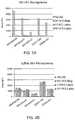
- FIG. 2Ais a chart comparing test results of the apparatus of FIG. 1 against the test results of a standard dual path platform apparatus and showing the depletion of non-pandemic flu antibodies by the apparatus of FIG. 1
- FIG. 2Bis a chart comparing test results of the apparatus of FIG. 1 against the test results of a standard dual path platform apparatus and showing non-depletion of flu-B antibodies by the apparatus of FIG. 1 .
- FIG. 3is a diagram showing a kit including a water vial, a vial with conjugate, a vial with diluent, a blood collection and transfer device, three transfer pipettes, and a filter chamber.
- FIG. 4Ais a diagram depicting a first alternative embodiment.
- FIG. 4Bis a diagram depicting a second alternative embodiment.
- FIG. 4Cis a diagram depicting a third alternative embodiment.
- FIGS. 5A-5Dare diagrams depicting embodiments of Dengue immunoassay device test cells.
- FIGS. 6A-6B and 7A-7Bare diagrams depicting embodiments of Zika immunoassay device test cells.
- FIG. 8is a diagram of an IgM/IgG immunoassay device test cell.
- FIG. 9is a diagram of another IgM/IgG immunoassay device test cell.
- an immunoassay device test cell 10 for testing for the presence of a first ligand in a sampleincludes a housing 20 having a top wall 21 defining first and second holes 24 , 26 , and a window 28 , and first and second sorbent or bibulous materials 30 , 32 defining perpendicular horizontal or lateral flow paths in the housing.
- the first sorbent material 30includes a plurality of zones and may be made from a plurality of materials.
- a first zone 31(sometimes called a filter zone) is located at the first hole 24 and extends to a second zone 33 (sometimes called a test zone) which is located at the junction of a “T”.
- the first zone 31preferably includes a filter 31 a , a pad 31 b on or in which a conjugate 39 having desired antigens or antibodies with attached colored markers is deposited and immobilized, and a first portion of a thin membrane or sorbent or bibulous material 30 typically made from nitrocellulose with a plastic backing (not shown).
- conjugate 39may be a gold sol conjugated to protein A.
- the first zone 31is adapted to receive a buffer solution, to cause the buffer solution to contact the conjugate, thereby mobilizing the conjugate, and to wick the conjugate-carrying buffer solution to the second zone 33 .
- the second (test) zone 33includes a second portion of the thin membrane 30 which can be printed with a test line 50 having immobilized first ligand binding molecules such as antigens or antibodies (depending on whether the test cell is designed to test for the presence of antibodies or antigens) on the membrane as is well known in the art.
- the test line 50may be seen through the window 28 of clear plastic provided in the housing.
- a third zone 35(sometimes called a control zone) which includes a third portion of the thin membrane 30 may also be printed with a control line 60 typically containing antibodies to the conjugate antigens (or in some cases antibodies which will bind to conjugate antibodies, or even antigens which will bind to conjugate antibodies) as is well known in the art.
- a fourth zone 37(sometimes called a reservoir zone) may be provided as a wicking reservoir as is also well known in the art.
- the fourth zone 37includes a relatively thicker absorbent paper.
- a thin, preferably transparent plastic film or card 38 ahaving an adhesive which keeps the sorbent materials in place.
- the card 38 amay be cut with an opening at hole 24 so that it does not block liquid access to the hole 24 .
- the second sorbent material 32may also be made from a plurality of materials and include a plurality of zones.
- the first zone 62(sometimes called a filter zone) includes a filter or pad 32 a and a pad 32 b on or in which second-ligand binding molecules are provided and immobilized, where the second ligand is different than but related to the first ligand, and a first portion of a thin membrane or sorbent or bibulous material 32 typically made from nitrocellulose with a backing (not shown).
- the second-ligand binding moleculesmay include antigens or antibodies or other molecules such as aptamers, nucleic acids, etc. that bind to ligands that are similar to but different than the first ligands.
- the second-ligand binding moleculesmay be provided as a conjugate 41 having desired antigens or antibodies with attached particles.
- the first zone 62is located at the second hole 26 and extends to the second zone 63 .
- the second zone 63includes a second portion of the thin membrane 32 which is in contact with the second zone 33 of the first sorbent material 30 .
- the first sorbent material 30overlies the second sorbent material 32 such that the membranes are in contact with each other (as opposed to the backings contacting the membranes or each other), and such that the test line 50 is effectively located between the membranes.
- test line 50could be printed on the second zone 63 of the second sorbent material 32 instead of, or in addition to the second zone 33 of the first sorbent material 30 .
- a thin plastic film or card 38 b having an adhesive which keeps the second sorbent material in placemay be utilized. With the provided arrangement it takes time for the sample to travel from its application point to the second zone 63 and the test site, and application of sample to the second flow path does not immediately wet the test site.
- the conjugate 41 on the conjugate pad 32 bincludes antigens conjugated with a particle that is not readily visible to the human eye against the background of the test area.
- the particleis a white latex.
- a white latexis a 0.32 micron white latex bead available from Thermo Fisher Scientific, Inc., Holtsville, N.Y.
- the antigens of conjugate 41are different than but are related to the antigens of test line 50 .
- the second conjugatein an embodiment directed to detecting pandemic influenza (“flu”), includes antigens of at least one influenza (“flu”) antigen (e.g., two different flu A antigens such as H1 and H3 flu antigens) and the test site is provided with immobilized antigen of at least the pandemic influenza antigen of interest which is different from but related to the at least one flu antigen of the immobilized conjugate 41 .
- the second conjugateincludes antibodies conjugated with white latex and the test site 50 includes antibodies different than but related to the antibodies of the conjugate 41 .
- the second conjugateis used as a depleting mechanism that captures and thereby depletes antibodies related to the antibodies that are being detected at the test site.
- the second conjugatemay be provided with one or more flu-A antigens; i.e., there may be a plurality of slightly different second conjugates.
- flu-A antibodies in the samplethat may otherwise be captured or retained at the test site (because of their structure which can be similar in many ways to the related flu-B antibodies) are generally captured by the second immobilized conjugate; i.e., the number of flu-A antibodies reaching the test site is depleted.
- the test sitecould include a flu-A antigen for identifying the presence of a particular flu-A antibody in the sample, and the second conjugate may be provided with one or more flu-B antigens and one or more flu-A antigens that are different from but related to the particular flu-A antigen at the test site. Further, it will be appreciated that the test site may be provided with more than one test line, containing different flu antigens. Those flu antigens could include a plurality of flu-A antigens, a plurality of flu-B antigens, or one or more flu-A and one or more flu-B antigens. The second immobilized conjugate will be adjusted accordingly to include conjugate that will deplete those antigens that are related to the antigens of the test lines but are not the subject of the test.
- the use of a white latex conjugate as the immobilized depleting conjugatereduces the visibility of the conjugate should it be loosened and travel with the sample to the test site and get captured at the test site.
- latex beads of a size larger than the pore size of the second migration pathmay be utilized in order to prevent movement of the conjugate along the second migration path.
- the membranescan have different pore sizes. For example, if membrane 31 (for the first conjugate migration) has a 3 ⁇ pore size, and membrane 32 (for the sample migration) has a 15 ⁇ pore size, sample applied to membrane 32 will tend to migrate and stay in the sample membrane 32 and will tend not to migrate into the conjugate membrane 31 .
- the immunoassay of FIGS. 1, 1A and 1Bis preferably utilized as follows.
- a samplepossibly containing antibodies (or antigens) is optionally diluted (e.g., with buffer) and provided to the second opening or hole 26 .
- the sampledoes not immediately wet the test site but is allowed to take time to migrate from pad 32 a to conjugate pad 32 b and then from zone 61 of the second sorbent material 32 to its second zone 63 which is contact with the second zone 33 of the first sorbent material 30 .
- a measured amount of liquidsuch as a buffer solution may be added to hole 26 to help in the migration of the sample.
- the sampleincludes antigens or antibodies that react with the second conjugate 41 of conjugate pad 32 b , those antigens or antibodies are captured by the conjugate 41 and are depleted from the sample before reaching the test line 50 which is printed atop the second zone 33 of the first sorbent material or infused therein.
- the conjugate 41loosens from the pad 32 b and travels along membrane 32 down to the test site and is captured there, the conjugate 41 will not be particularly visible because the white latex particles will not be seen on the white background of the test site.
- a liquid such as a buffer solution(not shown) is added to the first opening 24 .
- the test site (and control site 60 if provided)is inspected via window 28 in order to determine whether the sample is “positive” or not.
- a “positive” test indicating the presence of the antibody (or antigen) in the sampleis obtained when both the test site 50 and the control site 60 show lines of color.
- a “negative” test indicating the lack of the presence of the antibody (or antigen) in the sampleis obtained when only the control site 60 shows a line of color.
- the use of the apparatusmay be expedited by providing the housing with numbering and/or lettering to indicate that hole 26 is for receiving the sample (and optionally some buffer) and is to be used first, and that hole 24 is for receiving the buffer solution and is to be used second.
- the immunoassay 10functions as follows. Because the test line 50 is provided with antigens (or antibodies) immobilized on a membrane, if the test sample contains antibodies to the antigens (or antigens to the antibodies), the antibodies (or antigens) will bind themselves to the antigens (or antibodies) at the test line.
- test samplepasses through a conjugate pad 32 b having immobilized second conjugate 41 with antigens (or antibodies) that are related to but different than the antigens (or antibodies) of the test line
- antigensor antibodies
- related antibodies or antigens to those being tested, if presentwill be captured by the congugate 41 and held at the conjugate pad 32 b , and when the test sample reaches the test line, the antibodies (or antigens) of the sample, if present, will bind to the antigen (or antibody) at the test line.
- the related antibodies (or antigens)are depleted, they will not reach the test line, and if they do, they will already be conjugated with a latex that will reduce their activity at the test site.
- the test sitewill be more specific to the antibodies or antigens whose presence is to be detected.
- the first conjugate 39 containing an antigen for the antibody (or antibody for the antigen) coupled to a colored markeris caused to migrate to the test line. If the test sample contains the antibodies (or antigens) which are now held at the test line 50 , the antigen (or antibody) of the conjugate will bind itself to the antibodies (or antigens) and the colored marker will cause a colored line to appear at the test site 50 . If the test sample does not contain antibodies (or antigens), the conjugate will not have the antibodies (antigens) to bind to at the test line 50 , and no colored line will appear at the test site 50 .
- control line 60is provided with antibodies (or antigens)
- the antigens (or antibodies) of the conjugatewill always bind to the antibodies (or antigens) in the control line 60 , thereby causing a colored line to appear at the control site 60 if the conjugate reaches the control site 60 .
- a colored lineshould always appear at the control site 60 , thereby providing a control for the test.
- FIG. 2Ait can be seen that the apparatus of FIGS. 1, 1A and 1B can provide improved test results relative to a standard dual path platform apparatus such as described and shown in previously incorporated U.S. Pat. No. 7,189,522.
- three sets of five test apparatussuch as described above with reference to FIGS. 1, 1A and 1B were prepared with a second conjugate pad 32 b provided with a conjugate 41 having H3 and H1 flu-A antigen conjugated with beads, and a DPP test line provided with Flu A antigens.
- One set of five apparatusutilized magnetic beads separately conjugated with H1 antigen and H3 antigen (H1+H3 Mag).
- a second setutilized latex beads separately conjugated with H1 and H3 antigen (H1+H3 Latex).
- a third setutilized latex beads with combined H1 and H3 conjugation (H1/H3 Latex).
- a set of devicessuch as described and shown in previously incorporated U.S. Pat. No. 7,189,522 were provided (No Ad) with a test line having the same flu-A antigens. Test samples from five different individuals having H3 antibodies were prepared and applied to the second flow paths of the sets of devices described above with reference to FIGS. 1, 1A and 1B and the set of devices of U.S. Pat. No. 7,189,522. After waiting for the samples to reach the test sites, buffer was added to the first migration path of each device to move the marker conjugate to the test sites.
- the intensity of the signals at each test sitewas measured and plotted.
- the test lines of the five standard dual path platform apparatusshowed a relative intensity (with a digital reader) ranging from about 700 to well over 4000 compared to a relative intensity of nearly zero for the apparatus of FIGS. 1, 1A , and 1 B utilizing the beads for the magnetic and latex beads.
- the white particleprevents the conjugate from being seen against the white background of card 38 b over which the test line 50 is located. It should be appreciated that by depleting flu-A H1 and H3 (seasonal flu) with the latex conjugate system in the path of the sample, the sensitivity and specificity of the test with a test line for pandemic flu A will be increased because of the elimination of the cross-reactivity between the seasonal and pandemic flu A antigens.
- the conjugate in the sample flow pathutilizes fragments or fractions of seasonal flu H1 and H3 conjugated to latex particles.
- the fragmentsare immunodominant portions of the particle that will not substantially cross-react with other flu antigens and are different from the H1 and H3 antibodies that might be used as capture antibodies at the test site in the membrane (the whole molecule of H1 and H3).
- the H1 and H3 fragment conjugateswill have minimal cross-reactivity with pandemic flu antigens resulting in a better detection of a pandemic flu at the test line.
- FIG. 2 bother samples were prepared having flu-B/Bris antibodies.
- the sampleswere applied to a setsof the standard dual path platform apparatus such as described in previously incorporated U.S. Pat. No. 7,189,522 where the test line had flu-B/Bris antigen (No Ad) and to sets of devices such as shown in FIGS. 1, 1A and 1B where the second conjugate pad 32 b was provided with a conjugate 41 having H1 and H3 flu-A antigens conjugated to beads, and a test line provided with flu-B/Bris antigens.
- the test linehad flu-B/Bris antigen (No Ad) and to sets of devices such as shown in FIGS. 1, 1A and 1B where the second conjugate pad 32 b was provided with a conjugate 41 having H1 and H3 flu-A antigens conjugated to beads, and a test line provided with flu-B/Bris antigens.
- No Adflu-B/Bris antigen
- one set of apparatusutilized magnetic beads separately conjugated to H1 and H3 (H1+H3 Mag), a second set utilized 0.32 micron white latex beads separately conjugated (H1+H3 Latex), while a third set utilized the white latex beads with combined conjungation (H1/H3 Latex).
- H1+H3 Maga second set utilized 0.32 micron white latex beads separately conjugated
- H1+H3 Latexa third set utilized the white latex beads with combined conjungation (H1/H3 Latex).
- FIG. 2Bthe positive results at the test line of the apparatus 10 of FIG. 1 is just as strong as the test lines of the standard dual path platform apparatus showing that the conjugate 41 located in the second migration path did not interfere with the results, as the signals at the test lines were nearly the same for all tests of a particular sample. Taking FIGS. 2A and 2B together, it will be appreciated that the apparatus 10 of FIGS. 1, 1A, and 1B has higher sensitivity.
- a kit 100is seen that includes a water vial 101 with water 102 , a vial 103 with freeze dried latex conjugate 104 , a diluent vial 105 with a diluent 106 , a blood collection and transfer device 107 , four transfer pipettes 108 a , 108 b , 108 c , 108 d , and a filter chamber assembly 109 .
- the kitcould have different numbers of elements.
- a “wet” latex conjugatemay be stored utilizing water and/or diluent.
- kit 100may be used in conjunction with an immunoassay device test cell such as device 10 of FIGS. 1, 1A, and 1B .
- kit 100may be used in conjunction with other immnoassay devices such as ELISA (enzyme-linked immunosorbent assay).
- kit 100may be used in conjunction with an immunoassay device test cell such as described in previously incorporated U.S. Pat. No. 7,189,522.
- the water 102 in vial 101may be mixed with the freeze dried latex conjugate 104 in vial 103 by using a pipette 108 a and transferring the water to the latex vial.
- the vial 103may be inverted multiple times in order to cause the freeze dried latex conjugate to be reconstituted.
- the reconstituted latexmay be stored in a refrigerator if desired.
- the dried latex conjugateis a conjugate of one or more flu antigens such as H1 and H3 with microbeads of latex.
- the latex beadsmay be of an easily visible color, e.g., blue.
- the samplee.g., blood
- a desired mannere.g., a fingerstick
- a blood collection and transfer device 107such as a Minivette POCT manufactured by Sarstedt, Newton, N.C.
- the blood samplemay be transferred into the diluent vial 105 containing a diluent 106 such as heparin or EDTA.
- the reconstituted latex conjugatemay then be transferred into the diluent vial 105 by using a pipette 108 b , and the blood and reconstituted latex conjugate may be mixed by inverting multiple times over a period of time and also giving antibodies in the blood an opportunity to be captured by the latex conjugate.
- the contents of the sample diluent vial 105may then be transferred with pipette 108 c to a filter chamber 109 such as a GE Healthcare Life Sciences Mini-UniPrep filter chamber comprising a filter 109 a , compressor 109 b , plunger 109 c , and a tube 109 d , although other filter mechanisms could be utilized.
- the filter 109 acan be plunged into the sample mixture, and the filtered sample can be collected in the tube 109 d of the filter chamber.
- the filteris chosen to have pores that are smaller than the size of the latex conjugate beads.
- the contents of the sample diluent vial 105 that were transferred to the filter chamber 109may have appeared to be dark blue (due to the blue latex conjugate and the blood), the contents of the tube 109 d should be light red (the color of diluted blood). Regardless, it will be appreciated that the ligands that are related to but not the same as the ligands of interest will have been removed from the sample.
- the contents of tube 109 dare then transferred by pipette 108 d and used in conjunction with an immunoassay device.
- the immunoassay deviceis an otherwise prior art type device such as ELISA (enzyme-linked immunosorbent assay) or a LUMINEX assay sold by Thermo Fisher Scientific, Holtsville, N.Y. When provided with a sample that is processed in this manner, the results of the ELISA and the LUMINEX devices are enhanced.
- the immunoassay device to which the contents of tube 109 d are transferredis an immunoassay device test cell such as described in previously incorporated U.S. Pat. No.
- 7,189,522such as by applying a selected amount of the contents to the (second) location for receiving the liquid sample, waiting for the liquid sample to reach the test site via the second migration path, and then applying buffer or a buffer—conjugate subsystem to the first location to cause a conjugate to reach the test site via the first migration path.
- buffer or a buffer—conjugate subsystemto the first location to cause a conjugate to reach the test site via the first migration path.
- the kitincludes a conjugate which may be maintained in a wet form with or without buffer, or may be maintained in a freeze-dried conjugate format which may be reconstituted with water and/or a buffer solution.
- the latex conjugatecomprises white latex beads with antibodies or antigens conjugated thereto.
- the sample and conjugateare mixed together to permit the conjugate to deplete interfering antigens or antibodies.
- the mixed sample and conjugatemay then be applied to an immunoassay device test cell such as described in previously incorporated U.S. Pat. No.
- 7,189,522such as by applying a selected amount of the contents to the (second) location for receiving the liquid sample, waiting for the mixed sample and conjugate to reach the test site via the second migration path, and then applying buffer or a buffer—conjugate subsystem to the first location to cause a conjugate to reach the test site via the first migration path.
- buffer or a buffer—conjugate subsystemto the first location to cause a conjugate to reach the test site via the first migration path.
- FIGS. 4A-4Cadditional embodiments are provided that result in an apparatus having an enhanced test signal.
- FIGS. 4A-4Care described with reference to HIV test devices although they are not limited thereto.
- the embodiments of FIGS. 4A and 4Bare similar to that of FIGS. 1, 1A, and 1B except that the conjugates provided on pads 31 b and 32 b are different, and the immobilized test line antigen is an HIV antibody rather than a flu antibody.
- conjugate 41 a in the sample migration path 32includes a latex particle (e.g., a white latex) to which a MAb-1 p24 antibody and a first interim binding agent (e.g., biotin antigen) are conjugated.
- a latex particlee.g., a white latex
- a first interim binding agente.g., biotin antigen
- the test line 50is provided with a monoclonal anti-HIV antibody protein (MAb-2 p24).
- the buffer-conjugate subsystem of the first migration path 30is provided with a conjugate 39 a including a marker (e.g., blue latex or gold sol) and a second interim binding agent (e.g., streptavidin) conjugated thereto that is chosen to bind to the first interim binding agent.
- a markere.g., blue latex or gold sol
- a second interim binding agente.g., streptavidin
- the HIV p24 antigen in the samplewill bind to the MAb-1 p24 of the conjugate, and the sample with the antigen of interest bound to the conjugate will travel to the test line 50 where the p24 antigen of the sample will be caught by the MAb-2 p24 antibody at the test line.
- the marker conjugatewill move to the test line where the first interim binding agent will bind with the second interim binding agent, and the marker will appear at the test line.
- the embodiment of FIG. 4Bis very similar to the embodiment of FIG. 4A , except that instead of the second interim binding agent of conjugate 39 a being a tetrameric protein such as streptavidin, the second interim binding agent is an anti-biotin antibody.
- the second interim binding agentis an anti-biotin antibody.
- FIG. 4Cis likewise similar to the embodiments of FIGS. 4A and 4B , except that a double interim binding arrangement is utilized. More particularly, the second sorbent material 32 is provided with a pad 32 c in addition to pad 32 b .
- pad 32 bis provided with MAb-1 HIV p24 antigen conjugated with biotin 41 x with the biotin acting as a first interim binding agent of a first pair
- pad 32 cis provided with particles such as a white latex particles conjugated with streptavidin and a secondary antigen such as FITC-A2 (fluorescein isothiocyanate) 41 y .
- FITC-A2fluorescein isothiocyanate
- the streptavidin of particles 41 yact as a second interim binding agent of a first pair, and the FITC-A2 acts as a first interim binding agent of a second pair.
- Pad 31 bis provided with a conjugate 39 z having a marker to which is conjugated an anti-FITC antibody which acts as a second interim binding agent of a second pair.
- the biotinwill bind to the streptavidin of the conjugate 41 y ; i.e., the first and second interim binding agents of the first pair bind together, and the complex of the p24 antigen—MAB-1 HIV p24 antibody with biotin—streptavidin/white latex/FITC antigen conjugate 41 y will move to the test site that includes MAB-2 HIV p24 antibody.
- the p24 antigen of the samplewill bind to the MAB-2 HIV p24 antibody of the test site, and the entire previously-described complex will be held at the test site.
- the anti-FITC antibodyWhen buffer is then added to the first migration path and marker-anti-FITC antibody conjugate is moved to the test site, the anti-FITC antibody will bind to the FITC-A2 being held at the test site; i.e., the first and second interim binding agents of the second pair bind together. As a result, the marker will be held at the test line and provide a positive test result.
- FIGS. 4A-4Cmay all be used in conjunction with a sample being provided directly to the apparatus or with a sample such as the previously described sample contained in tube 109 d which has resulted from a sample having been previously mixed with a depletion conjugate for antigens or antibodies different from but related to the antigen or antibody of interest and then filtered.
- the molecules and conjugates on pads 32 b and 31 b , and 32 care appropriately selected, as are the molecules on the test line 50 and the freeze-dried depletion conjugate 104 .
- FIGS. 5A-5Dfour different embodiments of immunoassays for detecting the flavivirus /arbovirus Dengue are provided.
- FIGS. 5A and 5Bare respectively directed to IgG and IgM Dengue immunoassays that utilize latex particles in a depletion zone
- FIGS. 5C and 5Dare respectively directed to IgG and IgM Dengue immunoassays that do not utilize latex particles in the depletion zone.
- FIGS. 5A and 5Bare respectively directed to IgG and IgM Dengue immunoassays that utilize latex particles in a depletion zone
- FIGS. 5C and 5Dare respectively directed to IgG and IgM Dengue immunoassays that do not utilize latex particles in the depletion zone.
- the depletion zoneare provided with recombinant antigens and/or synthetic peptide antigens of at least one of the Zika, West Nile and Yellow fever flaviviruses , which are related to, but are different than the Dengue flavivirus . In some cases, two, or three of those flaviviruses are provided in the depletion zone.
- Dengue, Zika, West Nile , and Yellow feverare considered related because they are a genus of virus in the family Flaviviridae which are positive, single-stranded, enveloped RNA viruses that have significant antigenic cross-reactivity as they share antigenic sites on a fusion loop of a domain (Domain II) of their envelope proteins of their lipid membranes.
- flavivirusesare antigenically related to various degrees, and immunological cross-reactions have been implicated not only in cross-protection but, under certain conditions, also in infection enhancement phenomena that may exacerbate disease in humans and/or facilitate vector transmission.
- the depletion zonesalso include recombinant antigens to one or more alphaviruses (of the family Togaviridae) such as Chikungunya.
- alphavirusesof the family Togaviridae
- Alphaviruswere originally classified as flaviviruses because of the cross-reactivity or their serocomplexes, but were later found to be of a separate, albeit related family. All alphaviruses share antigenic sites on the capsid and on a fusion loop of a domain (Domain II) of their envelope proteins (as do the flaviviruses ).
- Alphavirus RNAis a single 42S strand of approximately 4 ⁇ 10 6 daltons that is capped and polyadenylated. Flavivirus RNA is a single 40S (ca. 10.9 kilobases) positive-sense strand and is capped at the 5′ end, but, unlike alphaviruses , has no poly A segment at the 3′ end.
- the flavivirus virionhas a single capsid protein (C) that is approximately 13,000 daltons.
- the envelopeconsists of a lipid bilayer, a single envelope protein (E) of 51,000-59,000 daltons, and a small nonglycosylated protein (M) of approximately 8,500 daltons.
- flavivirusesand alphaviruses are cross-reactive, serological diagnostic assays for detections of a single flavivirus or alphavirus are found to have lower specificity than desired; whereas high specificity is important to avoid false positive results. False positive results can lead to misdiagnosis and improper treatment or vaccination. Among flaviviruses , cross-reactivity (and resultingly, false positive test results) can be as high as 25%-40%.
- serological assayshave been used for vaccine monitoring as the immune-status of a patient prior to vaccination can have impact in the efficacy of the vaccine. Therefore, a diagnostic test for the IgG of a particular flavivirus or alphavirus can provide additional information regarding the desirability of a patient getting a vaccine or not. For example, there exist studies which indicate that a prior flavivirus infection (such as Dengue ) can improve the efficacy of the Dengue vaccine with the prior antibody titer effectively acting as a first dose of the vaccine. Accordingly, a diagnostic IgG Dengue test can discriminate a patient with a prior Dengue IgG infection from a patient who never had such an infection. If there is a potential risk associated with vaccination of a non-previously-infected patient, then a highly specific test could help avoid the vaccination risks.
- a prior flavivirus infectionsuch as Dengue
- a highly specific immunoassay device test cell 110 a for testing for the presence of Dengue IgG in a sampleincludes first and second sorbent or bibulous materials 130 a , 132 a defining perpendicular horizontal or lateral flow paths.
- Test cell 110 ais substantially the same as immunoassay device test cell 10 of FIGS. 1, 1A, and 1B and includes the same elements thereof, except for the specifics of the depletion zone located on or in the second sorbent material 132 a (of the second flow path), and the specifics of the test line 150 a located at the intersection of the first and second sorbent materials.
- test cell 110 amay include a control line 160 a , a housing (not shown), the sorbent materials 130 a , 132 a may include various zones, the marker conjugate 139 a of test cell 110 a is provided with visible particles (e.g., gold sol) conjugated with Protein A or anti-human IgG (which both bind human IgG), etc.
- test cell 110 ais specific for testing Dengue IgG, so the test line 150 a includes immobilized Dengue antigens, while the depletion zone is provided with second conjugates 141 a of particles (e.g., latex) conjugated to one or more antigens against viruses that are different than but related to and cross-reactive with Dengue .
- particlese.g., latex
- the conjugates 141 a of the depletion zoneare conjugates of a latex with antigens of one or more of Zika, Chikungunya, West Nile , and Yellow fever .
- the conjugates of depletion zone 141 aare conjugates of a latex with antigens of two or more of Zika, Chikungunya, West Nile , and Yellow fever .
- the conjugates 141 a of the depletion zoneare conjugates of a latex with antigens of three or more of Zika, Chikungunya, West Nile , and Yellow fever .
- the conjugates 141 a of the depletion zoneare conjugates of a latex with antigens of at least all four of Zika, Chikungunya, West Nile , and Yellow fever .
- the antigensare specific to each of their respective virus antibodies.
- the specific antigensare one or more of recombinant antigens, synthetic peptides, and lysate antigens. Accordingly, the second conjugate is used as a depleting mechanism that captures and thereby depletes antibodies different than but related to the Dengue antibodies that are to be detected at the test site.
- the particles of the second conjugates 141 a in the depletion zoneare not readily visible to the human eye against the background of the test area, such as conjugates using white latex particles.
- the second conjugatesare immobilized in the depletion zone so that they, and the antibodies that attach themselves to the conjugate do not travel with the sample to the test line.
- the use of a white latex conjugate as the immobilized depleting conjugatereduces the visibility of the conjugate should it be loosened and travel with the sample to the test site and get captured at the test site.
- latex beads of a size larger than the pore size of the second migration pathmay be utilized in order to prevent movement of the conjugate along the second migration path.
- the second conjugateis used as a depleting mechanism that captures and thereby depletes antibodies related to the antibodies that are being detected at the test site.
- the immunoassay device test cell 110 b of FIG. 5 bis provided for testing for the presence of Dengue IgM in a sample and includes first and second sorbent or bibulous materials 130 b , 132 b defining perpendicular horizontal or lateral flow paths.
- Test cell 110 bis substantially the same as immunoassay device test cell 110 a of FIG. 5 a and includes the same elements thereof, except for the specifics of the conjugate 139 b in the conjugate zone located on or in the first sorbent material 130 b .
- test cell 110 bmay include a test line 150 b with immobilized Dengue antigen, control line 160 b , a housing (not shown), and the sorbent materials 130 b , 132 b may include various zones.
- the specifics of the depletion zone located on or in the second sorbent material 132 b (of the sample flow path)may be the same as depletion zone of test cell 110 a .
- the marker conjugate 139 b of test cell 110 bis provided with anti-human IgM antigen (as opposed to anti-human IgG) that are conjugated to visible particles (e.g., gold sol).
- the presence of Dengue IgM antibodies in the sampleis detected at the test line 150 b even in the presence of antibodies from interfering (cross-reactive) flaviviruses and/or alphaviruses.
- the immunoassay device 110 c of FIG. 5Cis similar to the device 110 a of FIG. 5A and includes the same elements (e.g., sorbent strips 130 c , 132 c , test line 150 c , control line 160 c ) with the exception that the depletion zone does not utilize a conjugate, but instead utilizes antigens 141 c of one or more of Zika, Chikungunya, West Nile , and Yellow fever which may be mixed with a stabilizer/blocker and sprayed on the sorbent strip 132 c .
- the antigensmay be immobilized if desired.
- Dengue IgGis detected at the test line 150 c even in the presence of interfering (cross-reactive) flaviviruses and/or alphaviruses , as the interfering antibodies are depleted by the antigens in the depletion zone. Even if the interfering flaviviruses and/or alphaviruses travel with the sample (because the antigens 141 c in the depletion zone are not immobilized), their reactive sites will have been occupied by the specific antigens of the depletion zone so that they will not react (i.e., be captured) at the test line 150 c.
- the immunoassay device 110 d of FIG. 5Dis similar to the device 110 b of FIG. 5B and includes the same elements (e.g., sorbent strips 130 d , 132 d , anti-human IgM conjugate 139 d , test line 150 d , control line 160 c ) with the exception that the depletion zone does not utilize a conjugate, but instead utilizes antigens 141 d of one or more of Zika, Chikungunya, West Nile , and Yellow fever which may be mixed with a stabilizer/blocker and sprayed on the sorbent strip 132 d .
- elementse.g., sorbent strips 130 d , 132 d , anti-human IgM conjugate 139 d , test line 150 d , control line 160 c
- antigens 141 dof one or more of Zika, Chikungunya, West Nile , and Yellow fever which may be mixed with a stabilizer/blocker
- Dengue IgMis detected at the test line 150 d even in the presence of interfering (cross-reactive) flaviviruses and/or alphaviruses , as the interfering antibodies are depleted by the antigens of the depletion zone.
- the immunoassays of FIGS. 5A-5Dmay be utilized as follows. First, a sample (not shown) possibly containing Dengue antibodies is optionally diluted (e.g., with buffer) and provided to the second sorbent strip. The sample does not immediately wet the test site but is allowed to take time to migrate from its location of application to the depletion zone, and then to the test site. If the sample is not first diluted, optionally, after providing the sample to the second sorbent strip, a measured amount of liquid such as a buffer solution may be added to the second sorbent strip to help in the migration of the sample.
- a samplepossibly containing Dengue antibodies is optionally diluted (e.g., with buffer) and provided to the second sorbent strip.
- the sampledoes not immediately wet the test site but is allowed to take time to migrate from its location of application to the depletion zone, and then to the test site.
- a measured amount of liquidsuch as a buffer solution may be added to the second sorbent strip to help in the migration
- the sampleincludes antigens or antibodies that react with the antigens in the depletion zone, those antibodies are captured at the depletion zone and are depleted from the sample before reaching the test line.
- the depletion zone antigensare not immobilized, or loosen from the sorbent strip and travels down to the test site, many of the reactive sites on the antibodies of the related flaviviruses and/or alphaviruses are occupied with depletion zone recombinant antigens so that they will not bind to the Dengue antigen at the test line.
- Dengue antibodiesare present in the sample, they will generally not be depleted significantly by the specific antigens in the depletion zone, but will travel down to the test line and bind to the Dengue antigens immobilized at the test line.
- a liquidsuch as a buffer solution is added to the first sorbent strip. The solution is added to a location which permits it to cause the conjugate on the first sorbent strip to migrate to the test site (and control site, if provided), and to bind with the antibodies of the sample (if present) that are captured at the test site. The test site and control site are then inspected in order to determine whether the sample is “positive” or not.
- a “positive” test indicating the presence of the antibody in the sampleis obtained when both the test site and the control site show lines of color.
- a “negative” test indicating the lack of the presence of the antibody in the sampleis obtained when only the control site shows a line of color.
- the use of the immunoassay apparatusmay be expedited by providing a housing for the sorbent strip, with the housing having holes and numbering and/or lettering to indicate that one hole in the housing is for receiving the sample (and optionally some buffer) and is to be used first, and that another hole is for receiving the buffer solution (that moves the marker conjugate) and is to be used second.
- FIGS. 5A-5Dfunction as follows. Because the test line is provided with antigens immobilized on a membrane, if the test sample contains antibodies to the antigens, the antibodies will bind themselves to the antigens at the test line. Because the test sample passes through a depletion zone having specific recombinant antigens that are related to but different than the antigens of the test line, related antibodies to those being tested, if present, will be captured by the recombinant antigens and, if immobilized, held at the depletion zone. When the test sample reaches the test line, the antibodies of the sample, if present, will bind to the antigen at the test line.
- the related antibodiesare depleted, they will not reach the test line (if immobilized), and if they do, they will already attached to antigens that will reduce their activity at the test site. Regardless, the test site will be more specific to the antibodies whose presence is to be detected.
- the marker conjugateis caused to migrate to the test line. If the test sample contains the antibodies which are now held at the test line, the conjugate will bind itself to the antibodies of the sample and the colored marker will cause a colored line to appear at the test site. If the test sample does not contain antibodies of interest, the conjugate will not bind to at the test line, and no colored line will appear at the test site.
- the anti-human IgM marker conjugatewill not bind to the IgG antibodies and a colored line will not appear at the test line.
- the control lineis provided with antibodies to which the conjugate will bind, the conjugate will always bind to the antibodies in the control line, thereby causing a colored line to appear at the control site if the conjugate reaches the control site.
- a colored lineshould always appear at the control site, thereby providing a control for the test.
- any specific antigensmay be utilized in the depletion zone, including, but not limited to recombinant antigens, synthetic peptides, and lysates that are specific to the cross-reacting flavivirus and/or alphavirus antigens.
- the synthetic peptides, lysates and recombinant antigensmay be provided either as part of a conjugate or in conjunction with stabilizer/blockers.
- FIGS. 5A-5Dare directed to immunoassays for Dengue IgG and Dengue IgM, it will be appreciated that a highly specific immunoassay for any flavivirus or alphavirus may be generated using the teachings herein, and by adjusting the specifics of the test line and the depletion zone.
- highly specific immunoassays for Zika IgGare seen in FIGS. 6A and 6B .
- the immunoassay 210 a of FIG. 6Ais essentially identical to immunoassay 110 a of FIG.
- test line 250 ahas immobilized Zika antigen instead of immobilized Dengue antigen
- the depletion zoneis provided with second conjugates 241 a of particles (e.g., latex) conjugated to one or more specific antigens against viruses that are different than but related to and cross-reactive with Zika such as antigens of one or more of Dengue, Chikungunya, West Nile , and Yellow fever .
- the second conjugateis used as a depleting mechanism that captures and thereby depletes antibodies different than but related to the Zika antibodies that are to be detected at the test site.
- the highly specific immunoassay 210 b of FIG. 6Bis essentially identical to immunoassay 110 c of FIG. 5C (with sorbent strips 230 b , 232 b , conjugate 239 b , test line 250 b , and control line 260 b ) except that test line 250 b has immobilized Zika antigen instead of immobilized Dengue antigen, and the depletion zone is provided with one or more recombinant antigens 241 b against viruses that are different than but related to and cross-reactive with Zika such as recombinant antigens of one or more of Dengue, Chikungunya, West Nile , and Yellow ever which may be mixed with a stabilizer/blocker and sprayed on the sorbent strip 232 b . Accordingly, the recombinant antigens are used as a depleting mechanism that captures and depletes antibodies different than but related to the Zika antibodies that are to be detected at the test site.
- FIGS. 6A and 6Bare used in the same manner and in function in the same manner as those previously described with respect to FIGS. 5A-5D .
- FIG. 7Aan immunoassay 310 a for detecting a flavivirus IgM or alphavirus IgM is shown.
- the immunoassay 310 ais similar to the immunoassay device of FIG. 5B , except that the depletion zone, rather than being provided with a conjugate of latex and specific recombinant antigens or synthetic peptides is provided with a conjugate 341 a of latex particles conjugated with anti-human IgG antigen.
- immunoassay 310 aincludes first and second sorbent materials 330 a , 332 a , a marker conjugate 339 a with anti-human IgM antigen conjugated to particles (e.g., gold sol) and located in or on the flow path of the first sorbent material 330 a , the depletion zone with the aforementioned conjugate 341 a of latex particles conjugated with anti-human IgG antigen in or on the flow path of the second sorbent material 332 a , a test line 350 a with the immobilized flavivirus or alphavirus IgM antigen (e.g., Zika IgM antigen or Dengue IgM antigen, or Chikungunya IgM, etc.) at the junction of the first and second sorbent materials, and a control line 360 a .
- particlese.g., gold sol
- the provided immunoassay device 310 ais used in the same manner as previously described devices, but functions differently from those devices due to the fact that the conjugate 341 a of the depletion zone is not provided with recombinant antigens that are different than but related to the test line antigen. Rather, the depletion zone conjugates 341 a are directed to broadly depleting IgG antibodies, including all IgG antibodies of flaviviruses and alphaviruses that may be related to the flavivirus or alphavirus being detected, other IgG antibodies that may not be related to the flavivirus or alphavirus being detected, and even IgG antibodies of the flavivirus or alphavirus being detected.
- IgM antibodies of the flavivirus or alpharvirus being detectedwill not be significantly depleted and will migrate down to the test line 350 a where they will bind with the flavivirus or alphavirus IgG antigen.
- the marker conjugate 339 a on the flow path of the first sorbent material 330 ais washed down to the test line 350 a containing the IgM antibodies of the flavivirus or alphavirus being detected, the anti-human IgM antigen of the marker conjugate 339 a will bind to free binding sites on the IgM antibodies of the sample at the test line 350 a , thereby providing a detectable (e.g., visible) result.
- an immunoassay with a high sensitivity to IgM antibodiesis obtained, because IgG antibodies that would otherwise bind to the common antigen at the test line and thereby decrease the capacity of IgM binding, will be depleted.
- the depletion conjugatesare sprayed onto the sorbent material which will carry the sample.
- the latex particlesmay be size selected to provide control of depletion reagents.
- relatively smaller particlese.g., under 1000 nm diameter
- larger size particlese.g., over 1000 nm diameter
- Immunoassay 310 bis similar to immunoassay 310 a (with sorbent strips 330 b , 332 b , anti-human IgM conjugate 339 b , test line 350 b having the immobilized flavivirus or alphavirus IgM antigen, control line 360 b , etc.) except that instead of providing conjugates 341 a with latex particles and anti-human IgG, the depletion zone of immunoassay 310 b utilizes a mixture 341 b of the same anti-human IgG antibodies previously described with respect to immunoassay 310 a , but mixed with a stabilizer/blocker and sprayed onto the depletion zone of the sample migration path of sorbent strip 332 b .
- the immunoassay 310 bis used in the same manner and will function substantially in the same manner as immunoassay 310 a , except that the depletion mixture 341
- FIGS. 7A and 7Bare used in the same manner and in function in the same manner as those previously described with respect to FIGS. 5A-5D .
- Immunoassay 410is directed to detecting both a flavivirus (or alphavirus ) IgM antibody and a flavivirus (or alphavirus ) IgG antibody.
- the immunoassay 410includes (i) a first sorbent strip 430 a with marker conjugate 439 a having anti-human IgM antigen conjugated to particles (e.g., gold sol) located in or on the flow path of the first sorbent material 430 a , (ii) a second sorbent strip 432 , separate and distinct from the first sorbent strip, for receiving the sample and containing a first depletion zone with either conjugates 441 a of latex particles conjugate with anti-human IgG or a mixture of the same anti-human IgG antibodies with a stabilizer/blocker which are sprayed onto the first depletion zone, and a second depletion zone (distinct from the first depletion zone) with either conjugates 441 b of latex and specific recombinant antigens or synthetic peptides of flaviviruses (or alphaviruses ) or a mixture of the same with a stabilizer/blocker which are sprayed onto the second de
- the immunoassayhas at least one first test line 450 a having at least one immobilized first flavivirus antigen located at an intersection of the first sorbent strip 430 a and second sorbent strip 432 , and at least one second test line 450 b with at least one immobilized flavivirus antigen located at an intersection of the third sorbent strip 430 b and the second sorbent strip 432 .
- First and second control lines 460 a , 460 bmay be located on the first and third sorbent strips.
- a fourth sorbent strip 430 cmay be provided to connect the first and third sorbent strips 430 a , 430 b .
- the first, third and fourth sorbent stripsare integral with each other such that buffer applied to single location at a portion of the strip (the “fourth” sorbent strip) will spread to both the first and third sorbent strips and push the respective marker conjugates 439 a , 439 b to the respective test lines 450 a , 450 b and control lines 460 a , 460 b .
- the second sorbent strip 432may be split up into two separate strips, with one containing the first depletion zone and the other containing the second depletion zone.
- the second sorbent stripbe a single strip so that a single sample applied at a location between the two conjugate zones will migrate (independently or using a buffer) to the respective depletion zones 431 a , 431 b and then to the respective test lines 450 a , 450 b.
- immunoassay 410has aspects that are similar to both the immunoassay device 110 a of FIG. 5A and the immunoassay device 310 a of FIG. 7 A. More particularly, the first sorbent strip 430 a with test zone 450 a and the portion of the second sorbent strip 432 containing depletion conjugates or molecules for broadly depleting IgG antibodies function much in the same manner as the immunoassays 310 a and 310 b of FIGS. 7A and 7B for detecting IgM antibodies.
- depletion zone 441 adepletes all IgG antibodies, including flaviviruses and alphaviruses that may be related to the flavivirus or alphavirus being detected, other IgG antibodies that may not be related to the flavivirus or alphavirus being detected, and even IgG antibodies of the flavivirus or alphavirus being detected.
- IgM antibodies of the flavivirus or alpharvirus being detectedif present in the sample, will not be significantly depleted and will migrate down to the test line 450 a where they will bind with the flavivirus or alphavirus IgG antigen.
- the anti-human IgM antigen of the marker conjugate 439 awill bind to free binding sites on the IgM antibodies of the sample at the test line 450 a , thereby providing a detectable (e.g., visible) result much as described in the immunoassays 310 a and 310 b of FIGS. 7A and 7B .
- the third sorbent strip 430 c with test zone 450 b and the portion of the second sorbent strip 432 containing molecules for depleting cross-reactive antibodies to the antibody being tested for at test line 450 bfunction much in the same manner as the immunoassays 110 a and 110 c of FIGS. 5A and 5C .
- depletion zone 441 bis provided with specific recombinant antigens or synthetic peptides of flaviviruses (or alphaviruses ) for depleting antibodies of flaviviruses (and alphaviruses ) related to but different than the flavivirus (or alphavirus ) being tested at the test zone.
- the Protein A or anti-human IgG of the marker conjugate 439 bwill bind to free binding sites on the IgG antibodies of the sample at the test line 450 b , thereby providing a detectable (e.g., visible) result much as described in the immunoassays 210 a and 210 c of FIGS. 5A and 5C .
- the immunoassay of FIG. 8may be utilized as follows. First, a sample (not shown) possibly containing IgG and/or IgM antibodies for the disease(s) being tested for is optionally diluted (e.g., with buffer) and provided to the second sorbent strip between the depletion zones. The sample does not immediately wet the test sites but is allowed to take time to migrate from its location of application to the depletion zones, and then to the test sites. If the sample is not first diluted, optionally, after providing the sample to the second sorbent strip, a measured amount of liquid such as a buffer solution may be added to the second sorbent strip to help in the migration of the sample.
- a samplepossibly containing IgG and/or IgM antibodies for the disease(s) being tested for is optionally diluted (e.g., with buffer) and provided to the second sorbent strip between the depletion zones. The sample does not immediately wet the test sites but is allowed to take time to migrate from its location of application to the depletion zones
- the sampleincludes antigens or antibodies that react with the antigens in one or both of the depletion zones, those antibodies are captured at the depletion zones and are depleted from the sample before reaching the respective test lines.
- those antibodiesare captured at the depletion zones and are depleted from the sample before reaching the respective test lines.
- the antigens of the depletion zonesare not immobilized, or loosen from the sorbent strip and travel down to the test sites, many of the reactive sites on the antibodies of the related flaviviruses and/or alphaviruses are occupied with depletion zone recombinant antigens so that they will not bind to the antigen at the test line.
- a liquidsuch as a buffer solution is added to the first and third sorbent strips (e.g., via the fourth sorbent strip).
- the solutionis added to a location which permits it to cause the conjugates on the first and third sorbent strips to migrate to the respective test sites (and control sites, if provided), and to bind with the antibodies of the sample (if present) that are captured at the respective test sites.
- test sites and control sitesare then inspected in order to determine whether the sample is “positive” or not.
- a “positive” testindicating the presence of the antibody in the sample is obtained when both the test site and the control site show lines of color.
- a “negative” testindicating the lack of the presence of the antibody in the sample is obtained when only the control site shows a line of color.
- the use of the immunoassay apparatusmay be expedited by providing a housing for the sorbent strips, with the housing having holes and numbering and/or lettering to indicate that one hole in the housing is for receiving the sample (and optionally some buffer) and is to be used first, and that another hole (or holes) is for receiving the buffer solution that moves the marker conjugate and is to be used second.
- the immunoassay 410may be directed to detecting IgG and IgM antibodies of a single flavivirus or alphavirus disease (e.g., Dengue, Zika, Chikungunya , etc.), or IgG antibodies to one disease and IgM antibodies to another disease.
- a single flavivirus or alphavirus diseasee.g., Dengue, Zika, Chikungunya , etc.
- IgG antibodies to one disease and IgM antibodies to another diseasee.g., Dengue, Zika, Chikungunya , etc.
- Immunoassay 510is directed to detecting both flavivirus (or alphavirus ) IgM antibodies of multiple diseases and flavivirus (or alphavirus ) IgG antibodies of one or more diseases.
- the immunoassay 510includes (i) a first sorbent strip 530 a with marker conjugate 539 a having anti-human IgM antigen conjugated to particles (e.g., gold sol) located in or on the flow path of the first sorbent material 530 a , (ii) a second sorbent strip 532 , separate and distinct from the first sorbent strip, for receiving the sample and containing a first depletion zone with either conjugates 541 a of latex particles conjugate with anti-human IgG or a mixture of the same anti-human IgG antibodies with a stabilizer/blocker which are sprayed onto the first depletion zone, and a second depletion zone (distinct from the first depletion zone) with either conjugates 541 b of latex and specific recombinant antigens or synthetic peptides of flaviviruses (or alphaviruses ) or a mixture of the same with a stabilizer/blocker which are sprayed onto the second de
- Immunoassay 510has first test lines 550 a - 1 , 550 a - 2 , 550 a - 3 having immobilized flavivirus antigens located at an intersection of the first sorbent strip 530 a and second sorbent strip 532 , and at least one second test line 550 b with at least one immobilized flavivirus antigen located at an intersection of the third sorbent strip 430 b and the second sorbent strip 432 .
- First and second control lines 560 a , 560 bmay be located on the first and third sorbent strips.
- a fourth sorbent strip 530 cmay be provided to connect the first and third sorbent strips 530 a , 530 b .
- the first, third and fourth sorbent stripsare integral with each other such that buffer applied to single location at a portion of the strip (the “fourth” sorbent strip) will spread to both the first and third sorbent strips and push the marker conjugate 539 a to the test lines 550 a - 1 , 550 a - 2 , 550 a - 3 (and control line 560 a ) and marker conjugate 539 b to test line 550 b and control line 460 b .
- the second sorbent strip 532may be split up into two separate strips, with one containing the first depletion zone and the other containing the second depletion zone.
- the second sorbent stripbe a single strip so that a single sample applied at a location between the two conjugate zones will migrate (independently or using a buffer) to the respective depletion zones 531 a , 531 b and then to the respective test lines.
- immunoassay 510is similar to immunoassay 410 except that it is capable of testing for current infection by multiple diseases and is capable of testing for at least one previous infection. More particularly, the first sorbent strip 530 a with test zones 550 a - 1 , 550 a - 2 , 550 a - 3 and the portion of the second sorbent strip 532 containing depletion conjugates or molecules for broadly depleting IgG antibodies function much in the same manner as the immunoassay 410 of FIG. 8 for detecting IgM antibodies, except that IgM antibodies of multiple diseases are being detected.
- depletion zone 541 adepletes IgG antibodies, including IgG antibodies of flaviviruses and alphaviruses that may be related to the flavivirus (es) and/or alphavirus (es) being detected, other IgG antibodies that may not be related to the flaviviruses and/or alphaviruses being detected, and even IgG antibodies of the flaviviruses and/or alphaviruses being detected.
- IgM antibodies of the flaviviruses and/or alphaviruses being detectedwill not be significantly depleted and will migrate down to the test line 550 a - 1 , 550 a - 2 , 550 a - 3 where they will bind with the flavivirus and/or alphavirus IgG antigens.
- the anti-human IgM antigen of the marker conjugate 539 awill bind to free binding sites on the IgM antibodies of the sample, if present at the one or more test lines, thereby providing detectable (e.g., visible) results; both positive and negative.
- test line 550 a - 1may comprise Zika -specific antigen
- line 550 a - 2may comprise Dengue -specific antigen
- line 551 a - 3comprise may Chikungunya -specific antigen.
- the signals at lines 550 a - 1 and 550 a - 2will show a positive result, and any signal at line 550 a - 3 will be small relative to the signals at lines 550 a - 1 and 550 a - 2 .
- the third sorbent strip 530 c with the one or more test zones 550 b and the portion of the second sorbent strip 532 containing molecules for depleting cross-reactive antibodies to the antibody being tested for at test line(s) 550 bfunction much in the same manner as the immunoassays 110 a and 110 c of FIGS. 5A and 5C .
- depletion zone 541 bis provided with specific recombinant antigens or synthetic peptides of flaviviruses (or alphaviruses ) for depleting antibodies of flaviviruses (and alphaviruses ) related to but different than the flavivirus (or alphavirus ) being tested at the test zone.
- the Protein A or anti-human IgG of the marker conjugate 539 bwill bind to free binding sites on the IgG antibodies of the sample at the test line 550 b , thereby providing a detectable (e.g., visible) result much as described in the immunoassays 510 a and 510 c of FIGS. 5A and 5C .
- more than one test lineis provided at the intersection of the second sorbent strip 532 and the third sorbent strip 530 b for detecting IgG antibodies of more than one disease.
- the depletion molecules 541 bmay include depletion molecules for diseases different than but related to the diseases for which the test is being provided. For example, if test lines are provided for detecting IgG antibodies to Zika and Dengue , the depletion molecules 541 b may include depletion molecules for, e.g., Chikungunya, West Nile virus , and Yellow fever . Again, while there may be cross-reactivity, the relative strength of the test lines may provide an indication as to whether the antibodies are present, or whether cross-reactivity is being detected.
- the depletion molecules 541 bmay not include depletion molecules for the diseases different than but related to the diseases for which the test is being provided, but may include recombinant antigens or synthetic peptides against cross-reactive portions (e.g., the envelope (EP)) of the antibodies for the diseases for which the test is being provided, and the test lines may include non-structural antigen (e.g., NS1) for detection of the specific IgG of the same antibodies.
- the envelope (EP) antigenhas higher cross-reactivity between Dengue and Zika virus and therefore a depletion reagents could be designed with EP antigen to remove cross-reacting antibodies to the Zika virus .
- Non-Structural antigen (NS1)is more specific for Dengue and Zika and therefore it could be use as capture line in the membrane for achieving higher specificity. It is also possible to use specific domain ( Zika -EDI) Zika virus as depletion and another specific domain ( Zika -EDIII) Zika virus as a capture.
- the immunoassay of FIG. 9may be utilized as follows. First, a sample (not shown) possibly containing IgG and/or IgM antibodies for the diseases being tested for is optionally diluted (e.g., with buffer) and provided to the second sorbent strip between the depletion zones. The sample does not immediately wet the test sites but is allowed to take time to migrate from its location of application to the depletion zones, and then to the test sites. If the sample is not first diluted, optionally, after providing the sample to the second sorbent strip, a measured amount of liquid such as a buffer solution may be added to the second sorbent strip to help in the migration of the sample.
- a samplepossibly containing IgG and/or IgM antibodies for the diseases being tested for is optionally diluted (e.g., with buffer) and provided to the second sorbent strip between the depletion zones. The sample does not immediately wet the test sites but is allowed to take time to migrate from its location of application to the depletion zones, and then to the test
- the sampleincludes antigens or antibodies that react with the antigens in one or both of the depletion zones, those antibodies are captured at the depletion zones and are depleted from the sample before reaching the respective test lines.
- those antibodiesare captured at the depletion zones and are depleted from the sample before reaching the respective test lines.
- the antigens of the depletion zonesare not immobilized, or loosen from the sorbent strip and travel down to the test sites, many of the reactive sites on the antibodies of the related flaviviruses and/or alphaviruses are occupied with depletion zone recombinant antigens so that they will not bind to the antigen at the test line.
- a liquidsuch as a buffer solution is added to the first and third sorbent strips (e.g., via the fourth sorbent strip).
- the solutionis added to a location which permits it to cause the conjugates on the first and third sorbent strips to migrate to the respective test sites (and control sites, if provided), and to bind with the antibodies of the sample (if present) that are captured at the respective test sites.
- the test sites and control sitesare then inspected in order to determine whether the sample is “positive” or not.
- a “positive” testindicating the presence of the antibody in the sample is obtained when both the test site and the control site show lines of color.
- a “negative” testindicating the lack of the presence of the antibody in the sample is obtained when only the control site shows a line of color.
- the use of the immunoassay apparatusmay be expedited by providing a housing for the sorbent strips, with the housing having holes and numbering and/or lettering to indicate that one hole in the housing is for receiving the sample (and optionally some buffer) and is to be used first, and that another hole (or holes) is for receiving the buffer solution that moves the marker conjugate and is to be used second.
- test cellsare described as having holes in the top wall of a housing for receiving the sample and the buffer-solution or buffer-conjugate subsystem, it will be appreciated that one or both holes may be provided in the end wall or side wall of the housing.
- the sorbent materialwas described as preferably including a thin plastic backing, it will be appreciated that the plastic backing could be provided only at certain locations or not be provided at all. Where only partial backings or no backings are provided, the test and control sites can be located on either or both sides of the sorbent material.
- test strip and control stripare shown is being rectangular in configuration (i.e., lines), it will be appreciated that the test and control sites can be configured differently such as in circles, squares, ovals, a broken line, etc. In fact, the test site and control site can be configured differently from each other.
- the housingmay be modified in additional ways to include separate windows for each test line.
- the embodimentswere described in conjunction with the use of a buffer solution which is added to the migration path of the conjugate and optionally to the migration path of the sample, it will be appreciated that one or more buffers may be chosen as desired to be added to the migration paths depending upon the test or tests to be conducted. Thus, buffers such as phosphate buffers or TRIS (tris hydroxymethylaminomethane) buffers are often utilized. However, the embodiments are intended to encompass the use of any diluent including water.
- the diluentmay, if needed, may be added to and mixed with the sample prior to adding the sample to the sorbent material or the sample may be deposited first and the diluent may be added thereafter.
- any diluent capable of causing the conjugate of the “non-sample” path to migratemay be utilized, and may be premixed with the conjugate in a liquid conjugate system, or provided to the migration path for the conjugate in a dry conjugate system.
Landscapes
- Health & Medical Sciences (AREA)
- Life Sciences & Earth Sciences (AREA)
- Immunology (AREA)
- Engineering & Computer Science (AREA)
- Chemical & Material Sciences (AREA)
- Molecular Biology (AREA)
- Biomedical Technology (AREA)
- Hematology (AREA)
- Urology & Nephrology (AREA)
- Physics & Mathematics (AREA)
- Biochemistry (AREA)
- Cell Biology (AREA)
- Food Science & Technology (AREA)
- Medicinal Chemistry (AREA)
- Biotechnology (AREA)
- Analytical Chemistry (AREA)
- Microbiology (AREA)
- General Health & Medical Sciences (AREA)
- General Physics & Mathematics (AREA)
- Pathology (AREA)
- Virology (AREA)
- Tropical Medicine & Parasitology (AREA)
- Chemical Kinetics & Catalysis (AREA)
- AIDS & HIV (AREA)
- Investigating Or Analysing Biological Materials (AREA)
- Apparatus Associated With Microorganisms And Enzymes (AREA)
- Automatic Analysis And Handling Materials Therefor (AREA)
- Investigating Or Analysing Materials By The Use Of Chemical Reactions (AREA)
- Sampling And Sample Adjustment (AREA)
- Investigating Or Analyzing Non-Biological Materials By The Use Of Chemical Means (AREA)
- Measuring Or Testing Involving Enzymes Or Micro-Organisms (AREA)
Abstract
Description
Claims (18)
Priority Applications (2)
| Application Number | Priority Date | Filing Date | Title |
|---|---|---|---|
| US15/967,479US10976315B2 (en) | 2014-04-02 | 2018-04-30 | Immunoassay utilizing trapping conjugate |
| US17/228,666US20210293808A1 (en) | 2014-04-02 | 2021-04-12 | Immunoassay utilizing trapping conjugate |
Applications Claiming Priority (5)
| Application Number | Priority Date | Filing Date | Title |
|---|---|---|---|
| US201461974060P | 2014-04-02 | 2014-04-02 | |
| US14/631,084US9885710B2 (en) | 2014-04-02 | 2015-02-25 | Immunoassay utilizing trapping conjugate |
| US15/878,801US10473655B2 (en) | 2014-04-02 | 2018-01-24 | Immunoassay utilizing trapping conjugate |
| US15/955,595US10598657B2 (en) | 2014-04-02 | 2018-04-17 | Immunoassay utilizing trapping conjugate |
| US15/967,479US10976315B2 (en) | 2014-04-02 | 2018-04-30 | Immunoassay utilizing trapping conjugate |
Related Parent Applications (1)
| Application Number | Title | Priority Date | Filing Date |
|---|---|---|---|
| US15/955,595ContinuationUS10598657B2 (en) | 2014-04-02 | 2018-04-17 | Immunoassay utilizing trapping conjugate |
Related Child Applications (1)
| Application Number | Title | Priority Date | Filing Date |
|---|---|---|---|
| US17/228,666ContinuationUS20210293808A1 (en) | 2014-04-02 | 2021-04-12 | Immunoassay utilizing trapping conjugate |
Publications (2)
| Publication Number | Publication Date |
|---|---|
| US20180292402A1 US20180292402A1 (en) | 2018-10-11 |
| US10976315B2true US10976315B2 (en) | 2021-04-13 |
Family
ID=54209555
Family Applications (8)
| Application Number | Title | Priority Date | Filing Date |
|---|---|---|---|
| US14/631,006ActiveUS9891216B2 (en) | 2014-04-02 | 2015-02-25 | Immunoassay methods utilizing trapping conjugate |
| US14/631,084Active2035-03-13US9885710B2 (en) | 2014-04-02 | 2015-02-25 | Immunoassay utilizing trapping conjugate |
| US15/791,299ActiveUS10908158B2 (en) | 2014-04-02 | 2017-10-23 | Immunoassay methods utilizing trapping conjugate |
| US15/878,801ActiveUS10473655B2 (en) | 2014-04-02 | 2018-01-24 | Immunoassay utilizing trapping conjugate |
| US15/955,595ActiveUS10598657B2 (en) | 2014-04-02 | 2018-04-17 | Immunoassay utilizing trapping conjugate |
| US15/967,479ActiveUS10976315B2 (en) | 2014-04-02 | 2018-04-30 | Immunoassay utilizing trapping conjugate |
| US17/165,116AbandonedUS20210156857A1 (en) | 2014-04-02 | 2021-02-02 | Immunoassay Methods Utilizing Trapping Conjugate |
| US17/228,666AbandonedUS20210293808A1 (en) | 2014-04-02 | 2021-04-12 | Immunoassay utilizing trapping conjugate |
Family Applications Before (5)
| Application Number | Title | Priority Date | Filing Date |
|---|---|---|---|
| US14/631,006ActiveUS9891216B2 (en) | 2014-04-02 | 2015-02-25 | Immunoassay methods utilizing trapping conjugate |
| US14/631,084Active2035-03-13US9885710B2 (en) | 2014-04-02 | 2015-02-25 | Immunoassay utilizing trapping conjugate |
| US15/791,299ActiveUS10908158B2 (en) | 2014-04-02 | 2017-10-23 | Immunoassay methods utilizing trapping conjugate |
| US15/878,801ActiveUS10473655B2 (en) | 2014-04-02 | 2018-01-24 | Immunoassay utilizing trapping conjugate |
| US15/955,595ActiveUS10598657B2 (en) | 2014-04-02 | 2018-04-17 | Immunoassay utilizing trapping conjugate |
Family Applications After (2)
| Application Number | Title | Priority Date | Filing Date |
|---|---|---|---|
| US17/165,116AbandonedUS20210156857A1 (en) | 2014-04-02 | 2021-02-02 | Immunoassay Methods Utilizing Trapping Conjugate |
| US17/228,666AbandonedUS20210293808A1 (en) | 2014-04-02 | 2021-04-12 | Immunoassay utilizing trapping conjugate |
Country Status (14)
| Country | Link |
|---|---|
| US (8) | US9891216B2 (en) |
| EP (2) | EP3578635B1 (en) |
| CN (2) | CN111077309B (en) |
| AU (3) | AU2015241521B2 (en) |
| BR (1) | BR112016022829B1 (en) |
| CA (1) | CA2944488C (en) |
| DK (2) | DK3578635T3 (en) |
| ES (2) | ES2748688T3 (en) |
| GB (1) | GB2548653A (en) |
| MX (2) | MX389472B (en) |
| MY (1) | MY198463A (en) |
| PE (1) | PE20170259A1 (en) |
| SG (1) | SG11201608278WA (en) |
| WO (1) | WO2015153018A2 (en) |
Cited By (1)
| Publication number | Priority date | Publication date | Assignee | Title |
|---|---|---|---|---|
| US12111312B2 (en) | 2020-05-11 | 2024-10-08 | Chembio Diagnostic Systems, Inc. | Coronavirus IgG/IgM multiplexed dual path immunoassay device |
Families Citing this family (9)
| Publication number | Priority date | Publication date | Assignee | Title |
|---|---|---|---|---|
| ES2748688T3 (en) | 2014-04-02 | 2020-03-17 | Chembio Diagnostic Systems Inc | Immunoassay using conjugate capture |
| JP2018526645A (en) | 2015-08-27 | 2018-09-13 | クイデル コーポレーション | Immunoassay test apparatus having two fluid flow paths for detection and differentiation of two or more analytes |
| WO2017072078A1 (en)* | 2015-10-29 | 2017-05-04 | Thomas Bruderer | Subtractive immunoassay method and lateral flow immunochromatography assay strip for performing the method |
| EP3665187A4 (en)* | 2017-08-08 | 2021-04-21 | OraSure Technologies, Inc. | Assay methods for improved analyte detection |
| CN117030711A (en)* | 2017-08-10 | 2023-11-10 | 爱科来株式会社 | Analysis device and analysis method |
| JP7241696B2 (en)* | 2017-12-22 | 2023-03-17 | 株式会社三和化学研究所 | Immunochromatographic device |
| WO2021205433A1 (en)* | 2020-04-05 | 2021-10-14 | Rapid Bio Pass Ltd. | Lateral flow test utensils |
| US12194909B2 (en) | 2021-10-25 | 2025-01-14 | Trendsetter Vulcan Offshore, Inc. | Monitoring system designed to extract critical natural frequencies of a cargo ship |
| US20240328957A1 (en)* | 2023-04-03 | 2024-10-03 | Burst Diagnostics Llc | Chemiluminescence microfluidic immunoassay device and methods of use thereof |
Citations (351)
| Publication number | Priority date | Publication date | Assignee | Title |
|---|---|---|---|---|
| US3960488A (en) | 1974-04-01 | 1976-06-01 | General Electric Company | Method and apparatus for quantitative surface inhibition test |
| US4041146A (en) | 1975-05-01 | 1977-08-09 | General Electric Company | Method for detection of biological particles |
| US4042335A (en) | 1975-07-23 | 1977-08-16 | Eastman Kodak Company | Integral element for analysis of liquids |
| US4059405A (en) | 1972-04-11 | 1977-11-22 | Damon Corporation | Method and apparatus for analysis of constituent carried in fibrous medium |
| US4094647A (en) | 1976-07-02 | 1978-06-13 | Thyroid Diagnostics, Inc. | Test device |
| US4144306A (en) | 1977-01-14 | 1979-03-13 | Eastman Kodak Company | Element for analysis of liquids |
| US4235601A (en) | 1979-01-12 | 1980-11-25 | Thyroid Diagnostics, Inc. | Test device and method for its use |
| US4313734A (en) | 1978-07-13 | 1982-02-02 | Akzona Incorporated | Metal sol particle immunoassay |
| US4323536A (en) | 1980-02-06 | 1982-04-06 | Eastman Kodak Company | Multi-analyte test device |
| US4361537A (en) | 1979-01-12 | 1982-11-30 | Thyroid Diagnostics, Inc. | Test device and method for its use |
| US4366241A (en) | 1980-08-07 | 1982-12-28 | Syva Company | Concentrating zone method in heterogeneous immunoassays |
| US4373932A (en) | 1980-01-11 | 1983-02-15 | Akzona Incorporated | Application of water-dispersible hydrophobic dyes or pigments as labels in immunoassays |
| US4522786A (en) | 1983-08-10 | 1985-06-11 | E. I. Du Pont De Nemours And Company | Multilayered test device for detecting analytes in liquid test samples |
| US4532107A (en) | 1984-03-26 | 1985-07-30 | Miles Laboratories, Inc. | Reagent test device |
| US4588555A (en) | 1982-10-04 | 1986-05-13 | Fmc Corporation | Device for use in chemical reactions and analyses |
| US4595654A (en) | 1983-11-07 | 1986-06-17 | Immunomedics Inc. | Method for detecting immune complexes in serum |
| US4632901A (en) | 1984-05-11 | 1986-12-30 | Hybritech Incorporated | Method and apparatus for immunoassays |
| US4668619A (en) | 1980-10-30 | 1987-05-26 | Miles Laboratories, Inc. | Multilayer homogeneous specific binding assay device |
| US4703017A (en) | 1984-02-14 | 1987-10-27 | Becton Dickinson And Company | Solid phase assay with visual readout |
| US4740468A (en) | 1985-02-14 | 1988-04-26 | Syntex (U.S.A.) Inc. | Concentrating immunochemical test device and method |
| US4770853A (en) | 1986-12-03 | 1988-09-13 | New Horizons Diagnostics Corporation | Device for self contained solid phase immunodiffusion assay |
| WO1988008534A1 (en) | 1987-04-27 | 1988-11-03 | Unilever Plc | Immunoassays and devices therefor |
| US4786595A (en) | 1982-11-19 | 1988-11-22 | Fuji Photo Film Co., Ltd. | Analytical element for analysis of an analyte |
| EP0299359A2 (en) | 1987-07-16 | 1989-01-18 | Abbott Laboratories | Reagent delivery system for use in solid-phase analytical devices |
| US4826759A (en) | 1984-10-04 | 1989-05-02 | Bio-Metric Systems, Inc. | Field assay for ligands |
| US4855240A (en) | 1987-05-13 | 1989-08-08 | Becton Dickinson And Company | Solid phase assay employing capillary flow |
| US4857453A (en) | 1987-04-07 | 1989-08-15 | Syntex (U.S.A.) Inc. | Immunoassay device |
| US4870003A (en) | 1987-06-15 | 1989-09-26 | Coulter Corporation | Simultaneous enzyme immunoassay for detecting antigen and/or antibody in humans |
| US4886742A (en) | 1987-06-15 | 1989-12-12 | Coulter Corporation | Enzyme immunoassay for detecting HIV antigens in human sera |
| US4906439A (en) | 1986-03-25 | 1990-03-06 | Pb Diagnostic Systems, Inc. | Biological diagnostic device and method of use |
| US4912034A (en) | 1987-09-21 | 1990-03-27 | Biogenex Laboratories | Immunoassay test device and method |
| US4920046A (en) | 1987-02-20 | 1990-04-24 | Becton, Dickinson And Company | Process, test device, and test kit for a rapid assay having a visible readout |
| US4943522A (en) | 1987-06-01 | 1990-07-24 | Quidel | Lateral flow, non-bibulous membrane assay protocols |
| US4956302A (en) | 1987-09-11 | 1990-09-11 | Abbott Laboratories | Lateral flow chromatographic binding assay device |
| US4956275A (en) | 1987-04-14 | 1990-09-11 | Molecular Devices Corporation | Migratory detection immunoassay |
| US4960710A (en) | 1988-06-06 | 1990-10-02 | Miles Inc. | Device and method of assaying for trace mounts of proteins |
| US4960691A (en) | 1986-09-29 | 1990-10-02 | Abbott Laboratories | Chromatographic test strip for determining ligands or receptors |
| US4981786A (en) | 1987-09-04 | 1991-01-01 | Syntex (U.S.A.) Inc. | Multiple port assay device |
| US4981785A (en) | 1988-06-06 | 1991-01-01 | Ventrex Laboratories, Inc. | Apparatus and method for performing immunoassays |
| US5004584A (en) | 1987-06-26 | 1991-04-02 | Rayman Gerard A | Device for testing fluids |
| US5006464A (en) | 1987-10-01 | 1991-04-09 | E-Y Laboratories, Inc. | Directed flow diagnostic device and method |
| US5006474A (en) | 1987-12-16 | 1991-04-09 | Disease Detection International Inc. | Bi-directional lateral chromatographic test device |
| US5075077A (en) | 1988-08-02 | 1991-12-24 | Abbott Laboratories | Test card for performing assays |
| US5075078A (en)* | 1989-10-05 | 1991-12-24 | Abbott Laboratories | Self-performing immunochromatographic device |
| US5087556A (en) | 1989-05-17 | 1992-02-11 | Actimed Laboratories, Inc. | Method for quantitative analysis of body fluid constituents |
| US5091153A (en) | 1990-10-11 | 1992-02-25 | Toxi-Lab Incorporated | Chemical analysis test device |
| US5104793A (en) | 1988-02-16 | 1992-04-14 | Boehringer Mannheim Corporation | Method for determining an analyte in a liquid sample using a zoned test device and an inhibitor for a label used in said method |
| US5104811A (en) | 1986-12-19 | 1992-04-14 | Boehringer Mannheim Gmbh | Test carrier and method for the analytical determination of components of body fluids |
| US5110550A (en) | 1988-07-30 | 1992-05-05 | Boehringer Mannheim Gmbh | Test carrier for the analytical determination of a component of a liquid sample |
| US5132208A (en) | 1987-05-09 | 1992-07-21 | Boehringer Mannheim Gmbh | Test carrier for the analysis of a sample liquid |
| US5137808A (en) | 1987-04-07 | 1992-08-11 | Syntex (U.S.A.) Inc. | Immunoassay device |
| US5147780A (en) | 1989-12-01 | 1992-09-15 | Sangstat Medical Corporation | Multiwell stat test |
| US5156952A (en) | 1986-11-07 | 1992-10-20 | Syntex (U.S.A.) Inc. | Qualitative immunochromatographic method and device |
| US5162238A (en) | 1987-11-28 | 1992-11-10 | Boehringer Mannheim Gmbh | Test carrier for analysis of a liquid sample |
| US5169789A (en) | 1986-12-03 | 1992-12-08 | New Horizons Diagnostics Corporation | Device and method for self contained solid phase immunodiffusion assay |
| US5173433A (en) | 1990-10-11 | 1992-12-22 | Toxi-Lab Incorporated | Method for chemical analysis |
| WO1993003175A1 (en) | 1991-07-29 | 1993-02-18 | Serex, Inc. | Method and device for detecting the presence of analyte in a sample |
| US5200321A (en) | 1990-09-07 | 1993-04-06 | The United States Of America As Represented By The Secretary Of The Navy | Microassay on a card |
| US5202268A (en) | 1988-12-30 | 1993-04-13 | Environmental Diagnostics, Inc. | Multi-layered test card for the determination of substances in liquids |
| JPH05104052A (en) | 1991-10-14 | 1993-04-27 | Toshiba Corp | Liquid substance coating device |
| US5215446A (en) | 1990-11-22 | 1993-06-01 | Brother Kogyo Kabushiki Kaisha | Piezoelectric pump which uses a piezoelectric actuator |
| US5217905A (en) | 1988-04-28 | 1993-06-08 | Compagnie Oris Industrie S.A. | Device and method for the rapid qualitative and quantitative determination of the presence of a reactive ligand in a fluid |
| US5219762A (en) | 1988-12-27 | 1993-06-15 | Mochida Pharmaceutical Co., Ltd. | Method and device for measuring a target substance in a liquid sample |
| US5223436A (en) | 1987-09-30 | 1993-06-29 | Boehringer Mannheim Gmbh | Test carrier for the analytical determination of a component of a liquid sample |
| USRE34312E (en) | 1987-02-23 | 1993-07-13 | Boehringer Mannheim Gmbh | Process and reagent for the determination of an antibody |
| US5232835A (en) | 1986-11-07 | 1993-08-03 | Syntex (U.S.A.) Inc. | Qualitative immunochromatographic method and device |
| US5238649A (en) | 1988-02-09 | 1993-08-24 | Nason Frederic L | Specimen test unit |
| US5240735A (en) | 1988-09-30 | 1993-08-31 | Miles Inc. | Method of manufacturing a test article for the determination of protein |
| US5244788A (en) | 1992-07-01 | 1993-09-14 | Hubscher Thomas T | Method and apparatus for performing determinations of immune rectants in biological fluids |
| US5244631A (en) | 1990-09-19 | 1993-09-14 | Terumo Kabushiki Kaisha | Testing device |
| USRE34405E (en) | 1983-08-01 | 1993-10-12 | Abbott Laboratories | Determination of analytes in particle-containing medium |
| US5252496A (en) | 1989-12-18 | 1993-10-12 | Princeton Biomeditech Corporation | Carbon black immunochemical label |
| US5270166A (en) | 1992-03-30 | 1993-12-14 | Abbott Laboratories | Immunoassays employing generic anti-hapten antibodies and materials for use therein |
| US5275785A (en) | 1987-10-30 | 1994-01-04 | Unilever Patent Holdings B.V. | Test device for detecting an analyte in a liquid sample |
| US5281540A (en) | 1988-08-02 | 1994-01-25 | Abbott Laboratories | Test array for performing assays |
| WO1994006013A1 (en) | 1992-09-08 | 1994-03-17 | Pacific Biotech, Inc. | Analyte detection device including hydrophobic means for improved fluid flow |
| US5296192A (en) | 1992-04-03 | 1994-03-22 | Home Diagnostics, Inc. | Diagnostic test strip |
| US5300439A (en) | 1990-08-06 | 1994-04-05 | Miles Inc. | Method, device, and composition for the assay of ions |
| US5306623A (en) | 1989-08-28 | 1994-04-26 | Lifescan, Inc. | Visual blood glucose concentration test strip |
| US5308775A (en) | 1992-09-15 | 1994-05-03 | Abbott Laboratories | Assay devices for concurrently detecting an analyte and confirming the test result |
| US5332548A (en) | 1991-12-30 | 1994-07-26 | Moore Robert E | Analytical device and method of using same |
| US5334502A (en) | 1991-11-27 | 1994-08-02 | Osborn Laboratories, Inc. | Method of collecting, identifying, and quantifying saliva |
| US5338513A (en) | 1988-07-30 | 1994-08-16 | Boehringer Mannheim Gmbh | Test carrier for the analytical determination of a component of a liquid sample |
| US5340748A (en) | 1990-07-18 | 1994-08-23 | Abbott Laboratories | Analyte-substitute reagent for use in specific binding assay methods, devices and kits |
| US5356782A (en) | 1992-09-03 | 1994-10-18 | Boehringer Mannheim Corporation | Analytical test apparatus with on board negative and positive control |
| US5362654A (en) | 1984-07-20 | 1994-11-08 | Sangstat Medical Corporation | Self-contained quantitative assay |
| US5384264A (en) | 1992-11-05 | 1995-01-24 | Syntron Bioresearch, Inc. | Method and apparatus for single step assays of ligand-containing fluids |
| US5391478A (en) | 1988-05-10 | 1995-02-21 | E. I. Du Pont De Nemours And Company | Assay device and immunoassay |
| US5399316A (en) | 1992-03-13 | 1995-03-21 | Olympus Optical Co., Ltd. | Reaction vessel for conducting an immunological assay |
| US5411858A (en) | 1989-05-17 | 1995-05-02 | Actimed Laboratories, Inc. | Manufacturing process for sample initiated assay device |
| US5418142A (en) | 1989-08-28 | 1995-05-23 | Lifescan, Inc. | Glucose test strip for whole blood |
| US5418167A (en) | 1988-11-29 | 1995-05-23 | Minnesota Mining And Manufacturing Company | Rapid read-out biological indicator |
| US5418136A (en) | 1991-10-01 | 1995-05-23 | Biostar, Inc. | Devices for detection of an analyte based upon light interference |
| US5424215A (en) | 1994-02-07 | 1995-06-13 | Miles Inc. | Assay for the determination of protein in a biological sample |
| US5424220A (en) | 1992-01-31 | 1995-06-13 | Boehringer Mannheim Gmbh | Analysis element and method for determination of an analyte in a liquid sample |
| US5435970A (en) | 1989-12-18 | 1995-07-25 | Environmental Diagnostics, Inc. | Device for analysis for constituents in biological fluids |
| US5468648A (en) | 1991-05-29 | 1995-11-21 | Smithkline Diagnostics, Inc. | Interrupted-flow assay device |
| US5470713A (en) | 1989-04-26 | 1995-11-28 | Diagnostic Products Corporation | Method and element for measuring analytes in biological fluids using immobilized binder-analyte labeled complex |
| US5474902A (en) | 1990-10-08 | 1995-12-12 | Akzo Nobel N.V. | Semi-permeable capillary assay device |
| US5482830A (en) | 1986-02-25 | 1996-01-09 | Biostar, Inc. | Devices and methods for detection of an analyte based upon light interference |
| US5494830A (en) | 1987-10-21 | 1996-02-27 | Hubscher; Thomas T. | Methods for performing determinations of immune reactants in biological fluids |
| US5500350A (en) | 1985-10-30 | 1996-03-19 | Celltech Limited | Binding assay device |
| US5514557A (en) | 1994-06-06 | 1996-05-07 | Genetic Testing Institute Inc. | Method and kit for detecting antibodies specific for HLA and/or platelet glycoproteins |
| US5521102A (en) | 1994-08-08 | 1996-05-28 | Quidel Corporation | Controlled sensitivity immunochromatographic assay |
| US5532133A (en) | 1993-06-02 | 1996-07-02 | New York University | Plasmodium vivax blood stage antigens, PvESP-1, antibodies, and diagnostic assays |
| US5541057A (en) | 1989-09-18 | 1996-07-30 | Biostar, Inc. | Methods for detection of an analyte |
| US5550063A (en) | 1991-02-11 | 1996-08-27 | Biostar, Inc. | Methods for production of an optical assay device |
| US5552272A (en) | 1993-06-10 | 1996-09-03 | Biostar, Inc. | Detection of an analyte by fluorescence using a thin film optical device |
| US5558834A (en) | 1991-10-03 | 1996-09-24 | Bayer Corporation | Device and method of seperating and assaying whole blood |
| US5567594A (en) | 1991-04-26 | 1996-10-22 | Enteron, L.P. | Methods and compositions for the detection and treatment of diseases associated with antigens of microorganisms |
| US5571667A (en) | 1987-10-01 | 1996-11-05 | Chu; Albert E. | Elongated membrane flow-through diagnostic device and method |
| US5591645A (en) | 1987-03-27 | 1997-01-07 | Becton, Dickinson & Co. | Solid phase chromatographic immunoassay |
| US5607863A (en) | 1991-05-29 | 1997-03-04 | Smithkline Diagnostics, Inc. | Barrier-controlled assay device |
| US5616467A (en) | 1988-01-13 | 1997-04-01 | Nycomed As | Method and kit for analyte detection employing gold-sol bound antibodies |
| US5620845A (en) | 1988-06-06 | 1997-04-15 | Ampcor, Inc. | Immunoassay diagnostic kit |
| US5622871A (en) | 1987-04-27 | 1997-04-22 | Unilever Patent Holdings B.V. | Capillary immunoassay and device therefor comprising mobilizable particulate labelled reagents |
| US5624809A (en) | 1988-05-17 | 1997-04-29 | Behringwerke Ag | Device for immunochromatographic analysis |
| US5629164A (en) | 1990-11-14 | 1997-05-13 | Southern Research Institute | Enzyme immunoassay device |
| US5639671A (en) | 1989-09-18 | 1997-06-17 | Biostar, Inc. | Methods for optimizing of an optical assay device |
| US5641639A (en) | 1987-04-29 | 1997-06-24 | Celltech Therapeutics Limited | Binding assay device |
| US5648274A (en) | 1991-05-29 | 1997-07-15 | Smithkline Diagnostics, Inc. | Competitive immunoassay device |
| US5658801A (en) | 1994-05-03 | 1997-08-19 | Spectral Diagnostics Inc. | Medical test kit |
| US5658723A (en) | 1987-04-03 | 1997-08-19 | Cardiovascular Diagnostics, Inc. | Immunoassay system using forced convection currents |
| US5670381A (en) | 1988-01-29 | 1997-09-23 | Abbott Laboratories | Devices for performing ion-capture binding assays |
| US5686315A (en) | 1991-06-14 | 1997-11-11 | Quidel Corporation | Assay device for one step detection of analyte |
| US5695930A (en) | 1994-11-10 | 1997-12-09 | Weinstein; David E. | HIV test kit method for detecting anti-HIV-I antibodies in saliva |
| US5695928A (en) | 1993-12-10 | 1997-12-09 | Novartis Corporation | Rapid immunoassay for detection of antibodies or antigens incorporating simultaneous sample extraction and immunogenic reaction |
| US5710005A (en) | 1996-10-29 | 1998-01-20 | Biocode, Inc. | Analyte detection with a gradient lateral flow device |
| US5714341A (en) | 1994-03-30 | 1998-02-03 | Epitope, Inc. | Saliva assay method and device |
| US5714389A (en) | 1988-06-27 | 1998-02-03 | Carter-Wallace, Inc. | Test device and method for colored particle immunoassay |
| US5723345A (en) | 1994-06-28 | 1998-03-03 | Mochida Pharmaceutical Co., Ltd. | Method and device for specific binding assay |
| US5726010A (en) | 1991-07-31 | 1998-03-10 | Idexx Laboratories, Inc. | Reversible flow chromatographic binding assay |
| US5736188A (en) | 1994-08-08 | 1998-04-07 | Alcock; Susan | Printed fluid transport devices |
| US5739041A (en) | 1995-05-02 | 1998-04-14 | Carter Wallace, Inc. | Diagnostic detection device |
| US5743960A (en) | 1996-07-26 | 1998-04-28 | Bio-Dot, Inc. | Precision metered solenoid valve dispenser |
| US5766962A (en) | 1995-12-22 | 1998-06-16 | Universal Healthwatch, Inc. | Device for collecting and testing samples |
| US5766961A (en) | 1991-01-11 | 1998-06-16 | Quidel Corporation | One-step lateral flow nonbibulous assay |
| US5773234A (en) | 1995-08-07 | 1998-06-30 | Quidel Corporation | Method and device for chlamydia detection |
| US5786220A (en) | 1995-04-28 | 1998-07-28 | Quidel Corporation | Assays and devices for distinguishing between normal and abnormal pregnancy |
| US5798273A (en) | 1996-09-25 | 1998-08-25 | Becton Dickinson And Company | Direct read lateral flow assay for small analytes |
| US5804391A (en) | 1994-11-04 | 1998-09-08 | Boehringer Mannheim Gmbh | Elimination of rheumatoid factor interference using anti-FD antibodies |
| US5807756A (en) | 1995-01-10 | 1998-09-15 | At Point Bio | Ceramic assembly for use in biological assays |
| US5814522A (en) | 1995-06-24 | 1998-09-29 | Boeringer Mannheim Gmbh | Multilayer analytical element for the determination of an analyte in a liquid |
| US5824268A (en) | 1995-05-19 | 1998-10-20 | Universal Health Watch, Inc. | Rapid self-contained assay format |
| US5827646A (en) | 1993-09-14 | 1998-10-27 | Akzo Nobel N.V. | Diagnostic reagents for the detection of antibodies to EBV |
| US5846838A (en) | 1991-05-29 | 1998-12-08 | Smithkline Diagnostics, Inc. | Opposable-element assay device employing conductive barrier |
| US5853670A (en) | 1993-11-26 | 1998-12-29 | British Technology Group Limited | Liquid transfer device for controlling liquid flow |
| US5861265A (en) | 1987-04-29 | 1999-01-19 | Alusuisse Holdings Ag | Binding assay method using a signal preventing reagent |
| US5872713A (en) | 1996-10-30 | 1999-02-16 | Mercury Diagnostics, Inc. | Synchronized analyte testing system |
| US5874216A (en) | 1996-02-23 | 1999-02-23 | Ensys Environmental Products, Inc. | Indirect label assay device for detecting small molecules and method of use thereof |
| US5877028A (en) | 1991-05-29 | 1999-03-02 | Smithkline Diagnostics, Inc. | Immunochromatographic assay device |
| US5879951A (en) | 1997-01-29 | 1999-03-09 | Smithkline Diagnostics, Inc. | Opposable-element assay device employing unidirectional flow |
| US5885526A (en) | 1997-03-25 | 1999-03-23 | Chu; Albert E. | Analytical device for membrane-based assays |
| US5885527A (en) | 1992-05-21 | 1999-03-23 | Biosite Diagnostics, Inc. | Diagnostic devices and apparatus for the controlled movement of reagents without membrances |
| US5891650A (en) | 1993-11-23 | 1999-04-06 | Genentech Incorporated | Kinase receptor activation assay |
| US5900379A (en) | 1996-04-11 | 1999-05-04 | Mizuho Usa, Inc. | Analytical device |
| US5902722A (en) | 1997-12-05 | 1999-05-11 | The Perkin-Elmer Corporation | Method of detecting organisms in a sample |
| US5912116A (en) | 1988-03-14 | 1999-06-15 | Nextec Applications, Inc. | Methods of measuring analytes with barrier webs |
| US5922615A (en) | 1990-03-12 | 1999-07-13 | Biosite Diagnostics Incorporated | Assay devices comprising a porous capture membrane in fluid-withdrawing contact with a nonabsorbent capillary network |
| US5922533A (en) | 1997-08-15 | 1999-07-13 | Abbott Laboratories | Rapid assay for simultaneous detection and differentiation of antibodies to HIV groups |
| US5935331A (en) | 1994-09-09 | 1999-08-10 | Matsushita Electric Industrial Co., Ltd. | Apparatus and method for forming films |
| US5939272A (en) | 1989-01-10 | 1999-08-17 | Biosite Diagnostics Incorporated | Non-competitive threshold ligand-receptor assays |
| US5939252A (en) | 1997-05-09 | 1999-08-17 | Lennon; Donald J. | Detachable-element assay device |
| US5948695A (en) | 1997-06-17 | 1999-09-07 | Mercury Diagnostics, Inc. | Device for determination of an analyte in a body fluid |
| US5955377A (en) | 1991-02-11 | 1999-09-21 | Biostar, Inc. | Methods and kits for the amplification of thin film based assays |
| US5958790A (en) | 1984-12-20 | 1999-09-28 | Nycomed Imaging As | Solid phase transverse diffusion assay |
| US5965458A (en) | 1992-12-29 | 1999-10-12 | Oy Medix Biochemica A.B. | Test strip, its production and use |
| US5972720A (en) | 1996-06-05 | 1999-10-26 | Roche Diagnostics Gmbh | Stabilization of metal conjugates |
| US5976895A (en) | 1996-03-11 | 1999-11-02 | American Biomedica Corporation | Device for the collection, testing and shipment of body fluid samples |
| US5985675A (en) | 1997-12-31 | 1999-11-16 | Charm Sciences, Inc. | Test device for detection of an analyte |
| US5998221A (en) | 1996-09-25 | 1999-12-07 | Becton, Dickinson And Company | Non-instrumented assay with quantitative and qualitative results |
| US5998220A (en) | 1991-05-29 | 1999-12-07 | Beckman Coulter, Inc. | Opposable-element assay devices, kits, and methods employing them |
| US6008056A (en) | 1997-05-28 | 1999-12-28 | Epitope, Inc. | Sample volume control for lateral flow chromatography |
| US6027890A (en) | 1996-01-23 | 2000-02-22 | Rapigene, Inc. | Methods and compositions for enhancing sensitivity in the analysis of biological-based assays |
| US6040195A (en) | 1997-06-10 | 2000-03-21 | Home Diagnostics, Inc. | Diagnostic sanitary test strip |
| US6046057A (en) | 1997-10-24 | 2000-04-04 | Carter-Wallace, Inc. | Analyte assaying device |
| US6046013A (en) | 1997-08-01 | 2000-04-04 | Gti | Process for identifying specific antibodies associated with HLA |
| US6057166A (en) | 1995-12-22 | 2000-05-02 | Universal Healthwatch, Inc. | Fecal test method |
| US6060326A (en) | 1997-04-07 | 2000-05-09 | Heska Corporation | Method to detect canine IgE and kit therefor |
| US6063337A (en) | 1997-08-21 | 2000-05-16 | Lre Technology Partner Gmbh | Test strip for measuring device for optically detecting the concentration of a substance in a body fluid |
| US6087184A (en) | 1997-11-10 | 2000-07-11 | Beckman Coulter, Inc. | Opposable-element chromatographic assay device for detection of analytes |
| US6106732A (en) | 1998-04-16 | 2000-08-22 | Binax Services, Inc. | Integral blood plasma or serum isolation, metering and transport device |
| DE19917093A1 (en) | 1999-04-15 | 2000-10-19 | Backes Claus H | Aerosol applicator, in particular, for liquid medicaments comprises micropump which sucks liquid from its container and delivers it into atomizing nozzle that points into atomizing chamber |
| US6140136A (en) | 1998-09-18 | 2000-10-31 | Syntron Bioresearch, Inc. | Analytical test device and method of use |
| US6168956B1 (en) | 1991-05-29 | 2001-01-02 | Beckman Coulter, Inc. | Multiple component chromatographic assay device |
| US6187268B1 (en) | 1998-07-27 | 2001-02-13 | Bayer Corporation | Transparent flow through membrane for dry reagent analytical devices |
| US6194220B1 (en) | 1996-09-25 | 2001-02-27 | Becton, Dickinson And Company | Non-instrumented assay with quantitative and qualitative results |
| US6221625B1 (en) | 1997-04-23 | 2001-04-24 | Fujirebio Inc. | Enzyme-labeled immunoassay and device therefor |
| US6221678B1 (en) | 1997-10-06 | 2001-04-24 | Enterix Inc | Apparatus and method for analyte detection |
| US6224831B1 (en) | 1998-03-31 | 2001-05-01 | John Co., Inc. | Microassay device and methods |
| US6235464B1 (en) | 1988-11-17 | 2001-05-22 | Glenn L. Henderson | Immunoassay on a preblocked solid surface |
| US6248598B1 (en) | 1998-09-17 | 2001-06-19 | Stuart C. Bogema | Immunoassay that provides for both collection of saliva and assay of saliva for one or more analytes with visual readout |
| US6258548B1 (en) | 1997-06-05 | 2001-07-10 | A-Fem Medical Corporation | Single or multiple analyte semi-quantitative/quantitative rapid diagnostic lateral flow test system for large molecules |
| US6271046B1 (en) | 1997-10-06 | 2001-08-07 | Enterix, Inc. | Apparatus and method for analyte detection |
| US6271040B1 (en) | 1992-05-21 | 2001-08-07 | Biosite Diagnostics Incorporated | Diagnostic devices method and apparatus for the controlled movement of reagents without membranes |
| US20010012637A1 (en) | 1996-03-11 | 2001-08-09 | American Biomedica Corporation | Device for the testing of body fluid samples |
| US6287875B1 (en) | 1997-05-02 | 2001-09-11 | Silver Lake Corporation | Internally referenced competitive assays |
| US6297020B1 (en) | 1999-03-01 | 2001-10-02 | Bayer Corporation | Device for carrying out lateral-flow assays involving more than one analyte |
| US20010026942A1 (en) | 1998-08-14 | 2001-10-04 | Biocontrol Systems, Inc. | Detection of contaminants using self-contained devices employing target material binding dyes |
| US20010026944A1 (en) | 1999-04-21 | 2001-10-04 | Roy Chung | Immunoassay system |
| US6300142B1 (en) | 1997-11-28 | 2001-10-09 | Provalis Diagnostics Ltd | Device and apparatus for conducting an assay |
| US20010034068A1 (en) | 1998-07-14 | 2001-10-25 | Spivey Robin James | Screening device and methods of screening immunoassay tests and agglutination tests |
| USRE37437E1 (en) | 1984-12-15 | 2001-11-06 | Dade Behring Marburg, Gmbh | Sheet-like diagnostic device |
| US20010039057A1 (en) | 1996-04-05 | 2001-11-08 | Douglas Joel S. | Devices for testing for the presence and/or concentration of an analyte in a body fluid |
| US6316264B1 (en) | 1999-12-17 | 2001-11-13 | Bayer Corporation | Test strip for the assay of an analyte in a liquid sample |
| US6316205B1 (en) | 2000-01-28 | 2001-11-13 | Genelabs Diagnostics Pte Ltd. | Assay devices and methods of analyte detection |
| US6319676B1 (en) | 1995-05-02 | 2001-11-20 | Carter Wallace, Inc. | Diagnostic detection device and method |
| US6326214B1 (en) | 1998-03-04 | 2001-12-04 | Yung Hsiang Liu | Immunity testing device |
| US20010048893A1 (en) | 1999-01-27 | 2001-12-06 | Philip R. Norris | Diagnostic assay device |
| US6335205B1 (en) | 1996-03-13 | 2002-01-01 | Roche Diagnostics Gmbh | Method and test strip for determining an analyte |
| US20020001853A1 (en) | 1997-10-24 | 2002-01-03 | Robert J. Obremski | Detection of very low quantities of analyte bound to a solid phase |
| US20020015663A1 (en) | 1989-09-21 | 2002-02-07 | Andrew S. Goldstein | Oral collection device and kit |
| US20020019062A1 (en) | 1999-06-18 | 2002-02-14 | Peter Lea | Assay devices |
| US6352862B1 (en) | 1989-02-17 | 2002-03-05 | Unilever Patent Holdings B.V. | Analytical test device for imuno assays and methods of using same |
| US20020031839A1 (en) | 1997-08-29 | 2002-03-14 | Mcneirney John C. | Method and apparatus for rapid analysis of analytes in biological samples |
| US6362008B1 (en) | 1997-12-23 | 2002-03-26 | Barbara A. Kohn | Generic signalling mechanism for detection of analytes |
| US6368876B1 (en) | 1995-05-18 | 2002-04-09 | Genzyme Diagnostics | One step immunochromatographic device and method of use |
| US6372516B1 (en) | 2000-09-07 | 2002-04-16 | Sun Biomedical Laboratories, Inc. | Lateral flow test device |
| US6372514B1 (en) | 1998-09-18 | 2002-04-16 | Syntron Bioresearch, Inc. | Even fluid front for liquid sample on test strip device |
| US6372515B1 (en) | 1996-03-11 | 2002-04-16 | American Bio Medica Corporation | Device for the testing of fluid samples and process for making the device |
| US20020048819A1 (en) | 2000-10-24 | 2002-04-25 | Kenneth Alley | Test strip for use in an apparatus for sampling and testing a specimen |
| US20020046614A1 (en) | 2000-10-24 | 2002-04-25 | Kenneth Alley | Apparatus for sampling and testing a specimen |
| US20020058330A1 (en) | 1997-06-10 | 2002-05-16 | Patrick Carroll | Diagnostic sanitary test strip |
| US20020057991A1 (en) | 2000-06-02 | 2002-05-16 | Kelly Tim Allen | Self-contained devices for detecting biological contaminants |
| US6399398B1 (en) | 1994-09-23 | 2002-06-04 | Unipath Limited | Assay device |
| US6403383B1 (en) | 1996-03-11 | 2002-06-11 | American Bio Medica Corp. | Diagnostic test kit for immunological assays of fluid samples |
| US6403384B1 (en) | 1998-11-16 | 2002-06-11 | Umedik, Inc. | Device and method for analyzing a biologic sample |
| US6413473B1 (en) | 1999-02-05 | 2002-07-02 | Taylor Technologies, Inc. | Multicomponent test systems useful in analyzing liquid samples, and uses therefor |
| US6413784B1 (en) | 1996-12-05 | 2002-07-02 | Idego Aps | Multi-sectioned fluid delivery devices for detecting target molecules by immunoassay |
| US20020110803A1 (en) | 2001-02-09 | 2002-08-15 | Dhar Tarun K. | Highly cost-effective analytical device for performing immunoassays with ultra high sensitivity |
| US6436722B1 (en) | 2000-04-18 | 2002-08-20 | Idexx Laboratories, Inc. | Device and method for integrated diagnostics with multiple independent flow paths |
| US20020119497A1 (en) | 2000-08-21 | 2002-08-29 | Thomas Wild | Binding assays |
| US6455324B1 (en) | 1997-12-19 | 2002-09-24 | Amira Medical | Embossed test strip system |
| US20020142291A1 (en) | 2000-04-14 | 2002-10-03 | Bauer Jeffrey S. | Positive detection lateral-flow apparatus and method for small and large analytes |
| US20020155028A1 (en) | 2001-04-23 | 2002-10-24 | Wong Raphael C. | Lateral flow contact test apparatus |
| US6472226B1 (en) | 1998-10-02 | 2002-10-29 | Genosis Limited | Assay using porosity-reduction to inhibit migration |
| US20020164670A1 (en) | 1999-10-27 | 2002-11-07 | Forrest Gordon Coulter | Lateral flow device utilising particulate carrier |
| US20020173050A1 (en) | 2001-03-30 | 2002-11-21 | Dinello Robert K. | Prewetting stop flow test strip |
| US20020172937A1 (en) | 1999-02-19 | 2002-11-21 | Kirti Dave | Rapid assay for arthopod-borne disease vectors and pathogens |
| US6489129B1 (en) | 1996-11-29 | 2002-12-03 | Roche Diagnostics Gmbh | Antigen-specific IgM detection |
| US6492127B2 (en) | 2001-01-23 | 2002-12-10 | Varian, Inc. | Lateral flow testing device with on-board chemical reactant |
| US20020192839A1 (en) | 1998-03-30 | 2002-12-19 | Epitope, Inc. | Collection device for single step assay of oral fluids |
| US6500629B1 (en) | 1999-09-13 | 2002-12-31 | Equitech Laboratories, Inc. | Materials and methods for detection and quantitation of an analyte |
| US6502766B1 (en) | 2000-07-24 | 2003-01-07 | The Procter & Gamble Company | Liquid sprayers |
| US6503722B1 (en) | 1997-04-04 | 2003-01-07 | Biosite Diagnostics | Diagnostic tests and kits for Clostridium difficile |
| US6511814B1 (en) | 1999-03-26 | 2003-01-28 | Idexx Laboratories, Inc. | Method and device for detecting analytes in fluids |
| US6514773B1 (en) | 1994-07-25 | 2003-02-04 | Securetec Detektions-Systeme Ag | Method of detecting surface contamination by an analyte |
| US6514769B2 (en) | 1999-07-29 | 2003-02-04 | Jin Po Lee | Multiple analyte assay device with sample integrity monitoring system |
| EP1284422A1 (en) | 2000-05-25 | 2003-02-19 | Nichirei Corporation | Analyzing device |
| US6528325B1 (en) | 2000-10-13 | 2003-03-04 | Dexall Biomedical Labs, Inc. | Method for the visual detection of specific antibodies in human serum by the use of lateral flow assays |
| US6528322B1 (en) | 1999-08-06 | 2003-03-04 | Pharmacia Ab | Analytical method and apparatus |
| US6528323B1 (en) | 1999-06-14 | 2003-03-04 | Praxsys Biosystems, Inc. | Bidirectional lateral flow test strip and method |
| US6528321B1 (en) | 2000-06-26 | 2003-03-04 | Beckman Coulter, Inc. | Opposable-element chromatographic assay device for detection of analytes in whole blood samples |
| US20030045001A1 (en) | 2001-08-29 | 2003-03-06 | Deborah Burgess | Immunochromatographic test strip with arcuate sample application zone for ease-of-use in the field |
| US20030049658A1 (en)* | 1999-12-10 | 2003-03-13 | David Smart | Assay |
| US6534324B1 (en) | 2000-05-12 | 2003-03-18 | Mizuho Usa, Inc. | Rapid assay strip and method of rapid competitive assay |
| US6544474B2 (en) | 1997-09-30 | 2003-04-08 | Amira Medical | Device for determination of an analyte in a body fluid using small sample sizes |
| US6548309B1 (en) | 1998-03-19 | 2003-04-15 | Binax, Inc. | Procedure for assay of liquids containing undissolved solids, semisolids or colloids |
| US6551842B1 (en) | 1999-03-26 | 2003-04-22 | Idexx Laboratories, Inc. | Method and device for detecting analytes in fluids |
| WO2003041733A1 (en) | 2001-11-14 | 2003-05-22 | Prion Developmental Laboratories, Inc. | Prion-detection business methods |
| US20030118480A1 (en) | 2001-12-21 | 2003-06-26 | Kaylor Rosann Marie | Diagnostic device, system and method |
| US20030124740A1 (en) | 2001-12-28 | 2003-07-03 | Bachand Steven S. | Binding assay device with non-absorbent carrier material |
| US6593085B1 (en) | 1997-12-19 | 2003-07-15 | Panbio Pty Ltd | Assay method and apparatus |
| US6592815B1 (en) | 1997-12-04 | 2003-07-15 | Roche Diagnostics Gmbh | Analytical test element with a narrowed capillary channel |
| US20030138351A1 (en) | 1999-04-30 | 2003-07-24 | Donald E. Etes | Disposable devices for testing body fluids |
| US6602719B1 (en) | 1999-03-26 | 2003-08-05 | Idexx Laboratories, Inc. | Method and device for detecting analytes in fluids |
| US20030180967A1 (en) | 2002-03-19 | 2003-09-25 | Matsushita Electric Industrial Co., Ltd. | Immunoassay method, test strip for use therein, and immunoassay apparatus |
| US6627459B1 (en) | 2000-04-19 | 2003-09-30 | Applied Biotech, Inc. | Immunoassay controls |
| US6632681B1 (en) | 2000-07-24 | 2003-10-14 | Ey Laboratories | Reagent delivery device and method of use |
| US6645732B2 (en) | 1996-11-29 | 2003-11-11 | Roche Diagnostics Gmbh | Antigen-specific IgG detection |
| US6656744B2 (en) | 1995-08-09 | 2003-12-02 | Quidel Corporation | One-step lateral flow assays |
| US6656745B1 (en) | 2000-06-02 | 2003-12-02 | Francis X. Cole | Devices and methods for a multi-level, semi-quantitative immunodiffusion assay |
| US6660469B1 (en) | 1999-05-18 | 2003-12-09 | Krysium Advisors Limited | Apparatus and method testing a biological fluid |
| US6663833B1 (en) | 1998-03-10 | 2003-12-16 | Strategic Diagnostics Inc. | Integrated assay device and methods of production and use |
| US20040001767A1 (en) | 2002-07-01 | 2004-01-01 | Peters Richard D. | Piezoelectric micropump with diaphragm and valves |
| US6673628B2 (en) | 1998-08-06 | 2004-01-06 | Spectral Diagnostics, Inc. | Analytical test device and method |
| US20040014157A1 (en) | 2002-07-19 | 2004-01-22 | Ronald Sommer | Use of dual conjugated labels in the elimination of serum interference in immunochromatographic assays |
| US6686167B2 (en) | 2001-02-03 | 2004-02-03 | Padma S. Bagaria | Test device for detecting semen and method of use |
| USRE38430E1 (en) | 1987-03-27 | 2004-02-17 | Becton, Dickinson And Company | Solid phase chromatographic immunoassay |
| US6703196B1 (en) | 1997-12-24 | 2004-03-09 | Roche Diagnostics Corporation | General purpose structure of an analytical element and its application for analyte determination |
| US6706539B2 (en) | 1997-11-24 | 2004-03-16 | Quidel Corporation | Enzyme substrate delivery and product registration in one step enzyme immunoassays |
| US6713309B1 (en) | 1999-07-30 | 2004-03-30 | Large Scale Proteomics Corporation | Microarrays and their manufacture |
| US6727073B1 (en) | 1999-11-19 | 2004-04-27 | Binax, Inc. | Method for detecting enteric disease |
| US20040087036A1 (en) | 1998-11-18 | 2004-05-06 | Roy Chung | Immunoassay system |
| US6750031B1 (en) | 1996-01-11 | 2004-06-15 | The United States Of America As Represented By The Secretary Of The Navy | Displacement assay on a porous membrane |
| US6753190B1 (en) | 1998-07-01 | 2004-06-22 | Nitto Denko Corporation | Immunologic test method and immunologic test kit |
| US20040142495A1 (en) | 2002-12-16 | 2004-07-22 | Hartman William G. | Analyte detecting article and method |
| US20040161859A1 (en) | 2003-02-13 | 2004-08-19 | Huiyan Guo | Lateral flow immunoassay controls |
| US6790611B2 (en) | 2000-10-17 | 2004-09-14 | Besst-Test Aps | Assay for directly detecting a RS virus related biological cell in a body fluid sample |
| US20040184954A1 (en) | 2003-03-18 | 2004-09-23 | Huiyan Guo | Lateral flow immunoassay devices for testing saliva and other liquid samples and methods of use of same |
| US6797481B1 (en) | 2000-10-17 | 2004-09-28 | Dade Behring Marburg Gmbh | Simultaneous screening of multiple analytes |
| WO2004084274A2 (en) | 2003-03-17 | 2004-09-30 | Par Technologies, Llc. | Piezoelectric actuator and pump using same |
| DE10313158A1 (en) | 2003-03-18 | 2004-10-07 | Siemens Ag | Micropump with piezoelectric membrane actuator contacting inner contour of opposing part of pump chamber wall in its deformed condition |
| US20040197769A1 (en)* | 2002-10-31 | 2004-10-07 | Wong Susan J. | Diagnostic test for West Nile virus |
| US6808889B2 (en) | 1999-03-16 | 2004-10-26 | Serex, Inc. | Method and device for detection of Apo A, Apo B and the ratio thereof in saliva |
| US6812038B1 (en) | 1999-11-18 | 2004-11-02 | Pharmacia Diagnostics Ab | Assay device and use thereof |
| US20040219694A1 (en) | 2001-09-11 | 2004-11-04 | Chittock Roger Steward | Lateral flow test format for enzyme assays |
| US20040235189A1 (en) | 2003-05-08 | 2004-11-25 | Lu Wei Zhao | Reversed chromatographic immunoassay |
| US6824997B1 (en) | 1998-09-18 | 2004-11-30 | Binax, Inc. | Process and materials for the rapid detection of streptococcus pneumoniae employing purified antigen-specific antibodies |
| US20040241779A1 (en) | 2003-02-24 | 2004-12-02 | Piasio Roger N. | Dry chemistry, lateral flow-reconstituted chromatographic enzyme-driven assays |
| US6828110B2 (en) | 2000-01-06 | 2004-12-07 | Biosite Incorporated | Assays for detection of Bacillus anthracis |
| US20040248322A1 (en) | 2003-06-06 | 2004-12-09 | David Charlton | Diagnostic test for analytes in a sample |
| US6855561B2 (en) | 2001-09-10 | 2005-02-15 | Quidel Corporation | Method for adding an apparent non-signal line to a lateral flow assay |
| US6867051B1 (en) | 1998-02-03 | 2005-03-15 | Adeza Biomedical, Inc. | Point of care diagnostic systems |
| US20050074900A1 (en) | 2003-10-07 | 2005-04-07 | Morgan Nicole Y. | Microfluidic flow-through immunoassay for simultaneous detection of multiple proteins in a biological sample |
| US20050079629A1 (en) | 2003-09-23 | 2005-04-14 | Huiyan Guo | Lateral flow assay devices and methods of use |
| US20050112779A1 (en) | 2003-11-21 | 2005-05-26 | Kimberly-Clark Worldwide, Inc. | Extension of the dynamic detection range of assay devices |
| US20050112782A1 (en) | 1992-05-21 | 2005-05-26 | Biosite, Inc. | Diagnostic devices and apparatus for the controlled movement of reagents without membranes |
| US20050112780A1 (en) | 2003-11-21 | 2005-05-26 | Kimberly-Clark Worldwide, Inc. | Method for extending the dynamic detection range of assay devices |
| US6905835B2 (en) | 2000-06-07 | 2005-06-14 | Centro De Ingenieria Genetica Y Biotecnologia Cigb | Assay for anti transglutaminase antibodies detection useful in celicac disease diagnosis |
| US20050130319A1 (en) | 2003-12-15 | 2005-06-16 | Xerox Corporation | Molecular binding event detection using separation channels |
| US20050130293A1 (en) | 1998-04-23 | 2005-06-16 | Blatt Joel M. | Dry reagent strip configuration, composition and method for multiple analyte determination |
| US20050136500A1 (en) | 2003-12-19 | 2005-06-23 | Kimberly-Clark Worldwide; Inc. | Flow-through assay devices |
| US20050142032A1 (en) | 2003-10-31 | 2005-06-30 | Joachim Hoenes | Test system for determining an analyte in a liquid sample |
| US20050164404A1 (en) | 2001-12-12 | 2005-07-28 | Proteome Systems Intellectual Property Pty Ltd. | Diagnostic testing process |
| US6924153B1 (en) | 1997-03-06 | 2005-08-02 | Quidel Corporation | Quantitative lateral flow assays and devices |
| WO2005070324A2 (en) | 2004-01-20 | 2005-08-04 | Koninklijke Philips Electronics, N.V. | Droplet jet system for cleaning teeth |
| US6927068B2 (en) | 2002-01-30 | 2005-08-09 | The United States Of America As Represented By The Secretary Of The Navy | Rapid and non-invasive method to evaluate immunization status of a patient |
| US20050175992A1 (en) | 2004-02-09 | 2005-08-11 | Rapid Pathogen Screening Inc. | Method for the rapid diagnosis of targets in human body fluids |
| US20050208677A1 (en) | 1999-01-15 | 2005-09-22 | Medtox Scientific, Inc. | Lateral flow test strip |
| US20050227371A1 (en) | 2004-03-23 | 2005-10-13 | Quidel Corporation | Hybrid phase lateral flow assay |
| US20050244985A1 (en) | 2003-09-19 | 2005-11-03 | Zbx Corporation | Chromatographic Assay Device and Methods |
| US7018847B2 (en) | 2000-05-05 | 2006-03-28 | Pharmacia Diagnostics Ab | Assay device with timer function |
| US20060099719A1 (en) | 2004-11-05 | 2006-05-11 | Roche Diagnostics Operations, Inc. | Biochemical assay |
| US7049130B2 (en) | 1997-06-10 | 2006-05-23 | Home Diagnostics, Inc. | Diagnostic sanitary test strip |
| US20060121626A1 (en) | 2004-12-03 | 2006-06-08 | Genzyme Corporation | Diagnostic assay device |
| US20060134803A1 (en) | 2004-12-16 | 2006-06-22 | Javanbakhsh Esfandiari | Immunoassay devices and use thereof |
| US20060148097A1 (en) | 2005-01-06 | 2006-07-06 | Sysmex Corporation | Test strip for immunochromatography |
| US20060166374A1 (en) | 2005-01-21 | 2006-07-27 | Hubscher Thomas T | Method for the visual detection of specific antibodies by the use of lateral flow assays |
| US20070020768A1 (en) | 2005-05-23 | 2007-01-25 | Gerd Rundstrom | Two step lateral flow assay methods and devices |
| US7189522B2 (en) | 2005-03-11 | 2007-03-13 | Chembio Diagnostic Systems, Inc. | Dual path immunoassay device |
| US20070059682A1 (en) | 2005-09-13 | 2007-03-15 | Rapid Pathogen Screening Inc. | Method to increase specificity and/or accuracy of lateral flow immunoassays |
| US20070184492A1 (en) | 2004-01-28 | 2007-08-09 | Naishu Wang | Delayed and diffused flow rapid confirmatory immunological testing apparatus and method |
| US20080138842A1 (en) | 2006-12-11 | 2008-06-12 | Hans Boehringer | Indirect lateral flow sandwich assay |
| US20080194013A1 (en) | 2004-06-07 | 2008-08-14 | Denka Seiken Co., Ltd. | Chromatography Detection Apparatus, Detection Method, and Kit Utilizing the Same |
| US20080318341A1 (en) | 2005-03-11 | 2008-12-25 | Javanbakhsh Esfandiari | Dual Path Immunoassay Device |
| EP2065706A2 (en) | 2007-11-29 | 2009-06-03 | Fujifilm Corporation | A measurement kit and an immunochromatography method |
| US20090148933A1 (en) | 2006-03-15 | 2009-06-11 | Micronics, Inc. | Integrated nucleic acid assays |
| WO2009075894A1 (en) | 2007-12-13 | 2009-06-18 | Beckman Coulter, Inc | Device and methods for detecting a target cell |
| US20090181470A1 (en) | 2007-11-29 | 2009-07-16 | Hiroyuki Chiku | Measurement kit and an immunochromatography method |
| US20100047857A1 (en) | 2006-12-12 | 2010-02-25 | Response Biomedical Corporation | Multiple analyte immunoassay |
| US20100112725A1 (en) | 2005-02-09 | 2010-05-06 | Rapid Pathogen Screening, Inc | Method to increase specificity and/or accuracy of lateral flow immunoassays |
| US20100285490A1 (en) | 2006-12-29 | 2010-11-11 | Invitrogen Corporation | Detection apparatus |
| US20110124130A1 (en) | 2008-04-03 | 2011-05-26 | Peter Wagner | Device and method for analysis of samples with depletion of analyte content |
| US20120003727A1 (en) | 2006-03-10 | 2012-01-05 | Javanbakhsh Esfandiari | Immunoassay Device for Detecting Antibodies and Antigens |
| US20120282154A1 (en) | 2004-01-28 | 2012-11-08 | Bamburgh Marrsh Llc | Specimen sample collection device and test system |
| WO2013105090A1 (en) | 2012-01-11 | 2013-07-18 | Aptateck Bio Ltd. | A versatile lateral flow strip device |
| US20130225448A1 (en) | 2011-01-18 | 2013-08-29 | Symbolics, Llc | Lateral flow assays using two dimensional features |
| US20130309656A1 (en) | 2012-05-17 | 2013-11-21 | David C. Davis | Antibody detection method and device for a saliva sample from a non-human animal |
| US8603835B2 (en) | 2011-02-10 | 2013-12-10 | Chembio Diagnostic Systems, Inc. | Reduced step dual path immunoassay device and method |
| US9885710B2 (en) | 2014-04-02 | 2018-02-06 | Chembio Diagnostic Systems, Inc. | Immunoassay utilizing trapping conjugate |
Family Cites Families (7)
| Publication number | Priority date | Publication date | Assignee | Title |
|---|---|---|---|---|
| NZ306319A (en)* | 1995-05-08 | 1999-08-30 | Hoechst Marion Roussel Inc | 1-alkylphenyl substituted 4-[4-(hydroxydiphenylmethyl)-1-piperidinyl]-butanol derivatives and medicaments |
| GB0905519D0 (en)* | 2009-03-31 | 2009-05-13 | Biofortuna Ltd | Assay method and device |
| CN101762693A (en)* | 2009-06-24 | 2010-06-30 | 北京科美东雅生物技术有限公司 | Magnetic immunochromatographic strip for detection of TP antibody and preparation method thereof |
| US8105843B2 (en)* | 2009-11-04 | 2012-01-31 | Buchanan Thomas M | Methods and devices to enhance sensitivity and evaluate sample adequacy and reagent reactivity in rapid lateral flow immunoassays |
| JP5866291B2 (en)* | 2010-09-30 | 2016-02-17 | 積水メディカル株式会社 | Immunochromatographic test strip and method for producing the same |
| EP2524699A1 (en)* | 2011-05-17 | 2012-11-21 | Trion Research GmbH | Vaccine preparation containing trifunctional antibodies with antigen immunogenicity enhancer properties |
| KR101280054B1 (en)* | 2012-05-31 | 2013-06-28 | 에스디 바이오센서 주식회사 | A freeze-drying conjugate-construct for point-of-care testing (poct) immunochromatography, a kit for immunoassay using the same, and a method for analysis using the kit |
- 2015
- 2015-02-25ESES15774432Tpatent/ES2748688T3/enactiveActive
- 2015-02-25PEPE2016001888Apatent/PE20170259A1/enunknown
- 2015-02-25GBGB1618404.6Apatent/GB2548653A/ennot_activeWithdrawn
- 2015-02-25MXMX2020009616Apatent/MX389472B/enunknown
- 2015-02-25SGSG11201608278WApatent/SG11201608278WA/enunknown
- 2015-02-25WOPCT/US2015/017480patent/WO2015153018A2/enactiveApplication Filing
- 2015-02-25DKDK19183841.6Tpatent/DK3578635T3/enactive
- 2015-02-25MYMYPI2020000738Apatent/MY198463A/enunknown
- 2015-02-25CACA2944488Apatent/CA2944488C/enactiveActive
- 2015-02-25CNCN201911173590.7Apatent/CN111077309B/enactiveActive
- 2015-02-25AUAU2015241521Apatent/AU2015241521B2/enactiveActive
- 2015-02-25CNCN201580029444.0Apatent/CN106574223B/enactiveActive
- 2015-02-25USUS14/631,006patent/US9891216B2/enactiveActive
- 2015-02-25ESES19183841Tpatent/ES2906853T3/enactiveActive
- 2015-02-25DKDK15774432.7Tpatent/DK3126486T3/enactive
- 2015-02-25EPEP19183841.6Apatent/EP3578635B1/enactiveActive
- 2015-02-25USUS14/631,084patent/US9885710B2/enactiveActive
- 2015-02-25MXMX2016012881Apatent/MX375411B/enactiveIP Right Grant
- 2015-02-25BRBR112016022829-4Apatent/BR112016022829B1/enactiveIP Right Grant
- 2015-02-25EPEP15774432.7Apatent/EP3126486B1/enactiveActive
- 2017
- 2017-10-23USUS15/791,299patent/US10908158B2/enactiveActive
- 2018
- 2018-01-24USUS15/878,801patent/US10473655B2/enactiveActive
- 2018-04-17USUS15/955,595patent/US10598657B2/enactiveActive
- 2018-04-30USUS15/967,479patent/US10976315B2/enactiveActive
- 2020
- 2020-03-13AUAU2020201874Apatent/AU2020201874B2/enactiveActive
- 2021
- 2021-02-02USUS17/165,116patent/US20210156857A1/ennot_activeAbandoned
- 2021-04-12USUS17/228,666patent/US20210293808A1/ennot_activeAbandoned
- 2021-09-08AUAU2021229173Apatent/AU2021229173A1/ennot_activeAbandoned
Patent Citations (419)
| Publication number | Priority date | Publication date | Assignee | Title |
|---|---|---|---|---|
| US6228660B1 (en) | 1908-04-27 | 2001-05-08 | Conopco Inc. | Capillary immunoassay and device therefor comprising mobilizable particulate labelled reagents |
| US4059405A (en) | 1972-04-11 | 1977-11-22 | Damon Corporation | Method and apparatus for analysis of constituent carried in fibrous medium |
| US4059405B1 (en) | 1972-04-11 | 1984-02-07 | ||
| US3960488A (en) | 1974-04-01 | 1976-06-01 | General Electric Company | Method and apparatus for quantitative surface inhibition test |
| US4041146A (en) | 1975-05-01 | 1977-08-09 | General Electric Company | Method for detection of biological particles |
| US4042335A (en) | 1975-07-23 | 1977-08-16 | Eastman Kodak Company | Integral element for analysis of liquids |
| US4094647A (en) | 1976-07-02 | 1978-06-13 | Thyroid Diagnostics, Inc. | Test device |
| US4144306A (en) | 1977-01-14 | 1979-03-13 | Eastman Kodak Company | Element for analysis of liquids |
| US4313734A (en) | 1978-07-13 | 1982-02-02 | Akzona Incorporated | Metal sol particle immunoassay |
| US4235601A (en) | 1979-01-12 | 1980-11-25 | Thyroid Diagnostics, Inc. | Test device and method for its use |
| US4361537A (en) | 1979-01-12 | 1982-11-30 | Thyroid Diagnostics, Inc. | Test device and method for its use |
| US4373932A (en) | 1980-01-11 | 1983-02-15 | Akzona Incorporated | Application of water-dispersible hydrophobic dyes or pigments as labels in immunoassays |
| US4323536A (en) | 1980-02-06 | 1982-04-06 | Eastman Kodak Company | Multi-analyte test device |
| US4366241A (en) | 1980-08-07 | 1982-12-28 | Syva Company | Concentrating zone method in heterogeneous immunoassays |
| US4366241B1 (en) | 1980-08-07 | 1988-10-18 | ||
| US4668619A (en) | 1980-10-30 | 1987-05-26 | Miles Laboratories, Inc. | Multilayer homogeneous specific binding assay device |
| US4588555A (en) | 1982-10-04 | 1986-05-13 | Fmc Corporation | Device for use in chemical reactions and analyses |
| US4786595A (en) | 1982-11-19 | 1988-11-22 | Fuji Photo Film Co., Ltd. | Analytical element for analysis of an analyte |
| USRE34405E (en) | 1983-08-01 | 1993-10-12 | Abbott Laboratories | Determination of analytes in particle-containing medium |
| US4522786A (en) | 1983-08-10 | 1985-06-11 | E. I. Du Pont De Nemours And Company | Multilayered test device for detecting analytes in liquid test samples |
| US4595654A (en) | 1983-11-07 | 1986-06-17 | Immunomedics Inc. | Method for detecting immune complexes in serum |
| US4703017C1 (en) | 1984-02-14 | 2001-12-04 | Becton Dickinson Co | Solid phase assay with visual readout |
| US4703017A (en) | 1984-02-14 | 1987-10-27 | Becton Dickinson And Company | Solid phase assay with visual readout |
| US4532107A (en) | 1984-03-26 | 1985-07-30 | Miles Laboratories, Inc. | Reagent test device |
| US4632901A (en) | 1984-05-11 | 1986-12-30 | Hybritech Incorporated | Method and apparatus for immunoassays |
| US5362654A (en) | 1984-07-20 | 1994-11-08 | Sangstat Medical Corporation | Self-contained quantitative assay |
| US4826759A (en) | 1984-10-04 | 1989-05-02 | Bio-Metric Systems, Inc. | Field assay for ligands |
| USRE37437E1 (en) | 1984-12-15 | 2001-11-06 | Dade Behring Marburg, Gmbh | Sheet-like diagnostic device |
| USRE38688E1 (en) | 1984-12-15 | 2005-01-18 | Dade Behring Marburg Gmbh | Sheet-like diagnostic device |
| US5958790A (en) | 1984-12-20 | 1999-09-28 | Nycomed Imaging As | Solid phase transverse diffusion assay |
| US4740468A (en) | 1985-02-14 | 1988-04-26 | Syntex (U.S.A.) Inc. | Concentrating immunochemical test device and method |
| US5500350A (en) | 1985-10-30 | 1996-03-19 | Celltech Limited | Binding assay device |
| US5604110A (en) | 1985-10-30 | 1997-02-18 | Celltech Therapeutics Ltd. | Binding assay device |
| US5482830A (en) | 1986-02-25 | 1996-01-09 | Biostar, Inc. | Devices and methods for detection of an analyte based upon light interference |
| US4906439A (en) | 1986-03-25 | 1990-03-06 | Pb Diagnostic Systems, Inc. | Biological diagnostic device and method of use |
| US4960691A (en) | 1986-09-29 | 1990-10-02 | Abbott Laboratories | Chromatographic test strip for determining ligands or receptors |
| US5156952A (en) | 1986-11-07 | 1992-10-20 | Syntex (U.S.A.) Inc. | Qualitative immunochromatographic method and device |
| US5232835A (en) | 1986-11-07 | 1993-08-03 | Syntex (U.S.A.) Inc. | Qualitative immunochromatographic method and device |
| US5169789A (en) | 1986-12-03 | 1992-12-08 | New Horizons Diagnostics Corporation | Device and method for self contained solid phase immunodiffusion assay |
| US4770853A (en) | 1986-12-03 | 1988-09-13 | New Horizons Diagnostics Corporation | Device for self contained solid phase immunodiffusion assay |
| US5104811A (en) | 1986-12-19 | 1992-04-14 | Boehringer Mannheim Gmbh | Test carrier and method for the analytical determination of components of body fluids |
| US4920046A (en) | 1987-02-20 | 1990-04-24 | Becton, Dickinson And Company | Process, test device, and test kit for a rapid assay having a visible readout |
| USRE34312E (en) | 1987-02-23 | 1993-07-13 | Boehringer Mannheim Gmbh | Process and reagent for the determination of an antibody |
| US5591645A (en) | 1987-03-27 | 1997-01-07 | Becton, Dickinson & Co. | Solid phase chromatographic immunoassay |
| USRE38430E1 (en) | 1987-03-27 | 2004-02-17 | Becton, Dickinson And Company | Solid phase chromatographic immunoassay |
| US6197494B1 (en) | 1987-04-03 | 2001-03-06 | Cardiovascular Diagnostics, Inc. | Apparatus for performing assays on liquid samples accurately, rapidly and simply |
| US5658723A (en) | 1987-04-03 | 1997-08-19 | Cardiovascular Diagnostics, Inc. | Immunoassay system using forced convection currents |
| US5137808A (en) | 1987-04-07 | 1992-08-11 | Syntex (U.S.A.) Inc. | Immunoassay device |
| US4857453A (en) | 1987-04-07 | 1989-08-15 | Syntex (U.S.A.) Inc. | Immunoassay device |
| US4956275A (en) | 1987-04-14 | 1990-09-11 | Molecular Devices Corporation | Migratory detection immunoassay |
| US7109042B2 (en) | 1987-04-27 | 2006-09-19 | Inverness Medical Switzerland Gmbh | Assays |
| US20050244986A1 (en) | 1987-04-27 | 2005-11-03 | Inverness Medical Switzerland Gmbh | Assays |
| WO1988008534A1 (en) | 1987-04-27 | 1988-11-03 | Unilever Plc | Immunoassays and devices therefor |
| US5602040A (en) | 1987-04-27 | 1997-02-11 | Unilever Patent Holdings B.V. | Assays |
| US5656503A (en) | 1987-04-27 | 1997-08-12 | Unilever Patent Holdings B.V. | Test device for detecting analytes in biological samples |
| US6187598B1 (en) | 1987-04-27 | 2001-02-13 | Conopco Inc. | Capillary immunoassay and device therefor comprising mobilizable particulate labelled reagents |
| US5622871A (en) | 1987-04-27 | 1997-04-22 | Unilever Patent Holdings B.V. | Capillary immunoassay and device therefor comprising mobilizable particulate labelled reagents |
| US6818455B2 (en) | 1987-04-27 | 2004-11-16 | Inverness Medical Switzerland Gmbh | Capillary immunoassay and device therefor comprising mobilizable particulate labelled reagents |
| US5861265A (en) | 1987-04-29 | 1999-01-19 | Alusuisse Holdings Ag | Binding assay method using a signal preventing reagent |
| US5641639A (en) | 1987-04-29 | 1997-06-24 | Celltech Therapeutics Limited | Binding assay device |
| US5132208A (en) | 1987-05-09 | 1992-07-21 | Boehringer Mannheim Gmbh | Test carrier for the analysis of a sample liquid |
| US4855240A (en) | 1987-05-13 | 1989-08-08 | Becton Dickinson And Company | Solid phase assay employing capillary flow |
| US4943522A (en) | 1987-06-01 | 1990-07-24 | Quidel | Lateral flow, non-bibulous membrane assay protocols |
| US4870003A (en) | 1987-06-15 | 1989-09-26 | Coulter Corporation | Simultaneous enzyme immunoassay for detecting antigen and/or antibody in humans |
| US4886742A (en) | 1987-06-15 | 1989-12-12 | Coulter Corporation | Enzyme immunoassay for detecting HIV antigens in human sera |
| US5004584A (en) | 1987-06-26 | 1991-04-02 | Rayman Gerard A | Device for testing fluids |
| EP0299359A2 (en) | 1987-07-16 | 1989-01-18 | Abbott Laboratories | Reagent delivery system for use in solid-phase analytical devices |
| US4981786A (en) | 1987-09-04 | 1991-01-01 | Syntex (U.S.A.) Inc. | Multiple port assay device |
| US4956302A (en) | 1987-09-11 | 1990-09-11 | Abbott Laboratories | Lateral flow chromatographic binding assay device |
| US4912034A (en) | 1987-09-21 | 1990-03-27 | Biogenex Laboratories | Immunoassay test device and method |
| US5223436A (en) | 1987-09-30 | 1993-06-29 | Boehringer Mannheim Gmbh | Test carrier for the analytical determination of a component of a liquid sample |
| US5571667A (en) | 1987-10-01 | 1996-11-05 | Chu; Albert E. | Elongated membrane flow-through diagnostic device and method |
| US5006464A (en) | 1987-10-01 | 1991-04-09 | E-Y Laboratories, Inc. | Directed flow diagnostic device and method |
| US5494830A (en) | 1987-10-21 | 1996-02-27 | Hubscher; Thomas T. | Methods for performing determinations of immune reactants in biological fluids |
| US5275785A (en) | 1987-10-30 | 1994-01-04 | Unilever Patent Holdings B.V. | Test device for detecting an analyte in a liquid sample |
| US5162238A (en) | 1987-11-28 | 1992-11-10 | Boehringer Mannheim Gmbh | Test carrier for analysis of a liquid sample |
| US5006474A (en) | 1987-12-16 | 1991-04-09 | Disease Detection International Inc. | Bi-directional lateral chromatographic test device |
| US5616467A (en) | 1988-01-13 | 1997-04-01 | Nycomed As | Method and kit for analyte detection employing gold-sol bound antibodies |
| US5670381A (en) | 1988-01-29 | 1997-09-23 | Abbott Laboratories | Devices for performing ion-capture binding assays |
| US5238649A (en) | 1988-02-09 | 1993-08-24 | Nason Frederic L | Specimen test unit |
| US5104793A (en) | 1988-02-16 | 1992-04-14 | Boehringer Mannheim Corporation | Method for determining an analyte in a liquid sample using a zoned test device and an inhibitor for a label used in said method |
| US5912116A (en) | 1988-03-14 | 1999-06-15 | Nextec Applications, Inc. | Methods of measuring analytes with barrier webs |
| US5217905A (en) | 1988-04-28 | 1993-06-08 | Compagnie Oris Industrie S.A. | Device and method for the rapid qualitative and quantitative determination of the presence of a reactive ligand in a fluid |
| US5391478A (en) | 1988-05-10 | 1995-02-21 | E. I. Du Pont De Nemours And Company | Assay device and immunoassay |
| US5624809A (en) | 1988-05-17 | 1997-04-29 | Behringwerke Ag | Device for immunochromatographic analysis |
| US5620845A (en) | 1988-06-06 | 1997-04-15 | Ampcor, Inc. | Immunoassay diagnostic kit |
| US4960710A (en) | 1988-06-06 | 1990-10-02 | Miles Inc. | Device and method of assaying for trace mounts of proteins |
| US4981785A (en) | 1988-06-06 | 1991-01-01 | Ventrex Laboratories, Inc. | Apparatus and method for performing immunoassays |
| US5989921A (en) | 1988-06-27 | 1999-11-23 | Carter Wallace, Inc. | Test device and method for colored particle immunoassay |
| US5714389A (en) | 1988-06-27 | 1998-02-03 | Carter-Wallace, Inc. | Test device and method for colored particle immunoassay |
| US6485982B1 (en) | 1988-06-27 | 2002-11-26 | Armkel, Llc | Test device and method for colored particle immunoassay |
| US5338513A (en) | 1988-07-30 | 1994-08-16 | Boehringer Mannheim Gmbh | Test carrier for the analytical determination of a component of a liquid sample |
| US5110550A (en) | 1988-07-30 | 1992-05-05 | Boehringer Mannheim Gmbh | Test carrier for the analytical determination of a component of a liquid sample |
| US5281540A (en) | 1988-08-02 | 1994-01-25 | Abbott Laboratories | Test array for performing assays |
| US5075077A (en) | 1988-08-02 | 1991-12-24 | Abbott Laboratories | Test card for performing assays |
| US5240735A (en) | 1988-09-30 | 1993-08-31 | Miles Inc. | Method of manufacturing a test article for the determination of protein |
| US6235464B1 (en) | 1988-11-17 | 2001-05-22 | Glenn L. Henderson | Immunoassay on a preblocked solid surface |
| US5418167A (en) | 1988-11-29 | 1995-05-23 | Minnesota Mining And Manufacturing Company | Rapid read-out biological indicator |
| US6623955B2 (en) | 1988-11-29 | 2003-09-23 | 3M Innovative Properties Company | Rapid read-out biological indicator |
| US5219762A (en) | 1988-12-27 | 1993-06-15 | Mochida Pharmaceutical Co., Ltd. | Method and device for measuring a target substance in a liquid sample |
| US5202268A (en) | 1988-12-30 | 1993-04-13 | Environmental Diagnostics, Inc. | Multi-layered test card for the determination of substances in liquids |
| US5939272A (en) | 1989-01-10 | 1999-08-17 | Biosite Diagnostics Incorporated | Non-competitive threshold ligand-receptor assays |
| US6352862B1 (en) | 1989-02-17 | 2002-03-05 | Unilever Patent Holdings B.V. | Analytical test device for imuno assays and methods of using same |
| US5470713A (en) | 1989-04-26 | 1995-11-28 | Diagnostic Products Corporation | Method and element for measuring analytes in biological fluids using immobilized binder-analyte labeled complex |
| US5087556A (en) | 1989-05-17 | 1992-02-11 | Actimed Laboratories, Inc. | Method for quantitative analysis of body fluid constituents |
| US5411858A (en) | 1989-05-17 | 1995-05-02 | Actimed Laboratories, Inc. | Manufacturing process for sample initiated assay device |
| US5306623A (en) | 1989-08-28 | 1994-04-26 | Lifescan, Inc. | Visual blood glucose concentration test strip |
| US5418142A (en) | 1989-08-28 | 1995-05-23 | Lifescan, Inc. | Glucose test strip for whole blood |
| US5639671A (en) | 1989-09-18 | 1997-06-17 | Biostar, Inc. | Methods for optimizing of an optical assay device |
| US5541057A (en) | 1989-09-18 | 1996-07-30 | Biostar, Inc. | Methods for detection of an analyte |
| US5869272A (en) | 1989-09-18 | 1999-02-09 | Biostar, Inc. | Methods for detection of gram negative bacteria |
| US5629214A (en) | 1989-09-18 | 1997-05-13 | Biostar, Inc. | Methods for forming an optical device for detecting the presence or amount of an analyte |
| US20020015663A1 (en) | 1989-09-21 | 2002-02-07 | Andrew S. Goldstein | Oral collection device and kit |
| US5075078A (en)* | 1989-10-05 | 1991-12-24 | Abbott Laboratories | Self-performing immunochromatographic device |
| US5147780A (en) | 1989-12-01 | 1992-09-15 | Sangstat Medical Corporation | Multiwell stat test |
| US6737277B1 (en) | 1989-12-18 | 2004-05-18 | Pmb-Selfcare Llc | Immunoassay devices and materials |
| US5559041A (en) | 1989-12-18 | 1996-09-24 | Princeton Biomeditech Corporation | Immunoassay devices and materials |
| US5435970A (en) | 1989-12-18 | 1995-07-25 | Environmental Diagnostics, Inc. | Device for analysis for constituents in biological fluids |
| US5252496A (en) | 1989-12-18 | 1993-10-12 | Princeton Biomeditech Corporation | Carbon black immunochemical label |
| US5728587A (en) | 1989-12-18 | 1998-03-17 | Pmb-Selfcare, Llc | Immunoassay devices and materials |
| US6297060B1 (en) | 1990-03-12 | 2001-10-02 | Biosite Diagnostics, Inc. | Assay devices comprising a porous capture membrane in fluid-withdrawing contact with a nonabsorbent capillary network |
| US5922615A (en) | 1990-03-12 | 1999-07-13 | Biosite Diagnostics Incorporated | Assay devices comprising a porous capture membrane in fluid-withdrawing contact with a nonabsorbent capillary network |
| US5501985A (en) | 1990-07-18 | 1996-03-26 | Abbott Laboratories | Analyte-substitute reagent for use in specific binding assay methods, devices and kits |
| US5340748A (en) | 1990-07-18 | 1994-08-23 | Abbott Laboratories | Analyte-substitute reagent for use in specific binding assay methods, devices and kits |
| US5300439A (en) | 1990-08-06 | 1994-04-05 | Miles Inc. | Method, device, and composition for the assay of ions |
| US5200321A (en) | 1990-09-07 | 1993-04-06 | The United States Of America As Represented By The Secretary Of The Navy | Microassay on a card |
| US5369007A (en) | 1990-09-07 | 1994-11-29 | The United States Of America As Represented By The Secretary Of The Navy | Microassay on a card |
| US5244631A (en) | 1990-09-19 | 1993-09-14 | Terumo Kabushiki Kaisha | Testing device |
| US5474902A (en) | 1990-10-08 | 1995-12-12 | Akzo Nobel N.V. | Semi-permeable capillary assay device |
| US5091153A (en) | 1990-10-11 | 1992-02-25 | Toxi-Lab Incorporated | Chemical analysis test device |
| US5173433A (en) | 1990-10-11 | 1992-12-22 | Toxi-Lab Incorporated | Method for chemical analysis |
| US5629164A (en) | 1990-11-14 | 1997-05-13 | Southern Research Institute | Enzyme immunoassay device |
| US5215446A (en) | 1990-11-22 | 1993-06-01 | Brother Kogyo Kabushiki Kaisha | Piezoelectric pump which uses a piezoelectric actuator |
| US5770460A (en) | 1991-01-11 | 1998-06-23 | Quidel Corporation | One-step lateral flow nonbibulous assay |
| US5766961A (en) | 1991-01-11 | 1998-06-16 | Quidel Corporation | One-step lateral flow nonbibulous assay |
| US5955377A (en) | 1991-02-11 | 1999-09-21 | Biostar, Inc. | Methods and kits for the amplification of thin film based assays |
| US5550063A (en) | 1991-02-11 | 1996-08-27 | Biostar, Inc. | Methods for production of an optical assay device |
| US5567594A (en) | 1991-04-26 | 1996-10-22 | Enteron, L.P. | Methods and compositions for the detection and treatment of diseases associated with antigens of microorganisms |
| US6168956B1 (en) | 1991-05-29 | 2001-01-02 | Beckman Coulter, Inc. | Multiple component chromatographic assay device |
| US5846838A (en) | 1991-05-29 | 1998-12-08 | Smithkline Diagnostics, Inc. | Opposable-element assay device employing conductive barrier |
| US5468648A (en) | 1991-05-29 | 1995-11-21 | Smithkline Diagnostics, Inc. | Interrupted-flow assay device |
| US5877028A (en) | 1991-05-29 | 1999-03-02 | Smithkline Diagnostics, Inc. | Immunochromatographic assay device |
| US6017767A (en) | 1991-05-29 | 2000-01-25 | Beckman Coulter, Inc. | Assay device |
| US5869345A (en) | 1991-05-29 | 1999-02-09 | Smithkline Diagnostics, Inc. | Opposable-element assay device employing conductive barrier |
| US5648274A (en) | 1991-05-29 | 1997-07-15 | Smithkline Diagnostics, Inc. | Competitive immunoassay device |
| US5998220A (en) | 1991-05-29 | 1999-12-07 | Beckman Coulter, Inc. | Opposable-element assay devices, kits, and methods employing them |
| US5607863A (en) | 1991-05-29 | 1997-03-04 | Smithkline Diagnostics, Inc. | Barrier-controlled assay device |
| US5686315A (en) | 1991-06-14 | 1997-11-11 | Quidel Corporation | Assay device for one step detection of analyte |
| WO1993003175A1 (en) | 1991-07-29 | 1993-02-18 | Serex, Inc. | Method and device for detecting the presence of analyte in a sample |
| US5451504A (en) | 1991-07-29 | 1995-09-19 | Serex, Inc. | Method and device for detecting the presence of analyte in a sample |
| US5750333A (en) | 1991-07-31 | 1998-05-12 | Idexx Laboratories, Inc. | Reversible flow chromatographic binding assay |
| US5726010A (en) | 1991-07-31 | 1998-03-10 | Idexx Laboratories, Inc. | Reversible flow chromatographic binding assay |
| US5418136A (en) | 1991-10-01 | 1995-05-23 | Biostar, Inc. | Devices for detection of an analyte based upon light interference |
| US5558834A (en) | 1991-10-03 | 1996-09-24 | Bayer Corporation | Device and method of seperating and assaying whole blood |
| JPH05104052A (en) | 1991-10-14 | 1993-04-27 | Toshiba Corp | Liquid substance coating device |
| US5334502A (en) | 1991-11-27 | 1994-08-02 | Osborn Laboratories, Inc. | Method of collecting, identifying, and quantifying saliva |
| US5332548A (en) | 1991-12-30 | 1994-07-26 | Moore Robert E | Analytical device and method of using same |
| US5424220A (en) | 1992-01-31 | 1995-06-13 | Boehringer Mannheim Gmbh | Analysis element and method for determination of an analyte in a liquid sample |
| US5399316A (en) | 1992-03-13 | 1995-03-21 | Olympus Optical Co., Ltd. | Reaction vessel for conducting an immunological assay |
| US5270166A (en) | 1992-03-30 | 1993-12-14 | Abbott Laboratories | Immunoassays employing generic anti-hapten antibodies and materials for use therein |
| US5296192A (en) | 1992-04-03 | 1994-03-22 | Home Diagnostics, Inc. | Diagnostic test strip |
| US6271040B1 (en) | 1992-05-21 | 2001-08-07 | Biosite Diagnostics Incorporated | Diagnostic devices method and apparatus for the controlled movement of reagents without membranes |
| US5885527A (en) | 1992-05-21 | 1999-03-23 | Biosite Diagnostics, Inc. | Diagnostic devices and apparatus for the controlled movement of reagents without membrances |
| US20050112782A1 (en) | 1992-05-21 | 2005-05-26 | Biosite, Inc. | Diagnostic devices and apparatus for the controlled movement of reagents without membranes |
| US5244788A (en) | 1992-07-01 | 1993-09-14 | Hubscher Thomas T | Method and apparatus for performing determinations of immune rectants in biological fluids |
| US5356782A (en) | 1992-09-03 | 1994-10-18 | Boehringer Mannheim Corporation | Analytical test apparatus with on board negative and positive control |
| WO1994006013A1 (en) | 1992-09-08 | 1994-03-17 | Pacific Biotech, Inc. | Analyte detection device including hydrophobic means for improved fluid flow |
| US5308775A (en) | 1992-09-15 | 1994-05-03 | Abbott Laboratories | Assay devices for concurrently detecting an analyte and confirming the test result |
| US5384264A (en) | 1992-11-05 | 1995-01-24 | Syntron Bioresearch, Inc. | Method and apparatus for single step assays of ligand-containing fluids |
| US5965458A (en) | 1992-12-29 | 1999-10-12 | Oy Medix Biochemica A.B. | Test strip, its production and use |
| US5532133A (en) | 1993-06-02 | 1996-07-02 | New York University | Plasmodium vivax blood stage antigens, PvESP-1, antibodies, and diagnostic assays |
| US5552272A (en) | 1993-06-10 | 1996-09-03 | Biostar, Inc. | Detection of an analyte by fluorescence using a thin film optical device |
| US5827646A (en) | 1993-09-14 | 1998-10-27 | Akzo Nobel N.V. | Diagnostic reagents for the detection of antibodies to EBV |
| US5891650A (en) | 1993-11-23 | 1999-04-06 | Genentech Incorporated | Kinase receptor activation assay |
| US5853670A (en) | 1993-11-26 | 1998-12-29 | British Technology Group Limited | Liquid transfer device for controlling liquid flow |
| US5695928A (en) | 1993-12-10 | 1997-12-09 | Novartis Corporation | Rapid immunoassay for detection of antibodies or antigens incorporating simultaneous sample extraction and immunogenic reaction |
| US6503702B1 (en) | 1993-12-10 | 2003-01-07 | Syngenta Investment Corporation | Rapid immunoassay for detection of antibodies or antigens incorporating simultaneous sample extraction and immunogenic reaction |
| US5424215A (en) | 1994-02-07 | 1995-06-13 | Miles Inc. | Assay for the determination of protein in a biological sample |
| US5714341A (en) | 1994-03-30 | 1998-02-03 | Epitope, Inc. | Saliva assay method and device |
| US5658801A (en) | 1994-05-03 | 1997-08-19 | Spectral Diagnostics Inc. | Medical test kit |
| US5514557A (en) | 1994-06-06 | 1996-05-07 | Genetic Testing Institute Inc. | Method and kit for detecting antibodies specific for HLA and/or platelet glycoproteins |
| US5723345A (en) | 1994-06-28 | 1998-03-03 | Mochida Pharmaceutical Co., Ltd. | Method and device for specific binding assay |
| US6514773B1 (en) | 1994-07-25 | 2003-02-04 | Securetec Detektions-Systeme Ag | Method of detecting surface contamination by an analyte |
| US5521102A (en) | 1994-08-08 | 1996-05-28 | Quidel Corporation | Controlled sensitivity immunochromatographic assay |
| US5736188A (en) | 1994-08-08 | 1998-04-07 | Alcock; Susan | Printed fluid transport devices |
| US5935331A (en) | 1994-09-09 | 1999-08-10 | Matsushita Electric Industrial Co., Ltd. | Apparatus and method for forming films |
| US6399398B1 (en) | 1994-09-23 | 2002-06-04 | Unipath Limited | Assay device |
| US5804391A (en) | 1994-11-04 | 1998-09-08 | Boehringer Mannheim Gmbh | Elimination of rheumatoid factor interference using anti-FD antibodies |
| US5695930A (en) | 1994-11-10 | 1997-12-09 | Weinstein; David E. | HIV test kit method for detecting anti-HIV-I antibodies in saliva |
| US5807756A (en) | 1995-01-10 | 1998-09-15 | At Point Bio | Ceramic assembly for use in biological assays |
| US5786220A (en) | 1995-04-28 | 1998-07-28 | Quidel Corporation | Assays and devices for distinguishing between normal and abnormal pregnancy |
| US5739041A (en) | 1995-05-02 | 1998-04-14 | Carter Wallace, Inc. | Diagnostic detection device |
| US6767714B2 (en) | 1995-05-02 | 2004-07-27 | Armkel, Llc | Diagnostic detection device and method |
| US6319676B1 (en) | 1995-05-02 | 2001-11-20 | Carter Wallace, Inc. | Diagnostic detection device and method |
| US7045342B2 (en) | 1995-05-02 | 2006-05-16 | Church & Dwight Co., Inc. | Diagnostic detection device and method |
| US6368876B1 (en) | 1995-05-18 | 2002-04-09 | Genzyme Diagnostics | One step immunochromatographic device and method of use |
| US5824268A (en) | 1995-05-19 | 1998-10-20 | Universal Health Watch, Inc. | Rapid self-contained assay format |
| US5814522A (en) | 1995-06-24 | 1998-09-29 | Boeringer Mannheim Gmbh | Multilayer analytical element for the determination of an analyte in a liquid |
| US5773234A (en) | 1995-08-07 | 1998-06-30 | Quidel Corporation | Method and device for chlamydia detection |
| US6656744B2 (en) | 1995-08-09 | 2003-12-02 | Quidel Corporation | One-step lateral flow assays |
| US6057166A (en) | 1995-12-22 | 2000-05-02 | Universal Healthwatch, Inc. | Fecal test method |
| US5766962A (en) | 1995-12-22 | 1998-06-16 | Universal Healthwatch, Inc. | Device for collecting and testing samples |
| US6808937B2 (en) | 1996-01-11 | 2004-10-26 | The United States Of America As Represented By The Secretary Of The Navy | Displacement assay on a porous membrane |
| US6750031B1 (en) | 1996-01-11 | 2004-06-15 | The United States Of America As Represented By The Secretary Of The Navy | Displacement assay on a porous membrane |
| US6027890A (en) | 1996-01-23 | 2000-02-22 | Rapigene, Inc. | Methods and compositions for enhancing sensitivity in the analysis of biological-based assays |
| US5874216A (en) | 1996-02-23 | 1999-02-23 | Ensys Environmental Products, Inc. | Indirect label assay device for detecting small molecules and method of use thereof |
| US6376195B1 (en) | 1996-02-23 | 2002-04-23 | Strategic Diagnostics Inc. | Indirect label assay device for detecting small molecules and method of use thereof |
| US6406922B2 (en) | 1996-03-11 | 2002-06-18 | American Bio Medica Corp. | Device for the testing of body fluid samples |
| US5976895A (en) | 1996-03-11 | 1999-11-02 | American Biomedica Corporation | Device for the collection, testing and shipment of body fluid samples |
| US6403383B1 (en) | 1996-03-11 | 2002-06-11 | American Bio Medica Corp. | Diagnostic test kit for immunological assays of fluid samples |
| US6372515B1 (en) | 1996-03-11 | 2002-04-16 | American Bio Medica Corporation | Device for the testing of fluid samples and process for making the device |
| US20010012637A1 (en) | 1996-03-11 | 2001-08-09 | American Biomedica Corporation | Device for the testing of body fluid samples |
| US6335205B1 (en) | 1996-03-13 | 2002-01-01 | Roche Diagnostics Gmbh | Method and test strip for determining an analyte |
| US6818180B2 (en) | 1996-04-05 | 2004-11-16 | Roche Diagnostics Operations, Inc. | Devices for testing for the presence and/or concentration of an analyte in a body fluid |
| US20010039057A1 (en) | 1996-04-05 | 2001-11-08 | Douglas Joel S. | Devices for testing for the presence and/or concentration of an analyte in a body fluid |
| US5900379A (en) | 1996-04-11 | 1999-05-04 | Mizuho Usa, Inc. | Analytical device |
| US5972720A (en) | 1996-06-05 | 1999-10-26 | Roche Diagnostics Gmbh | Stabilization of metal conjugates |
| US5743960A (en) | 1996-07-26 | 1998-04-28 | Bio-Dot, Inc. | Precision metered solenoid valve dispenser |
| US5798273A (en) | 1996-09-25 | 1998-08-25 | Becton Dickinson And Company | Direct read lateral flow assay for small analytes |
| US6194220B1 (en) | 1996-09-25 | 2001-02-27 | Becton, Dickinson And Company | Non-instrumented assay with quantitative and qualitative results |
| US5998221A (en) | 1996-09-25 | 1999-12-07 | Becton, Dickinson And Company | Non-instrumented assay with quantitative and qualitative results |
| US5710005A (en) | 1996-10-29 | 1998-01-20 | Biocode, Inc. | Analyte detection with a gradient lateral flow device |
| US6140134A (en) | 1996-10-29 | 2000-10-31 | Biocode, Inc. | Analyte detection with a gradient lateral flow device |
| US5872713A (en) | 1996-10-30 | 1999-02-16 | Mercury Diagnostics, Inc. | Synchronized analyte testing system |
| US6645732B2 (en) | 1996-11-29 | 2003-11-11 | Roche Diagnostics Gmbh | Antigen-specific IgG detection |
| US6489129B1 (en) | 1996-11-29 | 2002-12-03 | Roche Diagnostics Gmbh | Antigen-specific IgM detection |
| US6413784B1 (en) | 1996-12-05 | 2002-07-02 | Idego Aps | Multi-sectioned fluid delivery devices for detecting target molecules by immunoassay |
| US5879951A (en) | 1997-01-29 | 1999-03-09 | Smithkline Diagnostics, Inc. | Opposable-element assay device employing unidirectional flow |
| US20070243630A1 (en) | 1997-03-06 | 2007-10-18 | Quidel Corporation | Quantitative lateral flow assays and devices |
| US20050170527A1 (en) | 1997-03-06 | 2005-08-04 | Hans Boehringer | Quantitative lateral flow assays and devices |
| US6924153B1 (en) | 1997-03-06 | 2005-08-02 | Quidel Corporation | Quantitative lateral flow assays and devices |
| US5885526A (en) | 1997-03-25 | 1999-03-23 | Chu; Albert E. | Analytical device for membrane-based assays |
| US6503722B1 (en) | 1997-04-04 | 2003-01-07 | Biosite Diagnostics | Diagnostic tests and kits for Clostridium difficile |
| US6060326A (en) | 1997-04-07 | 2000-05-09 | Heska Corporation | Method to detect canine IgE and kit therefor |
| US6221625B1 (en) | 1997-04-23 | 2001-04-24 | Fujirebio Inc. | Enzyme-labeled immunoassay and device therefor |
| US6649418B1 (en) | 1997-05-02 | 2003-11-18 | Silver Lake Research Corporation | Internally referenced competitive assays |
| US6287875B1 (en) | 1997-05-02 | 2001-09-11 | Silver Lake Corporation | Internally referenced competitive assays |
| US6368875B1 (en) | 1997-05-02 | 2002-04-09 | Mark S. Geisberg | Internally referenced competitive assays |
| US5939252A (en) | 1997-05-09 | 1999-08-17 | Lennon; Donald J. | Detachable-element assay device |
| US6008056A (en) | 1997-05-28 | 1999-12-28 | Epitope, Inc. | Sample volume control for lateral flow chromatography |
| US6258548B1 (en) | 1997-06-05 | 2001-07-10 | A-Fem Medical Corporation | Single or multiple analyte semi-quantitative/quantitative rapid diagnostic lateral flow test system for large molecules |
| US7049130B2 (en) | 1997-06-10 | 2006-05-23 | Home Diagnostics, Inc. | Diagnostic sanitary test strip |
| US20020058330A1 (en) | 1997-06-10 | 2002-05-16 | Patrick Carroll | Diagnostic sanitary test strip |
| US6991940B2 (en) | 1997-06-10 | 2006-01-31 | Home Diagnostics, Inc. | Diagnostic sanitary test strip |
| US6284550B1 (en) | 1997-06-10 | 2001-09-04 | Home Diagnostics, Inc. | Diagnostic sanitary test strip |
| US6040195A (en) | 1997-06-10 | 2000-03-21 | Home Diagnostics, Inc. | Diagnostic sanitary test strip |
| US5948695A (en) | 1997-06-17 | 1999-09-07 | Mercury Diagnostics, Inc. | Device for determination of an analyte in a body fluid |
| US20020052050A1 (en) | 1997-06-17 | 2002-05-02 | Douglas Joel S. | Device for determination of an analyte a in body fluid |
| US6780651B2 (en) | 1997-06-17 | 2004-08-24 | Roche Diagnostics Operations, Inc. | Device for determination of an analyte in a body fluid |
| US6271045B1 (en) | 1997-06-17 | 2001-08-07 | Amira Medical | Device for determination of an analyte in a body fluid |
| US6046013A (en) | 1997-08-01 | 2000-04-04 | Gti | Process for identifying specific antibodies associated with HLA |
| US5922533A (en) | 1997-08-15 | 1999-07-13 | Abbott Laboratories | Rapid assay for simultaneous detection and differentiation of antibodies to HIV groups |
| US6063337A (en) | 1997-08-21 | 2000-05-16 | Lre Technology Partner Gmbh | Test strip for measuring device for optically detecting the concentration of a substance in a body fluid |
| US20020031839A1 (en) | 1997-08-29 | 2002-03-14 | Mcneirney John C. | Method and apparatus for rapid analysis of analytes in biological samples |
| US6544474B2 (en) | 1997-09-30 | 2003-04-08 | Amira Medical | Device for determination of an analyte in a body fluid using small sample sizes |
| US6221678B1 (en) | 1997-10-06 | 2001-04-24 | Enterix Inc | Apparatus and method for analyte detection |
| US6271046B1 (en) | 1997-10-06 | 2001-08-07 | Enterix, Inc. | Apparatus and method for analyte detection |
| US6046057A (en) | 1997-10-24 | 2000-04-04 | Carter-Wallace, Inc. | Analyte assaying device |
| US6277650B1 (en) | 1997-10-24 | 2001-08-21 | Carter-Wallace, Inc. | Analyte assaying device |
| US20020001853A1 (en) | 1997-10-24 | 2002-01-03 | Robert J. Obremski | Detection of very low quantities of analyte bound to a solid phase |
| US6087184A (en) | 1997-11-10 | 2000-07-11 | Beckman Coulter, Inc. | Opposable-element chromatographic assay device for detection of analytes |
| US6706539B2 (en) | 1997-11-24 | 2004-03-16 | Quidel Corporation | Enzyme substrate delivery and product registration in one step enzyme immunoassays |
| US6300142B1 (en) | 1997-11-28 | 2001-10-09 | Provalis Diagnostics Ltd | Device and apparatus for conducting an assay |
| US6592815B1 (en) | 1997-12-04 | 2003-07-15 | Roche Diagnostics Gmbh | Analytical test element with a narrowed capillary channel |
| US5902722A (en) | 1997-12-05 | 1999-05-11 | The Perkin-Elmer Corporation | Method of detecting organisms in a sample |
| US6455324B1 (en) | 1997-12-19 | 2002-09-24 | Amira Medical | Embossed test strip system |
| US6593085B1 (en) | 1997-12-19 | 2003-07-15 | Panbio Pty Ltd | Assay method and apparatus |
| US6362008B1 (en) | 1997-12-23 | 2002-03-26 | Barbara A. Kohn | Generic signalling mechanism for detection of analytes |
| US6703196B1 (en) | 1997-12-24 | 2004-03-09 | Roche Diagnostics Corporation | General purpose structure of an analytical element and its application for analyte determination |
| US6475805B1 (en) | 1997-12-31 | 2002-11-05 | Charm Sciences, Inc. | Method for detection of an analyte |
| US5985675A (en) | 1997-12-31 | 1999-11-16 | Charm Sciences, Inc. | Test device for detection of an analyte |
| US6867051B1 (en) | 1998-02-03 | 2005-03-15 | Adeza Biomedical, Inc. | Point of care diagnostic systems |
| US6326214B1 (en) | 1998-03-04 | 2001-12-04 | Yung Hsiang Liu | Immunity testing device |
| US6663833B1 (en) | 1998-03-10 | 2003-12-16 | Strategic Diagnostics Inc. | Integrated assay device and methods of production and use |
| US6548309B1 (en) | 1998-03-19 | 2003-04-15 | Binax, Inc. | Procedure for assay of liquids containing undissolved solids, semisolids or colloids |
| US20020192839A1 (en) | 1998-03-30 | 2002-12-19 | Epitope, Inc. | Collection device for single step assay of oral fluids |
| US6224831B1 (en) | 1998-03-31 | 2001-05-01 | John Co., Inc. | Microassay device and methods |
| US6106732A (en) | 1998-04-16 | 2000-08-22 | Binax Services, Inc. | Integral blood plasma or serum isolation, metering and transport device |
| US20050130293A1 (en) | 1998-04-23 | 2005-06-16 | Blatt Joel M. | Dry reagent strip configuration, composition and method for multiple analyte determination |
| US6753190B1 (en) | 1998-07-01 | 2004-06-22 | Nitto Denko Corporation | Immunologic test method and immunologic test kit |
| US20010034068A1 (en) | 1998-07-14 | 2001-10-25 | Spivey Robin James | Screening device and methods of screening immunoassay tests and agglutination tests |
| US6187268B1 (en) | 1998-07-27 | 2001-02-13 | Bayer Corporation | Transparent flow through membrane for dry reagent analytical devices |
| US6673628B2 (en) | 1998-08-06 | 2004-01-06 | Spectral Diagnostics, Inc. | Analytical test device and method |
| US20010026942A1 (en) | 1998-08-14 | 2001-10-04 | Biocontrol Systems, Inc. | Detection of contaminants using self-contained devices employing target material binding dyes |
| US6248598B1 (en) | 1998-09-17 | 2001-06-19 | Stuart C. Bogema | Immunoassay that provides for both collection of saliva and assay of saliva for one or more analytes with visual readout |
| US6140136A (en) | 1998-09-18 | 2000-10-31 | Syntron Bioresearch, Inc. | Analytical test device and method of use |
| US6824997B1 (en) | 1998-09-18 | 2004-11-30 | Binax, Inc. | Process and materials for the rapid detection of streptococcus pneumoniae employing purified antigen-specific antibodies |
| US6372514B1 (en) | 1998-09-18 | 2002-04-16 | Syntron Bioresearch, Inc. | Even fluid front for liquid sample on test strip device |
| US6472226B1 (en) | 1998-10-02 | 2002-10-29 | Genosis Limited | Assay using porosity-reduction to inhibit migration |
| US6403384B1 (en) | 1998-11-16 | 2002-06-11 | Umedik, Inc. | Device and method for analyzing a biologic sample |
| US20040087036A1 (en) | 1998-11-18 | 2004-05-06 | Roy Chung | Immunoassay system |
| US20050208677A1 (en) | 1999-01-15 | 2005-09-22 | Medtox Scientific, Inc. | Lateral flow test strip |
| US20010048893A1 (en) | 1999-01-27 | 2001-12-06 | Philip R. Norris | Diagnostic assay device |
| US6413473B1 (en) | 1999-02-05 | 2002-07-02 | Taylor Technologies, Inc. | Multicomponent test systems useful in analyzing liquid samples, and uses therefor |
| US20020172937A1 (en) | 1999-02-19 | 2002-11-21 | Kirti Dave | Rapid assay for arthopod-borne disease vectors and pathogens |
| US6844200B2 (en) | 1999-03-01 | 2005-01-18 | Bayer Corporation | Device for carrying out lateral-flow assays involving more than one analyte |
| US6297020B1 (en) | 1999-03-01 | 2001-10-02 | Bayer Corporation | Device for carrying out lateral-flow assays involving more than one analyte |
| US6808889B2 (en) | 1999-03-16 | 2004-10-26 | Serex, Inc. | Method and device for detection of Apo A, Apo B and the ratio thereof in saliva |
| US6602719B1 (en) | 1999-03-26 | 2003-08-05 | Idexx Laboratories, Inc. | Method and device for detecting analytes in fluids |
| US6511814B1 (en) | 1999-03-26 | 2003-01-28 | Idexx Laboratories, Inc. | Method and device for detecting analytes in fluids |
| US6551842B1 (en) | 1999-03-26 | 2003-04-22 | Idexx Laboratories, Inc. | Method and device for detecting analytes in fluids |
| DE19917093A1 (en) | 1999-04-15 | 2000-10-19 | Backes Claus H | Aerosol applicator, in particular, for liquid medicaments comprises micropump which sucks liquid from its container and delivers it into atomizing nozzle that points into atomizing chamber |
| US20010026944A1 (en) | 1999-04-21 | 2001-10-04 | Roy Chung | Immunoassay system |
| US20030138351A1 (en) | 1999-04-30 | 2003-07-24 | Donald E. Etes | Disposable devices for testing body fluids |
| US6660469B1 (en) | 1999-05-18 | 2003-12-09 | Krysium Advisors Limited | Apparatus and method testing a biological fluid |
| US6528323B1 (en) | 1999-06-14 | 2003-03-04 | Praxsys Biosystems, Inc. | Bidirectional lateral flow test strip and method |
| US20020019062A1 (en) | 1999-06-18 | 2002-02-14 | Peter Lea | Assay devices |
| US6514769B2 (en) | 1999-07-29 | 2003-02-04 | Jin Po Lee | Multiple analyte assay device with sample integrity monitoring system |
| US6887701B2 (en) | 1999-07-30 | 2005-05-03 | Large Scale Proteomics Corporation | Microarrays and their manufacture |
| US6846635B1 (en) | 1999-07-30 | 2005-01-25 | Large Scale Proteomics Corp. | Microarrays and their manufacture |
| US6713309B1 (en) | 1999-07-30 | 2004-03-30 | Large Scale Proteomics Corporation | Microarrays and their manufacture |
| US6528322B1 (en) | 1999-08-06 | 2003-03-04 | Pharmacia Ab | Analytical method and apparatus |
| US6500629B1 (en) | 1999-09-13 | 2002-12-31 | Equitech Laboratories, Inc. | Materials and methods for detection and quantitation of an analyte |
| US20020164670A1 (en) | 1999-10-27 | 2002-11-07 | Forrest Gordon Coulter | Lateral flow device utilising particulate carrier |
| US6812038B1 (en) | 1999-11-18 | 2004-11-02 | Pharmacia Diagnostics Ab | Assay device and use thereof |
| US6727073B1 (en) | 1999-11-19 | 2004-04-27 | Binax, Inc. | Method for detecting enteric disease |
| US20030049658A1 (en)* | 1999-12-10 | 2003-03-13 | David Smart | Assay |
| US6316264B1 (en) | 1999-12-17 | 2001-11-13 | Bayer Corporation | Test strip for the assay of an analyte in a liquid sample |
| US6828110B2 (en) | 2000-01-06 | 2004-12-07 | Biosite Incorporated | Assays for detection of Bacillus anthracis |
| US6617116B2 (en) | 2000-01-28 | 2003-09-09 | Genelabs Diagnostics Pte. Ltd. | Assay devices and methods of analyte detection |
| US6316205B1 (en) | 2000-01-28 | 2001-11-13 | Genelabs Diagnostics Pte Ltd. | Assay devices and methods of analyte detection |
| US6849414B2 (en) | 2000-01-28 | 2005-02-01 | Genelabs Diagnostics Pte Ltd. | Assay devices and methods of analyte detection |
| US20020142291A1 (en) | 2000-04-14 | 2002-10-03 | Bauer Jeffrey S. | Positive detection lateral-flow apparatus and method for small and large analytes |
| US6699722B2 (en) | 2000-04-14 | 2004-03-02 | A-Fem Medical Corporation | Positive detection lateral-flow apparatus and method for small and large analytes |
| US6436722B1 (en) | 2000-04-18 | 2002-08-20 | Idexx Laboratories, Inc. | Device and method for integrated diagnostics with multiple independent flow paths |
| US6627459B1 (en) | 2000-04-19 | 2003-09-30 | Applied Biotech, Inc. | Immunoassay controls |
| US7018847B2 (en) | 2000-05-05 | 2006-03-28 | Pharmacia Diagnostics Ab | Assay device with timer function |
| US6534324B1 (en) | 2000-05-12 | 2003-03-18 | Mizuho Usa, Inc. | Rapid assay strip and method of rapid competitive assay |
| US20030143639A1 (en) | 2000-05-25 | 2003-07-31 | Hiroshisa Matsushita | Analyzing device |
| EP1284422A1 (en) | 2000-05-25 | 2003-02-19 | Nichirei Corporation | Analyzing device |
| US7270995B2 (en) | 2000-05-25 | 2007-09-18 | Nichirei Biosciences | Analyzing device |
| US6656745B1 (en) | 2000-06-02 | 2003-12-02 | Francis X. Cole | Devices and methods for a multi-level, semi-quantitative immunodiffusion assay |
| US20020057991A1 (en) | 2000-06-02 | 2002-05-16 | Kelly Tim Allen | Self-contained devices for detecting biological contaminants |
| US6863866B2 (en) | 2000-06-02 | 2005-03-08 | Biocontrol Systems, Inc. | Self-contained devices for detecting biological contaminants |
| US6905835B2 (en) | 2000-06-07 | 2005-06-14 | Centro De Ingenieria Genetica Y Biotecnologia Cigb | Assay for anti transglutaminase antibodies detection useful in celicac disease diagnosis |
| US6528321B1 (en) | 2000-06-26 | 2003-03-04 | Beckman Coulter, Inc. | Opposable-element chromatographic assay device for detection of analytes in whole blood samples |
| US6632681B1 (en) | 2000-07-24 | 2003-10-14 | Ey Laboratories | Reagent delivery device and method of use |
| US6502766B1 (en) | 2000-07-24 | 2003-01-07 | The Procter & Gamble Company | Liquid sprayers |
| US20020119497A1 (en) | 2000-08-21 | 2002-08-29 | Thomas Wild | Binding assays |
| US6372516B1 (en) | 2000-09-07 | 2002-04-16 | Sun Biomedical Laboratories, Inc. | Lateral flow test device |
| US6528325B1 (en) | 2000-10-13 | 2003-03-04 | Dexall Biomedical Labs, Inc. | Method for the visual detection of specific antibodies in human serum by the use of lateral flow assays |
| US6824975B2 (en) | 2000-10-13 | 2004-11-30 | Dexall Biomedical Labs, Inc | Incorporation of selective binding substances in a solid phase assay device |
| US6797481B1 (en) | 2000-10-17 | 2004-09-28 | Dade Behring Marburg Gmbh | Simultaneous screening of multiple analytes |
| US6790611B2 (en) | 2000-10-17 | 2004-09-14 | Besst-Test Aps | Assay for directly detecting a RS virus related biological cell in a body fluid sample |
| US20020048819A1 (en) | 2000-10-24 | 2002-04-25 | Kenneth Alley | Test strip for use in an apparatus for sampling and testing a specimen |
| US20020046614A1 (en) | 2000-10-24 | 2002-04-25 | Kenneth Alley | Apparatus for sampling and testing a specimen |
| US6492127B2 (en) | 2001-01-23 | 2002-12-10 | Varian, Inc. | Lateral flow testing device with on-board chemical reactant |
| US6686167B2 (en) | 2001-02-03 | 2004-02-03 | Padma S. Bagaria | Test device for detecting semen and method of use |
| US20020110803A1 (en) | 2001-02-09 | 2002-08-15 | Dhar Tarun K. | Highly cost-effective analytical device for performing immunoassays with ultra high sensitivity |
| US20020173050A1 (en) | 2001-03-30 | 2002-11-21 | Dinello Robert K. | Prewetting stop flow test strip |
| US6767710B2 (en) | 2001-03-30 | 2004-07-27 | Praxsys Biosystems, Llc | Prewetting stop flow test strip |
| US20020155028A1 (en) | 2001-04-23 | 2002-10-24 | Wong Raphael C. | Lateral flow contact test apparatus |
| US20030045001A1 (en) | 2001-08-29 | 2003-03-06 | Deborah Burgess | Immunochromatographic test strip with arcuate sample application zone for ease-of-use in the field |
| US6855561B2 (en) | 2001-09-10 | 2005-02-15 | Quidel Corporation | Method for adding an apparent non-signal line to a lateral flow assay |
| US20040219694A1 (en) | 2001-09-11 | 2004-11-04 | Chittock Roger Steward | Lateral flow test format for enzyme assays |
| WO2003041733A1 (en) | 2001-11-14 | 2003-05-22 | Prion Developmental Laboratories, Inc. | Prion-detection business methods |
| US20050164404A1 (en) | 2001-12-12 | 2005-07-28 | Proteome Systems Intellectual Property Pty Ltd. | Diagnostic testing process |
| US20030118480A1 (en) | 2001-12-21 | 2003-06-26 | Kaylor Rosann Marie | Diagnostic device, system and method |
| US20030124740A1 (en) | 2001-12-28 | 2003-07-03 | Bachand Steven S. | Binding assay device with non-absorbent carrier material |
| US6927068B2 (en) | 2002-01-30 | 2005-08-09 | The United States Of America As Represented By The Secretary Of The Navy | Rapid and non-invasive method to evaluate immunization status of a patient |
| US20030180967A1 (en) | 2002-03-19 | 2003-09-25 | Matsushita Electric Industrial Co., Ltd. | Immunoassay method, test strip for use therein, and immunoassay apparatus |
| US20040001767A1 (en) | 2002-07-01 | 2004-01-01 | Peters Richard D. | Piezoelectric micropump with diaphragm and valves |
| US20040014157A1 (en) | 2002-07-19 | 2004-01-22 | Ronald Sommer | Use of dual conjugated labels in the elimination of serum interference in immunochromatographic assays |
| US20040197769A1 (en)* | 2002-10-31 | 2004-10-07 | Wong Susan J. | Diagnostic test for West Nile virus |
| US20040142495A1 (en) | 2002-12-16 | 2004-07-22 | Hartman William G. | Analyte detecting article and method |
| US20040161859A1 (en) | 2003-02-13 | 2004-08-19 | Huiyan Guo | Lateral flow immunoassay controls |
| US20040241779A1 (en) | 2003-02-24 | 2004-12-02 | Piasio Roger N. | Dry chemistry, lateral flow-reconstituted chromatographic enzyme-driven assays |
| WO2004084274A2 (en) | 2003-03-17 | 2004-09-30 | Par Technologies, Llc. | Piezoelectric actuator and pump using same |
| DE10313158A1 (en) | 2003-03-18 | 2004-10-07 | Siemens Ag | Micropump with piezoelectric membrane actuator contacting inner contour of opposing part of pump chamber wall in its deformed condition |
| US20040184954A1 (en) | 2003-03-18 | 2004-09-23 | Huiyan Guo | Lateral flow immunoassay devices for testing saliva and other liquid samples and methods of use of same |
| US20040235189A1 (en) | 2003-05-08 | 2004-11-25 | Lu Wei Zhao | Reversed chromatographic immunoassay |
| US20040248322A1 (en) | 2003-06-06 | 2004-12-09 | David Charlton | Diagnostic test for analytes in a sample |
| US20050244985A1 (en) | 2003-09-19 | 2005-11-03 | Zbx Corporation | Chromatographic Assay Device and Methods |
| US20050079629A1 (en) | 2003-09-23 | 2005-04-14 | Huiyan Guo | Lateral flow assay devices and methods of use |
| US20050074900A1 (en) | 2003-10-07 | 2005-04-07 | Morgan Nicole Y. | Microfluidic flow-through immunoassay for simultaneous detection of multiple proteins in a biological sample |
| US20050142032A1 (en) | 2003-10-31 | 2005-06-30 | Joachim Hoenes | Test system for determining an analyte in a liquid sample |
| US20050112780A1 (en) | 2003-11-21 | 2005-05-26 | Kimberly-Clark Worldwide, Inc. | Method for extending the dynamic detection range of assay devices |
| US20050112779A1 (en) | 2003-11-21 | 2005-05-26 | Kimberly-Clark Worldwide, Inc. | Extension of the dynamic detection range of assay devices |
| US20050130319A1 (en) | 2003-12-15 | 2005-06-16 | Xerox Corporation | Molecular binding event detection using separation channels |
| US20050136500A1 (en) | 2003-12-19 | 2005-06-23 | Kimberly-Clark Worldwide; Inc. | Flow-through assay devices |
| WO2005070324A2 (en) | 2004-01-20 | 2005-08-04 | Koninklijke Philips Electronics, N.V. | Droplet jet system for cleaning teeth |
| US20120282154A1 (en) | 2004-01-28 | 2012-11-08 | Bamburgh Marrsh Llc | Specimen sample collection device and test system |
| US20070184492A1 (en) | 2004-01-28 | 2007-08-09 | Naishu Wang | Delayed and diffused flow rapid confirmatory immunological testing apparatus and method |
| US20050175992A1 (en) | 2004-02-09 | 2005-08-11 | Rapid Pathogen Screening Inc. | Method for the rapid diagnosis of targets in human body fluids |
| US20050227371A1 (en) | 2004-03-23 | 2005-10-13 | Quidel Corporation | Hybrid phase lateral flow assay |
| US20080194013A1 (en) | 2004-06-07 | 2008-08-14 | Denka Seiken Co., Ltd. | Chromatography Detection Apparatus, Detection Method, and Kit Utilizing the Same |
| US20060099719A1 (en) | 2004-11-05 | 2006-05-11 | Roche Diagnostics Operations, Inc. | Biochemical assay |
| US20060121626A1 (en) | 2004-12-03 | 2006-06-08 | Genzyme Corporation | Diagnostic assay device |
| US20060134803A1 (en) | 2004-12-16 | 2006-06-22 | Javanbakhsh Esfandiari | Immunoassay devices and use thereof |
| US20060148097A1 (en) | 2005-01-06 | 2006-07-06 | Sysmex Corporation | Test strip for immunochromatography |
| US20060166374A1 (en) | 2005-01-21 | 2006-07-27 | Hubscher Thomas T | Method for the visual detection of specific antibodies by the use of lateral flow assays |
| US20100112725A1 (en) | 2005-02-09 | 2010-05-06 | Rapid Pathogen Screening, Inc | Method to increase specificity and/or accuracy of lateral flow immunoassays |
| US20080318341A1 (en) | 2005-03-11 | 2008-12-25 | Javanbakhsh Esfandiari | Dual Path Immunoassay Device |
| US20140045172A1 (en) | 2005-03-11 | 2014-02-13 | Chembio Diagnostics Systems, Inc. | Dual Path Immunoassay Device |
| US20110151584A1 (en) | 2005-03-11 | 2011-06-23 | Javanbakhsh Esfandiari | Dual Path Immunoassay Device |
| US8507259B2 (en) | 2005-03-11 | 2013-08-13 | Chembio Diagnostics Systems, Inc. | Dual path immunoassay device |
| US7879597B2 (en) | 2005-03-11 | 2011-02-01 | Chembio Diagnostic Systems, Inc. | Dual path immunoassay device |
| US7682801B2 (en) | 2005-03-11 | 2010-03-23 | Chembio Diagnostic Systems, Inc. | Dual path immunoassay device |
| US7189522B2 (en) | 2005-03-11 | 2007-03-13 | Chembio Diagnostic Systems, Inc. | Dual path immunoassay device |
| US20070020768A1 (en) | 2005-05-23 | 2007-01-25 | Gerd Rundstrom | Two step lateral flow assay methods and devices |
| US20070059682A1 (en) | 2005-09-13 | 2007-03-15 | Rapid Pathogen Screening Inc. | Method to increase specificity and/or accuracy of lateral flow immunoassays |
| US20120003727A1 (en) | 2006-03-10 | 2012-01-05 | Javanbakhsh Esfandiari | Immunoassay Device for Detecting Antibodies and Antigens |
| US20090148933A1 (en) | 2006-03-15 | 2009-06-11 | Micronics, Inc. | Integrated nucleic acid assays |
| US20080138842A1 (en) | 2006-12-11 | 2008-06-12 | Hans Boehringer | Indirect lateral flow sandwich assay |
| US20100047857A1 (en) | 2006-12-12 | 2010-02-25 | Response Biomedical Corporation | Multiple analyte immunoassay |
| US20100285490A1 (en) | 2006-12-29 | 2010-11-11 | Invitrogen Corporation | Detection apparatus |
| US20090181470A1 (en) | 2007-11-29 | 2009-07-16 | Hiroyuki Chiku | Measurement kit and an immunochromatography method |
| EP2065706A2 (en) | 2007-11-29 | 2009-06-03 | Fujifilm Corporation | A measurement kit and an immunochromatography method |
| WO2009075894A1 (en) | 2007-12-13 | 2009-06-18 | Beckman Coulter, Inc | Device and methods for detecting a target cell |
| US20110124130A1 (en) | 2008-04-03 | 2011-05-26 | Peter Wagner | Device and method for analysis of samples with depletion of analyte content |
| US20130225448A1 (en) | 2011-01-18 | 2013-08-29 | Symbolics, Llc | Lateral flow assays using two dimensional features |
| US8603835B2 (en) | 2011-02-10 | 2013-12-10 | Chembio Diagnostic Systems, Inc. | Reduced step dual path immunoassay device and method |
| WO2013105090A1 (en) | 2012-01-11 | 2013-07-18 | Aptateck Bio Ltd. | A versatile lateral flow strip device |
| US20130309656A1 (en) | 2012-05-17 | 2013-11-21 | David C. Davis | Antibody detection method and device for a saliva sample from a non-human animal |
| US9885710B2 (en) | 2014-04-02 | 2018-02-06 | Chembio Diagnostic Systems, Inc. | Immunoassay utilizing trapping conjugate |
| US9891216B2 (en) | 2014-04-02 | 2018-02-13 | Chembio Diagnostic Systems, Inc. | Immunoassay methods utilizing trapping conjugate |
| US20180045722A1 (en)* | 2014-04-02 | 2018-02-15 | Chembio Diagnostic Systems, Inc. | Immunoassay Methods Utilizing Trapping Conjugate |
| US20180149645A1 (en)* | 2014-04-02 | 2018-05-31 | Chembio Diagnostic Systems, Inc. | Immunoassay Utilizing Trapping Conjugate |
| US20180292401A1 (en)* | 2014-04-02 | 2018-10-11 | Chembio Diagnostic Systems, Inc. | Immunoassay Utilizing Trapping Conjugate |
Non-Patent Citations (11)
| Title |
|---|
| "An integrated microfluidic biochemical detection system for protein analysis with magnetic bead-based sampling capabilities," Choi et al., Lab Chip, 2002, 2, pp. 27-30. |
| "Testing for p24 Antigen," Centers for Disease Control and Prevention. Model Performance Evaluation Program. Human Immunodeficiency Virus Type 1 (HIV-1), 2002. |
| A printout from http://en.wikipedia.org/wiki/P24retreived on Oct. 2, 2012. |
| Australian Office Action dated Jun. 17, 2019 of Application No. 2015241521. |
| Colombia Office Action dated Apr. 1, 2020 of Application No. NC2016/0002618. |
| Extended European Search Report dated Nov. 8, 2019 of Application No. 19183841.6. |
| Ho, David W. T., et al, Rapid Diagnosis of Acute Epstein-Barr Virus Infection by an Indirect Enzyme-Linked Immunosorbent Assay for Specific Immunoglobulin M (IgM) Antibody without Rheumatoid Factor and Specific IgG Interference; Journal of Clinical Microbiology, vol. 27, No. 5; May 1989, pp. 952-958, American Society for Microbiology. |
| Malaysia Office Action and Search Report dated Dec. 12, 2019 of Application No. PI2016703612. |
| Martins, Thomas B., et al., An Evaluation of the Effectiveness of Three Immunoglobulin G (IgG) Removal Procedures for Routine IgM Serological Testing; Clinical and Diagnostic Laboratory Immunology, Jan. 1995, vol. 2, No. 1; pp. 98-103; American Society for Microbiology. |
| Office Action dated Oct. 29, 2019 of Brazilian Patent Application No. BR112016022829-4. |
| Written Opinion dated Nov. 4, 2019 of Singapore Patent Application No. 11201608278W. |
Cited By (1)
| Publication number | Priority date | Publication date | Assignee | Title |
|---|---|---|---|---|
| US12111312B2 (en) | 2020-05-11 | 2024-10-08 | Chembio Diagnostic Systems, Inc. | Coronavirus IgG/IgM multiplexed dual path immunoassay device |
Also Published As
Similar Documents
| Publication | Publication Date | Title |
|---|---|---|
| US20210293808A1 (en) | Immunoassay utilizing trapping conjugate | |
| AU2020233741B2 (en) | Method and device for combined detection of viral and bacterial infections | |
| US7189522B2 (en) | Dual path immunoassay device | |
| CN111164095B (en) | Assays for improved analyte detection | |
| US20190064162A1 (en) | Rapid Screening Assay for Qualitative Detection of Multiple Febrile Illnesses | |
| US20120003727A1 (en) | Immunoassay Device for Detecting Antibodies and Antigens |
Legal Events
| Date | Code | Title | Description |
|---|---|---|---|
| FEPP | Fee payment procedure | Free format text:ENTITY STATUS SET TO UNDISCOUNTED (ORIGINAL EVENT CODE: BIG.); ENTITY STATUS OF PATENT OWNER: SMALL ENTITY | |
| AS | Assignment | Owner name:CHEMBIO DIAGNOSTIC SYSTEMS, INC., NEW YORK Free format text:ASSIGNMENT OF ASSIGNORS INTEREST;ASSIGNOR:ESFANDIARI, JAVANBAKHSH;REEL/FRAME:045749/0633 Effective date:20180502 | |
| FEPP | Fee payment procedure | Free format text:ENTITY STATUS SET TO SMALL (ORIGINAL EVENT CODE: SMAL); ENTITY STATUS OF PATENT OWNER: SMALL ENTITY | |
| STPP | Information on status: patent application and granting procedure in general | Free format text:FINAL REJECTION MAILED | |
| STPP | Information on status: patent application and granting procedure in general | Free format text:ADVISORY ACTION MAILED | |
| STPP | Information on status: patent application and granting procedure in general | Free format text:DOCKETED NEW CASE - READY FOR EXAMINATION | |
| AS | Assignment | Owner name:PERCEPTIVE CREDIT HOLDINGS II, LP, AS ADMINISTRATI Free format text:SECURITY INTEREST;ASSIGNORS:CHEMBIO DIAGNOSTICS, INC.;CHEMBIO DIAGNOSTIC SYSTEMS INC.;REEL/FRAME:050296/0001 Effective date:20190903 Owner name:PERCEPTIVE CREDIT HOLDINGS II, LP, AS ADMINISTRATIVE AGENT, NEW YORK Free format text:SECURITY INTEREST;ASSIGNORS:CHEMBIO DIAGNOSTICS, INC.;CHEMBIO DIAGNOSTIC SYSTEMS INC.;REEL/FRAME:050296/0001 Effective date:20190903 | |
| STPP | Information on status: patent application and granting procedure in general | Free format text:NON FINAL ACTION MAILED | |
| STPP | Information on status: patent application and granting procedure in general | Free format text:FINAL REJECTION MAILED | |
| STPP | Information on status: patent application and granting procedure in general | Free format text:NOTICE OF ALLOWANCE MAILED -- APPLICATION RECEIVED IN OFFICE OF PUBLICATIONS | |
| STPP | Information on status: patent application and granting procedure in general | Free format text:PUBLICATIONS -- ISSUE FEE PAYMENT VERIFIED | |
| STCF | Information on status: patent grant | Free format text:PATENTED CASE | |
| FEPP | Fee payment procedure | Free format text:ENTITY STATUS SET TO UNDISCOUNTED (ORIGINAL EVENT CODE: BIG.); ENTITY STATUS OF PATENT OWNER: LARGE ENTITY | |
| MAFP | Maintenance fee payment | Free format text:PAYMENT OF MAINTENANCE FEE, 4TH YEAR, LARGE ENTITY (ORIGINAL EVENT CODE: M1551); ENTITY STATUS OF PATENT OWNER: LARGE ENTITY Year of fee payment:4 |