US10936090B2 - Interactive 3D cursor for use in medical imaging - Google Patents
Interactive 3D cursor for use in medical imagingDownload PDFInfo
- Publication number
- US10936090B2 US10936090B2US17/021,548US202017021548AUS10936090B2US 10936090 B2US10936090 B2US 10936090B2US 202017021548 AUS202017021548 AUS 202017021548AUS 10936090 B2US10936090 B2US 10936090B2
- Authority
- US
- United States
- Prior art keywords
- eye image
- volume
- right eye
- left eye
- interest
- Prior art date
- Legal status (The legal status is an assumption and is not a legal conclusion. Google has not performed a legal analysis and makes no representation as to the accuracy of the status listed.)
- Active
Links
Images


Classifications
- G—PHYSICS
- G06—COMPUTING OR CALCULATING; COUNTING
- G06F—ELECTRIC DIGITAL DATA PROCESSING
- G06F3/00—Input arrangements for transferring data to be processed into a form capable of being handled by the computer; Output arrangements for transferring data from processing unit to output unit, e.g. interface arrangements
- G06F3/01—Input arrangements or combined input and output arrangements for interaction between user and computer
- G06F3/03—Arrangements for converting the position or the displacement of a member into a coded form
- G06F3/033—Pointing devices displaced or positioned by the user, e.g. mice, trackballs, pens or joysticks; Accessories therefor
- G06F3/0346—Pointing devices displaced or positioned by the user, e.g. mice, trackballs, pens or joysticks; Accessories therefor with detection of the device orientation or free movement in a 3D space, e.g. 3D mice, 6-DOF [six degrees of freedom] pointers using gyroscopes, accelerometers or tilt-sensors
- A—HUMAN NECESSITIES
- A61—MEDICAL OR VETERINARY SCIENCE; HYGIENE
- A61B—DIAGNOSIS; SURGERY; IDENTIFICATION
- A61B5/00—Measuring for diagnostic purposes; Identification of persons
- A61B5/48—Other medical applications
- A61B5/4887—Locating particular structures in or on the body
- A61B5/489—Blood vessels
- G—PHYSICS
- G06—COMPUTING OR CALCULATING; COUNTING
- G06F—ELECTRIC DIGITAL DATA PROCESSING
- G06F3/00—Input arrangements for transferring data to be processed into a form capable of being handled by the computer; Output arrangements for transferring data from processing unit to output unit, e.g. interface arrangements
- G06F3/01—Input arrangements or combined input and output arrangements for interaction between user and computer
- G06F3/03—Arrangements for converting the position or the displacement of a member into a coded form
- G06F3/033—Pointing devices displaced or positioned by the user, e.g. mice, trackballs, pens or joysticks; Accessories therefor
- G06F3/0354—Pointing devices displaced or positioned by the user, e.g. mice, trackballs, pens or joysticks; Accessories therefor with detection of 2D relative movements between the device, or an operating part thereof, and a plane or surface, e.g. 2D mice, trackballs, pens or pucks
- G06F3/03543—Mice or pucks
- G—PHYSICS
- G06—COMPUTING OR CALCULATING; COUNTING
- G06F—ELECTRIC DIGITAL DATA PROCESSING
- G06F3/00—Input arrangements for transferring data to be processed into a form capable of being handled by the computer; Output arrangements for transferring data from processing unit to output unit, e.g. interface arrangements
- G06F3/01—Input arrangements or combined input and output arrangements for interaction between user and computer
- G06F3/048—Interaction techniques based on graphical user interfaces [GUI]
- G06F3/0481—Interaction techniques based on graphical user interfaces [GUI] based on specific properties of the displayed interaction object or a metaphor-based environment, e.g. interaction with desktop elements like windows or icons, or assisted by a cursor's changing behaviour or appearance
- G06F3/04812—Interaction techniques based on cursor appearance or behaviour, e.g. being affected by the presence of displayed objects
- G—PHYSICS
- G06—COMPUTING OR CALCULATING; COUNTING
- G06F—ELECTRIC DIGITAL DATA PROCESSING
- G06F3/00—Input arrangements for transferring data to be processed into a form capable of being handled by the computer; Output arrangements for transferring data from processing unit to output unit, e.g. interface arrangements
- G06F3/01—Input arrangements or combined input and output arrangements for interaction between user and computer
- G06F3/048—Interaction techniques based on graphical user interfaces [GUI]
- G06F3/0481—Interaction techniques based on graphical user interfaces [GUI] based on specific properties of the displayed interaction object or a metaphor-based environment, e.g. interaction with desktop elements like windows or icons, or assisted by a cursor's changing behaviour or appearance
- G06F3/04815—Interaction with a metaphor-based environment or interaction object displayed as three-dimensional, e.g. changing the user viewpoint with respect to the environment or object
- G—PHYSICS
- G06—COMPUTING OR CALCULATING; COUNTING
- G06F—ELECTRIC DIGITAL DATA PROCESSING
- G06F3/00—Input arrangements for transferring data to be processed into a form capable of being handled by the computer; Output arrangements for transferring data from processing unit to output unit, e.g. interface arrangements
- G06F3/01—Input arrangements or combined input and output arrangements for interaction between user and computer
- G06F3/048—Interaction techniques based on graphical user interfaces [GUI]
- G06F3/0484—Interaction techniques based on graphical user interfaces [GUI] for the control of specific functions or operations, e.g. selecting or manipulating an object, an image or a displayed text element, setting a parameter value or selecting a range
- G06F3/04842—Selection of displayed objects or displayed text elements
- G—PHYSICS
- G06—COMPUTING OR CALCULATING; COUNTING
- G06T—IMAGE DATA PROCESSING OR GENERATION, IN GENERAL
- G06T19/00—Manipulating 3D models or images for computer graphics
- G—PHYSICS
- G06—COMPUTING OR CALCULATING; COUNTING
- G06T—IMAGE DATA PROCESSING OR GENERATION, IN GENERAL
- G06T19/00—Manipulating 3D models or images for computer graphics
- G06T19/006—Mixed reality
- G—PHYSICS
- G06—COMPUTING OR CALCULATING; COUNTING
- G06T—IMAGE DATA PROCESSING OR GENERATION, IN GENERAL
- G06T7/00—Image analysis
- G06T7/0002—Inspection of images, e.g. flaw detection
- G06T7/0012—Biomedical image inspection
- G—PHYSICS
- G06—COMPUTING OR CALCULATING; COUNTING
- G06T—IMAGE DATA PROCESSING OR GENERATION, IN GENERAL
- G06T7/00—Image analysis
- G06T7/0002—Inspection of images, e.g. flaw detection
- G06T7/0012—Biomedical image inspection
- G06T7/0014—Biomedical image inspection using an image reference approach
- G—PHYSICS
- G06—COMPUTING OR CALCULATING; COUNTING
- G06T—IMAGE DATA PROCESSING OR GENERATION, IN GENERAL
- G06T7/00—Image analysis
- G06T7/60—Analysis of geometric attributes
- G06T7/62—Analysis of geometric attributes of area, perimeter, diameter or volume
- H—ELECTRICITY
- H04—ELECTRIC COMMUNICATION TECHNIQUE
- H04N—PICTORIAL COMMUNICATION, e.g. TELEVISION
- H04N13/00—Stereoscopic video systems; Multi-view video systems; Details thereof
- H04N13/10—Processing, recording or transmission of stereoscopic or multi-view image signals
- H04N13/106—Processing image signals
- H04N13/172—Processing image signals image signals comprising non-image signal components, e.g. headers or format information
- H04N13/183—On-screen display [OSD] information, e.g. subtitles or menus
- H—ELECTRICITY
- H04—ELECTRIC COMMUNICATION TECHNIQUE
- H04N—PICTORIAL COMMUNICATION, e.g. TELEVISION
- H04N13/00—Stereoscopic video systems; Multi-view video systems; Details thereof
- H04N13/30—Image reproducers
- H04N13/332—Displays for viewing with the aid of special glasses or head-mounted displays [HMD]
- H04N13/344—Displays for viewing with the aid of special glasses or head-mounted displays [HMD] with head-mounted left-right displays
- A—HUMAN NECESSITIES
- A61—MEDICAL OR VETERINARY SCIENCE; HYGIENE
- A61B—DIAGNOSIS; SURGERY; IDENTIFICATION
- A61B6/00—Apparatus or devices for radiation diagnosis; Apparatus or devices for radiation diagnosis combined with radiation therapy equipment
- A61B6/46—Arrangements for interfacing with the operator or the patient
- A61B6/461—Displaying means of special interest
- A61B6/466—Displaying means of special interest adapted to display 3D data
- G—PHYSICS
- G06—COMPUTING OR CALCULATING; COUNTING
- G06T—IMAGE DATA PROCESSING OR GENERATION, IN GENERAL
- G06T2207/00—Indexing scheme for image analysis or image enhancement
- G06T2207/10—Image acquisition modality
- G06T2207/10072—Tomographic images
- G—PHYSICS
- G06—COMPUTING OR CALCULATING; COUNTING
- G06T—IMAGE DATA PROCESSING OR GENERATION, IN GENERAL
- G06T2207/00—Indexing scheme for image analysis or image enhancement
- G06T2207/10—Image acquisition modality
- G06T2207/10072—Tomographic images
- G06T2207/10081—Computed x-ray tomography [CT]
- G—PHYSICS
- G06—COMPUTING OR CALCULATING; COUNTING
- G06T—IMAGE DATA PROCESSING OR GENERATION, IN GENERAL
- G06T2207/00—Indexing scheme for image analysis or image enhancement
- G06T2207/10—Image acquisition modality
- G06T2207/10072—Tomographic images
- G06T2207/10104—Positron emission tomography [PET]
- G—PHYSICS
- G06—COMPUTING OR CALCULATING; COUNTING
- G06T—IMAGE DATA PROCESSING OR GENERATION, IN GENERAL
- G06T2207/00—Indexing scheme for image analysis or image enhancement
- G06T2207/20—Special algorithmic details
- G06T2207/20092—Interactive image processing based on input by user
- G06T2207/20104—Interactive definition of region of interest [ROI]
- G—PHYSICS
- G06—COMPUTING OR CALCULATING; COUNTING
- G06T—IMAGE DATA PROCESSING OR GENERATION, IN GENERAL
- G06T2207/00—Indexing scheme for image analysis or image enhancement
- G06T2207/30—Subject of image; Context of image processing
- G06T2207/30004—Biomedical image processing
- G06T2207/30096—Tumor; Lesion
- G—PHYSICS
- G06—COMPUTING OR CALCULATING; COUNTING
- G06T—IMAGE DATA PROCESSING OR GENERATION, IN GENERAL
- G06T2207/00—Indexing scheme for image analysis or image enhancement
- G06T2207/30—Subject of image; Context of image processing
- G06T2207/30204—Marker
- G—PHYSICS
- G06—COMPUTING OR CALCULATING; COUNTING
- G06T—IMAGE DATA PROCESSING OR GENERATION, IN GENERAL
- G06T2210/00—Indexing scheme for image generation or computer graphics
- G06T2210/41—Medical
Definitions
- aspects of this disclosureare generally related to human-machine interfaces, and more particularly to cursors.
- the typical arrow-shaped cursor presented by a computer operating systemis zero-dimensional.
- a zero-dimensional cursordesignates the location of a single point in a space such as a two-dimensional window presented on a monitor.
- Mouse buttonscan be used in combination with movement of the cursor to select objects in the two-dimensional space, but at any given instant of time a zero-dimensional cursor position designates only a single point in space.
- the current standard for diagnostic radiologists reviewing computed tomography (CT) or magnetic resonance imaging (MRI) studiesis a slice-by-slice method.
- a conventional keyboard, monitor, and mouse with a zero-dimensional cursorare used for manipulating the images.
- the use of mouse buttons and cursor movement for manipulating the imagescan become burdensome.
- many imagesare included in radiology studies that are performed for the follow up of cancer to determine the response to treatment.
- the ability to recognize and analyze differences between imagescan be important.
- I-SPYSerial Studies to Predict Your Therapeutic Response with Imaging and Molecular Analysis
- I-SPYMagnetic resonance imaging
- NACTneoadjuvant chemotherapy
- an area of intereste.g. tumor
- MRmixed reality
- VRvirtual reality
- an area of interestmay be in close proximity to structures that are similar in composition/density. Isolating the area of interest for better examination may be difficult.
- a methodcomprises: generating a three-dimensional cursor that has a non-zero volume; responsive to a first input, moving the three-dimensional cursor within a three-dimensional image; responsive to a second input, selecting a volume of the three-dimensional image designated by the three-dimensional cursor; and responsive to a third input, presenting a modified version of the selected volume of the three-dimensional image.
- presenting the modified version of the selected volume of the three-dimensional imagecomprises removing an un-selected volume of the three-dimensional image from view.
- presenting the modified version of the selected volume of the three-dimensional imagecomprises changing transparency of presented tissues within the selected volume.
- presenting the modified version of the selected volume of the three-dimensional imagecomprises filtering a selected tissue to remove the selected tissue from view. In some implementations presenting the three-dimensional cursor with measurement markings on at least one edge, surface or side. In some implementations presenting the modified version of the selected volume of the three-dimensional image comprises presenting inputted location indicators. In some implementations presenting the modified version of the selected volume of the three-dimensional image comprises presenting inputted annotations. Some implementations comprise changing a size dimension of the three-dimensional cursor responsive to a fourth input. Some implementations comprise changing a geometric shape of the three-dimensional cursor responsive to a fifth input. Some implementations comprise automatically generating a statistical representation of the selected volume of the three-dimensional image.
- presenting the modified version of the selected volume of the three-dimensional imagecomprises presenting at least one tissue type with false color. In some implementations presenting the modified version of the selected volume of the three-dimensional image comprises presenting volumetric changes over time with false color. Some implementations comprise presenting multiple computed tomography images associated with the selected volume using reference lines. Some implementations comprise presenting multiple axial computed tomography images associated with the selected volume using reference lines. Some implementations comprise presenting a maximum intensity projection (MIP) image of a positron emission tomography (PET) scan with the three-dimensional cursor overlaid thereon to indicate orientation and location of the selected volume. Some implementations comprise presenting a radiology report enhanced with information obtained using the three-dimensional cursor.
- MIPmaximum intensity projection
- PETpositron emission tomography
- Some implementationscomprise automatically calculating and presenting a quantitative analysis and a qualitative analysis associated with multiple time points. Some implementations comprise presenting the modified version of the selected volume of the three-dimensional image comprises presenting inputted registration markers. Some implementations comprise automatically calculating volumetric change based on the registration markers. Some implementations comprise automatically re-orienting the selected volume of the three-dimensional image based on the registration markers. Some implementations comprise using multiple volumes selected with the three-dimensional cursor to designate a pre-operative planning pathway for guiding surgical intervention. Some implementations comprise presenting the selected volume with an augmented reality, virtual reality or mixed reality headset.
- an apparatuscomprises: a computing device; and a human-machine interface comprising a three-dimensional cursor that has a non-zero volume; the human-machine interface moving the three-dimensional cursor within a three-dimensional image responsive to a first input; the human-machine interface selecting a volume of the three-dimensional image designated by the three-dimensional cursor responsive to a second input; and the human-machine interface presenting a modified version of the selected volume of the three-dimensional image responsive to a third input.
- the human-machine interfaceremoves an un-selected volume of the three-dimensional image from view.
- the human-machine interfacechanges transparency of presented tissues within the selected volume.
- the human-machine interfacefilters a selected tissue to remove the selected tissue from view.
- the human-machine interfacepresents the three-dimensional cursor with measurement markings on at least one edge, surface or side.
- the human-machine interfacereceives and implements inputted location indicators.
- the human-machine interfacereceives and implements inputted annotations.
- the human-machine interfacechanges a size dimension of the three-dimensional cursor responsive to a fourth input.
- the human-machine interfacechanges a geometric shape of the three-dimensional cursor responsive to a fifth input.
- the human-machine interfaceautomatically generates and presents a statistical representation of the selected volume of the three-dimensional image.
- the human-machine interfacepresents at least one tissue type with false color. In some implementations, the human-machine interface presents volumetric changes over time with false color. In some implementations, the human-machine interface presents multiple computed tomography images associated with the selected volume using reference lines. In some implementations, the human-machine interface presents multiple axial computed tomography images associated with the selected volume using reference lines. In some implementations, the human-machine interface presents a maximum intensity projection (MIP) image of a positron emission tomography (PET) scan with the three-dimensional cursor overlaid thereon to indicate orientation and location of the selected volume. In some implementations, the human-machine interface presents a radiology report enhanced with information obtained using the three-dimensional cursor.
- MIPmaximum intensity projection
- PETpositron emission tomography
- the human-machine interfaceautomatically calculates and presents a quantitative analysis and a qualitative analysis associated with multiple time points.
- the human-machine interfacepresents inputted registration markers.
- the human-machine interfaceautomatically calculates volumetric change after appropriate registration using the registration markers.
- the human-machine interfaceautomatically re-orients the selected volume of the three-dimensional image based on the registration markers.
- the human-machine interfacepresents multiple volumes selected with the three-dimensional cursor to designate a pre-operative planning pathway for guiding surgical intervention.
- the human-machine interfacepresents the selected volume with an augmented reality, virtual reality or mixed reality headset.
- FIG. 1Aillustrates a 3D cursor selecting a volume of interest from a three-dimensional medical image.
- FIG. 1Billustrates the volume of interest selected with the 3D cursor; unselected portions have been removed from view.
- FIG. 1Cillustrates modification of the transparency of the selected volume of interest.
- FIG. 1Dillustrates filtering of selected areas of the selected volume of interest.
- FIG. 2illustrates a variant of the 3D cursor of FIG. 1A with measurement markings on edges and sides.
- FIG. 3illustrates location indicators and annotations positioned relative to the portion of the image within the selected volume of interest.
- FIGS. 4A, 4B, and 4Cillustrate three different examples of geometric shapes of the 3D cursor of FIG. 1A .
- FIG. 5illustrates presentation of a quantitative analysis of tissues inside of the volume of interest selected with the 3D cursor of FIG. 1A .
- FIG. 6illustrates use of false color and transparency changes to enhance viewing of the selected volume of interest.
- FIG. 7illustrates association of multiple computed tomography (CT) images of the chest in lung windows with the interactive 3D cursor using reference lines.
- CTcomputed tomography
- FIG. 8illustrates association of multiple axial computed tomography (CT) slices of the chest in lung windows with the interactive 3D cursor using reference lines.
- CTcomputed tomography
- FIG. 9illustrates a maximum intensity projection (MIP) image of a fludeoxyglucose (18F) positron emission tomography (PET) scan in which two varying sized interactive 3D cursors are overlaid to indicate 3D cursor shape, size, orientation, and location when respective volumes of interest were selected.
- MIPmaximum intensity projection
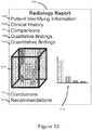
- FIG. 10illustrates a radiology report enhanced with information obtained using the interactive 3D cursor and including quantitative and qualitative analysis.
- FIG. 11illustrates a radiology report enhanced with information obtained using the interactive 3D cursor, and including added quantitative and qualitative analysis at multiple time points.
- FIGS. 12A, 12B and 12Cillustrate a technique for correction for mis-registration at multiple time points using three or more markers.
- FIG. 13illustrates use of multiple interactive 3D cursors to select volumes of interest to designate a safe pre-operative planning pathway for guiding surgical intervention.
- FIG. 14illustrates use of the interactive 3D cursor in an educational setting.
- FIG. 15illustrates process steps on a radiologist's review of a patient's image with integration of the interactive 3D cursor.
- FIG. 16illustrates a system for use of the interactive 3D cursor.
- Some aspects, features and implementations described hereinmay include machines such as computers, electronic components, radiological components, optical components, and processes such as computer-implemented steps. It will be apparent to those of ordinary skill in the art that the computer-implemented steps may be stored as computer-executable instructions on a non-transitory computer-readable medium. Furthermore, it will be understood by those of ordinary skill in the art that the computer-executable instructions may be executed on a variety of tangible processor devices. For ease of exposition, not every step, device or component that may be part of a computer or data storage system is described herein. Those of ordinary skill in the art will recognize such steps, devices and components in view of the teachings of the present disclosure and the knowledge generally available to those of ordinary skill in the art. The corresponding machines and processes are therefore enabled and within the scope of the disclosure.
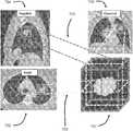
- FIG. 1Aillustrates a 3D (three-dimensional) cursor 100 overlaid on a three-dimensional medical image 102 .
- the 3D cursor 100defines a cubic volume of interest.
- the medical image 102could include any portion of a body, or an entire body, for example and without limitation.
- the medical image 102includes different types of tissue. More specifically, the image includes a background material 104 , such as fat, a lobulated mass 106 , a tubular-shaped vein 108 , and an artery 110 .
- the 3D cursor 100can be moved relative to the image, e.g. in three dimensions, such as by manipulating an IO device such as a 3D mouse, for example and without limitation.
- a button click or other inputdesignates (selects) the portion of the image that is located inside the three-dimensional volume of the 3D cursor 100 . Distinguishing between a 3D image portion selected by a 3D cursor and other unselected image portions is described in US 2016/0026266 and U.S. Pat. No. 8,384,771, both of which are incorporated by reference.
- FIG. 1Billustrates the selected image portion of FIG. 1A . More particularly, unselected portions of the image located outside of an image portion 112 selected with the 3D cursor 100 have been filtered-out or otherwise completely removed from view. Consequently, the removed portions of the image do not obstruct or hinder the view of the selected image portion. Moreover, the selected image portion 112 can be manipulated and viewed as a separate and distinct image from the larger medical image 102 from which it was selected.
- FIG. 1Cillustrates modification of the transparency of the selected image portion 112 . More specifically, transparency may be decreased and/or increased such that tissues and other features can be better observed, e.g. such that overlapping tissues and features are visible. For example, tissues and features located proximate to the back of the selected image portion such as lobulated mass 106 can be seen through overlapping tissues and features located proximate to the front of the selected image portion such as vein 108 , when transparency is sufficiently increased.
- the transparencymay be manipulated with the IO device to achieve various levels of transparency. Further, different levels of transparency may be applied to different portions of the selected image portion.
- FIG. 1Dillustrates filtering of selected areas or tissues of the selected image portion 112 to remove those areas or tissues from view.
- the background material 104 , vein 108 , and an artery 110have been removed from view, leaving only the lobulated mass 106 .
- the tissues to be filtered (removed from view)may be selected based on geometric shape, color, brightness, density, and any other of a variety of available image data, either alone or in combination.
- a designated volume defined by a geometric shapemay be removed, e.g. a geometric shape that traverses tissue boundaries.
- Transparency modification and tissue filteringfacilitate presentation of certain tissue types of concern, both within the cursor and outside of the cursor.
- the medical professionalmust see through any tissue within the cursor but external to the tissue type of concern from the viewing point of the medical professional, thus degrading the visibility of the tissue of concern.
- the illustrated improvementsenable the medical professional to change the transparency of any tissue within the cursor-defined volume but external to the tissue type of concern.
- tissue types not of concernare subtracted from the volume contained within the interactive 3D cursor, leaving only the tissue of concern in the presented image.
- Multiple interactive 3D cursors in combinationcan be used to obtain varying patterns of tissue subtraction. This helps to overcome the limitations of degraded visibility due to tissue within the cursor but external to the tissue type of concern from the viewing point of the medical professional.
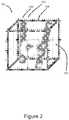
- FIG. 2illustrates an implementation of the 3D cursor 100 with dimensional measurement markings.
- Dimensional measurement markingsmay be available as a feature that can be turned ON and OFF.
- the 3D cursoris a 2 cm by 2 cm by 2 cm cube.
- the dimensional measurement markingsinclude tick marks 200 , 202 , and 204 that respectively designate 1 mm, 5 mm, and 1 cm increments along the edges of the cube (and thus representing three dimensions). Tick marks that represent different magnitudes may be uniquely represented to facilitate visual size determination of the lobulated mass 106 that represents the lesion of interest.
- 1 cm markings 206are presented in each of two dimensions on each side of the cube.
- the dimensional measurement markingscan help serve as a reference for radiologist's activities to include visual assessment, orientation, comparisons with prior scans or measurements. Advantages may include mitigating the current lack of metrics are available to the medical professional to understand the size of the cursor and/or of the tissue elements contained within the cursor. This implementation places measurement metrics on each edge and side of the cursor to help enable the medical professional to rapidly understand the size of the subtended volume within the cursor. In the case where the cursor encapsulates a volume of concern such as a tumor, the three-dimensional size could be recorded in the medical professional report. This can help the visual assessment of each portion of the tumor to aid in the assessment of small changes in size of findings including lobulations of a mass's margin and spiculations.
- location indicators 300 and annotations 302may be placed by the radiologist or by automated techniques to highlight locations or regions of concern within the interactive 3D cursor.
- the location indicatorsmay specify a point or region within the volume of the 3D cursor.
- Annotationscan be added manually by the radiologist or by automated techniques to describe areas that are of concern, e.g., growing, spiculation, irregular margin, indistinct margin, etc. If spiculations are on the surface of a tumor, this could be an indicator of potential malignancy.
- the location indicatorssuch as, but not limited to, arrow(s) pointing to key regions of interest within/outside the 3D cursor helps to overcome the limitation of the inability to mark key points within the cursor. This feature will be useful in discussions between medical professions regarding a patient's condition. It will also be useful in communicating imaging findings between a medical professional and a patient.
- the 3D cursormay be may be implemented in a wide variety of different shapes. Examples include but are not limited to cube, cuboid, cylinder, sphere, ellipsoid, cone and tetrahedron.
- the shapesare not necessarily regular, and the lengths of edges may be resized, e.g. overall geometric shape scaling or changing individual edges, sides, or surfaces.
- FIGS. 4A and 4Billustrate cuboid 3D cursors 400 , 402 for which edge length has been set or selected based on the dimensions and orientation of the respective feature of interest 404 , 406 .
- FIG. 4Cillustrates a spherical 3D cursor 408 for which the diameter may be set or selected based on the dimensions of the feature of interest 410 .
- cursor geometric shapemay be changed.
- the ability to change the size, shape, and individual dimensions of the 3D cursorenables the cursor to be customized based on the particular volume of interest to the medical professional.
- a fixed-shape, fixed-size cursormight be too large or too small, e.g. so as to include a significant amount of tissue not of interest. For example, in examining the lungs, placement of a cube-shaped cursor could cause ribs to be included in the image. Changing the shape of the 3D cursor would help to overcome this limitation.
- Customizationcould be accomplished by wide variety of techniques, possibly including but not limited to selecting an edge, side or vertex of the original 3D cursor with a second type of cursor 412 , and then “clicking and dragging” the selected edge, side, or vertex in the desired direction to expand or reduce the volume of the original 3D cursor.
- the interfacemay also enable selection and change between multiple 3D geometric shapes, e.g. changing from cuboid to spherical. Scrolling on the conventional slices while simultaneously drawing shapes can also be performed to generate the prescribed 3D cursor volume.
- the interactive 3D cursorthus provides an efficient interface for tissue subtraction to provide enhanced visualization of the tumor.
- FIG. 5illustrates presentation of a quantitative analysis 500 of all tissues inside a volume selected with the 3D cursor.
- the illustrated exampleincludes a bar graph but it is to be understood that any of a wide variety of charts, graphs, and other techniques for presentation of data might be implemented.
- Quantitative analysiscan help the radiologist understand how a feature of interest such as tumor 502 (e.g., the lobulated mass 106 , FIG. 1B ) is changing in volume 504 over multiple time points.
- the interfacemay include a statistical representation of the tissue types, possibly including but not limited to a histogram bar chart to depict the volume (e.g., number of voxels per unit volume) of the different types of tissue within the cursor, distinct markings for different types of tissue such as, but not limited to, color coding the bars of the histogram bar chart.
- a histogram bar chartto depict the volume (e.g., number of voxels per unit volume) of the different types of tissue within the cursor, distinct markings for different types of tissue such as, but not limited to, color coding the bars of the histogram bar chart.
- FIG. 6illustrates an implementation of the interactive 3D cursor 100 with false color and transparency to enhance viewing. False color and transparency may be dynamically adjusted and turned ON and OFF. Different false colors may be applied to different tissue types within the volume of the 3D cursor. The colors could be selected to correspond to the colors used in the statistical representation ( FIG. 5 ). Alternatively, a respective unique false color could be selected for each different tissue type, or tissue types of particular interest or concern, and/or additional features of concern, e.g., irregular margin, indistinct margin, spiculation, etc.
- the background material 104(fat) is depicted in light gray
- the artery 110is depicted in red
- the vein 108is depicted in blue
- the lobulated mass 106is multicolored. Different colors may be selected or used to indicate stability of the lobulated mass 106 over time. For example, green may be used to indicate a stable volume 112 while orange is used to denote a slow growth volume 114 , thereby providing a visual warning indicator. Red may be used to indicate high rate of growth or concerning margin volume 116 .
- the extent of the volume of the lobulated masscan be determined automatically, e.g. based on density. Moreover, changes in volume of sub-regions of the lobulated mass may also be automatically determined, and color coding may be automatically implemented. This can help the radiologist understand how the mass is changing in volume over multiple time points.
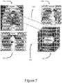
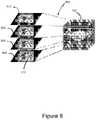
- FIG. 7illustrates association of multiple computed tomography (CT) images of the chest in lung windows with the interactive 3D cursor 100 using reference lines 700 .
- the illustrated exampleincludes an axial image 702 , a sagittal image 704 , and a coronal image 706 of the chest in lung windows.
- An advantageis enhanced ability to cross reference the 3D cursor to the original 2D slices 702 , 704 , 706 from which total 3D volume was obtained.
- Medical professionalshave experience and familiarity with 2D slices and may feel more confident in their findings given the capability to switch back and forth between the 2D and 3D volumetric approaches.
- a small display adjacent to the interactive 3D cursorcould indicate which 2D slices contain tissue within in the interactive 3D cursor. Then the medical professional could direct the system to automatically select those slices which have tissue within the cursor and display them on a nearby 2D display unit.
- a corresponding visible boundary of the 3D cursore.g., red
- FIG. 8illustrates association of multiple axial computed tomography (CT) slices 800 , 802 , 804 , 806 of the chest in lung windows with the interactive 3D cursor 100 using reference lines 808 .
- CTcomputed tomography
- the multiple axial computed tomography (CT) slices of the chest in lung windowsshow the location of the 3D cursor, i.e. the slice area that includes a cross-section of the 3D cursor, which in the illustrated example has selected a left upper lobe mass.
- Boundaries 810 of the 3D cursor in the slicesare depicted in a color, e.g. red.
- the lung cancer mass 106is depicted in gray, surrounded by black that indicates non-cancerous lung tissue.
- This implementationhelps the medical professional to rapidly visualize where the interactive 3D cursor is located relative to the slice images and the body. It also enables the medical professional to visualize the entire volumetric data with the interactive 3D cursor accurately positioned within the volume. Transparency of tissue within the 3D volume could be changed so that the interactive 3D cursor would stand out. This would help avoid left-right orientation mistakes that might occur during treatment. Multiple interactive 3D cursors which could be of differing sizes and/or shapes could be created and displayed.
- FIG. 9illustrates overlay of 3D cursors 100 a , 100 b on a maximum intensity projection (MIP) image 900 of a fludeoxyglucose (18F) positron emission tomography (PET) scan.
- MIPmaximum intensity projection
- PEGpositron emission tomography
- FIG. 10illustrates a radiology report 1000 enhanced with information obtained from the interactive 3D cursor.
- Qualitative findings 1002 and quantitative findings 1004may be included along with patient identifying information 1006 , clinical history 1008 , comparisons 1010 , conclusions 1012 , and recommendations 1014 .
- Also includedare a selected volume image 1016 and statistical graphic 1018 . This helps to quantitatively track changes in volumes of concern (e.g., tumors) over time.
- FIG. 11illustrates a radiology report 1100 enhanced with information obtained from the interactive 3D cursor at multiple time points.
- Qualitative findings 1002 and quantitative findings 1004may be included along with patient identifying information 1006 , clinical history 1008 , comparisons 1010 , conclusions 1012 , and recommendations 1014 .
- follow up reportscan include current and prior exams 1110 , 1112 with quantitative analysis and qualitative analysis on how the lesion has changed over time. This may facilitate selection of a lesion (e.g., tumor) at multiple time points using an interactive 3D cursor; qualitative assessment of the lesion at multiple time points; and, quantitative assessment of the lesion at multiple time points.
- a lesione.g., tumor
- a report of current findings as outlined in the previous implementationcould be placed in a report together with the data obtained from an earlier examination. This would enable tracking over time the progress of treatment or that of changes in tissues of interest/concern.
- FIGS. 12A, 12B, and 12Cillustrate a registration technique by which mis-registration can be corrected at multiple time points through the use of three or more markers 12 , 14 , 16 .
- the mass 106 within each 3D cursor 100 imageis noted using different locations within the interactive 3D cursor and different orientations.
- the usermarks similar locations on each image of the mass with registration markers.
- a yellow marker 12 , a red marker 14 , and a blue marker 16correspond to the same respective parts of the mass on each scan.
- tissues within the interactive 3D cursorare aligned in accordance with markers. Many soft tissues within the body can change in orientation from one scan to the next due to patient movement.
- Corresponding mis-registrationcan limit the ability to properly track how a lesion changes over time.
- This techniqueprovides a method to correct for such mis-registration.
- Three or more recognizable spots of the lesione.g., tumor
- these locationscan be automatically aligned with one another. Shadows can be added to help bring out depth perception. Proper alignment will accurately align the shadows. This enhances visual assessment for how a lesion is changing over time to include changes in tumor composition, size and morphology.
- FIG. 13illustrates use of multiple image volumes selected with the 3D cursor to designate a safe pre-operative planning pathway to guide surgical intervention.
- multiple green interactive 3D cursors 1300mark a surgeon-selected dissection pathway that is deemed safe in the pre-operative setting.
- the interactive 3D cursor 100 containing the cancerous lesion 106is shown at a distal end of the planned surgical path represented by abutting or overlapping volumes selected with the 3D cursors 1300 .
- the selected path that the surgeon will exciseavoids the artery 110 with a minimum clearance of 10 mm. This provides the advantage of 3D depiction of possible surgical cuts.
- the pathcould include, but is not limited to, one or more of the following properties: a serpentine shape; measurements could subsequently be made to measure absolute distance between a point on the planned path to some region of concern (e.g., artery); the path could also be projected on a head mounted display at different intervals during the course of the operation. This feature would facilitate surgical planning as well as a potential to improve accuracy of the surgery.
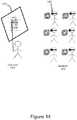
- FIG. 14illustrates use of the interactive 3D cursor in an educational setting. Students 1400 are depicted wearing AR (augmented reality) headsets 1402 and an instructor 1404 is pointing to an abnormality on the board 1406 . This facilitates presentation of medical information (e.g., anatomy) in a classroom environment.
- the interactive 3D cursorcould be placed around the organ of interest and other parts of the body could be eliminated. Items from implementations discussed above such as metrics and arrows could be used.
- the studentswould be provided 3D head displays and joined into a display system so that they could see the tissue within the interactive 3D cursor. This would eliminate any confusion on the part of the students as to what specific detail in the imagery was being discussed.
- FIG. 15illustrates process steps on a radiologist's review of a patient's image with integration of the interactive 3D cursor into his/her practice.
- Step 1is to create an interactive 3D cursor volume and shape that approximates the size and shape of patient organ/tissue corresponding to the item currently being inspected on the checklist.
- Step 2is to position the interactive 3D cursor over the organ/tissue to be inspected.
- the interactive 3D cursor as it is located within the total 3D image volumemay be presented on a display.
- Step 3is to subtract from view all tissue external to the interactive 3D cursor.
- the interactive 3D cursormay be rotated to permit viewing from multiple angles.
- Step 4is to generate a statistical representation e.g., a histogram of tissue densities—color coded with the types of tissue that are suspicious.
- Step 5is to subtract from view additional tissue within the interactive 3D cursor as deemed appropriate by the medical professional.
- Step 6is to inspect the volume within the cursor and identify region(s) of interest and place indicators, annotations, and registration markers relative to region(s) of interest.
- Step 7is to extract a statistical representation and capture imagery showing indicators, annotations, and registration markers and residual tissue within the interactive 3D cursor to be inserted into the medical professional's report.
- Step 8is to use cross-referencing as described the above to confirm findings.
- Step 9is to iterate on the other items on the checklist until finished.
- Step 10is to prepare the report of the medical professional's findings. This procedure provides an opportunity to enhance medical image review process by medical professionals.
- FIG. 16illustrates a system for use of the interactive 3D cursor.
- a medical imaging device 1600is connected to a computer workstation 1602 .
- a wide variety of medical imaging devices and computer workstationscould be used. Images are captured by the medical imaging device and sent to the computer workstation.
- the computer workstationincludes non-volatile storage, computer-readable memory, processors, and a variety of other resources including but not limited to IO devices that provide a human-machine interface.
- the IO devicesinclude a monitor 1604 , keyboard 1606 , 3D mouse 1608 , and VR headset 1610 .
- the IO devicesare used to prompt a software program that runs on the computer workstation to perform the various process steps and implement the various features that have already been described above.
- the interactive 3D cursorThere are multiple potential advantages of the interactive 3D cursor. For example, there is reduction in time spent for classification of multiple lesions. The radiologist doesn't have to sort through many prior imaging studies to find the lesion and the interactive 3D cursor will save time. There is reduction in error when tracking multiple lesions, i.e. reducing the likelihood of mistakes when identifying different specific lesions that are nearby one another when comparing multiple scans.
- One possibilityis to analyze the images obtained using the 3D cursor and using multiple uniquely tagged (e.g. numbered) cursors for any suspicious regions. The medical profession could then switch to slices for confirmation.
Landscapes
- Engineering & Computer Science (AREA)
- Theoretical Computer Science (AREA)
- Physics & Mathematics (AREA)
- General Engineering & Computer Science (AREA)
- General Physics & Mathematics (AREA)
- Health & Medical Sciences (AREA)
- Human Computer Interaction (AREA)
- Medical Informatics (AREA)
- Life Sciences & Earth Sciences (AREA)
- General Health & Medical Sciences (AREA)
- Computer Vision & Pattern Recognition (AREA)
- Radiology & Medical Imaging (AREA)
- Nuclear Medicine, Radiotherapy & Molecular Imaging (AREA)
- Quality & Reliability (AREA)
- Computer Graphics (AREA)
- Computer Hardware Design (AREA)
- Software Systems (AREA)
- Pathology (AREA)
- Biophysics (AREA)
- Public Health (AREA)
- Animal Behavior & Ethology (AREA)
- Surgery (AREA)
- Molecular Biology (AREA)
- Heart & Thoracic Surgery (AREA)
- Biomedical Technology (AREA)
- Veterinary Medicine (AREA)
- Geometry (AREA)
- Signal Processing (AREA)
- Multimedia (AREA)
- High Energy & Nuclear Physics (AREA)
- Optics & Photonics (AREA)
- Vascular Medicine (AREA)
- Apparatus For Radiation Diagnosis (AREA)
- Magnetic Resonance Imaging Apparatus (AREA)
- Ultra Sonic Daignosis Equipment (AREA)
Abstract
Description
This application is a Continuation of U.S. patent application Ser. No. 15/878,463, filed Jan. 24, 2018, now U.S. Pat. No. 10,795,457, which is a Continuation-in-Part of U.S. patent application Ser. No. 14/877,442, filed Oct. 7, 2015, now U.S. Pat. No. 9,980,691, which is a Continuation-in-Part of U.S. patent application Ser. No. 12/176,569, filed Jul. 21, 2008, now U.S. Pat. No. 9,349,183, which is a Continuation-in-Part of U.S. patent application Ser. No. 11/941,578, filed Nov. 16, 2007, now U.S. Pat. No. 8,384,771, which claims the benefit of and priority under 35 U.S.C. § 119(e) to U.S. Patent Application No. 60/877,931, filed Dec. 28, 2006, each of which are incorporated herein by reference in their entirety.
Aspects of this disclosure are generally related to human-machine interfaces, and more particularly to cursors.
The typical arrow-shaped cursor presented by a computer operating system is zero-dimensional. A zero-dimensional cursor designates the location of a single point in a space such as a two-dimensional window presented on a monitor. Mouse buttons can be used in combination with movement of the cursor to select objects in the two-dimensional space, but at any given instant of time a zero-dimensional cursor position designates only a single point in space.
The current standard for diagnostic radiologists reviewing computed tomography (CT) or magnetic resonance imaging (MRI) studies is a slice-by-slice method. A conventional keyboard, monitor, and mouse with a zero-dimensional cursor are used for manipulating the images. The use of mouse buttons and cursor movement for manipulating the images can become burdensome. For example, many images are included in radiology studies that are performed for the follow up of cancer to determine the response to treatment. The ability to recognize and analyze differences between images can be important. As an example, the recent Investigation of Serial Studies to Predict Your Therapeutic Response with Imaging and Molecular Analysis (I-SPY) trial tracked the changes in the tumor over multiple magnetic resonance imaging (MRI) scans during the administration of neoadjuvant chemotherapy (NACT). It has been noted that the phenotypic appearance (e.g., shape, margins) of a tumor correlated with the pathologic response to NACT. A more efficient and accurate interface for manipulating and presenting medical images would therefore have utility.
Known techniques for 3D viewing of medical images are described in U.S. Pat. No. 9,349,183, Method and Apparatus for Three Dimensional Viewing of Images, issued to Douglas, U.S. Pat. No. 8,384,771, Method and Apparatus for Three Dimensional Viewing of Images, issued to Douglas, Douglas, D. B., Petricoin, E. F., Liotta L., Wilson, E. D3D augmented reality imaging system: proof of concept in mammography.Med Devices(Auckl), 2016; 9:277-83, Douglas, D. B., Boone, J. M., Petricoin, E., Liotta, L., Wilson, E. Augmented Reality Imaging System: 3D Viewing of a Breast Cancer.J Nat Sci.2016; 2(9), and Douglas, D. B., Wilke, C. A., Gibson, J. D., Boone, J. M., Wintermark, M. Augmented Reality: Advances in Diagnostic Imaging.Multimodal Technologies and Interaction,2017; 1(4):29. In D3D imaging, the radiologist wears an augmented reality (AR), mixed reality (MR) or virtual reality (VR) headset and uses a joystick or gaming controller. Advantages include improved depth perception and human machine interface. Still, there are several challenges faced with this approach. First, an area of interest (e.g. tumor) may be in close proximity to structures that are similar in composition/density. Isolating the area of interest for better examination may be difficult. Second, many soft tissues in the body are mobile and deformable, so it can be difficult to achieve the best orientation to properly compare the tumor at multiple time points. Efficiently aligning the orientation to do so may be difficult. Third, certain portions of a tumor can respond to treatment and decrease in size while other portions of a tumor demonstrate increases in size. The pattern of tumor shrinkage has important prognostic implications. Furthermore, composition and complex morphologic features including spiculations (spikes extending from the surface), irregular margins and enhancement also have important implications. Consequently, there is a need for a system that facilitates recognition of the subtle, yet important changes in size, shape and margins. Fourth, a patient with metastatic cancer has several areas of interest in different areas of the body. It is difficult and time consuming to find each of the areas of interest at every time point to determine interval change. Consequently, there is a need for a system that enables the observer to do this efficiently.
All examples, aspects and features mentioned in this document can be combined in any technically possible way.
In accordance with an aspect of the invention a method comprises: generating a three-dimensional cursor that has a non-zero volume; responsive to a first input, moving the three-dimensional cursor within a three-dimensional image; responsive to a second input, selecting a volume of the three-dimensional image designated by the three-dimensional cursor; and responsive to a third input, presenting a modified version of the selected volume of the three-dimensional image. In some implementations presenting the modified version of the selected volume of the three-dimensional image comprises removing an un-selected volume of the three-dimensional image from view. In some implementations presenting the modified version of the selected volume of the three-dimensional image comprises changing transparency of presented tissues within the selected volume. In some implementations presenting the modified version of the selected volume of the three-dimensional image comprises filtering a selected tissue to remove the selected tissue from view. In some implementations presenting the three-dimensional cursor with measurement markings on at least one edge, surface or side. In some implementations presenting the modified version of the selected volume of the three-dimensional image comprises presenting inputted location indicators. In some implementations presenting the modified version of the selected volume of the three-dimensional image comprises presenting inputted annotations. Some implementations comprise changing a size dimension of the three-dimensional cursor responsive to a fourth input. Some implementations comprise changing a geometric shape of the three-dimensional cursor responsive to a fifth input. Some implementations comprise automatically generating a statistical representation of the selected volume of the three-dimensional image. In some implementations presenting the modified version of the selected volume of the three-dimensional image comprises presenting at least one tissue type with false color. In some implementations presenting the modified version of the selected volume of the three-dimensional image comprises presenting volumetric changes over time with false color. Some implementations comprise presenting multiple computed tomography images associated with the selected volume using reference lines. Some implementations comprise presenting multiple axial computed tomography images associated with the selected volume using reference lines. Some implementations comprise presenting a maximum intensity projection (MIP) image of a positron emission tomography (PET) scan with the three-dimensional cursor overlaid thereon to indicate orientation and location of the selected volume. Some implementations comprise presenting a radiology report enhanced with information obtained using the three-dimensional cursor. Some implementations comprise automatically calculating and presenting a quantitative analysis and a qualitative analysis associated with multiple time points. Some implementations comprise presenting the modified version of the selected volume of the three-dimensional image comprises presenting inputted registration markers. Some implementations comprise automatically calculating volumetric change based on the registration markers. Some implementations comprise automatically re-orienting the selected volume of the three-dimensional image based on the registration markers. Some implementations comprise using multiple volumes selected with the three-dimensional cursor to designate a pre-operative planning pathway for guiding surgical intervention. Some implementations comprise presenting the selected volume with an augmented reality, virtual reality or mixed reality headset.
In accordance with an aspect of the invention an apparatus comprises: a computing device; and a human-machine interface comprising a three-dimensional cursor that has a non-zero volume; the human-machine interface moving the three-dimensional cursor within a three-dimensional image responsive to a first input; the human-machine interface selecting a volume of the three-dimensional image designated by the three-dimensional cursor responsive to a second input; and the human-machine interface presenting a modified version of the selected volume of the three-dimensional image responsive to a third input. In some implementations, the human-machine interface removes an un-selected volume of the three-dimensional image from view. In some implementations, the human-machine interface changes transparency of presented tissues within the selected volume. In some implementations, the human-machine interface filters a selected tissue to remove the selected tissue from view. In some implementations, the human-machine interface presents the three-dimensional cursor with measurement markings on at least one edge, surface or side. In some implementations, the human-machine interface receives and implements inputted location indicators. In some implementations, the human-machine interface receives and implements inputted annotations. In some implementations, the human-machine interface changes a size dimension of the three-dimensional cursor responsive to a fourth input. In some implementations, the human-machine interface changes a geometric shape of the three-dimensional cursor responsive to a fifth input. In some implementations, the human-machine interface automatically generates and presents a statistical representation of the selected volume of the three-dimensional image. In some implementations, the human-machine interface presents at least one tissue type with false color. In some implementations, the human-machine interface presents volumetric changes over time with false color. In some implementations, the human-machine interface presents multiple computed tomography images associated with the selected volume using reference lines. In some implementations, the human-machine interface presents multiple axial computed tomography images associated with the selected volume using reference lines. In some implementations, the human-machine interface presents a maximum intensity projection (MIP) image of a positron emission tomography (PET) scan with the three-dimensional cursor overlaid thereon to indicate orientation and location of the selected volume. In some implementations, the human-machine interface presents a radiology report enhanced with information obtained using the three-dimensional cursor. In some implementations, the human-machine interface automatically calculates and presents a quantitative analysis and a qualitative analysis associated with multiple time points. In some implementations, the human-machine interface presents inputted registration markers. In some implementations, the human-machine interface automatically calculates volumetric change after appropriate registration using the registration markers. In some implementations, the human-machine interface automatically re-orients the selected volume of the three-dimensional image based on the registration markers. In some implementations, the human-machine interface presents multiple volumes selected with the three-dimensional cursor to designate a pre-operative planning pathway for guiding surgical intervention. In some implementations, the human-machine interface presents the selected volume with an augmented reality, virtual reality or mixed reality headset.
The patent or application file contains at least one drawing executed in color. Copies of this patent or patent application publication with color drawing(s) will be provided by the Office upon request and payment of the necessary fee.
Some aspects, features and implementations described herein may include machines such as computers, electronic components, radiological components, optical components, and processes such as computer-implemented steps. It will be apparent to those of ordinary skill in the art that the computer-implemented steps may be stored as computer-executable instructions on a non-transitory computer-readable medium. Furthermore, it will be understood by those of ordinary skill in the art that the computer-executable instructions may be executed on a variety of tangible processor devices. For ease of exposition, not every step, device or component that may be part of a computer or data storage system is described herein. Those of ordinary skill in the art will recognize such steps, devices and components in view of the teachings of the present disclosure and the knowledge generally available to those of ordinary skill in the art. The corresponding machines and processes are therefore enabled and within the scope of the disclosure.
Transparency modification and tissue filtering facilitate presentation of certain tissue types of concern, both within the cursor and outside of the cursor. Currently, the medical professional must see through any tissue within the cursor but external to the tissue type of concern from the viewing point of the medical professional, thus degrading the visibility of the tissue of concern. The illustrated improvements enable the medical professional to change the transparency of any tissue within the cursor-defined volume but external to the tissue type of concern. Alternatively, tissue types not of concern are subtracted from the volume contained within the interactive 3D cursor, leaving only the tissue of concern in the presented image. Multiple interactive 3D cursors in combination can be used to obtain varying patterns of tissue subtraction. This helps to overcome the limitations of degraded visibility due to tissue within the cursor but external to the tissue type of concern from the viewing point of the medical professional.
The dimensional measurement markings can help serve as a reference for radiologist's activities to include visual assessment, orientation, comparisons with prior scans or measurements. Advantages may include mitigating the current lack of metrics are available to the medical professional to understand the size of the cursor and/or of the tissue elements contained within the cursor. This implementation places measurement metrics on each edge and side of the cursor to help enable the medical professional to rapidly understand the size of the subtended volume within the cursor. In the case where the cursor encapsulates a volume of concern such as a tumor, the three-dimensional size could be recorded in the medical professional report. This can help the visual assessment of each portion of the tumor to aid in the assessment of small changes in size of findings including lobulations of a mass's margin and spiculations.
Referring toFIG. 3 ,location indicators 300 andannotations 302 may be placed by the radiologist or by automated techniques to highlight locations or regions of concern within the interactive 3D cursor. The location indicators may specify a point or region within the volume of the 3D cursor. Annotations can be added manually by the radiologist or by automated techniques to describe areas that are of concern, e.g., growing, spiculation, irregular margin, indistinct margin, etc. If spiculations are on the surface of a tumor, this could be an indicator of potential malignancy. The location indicators, such as, but not limited to, arrow(s) pointing to key regions of interest within/outside the 3D cursor helps to overcome the limitation of the inability to mark key points within the cursor. This feature will be useful in discussions between medical professions regarding a patient's condition. It will also be useful in communicating imaging findings between a medical professional and a patient.
Referring toFIGS. 4A, 4B, and 4C , the 3D cursor may be may be implemented in a wide variety of different shapes. Examples include but are not limited to cube, cuboid, cylinder, sphere, ellipsoid, cone and tetrahedron. The shapes are not necessarily regular, and the lengths of edges may be resized, e.g. overall geometric shape scaling or changing individual edges, sides, or surfaces. For example,FIGS. 4A and 4B illustratecuboid 3D cursors interest FIG. 4C illustrates aspherical 3D cursor 408 for which the diameter may be set or selected based on the dimensions of the feature ofinterest 410. In addition to dimensional changes, cursor geometric shape may be changed.
The ability to change the size, shape, and individual dimensions of the 3D cursor enables the cursor to be customized based on the particular volume of interest to the medical professional. A fixed-shape, fixed-size cursor might be too large or too small, e.g. so as to include a significant amount of tissue not of interest. For example, in examining the lungs, placement of a cube-shaped cursor could cause ribs to be included in the image. Changing the shape of the 3D cursor would help to overcome this limitation. Customization could be accomplished by wide variety of techniques, possibly including but not limited to selecting an edge, side or vertex of the original 3D cursor with a second type ofcursor 412, and then “clicking and dragging” the selected edge, side, or vertex in the desired direction to expand or reduce the volume of the original 3D cursor. The interface may also enable selection and change between multiple 3D geometric shapes, e.g. changing from cuboid to spherical. Scrolling on the conventional slices while simultaneously drawing shapes can also be performed to generate the prescribed 3D cursor volume. The interactive 3D cursor thus provides an efficient interface for tissue subtraction to provide enhanced visualization of the tumor.
There are multiple potential advantages of the interactive 3D cursor. For example, there is reduction in time spent for classification of multiple lesions. The radiologist doesn't have to sort through many prior imaging studies to find the lesion and the interactive 3D cursor will save time. There is reduction in error when tracking multiple lesions, i.e. reducing the likelihood of mistakes when identifying different specific lesions that are nearby one another when comparing multiple scans. One possibility is to analyze the images obtained using the 3D cursor and using multiple uniquely tagged (e.g. numbered) cursors for any suspicious regions. The medical profession could then switch to slices for confirmation.
Several features, aspects, embodiments and implementations have been described. Nevertheless, it will be understood that a wide variety of modifications and combinations may be made without departing from the scope of the inventive concepts described herein. Accordingly, those modifications and combinations are within the scope of the following claims.
Claims (28)
1. A display unit comprising:
a processor;
a left eye display;
a right eye display;
a non-transitory memory having computer-executable instructions stored thereupon, which when executed by the processor of the display unit, cause the display unit to perform the operations of:
selecting a viewpoint of a volume of interest for a left eye of a user;
selecting a viewpoint of the volume of interest for a right eye of the user;
determining a first convergence point for the left eye of the user and the right eye of the user;
displaying a first left eye image on the left eye display and a first right eye image on the right eye display, wherein the first left eye image and the first right eye image are at least based on the volume of interest and the first convergence point;
determining a second convergence point for the left eye of the user and the right eye of the user; and
displaying a second left eye image on the left eye display and a second right eye image on the right eye display, wherein the second left eye image and the second right eye image are at least based on the volume of interest and the second convergence point.
2. The display unit ofclaim 1 , wherein the volume of interest during the display of the second left eye image and the second right eye image is filtered.
3. The display unit ofclaim 2 , wherein items within the volume of interest during the display of the second left eye image and the second right eye image are selected to be filtered by voxel property.
4. The display unit ofclaim 2 , wherein items within the volume of interest during the display of the second left eye image and the second right eye image are selected to be filtered by a geometric boundary method wherein. items on one side of a geometric boundary are selected for removal.
5. The display unit ofclaim 1 , further comprising instructions that zoom in on a portion of the volume of interest during the display of the second left eye image and the second right eye image.
6. The display unit ofclaim 1 , further comprising instructions that rotate a portion of the volume of interest during the display of the second left eye image and the second right eye image.
7. The display unit ofclaim 1 , further comprising instructions that color a portion of the volume of interest during the display of the second left eye image and the second right eye image.
8. A display unit comprising:
a communications interface, wherein the communications interface is in communication with a non-transitory memory and a processor, the non-transitory memory having computer-executable instructions, which when executed by the processor, perform the operations of:
selecting a viewpoint of a volume of interest for a left eye of a user;
selecting a viewpoint of the volume of interest for a right eye of the user;
determining a first convergence point for the left eye of the user and the right eye of the user;
determining a first left eye image and a first right eye image wherein the first left eye image and the first right eye image are based on the volume of interest and the first convergence point;
determining a second convergence point for the left eye of the user and the right eye of the user; and
determining a second left eye image and a second right eye image wherein the second left eye image and the second right eye image are based on the volume of interest and the second convergence point; and
a left eye display configured to display the first left eye image and subsequently display the second left eye image; and
a right eye display configured to display the first right eye image and subsequently display the second right eye image.
9. The display unit ofclaim 8 , further comprising wherein the volume of interest during the display of the second left eye image and the second right eye image is filtered.
10. The display unit ofclaim 9 , wherein items within the volume of interest during the display of the second left eye image and the second right eye image are selected to be filtered by voxel property.
11. The display unit ofclaim 9 , wherein items within the volume of interest during the display of the second left eye image and the second right eye image are selected to be filtered by a geometric boundary method wherein items on one side of a geometric boundary are selected for removal.
12. The display unit ofclaim 8 , further comprising zooming in on a portion of the volume of interest during the display of the second left eye image and the second right eye image.
13. The display unit ofclaim 8 , further comprising rotating a portion of the volume of interest during the display of the second left eye image and the second right eye image.
14. The display unit ofclaim 8 , further comprising coloring a portion of the volume of interest during the display of the second left eye image and the second right eye image.
15. A non-transitory computer storage medium having computer-executable instructions stored thereon which, when executed by a processor, cause the processor to perform the operations of:
selecting a viewpoint of a volume of interest for a left eye of a user;
selecting a viewpoint of the volume of interest for a right eye of the user;
determining a first convergence point for the left eye of the user and the right eye of the user;
determining a first left eye image and a first right eye image, wherein the first left eye image and the first right eye image are based on the volume of interest and the first convergence point;
determining a second convergence point for the left eye of the user and the right eye of the user; and
determining a second left eye image and a second right eye image, wherein the second left eye image and the second right eye image are based on the volume of interest and the second convergence point.
16. The non-transitory computer storage medium ofclaim 15 , wherein the volume of interest during the determining a the second left eye image and the second right eye image is filtered.
17. The non-transitory computer storage medium ofclaim 16 , wherein items within the volume of interest during the determining of the second left eye image and the second right eye image are selected to be filtered by voxel property.
18. The non-transitory computer storage medium ofclaim 16 , wherein items within the volume of interest during the determining of the second left eye image and the second right eye image are selected to be filtered by a geometric boundary method wherein items on one side of a geometric boundary are selected for removal.
19. The non-transitory computer storage medium ofclaim 15 , further comprising instructions that zoom in on a portion of the volume of interest during the determining of the second left eye image and the second right eye image.
20. The non-transitory computer storage medium ofclaim 15 , further comprising instructions that rotate a portion of the volume of interest during the determining of the second left eye image and the second right eye image.
21. The non-transitory computer storage medium ofclaim 15 , further comprising instructions that color a portion of the volume of interest during the determining of the second left eye image and the second right eye image.
22. A method comprising:
selecting a viewpoint of a volume of interest for a left eye of a user;
selecting a viewpoint of the volume of interest for a right eye of the user;
determining a first volume of interest;
determining a first convergence point for the left eye of the user and the right eye of the user;
displaying a first left eye image on a left eye display and a first right eye image on a right eye display, wherein the first left eye image and the first right eye image are based on the first volume of interest and the first convergence point;
determining a second volume of interest;
determining a second convergence point for the left eye of the user and the right eye of the user; and
displaying a second left eye image on the left eye display and a second right eye image on the right eye display, wherein the second left eye image and the second right eye image are based on the second volume of interest and the second convergence point.
23. The method ofclaim 22 , wherein the second volume of interest is filtered.
24. The method ofclaim 23 , wherein items within the second are selected to be filtered by voxel property.
25. The method ofclaim 23 , wherein items within the second volume of interest are selected to be filtered by a geometric boundary method wherein items on one side of a geometric boundary are selected for removal.
26. The method ofclaim 22 , further comprising zooming in on a portion of the second volume of interest.
27. The method ofclaim 22 , further comprising rotating a portion of the second volume of interest.
28. The method ofclaim 22 , further comprising coloring a portion of the second volume of interest.
Priority Applications (6)
| Application Number | Priority Date | Filing Date | Title |
|---|---|---|---|
| US17/021,548US10936090B2 (en) | 2006-12-28 | 2020-09-15 | Interactive 3D cursor for use in medical imaging |
| US17/095,411US10942586B1 (en) | 2006-12-28 | 2020-11-11 | Interactive 3D cursor for use in medical imaging |
| US17/122,518US11036311B2 (en) | 2006-12-28 | 2020-12-15 | Method and apparatus for 3D viewing of images on a head display unit |
| US17/122,549US11016579B2 (en) | 2006-12-28 | 2020-12-15 | Method and apparatus for 3D viewing of images on a head display unit |
| US17/339,341US11520415B2 (en) | 2006-12-28 | 2021-06-04 | Interactive 3D cursor for use in medical imaging |
| US18/047,256US12333087B2 (en) | 2006-12-28 | 2022-10-17 | Interactive 3D cursor |
Applications Claiming Priority (6)
| Application Number | Priority Date | Filing Date | Title |
|---|---|---|---|
| US87793106P | 2006-12-28 | 2006-12-28 | |
| US11/941,578US8384771B1 (en) | 2006-12-28 | 2007-11-16 | Method and apparatus for three dimensional viewing of images |
| US12/176,569US9349183B1 (en) | 2006-12-28 | 2008-07-21 | Method and apparatus for three dimensional viewing of images |
| US14/877,442US9980691B2 (en) | 2006-12-28 | 2015-10-07 | Method and apparatus for three dimensional viewing of images |
| US15/878,463US10795457B2 (en) | 2006-12-28 | 2018-01-24 | Interactive 3D cursor |
| US17/021,548US10936090B2 (en) | 2006-12-28 | 2020-09-15 | Interactive 3D cursor for use in medical imaging |
Related Parent Applications (1)
| Application Number | Title | Priority Date | Filing Date |
|---|---|---|---|
| US15/878,463ContinuationUS10795457B2 (en) | 2006-12-28 | 2018-01-24 | Interactive 3D cursor |
Related Child Applications (3)
| Application Number | Title | Priority Date | Filing Date |
|---|---|---|---|
| US17/095,411ContinuationUS10942586B1 (en) | 2006-12-28 | 2020-11-11 | Interactive 3D cursor for use in medical imaging |
| US17/122,549ContinuationUS11016579B2 (en) | 2006-12-28 | 2020-12-15 | Method and apparatus for 3D viewing of images on a head display unit |
| US17/122,518ContinuationUS11036311B2 (en) | 2006-12-28 | 2020-12-15 | Method and apparatus for 3D viewing of images on a head display unit |
Publications (2)
| Publication Number | Publication Date |
|---|---|
| US20200409480A1 US20200409480A1 (en) | 2020-12-31 |
| US10936090B2true US10936090B2 (en) | 2021-03-02 |
Family
ID=67298617
Family Applications (8)
| Application Number | Title | Priority Date | Filing Date |
|---|---|---|---|
| US15/878,463Active2027-11-29US10795457B2 (en) | 2006-12-28 | 2018-01-24 | Interactive 3D cursor |
| US16/506,073Active2027-11-19US11202061B1 (en) | 2006-12-28 | 2019-07-09 | Illustrating direction of blood flow via pointers |
| US17/021,548ActiveUS10936090B2 (en) | 2006-12-28 | 2020-09-15 | Interactive 3D cursor for use in medical imaging |
| US17/095,411ActiveUS10942586B1 (en) | 2006-12-28 | 2020-11-11 | Interactive 3D cursor for use in medical imaging |
| US17/122,518ActiveUS11036311B2 (en) | 2006-12-28 | 2020-12-15 | Method and apparatus for 3D viewing of images on a head display unit |
| US17/122,549ActiveUS11016579B2 (en) | 2006-12-28 | 2020-12-15 | Method and apparatus for 3D viewing of images on a head display unit |
| US17/339,341ActiveUS11520415B2 (en) | 2006-12-28 | 2021-06-04 | Interactive 3D cursor for use in medical imaging |
| US18/047,256ActiveUS12333087B2 (en) | 2006-12-28 | 2022-10-17 | Interactive 3D cursor |
Family Applications Before (2)
| Application Number | Title | Priority Date | Filing Date |
|---|---|---|---|
| US15/878,463Active2027-11-29US10795457B2 (en) | 2006-12-28 | 2018-01-24 | Interactive 3D cursor |
| US16/506,073Active2027-11-19US11202061B1 (en) | 2006-12-28 | 2019-07-09 | Illustrating direction of blood flow via pointers |
Family Applications After (5)
| Application Number | Title | Priority Date | Filing Date |
|---|---|---|---|
| US17/095,411ActiveUS10942586B1 (en) | 2006-12-28 | 2020-11-11 | Interactive 3D cursor for use in medical imaging |
| US17/122,518ActiveUS11036311B2 (en) | 2006-12-28 | 2020-12-15 | Method and apparatus for 3D viewing of images on a head display unit |
| US17/122,549ActiveUS11016579B2 (en) | 2006-12-28 | 2020-12-15 | Method and apparatus for 3D viewing of images on a head display unit |
| US17/339,341ActiveUS11520415B2 (en) | 2006-12-28 | 2021-06-04 | Interactive 3D cursor for use in medical imaging |
| US18/047,256ActiveUS12333087B2 (en) | 2006-12-28 | 2022-10-17 | Interactive 3D cursor |
Country Status (1)
| Country | Link |
|---|---|
| US (8) | US10795457B2 (en) |
Cited By (1)
| Publication number | Priority date | Publication date | Assignee | Title |
|---|---|---|---|---|
| US11520415B2 (en) | 2006-12-28 | 2022-12-06 | D3D Technologies, Inc. | Interactive 3D cursor for use in medical imaging |
Families Citing this family (23)
| Publication number | Priority date | Publication date | Assignee | Title |
|---|---|---|---|---|
| US11228753B1 (en) | 2006-12-28 | 2022-01-18 | Robert Edwin Douglas | Method and apparatus for performing stereoscopic zooming on a head display unit |
| US11275242B1 (en) | 2006-12-28 | 2022-03-15 | Tipping Point Medical Images, Llc | Method and apparatus for performing stereoscopic rotation of a volume on a head display unit |
| US11315307B1 (en) | 2006-12-28 | 2022-04-26 | Tipping Point Medical Images, Llc | Method and apparatus for performing rotating viewpoints using a head display unit |
| US8017915B2 (en)* | 2008-03-14 | 2011-09-13 | Reflexion Medical, Inc. | Method and apparatus for emission guided radiation therapy |
| US10867445B1 (en)* | 2016-11-16 | 2020-12-15 | Amazon Technologies, Inc. | Content segmentation and navigation |
| EP3641884B8 (en) | 2017-06-22 | 2024-01-03 | RefleXion Medical, Inc. | Methods for calculating bounded dose-volume histograms (dvh) for evaluating a treatment plan |
| EP4342521A3 (en) | 2017-07-11 | 2024-05-08 | RefleXion Medical Inc. | Methods for pet detector afterglow management |
| US20190021677A1 (en)* | 2017-07-18 | 2019-01-24 | Siemens Healthcare Gmbh | Methods and systems for classification and assessment using machine learning |
| EP4592713A2 (en) | 2017-08-09 | 2025-07-30 | RefleXion Medical, Inc. | Systems and methods for fault detection in emission-guided radiotherapy |
| CN111372653B (en) | 2017-09-22 | 2022-06-17 | 反射医疗公司 | System and method for shuttle mode radiation delivery |
| WO2019099551A1 (en) | 2017-11-14 | 2019-05-23 | Reflexion Medical, Inc. | Systems and methods for patient monitoring for radiotherapy |
| WO2019160958A1 (en) | 2018-02-13 | 2019-08-22 | Reflexion Medical, Inc. | Beam station treatment planning and radiation delivery methods |
| US11417071B1 (en) | 2018-02-23 | 2022-08-16 | Red Pacs, Llc | Virtual toolkit for radiologists |
| US11442534B1 (en) | 2018-04-10 | 2022-09-13 | Red Pacs, Llc | Smart glasses system |
| US11207133B1 (en) | 2018-09-10 | 2021-12-28 | David Byron Douglas | Method and apparatus for the interaction of virtual tools and geo-registered tools |
| US11158045B2 (en) | 2018-10-10 | 2021-10-26 | David Byron Douglas | Method and apparatus for performing 3D imaging examinations of a structure under differing configurations and analyzing morphologic changes |
| WO2021067843A1 (en)* | 2019-10-03 | 2021-04-08 | Whiterabbit.Ai Inc. | Methods and systems for management and visualization of radiological data |
| EP4147449A1 (en)* | 2020-05-06 | 2023-03-15 | InterDigital CE Patent Holdings | 3d scene transmission with alpha layers |
| EP4192576A4 (en) | 2020-08-07 | 2024-08-28 | RefleXion Medical Inc. | MULTI-SENSOR GUIDED RADIOTHERAPY |
| US20230298291A1 (en)* | 2022-03-16 | 2023-09-21 | Autodesk, Inc. | Voxel-based approach for design models |
| EP4471714A1 (en)* | 2023-05-30 | 2024-12-04 | Koninklijke Philips N.V. | A computer-implemented method and image processing device for marking objects on 3d images |
| US12020394B1 (en)* | 2023-06-14 | 2024-06-25 | Procore Technologies, Inc. | Visualization tool for cross sections |
| US20250138709A1 (en)* | 2023-10-27 | 2025-05-01 | GE Precision Healthcare LLC | Methods and systems for medical image rendering |
Citations (202)
| Publication number | Priority date | Publication date | Assignee | Title |
|---|---|---|---|---|
| US4472737A (en) | 1982-08-31 | 1984-09-18 | Tokyo Shibaura Denki Kabushiki Kaisha | Stereographic tomogram observing apparatus |
| US4808979A (en) | 1987-04-02 | 1989-02-28 | Tektronix, Inc. | Cursor for use in 3-D imaging systems |
| US4952024A (en) | 1986-08-29 | 1990-08-28 | Gale Thomas S | Three-dimensional sight and sound reproduction apparatus for individual use |
| US4987527A (en) | 1987-10-26 | 1991-01-22 | Hitachi, Ltd. | Perspective display device for displaying and manipulating 2-D or 3-D cursor, 3-D object and associated mark position |
| US5049987A (en) | 1989-10-11 | 1991-09-17 | Reuben Hoppenstein | Method and apparatus for creating three-dimensional television or other multi-dimensional images |
| US5200819A (en) | 1988-05-27 | 1993-04-06 | The University Of Connecticut | Multi-dimensional imaging system for endoscope |
| US5233458A (en) | 1991-07-01 | 1993-08-03 | Kaiser Aerospace And Electronics Corporation | Method and apparatus to reduce binocular rivalry in a partial overlap binocular display |
| EP0571827A1 (en) | 1992-05-27 | 1993-12-01 | International Business Machines Corporation | System and method for augmentation of endoscopic surgery |
| US5278884A (en) | 1992-12-18 | 1994-01-11 | General Electric Company | Complete 3D CT data acquisition using practical scanning paths on the surface of a sphere |
| US5293529A (en) | 1991-03-12 | 1994-03-08 | Matsushita Electric Industrial Co., Ltd. | Three-dimensional information handling system |
| US5371778A (en) | 1991-11-29 | 1994-12-06 | Picker International, Inc. | Concurrent display and adjustment of 3D projection, coronal slice, sagittal slice, and transverse slice images |
| US5402191A (en) | 1992-12-09 | 1995-03-28 | Imax Corporation | Method and apparatus for presenting stereoscopic images |
| US5510832A (en) | 1993-12-01 | 1996-04-23 | Medi-Vision Technologies, Inc. | Synthesized stereoscopic imaging system and method |
| US5621867A (en) | 1992-08-26 | 1997-04-15 | Namco Ltd. | Image synthesizing system |
| US5627582A (en) | 1993-11-29 | 1997-05-06 | Canon Kabushiki Kaisha | Stereoscopic compression processing with added phase reference |
| US5659625A (en) | 1992-06-04 | 1997-08-19 | Marquardt; Stephen R. | Method and apparatus for analyzing facial configurations and components |
| US5682172A (en) | 1994-12-30 | 1997-10-28 | Forte Technologies, Inc. | Headset for presenting video and audio signals to a wearer |
| US5682437A (en) | 1994-09-22 | 1997-10-28 | Sanyo Electric Co., Ltd. | Method of converting two-dimensional images into three-dimensional images |
| US5696521A (en) | 1994-06-22 | 1997-12-09 | Astounding Technologies (M) Sdn. Bhd. | Video headset |
| US5708359A (en) | 1995-12-04 | 1998-01-13 | The Board Of Trustees Of The University Of Illinois | Interactive, stereoscopic magnetic resonance imaging system |
| US5841830A (en) | 1997-02-19 | 1998-11-24 | Picker International, Inc. | 3D CT fluoroscopy |
| US5850352A (en) | 1995-03-31 | 1998-12-15 | The Regents Of The University Of California | Immersive video, including video hypermosaicing to generate from multiple video views of a scene a three-dimensional video mosaic from which diverse virtual video scene images are synthesized, including panoramic, scene interactive and stereoscopic images |
| WO1999023586A2 (en) | 1997-10-30 | 1999-05-14 | Dr. Baldeweg Gmbh | Method and device for processing imaged objects |
| JPH11232010A (en) | 1998-02-18 | 1999-08-27 | Dainippon Screen Mfg Co Ltd | Method and device for three-dimensional instruction |
| US6002518A (en) | 1990-06-11 | 1999-12-14 | Reveo, Inc. | Phase-retardation based system for stereoscopic viewing micropolarized spatially-multiplexed images substantially free of visual-channel cross-talk and asymmetric image distortion |
| US6034716A (en) | 1997-12-18 | 2000-03-07 | Whiting; Joshua B. | Panoramic digital camera system |
| US6052100A (en) | 1994-03-16 | 2000-04-18 | The United States Of America Represented By The Secertary Of The Navy | Computer controlled three-dimensional volumetric display |
| US6057827A (en) | 1993-06-18 | 2000-05-02 | Artifice, Inc. | Automatic pointer positioning for 3D computer modeling |
| US6066095A (en) | 1998-05-13 | 2000-05-23 | Duke University | Ultrasound methods, systems, and computer program products for determining movement of biological tissues |
| US6084937A (en) | 1998-07-27 | 2000-07-04 | Siemens Corporate Research, Inc. | Adaptive mask boundary correction in a cone beam imaging system |
| US6100862A (en) | 1998-04-20 | 2000-08-08 | Dimensional Media Associates, Inc. | Multi-planar volumetric display system and method of operation |
| US6108005A (en) | 1996-08-30 | 2000-08-22 | Space Corporation | Method for producing a synthesized stereoscopic image |
| US6115449A (en) | 1998-10-10 | 2000-09-05 | Nanotek Instruments, Inc. | Apparatus for quantitative stereoscopic radiography |
| US6124977A (en) | 1997-11-12 | 2000-09-26 | Olympus Optical Co., Ltd. | Image display apparatus |
| US6130930A (en) | 1999-03-22 | 2000-10-10 | Siemens Corporate Research, Inc. | Exact region of interest cone beam imaging without circle scans |
| US6225979B1 (en) | 1997-07-22 | 2001-05-01 | Sanyo Electric Co., Ltd. | Cursor display method and apparatus for perceiving cursor as stereoscopic image in stereoscopic display region, and recording medium recorded with cursor display program that can be read out by computer |
| US6272366B1 (en) | 1994-10-27 | 2001-08-07 | Wake Forest University | Method and system for producing interactive three-dimensional renderings of selected body organs having hollow lumens to enable simulated movement through the lumen |
| US6275561B1 (en) | 1998-01-13 | 2001-08-14 | U.S. Philips Corporation | Computer tomagraphy method with helicoidal scanning of an examination area |
| US6276799B1 (en) | 1997-10-15 | 2001-08-21 | The Lions Eye Institute Of Western Australia Incorporated | Stereo optic disc analyzer |
| US6342378B1 (en) | 1998-08-07 | 2002-01-29 | The Regents Of The University Of California | Biogasification of solid waste with an anaerobic-phased solids-digester system |
| US6342878B1 (en) | 1997-09-18 | 2002-01-29 | Aerospatiale Societe Nationale Industrielle | Input peripheral for computer with automatic switching between 3D and 2D operation modes, and its interaction process with a display screen |
| US20020068863A1 (en) | 2000-12-04 | 2002-06-06 | Slack Christopher C. | Method and system for selecting and displaying medical image data |
| US20020101658A1 (en) | 2001-01-29 | 2002-08-01 | Reuben Hoppenstein | Electronic virtual lens for observing 3-D or 4-D images |
| US20020112237A1 (en) | 2000-04-10 | 2002-08-15 | Kelts Brett R. | System and method for providing an interactive display interface for information objects |
| US6442417B1 (en) | 1999-11-29 | 2002-08-27 | The Board Of Trustees Of The Leland Stanford Junior University | Method and apparatus for transforming view orientations in image-guided surgery |
| US6449309B1 (en) | 1996-03-12 | 2002-09-10 | Olympus Optical Co., Ltd. | Stereoscopic display that controls binocular parallax between two images and controls image reconstitution according to parallax data |
| US6466185B2 (en) | 1998-04-20 | 2002-10-15 | Alan Sullivan | Multi-planar volumetric display system and method of operation using psychological vision cues |
| US6476607B1 (en) | 2000-12-08 | 2002-11-05 | Koninklijke Philips Electronics N.V. | MRI method and apparatus for rapid acquisition of multiple views through a volume |
| US6490335B1 (en) | 2001-11-21 | 2002-12-03 | Ge Medical Systems Global Technologies Company Llc | Helical segment image reconstruction |
| US20030026474A1 (en) | 2001-07-31 | 2003-02-06 | Kotaro Yano | Stereoscopic image forming apparatus, stereoscopic image forming method, stereoscopic image forming system and stereoscopic image forming program |
| US6532008B1 (en) | 2000-03-13 | 2003-03-11 | Recherches Point Lab Inc. | Method and apparatus for eliminating steroscopic cross images |
| US6545650B1 (en) | 1998-06-23 | 2003-04-08 | Nec Corporation | Apparatus for three-dimensionally displaying object and method of doing the same |
| US6549803B1 (en) | 2000-05-08 | 2003-04-15 | Image-Guided Neurologics Inc. | Method and apparatus for targeting material delivery to tissue |
| US20030107644A1 (en) | 2000-05-09 | 2003-06-12 | Boo-Jin Choi | Movie camera and photographing method for obtaining three-dimenstional image |
| US6580448B1 (en) | 1995-05-15 | 2003-06-17 | Leica Microsystems Ag | Process and device for the parallel capture of visual information |
| US20030194119A1 (en) | 2002-04-15 | 2003-10-16 | General Electric Company | Semi-automatic segmentation algorithm for pet oncology images |
| US20030204364A1 (en) | 2002-04-26 | 2003-10-30 | Goodwin William A. | 3-d selection and manipulation with a multiple dimension haptic interface |
| US20030218720A1 (en) | 2002-02-07 | 2003-11-27 | Olympus Optical Co., Ltd. | Three-dimensional observation apparatus and method of three-dimensional observation |
| WO2003100542A2 (en) | 2002-05-24 | 2003-12-04 | Dynapix Intelligence Imaging Inc. | A method and apparatus for integrative multiscale 3d image documentation and navigation |
| US6676259B1 (en) | 2002-06-21 | 2004-01-13 | International Business Machines Corporation | Stereographic display device |
| US6692441B1 (en) | 2002-11-12 | 2004-02-17 | Koninklijke Philips Electronics N.V. | System for identifying a volume of interest in a volume rendered ultrasound image |
| US20040054248A1 (en) | 2000-08-21 | 2004-03-18 | Yoav Kimchy | Radioactive emission detector equipped with a position tracking system |
| US6711231B2 (en) | 2001-12-10 | 2004-03-23 | Ge Medical Systems Global Technology Company, Llc | Methods and apparatus to assist and facilitate vessel analysis |
| US20040070584A1 (en) | 2000-11-25 | 2004-04-15 | Soon-Hyoung Pyo | 3-dimensional multiplanar reformatting system and method and computer-readable recording medium having 3-dimensional multiplanar reformatting program recorded thereon |
| US6792071B2 (en) | 2002-03-27 | 2004-09-14 | Agfa-Gevaert | Method of performing geometric measurements on digital radiological images |
| US20040204644A1 (en) | 2002-12-04 | 2004-10-14 | Imaging Therapeutics, Inc. | Fusion of multiple imaging planes for isotropic imaging in MRI and quantitative image analysis using isotropic or near-isotropic imaging |
| US20040223636A1 (en) | 1999-11-19 | 2004-11-11 | Edic Peter Michael | Feature quantification from multidimensional image data |
| US20040246269A1 (en) | 2002-11-29 | 2004-12-09 | Luis Serra | System and method for managing a plurality of locations of interest in 3D data displays ("Zoom Context") |
| US20040254454A1 (en) | 2001-06-13 | 2004-12-16 | Kockro Ralf Alfons | Guide system and a probe therefor |
| US20050017938A1 (en) | 2002-01-28 | 2005-01-27 | O'donnell Eugene Murphy | Stereoscopic light engine architecture |
| US20050030621A1 (en) | 2002-10-16 | 2005-02-10 | Susumu Takahashi | Stereoscopic display unit and stereoscopic vision observation device |
| US6862364B1 (en) | 1999-10-27 | 2005-03-01 | Canon Kabushiki Kaisha | Stereo image processing for radiography |
| US20050062684A1 (en) | 2000-01-28 | 2005-03-24 | Geng Zheng J. | Method and apparatus for an interactive volumetric three dimensional display |
| US20050065424A1 (en) | 2003-06-06 | 2005-03-24 | Ge Medical Systems Information Technologies, Inc. | Method and system for volumemetric navigation supporting radiological reading in medical imaging systems |
| US20050065423A1 (en) | 2003-06-06 | 2005-03-24 | Ge Medical Systems Information Technologies, Inc. | Method and system for linking location information between software applications for viewing diagnostic medical images |
| US20050096530A1 (en) | 2003-10-29 | 2005-05-05 | Confirma, Inc. | Apparatus and method for customized report viewer |
| US20050110791A1 (en) | 2003-11-26 | 2005-05-26 | Prabhu Krishnamoorthy | Systems and methods for segmenting and displaying tubular vessels in volumetric imaging data |
| US20050148848A1 (en) | 2003-11-03 | 2005-07-07 | Bracco Imaging, S.P.A. | Stereo display of tube-like structures and improved techniques therefor ("stereo display") |
| US20050152591A1 (en) | 2004-01-08 | 2005-07-14 | Kiraly Atilla P. | System and method for filtering a medical image |
| US20050151152A1 (en) | 2003-12-19 | 2005-07-14 | Eastman Kodak Company | 3D stereo OLED display |
| US20050244050A1 (en) | 2002-04-25 | 2005-11-03 | Toshio Nomura | Image data creation device, image data reproduction device, and image data recording medium |
| US20050278408A1 (en) | 2004-05-25 | 2005-12-15 | Ziosoft, Inc. | Image processing system for volume rendering |
| US20060013472A1 (en) | 2003-10-02 | 2006-01-19 | Kenji Kagitani | Image processing apparatus and image processing method |
| US20060026533A1 (en) | 2004-08-02 | 2006-02-02 | Joshua Napoli | Method for pointing and selection of regions in 3-D image displays |
| US20060033992A1 (en) | 2002-12-02 | 2006-02-16 | Solomon Dennis J | Advanced integrated scanning focal immersive visual display |
| US20060058605A1 (en) | 2004-08-27 | 2006-03-16 | Harald Deischinger | User interactive method for indicating a region of interest |
| US20060056680A1 (en) | 2004-09-13 | 2006-03-16 | Sandy Stutsman | 3D volume construction from DICOM data |
| US7020236B2 (en) | 2002-05-06 | 2006-03-28 | Koninklijke Philips Electronics N.V. | Cone beam CT scanners with reduced scan length |
| US20060079755A1 (en) | 2004-08-25 | 2006-04-13 | Stazzone Madelyn M | Method and apparatus for acquiring overlapped medical image slices |
| US20060077204A1 (en) | 2004-10-08 | 2006-04-13 | Hanspeter Pfister | Sample rate adaptive filtering for volume rendering |
| US7058156B2 (en) | 2002-10-18 | 2006-06-06 | Siemens Aktiengesellschaft | Imaging method for a multi-slice spiral CT scan with 3D reconstruction, and a computed tomography unit for carrying out this method |
| US20060120583A1 (en) | 2004-11-10 | 2006-06-08 | Agfa-Gevaert | Method of performing measurements on digital images |
| US20060171028A1 (en) | 2005-01-28 | 2006-08-03 | Hitachi Displays, Ltd. | Device and method for display capable of stereoscopic vision |
| US20060177133A1 (en) | 2004-11-27 | 2006-08-10 | Bracco Imaging, S.P.A. | Systems and methods for segmentation of volumetric objects by contour definition using a 2D interface integrated within a 3D virtual environment ("integrated contour editor") |
| US20060210111A1 (en) | 2005-03-16 | 2006-09-21 | Dixon Cleveland | Systems and methods for eye-operated three-dimensional object location |
| US20060210147A1 (en) | 2005-03-04 | 2006-09-21 | Takuya Sakaguchi | Image processing apparatus |
| US20060227103A1 (en) | 2005-04-08 | 2006-10-12 | Samsung Electronics Co., Ltd. | Three-dimensional display device and method using hybrid position-tracking system |
| US20060238441A1 (en) | 2005-04-25 | 2006-10-26 | The Boeing Company | Method and apparatus for displaying a stereoscopic image |
| US20060239523A1 (en) | 2005-04-05 | 2006-10-26 | Bradley University | Radiographic imaging display apparatus and method |
| US20060268104A1 (en) | 2005-05-26 | 2006-11-30 | Real D | Ghost-compensation for improved stereoscopic projection |
| CN1885233A (en) | 2006-06-27 | 2006-12-27 | 刘金刚 | Three-dimensional desktop system displaying and operating method |
| US20070035830A1 (en) | 2005-07-11 | 2007-02-15 | Neurok Optics Llc | Two-panel liquid crystal system with circular polarization and polarizer glasses suitable for three dimensional imaging |
| US20070058249A1 (en) | 2005-09-09 | 2007-03-15 | Kenji Hirose | Medical stereo observation system |
| US7193626B2 (en) | 2002-03-08 | 2007-03-20 | Topcon Corporation | Device and method for displaying stereo image |
| US20070085902A1 (en) | 2005-10-18 | 2007-04-19 | Texas Instruments Incorporated | System and method for displaying stereoscopic digital motion picture images |
| US20070103459A1 (en) | 2005-11-07 | 2007-05-10 | General Electric Company | Method and apparatus for integrating three-dimensional and two-dimensional monitors with medical diagnostic imaging workstations |
| US20070115204A1 (en) | 2003-11-28 | 2007-05-24 | Siemens Aktiengesellschaft | Method of navigation in three-dimensional image data |
| US20070118408A1 (en) | 2005-11-23 | 2007-05-24 | General Electric Company | System and method for radiology decision support using picture-in-picture |
| US20070116357A1 (en) | 2005-11-23 | 2007-05-24 | Agfa-Gevaert | Method for point-of-interest attraction in digital images |
| EP1791087A1 (en) | 2005-11-23 | 2007-05-30 | Agfa HealthCare NV | Method for point-of-interest attraction in digital images |
| WO2007063442A1 (en) | 2005-12-02 | 2007-06-07 | Koninklijke Philips Electronics, N.V. | System and method for user interaction in data-driven mesh generation for parameter reconstruction from imaging data |
| US20070147671A1 (en) | 2005-12-22 | 2007-06-28 | Eastman Kodak Company | Analyzing radiological image using 3D stereo pairs |
| US20070146325A1 (en) | 2005-12-27 | 2007-06-28 | Timothy Poston | Computer input device enabling three degrees of freedom and related input and feedback methods |
| US20070165927A1 (en) | 2005-10-18 | 2007-07-19 | 3Tp Llc | Automated methods for pre-selection of voxels and implementation of pharmacokinetic and parametric analysis for dynamic contrast enhanced MRI and CT |
| US20070167801A1 (en) | 2005-12-02 | 2007-07-19 | Webler William E | Methods and apparatuses for image guided medical procedures |
| US20070188520A1 (en) | 2006-01-26 | 2007-08-16 | Finley William D | 3D presentation process and method |
| EP1843296A1 (en) | 2006-04-06 | 2007-10-10 | Siemens Aktiengesellschaft | Method for reproducible creation of views of tomographic image data |
| US20070279435A1 (en) | 2006-06-02 | 2007-12-06 | Hern Ng | Method and system for selective visualization and interaction with 3D image data |
| US7324085B2 (en) | 2002-01-25 | 2008-01-29 | Autodesk, Inc. | Techniques for pointing to locations within a volumetric display |
| US20080025584A1 (en) | 2006-07-28 | 2008-01-31 | Varian Medical Systems Technologies, Inc. | Anatomic orientation in medical images |
| US20080033240A1 (en) | 2005-10-20 | 2008-02-07 | Intuitive Surgical Inc. | Auxiliary image display and manipulation on a computer display in a medical robotic system |
| US20080037843A1 (en) | 2006-08-11 | 2008-02-14 | Accuray Incorporated | Image segmentation for DRR generation and image registration |
| US20080055305A1 (en) | 2006-08-31 | 2008-03-06 | Kent State University | System and methods for multi-dimensional rendering and display of full volumetric data sets |
| US20080062173A1 (en) | 2006-09-13 | 2008-03-13 | Eric Tashiro | Method and apparatus for selecting absolute location on three-dimensional image on navigation display |
| US20080100612A1 (en) | 2006-10-27 | 2008-05-01 | Dastmalchi Shahram S | User interface for efficiently displaying relevant oct imaging data |
| US20080117233A1 (en) | 2005-01-26 | 2008-05-22 | Jonathan Mather | Multiple-Viewer Multiple-View Display And Display Controller |
| JP2008220406A (en) | 2007-03-08 | 2008-09-25 | Hitachi Medical Corp | Image processing apparatus and image processing program |
| US20080267527A1 (en) | 2004-04-14 | 2008-10-30 | Koninklijke Philips Electronics, N.V. | Ghost Artifact Reduction for Rendering 2.5D Graphics |
| US20080281182A1 (en) | 2007-05-07 | 2008-11-13 | General Electric Company | Method and apparatus for improving and/or validating 3D segmentations |
| JP2009000167A (en) | 2007-06-19 | 2009-01-08 | Toshiba Corp | Medical image diagnostic apparatus and medical image display apparatus |
| US20090016491A1 (en) | 2007-07-12 | 2009-01-15 | Fujifilm Corporation | Medical image display apparatus, method and program, and recording medium for the program |
| US20090034684A1 (en) | 2007-08-02 | 2009-02-05 | Sylvain Bernard | Method and system for displaying tomosynthesis images |
| US20090051685A1 (en) | 2007-08-21 | 2009-02-26 | Ayako Takagi | Stereoscopic image display apparatus |
| US20090080765A1 (en) | 2007-09-20 | 2009-03-26 | General Electric Company | System and method to generate a selected visualization of a radiological image of an imaged subject |
| US7524053B2 (en) | 2006-05-12 | 2009-04-28 | Real D | 3-D eyewear |
| US20090219283A1 (en) | 2008-02-29 | 2009-09-03 | Disney Enterprises, Inc. | Non-linear depth rendering of stereoscopic animated images |
| US20090231697A1 (en) | 2008-03-13 | 2009-09-17 | Marcus Michael A | Stereoscopic display using multi-linear electromechanical modulator |
| US20090232275A1 (en) | 2008-03-13 | 2009-09-17 | Oy Ajat, Ltd. | Single sensor multi-functional dental extra-oral x-ray imaging system and method |
| US7604597B2 (en) | 2003-04-21 | 2009-10-20 | Aloka Co., Ltd. | Ultrasonic diagnostic apparatus |
| US20090304232A1 (en) | 2006-07-14 | 2009-12-10 | Panasonic Corporation | Visual axis direction detection device and visual line direction detection method |
| US20090324052A1 (en) | 2006-09-06 | 2009-12-31 | Agency For Science, Technology And Research | Detection and localization of vascular occlusion from angiography data |
| US7643025B2 (en) | 2003-09-30 | 2010-01-05 | Eric Belk Lange | Method and apparatus for applying stereoscopic imagery to three-dimensionally defined substrates |
| US7654826B2 (en) | 2004-10-04 | 2010-02-02 | Solid Terrain Modeling | Three-dimensional cartographic user interface system |
| US20100194861A1 (en) | 2009-01-30 | 2010-08-05 | Reuben Hoppenstein | Advance in Transmission and Display of Multi-Dimensional Images for Digital Monitors and Television Receivers using a virtual lens |
| US7773074B2 (en) | 2005-06-28 | 2010-08-10 | Siemens Medical Solutions Usa, Inc. | Medical diagnostic imaging three dimensional navigation device and methods |
| US20100201785A1 (en) | 2001-11-07 | 2010-08-12 | Maria Lantin | Method and system for displaying stereoscopic detail-in-context presentations |
| US7786990B2 (en) | 2006-11-22 | 2010-08-31 | Agfa Healthcare Inc. | Cursor mode display system and method |
| US7796790B2 (en) | 2003-10-17 | 2010-09-14 | Koninklijke Philips Electronics N.V. | Manual tools for model based image segmentation |
| US20100246911A1 (en) | 2009-03-31 | 2010-09-30 | General Electric Company | Methods and systems for displaying quantitative segmental data in 4d rendering |
| US7808449B2 (en) | 2006-12-29 | 2010-10-05 | Texas Instruments Incorporated | Method and system for multi-channel viewing applications |
| US7840047B2 (en) | 2006-04-06 | 2010-11-23 | Siemens Aktiengesellschaft | Method for reproducibly generating views of tomographic image data |
| US20110026808A1 (en) | 2009-07-06 | 2011-02-03 | Samsung Electronics Co., Ltd. | Apparatus, method and computer-readable medium generating depth map |
| US20110107270A1 (en) | 2009-10-30 | 2011-05-05 | Bai Wang | Treatment planning in a virtual environment |
| US20110109620A1 (en) | 2009-11-12 | 2011-05-12 | Samsung Electronics Co., Ltd. | Image processing apparatus and method for enhancing depth perception |
| US20110194728A1 (en) | 2010-02-08 | 2011-08-11 | Chris Scott Kutcka | Method and system for forensic marking of stereoscopic 3D content media |
| US20110228051A1 (en)* | 2010-03-17 | 2011-09-22 | Goksel Dedeoglu | Stereoscopic Viewing Comfort Through Gaze Estimation |
| US20110273543A1 (en) | 2009-01-21 | 2011-11-10 | Nikon Corporation | Image processing apparatus, image processing method, recording method, and recording medium |
| US8078000B2 (en) | 2005-07-28 | 2011-12-13 | Siemens Aktiengesellschaft | Method for the improved display of co-registered 2D-3D images in medical imaging |
| US20120008735A1 (en) | 2010-06-08 | 2012-01-12 | Accuray, Inc. | Imaging Methods for Image-Guided Radiation Treatment |
| US20120013711A1 (en) | 2009-04-08 | 2012-01-19 | Stergen Hi-Tech Ltd. | Method and system for creating three-dimensional viewable video from a single video stream |
| US20120019636A1 (en) | 2010-07-23 | 2012-01-26 | Gefen, Llc. | Stereoscopic 3D Images and Video on a Non-Stereoscopic 3D Capable Screen |
| US20120038631A1 (en) | 2009-01-21 | 2012-02-16 | Christopher A Mayhew | System and method for three-dimensional visualization of geographical data |
| US20120056998A1 (en)* | 2010-09-08 | 2012-03-08 | Lg Electronics Inc. | Mobile terminal and controlling method thereof |
| US8175683B2 (en) | 2003-12-30 | 2012-05-08 | Depuy Products, Inc. | System and method of designing and manufacturing customized instrumentation for accurate implantation of prosthesis by utilizing computed tomography data |
| US20120113235A1 (en) | 2010-11-08 | 2012-05-10 | Sony Corporation | 3d glasses, systems, and methods for optimized viewing of 3d video content |
| US20120120207A1 (en) | 2009-12-28 | 2012-05-17 | Hiroaki Shimazaki | Image playback device and display device |
| US20120127284A1 (en) | 2010-11-18 | 2012-05-24 | Avi Bar-Zeev | Head-mounted display device which provides surround video |
| US20120162219A1 (en) | 2010-12-27 | 2012-06-28 | Takahiro Kobayashi | Display device and video viewing system |
| US20120190439A1 (en) | 2010-08-03 | 2012-07-26 | Bby Solutions, Inc. | Multiple simultaneous programs on a display |
| US20120190967A1 (en) | 2010-12-23 | 2012-07-26 | Werner Nahm | Arrangement and method for quantitatively determining the blood flow within blood vessels |
| US8233103B2 (en) | 2008-11-17 | 2012-07-31 | X6D Limited | System for controlling the operation of a pair of 3D glasses having left and right liquid crystal viewing shutters |
| US20120206665A1 (en) | 2009-11-13 | 2012-08-16 | Akira Sakai | Three-dimensional video recognition system, video display device and active shutter glasses |
| US20120215218A1 (en) | 2011-02-23 | 2012-08-23 | Lipani John D | System and Method for Electrical Stimulation of the Lumbar Vertebral Column |
| US20120224755A1 (en) | 2011-03-02 | 2012-09-06 | Andy Wu | Single-Action Three-Dimensional Model Printing Methods |
| US20120229595A1 (en) | 2011-03-11 | 2012-09-13 | Miller Michael L | Synthesized spatial panoramic multi-view imaging |
| US20120242569A1 (en) | 2011-03-25 | 2012-09-27 | Sony Corporation | Display |
| US20120269424A1 (en) | 2011-04-21 | 2012-10-25 | Masaru Ebata | Stereoscopic image generation method and stereoscopic image generation system |
| US20120287361A1 (en) | 2009-11-18 | 2012-11-15 | Sharp Kabushiki Kaisha | Stereo image display device |
| US20130003020A1 (en) | 2011-06-30 | 2013-01-03 | Disney Enterprises, Inc. | 3d display system with rear projection screens formed of water mist or spray |
| US8363096B1 (en) | 2007-08-21 | 2013-01-29 | Echopixel Technologies, Inc. | Method and apparatus for displaying stereoscopic 3D images with a liquid crystal panel |
| US8384771B1 (en) | 2006-12-28 | 2013-02-26 | David Byron Douglas | Method and apparatus for three dimensional viewing of images |
| US20130057830A1 (en) | 2011-09-07 | 2013-03-07 | Industrial Technology Research Institute | Stereoscopic display system and screen module |
| US8398541B2 (en) | 2006-06-06 | 2013-03-19 | Intuitive Surgical Operations, Inc. | Interactive user interfaces for robotic minimally invasive surgical systems |
| US20130141552A1 (en) | 2011-12-02 | 2013-06-06 | Lg Display Co., Ltd. | Polarized glasses type stereoscopic image display device and fabrication method thereof |
| US20130182085A1 (en) | 2011-12-31 | 2013-07-18 | Resonance Technology, Inc. | Mri-compatible 3d television and display system |
| US20130242063A1 (en) | 2012-03-19 | 2013-09-19 | Seiko Epson Corporation | Video display apparatus and video display method |
| US8542326B2 (en) | 2008-11-17 | 2013-09-24 | X6D Limited | 3D shutter glasses for use with LCD displays |
| US20130278727A1 (en) | 2010-11-24 | 2013-10-24 | Stergen High-Tech Ltd. | Method and system for creating three-dimensional viewable video from a single video stream |
| USD692941S1 (en) | 2009-11-16 | 2013-11-05 | X6D Limited | 3D glasses |
| US20140065663A1 (en) | 2012-09-06 | 2014-03-06 | Numira Biosciences, Inc. | Methods for evaluating quantifying fibrosis in lung tissue |
| US20140176685A1 (en) | 2011-06-28 | 2014-06-26 | Hitachi Medical Corporation | Image processing method and image processing apparatus |
| US20140253698A1 (en) | 2013-03-11 | 2014-09-11 | Allan Thomas Evans | System, apparatus, and method for enhancing stereoscopic images |
| US8866883B2 (en) | 2007-06-29 | 2014-10-21 | 3M Innovative Properties Company | Synchronized views of video data and three-dimensional model data |
| US20140347726A1 (en) | 2013-05-21 | 2014-11-27 | Electronics And Telecommunications Research Institute | Selective hybrid-type 3d image viewing device and display method using same |
| US9077982B2 (en) | 2011-11-29 | 2015-07-07 | Samsung Electronics Co., Ltd. | Device and method for displaying 3D image and device and method for receiving 3D image by using light of different wavelengths |
| US9094676B1 (en) | 2010-09-29 | 2015-07-28 | Nvidia Corporation | System, method, and computer program product for applying a setting based on a determined phase of a frame |
| US9338445B2 (en) | 2011-08-04 | 2016-05-10 | Dolby Laboratories Licensing Corporation | Method and apparatus for full resolution 3D display |
| US9349183B1 (en) | 2006-12-28 | 2016-05-24 | David Byron Douglas | Method and apparatus for three dimensional viewing of images |
| US9473766B2 (en) | 2006-12-28 | 2016-10-18 | David Byron Douglas | Method and apparatus for three dimensional viewing of images |
| US20160302895A1 (en) | 2007-06-29 | 2016-10-20 | 3M Innovative Properties Company | Video-assisted margin marking for dental models |
| US9980691B2 (en) | 2006-12-28 | 2018-05-29 | David Byron Douglas | Method and apparatus for three dimensional viewing of images |
| US20200264709A9 (en) | 2006-12-28 | 2020-08-20 | Kathleen M Douglas | Interactive 3d cursor for use in medical imaging |
Family Cites Families (241)
| Publication number | Priority date | Publication date | Assignee | Title |
|---|---|---|---|---|
| US1004251A (en) | 1907-10-21 | 1911-09-26 | Western Electric Co | Machine for coating wire. |
| US1037330A (en) | 1909-05-04 | 1912-09-03 | Sandwich Electric Company | Signaling system. |
| US1001981A (en) | 1909-06-18 | 1911-08-29 | New York Woven Wire Mattress Co | Woven-wire bed-spring. |
| US1013612A (en) | 1911-02-25 | 1912-01-02 | Milton C Peters | Mixing apparatus. |
| US1029708A (en) | 1911-03-03 | 1912-06-18 | John L Musser | Apparatus for treating clay. |
| US1054525A (en) | 1911-06-08 | 1913-02-25 | Andres Giacometti | Automatic key for safety-locks. |
| US1008868A (en) | 1911-06-17 | 1911-11-14 | Harry R Greenlee | Metallic tie and rail-fastener. |
| US1041780A (en) | 1911-07-11 | 1912-10-22 | Joseph A Groebli | Bead-sewing machine. |
| US1079545A (en) | 1912-02-26 | 1913-11-25 | Clyde Delay | Poultry-brooder. |
| US1049274A (en) | 1912-07-03 | 1912-12-31 | Wilhelm T Santala | Windmill. |
| US5493595A (en) | 1982-02-24 | 1996-02-20 | Schoolman Scientific Corp. | Stereoscopically displayed three dimensional medical imaging |
| US5488952A (en) | 1982-02-24 | 1996-02-06 | Schoolman Scientific Corp. | Stereoscopically display three dimensional ultrasound imaging |
| JPS62293381A (en) | 1986-06-11 | 1987-12-19 | Toshiba Corp | Stereoscopic image display device |
| US4871233A (en) | 1987-05-12 | 1989-10-03 | Sheiman David M | Thin plate prism and stereoscopic system |
| US4896210A (en) | 1987-11-16 | 1990-01-23 | Brokenshire Daniel A | Stereoscopic graphics display terminal with image data processing |
| US5162897A (en) | 1989-08-02 | 1992-11-10 | Hitachi, Ltd. | Projection type stereoscopic image display system |
| US5113285A (en) | 1990-09-28 | 1992-05-12 | Honeywell Inc. | Full color three-dimensional flat panel display |
| US5146687A (en) | 1991-02-25 | 1992-09-15 | Bjorn Kjellstrom | Compass |
| US5524187A (en) | 1991-03-25 | 1996-06-04 | The Trustees Of Columbia University | Worlds-within-worlds nested display and interaction system and method |
| ES2043549B1 (en) | 1992-04-30 | 1996-10-01 | Jp Producciones Sl | INTEGRAL RECORDING SYSTEM, PROJECTION-VISUALIZATION-AUDITION OF IMAGES AND / OR PERFECTED VIRTUAL REALITY. |
| US5287437A (en) | 1992-06-02 | 1994-02-15 | Sun Microsystems, Inc. | Method and apparatus for head tracked display of precomputed stereo images |
| US5345281A (en) | 1992-12-17 | 1994-09-06 | John Taboada | Eye tracking system and method |
| US5564810A (en) | 1992-12-31 | 1996-10-15 | Honeywell Inc. | Full color stereoscopic display with color multiplexing |
| US5644324A (en) | 1993-03-03 | 1997-07-01 | Maguire, Jr.; Francis J. | Apparatus and method for presenting successive images |
| AU7062194A (en) | 1993-06-17 | 1995-01-17 | Allan Rosencwaig | Stereoscopic vision system |
| DE69434685T2 (en) | 1993-08-04 | 2006-09-14 | Canon K.K. | Image processing method and apparatus |
| JPH0775137A (en) | 1993-08-31 | 1995-03-17 | Sony Corp | Spectacles for stereoscopic image |
| JP3311830B2 (en) | 1993-09-20 | 2002-08-05 | 株式会社東芝 | 3D video creation device |
| EP0644701B1 (en) | 1993-09-20 | 1999-12-01 | Canon Kabushiki Kaisha | Image taking and/or displaying system |
| US6111598A (en) | 1993-11-12 | 2000-08-29 | Peveo, Inc. | System and method for producing and displaying spectrally-multiplexed images of three-dimensional imagery for use in flicker-free stereoscopic viewing thereof |
| JPH07241288A (en)* | 1994-03-04 | 1995-09-19 | Hitachi Ltd | Ultrasonic device |
| US5880883A (en) | 1994-12-07 | 1999-03-09 | Canon Kabushiki Kaisha | Apparatus for displaying image recognized by observer as stereoscopic image, and image pick-up apparatus |
| EP0717569A3 (en) | 1994-12-16 | 2000-03-01 | Terumo Kabushiki Kaisha | Stereoscopic ocular fundus camera |
| JP2716388B2 (en) | 1994-12-26 | 1998-02-18 | 日本電気株式会社 | Display device for stereoscopic images |
| US5714997A (en) | 1995-01-06 | 1998-02-03 | Anderson; David P. | Virtual reality television system |
| GB2297389A (en) | 1995-01-28 | 1996-07-31 | Sharp Kk | Three dimensional display having autostereoscopic and stereoscopic modes |
| US6384859B1 (en) | 1995-03-29 | 2002-05-07 | Sanyo Electric Co., Ltd. | Methods for creating an image for a three-dimensional display, for calculating depth information and for image processing using the depth information |
| CA2146811C (en) | 1995-04-11 | 2003-07-01 | David Michael Moore Dean | Method and apparatus for presenting stereoscopic images |
| DE19681455T1 (en) | 1995-06-15 | 1998-07-02 | Regent Of The University Of Mi | Method and device for a composition and a representation of a three-dimensional image of two-dimensional ultrasound (scanning data) |
| DE19534750A1 (en) | 1995-09-19 | 1997-03-20 | David Schlesinger | 3=D indicating system with two image prodn. units for respectively producing part image |
| US6377230B1 (en) | 1995-10-05 | 2002-04-23 | Semiconductor Energy Laboratory Co., Ltd. | Three dimensional display unit and display method |
| US6570629B1 (en) | 1995-10-14 | 2003-05-27 | Semiconductor Energy Laboratory Co., Ltd. | Display unit including first and second active matrix regions that is provided completely outside each other |
| US6608652B1 (en) | 1995-10-14 | 2003-08-19 | Semiconductor Energy Laboratory Co., Ltd. | Image display system and method |
| JPH09168171A (en) | 1995-12-14 | 1997-06-24 | Semiconductor Energy Lab Co Ltd | Display device and its display system |
| US6252707B1 (en) | 1996-01-22 | 2001-06-26 | 3Ality, Inc. | Systems for three-dimensional viewing and projection |
| US5973831A (en) | 1996-01-22 | 1999-10-26 | Kleinberger; Paul | Systems for three-dimensional viewing using light polarizing layers |
| JPH09205660A (en) | 1996-01-24 | 1997-08-05 | Shinmei Denki Kk | Electronic video stereoscopic system and stereoscope. |
| JP2907105B2 (en) | 1996-03-27 | 1999-06-21 | 日本電気株式会社 | 3D display device |
| DE19620371A1 (en) | 1996-05-21 | 1997-12-04 | Philips Patentverwaltung | X-ray procedure |
| AU6262196A (en) | 1996-05-24 | 1998-01-05 | Reveo, Inc. | Flicker-free stereoscopic 3-d display system using spectral-multiplexing |
| GB2317524A (en) | 1996-09-19 | 1998-03-25 | Sharp Kk | Three dimensional stereoscopic projection display |
| US6847336B1 (en)* | 1996-10-02 | 2005-01-25 | Jerome H. Lemelson | Selectively controllable heads-up display system |
| DE19641480A1 (en) | 1996-10-09 | 1998-04-30 | Tan Helmut | Method for stereoscopic projection of 3D image representations on an image display device |
| US5986662A (en) | 1996-10-16 | 1999-11-16 | Vital Images, Inc. | Advanced diagnostic viewer employing automated protocol selection for volume-rendered imaging |
| JP3816599B2 (en) | 1996-10-29 | 2006-08-30 | オリンパス株式会社 | Body cavity treatment observation system |
| US6346940B1 (en) | 1997-02-27 | 2002-02-12 | Kabushiki Kaisha Toshiba | Virtualized endoscope system |
| WO1998043441A1 (en) | 1997-03-27 | 1998-10-01 | Litton Systems, Inc. | Autostereoscopic projection system |
| JPH10276455A (en) | 1997-03-28 | 1998-10-13 | Sony Corp | Video display method and video display device |
| US5975893A (en) | 1997-06-20 | 1999-11-02 | Align Technology, Inc. | Method and system for incrementally moving teeth |
| DE19836681B4 (en) | 1997-09-19 | 2008-03-27 | Carl Zeiss Ag | Stereoscopic recording and playback system |
| EP0918242A1 (en) | 1997-10-24 | 1999-05-26 | Bruno Humair | Stereoscopic viewing device |
| US6762794B1 (en) | 1997-12-03 | 2004-07-13 | Canon Kabushiki Kaisha | Image pick-up apparatus for stereoscope |
| US5934288A (en) | 1998-04-23 | 1999-08-10 | General Electric Company | Method and apparatus for displaying 3D ultrasound data using three modes of operation |
| US6608628B1 (en) | 1998-11-06 | 2003-08-19 | The United States Of America As Represented By The Administrator Of The National Aeronautics And Space Administration (Nasa) | Method and apparatus for virtual interactive medical imaging by multiple remotely-located users |
| US6297799B1 (en) | 1998-11-12 | 2001-10-02 | James Knittel | Three-dimensional cursor for a real-time volume rendering system |
| US6211884B1 (en) | 1998-11-12 | 2001-04-03 | Mitsubishi Electric Research Laboratories, Inc | Incrementally calculated cut-plane region for viewing a portion of a volume data set in real-time |
| DE19854131A1 (en) | 1998-11-24 | 2000-05-31 | Siemens Ag | Displaying and processing image displayed on monitor for medical examination apparatus by defining volume region of image which is to be removed from display using marking device |
| US6413219B1 (en) | 1999-03-31 | 2002-07-02 | General Electric Company | Three-dimensional ultrasound data display using multiple cut planes |
| US6407737B1 (en) | 1999-05-20 | 2002-06-18 | Terarecon, Inc. | Rendering a shear-warped partitioned volume data set |
| JP4342638B2 (en) | 1999-05-25 | 2009-10-14 | アロカ株式会社 | Ultrasonic diagnostic equipment |
| GB9912438D0 (en) | 1999-05-27 | 1999-07-28 | United Bristol Healthcare Nhs | Method and apparatus for displaying volumetric data |
| FR2795207B1 (en) | 1999-06-21 | 2001-08-17 | Ge Medical Syst Sa | METHOD FOR VIEWING A PART OF A THREE-DIMENSIONAL IMAGE |
| EP1197094B1 (en) | 1999-07-13 | 2004-02-11 | Surgivision Ltd. | Stereoscopic video observation and image magnification system |
| US6429861B1 (en) | 1999-08-03 | 2002-08-06 | Acuson Corporation | Method and apparatus for editing 3-D medical diagnostic ultrasound images |
| US6606091B2 (en) | 2000-02-07 | 2003-08-12 | Siemens Corporate Research, Inc. | System for interactive 3D object extraction from slice-based medical images |
| US6633789B1 (en) | 2000-02-17 | 2003-10-14 | Align Technology, Inc. | Effiicient data representation of teeth model |
| US20030020809A1 (en) | 2000-03-15 | 2003-01-30 | Gibbon Michael A | Methods and apparatuses for superimposition of images |
| WO2001074268A1 (en) | 2000-03-30 | 2001-10-11 | Align Technology, Inc. | System and method for separating three-dimensional models |
| CA2317336A1 (en) | 2000-09-06 | 2002-03-06 | David Cowperthwaite | Occlusion resolution operators for three-dimensional detail-in-context |
| US6885886B2 (en) | 2000-09-11 | 2005-04-26 | Brainlab Ag | Method and system for visualizing a body volume and computer program product |
| US7006085B1 (en) | 2000-10-30 | 2006-02-28 | Magic Earth, Inc. | System and method for analyzing and imaging three-dimensional volume data sets |
| WO2002043008A1 (en) | 2000-11-25 | 2002-05-30 | Infinitt Co., Ltd. | 3 dimensional slab rendering system, method and computer-readable medium |
| EP1346322A1 (en) | 2000-12-22 | 2003-09-24 | Koninklijke Philips Electronics N.V. | Stereoscopic viewing of a region between clipping planes |
| TW483532U (en) | 2001-02-06 | 2002-04-11 | Delta Electronics Inc | Improvement on color management system of liquid-crystal-display projector |
| KR100669624B1 (en) | 2001-02-21 | 2007-01-15 | 엘지전자 주식회사 | Stereoscopic Image Display Using Circular Polarization |
| EP1393259B1 (en) | 2001-05-11 | 2018-06-13 | Koninklijke Philips N.V. | Method, system and computer program for producing a medical report |
| SE0102584D0 (en) | 2001-07-23 | 2001-07-23 | Ck Man Ab | Methods and apparatus for image presentation |
| US20030117395A1 (en) | 2001-08-17 | 2003-06-26 | Byoungyi Yoon | Method and system for calculating a photographing ratio of a camera |
| AU2002361572A1 (en) | 2001-10-19 | 2003-04-28 | University Of North Carolina At Chape Hill | Methods and systems for dynamic virtual convergence and head mountable display |
| EP1455634A2 (en) | 2001-11-21 | 2004-09-15 | Viatronix Incorporated | Imaging system and method for cardiac analysis |
| DE10204430A1 (en) | 2002-02-04 | 2003-08-07 | Zeiss Carl | Stereo microscopy method and stereo microscopy system |
| US6812929B2 (en) | 2002-03-11 | 2004-11-02 | Sun Microsystems, Inc. | System and method for prefetching data from a frame buffer |
| AU2003218182B2 (en) | 2002-03-14 | 2008-12-18 | Netkisr Inc. | System and method for analyzing and displaying computed tomography data |
| WO2003079272A1 (en) | 2002-03-15 | 2003-09-25 | University Of Washington | Materials and methods for simulating focal shifts in viewers using large depth of focus displays |
| WO2003083781A1 (en) | 2002-03-29 | 2003-10-09 | Koninklijke Philips Electronics N.V. | Method, system and computer program for stereoscopic viewing of 3d medical images |
| FR2838598B1 (en) | 2002-04-11 | 2004-10-29 | Comex Nucleaire | STEREOSCOPIC TELEVISION METHOD AND DEVICE |
| AU2003214630A1 (en) | 2002-04-15 | 2003-10-27 | Yitzhak Weissman | Stereoscopic display apparatus particularly useful with lcd projectors |
| JP4129375B2 (en) | 2002-08-13 | 2008-08-06 | 株式会社東芝 | Medical image diagnostic apparatus and image region designation support method |
| US7466336B2 (en) | 2002-09-05 | 2008-12-16 | Eastman Kodak Company | Camera and method for composing multi-perspective images |
| US7289841B2 (en) | 2002-10-25 | 2007-10-30 | Koninklijke Philips Electronics N.V. | Method and apparatus for volumetric cardiac computed tomography imaging |
| US7321682B2 (en) | 2002-11-12 | 2008-01-22 | Namco Bandai Games, Inc. | Image generation system, image generation method, program, and information storage medium |
| JP4490074B2 (en) | 2003-04-17 | 2010-06-23 | ソニー株式会社 | Stereoscopic image processing apparatus, stereoscopic image display apparatus, stereoscopic image providing method, and stereoscopic image processing system |
| US6846289B2 (en) | 2003-06-06 | 2005-01-25 | Fischer Imaging Corporation | Integrated x-ray and ultrasound medical imaging system |
| US7301510B2 (en) | 2003-07-16 | 2007-11-27 | The Boeing Company | Methods and apparatus for reducing frame violations in a stereoscopic display |
| WO2005006987A1 (en)* | 2003-07-22 | 2005-01-27 | Hitachi Medical Corporation | Ultrasonographic device and ultrasonographic method |
| DE10338145B4 (en) | 2003-08-15 | 2022-02-24 | "Stiftung Caesar" (Center Of Advanced European Studies And Research) | Process for displaying 3D image data |
| DE10339979B4 (en) | 2003-08-29 | 2005-11-17 | Tomtec Imaging Systems Gmbh | Method and device for displaying a predeterminable area in multi-dimensional data sets |
| EP1676449B1 (en) | 2003-10-21 | 2008-11-26 | Barco N.V. | Method and device for performing stereoscopic image display based on color selective filters |
| JP4403378B2 (en) | 2003-10-24 | 2010-01-27 | ソニー株式会社 | Stereoscopic image processing apparatus, stereoscopic image display method, and computer program |
| EP1723802A1 (en) | 2003-12-12 | 2006-11-22 | Headplay, Inc. | Multiple imaging arrangements for head mounted displays |
| US7241142B2 (en) | 2004-03-19 | 2007-07-10 | Align Technology, Inc. | Root-based tooth moving sequencing |
| KR100616556B1 (en) | 2004-06-12 | 2006-08-28 | 김은수 | Lossless polarized stereoscopic display method and apparatus |
| US20050285844A1 (en) | 2004-06-29 | 2005-12-29 | Ge Medical Systems Information Technologies, Inc. | 3D display system and method |
| US8248458B2 (en) | 2004-08-06 | 2012-08-21 | University Of Washington Through Its Center For Commercialization | Variable fixation viewing distance scanned light displays |
| US7715608B2 (en) | 2004-08-10 | 2010-05-11 | Siemens Medical Solutions Usa, Inc. | System and method for 3D visualization of lung perfusion or density and statistical analysis thereof |
| JP2006055213A (en) | 2004-08-17 | 2006-03-02 | Konica Minolta Medical & Graphic Inc | Image processor and program |
| US8159526B2 (en) | 2004-09-17 | 2012-04-17 | Seiko Epson Corporation | Stereoscopic image display system |
| WO2006038744A1 (en) | 2004-10-07 | 2006-04-13 | Dong-Yoon Kim | Digital image projection system and method for 3-dimensional stereoscopic display |
| JP2006113088A (en) | 2004-10-12 | 2006-04-27 | Matsushita Electric Ind Co Ltd | Crosstalk cancellation polarization 3D system |
| US7411636B2 (en) | 2004-11-23 | 2008-08-12 | Fergason Patent Properties, Llc | Stereoscopic liquid crystal display (LCD) with polarization method |
| WO2006060236A2 (en) | 2004-11-23 | 2006-06-08 | Fergason Patent Properties, Llc | Monitor for showing high-resolution and three-dimensional images |
| KR100659327B1 (en) | 2004-12-10 | 2006-12-19 | 주식회사 드림한스 | Stereoscopic image projector |
| US7563228B2 (en) | 2005-01-24 | 2009-07-21 | Siemens Medical Solutions Usa, Inc. | Stereoscopic three or four dimensional ultrasound imaging |
| US20060181482A1 (en)* | 2005-02-03 | 2006-08-17 | Iaquinto John M | Apparatus for providing visual data during an operation |
| US8073210B2 (en) | 2005-02-14 | 2011-12-06 | University Of Lowa Research Foundation | Methods of smoothing segmented regions and related devices |
| JP4319165B2 (en) | 2005-04-28 | 2009-08-26 | 株式会社モリタ製作所 | CT image display method and apparatus |
| US10492749B2 (en) | 2005-05-03 | 2019-12-03 | The Regents Of The University Of California | Biopsy systems for breast computed tomography |
| US7907167B2 (en) | 2005-05-09 | 2011-03-15 | Infinite Z, Inc. | Three dimensional horizontal perspective workstation |
| JP4999012B2 (en) | 2005-06-06 | 2012-08-15 | インチュイティブ サージカル,インコーポレイテッド | Laparoscopic ultrasonic robotic surgical system |
| DE102005035929A1 (en) | 2005-07-28 | 2007-02-01 | Siemens Ag | Method for displaying a plurality of images and an image system for carrying out the method |
| JP4717728B2 (en) | 2005-08-29 | 2011-07-06 | キヤノン株式会社 | Stereo display device and control method thereof |
| ATE480106T1 (en) | 2005-11-04 | 2010-09-15 | Koninkl Philips Electronics Nv | PLAYBACK OF IMAGE DATA FOR MULTIPLE VIEW DISPLAY |
| WO2007059477A2 (en) | 2005-11-11 | 2007-05-24 | The Uab Research Foundation | Virtual patient simulator |
| US8199168B2 (en) | 2005-11-15 | 2012-06-12 | General Electric Company | System and method for 3D graphical prescription of a medical imaging volume |
| US20070263915A1 (en)* | 2006-01-10 | 2007-11-15 | Adi Mashiach | System and method for segmenting structures in a series of images |
| KR100766622B1 (en) | 2006-02-15 | 2007-10-15 | 재단법인 아산사회복지재단 | Medical image storage transmission system and method for providing various medical images |
| US20070206155A1 (en) | 2006-03-03 | 2007-09-06 | Real D | Steady state surface mode device for stereoscopic projection |
| WO2007112557A1 (en) | 2006-03-31 | 2007-10-11 | Calgary Scientific Inc. | Super resolution contextual close-up visualization of volumetric data |
| DE102006019169A1 (en) | 2006-04-21 | 2007-10-25 | Expert Treuhand Gmbh | Autostereoscopic adapter disc with real-time image synthesis |
| US7945083B2 (en) | 2006-05-25 | 2011-05-17 | Carestream Health, Inc. | Method for supporting diagnostic workflow from a medical imaging apparatus |
| US20070279436A1 (en) | 2006-06-02 | 2007-12-06 | Hern Ng | Method and system for selective visualization and interaction with 3D image data, in a tunnel viewer |
| US20070285774A1 (en) | 2006-06-12 | 2007-12-13 | The Boeing Company | Augmenting brightness performance of a beam-splitter in a stereoscopic display |
| US7542210B2 (en) | 2006-06-29 | 2009-06-02 | Chirieleison Sr Anthony | Eye tracking head mounted display |
| US7986821B2 (en) | 2006-08-15 | 2011-07-26 | General Electric Company | Processes and apparatus for imaging protocols and analysis |
| US20080094398A1 (en) | 2006-09-19 | 2008-04-24 | Bracco Imaging, S.P.A. | Methods and systems for interacting with a 3D visualization system using a 2D interface ("DextroLap") |
| US20080088621A1 (en) | 2006-10-11 | 2008-04-17 | Jean-Jacques Grimaud | Follower method for three dimensional images |
| JP2008237749A (en) | 2007-03-28 | 2008-10-09 | Olympus Medical Systems Corp | Stereoscopic observation system |
| AU2008244494B2 (en) | 2007-04-23 | 2010-10-21 | California Institute Of Technology | Single-lens 3-D imaging device using a polarization-coded aperture mask combined with a polarization-sensitive sensor |
| US20080267499A1 (en) | 2007-04-30 | 2008-10-30 | General Electric Company | Method and system for automatic detection of objects in an image |
| GB0709134D0 (en) | 2007-05-11 | 2007-06-20 | Surman Philip | Multi-user autostereoscopic Display |
| US8520024B2 (en) | 2007-05-18 | 2013-08-27 | Uab Research Foundation | Virtual interactive presence systems and methods |
| JP4166260B1 (en) | 2007-05-19 | 2008-10-15 | 株式会社 システム・プロ | 3D image projection method and 3D image projection apparatus |
| JP2009022476A (en) | 2007-07-18 | 2009-02-05 | Ziosoft Inc | Image display apparatus, control method of image display apparatus and control program of image display apparatus |
| CA2691375C (en) | 2007-07-18 | 2014-05-20 | Elbit Systems Ltd. | Aircraft landing assistance |
| US20090040227A1 (en) | 2007-08-06 | 2009-02-12 | Vrba Joseph A | Indicators for Interactive Analysis of Virtual Three-Dimensional Machine Data |
| JP4519898B2 (en) | 2007-11-06 | 2010-08-04 | ザイオソフト株式会社 | Medical image processing apparatus and medical image processing program |
| US8493437B2 (en) | 2007-12-11 | 2013-07-23 | Raytheon Bbn Technologies Corp. | Methods and systems for marking stereo pairs of images |
| WO2009076303A2 (en) | 2007-12-11 | 2009-06-18 | Bbn Technologies, Corp. | Methods and systems for marking and viewing stereo pairs of images |
| US8160341B2 (en) | 2007-12-21 | 2012-04-17 | Siemens Medical Solutions Usa, Inc. | Systems and methods for automatic robust anatomy detection through local voting and prediction |
| US20090219383A1 (en) | 2007-12-21 | 2009-09-03 | Charles Gregory Passmore | Image depth augmentation system and method |
| US7957061B1 (en) | 2008-01-16 | 2011-06-07 | Holovisions LLC | Device with array of tilting microcolumns to display three-dimensional images |
| US8217995B2 (en) | 2008-01-18 | 2012-07-10 | Lockheed Martin Corporation | Providing a collaborative immersive environment using a spherical camera and motion capture |
| US8112722B2 (en) | 2008-02-21 | 2012-02-07 | Honeywell International Inc. | Method and system of controlling a cursor in a three-dimensional graphical environment |
| US8793619B2 (en) | 2008-03-03 | 2014-07-29 | The United States Of America, As Represented By The Secretary Of The Navy | Graphical user control for multidimensional datasets |
| US20090238378A1 (en) | 2008-03-18 | 2009-09-24 | Invism, Inc. | Enhanced Immersive Soundscapes Production |
| WO2009120196A1 (en)* | 2008-03-27 | 2009-10-01 | Analogic Corporation | Method of and system for three-dimensional workstation for security and medical applications |
| US8189035B2 (en) | 2008-03-28 | 2012-05-29 | Sharp Laboratories Of America, Inc. | Method and apparatus for rendering virtual see-through scenes on single or tiled displays |
| US20090282429A1 (en) | 2008-05-07 | 2009-11-12 | Sony Ericsson Mobile Communications Ab | Viewer tracking for displaying three dimensional views |
| US20090316114A1 (en) | 2008-06-18 | 2009-12-24 | Dolby Laboratories Licensing Corporation | Method and apparatus for light recapture and sequential channel illumination |
| DE102008035412A1 (en) | 2008-07-29 | 2010-02-04 | Sirona Dental Systems Gmbh | Method for producing a dental 3D X-ray image and X-ray device therefor |
| US20100081912A1 (en)* | 2008-09-30 | 2010-04-01 | Nellcor Puritan Bennett Llc | Ultrasound-Optical Doppler Hemometer and Technique for Using the Same |
| US8565505B2 (en) | 2008-11-05 | 2013-10-22 | Koninklijke Philips N.V. | Automated sequential planning of MR scans |
| JP5321009B2 (en) | 2008-11-21 | 2013-10-23 | ソニー株式会社 | Image signal processing apparatus, image signal processing method, and image projection apparatus |
| US8745536B1 (en) | 2008-11-25 | 2014-06-03 | Perceptive Pixel Inc. | Volumetric data exploration using multi-point input controls |
| JP4676021B2 (en) | 2009-04-16 | 2011-04-27 | 富士フイルム株式会社 | Diagnosis support apparatus, diagnosis support program, and diagnosis support method |
| US9675432B2 (en) | 2009-05-19 | 2017-06-13 | Dentca, Inc. | Method and apparatus for preparing removable dental prosthesis |
| CA2772783C (en) | 2009-09-11 | 2014-07-08 | Thomson Licensing | Method and system for three-dimensional (3d) projection |
| US8743115B1 (en) | 2009-10-23 | 2014-06-03 | Paradigm Sciences Ltd. | Systems and methods for coordinated editing of seismic data in dual model |
| EP2502410B1 (en) | 2009-11-19 | 2019-05-01 | eSight Corporation | A method for augmenting sight |
| US20110141246A1 (en) | 2009-12-15 | 2011-06-16 | Justin Michael Schwartz | System and Method for Producing Stereoscopic Images |
| US9204858B2 (en)* | 2010-02-05 | 2015-12-08 | Ultrasonix Medical Corporation | Ultrasound pulse-wave doppler measurement of blood flow velocity and/or turbulence |
| JP5606832B2 (en) | 2010-03-05 | 2014-10-15 | 富士フイルム株式会社 | Image diagnosis support apparatus, method, and program |
| KR101083808B1 (en) | 2010-03-12 | 2011-11-18 | 한국과학기술원 | Real-time rendering method of virtual fluoroscopy image for simulation of fluoroscopy |
| CN102222352B (en) | 2010-04-16 | 2014-07-23 | 株式会社日立医疗器械 | Image processing method and image processing apparatus |
| KR20110125416A (en) | 2010-05-13 | 2011-11-21 | 삼성전자주식회사 | Stereoscopic Display and Driving Method |
| US8615118B2 (en)* | 2010-05-28 | 2013-12-24 | The University Of Maryland, Baltimore | Techniques for tomographic image by background subtraction |
| WO2011160200A1 (en) | 2010-06-25 | 2011-12-29 | Isee3D Inc. | Method and apparatus for generating three-dimensional image information |
| KR101162053B1 (en) | 2010-06-29 | 2012-07-03 | 주식회사 레드로버 | Stereoscopic image projector and optical converting apparatus for the stereoscopic image projector |
| US8704879B1 (en) | 2010-08-31 | 2014-04-22 | Nintendo Co., Ltd. | Eye tracking enabling 3D viewing on conventional 2D display |
| KR101174075B1 (en) | 2010-08-31 | 2012-08-16 | 유한회사 마스터이미지쓰리디아시아 | Stereoscopic Image Display System, and Stereoscopic Image Player and Stereoscopic Image Player For the Same |
| WO2012035768A1 (en) | 2010-09-14 | 2012-03-22 | 学校法人幾徳学園 | Information display device |
| US9042619B2 (en) | 2010-09-16 | 2015-05-26 | Siemens Aktiengesellschaft | Method and system for automatic native and bypass coronary ostia detection in cardiac computed tomography volumes |
| JP2012073434A (en) | 2010-09-29 | 2012-04-12 | Fujifilm Corp | Adjustment method of stereoscopic display and adjusting device used for the same, and stereoscopic image display method and display unit used for the same |
| DE112010005944T5 (en) | 2010-10-19 | 2013-08-14 | Mitsubishi Electric Corporation | Stereoscopic three-dimensional display device |
| KR20120051308A (en)* | 2010-11-12 | 2012-05-22 | 삼성전자주식회사 | Method for improving 3 dimensional effect and reducing visual fatigue and apparatus of enabling the method |
| JP2012105796A (en) | 2010-11-17 | 2012-06-07 | Fujifilm Corp | Radiation image display device and method |
| WO2012071546A1 (en) | 2010-11-24 | 2012-05-31 | Edda Technology, Inc. | System and method for interactive three dimensional operation guidance system for soft organs based on anatomic map |
| JP5707904B2 (en) | 2010-12-03 | 2015-04-30 | ソニー株式会社 | 3D data analysis apparatus and 3D data analysis method |
| JP5608569B2 (en) | 2011-01-05 | 2014-10-15 | Necパーソナルコンピュータ株式会社 | Display device, information processing device, and display control method |
| GB2487537A (en) | 2011-01-24 | 2012-08-01 | Hospital Authority | Stereoscopic video transmission over the internet with separate IP channels for the left and right images. |
| US9245374B2 (en) | 2011-02-22 | 2016-01-26 | 3M Innovative Properties Company | Space carving in 3D data acquisition |
| US10019812B2 (en) | 2011-03-04 | 2018-07-10 | General Electric Company | Graphic overlay for measuring dimensions of features using a video inspection device |
| JP6005905B2 (en) | 2011-04-06 | 2016-10-12 | 東芝メディカルシステムズ株式会社 | Image processing system, image processing apparatus, and image processing method |
| WO2012144453A1 (en) | 2011-04-22 | 2012-10-26 | シャープ株式会社 | Display device |
| US20120306849A1 (en) | 2011-05-31 | 2012-12-06 | General Electric Company | Method and system for indicating the depth of a 3d cursor in a volume-rendered image |
| US8964008B2 (en) | 2011-06-17 | 2015-02-24 | Microsoft Technology Licensing, Llc | Volumetric video presentation |
| US8907944B2 (en) | 2011-06-28 | 2014-12-09 | General Electric Company | Method and system for navigating, segmenting, and extracting a three-dimensional image |
| ITTO20110653A1 (en) | 2011-07-20 | 2013-01-21 | Inst Rundfunktechnik Gmbh | VISOR UNIT FOR 3D DISPLAY OF IMAGES |
| DE102011080588A1 (en) | 2011-08-08 | 2013-02-14 | Siemens Aktiengesellschaft | Method for registering three-dimensional image data set of e.g. abdominal aorta in medical imaging, involves performing fine registration of 3D image data set of aorta and overlaying model of aorta with non-aorta |
| JP5899884B2 (en) | 2011-12-12 | 2016-04-06 | セイコーエプソン株式会社 | Polarization conversion device and projector system |
| US20150077713A1 (en) | 2012-02-15 | 2015-03-19 | Osram Gmbh | Method and projector for projecting a 3d image onto a projection surface |
| JP5974547B2 (en) | 2012-03-02 | 2016-08-23 | セイコーエプソン株式会社 | Polarization control apparatus, projector system, polarization control method, and image control method |
| EP2831610B1 (en) | 2012-03-29 | 2021-05-12 | Koninklijke Philips N.V. | Mri method for assigning individual pixels or voxels tissue - specific pet attenuation values |
| US8712137B2 (en) | 2012-05-29 | 2014-04-29 | General Electric Company | Methods and system for displaying segmented images |
| US9116666B2 (en) | 2012-06-01 | 2015-08-25 | Microsoft Technology Licensing, Llc | Gesture based region identification for holograms |
| US9131913B2 (en) | 2012-06-14 | 2015-09-15 | Carestream Health, Inc. | Region-selective fluoroscopic image compression |
| DE102012214589B4 (en) | 2012-08-16 | 2016-02-25 | Siemens Aktiengesellschaft | Method and device for displaying a biological hollow organ |
| US20140063376A1 (en) | 2012-08-30 | 2014-03-06 | 3Nd Technology Limited | Liquid-crystal optical modules and multi-purpose eyewear using the same |
| CN102968791B (en) | 2012-10-26 | 2016-12-21 | 深圳市旭东数字医学影像技术有限公司 | Exchange method that 3 d medical images figure shows and system thereof |
| US9335027B2 (en) | 2013-01-02 | 2016-05-10 | Massachusetts Institute Of Technology | Methods and apparatus for transparent display using scattering nanoparticles |
| US9679104B2 (en) | 2013-01-17 | 2017-06-13 | Edico Genome, Corp. | Bioinformatics systems, apparatuses, and methods executed on an integrated circuit processing platform |
| US20140210965A1 (en) | 2013-01-31 | 2014-07-31 | Jason David Goodman | Method for Securing Visual Media Utilizing Encoding Process With Optical Translation for Decoded Image Viewing |
| AU2014218525B2 (en) | 2013-02-25 | 2019-01-24 | Commonwealth Scientific And Industrial Research Organisation | 3D imaging method and system |
| US9691175B2 (en) | 2013-04-30 | 2017-06-27 | Bentley Systems, Incorporated | 3-D models as a navigable container for 2-D raster images |
| JP6245840B2 (en) | 2013-05-14 | 2017-12-13 | 東芝メディカルシステムズ株式会社 | Image processing apparatus, method, program, and stereoscopic image display apparatus |
| JP2015036084A (en) | 2013-08-15 | 2015-02-23 | 学校法人近畿大学 | Image processing system |
| WO2015059600A1 (en) | 2013-10-23 | 2015-04-30 | Koninklijke Philips N.V. | Method to support tumor response measurements |
| WO2015069049A1 (en) | 2013-11-06 | 2015-05-14 | 주식회사 바텍 | Method and apparatus for displaying three-dimensional dental image |
| US20160287201A1 (en) | 2013-11-18 | 2016-10-06 | Koninklijke Philips N.V. | One or more two dimensional (2d) planning projection images based on three dimensional (3d) pre-scan image data |
| US9595088B2 (en)* | 2013-11-20 | 2017-03-14 | Toshiba Medical Systems Corporation | Method of, and apparatus for, visualizing medical image data |
| US9710711B2 (en)* | 2014-06-26 | 2017-07-18 | Adidas Ag | Athletic activity heads up display systems and methods |
| US11227427B2 (en) | 2014-08-11 | 2022-01-18 | Covidien Lp | Treatment procedure planning system and method |
| JP6560745B2 (en) | 2014-09-24 | 2019-08-14 | コーニンクレッカ フィリップス エヌ ヴェKoninklijke Philips N.V. | Visualizing volumetric images of anatomy |
| US10409235B2 (en) | 2014-11-12 | 2019-09-10 | Siemens Healthcare Gmbh | Semantic medical image to 3D print of anatomic structure |
| US9600929B1 (en)* | 2014-12-01 | 2017-03-21 | Ngrain (Canada) Corporation | System, computer-readable medium and method for 3D-differencing of 3D voxel models |
| US9538962B1 (en) | 2014-12-31 | 2017-01-10 | Verily Life Sciences Llc | Heads-up displays for augmented reality network in a medical environment |
| US10136124B2 (en) | 2015-04-27 | 2018-11-20 | Microsoft Technology Licensing, Llc | Stereoscopic display of objects |
| US10803662B2 (en)* | 2015-05-22 | 2020-10-13 | The University Of North Carolina At Chapel Hill | Methods, systems, and computer readable media for transoral lung access |
| CN107613897B (en) | 2015-10-14 | 2021-12-17 | 外科手术室公司 | Augmented reality surgical navigation |
| WO2017075255A1 (en) | 2015-10-29 | 2017-05-04 | Shell Oil Company | Computer system and method for generating attribute renderings from a seismic data volume |
| US10088686B2 (en) | 2016-12-16 | 2018-10-02 | Microsoft Technology Licensing, Llc | MEMS laser scanner having enlarged FOV |
- 2018
- 2018-01-24USUS15/878,463patent/US10795457B2/enactiveActive
- 2019
- 2019-07-09USUS16/506,073patent/US11202061B1/enactiveActive
- 2020
- 2020-09-15USUS17/021,548patent/US10936090B2/enactiveActive
- 2020-11-11USUS17/095,411patent/US10942586B1/enactiveActive
- 2020-12-15USUS17/122,518patent/US11036311B2/enactiveActive
- 2020-12-15USUS17/122,549patent/US11016579B2/enactiveActive
- 2021
- 2021-06-04USUS17/339,341patent/US11520415B2/enactiveActive
- 2022
- 2022-10-17USUS18/047,256patent/US12333087B2/enactiveActive
Patent Citations (215)
| Publication number | Priority date | Publication date | Assignee | Title |
|---|---|---|---|---|
| US4472737A (en) | 1982-08-31 | 1984-09-18 | Tokyo Shibaura Denki Kabushiki Kaisha | Stereographic tomogram observing apparatus |
| US4952024A (en) | 1986-08-29 | 1990-08-28 | Gale Thomas S | Three-dimensional sight and sound reproduction apparatus for individual use |
| US4808979A (en) | 1987-04-02 | 1989-02-28 | Tektronix, Inc. | Cursor for use in 3-D imaging systems |
| US4987527A (en) | 1987-10-26 | 1991-01-22 | Hitachi, Ltd. | Perspective display device for displaying and manipulating 2-D or 3-D cursor, 3-D object and associated mark position |
| US5200819A (en) | 1988-05-27 | 1993-04-06 | The University Of Connecticut | Multi-dimensional imaging system for endoscope |
| US5049987A (en) | 1989-10-11 | 1991-09-17 | Reuben Hoppenstein | Method and apparatus for creating three-dimensional television or other multi-dimensional images |
| US6002518A (en) | 1990-06-11 | 1999-12-14 | Reveo, Inc. | Phase-retardation based system for stereoscopic viewing micropolarized spatially-multiplexed images substantially free of visual-channel cross-talk and asymmetric image distortion |
| US5293529A (en) | 1991-03-12 | 1994-03-08 | Matsushita Electric Industrial Co., Ltd. | Three-dimensional information handling system |
| US5233458A (en) | 1991-07-01 | 1993-08-03 | Kaiser Aerospace And Electronics Corporation | Method and apparatus to reduce binocular rivalry in a partial overlap binocular display |
| US5371778A (en) | 1991-11-29 | 1994-12-06 | Picker International, Inc. | Concurrent display and adjustment of 3D projection, coronal slice, sagittal slice, and transverse slice images |
| EP0571827A1 (en) | 1992-05-27 | 1993-12-01 | International Business Machines Corporation | System and method for augmentation of endoscopic surgery |
| US5867588A (en) | 1992-06-04 | 1999-02-02 | Marquardt; Stephen R. | Method and apparatus for analyzing facial configurations and components |
| US5659625A (en) | 1992-06-04 | 1997-08-19 | Marquardt; Stephen R. | Method and apparatus for analyzing facial configurations and components |
| US5621867A (en) | 1992-08-26 | 1997-04-15 | Namco Ltd. | Image synthesizing system |
| US5402191A (en) | 1992-12-09 | 1995-03-28 | Imax Corporation | Method and apparatus for presenting stereoscopic images |
| US5278884A (en) | 1992-12-18 | 1994-01-11 | General Electric Company | Complete 3D CT data acquisition using practical scanning paths on the surface of a sphere |
| US6057827A (en) | 1993-06-18 | 2000-05-02 | Artifice, Inc. | Automatic pointer positioning for 3D computer modeling |
| US5627582A (en) | 1993-11-29 | 1997-05-06 | Canon Kabushiki Kaisha | Stereoscopic compression processing with added phase reference |
| US5510832A (en) | 1993-12-01 | 1996-04-23 | Medi-Vision Technologies, Inc. | Synthesized stereoscopic imaging system and method |
| US6052100A (en) | 1994-03-16 | 2000-04-18 | The United States Of America Represented By The Secertary Of The Navy | Computer controlled three-dimensional volumetric display |
| US5696521A (en) | 1994-06-22 | 1997-12-09 | Astounding Technologies (M) Sdn. Bhd. | Video headset |
| US5682437A (en) | 1994-09-22 | 1997-10-28 | Sanyo Electric Co., Ltd. | Method of converting two-dimensional images into three-dimensional images |
| US6272366B1 (en) | 1994-10-27 | 2001-08-07 | Wake Forest University | Method and system for producing interactive three-dimensional renderings of selected body organs having hollow lumens to enable simulated movement through the lumen |
| US5682172A (en) | 1994-12-30 | 1997-10-28 | Forte Technologies, Inc. | Headset for presenting video and audio signals to a wearer |
| US5850352A (en) | 1995-03-31 | 1998-12-15 | The Regents Of The University Of California | Immersive video, including video hypermosaicing to generate from multiple video views of a scene a three-dimensional video mosaic from which diverse virtual video scene images are synthesized, including panoramic, scene interactive and stereoscopic images |
| US6580448B1 (en) | 1995-05-15 | 2003-06-17 | Leica Microsystems Ag | Process and device for the parallel capture of visual information |
| US5708359A (en) | 1995-12-04 | 1998-01-13 | The Board Of Trustees Of The University Of Illinois | Interactive, stereoscopic magnetic resonance imaging system |
| US6449309B1 (en) | 1996-03-12 | 2002-09-10 | Olympus Optical Co., Ltd. | Stereoscopic display that controls binocular parallax between two images and controls image reconstitution according to parallax data |
| USRE39342E1 (en) | 1996-08-30 | 2006-10-17 | For3D, Inc. | Method for producing a synthesized stereoscopic image |
| US6108005A (en) | 1996-08-30 | 2000-08-22 | Space Corporation | Method for producing a synthesized stereoscopic image |
| US5841830A (en) | 1997-02-19 | 1998-11-24 | Picker International, Inc. | 3D CT fluoroscopy |
| US6225979B1 (en) | 1997-07-22 | 2001-05-01 | Sanyo Electric Co., Ltd. | Cursor display method and apparatus for perceiving cursor as stereoscopic image in stereoscopic display region, and recording medium recorded with cursor display program that can be read out by computer |
| US6342878B1 (en) | 1997-09-18 | 2002-01-29 | Aerospatiale Societe Nationale Industrielle | Input peripheral for computer with automatic switching between 3D and 2D operation modes, and its interaction process with a display screen |
| US6276799B1 (en) | 1997-10-15 | 2001-08-21 | The Lions Eye Institute Of Western Australia Incorporated | Stereo optic disc analyzer |
| WO1999023586A2 (en) | 1997-10-30 | 1999-05-14 | Dr. Baldeweg Gmbh | Method and device for processing imaged objects |
| US6734847B1 (en) | 1997-10-30 | 2004-05-11 | Dr. Baldeweg Gmbh | Method and device for processing imaged objects |
| US6124977A (en) | 1997-11-12 | 2000-09-26 | Olympus Optical Co., Ltd. | Image display apparatus |
| US6034716A (en) | 1997-12-18 | 2000-03-07 | Whiting; Joshua B. | Panoramic digital camera system |
| US6275561B1 (en) | 1998-01-13 | 2001-08-14 | U.S. Philips Corporation | Computer tomagraphy method with helicoidal scanning of an examination area |
| JPH11232010A (en) | 1998-02-18 | 1999-08-27 | Dainippon Screen Mfg Co Ltd | Method and device for three-dimensional instruction |
| US6466185B2 (en) | 1998-04-20 | 2002-10-15 | Alan Sullivan | Multi-planar volumetric display system and method of operation using psychological vision cues |
| US6100862A (en) | 1998-04-20 | 2000-08-08 | Dimensional Media Associates, Inc. | Multi-planar volumetric display system and method of operation |
| US6066095A (en) | 1998-05-13 | 2000-05-23 | Duke University | Ultrasound methods, systems, and computer program products for determining movement of biological tissues |
| US6545650B1 (en) | 1998-06-23 | 2003-04-08 | Nec Corporation | Apparatus for three-dimensionally displaying object and method of doing the same |
| US6084937A (en) | 1998-07-27 | 2000-07-04 | Siemens Corporate Research, Inc. | Adaptive mask boundary correction in a cone beam imaging system |
| US6342378B1 (en) | 1998-08-07 | 2002-01-29 | The Regents Of The University Of California | Biogasification of solid waste with an anaerobic-phased solids-digester system |
| US6115449A (en) | 1998-10-10 | 2000-09-05 | Nanotek Instruments, Inc. | Apparatus for quantitative stereoscopic radiography |
| US6130930A (en) | 1999-03-22 | 2000-10-10 | Siemens Corporate Research, Inc. | Exact region of interest cone beam imaging without circle scans |
| US6862364B1 (en) | 1999-10-27 | 2005-03-01 | Canon Kabushiki Kaisha | Stereo image processing for radiography |
| US20040223636A1 (en) | 1999-11-19 | 2004-11-11 | Edic Peter Michael | Feature quantification from multidimensional image data |
| US6442417B1 (en) | 1999-11-29 | 2002-08-27 | The Board Of Trustees Of The Leland Stanford Junior University | Method and apparatus for transforming view orientations in image-guided surgery |
| US20050062684A1 (en) | 2000-01-28 | 2005-03-24 | Geng Zheng J. | Method and apparatus for an interactive volumetric three dimensional display |
| US6532008B1 (en) | 2000-03-13 | 2003-03-11 | Recherches Point Lab Inc. | Method and apparatus for eliminating steroscopic cross images |
| US20020112237A1 (en) | 2000-04-10 | 2002-08-15 | Kelts Brett R. | System and method for providing an interactive display interface for information objects |
| US6549803B1 (en) | 2000-05-08 | 2003-04-15 | Image-Guided Neurologics Inc. | Method and apparatus for targeting material delivery to tissue |
| US20030107644A1 (en) | 2000-05-09 | 2003-06-12 | Boo-Jin Choi | Movie camera and photographing method for obtaining three-dimenstional image |
| US20040054248A1 (en) | 2000-08-21 | 2004-03-18 | Yoav Kimchy | Radioactive emission detector equipped with a position tracking system |
| US20040070584A1 (en) | 2000-11-25 | 2004-04-15 | Soon-Hyoung Pyo | 3-dimensional multiplanar reformatting system and method and computer-readable recording medium having 3-dimensional multiplanar reformatting program recorded thereon |
| US6487432B2 (en) | 2000-12-04 | 2002-11-26 | Ge Medical Systems Global Technologies Company Llc | Method and system for selecting and displaying medical image data |
| JP2002330958A (en) | 2000-12-04 | 2002-11-19 | Ge Medical Systems Global Technology Co Llc | Method and device for selecting and displaying medical image data |
| US20020068863A1 (en) | 2000-12-04 | 2002-06-06 | Slack Christopher C. | Method and system for selecting and displaying medical image data |
| US6476607B1 (en) | 2000-12-08 | 2002-11-05 | Koninklijke Philips Electronics N.V. | MRI method and apparatus for rapid acquisition of multiple views through a volume |
| US20020101658A1 (en) | 2001-01-29 | 2002-08-01 | Reuben Hoppenstein | Electronic virtual lens for observing 3-D or 4-D images |
| US20040254454A1 (en) | 2001-06-13 | 2004-12-16 | Kockro Ralf Alfons | Guide system and a probe therefor |
| US20030026474A1 (en) | 2001-07-31 | 2003-02-06 | Kotaro Yano | Stereoscopic image forming apparatus, stereoscopic image forming method, stereoscopic image forming system and stereoscopic image forming program |
| US20100201785A1 (en) | 2001-11-07 | 2010-08-12 | Maria Lantin | Method and system for displaying stereoscopic detail-in-context presentations |
| US6490335B1 (en) | 2001-11-21 | 2002-12-03 | Ge Medical Systems Global Technologies Company Llc | Helical segment image reconstruction |
| US6711231B2 (en) | 2001-12-10 | 2004-03-23 | Ge Medical Systems Global Technology Company, Llc | Methods and apparatus to assist and facilitate vessel analysis |
| US7324085B2 (en) | 2002-01-25 | 2008-01-29 | Autodesk, Inc. | Techniques for pointing to locations within a volumetric display |
| US20050017938A1 (en) | 2002-01-28 | 2005-01-27 | O'donnell Eugene Murphy | Stereoscopic light engine architecture |
| US20030218720A1 (en) | 2002-02-07 | 2003-11-27 | Olympus Optical Co., Ltd. | Three-dimensional observation apparatus and method of three-dimensional observation |
| US7193626B2 (en) | 2002-03-08 | 2007-03-20 | Topcon Corporation | Device and method for displaying stereo image |
| US6792071B2 (en) | 2002-03-27 | 2004-09-14 | Agfa-Gevaert | Method of performing geometric measurements on digital radiological images |
| US20030194119A1 (en) | 2002-04-15 | 2003-10-16 | General Electric Company | Semi-automatic segmentation algorithm for pet oncology images |
| US20050244050A1 (en) | 2002-04-25 | 2005-11-03 | Toshio Nomura | Image data creation device, image data reproduction device, and image data recording medium |
| US20030204364A1 (en) | 2002-04-26 | 2003-10-30 | Goodwin William A. | 3-d selection and manipulation with a multiple dimension haptic interface |
| US7020236B2 (en) | 2002-05-06 | 2006-03-28 | Koninklijke Philips Electronics N.V. | Cone beam CT scanners with reduced scan length |
| WO2003100542A2 (en) | 2002-05-24 | 2003-12-04 | Dynapix Intelligence Imaging Inc. | A method and apparatus for integrative multiscale 3d image documentation and navigation |
| US6676259B1 (en) | 2002-06-21 | 2004-01-13 | International Business Machines Corporation | Stereographic display device |
| US20050030621A1 (en) | 2002-10-16 | 2005-02-10 | Susumu Takahashi | Stereoscopic display unit and stereoscopic vision observation device |
| US7058156B2 (en) | 2002-10-18 | 2006-06-06 | Siemens Aktiengesellschaft | Imaging method for a multi-slice spiral CT scan with 3D reconstruction, and a computed tomography unit for carrying out this method |
| US6692441B1 (en) | 2002-11-12 | 2004-02-17 | Koninklijke Philips Electronics N.V. | System for identifying a volume of interest in a volume rendered ultrasound image |
| US20040246269A1 (en) | 2002-11-29 | 2004-12-09 | Luis Serra | System and method for managing a plurality of locations of interest in 3D data displays ("Zoom Context") |
| US20060033992A1 (en) | 2002-12-02 | 2006-02-16 | Solomon Dennis J | Advanced integrated scanning focal immersive visual display |
| US20040204644A1 (en) | 2002-12-04 | 2004-10-14 | Imaging Therapeutics, Inc. | Fusion of multiple imaging planes for isotropic imaging in MRI and quantitative image analysis using isotropic or near-isotropic imaging |
| US7604597B2 (en) | 2003-04-21 | 2009-10-20 | Aloka Co., Ltd. | Ultrasonic diagnostic apparatus |
| US20050065424A1 (en) | 2003-06-06 | 2005-03-24 | Ge Medical Systems Information Technologies, Inc. | Method and system for volumemetric navigation supporting radiological reading in medical imaging systems |
| US20050065423A1 (en) | 2003-06-06 | 2005-03-24 | Ge Medical Systems Information Technologies, Inc. | Method and system for linking location information between software applications for viewing diagnostic medical images |
| US7643025B2 (en) | 2003-09-30 | 2010-01-05 | Eric Belk Lange | Method and apparatus for applying stereoscopic imagery to three-dimensionally defined substrates |
| US20060013472A1 (en) | 2003-10-02 | 2006-01-19 | Kenji Kagitani | Image processing apparatus and image processing method |
| US7796790B2 (en) | 2003-10-17 | 2010-09-14 | Koninklijke Philips Electronics N.V. | Manual tools for model based image segmentation |
| US20050096530A1 (en) | 2003-10-29 | 2005-05-05 | Confirma, Inc. | Apparatus and method for customized report viewer |
| US20050148848A1 (en) | 2003-11-03 | 2005-07-07 | Bracco Imaging, S.P.A. | Stereo display of tube-like structures and improved techniques therefor ("stereo display") |
| US20050110791A1 (en) | 2003-11-26 | 2005-05-26 | Prabhu Krishnamoorthy | Systems and methods for segmenting and displaying tubular vessels in volumetric imaging data |
| US20070115204A1 (en) | 2003-11-28 | 2007-05-24 | Siemens Aktiengesellschaft | Method of navigation in three-dimensional image data |
| US20050151152A1 (en) | 2003-12-19 | 2005-07-14 | Eastman Kodak Company | 3D stereo OLED display |
| US8175683B2 (en) | 2003-12-30 | 2012-05-08 | Depuy Products, Inc. | System and method of designing and manufacturing customized instrumentation for accurate implantation of prosthesis by utilizing computed tomography data |
| US20050152591A1 (en) | 2004-01-08 | 2005-07-14 | Kiraly Atilla P. | System and method for filtering a medical image |
| US20080267527A1 (en) | 2004-04-14 | 2008-10-30 | Koninklijke Philips Electronics, N.V. | Ghost Artifact Reduction for Rendering 2.5D Graphics |
| US7822265B2 (en) | 2004-04-14 | 2010-10-26 | Koninklijke Philips Electronics N.V. | Ghost artifact reduction for rendering 2.5D graphics |
| US7647593B2 (en) | 2004-05-25 | 2010-01-12 | Ziosoft, Inc. | Image processing system for volume rendering |
| US20050278408A1 (en) | 2004-05-25 | 2005-12-15 | Ziosoft, Inc. | Image processing system for volume rendering |
| US20060026533A1 (en) | 2004-08-02 | 2006-02-02 | Joshua Napoli | Method for pointing and selection of regions in 3-D image displays |
| US20060079755A1 (en) | 2004-08-25 | 2006-04-13 | Stazzone Madelyn M | Method and apparatus for acquiring overlapped medical image slices |
| US20060058605A1 (en) | 2004-08-27 | 2006-03-16 | Harald Deischinger | User interactive method for indicating a region of interest |
| US20060056680A1 (en) | 2004-09-13 | 2006-03-16 | Sandy Stutsman | 3D volume construction from DICOM data |
| US7654826B2 (en) | 2004-10-04 | 2010-02-02 | Solid Terrain Modeling | Three-dimensional cartographic user interface system |
| US7298372B2 (en) | 2004-10-08 | 2007-11-20 | Mitsubishi Electric Research Laboratories, Inc. | Sample rate adaptive filtering for volume rendering |
| US20060077204A1 (en) | 2004-10-08 | 2006-04-13 | Hanspeter Pfister | Sample rate adaptive filtering for volume rendering |
| US20060120583A1 (en) | 2004-11-10 | 2006-06-08 | Agfa-Gevaert | Method of performing measurements on digital images |
| US20060177133A1 (en) | 2004-11-27 | 2006-08-10 | Bracco Imaging, S.P.A. | Systems and methods for segmentation of volumetric objects by contour definition using a 2D interface integrated within a 3D virtual environment ("integrated contour editor") |
| US20080117233A1 (en) | 2005-01-26 | 2008-05-22 | Jonathan Mather | Multiple-Viewer Multiple-View Display And Display Controller |
| US20060171028A1 (en) | 2005-01-28 | 2006-08-03 | Hitachi Displays, Ltd. | Device and method for display capable of stereoscopic vision |
| US20060210147A1 (en) | 2005-03-04 | 2006-09-21 | Takuya Sakaguchi | Image processing apparatus |
| US20060210111A1 (en) | 2005-03-16 | 2006-09-21 | Dixon Cleveland | Systems and methods for eye-operated three-dimensional object location |
| US20060239523A1 (en) | 2005-04-05 | 2006-10-26 | Bradley University | Radiographic imaging display apparatus and method |
| US20060227103A1 (en) | 2005-04-08 | 2006-10-12 | Samsung Electronics Co., Ltd. | Three-dimensional display device and method using hybrid position-tracking system |
| US20060238441A1 (en) | 2005-04-25 | 2006-10-26 | The Boeing Company | Method and apparatus for displaying a stereoscopic image |
| US20060268104A1 (en) | 2005-05-26 | 2006-11-30 | Real D | Ghost-compensation for improved stereoscopic projection |
| US20130245375A1 (en) | 2005-06-06 | 2013-09-19 | The Johns Hopkins University c/o John Hopkins Technology Transfer | Interactive user interfaces for robotic minimally invasive surgical systems |
| US7773074B2 (en) | 2005-06-28 | 2010-08-10 | Siemens Medical Solutions Usa, Inc. | Medical diagnostic imaging three dimensional navigation device and methods |
| US20070035830A1 (en) | 2005-07-11 | 2007-02-15 | Neurok Optics Llc | Two-panel liquid crystal system with circular polarization and polarizer glasses suitable for three dimensional imaging |
| US8078000B2 (en) | 2005-07-28 | 2011-12-13 | Siemens Aktiengesellschaft | Method for the improved display of co-registered 2D-3D images in medical imaging |
| US20070058249A1 (en) | 2005-09-09 | 2007-03-15 | Kenji Hirose | Medical stereo observation system |
| US20070165927A1 (en) | 2005-10-18 | 2007-07-19 | 3Tp Llc | Automated methods for pre-selection of voxels and implementation of pharmacokinetic and parametric analysis for dynamic contrast enhanced MRI and CT |
| US20070085902A1 (en) | 2005-10-18 | 2007-04-19 | Texas Instruments Incorporated | System and method for displaying stereoscopic digital motion picture images |
| US20080033240A1 (en) | 2005-10-20 | 2008-02-07 | Intuitive Surgical Inc. | Auxiliary image display and manipulation on a computer display in a medical robotic system |
| US20070103459A1 (en) | 2005-11-07 | 2007-05-10 | General Electric Company | Method and apparatus for integrating three-dimensional and two-dimensional monitors with medical diagnostic imaging workstations |
| EP1791087A1 (en) | 2005-11-23 | 2007-05-30 | Agfa HealthCare NV | Method for point-of-interest attraction in digital images |
| US20070116357A1 (en) | 2005-11-23 | 2007-05-24 | Agfa-Gevaert | Method for point-of-interest attraction in digital images |
| US20070118408A1 (en) | 2005-11-23 | 2007-05-24 | General Electric Company | System and method for radiology decision support using picture-in-picture |
| US20070167801A1 (en) | 2005-12-02 | 2007-07-19 | Webler William E | Methods and apparatuses for image guided medical procedures |
| WO2007063442A1 (en) | 2005-12-02 | 2007-06-07 | Koninklijke Philips Electronics, N.V. | System and method for user interaction in data-driven mesh generation for parameter reconstruction from imaging data |
| US20070147671A1 (en) | 2005-12-22 | 2007-06-28 | Eastman Kodak Company | Analyzing radiological image using 3D stereo pairs |
| US20070146325A1 (en) | 2005-12-27 | 2007-06-28 | Timothy Poston | Computer input device enabling three degrees of freedom and related input and feedback methods |
| US20070188520A1 (en) | 2006-01-26 | 2007-08-16 | Finley William D | 3D presentation process and method |
| US7840047B2 (en) | 2006-04-06 | 2010-11-23 | Siemens Aktiengesellschaft | Method for reproducibly generating views of tomographic image data |
| EP1843296A1 (en) | 2006-04-06 | 2007-10-10 | Siemens Aktiengesellschaft | Method for reproducible creation of views of tomographic image data |
| US7524053B2 (en) | 2006-05-12 | 2009-04-28 | Real D | 3-D eyewear |
| US20070279435A1 (en) | 2006-06-02 | 2007-12-06 | Hern Ng | Method and system for selective visualization and interaction with 3D image data |
| US8398541B2 (en) | 2006-06-06 | 2013-03-19 | Intuitive Surgical Operations, Inc. | Interactive user interfaces for robotic minimally invasive surgical systems |
| CN1885233A (en) | 2006-06-27 | 2006-12-27 | 刘金刚 | Three-dimensional desktop system displaying and operating method |
| US20090304232A1 (en) | 2006-07-14 | 2009-12-10 | Panasonic Corporation | Visual axis direction detection device and visual line direction detection method |
| US20080025584A1 (en) | 2006-07-28 | 2008-01-31 | Varian Medical Systems Technologies, Inc. | Anatomic orientation in medical images |
| US20080037843A1 (en) | 2006-08-11 | 2008-02-14 | Accuray Incorporated | Image segmentation for DRR generation and image registration |
| US20080055305A1 (en) | 2006-08-31 | 2008-03-06 | Kent State University | System and methods for multi-dimensional rendering and display of full volumetric data sets |
| US20090324052A1 (en) | 2006-09-06 | 2009-12-31 | Agency For Science, Technology And Research | Detection and localization of vascular occlusion from angiography data |
| US20080062173A1 (en) | 2006-09-13 | 2008-03-13 | Eric Tashiro | Method and apparatus for selecting absolute location on three-dimensional image on navigation display |
| US20080100612A1 (en) | 2006-10-27 | 2008-05-01 | Dastmalchi Shahram S | User interface for efficiently displaying relevant oct imaging data |
| US7786990B2 (en) | 2006-11-22 | 2010-08-31 | Agfa Healthcare Inc. | Cursor mode display system and method |
| US9349183B1 (en) | 2006-12-28 | 2016-05-24 | David Byron Douglas | Method and apparatus for three dimensional viewing of images |
| US20200264709A9 (en) | 2006-12-28 | 2020-08-20 | Kathleen M Douglas | Interactive 3d cursor for use in medical imaging |
| US9473766B2 (en) | 2006-12-28 | 2016-10-18 | David Byron Douglas | Method and apparatus for three dimensional viewing of images |
| US8384771B1 (en) | 2006-12-28 | 2013-02-26 | David Byron Douglas | Method and apparatus for three dimensional viewing of images |
| US9980691B2 (en) | 2006-12-28 | 2018-05-29 | David Byron Douglas | Method and apparatus for three dimensional viewing of images |
| US7808449B2 (en) | 2006-12-29 | 2010-10-05 | Texas Instruments Incorporated | Method and system for multi-channel viewing applications |
| JP2008220406A (en) | 2007-03-08 | 2008-09-25 | Hitachi Medical Corp | Image processing apparatus and image processing program |
| US20080281182A1 (en) | 2007-05-07 | 2008-11-13 | General Electric Company | Method and apparatus for improving and/or validating 3D segmentations |
| JP2009000167A (en) | 2007-06-19 | 2009-01-08 | Toshiba Corp | Medical image diagnostic apparatus and medical image display apparatus |
| US8866883B2 (en) | 2007-06-29 | 2014-10-21 | 3M Innovative Properties Company | Synchronized views of video data and three-dimensional model data |
| US20160302895A1 (en) | 2007-06-29 | 2016-10-20 | 3M Innovative Properties Company | Video-assisted margin marking for dental models |
| JP2009018048A (en) | 2007-07-12 | 2009-01-29 | Fujifilm Corp | MEDICAL IMAGE DISPLAY DEVICE, METHOD, AND PROGRAM |
| US20090016491A1 (en) | 2007-07-12 | 2009-01-15 | Fujifilm Corporation | Medical image display apparatus, method and program, and recording medium for the program |
| US20090034684A1 (en) | 2007-08-02 | 2009-02-05 | Sylvain Bernard | Method and system for displaying tomosynthesis images |
| US8363096B1 (en) | 2007-08-21 | 2013-01-29 | Echopixel Technologies, Inc. | Method and apparatus for displaying stereoscopic 3D images with a liquid crystal panel |
| US20090051685A1 (en) | 2007-08-21 | 2009-02-26 | Ayako Takagi | Stereoscopic image display apparatus |
| US20090080765A1 (en) | 2007-09-20 | 2009-03-26 | General Electric Company | System and method to generate a selected visualization of a radiological image of an imaged subject |
| US20090219283A1 (en) | 2008-02-29 | 2009-09-03 | Disney Enterprises, Inc. | Non-linear depth rendering of stereoscopic animated images |
| US8228327B2 (en) | 2008-02-29 | 2012-07-24 | Disney Enterprises, Inc. | Non-linear depth rendering of stereoscopic animated images |
| US20090231697A1 (en) | 2008-03-13 | 2009-09-17 | Marcus Michael A | Stereoscopic display using multi-linear electromechanical modulator |
| US20090232275A1 (en) | 2008-03-13 | 2009-09-17 | Oy Ajat, Ltd. | Single sensor multi-functional dental extra-oral x-ray imaging system and method |
| US8542326B2 (en) | 2008-11-17 | 2013-09-24 | X6D Limited | 3D shutter glasses for use with LCD displays |
| US8233103B2 (en) | 2008-11-17 | 2012-07-31 | X6D Limited | System for controlling the operation of a pair of 3D glasses having left and right liquid crystal viewing shutters |
| US20110273543A1 (en) | 2009-01-21 | 2011-11-10 | Nikon Corporation | Image processing apparatus, image processing method, recording method, and recording medium |
| US20120038631A1 (en) | 2009-01-21 | 2012-02-16 | Christopher A Mayhew | System and method for three-dimensional visualization of geographical data |
| US20100194861A1 (en) | 2009-01-30 | 2010-08-05 | Reuben Hoppenstein | Advance in Transmission and Display of Multi-Dimensional Images for Digital Monitors and Television Receivers using a virtual lens |
| US20100246911A1 (en) | 2009-03-31 | 2010-09-30 | General Electric Company | Methods and systems for displaying quantitative segmental data in 4d rendering |
| US20120013711A1 (en) | 2009-04-08 | 2012-01-19 | Stergen Hi-Tech Ltd. | Method and system for creating three-dimensional viewable video from a single video stream |
| US20110026808A1 (en) | 2009-07-06 | 2011-02-03 | Samsung Electronics Co., Ltd. | Apparatus, method and computer-readable medium generating depth map |
| US20110107270A1 (en) | 2009-10-30 | 2011-05-05 | Bai Wang | Treatment planning in a virtual environment |
| US20110109620A1 (en) | 2009-11-12 | 2011-05-12 | Samsung Electronics Co., Ltd. | Image processing apparatus and method for enhancing depth perception |
| US20120206665A1 (en) | 2009-11-13 | 2012-08-16 | Akira Sakai | Three-dimensional video recognition system, video display device and active shutter glasses |
| USD692941S1 (en) | 2009-11-16 | 2013-11-05 | X6D Limited | 3D glasses |
| US20120287361A1 (en) | 2009-11-18 | 2012-11-15 | Sharp Kabushiki Kaisha | Stereo image display device |
| US20120120207A1 (en) | 2009-12-28 | 2012-05-17 | Hiroaki Shimazaki | Image playback device and display device |
| US20110194728A1 (en) | 2010-02-08 | 2011-08-11 | Chris Scott Kutcka | Method and system for forensic marking of stereoscopic 3D content media |
| US20110228051A1 (en)* | 2010-03-17 | 2011-09-22 | Goksel Dedeoglu | Stereoscopic Viewing Comfort Through Gaze Estimation |
| US20120008735A1 (en) | 2010-06-08 | 2012-01-12 | Accuray, Inc. | Imaging Methods for Image-Guided Radiation Treatment |
| US20120008734A1 (en) | 2010-06-08 | 2012-01-12 | Accuray, Inc. | Target Tracking for Image-Guided Radiation Treatment |
| US20120019636A1 (en) | 2010-07-23 | 2012-01-26 | Gefen, Llc. | Stereoscopic 3D Images and Video on a Non-Stereoscopic 3D Capable Screen |
| US20120190439A1 (en) | 2010-08-03 | 2012-07-26 | Bby Solutions, Inc. | Multiple simultaneous programs on a display |
| US20120056998A1 (en)* | 2010-09-08 | 2012-03-08 | Lg Electronics Inc. | Mobile terminal and controlling method thereof |
| US9094676B1 (en) | 2010-09-29 | 2015-07-28 | Nvidia Corporation | System, method, and computer program product for applying a setting based on a determined phase of a frame |
| US20120113235A1 (en) | 2010-11-08 | 2012-05-10 | Sony Corporation | 3d glasses, systems, and methods for optimized viewing of 3d video content |
| US20120127284A1 (en) | 2010-11-18 | 2012-05-24 | Avi Bar-Zeev | Head-mounted display device which provides surround video |
| US20130278727A1 (en) | 2010-11-24 | 2013-10-24 | Stergen High-Tech Ltd. | Method and system for creating three-dimensional viewable video from a single video stream |
| US20120190967A1 (en) | 2010-12-23 | 2012-07-26 | Werner Nahm | Arrangement and method for quantitatively determining the blood flow within blood vessels |
| US20120162219A1 (en) | 2010-12-27 | 2012-06-28 | Takahiro Kobayashi | Display device and video viewing system |
| US20120215218A1 (en) | 2011-02-23 | 2012-08-23 | Lipani John D | System and Method for Electrical Stimulation of the Lumbar Vertebral Column |
| US20120224755A1 (en) | 2011-03-02 | 2012-09-06 | Andy Wu | Single-Action Three-Dimensional Model Printing Methods |
| US20120229595A1 (en) | 2011-03-11 | 2012-09-13 | Miller Michael L | Synthesized spatial panoramic multi-view imaging |
| US20120242569A1 (en) | 2011-03-25 | 2012-09-27 | Sony Corporation | Display |
| US20120269424A1 (en) | 2011-04-21 | 2012-10-25 | Masaru Ebata | Stereoscopic image generation method and stereoscopic image generation system |
| US20140176685A1 (en) | 2011-06-28 | 2014-06-26 | Hitachi Medical Corporation | Image processing method and image processing apparatus |
| US20130003020A1 (en) | 2011-06-30 | 2013-01-03 | Disney Enterprises, Inc. | 3d display system with rear projection screens formed of water mist or spray |
| US8567954B2 (en) | 2011-06-30 | 2013-10-29 | Disney Enterprises, Inc. | 3D display system with rear projection screens formed of water mist or spray |
| US9338445B2 (en) | 2011-08-04 | 2016-05-10 | Dolby Laboratories Licensing Corporation | Method and apparatus for full resolution 3D display |
| US20130057830A1 (en) | 2011-09-07 | 2013-03-07 | Industrial Technology Research Institute | Stereoscopic display system and screen module |
| US9077982B2 (en) | 2011-11-29 | 2015-07-07 | Samsung Electronics Co., Ltd. | Device and method for displaying 3D image and device and method for receiving 3D image by using light of different wavelengths |
| US20130141552A1 (en) | 2011-12-02 | 2013-06-06 | Lg Display Co., Ltd. | Polarized glasses type stereoscopic image display device and fabrication method thereof |
| US20130182085A1 (en) | 2011-12-31 | 2013-07-18 | Resonance Technology, Inc. | Mri-compatible 3d television and display system |
| US20130242063A1 (en) | 2012-03-19 | 2013-09-19 | Seiko Epson Corporation | Video display apparatus and video display method |
| US20140065663A1 (en) | 2012-09-06 | 2014-03-06 | Numira Biosciences, Inc. | Methods for evaluating quantifying fibrosis in lung tissue |
| US20140253698A1 (en) | 2013-03-11 | 2014-09-11 | Allan Thomas Evans | System, apparatus, and method for enhancing stereoscopic images |
| US20140347726A1 (en) | 2013-05-21 | 2014-11-27 | Electronics And Telecommunications Research Institute | Selective hybrid-type 3d image viewing device and display method using same |
Non-Patent Citations (61)
| Title |
|---|
| Bakalash, Reuven et al. "Medicube: A 3D Medical Imaging Architecture" Computer &Graphics vol. 13, No. 2, pp. 151-157; 1989. |
| Documents filed with U.S. District Court Proceedings for D3D Technologies, Inc. v. Microsoft Corporation; U.S. District Court, Middle District of Florida Orlando Division; Civil Action No. 6:20-cv-01699-GAP-DCI; Includes publicly available documents filed from Nov. 9, 2020-Jan. 4, 2021; (1,536 pages). |
| Documents filed with U.S. District Court Proceedings for D3D Technologies, Inc. v. Microsoft Corporation; U.S. District Court, Middle District of Florida Orlando Division; Civil Action No. 6:20-cv-01699-GAP-DCI; Includes publicly available documents filed from Sep. 16, 2020-Oct. 6, 2020; (991 pages). |
| Douglas, David B, et al. "Augmented Reality: Advances in Diagnostic Imaging: Multimodal Technologies and Interaction" 2017;1(4):29. |
| Douglas, David B. et al. "Augmented Reality Imaging System: 3D Viewing of a Breast Cancer" J Nat Sci, 2016;2(9). |
| Douglas, David B. et al. "D3D Augmented Reality Imaging System: Proof of Concept in Mammography" Med Devices (Auckl), 2016; 9:277-83. |
| Engel, K., et al. "Combining Local and Remote Visualization Techniques for Interactive Volume Rendering in Medical Applications" Proceedings Visualization 2000. VIS 2000 (Cat. No. 00CH37145), Salt Lake City, UT, USA, 2000, pp. 449-452. |
| Erickson, Bradley J. "A Desktop Computer-Based Workstation for Display and Analysis of 3- and 4-Dimensional Biomedical Images" Computer Methods and Programs in Biomedicine, 30; pp. 97-110; 1989. |
| Goodsitt, Mitchel M. et al "Stereomammography: Evaluation of Depth Perception using a Virtual 3D Cursor" Med. Phys. 27 (6), Jun. 2000. |
| Haker, Steven et al. "Nondistorting Flattening Maps and the 3-D Visualization of Colon CT Images" IEEE Transactions of Medical Imaging; vol. 19, No. 7; Jul. 2000; 665-670. |
| Hinckley, Ken "Haptic Issues forVirtual Manipulation" A Dissertation Presented to the Faculty of the School of Engineering and Applied Science at the University of Virginia; Dec. 1996. |
| Hinckley, Ken, et al. "New Applications for the Touchscreen in 2D and 3D Medical Imaging Workstations" Proc. SPIE Medical Imaging '95; Image Display, SPIE vol. 2431, pp. 110-118. |
| Hong, Lichan et al. "Reconstruction and Visualization of 3D Models of Colonic Surface" IEEE Transactions on Nuclear Science, vol. 44, No. 3, Jun. 1997. |
| Hui, Y.W. et al "3D Cursors for Volume Rendering Applications" EEE Conference on Nuclear Science Symposium and Medical Imaging, Orlando, FL, USA, 1992, pp. 1243-1245 vol. 2. |
| IBM "Fast Inspection of Contents of a Volume of 3D Data" IBM Technical Disclosure Bulletin; Feb. 1, 1994; vol. 37, Issue 2A. |
| Interrante, Victoria et al. "Strategies for Effectively Visualizing 3D Flow with Volume LIC" IEEE Visualization Conference; 1997; pp. 1-4. |
| Kapur, Ajay et al. "Combination of Digital Mammography with Semi-Automated 3D Breast Ultrasound" NIH Public Access; Author Manuscript; Technol Cancer Res Treat, 3(4); 325-334; Aug. 2004. |
| Kaufman, A., et a. "Real-Time Volume Rendering" International Journal of Imaging Systems and Technology, special issue on 3D Imaging; 2000. |
| Klein, GJ et al. "A 3D Navigational Environment for Specifying Positron Emission Tomography Volumes-of-Interest" 1995 IEEE Nuclear Science Symposium and Medical Imaging Conference Record, San Francisco, CA, USA, 1995, pp. 1452-1455 vol. 3. |
| Kok, Arlan J.F. et al. "A Multimodal Virtual Reality Interface for 3D Interaction with VTK" Knowledge and Information Systems; 2007. |
| Kreeger, Kevin et al. "Interactive Volume Segmentation with the PAVLOV Architecture" Proceedings 1999 IEEE Parallel Visualization and Graphics Symposium (Cat. No. 99EX381), San Francisco, CA, USA, 1999, pp. 61-119. |
| Li, Yanhong et al. "Tinkerbell—A Tool for Interactive Segmentation of 3D Data" Journal of Structural Biology 120, 266-275; 1997. |
| Löbbert, Sebastian et al. "Visualisation of Two-Dimensional Volumes" 2004. |
| Loh, Yong Chong et al. "Surgical Planning System with Real-Time Volume Rendering" Proceedings International Workshop on Medical Imaging and Augmented Reality, Shatin, Hong Kong, China, 2001, pp. 259-261. |
| Martin, RW et al. "Stereographic Viewing of 3D Ultrasound Images: A Novelty or a Tool?" 1995 IEEE Ultrasonics Symposium; IEEE Press 1431-1434. |
| Notice of Allowance for U.S. Appl. No. 11/941,578, dated Dec. 21, 2012. |
| Notice of Allowance for U.S. Appl. No. 12/176,569, dated May 29, 2015. |
| Notice of Allowance for U.S. Appl. No. 14/313,398 dated Jul. 15, 2016. |
| Notice of Allowance for U.S. Appl. No. 14/877,442 dated Apr. 5. 2018. |
| Notice of Allowance for U.S. Appl. No. 15/878,463 dated Aug. 10, 2020. |
| Office Action for U.S. Appl. No. 11/941,578, dated Feb. 22, 2012. |
| Office Action for U.S. Appl. No. 11/941,578, dated Sep. 29, 2011. |
| Office Action for U.S. Appl. No. 12/176,569, dated Apr. 4, 2012. |
| Office Action for U.S. Appl. No. 12/176,569, dated Feb. 5, 2015. |
| Office Action for U.S. Appl. No. 12/176,569, dated Jul. 15, 2014. |
| Office Action for U.S. Appl. No. 12/176,569, dated Oct. 26, 2012. |
| Office Action for U.S. Appl. No. 14/313,398 dated May 12, 2016. |
| Office Action for U.S. Appl. No. 14/313,398 dated Sep. 25, 2015. |
| Office Action for U.S. Appl. No. 14/877,442 dated Dec. 5, 2017. |
| Office Action for U.S. Appl. No. 14/877,442 dated Jul. 14, 2017. |
| Office Action for U.S. Appl. No. 15/878,463 dated Feb. 24, 2020. |
| Office Action for U.S. Appl. No. 15/878,463 dated Jun. 13, 2019. |
| Office Action for U.S. Appl. No. 15/878,463 dated Sep. 24, 2019. |
| Peterson, Christine M. et al. "Volvulus of the Gastrointestinal Tract: Appearances at Multi-Modality Imaging" Radiographics; vol. 29, No. 5; Sep.-Oct. 2009; pp. 1281-1293. |
| Piekarski, Wayne "Interactive 3D Modelling in Outdoor Augmented Reality Worlds" Wearable Computer Lab, School of Computer and Information Science; The University of South Australia; Feb. 2004. |
| Robb, R.A. et al. "Interactive Display and Analysis of 3-D Medical Images" IEEE Transactions on Medical Imaging, vol. 8, No. 3, Sep. 1989. |
| Robb, R.A., et al. "A Workstation for Interactive Display and Quantitative Analysis of 3-D and 4-D Biomedical Images" Biodynamics Research Unit, IEEE, 1986. |
| Skoglund, T. et al. "3D Reconstruction of Biological Objects from Sequential Image Planes—Applied on Cerebral Cortex from CAT" Computerized Medical Imaging and Graphics; vol. 17, No. 3, pp. 165-174; 1993. |
| Steinicke, Frank et al. "Towards Applicable 3D User Interfaces for Everyday Working Environments" Conference Paper; Sep. 2007. |
| Subramanian, Sriram "Tangible Interfaces for Volume Navigation" CIP-Data Library Technische University Eindhoven; 2004. |
| U.S. Appl. No. 17/095,411, filed Nov. 11, 2020, Douglas et al. |
| U.S. Appl. No. 17/122,518, filed Dec. 15, 2020, Douglas et al. |
| U.S. Appl. No. 17/122,549, filed Dec. 15, 2020, Douglas et al. |
| U.S. Appl. No. 60/842,377, filed Sep. 6, 2006, Nowinski. |
| U.S. Appl. No. 60/854,872, filed Oct. 27, 2006, Dastmalchi et al. |
| Vanacken, Lode et al. "Exploring the Effects of Environment Density and Target Visibility on Object Selection in 3D Virtual Environments" IEEE Symposium on 3D User Interfaces Mar. 10-11, 2007. |
| Ware, Colin et al. "Selection Using a One-Eyed Cursor in a Fish Tank VR Environment" Faculty of Computer Science, University of new Brunswick; Apr. 20, 2000. |
| Wither, Jason et al. "Pictorial Depth Cues for Outdoor Augmented Reality" Ninth IEEE International Symposium on Wearable Computers (ISWC'05), Osaka, 2005, pp. 92-99. |
| Wong, Terence Z. et al. "Stereoscopically Guided Characterization of Three-Dimensional Dynamic MR Images of the Breast" Radiology, 1996; 198:288-291. |
| Yushkevich, Paul A et al. "User-Guided 3D Active Contour Segmentation of Anatomical Structures: Significantly Improved Efficiency and Reliability" NeuroImage 31; 1116-1128; 2006. |
| Zhai, Shumin et al. "The Partial Occlusion Effect: Utilizing Semi-Transparency in 3D Human Computer Interaction" ACM Transactions on Computer-Human Interaction, 3(3), 254-284; 1996. |
Cited By (1)
| Publication number | Priority date | Publication date | Assignee | Title |
|---|---|---|---|---|
| US11520415B2 (en) | 2006-12-28 | 2022-12-06 | D3D Technologies, Inc. | Interactive 3D cursor for use in medical imaging |
Also Published As
| Publication number | Publication date |
|---|---|
| US20200409480A1 (en) | 2020-12-31 |
| US20210124431A1 (en) | 2021-04-29 |
| US11016579B2 (en) | 2021-05-25 |
| US11520415B2 (en) | 2022-12-06 |
| US20210124430A1 (en) | 2021-04-29 |
| US11202061B1 (en) | 2021-12-14 |
| US11036311B2 (en) | 2021-06-15 |
| US20200264709A9 (en) | 2020-08-20 |
| US20210294435A1 (en) | 2021-09-23 |
| US20230341952A1 (en) | 2023-10-26 |
| US10795457B2 (en) | 2020-10-06 |
| US10942586B1 (en) | 2021-03-09 |
| US20210072844A1 (en) | 2021-03-11 |
| US12333087B2 (en) | 2025-06-17 |
| US20190227641A1 (en) | 2019-07-25 |
Similar Documents
| Publication | Publication Date | Title |
|---|---|---|
| US10942586B1 (en) | Interactive 3D cursor for use in medical imaging | |
| EP1979856B1 (en) | Enhanced navigational tools for comparing medical images | |
| EP1568322B1 (en) | Computer-aided diagnostic apparatus | |
| US10342633B2 (en) | Medical image data processing system and method | |
| JP6051158B2 (en) | Cutting simulation apparatus and cutting simulation program | |
| US20080084415A1 (en) | Orientation of 3-dimensional displays as a function of the regions to be examined | |
| GB2496255A (en) | Generating an enhanced image from medical imaging data | |
| EP4451225A2 (en) | Method for visualizing at least a zone of an object in at least one interface | |
| US20200118319A1 (en) | Visualization of anatomical cavities | |
| KR101464330B1 (en) | Method of comparing preoperative respiratory level with intraoperative respiratory level | |
| US20250225654A1 (en) | Method for displaying a 3d model of a patient | |
| EP4258216A1 (en) | Method for displaying a 3d model of a patient | |
| EP3326537B1 (en) | Visualization of distances to walls of anatomical cavities | |
| CN117316393A (en) | Method, apparatus, device, medium and program product for precision adjustment | |
| CN120388123A (en) | Single rendering of a medical image set |
Legal Events
| Date | Code | Title | Description |
|---|---|---|---|
| FEPP | Fee payment procedure | Free format text:ENTITY STATUS SET TO UNDISCOUNTED (ORIGINAL EVENT CODE: BIG.); ENTITY STATUS OF PATENT OWNER: SMALL ENTITY | |
| FEPP | Fee payment procedure | Free format text:ENTITY STATUS SET TO SMALL (ORIGINAL EVENT CODE: SMAL); ENTITY STATUS OF PATENT OWNER: SMALL ENTITY | |
| STCF | Information on status: patent grant | Free format text:PATENTED CASE | |
| MAFP | Maintenance fee payment | Free format text:PAYMENT OF MAINTENANCE FEE, 4TH YR, SMALL ENTITY (ORIGINAL EVENT CODE: M2551); ENTITY STATUS OF PATENT OWNER: SMALL ENTITY Year of fee payment:4 |