US10842916B2 - Injectable porous device for treatment of dry and wet age-related macular degeneration or diabetic retinopathy - Google Patents
Injectable porous device for treatment of dry and wet age-related macular degeneration or diabetic retinopathyDownload PDFInfo
- Publication number
- US10842916B2 US10842916B2US15/192,867US201615192867AUS10842916B2US 10842916 B2US10842916 B2US 10842916B2US 201615192867 AUS201615192867 AUS 201615192867AUS 10842916 B2US10842916 B2US 10842916B2
- Authority
- US
- United States
- Prior art keywords
- microns
- microporous
- pores
- diameter
- mean
- Prior art date
- Legal status (The legal status is an assumption and is not a legal conclusion. Google has not performed a legal analysis and makes no representation as to the accuracy of the status listed.)
- Active
Links
- 206010012689Diabetic retinopathyDiseases0.000titledescription6
- 238000011282treatmentMethods0.000titledescription4
- 208000000208Wet Macular DegenerationDiseases0.000titledescription2
- 208000011325dry age related macular degenerationDiseases0.000titledescription2
- 239000011148porous materialSubstances0.000claimsabstractdescription68
- 238000000034methodMethods0.000claimsabstractdescription28
- 230000015572biosynthetic processEffects0.000claimsabstractdescription10
- 239000013536elastomeric materialSubstances0.000claimsabstractdescription8
- 238000009825accumulationMethods0.000claimsabstractdescription6
- 208000002780macular degenerationDiseases0.000claimsdescription28
- 206010064930age-related macular degenerationDiseases0.000claimsdescription24
- 238000003780insertionMethods0.000claimsdescription12
- 230000037431insertionEffects0.000claimsdescription12
- -1polypropylenePolymers0.000claimsdescription6
- 239000000017hydrogelSubstances0.000claimsdescription3
- 230000002250progressing effectEffects0.000claimsdescription3
- 229920002379silicone rubberPolymers0.000claimsdescription3
- 239000000020NitrocelluloseSubstances0.000claimsdescription2
- 239000004698PolyethyleneSubstances0.000claimsdescription2
- 239000004743PolypropyleneSubstances0.000claimsdescription2
- FJWGYAHXMCUOOM-QHOUIDNNSA-N[(2s,3r,4s,5r,6r)-2-[(2r,3r,4s,5r,6s)-4,5-dinitrooxy-2-(nitrooxymethyl)-6-[(2r,3r,4s,5r,6s)-4,5,6-trinitrooxy-2-(nitrooxymethyl)oxan-3-yl]oxyoxan-3-yl]oxy-3,5-dinitrooxy-6-(nitrooxymethyl)oxan-4-yl] nitrateChemical compoundO([C@@H]1O[C@@H]([C@H]([C@H](O[N+]([O-])=O)[C@H]1O[N+]([O-])=O)O[C@H]1[C@@H]([C@@H](O[N+]([O-])=O)[C@H](O[N+]([O-])=O)[C@@H](CO[N+]([O-])=O)O1)O[N+]([O-])=O)CO[N+](=O)[O-])[C@@H]1[C@@H](CO[N+]([O-])=O)O[C@@H](O[N+]([O-])=O)[C@H](O[N+]([O-])=O)[C@H]1O[N+]([O-])=OFJWGYAHXMCUOOM-QHOUIDNNSA-N0.000claimsdescription2
- 229920002301cellulose acetatePolymers0.000claimsdescription2
- 230000000694effectsEffects0.000claimsdescription2
- 238000002513implantationMethods0.000claimsdescription2
- 229920001220nitrocellulosPolymers0.000claimsdescription2
- 229920000573polyethylenePolymers0.000claimsdescription2
- 229920001155polypropylenePolymers0.000claimsdescription2
- 229920001343polytetrafluoroethylenePolymers0.000claimsdescription2
- 239000004810polytetrafluoroethyleneSubstances0.000claimsdescription2
- 229920002635polyurethanePolymers0.000claimsdescription2
- 239000004814polyurethaneSubstances0.000claimsdescription2
- 239000004945silicone rubberSubstances0.000claimsdescription2
- 210000002540macrophageAnatomy0.000description19
- 210000001519tissueAnatomy0.000description12
- 238000002347injectionMethods0.000description9
- 239000007924injectionSubstances0.000description9
- 239000012620biological materialSubstances0.000description8
- 239000007943implantSubstances0.000description6
- 210000001525retinaAnatomy0.000description6
- 201000004569BlindnessDiseases0.000description5
- 206010025421MaculeDiseases0.000description5
- 239000000463materialSubstances0.000description5
- 210000000440neutrophilAnatomy0.000description5
- 230000004393visual impairmentEffects0.000description5
- 241000894006BacteriaSpecies0.000description4
- 230000035508accumulationEffects0.000description4
- 210000004204blood vesselAnatomy0.000description4
- 229920000642polymerPolymers0.000description4
- 201000010099diseaseDiseases0.000description3
- 208000037265diseases, disorders, signs and symptomsDiseases0.000description3
- 239000003814drugSubstances0.000description3
- 229940079593drugDrugs0.000description3
- 239000012530fluidSubstances0.000description3
- 238000001878scanning electron micrographMethods0.000description3
- IJGRMHOSHXDMSA-UHFFFAOYSA-NAtomic nitrogenChemical compoundN#NIJGRMHOSHXDMSA-UHFFFAOYSA-N0.000description2
- 102000004127CytokinesHuman genes0.000description2
- 108090000695CytokinesProteins0.000description2
- 241000282414Homo sapiensSpecies0.000description2
- 206010061218InflammationDiseases0.000description2
- 230000000844anti-bacterial effectEffects0.000description2
- 230000003110anti-inflammatory effectEffects0.000description2
- 239000007864aqueous solutionSubstances0.000description2
- 210000002469basement membraneAnatomy0.000description2
- 230000008901benefitEffects0.000description2
- 210000004027cellAnatomy0.000description2
- 230000006835compressionEffects0.000description2
- 238000007906compressionMethods0.000description2
- 230000035876healingEffects0.000description2
- 230000002757inflammatory effectEffects0.000description2
- 230000004054inflammatory processEffects0.000description2
- 239000007788liquidSubstances0.000description2
- 208000018769loss of visionDiseases0.000description2
- 231100000864loss of visionToxicity0.000description2
- 230000001575pathological effectEffects0.000description2
- 229920001296polysiloxanePolymers0.000description2
- 230000000750progressive effectEffects0.000description2
- 230000002207retinal effectEffects0.000description2
- 230000002000scavenging effectEffects0.000description2
- 238000007789sealingMethods0.000description2
- 208000024891symptomDiseases0.000description2
- 230000001225therapeutic effectEffects0.000description2
- 208000034309Bacterial disease carrierDiseases0.000description1
- 208000014087Chorioretinal diseaseDiseases0.000description1
- 208000005590Choroidal NeovascularizationDiseases0.000description1
- 206010060823Choroidal neovascularisationDiseases0.000description1
- 208000008069Geographic AtrophyDiseases0.000description1
- 210000004322M2 macrophageAnatomy0.000description1
- 241000699670Mus sp.Species0.000description1
- FAPWRFPIFSIZLT-UHFFFAOYSA-MSodium chlorideChemical compound[Na+].[Cl-]FAPWRFPIFSIZLT-UHFFFAOYSA-M0.000description1
- 206010047571Visual impairmentDiseases0.000description1
- 230000005856abnormalityEffects0.000description1
- 239000013543active substanceSubstances0.000description1
- 230000001464adherent effectEffects0.000description1
- 230000002491angiogenic effectEffects0.000description1
- 239000003242anti bacterial agentSubstances0.000description1
- 238000011122anti-angiogenic therapyMethods0.000description1
- 229940121363anti-inflammatory agentDrugs0.000description1
- 239000002260anti-inflammatory agentSubstances0.000description1
- 229940124599anti-inflammatory drugDrugs0.000description1
- 229940088710antibiotic agentDrugs0.000description1
- 238000013459approachMethods0.000description1
- 230000001580bacterial effectEffects0.000description1
- 239000000560biocompatible materialSubstances0.000description1
- 229920002988biodegradable polymerPolymers0.000description1
- 239000004621biodegradable polymerSubstances0.000description1
- 230000000740bleeding effectEffects0.000description1
- 238000009835boilingMethods0.000description1
- 210000001775bruch membraneAnatomy0.000description1
- 230000001413cellular effectEffects0.000description1
- 239000005482chemotactic factorSubstances0.000description1
- 210000004087corneaAnatomy0.000description1
- 230000006378damageEffects0.000description1
- 230000012969defense response to bacteriumEffects0.000description1
- 230000006735deficitEffects0.000description1
- 230000007850degenerationEffects0.000description1
- 230000001066destructive effectEffects0.000description1
- 230000006866deteriorationEffects0.000description1
- 238000011161developmentMethods0.000description1
- 230000018109developmental processEffects0.000description1
- 238000001035dryingMethods0.000description1
- 210000003038endotheliumAnatomy0.000description1
- 210000000981epitheliumAnatomy0.000description1
- 230000002349favourable effectEffects0.000description1
- 239000008187granular materialSubstances0.000description1
- 239000003102growth factorSubstances0.000description1
- 230000000887hydrating effectEffects0.000description1
- 208000015181infectious diseaseDiseases0.000description1
- 208000014674injuryDiseases0.000description1
- 230000002045lasting effectEffects0.000description1
- 239000003550markerSubstances0.000description1
- 230000007246mechanismEffects0.000description1
- 239000012229microporous materialSubstances0.000description1
- 239000000203mixtureSubstances0.000description1
- 238000012986modificationMethods0.000description1
- 230000004048modificationEffects0.000description1
- 230000037125natural defenseEffects0.000description1
- 229910052757nitrogenInorganic materials0.000description1
- 230000000242pagocytic effectEffects0.000description1
- 108091008695photoreceptorsProteins0.000description1
- 108090000623proteins and genesProteins0.000description1
- 102000004169proteins and genesHuman genes0.000description1
- 230000008929regenerationEffects0.000description1
- 238000011069regeneration methodMethods0.000description1
- 239000000790retinal pigmentSubstances0.000description1
- 210000003583retinal pigment epitheliumAnatomy0.000description1
- 210000004872soft tissueAnatomy0.000description1
- 239000000126substanceSubstances0.000description1
- 238000002560therapeutic procedureMethods0.000description1
- 230000008733traumaEffects0.000description1
- 208000029257vision diseaseDiseases0.000description1
- 239000011800void materialSubstances0.000description1
- 239000002699waste materialSubstances0.000description1
- XLYOFNOQVPJJNP-UHFFFAOYSA-NwaterSubstancesOXLYOFNOQVPJJNP-UHFFFAOYSA-N0.000description1
Images


Classifications
- A—HUMAN NECESSITIES
- A61—MEDICAL OR VETERINARY SCIENCE; HYGIENE
- A61F—FILTERS IMPLANTABLE INTO BLOOD VESSELS; PROSTHESES; DEVICES PROVIDING PATENCY TO, OR PREVENTING COLLAPSING OF, TUBULAR STRUCTURES OF THE BODY, e.g. STENTS; ORTHOPAEDIC, NURSING OR CONTRACEPTIVE DEVICES; FOMENTATION; TREATMENT OR PROTECTION OF EYES OR EARS; BANDAGES, DRESSINGS OR ABSORBENT PADS; FIRST-AID KITS
- A61F9/00—Methods or devices for treatment of the eyes; Devices for putting in contact-lenses; Devices to correct squinting; Apparatus to guide the blind; Protective devices for the eyes, carried on the body or in the hand
- A61F9/0008—Introducing ophthalmic products into the ocular cavity or retaining products therein
- A61F9/0017—Introducing ophthalmic products into the ocular cavity or retaining products therein implantable in, or in contact with, the eye, e.g. ocular inserts
- A—HUMAN NECESSITIES
- A61—MEDICAL OR VETERINARY SCIENCE; HYGIENE
- A61B—DIAGNOSIS; SURGERY; IDENTIFICATION
- A61B17/00—Surgical instruments, devices or methods
- A61B17/34—Trocars; Puncturing needles
- A61B17/3417—Details of tips or shafts, e.g. grooves, expandable, bendable; Multiple coaxial sliding cannulas, e.g. for dilating
- A—HUMAN NECESSITIES
- A61—MEDICAL OR VETERINARY SCIENCE; HYGIENE
- A61F—FILTERS IMPLANTABLE INTO BLOOD VESSELS; PROSTHESES; DEVICES PROVIDING PATENCY TO, OR PREVENTING COLLAPSING OF, TUBULAR STRUCTURES OF THE BODY, e.g. STENTS; ORTHOPAEDIC, NURSING OR CONTRACEPTIVE DEVICES; FOMENTATION; TREATMENT OR PROTECTION OF EYES OR EARS; BANDAGES, DRESSINGS OR ABSORBENT PADS; FIRST-AID KITS
- A61F9/00—Methods or devices for treatment of the eyes; Devices for putting in contact-lenses; Devices to correct squinting; Apparatus to guide the blind; Protective devices for the eyes, carried on the body or in the hand
- A61F9/007—Methods or devices for eye surgery
- A61F9/00781—Apparatus for modifying intraocular pressure, e.g. for glaucoma treatment
- A—HUMAN NECESSITIES
- A61—MEDICAL OR VETERINARY SCIENCE; HYGIENE
- A61L—METHODS OR APPARATUS FOR STERILISING MATERIALS OR OBJECTS IN GENERAL; DISINFECTION, STERILISATION OR DEODORISATION OF AIR; CHEMICAL ASPECTS OF BANDAGES, DRESSINGS, ABSORBENT PADS OR SURGICAL ARTICLES; MATERIALS FOR BANDAGES, DRESSINGS, ABSORBENT PADS OR SURGICAL ARTICLES
- A61L27/00—Materials for grafts or prostheses or for coating grafts or prostheses
- A61L27/14—Macromolecular materials
- A—HUMAN NECESSITIES
- A61—MEDICAL OR VETERINARY SCIENCE; HYGIENE
- A61L—METHODS OR APPARATUS FOR STERILISING MATERIALS OR OBJECTS IN GENERAL; DISINFECTION, STERILISATION OR DEODORISATION OF AIR; CHEMICAL ASPECTS OF BANDAGES, DRESSINGS, ABSORBENT PADS OR SURGICAL ARTICLES; MATERIALS FOR BANDAGES, DRESSINGS, ABSORBENT PADS OR SURGICAL ARTICLES
- A61L27/00—Materials for grafts or prostheses or for coating grafts or prostheses
- A61L27/50—Materials characterised by their function or physical properties, e.g. injectable or lubricating compositions, shape-memory materials, surface modified materials
- A61L27/56—Porous materials, e.g. foams or sponges
- A—HUMAN NECESSITIES
- A61—MEDICAL OR VETERINARY SCIENCE; HYGIENE
- A61L—METHODS OR APPARATUS FOR STERILISING MATERIALS OR OBJECTS IN GENERAL; DISINFECTION, STERILISATION OR DEODORISATION OF AIR; CHEMICAL ASPECTS OF BANDAGES, DRESSINGS, ABSORBENT PADS OR SURGICAL ARTICLES; MATERIALS FOR BANDAGES, DRESSINGS, ABSORBENT PADS OR SURGICAL ARTICLES
- A61L2400/00—Materials characterised by their function or physical properties
- A61L2400/06—Flowable or injectable implant compositions
- A—HUMAN NECESSITIES
- A61—MEDICAL OR VETERINARY SCIENCE; HYGIENE
- A61L—METHODS OR APPARATUS FOR STERILISING MATERIALS OR OBJECTS IN GENERAL; DISINFECTION, STERILISATION OR DEODORISATION OF AIR; CHEMICAL ASPECTS OF BANDAGES, DRESSINGS, ABSORBENT PADS OR SURGICAL ARTICLES; MATERIALS FOR BANDAGES, DRESSINGS, ABSORBENT PADS OR SURGICAL ARTICLES
- A61L2430/00—Materials or treatment for tissue regeneration
- A61L2430/16—Materials or treatment for tissue regeneration for reconstruction of eye parts, e.g. intraocular lens, cornea
Definitions
- This inventiongenerally relates to age-related macular degeneration (AMD) and diabetic retinopathy, two common diseases of the eye causing progressive vision impairment and blindness.
- AMDage-related macular degeneration
- diabetic retinopathytwo common diseases of the eye causing progressive vision impairment and blindness.
- Age-Related Macular Degenerationis a common cause of severe loss of vision in the elderly population.
- Two subgroups of AMDare distinguished: atrophic (dry form) and exudative (wet form).
- the dry formalso known as geographic atrophy
- the exudative formis linked to choroidal neovascularization of the subretinal macular region, with subsequent bleeding and/or fluid leakage, which may result in a sudden loss of vision; it is the most rapidly progressing form of AMD. More than 80% of all people with intermediate and advanced AMD have the dry form, yet this form may progress to the wet form which leads to significantly more vision loss.
- the early stage of age-related macular degenerationis associated with minimal visual impairment and is characterized by large drusen and pigmentary abnormalities in the macula. Drusen are accumulations of acellular, amorphous debris subjacent to the basement membrane of the retinal pigment epithelium. Nearly all people over the age of 50 years have at least one small druse in one or both eyes. Only eyes with large drusen are at risk for late age-related macular degeneration.
- drusenis a clinical feature common to both types of AMD.
- the deterioration of the retinais associated with drusen under the macula, i.e., the formation of small yellow deposits under the macula. This phenomenon is believed to lead to a thinning and drying out of the macula, causing progressive loss of function.
- the amount of central vision lossis directly related to the location and amount of retinal thinning caused by the drusen.
- the dry form of macular degenerationis much more common than the wet type of macular degeneration and it tends to progress more slowly than the wet type. However, a certain percentage of the “dry” type of macular degeneration turns to “wet” with the passage of time. There is no known cure for the “dry” type of macular degeneration.
- Diabetic retinopathyis a disease of the eye that causes blood vessels to swell and leak fluid around the retina. Treatments available for slowing the progression of diabetic retinopathy include laser sealing of blood vessels and repeated injection of anti-inflammatory drugs.
- injectable porous devicescapable of preventing the progression of or reversing the pathological symptoms of AMD (both dry and wet forms) and methods for treating AMD by using the same.
- the devicesare also capable of preventing progression of diabetic retinopathy.
- a method for reducing or preventing the formation of drusen in an eye of a mammalian subject in need thereofcomprising:
- each microporous devicehaving a device body and an outer surface
- microporous deviceis formed of a biocompatible elastomeric material and comprises a plurality of interconnected pores throughout the device body and extending to the outer surface, and
- substantially all the interconnected pores in the microporous deviceare each interconnected to at least 2 other pores, a mean diameter of the pores being between about 5 and about 50 micrometers, and any two adjacent pores are connected by a throat, a mean throat diameter being at least 5 micrometers.
- the device disclosed hereinhas a porous biomaterial with pore geometry optimized to attract a high concentration of macrophage cells into its pores when surgically implanted into soft tissue.
- An especially suitable materialis STAR® (Sphere Templated Angiogenic Regeneration) Biomaterial, as described in U.S. Pat. No. 8,318,193, which is incorporated herein by reference in its entirety.
- the suitable biomaterialis formed of an elastomeric material having a plurality of substantially interconnected pores. See also FIG. 2 .
- the microporous structure of the devicemakes it possible to attract and build up a sufficient local concentration of functional macrophage cells in proximity to the retina.
- the scavenging function of these phagocytic macrophage cellscan reduce drusen accumulation or reduce the concentration of drusen to nonpathologic level.
- the deviceis capable of increasing the local concentration of macrophage chemotactic factor, which can restore the body's ability to attract functional macrophages for scavenging and removal of drusen and other cellular waste products known to contribute to progression of AMD.
- the devicecan prevent the local buildup of inflammatory cytokines that cause the formation of drusen.
- the pore structure of the devicepromotes the formation of a permanent “pro-healing” zone in the tissue near the implant.
- the immediate surroundings of the porous implantare enriched in favorable M2 phenotype macrophages.
- M2 macrophagesare “pro-healing” because they release anti-inflammatory cytokines that protect tissues from the destructive effects of inflammation.
- Another aspect of the deviceis that it reduces the local concentration of bacteria in the tissue. This further helps prevent the progression of AMD by alleviating the level of inflammation.
- a further aspect of the deviceis that it is capable of restoring a healthy natural balance of growth factors in the tissue around the device. This can prevent the formation of leaking blood vessels that is characteristic of the more advanced “wet” form of AMD. This aspect of the device also makes it useful for treating diabetic retinopathy.
- the devicemay comprise a single device, while in other embodiments, it may comprise a plurality of porous granules, rods, or other shapes, as described in U.S. Pat. No. 8,927,022, which is incorporated herein by reference in its entirety.
- the devicecan be implanted into the subcleral space, retrobulbar space, peribulbar space, sub-tenon's space, vitreous humour, or other tissue of the eye by injecting through a needle or other tubular insertion device.
- An elongated rod shapeis particularly well-suited for this.
- the devicemay be inserted via an ab interno approach (inserting a needle through the cornea to reach the desired tissue).
- the preferred embodimentsare both compressible (due to porosity) and elastic (due to elastomeric material). It is possible to squeeze the device into a tubular needle with smaller internal diameter than the diameter of the device. It also allows for the device to exit the needle during implantation.
- multiple injectionsmay be used in combination in order to achieve efficacy.
- the devicemay be larger than an injectable size, in which case it can be surgically placed through an incision into a subscleral pocket or other tissue of the eye.
- the devicemay be preloaded with drugs or other biologically active agents, such as anti-inflammatories, antibiotics, or biologically active proteins.
- the loaded drugmay be released over time.
- the drugmay provide activity while permanently linked to the device.
- An advantageous aspect of the inventionis that the pore geometry is resistant to infection. The surfaces of the pores within the device become fully covered with adherent macrophages of antibacterial phenotype. In this way, the device harnesses the body's natural defense mechanisms to provide lasting antibacterial activity.
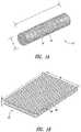
- FIG. 1Ais a schematic drawing of an injectable soft porous rod-shaped device according to one embodiment of the present disclosure.
- FIG. 1Bis a schematic drawing of a soft porous sheet-shaped device according to one embodiment of the present disclosure.
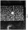
- FIG. 2is a scanning electron microscopy (SEM) image of an injectable soft porous rod-shaped device of according to one embodiment.
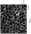
- FIG. 3shows the open-pore structure of the device, indicating pore and throat dimensions.
- FIGS. 4A-4Cshow schematically a soft porous injectable rod-shaped device being radially compressed within the bore of an injection needle ( FIG. 4A ), partially ejected from the end of the needle ( FIG. 4B ), and fully ejected beyond the tip of the needle ( FIG. 4C ).
- FIG. 5shows tissue sections of sphere-templated microporous scaffolds of various pore diameters implanted subcutaneously in mice for 28 days; sections have been stained with BM8 macrophage marker. Pore sizes: (A) 20 micrometers; (B) 35 micrometers; (C) 50 micrometers; (D) 70 micrometers (E) 90 micrometers.
- the scaffold with 35-micrometer porescontains the highest concentration of macrophages.
- drusenExtracellular deposits known as “drusen” have been known to accumulate within the eyes of human beings as they age. Drusen can be observed directly under funduscopic examination and may be classified as either soft drusen or hard drusen, depending on relative size, abundance, and shape. Drusen typically forms beneath the basement membrane of the retinal pigmented epithelium (RPE) and the inner collagenous layer of Bruch membrane. Excessive or confluent areas of drusen in the macula are associated with the development of chorioretinal disorders, such as age-related macular degeneration (AMD). The devices are thus capable of preventing the progression of AMD and/or reducing the pathological symptoms of the disease.
- RPEretinal pigmented epithelium
- AMDage-related macular degeneration
- the deviceis capable of providing benefits for both “dry” AMD and “wet” AMD patients.
- the device or implantis microporous and is designed to be injected into the tissue of the eye, such as into the retrobulbar space or peribulbar space behind the retina.
- Alternative embodiments of the devicecan be injected into the subscleral space or into the vitreous humour.
- a method for reducing or preventing the formation of drusen in an eye of a mammalian subject in need thereofcomprising implanting one or more microporous devices into the eye, each microporous device having a device body and an outer surface, wherein the microporous device is formed of a biocompatible elastomeric material and comprises a plurality of interconnected pores throughout the device body and extending to the outer surface.
- the microporous devicehas an open-cell structure, which comprises interconnected pores throughout the entire bulk of the device body and extending to the device surface.
- FIG. 1Ais a schematic drawing of an embodiment of the present disclosure that shows the device body 10 having an elongated rod shape with length (L) and diameter (d).
- the device bodyis of a soft porous material having pores ( 16 ) within the device body and extends to the outer surface ( 18 ).
- the lengthis within the range of 3-10 mm, or more typically, within the range of 3-5 mm.
- the diameteris within the range of 0.2-0.8 mm, or more typically, within the range of 0.4-0.6 mm.
- rod shapesare particularly compatible with injection
- the device or implant of the present disclosurecan be in any shapes and be inserted by any means known to a skilled person in the art.
- a flat sheet shapemay be useful to allow enough device volume and surface area for sufficient efficacy.
- the longest dimension of the device, regardless of the shape,is about 3-10 mm; whereas the thickness of the device (e.g., the diameter of a rod shape device, or the thickness of a sheet-shaped device) is about 0.2-0.8 mm.
- FIG. 1Bis a schematic drawing of another embodiment of the device body 42 which is a sheet or film of the microporous material.
- the sheethas pores ( 46 ) throughout its entire device body and extending to the outer surface ( 48 ).
- the length (L), width (W) and thickness (d)are not particularly limited other than that the sheet fits in an anatomical space or surgically-created space in the eye.
- the sheet thicknessshould be thin enough to fit into the desired tissue space.
- a thickness (d)is about 0.2 mm to 0.8 mm, or preferably about 0.2 mm-0.5 mm.
- FIG. 2is a scanning electron microscopy (SEM) image showing a rod-shaped device body 10 according to one embodiment of the present disclosure.
- the device bodyis made of a biocompatible material, e.g., an elastomeric material, and comprises a plurality of substantially interconnected pores.
- the deviceis composed of a sphere-templated silicone material as described in U.S. Pat. No. 8,318,193.
- FIG. 3is a scanning electron microscopy (SEM) image showing the cross section of a device of the sphere-templated porous biomaterial that is particularly suitable for the present invention.
- the pore structurecomprises a network of interconnected void spaces referred to herein as pores 20 . Neighboring pores 20 are joined or connected by openings or “throats” 30 .
- the pores 20can be spherical as in FIG. 2 , or they can be any other pore shape that results in a generally open-cell pore structure.
- the throats 30can be circular as in FIG. 2 , or they can be any other 2D shape that defines the size of the openings between neighboring pores 20 . If the pore throats are not circular, then the throat diameter t is defined as the diameter of the largest spherical object that can pass through the throats 30 .
- the pore sizes and the throat diameterscan be controlled to allow macrophages or neutrophils to infiltrate, as well as enhance the accessible areas for the macrophages and neutrophils.
- substantially all the interconnected pores (i.e., at least 90%, or at least 95% or at least 98%) in the microporous deviceare each interconnected to at least 2 other pores, a mean diameter of the pores being between about 5 and about 50 micrometers, and any two adjacent pores are connected by a throat, a mean throat diameter being at least 5 micrometers, or at least 8 microns, or at least 10 microns.
- the mean throat diameteris between about 8 to about 15 microns.
- substantially all of the poresmeans at least 90%, or at least 95% or at least 98% of all of the pores.
- “about”refers to a range of values ⁇ 20% of a specified value.
- the phrase “about 10 micrometers”includes a range of ⁇ 20% of 10 micrometers, namely, 8-12 micrometers.
- throat diameter tis large enough to permit host macrophages and neutrophils to infiltrate the pore structure. These cells are capable of attacking and destroying bacteria and preventing bacterial colonization.
- a human macrophageis typically 15-20 microns in diameter, but is capable of squeezing through openings as small as 5 microns in diameter.
- a neutrophilis similar in size to a macrophage. Accordingly, the throat diameter t should be at least 5 microns. In various embodiments, the throat diameter should be at least 8 microns, or at least 10 microns.
- throat diameters smaller than 5 micronsmay be formed in the course of producing the porous material (e.g., according to the methods disclosed in U.S. Pat. No. 8,318,193), care should be taken to minimize the percentage of these smaller throat diameters.
- a bacterial cellis much smaller than a macrophage, typically 1 to 2 microns in size. Pores having throats in the 1 to 5 micron size range can allow bacteria to enter while preventing access to the much larger macrophages and neutrophils that would ordinarily attack and destroy the bacteria. Thus, in certain embodiment, only a very small percentage of throats (less than 2%, more preferably less than 1%) have diameter t in the 1 to 5 micron size range.
- At least 90% of all the throats in the microporous devicehave diameters of at least 5 microns. In various other embodiments, at least 95%, or at least 98% or at least 99% of all the throats in the microporous device have diameters of at least 5 microns.
- At least 90% of all the throats in the microporous devicehave diameters of at least 5 microns. In various other embodiments, at least 95%, or at least 98% or at least 99% of all the throats in the microporous device have diameters of at least 5 microns.
- At least 90% of all the throats in the microporous devicehave diameters of at least 8 microns. In various other embodiments, at least 95%, or at least 98% or at least 99% of all the throats in the microporous device have diameters of at least 8 microns.
- the pore structurehas high bioavailable surface area, where “bioavailable surface area” is defined as the surface area accessible to macrophages. Surface area is inversely proportional to pore size, so the size of the pores 20 is an important parameter for measuring the bioavailable surface area.
- the average or mean pore diameter Pshould be less than 50 microns, more preferably less than 40 microns, and most preferably less than 30 microns. It is preferable that the pore size be the smallest possible size wherein the pores can be interconnected by throats of the optimal 8 to 15 micron size range. Preferably, the throat diameter t should be about 40% of the pore diameter P, such as between 30% and 45%, or between 35% and 45%.
- ratios larger than 45%are undesirable.
- ratios smaller than 30%may have larger pores and thus lower bioavailable surfaces area, so that the device does not attract macrophages into its porous interior at effective concentrations for antibacterial defense or therapeutic efficacy.
- the total outer surface areas of the device or implantmay be in the range of 3-250 mm 2 , or 25-200 mm 2 .
- the devicecan be made from any elastomeric polymer.
- a particularly suitable polymeris silicone rubber.
- Nusil MED-4830, MED-4840, MED-4850, MED-4860, and MED-6215are particular suitable compositions.
- Other possible biostable materialsinclude polyurethanes, polypropylene, polyethylene, cellulose nitrate, cellulose acetate, polytetrafluoroethylene, or hydrogels.
- the devicecan be made from a biodegradable polymer.
- the devicemay be made from a transparent biomaterial having a refractive index very close to that of the vitreous humour. This is advantageous for embodiments where the device is injected into the vitreous humour, as the close match in refractive index between the porous biomaterial and the ingrown vitreous humour inside the pores ensures translucency to minimize interference with the light path to the retina.
- a transparent biomaterialis NuSil MED-6250 silicone elastomer, which has refractive index of 1.41.
- the refractive index of vitreous humouris 1.37.
- the elastomeric polymershould have a low durometer value when measured in its nonporous form, ideally between 30 and 60 Shore A.
- a low durometer value combined with porosityis less irritating and less inflammatory to tissues than more rigid materials.
- the elastomeric polymershould have maximum elongation strain greater than 100%, more preferably greater than 300%, and most preferably greater than 500%. In some embodiments, high elongation facilitates injection through a needle or insertion tool.
- an injection needle or insertion toolit is desirable for an injection needle or insertion tool to have a small diameter (typically 0.2-0.3 mm ID) so that trauma to the eye tissues is minimized during the insertion procedure.
- a diameter of at least 0.5 mmis preferable.
- the inner diameter of the tubular needle or insertion toolis preferably substantially smaller in diameter than the outer diameter of the device.
- One way to accomplish such radial compressionis to stretch the device under axial tension to at least 300% strain. Silicones and other similar elastomeric materials have a Poisson's ratio of nearly 0.5, so for example, a 300% axial tensile strain produces a radial compression to a compressed diameter of about 50% of the relaxed diameter.
- FIG. 4Ashows a cross-sectional drawing of an injection needle 40 with radially compressed soft porous device body 10 fully inside the bore of the needle having an internal diameter d 1 .
- the compressed devicehas a compressed diameter that is the same as the internal diameter d 1 .
- FIG. 4Bshows the radially compressed proximal end 11 of the device body 10 inside the end the needle and the partially ejected radially expanded distal end 12 of the device body 10 beyond the distal tip of the needle. The portion of the device that is outside of the needle expands into its relaxed diameter d 2 .
- FIG. 4Cshows the device body 10 fully expanded and fully ejected beyond the distal end of the injection needle.
- a further embodimentprovides a needle preloaded with a microporous device comprising: a needle having an interior diameter of 0.2-0.3 mm, a microporous device having a relaxed diameter of 0.3-0.8 mm, wherein the microporous device is radially compressed and placed in the needle such that the needle and the microporous device are aligned longitudinally, and wherein the microporous device is as described herein.
- a “relaxed diameter”is the diameter of the microporous device in its natural, uncompressed shape, which may be the shape prior to being inserted into the needle, the shape after exiting the needle, or the shape while being implanted in the eye.
- the devicemay be axially stretched.
- One methodcomprises the steps of: 1) hydrating the pores of the device in aqueous biocompatible saline solution or other biocompatible aqueous substance, 2) elongating the device by gripping the ends and applying an axial strain, 3) flash freeze the aqueous solution within the pores of the hydrated and stretched device (e.g., by submerging in liquid nitrogen or other low-boiling point liquid), 4) loading the narrowed and elongated device (stiffened by the frozen water within the pores) into the tubular nose member of the insertion tool, and 5) allowing the aqueous solution to thaw, causing the device to expand against the inner wall of the tubular needle.
- the deviceis ready for injection into the retrobulbar space (or other suitable site in close proximity to the retina) by application of gentle fluid pressure
- FIG. 5shows a correlation between the pore sizes and the local concentrations of macrophages. See also Marshall A J. Porous hydrogels with well-defined pore structure for biomaterials applications. Ph.D. Dissertation, University of Washington. 2004 AAT 3151637.
Landscapes
- Health & Medical Sciences (AREA)
- Life Sciences & Earth Sciences (AREA)
- Veterinary Medicine (AREA)
- Public Health (AREA)
- General Health & Medical Sciences (AREA)
- Animal Behavior & Ethology (AREA)
- Chemical & Material Sciences (AREA)
- Ophthalmology & Optometry (AREA)
- Heart & Thoracic Surgery (AREA)
- Biomedical Technology (AREA)
- Engineering & Computer Science (AREA)
- Surgery (AREA)
- Epidemiology (AREA)
- Dermatology (AREA)
- Medicinal Chemistry (AREA)
- Oral & Maxillofacial Surgery (AREA)
- Transplantation (AREA)
- Vascular Medicine (AREA)
- Nuclear Medicine, Radiotherapy & Molecular Imaging (AREA)
- Dispersion Chemistry (AREA)
- Pathology (AREA)
- Medical Informatics (AREA)
- Molecular Biology (AREA)
- Materials For Medical Uses (AREA)
- Medicinal Preparation (AREA)
- Prostheses (AREA)
Abstract
Description
Claims (17)
Priority Applications (1)
| Application Number | Priority Date | Filing Date | Title |
|---|---|---|---|
| US15/192,867US10842916B2 (en) | 2015-06-24 | 2016-06-24 | Injectable porous device for treatment of dry and wet age-related macular degeneration or diabetic retinopathy |
Applications Claiming Priority (3)
| Application Number | Priority Date | Filing Date | Title |
|---|---|---|---|
| US201562184151P | 2015-06-24 | 2015-06-24 | |
| US201562204877P | 2015-08-13 | 2015-08-13 | |
| US15/192,867US10842916B2 (en) | 2015-06-24 | 2016-06-24 | Injectable porous device for treatment of dry and wet age-related macular degeneration or diabetic retinopathy |
Publications (2)
| Publication Number | Publication Date |
|---|---|
| US20160375178A1 US20160375178A1 (en) | 2016-12-29 |
| US10842916B2true US10842916B2 (en) | 2020-11-24 |
Family
ID=56511889
Family Applications (1)
| Application Number | Title | Priority Date | Filing Date |
|---|---|---|---|
| US15/192,867ActiveUS10842916B2 (en) | 2015-06-24 | 2016-06-24 | Injectable porous device for treatment of dry and wet age-related macular degeneration or diabetic retinopathy |
Country Status (5)
| Country | Link |
|---|---|
| US (1) | US10842916B2 (en) |
| EP (1) | EP3313466B1 (en) |
| CN (1) | CN107809977B (en) |
| ES (1) | ES2986006T3 (en) |
| WO (1) | WO2016210346A1 (en) |
Families Citing this family (2)
| Publication number | Priority date | Publication date | Assignee | Title |
|---|---|---|---|---|
| US20210228770A1 (en)* | 2018-06-05 | 2021-07-29 | Corneat Vision Ltd. | A synthetic ophthalmic graft patch |
| CN109464705B (en)* | 2018-11-19 | 2021-08-17 | 爱尔眼科医院集团股份有限公司 | A kind of RPE cell sheet and its application and preparation method |
Citations (44)
| Publication number | Priority date | Publication date | Assignee | Title |
|---|---|---|---|---|
| US5520631A (en) | 1994-07-22 | 1996-05-28 | Wound Healing Of Oklahoma | Method and apparatus for lowering the intraocular pressure of an eye |
| US5704907A (en) | 1994-07-22 | 1998-01-06 | Wound Healing Of Oklahoma | Method and apparatus for lowering the intraocular pressure of an eye |
| US5807406A (en) | 1994-10-07 | 1998-09-15 | Baxter International Inc. | Porous microfabricated polymer membrane structures |
| US5882327A (en) | 1997-04-17 | 1999-03-16 | Jacob; Jean T. | Long-term glaucoma drainage implant |
| US6102045A (en) | 1994-07-22 | 2000-08-15 | Premier Laser Systems, Inc. | Method and apparatus for lowering the intraocular pressure of an eye |
| US20030055372A1 (en) | 1999-04-26 | 2003-03-20 | Lynch Mary G. | Shunt device and method for treating glaucoma |
| US6579235B1 (en) | 1999-11-01 | 2003-06-17 | The Johns Hopkins University | Method for monitoring intraocular pressure using a passive intraocular pressure sensor and patient worn monitoring recorder |
| US6616699B2 (en) | 1999-07-20 | 2003-09-09 | Peter Paul Zilla | Foam-type vascular prosthesis with well-defined angio-permissive open porosity |
| WO2003093196A1 (en) | 2002-05-06 | 2003-11-13 | Biomet Deutschland Gmbh | Method of preparing porous calcium phosphate granules |
| US6699210B2 (en) | 1999-04-27 | 2004-03-02 | The Arizona Board Of Regents | Glaucoma shunt and a method of making and surgically implanting the same |
| US6939299B1 (en) | 1999-12-13 | 2005-09-06 | Kurt Petersen | Implantable continuous intraocular pressure sensor |
| US20050244500A1 (en) | 2004-04-30 | 2005-11-03 | Allergan, Inc. | Intravitreal implants in conjuction with photodynamic therapy to improve vision |
| US7037335B2 (en) | 2002-11-19 | 2006-05-02 | Eagle Vision, Inc. | Bulbous scleral implants for the treatment of eye disorders such as presbyopia and glaucoma |
| US20060136071A1 (en) | 2002-12-23 | 2006-06-22 | Maspero Fabrizio A | Biodegradable biocompatible implant |
| US20060276831A1 (en)* | 2005-02-04 | 2006-12-07 | Porter Stephen C | Porous materials for use in aneurysms |
| US7160264B2 (en) | 2002-12-19 | 2007-01-09 | Medtronic-Xomed, Inc. | Article and method for ocular aqueous drainage |
| US7207965B2 (en) | 2003-06-16 | 2007-04-24 | Solx, Inc. | Shunt for the treatment of glaucoma |
| US20080228127A1 (en) | 2006-11-10 | 2008-09-18 | Glaukos Corporation | Uveoscleral shunt and methods for implanting same |
| US7431709B2 (en) | 2003-12-05 | 2008-10-07 | Innfocus, Llc | Glaucoma implant device |
| US7594899B2 (en) | 2004-12-03 | 2009-09-29 | Innfocus, Llc | Glaucoma implant device |
| US20090275924A1 (en) | 2006-04-26 | 2009-11-05 | Eastern Virginia Medical School | Systems and Methods for Monitoring and Controlling Internal Pressure of an Eye or Body Part |
| US20090299216A1 (en) | 2008-06-02 | 2009-12-03 | Po-Jui Chen | System, apparatus and method for biomedical wireless pressure sensing |
| US7678065B2 (en) | 2001-05-02 | 2010-03-16 | Glaukos Corporation | Implant with intraocular pressure sensor for glaucoma treatment |
| US7677107B2 (en) | 2007-07-03 | 2010-03-16 | Endotronix, Inc. | Wireless pressure sensor and method for fabricating wireless pressure sensor for integration with an implantable device |
| US20100168644A1 (en) | 2001-01-09 | 2010-07-01 | Brown J David | Glaucoma Treatment Device and Method |
| US20100249691A1 (en) | 2009-03-26 | 2010-09-30 | Abbott Medical Optics Inc. | Glaucoma shunts with flow management and improved surgical performance |
| US7837644B2 (en) | 2004-12-03 | 2010-11-23 | Innfocus, Llc | Glaucoma implant device |
| US20110071456A1 (en) | 2009-09-21 | 2011-03-24 | Rickard Matthew J A | Lumen Clearing Valve For Glaucoma Drainage Device |
| US20110082385A1 (en) | 2008-04-17 | 2011-04-07 | Yale University | Method for implanting intraocular pressure sensor |
| US20110098629A1 (en) | 2006-01-17 | 2011-04-28 | Juan Jr Eugene De | Glaucoma treatment device |
| WO2011066441A1 (en) | 2009-11-25 | 2011-06-03 | Healionics Corporation | Implantable medical devices having microporous surface layers and method for reducing foreign body response to the same |
| US7972628B2 (en) | 2003-10-01 | 2011-07-05 | University Of Washington | Porous biomaterials |
| US20110196281A1 (en) | 1999-04-26 | 2011-08-11 | Glaukos Corporation | Shunt device and method for treating ocular disorders |
| US8007459B2 (en) | 2002-09-21 | 2011-08-30 | Glaukos Corporation | Ocular implant with anchoring mechanism and multiple outlets |
| WO2011127395A1 (en) | 2010-04-08 | 2011-10-13 | Healionics Corporation | Implantable medical devices having microporous surface layers and method for reducing foreign body response to the same |
| US20120184892A1 (en) | 2011-01-14 | 2012-07-19 | Ecole Polytechnique Federale De Lausanne (Epfl) | Apparatus and methods for treating excess intraocular fluid |
| US20120220917A1 (en) | 2007-07-17 | 2012-08-30 | Tom Silvestrini | Ocular implant with hydrogel expansion capabilities |
| US8257295B2 (en) | 2009-09-21 | 2012-09-04 | Alcon Research, Ltd. | Intraocular pressure sensor with external pressure compensation |
| US20120310137A1 (en) | 2011-06-02 | 2012-12-06 | Silvestrini Thomas A | Eye shunt with porous structure |
| US20130022648A1 (en) | 2009-11-25 | 2013-01-24 | Healionics Corporation | Granules of porous biocompatible materials |
| US8444588B2 (en) | 2003-05-05 | 2013-05-21 | Transcend Medical, Inc. | Internal shunt and method for treating glaucoma |
| US8475374B2 (en) | 2007-08-23 | 2013-07-02 | Purdue Research Foundation | Intra-occular pressure sensor |
| US20130274691A1 (en)* | 2010-08-05 | 2013-10-17 | Forsight Vision4, Inc. | Combined drug delivery methods and apparatus |
| US8926510B2 (en) | 2011-04-27 | 2015-01-06 | Istar Medical Sa | Device and method for glaucoma management and treatment |
Family Cites Families (1)
| Publication number | Priority date | Publication date | Assignee | Title |
|---|---|---|---|---|
| WO2007084582A2 (en)* | 2006-01-17 | 2007-07-26 | Forsight Labs, Llc | Drug delivery treatment device |
- 2016
- 2016-06-24WOPCT/US2016/039389patent/WO2016210346A1/ennot_activeCeased
- 2016-06-24EPEP16742085.0Apatent/EP3313466B1/enactiveActive
- 2016-06-24ESES16742085Tpatent/ES2986006T3/enactiveActive
- 2016-06-24CNCN201680036774.7Apatent/CN107809977B/enactiveActive
- 2016-06-24USUS15/192,867patent/US10842916B2/enactiveActive
Patent Citations (52)
| Publication number | Priority date | Publication date | Assignee | Title |
|---|---|---|---|---|
| US5704907A (en) | 1994-07-22 | 1998-01-06 | Wound Healing Of Oklahoma | Method and apparatus for lowering the intraocular pressure of an eye |
| US6102045A (en) | 1994-07-22 | 2000-08-15 | Premier Laser Systems, Inc. | Method and apparatus for lowering the intraocular pressure of an eye |
| US5520631A (en) | 1994-07-22 | 1996-05-28 | Wound Healing Of Oklahoma | Method and apparatus for lowering the intraocular pressure of an eye |
| US5807406A (en) | 1994-10-07 | 1998-09-15 | Baxter International Inc. | Porous microfabricated polymer membrane structures |
| US5882327A (en) | 1997-04-17 | 1999-03-16 | Jacob; Jean T. | Long-term glaucoma drainage implant |
| US7220238B2 (en) | 1999-04-26 | 2007-05-22 | Gmp Vision Solutions, Inc. | Shunt device and method for treating glaucoma |
| US20030055372A1 (en) | 1999-04-26 | 2003-03-20 | Lynch Mary G. | Shunt device and method for treating glaucoma |
| US20110196281A1 (en) | 1999-04-26 | 2011-08-11 | Glaukos Corporation | Shunt device and method for treating ocular disorders |
| US8152752B2 (en) | 1999-04-26 | 2012-04-10 | Glaukos Corporation | Shunt device and method for treating glaucoma |
| US8388568B2 (en) | 1999-04-26 | 2013-03-05 | Glaukos Corporation | Shunt device and method for treating ocular disorders |
| US6699210B2 (en) | 1999-04-27 | 2004-03-02 | The Arizona Board Of Regents | Glaucoma shunt and a method of making and surgically implanting the same |
| US6616699B2 (en) | 1999-07-20 | 2003-09-09 | Peter Paul Zilla | Foam-type vascular prosthesis with well-defined angio-permissive open porosity |
| US6579235B1 (en) | 1999-11-01 | 2003-06-17 | The Johns Hopkins University | Method for monitoring intraocular pressure using a passive intraocular pressure sensor and patient worn monitoring recorder |
| US6939299B1 (en) | 1999-12-13 | 2005-09-06 | Kurt Petersen | Implantable continuous intraocular pressure sensor |
| US20100168644A1 (en) | 2001-01-09 | 2010-07-01 | Brown J David | Glaucoma Treatment Device and Method |
| US7678065B2 (en) | 2001-05-02 | 2010-03-16 | Glaukos Corporation | Implant with intraocular pressure sensor for glaucoma treatment |
| WO2003093196A1 (en) | 2002-05-06 | 2003-11-13 | Biomet Deutschland Gmbh | Method of preparing porous calcium phosphate granules |
| US8007459B2 (en) | 2002-09-21 | 2011-08-30 | Glaukos Corporation | Ocular implant with anchoring mechanism and multiple outlets |
| US7037335B2 (en) | 2002-11-19 | 2006-05-02 | Eagle Vision, Inc. | Bulbous scleral implants for the treatment of eye disorders such as presbyopia and glaucoma |
| US7160264B2 (en) | 2002-12-19 | 2007-01-09 | Medtronic-Xomed, Inc. | Article and method for ocular aqueous drainage |
| US20060136071A1 (en) | 2002-12-23 | 2006-06-22 | Maspero Fabrizio A | Biodegradable biocompatible implant |
| US8444588B2 (en) | 2003-05-05 | 2013-05-21 | Transcend Medical, Inc. | Internal shunt and method for treating glaucoma |
| US7207965B2 (en) | 2003-06-16 | 2007-04-24 | Solx, Inc. | Shunt for the treatment of glaucoma |
| US8318193B2 (en) | 2003-10-01 | 2012-11-27 | University Of Washington | Crosslinked porous biomaterials |
| US7972628B2 (en) | 2003-10-01 | 2011-07-05 | University Of Washington | Porous biomaterials |
| US7431709B2 (en) | 2003-12-05 | 2008-10-07 | Innfocus, Llc | Glaucoma implant device |
| US20050244500A1 (en) | 2004-04-30 | 2005-11-03 | Allergan, Inc. | Intravitreal implants in conjuction with photodynamic therapy to improve vision |
| US7837644B2 (en) | 2004-12-03 | 2010-11-23 | Innfocus, Llc | Glaucoma implant device |
| US7594899B2 (en) | 2004-12-03 | 2009-09-29 | Innfocus, Llc | Glaucoma implant device |
| US20060276831A1 (en)* | 2005-02-04 | 2006-12-07 | Porter Stephen C | Porous materials for use in aneurysms |
| US20110098629A1 (en) | 2006-01-17 | 2011-04-28 | Juan Jr Eugene De | Glaucoma treatment device |
| US20090275924A1 (en) | 2006-04-26 | 2009-11-05 | Eastern Virginia Medical School | Systems and Methods for Monitoring and Controlling Internal Pressure of an Eye or Body Part |
| US20080228127A1 (en) | 2006-11-10 | 2008-09-18 | Glaukos Corporation | Uveoscleral shunt and methods for implanting same |
| US7677107B2 (en) | 2007-07-03 | 2010-03-16 | Endotronix, Inc. | Wireless pressure sensor and method for fabricating wireless pressure sensor for integration with an implantable device |
| US20120220917A1 (en) | 2007-07-17 | 2012-08-30 | Tom Silvestrini | Ocular implant with hydrogel expansion capabilities |
| US8475374B2 (en) | 2007-08-23 | 2013-07-02 | Purdue Research Foundation | Intra-occular pressure sensor |
| US20110082385A1 (en) | 2008-04-17 | 2011-04-07 | Yale University | Method for implanting intraocular pressure sensor |
| US20090299216A1 (en) | 2008-06-02 | 2009-12-03 | Po-Jui Chen | System, apparatus and method for biomedical wireless pressure sensing |
| US20100249691A1 (en) | 2009-03-26 | 2010-09-30 | Abbott Medical Optics Inc. | Glaucoma shunts with flow management and improved surgical performance |
| US8257295B2 (en) | 2009-09-21 | 2012-09-04 | Alcon Research, Ltd. | Intraocular pressure sensor with external pressure compensation |
| US20110071456A1 (en) | 2009-09-21 | 2011-03-24 | Rickard Matthew J A | Lumen Clearing Valve For Glaucoma Drainage Device |
| US20130022648A1 (en) | 2009-11-25 | 2013-01-24 | Healionics Corporation | Granules of porous biocompatible materials |
| US8372423B2 (en) | 2009-11-25 | 2013-02-12 | Healionics Corporation | Implantable medical devices having microporous surface layers and method for reducing foreign body response to the same |
| US20110257623A1 (en) | 2009-11-25 | 2011-10-20 | Healionics Corporation | Implantable medical devices having microporous surface layers and method for reducing foreign body response to the same |
| WO2011066441A1 (en) | 2009-11-25 | 2011-06-03 | Healionics Corporation | Implantable medical devices having microporous surface layers and method for reducing foreign body response to the same |
| US8647393B2 (en) | 2009-11-25 | 2014-02-11 | Healionics Corporation | Implantable medical devices having microporous surface layers and method for reducing foreign body response to the same |
| CN103037911A (en) | 2010-04-08 | 2013-04-10 | 矽瑞奥科技公司 | Implantable medical devices having microporous surface layers and method for reducing foreign body response to the same |
| WO2011127395A1 (en) | 2010-04-08 | 2011-10-13 | Healionics Corporation | Implantable medical devices having microporous surface layers and method for reducing foreign body response to the same |
| US20130274691A1 (en)* | 2010-08-05 | 2013-10-17 | Forsight Vision4, Inc. | Combined drug delivery methods and apparatus |
| US20120184892A1 (en) | 2011-01-14 | 2012-07-19 | Ecole Polytechnique Federale De Lausanne (Epfl) | Apparatus and methods for treating excess intraocular fluid |
| US8926510B2 (en) | 2011-04-27 | 2015-01-06 | Istar Medical Sa | Device and method for glaucoma management and treatment |
| US20120310137A1 (en) | 2011-06-02 | 2012-12-06 | Silvestrini Thomas A | Eye shunt with porous structure |
Non-Patent Citations (9)
| Title |
|---|
| Chen et al., "Wireless Intraocular Pressure Sensing Using Microfabricated Minimally Invasive Flexible-Coiled LC Sensor Implant," Journal of Microelectromechanical Systems 19(4):721-734, 2010. |
| International Search Report and Written Opinion, dated Sep. 27, 2016, for International Application No. PCT/US2016/039389, 13 pages. |
| Kakaday et al., "Design of a Wireless Intraocular Pressure Monitoring System for a Glaucoma Drainage Implant," Proceedings of the 13th International Conference on Biomedical Engineering, Singapore, Singapore, Dec. 3-6, 2008, pp. 198-201. |
| Lim et al., "Glaucoma drainage devices; past, present, and future," British Journal of Ophthalmology 82(9):1083-1089, 1998. (8 pages). |
| Marshall et al., "Quantitative Characterization of Sphere-Templated Porous Biomaterials," AIChE Journal 51(4):1221-1232, 2005. |
| Marshall et al., "Sphere Templated Angiogenic Regeneration (STAR) Biomaterials for Ophthalmic Applications," Annual Meeting of the Society for Biomaterials, Seattle, Washington, USA, Apr. 21-24, 2010, 1 page. |
| Oatts et al., "In Vitro and in Vivo Comparison of Two Suprachoroidal Shunts," Investigative Ophthalmology & Visual Science 54(8):5416-5423, 2013. |
| Patel, "Comparison of Glaucoma Drainage Implants: M4 vs FP7 and S2," Medscape Ophthalmology, 2013. (3 pages). |
| Ryan et al., "Retina, Fifth Edition," Elsevier Saunders 1:1168-1182, 2012. (16 pages). |
Also Published As
| Publication number | Publication date |
|---|---|
| EP3313466A1 (en) | 2018-05-02 |
| ES2986006T3 (en) | 2024-11-08 |
| US20160375178A1 (en) | 2016-12-29 |
| CN107809977B (en) | 2021-04-09 |
| EP3313466B1 (en) | 2024-08-07 |
| CN107809977A (en) | 2018-03-16 |
| WO2016210346A1 (en) | 2016-12-29 |
| EP3313466C0 (en) | 2024-08-07 |
Similar Documents
| Publication | Publication Date | Title |
|---|---|---|
| JP5662436B2 (en) | Fiber matrix that maintains space in soft tissue | |
| US6589286B1 (en) | Eustachian tube stent | |
| US5344451A (en) | Synthetic reconstructive implant device | |
| US6033437A (en) | Pegs for orbital implants | |
| CN108778398B (en) | Dry eye treatment device and method | |
| US20220125638A1 (en) | Glaucoma treatment devices and methods | |
| KR20090015077A (en) | Methods and compositions for the treatment of open and closed window spinal cord injury | |
| CN106456377A (en) | Drug delivery systems and related methods of use | |
| JPH08500035A (en) | Intrastromal hybrid ring | |
| RU2398566C2 (en) | New use of viscoelastic composition | |
| US20240285828A1 (en) | Composition using fibrotic acellular dermal matrix, and method for preparing same | |
| US10842916B2 (en) | Injectable porous device for treatment of dry and wet age-related macular degeneration or diabetic retinopathy | |
| de Moraes KARKOS et al. | The surgical repair of the cornea of the dog using pericardium as a keratoprosthesis | |
| WO1994014390A1 (en) | Device for orbital implant | |
| CN109550083B (en) | A kind of occluder for preventing the formation of traumatic neuroma and preparation method thereof | |
| RU2123832C1 (en) | Method for creating rest stump and conjunctive vault after operation | |
| Muir et al. | Air, methylcellulose, sodium hyaluronate and the corneal endothelium. Endothelial protective agents | |
| McCurdy Jr | Capsular contracture following augmentation mammaplasty: Etiology and pathogenesis | |
| RU2485915C1 (en) | Method of eye ball stump prosthetics | |
| EP0746270B1 (en) | Orbital implant having synthetic and absorbable coating | |
| RU48768U1 (en) | COLLAGENIC DRAINAGE FOR ANTI-GLACOMATOUS OPERATIONS | |
| KR20250021684A (en) | Drainage device for intraocular pressure control and method of manufacturing the same | |
| KR20240069643A (en) | 3-Dimensional Biocompatible Structure for Regeneration of Body Vessel | |
| DE69231578T2 (en) | IMPLANT FOR THE EYE CAVE WITH RESORBABLE COATING | |
| EP3903749A1 (en) | Controlled release glaucoma stent |
Legal Events
| Date | Code | Title | Description |
|---|---|---|---|
| AS | Assignment | Owner name:HEALIONICS CORPORATION, WASHINGTON Free format text:ASSIGNMENT OF ASSIGNORS INTEREST;ASSIGNORS:MARSHALL, ANDREW J.;MAGINNESS, MAX;REEL/FRAME:039316/0045 Effective date:20160802 | |
| STCV | Information on status: appeal procedure | Free format text:NOTICE OF APPEAL FILED | |
| STPP | Information on status: patent application and granting procedure in general | Free format text:DOCKETED NEW CASE - READY FOR EXAMINATION | |
| STPP | Information on status: patent application and granting procedure in general | Free format text:NON FINAL ACTION MAILED | |
| STPP | Information on status: patent application and granting procedure in general | Free format text:RESPONSE TO NON-FINAL OFFICE ACTION ENTERED AND FORWARDED TO EXAMINER | |
| STPP | Information on status: patent application and granting procedure in general | Free format text:FINAL REJECTION MAILED | |
| STPP | Information on status: patent application and granting procedure in general | Free format text:NOTICE OF ALLOWANCE MAILED -- APPLICATION RECEIVED IN OFFICE OF PUBLICATIONS | |
| STPP | Information on status: patent application and granting procedure in general | Free format text:PUBLICATIONS -- ISSUE FEE PAYMENT VERIFIED | |
| STCF | Information on status: patent grant | Free format text:PATENTED CASE | |
| MAFP | Maintenance fee payment | Free format text:PAYMENT OF MAINTENANCE FEE, 4TH YR, SMALL ENTITY (ORIGINAL EVENT CODE: M2551); ENTITY STATUS OF PATENT OWNER: SMALL ENTITY Year of fee payment:4 |