US10813567B2 - System and method for composite display of subcutaneous cardiac monitoring data - Google Patents
System and method for composite display of subcutaneous cardiac monitoring dataDownload PDFInfo
- Publication number
- US10813567B2 US10813567B2US16/595,226US201916595226AUS10813567B2US 10813567 B2US10813567 B2US 10813567B2US 201916595226 AUS201916595226 AUS 201916595226AUS 10813567 B2US10813567 B2US 10813567B2
- Authority
- US
- United States
- Prior art keywords
- ecg
- cardiac
- plot
- data
- interval
- Prior art date
- Legal status (The legal status is an assumption and is not a legal conclusion. Google has not performed a legal analysis and makes no representation as to the accuracy of the status listed.)
- Active
Links
Images


Classifications
- A—HUMAN NECESSITIES
- A61—MEDICAL OR VETERINARY SCIENCE; HYGIENE
- A61B—DIAGNOSIS; SURGERY; IDENTIFICATION
- A61B5/00—Measuring for diagnostic purposes; Identification of persons
- A61B5/24—Detecting, measuring or recording bioelectric or biomagnetic signals of the body or parts thereof
- A61B5/316—Modalities, i.e. specific diagnostic methods
- A61B5/318—Heart-related electrical modalities, e.g. electrocardiography [ECG]
- A61B5/346—Analysis of electrocardiograms
- A61B5/349—Detecting specific parameters of the electrocardiograph cycle
- A61B5/352—Detecting R peaks, e.g. for synchronising diagnostic apparatus; Estimating R-R interval
- A61B5/0456—
- A—HUMAN NECESSITIES
- A61—MEDICAL OR VETERINARY SCIENCE; HYGIENE
- A61B—DIAGNOSIS; SURGERY; IDENTIFICATION
- A61B5/00—Measuring for diagnostic purposes; Identification of persons
- A61B5/68—Arrangements of detecting, measuring or recording means, e.g. sensors, in relation to patient
- A61B5/6846—Arrangements of detecting, measuring or recording means, e.g. sensors, in relation to patient specially adapted to be brought in contact with an internal body part, i.e. invasive
- A61B5/6847—Arrangements of detecting, measuring or recording means, e.g. sensors, in relation to patient specially adapted to be brought in contact with an internal body part, i.e. invasive mounted on an invasive device
- A61B5/686—Permanently implanted devices, e.g. pacemakers, other stimulators, biochips
- A—HUMAN NECESSITIES
- A61—MEDICAL OR VETERINARY SCIENCE; HYGIENE
- A61B—DIAGNOSIS; SURGERY; IDENTIFICATION
- A61B5/00—Measuring for diagnostic purposes; Identification of persons
- A61B5/02—Detecting, measuring or recording for evaluating the cardiovascular system, e.g. pulse, heart rate, blood pressure or blood flow
- A61B5/024—Measuring pulse rate or heart rate
- A61B5/02405—Determining heart rate variability
- A—HUMAN NECESSITIES
- A61—MEDICAL OR VETERINARY SCIENCE; HYGIENE
- A61B—DIAGNOSIS; SURGERY; IDENTIFICATION
- A61B5/00—Measuring for diagnostic purposes; Identification of persons
- A61B5/02—Detecting, measuring or recording for evaluating the cardiovascular system, e.g. pulse, heart rate, blood pressure or blood flow
- A61B5/024—Measuring pulse rate or heart rate
- A61B5/0245—Measuring pulse rate or heart rate by using sensing means generating electric signals, i.e. ECG signals
- A61B5/04085—
- A61B5/04087—
- A61B5/0422—
- A61B5/0432—
- A61B5/044—
- A61B5/04525—
- A—HUMAN NECESSITIES
- A61—MEDICAL OR VETERINARY SCIENCE; HYGIENE
- A61B—DIAGNOSIS; SURGERY; IDENTIFICATION
- A61B5/00—Measuring for diagnostic purposes; Identification of persons
- A61B5/24—Detecting, measuring or recording bioelectric or biomagnetic signals of the body or parts thereof
- A61B5/25—Bioelectric electrodes therefor
- A61B5/251—Means for maintaining electrode contact with the body
- A61B5/257—Means for maintaining electrode contact with the body using adhesive means, e.g. adhesive pads or tapes
- A61B5/259—Means for maintaining electrode contact with the body using adhesive means, e.g. adhesive pads or tapes using conductive adhesive means, e.g. gels
- A—HUMAN NECESSITIES
- A61—MEDICAL OR VETERINARY SCIENCE; HYGIENE
- A61B—DIAGNOSIS; SURGERY; IDENTIFICATION
- A61B5/00—Measuring for diagnostic purposes; Identification of persons
- A61B5/24—Detecting, measuring or recording bioelectric or biomagnetic signals of the body or parts thereof
- A61B5/25—Bioelectric electrodes therefor
- A61B5/279—Bioelectric electrodes therefor specially adapted for particular uses
- A61B5/28—Bioelectric electrodes therefor specially adapted for particular uses for electrocardiography [ECG]
- A61B5/282—Holders for multiple electrodes
- A—HUMAN NECESSITIES
- A61—MEDICAL OR VETERINARY SCIENCE; HYGIENE
- A61B—DIAGNOSIS; SURGERY; IDENTIFICATION
- A61B5/00—Measuring for diagnostic purposes; Identification of persons
- A61B5/24—Detecting, measuring or recording bioelectric or biomagnetic signals of the body or parts thereof
- A61B5/25—Bioelectric electrodes therefor
- A61B5/279—Bioelectric electrodes therefor specially adapted for particular uses
- A61B5/28—Bioelectric electrodes therefor specially adapted for particular uses for electrocardiography [ECG]
- A61B5/283—Invasive
- A61B5/287—Holders for multiple electrodes, e.g. electrode catheters for electrophysiological study [EPS]
- A—HUMAN NECESSITIES
- A61—MEDICAL OR VETERINARY SCIENCE; HYGIENE
- A61B—DIAGNOSIS; SURGERY; IDENTIFICATION
- A61B5/00—Measuring for diagnostic purposes; Identification of persons
- A61B5/24—Detecting, measuring or recording bioelectric or biomagnetic signals of the body or parts thereof
- A61B5/316—Modalities, i.e. specific diagnostic methods
- A61B5/318—Heart-related electrical modalities, e.g. electrocardiography [ECG]
- A61B5/333—Recording apparatus specially adapted therefor
- A—HUMAN NECESSITIES
- A61—MEDICAL OR VETERINARY SCIENCE; HYGIENE
- A61B—DIAGNOSIS; SURGERY; IDENTIFICATION
- A61B5/00—Measuring for diagnostic purposes; Identification of persons
- A61B5/24—Detecting, measuring or recording bioelectric or biomagnetic signals of the body or parts thereof
- A61B5/316—Modalities, i.e. specific diagnostic methods
- A61B5/318—Heart-related electrical modalities, e.g. electrocardiography [ECG]
- A61B5/339—Displays specially adapted therefor
- A—HUMAN NECESSITIES
- A61—MEDICAL OR VETERINARY SCIENCE; HYGIENE
- A61B—DIAGNOSIS; SURGERY; IDENTIFICATION
- A61B5/00—Measuring for diagnostic purposes; Identification of persons
- A61B5/24—Detecting, measuring or recording bioelectric or biomagnetic signals of the body or parts thereof
- A61B5/316—Modalities, i.e. specific diagnostic methods
- A61B5/318—Heart-related electrical modalities, e.g. electrocardiography [ECG]
- A61B5/346—Analysis of electrocardiograms
- A61B5/349—Detecting specific parameters of the electrocardiograph cycle
- A61B5/35—Detecting specific parameters of the electrocardiograph cycle by template matching
- A—HUMAN NECESSITIES
- A61—MEDICAL OR VETERINARY SCIENCE; HYGIENE
- A61B—DIAGNOSIS; SURGERY; IDENTIFICATION
- A61B5/00—Measuring for diagnostic purposes; Identification of persons
- A61B5/68—Arrangements of detecting, measuring or recording means, e.g. sensors, in relation to patient
- A61B5/6801—Arrangements of detecting, measuring or recording means, e.g. sensors, in relation to patient specially adapted to be attached to or worn on the body surface
- A61B5/6813—Specially adapted to be attached to a specific body part
- A61B5/6823—Trunk, e.g., chest, back, abdomen, hip
- A—HUMAN NECESSITIES
- A61—MEDICAL OR VETERINARY SCIENCE; HYGIENE
- A61B—DIAGNOSIS; SURGERY; IDENTIFICATION
- A61B5/00—Measuring for diagnostic purposes; Identification of persons
- A61B5/72—Signal processing specially adapted for physiological signals or for diagnostic purposes
- A61B5/7203—Signal processing specially adapted for physiological signals or for diagnostic purposes for noise prevention, reduction or removal
- A—HUMAN NECESSITIES
- A61—MEDICAL OR VETERINARY SCIENCE; HYGIENE
- A61B—DIAGNOSIS; SURGERY; IDENTIFICATION
- A61B2560/00—Constructional details of operational features of apparatus; Accessories for medical measuring apparatus
- A61B2560/04—Constructional details of apparatus
- A61B2560/0462—Apparatus with built-in sensors
- A61B2560/0468—Built-in electrodes
- A—HUMAN NECESSITIES
- A61—MEDICAL OR VETERINARY SCIENCE; HYGIENE
- A61B—DIAGNOSIS; SURGERY; IDENTIFICATION
- A61B2562/00—Details of sensors; Constructional details of sensor housings or probes; Accessories for sensors
- A61B2562/16—Details of sensor housings or probes; Details of structural supports for sensors
- A61B2562/162—Capsule shaped sensor housings, e.g. for swallowing or implantation
- A—HUMAN NECESSITIES
- A61—MEDICAL OR VETERINARY SCIENCE; HYGIENE
- A61B—DIAGNOSIS; SURGERY; IDENTIFICATION
- A61B5/00—Measuring for diagnostic purposes; Identification of persons
- A61B5/02—Detecting, measuring or recording for evaluating the cardiovascular system, e.g. pulse, heart rate, blood pressure or blood flow
- A61B5/0205—Simultaneously evaluating both cardiovascular conditions and different types of body conditions, e.g. heart and respiratory condition
- A61B5/02055—Simultaneously evaluating both cardiovascular condition and temperature
- A61B5/04011—
- A61B5/04017—
- A61B5/046—
- A61B5/0464—
- A61B5/0468—
- A—HUMAN NECESSITIES
- A61—MEDICAL OR VETERINARY SCIENCE; HYGIENE
- A61B—DIAGNOSIS; SURGERY; IDENTIFICATION
- A61B5/00—Measuring for diagnostic purposes; Identification of persons
- A61B5/24—Detecting, measuring or recording bioelectric or biomagnetic signals of the body or parts thereof
- A61B5/316—Modalities, i.e. specific diagnostic methods
- A61B5/318—Heart-related electrical modalities, e.g. electrocardiography [ECG]
- A61B5/339—Displays specially adapted therefor
- A61B5/341—Vectorcardiography [VCG]
- A—HUMAN NECESSITIES
- A61—MEDICAL OR VETERINARY SCIENCE; HYGIENE
- A61B—DIAGNOSIS; SURGERY; IDENTIFICATION
- A61B5/00—Measuring for diagnostic purposes; Identification of persons
- A61B5/24—Detecting, measuring or recording bioelectric or biomagnetic signals of the body or parts thereof
- A61B5/316—Modalities, i.e. specific diagnostic methods
- A61B5/318—Heart-related electrical modalities, e.g. electrocardiography [ECG]
- A61B5/346—Analysis of electrocardiograms
- A61B5/349—Detecting specific parameters of the electrocardiograph cycle
- A61B5/361—Detecting fibrillation
- A—HUMAN NECESSITIES
- A61—MEDICAL OR VETERINARY SCIENCE; HYGIENE
- A61B—DIAGNOSIS; SURGERY; IDENTIFICATION
- A61B5/00—Measuring for diagnostic purposes; Identification of persons
- A61B5/24—Detecting, measuring or recording bioelectric or biomagnetic signals of the body or parts thereof
- A61B5/316—Modalities, i.e. specific diagnostic methods
- A61B5/318—Heart-related electrical modalities, e.g. electrocardiography [ECG]
- A61B5/346—Analysis of electrocardiograms
- A61B5/349—Detecting specific parameters of the electrocardiograph cycle
- A61B5/363—Detecting tachycardia or bradycardia
- A—HUMAN NECESSITIES
- A61—MEDICAL OR VETERINARY SCIENCE; HYGIENE
- A61B—DIAGNOSIS; SURGERY; IDENTIFICATION
- A61B5/00—Measuring for diagnostic purposes; Identification of persons
- A61B5/24—Detecting, measuring or recording bioelectric or biomagnetic signals of the body or parts thereof
- A61B5/316—Modalities, i.e. specific diagnostic methods
- A61B5/318—Heart-related electrical modalities, e.g. electrocardiography [ECG]
- A61B5/346—Analysis of electrocardiograms
- A61B5/349—Detecting specific parameters of the electrocardiograph cycle
- A61B5/364—Detecting abnormal ECG interval, e.g. extrasystoles, ectopic heartbeats
- A—HUMAN NECESSITIES
- A61—MEDICAL OR VETERINARY SCIENCE; HYGIENE
- A61B—DIAGNOSIS; SURGERY; IDENTIFICATION
- A61B5/00—Measuring for diagnostic purposes; Identification of persons
- A61B5/74—Details of notification to user or communication with user or patient; User input means
- A61B5/742—Details of notification to user or communication with user or patient; User input means using visual displays
Definitions
- This applicationrelates in general to electrocardiographic monitoring and, in particular, to a system and method for composite display of subcutaneous cardiac monitoring data.
- An electrocardiogramallows physicians to diagnose cardiac function by visually tracing the cutaneous electrical signals (action potentials) that are generated by the propagation of the transmembrane ionic currents that trigger the depolarization of cardiac fibers.
- An ECG tracecontains alphabetically-labeled waveform deflections that represent distinct features within the cyclic cardiac activation sequence.
- the P-waverepresents atrial depolarization, which causes atrial contraction.
- the QRS-complexrepresents ventricular depolarization.
- the T-waverepresents ventricular repolarization.
- the R-waveis often used as an abbreviation for the QRS-complex.
- An R-R intervalspans the period between successive R-waves and, in a normal heart, is 600 milliseconds (ms) to one second long, which respectively correspond to 100 to 60 beats per minute (bpm).
- the R-waveis the largest waveform generated during normal conduction and represents the cardiac electrical stimuli passing through the ventricular walls.
- R-R intervalsprovide information that allows a physician to understand at a glance the context of cardiac rhythms both before and after a suspected rhythm abnormality and can be of conformational and collaborative value in cardiac arrhythmia diagnosis and treatment.
- R-R intervalshave also been visualized in Poincare plots, which graph RR(n) on the x-axis and RR(n+1) on the y-axis.
- a Poincare plotfails to preserve the correlation between an R-R interval and the R-R interval's time of occurrence and the linearity of time and associated contextual information, before and after a specific cardiac rhythm, are lost.
- the P-waveis a critical component of arrhythmia monitoring and diagnosis performed every day hundreds of thousands of times across the United States. Without a knowledge of the relationship of these two basic symbols, heart rhythm disorders cannot be reliably diagnosed. Visualizing both the P-wave and the R-wave allow for the specific identification of a variety of atrial tachyarrhythmias (also known as supraventricular tachyarrhythmias, or SVTs), ventricular tachyarrhythmias (VTs), and bradycardias related to sinus node and atrioventricular (AV) node dysfunction.
- atrial tachyarrhythmiasalso known as supraventricular tachyarrhythmias, or SVTs
- VTsventricular tachyarrhythmias
- bradycardiasrelated to sinus node and atrioventricular (AV) node dysfunction.
- Cardiac rhythm disordersmay present with lightheadedness, fainting, chest pain, hypoxia, syncope, palpitations, and congestive heart failure (CHF), yet rhythm disorders are often sporadic in occurrence and may not show up in-clinic during a conventional 12-second ECG.
- Continuous ECG monitoring with P-wave-centric action potential acquisition over an extended periodis more apt to capture sporadic cardiac events.
- recording sufficient ECG and related physiological data over an extended periodremains a significant challenge. For instance, maintaining continual contact between ECG electrodes of conventional ambulatory dermal ECG monitors and the skin after a day or two of ambulatory ECG monitoring has been a problem.
- subcutaneous ECG monitorscan perform monitoring for an extended period of time, up to three years, such subcutaneous ECG monitors, because of their small size, have greater problems of demonstrating a clear and dependable P-wave.
- the issues related to a tiny atrial voltageare exacerbated by the small size of insertable cardiac monitors (ICMs), the signal processing limits imposed upon them by virtue of their reduced electrode size, and restricted inter-electrode spacing.
- ICMsinsertable cardiac monitors
- Conventional subcutaneous ICMs, as well as most conventional surface ECG monitorsare notorious for poor visualization of the P-wave, which remains the primary reason that heart rhythm disorders cannot precisely be identified today from ICMs.
- the P-wavemay not actually appear on an ECG because the P-wave's visibility is strongly dependent upon the signal capturing ability of the ECG recording device's sensing circuitry. This situation is further influenced by several factors, including electrode configuration, electrode surface areas and shapes, inter-electrode spacing; where the electrodes are placed on or within the body relative to the heart's atria. Further, the presence or absence of ambient noise and the means to limit the ambient noise is a key aspect of whether the low amplitude atrial signal can be seen.
- ICMsare often used after diagnostic measures when dermal ECG monitors fail to identify a suspected arrhythmia. Consequently, when a physician is strongly suspicious of a serious cardiac rhythm disorder that may have caused loss of consciousness or stroke, for example, the physician will often proceed to the insertion of an ICM under the skin of the thorax. Although traditionally, the quality of the signal is limited with ICMs with respect to identifying the P-wave, the duration of monitoring is hoped to compensate for poor P-wave recording. This situation has led to a dependence on scrutiny of R-wave behavior, such as RR interval (R-wave-to-R-wave interval) behavior, often used as a surrogate for diagnosing atrial fibrillation, a potential cause of stroke.
- R-wave behaviorsuch as RR interval (R-wave-to-R-wave interval) behavior, often used as a surrogate for diagnosing atrial fibrillation, a potential cause of stroke.
- the P-waveis the most difficult ECG signal to capture by virtue of originating in the low tissue mass atria and having both low voltage amplitude and relatively low frequency content.
- ICMsare popular, albeit limited in their diagnostic yield.
- the current realm of ICM devicesuse a loop recorder where cumulative ECG data lasting for around an hour is continually overwritten unless an episode of pre-programmed interest occurs or a patient marker is manually triggered.
- the limited temporal window afforded by the recordation loopis yet another restriction on the evaluation of the P-wave, and related cardiac morphologies, and further compromises diagnostic opportunities.
- Medtronic's Reveal LINQ ICMdelivers long-term subcutaneous ECG monitoring for up to three years, depending on programming.
- the monitoris able to store up to 59 minutes of ECG data, include up to 30 minutes of patient-activated episodes, 27 minutes of automatically detected episodes, and two minutes of the longest atrial fibrillation (AF) episode stored since the last interrogation of the device.
- the focus of the deviceis more directed to recording duration and programming options for recording time and patient interactions rather than signal fidelity.
- the Reveal LINQ ICMis intended for general purpose ECG monitoring and lacks an engineering focus on P-wave visualization.
- the device's recording circuitryis intended to secure the ventricular signal by capturing the R-wave, and is designed to accommodate placement over a broad range of subcutaneous implantation sites, which is usually sufficient if one is focused on the R-wave given its amplitude and frequency content, but of limited value in capturing the low-amplitude, low-frequency content P-wave.
- electrode spacing, surface areas, and shapesare dictated (and limited) by the physical size of the monitor's housing which is quite small, an aesthetic choice, but unrealistic with respect to capturing the P-wave.
- R-R interval datais presented to physicians in a format that includes views of relevant near field and far field ECG data, which together provide contextual information that improves diagnostic accuracy.
- the near field (or short duration) ECG data viewprovides a “pinpoint” classical view of an ECG at traditional recording speed in a manner that is known to and widely embraced by physicians.
- the near field ECG datais coupled to a far field (or medium duration) ECG data view that provides an “intermediate” lower resolution, pre- and post-event contextual view.
- Both near field and far field ECG data viewsare temporally keyed to an extended duration R-R interval data view.
- the R-R interval data viewis scaled non-linearly to maximize the visual differentiation for frequently-occurring heart rate ranges, such that a single glance allows the physician to make a diagnosis. All three views are presented simultaneously, thereby allowing an interpreting physician to diagnose rhythm and the pre- and post-contextual events leading up to a cardiac rhythm of interest.
- the durations of the classical “pinpoint” view, the pre- and post-event “intermediate” view, and the R-R interval plotare flexible and adjustable.
- a temporal point of referenceis identified in the R-R interval plot and the ECG data that is temporally associated with the point of reference is displayed in the near field and far field ECG data views.
- diagnostically relevant cardiac eventscan be identified as the temporal point of reference.
- the temporal point of referencewill generally be placed in the center of the R-R interval data to allow pre- and post-event heart rhythm and ECG waveform data to present in the correct context.
- the pinpoint “snapshot” and intermediate views of ECG data with the extended term R-R interval dataallow a physician to comparatively view heart rate context and patterns of behavior prior to and after a clinically meaningful arrhythmia, patient concern or other indicia, thereby enhancing diagnostic specificity of cardiac rhythm disorders and providing physiological context to improve diagnostic ability.
- the ECG data from which the R-R interval plot is createdcan be obtained through a continuously-recording subcutaneous insertable cardiac monitor (ICM), such as one described in commonly-owned U.S. patent application Ser. No. 15/832,385, filed Dec. 5, 2017, pending, the disclosure of which is incorporated by reference.
- ICMsubcutaneous insertable cardiac monitor
- the sensing circuitry and the physical layout of the electrodesare specifically optimized to capture electrical signals from the propagation of low amplitude, relatively low frequency content cardiac action potentials, particularly the P-waves that are generated during atrial activation.
- the ICMis intended to be implanted centrally and positioned axially and slightly to either the left or right of the sternal midline in the parasternal region of the chest.
- a system and method for facilitating a cardiac rhythm disorder diagnosis based on subcutaneous cardiac monitoring dataCardiac action potentials of a patient are recorded over a set time period by a subcutaneous insertable cardiac monitor that includes an implantable housing for implantation within a living body, at least one pair of electrocardiographic (ECG) sensing electrodes provided on opposite ends of the implantable housing operatively placed to facilitate sensing in closest proximity to low amplitude, low frequency content cardiac action potentials that are generated during atrial activation, and electronic circuitry provided within the housing assembly that includes a low power microcontroller operable to execute under modular micro program control as specified in firmware, an ECG front end circuit interfaced to the microcontroller and configured to capture the cardiac action potentials sensed by the pair of ECG sensing electrodes which are output as ECG signals, and a non-volatile memory electrically interfaced with the microcontroller and operable to continuously store samples of the ECG signals as ECG data.
- ECGelectrocardiographic
- the ECG data recorded by the subcutaneous insertable cardiac monitoris received by a computer, the computer including a processor and memory within which code for execution by the processor is stored.
- a plurality of R-wave peaks in the ECG dataare identified by the computer.
- a difference between recording times of successive pairs of the R-wave peak as R-R intervalsis calculated by the computer and a heart rate associated with each time difference is determined by the computer.
- An R-R interval plot of the ECG datathat includes the R-R intervals plotted along an x-axis of the plot and the heart rates associated with the R-R intervals plotted along a y-axis of the plot is formed by the computer.
- a diagnostic composite plotis generated by the computer, the diagnostic composite plot including the R-R interval plot, a near field view of a portion of the ECG data, and an intermediate field view of a different portion of the ECG data for diagnosis of a cardiac event.
- the foregoing aspectsenhance the presentation of diagnostically relevant R-R interval data, reduce time and effort needed to gather relevant information by a clinician and provide the clinician with a concise and effective diagnostic tool, which is critical to accurate arrhythmia and cardiac rhythm disorder diagnoses.
- Custom software packageshave been used to identify diagnostically relevant cardiac events from the electrocardiography data, but usually require a cardiologist's diagnosis and verification.
- the foregoing approachaids the cardiologist's diagnostic job by facilitating presentation of ECG-based background information prior to and after the identified event.
- FIG. 1is a graph showing, by way of example, a single ECG waveform.
- FIG. 2is a graph showing, by way of example, a prior art Poincaré R-R interval plot.
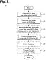
- FIG. 3is a flow diagram showing a method for facilitating diagnosis of cardiac rhythm disorders with the aid of a digital computer in accordance with one embodiment.
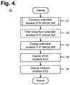
- FIG. 4is a flow diagram showing a routine for constructing and displaying a diagnostic composite plot for use in the method of FIG. 3 .
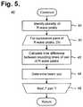
- FIG. 5is a flow diagram showing a routine for constructing an extended-duration R-R interval plot for use in the routine of FIG. 4 .
- FIG. 6is a diagram showing, by way of example, a diagnostic composite plot generated by the method of FIG. 3 .
- FIG. 7is a diagram showing, by way of example, a diagnostic composite plot for facilitating the diagnosis of sinus rhythm (SR) transitioning into atrial fibrillation (AF).
- SRsinus rhythm
- AFatrial fibrillation
- FIG. 8is a diagram showing, by way of example, a diagnostic composite plot for facilitating the diagnosis of 3:1 atrial flutter (AFL) transitioning into SR.
- AFLatrial flutter
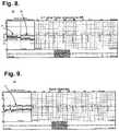
- FIG. 9is a diagram showing, by way of example, a diagnostic composite plot for facilitating the diagnosis of atrial trigeminy.
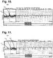
- FIG. 10is a diagram showing, by way of example, a diagnostic composite plot for facilitating the diagnosis of maximum heart rate in an episode of AF during exercise.
- FIG. 11is a diagram showing, by way of example, a diagnostic composite plot for facilitating the diagnosis of SR transitioning into AFL transitioning into AF.
- FIG. 12is a diagram showing, by way of example, a diagnostic composite plot for facilitating the diagnosis of sinus tachycardia and palpitations that occurred during exercise accompanied by a jump in heart rate.
- FIG. 13is a diagram showing, by way of example, a diagnostic composite plot for facilitating the diagnosis of bradycardia.
- FIG. 14is a block diagram showing a system for facilitating diagnosis of cardiac rhythm disorders with the aid of a digital computer in accordance with one embodiment.
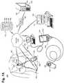
- FIG. 15is a diagram showing, by way of example, a subcutaneous P-wave centric insertable cardiac monitor (ICM) for long term electrocardiographic monitoring in accordance with one embodiment.
- ICMsubcutaneous P-wave centric insertable cardiac monitor
- FIGS. 16 and 17are respectively top and bottom perspective views showing the ICM of FIG. 15 .
- FIG. 18is a bottom perspective view showing the ICM of FIG. 15 in accordance with a further embodiment.
- FIGS. 19 and 20are respectively top and bottom perspective views showing an ICM in accordance with a still further embodiment.
- FIG. 21is a plan view showing further electrode configurations.
- FIG. 22is a functional block diagram showing the P-wave focused component architecture of the circuitry of the ICM of FIG. 15 .
- a normal healthy cardiac cyclerepeats through an expected sequence of events that can be visually traced through an ECG.
- Each cyclestarts with cardiac depolarization originating high in the right atrium in the sinoatrial (SA) node before spreading leftward towards the left atrium and inferiorly towards the atrioventricular (AV) node.
- SAsinoatrial
- AVatrioventricular
- the depolarization impulsetransits the Bundle of His and moves into the right and left bundle branches and Purkinje fibers to activate the right and left ventricles.
- FIG. 1is a graph showing, by way of example, a single ECG waveform 10 .
- the x-axisrepresents approximate time in units of tenths of a second and the y-axis represents approximate cutaneous electrical signal strength in units of millivolts.
- ECGsare typically printed or displayed at an effective paper speed of 25 millimeters (mm) per second.
- an ECGmay be provided to a physician in traditional paper-printed form, in “virtual” electronic display form, or both, the term “effective paper speed” is nevertheless still widely applied as a metric to normalize the recorded ECG signal to a standardized grid of 1 mm squares (omitted for the sake of clarity in FIG. 1 ), whereby each 1 mm horizontal box in the grid corresponds to 0.04 s (40 ms) of recorded time.
- Other effective paper speeds, grid sizes and units of displayare possible.
- a full ECGconsists of a stream of alphabetically-labeled waveforms 10 that collectively cover cardiac performance over a period of observation.
- the P-wave 11will normally have a smooth, normally upward, positive waveform that indicates atrial depolarization.
- the QRS complex 17will usually follow, often with a downward deflection of a Q-wave 12 , followed by a larger upward deflection of an R-wave 13 , and be terminated with a downward waveform of the S-wave 14 , which are collectively representative of ventricular depolarization.
- the T-wave 15will normally be a modest upward waveform, representative of ventricular repolarization, while the U-wave 16 , which is often not directly observable, will indicate the recovery period of the Purkinje conduction fibers.
- AFatrial fibrillation
- ECG R-R intervala cloud-like pattern of irregular R-R intervals due to an abnormal conduction of impulses to the ventricles.
- Gaussian-like distributionto these R-R intervals during AF.
- Atrial flutteris an abnormal heart rhythm in which cardiac impulses travel along pathways within the right atrium in an organized circular motion, causing the atria to beat faster than and out of sync with the ventricles. During AFL, the heart beats quickly, yet with a regular pattern.
- AFLpresents in an electrogram (e-gram) as a “sawtooth” pattern
- e-gramelectrogram
- R-R interval patternsthat usually manifest as 2:1 atrioventricular (AV) conduction or 4:1 atrioventricular conduction.
- the conduction through the AV nodeis variable and not fixed.
- FIG. 2is a graph showing, by way of example, a prior art Poincare R-R interval plot 18 .
- the x-axisrepresents the duration of R-R interval n in units of milliseconds (ms).
- the y-axisrepresents the duration of R-R interval n+1 also in units of ms.
- the x- and y-axesuse the same units, so as to form a trend line 19 along the 45-degree angle.
- the dot representing the two intervalsfalls onto the 45-degree trend line 19 .
- the dot representing the two intervalsfalls off the 45-degree trend line 19 and, as the difference between successive R-R intervals increases, the dots fall further away from the trend line 19 .
- the number of dots deviating from the trend line 19 in a Poincare plotcan indicate the frequency of occurrence of irregular heartbeats when compared to the number of dots on the trend line 19 .
- the distance of the dots to the trend line 19can approximate the extent of heart rate change from one heartbeat to the next.
- significant changes in heart rateparticularly spikes in heart rate, such as due to sinus rhythm transitions to atrial flutter, may be masked, distorted or even omitted in a Poincare plot if the change occurs over non-successive heartbeats.
- a Poincare plotis more useful as a mathematical tool than a physiological one, and therefore a Poincare plot cannot truly represent what the heart is doing serially over time with respect to changes in the heart's normal and abnormal physiology.
- FIG. 3is a flow diagram showing a method 20 for facilitating diagnosis of cardiac rhythm disorders with the aid of a digital computer in accordance with one embodiment.
- the method 20can be implemented in software and execution of the software can be performed on a computer, such as further described infra with reference to FIG. 14 , as a series of process or method modules or steps.
- ECG recordationAs a precursor step, the cutaneous action potentials of a patient are monitored and recorded as ECG data over a set time period (step 21 ), which can be over a short term or extended time frame.
- ECG recordationcan be provided through various kinds of ECG-capable monitoring ensembles, including a standardized 12-lead ECG setup, such as used for clinical ECG monitoring, a portable Holter-type ECG recorder for traditional ambulatory ECG monitoring, or a wearable ambulatory ECG monitor, such as a flexible extended wear electrode patch and a removable reusable (or single use) monitor recorder, such as described in commonly-assigned U.S. Pat. No.
- the ECG and any physiological dataare downloaded or retrieved into a digital computer, as further described infra with reference to FIG. 14 , with, for instance, the assistance of a download station or similar device, or via wireless connection, if so equipped, and a vector of the downloaded or retrieved ECG data is obtained (step 22 ).
- the vector of ECG datarepresents a 40-minute (or other duration) time span that is used in constructing the plot of R-R interval data, although other pre-event and post-event time spans are possible.
- a potentially-actionable cardiac event within the vector of ECG datacan be identified and the ECG data during, prior to and after the event is selected (step 23 ).
- the eventcould be identified with the assistance of a software package, such as Holter LX Analysis Software, licensed by NorthEast Monitoring, Inc., Maynard, Mass.; IntelliSpace Cardiovascular Image and Information management system, licensed Koninklijke Philips N.V., Amsterdam, Netherlands; MoMe System, licensed by InfoBionic, Lowell, Mass.; Pyramis ECG Management, licensed by Mortara Instrument Inc., Milwaukee, Wis.; ICS Clinical Suite, licensed by Spacelabs Healthcare Inc., Snoqualmie, Wash.; or a customized software package.
- a software packagesuch as Holter LX Analysis Software, licensed by NorthEast Monitoring, Inc., Maynard, Mass.; IntelliSpace Cardiovascular Image and Information management system, licensed Koninklijke Philips N.V., Amsterdam, Netherlands; MoMe System, licensed by InfoBionic, Lowell, Mass.; Pyramis ECG Management, licensed by Mortara Instrument Inc., Milwaukee, Wis.; ICS Clinical Suite, licensed by Spacelabs Healthcare Inc., Snoqualmie, Wash.; or
- a diagnostic composite plotis constructed that includes one or more temporal points of reference into the ECG data, which provide important diagnostic context, and a plot of R-R interval data is constructed based on the vector of ECG data (step 24 ), as further described infra with reference to FIG. 4 .
- both near field and far field contextual views of the ECG dataare constructed and displayed. Both views are temporally keyed to an extended duration R-R interval data view that, in one embodiment, is scaled non-linearly to maximize the visual differentiation for frequently-occurring heart rate ranges, such that a single glance allows the physician to make a diagnosis. All three views are presented simultaneously, thereby allowing the interpreting physician to diagnose rhythm and the pre- and post-contextual events leading up to a cardiac rhythm of interest.
- findings made through interpretation of heart rate variability patterns in the diagnostic composite plotcan be analyzed to form a diagnosis of a cardiac rhythm disorder (step 25 ), such as the cardiac rhythm disorders listed, by way of example, in Table 1.
- a cardiac rhythm disordersuch as the cardiac rhythm disorders listed, by way of example, in Table 1.
- the heart rate variability patterns in the diagnostic composite plotcould be provided to a system that programmatically detects AF by virtue of looking for the classic Gaussian-type distribution on the “cloud” of heart rate variability formed in the plot of R-R interval data, which can be corroborated by the accompanying contextual ECG data.
- a cardiac rhythm therapy delivery devicesuch as an implantable medical device (IMD) (not shown), including a pacemaker, implantable cardioverter defibrillator (ICD), or similar devices.
- IMDimplantable medical device
- ICDimplantable cardioverter defibrillator
- a diagnostic composite plotis constructed and displayed to help physicians identify and diagnose temporally-related cardiac dysrhythmic patterns.
- the diagnostic composite plotincludes ECG traces from two or more temporal points of reference and a plot of R-R interval data, although other configurations of ECG data plots when combined with the R-R interval plot will also provide critical information.
- FIG. 4is a flow diagram showing a routine 30 for constructing and displaying a diagnostic composite plot for use in the method 20 of FIG. 3 . Specific examples of diagnostic composite plots are discussed in detail infra with reference to FIGS. 7-13 .
- R-R interval datais presented to physicians in a format that includes views of relevant near field and far field ECG data, which together provide contextual information that improves diagnostic accuracy.
- other views of ECG datacan be provided in addition to or in lieu of the near field and far field ECG data views.
- the near field (or short duration) ECG dataprovides a “pinpoint” classical view of an ECG at traditional recording speed in a manner that is known to and widely embraced by physicians.
- the near field ECG datais coupled to a far field (or medium duration) ECG data view that provides an “intermediate” lower resolution, pre- and post-event contextual view.
- the extended-duration R-R interval plotis first constructed (step 31 ), as further described infra with reference to FIG. 5 .
- noisecan be filtered from the R-R interval plot (step 32 ), which is then displayed (step 33 ).
- Noise filteringcan include low-pass or high-pass filtering or other forms of signal processing, including automatic gain control, such as described in commonly-assigned U.S. patent application Ser. No. 14/997,416, cited supra.
- Rhythm disordershave different weightings depending upon the context with which they occur.
- the R-R interval data view and the multiple views of the ECG dataprovide that necessary context.
- the short and medium duration ECG data that accompanies the extended-duration R-R interval plotrepresents the ECG data “zoomed” in around a temporal point of reference identified in the center (or other location) of the R-R interval plot, thereby providing a visual context to the physician that allows temporal assessment of cardiac rhythm changes in various complementary views of the heart's behavior.
- the durations of the classical “pinpoint” view, the pre- and post-event “intermediate” view, and the R-R interval plotare flexible and adjustable.
- the diagnostic composite plotdisplays R-R interval data over a forty-minute duration and ECG data over short and medium durations (steps 34 and 35 ), such as four-second and 24-second durations that provide two- and 12-second segments of the ECG data before and after the R-R interval plot's temporal point of reference, which is generally in the center of the R-R interval plot, although other locations in the R-R interval plot could be identified as the temporal point of reference.
- the pinpoint “snapshot” and intermediate views of ECG data with the extended term R-R interval datacomparatively depicts heart rate context and patterns of behavior prior to and after a clinically meaningful arrhythmia or patient concern, thereby enhancing diagnostic specificity of cardiac rhythm disorders and providing physiological context to improve diagnostic ability.
- diagnostically relevant cardiac eventscan be identified and the R-R interval plot can be constructed with a cardiac event centered in the middle (or other location) of the plot, which thereby allows pre- and post-event heart rhythm data to be contextually “framed” through the pinpoint and intermediate ECG data views.
- Other durations, intervals and presentations of ECG dataare possible.
- the extended-duration R-R interval plotpresents beat-to-beat heart rate variability in a format that is intuitive and contextual, yet condensed.
- the format of the R-R interval plotis selected to optimize visualization of cardiac events in a compressed, yet understandable field of view, that allows for compact presentation of the data akin to a cardiologists understanding of clinical events.
- FIG. 5is a flow diagram showing a routine 40 for constructing an extended-duration R-R interval plot for use in the routine 30 of FIG. 4 .
- the duration of the R-R interval plotcan vary from less than one minute to the entire duration of the recording.
- a plurality of R-wave peaksis first selected out of the vector of ECG data (step 41 ) appropriate to the duration of the R-R interval plot to be constructed.
- each recording time differencerepresents the length of one heartbeat.
- the heart rate associated with the recording time differenceis determined by taking an inverse of the recording time difference and normalizing the inverse to beats per minute (step 44 ). Taking the inverse of the recording time difference yields a heart rate expressed in beats per second, which can be adjusted by a factor of 60 to provide a heart rate expressed in bpm. Calculation of the differences between the recording times and the associated heart rate continues for all of the remaining pairs of the R-wave peaks (step 44 ).
- the pairings of R-R intervals and associated heart ratesare formed into a two-dimensional plot.
- R-R intervalsare plotted along the x-axis and associated heart rates are plotted along the y-axis.
- the range and scale of the y-axiscan be adjusted according to the range and frequency of normal or patient-specific heart rates, so as to increase the visual distinctions between the heart rates that correspond to different R-R intervals.
- the y-axis of the R-R interval plothas a range of 20 to 300 beats per minute and R-R intervals corresponding to heart rates falling extremely outside of this range are excluded to allow easy visualization of 99+% of the heart rate possibilities.
- they-axishas a non-linear scale that is calculated as a function of the x-axis (R-R interval), such that:
- y( x - min ⁇ ⁇ bpm max ⁇ ⁇ bpm - min ⁇ ⁇ bpm ) n
- xis the time difference
- min bpmis the minimum heart rate
- max bpmis the maximum heart rate
- the overall effectis to accentuate the spatial differences in frequently-occurring ranges of heart rate and de-emphasize the spatial differential in ranges of heart rate where a deviation from norm would have been apparent, thus maximizing the spatial efficiency in data presentation.
- the goalis to show cardiac events in a simple, small visual contextual format. Larger scales and larger formats bely the practical limits of single-page presentations for the easy visualization at a glance by the busy physician.
- the visual distinctions between the heart rates that correspond to different R-R intervalsstand out, especially when plotted on a non-linear scale. Other y-axis ranges and scales are possible as may be selected by distinct clinical needs and specific diagnostic requirements.
- the diagnostic composite plotincludes a single, long range view of R-R interval data and a pair of pinpoint ECG data views that together help to facilitate rhythm disorder diagnosis by placing focused long-term heart rate information alongside short-term and medium-term ECG information.
- Such pairing of ECG and R-R interval datais unique in its ability to inform the physician of events prior to, during and after a cardiovascular event.
- FIG. 6is a diagram showing, by way of example, a diagnostic composite plot 50 generated by the method 30 of FIG. 3 . Note that the diagnostic composite plot can be tailored to include more than one view of R-R interval data and as many views of contextual ECG data as needed.
- a background information plot presenting an extended far field of related informationcan be included, such as activity amount, activity intensity, posture, syncope impulse detection, respiratory rate, blood pressure, oxygen saturation (SpO 2 ), blood carbon dioxide level (pCO 2 ), glucose, lung wetness, and temperature.
- Other forms of background informationare possible.
- background informationcan be layered on top of or keyed to the diagnostic composite plot 50 , particularly at key points of time in the R-R interval data plot, so that the context provided by each item of background information can be readily accessed by the reviewing physician.
- the diagnostic composite plot 50includes an ECG plot presenting a near field (short duration) view 51 , an ECG plot presenting an intermediate field (medium duration) view 52 , and an R-R interval data plot presenting a far field (extended duration) view 53 .
- the three views 51 , 52 , 53are juxtaposed alongside one other to allow quick back and forth referencing of the full context of the heart's normal and abnormal physiology.
- a temporal point of referencewhich could be a diagnostically relevant cardiac event, patient concern or other indicia, would be identified and centered on the x-axis in all three views.
- the placement of the temporal point of reference in the middle of all three x-axesenables the ECG data to be temporally keyed to the R-R interval data appearing in the center 60 of the R-R interval data view 53 , with a near field view 51 of an ECG displayed at normal (paper-based) recording speed and a far field view 52 that presents the ECG data occurring before and after the center 60 .
- the near field view 51provides the ECG data corresponding to the R-R interval data at the center 60 (or other location) in a format that is familiar to all physicians, while the intermediate field view 52 enables presentation of the broader ECG data context going beyond the borders of the near field view 51 .
- the center 60can be slidably adjusted backwards and forwards in time, with the near field view 51 and the far field view 52 of the ECG data automatically adjusting accordingly to stay in context with the R-R interval data view 51 .
- multiple temporal points of referencecan be identified with each temporal point of reference being optionally accompanied by one or more dedicated sets of ECG data views.
- the collection of plotsare conveniently arranged close enough to one another to facilitate printing on a single page of standard sized paper (or physical paper substitute, such as a PDF file), although other layouts of the plots are possible.
- the far field view 53is plotted with time in the x-axis and heart rate in the y-axis.
- the R-R intervalsare calculated by measuring the time occurring between successive R-wave peaks.
- the far field view 53presents R-R interval data (expressed as heart rate in bpm) that begins about 20 minutes prior to and ends about 20 minutes following the center 60 , although other durations are possible.
- the near field view 51 and intermediate field view 52present ECG data relative to the center 60 of the far field view 53 .
- the near field view 51provides a pinpoint or short duration view of the ECG data.
- the near field view 51presents ECG data 55 that begins about two seconds prior to and ends about two seconds following the center 60 , although other durations are possible.
- the intermediate field view 52provides additional contextual ECG information allowing the physician to assess the ECG itself and gather a broader view of the rhythm before and after a “blow-up” of the specific arrhythmia of interest.
- the intermediate field view 52presents ECG data 56 that begins about 12 seconds prior to and ends about 12 seconds following the center 60 , although other durations are possible.
- the eight-second interval of the ECG data 56 in the intermediate field view 52 that makes up the ECG data 56 in the near field view 51is visually highlighted, here, with a surrounding box 57 .
- other views of the ECG dataeither in addition to or in lieu of the near field view 51 and the far field view 52 are possible.
- an ECG plot presenting an extended far field view 54 of the background informationcan be included in the diagnostic composite plot 50 .
- the background informationis presented as average heart rate with day and night periods 58 alternately shaded along the x-axis.
- Other types of background informationsuch as activity amount, activity intensity, posture, syncope impulse detection, respiratory rate, blood pressure, oxygen saturation (SpO 2 ), blood carbon dioxide level (pCO 2 ), glucose, lung wetness, and temperature, are possible.
- FIG. 7is a diagram showing, by way of example, a diagnostic composite plot 70 for facilitating the diagnosis of sinus rhythm (SR) transitioning into AF.
- SRsinus rhythm
- SRis indicated through the presence of a reasonably steady baseline, but with subsidiary lines of premature beats and their compensatory pauses.
- SRmanifests as a shadowing 71 of a high heart rate line and a low heart rate line.
- AFis characterized by irregular heartbeats with a somewhat random variation of R-R intervals, although within a limited range and concentrating in a Gaussian-like distribution pattern around a mean that varies over time.
- AFcan be diagnosed by viewing a near field view 51 of ECG data showing heartbeats with reversed P-wave and irregular R-R intervals, this approach may be unclear when viewing “snippets” of ECG data, especially when associated with poor quality ECG signals.
- the presence of AFcan also be confirmed through a far field view 53 of R-R interval data, in which the R-R intervals assume superficially appearing disorganized, spread-out and decentralized scattered cloud 72 along the x-axis, in comparison to a concentrated, darkened line typical of a more organized cardiac rhythm.
- FIG. 8is a diagram showing, by way of example, a diagnostic composite plot 80 for facilitating the diagnosis of 3:1 atrial flutter (AFL) transitioning into SR with frequent premature ectopic atrial beats.
- AFLatrial flutter
- the R-R intervalshave a discernible aggregated line in the middle of the cloud 81 when the rhythm has yet to stabilize into a set pattern, not quite AF and not quite AFL.
- a dense line representing firm 3 : 1 atrial flutterstabilizes the rhythm prior to the transition into SR associated with the presence of two seesawing baselines that result from frequent atrial ectopy causing short coupling intervals and then compensatory long coupling intervals.
- SRis indicated by the middle of the three lines with a low heart rate line consistent with the compensatory pause (long coupling interval) and a high heart rate line with the shortest coupling interval representing the series of atrial premature beats 82 , and thus, at a faster heart rate.
- FIG. 9is a diagram showing, by way of example, a diagnostic composite plot 90 for facilitating the diagnosis of atrial trigeminy.
- Atrial trigeminyis characterized by three heartbeat rates appearing intermittently yet reasonably regularly.
- atrial trigeminycan be diagnosed by viewing a near field view 51 of ECG data, the pattern is significantly more recognizable in a far field view 53 of R-R interval data, in which a repeating pattern of three distinct heartbeat lines are persistently present and clearly visible 91 .
- This viewalso provides the physician with a qualitative feel for the frequency of the event troubling the patient that is not discernible from a single ECG strip.
- FIG. 10is a diagram showing, by way of example, a diagnostic composite plot 100 for facilitating the diagnosis of maximum heart rate in an episode of AF during exercise.
- AFmanifests through a dispersed cloud of dots (Gaussian-like distribution) without a discernible main heart rate line representing regular heartbeats 101 .
- the maximum heartbeatcan be located by an increase in heart rate clustered about the cloud 102 .
- individual dots above the 200 bpm range throughout the entire 40-minute rangeindicates the maximum heart rate during exercise.
- the very rapid rise in heart ratecan be critical to patient management, as such bumps in rate by exercise can prove serious and even trigger cardiac arrest. Their very presence is easily visualized in the R-R interval data plot, thereby allowing the physician to alter therapy sufficiently to control such potentially damaging rises in heart rate.
- FIG. 11is a diagram showing, by way of example, a diagnostic composite plot 110 for facilitating the diagnosis of SR transitioning into AFL transitioning into AF.
- SRmanifests as an uneven main heart rate line with a fluctuating height 111 .
- the main heart rate linebreaks away at a lower heart rate than the SR main heart rate line 112 .
- the episode of AFLfurther evolves into AF as characterized by a dispersed cloud of irregular heartbeats without concentrated heart rate lines 113 .
- This viewprovides critical information to the physician managing AF patients in that, at a glance, the view provides data that tells the physician that the patient's AF may be the consequence of AFL. Such knowledge may alter both drug and procedure therapies, like catheter ablation details of intervention.
- FIG. 12is a diagram showing, by way of example, a diagnostic composite plot 120 for facilitating the diagnosis of sinus tachycardia and palpitations that occurred during exercise accompanied by a jump in heart rate.
- sinus tachycardiais indicated by the presence of a baseline heart rate of about 60 bpm 121 that spikes up to around 100 bpm 122 and gradually slopes down with a wide tail 123 , reflecting a sharp rise of heart rates followed by a gradual decline.
- the associated ECG data in the near field and intermediate field viewscan confirm the rhythm as sinus rhythm and a normal response to exercise.
- FIG. 13is a diagram showing, by way of example, a diagnostic composite plot 90 for facilitating the diagnosis of bradycardia during sleep and a R-R interval pattern characteristic of sleep.
- Bradycardiarefers to a resting heart rate of under 60 bpm. Bradycardia during sleep is often tempered with occasional spikes of rapid heart rate, which can be a secondary compensatory response to dreaming, snoring or sleep apnea.
- bradycardiamanifests as the presence of a base line heart rate in the range of about 50 bpm 131 , coupled with multiple spikes of dots 132 representing intermittent episodes of elevated heart rate. Such elevations in heart rate during a pre-dominantly slower rate may be signs of a cardio-respiratory disorder. Still other applications of the diagnostic composite plot 80 are possible.
- FIG. 14is a block diagram showing a system 140 for facilitating diagnosis of cardiac rhythm disorders with the aid of a digital computer 150 in accordance with one embodiment.
- Each diagnostic composite plot 151is based on ECG data 166 that has either been recorded by a conventional electrocardiograph (not shown) or retrieved or obtained from some other type of ECG monitoring and recording device. Following completion of the ECG monitoring, the ECG data is assembled into a diagnostic composite plot 151 , which can be used by a physician to diagnosis and, if required, treat a cardiac rhythm disorder, or for other health care or related purposes.
- Each diagnostic composite plot 151is based on ECG data 166 that has been recorded over a period of observation, which can be for just a short term, such as during a clinic appointment, or over an extended time frame of months.
- ECG recordation and, in some cases, physiological monitoringcan be provided through various types of ECG-capable monitoring ensembles, including a standardized 12-lead ECG setup (not shown), such as used for clinical ECG monitoring, a portable Holter-type ECG recorder for traditional ambulatory ECG monitoring (also not shown), or a wearable ambulatory ECG monitor.
- One form of ambulatory ECG monitor 142 particularly suited to monitoring and recording ECG and physiological dataemploys an electrode patch 143 and a removable reusable (or single use) monitor recorder 144 , such as described in commonly-assigned U.S. patent application Ser. No. 14/997,416, cited supra.
- the electrode patch 143 and monitor recorder 144are synergistically optimized to capture electrical signals from the propagation of low amplitude, relatively low frequency content cardiac action potentials, particularly the P-waves generated during atrial activation.
- the ECG monitor 142sits centrally (in the midline) on the patient's chest along the sternum 169 oriented top-to-bottom.
- the ECG monitor 142interfaces to a pair of cutaneous electrodes (not shown) on the electrode patch 143 that are adhered to the patient's skin along the sternal midline (or immediately to either side of the sternum 169 ).
- the ECG monitor 142has a unique narrow “hourglass”-like shape that significantly improves the ability of the monitor to be comfortably worn by the patient 141 for an extended period of time and to cutaneously sense cardiac electric signals, particularly the P-wave (or atrial activity) and, to a lesser extent, the QRS interval signals in the ECG waveforms indicating ventricular activity.
- the electrode patch 143itself is shaped to conform to the contours of the patient's chest approximately centered on the sternal midline.
- a layer of non-irritating adhesivesuch as hydrocolloid, is provided at least partially on the underside, or contact, surface of the electrode patch, but only on the electrode patch's distal and proximal ends.
- a strain reliefis defined in the electrode patch's flexible circuit using cutouts partially extending transversely from each opposite side of the flexible circuit and continuing longitudinally towards each other to define in ‘S’-shaped pattern.
- the electrode patch 143is made from a type of stretchable spunlace fabric.
- the outward-facing aspect of the backingto which a (non-stretchable) flexible circuit is fixedly attached, stretches at a different rate than the backing's skin-facing aspect, where a skin adhesive removably affixes the electrode patch 143 to the skin.
- a skin adhesiveremovably affixes the electrode patch 143 to the skin.
- the monitor recorder 142senses and records the patient's ECG data 166 and physiological data (not shown) into a memory onboard the monitor recorder 144 .
- the recorded datacan be downloaded using a download station 145 , which could be a dedicated download station 145 that permits the retrieval of stored ECG data 166 and physiological data, if applicable, execution of diagnostics on or programming of the monitor recorder 144 , or performance of other functions.
- the monitor recorder 144has a set of electrical contacts (not shown) that enable the monitor recorder 144 to physically interface to a set of terminals 148 .
- the download station 145can be operated through user controls 149 to execute a communications or data download program 146 (“Download”) or similar program that interacts with the monitor recorder 144 via the physical interface to retrieve the stored ECG data 166 .
- the download station 145could alternatively be a server, personal computer, tablet or handheld computer, smart mobile device, or purpose-built device designed specific to the task of interfacing with a monitor recorder 144 . Still other forms of download station 145 are possible.
- the ECG data 166 from the monitor recorder 144can be offloaded wirelessly.
- the ECG data 166can be retrieved from the download station 145 using a control program 157 (“Ctl”) or analogous application executing on a personal digital computer 156 or other connectable computing device, via a hard wired link 158 , wireless link (not shown), or by physical transfer of storage media (not shown).
- the personal digital computer 156may also execute middleware (not shown) that converts the ECG data 166 into a format suitable for use by a third-party post-monitoring analysis program.
- the personal digital computer 156stores the ECG data 166 along with each patient's electronic medical records (EMRs) 165 in the secure database 64 , as further discussed infra.
- the download station 145is able to directly interface with other devices over a computer communications network 155 , which could be a combination of local area and wide area networks, including the Internet or another telecommunications network, over wired or wireless connections.
- a client-server modelcan be employed for ECG data 166 analysis.
- a server 62executes a patient management program 160 (“Mgt”) or similar application that accesses the retrieved ECG data 166 and other information in the secure database 164 cataloged with each patient's EMRs 165 .
- the patients' EMRscan be supplemented with other information (not shown), such as medical history, testing results, and so forth, which can be factored into automated diagnosis and treatment.
- the patient management program 160also maintains and safeguards the secure database 164 to limit access to patient EMRs 165 to only authorized parties for appropriate medical or other uses, such as mandated by state or federal law, such as under the Health Insurance Portability and Accountability Act (HIPAA) or per the European Union's Data Protection Directive.
- HIPAAHealth Insurance Portability and Accountability Act
- Other schemes and safeguards to protect and maintain the integrity of patient EMRs 165are possible.
- the wearable monitor 142can interoperate wirelessly with other wearable or implantable physiology monitors and activity sensors 152 , such as activity trackers worn on the wrist or body, and with mobile devices 153 , including smart watches and smartphones.
- Wearable or implantable physiology monitors and activity sensors 152encompass a wide range of wirelessly interconnectable devices that measure or monitor a patient's physiological data, such as heart rate, temperature, blood pressure, respiratory rate, blood pressure, blood sugar (with or without an appropriate subcutaneous probe), oxygen saturation, minute ventilation, and so on; physical states, such as movement, sleep, footsteps, and the like; and performance, including calories burned or estimated blood glucose level.
- wearable and implantable physiology monitors and activity sensors 152are capable of wirelessly interfacing with mobile devices 153 , particularly smart mobile devices, including so-called “smartphones” and “smart watches,” as well as with personal computers and tablet or handheld computers, to download monitoring data either in real-time or in batches through an application (“App”) or similar program.
- mobile devices 153particularly smart mobile devices, including so-called “smartphones” and “smart watches,” as well as with personal computers and tablet or handheld computers, to download monitoring data either in real-time or in batches through an application (“App”) or similar program.
- Appapplication
- ECG data 166Based on the ECG data 166 , physicians can rely on the data as medically certifiable and are able to directly proceed with diagnosing cardiac rhythm disorders and determining the appropriate course of treatment for the patient 141 , including undertaking further medical interventions as appropriate.
- the ECG data 166can be retrieved by a digital computer 150 over the network 155 .
- a diagnostic composite plot 151 that includes multiple temporal points of reference and a plot of R-R interval datais then constructed based on the ECG data 166 , as discussed in detail supra with reference to FIG. 3 , and displayed or, alternatively, printed, for use by a physician.
- diagnostic findings 168 for a patientexceed a threshold level of tolerance, which may be tailored to a specific client, disease or medical condition group, or applied to a general patient population, in a still further embodiment, therapeutic treatment (“Therapy”) to address diagnosed disorder findings can be generated and, optionally, programmed into a cardiac rhythm therapy delivery device, such as an IMD (not shown), including a pacemaker, implantable cardioverter defibrillator (ICD), or similar devices.
- IMDimplantable cardioverter defibrillator
- the ECG data 166can be recorded using a subcutaneous insertable cardiac monitor.
- a subcutaneous insertable cardiac monitorLong-term electrocardiographic and physiological monitoring over a period lasting up to several years in duration can be provided through a continuously-recording subcutaneous insertable cardiac monitor (ICM), such as one described in commonly-owned U.S. patent application Ser. No. 15/832,385, filed Dec. 5, 2017, pending, the disclosure of which is incorporated by reference.
- FIG. 15is a diagram showing, by way of example, a subcutaneous P-wave centric ICM 212 for long term electrocardiographic monitoring in accordance with one embodiment.
- the ICM 212is implanted in the parasternal region 211 of a patient 210 .
- the sensing circuitry and components, compression algorithms, and the physical layout of the electrodesare specifically optimized to capture electrical signals from the propagation of low amplitude, relatively low frequency content cardiac action potentials, particularly the P-waves generated during atrial activation.
- either location to the right or left of the sternal midlineis acceptable; placement of the device, if possible, should bridge the vertical height of the heart, which lies underneath the sternum 203 , thereby placing the ICM 212 in close proximity to the anterior right atrium and the left atrial appendage that lie immediately beneath.
- the ICM 212is shaped to fit comfortably within the body under the skin and to conform to the contours of the patient's parasternal region 211 when implanted immediately to either side of the sternum 203 , but could be implanted in other locations of the body. In most adults, the proximal end 213 of the ICM 212 is generally positioned below the manubrium 8 but, depending upon patient's vertical build, the ICM 212 may actually straddle the region over the manubrium 8 .
- the distal end 214 of the ICM 212generally extends towards the xiphoid process 9 and lower sternum but, depending upon the patient's build, may actually straddle the region over or under the xiphoid process 9 , lower sternum and upper abdomen.
- the majority of the left atriumconstitutes the portion of the heart furthest away from the surface of the skin on the chest and harbors the atrial tissue most likely to be the source of serious arrhythmias, like atrial fibrillation.
- the ventricleswhich generate larger amplitude signals, are located anteriorly as in the case of the anterior right ventricle and most of the anterior left ventricle situated relatively close to the skin surface of the central and left anterior chest.
- both the P-wave and the R-waveare required for the physician to make a proper rhythm diagnosis from the dozens of arrhythmias that can occur.
- the quality of P-wavesis more susceptible to weakening from distance and the intervening tissues and structures and from signal attenuation and signal processing than the high voltage waveforms associated with ventricular activation.
- the added value of avoiding further signal attenuation resulting from dermal impedancemakes a subcutaneous P-wave centric ICM even more likely to match, or even outperform dermal ambulatory monitors designed to analogous engineering considerations and using similar sensing circuitry and components, compression algorithms, and physical layout of electrodes, such as described in U.S. Pat. No. 9,545,204, issued Jan.
- the ICM 212can be implanted in the patient's chest using, for instance, a minimally invasive subcutaneous implantation instrument or other suitable surgical implement.
- the ICM 212is positioned slightly to the right or left of midline, covering the center third of the chest, roughly between the second and sixth ribs, approximately spanning between the level of the manubrium 8 and the level of the xiphoid process 9 on the inferior border of the sternum 203 , depending upon the vertical build of the patient 210 .
- an ECG devicesincluding dermal ECG monitors and ICMs
- cardiac, cellular, extracellular, vector of current flow, and physical factorslike obesity, dermatitis, lung disease, large breasts, and high impedance skin, as can occur in dark-skinned individuals.
- ECG sensing subcutaneously in the parasternal region 211significantly improves the ability of the ICM 212 to counter some of the effects of these factors, particularly high skin impedance and impedance from subcutaneous fat.
- the ICM 212exhibits superior performance when compared to conventional dermal ECG monitors to existing implantable loop recorders, ICMs, and other forms of implantable monitoring devices by virtue of its engineering and proven P-wave documentation above the skin, as discussed in W. M. Smith et al., “Comparison of diagnostic value using a small, single channel, P-wave centric sternal ECG monitoring patch with a standard 3-lead Holter system over 24 hours,” Am. Heart J., Mar. 20217; 2185:67-73, the disclosure of which is incorporated by reference.
- the ECG electrodes of the ICM 212are subcutaneously positioned with the upper or superior pole (ECG electrode) slightly to the right or left of the sternal midline in the region of the manubrium 8 and, on the distal end 214 , the lower or inferior pole (ECG electrode) is similarly situated slightly to the right or left of the sternal midline in the region of the xiphoid process 9 and lower sternum 203 .
- the ECG electrodes of the ICM 212are placed primarily in a north-to-south orientation along the sternum 203 that corresponds to the north-to-south waveform vector exhibited during atrial activation.
- This orientationcorresponds to the aVF lead used in a conventional 12-lead ECG that is used to sense positive or upright P-waves.
- the electrode spacing and the electrodes' shapes and surface areasmimic the electrodes used in the ICM's dermal cousin, designed as part of the optimal P-wave sensing electrode configuration, such as provided with the dermal ambulatory monitors cited supra.
- FIGS. 16 and 17are respectively top and bottom perspective views showing the ICM 212 of FIG. 1 .
- the ICM 212is constructed with a hermetically sealed implantable housing 215 with at least one ECG electrode forming a superior pole on the proximal end 213 and at least one ECG electrode forming an inferior pole on the distal end 214 .
- the housing 215When implanted, the housing 215 is oriented most cephalad.
- the housing 215is constructed of titanium, stainless steel or other biocompatible material.
- the housing 215contains the sensing, recordation and interfacing circuitry of the ICM 212 , plus a long life battery.
- a wireless antennais integrated into or within the housing 215 and can be positioned to wrap around the housing's internal periphery or location suited to signal reception. Other wireless antenna placement or integrations are possible.
- the ICM 212has four ECG electrodes 216 , 217 , 218 , 219 . There could also be additional ECG electrodes, as discussed infra.
- the ECG electrodesinclude two ventral (or dorsal) ECG electrodes 218 , 219 and two wraparound ECG electrodes 216 , 217 .
- One ventral ECG electrode 218is formed on the proximal end 213 and one ventral ECG electrode 219 is formed on the distal end 214 .
- One wraparound ECG electrode 216is formed circumferentially about the proximal end 213 and one wraparound ECG electrode 217 is formed circumferentially about the distal end 214 .
- Each wraparound ECG electrode 216 , 217is electrically insulated from its respective ventral ECG electrode 218 , 219 by a periphery 220 , 221 .
- the four ECG electrodes 216 , 217 , 218 , 219are programmatically controlled by a microcontroller through onboard firmware programming to enable a physician to choose from several different electrode configurations that vary the electrode surface areas, shapes, and inter-electrode spacing.
- the sensing circuitrycan be programmed, either pre-implant or in situ, to use different combinations of the available ECG electrodes (and thereby changing electrode surface areas, shapes, and inter-electrode spacing), including pairing the two ventral ECG electrodes 216 , 217 , the two wraparound ECG electrodes 218 , 219 , or one ventral ECG electrode 216 , 217 with one wraparound ECG electrode 218 , 219 located on the opposite end of the housing 215 .
- the periphery 220 , 221can be programmatically controlled to logically combine the wraparound ECG electrode 216 , 217 on one end of the ICM 212 with its corresponding ventral ECG electrode 218 , 219 to form a single virtual ECG electrode with larger surface area and shape.
- the two ECG electrodes that are only on one end of the ICM 212for instance, wraparound ECG electrode 216 and ventral ECG electrode 218 , could be paired; however, the minimal inter-electrode spacing would likely yield a signal of poor fidelity in most situations.
- the housing 215 and contained circuitrycan be provided as a standalone ICM core assembly to which a pair of compatible ECG electrodes can be operatively coupled to form a full implantable ICM device.
- FIG. 18is a bottom perspective view showing the ICM 212 of FIG. 15 in accordance with a further embodiment.
- An additional pair of ventral ECG electrodes 222 , 223are included on the housing's ventral surface.
- ventral ECG electrodes 222 , 223are spaced closer together than the ventral ECG electrodes 218 , 219 on the ends of the housing 215 and a physician can thus choose to pair the two inner ventral ECG electrodes 222 , 223 by themselves to allow for minimal electrode-to-electrode spacing, or with the other ECG electrodes 216 , 217 , 218 , 219 to vary electrode surface areas, shapes, and inter-electrode spacing even further to explore optimal configurations to acquire the P-wave.
- FIGS. 19 and 20are respectively top and bottom perspective views showing an ICM 230 in accordance with a still further embodiment.
- the ICM 230has a housing 31 with a tapered extension 32 that is terminated on the distal end with an electrode 34 .
- the housing 31includes a pair of ECG electrodes electrically insulated by a periphery 37 that include a ventral ECG electrode 33 and a wraparound ECG electrode 34 .
- a ventral ECG electrode 36is oriented on the housing's distal end before the tapered extension 32 .
- Still other housing structures and electrode configurationsare possible.
- the basic electrode layoutis sufficient to sense cardiac action potentials in a wide range of patients. Differences in thoracic tissue density and skeletal structure from patient to patient, though, can affect the ability of the sensing electrodes to efficaciously capture action potential signals, yet the degree to which signal acquisition is affected may not be apparent until after an ICM has been implanted and deployed, when the impacts of the patient's physical constitution and his patterns of mobility and physical movement on ICM monitoring can be fully assessed.
- the electrodescan be configured post-implant to allow the ICM to better adapt to a particular patient's physiology.
- electrode configurations having more than two sensing electrodesare possible.
- FIG. 21is a plan view showing further electrode configurations. Referring first to FIG. 21( a ) , a single disc ECG electrode 40 could be bifurcated to form a pair of half-circle ECG electrodes 241 , 242 that could be programmatically selected or combined to accommodate a particular patients ECG signal characteristics post-ICM implant. Referring next to FIG.
- a single disc ECG electrode 245could be divided into three sections, a pair of crescent-shaped ECG electrodes 246 , 247 surrounding a central semicircular ECG electrode 48 that could similarly be programmatically selected or combined. Still other ECG electrode configurations are possible.
- FIG. 22is a functional block diagram showing the P-wave focused component architecture of the circuitry 280 of the ICM 212 of FIG. 15 .
- the circuitry 280is powered through the long life battery 21 provided in the housing 215 .
- Operation of the circuitry 280 of the ICM 212is managed by a microcontroller 281 , such as the EFM32 Tiny Gecko 32-bit microcontroller, manufactured by Silicon Laboratories Inc., Austin, Tex.
- the microcontroller 281has flexible energy management modes and includes a direct memory access controller and built-in analog-to-digital and digital-to-analog converters (ADC and DAC, respectively).
- the microcontroller 281also includes a program memory unit containing internal flash memory (not shown) that is readable, writeable, and externally programmable.
- the microcontroller 281operates under modular micro program control as specified in firmware stored in the internal flash memory.
- the microcontroller 281draws power from the battery provided in the housing 215 and connects to the ECG front end circuit 283 .
- the front end circuit 283measures raw subcutaneous electrical signals using a driven reference signal that eliminates common mode noise, as further described infra.
- the circuitry 280 of the ICM 212also includes a flash memory 82 external to the microcontroller 281 , which the microcontroller 281 uses for continuously storing samples of ECG monitoring signal data and other physiology, such as respiratory rate, blood oxygen saturation level (SpO 2 ), blood pressure, temperature sensor, and physical activity, and device and related information.
- the flash memory 82also draws power from the battery provided in the housing 215 . Data is stored in a serial flash memory circuit, which supports read, erase and program operations over a communications bus.
- the flash memory 82enables the microcontroller 281 to store digitized ECG data.
- the communications busfurther enables the flash memory 82 to be directly accessed wirelessly through a transceiver 285 coupled to an antenna 217 built into (or provided with) the housing 215 .
- the transceiver 285can be used for wirelessly interfacing over Bluetooth or other types of wireless technologies for exchanging data over a short distance with a paired mobile device, including smartphones and smart watches, that are designed to communicate over a public communications infrastructure, such as a cellular communications network, and, in a further embodiment, other wearable (or implantable) physiology monitors, such as activity trackers worn on the wrist or body.
- Other types of device pairingsare possible, including with a desktop computer or purpose-built bedside monitor.
- the transceiver 285can be used to offload stored ECG monitoring data and other physiology data and information, such as by wirelessly transmitting the samples of ECG signals stored in the flash memory 82 to the download station and any other physiological data to the download station 145 , and for device firmware reprogramming.
- the flash memory 82can be accessed through an inductive coupling (not shown), with the accessed samples of the ECG signals being provided to the download station 145 .
- the microcontroller 281includes functionality that enables the acquisition of samples of analog ECG signals, which are converted into a digital representation.
- the microcontroller 281implements a loop recorder feature that will acquire, sample, digitize, signal process, and store digitized ECG data into available storage locations in the flash memory 82 until all memory storage locations are filled, after which existing stored digitized ECG data will either be overwritten through a sliding window protocol, albeit at the cost of potentially losing the stored data that was overwritten, if not previously downloaded, or transmitted wirelessly to an external receiver, such as the download station 145 , to unburden the flash memory.
- the stored digitized ECG datacan be maintained permanently until downloaded or erased to restore memory capacity. Data download or erasure can also occur before all storage locations are filled, which would free up memory space sooner, albeit at the cost of possibly interrupting monitoring while downloading or erasure is performed. Still other modes of data storage and capacity recovery are possible.
- the circuitry 280 of the ICM 212can include functionality to programmatically select pairings of sensing electrodes when the ICM 212 is furnished with three or more electrodes.
- multiple sensing electrodescould be provided on the ICM 212 to provide a physician the option of fine-tuning the sensing dipole (or tripole or multipole) in situ by parking active electrodes and designating any remaining electrodes inert.
- the pairing selectioncan be made remotely through an inductive coupling or by the transceiver 285 via, for instance, a paired mobile device.
- the sensing electrode configurationincluding number of electrodes, electrode-to-electrode spacing, and electrode size, shape, surface area, and placement, can be modified at any time during the implantation of the ICM 212 .
- the circuitry 280 of the ICM 212can include an actigraphy sensor 84 implemented as a 3-axis accelerometer.
- the accelerometermay be configured to generate interrupt signals to the microcontroller 281 by independent initial wake up and free fall events, as well as by device position.
- the actigraphy provided by the accelerometercan be used during post-monitoring analysis to correct the orientation of the ICM 212 if, for instance, the ICM 212 has been inadvertently implanted upside down, that is, with the ICM's housing oriented caudally, as well as for other event occurrence analyses.
- the circuitry 280 of the ICM 212can include one or more physiology sensors.
- a physiology sensorcan be provided as part of the circuitry 280 of the ICM 212 , or can be provided on the electrode assembly 14 with communication with the microcontroller 281 provided through a circuit trace.
- the physiology sensorcan include an SpO 2 sensor, blood pressure sensor, temperature sensor, respiratory rate sensor, glucose sensor, airflow sensor, volumetric pressure sensing, or other types of sensor or telemetric input sources.
- firmware with programming instructionscan be downloaded into the microcontroller's internal flash memory.
- the firmwarecan include heuristics to signal patient and physician with alerts over health conditions or arrhythmias of selected medical concern, such as where a heart pattern particular to the patient is identified and the ICM 212 is thereby reprogrammed to watch for a reoccurrence of that pattern, after which an alert will be generated and sent to the physician (or other caregiver) through the transceiver 285 via, for instance, a paired mobile device.
- the firmwarecan include heuristics that can be downloaded to the ICM 212 to actively identify or narrow down a pattern (or even the underlying cause) of sporadic cardiac conditions, for instance, atrial tachycardia (AT), atrial fibrillation (AF), atrial flutter (AFL), AV node reciprocating tachycardia, ventricular tachycardia (VT), sinus bradycardia, asystole, complete heart block, and other cardiac arrhythmias, again, after which an alert will be generated and sent to the physician (or other caregiver) through the transceiver 285 .
- ATatrial tachycardia
- AFatrial fibrillation
- AFLatrial flutter
- AV node reciprocating tachycardiaventricular tachycardia
- VTventricular tachycardia
- sinus bradycardiaasystole
- complete heart blockcomplete heart block
- other cardiac arrhythmiasagain, after which an alert will
- an alert that includes a compressed ECG digitized samplecan also be wirelessly transmitted by the ICM 212 upon the triggering of a preset condition, such as an abnormally low heart rate in excess of 170 beats per minute (bpm), an abnormally low heart rate falling below 30 bpm, or AF detected by onboard analysis of RR interval variability by the microcontroller 61 .
- a preset conditionsuch as an abnormally low heart rate in excess of 170 beats per minute (bpm), an abnormally low heart rate falling below 30 bpm, or AF detected by onboard analysis of RR interval variability by the microcontroller 61 .
- a similar methodology of creating firmware programming tailored to the monitoring and medical diagnostic needs of a specific patientcan be used for other conditions or symptoms, such as syncope, palpitations, dizziness and giddiness, unspecified convulsions, abnormal ECG, transient cerebral ischemic attacks and related syndromes, cerebral infarction, occlusion and stenosis of pre-cerebral and cerebral arteries not resulting in cerebral infarction personal history of transient ischemic attack, and cerebral infarction without residual deficits, to trigger an alert and involve the physician or initiate automated analysis and follow up back at the patient's clinic.
- the circuitry 280 of the ICM 212can accommodate patient-interfaceable components, including an external tactile feedback device (not shown) that wirelessly interfaces to the ICM 212 through the transceiver 285 .
- a patient 210can press the external tactile feedback device to mark events, such as a syncope episode, or to perform other functions.
- the circuitry 280can also accommodate triggering an external buzzer 267 , such as a speaker, magnetic resonator or piezoelectric buzzer, implemented as part of the external tactile feedback device or as a separate wirelessly-interfaceable component.
- the buzzer 67can be used by the microcontroller 281 to indirectly output feedback to a patient 210 , such as a low battery or other error condition or warning.
- Still other components, provided as either part of the circuitry 280 of the ICM 212 or as external wirelessly-interfaceable devices,are possible.
- the ECG front end circuit 283 of the ICM 212measures raw subcutaneous electrical signals using a driven reference signal, such as described in U.S. Pat. Nos. 9,700,227, 9,7217,433, and 9,615,763, cited supra.
- the driven reference signaleffectively reduces common mode noise, power supply noise and system noise, which is critical to preserving the characteristics of low amplitude cardiac action potentials, especially the P wave signals originating from the atria.
- the ECG front end circuit 283is organized into a passive input filter stage, a unity gain voltage follower stage, a passive high pass filtering stage, a voltage amplification and active filtering stage, and an anti-aliasing passive filter stage, plus a reference generator.
- the passive input filter stagepassively shifts the frequency response poles downward to counter the high electrode impedance from the patient on the signal lead and reference lead, which reduces high frequency noise.
- the unity gain voltage follower stageallows the circuit to accommodate a very high input impedance, so as not to disrupt the subcutaneous potentials or the filtering effect of the previous stage.
- the passive high pass filtering stageincludes a high pass filter that removes baseline wander and any offset generated from the previous stage.
- the voltage amplification and active filtering stageamplifies or de-amplifies (or allows to pass-through) the voltage of the input signal, while applying a low pass filter.
- the anti-aliasing passive filter stageprovides an anti-aliasing low pass filter.
- the reference generatordrives a driven reference signal containing power supply noise and system noise to the reference lead and is connected directly to the patient, thereby avoiding the thermal noise of the protection resistor that is included as part of the protection circuit.
Landscapes
- Health & Medical Sciences (AREA)
- Life Sciences & Earth Sciences (AREA)
- Engineering & Computer Science (AREA)
- Cardiology (AREA)
- Public Health (AREA)
- Medical Informatics (AREA)
- Veterinary Medicine (AREA)
- Physics & Mathematics (AREA)
- General Health & Medical Sciences (AREA)
- Biophysics (AREA)
- Pathology (AREA)
- Biomedical Technology (AREA)
- Heart & Thoracic Surgery (AREA)
- Animal Behavior & Ethology (AREA)
- Molecular Biology (AREA)
- Surgery (AREA)
- Physiology (AREA)
- Signal Processing (AREA)
- Computer Vision & Pattern Recognition (AREA)
- Artificial Intelligence (AREA)
- Psychiatry (AREA)
- Chemical & Material Sciences (AREA)
- Dispersion Chemistry (AREA)
- Measurement And Recording Of Electrical Phenomena And Electrical Characteristics Of The Living Body (AREA)
Abstract
Description
| TABLE 1 |
| Cardiac Rhythm Disorders |
| Normal sinus rhythm | ||
| Sinus Bradycardia | ||
| Sinus Tachycardia | ||
| Premature atrial and ventricular beats | ||
| Ectopic atrial tachycardia | ||
| Atrial fibrillation | ||
| Atrial flutter | ||
| Atrial or ventricular bigeminy, trigeminy or quadrigeminy | ||
| Sinus Bradycardia | ||
| Fusion beats | ||
| Interpolated ventricular premature beats | ||
| Intraventricular conduction delay | ||
| Junctional rhythm | ||
| AV Nodal re-entrant tachycardia | ||
| AV re-entrant tachycardia | ||
| Wolff-Parkinson-White Syndrome and Pre-excitation | ||
| Ventricular tachycardia | ||
| Accelerated idioventricular rhythm | ||
| AV Wenckebach block | ||
| AV Type II block | ||
| Sinoatrial block | ||
where x is the time difference, min bpm is the minimum heart rate, max bpm is the maximum heart rate, and n<1. The non-linear scale of the y-axis accentuates the spatial distance between successive heart rates when heart rate is low. For example, when n=2, the spatial difference between 50 and 60 bpm is 32% larger than the spatial difference between 90 bpm and 100 bpm, and 68% larger than the spatial difference between 150 bpm and 160 bpm. As a result the overall effect is to accentuate the spatial differences in frequently-occurring ranges of heart rate and de-emphasize the spatial differential in ranges of heart rate where a deviation from norm would have been apparent, thus maximizing the spatial efficiency in data presentation. The goal is to show cardiac events in a simple, small visual contextual format. Larger scales and larger formats bely the practical limits of single-page presentations for the easy visualization at a glance by the busy physician. The visual distinctions between the heart rates that correspond to different R-R intervals stand out, especially when plotted on a non-linear scale. Other y-axis ranges and scales are possible as may be selected by distinct clinical needs and specific diagnostic requirements.
Claims (16)
Priority Applications (3)
| Application Number | Priority Date | Filing Date | Title |
|---|---|---|---|
| US16/595,226US10813567B2 (en) | 2013-09-25 | 2019-10-07 | System and method for composite display of subcutaneous cardiac monitoring data |
| US17/080,757US11445969B2 (en) | 2013-09-25 | 2020-10-26 | System and method for event-centered display of subcutaneous cardiac monitoring data |
| US17/944,899US11653870B2 (en) | 2013-09-25 | 2022-09-14 | System and method for display of subcutaneous cardiac monitoring data |
Applications Claiming Priority (12)
| Application Number | Priority Date | Filing Date | Title |
|---|---|---|---|
| US201361882403P | 2013-09-25 | 2013-09-25 | |
| US14/080,725US9730593B2 (en) | 2013-09-25 | 2013-11-14 | Extended wear ambulatory electrocardiography and physiological sensor monitor |
| US14/488,230US9700227B2 (en) | 2013-09-25 | 2014-09-16 | Ambulatory electrocardiography monitoring patch optimized for capturing low amplitude cardiac action potential propagation |
| US14/614,265US9408545B2 (en) | 2013-09-25 | 2015-02-04 | Method for efficiently encoding and compressing ECG data optimized for use in an ambulatory ECG monitor |
| US201562132497P | 2015-03-12 | 2015-03-12 | |
| US14/997,416US9345414B1 (en) | 2013-09-25 | 2016-01-15 | Method for providing dynamic gain over electrocardiographic data with the aid of a digital computer |
| US15/066,883US9408551B2 (en) | 2013-11-14 | 2016-03-10 | System and method for facilitating diagnosis of cardiac rhythm disorders with the aid of a digital computer |
| US15/231,752US10045709B2 (en) | 2013-09-25 | 2016-08-08 | System and method for facilitating a cardiac rhythm disorder diagnosis with the aid of a digital computer |
| US15/832,385US20190167139A1 (en) | 2017-12-05 | 2017-12-05 | Subcutaneous P-Wave Centric Insertable Cardiac Monitor For Long Term Electrocardiographic Monitoring |
| US16/102,608US10736532B2 (en) | 2013-09-25 | 2018-08-13 | System and method for facilitating a cardiac rhythm disorder diagnosis with the aid of a digital computer |
| US16/178,463US10433751B2 (en) | 2013-09-25 | 2018-11-01 | System and method for facilitating a cardiac rhythm disorder diagnosis based on subcutaneous cardiac monitoring data |
| US16/595,226US10813567B2 (en) | 2013-09-25 | 2019-10-07 | System and method for composite display of subcutaneous cardiac monitoring data |
Related Parent Applications (2)
| Application Number | Title | Priority Date | Filing Date |
|---|---|---|---|
| US15/832,385Continuation-In-PartUS20190167139A1 (en) | 2013-09-25 | 2017-12-05 | Subcutaneous P-Wave Centric Insertable Cardiac Monitor For Long Term Electrocardiographic Monitoring |
| US16/178,463ContinuationUS10433751B2 (en) | 2013-09-25 | 2018-11-01 | System and method for facilitating a cardiac rhythm disorder diagnosis based on subcutaneous cardiac monitoring data |
Related Child Applications (2)
| Application Number | Title | Priority Date | Filing Date |
|---|---|---|---|
| US16/178,463Continuation-In-PartUS10433751B2 (en) | 2013-09-25 | 2018-11-01 | System and method for facilitating a cardiac rhythm disorder diagnosis based on subcutaneous cardiac monitoring data |
| US17/080,757ContinuationUS11445969B2 (en) | 2013-09-25 | 2020-10-26 | System and method for event-centered display of subcutaneous cardiac monitoring data |
Publications (2)
| Publication Number | Publication Date |
|---|---|
| US20200029847A1 US20200029847A1 (en) | 2020-01-30 |
| US10813567B2true US10813567B2 (en) | 2020-10-27 |
Family
ID=65517545
Family Applications (4)
| Application Number | Title | Priority Date | Filing Date |
|---|---|---|---|
| US16/178,463ActiveUS10433751B2 (en) | 2013-09-25 | 2018-11-01 | System and method for facilitating a cardiac rhythm disorder diagnosis based on subcutaneous cardiac monitoring data |
| US16/595,226ActiveUS10813567B2 (en) | 2013-09-25 | 2019-10-07 | System and method for composite display of subcutaneous cardiac monitoring data |
| US17/080,757ActiveUS11445969B2 (en) | 2013-09-25 | 2020-10-26 | System and method for event-centered display of subcutaneous cardiac monitoring data |
| US17/944,899ActiveUS11653870B2 (en) | 2013-09-25 | 2022-09-14 | System and method for display of subcutaneous cardiac monitoring data |
Family Applications Before (1)
| Application Number | Title | Priority Date | Filing Date |
|---|---|---|---|
| US16/178,463ActiveUS10433751B2 (en) | 2013-09-25 | 2018-11-01 | System and method for facilitating a cardiac rhythm disorder diagnosis based on subcutaneous cardiac monitoring data |
Family Applications After (2)
| Application Number | Title | Priority Date | Filing Date |
|---|---|---|---|
| US17/080,757ActiveUS11445969B2 (en) | 2013-09-25 | 2020-10-26 | System and method for event-centered display of subcutaneous cardiac monitoring data |
| US17/944,899ActiveUS11653870B2 (en) | 2013-09-25 | 2022-09-14 | System and method for display of subcutaneous cardiac monitoring data |
Country Status (1)
| Country | Link |
|---|---|
| US (4) | US10433751B2 (en) |
Families Citing this family (22)
| Publication number | Priority date | Publication date | Assignee | Title |
|---|---|---|---|---|
| DK2568878T3 (en) | 2010-05-12 | 2018-10-29 | Irhythm Tech Inc | Interior features and design elements for long-term adhesion |
| KR102145450B1 (en) | 2013-01-24 | 2020-08-18 | 아이리듬 테크놀로지스, 아이엔씨 | Physiological monitoring device |
| WO2016070128A1 (en) | 2014-10-31 | 2016-05-06 | Irhythm Technologies, Inc. | Wireless physiological monitoring device and systems |
| US11331034B2 (en) | 2015-10-27 | 2022-05-17 | Cardiologs Technologies Sas | Automatic method to delineate or categorize an electrocardiogram |
| US10779744B2 (en) | 2015-10-27 | 2020-09-22 | Cardiologs Technologies Sas | Automatic method to delineate or categorize an electrocardiogram |
| US10426364B2 (en) | 2015-10-27 | 2019-10-01 | Cardiologs Technologies Sas | Automatic method to delineate or categorize an electrocardiogram |
| US11672464B2 (en) | 2015-10-27 | 2023-06-13 | Cardiologs Technologies Sas | Electrocardiogram processing system for delineation and classification |
| US10827938B2 (en) | 2018-03-30 | 2020-11-10 | Cardiologs Technologies Sas | Systems and methods for digitizing electrocardiograms |
| US10186011B2 (en)* | 2017-04-28 | 2019-01-22 | Intel Corporation | Programmable coarse grained and sparse matrix compute hardware with advanced scheduling |
| JP2020531225A (en) | 2017-08-25 | 2020-11-05 | カーディオログス テクノロジーズ エスアーエス | User interface for electrocardiogram analysis |
| US12016694B2 (en) | 2019-02-04 | 2024-06-25 | Cardiologs Technologies Sas | Electrocardiogram processing system for delineation and classification |
| US12201454B2 (en)* | 2019-09-13 | 2025-01-21 | Medtronic, Inc. | System and method for detecting medical device delivery tool position |
| EP4103051A1 (en) | 2020-02-12 | 2022-12-21 | Irhythm Technologies, Inc. | Non-invasive cardiac monitor and methods of using recorded cardiac data to infer a physiological characteristic of a patient |
| US11602313B2 (en)* | 2020-07-28 | 2023-03-14 | Medtronic, Inc. | Determining a fall risk responsive to detecting body position movements |
| WO2022032118A1 (en) | 2020-08-06 | 2022-02-10 | Irhythm Technologies, Inc. | Electrical components for physiological monitoring device |
| US11350865B2 (en) | 2020-08-06 | 2022-06-07 | Irhythm Technologies, Inc. | Wearable device with bridge portion |
| US11678831B2 (en) | 2020-08-10 | 2023-06-20 | Cardiologs Technologies Sas | Electrocardiogram processing system for detecting and/or predicting cardiac events |
| US20220183607A1 (en)* | 2020-12-14 | 2022-06-16 | Zoll Medical Israel Ltd. | Contextual biometric information for use in cardiac health monitoring |
| US11877218B1 (en)* | 2021-07-13 | 2024-01-16 | T-Mobile Usa, Inc. | Multi-factor authentication using biometric and subscriber data systems and methods |
| USD1063079S1 (en) | 2021-08-06 | 2025-02-18 | Irhythm Technologies, Inc. | Physiological monitoring device |
| CN115736870B (en)* | 2022-11-25 | 2025-08-08 | 深圳市维亿魄科技有限公司 | A PPG-based atrial fibrillation premature beat identification auxiliary system |
| US20250275725A1 (en)* | 2024-03-04 | 2025-09-04 | Boston Scientific Cardiac Diagnostics, Inc. | Ecg data compression and decompression |
Citations (420)
| Publication number | Priority date | Publication date | Assignee | Title |
|---|---|---|---|---|
| US3215136A (en) | 1962-07-06 | 1965-11-02 | Holter Res Foundation Inc | Electrocardiographic means |
| US3569852A (en) | 1969-01-23 | 1971-03-09 | American Optical Corp | Frequency selective variable gain amplifier |
| US3602215A (en) | 1968-09-16 | 1971-08-31 | Honeywell Inc | Electrode failure detection device |
| US3699948A (en) | 1970-05-16 | 1972-10-24 | Jeol Ltd | Recording device with automatic gain range changer and d.c. correction means |
| US3718772A (en) | 1971-12-09 | 1973-02-27 | Del Mar Eng Lab | Dynamic egg presentation |
| US3893453A (en) | 1974-02-11 | 1975-07-08 | American Optical Corp | Compressed data display system |
| US4123785A (en) | 1976-08-25 | 1978-10-31 | Del Mar Avionics | Recorder for cardiac signals with manually actuated event marking |
| US4151513A (en) | 1975-03-06 | 1979-04-24 | Medtronic, Inc. | Apparatus for sensing and transmitting a pacemaker's stimulating pulse |
| US4328814A (en) | 1980-06-04 | 1982-05-11 | The Kendall Company | Precordial ECG strip |
| US4441500A (en) | 1980-04-17 | 1984-04-10 | Ferris Manufacturing Corp. | EKG Electrode |
| US4532934A (en) | 1978-11-01 | 1985-08-06 | Del Mar Avionics | Pacemaker monitoring recorder and malfunction analyzer |
| US4546342A (en) | 1983-12-14 | 1985-10-08 | Digital Recording Research Limited Partnership | Data compression method and apparatus |
| US4550502A (en) | 1983-04-15 | 1985-11-05 | Joseph Grayzel | Device for analysis of recorded electrocardiogram |
| US4580572A (en) | 1983-06-01 | 1986-04-08 | Bio-Stimu Trend Corp. | Garment apparatus for delivering or receiving electric impulses |
| US4635646A (en) | 1983-11-10 | 1987-01-13 | Ascher Gilles | Portable apparatus for monitoring heart activity |
| US4653022A (en) | 1984-08-09 | 1987-03-24 | Kabushiki Kaisha Tatebe Seishudo | Portable electrocardiogram storing apparatus |
| US4716903A (en) | 1986-10-06 | 1988-01-05 | Telectronics N.V. | Storage in a pacemaker memory |
| US4809705A (en) | 1984-10-11 | 1989-03-07 | Gilles Ascher | Portable electrocardiogram monitor |
| US4915656A (en) | 1988-10-21 | 1990-04-10 | Physio-Control Corporation | Discriminating medical electrode connector |
| US5007429A (en) | 1987-09-21 | 1991-04-16 | Pulsetrend, Inc. | Interface using 12-digit keypad for programming parameters in ambulatory blood pressure monitor |
| US5025794A (en) | 1988-08-30 | 1991-06-25 | Corazonix Corporation | Method for analysis of electrocardiographic signal QRS complex |
| US5107480A (en) | 1989-10-23 | 1992-04-21 | U.S. Philips Corporation | Device for reading digital information recorded on an information carrier, and peak detector and information carrier for such a device |
| US5168876A (en) | 1990-10-30 | 1992-12-08 | American Home Products Corporation | Connector device for use in monitoring fetal heart rate |
| US5215098A (en) | 1991-08-12 | 1993-06-01 | Telectronics Pacing Systems, Inc. | Data compression of cardiac electrical signals using scanning correlation and temporal data compression |
| US5231990A (en) | 1992-07-09 | 1993-08-03 | Spacelabs, Medical, Inc. | Application specific integrated circuit for physiological monitoring |
| USD341423S (en) | 1991-02-14 | 1993-11-16 | Chris Bible | Ambulatory cardiac monitor |
| US5263481A (en) | 1992-05-21 | 1993-11-23 | Jens Axelgaard | Electrode system with disposable gel |
| US5265579A (en) | 1992-09-21 | 1993-11-30 | Ferrari R Keith | X-ray transparent monitoring electrode and method for making |
| US5333615A (en) | 1992-06-22 | 1994-08-02 | William Craelius | Apparatus for digitally recording and analyzing electrocardial and other bioelectric signals |
| US5341806A (en) | 1991-04-18 | 1994-08-30 | Physio-Control Corporation | Multiple electrode strip |
| US5348008A (en) | 1991-11-25 | 1994-09-20 | Somnus Corporation | Cardiorespiratory alert system |
| US5355891A (en) | 1992-01-13 | 1994-10-18 | Oxford Medical Limited | ECG analyzer |
| US5365935A (en) | 1991-09-10 | 1994-11-22 | Ralin, Inc. | Portable, multi-channel ECG data monitor/recorder |
| US5365934A (en) | 1991-06-28 | 1994-11-22 | Life Fitness | Apparatus and method for measuring heart rate |
| JPH06319711A (en) | 1993-05-13 | 1994-11-22 | Sharp Corp | Portable electrocardiograph |
| US5392784A (en) | 1993-08-20 | 1995-02-28 | Hewlett-Packard Company | Virtual right leg drive and augmented right leg drive circuits for common mode voltage reduction in ECG and EEG measurements |
| US5402780A (en) | 1993-09-02 | 1995-04-04 | Faasse, Jr.; Adrian L. | Medical electrode with offset contact stud |
| USD357069S (en) | 1993-08-25 | 1995-04-04 | Quinton Instrument Company | Medical electrode |
| US5402884A (en) | 1992-09-24 | 1995-04-04 | Surviva Link Corporation | Medical electrode packaging technology |
| US5451876A (en) | 1993-10-18 | 1995-09-19 | General Electric Company | MRI system with dynamic receiver gain |
| US5450845A (en) | 1993-01-11 | 1995-09-19 | Axelgaard; Jens | Medical electrode system |
| US5458141A (en) | 1993-08-04 | 1995-10-17 | Quinton Instrument Company | Abrasive skin electrode |
| US5473537A (en) | 1993-07-30 | 1995-12-05 | Psychresources Development Company | Method for evaluating and reviewing a patient's condition |
| US5483969A (en) | 1994-09-21 | 1996-01-16 | Medtronic, Inc. | Method and apparatus for providing a respiratory effort waveform for the treatment of obstructive sleep apnea |
| US5511553A (en) | 1989-02-15 | 1996-04-30 | Segalowitz; Jacob | Device-system and method for monitoring multiple physiological parameters (MMPP) continuously and simultaneously |
| US5540733A (en) | 1994-09-21 | 1996-07-30 | Medtronic, Inc. | Method and apparatus for detecting and treating obstructive sleep apnea |
| US5546952A (en) | 1994-09-21 | 1996-08-20 | Medtronic, Inc. | Method and apparatus for detection of a respiratory waveform |
| US5549655A (en) | 1994-09-21 | 1996-08-27 | Medtronic, Inc. | Method and apparatus for synchronized treatment of obstructive sleep apnea |
| US5582181A (en) | 1994-09-10 | 1996-12-10 | Hewlett-Packard Company | Device and a method for equalizing a patient's potential with respect to medical equipment |
| USD377983S (en) | 1995-09-13 | 1997-02-11 | Mohamed Sabri | Cardiac monitor |
| US5601089A (en) | 1993-03-12 | 1997-02-11 | Hewlett-Packard Company | Method and apparatus for boosting the amplitude of ECG signals within a predetermined frequency range |
| US5623935A (en) | 1994-07-07 | 1997-04-29 | Ela Medical, S.A. | Data compression methods and apparatus for use with physiological data |
| US5682901A (en) | 1993-08-03 | 1997-11-04 | Kamen; Peter Walter | Method and apparatus for measuring autonomic activity of a patient |
| US5697955A (en) | 1996-05-10 | 1997-12-16 | Survivalink Corporation | Defibrillator electrodes and date code detector circuit |
| US5724967A (en) | 1995-11-21 | 1998-03-10 | Nellcor Puritan Bennett Incorporated | Noise reduction apparatus for low level analog signals |
| US5749902A (en) | 1996-05-22 | 1998-05-12 | Survivalink Corporation | Recorded data correction method and apparatus for isolated clock systems |
| US5788633A (en) | 1997-01-28 | 1998-08-04 | Hewlett-Packard Company | ECG electrode strip with elongated slots |
| US5817151A (en) | 1996-06-04 | 1998-10-06 | Survivalink Corporation | Circuit detectable packaged medical electrodes |
| US5819741A (en) | 1994-10-07 | 1998-10-13 | Ortivus Medical Ab | Cardiac monitoring system and method |
| WO1998052463A1 (en) | 1997-05-21 | 1998-11-26 | Quinton Instrument Company | Ecg noise detection system |
| US5876351A (en) | 1997-04-10 | 1999-03-02 | Mitchell Rohde | Portable modular diagnostic medical device |
| USD407159S (en) | 1998-04-30 | 1999-03-23 | Anne-Marie Roberg | Pre-natal heartbeat monitor |
| US5906583A (en) | 1997-08-20 | 1999-05-25 | R.Z. Comparative Diagnostics Ltd. | Automatic cardiometer |
| JPH11188015A (en) | 1997-12-25 | 1999-07-13 | Suzuki Motor Corp | Biological signal measuring apparatus |
| US5951598A (en) | 1997-01-14 | 1999-09-14 | Heartstream, Inc. | Electrode system |
| US5957857A (en) | 1998-05-07 | 1999-09-28 | Cardiac Pacemakers, Inc. | Apparatus and method for automatic sensing threshold determination in cardiac pacemakers |
| US5984102A (en) | 1992-09-24 | 1999-11-16 | Survivalink Corporation | Medical electrode packaging technology |
| US6032064A (en) | 1996-10-11 | 2000-02-29 | Aspect Medical Systems, Inc. | Electrode array system for measuring electrophysiological signals |
| US6038469A (en) | 1994-10-07 | 2000-03-14 | Ortivus Ab | Myocardial ischemia and infarction analysis and monitoring method and apparatus |
| US6101413A (en) | 1996-06-04 | 2000-08-08 | Survivalink Corporation | Circuit detectable pediatric defibrillation electrodes |
| US6115638A (en) | 1998-05-04 | 2000-09-05 | Survivalink Corporation | Medical electrode with conductive release liner |
| US6117077A (en) | 1999-01-22 | 2000-09-12 | Del Mar Medical Systems, Llc | Long-term, ambulatory physiological recorder |
| US6134479A (en) | 1998-07-09 | 2000-10-17 | Survivalink Corporation | Electrode triad for external defibrillation |
| US6148233A (en) | 1997-03-07 | 2000-11-14 | Cardiac Science, Inc. | Defibrillation system having segmented electrodes |
| US6149781A (en) | 1994-01-10 | 2000-11-21 | Forand; James L. | Method and apparatus for electrochemical processing |
| US6149602A (en) | 1996-09-10 | 2000-11-21 | Arcelus; Almudena | User-worn electrocardiogram viewer device |
| WO2000078213A2 (en) | 1999-06-09 | 2000-12-28 | Aspect Medical Systems, Inc. | A smart electrophysiological sensor system with automatic authentication and validation |
| US6188407B1 (en) | 1998-03-04 | 2001-02-13 | Critikon Company, Llc | Reconfigurable user interface for modular patient monitor |
| USD443063S1 (en) | 2000-08-25 | 2001-05-29 | Nexan Limited | Chest multisensor array |
| DE19955211A1 (en) | 1999-11-17 | 2001-05-31 | Siemens Ag | Patient referral method for referring patient to other medical department |
| US6245025B1 (en) | 1995-08-10 | 2001-06-12 | Toeroek Miklos | Method and apparatus for measuring fetal heart rate and an electroacoustic sensor for receiving fetal heart sounds |
| US6246330B1 (en) | 1998-05-29 | 2001-06-12 | Wyn Y. Nielsen | Elimination-absorber monitoring system |
| US6249696B1 (en) | 1999-01-15 | 2001-06-19 | Medtronic Physio-Control Manufacturing Corp. | Method and apparatus for increasing the low frequency dynamic range of a digital ECG measuring system |
| USD445507S1 (en) | 2000-08-25 | 2001-07-24 | Nexan Limited | Electronics unit for chest multisensor array |
| US6269267B1 (en) | 1998-09-02 | 2001-07-31 | Agilent Technologies, Inc. | Configurable arrhythmia analysis algorithm with security interface |
| US6272385B1 (en) | 1998-09-01 | 2001-08-07 | Agilent Technologies, Inc. | Independently deployable sealed defibrillator electrode pad and method of use |
| US6304783B1 (en) | 1999-10-14 | 2001-10-16 | Heartstream, Inc. | Defibrillator system including a removable monitoring electrodes adapter and method of detecting the monitoring adapter |
| US6304773B1 (en) | 1998-05-21 | 2001-10-16 | Medtronic Physio-Control Manufacturing Corp. | Automatic detection and reporting of cardiac asystole |
| US20020013717A1 (en) | 2000-12-28 | 2002-01-31 | Masahiro Ando | Exercise body monitor with functions to verify individual policy holder and wear of the same, and a business model for a discounted insurance premium for policy holder wearing the same |
| US20020013538A1 (en) | 1997-09-30 | 2002-01-31 | David Teller | Method and apparatus for health signs monitoring |
| US20020016798A1 (en) | 2000-07-25 | 2002-02-07 | Kabushiki Kaisha Toshiba | Text information analysis apparatus and method |
| US6381482B1 (en) | 1998-05-13 | 2002-04-30 | Georgia Tech Research Corp. | Fabric or garment with integrated flexible information infrastructure |
| US6416471B1 (en) | 1999-04-15 | 2002-07-09 | Nexan Limited | Portable remote patient telemonitoring system |
| US6424860B1 (en) | 1994-10-07 | 2002-07-23 | Ortivus Ab | Myocardial ischemia and infarction analysis and monitoring method and apparatus |
| US6427085B1 (en) | 1999-05-11 | 2002-07-30 | Cardiac Pacemakers, Inc. | Cardiac sense amplifier for capture verification |
| US20020103422A1 (en) | 2001-02-01 | 2002-08-01 | Martin Harder | Apparatus for the implementation of a physiologically controlled measurement at a living subject |
| US20020109621A1 (en) | 2000-04-18 | 2002-08-15 | Motorola, Inc. | Wireless system protocol for telemetry monitoring |
| US20020120310A1 (en) | 2000-08-22 | 2002-08-29 | Linden Gregory J. | Medical device systems implemented network system for remote patient management |
| US20020128686A1 (en) | 1999-01-11 | 2002-09-12 | Minogue Michael Conor | Electrotherapy device and method |
| US6456872B1 (en) | 1999-06-23 | 2002-09-24 | Ela Medical S.A. | Holter-type apparatus for recording physiological signals indicative of cardiac activity |
| US6454708B1 (en) | 1999-04-15 | 2002-09-24 | Nexan Limited | Portable remote patient telemonitoring system using a memory card or smart card |
| US6463320B1 (en) | 1999-12-22 | 2002-10-08 | Ge Medical Systems Information Technologies, Inc. | Clinical research workstation |
| US20020184055A1 (en) | 2001-05-29 | 2002-12-05 | Morteza Naghavi | System and method for healthcare specific operating system |
| US20020193668A1 (en) | 2001-04-30 | 2002-12-19 | Dave Munneke | Method and system for compressing and storing data in a medical device having limited storage |
| US20030028811A1 (en) | 2000-07-12 | 2003-02-06 | Walker John David | Method, apparatus and system for authenticating fingerprints, and communicating and processing commands and information based on the fingerprint authentication |
| WO2003032192A1 (en) | 2001-10-12 | 2003-04-17 | Studico Pty Ltd | Service provider selection and management system and method |
| US20030073916A1 (en) | 2000-04-28 | 2003-04-17 | Cardiac Pacemakers, Inc. | Automatic input impedance balancing for electrocardiogram (ECG) sensing applications |
| US20030083559A1 (en) | 2001-10-31 | 2003-05-01 | Thompson David L. | Non-contact monitor |
| US20030097078A1 (en) | 2001-11-16 | 2003-05-22 | Parama Tech | Portable biological data measuring apparatus |
| US20030139785A1 (en) | 1999-12-24 | 2003-07-24 | Medtronic, Inc. | Method and a system for using implanted medical device data for accessing therapies |
| US6605046B1 (en) | 1991-06-03 | 2003-08-12 | Del Mar Medical Systems, Llc | Ambulatory physio-kinetic monitor with envelope enclosure |
| US6607485B2 (en) | 1999-06-03 | 2003-08-19 | Cardiac Intelligence Corporation | Computer readable storage medium containing code for automated collection and analysis of patient information retrieved from an implantable medical device for remote patient care |
| US6611705B2 (en) | 2000-07-18 | 2003-08-26 | Motorola, Inc. | Wireless electrocardiograph system and method |
| US20030176802A1 (en) | 2002-03-14 | 2003-09-18 | Inovise Medical, Inc. | Color-coded ECG |
| US20030211797A1 (en) | 2002-05-10 | 2003-11-13 | Hill Ian Gregory | Plural layer woven electronic textile, article and method |
| US6671547B2 (en) | 2001-06-13 | 2003-12-30 | Koninklijke Philips Electronics N.V. | Adaptive analysis method for an electrotherapy device and apparatus |
| US20040008123A1 (en) | 2002-07-15 | 2004-01-15 | Battelle Memorial Institute | System and method for tracking medical devices |
| US20040019288A1 (en) | 2002-07-23 | 2004-01-29 | Eric Kinast | Patient-worn medical monitoring device |
| US6694186B2 (en) | 1999-11-16 | 2004-02-17 | Cardiac Intelligence Corporation | Automated collection and analysis patient care system for managing the pathophysiological outcomes of atrial fibrillation |
| US20040034284A1 (en) | 2002-04-10 | 2004-02-19 | Aversano Thomas R. | Patient initiated emergency response system |
| US20040049132A1 (en) | 2000-06-15 | 2004-03-11 | The Procter & Gamble Company | Device for body activity detection and processing |
| US20040049120A1 (en) | 2002-09-11 | 2004-03-11 | Jian Cao | Methods and apparatus for cardiac R-wave sensing in a subcutaneous ECG waveform |
| US6719701B2 (en) | 2002-01-28 | 2004-04-13 | Pacesetter, Inc. | Implantable syncope monitor and method of using the same |
| US20040073127A1 (en) | 2001-07-17 | 2004-04-15 | Gmp Companies, Inc. | Wireless ECG system |
| JP2004129788A (en) | 2002-10-09 | 2004-04-30 | Nippon Koden Corp | Biological information processing device |
| US20040087836A1 (en) | 2002-10-31 | 2004-05-06 | Green Michael R. | Computer system and method for closed-loop support of patient self-testing |
| US20040088019A1 (en) | 2002-10-31 | 2004-05-06 | Rueter John C. | Atrial capture detection via atrial-ventricular conduction |
| US20040093192A1 (en) | 2001-03-19 | 2004-05-13 | Assaf Hasson | Apparatus and method for effecient representation of periodic and nearly periodic signals for analysis |
| US20040116784A1 (en) | 2002-12-13 | 2004-06-17 | Intercure Ltd. | Apparatus and method for beneficial modification of biorhythmic activity |
| US6754523B2 (en) | 2000-11-28 | 2004-06-22 | J. Gerald Toole | Method of analysis of the electrocardiogram |
| US20040148194A1 (en) | 2003-01-27 | 2004-07-29 | Wellons David L. | Virtual physician office systems and methods |
| US20040163034A1 (en) | 2002-10-17 | 2004-08-19 | Sean Colbath | Systems and methods for labeling clusters of documents |
| US6782293B2 (en) | 2001-09-14 | 2004-08-24 | Zoll Medical Corporation | Defibrillation electrode assembly including CPR pad |
| US20040167416A1 (en) | 2003-02-26 | 2004-08-26 | Medtronic, Inc. | Method and apparatus for monitoring heart function in a subcutaneously implanted device |
| US20040210165A1 (en) | 2001-10-26 | 2004-10-21 | George Marmaropoulos | Selectively applied wearable medical sensors |
| US20040207530A1 (en) | 2001-06-11 | 2004-10-21 | Nielsen Wyn Y. | Elimination - absorber monitoring system |
| US20040236202A1 (en) | 2003-05-22 | 2004-11-25 | Burton Steven Angell | Expandable strap for use in electrical impedance tomography |
| US20040243435A1 (en) | 2003-05-29 | 2004-12-02 | Med-Sched, Inc. | Medical information management system |
| US20040256453A1 (en) | 2003-06-23 | 2004-12-23 | Robert Lammle | Method and system for providing pharmaceutical product information to a patient |
| US20040260188A1 (en) | 2003-06-17 | 2004-12-23 | The General Hospital Corporation | Automated auscultation system |
| US20040260192A1 (en) | 2003-06-12 | 2004-12-23 | Norihito Yamamoto | Electrocardiograph and method of displaying electrocardiographic wave |
| US20050010139A1 (en) | 2002-02-07 | 2005-01-13 | Kamiar Aminian | Body movement monitoring device |
| US6856832B1 (en) | 1997-12-25 | 2005-02-15 | Nihon Kohden Corporation | Biological signal detection apparatus Holter electrocardiograph and communication system of biological signals |
| US6860897B2 (en) | 1999-06-03 | 2005-03-01 | Cardiac Intelligence Corporation | System and method for providing feedback to an individual patient for automated remote patient care |
| US6866629B2 (en) | 1999-07-26 | 2005-03-15 | Cardiac Intelligence Corporation | Automated system and method for establishing a patient status reference baseline |
| US6887201B2 (en) | 1999-07-26 | 2005-05-03 | Cardiac Intelligence Corporation | System and method for determining a reference baseline of regularly retrieved patient information for automated remote patient care |
| US20050096717A1 (en) | 1999-12-01 | 2005-05-05 | Bishay Jon M. | Apparatus and method for coupling therapeutic and/or monitoring equipment to a patient |
| US6895261B1 (en) | 2000-07-13 | 2005-05-17 | Thomas R. Palamides | Portable, wireless communication apparatus integrated with garment |
| US20050108055A1 (en) | 2002-11-13 | 2005-05-19 | Biomedical Systems Corporation | Method and system for collecting and analyzing holter data employing a web site |
| US6913577B2 (en) | 1999-11-16 | 2005-07-05 | Cardiac Intelligence Corporation | System and method for diagnosing and monitoring myocardial ischemia for automated remote patient care |
| US20050154267A1 (en) | 1999-06-03 | 2005-07-14 | Bardy Gust H. | System and method for providing voice feedback for automated remote patient care |
| US20050151640A1 (en) | 2003-12-31 | 2005-07-14 | Ge Medical Systems Information Technologies, Inc. | Notification alarm transfer methods, system, and device |
| US20050215918A1 (en) | 2004-03-24 | 2005-09-29 | Frantz Ann K | Thoracic impedance monitor and electrode array and method of use |
| US20050222513A1 (en) | 2004-03-30 | 2005-10-06 | Hadley David M | Methods for quantifying the morphology and amplitude of cardiac action potential alternans |
| US20050245839A1 (en) | 2002-08-22 | 2005-11-03 | John Stivoric | Non-invasive temperature monitoring device |
| US20050261564A1 (en) | 2004-05-21 | 2005-11-24 | Electronics And Telecommunications Research Institute | Wearable physiological signal detection module and measurement apparatus having the same |
| US6970731B1 (en) | 1998-09-21 | 2005-11-29 | Georgia Tech Research Corp. | Fabric-based sensor for monitoring vital signs |
| US20050275416A1 (en) | 2004-06-10 | 2005-12-15 | Quasar, Inc. | Garment incorporating embedded physiological sensors |
| US6978169B1 (en) | 2002-04-04 | 2005-12-20 | Guerra Jim J | Personal physiograph |
| WO2006009767A1 (en) | 2004-06-18 | 2006-01-26 | Neuronetrix, Inc | Wireless electrode for biopotential measurement |
| US6993377B2 (en) | 2002-02-22 | 2006-01-31 | The Board Of Trustees Of The University Of Arkansas | Method for diagnosing heart disease, predicting sudden death, and analyzing treatment response using multifractal analysis |
| US20060025696A1 (en) | 2004-05-25 | 2006-02-02 | Kurzweil Raymond C | Cooperative processing with mobile monitoring device and computer system |
| US20060025824A1 (en) | 2004-05-12 | 2006-02-02 | Freeman Gary A | Automatic therapy advisor |
| US20060030767A1 (en) | 2002-12-13 | 2006-02-09 | Burrhus Lang | Medical electrode |
| US20060030904A1 (en) | 2004-08-09 | 2006-02-09 | Sylvia Quiles | Secure remote access for an implantable medical device |
| WO2006014806A2 (en) | 2004-07-26 | 2006-02-09 | Dennis Garboski | Medical monitoring system |
| US20060041201A1 (en) | 2004-08-23 | 2006-02-23 | Khosrow Behbehani | System, software, and method for detection of sleep-disordered breathing using an electrocardiogram |
| US7020508B2 (en) | 2002-08-22 | 2006-03-28 | Bodymedia, Inc. | Apparatus for detecting human physiological and contextual information |
| US7027864B2 (en) | 2002-04-17 | 2006-04-11 | Koninklijke Philips Electronics N.V. | Defibrillation system and method designed for rapid attachment |
| US20060084883A1 (en) | 2004-10-19 | 2006-04-20 | University Of Washington | Long-term monitoring for detection of atrial fibrillation |
| US20060100530A1 (en) | 2000-11-28 | 2006-05-11 | Allez Physionix Limited | Systems and methods for non-invasive detection and monitoring of cardiac and blood parameters |
| US20060111642A1 (en) | 2004-11-22 | 2006-05-25 | Baura Gail D | Method and apparatus for signal assessment including event rejection |
| US20060122469A1 (en) | 2004-11-16 | 2006-06-08 | Martel Normand M | Remote medical monitoring system |
| US20060124193A1 (en) | 2004-12-13 | 2006-06-15 | Orr Lawrence W | Elastic fabric with sinusoidally disposed wires |
| US7065401B2 (en) | 2002-05-08 | 2006-06-20 | Michael Worden | Method of applying electrical signals to a patient and automatic wearable external defibrillator |
| US7085601B1 (en) | 1999-11-17 | 2006-08-01 | Koninklijke Philips Electronics N.V. | External atrial defibrillator and method for personal termination of atrial fibrillation |
| US7104955B2 (en) | 1999-06-03 | 2006-09-12 | Cardiac Intelligence Corporation | System and method for collection and analysis of regularly retrieved patient information for automated remote patient care |
| US20060224072A1 (en) | 2005-03-31 | 2006-10-05 | Cardiovu, Inc. | Disposable extended wear heart monitor patch |
| US20060229522A1 (en) | 2005-04-08 | 2006-10-12 | Exelys, Llc | Portable cardiac monitor including RF communication |
| US20060235320A1 (en) | 2004-05-12 | 2006-10-19 | Zoll Medical Corporation | ECG rhythm advisory method |
| US7134996B2 (en) | 1999-06-03 | 2006-11-14 | Cardiac Intelligence Corporation | System and method for collection and analysis of patient information for automated remote patient care |
| US7137389B2 (en) | 1996-09-23 | 2006-11-21 | Resmed Limited | Method and apparatus for determining instantaneous inspired volume of a subject during ventilatory assistance |
| US20060264767A1 (en) | 2005-05-17 | 2006-11-23 | Cardiovu, Inc. | Programmable ECG sensor patch |
| US20070003115A1 (en) | 2005-06-30 | 2007-01-04 | Eastman Kodak Company | Remote diagnostic device for medical testing |
| US20070038057A1 (en) | 2005-08-12 | 2007-02-15 | Nam Seung H | Garment for measuring physiological signal |
| US20070050209A1 (en) | 2004-11-08 | 2007-03-01 | Paul Yered | Method for Providing Prescriptions and Additional Services at Lower Costs Using an Ethnic and Demographic Prescription Program |
| US20070078354A1 (en) | 2005-10-04 | 2007-04-05 | Welch Allyn, Inc. | Method and apparatus for removing baseline wander from an ECG signal |
| US20070078324A1 (en) | 2005-09-30 | 2007-04-05 | Textronics, Inc. | Physiological Monitoring Wearable Having Three Electrodes |
| JP2007082938A (en) | 2005-09-26 | 2007-04-05 | Ritsumeikan | ECG measurement device |
| US20070088406A1 (en) | 2004-06-10 | 2007-04-19 | Ndi Medical, Llc | Systems and methods for clinician control of stimulation systems |
| US20070093719A1 (en) | 2005-10-20 | 2007-04-26 | Nichols Allen B Jr | Personal heart rhythm recording device |
| US20070089800A1 (en) | 2005-10-24 | 2007-04-26 | Sensatex, Inc. | Fabrics and Garments with Information Infrastructure |
| US20070100248A1 (en) | 2005-10-31 | 2007-05-03 | Van Dam Peter M | Method of and apparatus for classifying arrhythmias using scatter plot analysis |
| US7215991B2 (en) | 1993-09-04 | 2007-05-08 | Motorola, Inc. | Wireless medical diagnosis and monitoring equipment |
| US20070123801A1 (en) | 2005-11-28 | 2007-05-31 | Daniel Goldberger | Wearable, programmable automated blood testing system |
| US20070136091A1 (en) | 2005-12-13 | 2007-06-14 | Mctaggart Ryan | Method and system for patient transfers and referrals |
| WO2007066270A2 (en) | 2005-12-08 | 2007-06-14 | Koninklijke Philips Electronics N.V. | Medical sensor having electrodes and a motion sensor |
| US20070131595A1 (en) | 2005-12-13 | 2007-06-14 | Gambro Lundia Ab | Method for conductivity calculation in a treatment fluid upstream and downstream a filtration unit in apparatuses for the blood treatment |
| US20070185390A1 (en) | 2003-08-19 | 2007-08-09 | Welch Allyn, Inc. | Information workflow for a medical diagnostic workstation |
| WO2007092543A2 (en) | 2006-02-06 | 2007-08-16 | The Board Of Trustees Of The Leland Stanford Junior University | Non-invasive cardiac monitor and methods of using continuously recorded cardiac data |
| US20070203415A1 (en) | 1999-11-16 | 2007-08-30 | Bardy Gust H | System and method for determining edema through remote patient monitoring |
| US20070208232A1 (en) | 2006-03-03 | 2007-09-06 | Physiowave Inc. | Physiologic monitoring initialization systems and methods |
| US20070208266A1 (en) | 2006-03-03 | 2007-09-06 | Cardiac Science Corporation | Methods for quantifying the risk of cardiac death using exercise induced heart rate variability metrics |
| US20070208233A1 (en) | 2006-03-03 | 2007-09-06 | Physiowave Inc. | Integrated physiologic monitoring systems and methods |
| US7277752B2 (en) | 2002-06-11 | 2007-10-02 | Matos Jeffrey A | System for cardiac resuscitation |
| US20070244405A1 (en) | 2006-04-17 | 2007-10-18 | Xue Joel Q | Multi-tier system for cardiology and patient monitoring data analysis |
| US7294108B1 (en) | 2005-01-27 | 2007-11-13 | Pacesetter, Inc. | Cardiac event microrecorder and method for implanting same |
| EP1859833A2 (en) | 1997-08-01 | 2007-11-28 | Harbinger Medical Inc. | System and method of non-invasively determining a patient's susceptibility to arrhythmia |
| US20070276275A1 (en) | 2006-05-24 | 2007-11-29 | University Of Miami | Screening method and system to estimate the severity of injury in critically ill patients |
| US20070276270A1 (en) | 2006-05-24 | 2007-11-29 | Bao Tran | Mesh network stroke monitoring appliance |
| US20070299617A1 (en) | 2006-06-27 | 2007-12-27 | Willis John P | Biofouling self-compensating biosensor |
| US20070299325A1 (en) | 2004-08-20 | 2007-12-27 | Brian Farrell | Physiological status monitoring system |
| USD558882S1 (en) | 2006-03-14 | 2008-01-01 | Unomedical Limited | Biomedical electrode for attachment to skin |
| WO2008010216A2 (en) | 2006-07-18 | 2008-01-24 | Biopad Ltd | Fetal motor activity monitoring apparatus and pad therfor |
| US20080027339A1 (en) | 2004-12-22 | 2008-01-31 | Dainippon Sumitomo Pharma Co., Ltd. | Electrocardiogram Waveform Correction Display and Electrocardiogram Waveform Correction Display Method |
| US7328061B2 (en) | 2000-10-06 | 2008-02-05 | Ge Medical Systems Information Technologies | Method and apparatus for perioperative assessment of cardiovascular risk |
| US20080088467A1 (en) | 2006-10-12 | 2008-04-17 | Ammar Al-Ali | System and method for monitoring the life of a physiological sensor |
| US20080091089A1 (en) | 2006-10-12 | 2008-04-17 | Kenneth Shane Guillory | Single use, self-contained surface physiological monitor |
| US20080091097A1 (en) | 2004-06-23 | 2008-04-17 | Ditf Deutsche Institute Fur Textil-Und Faserforschung | Garment With Integrated Sensor System |
| US20080108890A1 (en) | 2006-11-07 | 2008-05-08 | Leadtek Research Inc. | Physiological data measuring apparatus and measuring strip |
| US20080114232A1 (en) | 2004-11-16 | 2008-05-15 | Tapuz Medical Technologies (T.M.T. 2004) Ltd. | Ergometry Belt |
| WO2008057884A2 (en) | 2006-11-01 | 2008-05-15 | Welch Allyn, Inc. | Body worn physiological sensor device having a disposable electrode module |
| US20080143080A1 (en) | 2006-10-27 | 2008-06-19 | Textronics, Inc. | Wearable article with band portion adapted to include textile-based electrodes and method of making such article |
| US20080177168A1 (en) | 2006-12-05 | 2008-07-24 | Mark Callahan | Ecg lead wire organizer and dispenser |
| WO2008092098A2 (en) | 2007-01-25 | 2008-07-31 | Lifesync Corporation | Radiolucent electrode or sensor assembly |
| US7412395B2 (en) | 2000-12-29 | 2008-08-12 | Ge Medical Systems Information Technologies, Inc. | Automated scheduling of emergency procedure based on identification of high-risk patient |
| US20080194927A1 (en) | 1999-06-03 | 2008-08-14 | Kenknight Bruce | System and method for processing directly-collected normalized voice feedback for use in automated patient care |
| US20080208009A1 (en) | 2004-07-09 | 2008-08-28 | Dror Shklarski | Wearable Device, System and Method for Measuring Vital Parameters |
| US7429938B1 (en) | 2007-03-26 | 2008-09-30 | Medtronic, Inc. | Method of compressing waveform data with differential entropy based compression |
| US20080284599A1 (en) | 2005-04-28 | 2008-11-20 | Proteus Biomedical, Inc. | Pharma-Informatics System |
| US20080288026A1 (en) | 2005-11-30 | 2008-11-20 | Koninklijke Philips Electronics N. V. | Electro-Mechanical Connector for Thin Medical Monitoring Patch |
| US20080294024A1 (en) | 2007-05-24 | 2008-11-27 | Cosentino Daniel L | Glucose meter system and monitor |
| US20080306359A1 (en) | 2005-09-01 | 2008-12-11 | Zdeblick Mark J | Medical Diagnostic and Treatment Platform Using Near-Field Wireless Communication of Information Within a Patient's Body |
| US20080312522A1 (en) | 2007-06-15 | 2008-12-18 | Gordon Ian Rowlandson | System and apparatus for collecting physiological signals from a plurality of electrodes |
| US20080309481A1 (en) | 2007-06-15 | 2008-12-18 | Hitachi, Ltd. | Sensor node and sensor network system |
| US20090012979A1 (en) | 2007-07-02 | 2009-01-08 | Arash Bateni | Methods and systems for partitioning of datasets for retail sales and demand chain management analysis |
| US20090012412A1 (en) | 2005-02-25 | 2009-01-08 | Joseph Wiesel | Detecting Atrial Fibrillation, Method of and Apparatus for |
| US20090054737A1 (en) | 2007-08-24 | 2009-02-26 | Surendar Magar | Wireless physiological sensor patches and systems |
| US20090054952A1 (en) | 2007-08-23 | 2009-02-26 | Arkady Glukhovsky | System for transmitting electrical current to a bodily tissue |
| US20090062897A1 (en) | 2007-09-04 | 2009-03-05 | Jens Axelgaard | Differential diameter electrode |
| US20090076397A1 (en) | 2007-09-14 | 2009-03-19 | Corventis, Inc. | Adherent Emergency Patient Monitor |
| US20090076343A1 (en) | 2007-09-14 | 2009-03-19 | Corventis, Inc. | Energy Management for Adherent Patient Monitor |
| US20090076341A1 (en) | 2007-09-14 | 2009-03-19 | Corventis, Inc. | Adherent Athletic Monitor |
| US20090076336A1 (en) | 2007-09-14 | 2009-03-19 | Corventis, Inc. | Medical Device Automatic Start-up Upon Contact to Patient Tissue |
| WO2009036306A1 (en) | 2007-09-14 | 2009-03-19 | Corventis, Inc. | Adherent cardiac monitor with advanced sensing capabilities |
| WO2009036327A1 (en) | 2007-09-14 | 2009-03-19 | Corventis, Inc. | Adherent device for respiratory monitoring and sleep disordered breathing |
| US20090076342A1 (en) | 2007-09-14 | 2009-03-19 | Corventis, Inc. | Adherent Multi-Sensor Device with Empathic Monitoring |
| WO2009036313A1 (en) | 2007-09-14 | 2009-03-19 | Corventis, Inc. | Adherent device with multiple physiological sensors |
| US20090076559A1 (en) | 2007-09-14 | 2009-03-19 | Corventis, Inc. | Adherent Device for Cardiac Rhythm Management |
| US20090076401A1 (en) | 2007-09-14 | 2009-03-19 | Corventis, Inc. | Injectable Physiological Monitoring System |
| US20090076349A1 (en) | 2007-09-14 | 2009-03-19 | Corventis, Inc. | Adherent Multi-Sensor Device with Implantable Device Communication Capabilities |
| US20090088652A1 (en) | 2007-09-28 | 2009-04-02 | Kathleen Tremblay | Physiological sensor placement and signal transmission device |
| US20090112116A1 (en) | 2003-09-18 | 2009-04-30 | Kent Lee | System And Method For Discrimination Of Central And Obstructive Disordered Breathing Events |
| US20090131759A1 (en) | 2003-11-04 | 2009-05-21 | Nathaniel Sims | Life sign detection and health state assessment system |
| US20090156908A1 (en) | 2007-12-14 | 2009-06-18 | Transoma Medical, Inc. | Deriving Patient Activity Information from Sensed Body Electrical Information |
| US7552031B2 (en) | 2000-12-15 | 2009-06-23 | Apple Inc. | Personal items network, and associated methods |
| US20090216132A1 (en) | 2005-03-21 | 2009-08-27 | Tuvi Orbach | System for Continuous Blood Pressure Monitoring |
| WO2009112979A1 (en) | 2008-03-10 | 2009-09-17 | Koninklijke Philips Electronics N.V. | Cellphone handset with a custom control program for an egg monitoring system |
| WO2009112976A1 (en) | 2008-03-10 | 2009-09-17 | Koninklijke Philips Electronics N.V. | Cellphone handset with cover for an ecg monitoring system |
| WO2009112978A1 (en) | 2008-03-10 | 2009-09-17 | Koninklijke Philips Electronics N.V. | A method for refurbishing ecg monitoring systems |
| JP2009219554A (en) | 2008-03-13 | 2009-10-01 | Denso Corp | Electrocardiographic waveform measuring apparatus |
| US20090270708A1 (en) | 2008-04-25 | 2009-10-29 | Taiwan Textile Research Institute | Sports clothes |
| US20090270747A1 (en) | 2008-04-24 | 2009-10-29 | Peter Van Dam | Template Matching Method for Monitoring of ECG Morphology Changes |
| WO2009142975A1 (en) | 2008-05-23 | 2009-11-26 | Medtronic, Inc. | Fall detection algorithm utilizing a three-axis accelerometer |
| US20090292194A1 (en) | 2008-05-23 | 2009-11-26 | Corventis, Inc. | Chiropractic Care Management Systems and Methods |
| USD606656S1 (en) | 2008-04-04 | 2009-12-22 | Seiko Epson Corporation | Wrist watch type purse sensor |
| US20100007413A1 (en) | 2006-11-10 | 2010-01-14 | Koninklijke Philips Electronics N.V. | Ecg electrode contact quality measurement system |
| US20100022897A1 (en) | 2006-05-24 | 2010-01-28 | Dawood Parker | Heart Monitor |
| US20100056881A1 (en) | 2008-08-29 | 2010-03-04 | Corventis, Inc. | Method and Apparatus For Acute Cardiac Monitoring |
| US20100081913A1 (en) | 2006-12-07 | 2010-04-01 | Koninklijke Philips Electronics N.V. | Handheld, repositionable ecg detector |
| US7706870B2 (en) | 2007-07-10 | 2010-04-27 | Yuan Ze University | Method for analyzing irreversible apneic coma (IAC) |
| WO2010066507A1 (en) | 2008-12-10 | 2010-06-17 | Robert Bosch Gmbh | Method for remote diagnostics monitoring and support of patients and device and telemedical center |
| US20100174229A1 (en) | 2009-01-06 | 2010-07-08 | Hospira, Inc. | System and method for authorized medication delivery |
| US7756721B1 (en) | 1997-03-14 | 2010-07-13 | Best Doctors, Inc. | Health care management system |
| US20100177100A1 (en) | 2009-01-09 | 2010-07-15 | Tony Carnes | System and method for customized display of physiological parameters |
| US20100185076A1 (en) | 2007-07-20 | 2010-07-22 | Electronics And Telecommunications Research Institute | Garment for measuring physiological signal and system for processing physiological signal |
| US20100191310A1 (en) | 2008-07-29 | 2010-07-29 | Corventis, Inc. | Communication-Anchor Loop For Injectable Device |
| US20100191154A1 (en) | 2007-07-17 | 2010-07-29 | Biopad, Ltd. | Fetal wellbeing monitoring apparatus and pad therefor |
| US7787943B2 (en) | 2007-07-25 | 2010-08-31 | Mcdonough Daniel K | Heart rate monitor for swimmers |
| US20100223020A1 (en) | 2007-10-24 | 2010-09-02 | Medtronic, Inc. | Remote calibration of an implantable patient sensor |
| WO2010105045A2 (en) | 2009-03-11 | 2010-09-16 | Corventis, Inc. | Method and apparatus for fall prevention and monitoring |
| US20100234715A1 (en) | 2007-08-03 | 2010-09-16 | Electronics And Telecommunications Research Institute | Garment for measuring physiological signals and method of fabricating the same |
| US20100234716A1 (en) | 2009-03-12 | 2010-09-16 | Corventis, Inc. | Method and Apparatus for Monitoring Fluid Content within Body Tissues |
| US20100280366A1 (en) | 2008-05-13 | 2010-11-04 | Lawrence Arne | Continuous field tomography systems and methods of using the same |
| US20100312188A1 (en) | 2008-12-15 | 2010-12-09 | Timothy Robertson | Body-Associated Receiver and Method |
| US20100324384A1 (en) | 2009-06-17 | 2010-12-23 | Jim Moon | Body-worn pulse oximeter |
| US7874993B2 (en) | 1999-11-16 | 2011-01-25 | Cardiac Pacemakers, Inc. | System and method for diagnosing and monitoring congestive heart failure |
| US20110021937A1 (en) | 2008-03-10 | 2011-01-27 | Koninklijke Philips Electronics N.V. | Watertight cardiac monitoring system |
| US7881785B2 (en) | 2008-03-26 | 2011-02-01 | Cardiac Science Corporation | Method and apparatus for defrosting a defibrillation electrode |
| US20110054286A1 (en) | 2009-08-27 | 2011-03-03 | William Crosby | Expandable Electrode Pad |
| US20110060215A1 (en) | 2009-03-30 | 2011-03-10 | Tupin Jr Joe Paul | Apparatus and method for continuous noninvasive measurement of respiratory function and events |
| US20110066041A1 (en) | 2009-09-15 | 2011-03-17 | Texas Instruments Incorporated | Motion/activity, heart-rate and respiration from a single chest-worn sensor, circuits, devices, processes and systems |
| US20110077497A1 (en) | 2008-05-01 | 2011-03-31 | Oster Craig D | Biomedical sensor system |
| WO2011047207A2 (en) | 2009-10-15 | 2011-04-21 | Masimo Corporation | Acoustic respiratory monitoring sensor having multiple sensing elements |
| US20110105861A1 (en) | 2009-09-01 | 2011-05-05 | Adidas Ag World Of Sports | Physiological Monitoring Garment |
| USD639437S1 (en) | 2010-10-08 | 2011-06-07 | Cardiac Science Corporation | Wearable ambulatory electrocardiographic monitor |
| US20110144470A1 (en) | 2009-12-14 | 2011-06-16 | Corventis, Inc. | Body adherent patch with electronics for physiologic monitoring |
| US20110160548A1 (en) | 2009-12-30 | 2011-06-30 | Avery Dennison Corporation | Apparatus and method for monitoring physiological parameters using electrical measurements |
| US20110224564A1 (en) | 2010-03-10 | 2011-09-15 | Sotera Wireless, Inc. | Body-worn vital sign monitor |
| US20110237922A1 (en) | 2003-10-30 | 2011-09-29 | Halthion Medical Technologies, Inc. | Physiological sensor device |
| US20110237924A1 (en) | 2003-10-30 | 2011-09-29 | Halthion Medical Technologies, Inc. | Physiological data collection system |
| US20110245699A1 (en) | 2010-03-30 | 2011-10-06 | Pacesetter, Inc. | Systems and methods related to st segment monitoring by an implantable medical device |
| US20110245711A1 (en) | 2010-04-05 | 2011-10-06 | Corventis, Inc. | Method and apparatus for personalized physiologic parameters |
| US20110288605A1 (en) | 2010-05-18 | 2011-11-24 | Zoll Medical Corporation | Wearable ambulatory medical device with multiple sensing electrodes |
| US20120003933A1 (en) | 2010-06-30 | 2012-01-05 | Welch Allyn, Inc. | Medical devices with proximity detection |
| US8108035B1 (en) | 2006-10-18 | 2012-01-31 | Pacesetter, Inc. | Systems and methods for detecting and compensating for changes in posture during ischemia detection a using an implantable medical device |
| US20120029316A1 (en) | 2010-07-27 | 2012-02-02 | Carefusion 303, Inc. | System and method for saving battery power in a patient monitoring system |
| US20120029315A1 (en) | 2010-07-27 | 2012-02-02 | Carefusion 303, Inc. | System and method for conserving battery power in a patient monitoring system |
| US20120029306A1 (en) | 2010-07-27 | 2012-02-02 | Carefusion 303, Inc. | Vital-signs monitor with encapsulation arrangement |
| US20120035432A1 (en) | 2010-08-03 | 2012-02-09 | Rodolphe Katra | Medical device and methods of monitoring a patient with renal dysfunction |
| US20120059668A1 (en) | 2010-09-02 | 2012-03-08 | Medical Management International, Inc. | Electronic health record sharing using hybrid architecture |
| US8135459B2 (en) | 2000-09-18 | 2012-03-13 | Cameron Health, Inc. | Unitary subcutaneous only implantable cardioverter-defibrillator and optional pacer |
| US20120078127A1 (en) | 2010-09-23 | 2012-03-29 | Mcdonald David | Physiological status monitoring system |
| EP2438851A2 (en) | 2010-10-08 | 2012-04-11 | Cardiac Science Corporation | Microcontrolled electrocardiographic monitoring circuit with differential voltage encoding |
| EP2438852A2 (en) | 2010-10-08 | 2012-04-11 | Cardiac Science Corporation | Microcontrolled electrocardiographic monitoring circuit with feedback control |
| US20120089412A1 (en) | 2010-10-08 | 2012-04-12 | Bardy Gust H | Computer-Implemented System And Method For Facilitating Patient Advocacy Through Online Healthcare Provisioning |
| US20120089001A1 (en) | 2010-10-08 | 2012-04-12 | Jon Mikalson Bishay | Ambulatory Electrocardiographic Monitor And Method Of Use |
| US20120088999A1 (en) | 2010-10-08 | 2012-04-12 | Jon Mikalson Bishay | Ambulatory Electrocardiographic Monitor With Jumpered Sensing Electrode For Providing Ease Of Use In Women And Method Of Use |
| US20120089037A1 (en) | 2010-10-08 | 2012-04-12 | Jon Mikalson Bishay | Ambulatory Electrocardiographic Monitor With Jumpered Sensing Electrode And Method Of Use |
| US20120089417A1 (en) | 2010-10-08 | 2012-04-12 | Bardy Gust H | Computer-Implemented System And Method For Mediating Patient-Initiated Physiological Monitoring |
| US20120088998A1 (en) | 2010-10-08 | 2012-04-12 | Bardy Gust H | Computer-Implemented System And Method For Evaluating Ambulatory Electrocardiographic Monitoring Of Cardiac Rhythm Disorders |
| US20120095352A1 (en) | 2006-05-16 | 2012-04-19 | Bao Tran | Health monitoring appliance |
| US20120101358A1 (en) | 2010-09-16 | 2012-04-26 | Bonniejean Boettcher | Apparatus and method for the automated measurement of sural nerve conduction velocity and amplitude |
| US20120101396A1 (en) | 2008-03-10 | 2012-04-26 | Koninklijke Philips Electronics N.V. | Continuous outpatinet ecg monitoring system |
| US8172761B1 (en) | 2004-09-28 | 2012-05-08 | Impact Sports Technologies, Inc. | Monitoring device with an accelerometer, method and system |
| EP2465415A1 (en) | 2010-12-20 | 2012-06-20 | General Electric Company | Single-use biomedical sensors |
| US20120165645A1 (en) | 2010-12-23 | 2012-06-28 | Zephyr Technology Corporation | System method and device for monitoring physiological parameters of a person |
| US20120172695A1 (en) | 2011-01-03 | 2012-07-05 | Samsung Electronics Co., Ltd. | Electrode member and apparatus for measuring biosignal including the electrode member |
| US20120220835A1 (en) | 2011-02-14 | 2012-08-30 | Wayne Chung | Wireless physiological sensor system and method |
| US8266008B1 (en) | 2006-08-15 | 2012-09-11 | Amazon Technologies, Inc. | Facilitating a supply of used items |
| US20120232929A1 (en) | 2011-03-09 | 2012-09-13 | Humetrix.Com, Inc. | Mobile device-based system for automated, real time health record exchange |
| US20120238910A1 (en) | 2011-03-18 | 2012-09-20 | Nike, Inc. | Apparel for physiological telemetry during athletics |
| US20120253847A1 (en) | 2011-03-31 | 2012-10-04 | General Electric Company | Health information telecommunications system and method |
| WO2012140559A1 (en) | 2011-04-11 | 2012-10-18 | Medic4All Ag | Pulse oximetry measurement triggering ecg measurement |
| WO2012146957A1 (en) | 2011-04-29 | 2012-11-01 | Koninklijke Philips Electronics N.V. | An apparatus for use in a fall detector or fall detection system, and a method of operating the same |
| US20120306662A1 (en) | 2011-06-06 | 2012-12-06 | Halthion Medical Technologies, Inc. | Data collection module for a physiological data collection system |
| US20120330126A1 (en) | 2009-12-23 | 2012-12-27 | Delta, Dansk Elektronik, Lys Og Akustik | Monitoring device for attachment to a surface of a subject |
| US8374688B2 (en) | 2007-09-14 | 2013-02-12 | Corventis, Inc. | System and methods for wireless body fluid monitoring |
| US20130041272A1 (en) | 2010-04-20 | 2013-02-14 | Wearable Information Technologies, S.L. (Weartech) | Sensor apparatus adapted to be incorporated in a garment |
| US20130079611A1 (en) | 2011-09-22 | 2013-03-28 | Nellcor Puritan Bennett Llc | Technique for remanufacturing a medical sensor |
| US20130077263A1 (en) | 2011-09-27 | 2013-03-28 | Under Armour, Inc. | Electronic Housing and Sensor Connection Arrangement |
| US8412317B2 (en) | 2008-04-18 | 2013-04-02 | Corventis, Inc. | Method and apparatus to measure bioelectric impedance of patient tissue |
| US20130085403A1 (en) | 2011-09-30 | 2013-04-04 | Medtronic, Inc. | Electrogram summary |
| US20130087609A1 (en) | 2011-06-17 | 2013-04-11 | The University of Washington through its Center for Commercialization, a public Institution of Hig | Medical Device Tracking System and Method |
| EP2589333A1 (en) | 2011-11-04 | 2013-05-08 | BIOTRONIK SE & Co. KG | Apparatus and system for long-term cutaneous cardiac monitoring |
| US20130158361A1 (en) | 1999-11-16 | 2013-06-20 | Cardiac Pacemakers, Inc. | System and method for prioritizing medical conditions |
| US8473047B2 (en) | 2010-08-03 | 2013-06-25 | Corventis, Inc. | Multifrequency bioimpedence device and related methods |
| US8478418B2 (en) | 2011-04-15 | 2013-07-02 | Infobionic, Inc. | Remote health monitoring system |
| US20130197380A1 (en) | 2012-01-10 | 2013-08-01 | The Regents Of The University Of Michigan | Atrial Fibrillation Classification Using Power Measurement |
| US20130225963A1 (en) | 2011-11-11 | 2013-08-29 | Georgia Tech Research Corporation | Automated Cell Patch Clamping Method and Apparatus |
| US20130225966A1 (en) | 2010-11-17 | 2013-08-29 | Agustin Maciá Barber | Sensor for Acquiring Physiological Signals |
| US20130231947A1 (en) | 2000-05-30 | 2013-09-05 | Vladimir Shusterman | Mobile System with Network-Distributed Data Processing for Biomedical Applications |
| US8538503B2 (en) | 2010-05-12 | 2013-09-17 | Irhythm Technologies, Inc. | Device features and design elements for long-term adhesion |
| US20130243105A1 (en) | 2012-03-16 | 2013-09-19 | National Cheng Kung University | Electrocardiogram signal compression and de-compression system |
| US8554311B2 (en) | 2011-06-17 | 2013-10-08 | General Electric Company | System and method of noise reduction in an electrocardiology study |
| US20130274584A1 (en) | 2012-03-02 | 2013-10-17 | Corventis, Inc. | Heuristic management of physiological data |
| US8594763B1 (en) | 2010-05-25 | 2013-11-26 | Neurowave Systems Inc. | Physiological electrode assembly for fast application |
| US8600486B2 (en) | 2011-03-25 | 2013-12-03 | Zoll Medical Corporation | Method of detecting signal clipping in a wearable ambulatory medical device |
| US20130324855A1 (en) | 2012-05-31 | 2013-12-05 | Nellcor Puritan Bennett Llc | Methods and systems for power optimization in a medical device |
| US20130324809A1 (en) | 2012-05-31 | 2013-12-05 | Nellcor Puritan Bennett Llc | Methods and systems for power optimization in a medical device |
| US20130324856A1 (en) | 2012-05-31 | 2013-12-05 | Nellcor Puritan Bennett Llc | Methods and systems for power optimization in a medical device |
| US20130325359A1 (en) | 2011-03-18 | 2013-12-05 | St. Jude Medical Ab | Hemodynamic status assessment |
| US20130325081A1 (en) | 2012-01-17 | 2013-12-05 | Pacesetter, Inc. | Leadless intra-cardiac medical device with dual chamber sensing through electrical and/or mechanical sensing |
| US20130338472A1 (en) | 2010-11-17 | 2013-12-19 | Smart Solutions Technologies, S.L. | Sensors |
| US8620418B1 (en) | 2013-01-04 | 2013-12-31 | Infobionic, Inc. | Systems and methods for processing and displaying patient electrocardiograph data |
| US8628020B2 (en) | 2007-10-24 | 2014-01-14 | Hmicro, Inc. | Flexible wireless patch for physiological monitoring and methods of manufacturing the same |
| US20140056452A1 (en) | 2012-08-21 | 2014-02-27 | Analog Devices, Inc. | Portable Device with Power Management Controls |
| US8668653B2 (en) | 2004-03-24 | 2014-03-11 | Nihon Kohden Corporation | Biological information measuring garment having sensor, biological information measuring system and equipment, and control method of equipment |
| US20140088399A1 (en) | 2012-09-21 | 2014-03-27 | Biotronik Se & Co. Kg | Method of enhancing the signal-to-noise ratio (snr) of measured electrocardiogram (ecg) signals and a cardiac device for use in detecting heartbeats |
| US20140107509A1 (en) | 2012-10-08 | 2014-04-17 | Perminova Inc | Internet-based system for collecting and analyzing data before, during, and after a cardiovascular procedure |
| US8718752B2 (en) | 2008-03-12 | 2014-05-06 | Corventis, Inc. | Heart failure decompensation prediction based on cardiac rhythm |
| US20140140359A1 (en) | 2012-08-13 | 2014-05-22 | Gurulogic Microsystems Oy | Encoder and method |
| US20140180027A1 (en) | 2012-12-20 | 2014-06-26 | U.S. Government, As Represented By The Secretary Of The Army | Estimation of Human Core Temperature based on Heart Rate System and Method |
| US20140189928A1 (en) | 2011-09-27 | 2014-07-10 | Under Armour, Inc. | Garment with Receptacle and Electronic Module |
| US20140194760A1 (en) | 2013-01-07 | 2014-07-10 | David E. Albert | Methods and systems for electrode placement |
| US20140206977A1 (en) | 2013-01-24 | 2014-07-24 | Irhythm Technologies, Inc. | Physiological monitoring device |
| US8790259B2 (en) | 2009-10-22 | 2014-07-29 | Corventis, Inc. | Method and apparatus for remote detection and monitoring of functional chronotropic incompetence |
| US20140215246A1 (en) | 2013-01-31 | 2014-07-31 | Salutron, Inc. | Ultra Low Power Actigraphy Based On Dynamic Threshold |
| US20140214134A1 (en) | 2012-03-12 | 2014-07-31 | Valencia Technologies Corporation | Closed Loop Chronic Electroacupuncture System Using Changes in Body Temperature or Impedance |
| US8818478B2 (en) | 2011-03-31 | 2014-08-26 | Adidas Ag | Sensor garment |
| US8823490B2 (en) | 2008-12-15 | 2014-09-02 | Corventis, Inc. | Patient monitoring systems and methods |
| US20140249852A1 (en) | 2013-03-04 | 2014-09-04 | Hello Inc. | Methods using patient monitoring devices with unique patient IDs and a telemetry system |
| US20140296651A1 (en) | 2013-04-01 | 2014-10-02 | Robert T. Stone | System and Method for Monitoring Physiological Characteristics |
| US20140343390A1 (en) | 2013-04-10 | 2014-11-20 | Omsignal Inc. | Textile blank with seamless knitted electrode system |
| US20140358193A1 (en) | 2011-11-11 | 2014-12-04 | Declan Lyons | Apparatus and methods for prevention of syncope |
| US20140364756A1 (en) | 2009-11-03 | 2014-12-11 | Vivaquant Llc | Ecg sensing apparatuses, systems and methods |
| US20150048836A1 (en) | 2012-03-30 | 2015-02-19 | Lifescan Scotland Limited | Battery status detection and storage method and system in medical monitoring |
| US20150065842A1 (en) | 2013-09-05 | 2015-03-05 | Industry-Academic Cooperation Foundation, Yonsei University | Clothing with minimized motion artifact having textile electrode kit mounted thereto |
| US20150094558A1 (en) | 2013-09-27 | 2015-04-02 | Covidien Lp | Modular physiological sensing patch |
| US20150165211A1 (en) | 2013-12-16 | 2015-06-18 | Atcor Medical Pty Ltd | Optimization of Pacemaker Settings with R-Wave Detection |
| US20150164349A1 (en) | 2013-12-12 | 2015-06-18 | Alivecor, Inc. | Methods and systems for arrhythmia tracking and scoring |
| US20150177175A1 (en) | 2013-12-23 | 2015-06-25 | Cilag Gmbh International | Multi-orientation test stripe |
| US9066664B2 (en) | 2009-03-13 | 2015-06-30 | Polar Electro Oy | Data transfer |
| US20150250422A1 (en) | 2012-10-12 | 2015-09-10 | Delta, Dansk Elektronik, Lys Og Akustik | Monitoring device |
| US20150257670A1 (en) | 2012-10-02 | 2015-09-17 | Xsynchro, Inc. | Ventricular Pacing In Cardiac-Related Applications |
| US20150305676A1 (en) | 2012-11-24 | 2015-10-29 | Healthwatch Ltd. | Vertical conductive textile traces and methods of knitting thereof |
| US20150359489A1 (en) | 2013-01-25 | 2015-12-17 | Vanderbilt University | Smart mobile health monitoring system and related methods |
| US9259154B2 (en) | 2005-09-27 | 2016-02-16 | Toumaz Technology Limited | Monitoring method and apparatus |
| US9277864B2 (en) | 2012-05-24 | 2016-03-08 | Vital Connect, Inc. | Modular wearable sensor device |
| US9339202B2 (en) | 2009-11-03 | 2016-05-17 | Vivaquant Llc | System for processing physiological data |
| US20160144192A1 (en) | 2014-11-24 | 2016-05-26 | AtaCor Medical, Inc. | Cardiac pacing sensing and control |
| US20160144190A1 (en) | 2014-11-26 | 2016-05-26 | Medtronic, Inc. | Atrial synchronized ventricular pacing system using intracardiac pacemaker and extracardiac atrial sensing |
| US9375179B2 (en) | 2010-12-23 | 2016-06-28 | Biosense Webster, Inc. | Single radio-transparent connector for multi-functional reference patch |
| US20160217691A1 (en) | 2013-11-13 | 2016-07-28 | Empire Technology Development Llc | Dispatch of automated external defibrillators |
| US9414786B1 (en) | 2009-11-03 | 2016-08-16 | Vivaquant Llc | ECG sensing with noise filtering |
| US20160235318A1 (en) | 2015-02-18 | 2016-08-18 | Medtronic, Inc. | Method and apparatus for identifying sick sinus syndrome in an implantable cardiac monitoring device |
| US9439566B2 (en) | 2008-12-15 | 2016-09-13 | Proteus Digital Health, Inc. | Re-wearable wireless device |
| US9597004B2 (en) | 2014-10-31 | 2017-03-21 | Irhythm Technologies, Inc. | Wearable monitor |
| US9603542B2 (en) | 2009-07-13 | 2017-03-28 | Koninklijke Philips N.V. | Electro-physiological measurement with reduced motion artifacts |
| US20170112399A1 (en) | 2015-10-23 | 2017-04-27 | Cardiac Pacemakers, Inc. | Multi-vector sensing in cardiac devices using a hybrid approach |
| WO2017072250A1 (en) | 2015-10-27 | 2017-05-04 | CardioLogs Technologies | An automatic method to delineate or categorize an electrocardiogram |
| US20170156592A1 (en) | 2015-12-02 | 2017-06-08 | Mediatek Inc. | Healthcare systems and monitoring method for physiological signals |
| US9700222B2 (en) | 2011-12-02 | 2017-07-11 | Lumiradx Uk Ltd | Health-monitor patch |
| US20170281032A1 (en) | 2016-03-29 | 2017-10-05 | Pacestter, Inc. | Method and system to discriminate rhythm patterns in cardiac activity |
| US20170366921A1 (en) | 2016-05-20 | 2017-12-21 | Pacesetter, Inc. | Dynamic Announcing For Creation Of Wireless Communication Connections |
| US20180078771A1 (en) | 2016-09-21 | 2018-03-22 | Cardiac Pacemakers, Inc. | Implantable cardiac monitor |
| US10034614B2 (en) | 2012-02-29 | 2018-07-31 | General Electric Company | Fractional flow reserve estimation |
| US10045708B2 (en) | 2013-08-26 | 2018-08-14 | Apple Inc. | Method of detecting the wearing limb of a wearable electronic device |
| US10049182B2 (en) | 2011-02-03 | 2018-08-14 | Isansys Lifecare Limited | Health monitoring |
| US20190059763A1 (en) | 2017-08-25 | 2019-02-28 | Cambridge Heartwear Limited | Method of detecting abnormalities in ecg signals |
Family Cites Families (117)
| Publication number | Priority date | Publication date | Assignee | Title |
|---|---|---|---|---|
| US4506678A (en) | 1982-06-07 | 1985-03-26 | Healthdyne, Inc. | Patient monitor for providing respiration and electrocardiogram signals |
| US4788983A (en) | 1985-07-31 | 1988-12-06 | Brink Loren S | Pulse rate controlled entertainment device |
| WO1992017240A1 (en) | 1991-04-05 | 1992-10-15 | Medtronic, Inc. | Subcutaneous multi-electrode sensing system |
| US5314453A (en) | 1991-12-06 | 1994-05-24 | Spinal Cord Society | Position sensitive power transfer antenna |
| US5956013A (en) | 1992-02-28 | 1999-09-21 | Hewlett-Packard Company | Method and apparatus for synchronizing a continuous ECG waveform display with a display of superimposed heartbeats |
| US5312446A (en) | 1992-08-26 | 1994-05-17 | Medtronic, Inc. | Compressed storage of data in cardiac pacemakers |
| US5297557A (en) | 1992-10-14 | 1994-03-29 | Del Mar Avionics | Stress test system with bidirectional filter |
| EP0944414B1 (en) | 1996-07-11 | 2005-11-09 | Medtronic, Inc. | Minimally invasive implantable device for monitoring physiologic events |
| US5860918A (en) | 1996-11-22 | 1999-01-19 | Hewlett-Packard Company | Representation of a review of a patent's physiological parameters |
| WO1998037926A1 (en) | 1997-02-26 | 1998-09-03 | Alfred E. Mann Foundation For Scientific Research | Battery-powered patient implantable device |
| PT1300129E (en) | 1997-12-29 | 2005-07-29 | Alza Corp | IMPLANT TUBE |
| US6434410B1 (en) | 1998-06-19 | 2002-08-13 | Aspect Medical Systems, Inc. | Electrode for measuring electrophysiological signals using liquid electrolytic gel with a high salt concentration |
| US8636648B2 (en) | 1999-03-01 | 2014-01-28 | West View Research, Llc | Endoscopic smart probe |
| US6858006B2 (en) | 2000-09-08 | 2005-02-22 | Wireless Medical, Inc. | Cardiopulmonary monitoring |
| US6456256B1 (en) | 2001-08-03 | 2002-09-24 | Cardiac Pacemakers, Inc. | Circumferential antenna for an implantable medical device |
| US20090182204A1 (en) | 2001-10-04 | 2009-07-16 | Semler Herbert J | Body composition, circulation, and vital signs monitor and method |
| US20050101875A1 (en) | 2001-10-04 | 2005-05-12 | Right Corporation | Non-invasive body composition monitor, system and method |
| US20030149349A1 (en) | 2001-12-18 | 2003-08-07 | Jensen Thomas P. | Integral patch type electronic physiological sensor |
| US7016529B2 (en) | 2002-03-15 | 2006-03-21 | Microsoft Corporation | System and method facilitating pattern recognition |
| US7052472B1 (en) | 2002-12-18 | 2006-05-30 | Dsp Diabetes Sentry Products, Inc. | Systems and methods for detecting symptoms of hypoglycemia |
| US20050058701A1 (en) | 2003-01-29 | 2005-03-17 | Yossi Gross | Active drug delivery in the gastrointestinal tract |
| US7214206B2 (en) | 2003-04-03 | 2007-05-08 | Valera Pharmaceuticals, Inc. | Implanting device and method of using same |
| US20050043640A1 (en) | 2003-08-21 | 2005-02-24 | Chang Alexander C. | Remote electrocardiogram for early detection of coronary heart disease |
| EP1681010B1 (en) | 2003-10-27 | 2012-10-10 | Olympus Corporation | Capsule type medical device |
| US20050113661A1 (en) | 2003-11-21 | 2005-05-26 | Alireza Nazeri | EKG recording accessory system (EKG RAS) |
| US7242978B2 (en) | 2003-12-03 | 2007-07-10 | Medtronic, Inc. | Method and apparatus for generating a template for arrhythmia detection and electrogram morphology classification |
| US20060030781A1 (en) | 2004-08-05 | 2006-02-09 | Adnan Shennib | Emergency heart sensor patch |
| US7546975B2 (en) | 2004-09-14 | 2009-06-16 | The Boeing Company | Tandem rotor wing rotational position control system |
| US20060111943A1 (en) | 2004-11-15 | 2006-05-25 | Wu Harry C | Method and system to edit and analyze longitudinal personal health data using a web-based application |
| US7996072B2 (en) | 2004-12-21 | 2011-08-09 | Cardiac Pacemakers, Inc. | Positionally adaptable implantable cardiac device |
| JP4731936B2 (en) | 2005-02-09 | 2011-07-27 | 本田技研工業株式会社 | Rotary variable resistor |
| US20070088419A1 (en) | 2005-10-13 | 2007-04-19 | Fiorina Mark A | Conductive pad assembly for electrical therapy device |
| US8545416B1 (en) | 2005-11-04 | 2013-10-01 | Cleveland Medical Devices Inc. | Integrated diagnostic and therapeutic system and method for improving treatment of subject with complex and central sleep apnea |
| US7742812B2 (en) | 2006-03-29 | 2010-06-22 | Medtronic, Inc. | Method and apparatus for detecting arrhythmias in a medical device |
| CA2653631A1 (en) | 2006-06-07 | 2007-12-13 | Unomedical A/S | Inserter |
| US8781568B2 (en) | 2006-06-23 | 2014-07-15 | Brian M. Dugan | Systems and methods for heart rate monitoring, data transmission, and use |
| CN101686800A (en) | 2007-02-01 | 2010-03-31 | 普罗秋斯生物医学公司 | Ingestible Event Marker System |
| US20090093687A1 (en) | 2007-03-08 | 2009-04-09 | Telfort Valery G | Systems and methods for determining a physiological condition using an acoustic monitor |
| US20080243012A1 (en) | 2007-03-29 | 2008-10-02 | Nihon Kohden Corporation | Method of compressing electrocardiogram data and electrocardiogram telemetry system using the same |
| US20090327715A1 (en) | 2007-05-04 | 2009-12-31 | Smith Kevin W | System and Method for Cryptographic Identification of Interchangeable Parts |
| FI20075426A0 (en) | 2007-06-08 | 2007-06-08 | Polar Electro Oy | Performance meter, transmission method and computer program product |
| EP2173244A2 (en) | 2007-07-26 | 2010-04-14 | Koninklijke Philips Electronics N.V. | Electrode for acquiring physiological signals of a recipient |
| US20090062670A1 (en) | 2007-08-30 | 2009-03-05 | Gary James Sterling | Heart monitoring body patch and system |
| US8082160B2 (en) | 2007-10-26 | 2011-12-20 | Hill-Rom Services, Inc. | System and method for collection and communication of data from multiple patient care devices |
| US9521960B2 (en) | 2007-10-31 | 2016-12-20 | The Nielsen Company (Us), Llc | Systems and methods providing en mass collection and centralized processing of physiological responses from viewers |
| US8180438B2 (en) | 2008-01-30 | 2012-05-15 | Greatbatch Ltd. | Minimally invasive physiologic parameter recorder and introducer system |
| US9510755B2 (en) | 2008-03-10 | 2016-12-06 | Koninklijke Philips N.V. | ECG monitoring sytstem with docking station |
| WO2009134246A1 (en) | 2008-04-29 | 2009-11-05 | Lotus Magnus, Llc. | Systems, devices, and methods for monitoring a subject |
| US9883809B2 (en) | 2008-05-01 | 2018-02-06 | Earlysense Ltd. | Monitoring, predicting and treating clinical episodes |
| US8688654B2 (en) | 2009-10-06 | 2014-04-01 | International Business Machines Corporation | Data compression algorithm selection and tiering |
| US9095274B2 (en) | 2008-08-31 | 2015-08-04 | Empire Technology Development Llc | Real time medical data analysis system |
| US9026212B2 (en) | 2008-09-23 | 2015-05-05 | Incube Labs, Llc | Energy harvesting mechanism for medical devices |
| EP2373375A4 (en) | 2008-12-02 | 2014-03-12 | Purdue Research Foundation | TRANSPARENT RADIO DETECTOR IMPLANT BOX |
| WO2010104952A2 (en) | 2009-03-10 | 2010-09-16 | Corventis, Inc. | Display systems for body-worn health monitoring devices |
| US8108311B2 (en) | 2009-04-09 | 2012-01-31 | General Electric Company | Systems and methods for constructing a local electronic medical record data store using a remote personal health record server |
| US20100298720A1 (en) | 2009-04-16 | 2010-11-25 | Potkay Joseph Allen | In Situ Energy Harvesting Systems for Implanted Medical Devices |
| US10588527B2 (en) | 2009-04-16 | 2020-03-17 | Braemar Manufacturing, Llc | Cardiac arrhythmia report |
| US20100317957A1 (en) | 2009-06-16 | 2010-12-16 | Tex-Ray Industrial Co., Ltd. | Three-dimensional wearable electrode set |
| US8464588B2 (en) | 2009-08-20 | 2013-06-18 | Cardiac Pacemaker, Inc. | Wireless acoustic interrogation of a packaged medical implant |
| US8688202B2 (en) | 2009-11-03 | 2014-04-01 | Vivaquant Llc | Method and apparatus for identifying cardiac risk |
| US8986207B2 (en) | 2009-11-12 | 2015-03-24 | Covidien Lp | Systems and methods for providing sensor arrays for detecting physiological characteristics |
| US20110160601A1 (en) | 2009-12-30 | 2011-06-30 | Yang Wang | Wire Free Self-Contained Single or Multi-Lead Ambulatory ECG Recording and Analyzing Device, System and Method Thereof |
| US11253159B2 (en) | 2010-01-31 | 2022-02-22 | Vladimir Shusterman | Tracking cardiac forces and arterial blood pressure using accelerometers |
| WO2011129816A1 (en) | 2010-04-13 | 2011-10-20 | Empire Technology Development Llc | Semantic compression |
| US8565863B2 (en) | 2010-06-17 | 2013-10-22 | General Electric Company | ECG front end and method for acquiring ECG signals |
| US9585620B2 (en) | 2010-07-27 | 2017-03-07 | Carefusion 303, Inc. | Vital-signs patch having a flexible attachment to electrodes |
| US20120029300A1 (en) | 2010-07-27 | 2012-02-02 | Carefusion 303, Inc. | System and method for reducing false alarms and false negatives based on motion and position sensing |
| US9055925B2 (en) | 2010-07-27 | 2015-06-16 | Carefusion 303, Inc. | System and method for reducing false alarms associated with vital-signs monitoring |
| JP6405090B2 (en) | 2010-09-23 | 2018-10-17 | シー・アール・バード・インコーポレーテッドC R Bard Incorporated | Medical system for tracking the position of a medical device within a patient's vasculature and method for operating the medical system |
| US20120108993A1 (en) | 2010-10-27 | 2012-05-03 | Medtronic, Inc. | Electrode shapes and positions for reducing loss of contact in an implantable ecg recorder |
| WO2012094569A2 (en) | 2011-01-07 | 2012-07-12 | Access Business Group International Llc | Health monitoring system |
| US8818260B2 (en) | 2011-01-14 | 2014-08-26 | Covidien, LP | Wireless relay module for remote monitoring systems |
| GB2487758A (en) | 2011-02-03 | 2012-08-08 | Isansys Lifecare Ltd | Health monitoring electrode assembly |
| US20140213937A1 (en) | 2011-02-15 | 2014-07-31 | The General Hospital Corporation | Systems and methods to monitor and quantify physiological stages |
| US20120265080A1 (en) | 2011-04-15 | 2012-10-18 | Xiong Yu | Non-contact sensing of physiological signals |
| US10335519B2 (en) | 2011-04-20 | 2019-07-02 | Trustees Of Tufts College | Dynamic silk coatings for implantable devices |
| US20130124891A1 (en) | 2011-07-15 | 2013-05-16 | Aliphcom | Efficient control of power consumption in portable sensing devices |
| US9220436B2 (en) | 2011-09-26 | 2015-12-29 | Covidien Lp | Technique for remanufacturing a BIS sensor |
| US8606351B2 (en) | 2011-12-28 | 2013-12-10 | General Electric Company | Compression of electrocardiograph signals |
| WO2013155503A1 (en) | 2012-04-13 | 2013-10-17 | Langer Alois A | Outpatient health emergency warning system |
| US10650917B2 (en) | 2012-07-02 | 2020-05-12 | Carefusion 303, Inc. | Patient-device association system |
| US9555234B2 (en) | 2012-07-26 | 2017-01-31 | Medtronic, Inc. | Implantable medical leads |
| US8945328B2 (en) | 2012-09-11 | 2015-02-03 | L.I.F.E. Corporation S.A. | Methods of making garments having stretchable and conductive ink |
| CA2890268C (en) | 2012-10-31 | 2024-02-20 | The Board Of Trustees Of The Leland Stanford Junior University | Wireless implantable sensing devices |
| KR101759806B1 (en) | 2012-11-01 | 2017-07-19 | 블루 스파크 테크놀러지스, 인크. | Body temperature logging patch |
| US9282911B2 (en) | 2012-11-27 | 2016-03-15 | Physio-Control, Inc. | Linear display of ECG signals |
| US8948935B1 (en) | 2013-01-02 | 2015-02-03 | Google Inc. | Providing a medical support device via an unmanned aerial vehicle |
| US20140324067A1 (en) | 2013-01-09 | 2014-10-30 | Senseonics, Incorporated | Subcutaneous tunneling and implantation tools for a disk-shaped sensor |
| US10251573B2 (en) | 2013-05-03 | 2019-04-09 | Medtronic, Inc. | Electrogram summary |
| WO2014194691A1 (en) | 2013-06-07 | 2014-12-11 | 北京丰拓生物技术有限公司 | Card type electrocardiogram measurement device |
| US20150018660A1 (en) | 2013-07-11 | 2015-01-15 | Alivecor, Inc. | Apparatus for Coupling to Computing Devices and Measuring Physiological Data |
| US20150324690A1 (en) | 2014-05-08 | 2015-11-12 | Microsoft Corporation | Deep Learning Training System |
| WO2015195806A1 (en) | 2014-06-17 | 2015-12-23 | University Of Virginia Patent Foundation | System and method for improved cardiac rhythm classification from time between heart beat intervals using non-linear dynamics |
| US20180116537A1 (en) | 2014-07-07 | 2018-05-03 | Zoll Medical Corporation | System and Method for Distinguishing a Cardiac Event From Noise in an Electrocardiogram (ECG) Signal |
| CA2872754A1 (en) | 2014-12-02 | 2016-06-02 | Carre Technologies Inc. | Low-power respiratory inductance plethysmography device, intelligent garments or wearable items equipped therewith and a method for respiratory activity analysis |
| IL236598A0 (en) | 2015-01-05 | 2015-05-31 | Superfish Ltd | Image similarity as a function of weighted descriptor similarities derived from neural networks |
| US10223635B2 (en) | 2015-01-22 | 2019-03-05 | Qualcomm Incorporated | Model compression and fine-tuning |
| JP2018510672A (en) | 2015-02-16 | 2018-04-19 | ヴェリリー ライフ サイエンシズ エルエルシー | Electrochemical sensor for bandage type continuous glucose monitoring system |
| US9949714B2 (en) | 2015-07-29 | 2018-04-24 | Htc Corporation | Method, electronic apparatus, and computer readable medium of constructing classifier for disease detection |
| US11154228B2 (en) | 2015-08-31 | 2021-10-26 | Zoll Medical Corporation | Electrode use indication |
| US20170065207A1 (en) | 2015-09-06 | 2017-03-09 | Cardiac Pacemakers, Inc. | Antennas for small imds |
| WO2017044638A1 (en) | 2015-09-08 | 2017-03-16 | Medwand Solutions, Inc. | Integrated medical device and home based system to measure and report vital patient physiological data via telemedicine |
| US10426364B2 (en) | 2015-10-27 | 2019-10-01 | Cardiologs Technologies Sas | Automatic method to delineate or categorize an electrocardiogram |
| FR3043902B1 (en) | 2015-11-19 | 2018-02-23 | @Health | METHOD AND SYSTEM FOR ACQUIRING AND ANALYZING PHYSIOLOGICAL DATA |
| US10045710B2 (en) | 2016-03-30 | 2018-08-14 | Medtronic, Inc. | Atrial arrhythmia episode detection in a cardiac medical device |
| WO2017180791A1 (en) | 2016-04-13 | 2017-10-19 | Cardiac Pacemakers, Inc. | Subcutaneous implant integrated instrument |
| US10463295B2 (en) | 2016-06-13 | 2019-11-05 | Medtronic, Inc. | Multi-parameter prediction of acute cardiac episodes and attacks |
| US10779767B2 (en) | 2016-08-10 | 2020-09-22 | Pacesetter, Inc. | Antenna for an implantable cardiac monitoring device |
| KR102631381B1 (en) | 2016-11-07 | 2024-01-31 | 삼성전자주식회사 | Convolutional neural network processing method and apparatus |
| WO2018169975A1 (en) | 2017-03-13 | 2018-09-20 | Profusa, Inc. | Inserter and method of inserting an implant under the skin |
| US10987517B2 (en) | 2017-03-15 | 2021-04-27 | Medtronic, Inc. | Detection of noise signals in cardiac signals |
| US11020002B2 (en) | 2017-08-10 | 2021-06-01 | Zoll Medical Israel Ltd. | Systems, devices and methods for physiological monitoring of patients |
| US11013436B2 (en) | 2017-09-06 | 2021-05-25 | Medtronic, Inc. | Marker monitoring via a medical device |
| WO2019073288A1 (en) | 2017-10-12 | 2019-04-18 | Gushev Marjan | Remote ecg monitoring and alerting methods and sensing device |
| US10874322B2 (en) | 2018-05-07 | 2020-12-29 | Pacesetter, Inc. | Method and system to detect premature ventricular contractions in cardiac activity signals |
| US11623102B2 (en) | 2018-07-31 | 2023-04-11 | Medtronic, Inc. | Wearable defibrillation apparatus configured to apply a machine learning algorithm |
| US20200352441A1 (en) | 2019-05-08 | 2020-11-12 | Orhan Soykan | Efficient Monitoring, Recording, and Analyzing of Physiological Signals |
- 2018
- 2018-11-01USUS16/178,463patent/US10433751B2/enactiveActive
- 2019
- 2019-10-07USUS16/595,226patent/US10813567B2/enactiveActive
- 2020
- 2020-10-26USUS17/080,757patent/US11445969B2/enactiveActive
- 2022
- 2022-09-14USUS17/944,899patent/US11653870B2/enactiveActive
Patent Citations (514)
| Publication number | Priority date | Publication date | Assignee | Title |
|---|---|---|---|---|
| US3215136A (en) | 1962-07-06 | 1965-11-02 | Holter Res Foundation Inc | Electrocardiographic means |
| US3602215A (en) | 1968-09-16 | 1971-08-31 | Honeywell Inc | Electrode failure detection device |
| US3569852A (en) | 1969-01-23 | 1971-03-09 | American Optical Corp | Frequency selective variable gain amplifier |
| US3699948A (en) | 1970-05-16 | 1972-10-24 | Jeol Ltd | Recording device with automatic gain range changer and d.c. correction means |
| US3718772A (en) | 1971-12-09 | 1973-02-27 | Del Mar Eng Lab | Dynamic egg presentation |
| US3893453A (en) | 1974-02-11 | 1975-07-08 | American Optical Corp | Compressed data display system |
| US4151513A (en) | 1975-03-06 | 1979-04-24 | Medtronic, Inc. | Apparatus for sensing and transmitting a pacemaker's stimulating pulse |
| US4123785A (en) | 1976-08-25 | 1978-10-31 | Del Mar Avionics | Recorder for cardiac signals with manually actuated event marking |
| US4532934A (en) | 1978-11-01 | 1985-08-06 | Del Mar Avionics | Pacemaker monitoring recorder and malfunction analyzer |
| US4441500A (en) | 1980-04-17 | 1984-04-10 | Ferris Manufacturing Corp. | EKG Electrode |
| US4328814A (en) | 1980-06-04 | 1982-05-11 | The Kendall Company | Precordial ECG strip |
| US4550502A (en) | 1983-04-15 | 1985-11-05 | Joseph Grayzel | Device for analysis of recorded electrocardiogram |
| US4580572A (en) | 1983-06-01 | 1986-04-08 | Bio-Stimu Trend Corp. | Garment apparatus for delivering or receiving electric impulses |
| US4635646A (en) | 1983-11-10 | 1987-01-13 | Ascher Gilles | Portable apparatus for monitoring heart activity |
| US4546342A (en) | 1983-12-14 | 1985-10-08 | Digital Recording Research Limited Partnership | Data compression method and apparatus |
| US4653022A (en) | 1984-08-09 | 1987-03-24 | Kabushiki Kaisha Tatebe Seishudo | Portable electrocardiogram storing apparatus |
| US4809705A (en) | 1984-10-11 | 1989-03-07 | Gilles Ascher | Portable electrocardiogram monitor |
| US4716903A (en) | 1986-10-06 | 1988-01-05 | Telectronics N.V. | Storage in a pacemaker memory |
| US5007429A (en) | 1987-09-21 | 1991-04-16 | Pulsetrend, Inc. | Interface using 12-digit keypad for programming parameters in ambulatory blood pressure monitor |
| US5025794A (en) | 1988-08-30 | 1991-06-25 | Corazonix Corporation | Method for analysis of electrocardiographic signal QRS complex |
| US4915656A (en) | 1988-10-21 | 1990-04-10 | Physio-Control Corporation | Discriminating medical electrode connector |
| US5511553A (en) | 1989-02-15 | 1996-04-30 | Segalowitz; Jacob | Device-system and method for monitoring multiple physiological parameters (MMPP) continuously and simultaneously |
| US5107480A (en) | 1989-10-23 | 1992-04-21 | U.S. Philips Corporation | Device for reading digital information recorded on an information carrier, and peak detector and information carrier for such a device |
| US5168876A (en) | 1990-10-30 | 1992-12-08 | American Home Products Corporation | Connector device for use in monitoring fetal heart rate |
| USD341423S (en) | 1991-02-14 | 1993-11-16 | Chris Bible | Ambulatory cardiac monitor |
| US5341806A (en) | 1991-04-18 | 1994-08-30 | Physio-Control Corporation | Multiple electrode strip |
| US6605046B1 (en) | 1991-06-03 | 2003-08-12 | Del Mar Medical Systems, Llc | Ambulatory physio-kinetic monitor with envelope enclosure |
| US5365934A (en) | 1991-06-28 | 1994-11-22 | Life Fitness | Apparatus and method for measuring heart rate |
| US5215098A (en) | 1991-08-12 | 1993-06-01 | Telectronics Pacing Systems, Inc. | Data compression of cardiac electrical signals using scanning correlation and temporal data compression |
| US5365935A (en) | 1991-09-10 | 1994-11-22 | Ralin, Inc. | Portable, multi-channel ECG data monitor/recorder |
| US5348008A (en) | 1991-11-25 | 1994-09-20 | Somnus Corporation | Cardiorespiratory alert system |
| US5355891A (en) | 1992-01-13 | 1994-10-18 | Oxford Medical Limited | ECG analyzer |
| US5263481A (en) | 1992-05-21 | 1993-11-23 | Jens Axelgaard | Electrode system with disposable gel |
| US5333615A (en) | 1992-06-22 | 1994-08-02 | William Craelius | Apparatus for digitally recording and analyzing electrocardial and other bioelectric signals |
| US5231990A (en) | 1992-07-09 | 1993-08-03 | Spacelabs, Medical, Inc. | Application specific integrated circuit for physiological monitoring |
| US5265579A (en) | 1992-09-21 | 1993-11-30 | Ferrari R Keith | X-ray transparent monitoring electrode and method for making |
| US5984102A (en) | 1992-09-24 | 1999-11-16 | Survivalink Corporation | Medical electrode packaging technology |
| US5402884A (en) | 1992-09-24 | 1995-04-04 | Surviva Link Corporation | Medical electrode packaging technology |
| US5850920A (en) | 1992-09-24 | 1998-12-22 | Survivalink Corporation | Medical electrode packaging technology |
| US5579919A (en) | 1992-09-24 | 1996-12-03 | Survivalink Corporation | Medical electrode packaging technology |
| US5450845A (en) | 1993-01-11 | 1995-09-19 | Axelgaard; Jens | Medical electrode system |
| US5601089A (en) | 1993-03-12 | 1997-02-11 | Hewlett-Packard Company | Method and apparatus for boosting the amplitude of ECG signals within a predetermined frequency range |
| JPH06319711A (en) | 1993-05-13 | 1994-11-22 | Sharp Corp | Portable electrocardiograph |
| US5473537A (en) | 1993-07-30 | 1995-12-05 | Psychresources Development Company | Method for evaluating and reviewing a patient's condition |
| US5682901A (en) | 1993-08-03 | 1997-11-04 | Kamen; Peter Walter | Method and apparatus for measuring autonomic activity of a patient |
| US5458141A (en) | 1993-08-04 | 1995-10-17 | Quinton Instrument Company | Abrasive skin electrode |
| US5392784A (en) | 1993-08-20 | 1995-02-28 | Hewlett-Packard Company | Virtual right leg drive and augmented right leg drive circuits for common mode voltage reduction in ECG and EEG measurements |
| USD357069S (en) | 1993-08-25 | 1995-04-04 | Quinton Instrument Company | Medical electrode |
| US5402780A (en) | 1993-09-02 | 1995-04-04 | Faasse, Jr.; Adrian L. | Medical electrode with offset contact stud |
| US7215991B2 (en) | 1993-09-04 | 2007-05-08 | Motorola, Inc. | Wireless medical diagnosis and monitoring equipment |
| US5451876A (en) | 1993-10-18 | 1995-09-19 | General Electric Company | MRI system with dynamic receiver gain |
| US6149781A (en) | 1994-01-10 | 2000-11-21 | Forand; James L. | Method and apparatus for electrochemical processing |
| US5623935A (en) | 1994-07-07 | 1997-04-29 | Ela Medical, S.A. | Data compression methods and apparatus for use with physiological data |
| US5582181A (en) | 1994-09-10 | 1996-12-10 | Hewlett-Packard Company | Device and a method for equalizing a patient's potential with respect to medical equipment |
| US5540733A (en) | 1994-09-21 | 1996-07-30 | Medtronic, Inc. | Method and apparatus for detecting and treating obstructive sleep apnea |
| US5549655A (en) | 1994-09-21 | 1996-08-27 | Medtronic, Inc. | Method and apparatus for synchronized treatment of obstructive sleep apnea |
| US5546952A (en) | 1994-09-21 | 1996-08-20 | Medtronic, Inc. | Method and apparatus for detection of a respiratory waveform |
| US5483969A (en) | 1994-09-21 | 1996-01-16 | Medtronic, Inc. | Method and apparatus for providing a respiratory effort waveform for the treatment of obstructive sleep apnea |
| US6038469A (en) | 1994-10-07 | 2000-03-14 | Ortivus Ab | Myocardial ischemia and infarction analysis and monitoring method and apparatus |
| US6424860B1 (en) | 1994-10-07 | 2002-07-23 | Ortivus Ab | Myocardial ischemia and infarction analysis and monitoring method and apparatus |
| US5819741A (en) | 1994-10-07 | 1998-10-13 | Ortivus Medical Ab | Cardiac monitoring system and method |
| US6245025B1 (en) | 1995-08-10 | 2001-06-12 | Toeroek Miklos | Method and apparatus for measuring fetal heart rate and an electroacoustic sensor for receiving fetal heart sounds |
| USD377983S (en) | 1995-09-13 | 1997-02-11 | Mohamed Sabri | Cardiac monitor |
| US5724967A (en) | 1995-11-21 | 1998-03-10 | Nellcor Puritan Bennett Incorporated | Noise reduction apparatus for low level analog signals |
| US5697955A (en) | 1996-05-10 | 1997-12-16 | Survivalink Corporation | Defibrillator electrodes and date code detector circuit |
| US5749902A (en) | 1996-05-22 | 1998-05-12 | Survivalink Corporation | Recorded data correction method and apparatus for isolated clock systems |
| US5817151A (en) | 1996-06-04 | 1998-10-06 | Survivalink Corporation | Circuit detectable packaged medical electrodes |
| US6101413A (en) | 1996-06-04 | 2000-08-08 | Survivalink Corporation | Circuit detectable pediatric defibrillation electrodes |
| US6149602A (en) | 1996-09-10 | 2000-11-21 | Arcelus; Almudena | User-worn electrocardiogram viewer device |
| US7137389B2 (en) | 1996-09-23 | 2006-11-21 | Resmed Limited | Method and apparatus for determining instantaneous inspired volume of a subject during ventilatory assistance |
| US6032064A (en) | 1996-10-11 | 2000-02-29 | Aspect Medical Systems, Inc. | Electrode array system for measuring electrophysiological signals |
| US5951598A (en) | 1997-01-14 | 1999-09-14 | Heartstream, Inc. | Electrode system |
| US5788633A (en) | 1997-01-28 | 1998-08-04 | Hewlett-Packard Company | ECG electrode strip with elongated slots |
| US6418342B1 (en) | 1997-03-07 | 2002-07-09 | Cardiac Science Inc. | Defibrillation system |
| US6671545B2 (en) | 1997-03-07 | 2003-12-30 | Cardiac Science, Inc. | Defibrillator with controller operating in a low power mode |
| US6148233A (en) | 1997-03-07 | 2000-11-14 | Cardiac Science, Inc. | Defibrillation system having segmented electrodes |
| US20030004547A1 (en) | 1997-03-07 | 2003-01-02 | Owen James M. | Defibrillation system |
| US6944498B2 (en) | 1997-03-07 | 2005-09-13 | Cardiac Science, Inc. | Method of utilizing an external defibrillator by replacing its electrodes |
| US6301502B1 (en) | 1997-03-07 | 2001-10-09 | Cardiac Science Inc. | Defibrillation system |
| US6304780B1 (en) | 1997-03-07 | 2001-10-16 | Cardiac Science Inc. | External defibrillator system with diagnostic module |
| US6374138B1 (en) | 1997-03-07 | 2002-04-16 | Cardiac Science Inc. | Defibrillation system |
| US6427083B1 (en) | 1997-03-07 | 2002-07-30 | Cardiac Science, Inc. | Defibrillation system |
| US6546285B1 (en) | 1997-03-07 | 2003-04-08 | Cardiac Science, Inc. | Long term wear electrode for defibrillation system |
| US7756721B1 (en) | 1997-03-14 | 2010-07-13 | Best Doctors, Inc. | Health care management system |
| US5876351A (en) | 1997-04-10 | 1999-03-02 | Mitchell Rohde | Portable modular diagnostic medical device |
| WO1998052463A1 (en) | 1997-05-21 | 1998-11-26 | Quinton Instrument Company | Ecg noise detection system |
| EP1859833A2 (en) | 1997-08-01 | 2007-11-28 | Harbinger Medical Inc. | System and method of non-invasively determining a patient's susceptibility to arrhythmia |
| US5906583A (en) | 1997-08-20 | 1999-05-25 | R.Z. Comparative Diagnostics Ltd. | Automatic cardiometer |
| US20020013538A1 (en) | 1997-09-30 | 2002-01-31 | David Teller | Method and apparatus for health signs monitoring |
| JPH11188015A (en) | 1997-12-25 | 1999-07-13 | Suzuki Motor Corp | Biological signal measuring apparatus |
| US6856832B1 (en) | 1997-12-25 | 2005-02-15 | Nihon Kohden Corporation | Biological signal detection apparatus Holter electrocardiograph and communication system of biological signals |
| US6188407B1 (en) | 1998-03-04 | 2001-02-13 | Critikon Company, Llc | Reconfigurable user interface for modular patient monitor |
| USD407159S (en) | 1998-04-30 | 1999-03-23 | Anne-Marie Roberg | Pre-natal heartbeat monitor |
| US6115638A (en) | 1998-05-04 | 2000-09-05 | Survivalink Corporation | Medical electrode with conductive release liner |
| US5957857A (en) | 1998-05-07 | 1999-09-28 | Cardiac Pacemakers, Inc. | Apparatus and method for automatic sensing threshold determination in cardiac pacemakers |
| US6381482B1 (en) | 1998-05-13 | 2002-04-30 | Georgia Tech Research Corp. | Fabric or garment with integrated flexible information infrastructure |
| US6304773B1 (en) | 1998-05-21 | 2001-10-16 | Medtronic Physio-Control Manufacturing Corp. | Automatic detection and reporting of cardiac asystole |
| US6246330B1 (en) | 1998-05-29 | 2001-06-12 | Wyn Y. Nielsen | Elimination-absorber monitoring system |
| US6134479A (en) | 1998-07-09 | 2000-10-17 | Survivalink Corporation | Electrode triad for external defibrillation |
| US6272385B1 (en) | 1998-09-01 | 2001-08-07 | Agilent Technologies, Inc. | Independently deployable sealed defibrillator electrode pad and method of use |
| US6269267B1 (en) | 1998-09-02 | 2001-07-31 | Agilent Technologies, Inc. | Configurable arrhythmia analysis algorithm with security interface |
| US6970731B1 (en) | 1998-09-21 | 2005-11-29 | Georgia Tech Research Corp. | Fabric-based sensor for monitoring vital signs |
| US20020128686A1 (en) | 1999-01-11 | 2002-09-12 | Minogue Michael Conor | Electrotherapy device and method |
| US6249696B1 (en) | 1999-01-15 | 2001-06-19 | Medtronic Physio-Control Manufacturing Corp. | Method and apparatus for increasing the low frequency dynamic range of a digital ECG measuring system |
| US6117077A (en) | 1999-01-22 | 2000-09-12 | Del Mar Medical Systems, Llc | Long-term, ambulatory physiological recorder |
| US6416471B1 (en) | 1999-04-15 | 2002-07-09 | Nexan Limited | Portable remote patient telemonitoring system |
| US6454708B1 (en) | 1999-04-15 | 2002-09-24 | Nexan Limited | Portable remote patient telemonitoring system using a memory card or smart card |
| US6427085B1 (en) | 1999-05-11 | 2002-07-30 | Cardiac Pacemakers, Inc. | Cardiac sense amplifier for capture verification |
| US20050154267A1 (en) | 1999-06-03 | 2005-07-14 | Bardy Gust H. | System and method for providing voice feedback for automated remote patient care |
| US6908431B2 (en) | 1999-06-03 | 2005-06-21 | Cardiac Intelligence Corporation | System and method for providing feedback to an individual patient for automated remote patient care |
| US20070293740A1 (en) | 1999-06-03 | 2007-12-20 | Bardy Gust H | System and method for evaluating a patient status for use in heart failure assessment |
| US20050228243A1 (en) | 1999-06-03 | 2005-10-13 | Bardy Gust H | System and method for providing feedback to an individual patient |
| US20070293738A1 (en) | 1999-06-03 | 2007-12-20 | Bardy Gust H | System and method for evaluating a patient status for use in heart failure assessment |
| US20080194927A1 (en) | 1999-06-03 | 2008-08-14 | Kenknight Bruce | System and method for processing directly-collected normalized voice feedback for use in automated patient care |
| US6705991B2 (en) | 1999-06-03 | 2004-03-16 | Cardiac Intelligence Corporation | System and method for providing patient status diagnosis for use in automated patient care |
| US7104955B2 (en) | 1999-06-03 | 2006-09-12 | Cardiac Intelligence Corporation | System and method for collection and analysis of regularly retrieved patient information for automated remote patient care |
| US20050182309A1 (en) | 1999-06-03 | 2005-08-18 | Bardy Gust H. | Product and method for analyzing normalized patient voice feedback in an automated collection and analysis patient care system |
| US8277378B2 (en) | 1999-06-03 | 2012-10-02 | Cardiac Pacemakers, Inc | System and method for collection and analysis of patient information for automated remote patient care |
| US20070293739A1 (en) | 1999-06-03 | 2007-12-20 | Bardy Gust H | System and method for processing voice feedback in conjunction with heart failure assessment |
| US20070293772A1 (en) | 1999-06-03 | 2007-12-20 | Bardy Gust H | System and method for processing voice feedback in conjunction with heart failure assessment |
| US20080208014A1 (en) | 1999-06-03 | 2008-08-28 | Kenknight Bruce | System and method for soliciting directly-collected normalized voice feedback for use in automated patient care |
| US6607485B2 (en) | 1999-06-03 | 2003-08-19 | Cardiac Intelligence Corporation | Computer readable storage medium containing code for automated collection and analysis of patient information retrieved from an implantable medical device for remote patient care |
| US20090069867A1 (en) | 1999-06-03 | 2009-03-12 | Kenknight Bruce | apparatus for providing on-patient communication |
| US6860897B2 (en) | 1999-06-03 | 2005-03-01 | Cardiac Intelligence Corporation | System and method for providing feedback to an individual patient for automated remote patient care |
| US20070100667A1 (en) | 1999-06-03 | 2007-05-03 | Bardy Gust H | System and method for analyzing data during automated patient management |
| US20060253006A1 (en) | 1999-06-03 | 2006-11-09 | Bardy Gust H | System and method for generating feeback on physiometry analyzed during automated patient management |
| US7134996B2 (en) | 1999-06-03 | 2006-11-14 | Cardiac Intelligence Corporation | System and method for collection and analysis of patient information for automated remote patient care |
| US20100185063A1 (en) | 1999-06-03 | 2010-07-22 | Cardiac Pacemakers, Inc. | System and Method for Providing Voice Feedback for Automated Remote Patient Care |
| WO2000078213A2 (en) | 1999-06-09 | 2000-12-28 | Aspect Medical Systems, Inc. | A smart electrophysiological sensor system with automatic authentication and validation |
| US6298255B1 (en) | 1999-06-09 | 2001-10-02 | Aspect Medical Systems, Inc. | Smart electrophysiological sensor system with automatic authentication and validation and an interface for a smart electrophysiological sensor system |
| US6456872B1 (en) | 1999-06-23 | 2002-09-24 | Ela Medical S.A. | Holter-type apparatus for recording physiological signals indicative of cardiac activity |
| US20070179357A1 (en) | 1999-07-26 | 2007-08-02 | Bardy Gust H | System and method for providing baseline data for automated patient management |
| US20050182308A1 (en) | 1999-07-26 | 2005-08-18 | Bardy Gust H. | System and method for determining a reference baseline record |
| US6960167B2 (en) | 1999-07-26 | 2005-11-01 | Cardiac Intelligence Corporation | System and method for patient monitoring using a reference baseline for use in automated patient care |
| US6893397B2 (en) | 1999-07-26 | 2005-05-17 | Cardiac Intelligence Corporation | System and method for determining a reference baseline of patient information for automated remote patient care |
| US7147600B2 (en) | 1999-07-26 | 2006-12-12 | Cardiac Intelligence Corporation | System and method for determining a reference baseline of patient information |
| US6887201B2 (en) | 1999-07-26 | 2005-05-03 | Cardiac Intelligence Corporation | System and method for determining a reference baseline of regularly retrieved patient information for automated remote patient care |
| US6866629B2 (en) | 1999-07-26 | 2005-03-15 | Cardiac Intelligence Corporation | Automated system and method for establishing a patient status reference baseline |
| US8308650B2 (en) | 1999-07-26 | 2012-11-13 | Cardiac Pacemakers, Inc. | System and method for generating baseline data for automated management of cardiovascular pressure |
| US20070293741A1 (en) | 1999-07-26 | 2007-12-20 | Bardy Gust H | System and method for determining a reference baseline for use in heart failure assessment |
| US20070265510A1 (en) | 1999-07-26 | 2007-11-15 | Bardy Gust H | System and method for determining a reference baseline for use in cardiac decompensation assessment |
| US7248916B2 (en) | 1999-07-26 | 2007-07-24 | Cardiac Intelligence Corporation | Automated system and method for establishing a patient status reference baseline |
| US6304783B1 (en) | 1999-10-14 | 2001-10-16 | Heartstream, Inc. | Defibrillator system including a removable monitoring electrodes adapter and method of detecting the monitoring adapter |
| US7959574B2 (en) | 1999-11-16 | 2011-06-14 | Cardiac Pacemakers, Inc. | System and method for analyzing a patient status for respiratory insufficiency for use in automated patient care |
| US20080051668A1 (en) | 1999-11-16 | 2008-02-28 | Bardy Gust H | System And Method For Automated Diagnosis Of Atrial Fibrillation Through Remote Monitoring |
| US8231539B2 (en) | 1999-11-16 | 2012-07-31 | Cardiac Pacemakers, Inc. | Automated patient care system and method for diagnosing and monitoring respiratory insufficiency |
| US20070203415A1 (en) | 1999-11-16 | 2007-08-30 | Bardy Gust H | System and method for determining edema through remote patient monitoring |
| US20070203423A1 (en) | 1999-11-16 | 2007-08-30 | Bardy Gust H | System and method for managing respiratory insufficiency through remote patient care |
| US6694186B2 (en) | 1999-11-16 | 2004-02-17 | Cardiac Intelligence Corporation | Automated collection and analysis patient care system for managing the pathophysiological outcomes of atrial fibrillation |
| US20130158361A1 (en) | 1999-11-16 | 2013-06-20 | Cardiac Pacemakers, Inc. | System and method for prioritizing medical conditions |
| US7874993B2 (en) | 1999-11-16 | 2011-01-25 | Cardiac Pacemakers, Inc. | System and method for diagnosing and monitoring congestive heart failure |
| US20080058661A1 (en) | 1999-11-16 | 2008-03-06 | Bardy Gust H | System And Method For Automated Diagnosis Of Myocardial Ischemia Through Remote Monitoring |
| US6704595B2 (en) | 1999-11-16 | 2004-03-09 | Cardiac Intelligence Corporation | Automated method for diagnosing and monitoring the outcomes of atrial fibrillation |
| US6913577B2 (en) | 1999-11-16 | 2005-07-05 | Cardiac Intelligence Corporation | System and method for diagnosing and monitoring myocardial ischemia for automated remote patient care |
| US6904312B2 (en) | 1999-11-16 | 2005-06-07 | Cardiac Intelligence Corporation | System and method for diagnosing and monitoring outcomes of atrial fibrillation for automated remote patient care |
| US20130123651A1 (en) | 1999-11-16 | 2013-05-16 | Cardiac Pacemakers, Inc. | System and method for analyzing a patient status for congestive heart failure for use in automated patient care |
| US8231540B2 (en) | 1999-11-16 | 2012-07-31 | Cardiac Pacemakers, Inc. | System and method for managing respiratory insufficiency in conjunction with heart failure assessment |
| US8366629B2 (en) | 1999-11-16 | 2013-02-05 | Cardiac Pacemakers, Inc. | System and method for diagnosing and monitoring congestive heart failure |
| DE19955211A1 (en) | 1999-11-17 | 2001-05-31 | Siemens Ag | Patient referral method for referring patient to other medical department |
| US7085601B1 (en) | 1999-11-17 | 2006-08-01 | Koninklijke Philips Electronics N.V. | External atrial defibrillator and method for personal termination of atrial fibrillation |
| US20050096717A1 (en) | 1999-12-01 | 2005-05-05 | Bishay Jon M. | Apparatus and method for coupling therapeutic and/or monitoring equipment to a patient |
| US6463320B1 (en) | 1999-12-22 | 2002-10-08 | Ge Medical Systems Information Technologies, Inc. | Clinical research workstation |
| US20030139785A1 (en) | 1999-12-24 | 2003-07-24 | Medtronic, Inc. | Method and a system for using implanted medical device data for accessing therapies |
| US20020109621A1 (en) | 2000-04-18 | 2002-08-15 | Motorola, Inc. | Wireless system protocol for telemetry monitoring |
| US20030073916A1 (en) | 2000-04-28 | 2003-04-17 | Cardiac Pacemakers, Inc. | Automatic input impedance balancing for electrocardiogram (ECG) sensing applications |
| US20130231947A1 (en) | 2000-05-30 | 2013-09-05 | Vladimir Shusterman | Mobile System with Network-Distributed Data Processing for Biomedical Applications |
| US20040049132A1 (en) | 2000-06-15 | 2004-03-11 | The Procter & Gamble Company | Device for body activity detection and processing |
| US20030028811A1 (en) | 2000-07-12 | 2003-02-06 | Walker John David | Method, apparatus and system for authenticating fingerprints, and communicating and processing commands and information based on the fingerprint authentication |
| US6895261B1 (en) | 2000-07-13 | 2005-05-17 | Thomas R. Palamides | Portable, wireless communication apparatus integrated with garment |
| US6611705B2 (en) | 2000-07-18 | 2003-08-26 | Motorola, Inc. | Wireless electrocardiograph system and method |
| US20020016798A1 (en) | 2000-07-25 | 2002-02-07 | Kabushiki Kaisha Toshiba | Text information analysis apparatus and method |
| US20020120310A1 (en) | 2000-08-22 | 2002-08-29 | Linden Gregory J. | Medical device systems implemented network system for remote patient management |
| USD445507S1 (en) | 2000-08-25 | 2001-07-24 | Nexan Limited | Electronics unit for chest multisensor array |
| USD443063S1 (en) | 2000-08-25 | 2001-05-29 | Nexan Limited | Chest multisensor array |
| US8135459B2 (en) | 2000-09-18 | 2012-03-13 | Cameron Health, Inc. | Unitary subcutaneous only implantable cardioverter-defibrillator and optional pacer |
| US7328061B2 (en) | 2000-10-06 | 2008-02-05 | Ge Medical Systems Information Technologies | Method and apparatus for perioperative assessment of cardiovascular risk |
| US20060100530A1 (en) | 2000-11-28 | 2006-05-11 | Allez Physionix Limited | Systems and methods for non-invasive detection and monitoring of cardiac and blood parameters |
| US6754523B2 (en) | 2000-11-28 | 2004-06-22 | J. Gerald Toole | Method of analysis of the electrocardiogram |
| US7552031B2 (en) | 2000-12-15 | 2009-06-23 | Apple Inc. | Personal items network, and associated methods |
| US20020013717A1 (en) | 2000-12-28 | 2002-01-31 | Masahiro Ando | Exercise body monitor with functions to verify individual policy holder and wear of the same, and a business model for a discounted insurance premium for policy holder wearing the same |
| US7412395B2 (en) | 2000-12-29 | 2008-08-12 | Ge Medical Systems Information Technologies, Inc. | Automated scheduling of emergency procedure based on identification of high-risk patient |
| US20020103422A1 (en) | 2001-02-01 | 2002-08-01 | Martin Harder | Apparatus for the implementation of a physiologically controlled measurement at a living subject |
| US20040093192A1 (en) | 2001-03-19 | 2004-05-13 | Assaf Hasson | Apparatus and method for effecient representation of periodic and nearly periodic signals for analysis |
| US20020193668A1 (en) | 2001-04-30 | 2002-12-19 | Dave Munneke | Method and system for compressing and storing data in a medical device having limited storage |
| US20020184055A1 (en) | 2001-05-29 | 2002-12-05 | Morteza Naghavi | System and method for healthcare specific operating system |
| US20040207530A1 (en) | 2001-06-11 | 2004-10-21 | Nielsen Wyn Y. | Elimination - absorber monitoring system |
| US6671547B2 (en) | 2001-06-13 | 2003-12-30 | Koninklijke Philips Electronics N.V. | Adaptive analysis method for an electrotherapy device and apparatus |
| US20040073127A1 (en) | 2001-07-17 | 2004-04-15 | Gmp Companies, Inc. | Wireless ECG system |
| US6782293B2 (en) | 2001-09-14 | 2004-08-24 | Zoll Medical Corporation | Defibrillation electrode assembly including CPR pad |
| WO2003032192A1 (en) | 2001-10-12 | 2003-04-17 | Studico Pty Ltd | Service provider selection and management system and method |
| US20040210165A1 (en) | 2001-10-26 | 2004-10-21 | George Marmaropoulos | Selectively applied wearable medical sensors |
| US20030083559A1 (en) | 2001-10-31 | 2003-05-01 | Thompson David L. | Non-contact monitor |
| US20030097078A1 (en) | 2001-11-16 | 2003-05-22 | Parama Tech | Portable biological data measuring apparatus |
| US6719701B2 (en) | 2002-01-28 | 2004-04-13 | Pacesetter, Inc. | Implantable syncope monitor and method of using the same |
| US20050010139A1 (en) | 2002-02-07 | 2005-01-13 | Kamiar Aminian | Body movement monitoring device |
| US6993377B2 (en) | 2002-02-22 | 2006-01-31 | The Board Of Trustees Of The University Of Arkansas | Method for diagnosing heart disease, predicting sudden death, and analyzing treatment response using multifractal analysis |
| US20030176802A1 (en) | 2002-03-14 | 2003-09-18 | Inovise Medical, Inc. | Color-coded ECG |
| US6978169B1 (en) | 2002-04-04 | 2005-12-20 | Guerra Jim J | Personal physiograph |
| US20040034284A1 (en) | 2002-04-10 | 2004-02-19 | Aversano Thomas R. | Patient initiated emergency response system |
| US7027864B2 (en) | 2002-04-17 | 2006-04-11 | Koninklijke Philips Electronics N.V. | Defibrillation system and method designed for rapid attachment |
| US7065401B2 (en) | 2002-05-08 | 2006-06-20 | Michael Worden | Method of applying electrical signals to a patient and automatic wearable external defibrillator |
| US20030211797A1 (en) | 2002-05-10 | 2003-11-13 | Hill Ian Gregory | Plural layer woven electronic textile, article and method |
| US7277752B2 (en) | 2002-06-11 | 2007-10-02 | Matos Jeffrey A | System for cardiac resuscitation |
| US20040008123A1 (en) | 2002-07-15 | 2004-01-15 | Battelle Memorial Institute | System and method for tracking medical devices |
| US20040019288A1 (en) | 2002-07-23 | 2004-01-29 | Eric Kinast | Patient-worn medical monitoring device |
| US7257438B2 (en) | 2002-07-23 | 2007-08-14 | Datascope Investment Corp. | Patient-worn medical monitoring device |
| US20060264730A1 (en) | 2002-08-22 | 2006-11-23 | Bodymedia, Inc. | Apparatus for detecting human physiological and contextual information |
| US7020508B2 (en) | 2002-08-22 | 2006-03-28 | Bodymedia, Inc. | Apparatus for detecting human physiological and contextual information |
| US20050245839A1 (en) | 2002-08-22 | 2005-11-03 | John Stivoric | Non-invasive temperature monitoring device |
| US20040049120A1 (en) | 2002-09-11 | 2004-03-11 | Jian Cao | Methods and apparatus for cardiac R-wave sensing in a subcutaneous ECG waveform |
| JP2004129788A (en) | 2002-10-09 | 2004-04-30 | Nippon Koden Corp | Biological information processing device |
| US20040163034A1 (en) | 2002-10-17 | 2004-08-19 | Sean Colbath | Systems and methods for labeling clusters of documents |
| US20040088019A1 (en) | 2002-10-31 | 2004-05-06 | Rueter John C. | Atrial capture detection via atrial-ventricular conduction |
| US20040087836A1 (en) | 2002-10-31 | 2004-05-06 | Green Michael R. | Computer system and method for closed-loop support of patient self-testing |
| US20050108055A1 (en) | 2002-11-13 | 2005-05-19 | Biomedical Systems Corporation | Method and system for collecting and analyzing holter data employing a web site |
| US20040116784A1 (en) | 2002-12-13 | 2004-06-17 | Intercure Ltd. | Apparatus and method for beneficial modification of biorhythmic activity |
| US20060030767A1 (en) | 2002-12-13 | 2006-02-09 | Burrhus Lang | Medical electrode |
| US20040148194A1 (en) | 2003-01-27 | 2004-07-29 | Wellons David L. | Virtual physician office systems and methods |
| US20040167416A1 (en) | 2003-02-26 | 2004-08-26 | Medtronic, Inc. | Method and apparatus for monitoring heart function in a subcutaneously implanted device |
| US20040236202A1 (en) | 2003-05-22 | 2004-11-25 | Burton Steven Angell | Expandable strap for use in electrical impedance tomography |
| US20040243435A1 (en) | 2003-05-29 | 2004-12-02 | Med-Sched, Inc. | Medical information management system |
| US20040260192A1 (en) | 2003-06-12 | 2004-12-23 | Norihito Yamamoto | Electrocardiograph and method of displaying electrocardiographic wave |
| US20040260188A1 (en) | 2003-06-17 | 2004-12-23 | The General Hospital Corporation | Automated auscultation system |
| US20040256453A1 (en) | 2003-06-23 | 2004-12-23 | Robert Lammle | Method and system for providing pharmaceutical product information to a patient |
| US20070185390A1 (en) | 2003-08-19 | 2007-08-09 | Welch Allyn, Inc. | Information workflow for a medical diagnostic workstation |
| US20090112116A1 (en) | 2003-09-18 | 2009-04-30 | Kent Lee | System And Method For Discrimination Of Central And Obstructive Disordered Breathing Events |
| US20110237924A1 (en) | 2003-10-30 | 2011-09-29 | Halthion Medical Technologies, Inc. | Physiological data collection system |
| US20110237922A1 (en) | 2003-10-30 | 2011-09-29 | Halthion Medical Technologies, Inc. | Physiological sensor device |
| US20090131759A1 (en) | 2003-11-04 | 2009-05-21 | Nathaniel Sims | Life sign detection and health state assessment system |
| US20050151640A1 (en) | 2003-12-31 | 2005-07-14 | Ge Medical Systems Information Technologies, Inc. | Notification alarm transfer methods, system, and device |
| US20050215918A1 (en) | 2004-03-24 | 2005-09-29 | Frantz Ann K | Thoracic impedance monitor and electrode array and method of use |
| US8668653B2 (en) | 2004-03-24 | 2014-03-11 | Nihon Kohden Corporation | Biological information measuring garment having sensor, biological information measuring system and equipment, and control method of equipment |
| US20050222513A1 (en) | 2004-03-30 | 2005-10-06 | Hadley David M | Methods for quantifying the morphology and amplitude of cardiac action potential alternans |
| US20060235320A1 (en) | 2004-05-12 | 2006-10-19 | Zoll Medical Corporation | ECG rhythm advisory method |
| US20060025824A1 (en) | 2004-05-12 | 2006-02-02 | Freeman Gary A | Automatic therapy advisor |
| US20050261564A1 (en) | 2004-05-21 | 2005-11-24 | Electronics And Telecommunications Research Institute | Wearable physiological signal detection module and measurement apparatus having the same |
| US20060025696A1 (en) | 2004-05-25 | 2006-02-02 | Kurzweil Raymond C | Cooperative processing with mobile monitoring device and computer system |
| US20050275416A1 (en) | 2004-06-10 | 2005-12-15 | Quasar, Inc. | Garment incorporating embedded physiological sensors |
| US20070088406A1 (en) | 2004-06-10 | 2007-04-19 | Ndi Medical, Llc | Systems and methods for clinician control of stimulation systems |
| WO2006009767A1 (en) | 2004-06-18 | 2006-01-26 | Neuronetrix, Inc | Wireless electrode for biopotential measurement |
| US20070270678A1 (en) | 2004-06-18 | 2007-11-22 | Fadem Kalford C | Wireless Electrode for Biopotential Measurement |
| US20080091097A1 (en) | 2004-06-23 | 2008-04-17 | Ditf Deutsche Institute Fur Textil-Und Faserforschung | Garment With Integrated Sensor System |
| US20080208009A1 (en) | 2004-07-09 | 2008-08-28 | Dror Shklarski | Wearable Device, System and Method for Measuring Vital Parameters |
| WO2006014806A2 (en) | 2004-07-26 | 2006-02-09 | Dennis Garboski | Medical monitoring system |
| US20060030904A1 (en) | 2004-08-09 | 2006-02-09 | Sylvia Quiles | Secure remote access for an implantable medical device |
| US20070299325A1 (en) | 2004-08-20 | 2007-12-27 | Brian Farrell | Physiological status monitoring system |
| US20060041201A1 (en) | 2004-08-23 | 2006-02-23 | Khosrow Behbehani | System, software, and method for detection of sleep-disordered breathing using an electrocardiogram |
| US8172761B1 (en) | 2004-09-28 | 2012-05-08 | Impact Sports Technologies, Inc. | Monitoring device with an accelerometer, method and system |
| US20060084883A1 (en) | 2004-10-19 | 2006-04-20 | University Of Washington | Long-term monitoring for detection of atrial fibrillation |
| US20070050209A1 (en) | 2004-11-08 | 2007-03-01 | Paul Yered | Method for Providing Prescriptions and Additional Services at Lower Costs Using an Ethnic and Demographic Prescription Program |
| US20060122469A1 (en) | 2004-11-16 | 2006-06-08 | Martel Normand M | Remote medical monitoring system |
| US20080114232A1 (en) | 2004-11-16 | 2008-05-15 | Tapuz Medical Technologies (T.M.T. 2004) Ltd. | Ergometry Belt |
| US20060111642A1 (en) | 2004-11-22 | 2006-05-25 | Baura Gail D | Method and apparatus for signal assessment including event rejection |
| US20060124193A1 (en) | 2004-12-13 | 2006-06-15 | Orr Lawrence W | Elastic fabric with sinusoidally disposed wires |
| US20080027339A1 (en) | 2004-12-22 | 2008-01-31 | Dainippon Sumitomo Pharma Co., Ltd. | Electrocardiogram Waveform Correction Display and Electrocardiogram Waveform Correction Display Method |
| US7294108B1 (en) | 2005-01-27 | 2007-11-13 | Pacesetter, Inc. | Cardiac event microrecorder and method for implanting same |
| US20090012412A1 (en) | 2005-02-25 | 2009-01-08 | Joseph Wiesel | Detecting Atrial Fibrillation, Method of and Apparatus for |
| US20090216132A1 (en) | 2005-03-21 | 2009-08-27 | Tuvi Orbach | System for Continuous Blood Pressure Monitoring |
| US20060224072A1 (en) | 2005-03-31 | 2006-10-05 | Cardiovu, Inc. | Disposable extended wear heart monitor patch |
| US20060229522A1 (en) | 2005-04-08 | 2006-10-12 | Exelys, Llc | Portable cardiac monitor including RF communication |
| US20080284599A1 (en) | 2005-04-28 | 2008-11-20 | Proteus Biomedical, Inc. | Pharma-Informatics System |
| US20060264767A1 (en) | 2005-05-17 | 2006-11-23 | Cardiovu, Inc. | Programmable ECG sensor patch |
| US20070003115A1 (en) | 2005-06-30 | 2007-01-04 | Eastman Kodak Company | Remote diagnostic device for medical testing |
| US20070038057A1 (en) | 2005-08-12 | 2007-02-15 | Nam Seung H | Garment for measuring physiological signal |
| US20080306359A1 (en) | 2005-09-01 | 2008-12-11 | Zdeblick Mark J | Medical Diagnostic and Treatment Platform Using Near-Field Wireless Communication of Information Within a Patient's Body |
| JP2007082938A (en) | 2005-09-26 | 2007-04-05 | Ritsumeikan | ECG measurement device |
| US9259154B2 (en) | 2005-09-27 | 2016-02-16 | Toumaz Technology Limited | Monitoring method and apparatus |
| US20070078324A1 (en) | 2005-09-30 | 2007-04-05 | Textronics, Inc. | Physiological Monitoring Wearable Having Three Electrodes |
| US20070078354A1 (en) | 2005-10-04 | 2007-04-05 | Welch Allyn, Inc. | Method and apparatus for removing baseline wander from an ECG signal |
| US20070093719A1 (en) | 2005-10-20 | 2007-04-26 | Nichols Allen B Jr | Personal heart rhythm recording device |
| US20070089800A1 (en) | 2005-10-24 | 2007-04-26 | Sensatex, Inc. | Fabrics and Garments with Information Infrastructure |
| US20070100248A1 (en) | 2005-10-31 | 2007-05-03 | Van Dam Peter M | Method of and apparatus for classifying arrhythmias using scatter plot analysis |
| US20070123801A1 (en) | 2005-11-28 | 2007-05-31 | Daniel Goldberger | Wearable, programmable automated blood testing system |
| US20080288026A1 (en) | 2005-11-30 | 2008-11-20 | Koninklijke Philips Electronics N. V. | Electro-Mechanical Connector for Thin Medical Monitoring Patch |
| WO2007066270A2 (en) | 2005-12-08 | 2007-06-14 | Koninklijke Philips Electronics N.V. | Medical sensor having electrodes and a motion sensor |
| US20070131595A1 (en) | 2005-12-13 | 2007-06-14 | Gambro Lundia Ab | Method for conductivity calculation in a treatment fluid upstream and downstream a filtration unit in apparatuses for the blood treatment |
| US20070136091A1 (en) | 2005-12-13 | 2007-06-14 | Mctaggart Ryan | Method and system for patient transfers and referrals |
| US20070225611A1 (en) | 2006-02-06 | 2007-09-27 | Kumar Uday N | Non-invasive cardiac monitor and methods of using continuously recorded cardiac data |
| WO2007092543A2 (en) | 2006-02-06 | 2007-08-16 | The Board Of Trustees Of The Leland Stanford Junior University | Non-invasive cardiac monitor and methods of using continuously recorded cardiac data |
| US20070249946A1 (en) | 2006-02-06 | 2007-10-25 | Kumar Uday N | Non-invasive cardiac monitor and methods of using continuously recorded cardiac data |
| US20070255153A1 (en) | 2006-02-06 | 2007-11-01 | Kumar Uday N | Non-invasive cardiac monitor and methods of using continuously recorded cardiac data |
| US20070208232A1 (en) | 2006-03-03 | 2007-09-06 | Physiowave Inc. | Physiologic monitoring initialization systems and methods |
| US20070208266A1 (en) | 2006-03-03 | 2007-09-06 | Cardiac Science Corporation | Methods for quantifying the risk of cardiac death using exercise induced heart rate variability metrics |
| US20070208233A1 (en) | 2006-03-03 | 2007-09-06 | Physiowave Inc. | Integrated physiologic monitoring systems and methods |
| US8200320B2 (en) | 2006-03-03 | 2012-06-12 | PhysioWave, Inc. | Integrated physiologic monitoring systems and methods |
| USD558882S1 (en) | 2006-03-14 | 2008-01-01 | Unomedical Limited | Biomedical electrode for attachment to skin |
| US20070244405A1 (en) | 2006-04-17 | 2007-10-18 | Xue Joel Q | Multi-tier system for cardiology and patient monitoring data analysis |
| US20120095352A1 (en) | 2006-05-16 | 2012-04-19 | Bao Tran | Health monitoring appliance |
| US20100022897A1 (en) | 2006-05-24 | 2010-01-28 | Dawood Parker | Heart Monitor |
| US20070276275A1 (en) | 2006-05-24 | 2007-11-29 | University Of Miami | Screening method and system to estimate the severity of injury in critically ill patients |
| US20070276270A1 (en) | 2006-05-24 | 2007-11-29 | Bao Tran | Mesh network stroke monitoring appliance |
| US20070299617A1 (en) | 2006-06-27 | 2007-12-27 | Willis John P | Biofouling self-compensating biosensor |
| WO2008010216A2 (en) | 2006-07-18 | 2008-01-24 | Biopad Ltd | Fetal motor activity monitoring apparatus and pad therfor |
| US8266008B1 (en) | 2006-08-15 | 2012-09-11 | Amazon Technologies, Inc. | Facilitating a supply of used items |
| US20080091089A1 (en) | 2006-10-12 | 2008-04-17 | Kenneth Shane Guillory | Single use, self-contained surface physiological monitor |
| US20080088467A1 (en) | 2006-10-12 | 2008-04-17 | Ammar Al-Ali | System and method for monitoring the life of a physiological sensor |
| US8108035B1 (en) | 2006-10-18 | 2012-01-31 | Pacesetter, Inc. | Systems and methods for detecting and compensating for changes in posture during ischemia detection a using an implantable medical device |
| US20080143080A1 (en) | 2006-10-27 | 2008-06-19 | Textronics, Inc. | Wearable article with band portion adapted to include textile-based electrodes and method of making such article |
| WO2008057884A2 (en) | 2006-11-01 | 2008-05-15 | Welch Allyn, Inc. | Body worn physiological sensor device having a disposable electrode module |
| US8965492B2 (en) | 2006-11-01 | 2015-02-24 | Welch Allyn, Inc. | Body worn physiological sensor device having a disposable electrode module |
| US20080139953A1 (en) | 2006-11-01 | 2008-06-12 | Welch Allyn, Inc. | Body worn physiological sensor device having a disposable electrode module |
| US9155484B2 (en) | 2006-11-01 | 2015-10-13 | Welch Allyn, Inc. | Body worn physiological sensor device having a disposable electrode module |
| US20080108890A1 (en) | 2006-11-07 | 2008-05-08 | Leadtek Research Inc. | Physiological data measuring apparatus and measuring strip |
| US20100007413A1 (en) | 2006-11-10 | 2010-01-14 | Koninklijke Philips Electronics N.V. | Ecg electrode contact quality measurement system |
| US8180425B2 (en) | 2006-12-05 | 2012-05-15 | Tyco Healthcare Group Lp | ECG lead wire organizer and dispenser |
| US20080177168A1 (en) | 2006-12-05 | 2008-07-24 | Mark Callahan | Ecg lead wire organizer and dispenser |
| US20100081913A1 (en) | 2006-12-07 | 2010-04-01 | Koninklijke Philips Electronics N.V. | Handheld, repositionable ecg detector |
| WO2008092098A2 (en) | 2007-01-25 | 2008-07-31 | Lifesync Corporation | Radiolucent electrode or sensor assembly |
| US7429938B1 (en) | 2007-03-26 | 2008-09-30 | Medtronic, Inc. | Method of compressing waveform data with differential entropy based compression |
| US20080294024A1 (en) | 2007-05-24 | 2008-11-27 | Cosentino Daniel L | Glucose meter system and monitor |
| US20080312522A1 (en) | 2007-06-15 | 2008-12-18 | Gordon Ian Rowlandson | System and apparatus for collecting physiological signals from a plurality of electrodes |
| US20080309481A1 (en) | 2007-06-15 | 2008-12-18 | Hitachi, Ltd. | Sensor node and sensor network system |
| US20090012979A1 (en) | 2007-07-02 | 2009-01-08 | Arash Bateni | Methods and systems for partitioning of datasets for retail sales and demand chain management analysis |
| US7706870B2 (en) | 2007-07-10 | 2010-04-27 | Yuan Ze University | Method for analyzing irreversible apneic coma (IAC) |
| US20100191154A1 (en) | 2007-07-17 | 2010-07-29 | Biopad, Ltd. | Fetal wellbeing monitoring apparatus and pad therefor |
| US20100185076A1 (en) | 2007-07-20 | 2010-07-22 | Electronics And Telecommunications Research Institute | Garment for measuring physiological signal and system for processing physiological signal |
| US7787943B2 (en) | 2007-07-25 | 2010-08-31 | Mcdonough Daniel K | Heart rate monitor for swimmers |
| US20100234715A1 (en) | 2007-08-03 | 2010-09-16 | Electronics And Telecommunications Research Institute | Garment for measuring physiological signals and method of fabricating the same |
| US20090054952A1 (en) | 2007-08-23 | 2009-02-26 | Arkady Glukhovsky | System for transmitting electrical current to a bodily tissue |
| US20090054737A1 (en) | 2007-08-24 | 2009-02-26 | Surendar Magar | Wireless physiological sensor patches and systems |
| US20090062897A1 (en) | 2007-09-04 | 2009-03-05 | Jens Axelgaard | Differential diameter electrode |
| US20130338448A1 (en) | 2007-09-14 | 2013-12-19 | Corventis, Inc. | Adherent cardiac monitor with advanced sensing capabilities |
| US8684925B2 (en) | 2007-09-14 | 2014-04-01 | Corventis, Inc. | Injectable device for physiological monitoring |
| US20090076343A1 (en) | 2007-09-14 | 2009-03-19 | Corventis, Inc. | Energy Management for Adherent Patient Monitor |
| US20090073991A1 (en) | 2007-09-14 | 2009-03-19 | Corventis, Inc. | Dynamic Pairing of Patients to Data Collection Gateways |
| WO2009036327A1 (en) | 2007-09-14 | 2009-03-19 | Corventis, Inc. | Adherent device for respiratory monitoring and sleep disordered breathing |
| US20090076397A1 (en) | 2007-09-14 | 2009-03-19 | Corventis, Inc. | Adherent Emergency Patient Monitor |
| US20090076346A1 (en) | 2007-09-14 | 2009-03-19 | Corventis, Inc. | Tracking and Security for Adherent Patient Monitor |
| US8249686B2 (en) | 2007-09-14 | 2012-08-21 | Corventis, Inc. | Adherent device for sleep disordered breathing |
| US8460189B2 (en) | 2007-09-14 | 2013-06-11 | Corventis, Inc. | Adherent cardiac monitor with advanced sensing capabilities |
| US20090076349A1 (en) | 2007-09-14 | 2009-03-19 | Corventis, Inc. | Adherent Multi-Sensor Device with Implantable Device Communication Capabilities |
| US9770182B2 (en) | 2007-09-14 | 2017-09-26 | Medtronic Monitoring, Inc. | Adherent device with multiple physiological sensors |
| US20090076342A1 (en) | 2007-09-14 | 2009-03-19 | Corventis, Inc. | Adherent Multi-Sensor Device with Empathic Monitoring |
| US8818481B2 (en) | 2007-09-14 | 2014-08-26 | Corventis, Inc. | Adherent device with multiple physiological sensors |
| US8795174B2 (en) | 2007-09-14 | 2014-08-05 | Corventis, Inc. | Adherent device with multiple physiological sensors |
| US8116841B2 (en) | 2007-09-14 | 2012-02-14 | Corventis, Inc. | Adherent device with multiple physiological sensors |
| US8591430B2 (en) | 2007-09-14 | 2013-11-26 | Corventis, Inc. | Adherent device for respiratory monitoring |
| US20130331665A1 (en) | 2007-09-14 | 2013-12-12 | Corventis, Inc. | System and methods for wireless body fluid monitoring |
| WO2009036306A1 (en) | 2007-09-14 | 2009-03-19 | Corventis, Inc. | Adherent cardiac monitor with advanced sensing capabilities |
| US20090076401A1 (en) | 2007-09-14 | 2009-03-19 | Corventis, Inc. | Injectable Physiological Monitoring System |
| US20130085347A1 (en) | 2007-09-14 | 2013-04-04 | Corventis, Inc. | Adherent device with multiple physiological sensors |
| US8790257B2 (en) | 2007-09-14 | 2014-07-29 | Corventis, Inc. | Multi-sensor patient monitor to detect impending cardiac decompensation |
| US8285356B2 (en) | 2007-09-14 | 2012-10-09 | Corventis, Inc. | Adherent device with multiple physiological sensors |
| US8688190B2 (en) | 2007-09-14 | 2014-04-01 | Corventis, Inc. | Adherent device for sleep disordered breathing |
| US20090076559A1 (en) | 2007-09-14 | 2009-03-19 | Corventis, Inc. | Adherent Device for Cardiac Rhythm Management |
| US8374688B2 (en) | 2007-09-14 | 2013-02-12 | Corventis, Inc. | System and methods for wireless body fluid monitoring |
| WO2009036313A1 (en) | 2007-09-14 | 2009-03-19 | Corventis, Inc. | Adherent device with multiple physiological sensors |
| US20090076341A1 (en) | 2007-09-14 | 2009-03-19 | Corventis, Inc. | Adherent Athletic Monitor |
| US20090076336A1 (en) | 2007-09-14 | 2009-03-19 | Corventis, Inc. | Medical Device Automatic Start-up Upon Contact to Patient Tissue |
| US20090088652A1 (en) | 2007-09-28 | 2009-04-02 | Kathleen Tremblay | Physiological sensor placement and signal transmission device |
| US20100223020A1 (en) | 2007-10-24 | 2010-09-02 | Medtronic, Inc. | Remote calibration of an implantable patient sensor |
| US8628020B2 (en) | 2007-10-24 | 2014-01-14 | Hmicro, Inc. | Flexible wireless patch for physiological monitoring and methods of manufacturing the same |
| US20090156908A1 (en) | 2007-12-14 | 2009-06-18 | Transoma Medical, Inc. | Deriving Patient Activity Information from Sensed Body Electrical Information |
| WO2009112978A1 (en) | 2008-03-10 | 2009-09-17 | Koninklijke Philips Electronics N.V. | A method for refurbishing ecg monitoring systems |
| WO2009112979A1 (en) | 2008-03-10 | 2009-09-17 | Koninklijke Philips Electronics N.V. | Cellphone handset with a custom control program for an egg monitoring system |
| US20110021937A1 (en) | 2008-03-10 | 2011-01-27 | Koninklijke Philips Electronics N.V. | Watertight cardiac monitoring system |
| WO2009112976A1 (en) | 2008-03-10 | 2009-09-17 | Koninklijke Philips Electronics N.V. | Cellphone handset with cover for an ecg monitoring system |
| US20120101396A1 (en) | 2008-03-10 | 2012-04-26 | Koninklijke Philips Electronics N.V. | Continuous outpatinet ecg monitoring system |
| US8718752B2 (en) | 2008-03-12 | 2014-05-06 | Corventis, Inc. | Heart failure decompensation prediction based on cardiac rhythm |
| JP2009219554A (en) | 2008-03-13 | 2009-10-01 | Denso Corp | Electrocardiographic waveform measuring apparatus |
| US7881785B2 (en) | 2008-03-26 | 2011-02-01 | Cardiac Science Corporation | Method and apparatus for defrosting a defibrillation electrode |
| US8260414B2 (en) | 2008-03-26 | 2012-09-04 | Cardiac Science Corporation | Method and apparatus for defrosting a defibrillation electrode |
| USD606656S1 (en) | 2008-04-04 | 2009-12-22 | Seiko Epson Corporation | Wrist watch type purse sensor |
| US20140012154A1 (en) | 2008-04-18 | 2014-01-09 | Corventis, Inc. | Method and apparatus to measure bioelectric impedance of patient tissue |
| US8412317B2 (en) | 2008-04-18 | 2013-04-02 | Corventis, Inc. | Method and apparatus to measure bioelectric impedance of patient tissue |
| US20090270747A1 (en) | 2008-04-24 | 2009-10-29 | Peter Van Dam | Template Matching Method for Monitoring of ECG Morphology Changes |
| US20090270708A1 (en) | 2008-04-25 | 2009-10-29 | Taiwan Textile Research Institute | Sports clothes |
| US20110077497A1 (en) | 2008-05-01 | 2011-03-31 | Oster Craig D | Biomedical sensor system |
| US20100280366A1 (en) | 2008-05-13 | 2010-11-04 | Lawrence Arne | Continuous field tomography systems and methods of using the same |
| US20090292194A1 (en) | 2008-05-23 | 2009-11-26 | Corventis, Inc. | Chiropractic Care Management Systems and Methods |
| WO2009142975A1 (en) | 2008-05-23 | 2009-11-26 | Medtronic, Inc. | Fall detection algorithm utilizing a three-axis accelerometer |
| US20100191310A1 (en) | 2008-07-29 | 2010-07-29 | Corventis, Inc. | Communication-Anchor Loop For Injectable Device |
| US20100056881A1 (en) | 2008-08-29 | 2010-03-04 | Corventis, Inc. | Method and Apparatus For Acute Cardiac Monitoring |
| WO2010066507A1 (en) | 2008-12-10 | 2010-06-17 | Robert Bosch Gmbh | Method for remote diagnostics monitoring and support of patients and device and telemedical center |
| US8823490B2 (en) | 2008-12-15 | 2014-09-02 | Corventis, Inc. | Patient monitoring systems and methods |
| US9439566B2 (en) | 2008-12-15 | 2016-09-13 | Proteus Digital Health, Inc. | Re-wearable wireless device |
| US20100312188A1 (en) | 2008-12-15 | 2010-12-09 | Timothy Robertson | Body-Associated Receiver and Method |
| US20100174229A1 (en) | 2009-01-06 | 2010-07-08 | Hospira, Inc. | System and method for authorized medication delivery |
| US20100177100A1 (en) | 2009-01-09 | 2010-07-15 | Tony Carnes | System and method for customized display of physiological parameters |
| WO2010105045A2 (en) | 2009-03-11 | 2010-09-16 | Corventis, Inc. | Method and apparatus for fall prevention and monitoring |
| US20100234716A1 (en) | 2009-03-12 | 2010-09-16 | Corventis, Inc. | Method and Apparatus for Monitoring Fluid Content within Body Tissues |
| US9066664B2 (en) | 2009-03-13 | 2015-06-30 | Polar Electro Oy | Data transfer |
| US20110060215A1 (en) | 2009-03-30 | 2011-03-10 | Tupin Jr Joe Paul | Apparatus and method for continuous noninvasive measurement of respiratory function and events |
| US20100324384A1 (en) | 2009-06-17 | 2010-12-23 | Jim Moon | Body-worn pulse oximeter |
| US9603542B2 (en) | 2009-07-13 | 2017-03-28 | Koninklijke Philips N.V. | Electro-physiological measurement with reduced motion artifacts |
| US20110054286A1 (en) | 2009-08-27 | 2011-03-03 | William Crosby | Expandable Electrode Pad |
| US20110105861A1 (en) | 2009-09-01 | 2011-05-05 | Adidas Ag World Of Sports | Physiological Monitoring Garment |
| US20110066041A1 (en) | 2009-09-15 | 2011-03-17 | Texas Instruments Incorporated | Motion/activity, heart-rate and respiration from a single chest-worn sensor, circuits, devices, processes and systems |
| WO2011047207A2 (en) | 2009-10-15 | 2011-04-21 | Masimo Corporation | Acoustic respiratory monitoring sensor having multiple sensing elements |
| US8790259B2 (en) | 2009-10-22 | 2014-07-29 | Corventis, Inc. | Method and apparatus for remote detection and monitoring of functional chronotropic incompetence |
| US9414786B1 (en) | 2009-11-03 | 2016-08-16 | Vivaquant Llc | ECG sensing with noise filtering |
| US9339202B2 (en) | 2009-11-03 | 2016-05-17 | Vivaquant Llc | System for processing physiological data |
| US20140364756A1 (en) | 2009-11-03 | 2014-12-11 | Vivaquant Llc | Ecg sensing apparatuses, systems and methods |
| US20110144470A1 (en) | 2009-12-14 | 2011-06-16 | Corventis, Inc. | Body adherent patch with electronics for physiologic monitoring |
| US20120330126A1 (en) | 2009-12-23 | 2012-12-27 | Delta, Dansk Elektronik, Lys Og Akustik | Monitoring device for attachment to a surface of a subject |
| US20110160548A1 (en) | 2009-12-30 | 2011-06-30 | Avery Dennison Corporation | Apparatus and method for monitoring physiological parameters using electrical measurements |
| US20110224564A1 (en) | 2010-03-10 | 2011-09-15 | Sotera Wireless, Inc. | Body-worn vital sign monitor |
| US20110245699A1 (en) | 2010-03-30 | 2011-10-06 | Pacesetter, Inc. | Systems and methods related to st segment monitoring by an implantable medical device |
| US20110245711A1 (en) | 2010-04-05 | 2011-10-06 | Corventis, Inc. | Method and apparatus for personalized physiologic parameters |
| US20130096395A1 (en) | 2010-04-05 | 2013-04-18 | Corventis, Inc. | Method and apparatus for personalized physiologic parameters |
| US20130041272A1 (en) | 2010-04-20 | 2013-02-14 | Wearable Information Technologies, S.L. (Weartech) | Sensor apparatus adapted to be incorporated in a garment |
| US20190021671A1 (en) | 2010-05-12 | 2019-01-24 | Irhythm Technologies, Inc. | Device features and design elements for long-term adhesion |
| US8538503B2 (en) | 2010-05-12 | 2013-09-17 | Irhythm Technologies, Inc. | Device features and design elements for long-term adhesion |
| US8560046B2 (en) | 2010-05-12 | 2013-10-15 | Irhythm Technologies, Inc. | Device features and design elements for long-term adhesion |
| US9241649B2 (en) | 2010-05-12 | 2016-01-26 | Irhythm Technologies, Inc. | Device features and design elements for long-term adhesion |
| US20160135746A1 (en) | 2010-05-12 | 2016-05-19 | iRythm Technologies, Inc. | Device features and design elements for long-term adhesion |
| US20110288605A1 (en) | 2010-05-18 | 2011-11-24 | Zoll Medical Corporation | Wearable ambulatory medical device with multiple sensing electrodes |
| US8594763B1 (en) | 2010-05-25 | 2013-11-26 | Neurowave Systems Inc. | Physiological electrode assembly for fast application |
| US20120003933A1 (en) | 2010-06-30 | 2012-01-05 | Welch Allyn, Inc. | Medical devices with proximity detection |
| US20120029316A1 (en) | 2010-07-27 | 2012-02-02 | Carefusion 303, Inc. | System and method for saving battery power in a patient monitoring system |
| US20120029306A1 (en) | 2010-07-27 | 2012-02-02 | Carefusion 303, Inc. | Vital-signs monitor with encapsulation arrangement |
| US20120029315A1 (en) | 2010-07-27 | 2012-02-02 | Carefusion 303, Inc. | System and method for conserving battery power in a patient monitoring system |
| US20120035432A1 (en) | 2010-08-03 | 2012-02-09 | Rodolphe Katra | Medical device and methods of monitoring a patient with renal dysfunction |
| US8473047B2 (en) | 2010-08-03 | 2013-06-25 | Corventis, Inc. | Multifrequency bioimpedence device and related methods |
| US20120059668A1 (en) | 2010-09-02 | 2012-03-08 | Medical Management International, Inc. | Electronic health record sharing using hybrid architecture |
| US20120101358A1 (en) | 2010-09-16 | 2012-04-26 | Bonniejean Boettcher | Apparatus and method for the automated measurement of sural nerve conduction velocity and amplitude |
| US20120078127A1 (en) | 2010-09-23 | 2012-03-29 | Mcdonald David | Physiological status monitoring system |
| US20120089417A1 (en) | 2010-10-08 | 2012-04-12 | Bardy Gust H | Computer-Implemented System And Method For Mediating Patient-Initiated Physiological Monitoring |
| US20120088999A1 (en) | 2010-10-08 | 2012-04-12 | Jon Mikalson Bishay | Ambulatory Electrocardiographic Monitor With Jumpered Sensing Electrode For Providing Ease Of Use In Women And Method Of Use |
| USD639437S1 (en) | 2010-10-08 | 2011-06-07 | Cardiac Science Corporation | Wearable ambulatory electrocardiographic monitor |
| US8285370B2 (en) | 2010-10-08 | 2012-10-09 | Cardiac Science Corporation | Microcontrolled electrocardiographic monitoring circuit with feedback control |
| EP2438851A2 (en) | 2010-10-08 | 2012-04-11 | Cardiac Science Corporation | Microcontrolled electrocardiographic monitoring circuit with differential voltage encoding |
| EP2438852A2 (en) | 2010-10-08 | 2012-04-11 | Cardiac Science Corporation | Microcontrolled electrocardiographic monitoring circuit with feedback control |
| US20120089412A1 (en) | 2010-10-08 | 2012-04-12 | Bardy Gust H | Computer-Implemented System And Method For Facilitating Patient Advocacy Through Online Healthcare Provisioning |
| US20120089001A1 (en) | 2010-10-08 | 2012-04-12 | Jon Mikalson Bishay | Ambulatory Electrocardiographic Monitor And Method Of Use |
| US8626277B2 (en) | 2010-10-08 | 2014-01-07 | Cardiac Science Corporation | Computer-implemented electrocardiographic data processor with time stamp correlation |
| US8239012B2 (en) | 2010-10-08 | 2012-08-07 | Cardiac Science Corporation | Microcontrolled electrocardiographic monitoring circuit with differential voltage encoding |
| US20120089037A1 (en) | 2010-10-08 | 2012-04-12 | Jon Mikalson Bishay | Ambulatory Electrocardiographic Monitor With Jumpered Sensing Electrode And Method Of Use |
| US20120089000A1 (en) | 2010-10-08 | 2012-04-12 | Jon Mikalson Bishay | Ambulatory Electrocardiographic Monitor For Providing Ease Of Use In Women And Method Of Use |
| US20120088998A1 (en) | 2010-10-08 | 2012-04-12 | Bardy Gust H | Computer-Implemented System And Method For Evaluating Ambulatory Electrocardiographic Monitoring Of Cardiac Rhythm Disorders |
| US8613709B2 (en) | 2010-10-08 | 2013-12-24 | Cardiac Science Corporation | Ambulatory electrocardiographic monitor for providing ease of use in women |
| US8613708B2 (en) | 2010-10-08 | 2013-12-24 | Cardiac Science Corporation | Ambulatory electrocardiographic monitor with jumpered sensing electrode |
| US20120302906A1 (en) | 2010-10-08 | 2012-11-29 | Jason Felix | Computer-Implemented Electrocardiographic Data Processor With Time Stamp Correlation |
| US8938287B2 (en) | 2010-10-08 | 2015-01-20 | Cardiac Science Corporation | Computer-implemented electrocardiograhic data processor with time stamp correlation |
| US20130338472A1 (en) | 2010-11-17 | 2013-12-19 | Smart Solutions Technologies, S.L. | Sensors |
| US20130225966A1 (en) | 2010-11-17 | 2013-08-29 | Agustin Maciá Barber | Sensor for Acquiring Physiological Signals |
| EP2465415A1 (en) | 2010-12-20 | 2012-06-20 | General Electric Company | Single-use biomedical sensors |
| US20120165645A1 (en) | 2010-12-23 | 2012-06-28 | Zephyr Technology Corporation | System method and device for monitoring physiological parameters of a person |
| US9375179B2 (en) | 2010-12-23 | 2016-06-28 | Biosense Webster, Inc. | Single radio-transparent connector for multi-functional reference patch |
| US20120172695A1 (en) | 2011-01-03 | 2012-07-05 | Samsung Electronics Co., Ltd. | Electrode member and apparatus for measuring biosignal including the electrode member |
| US10049182B2 (en) | 2011-02-03 | 2018-08-14 | Isansys Lifecare Limited | Health monitoring |
| US20120220835A1 (en) | 2011-02-14 | 2012-08-30 | Wayne Chung | Wireless physiological sensor system and method |
| US20120232929A1 (en) | 2011-03-09 | 2012-09-13 | Humetrix.Com, Inc. | Mobile device-based system for automated, real time health record exchange |
| US20120238910A1 (en) | 2011-03-18 | 2012-09-20 | Nike, Inc. | Apparel for physiological telemetry during athletics |
| US20130325359A1 (en) | 2011-03-18 | 2013-12-05 | St. Jude Medical Ab | Hemodynamic status assessment |
| US8600486B2 (en) | 2011-03-25 | 2013-12-03 | Zoll Medical Corporation | Method of detecting signal clipping in a wearable ambulatory medical device |
| US8798729B2 (en) | 2011-03-25 | 2014-08-05 | Zoll Medical Corporation | Method of detecting signal clipping in a wearable ambulatory medical device |
| US9204813B2 (en) | 2011-03-25 | 2015-12-08 | Zoll Medical Corporation | Method of detecting signal clipping in a wearable ambulatory medical device |
| US8818478B2 (en) | 2011-03-31 | 2014-08-26 | Adidas Ag | Sensor garment |
| US20120253847A1 (en) | 2011-03-31 | 2012-10-04 | General Electric Company | Health information telecommunications system and method |
| WO2012140559A1 (en) | 2011-04-11 | 2012-10-18 | Medic4All Ag | Pulse oximetry measurement triggering ecg measurement |
| US20130275158A1 (en) | 2011-04-15 | 2013-10-17 | Infobionic, Inc. | Remote health monitoring system |
| US8774932B2 (en) | 2011-04-15 | 2014-07-08 | Infobionic, Inc. | Remote health monitoring system |
| US8744561B2 (en) | 2011-04-15 | 2014-06-03 | Infobionic, Inc. | Remote health monitoring system |
| US8478418B2 (en) | 2011-04-15 | 2013-07-02 | Infobionic, Inc. | Remote health monitoring system |
| WO2012146957A1 (en) | 2011-04-29 | 2012-11-01 | Koninklijke Philips Electronics N.V. | An apparatus for use in a fall detector or fall detection system, and a method of operating the same |
| US20120306662A1 (en) | 2011-06-06 | 2012-12-06 | Halthion Medical Technologies, Inc. | Data collection module for a physiological data collection system |
| US8554311B2 (en) | 2011-06-17 | 2013-10-08 | General Electric Company | System and method of noise reduction in an electrocardiology study |
| US20130087609A1 (en) | 2011-06-17 | 2013-04-11 | The University of Washington through its Center for Commercialization, a public Institution of Hig | Medical Device Tracking System and Method |
| US20130079611A1 (en) | 2011-09-22 | 2013-03-28 | Nellcor Puritan Bennett Llc | Technique for remanufacturing a medical sensor |
| US20130077263A1 (en) | 2011-09-27 | 2013-03-28 | Under Armour, Inc. | Electronic Housing and Sensor Connection Arrangement |
| US20140189928A1 (en) | 2011-09-27 | 2014-07-10 | Under Armour, Inc. | Garment with Receptacle and Electronic Module |
| US20130085403A1 (en) | 2011-09-30 | 2013-04-04 | Medtronic, Inc. | Electrogram summary |
| EP2589333A1 (en) | 2011-11-04 | 2013-05-08 | BIOTRONIK SE & Co. KG | Apparatus and system for long-term cutaneous cardiac monitoring |
| US20130116533A1 (en) | 2011-11-04 | 2013-05-09 | Biotronik Se & Co. Kg | Long-term cutaneous cardiac monitoring |
| US20140358193A1 (en) | 2011-11-11 | 2014-12-04 | Declan Lyons | Apparatus and methods for prevention of syncope |
| US20130225963A1 (en) | 2011-11-11 | 2013-08-29 | Georgia Tech Research Corporation | Automated Cell Patch Clamping Method and Apparatus |
| US9700222B2 (en) | 2011-12-02 | 2017-07-11 | Lumiradx Uk Ltd | Health-monitor patch |
| US20130197380A1 (en) | 2012-01-10 | 2013-08-01 | The Regents Of The University Of Michigan | Atrial Fibrillation Classification Using Power Measurement |
| US20130325081A1 (en) | 2012-01-17 | 2013-12-05 | Pacesetter, Inc. | Leadless intra-cardiac medical device with dual chamber sensing through electrical and/or mechanical sensing |
| US10034614B2 (en) | 2012-02-29 | 2018-07-31 | General Electric Company | Fractional flow reserve estimation |
| US20130274584A1 (en) | 2012-03-02 | 2013-10-17 | Corventis, Inc. | Heuristic management of physiological data |
| US20140214134A1 (en) | 2012-03-12 | 2014-07-31 | Valencia Technologies Corporation | Closed Loop Chronic Electroacupuncture System Using Changes in Body Temperature or Impedance |
| US20130243105A1 (en) | 2012-03-16 | 2013-09-19 | National Cheng Kung University | Electrocardiogram signal compression and de-compression system |
| US20150048836A1 (en) | 2012-03-30 | 2015-02-19 | Lifescan Scotland Limited | Battery status detection and storage method and system in medical monitoring |
| US9277864B2 (en) | 2012-05-24 | 2016-03-08 | Vital Connect, Inc. | Modular wearable sensor device |
| US20130324856A1 (en) | 2012-05-31 | 2013-12-05 | Nellcor Puritan Bennett Llc | Methods and systems for power optimization in a medical device |
| US20130324855A1 (en) | 2012-05-31 | 2013-12-05 | Nellcor Puritan Bennett Llc | Methods and systems for power optimization in a medical device |
| US20130324809A1 (en) | 2012-05-31 | 2013-12-05 | Nellcor Puritan Bennett Llc | Methods and systems for power optimization in a medical device |
| US20140140359A1 (en) | 2012-08-13 | 2014-05-22 | Gurulogic Microsystems Oy | Encoder and method |
| US20140056452A1 (en) | 2012-08-21 | 2014-02-27 | Analog Devices, Inc. | Portable Device with Power Management Controls |
| US20140088399A1 (en) | 2012-09-21 | 2014-03-27 | Biotronik Se & Co. Kg | Method of enhancing the signal-to-noise ratio (snr) of measured electrocardiogram (ecg) signals and a cardiac device for use in detecting heartbeats |
| US20150257670A1 (en) | 2012-10-02 | 2015-09-17 | Xsynchro, Inc. | Ventricular Pacing In Cardiac-Related Applications |
| US20140107509A1 (en) | 2012-10-08 | 2014-04-17 | Perminova Inc | Internet-based system for collecting and analyzing data before, during, and after a cardiovascular procedure |
| US20150250422A1 (en) | 2012-10-12 | 2015-09-10 | Delta, Dansk Elektronik, Lys Og Akustik | Monitoring device |
| US20150305676A1 (en) | 2012-11-24 | 2015-10-29 | Healthwatch Ltd. | Vertical conductive textile traces and methods of knitting thereof |
| US20140180027A1 (en) | 2012-12-20 | 2014-06-26 | U.S. Government, As Represented By The Secretary Of The Army | Estimation of Human Core Temperature based on Heart Rate System and Method |
| US8620418B1 (en) | 2013-01-04 | 2013-12-31 | Infobionic, Inc. | Systems and methods for processing and displaying patient electrocardiograph data |
| US8798734B2 (en) | 2013-01-04 | 2014-08-05 | Infobionic Inc. | Systems and methods for processing and displaying patient electrocardiograph data |
| US20140194760A1 (en) | 2013-01-07 | 2014-07-10 | David E. Albert | Methods and systems for electrode placement |
| US20140206977A1 (en) | 2013-01-24 | 2014-07-24 | Irhythm Technologies, Inc. | Physiological monitoring device |
| US20150359489A1 (en) | 2013-01-25 | 2015-12-17 | Vanderbilt University | Smart mobile health monitoring system and related methods |
| US20140215246A1 (en) | 2013-01-31 | 2014-07-31 | Salutron, Inc. | Ultra Low Power Actigraphy Based On Dynamic Threshold |
| US20140249852A1 (en) | 2013-03-04 | 2014-09-04 | Hello Inc. | Methods using patient monitoring devices with unique patient IDs and a telemetry system |
| US20140296651A1 (en) | 2013-04-01 | 2014-10-02 | Robert T. Stone | System and Method for Monitoring Physiological Characteristics |
| US20140343390A1 (en) | 2013-04-10 | 2014-11-20 | Omsignal Inc. | Textile blank with seamless knitted electrode system |
| US10045708B2 (en) | 2013-08-26 | 2018-08-14 | Apple Inc. | Method of detecting the wearing limb of a wearable electronic device |
| US20150065842A1 (en) | 2013-09-05 | 2015-03-05 | Industry-Academic Cooperation Foundation, Yonsei University | Clothing with minimized motion artifact having textile electrode kit mounted thereto |
| US20150094558A1 (en) | 2013-09-27 | 2015-04-02 | Covidien Lp | Modular physiological sensing patch |
| US20160217691A1 (en) | 2013-11-13 | 2016-07-28 | Empire Technology Development Llc | Dispatch of automated external defibrillators |
| US20150164349A1 (en) | 2013-12-12 | 2015-06-18 | Alivecor, Inc. | Methods and systems for arrhythmia tracking and scoring |
| US20150165211A1 (en) | 2013-12-16 | 2015-06-18 | Atcor Medical Pty Ltd | Optimization of Pacemaker Settings with R-Wave Detection |
| US20150177175A1 (en) | 2013-12-23 | 2015-06-25 | Cilag Gmbh International | Multi-orientation test stripe |
| US9597004B2 (en) | 2014-10-31 | 2017-03-21 | Irhythm Technologies, Inc. | Wearable monitor |
| US20160144192A1 (en) | 2014-11-24 | 2016-05-26 | AtaCor Medical, Inc. | Cardiac pacing sensing and control |
| US20160144190A1 (en) | 2014-11-26 | 2016-05-26 | Medtronic, Inc. | Atrial synchronized ventricular pacing system using intracardiac pacemaker and extracardiac atrial sensing |
| US20160235318A1 (en) | 2015-02-18 | 2016-08-18 | Medtronic, Inc. | Method and apparatus for identifying sick sinus syndrome in an implantable cardiac monitoring device |
| US20170112399A1 (en) | 2015-10-23 | 2017-04-27 | Cardiac Pacemakers, Inc. | Multi-vector sensing in cardiac devices using a hybrid approach |
| WO2017072250A1 (en) | 2015-10-27 | 2017-05-04 | CardioLogs Technologies | An automatic method to delineate or categorize an electrocardiogram |
| US20170156592A1 (en) | 2015-12-02 | 2017-06-08 | Mediatek Inc. | Healthcare systems and monitoring method for physiological signals |
| US20170281032A1 (en) | 2016-03-29 | 2017-10-05 | Pacestter, Inc. | Method and system to discriminate rhythm patterns in cardiac activity |
| US20170366921A1 (en) | 2016-05-20 | 2017-12-21 | Pacesetter, Inc. | Dynamic Announcing For Creation Of Wireless Communication Connections |
| US20180078771A1 (en) | 2016-09-21 | 2018-03-22 | Cardiac Pacemakers, Inc. | Implantable cardiac monitor |
| US20190059763A1 (en) | 2017-08-25 | 2019-02-28 | Cambridge Heartwear Limited | Method of detecting abnormalities in ecg signals |
Non-Patent Citations (52)
| Title |
|---|
| 15 Of The Hottest Wearable Gadgets, URL <http://thehottestgadgets.com/2008/09/the-15-hottest-wearable-gadgets-001253> (Web page cached on Sep. 27, 2008). |
| A Voss et al., "Linear and Nonlinear Methods for Analyses of Cardiovascular Variability in Bipolar Disorders," Bipolar Disorders, votl. 8, No. 5p1, Oct. 1, 2006, pp. 441-452, XP55273826, DK ISSN: 1398-5647, DOI: 10.1111/i.1399-5618.2006.00364.x. |
| A VOSS, V BAIER, S SCHULZ, KJ BAR: "Linear and nonlinear methods for analyses of cardiovascular variability in bipolar disorders", BIPOLAR DISORDERS, BLACKWELL MUNKSGAARD, DK, vol. 8, no. 5p1, 1 October 2006 (2006-10-01), DK, pages 441 - 452, XP055273826, ISSN: 1398-5647, DOI: 10.1111/j.1399-5618.2006.00364.x |
| Actigraphy/ Circadian Rhythm SOMNOwatch, URL <http://www.somnomedics.eu/news-events/publications/somnowatchtm.html> (Web page cached on Jan. 23, 2010). |
| ADINSTRUMENTS:ECG Analysis Module For LabChart & PowerLab, 2008. |
| Alivecor, URL <http://www.businesswire.com/news/home/20121203005545/en/AliveCor%E2%80%99s-Heart-Monitor-iPhone-Receives-FDA-Clearance#.U7rtq7FVTyF> (Dec. 3, 2012). |
| Anonymous. "Omegawave Launches Consumer App 2.0 in U.S. Endurance Sportswire—Endurance Sportswire." Jul. 11, 2013. URL:http://endurancesportswire.com/omegawave-launches-consumer-app-2-0-in-u-s/. |
| Bharadwaj et al., Techniques for Accurate ECG signal processing, EE Times, URL <www.eetimes.com/document.asp?doc_id=1278571> (Feb. 14, 2011). |
| BIOPAC Systems, Inc. #AS148—Automated ECG Analysis , Mar. 24, 2006. |
| Chan et al. "Wireless Patch Sensor for Remote Monitoring of Heart Rate, Respiration, Activity, and Falls." pp. 6115-6118. 2013 35th Annual International Conference of the IEEE Engineering in Medical and Biology Society. Jul. 1, 2013. |
| Chen et al. "Monitoring Body Temperature of Newborn Infants At Neonatal Intensive Care Units Using Wearable Sensors," BodyNets 2010, Corfu Island, Greece. Sep. 10-12, 1210. |
| Daoud et al. "Fall Detection Using Shimmer Technology And Multiresolution Analysis." Aug. 2, 2013. URL: https://decibel.ni.com/content/docs/DOC-26652. |
| Dolcourt, See the Samsung Galaxy S5's Heart rate monitor in action, URL <http://www.cnet.com/news/see-the-samsung-galaxy-s5s-heart-rate-monitor-in-action> (Feb. 25, 2014). |
| Duttweiler et al., "Probability Estimation In Arithmetic And Adaptive-Huffman Entropy Coders," IEEE Transactions on Image Processing. vol. 4, No. 3, Mar. 1, 1995, pp. 237-246. |
| Epstein, Andrew E. et al.; ACC/AHA/HRS 2008 Guidelines for Device-Based Therapy of Cardiac Rhythm Abnormalities. J. Am. Coll. Cardiol. 2008; 51; el-e62, 66 Pgs. |
| Fitbit Tracker, URL <http://www.fitbit.com/> (Web page cached on Sep. 10, 2008.). |
| G. G. Ivanov, "HRV Analysis Under The Usage Of Different Electrocardiopraphy Systems," Apr. 15, 2008 (Apr. 15, 2008), XP55511209, Retrieved from the Internet: URL:http://www.drkucera.eu/upload_doc/hrv_analysis_(methodical_recommendations).pdf [retrieved on Oct. 1, 2018]. |
| Gupta et al., "An ECG Compression Technique For Telecardiology Application," India Conference (INDICON), 2011 Annual IEEE, Dec. 16, 2011, pp. 1-4. |
| Health Research—Hexoskin Biometric Shirt | Hexoskin URL:http://www.hexoskin.com/pages/health-research (Web page cached on Dec. 2, 2014). |
| http://www.gtec.at/Products/Software/g.BSanalyze-Specs-Features (2014). |
| http://www.originlab.com/origin#Data_Exploration 2015. |
| https://web.archive.org/web/20130831204020/http://www.biopac.com/research.asp?CatID=37&Main=Software (Aug. 2013). |
| Initial hands-on with Polar Loop activity tracker, URL <http://www.dcrainmaker.com/2013/09/polar-loop-firstlook.html> (Sep. 17, 2013). |
| Jacob Kastrenakes, "Apple Watch uses four sensors to detect your pulse," Sep. 9, 2014. URL: http://www.theverge.com/2014/9/9/6126991/apple-watch-four-back-sensors-detect-activity. |
| Kligfield, Paul et al., Recommendations for the Standardization and Interpretation of the Electrocardiogram: Part I. J.Am.Coll. Cardiol; 2007; 49; 1109-27, 75 Pgs. |
| Lauren Gravitz, "When Your Diet Needs A Band-Aid,"Technology Review, MIT. (May 1, 2009). |
| Libbus. "Adherent Cardiac Monitor With Wireless Fall Detection For Patients With Unexplained Syncope." Abstracts of the First AMA-IEEE Medical Technology Conference On Individualized Healthcare. May 22, 2010. |
| LIEBERMAN JONATHON: "How telemedicine is aiding prompt ECG diagnosis in primary care.", BRITISH JOURNAL OF COMMUNITY NURSING, MARK ALLEN PUBLISHING,, GB, vol. 13, no. 3, 1 March 2008 (2008-03-01), GB, pages 123 - 126, XP009155082, ISSN: 1462-4753 |
| Lieberman, Jonathan, "How Telemedicine Is Aiding Prompt ECG Diagnosis in Primary Care," British Journal of Community Nursing, vol. 13, No. 3, Mar. 1, 2008 (Mar. 1, 2008), pp. 123-126, XP009155082, ISSN: 1462-4753. |
| McManus et al., "A Novel Application for the Detection of an Irregular Pulse using an iPhone 4S in Patients with Atrial Fibrillation," vol. 10(3), pp. 315-319 (Mar. 2013.). |
| Nave et al., "ECG Compression Using Long-Term Prediction," IEEE Transactions on Biomedical Engineering, IEEE Service Center, NY, USA, vol. 40, No. 9, Sep. 1, 1993, pp. 877-885. |
| Nicole Lee, "Samsung Gear S review: an ambitious and painfully flawed smartwatch," Dec. 1, 2014. URL: http://www.engadget.com/2014/12/01/samsung-gear-s-review/. |
| Nike+ Fuel Band, URL <http://www.nike.com/us/en_us/c/nikeplus-fuelband> (Web page cached on Jan. 11, 2013.). |
| P. Libby et al.,"Braunwald's Heart Disease—A Textbook of Cardiovascular Medicine," Chs. 11, pp. 125-148 and 12, pp. 149-193 (8th ed. 2008), American Heart Association. |
| Pourbabaee Bahareh et al. "Feature Learning with Deep Convolutional Neural Networks for Screening Patients with Paroxysmal Atrial Fibrillation," 2016 Neural Networks (IJCNN), 2016 International Joint Conference on Neural Networks (IJCNN), IEEE, Jul. 24, 2016 (Jul. 24, 2016), pp. 5057-5064, XP032992832, DOI: 10.1109/IJCNN.2016.7727866. |
| POURBABAEE BAHAREH; ROSHTKHARI MEHRSAN JAVAN; KHORASANI KHASHAYAR: "Feature leaning with deep Convolutional Neural Networks for screening patients with paroxysmal atrial fibrillation", 2016 INTERNATIONAL JOINT CONFERENCE ON NEURAL NETWORKS (IJCNN), IEEE, 24 July 2016 (2016-07-24), pages 5057 - 5064, XP032992832, DOI: 10.1109/IJCNN.2016.7727866 |
| Pranav Rajpurkar et al. "Cardiologist-Level Arrhythmia Detection with Convolutional Neural Networks,"arxiv.org, Cornell University Library, 201 Olin Library Cornell University Ithaca, NY 14853, Jul. 6, 2017 (Jul. 6, 2017), XP080774895. |
| PRANAV RAJPURKAR; AWNI Y. HANNUN; MASOUMEH HAGHPANAHI; CODIE BOURN; ANDREW Y. NG: "Cardiologist-Level Arrhythmia Detection with Convolutional Neural Networks", ARXIV.ORG, CORNELL UNIVERSITY LIBRARY, 201 OLIN LIBRARY CORNELL UNIVERSITY ITHACA, NY 14853, 6 July 2017 (2017-07-06), 201 Olin Library Cornell University Ithaca, NY 14853, XP080774895 |
| Saadi et al. "Heart Rhythm Analysis Using ECG Recorded With A Novel Sternum Based Patch Technology—A Pilot Study." Cardio technix 2013—Proceedings of the International Congress on Cardiovascular Technologies, Sep. 20, 2013. |
| Seifert, Dan, Samsung dives into fitness wearable with the Gear Fit/ The Verge, URL <http://www.theverge.com/2014/2/24/5440310/samsung-dives-into-fitness-wearables-with-the-gear-flt> (Feb. 24, 2014). |
| Sittig et al., "A Computer-Based Outpatient Clinical Referral System," International Journal of Medical Informatics, Shannon, IR, vol. 55, No. 2, Aug. 1, 1999, pp. 149-158, XO004262434, ISSN: 1386-5056(99)00027-1. |
| Skretting et al., "Improved Huffman Coding Using Recursive Splitting," NORSIG, Jan. 1, 1999. |
| Sleepview, URL <http://www.clevemed.com/sleepview/overview.shtml> (Web page cached on Sep. 4, 2013.). |
| Smith, Jawbone Up, URL <http://www.businessinsider.com/fitbit-flex-vs-jawbone-up-2013-5?op=1> (Jun. 1, 2013). |
| Soper, Taylor, Samsung's new Galaxy S5 flagship phone has fingerprint reader, heart rate monitor, URL <http://www.geekwire.com/2014/samsung-galaxy-s5-fingerprint> (Feb. 24, 2014). |
| Varicard-Kardi Software User's Manual Rev. 1.1, Jul. 8, 2009 (Jul. 8, 2009), XP002757888, retrieved from the Internet: URL:http://www.ehrlich.tv/KARDiVAR-Software.pdf [retrieved on May 20, 2016]. |
| Vedapulse UK, Jan. 1, 2014 (Jan. 1, 2014), XP002757887, Retrieved from the Internet: URL:http://www.vedapulseuk.com/diagnostic/ [retrieved on May 19, 2016]. |
| Wei et al. "A Stretchable and Flexible System for Skin-Mounted Measurement of Motion Tracking and Physiological Signals." pp. 5772-5775. 2014 36th Annual International Conference of the IEEE Engineering in Medicine and Biology Society. Aug. 26, 2014. |
| Xiong Zhaohan et al. "Robust ECG Signal Classification for Detection of Atrial Fibrillation Using a Novel Neural Network," 2017 Computing in Cardiology (CinC), CCAL, Sep. 24, 2017 (Sep. 24, 2017), pp. 1-4, XP033343575, DOI: 10.22489/CinC.2017.066-138. |
| XIONG ZHAOHAN; STILES MARTIN K.; ZHAO JICHAO: "Robust ECG signal classification for detection of atrial fibrillation using a novel neural network", 2017 COMPUTING IN CARDIOLOGY (CINC), CCAL, 24 September 2017 (2017-09-24), pages 1 - 4, XP033343575, DOI: 10.22489/CinC.2017.066-138 |
| Zio Event Card, URL <http://www.irhythmtech.com/zio-solution/zio-event/> (Web page cached on Mar. 11, 2013.). |
| Zio Patch System, URL <http://www.irhythmtech.com/zio-solution/zio-system/index.html> (Web page cached on Sep. 8, 2013.). |
Also Published As
| Publication number | Publication date |
|---|---|
| US20190069794A1 (en) | 2019-03-07 |
| US10433751B2 (en) | 2019-10-08 |
| US20200029847A1 (en) | 2020-01-30 |
| US20210038105A1 (en) | 2021-02-11 |
| US11653870B2 (en) | 2023-05-23 |
| US20230000421A1 (en) | 2023-01-05 |
| US11445969B2 (en) | 2022-09-20 |
Similar Documents
| Publication | Publication Date | Title |
|---|---|---|
| US11653870B2 (en) | System and method for display of subcutaneous cardiac monitoring data | |
| US11647939B2 (en) | System and method for facilitating a cardiac rhythm disorder diagnosis with the aid of a digital computer | |
| US11826151B2 (en) | System and method for physiological data classification for use in facilitating diagnosis | |
| US11678830B2 (en) | Noise-separating cardiac monitor | |
| US11653868B2 (en) | Subcutaneous insertable cardiac monitor optimized for electrocardiographic (ECG) signal acquisition | |
| CA2978636C (en) | Display arrangement for diagnosis of cardiac rhythm disorders | |
| US11660035B2 (en) | Insertable cardiac monitor | |
| US11653880B2 (en) | System for cardiac monitoring with energy-harvesting-enhanced data transfer capabilities | |
| US11678798B2 (en) | System and method for remote ECG data streaming in real-time | |
| EP3488772B1 (en) | Ecg data classification for use in facilitating diagnosis of cardiac rhythm disorders with the aid of a digital computer |
Legal Events
| Date | Code | Title | Description |
|---|---|---|---|
| FEPP | Fee payment procedure | Free format text:ENTITY STATUS SET TO UNDISCOUNTED (ORIGINAL EVENT CODE: BIG.); ENTITY STATUS OF PATENT OWNER: LARGE ENTITY Free format text:ENTITY STATUS SET TO UNDISCOUNTED (ORIGINAL EVENT CODE: BIG.); ENTITY STATUS OF PATENT OWNER: SMALL ENTITY | |
| FEPP | Fee payment procedure | Free format text:ENTITY STATUS SET TO SMALL (ORIGINAL EVENT CODE: SMAL); ENTITY STATUS OF PATENT OWNER: LARGE ENTITY Free format text:ENTITY STATUS SET TO SMALL (ORIGINAL EVENT CODE: SMAL); ENTITY STATUS OF PATENT OWNER: SMALL ENTITY | |
| STPP | Information on status: patent application and granting procedure in general | Free format text:APPLICATION DISPATCHED FROM PREEXAM, NOT YET DOCKETED | |
| STPP | Information on status: patent application and granting procedure in general | Free format text:NON FINAL ACTION MAILED | |
| STPP | Information on status: patent application and granting procedure in general | Free format text:RESPONSE TO NON-FINAL OFFICE ACTION ENTERED AND FORWARDED TO EXAMINER | |
| STCF | Information on status: patent grant | Free format text:PATENTED CASE | |
| AS | Assignment | Owner name:JPMORGAN CHASE BANK, N.A., ILLINOIS Free format text:SECURITY AGREEMENT SUPPLEMENT;ASSIGNOR:BARDY DIAGNOSTICS, INC.;REEL/FRAME:058567/0677 Effective date:20211022 | |
| FEPP | Fee payment procedure | Free format text:ENTITY STATUS SET TO UNDISCOUNTED (ORIGINAL EVENT CODE: BIG.); ENTITY STATUS OF PATENT OWNER: LARGE ENTITY | |
| AS | Assignment | Owner name:HILL-ROM HOLDINGS, INC., ILLINOIS Free format text:RELEASE OF SECURITY INTEREST (SENT FOR RECORDAL OCTOBER 25, 2021);ASSIGNOR:JPMORGAN CHASE BANK, N.A.;REEL/FRAME:058516/0312 Effective date:20211213 Owner name:BARDY DIAGNOSTICS, INC., ILLINOIS Free format text:RELEASE OF SECURITY INTEREST (SENT FOR RECORDAL OCTOBER 25, 2021);ASSIGNOR:JPMORGAN CHASE BANK, N.A.;REEL/FRAME:058516/0312 Effective date:20211213 Owner name:VOALTE, INC., FLORIDA Free format text:RELEASE OF SECURITY INTEREST (SENT FOR RECORDAL OCTOBER 25, 2021);ASSIGNOR:JPMORGAN CHASE BANK, N.A.;REEL/FRAME:058516/0312 Effective date:20211213 Owner name:HILL-ROM, INC., ILLINOIS Free format text:RELEASE OF SECURITY INTEREST (SENT FOR RECORDAL OCTOBER 25, 2021);ASSIGNOR:JPMORGAN CHASE BANK, N.A.;REEL/FRAME:058516/0312 Effective date:20211213 Owner name:WELCH ALLYN, INC., NEW YORK Free format text:RELEASE OF SECURITY INTEREST (SENT FOR RECORDAL OCTOBER 25, 2021);ASSIGNOR:JPMORGAN CHASE BANK, N.A.;REEL/FRAME:058516/0312 Effective date:20211213 Owner name:ALLEN MEDICAL SYSTEMS, INC., ILLINOIS Free format text:RELEASE OF SECURITY INTEREST (SENT FOR RECORDAL OCTOBER 25, 2021);ASSIGNOR:JPMORGAN CHASE BANK, N.A.;REEL/FRAME:058516/0312 Effective date:20211213 Owner name:HILL-ROM SERVICES, INC., ILLINOIS Free format text:RELEASE OF SECURITY INTEREST (SENT FOR RECORDAL OCTOBER 25, 2021);ASSIGNOR:JPMORGAN CHASE BANK, N.A.;REEL/FRAME:058516/0312 Effective date:20211213 Owner name:BREATHE TECHNOLOGIES, INC., CALIFORNIA Free format text:RELEASE OF SECURITY INTEREST (SENT FOR RECORDAL OCTOBER 25, 2021);ASSIGNOR:JPMORGAN CHASE BANK, N.A.;REEL/FRAME:058516/0312 Effective date:20211213 | |
| MAFP | Maintenance fee payment | Free format text:PAYMENT OF MAINTENANCE FEE, 4TH YEAR, LARGE ENTITY (ORIGINAL EVENT CODE: M1551); ENTITY STATUS OF PATENT OWNER: LARGE ENTITY Year of fee payment:4 |