US10806535B2 - Robot-assisted driving systems and methods - Google Patents
Robot-assisted driving systems and methodsDownload PDFInfo
- Publication number
- US10806535B2 US10806535B2US16/265,818US201916265818AUS10806535B2US 10806535 B2US10806535 B2US 10806535B2US 201916265818 AUS201916265818 AUS 201916265818AUS 10806535 B2US10806535 B2US 10806535B2
- Authority
- US
- United States
- Prior art keywords
- instrument
- target
- medical
- medical instrument
- user
- Prior art date
- Legal status (The legal status is an assumption and is not a legal conclusion. Google has not performed a legal analysis and makes no representation as to the accuracy of the status listed.)
- Active
Links
- 238000000034methodMethods0.000titleabstractdescription85
- 238000003384imaging methodMethods0.000claimsdescription43
- 238000002594fluoroscopyMethods0.000claimsdescription36
- 230000004807localizationEffects0.000claimsdescription16
- 239000000835fiberSubstances0.000claimsdescription8
- 230000001225therapeutic effectEffects0.000claimsdescription2
- 239000002872contrast mediaSubstances0.000claims1
- 230000033001locomotionEffects0.000abstractdescription84
- 210000003484anatomyAnatomy0.000description63
- 238000003780insertionMethods0.000description24
- 230000037431insertionEffects0.000description24
- 238000002591computed tomographyMethods0.000description23
- 238000005259measurementMethods0.000description20
- 230000000007visual effectEffects0.000description17
- 125000001153fluoro groupChemical groupF*0.000description14
- 238000013519translationMethods0.000description14
- 208000012287ProlapseDiseases0.000description11
- 238000005452bendingMethods0.000description11
- 230000008859changeEffects0.000description11
- 238000004422calculation algorithmMethods0.000description10
- 230000006854communicationEffects0.000description8
- 238000004891communicationMethods0.000description8
- 238000004088simulationMethods0.000description8
- 238000012800visualizationMethods0.000description8
- 238000002595magnetic resonance imagingMethods0.000description7
- 210000000709aortaAnatomy0.000description6
- 210000004204blood vesselAnatomy0.000description6
- 239000000523sampleSubstances0.000description6
- 230000008685targetingEffects0.000description6
- 238000013459approachMethods0.000description5
- 230000003190augmentative effectEffects0.000description5
- 210000000056organAnatomy0.000description5
- 238000009877renderingMethods0.000description5
- 230000008901benefitEffects0.000description4
- ACGUYXCXAPNIKK-UHFFFAOYSA-NhexachloropheneChemical compoundOC1=C(Cl)C=C(Cl)C(Cl)=C1CC1=C(O)C(Cl)=CC(Cl)=C1ClACGUYXCXAPNIKK-UHFFFAOYSA-N0.000description4
- 238000010859live-cell imagingMethods0.000description4
- 238000013507mappingMethods0.000description4
- 238000012986modificationMethods0.000description4
- 230000004048modificationEffects0.000description4
- 239000013307optical fiberSubstances0.000description4
- 230000008569processEffects0.000description4
- 230000005855radiationEffects0.000description4
- 238000002679ablationMethods0.000description3
- 230000004913activationEffects0.000description3
- 238000002583angiographyMethods0.000description3
- 230000006399behaviorEffects0.000description3
- 238000004364calculation methodMethods0.000description3
- 239000003086colorantSubstances0.000description3
- 238000010586diagramMethods0.000description3
- 230000004927fusionEffects0.000description3
- 210000001035gastrointestinal tractAnatomy0.000description3
- 238000002347injectionMethods0.000description3
- 239000007924injectionSubstances0.000description3
- 239000007787solidSubstances0.000description3
- 238000002604ultrasonographyMethods0.000description3
- 210000005166vasculatureAnatomy0.000description3
- 238000012935AveragingMethods0.000description2
- 239000008280bloodSubstances0.000description2
- 210000004369bloodAnatomy0.000description2
- 125000004122cyclic groupChemical group0.000description2
- 238000003745diagnosisMethods0.000description2
- 238000005516engineering processMethods0.000description2
- 230000006870functionEffects0.000description2
- 230000003993interactionEffects0.000description2
- 238000013152interventional procedureMethods0.000description2
- 238000002608intravascular ultrasoundMethods0.000description2
- 238000002324minimally invasive surgeryMethods0.000description2
- 238000012014optical coherence tomographyMethods0.000description2
- 238000002600positron emission tomographyMethods0.000description2
- 230000004044responseEffects0.000description2
- 239000000243solutionSubstances0.000description2
- 239000013589supplementSubstances0.000description2
- 230000001502supplementing effectEffects0.000description2
- 206010002329AneurysmDiseases0.000description1
- OYPRJOBELJOOCE-UHFFFAOYSA-NCalciumChemical compound[Ca]OYPRJOBELJOOCE-UHFFFAOYSA-N0.000description1
- 206010028980NeoplasmDiseases0.000description1
- 208000007536ThrombosisDiseases0.000description1
- 206010068149Vessel perforationDiseases0.000description1
- 208000027418Wounds and injuryDiseases0.000description1
- 230000009471actionEffects0.000description1
- 230000003044adaptive effectEffects0.000description1
- 210000001765aortic valveAnatomy0.000description1
- 210000001367arteryAnatomy0.000description1
- 230000007175bidirectional communicationEffects0.000description1
- 238000001574biopsyMethods0.000description1
- 229910052791calciumInorganic materials0.000description1
- 239000011575calciumSubstances0.000description1
- 238000013131cardiovascular procedureMethods0.000description1
- 239000002131composite materialSubstances0.000description1
- 238000010968computed tomography angiographyMethods0.000description1
- 238000011217control strategyMethods0.000description1
- 238000012937correctionMethods0.000description1
- 230000008878couplingEffects0.000description1
- 238000010168coupling processMethods0.000description1
- 238000005859coupling reactionMethods0.000description1
- 230000006378damageEffects0.000description1
- 230000001934delayEffects0.000description1
- 238000013461designMethods0.000description1
- 238000001514detection methodMethods0.000description1
- 238000011161developmentMethods0.000description1
- 230000018109developmental processEffects0.000description1
- 230000000694effectsEffects0.000description1
- 210000001105femoral arteryAnatomy0.000description1
- 238000009472formulationMethods0.000description1
- 238000002513implantationMethods0.000description1
- 238000010348incorporationMethods0.000description1
- 208000014674injuryDiseases0.000description1
- 230000005865ionizing radiationEffects0.000description1
- 238000004519manufacturing processMethods0.000description1
- 239000003550markerSubstances0.000description1
- 239000000203mixtureSubstances0.000description1
- 238000012544monitoring processMethods0.000description1
- 238000005457optimizationMethods0.000description1
- 230000000737periodic effectEffects0.000description1
- 238000003825pressingMethods0.000description1
- 238000012545processingMethods0.000description1
- 230000002250progressing effectEffects0.000description1
- 230000000541pulsatile effectEffects0.000description1
- 238000011084recoveryMethods0.000description1
- 230000002040relaxant effectEffects0.000description1
- 210000002254renal arteryAnatomy0.000description1
- 230000008439repair processEffects0.000description1
- 230000029058respiratory gaseous exchangeEffects0.000description1
- 238000010845search algorithmMethods0.000description1
- 230000011218segmentationEffects0.000description1
- 238000000926separation methodMethods0.000description1
- 230000011664signalingEffects0.000description1
- 238000002603single-photon emission computed tomographyMethods0.000description1
- 238000010183spectrum analysisMethods0.000description1
- 230000000087stabilizing effectEffects0.000description1
- 230000003068static effectEffects0.000description1
- 239000000126substanceSubstances0.000description1
- 238000001356surgical procedureMethods0.000description1
- 230000026676system processEffects0.000description1
- 238000012546transferMethods0.000description1
- 210000001635urinary tractAnatomy0.000description1
- 230000002792vascularEffects0.000description1
- 238000012795verificationMethods0.000description1
Images


Classifications
- A—HUMAN NECESSITIES
- A61—MEDICAL OR VETERINARY SCIENCE; HYGIENE
- A61B—DIAGNOSIS; SURGERY; IDENTIFICATION
- A61B34/00—Computer-aided surgery; Manipulators or robots specially adapted for use in surgery
- A61B34/30—Surgical robots
- A61B34/37—Leader-follower robots
- A—HUMAN NECESSITIES
- A61—MEDICAL OR VETERINARY SCIENCE; HYGIENE
- A61B—DIAGNOSIS; SURGERY; IDENTIFICATION
- A61B34/00—Computer-aided surgery; Manipulators or robots specially adapted for use in surgery
- A61B34/20—Surgical navigation systems; Devices for tracking or guiding surgical instruments, e.g. for frameless stereotaxis
- A—HUMAN NECESSITIES
- A61—MEDICAL OR VETERINARY SCIENCE; HYGIENE
- A61B—DIAGNOSIS; SURGERY; IDENTIFICATION
- A61B34/00—Computer-aided surgery; Manipulators or robots specially adapted for use in surgery
- A61B34/25—User interfaces for surgical systems
- A—HUMAN NECESSITIES
- A61—MEDICAL OR VETERINARY SCIENCE; HYGIENE
- A61B—DIAGNOSIS; SURGERY; IDENTIFICATION
- A61B34/00—Computer-aided surgery; Manipulators or robots specially adapted for use in surgery
- A61B34/30—Surgical robots
- A—HUMAN NECESSITIES
- A61—MEDICAL OR VETERINARY SCIENCE; HYGIENE
- A61B—DIAGNOSIS; SURGERY; IDENTIFICATION
- A61B5/00—Measuring for diagnostic purposes; Identification of persons
- A61B5/06—Devices, other than using radiation, for detecting or locating foreign bodies ; Determining position of diagnostic devices within or on the body of the patient
- A61B5/065—Determining position of the probe employing exclusively positioning means located on or in the probe, e.g. using position sensors arranged on the probe
- A61B5/066—Superposing sensor position on an image of the patient, e.g. obtained by ultrasound or x-ray imaging
- A—HUMAN NECESSITIES
- A61—MEDICAL OR VETERINARY SCIENCE; HYGIENE
- A61B—DIAGNOSIS; SURGERY; IDENTIFICATION
- A61B6/00—Apparatus or devices for radiation diagnosis; Apparatus or devices for radiation diagnosis combined with radiation therapy equipment
- A61B6/50—Apparatus or devices for radiation diagnosis; Apparatus or devices for radiation diagnosis combined with radiation therapy equipment specially adapted for specific body parts; specially adapted for specific clinical applications
- A61B6/504—Apparatus or devices for radiation diagnosis; Apparatus or devices for radiation diagnosis combined with radiation therapy equipment specially adapted for specific body parts; specially adapted for specific clinical applications for diagnosis of blood vessels, e.g. by angiography
- A—HUMAN NECESSITIES
- A61—MEDICAL OR VETERINARY SCIENCE; HYGIENE
- A61B—DIAGNOSIS; SURGERY; IDENTIFICATION
- A61B34/00—Computer-aided surgery; Manipulators or robots specially adapted for use in surgery
- A61B34/10—Computer-aided planning, simulation or modelling of surgical operations
- A61B2034/107—Visualisation of planned trajectories or target regions
- A—HUMAN NECESSITIES
- A61—MEDICAL OR VETERINARY SCIENCE; HYGIENE
- A61B—DIAGNOSIS; SURGERY; IDENTIFICATION
- A61B34/00—Computer-aided surgery; Manipulators or robots specially adapted for use in surgery
- A61B34/20—Surgical navigation systems; Devices for tracking or guiding surgical instruments, e.g. for frameless stereotaxis
- A61B2034/2046—Tracking techniques
- A61B2034/2051—Electromagnetic tracking systems
- A—HUMAN NECESSITIES
- A61—MEDICAL OR VETERINARY SCIENCE; HYGIENE
- A61B—DIAGNOSIS; SURGERY; IDENTIFICATION
- A61B34/00—Computer-aided surgery; Manipulators or robots specially adapted for use in surgery
- A61B34/20—Surgical navigation systems; Devices for tracking or guiding surgical instruments, e.g. for frameless stereotaxis
- A61B2034/2046—Tracking techniques
- A61B2034/2061—Tracking techniques using shape-sensors, e.g. fiber shape sensors with Bragg gratings
- A—HUMAN NECESSITIES
- A61—MEDICAL OR VETERINARY SCIENCE; HYGIENE
- A61B—DIAGNOSIS; SURGERY; IDENTIFICATION
- A61B34/00—Computer-aided surgery; Manipulators or robots specially adapted for use in surgery
- A61B34/30—Surgical robots
- A61B2034/301—Surgical robots for introducing or steering flexible instruments inserted into the body, e.g. catheters or endoscopes
- A—HUMAN NECESSITIES
- A61—MEDICAL OR VETERINARY SCIENCE; HYGIENE
- A61B—DIAGNOSIS; SURGERY; IDENTIFICATION
- A61B90/00—Instruments, implements or accessories specially adapted for surgery or diagnosis and not covered by any of the groups A61B1/00 - A61B50/00, e.g. for luxation treatment or for protecting wound edges
- A61B90/36—Image-producing devices or illumination devices not otherwise provided for
- A61B2090/364—Correlation of different images or relation of image positions in respect to the body
- A61B2090/365—Correlation of different images or relation of image positions in respect to the body augmented reality, i.e. correlating a live optical image with another image
- A—HUMAN NECESSITIES
- A61—MEDICAL OR VETERINARY SCIENCE; HYGIENE
- A61B—DIAGNOSIS; SURGERY; IDENTIFICATION
- A61B90/00—Instruments, implements or accessories specially adapted for surgery or diagnosis and not covered by any of the groups A61B1/00 - A61B50/00, e.g. for luxation treatment or for protecting wound edges
- A61B90/36—Image-producing devices or illumination devices not otherwise provided for
- A61B90/37—Surgical systems with images on a monitor during operation
- A61B2090/374—NMR or MRI
- A—HUMAN NECESSITIES
- A61—MEDICAL OR VETERINARY SCIENCE; HYGIENE
- A61B—DIAGNOSIS; SURGERY; IDENTIFICATION
- A61B90/00—Instruments, implements or accessories specially adapted for surgery or diagnosis and not covered by any of the groups A61B1/00 - A61B50/00, e.g. for luxation treatment or for protecting wound edges
- A61B90/36—Image-producing devices or illumination devices not otherwise provided for
- A61B90/37—Surgical systems with images on a monitor during operation
- A61B2090/376—Surgical systems with images on a monitor during operation using X-rays, e.g. fluoroscopy
- A61B2090/3762—Surgical systems with images on a monitor during operation using X-rays, e.g. fluoroscopy using computed tomography systems [CT]
- A—HUMAN NECESSITIES
- A61—MEDICAL OR VETERINARY SCIENCE; HYGIENE
- A61B—DIAGNOSIS; SURGERY; IDENTIFICATION
- A61B90/00—Instruments, implements or accessories specially adapted for surgery or diagnosis and not covered by any of the groups A61B1/00 - A61B50/00, e.g. for luxation treatment or for protecting wound edges
- A61B90/39—Markers, e.g. radio-opaque or breast lesions markers
- A61B2090/3966—Radiopaque markers visible in an X-ray image
- A—HUMAN NECESSITIES
- A61—MEDICAL OR VETERINARY SCIENCE; HYGIENE
- A61B—DIAGNOSIS; SURGERY; IDENTIFICATION
- A61B5/00—Measuring for diagnostic purposes; Identification of persons
- A61B5/05—Detecting, measuring or recording for diagnosis by means of electric currents or magnetic fields; Measuring using microwaves or radio waves
- A61B5/055—Detecting, measuring or recording for diagnosis by means of electric currents or magnetic fields; Measuring using microwaves or radio waves involving electronic [EMR] or nuclear [NMR] magnetic resonance, e.g. magnetic resonance imaging
- A—HUMAN NECESSITIES
- A61—MEDICAL OR VETERINARY SCIENCE; HYGIENE
- A61B—DIAGNOSIS; SURGERY; IDENTIFICATION
- A61B5/00—Measuring for diagnostic purposes; Identification of persons
- A61B5/74—Details of notification to user or communication with user or patient; User input means
- A61B5/742—Details of notification to user or communication with user or patient; User input means using visual displays
- A61B5/7425—Displaying combinations of multiple images regardless of image source, e.g. displaying a reference anatomical image with a live image
- A—HUMAN NECESSITIES
- A61—MEDICAL OR VETERINARY SCIENCE; HYGIENE
- A61B—DIAGNOSIS; SURGERY; IDENTIFICATION
- A61B6/00—Apparatus or devices for radiation diagnosis; Apparatus or devices for radiation diagnosis combined with radiation therapy equipment
- A61B6/02—Arrangements for diagnosis sequentially in different planes; Stereoscopic radiation diagnosis
- A61B6/03—Computed tomography [CT]
- A61B6/032—Transmission computed tomography [CT]
Definitions
- This disclosurerelates generally to flexible medical instruments, and more particularly, to systems and methods for tracking and/or controlling the movement, location, position, orientation, or shape of one or more parts of a flexible medical instrument disposed within an anatomical structure.
- Minimally invasive proceduresare increasingly being used by medical practitioners to diagnose and treat medical conditions. Compared to open surgery, minimally invasive procedures involve smaller incision sizes, resulting in less injury to patients, improved recovery times, and reduced complications.
- a growing number of proceduresare performed minimally-invasively using an access point (e.g., an incision) positioned remotely from the site of diagnosis or treatment.
- an access pointe.g., an incision
- cardiovascular proceduressuch as aortic valve repairs and vascular stent implantations are performed by entering the patient's vasculature via a small incision in the femoral artery.
- Robotic surgical systemsare well suited for minimally invasive medical procedures, because they provide a highly controllable yet minimally sized system to facilitate instrument navigation to areas that may lie deep within a patient.
- the Magellan® robotic catheter system manufactured by Hansen Medical Inc.(Mountain View, Calif.) is one such robotic surgical system; it includes a telescoping catheter system formed of an inner elongate member and an outer elongate member. Both the inner and outer members have multi-directional articulation capabilities.
- Hansen Medical Inc.Menountain View, Calif.
- Both the inner and outer membershave multi-directional articulation capabilities.
- Such a systemis described, for example, in U.S. Pat. No. 8,827,948.
- the system's interfacerequires a user to direct the catheter's movement in multiple degrees of freedom.
- the usermust direct axial translation (i.e., insertion and/or retraction) as well as an articulation angle magnitude (i.e., the bend) and articulation angle direction (i.e., the roll or roll plane) of both the inner and outer members.
- the usermust also direct translation of a guidewire. While users can handle instrument navigation relatively well when navigating in a constrained space such as a narrow blood vessel, it becomes much more challenging to navigate in an organ, an ostium of a branch vessel, or other relatively open three-dimensional space. Navigation in such an open area forces the user to understand the three-dimensional relationship of the instrument relative to the anatomical target and determine in which plane the instrument will bend.
- a tracking systemmay be used to help locate the desired site of the procedure and visualize the navigation of the instrument to the desired site of the procedure. Tracking systems allow the user to visualize a patient's internal anatomy and the location and/or orientation of the instrument within the patient's anatomy.
- Many visualization systemsare not suitable for continuous real-time tracking of instruments though.
- some systemssuch as positron emission tomography (PET), X-ray computed tomography (CT), and magnetic resonance imaging (MM) produce and combine many cross-sectional images of an object to generate a computer-processed image; such an image capture process is slow and movement within the photographed field during the image capture process produces image artifacts that make such systems unsuitable for real-time tracking of moving instruments in a body.
- some visualization systemssuch as X-ray CT and fluoroscopy emit potentially harmful ionizing radiation, and the duration of their use should be limited when possible.
- Direct endoscopic imaginge.g., with an intraluminal camera
- Tracking systemssuch as electromagnetic (EM) tracking systems and fiber optic tracking systems provide a promising form of real-time instrument tracking.
- EM sensing functionsby placing an EM sensing coil (i.e., an EM sensor) in a fluctuating magnetic field.
- the fluctuating magnetic fieldinduces a current in the coil based on the coil's position and orientation within the field.
- the coil's position and orientationcan thus be determined by measuring the current in the coil.
- a single EM sensoris able to sense its position and orientation in three-dimensional space with five degrees of freedom (i.e., in every direction except roll). That is, the EM sensor is able to sense orientation in every direction except around the axial symmetric axis of the coil.
- Two EM sensors held fixed relative to each other on an instrumentmay be used to sense all six degrees of freedom of the instrument.
- a navigation system employing EM trackingan image of an anatomical space is acquired, the position and orientation of one or more EM sensors on an instrument are detected, and the system uses a registration between an EM sensor frame of reference and an anatomical space frame of reference to depict movement of the tracked instrument within the imaged anatomical space.
- the use of EM sensors to track medical instruments and localize them to a reference imageis described, for example, in U.S. Pat. Nos. 7,197,354 and 8,442,618.
- Fiber optic position tracking or shape sensing devicesare described, for example, in U.S. Pat. No. 7,772,541.
- a multi-core optical fiberis provided within a medical instrument, with a light source coupled to one end of the optical fiber and a detector coupled to the opposing end.
- the detectoris configured to detect light signals that pass through the optical fiber
- an associated controlleris configured to determine the geometric configuration of at least a portion of the medical instrument based on a spectral analysis of the reflected portions of the light signals.
- a fluoroscopic systemcan receive a pre-operative three-dimensional dataset from a CT or MRI and acquire two-dimensional images of the organ cavity or portion of the patient undergoing the interventional procedure. These systems can then generate a 3-D/2-D fusion visualization of the organ cavity or portion of the patient based on the acquired two-dimensional image and the three-dimensional image dataset. The three-dimensional image dataset is registered to the two-dimensional image.
- the three-dimensional image dataset and the two-dimensional imageare then displayed as a 3-D/2-D fusion visualization, providing a 3-D model.
- a three-dimensional modelis provided to help a user visualize the instrument in space, the instrument representation is ultimately projected onto a screen in two dimensions.
- Many usersfind it difficult to “think in three dimensions” (i.e., mentally convert two-dimensional images into the three-dimensional model).
- one aspect of the disclosureis directed to a method for driving a flexible medical instrument in three dimensional space within an anatomy.
- the methodperformed by a robotic medical system, includes at least some of the following elements: acquiring one or more images pre-procedurally; acquiring one or more intra-procedure images; registering the intra-procedure images with the pre-procedure images so that image frames from the pre-procedure images are matched to those from the intra-procedure images; inserting a flexible medical instrument intraluminally into a patient, the instrument having one or more tracking sensors embedded therein; acquiring localization information for the instrument from the tracking sensors and tracking the location and position of at least a portion of the medical instrument using a tracking subsystem; superimposing the current location and position of at least a portion of the medical instrument on the registered images; identifying a target in the registered images; receiving a user command to drive the instrument; calculating a movement suitable to move the medical
- superimposing the current location and position of at least a portion of the medical instrument on the registered imagesincludes overlaying a graphical representation of the instrument on at least two different images to depict the instrument relative to the anatomy in different viewing angles.
- the targetis identified within the registered images by the user.
- the userprovides the robotic medical system with an identification of the target using a user input device.
- the user command to drive the instrumentis received from the user via a user input device.
- calculating a suitable movementincludes calculating one or a series of bends, rolls, insertions, and/or retractions needed to move the instrument from its current location to or towards the target.
- calculating the suitable movementincludes determining a suitable change in instrument position in at least one degree of freedom.
- receiving the user commandincludes receiving a user command to drive the instrument in a single two-dimensional plane, and calculating the suitable movement includes identifying a suitable rotation of the instrument in a third dimension.
- navigating the medical instrument to the target locationincludes navigating a distal tip of a flexible inner member of the medical instrument to the target and driving a tubular outer member over the flexible inner member such that the tubular outer member generally follows over a path defined by the flexible inner member.
- assisting the user in navigating the medical instrument to the targetincludes controlling movement of the medical instrument in at least one un-commanded degree of freedom while the user commands movement in one or more other degrees of freedom.
- Another aspect of the present disclosureis directed to a method of driving a flexible instrument in three-dimensional space.
- the methodincludes: identifying a target location to which a user desires to drive a flexible instrument, the flexible instrument including a flexible inner member and a tubular outer member; navigating a distal tip of the flexible inner member to the target; and advancing the tubular outer member over the flexible inner member such that the tubular outer member follows over a path defined by the flexible inner member.
- the navigation of the distal tip of the flexible inner or outer member to the targetmay be performed automatically or in conjunction with user inputs.
- the navigation of some embodimentsincludes determining and commanding one or more instrument movements needed to navigate the flexible instrument to or towards the target location.
- an additional aspect of the disclosureis directed to a robotic medical system configured to perform such a method.
- the systemincludes a user input device, an instrument driver, a controller in electrical communication with the user input device and the instrument driver, a tracking subsystem, and a medical instrument comprising a guidewire, a flexible inner member, and a tubular outer member.
- the tracking subsystemincludes position tracking sensors integrated into distal portions of the flexible inner member, the flexible outer member, and the guidewire.
- the systemfurther includes a display screen.
- the robotic medical systemincludes a user input device, an instrument driver, and a controller in electrical communication with the user input device and the instrument driver.
- the controllerincludes a processor and memory with instructions stored thereon, and the instructions, when executed, cause the controller to: obtain a target location; receive one or more user inputs from the user input device; and in response to a user input commanding a movement of the flexible instrument, determine and command movement of the flexible instrument in at least one degree of freedom in order to help direct the instrument toward the target location.
- a robotic medical systemthat includes a flexible instrument, a tracking subsystem, a user workstation, an instrument driver, and a controller.
- the flexible instrumentincludes a proximal portion, a distal portion, and at least one controllable bending segment in the distal portion.
- the tracking subsystem of various embodimentsincludes at least one position tracking sensor integrated at the distal portion of the flexible instrument.
- the user workstation of various embodimentsincludes a user input device and a display screen.
- the instrument driveris operably coupled to the flexible instrument and includes motors and other hardware configured to insert and retract the flexible instrument and manipulate the at least one controllable bending segment.
- the controller of various embodimentsis in electrical communication with the user workstation and the instrument driver.
- the controllerincludes a processor and memory with instructions stored thereon, wherein the instructions, when executed, cause the controller to perform a method that includes: signaling the display screen to display an image of the flexible instrument over an image of an anatomical space, receiving an identification of a target location, determining one or more instrument movements needed to navigate the flexible instrument to or towards the target location, and sending commands to the instrument driver to thereby control navigation of the flexible instrument in the anatomical space in accordance with one or more user inputs received via the user input device and the one or more determined instrument movements.
- the systemfurther includes an imaging subsystem.
- An additional aspect of the disclosureis directed to a method for controlling navigation of a flexible instrument navigable in three dimensional space.
- the methodis performed by a robotic medical system that includes a flexible instrument, an imaging subsystem, a tracking subsystem having a tracking sensor integrated in the flexible instrument, an instrument driver, a workstation with a user input device and viewing screen, and a computer.
- the methodincludes: acquiring an image with the imaging subsystem, acquiring localization information from the tracking sensor, registering the localization information to the image, overlaying the localization information on the image for display to the user, receiving an input from the user selecting a target on the image, and controlling navigation of the flexible instrument toward the target.
- controlling navigation of the flexible instrumentincludes: receiving user inputs directing movement of the flexible instrument in a plane, directing movement of the flexible instrument in that plane based on the user inputs, and automatically determining and directing roll of the flexible instrument or movement in another plane.
- a further aspect of the disclosureis directed to a method of providing navigation assistance to a user who is navigating a flexible instrument in an anatomical space of a patient.
- the method of various embodimentsis performed by a computerized system and includes: acquiring and displaying at least two images of the anatomical space, each image displaying a different viewing angle; receiving a user designation of an anatomical target in each of the at least two images; calculating one or more movements required for the flexible instrument to move from a current position to or toward the anatomical target; and utilizing the calculated one or more movements to provide navigation assistance.
- FIG. 1illustrates a functional block diagram of one embodiment of a robot-assisted instrument driving system.
- FIG. 2illustrates a schematic representation of one embodiment of a robot-assisted instrument driving system provided within an operation room setup.
- FIG. 3illustrates a schematic representation of one embodiment of a robot-assisted instrument driving system.
- FIG. 4illustrates a schematic block diagram of one embodiment of a controller for a robot-assisted instrument driving system.
- FIG. 5illustrates a flow chart of one embodiment of a registration technique of correlating a sensor reference frame to other selective reference frames.
- FIG. 6illustrates a flow chart of one embodiment of a method of robotically assisting navigation of a medical instrument.
- FIG. 7illustrates a schematic representation of one embodiment of a medical instrument having tracking sensors integrated thereon.
- FIGS. 8A-8Eillustrate schematic representations of some embodiments of tracking sensors, as positioned within a medical instrument.
- FIG. 9illustrates a screen capture from the visual display of one embodiment of a robot-assisted instrument driving system.
- FIGS. 10A-10Deach illustrates a screen capture from the visual display of one embodiment of a robot-assisted instrument driving system.
- FIGS. 10A-10Ceach provides two views as captured from a virtual biplane display while FIG. 10D provides a magnified view of one view from the virtual biplane display.
- FIGS. 10A-10Dtogether illustrate one embodiment of a method performed by a robot-assisted instrument driving system.
- FIGS. 11A and 11Beach illustrates a screen capture of one embodiment of a virtual biplane formed from two views of a segmented cone beam CT outline overlaid on either a fluoroscopy image or a plain background.
- FIGS. 12A and 12Billustrate one embodiment of a virtual instrument from two different views: a side view and a front view, respectively. Both views may be provided within one embodiment of a virtual biplane.
- FIG. 13illustrates one embodiment of an anatomical image with a virtual instrument superimposed thereon.
- FIG. 14illustrates a schematic representation of one embodiment of a virtual instrument along with various measurements of the virtual instrument acquired by one embodiment of a robot-assisted instrument driving system.
- FIGS. 15A-15Eillustrate one embodiment of an anatomical image with a virtual instrument superimposed thereon. Through the series of figures, FIGS. 15A-15E together illustrate one embodiment of a method of robot-assisted instrument driving.
- an EM sensormay include, and is contemplated to include, a plurality of EM sensors.
- the claims and disclosuremay include terms such as “a plurality,” “one or more,” or “at least one;” however, the absence of such terms is not intended to mean, and should not be interpreted to mean, that a plurality is not conceived.
- connectionindicates that the two elements are physically and directly joined to each other.
- coupledindicates that the two elements are physically linked, either directly or through one or more elements positioned therebetween.
- Electrically coupledor “communicatively coupled” indicates that two elements are in wired or wireless communication with one another such that signals can be transmitted and received between the elements.
- the term “comprising” or “comprises”is intended to mean that the device, system, or method includes the recited elements, and may additionally include any other elements. “Consisting essentially of” shall mean that the device, system, or method includes the recited elements and excludes other elements of essential significance to the combination for the stated purpose. Thus, a device, system, or method consisting essentially of the elements as defined herein would not exclude other elements that do not materially affect the basic and novel characteristic(s) of the claimed invention. “Consisting of” shall mean that the device, system, or method includes the recited elements and excludes anything more than trivial or inconsequential elements. Embodiments defined by each of these transitional terms are within the scope of this disclosure.
- the instrument driving system 10includes an imaging subsystem 16 , a tracking subsystem 36 , a user workstation 31 , an instrument driver 22 , and a controller 34 .
- the instrument driving system 10is configured to assist in navigating a medical instrument 18 (not shown in FIG. 1 ) to a target location in a three-dimensional anatomical space.
- the medical instrument 18is a flexible and/or elongate medical device or any other tool that may be inserted into a body lumen.
- the instrumentmay be a catheter, sheath, leader, probe, biopsy needle, aspiration tool, endoscope, optical fiber, guidewire, tool for delivering or implanting a stent or valve, surgical tool, imaging tool, diagnostic tool, and/or therapeutic tool.
- the medical instrumentis robotically controlled.
- “Medical instrument,” “elongate instrument,” and “flexible instrument”are used interchangeably herein to refer generally to any robotically controlled instrument 18 configured for insertion into an anatomical lumen.
- the medical instrumentincludes a flexible inner member and a tubular outer member.
- the flexible inner memberis a guidewire and the tubular outer member is a leader catheter. In other embodiments, the flexible inner member is a leader catheter and the tubular outer member is a sheath catheter. In still other embodiments, the flexible inner member is a guidewire and the tubular outer member is a sheath catheter. In some embodiments, a guidewire, leader catheter, and sheath catheter are provided.
- the elongate instruments 18have one or more controllable bending sections or articulation sections.
- the bending sectionsare manipulatable to change the direction of the tip of the flexible instruments as they are being advanced into the patient.
- the deflection or bending of the tipis sometimes referred to as the “articulation angle” and the corresponding tip direction is sometimes referred to as the “heading direction”.
- the bending sectionmay be configured to bend directly in multiple planes relative to its non-articulated state, or it may be configured to first bend in one plane and be rotatable or rollable to reach another plane.
- the rotational orientation of the bending sectionis sometimes referred to as the “roll angle” or the “roll plane”.
- the elongate instrument 18has a proximal portion and a distal portion.
- proximal and distalare relational terms defined from the frame of reference of a clinician or robot arm.
- the proximal portionis configured to be positioned closer to the clinician or robot arm and the distal portion is configured to be positioned closer to the patient or advanced further into the patient.
- the anatomical spaceis a three-dimensional portion of a patient's vasculature, tracheobronchial airways, urinary tract, gastrointestinal tract, or any organ or space accessed via such lumens.
- Images of the anatomical spacemay be acquired using any suitable imaging subsystem 16 .
- Suitable imaging subsystems 16include, for example, X-ray, fluoroscopy, CT, PET, PET-CT, CT angiography, Cone-Beam CT, 3DRA, single-photon emission computed tomography (SPECT), MRI, Optical Coherence Tomography (OCT), and ultrasound.
- SPECTsingle-photon emission computed tomography
- MRIOptical Coherence Tomography
- OCTOptical Coherence Tomography
- the pre-procedural and/or intra-procedural imagesare acquired using a C-arm fluoroscope, such as described in U.S. Pat. No. 8,929,631, the disclosure of which is herein incorporated by reference in its entirety.
- the image and image acquiring devicei.e., the imager
- fluoroscopy imageand “C-arm,” respectively, but the invention is not limited to use with fluoroscopy images; the same techniques apply to a variety of imaging subsystems.
- the tracking subsystem 36tracks the medical instrument 18 as the medical instrument 18 progresses through the anatomical space.
- a tracking subsystem 36may also be referred to as a position tracking system, a shape tracking system, or a localization subsystem.
- the term “localization”is used in the art in reference to systems and methods for determining and/or monitoring the position (i.e., location and/or orientation) of objects, such as medical instruments or tools in a reference coordinate system. Any suitable tracking system may be used.
- the tracking subsystem 36includes one or more sensors placed on or in the medical instrument 18 to enable tracking of the instrument 18 .
- the tracking subsystem 36further includes a computerized tracking device configured to detect the one or more sensors and/or receive data from the one or more sensors.
- a computerized tracking deviceconfigured to detect the one or more sensors and/or receive data from the one or more sensors.
- an electromagnetic (EM) sensing coil systemis used.
- a fiber optic tracking system or other tracking or localization systemis used.
- the tracking sensor or localization sensoris often referred to herein as an EM sensor to avoid listing numerous sensors for each embodiment, but it should be emphasized that any tracking or localization sensor, including a fiber optic sensor, may be used.
- a “sensed” medical instrumentrefers to an instrument that has a position tracking sensor embedded therein and is being tracked.
- a “localized” medical instrumentrefers to a sensed instrument that has been localized to a reference coordinate system.
- the reference coordinate systemmay be an image of the patient or a part of the patient anatomy.
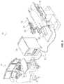
- FIG. 2provides one embodiment of an operating room setup that includes the robotically-assisted instrument driving system 10 .
- the depicted system 10includes a table 12 upon which a patient 14 may be placed, a fluoroscopy system or other imaging subsystem 16 , and a catheter or other medical instrument 18 .
- the depicted fluoroscopy system 16includes a C-arm 28 .
- a fluoroscopy panel 30is mounted to the C-arm 28 .
- the C-arm 28is selectively moveable during the procedure to permit various images of the patient to be taken by the fluoroscopy panel 30 .
- Attached to the table 12is a robotic arm (also referred to as a setup joint) 20 to which a robotic instrument driver 22 is coupled.
- One or more splayers 24may be mounted to the instrument driver 22 .
- the splayers 24are coupled to or form a portion of the medical instrument 18 .
- the medical instrument 18 of some embodimentsalso includes one or more pullwires disposed therein. The pullwires are attached to an articulation section of the medical instrument 18 and extend along a length of the instrument 18 to a proximal end. In such embodiments, the splayers 24 are positioned at the proximal end of the instrument 18 .
- Each of the splayers 24may include a pulley about which one of the pullwires is wound and an interface for coupling with the robotic instrument driver 22 .
- the componentsare configured such that a motor in the robotic instrument driver 22 rotationally drives an output shaft, which rotates the pulley of the splayer 24 and thereby adjusts tension in the pullwire to articulate the articulation section of the medical instrument 18 .
- the various components of the robotically-assisted instrument driving system 10are further visible in FIG. 3 .
- One or both of the user workstation 31 and the controller 34may be remotely positioned (i.e., free of a physical connection) with respect to the table 12 . In some embodiments, one or both of the user workstation 31 and the controller 34 are positioned in a separate room than the table 12 .
- the user workstation 31includes a computer, a control console having a user input device 33 , and a visual display 35 .
- the visual display 35may be a touch screen, LCD screen, or any other suitable display configured to present one or more images to a user.
- the user input device 33may include, but is not limited to, a multi-degree of freedom device having multiple joints and associated encoders.
- the user input device 33may additionally or alternatively include a keyboard, joystick, buttons, switches, knobs, trackballs, touchscreen, or any other input devices suitable for receiving commands from, and interfacing with, a user.
- the controller 34is a computing device. As shown in FIG. 4 , the controller 34 includes electronics, including a processor 50 , and memory 52 having instructions stored thereon. The instructions, when executed by the processor 50 , cause the processor 50 to perform various controls methods and execute various algorithms described elsewhere herein.
- the processor 50may be a general purpose microprocessor, a digital signal processor (DSP), a field programmable gate array (FPGA), an application specific integrated circuit (ASIC), or other programmable logic device, or other discrete computer-executable components designed to perform the functions described herein.
- the processor 50may also be formed of a combination of processing units, for example, a DSP and a microprocessor, a plurality of microprocessors, one or more microprocessors in conjunction with a DSP core, or any other suitable configuration.
- the processor 50is coupled, via one or more buses, to the memory 52 in order for the processor 50 to read information from and write information to the memory 52 .
- the processor 50may additionally or alternatively contain memory 52 .
- the memory 52can include, for example, processor cache.
- the memory 52may be any suitable computer-readable medium that stores computer-readable instructions for execution by computer-executable components.
- the computer-readable instructionsmay be stored on one or a combination of RAM, ROM, flash memory, EEPROM, hard disk drive, solid state drive, or any other suitable device.
- the computer-readable instructionsinclude application software stored in a non-transitory format. The software, when executed by the processor 50 , causes the processor 50 to perform one or more operations described elsewhere herein.
- the controller 34further includes one or more interfaces 54 (e.g., communication databuses or network interfaces) for receiving user inputs from the user input device 33 , transmitting images to the visual display 35 , and transmitting commands to the robotic instrument driver 22 .
- the controller 34is configured for bidirectional communication with the robotic instrument driver 22 , enabling the controller 34 to receive torque data or other feedback from the instrument driver 22 .
- the controller 34is physically coupled to the user input device 33 and/or visual display 35 of the user workstation 31 .
- the controller 34is physically coupled to the robotic instrument driver 22 .
- the controller 34is physically separate from, but communicatively coupled to the user workstation 31 and the robotic instrument driver 22 via a wireless connection.
- a communication link 32transfers signals between the user workstation 31 , the controller 34 , and the robotic instrument driver 22 .
- the communication link 32may be a wired or wireless communication link.
- Each element of the robotic surgical system 10 positioned within the operating suitemay define a separate reference frame to which localization sensors may be localized. More specifically, separate reference frames may be defined for each element of the robotic surgical system 10 .
- Such reference framesmay include, for example, the following shown in FIG. 2 : a table reference frame TRF for the table 12 , a setup joint reference frame SJF for the setup joint or arm 20 , a robotic instrument driver reference frame RRF for the robotic instrument driver 22 , a splayer reference frame SRF for the splayer 24 , and a fluoroscopy reference frame FF for the fluoroscopy panel 30 .
- Additional reference framesthat may be defined in the system include: a patient reference frame PRR for the patient 14 , a reference frame FRF for a tracking sensor disposed in or on the elongate instrument 18 , and a pre-operative 3-D anatomical model reference frame AMF for the model depicted on the visual display 35 .
- the robotic surgical system 10is designed to relate a coordinate system of the tracking sensor FRF of the elongate member 18 to either a fluoroscopy coordinate system FF or a pre-operative 3-D coordinate system AMF, as shown in FIG. 5 .
- the robotic surgical system 10may employ a variety of registration techniques to register the FRF to the FF or AMF, such as those described below or those described in U.S. Pat. No. 9,014,851 to Wong et al., the disclosure of which is herein incorporated by reference in its entirety.
- the position or shape tracking sensors incorporated into the medical instrument 18allow for real-time sensing of the instrument's position (i.e., location, orientation, and/or shape).
- the tracking sensoris integrated into the elongate instrument 18 and localized or registered to the anatomy or an image or model of the anatomy such that the position of the elongate instrument 18 is known relative to the anatomy, image, or model, a positionally-accurate representation of the instrument can be provided in the coordinate frame of the anatomical image or model.
- the tracking information of the sensorcan be used to update the position of the elongate instrument 18 relative to the anatomy, image, or model such that the representation of the elongate instrument can be displayed moving in real-time in an anatomical image or model.
- a target anatomymay be identified in multiple fluoroscopy views to localize the target's position in three dimensional (3-D) space relative to the elongate instrument.
- An aspect of the disclosure provided hereinis to make use of this situation where the 3-D position of an instrument and a target are known in real-time relative to a user's view of the patient's anatomy in order to allow for novel navigation strategies not possible with traditional robotic or manual minimally-invasive instrument navigation.
- Robotic assisted drivingas provided herein, enhances the capabilities of an instrument control or tracking system by allowing a user to easily navigate the instrument through the complex anatomy to a target location without exposing the patient to excessive radiation during the procedure.
- FIG. 6One method of performing robotic-assisted navigation is provided in FIG. 6 .
- the method 600is performed by a robotically-assisted instrument driving system, for example, the instrument driving system of FIGS. 1-4 .
- such a method 600is performed by the robotically-assisted instrument driving system 10 in response to execution of the instructions stored in the memory 52 of the controller 34 .
- the instrument driving systemacquires and displays two images of a relevant anatomy at different viewing angles.
- the imagesare acquired by the imaging subsystem, and any suitable imaging modality may be used.
- the systemlocalizes and displays the position of an elongate medical instrument relative to the images of the patient's anatomy.
- this stepincludes acquiring localization information for the instrument from a tracking sensor disposed in or on the instrument, tracking the position of at least a portion of the medical instrument using a tracking subsystem, correlating the position of the instrument to the patient's anatomy, and superimposing a positionally-accurate representation of the instrument on the two displayed images of the anatomy.
- the systemreceives a user designation of an anatomical target in both images.
- the designation of the anatomical target in both imagesmay be combined to compute a 3-D position (i.e., location, orientation, and/or shape) of an anatomical target.
- the systemcalculates one movement or a series of movements required for the instrument to move from its current position toward, to, or through the target. This calculation can be done continuously as the instrument moves through the anatomy or on-demand after a specific event or action is taken by the user. In some embodiments, these calculations are computed for multiple instrument components simultaneously (for example, for an inner member and an outer member). In various embodiments, the calculated movements include one or more of a magnitude and direction of articulation (e.g., a bend and a roll). In some embodiments, the calculated movements further include one or more of a magnitude and direction of axial translation of one or more instrument components (e.g., the inner member).
- the systemutilizes the calculated movements to provide navigation assistance.
- the form of navigation assistance that is providedmay vary widely between embodiments and/or modes.
- the navigation assistancemay include: providing step-by-step navigation instructions to the user, controlling navigation in one or more degrees of freedom, rejecting user-commanded movements that would navigate the instrument away from the target, driving the instrument towards the target while an auto-pilot indicator is actuated by the user, and/or driving the instrument to the target in a fully-automated manner.
- At least one tracking sensoris incorporated into the medical instrument to enable detection of the position (i.e., location, orientation, and/or shape) of the medical instrument.
- at least one tracking sensoris integrated into a flexible inner member of a medical instrument; in some embodiments, at least one tracking sensor is additionally or alternatively integrated into a tubular outer member of the medical instrument.
- EM sensorsare incorporated into the various components of the elongate instrument 700 . While numbered uniquely, one skilled in the art will appreciate that the medical instrument 18 of FIG. 1 may be formed of any embodiment of an instrument described herein and may include any of or all the features of the instrument 700 shown in FIG. 7 .
- the medical instrument 700includes an outer sheath catheter 710 , an inner leader catheter 720 , and a guidewire 730 .
- the sheath catheter 710 and leader catheter 720each have a flexible distal portion, referred to herein as the articulation section 716 , 726 , and a stiffer proximal portion, referred to herein as the shaft 712 , 722 .
- Two five-degree of freedom (DOF) sensors 714are located at the base of the sheath articulation section 716
- two 5-DOF sensors 724are located at the base of the leader articulation section 726
- a single 5-DOF sensor 734is located at or near the tip of the guidewire 730 .
- DOFfive-degree of freedom
- the two EM sensors in each of the sheath and the leaderform a pair of sensor coils in each instrument. These pairs of 5-DOF sensors enable tracking of each of the leader 720 and the sheath 710 in 6-DOF so that complete orientation, location, and heading are known.
- the guidewire 730there is only enough space for a single 5-DOF sensor 734 . In such embodiments, the guidewire position and direction are sensed, but not the roll.
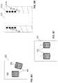
- two 5-DOF coils 804are combined into a rigid assembly in a medical instrument 800 with known sensor locations and with the two coils 804 configured to have different orientations of their symmetric axes 806 , as shown, for example, in FIG. 8A .
- Thisprovides a strong or accurate 6-DOF measurement because the EM sensing technology is well-suited for sensing the heading, or symmetric axis, of the coils. There is, however, often inadequate space to place two nonparallel coils into an elongate instrument such as a catheter.
- this limitationis overcome by spiraling the coils 814 around a perimeter of the tubular elongate instrument 810 , with a first coil spiral 814 a tilted slightly relative to a second coil spiral 814 b within the wall of the instrument 800 , as shown, for example, in the cross-section of FIG. 8B .
- Such a configurationrequires an elongate instrument with a relatively thick sidewall.
- An alternative embodimentplaces two coils 824 nominally parallel in the elongate instrument 820 to achieve the 6-DOF measurement, as shown in FIG. 8C .
- the coils 824are positioned diametrically opposite each other across a cross-section of the elongate instrument 820 , because changes in the relative position of the coils can be more accurately determined with increased separation.
- the coilsare positioned off-center (i.e., less than 180 degrees away from each other) due to the design of the elongate instrument. For example, in FIG. 8D , the placement of the central lumen 832 and pullwires 833 in the elongate instrument 830 creates a non-uniformly thick sidewall 831 , limiting placement of the coils 834 to thicker portions of the sidewall. In some embodiments, such as in FIG.
- the coils 824are parallel in both their orientation (e.g., axial alignment) and their position along the length of the elongate instrument 820 .
- the coils 844may not be placed perfectly parallel along the length of the elongate instrument due to manufacturing tolerances, as shown, for example, in FIG. 8E .
- the math to generate the 6-DOF measurementincludes the following, with reference made to FIG. 8E .
- a primary sensor 844 ais used to find the point B′, which is axially in line with a secondary sensor 844 b and directly perpendicular to the orientation of the primary sensor 844 a .
- the vector from A to B′defines the roll direction of the sensor coordinate frame.
- a position of the “combined sensor”can be computed as the midpoint of vector A ⁇ B′ if the sensors are embedded in diametrically opposing locations of the instrument wall. If the sensors are not centered around the instrument shaft, as in FIG. 8D , the relationship between the two sensors may be taken into consideration to adjust the position of the combined sensor.
- the heading orientation of the combined sensoris determined either by taking the heading of the primary sensor (H A ) or by averaging H A with the heading of the secondary sensor (H B ).
- a low-pass filteris applied on the roll measurements.
- Elongate instruments in a body lumengenerally do not roll very often or very quickly, so use of a low-pass filter does not significantly impact use of the elongate instrument or sensors.
- the low-pass filterdoes stabilize any display of sensor data that takes into consideration the roll information.
- a hybrid methodis used in which both the slight variations in heading between the coils and the difference in position of the coils is used to calculate the roll direction independently and then combined.
- the EM sensor pairsprovide position (x, y, z), heading (pitch and yaw), and roll orientation of the sheath and leader articulation sections.
- the EM sensor pairsmay have any of the configurations described with regards to FIGS. 8A-8E , or any other suitable configurations. Additional electromagnetic sensors may be added to different positions within the medical instrument to provide more information on the shape of the instrument.
- Registrationis a process that requires relating the reference frame of the sensor FRF to another reference frame of interest. If the positions of two or more objects are known in the same reference frame (i.e., are mapped to the same coordinate system), then the actual positions of each object relative to each other may be ascertained. Thus, with this information, a user can drive or manipulate one of the objects relative to the other objects.
- the sensor reference frame FRFis registered to the fluoroscopy reference frame FF or to the fluoroscopic image or anatomical model AMF using a known registration technique. There are many ways this registration can be performed.
- the sensor of the medical instrumentis measured in relation to the fluoroscopy system frame of reference FF.
- a sensing probewhich has an EM sensor and a radiopaque marker located in the same physical location on the probe.
- the sensing probeis placed into the field of view.
- the 2-D position of the probeis designated by the user in the fluoroscopy field of view (FOV) in images obtained at two different C-arm roll angles.
- the position of the probemay be designated by the user in three or more different locations.
- the visualization frameis the frame that the user (e.g., a physician) is viewing, and it may include a patient or a live 2-D or 3-D image.
- the live imagemay be acquired via fluoroscopy, ultrasound, digital subtraction angiography, live fluoroscopy with a contrast injection into the bloodstream, or other live imaging source.
- the goal of registrationis to determine the relationship of the frame of reference of the sensor FRF relative to the frame of reference of the patient PRF or to a 2-D or 3-D image or model of the patient.
- the tracked instrumentis simulated by rendering it with 3-D computer graphics and displaying, overlaying, or superimposing it on stored fluoroscopy images.
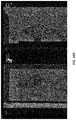
- One example of a simulated rendering of an elongate instrument 920 superimposed over the anatomy 910 captured in a stored fluoroscopy image 900is provided in FIG. 9 .
- This simulated elongate instrument 920is known as the virtual instrument or the virtual catheter.
- the location, orientation, and shape of the virtual instrument 920are estimated based on commanded data.
- sensor measurementsare used to improve the quality of the simulation and generate more accurate instrument shapes that are usable in clinical settings.
- one or more of the location, orientation, and shape of the virtual instrument 920is determined with the aid of tracking sensors.
- the current locations and orientations of the tracking sensors in an anatomical spaceare known from received sensor measurements.
- the fixed location and orientation of the sensors in each elongate instrumentare also known. From these known data points, a virtual instrument 920 can be drawn that passes through these points.
- the total lengths and insertion distances of the various components of the elongate instrumentare also known. Robotic movements of each component are tracked and this movement can be used to extrapolate the instrument shape between the sensor positions.
- the rotational orientation of the elongate instrumentmay also be determined from the sensors as described above to provide an entire 3-D reconstruction of the elongate instrument.
- One method for displaying the virtual instrumentinvolves using spline curves to interpolate the shape of the elongate instrument between sensors.
- an instrument modelcomprises a series of points connected such that they maintain realistic positions relative to one another.
- the virtual instrument seen by the useris rendered as a 3-D object that follows a path through the series of points.
- this virtual instrument informationmay be displayed to the user to help the user navigate.
- instinctiveness indicators 930such as the ring with colored cones shown in FIG. 9 may be added to the virtual instrument 920 and used to signal to the user which direction the instrument will bend when a specific user input command is activated.
- the directional conesare on opposing sides of the ring (i.e., 180 degrees apart).
- Directional indicators of any distinguishing colors or shapesmay be used.
- corresponding directional indicatorsmay be placed on the user input device. The virtual directional indicators are continuously updated with the position of the elongate instrument to represent the direction the instrument would bend if the corresponding directional indicator on the user input device is activated.
- a ringis provided around the virtual instrument 920 with an orange cone and an opposing purple cone.
- a left button or left side of the joystickmay be marked with the orange mark. Activation in this direction would bend the elongate instrument in the direction of the orange indicator on screen.
- a right button or right side of the joystickmay be marked with the purple mark, and activation in this direction would bend the elongate instrument 180° from the first direction.
- Such an embodimentprovides for more instinctive driving than simple “right” and “left” activation buttons, because the elongate instrument may rotate as it is advanced through the anatomy and the viewing angle of the C-arm may also rotate so it cannot be assured that bending the instrument to the left with the input device would result in the instrument bending to the left in the viewing plane.
- the presence of the 6-DOF position tracking sensors in the tip of the instrumentmay be used to communicate the actual roll orientation of the instrument tip in the given viewing plane.
- the colored cones on the ringor other instinctiveness indicators
- the modelmay use only a subset of sensor measurements to reduce the risk of over constraining the model. For example, the roll measurement at the instrument tip may not be rigidly enforced.
- the accuracy of the virtual instrument 920may be improved by tracking the elongate instrument via computer vision in a fluoroscopy image.
- Computer vision techniques to track cathetershave been described, for example, in US Publ. No. US2016/0228032, the disclosure of which is herein incorporated by reference.
- the similarity of the fluoroscopic instrument and the virtual instrumentcan be used to generate bias forces to move the physics model closer to the real instrument shape.
- fiber optic shape sensing sensorsmay be used to estimate the shape of the virtual instrument.
- the commanded robotic instrument insertion length or the commanded angle and heading orientation of an instrumentmay be tracked and compared to measured instrument position and heading based on sensor data and the delta may be used to update the physics model accordingly.
- a 3-D model of the anatomyis generated from pre-operative imaging, such as from a pre-op CT scan, and the instrument model interacts with the anatomy model to simulate instrument shape during a procedure.
- the intersection of the instrument shape with the geometric model of the anatomyproduces forces that are included in the simulation of the instrument.
- the time history of sensor locationsprovides insight as to the shape of the anatomy or the possible shape of the instrument. As an instrument passes through blood vessels, the instrument will often straighten or deform the anatomical shape. By tracking the path of the instrument through the anatomy over time, the relative shape of the deformed vessels may be determined and both the instrument model and the anatomical model may be updated.
- a measure of confidenceis displayed for each section of the virtual instrument shape so that physicians can make informed decisions. This measurement of confidence is guided by a few principles: the closer to a sensor, the higher the confidence; the higher the curvature between sensors, the lower the confidence; the greater the difference between the known and measured sensor-to-sensor distance, the lower the confidence; and the greater the difference in sensor orientations, the lower the confidence.
- the confidence measure in part of the virtual instrumentis shown non-numerically.
- the degree of transparency in the virtual instrument 920corresponds to the measure of confidence.
- Part of the instrumentmay be made fully transparent if the confidence is sufficiently low so that the questionable portion of the instrument is hidden from the user.
- low-confidencemay be represented by changing the color or texture of a part of the instrument or by adding animation, such as flashing or scrolling texture.
- a flashing iconsuch as a radiation icon, may be displayed beside the fluoroscopy image to urge the use of fluoroscopy when the confidence falls below a threshold value.
- a visualization mode called a “virtual biplane”is provided.
- the virtual instrumentis overlaid on the standard primary image and also on a secondary reference view.
- the concept of a virtual biplaneis introduced in US Publ. No. 2015/0223902 to Walker et al., the disclosure of which is herein incorporated by reference in its entirety. Displaying a representation of the instrument updated in real-time, overlaid on two different views of the anatomy is analogous to what a user would see in a biplane fluoroscopy system.
- the biplane viewis not an actual live biplane view, but rather, a simulation of the sensed instrument superimposed on the anatomical images.
- the catheter or instrument that is displayed, overlaid, or superimposed on the anatomical imageis referred to as the “virtual instrument” or “virtual catheter” as described above.
- the virtual instrumentis depicted in two different views of the anatomical background. In some embodiments, both provided views utilize fluoroscopy.
- the virtual biplaneincludes a first fluoroscopic view with an image of the sensed medical instrument overlaid or superimposed on top of the fluoroscopic view. This may be a live fluoroscopic view or a previously acquired fluoroscopic view.
- the commercially available fluoroscopic systemshave the capability of acquiring and storing images.
- the virtual biplane embodiment presented herealso includes a second view, which may be a reference view, for example, a previously-acquired view obtained via fluoroscopy at a different angulation of the C-arm.
- the first and second viewmay be shown at different magnifications.
- the first viewmay show an image at a lower magnification so that more of the instrument and anatomy is seen to help the user understand the global position in the patient whereas the second view may be a zoomed in or magnified view of an area of interest, usually in a different projection from the first view.
- the tracking sensor in the instrumenttracks its movement and the virtual instrument is updated in both views.
- Thisprovides live 3D tracking of the instrument displayed against images of the anatomy.
- the position sensor informationis registered to each image so that as the image changes (for example, due to a movement in the C-arm), the system can calculate where the sensor measurements line up with the updated image.
- the usermay change the anatomical images used for the virtual biplane.
- a lateral fluoroscopic projectionmight be preferred for at least one of the views so that the vessel is perpendicular to the viewing plane, whereas if a second vessel is pointing partly anterior but partly to the side of the patient, than the physician may wish to change over to a more oblique fluoroscopic projection.
- the reference imageshows the vessel anatomy at a specific instant in time. If the physician introduces a very inflexible or rigid instrument, the anatomy is deformed, and if the patient moves on the table, the overlay is no longer aligned. If said deformation or misalignment is not corrected in the overlaid reference image, an imprecision or a discrepancy arises when the reference image is superimposed. This can lead to uncertainties in navigation during an intervention in which the overlay serves as a navigation aid. Therefore, in various embodiments, the physician is provided with the option of refreshing the image by taking another live image at any point during the procedure.
- the imagesare acquired using a C-arm fluoroscope, with one viewing angle acquired prior to the procedure and the other viewing angle acquired intra-procedurally. In some embodiments, both views are displayed simultaneously, for example, adjacent to each other. In other embodiments, one or more of the images are generated by an imaging system that overlays a registered pre-operative or intra-operative 3-D image (e.g., 3-D rotational angiography or cone beam CT) on a live image. In still other embodiments, a pre-operative or intra-operative 3-D image is acquired and displayed, which is not registered or overlaid onto live imaging.
- a registered pre-operative or intra-operative 3-D imagee.g., 3-D rotational angiography or cone beam CT
- FIGS. 10A-10CAn example of a virtual biplane is provided in FIGS. 10A-10C .
- two viewsare displayed on a split screen.
- two display screensare provided, each displaying a different view.
- the left viewdirectly corresponds to the C-arm angle. If the C-arm is rotated, then this view, including the depicted EM sensing information will change; as the C-arm moves, the EM indicators and virtual instrument will update according to that new C-arm angle.
- the image directly related to the C-arm anglewill update based on the live fluoro image.
- the last imagewill be used as a reference image when driving an EM sensed instrument.
- the fluoro imagemay get out of sync with the EM sensor data if the C-arm is rotated but the user is not stepping on fluoro.
- a visual indicatormay be presented to the user to indicate that the fluoro image is no longer relevant.
- the outdated fluoro imagemay be blackened or given a hue or color or icon to show that it is old information.
- a sequence of fluoro images at different anglescan be acquired and stored, possibly using a predefined C-arm motion to acquire them, and the reference image can be updated based on C-arm motion even when the user is not stepping on fluoro.
- the second imageis always a stored image associated with a particular angulation of the C-arm.
- the particular C-arm angulationis provided and used to allow the sensed instrument information to update live according to that stored view.
- the right imageis the stored reference image.
- the stored reference imageis a fixed snapshot of a fluoro image taken sometime in the past, which corresponds to the particular C-arm angle.
- the usercan use this stored background image as a reference or roadmap as they are driving. Different visual indicators may be used to show that this is a stored image, such as colors, hues, or icons.
- a button or other user input devicecan be selected to store the live image and C-arm angulation for use as the stored reference in the future.
- the stored imageconsists of a sequence of images at different C-arm angles.
- the sequence of imagesmay be sequentially displayed as the C-arm moves even if the imaging is not live.
- the displayed image selected from the sequence of images at different C-arm anglesmay be chosen by the user through a user interface that allows the user to modify the viewing angle of the second image.
- the second imagemay automatically change to display images from various C-arm angles in a cyclic or periodic fashion providing an animation of the live EM information that provides the user with more three-dimensional information about the shape of the medical instrument or anatomy.
- the images stored in the virtual biplaneare often views directly from the fluoroscopy system or other imaging system.
- Such a featuremay be helpful in certain workflows, for example, when the user wants to do a contrast injection and store the image during a contrast injection or digital subtraction angiography (DSA) to show the anatomy of interest.
- DSAdigital subtraction angiography
- the fluoro systemmay also be used to play back a run of fluoro in the image stored during that playback.
- this systemmay also be used to capture a sequence of frames over the respiration cycle or pulse cycle and play back a video as a stored image instead of a static image.
- the virtual biplanemay include one or more renderings of the three-dimensional imaging of the anatomy such as a segmented CT or MM image or intraoperative cone beam CT, IVUS, or ultrasound.
- an image of the sensed medical instrumentis placed within the three-dimensional rendering of the anatomy based on the registration of the medical instrument to the anatomical model.
- Multiple different registration methodsmay be used as described above and in U.S. Pat. No. 9,014,851, the disclosure of which is herein incorporated by reference in its entirety.
- Such embodimentsprovide multiple views for the user during navigation of the medical instrument without requiring live imaging. For example, the embodiments of FIGS.
- 11A-Beach shows a cone beam CT outline overlaid on a fluoro image or alone on a black background. Similar imagery can additionally or alternatively be created using imaging gathered preoperatively, such as from a CT or MM.
- overlaying a representation of the 3-D anatomy(be it an outline, filled solid area, 3-D rendering, or composite of a stored fluoro image with 3-D imagery) on the background allows for the use of the 3-D data for guidance without additional fluoroscopy.
- This datacan be interfaced with the navigation system through either: a data connection between the navigation system and the imaging system, or displaying frame grabbed video directly or composited with other imaging within the system.
- two display screens or two portions of the same display screenshow, from different perspectives, views of the three-dimensional anatomical model with a simulation of the sensed medical instrument positioned therein.
- one of the displayed viewsis a simulated endoscopic view (also known as an “Endo View”), which provides the perspective of looking along the instrument or from the front of the instrument within the three-dimensional model of the anatomy.
- Endo Viewalso known as an “Endo View”
- an object of the present disclosureis to provide systems and methods for robotic assisted driving of a medical instrument to a target within an anatomical space.
- various methods of robotically assisted drivingalso require identification of the target.
- the sensed information of the medical instrument and the identified location of a targetcan be combined to provide robotically-assisted driving modes.
- modes and embodiments of identifying the targetare discussed in more detail below.
- targetsare referred to in a generic sense and may refer to a target position, a heading, an ostium shape, or other element or elements of interest to navigation.
- the targetis generally the 3-D center of an anatomical feature that the user would like to access, such as the ostium of a blood vessel coming off the aorta. While a blood vessel is frequently mentioned herein, a target could also correspond to a feature of an implanted device such as an aorta aneurysm endograft or a fenestrated graft.
- a targetmay refer to an anatomical feature such as an ablation site, the annulus of a coronary valve, a suspect nodule or a tumor and may be within any lumen of the body including the airway, gastrointestinal tract, or other lumen or within any organ accessed via a body lumen.
- targetin this discussion refers to the three-dimensional position at the center of the entrance of the feature to or through which the user wants to navigate.
- a targetis designated in three-dimensional space via one or more user selections of the target position on multiple imaging views.
- selectioni.e., designation
- the targetsinvolves a user interaction telling the system where the targets are and how they are oriented.
- the systemautomatically calculates where the targets would be based on known information such as a three-dimensional CT or other target locations, or a combination of both.
- the three-dimensional imagerymay be acquired from a preoperative CT or via imaging during the procedure, such as via cone beam CT.
- the usermay designate the target within an image using a variety of user input devices such as a mouse, trackball, buttons, joystick, or touchscreen.
- the usermanipulates a user input device to navigate a pointing device icon to the target in the displayed images.
- the usermay further manipulate a user input device (for example, with a button push, mouse click, or finger tap) to select the target.
- a trackball and buttonsare used by the user to designate targets of interest.
- other interfacesare utilized that also provide a means to identify and select points in the images, such as, but not limited to, a computer mouse, touchscreen, gesture device such as Xbox Kinect, joystick, etc.
- an interfaceallows the user to designate a position on a three-dimensional CT or other three-dimensional image and the system computes the target position, heading, ostium shape, vessel centerline, or other information useful for navigation.
- This interfacemay be at an operator workstation or may be positioned bedside or in any convenient location and may be designed to operate in a sterile or non-sterile environment based on the location chosen.
- there may be multiple operator interfacesso that, for example, an assistant can choose the target locations at a remote workstation and the physician can continue the navigation bedside.
- the imagery video from an imaging systemmay be frame grabbed by the system and then processed using computer vision techniques to determine anatomical features of interest to create targets or provide additional guidance as the user is creating the targets.
- the systemmay detect body lumen edges based on pixel density and automatically compute the diameters and centerlines of the lumens within the two views; such a system may then allow the user to designate a single point in one image to identify the target position, heading, and ostium size using the anatomical data reconstructed from the imagery.
- the headingis a single vector direction; in other embodiments, the heading may be a designated or computed lumen centerline.
- the userdesignates a position on the 3-D image and the imaging system computes the shape and orientation of the target opening or the direction of the target lumen based on the 3-D geometry.
- a useris prompted to designate the shape of the lumen entrance so that this entrance can be better shown within the user interface.
- the target position, heading direction, and radius of the lumenare identified by the user within at least one of the anatomical views and used to create a circle in space centered at the target position and perpendicular to the heading direction. It is also possible to use multiple points on the edge of the ostium or other element of interest to define a more complex shape such as an ellipse or any other closed shape. The user may designate these points on the display screen. Alternatively, multiple points or line segments may be sufficient to designate and define the shape of a vessel entrance.
- FIGS. 10A-10COne embodiment of selecting the target is shown in FIGS. 10A-10C .
- a useris shown in FIG. 10A selecting a target in a first image.
- the usermay manipulate a user input device to navigate a set of crosshairs over the target.
- the usermay also be able to manipulate the user input device to change the size of the crosshairs so as to approximate the size of the target.
- FIG. 10Bonce the target is selected in the first image, the system may determine a plane along which target is located, and a line projection may appear in the second image depicting the plane to facilitate target selection for the user.
- the useris shown in FIG. 10B moving the crosshairs along the line projection and selecting the location of the target in the second image.
- FIG. 10Cfollowing user identification of the target, the target (or a perimeter of the target) remains illuminated to facilitate visualization of the target during instrument navigation. Instrument navigation is then depicted in FIG. 10D .
- a first targetmay be designated by a user, and a second target may be calculated based on the location, heading, or shape of the first target. For example, if the user identifies a target for the inner member, the system may automatically compute a separate target for the outer member. In some embodiments, the position of the separate target for the outer member is selected such that when the outer member is aligned with the outer member target, the inner member may be automatically or semi-automatically positioned to align with the inner member target.
- the heading of the inner member target relative to the heading of the outer member tip or other part of the virtual instrumentmay be used to determine the distance of the outer member target from the inner member target.
- the systemtakes into consideration the known articulation length of the inner member to determine the outer member target position or heading.
- anatomical information from 3-D imagingsuch as a pre-op CT, allows the system to better compute the outer member target by taking into consideration lumen walls when computing the optimal outer member distance from the final target.
- 2-D anatomical information from one or more imagesmay be used to determine the position of the outer member target by creating constraints in three-dimensional space from the projection of the 2-D anatomical information.
- the userdetermines the outer member target based on the center line path from the 3-D dataset.
- the systemis configured to perform a method to modify the target after it is initially specified. For example, instead of setting a new target, the user may be able to use a mouse or other input device to move an existing target in 2-D or 3-D space.
- the input deviceis used to move the target in a single image to fine tune its location in relation to the image or anatomy during navigation.
- the systemautomatically fine tunes the target location based on other information such as 3-D imaging or other live intraoperative imaging such as IVUS.
- both inner and outer membersmay be similarly reconstructed in this fashion taking into consideration the anatomical shape.
- the systemmay determine the best target for the inner and outer members taking into consideration the angle of approach, the size of the lumen proximal to the target, the takeoff direction of the sharp vessel, and the size of the lumen distal to the target.
- targetsmay be marked within the 3-D imaging system making use of a registered pre-op CT, cone beam CT, or other imaging system.
- the ostia of vesselsmay be marked in a 3-D volume as described in U.S. Pat. No. 9,256,940, the disclosure of which is herein incorporated by reference in its entirety.
- These marksmay be exported directly to a flexible instrument navigation system (e.g., the Magellan® system by Hansen Medical Inc.). In this manner, physicians may use their familiar registration, segmentation, and marking toolset, and the data needed to improve navigation is exported to the navigation system as target or waypoint data.
- a trackball or other user input deviceis used to designate targets via the following sequence: (a) a pair of clicks, one in each of the two views of a virtual biplane, designate the target position for the inner member, (b) a pair of clicks, one in each of the two views of the virtual biplane, designate the target heading direction for the inner member; (c) one click on the heading line designates an outer member target position; and (d) one click designates the radius of the ostium (or the size of any other target).
- a pair of clicksis required to designate a 3-D position of the target.
- that clickor 2-D position in the screen space
- that lineis shown in the second view to aid in identifying the corresponding position of that feature in the alternate C-arm angulation.
- a second point on the target(referred to as the “heading position”) is required and it may be designated in much the same way as the target 3-D position.
- Two clicks of the anatomical feature distal to the target from two different viewsare processed to find the heading position on the anatomical feature, and the designated heading position is then used with the target position to calculate the heading direction.
- a line corresponding to the possible headingis drawn before each point is clicked to help the user understand the possible heading positions.
- the user input device positionis used to calculate the two-dimensional heading in the first image
- the user input device position in the second imageis used to calculate the three-dimensional heading based on the first two-dimensional heading in the first image
- the size of the target(e.g., the ostium radius) is calculated by using the closest distance between the line defined by the 2-D clicked point and the target position.
- Many other interfacesmay be used to set the target size or shape. For example, multiple clicks around the edges of a target, as seen within 2-D space, may be used to define a size and shape of the target in one view and to identify the target in the other view.
- the target radiusis traced, for example, using a mouse or trackball.
- the size of the target radiusis varied as the user input device is moved up and down or left and right and is shown on the screen with a dotted line until the user appropriately actuates the input device (for example, with a click) to set the target size.
- multiple iconscan be used to show that the target is designated.
- crosshairs or partial crosshairsmay show where the target is, a line from the center of that target may show the heading of the lumen, a circle or other shape corresponding to the perimeter of a target may be highlighted, and/or a radar screen showing the positioning of the target within the radar may be provided.
- different targetsmay be displayed in different places corresponding to the different members.
- the robotic driving systemcan help the user navigate.
- advanced driving modesare discussed in more detail below.
- the instrument driving systemis configured to perform one of the disclosed advanced driving modes.
- the instrument driving systemis configured to perform some of or all the disclosed advanced driving modes.
- the usermay select the level of assistance or control the user wishes to hand over to the robotic system.
- the one or more advanced driving modesare encoded for in software saved to memory within the controller 34 .
- the advanced driving modesare referred to herein as “robotic assisted driving” or “robotic assisted navigation” modes.
- user commands to the instrument driving systemcan be augmented with additional robot-determined movements to accomplish navigating the medical instrument to or through the target.
- a computer-augmented driving modeis available to the user.
- the usermay control instrument translation (e.g., insertion and retraction), and while translation is occurring, the robotic system may automatically control articulation magnitude and direction (i.e., bend and roll) and provide additional movements of the instrument tip to help track the instrument to the target location.
- the automatic selection of the optimal articulation amount and roll direction by the controllerprevents the user from needing to both perceive in three dimensions where the target is in relation to the instrument and determine the amount of bend and articulation needed to aim the instrument in that position in three dimensions.
- the robotic instrument driving system(and specifically, the controller or control algorithm of the robotic system) can assist the user in navigation even if the system does not know how to control all degrees of freedom of the instrument to achieve the user's goal.
- the usernavigates translation and one of articulation and roll, and the robotic system navigates the other of articulation and roll, as needed, to ensure the user-commanded movements lead the medical instrument to the intended location.
- the exact amount of help or movement provided by the systemmay vary depending on the application.
- FIGS. 12A-BOne example of computer-augmented driving is depicted in FIGS. 12A-B .
- FIG. 12Aprovides a 2-D representation of an elongate instrument, as it would appear to a user within a first view of a virtual biplane.
- FIG. 12Bprovides a 2-D representation of the elongate instrument from a different point of view, for example, as it may appear within a second view of the virtual biplane.
- the user's goalis to bend the virtual instrument 1200 (and the corresponding real instrument) towards the target 1210 located at the 9 o'clock position in the second view. As shown in FIG.
- the systemin the user's first attempt to navigate towards the target 1210 , the user inadvertently commanded articulation of the instrument towards 10 o'clock.
- the systemautomatically updates the mapping of the rotation input from the user to the desired rotation and automatically rotates the instrument tip from the 10 o'clock position to the 9 o'clock position.
- the robotic systemmay augment the user inputs with additional motions to accomplish the tasks. For example, with the target 1210 designated at the 9 o'clock position, when the user commands articulation of the instrument, the robotic system automatically supplements the commanded articulation with a roll command to achieve the desired motion or navigate toward the target.
- Robotic assisted drivinghas been explained above as helping or augmenting user commands.
- Robotic assisted drivingmay also include identifying or automatically choosing movements such as the heading direction of the instrument.
- the controller 34may use the position and/or heading of the instrument's articulation section and the location, shape, and/or heading of the target to automatically choose the preferred roll plane and articulation magnitude, for example, in order to reach the target or cannulate a target vessel most effectively.
- the exact implementationmay vary based on the application and the user commands. For example, if the user is commanding a roll motion as in FIG. 12B , then the automatic choosing of the preferred roll plane involves mapping the user input to a desired motion or adding additional robot-commanded movements to the user-commanded movements to direct the instrument tip towards the target.
- the automatic choosing of the preferred roll planeincludes augmenting the insertion motion with rotational movement to direct the instrument tip towards the target.
- the automatic choosing of a preferred articulation magnitudemay consist of mapping an articulation user-input command to a robot-determined desired articulation movement to thereby direct the instrument tip towards the target when the user commands articulation.
- robotic assisted drivingmay include supplementing an insertion motion with appropriate robot-determined articulation movements to direct the instrument tip towards the target.
- the controller 34commands the system to display a recommended path or shape of the instrument to the user.
- a recommended path or shape of the instrumentis provided in FIG. 13 .
- the recommended path 1310 through the anatomy 1300may be denoted with a hidden or dashed line or other suitable marking.
- the recommended path 1310depicts an optimal or suitable path within the anatomical image for getting the virtual instrument 1320 from its current position to the target.
- Such a displayallows the user to see how the instrument should be oriented.
- the usercan then control the instrument, using the path 1310 as a guide.
- the controller 34is configured to suspend movement of the instrument and notify the user automatically if the instrument deviates from the recommended path.
- the systemmay automatically reduce the insertion speed if the instrument has deviated from the path 1310 or if articulation has not yet reached a desired amount in order to minimize force on the anatomy.
- the controller 34may ensure that the instrument follows the recommended path by supplementing or adjusting the user-commanded movements with robot-determined course-correcting movements.
- the controller 34computes one or more insertion, articulation, and/or rotation movements needed to keep the instrument tip on the path 1310 .
- the controller 34commands the instrument driver 22 to execute motor actuations needed to implement the one or more movements.
- the controller 34 and the instrument driver 22work together to drive the instrument along the recommended path 1310 while an auto-pilot feature of the user input device is actuated.
- the usermust activate an input device in order to continue progress. At any time, the user may disengage the input device to stop all motion.
- the recommended path 1310may be derived from anatomical information provided by the imaging subsystem.
- the anatomical informationmay take the form of a 3-D model of the anatomy, and the recommended path may equal the centerline of a segmented body lumen.
- the anatomical informationmay be in a two-dimensional form such as frame grabbed images from the imaging subsystem.
- the anatomical informationis used to adjust the computed movements of the instrument as it navigates the anatomy by choosing articulation and roll values that keep the instrument away from the lumen walls.
- the anatomical informationallows the controller 34 to better determine when to insert one or more members of the instrument to achieve the best shape, maintain a sufficiently large distance away from the lumen wall, and/or enable the instrument to move in the lumen with minimal resistance.
- visual indicatorsare provided to help the user understand the relationship between the instrument and the targets and improve control over the instrument.
- Visual indicatorsmay be provided to show that the target algorithm is converging on a solution and aiming the instrument towards the target position. In some embodiments, this can be indicated using color on or around the target. For example, in one embodiment, a red border around the target is displayed when the instrument is far from the target, yellow is displayed when nearing the target, and green is displayed when the instrument is aligned with the target.
- convergence on a solution that aims the instrument correctly on the targetis depicted with a circle or other shape centered at the target position with a radius equal to the distance of the heading of the instrument from the target position.
- a geometric shapesuch as a square or circle changes in size to indicate how well the desired path is achieved.
- desirable paths for both the outer member 924 and the inner member 922 of an instrument 920may be indicated on the screen with lines, dots, geometric shapes, or other imagery.
- FIG. 9the following are depicted: a rectangle 940 around the target 915 , a rectangle 942 adjacent the location of the articulation section of the inner member 922 , and a rectangle 944 adjacent the location of the articulation section of the outer member 924 .
- the rectangle 940 around the target 915changes in color or size to indicate the extent to which the instrument is following a suitable path to the target.
- a dot representing a heading direction of each articulation sectionappears within the rectangles 942 , 944 when the articulation sections are in a suitable position in the anatomical space.
- the dotindicates the projection of a distal tip of the instrument onto the plane of the ostium circle or other target plane (similar to a laser targeting system on a rifle placing a dot wherever the rifle is aimed).
- the dotmay be provided to show the relationship between the tip of an articulation section and the target.
- a “target radar”is provided as a visual indicator to facilitate understanding of the instrument position relative to the target.
- the crosshairs and circlesshow the space of articulation for the instrument (including articulation magnitude and articulation direction or “roll”). This is similar to polar coordinates.
- the “x” within the “radar”corresponds to the location of the instrument. Its position relative to the center corresponds to the heading direction and the circle corresponds to the outline of the vessel ostium or other target plane.
- the “x”moves around the circles; when the instrument is articulated more, the x moves toward the outside of the circle, and when the instrument is relaxed, the x moves toward the center.
- a usercan determine which way to articulate and roll the instrument to align it with the target.
- a usermay also use the targeting radar to discern which direction the instrument is aiming within the target ostium. In a sense, this works as a simplified endoscopic view of the instrument direction and target.
- a 3-D version of the indicatormay be used that draws the indicator in 3-D space; the version may additionally draw the ostium with a projected heading of the instrument on the ostium surface defined by the ostium circle.
- some embodimentsmay display a shadow instrument representing the position the instrument is expected to assume if a user-entered movement command or the next proposed robot-determined command is implemented.
- a shadow instrumentrepresenting the position the instrument is expected to assume if a user-entered movement command or the next proposed robot-determined command is implemented.
- dotted lines or other indicatorsare provided to show the ideal instrument path or the boundary of suitable paths for the instrument, and the user is able to see the difference between the actual instrument shape and the ideal instrument path to identify if deviation is occurring.
- the systemmay allow the user to navigate with limited robotic assistance but will display one or more visual indicators indicating whether the instrument is following a suitable path. Visual cues such as colors, geometric shapes, diagrams, and/or a series of lights (for example, similar to airplane landing lights) may be displayed to show the relationship between the instrument and the target or the recommended path and the current path.
- visual feedbackis additionally provided to show when the various advanced driving modes have been enabled for various components of the instrument.
- the target for the inner membermay be the only visible target or may be specially highlighted
- the target for the outer membermay be the only visible target or may be specially highlighted.
- icons to the side of the virtual instrument or lighting on the user input devicemay indicate when various assisted driving buttons are enabled.
- the systemmay display an appropriate articulation magnitude and/or direction to the user for the user to follow, in effect, providing textual or graphical turn by turn directions or step-by-step instructions, for example, telling the user which user inputs to select and when.
- a “Driving Wizard”which may include a sequence of messages (or dialogues or text boxes or symbols) guiding the user towards the target, undesirable motions may also be blocked to ensure that the user drives the instrument correctly or consistently with the guidance.
- the systemmay allow the user to drive, but the controller may create an alert and/or automatically stop movement of the medical instrument if a user command would move the instrument in such a way that reaching the target would become difficult or impossible.
- the systemIn still another embodiment or driving mode, referred to as robot-controlled navigation, the system fully controls navigation of the medical instrument including one or more of the articulation, roll, and translation. This is considered automated navigation and is made possible once a target position, heading, and lumen size are set.
- the systemmay control all movement of the medical instrument once the target is identified; in some embodiments, the system may control all movement while the user is selecting an associated user input command.
- the controllermay calculate the amount of instrument articulation, rotation, and translation needed to reach the target in an optimal way. The translation, articulation, and rotation may be optimized so that the tip of the instrument is aligned with the target position as well as the target heading.
- the translation, articulation, and rollmay also be optimized so that the shape of the medical instrument does not collide with the anatomy, if the system is able to make use of three dimensional preoperative imaging, other three-dimensional imaging, or two-dimensional imaging showing the outline or projection of the 3-D anatomy.
- motion stopswhen the user stops actuating an automated driving button or other user input device.
- Some embodimentsmay automatically alternate between translating the instrument and modifying the articulation of the instrument.
- Other embodimentsmay automatically modify the articulation of the instrument as it is translated or allow the combination of user-commanded translation motions with robot-commanded control over articulation and roll.
- robotic controlled navigationallow the user to specify the shape of the anatomy in the region so that the system can better calculate the translation, articulation, and/or roll of the instrument to align with the target.
- Some embodimentsmay make use of many of these sensing and navigation modalities to automatically compute all articulation and translation of the instrument to achieve a system that is able to navigate the instrument along a prescribed trajectory or a centerline of a body lumen.
- Methods for extracting the centerline from an image volumeare described, for example, in U.S. Pat. No. 9,129,417, the disclosure of which is herein incorporated by reference in its entirety. This centerline generated from the 3-D volume may be used as the target for the automated robotic driving algorithm.
- the navigation systemmay analyze the 3-D data set imported from a pre-op CT, MRI, or cone beam CT directly to compute the sequence of targets in three dimensions.
- the navigation systemprocesses one or a small number of 2-D images from an imaging system to improve the targeting algorithms. For example, each single image provides a two-dimensional constraint on the lumen shape when the outline in the 2-D image is projected into three dimensions; such information can be used to inform the target shape, heading, or location. If many images are acquired, such as during a rotation of the C-arm, the navigation system can reconstruct the 3-D shape using 2-D to 3-D reconstruction techniques (similar to how a CT is reconstructed).
- Robotic assisted driving techniquesmay be used to access any anatomical target.
- One non-limiting exampleincludes the crossing of an occlusion in a blood vessel.
- an occluded blood vesselthere is no blood flowing so it is not possible to image the anatomy using an angiogram under fluoroscopy.
- recent developments in CT scanningcan identify the thrombus or calcium making up the occlusion and can identify the centerline of the occluded vessel.
- This three-dimensional information of the centerline of vesselscan then be used to generate a sequence of targets that comprise the catheter trajectory.
- the robotic assisted driving algorithm of some embodimentsis configured to use these targets as a path and navigate from the beginning of the occlusion to the location where the vessel reconstitutes by following this centerline while crossing the occlusion.
- the robotic control systemmay automatically extract the centerline data and follow it.
- robot-controlled navigationcan occur intermittently; for example, a user may begin driving the medical instrument and select the robot-controlled navigation on occasion in order to have the system make path corrections.
- it is important to prevent the computed articulation or roll from articulating the medical instrument in a constrained situationbecause it is important to prevent the instrument from pressing into the anatomy.
- Some embodimentsmay include a subsystem that monitors the instrument motion in relation to instrument commands such as articulation and insertion. By modeling the commands and comparing them to the measured catheter shape, the system is able to determine whether it is likely that the medical instrument is contacting the anatomy.
- Some embodimentsmay also calculate, based on this difference between the commanded shape and the measured shape, an estimate of the force applied on the instrument by the anatomy (and likewise, the force of the instrument on the anatomy). If the computed force gets large, the system may prevent further motion or cause a relaxation of the instrument to reduce this force on the anatomy. Some embodiments may also provide a message to the user or prevent assisted navigation when the computed force becomes too large. Some embodiments may also compute this force even when the user is not using assisted navigation to prevent the user from inadvertently causing too much force on the anatomy during navigation.
- the instrument commandsare represented as an articulation magnitude, roll angle, and insertion length, and therefore the controller directly modifies the commands to facilitate the driving.
- the commandsare modified to serve different tasks at different stages of driving. For example, in the beginning, the focus may be on cannulating a vessel, and the controller may focus on aiming at the target. As the procedure progresses, and the flexible instrument approaches the target, the controller may focus on bringing the instruments through the target, requiring a different strategy than aiming the instruments at the vessel.
- the modifying commandsmust be defined in the same coordinate system as the commands issued by the physician.
- the desired articulation and roll commandsare defined in a frame of reference of the instrument sensors, often located at the base of the articulation section.
- the frame of referencemay be computed from the virtual instrument shape, which can be defined by a combination of one or more of the sensor data, the articulation command to the medical instrument, a simulation of the instrument dynamics, and a model of instrument behavior.
- the desired articulation and roll commandsare generated to aim the instrument at the target.
- a search algorithmis employed to find the optimal articulation and roll angle.
- an optimization procedurecan determine the best articulation, roll, and insertion.
- the frame of reference of the instrumentis no longer attached to the base of the instrument's articulation section, but is instead calculated based on the shape of the virtual instrument constructed from sensor measurements as well as commands to the instrument.
- the coordinate frame at the distal tip of the instrumentis directly measured by the sensors, but the frame at the base of the articulation section is calculated from inverting the kinematics that describes the relationship between the articulation magnitude and the position of the distal tip.
- the resulting frame of referenceis no longer placed at the base of the articulation section, but instead takes into consideration the shape of the instrument and the command that caused the instrument to take the shape. This may lead to faster convergence and improved targeting performance.
- open loop controlcalculations from the sensors are used with the target location data to compute a single direction to move the medical instrument. This approach has the advantage of control stability, but variations in instrument behavior may prevent the instrument from aiming directly at the target.
- closed loop controlthe system takes into consideration the sensed position of the instrument as it moves and adjusts the instrument command accordingly to make the aim of the instrument converge on the target.
- a polar coordinate systemsuch as the targeting radar indicator displayed in FIG. 10D is used to compute the difference between the center of the target and the current heading of the instrument. A change in articulation and roll angles can be identified and added to the current instrument command to better align the instrument with a target.
- large commanded changesmay need to be divided into a series of smaller steps to prevent overshooting the target.
- a miniscule change or stepmay be magnified to overcome non-linear characteristics of the instrument, such as friction, dead-zone, or slack in a pullwire, to ensure that the instrument exhibits noticeable motion during assisted driving.
- a distance thresholdmay be implemented to stop further modifications to the command if the aim of the instrument is close enough to the target. The threshold prevents the instrument from unnecessarily overshooting the target and keeps the aim of the instrument from drifting away from the target once the target has been reached.
- the controller 34breaks them into driving commands.
- the instrumentmay be commanded to first relax if it needs to change roll direction more than 90 degrees instead of rotating the instrument. Once the instrument is fully relaxed, the roll angle can be set directly so that the instrument bends in the desired roll direction. This is similar to the adaptive catheter control strategies outlined in US Publ. No. 2016/0213884, the disclosure of which is herein incorporated by reference in its entirety.
- Robotic assisted drivingmay involve the task of advancing an instrument formed of a plurality of members towards or through a target.
- one or more of the above systems and methodsare used to robotically assist driving an inner member through the target.
- an enhanced robotic assisted driving algorithm and methodmay be implemented so that any coaxial outer members “follow” over the leading member.
- the shape of the inner memberprovides an ideal path for the outer member to follow.
- “following”involves inserting the outer member towards the target while articulating the outer member in the direction of the target, thereby following a path similar to the ideal path.
- additional stepsare needed to ensure accurate following and to avoid prolapse or loss of wire or instrument position.
- Prolapseis a situation where insertion of the instrument causes a proximal portion of the instrument to continue moving in the direction of a previous insertion command and bulge away from a target instead of changing directions and advancing with the distal tip toward the target.
- a prolapse in the leader catheter 1324is shown as a bulge above the level of the target ostium 1302 ; in this situation, further insertion of the leader catheter 1324 would cause it to buckle further up into the aorta 1304 and ultimately pull the tip of the leader catheter 1324 and the guidewire 1322 out of the ostium.
- This situationis important to avoid because it can add significant delays to a procedure.
- the prolapse of the leader catheter 1324 or other inner memberis often controlled by the orientation of the sheath catheter 1326 or other outer member. For example, in FIG.
- the likely cause of the prolapse in the leader catheter 1324is the fact that the tip of the sheath 1326 is aimed significantly away from the ostium 1302 of the vessel and the change in direction was too great. If the sheath catheter 1326 were instead articulated to point more towards the ostium 1302 , the leader catheter 1324 would be directed more towards the target and would have more support as it is inserted. The additional support enables the insertion motion to move the leader catheter 1324 through the ostium 1302 and into the vessel instead of further up into the aorta 1304 .
- avoiding prolapseinvolves retracting an inner member 1324 before articulating the outer member 1326 towards the bend or target. Because the controller 34 is aware of the shape of the instrument 1320 , some embodiments make use of this information to automatically avoid these prolapse situations or, if they are detected, to move the instrument 1320 in such a way that the prolapse is removed. In some embodiments, the controller 34 is programmed and the system configured to detect prolapse within the instrument and notify the user and/or stop motion.
- avoiding prolapseinvolves relaxing the outer member 1326 articulation then articulating the outer member 1326 in the path direction of the guidewire 1322 or inner member 1324 .
- the path of the guidewire 1322 or inner member 1324may be defined by the virtual instrument shape generated from the sensor data.
- the shape of the virtual instrument constructed from the sensor measurementsis naturally smooth and closely mimics that of the real instrument.
- the virtual instrumentprovides sufficient information to generate proper commands for the real instruments.
- instrument commandsmay be further refined based on the position and heading of the target as well as the shape of an instrument portion.
- the controller 34determines an appropriate articulation angle ⁇ cmd for the outer member based, in part, on the shape of the inner member. As shown in FIG. 14 , to determine the appropriate articulation angle ⁇ cmd , the average inner member shape in a local area is calculated, and the controller solves for the articulation angle ⁇ cmd that will make the outer member curvature match the average inner member shape the best. The controller may then command the instrument driver to achieve this articulation angle. For example, in FIG.
- a first heading value H 1is sampled at a first location L 1 proximal to the tip of the outer member 1404
- a second heading value H 2is sampled at a second location L 2 distal to the tip of the outer member 1404 .
- the heading valuesare averaged to find the average shape of the inner member 1402 in the sampled region, denoted as ⁇ track .
- the length of the articulation sectionis denoted as d, and the length of the sampled portion (i.e., the length of the inner member 1402 between L 1 and L 2 ) is denoted as d′.
- the curvature of the articulation sectionis determined from averaging the curvature across portions of the virtual instrument.
- the algorithms used to calculate the articulation and roll of an instrument in assisted drivingmay need to take into consideration the pulsatile flow in the arteries as well as heart and breath motions.
- biological motions of the patientmay be predicted and used to improve the performance of assisted driving.
- the targetmay appear to move in sync with the patient's motion. Motion of the target may be sensed based on the instrument motion, live imaging such as fluoroscopy, or user input.
- the systems and methods of some embodimentsdetect and compensate for this cyclic motion, stabilizing the algorithm to converge faster.
- Some embodimentsuse an adjusted or moving target during computations of the translation, articulation, and/or roll.
- FIGS. 15A-15EOne embodiment of robotic-assisted driving is provided in FIGS. 15A-15E .
- a virtual instrument 1510is superimposed on the anatomical image 1500 .
- the virtual instrument 1510includes a virtual guidewire 1512 , a virtual inner member 1514 , and a virtual outer member 1516 .
- the anatomyis representative of a patient's left renal artery branching from the aorta when viewed from an anterior/posterior projection, but similar branches are present in other lumens within the body.
- the dashed line 1520is provided in the visual display of some embodiments to show the desired target path of the instrument 1510 from its current position to the user-set target 1530 .
- the dashed line 1520(i.e., the desired path) may be positioned at the centerline of the body lumen when within a relatively large or straight lumen 1502 and may follow a lowest energy curve or optimal path based on the bending radius of the instrument as it navigates lumen branches 1504 .
- the end target 1530 for the instrument 1510is in the side branch vessel 1504 . If this target endpoint were established as the first target for both the inner member 1514 and the outer member 1516 , then both members would bend towards the target as shown by the solid arrows of FIG. 15A and deviate from the desired path. This would lead to both members colliding with the wall of the aorta 1502 .
- the controller 34is configured to determine and set one or more intermediate targets that the instrument components aim for along their path to the final user-designated target. For example, as shown in FIG. 15B , in the depicted progression, the controller 34 has set a first target point 1532 at the ostium of the vessel.
- FIGS. 15A-Edepict a single 2D image of the target and the instrument for simplicity.
- a second image of a different projectionis also provided.
- the concepts described here for a single 2D imagealso hold true in 3D.
- the inner and outer membersare both assigned a target in 3D space and are commanded to move towards those targets.
- the guidewire 1512may be advanced through the inner member target 1532 . Once the guidewire 1512 is advanced through the target 1532 , further forward motion of the instrument may be programmed to follow the guidewire.
- the inner member 1514may need to be relaxed or straightened as it advances beyond the apex of the bend to follow the guidewire 1512 ; however, in some embodiments, it may not be necessary to set new targets for the instrument.
- the inner member targetmay be relocated, as shown in FIG. 15D .
- the new target 1536is set at the distal end of the branch lumen while the outer member target 1534 remains outside the ostium of the vessel so that the outer member 1516 provides adequate support on the desired dashed line.
- the outer member 1516eventually is commanded by the controller 34 to “follow” the inner member 1514 , as shown in FIG. 15E .
- the controller 34may direct the outer member 1516 to follow the inner member 1514 or the guidewire 1512 .
- a new target pointmay be set for the outer member 1516 .
- control algorithm of the controller 34may be set up such that the instrument 1510 never advances passed the target.
- the goalis to get a catheter tip to a target and it might be desirable as a safety measure to never allow the catheter or other instrument to extend beyond the target point. This would reduce the risk of inadvertent vessel perforation.
- the degree of operator involvement in the setting of the targets and the driving towards or through the targetsmay vary.
- the operatoridentifies the end target
- the robotic systemidentifies a centerline or lowest energy path
- the operatorcontrol insertion of the guidewire and insertion and bend of the inner member
- the controller 34automatically determines and controls the roll of the inner member and all movement (i.e., insertion, bend, and roll) of the outer member.
Landscapes
- Health & Medical Sciences (AREA)
- Engineering & Computer Science (AREA)
- Life Sciences & Earth Sciences (AREA)
- Surgery (AREA)
- Medical Informatics (AREA)
- Nuclear Medicine, Radiotherapy & Molecular Imaging (AREA)
- Veterinary Medicine (AREA)
- Biomedical Technology (AREA)
- Heart & Thoracic Surgery (AREA)
- Molecular Biology (AREA)
- Animal Behavior & Ethology (AREA)
- General Health & Medical Sciences (AREA)
- Public Health (AREA)
- Robotics (AREA)
- Human Computer Interaction (AREA)
- Physics & Mathematics (AREA)
- Pathology (AREA)
- Biophysics (AREA)
- Radiology & Medical Imaging (AREA)
- Gynecology & Obstetrics (AREA)
- High Energy & Nuclear Physics (AREA)
- Optics & Photonics (AREA)
- Vascular Medicine (AREA)
- Dentistry (AREA)
- Oral & Maxillofacial Surgery (AREA)
- Endoscopes (AREA)
- Manipulator (AREA)
- Theoretical Computer Science (AREA)
- Pulmonology (AREA)
Abstract
Description
Claims (20)
Priority Applications (1)
| Application Number | Priority Date | Filing Date | Title |
|---|---|---|---|
| US16/265,818US10806535B2 (en) | 2015-11-30 | 2019-02-01 | Robot-assisted driving systems and methods |
Applications Claiming Priority (5)
| Application Number | Priority Date | Filing Date | Title |
|---|---|---|---|
| US201562261301P | 2015-11-30 | 2015-11-30 | |
| US201662304051P | 2016-03-04 | 2016-03-04 | |
| US15/365,230US10143526B2 (en) | 2015-11-30 | 2016-11-30 | Robot-assisted driving systems and methods |
| US16/201,787US10813711B2 (en) | 2015-11-30 | 2018-11-27 | Robot-assisted driving systems and methods |
| US16/265,818US10806535B2 (en) | 2015-11-30 | 2019-02-01 | Robot-assisted driving systems and methods |
Related Parent Applications (1)
| Application Number | Title | Priority Date | Filing Date |
|---|---|---|---|
| US16/201,787ContinuationUS10813711B2 (en) | 2015-11-30 | 2018-11-27 | Robot-assisted driving systems and methods |
Publications (2)
| Publication Number | Publication Date |
|---|---|
| US20190167367A1 US20190167367A1 (en) | 2019-06-06 |
| US10806535B2true US10806535B2 (en) | 2020-10-20 |
Family
ID=58776649
Family Applications (5)
| Application Number | Title | Priority Date | Filing Date |
|---|---|---|---|
| US15/365,230ActiveUS10143526B2 (en) | 2015-11-30 | 2016-11-30 | Robot-assisted driving systems and methods |
| US16/201,787ActiveUS10813711B2 (en) | 2015-11-30 | 2018-11-27 | Robot-assisted driving systems and methods |
| US16/265,818ActiveUS10806535B2 (en) | 2015-11-30 | 2019-02-01 | Robot-assisted driving systems and methods |
| US17/025,593Active2036-12-07US11464591B2 (en) | 2015-11-30 | 2020-09-18 | Robot-assisted driving systems and methods |
| US17/888,570ActiveUS11857281B2 (en) | 2015-11-30 | 2022-08-16 | Robot-assisted driving systems and methods |
Family Applications Before (2)
| Application Number | Title | Priority Date | Filing Date |
|---|---|---|---|
| US15/365,230ActiveUS10143526B2 (en) | 2015-11-30 | 2016-11-30 | Robot-assisted driving systems and methods |
| US16/201,787ActiveUS10813711B2 (en) | 2015-11-30 | 2018-11-27 | Robot-assisted driving systems and methods |
Family Applications After (2)
| Application Number | Title | Priority Date | Filing Date |
|---|---|---|---|
| US17/025,593Active2036-12-07US11464591B2 (en) | 2015-11-30 | 2020-09-18 | Robot-assisted driving systems and methods |
| US17/888,570ActiveUS11857281B2 (en) | 2015-11-30 | 2022-08-16 | Robot-assisted driving systems and methods |
Country Status (1)
| Country | Link |
|---|---|
| US (5) | US10143526B2 (en) |
Cited By (2)
| Publication number | Priority date | Publication date | Assignee | Title |
|---|---|---|---|---|
| US11712154B2 (en) | 2016-09-30 | 2023-08-01 | Auris Health, Inc. | Automated calibration of surgical instruments with pull wires |
| US12239380B2 (en)* | 2016-03-17 | 2025-03-04 | Koninklijke Philips N.V. | Control unit, system and method for controlling hybrid robot having rigid proximal portion and flexible distal portion |
Families Citing this family (270)
| Publication number | Priority date | Publication date | Assignee | Title |
|---|---|---|---|---|
| ES2465915T3 (en) | 2007-11-26 | 2014-06-09 | C.R. Bard, Inc. | Integrated system for intravascular catheter placement |
| US8672837B2 (en) | 2010-06-24 | 2014-03-18 | Hansen Medical, Inc. | Methods and devices for controlling a shapeable medical device |
| US20120071752A1 (en) | 2010-09-17 | 2012-03-22 | Sewell Christopher M | User interface and method for operating a robotic medical system |
| US20130030363A1 (en) | 2011-07-29 | 2013-01-31 | Hansen Medical, Inc. | Systems and methods utilizing shape sensing fibers |
| US10238837B2 (en) | 2011-10-14 | 2019-03-26 | Intuitive Surgical Operations, Inc. | Catheters with control modes for interchangeable probes |
| US9387048B2 (en) | 2011-10-14 | 2016-07-12 | Intuitive Surgical Operations, Inc. | Catheter sensor systems |
| US9452276B2 (en) | 2011-10-14 | 2016-09-27 | Intuitive Surgical Operations, Inc. | Catheter with removable vision probe |
| US20130303944A1 (en) | 2012-05-14 | 2013-11-14 | Intuitive Surgical Operations, Inc. | Off-axis electromagnetic sensor |
| US11871901B2 (en) | 2012-05-20 | 2024-01-16 | Cilag Gmbh International | Method for situational awareness for surgical network or surgical network connected device capable of adjusting function based on a sensed situation or usage |
| US20140148673A1 (en) | 2012-11-28 | 2014-05-29 | Hansen Medical, Inc. | Method of anchoring pullwire directly articulatable region in catheter |
| US9057600B2 (en) | 2013-03-13 | 2015-06-16 | Hansen Medical, Inc. | Reducing incremental measurement sensor error |
| US9566414B2 (en) | 2013-03-13 | 2017-02-14 | Hansen Medical, Inc. | Integrated catheter and guide wire controller |
| US9283046B2 (en) | 2013-03-15 | 2016-03-15 | Hansen Medical, Inc. | User interface for active drive apparatus with finite range of motion |
| US9629595B2 (en) | 2013-03-15 | 2017-04-25 | Hansen Medical, Inc. | Systems and methods for localizing, tracking and/or controlling medical instruments |
| US10849702B2 (en) | 2013-03-15 | 2020-12-01 | Auris Health, Inc. | User input devices for controlling manipulation of guidewires and catheters |
| US9271663B2 (en) | 2013-03-15 | 2016-03-01 | Hansen Medical, Inc. | Flexible instrument localization from both remote and elongation sensors |
| US9014851B2 (en) | 2013-03-15 | 2015-04-21 | Hansen Medical, Inc. | Systems and methods for tracking robotically controlled medical instruments |
| US11020016B2 (en) | 2013-05-30 | 2021-06-01 | Auris Health, Inc. | System and method for displaying anatomy and devices on a movable display |
| WO2014201165A1 (en) | 2013-06-11 | 2014-12-18 | Auris Surgical Robotics, Inc. | System for robotic assisted cataract surgery |
| CA2918879A1 (en) | 2013-07-24 | 2015-01-29 | Centre For Surgical Invention & Innovation | Multi-function mounting interface for an image-guided robotic system and quick release interventional toolset |
| EP3243476B1 (en) | 2014-03-24 | 2019-11-06 | Auris Health, Inc. | Systems and devices for catheter driving instinctiveness |
| US10046140B2 (en) | 2014-04-21 | 2018-08-14 | Hansen Medical, Inc. | Devices, systems, and methods for controlling active drive systems |
| EP3200718A4 (en) | 2014-09-30 | 2018-04-25 | Auris Surgical Robotics, Inc | Configurable robotic surgical system with virtual rail and flexible endoscope |
| US10314463B2 (en) | 2014-10-24 | 2019-06-11 | Auris Health, Inc. | Automated endoscope calibration |
| US11504192B2 (en) | 2014-10-30 | 2022-11-22 | Cilag Gmbh International | Method of hub communication with surgical instrument systems |
| JP6824967B2 (en) | 2015-09-18 | 2021-02-03 | オーリス ヘルス インコーポレイテッド | Tubular net navigation |
| US10143526B2 (en)* | 2015-11-30 | 2018-12-04 | Auris Health, Inc. | Robot-assisted driving systems and methods |
| WO2017098259A1 (en)* | 2015-12-10 | 2017-06-15 | Cambridge Medical Robotics Limited | Robotic system |
| EP3413830B1 (en)* | 2016-02-12 | 2022-06-15 | Intuitive Surgical Operations, Inc. | Computer program for using registered fluoroscopic images in image-guided surgery |
| EP4620418A2 (en) | 2016-06-30 | 2025-09-24 | Intuitive Surgical Operations, Inc. | Graphical user interface for displaying guidance information during an image-guided procedure |
| KR102764213B1 (en) | 2016-06-30 | 2025-02-07 | 인튜어티브 서지컬 오퍼레이션즈 인코포레이티드 | Graphical user interface for displaying guidance information in a plurality of modes during an image-guided procedure |
| US11241559B2 (en) | 2016-08-29 | 2022-02-08 | Auris Health, Inc. | Active drive for guidewire manipulation |
| US10244926B2 (en) | 2016-12-28 | 2019-04-02 | Auris Health, Inc. | Detecting endolumenal buckling of flexible instruments |
| JP7233841B2 (en)* | 2017-01-18 | 2023-03-07 | ケービー メディカル エスアー | Robotic Navigation for Robotic Surgical Systems |
| JP7071054B2 (en)* | 2017-01-20 | 2022-05-18 | キヤノン株式会社 | Information processing equipment, information processing methods and programs |
| WO2018183727A1 (en) | 2017-03-31 | 2018-10-04 | Auris Health, Inc. | Robotic systems for navigation of luminal networks that compensate for physiological noise |
| EP3606592B1 (en) | 2017-04-07 | 2025-01-08 | Bard Access Systems, Inc. | Optical fiber-based medical device tracking and monitoring system |
| WO2018195216A1 (en) | 2017-04-18 | 2018-10-25 | Intuitive Surgical Operations, Inc. | Graphical user interface for monitoring an image-guided procedure |
| AU2018266132A1 (en)* | 2017-05-09 | 2019-10-10 | Boston Scientific Scimed, Inc. | Operating room devices, methods, and systems |
| KR102643758B1 (en) | 2017-05-12 | 2024-03-08 | 아우리스 헬스, 인코포레이티드 | Biopsy devices and systems |
| US10022192B1 (en) | 2017-06-23 | 2018-07-17 | Auris Health, Inc. | Automatically-initialized robotic systems for navigation of luminal networks |
| US10299870B2 (en) | 2017-06-28 | 2019-05-28 | Auris Health, Inc. | Instrument insertion compensation |
| US10426559B2 (en) | 2017-06-30 | 2019-10-01 | Auris Health, Inc. | Systems and methods for medical instrument compression compensation |
| US10292774B2 (en)* | 2017-07-28 | 2019-05-21 | Zimmer, Inc. | Bone and tool tracking with optical waveguide modeling system in computer-assisted surgery using patient-attached multicore optical fiber |
| CN109584295B (en) | 2017-09-29 | 2022-08-26 | 阿里巴巴集团控股有限公司 | Method, device and system for automatically labeling target object in image |
| EP3694412A4 (en)* | 2017-10-10 | 2021-08-18 | Covidien LP | SYSTEM AND METHOD FOR IDENTIFYING AND MARKING A TARGET IN A FLUOROSCOPIC THREE-DIMENSIONAL RECONSTRUCTION |
| US10145747B1 (en) | 2017-10-10 | 2018-12-04 | Auris Health, Inc. | Detection of undesirable forces on a surgical robotic arm |
| US10016900B1 (en) | 2017-10-10 | 2018-07-10 | Auris Health, Inc. | Surgical robotic arm admittance control |
| US10555778B2 (en) | 2017-10-13 | 2020-02-11 | Auris Health, Inc. | Image-based branch detection and mapping for navigation |
| US11058493B2 (en) | 2017-10-13 | 2021-07-13 | Auris Health, Inc. | Robotic system configured for navigation path tracing |
| US11564756B2 (en) | 2017-10-30 | 2023-01-31 | Cilag Gmbh International | Method of hub communication with surgical instrument systems |
| US11026687B2 (en) | 2017-10-30 | 2021-06-08 | Cilag Gmbh International | Clip applier comprising clip advancing systems |
| US11311342B2 (en) | 2017-10-30 | 2022-04-26 | Cilag Gmbh International | Method for communicating with surgical instrument systems |
| US11229436B2 (en) | 2017-10-30 | 2022-01-25 | Cilag Gmbh International | Surgical system comprising a surgical tool and a surgical hub |
| US11925373B2 (en) | 2017-10-30 | 2024-03-12 | Cilag Gmbh International | Surgical suturing instrument comprising a non-circular needle |
| US11291510B2 (en) | 2017-10-30 | 2022-04-05 | Cilag Gmbh International | Method of hub communication with surgical instrument systems |
| US11510741B2 (en) | 2017-10-30 | 2022-11-29 | Cilag Gmbh International | Method for producing a surgical instrument comprising a smart electrical system |
| US11317919B2 (en) | 2017-10-30 | 2022-05-03 | Cilag Gmbh International | Clip applier comprising a clip crimping system |
| US11911045B2 (en) | 2017-10-30 | 2024-02-27 | Cllag GmbH International | Method for operating a powered articulating multi-clip applier |
| US11801098B2 (en) | 2017-10-30 | 2023-10-31 | Cilag Gmbh International | Method of hub communication with surgical instrument systems |
| EP3677208A4 (en)* | 2017-12-05 | 2020-09-09 | Sony Olympus Medical Solutions Inc. | DEVICE FOR PROCESSING MEDICAL INFORMATION AND METHODS FOR PROCESSING INFORMATION |
| EP3684282B1 (en) | 2017-12-06 | 2024-02-21 | Auris Health, Inc. | Systems to correct for uncommanded instrument roll |
| US11510736B2 (en)* | 2017-12-14 | 2022-11-29 | Auris Health, Inc. | System and method for estimating instrument location |
| WO2019125964A1 (en) | 2017-12-18 | 2019-06-27 | Auris Health, Inc. | Methods and systems for instrument tracking and navigation within luminal networks |
| US11179175B2 (en) | 2017-12-28 | 2021-11-23 | Cilag Gmbh International | Controlling an ultrasonic surgical instrument according to tissue location |
| US11013563B2 (en) | 2017-12-28 | 2021-05-25 | Ethicon Llc | Drive arrangements for robot-assisted surgical platforms |
| US11253315B2 (en) | 2017-12-28 | 2022-02-22 | Cilag Gmbh International | Increasing radio frequency to create pad-less monopolar loop |
| US11410259B2 (en) | 2017-12-28 | 2022-08-09 | Cilag Gmbh International | Adaptive control program updates for surgical devices |
| US11179208B2 (en) | 2017-12-28 | 2021-11-23 | Cilag Gmbh International | Cloud-based medical analytics for security and authentication trends and reactive measures |
| US11696760B2 (en) | 2017-12-28 | 2023-07-11 | Cilag Gmbh International | Safety systems for smart powered surgical stapling |
| US11464535B2 (en) | 2017-12-28 | 2022-10-11 | Cilag Gmbh International | Detection of end effector emersion in liquid |
| US10892995B2 (en) | 2017-12-28 | 2021-01-12 | Ethicon Llc | Surgical network determination of prioritization of communication, interaction, or processing based on system or device needs |
| US10943454B2 (en) | 2017-12-28 | 2021-03-09 | Ethicon Llc | Detection and escalation of security responses of surgical instruments to increasing severity threats |
| US11304745B2 (en) | 2017-12-28 | 2022-04-19 | Cilag Gmbh International | Surgical evacuation sensing and display |
| US11160605B2 (en) | 2017-12-28 | 2021-11-02 | Cilag Gmbh International | Surgical evacuation sensing and motor control |
| US11304699B2 (en) | 2017-12-28 | 2022-04-19 | Cilag Gmbh International | Method for adaptive control schemes for surgical network control and interaction |
| US11559307B2 (en) | 2017-12-28 | 2023-01-24 | Cilag Gmbh International | Method of robotic hub communication, detection, and control |
| US11234756B2 (en) | 2017-12-28 | 2022-02-01 | Cilag Gmbh International | Powered surgical tool with predefined adjustable control algorithm for controlling end effector parameter |
| US11202570B2 (en) | 2017-12-28 | 2021-12-21 | Cilag Gmbh International | Communication hub and storage device for storing parameters and status of a surgical device to be shared with cloud based analytics systems |
| US11864728B2 (en) | 2017-12-28 | 2024-01-09 | Cilag Gmbh International | Characterization of tissue irregularities through the use of mono-chromatic light refractivity |
| US10758310B2 (en) | 2017-12-28 | 2020-09-01 | Ethicon Llc | Wireless pairing of a surgical device with another device within a sterile surgical field based on the usage and situational awareness of devices |
| US11857152B2 (en) | 2017-12-28 | 2024-01-02 | Cilag Gmbh International | Surgical hub spatial awareness to determine devices in operating theater |
| US11311306B2 (en) | 2017-12-28 | 2022-04-26 | Cilag Gmbh International | Surgical systems for detecting end effector tissue distribution irregularities |
| US11056244B2 (en) | 2017-12-28 | 2021-07-06 | Cilag Gmbh International | Automated data scaling, alignment, and organizing based on predefined parameters within surgical networks |
| US11076921B2 (en) | 2017-12-28 | 2021-08-03 | Cilag Gmbh International | Adaptive control program updates for surgical hubs |
| US10966791B2 (en) | 2017-12-28 | 2021-04-06 | Ethicon Llc | Cloud-based medical analytics for medical facility segmented individualization of instrument function |
| US11432885B2 (en) | 2017-12-28 | 2022-09-06 | Cilag Gmbh International | Sensing arrangements for robot-assisted surgical platforms |
| US11317937B2 (en) | 2018-03-08 | 2022-05-03 | Cilag Gmbh International | Determining the state of an ultrasonic end effector |
| US11969142B2 (en) | 2017-12-28 | 2024-04-30 | Cilag Gmbh International | Method of compressing tissue within a stapling device and simultaneously displaying the location of the tissue within the jaws |
| US11273001B2 (en) | 2017-12-28 | 2022-03-15 | Cilag Gmbh International | Surgical hub and modular device response adjustment based on situational awareness |
| US11659023B2 (en)* | 2017-12-28 | 2023-05-23 | Cilag Gmbh International | Method of hub communication |
| US11109866B2 (en) | 2017-12-28 | 2021-09-07 | Cilag Gmbh International | Method for circular stapler control algorithm adjustment based on situational awareness |
| US11304720B2 (en) | 2017-12-28 | 2022-04-19 | Cilag Gmbh International | Activation of energy devices |
| US11571234B2 (en) | 2017-12-28 | 2023-02-07 | Cilag Gmbh International | Temperature control of ultrasonic end effector and control system therefor |
| US11376002B2 (en) | 2017-12-28 | 2022-07-05 | Cilag Gmbh International | Surgical instrument cartridge sensor assemblies |
| US11257589B2 (en) | 2017-12-28 | 2022-02-22 | Cilag Gmbh International | Real-time analysis of comprehensive cost of all instrumentation used in surgery utilizing data fluidity to track instruments through stocking and in-house processes |
| US20190201039A1 (en) | 2017-12-28 | 2019-07-04 | Ethicon Llc | Situational awareness of electrosurgical systems |
| US11291495B2 (en) | 2017-12-28 | 2022-04-05 | Cilag Gmbh International | Interruption of energy due to inadvertent capacitive coupling |
| US11786251B2 (en) | 2017-12-28 | 2023-10-17 | Cilag Gmbh International | Method for adaptive control schemes for surgical network control and interaction |
| US11446052B2 (en) | 2017-12-28 | 2022-09-20 | Cilag Gmbh International | Variation of radio frequency and ultrasonic power level in cooperation with varying clamp arm pressure to achieve predefined heat flux or power applied to tissue |
| US11284936B2 (en) | 2017-12-28 | 2022-03-29 | Cilag Gmbh International | Surgical instrument having a flexible electrode |
| US11051876B2 (en) | 2017-12-28 | 2021-07-06 | Cilag Gmbh International | Surgical evacuation flow paths |
| US10898622B2 (en) | 2017-12-28 | 2021-01-26 | Ethicon Llc | Surgical evacuation system with a communication circuit for communication between a filter and a smoke evacuation device |
| US11612444B2 (en) | 2017-12-28 | 2023-03-28 | Cilag Gmbh International | Adjustment of a surgical device function based on situational awareness |
| US11464559B2 (en) | 2017-12-28 | 2022-10-11 | Cilag Gmbh International | Estimating state of ultrasonic end effector and control system therefor |
| US20190206569A1 (en) | 2017-12-28 | 2019-07-04 | Ethicon Llc | Method of cloud based data analytics for use with the hub |
| US11903601B2 (en) | 2017-12-28 | 2024-02-20 | Cilag Gmbh International | Surgical instrument comprising a plurality of drive systems |
| US11308075B2 (en) | 2017-12-28 | 2022-04-19 | Cilag Gmbh International | Surgical network, instrument, and cloud responses based on validation of received dataset and authentication of its source and integrity |
| US11529187B2 (en) | 2017-12-28 | 2022-12-20 | Cilag Gmbh International | Surgical evacuation sensor arrangements |
| US11278281B2 (en) | 2017-12-28 | 2022-03-22 | Cilag Gmbh International | Interactive surgical system |
| US11602393B2 (en) | 2017-12-28 | 2023-03-14 | Cilag Gmbh International | Surgical evacuation sensing and generator control |
| US11419667B2 (en) | 2017-12-28 | 2022-08-23 | Cilag Gmbh International | Ultrasonic energy device which varies pressure applied by clamp arm to provide threshold control pressure at a cut progression location |
| US11026751B2 (en) | 2017-12-28 | 2021-06-08 | Cilag Gmbh International | Display of alignment of staple cartridge to prior linear staple line |
| US11832899B2 (en) | 2017-12-28 | 2023-12-05 | Cilag Gmbh International | Surgical systems with autonomously adjustable control programs |
| US11166772B2 (en) | 2017-12-28 | 2021-11-09 | Cilag Gmbh International | Surgical hub coordination of control and communication of operating room devices |
| US11540855B2 (en) | 2017-12-28 | 2023-01-03 | Cilag Gmbh International | Controlling activation of an ultrasonic surgical instrument according to the presence of tissue |
| US11069012B2 (en) | 2017-12-28 | 2021-07-20 | Cilag Gmbh International | Interactive surgical systems with condition handling of devices and data capabilities |
| US11559308B2 (en) | 2017-12-28 | 2023-01-24 | Cilag Gmbh International | Method for smart energy device infrastructure |
| US20190201142A1 (en) | 2017-12-28 | 2019-07-04 | Ethicon Llc | Automatic tool adjustments for robot-assisted surgical platforms |
| US12396806B2 (en) | 2017-12-28 | 2025-08-26 | Cilag Gmbh International | Adjustment of a surgical device function based on situational awareness |
| US11389164B2 (en) | 2017-12-28 | 2022-07-19 | Cilag Gmbh International | Method of using reinforced flexible circuits with multiple sensors to optimize performance of radio frequency devices |
| US11969216B2 (en) | 2017-12-28 | 2024-04-30 | Cilag Gmbh International | Surgical network recommendations from real time analysis of procedure variables against a baseline highlighting differences from the optimal solution |
| US11832840B2 (en) | 2017-12-28 | 2023-12-05 | Cilag Gmbh International | Surgical instrument having a flexible circuit |
| US11744604B2 (en) | 2017-12-28 | 2023-09-05 | Cilag Gmbh International | Surgical instrument with a hardware-only control circuit |
| US11678881B2 (en) | 2017-12-28 | 2023-06-20 | Cilag Gmbh International | Spatial awareness of surgical hubs in operating rooms |
| US11666331B2 (en) | 2017-12-28 | 2023-06-06 | Cilag Gmbh International | Systems for detecting proximity of surgical end effector to cancerous tissue |
| US11589888B2 (en) | 2017-12-28 | 2023-02-28 | Cilag Gmbh International | Method for controlling smart energy devices |
| US10944728B2 (en) | 2017-12-28 | 2021-03-09 | Ethicon Llc | Interactive surgical systems with encrypted communication capabilities |
| US20190201112A1 (en) | 2017-12-28 | 2019-07-04 | Ethicon Llc | Computer implemented interactive surgical systems |
| US12376855B2 (en) | 2017-12-28 | 2025-08-05 | Cilag Gmbh International | Safety systems for smart powered surgical stapling |
| US11896322B2 (en) | 2017-12-28 | 2024-02-13 | Cilag Gmbh International | Sensing the patient position and contact utilizing the mono-polar return pad electrode to provide situational awareness to the hub |
| US11423007B2 (en) | 2017-12-28 | 2022-08-23 | Cilag Gmbh International | Adjustment of device control programs based on stratified contextual data in addition to the data |
| US11786245B2 (en) | 2017-12-28 | 2023-10-17 | Cilag Gmbh International | Surgical systems with prioritized data transmission capabilities |
| US11132462B2 (en) | 2017-12-28 | 2021-09-28 | Cilag Gmbh International | Data stripping method to interrogate patient records and create anonymized record |
| US11896443B2 (en) | 2017-12-28 | 2024-02-13 | Cilag Gmbh International | Control of a surgical system through a surgical barrier |
| US12062442B2 (en) | 2017-12-28 | 2024-08-13 | Cilag Gmbh International | Method for operating surgical instrument systems |
| US10918310B2 (en) | 2018-01-03 | 2021-02-16 | Biosense Webster (Israel) Ltd. | Fast anatomical mapping (FAM) using volume filling |
| US11100631B2 (en) | 2017-12-28 | 2021-08-24 | Cilag Gmbh International | Use of laser light and red-green-blue coloration to determine properties of back scattered light |
| US11419630B2 (en) | 2017-12-28 | 2022-08-23 | Cilag Gmbh International | Surgical system distributed processing |
| US12127729B2 (en) | 2017-12-28 | 2024-10-29 | Cilag Gmbh International | Method for smoke evacuation for surgical hub |
| US11633237B2 (en) | 2017-12-28 | 2023-04-25 | Cilag Gmbh International | Usage and technique analysis of surgeon / staff performance against a baseline to optimize device utilization and performance for both current and future procedures |
| US11304763B2 (en) | 2017-12-28 | 2022-04-19 | Cilag Gmbh International | Image capturing of the areas outside the abdomen to improve placement and control of a surgical device in use |
| US11266468B2 (en) | 2017-12-28 | 2022-03-08 | Cilag Gmbh International | Cooperative utilization of data derived from secondary sources by intelligent surgical hubs |
| US11096693B2 (en) | 2017-12-28 | 2021-08-24 | Cilag Gmbh International | Adjustment of staple height of at least one row of staples based on the sensed tissue thickness or force in closing |
| US11576677B2 (en) | 2017-12-28 | 2023-02-14 | Cilag Gmbh International | Method of hub communication, processing, display, and cloud analytics |
| US12096916B2 (en) | 2017-12-28 | 2024-09-24 | Cilag Gmbh International | Method of sensing particulate from smoke evacuated from a patient, adjusting the pump speed based on the sensed information, and communicating the functional parameters of the system to the hub |
| WO2019133144A1 (en) | 2017-12-28 | 2019-07-04 | Ethicon Llc | Detection and escalation of security responses of surgical instruments to increasing severity threats |
| US11424027B2 (en) | 2017-12-28 | 2022-08-23 | Cilag Gmbh International | Method for operating surgical instrument systems |
| US10987178B2 (en) | 2017-12-28 | 2021-04-27 | Ethicon Llc | Surgical hub control arrangements |
| US11937769B2 (en) | 2017-12-28 | 2024-03-26 | Cilag Gmbh International | Method of hub communication, processing, storage and display |
| US20190201090A1 (en) | 2017-12-28 | 2019-07-04 | Ethicon Llc | Capacitive coupled return path pad with separable array elements |
| US10932872B2 (en) | 2017-12-28 | 2021-03-02 | Ethicon Llc | Cloud-based medical analytics for linking of local usage trends with the resource acquisition behaviors of larger data set |
| US11324557B2 (en) | 2017-12-28 | 2022-05-10 | Cilag Gmbh International | Surgical instrument with a sensing array |
| US11998193B2 (en) | 2017-12-28 | 2024-06-04 | Cilag Gmbh International | Method for usage of the shroud as an aspect of sensing or controlling a powered surgical device, and a control algorithm to adjust its default operation |
| US11147607B2 (en) | 2017-12-28 | 2021-10-19 | Cilag Gmbh International | Bipolar combination device that automatically adjusts pressure based on energy modality |
| US11818052B2 (en) | 2017-12-28 | 2023-11-14 | Cilag Gmbh International | Surgical network determination of prioritization of communication, interaction, or processing based on system or device needs |
| US11364075B2 (en) | 2017-12-28 | 2022-06-21 | Cilag Gmbh International | Radio frequency energy device for delivering combined electrical signals |
| JP2021508586A (en)* | 2018-01-02 | 2021-03-11 | コーニンクレッカ フィリップス エヌ ヴェKoninklijke Philips N.V. | Animation position display of OSS interventional device |
| KR20240118200A (en) | 2018-02-13 | 2024-08-02 | 아우리스 헬스, 인코포레이티드 | System and method for driving medical instrument |
| US11986233B2 (en) | 2018-03-08 | 2024-05-21 | Cilag Gmbh International | Adjustment of complex impedance to compensate for lost power in an articulating ultrasonic device |
| US12303159B2 (en) | 2018-03-08 | 2025-05-20 | Cilag Gmbh International | Methods for estimating and controlling state of ultrasonic end effector |
| US11534196B2 (en) | 2018-03-08 | 2022-12-27 | Cilag Gmbh International | Using spectroscopy to determine device use state in combo instrument |
| US11259830B2 (en) | 2018-03-08 | 2022-03-01 | Cilag Gmbh International | Methods for controlling temperature in ultrasonic device |
| US11096688B2 (en) | 2018-03-28 | 2021-08-24 | Cilag Gmbh International | Rotary driven firing members with different anvil and channel engagement features |
| WO2019191144A1 (en) | 2018-03-28 | 2019-10-03 | Auris Health, Inc. | Systems and methods for registration of location sensors |
| US11213294B2 (en) | 2018-03-28 | 2022-01-04 | Cilag Gmbh International | Surgical instrument comprising co-operating lockout features |
| US11207067B2 (en) | 2018-03-28 | 2021-12-28 | Cilag Gmbh International | Surgical stapling device with separate rotary driven closure and firing systems and firing member that engages both jaws while firing |
| JP7225259B2 (en) | 2018-03-28 | 2023-02-20 | オーリス ヘルス インコーポレイテッド | Systems and methods for indicating probable location of instruments |
| US11589865B2 (en) | 2018-03-28 | 2023-02-28 | Cilag Gmbh International | Methods for controlling a powered surgical stapler that has separate rotary closure and firing systems |
| CN117017505A (en) | 2018-03-28 | 2023-11-10 | 奥瑞斯健康公司 | Composite instrument and robotic system |
| US11278280B2 (en) | 2018-03-28 | 2022-03-22 | Cilag Gmbh International | Surgical instrument comprising a jaw closure lockout |
| US10973520B2 (en) | 2018-03-28 | 2021-04-13 | Ethicon Llc | Surgical staple cartridge with firing member driven camming assembly that has an onboard tissue cutting feature |
| US11471156B2 (en) | 2018-03-28 | 2022-10-18 | Cilag Gmbh International | Surgical stapling devices with improved rotary driven closure systems |
| US11090047B2 (en) | 2018-03-28 | 2021-08-17 | Cilag Gmbh International | Surgical instrument comprising an adaptive control system |
| US11219453B2 (en) | 2018-03-28 | 2022-01-11 | Cilag Gmbh International | Surgical stapling devices with cartridge compatible closure and firing lockout arrangements |
| US11138768B2 (en) | 2018-04-06 | 2021-10-05 | Medtronic Navigation, Inc. | System and method for artifact reduction in an image |
| WO2019217366A1 (en)* | 2018-05-11 | 2019-11-14 | Intuitive Surgical Operations, Inc. | Systems and methods related to registration for image guided surgery |
| US11084169B2 (en)* | 2018-05-23 | 2021-08-10 | General Electric Company | System and method for controlling a robotic arm |
| US10905499B2 (en) | 2018-05-30 | 2021-02-02 | Auris Health, Inc. | Systems and methods for location sensor-based branch prediction |
| KR102567087B1 (en) | 2018-05-31 | 2023-08-17 | 아우리스 헬스, 인코포레이티드 | Robotic systems and methods for navigation of luminal networks detecting physiological noise |
| JP7371026B2 (en) | 2018-05-31 | 2023-10-30 | オーリス ヘルス インコーポレイテッド | Path-based navigation of tubular networks |
| MX2020012904A (en) | 2018-05-31 | 2021-02-26 | Auris Health Inc | Image-based airway analysis and mapping. |
| US11395708B2 (en)* | 2018-06-20 | 2022-07-26 | Gabriel Gruionu | Systems and methods for automatic guidance of medical catheters and endoscopes |
| WO2020033318A1 (en) | 2018-08-07 | 2020-02-13 | Auris Health, Inc. | Combining strain-based shape sensing with catheter control |
| US11789099B2 (en)* | 2018-08-20 | 2023-10-17 | Children's Hospital Medical Center | System and method for guiding an invasive device |
| JP2020044610A (en)* | 2018-09-19 | 2020-03-26 | 株式会社デンソーウェーブ | Robot control method |
| KR102852843B1 (en) | 2018-09-28 | 2025-09-03 | 아우리스 헬스, 인코포레이티드 | System and method for docking medical devices |
| CN112804959B (en) | 2018-09-28 | 2025-01-28 | 奥瑞斯健康公司 | Robotic systems and methods for accompanying endoscopic and percutaneous medical procedures |
| JP7085093B2 (en)* | 2018-10-17 | 2022-06-16 | エンヴィジョン メディカル リミテッド | Insertion device Positioning guidance system and method |
| US11497566B2 (en)* | 2018-10-26 | 2022-11-15 | Biosense Webster (Israel) Ltd. | Loose mode for robot |
| US11123142B2 (en) | 2018-12-06 | 2021-09-21 | Biosense Webster (Israel) Ltd. | Quick registration of coordinate systems for robotic surgery |
| EP3682836A1 (en)* | 2019-01-21 | 2020-07-22 | Koninklijke Philips N.V. | Assisting in moving an insertion element within an object |
| US20200237403A1 (en)* | 2019-01-30 | 2020-07-30 | Bard Access Systems, Inc. | Systems And Methods For Tracking Medical Devices |
| US11377231B2 (en)* | 2019-02-06 | 2022-07-05 | Honeywell International Inc. | Automatically adjustable landing lights for aircraft |
| US11464511B2 (en) | 2019-02-19 | 2022-10-11 | Cilag Gmbh International | Surgical staple cartridges with movable authentication key arrangements |
| US11357503B2 (en) | 2019-02-19 | 2022-06-14 | Cilag Gmbh International | Staple cartridge retainers with frangible retention features and methods of using same |
| US11331100B2 (en) | 2019-02-19 | 2022-05-17 | Cilag Gmbh International | Staple cartridge retainer system with authentication keys |
| US11317915B2 (en) | 2019-02-19 | 2022-05-03 | Cilag Gmbh International | Universal cartridge based key feature that unlocks multiple lockout arrangements in different surgical staplers |
| US11369377B2 (en) | 2019-02-19 | 2022-06-28 | Cilag Gmbh International | Surgical stapling assembly with cartridge based retainer configured to unlock a firing lockout |
| US11903751B2 (en)* | 2019-04-04 | 2024-02-20 | Medtronic Navigation, Inc. | System and method for displaying an image |
| US11589772B2 (en) | 2019-05-23 | 2023-02-28 | Biosense Webster (Israel) Ltd. | Probe with radiopaque tag |
| USD950728S1 (en) | 2019-06-25 | 2022-05-03 | Cilag Gmbh International | Surgical staple cartridge |
| USD964564S1 (en) | 2019-06-25 | 2022-09-20 | Cilag Gmbh International | Surgical staple cartridge retainer with a closure system authentication key |
| USD952144S1 (en) | 2019-06-25 | 2022-05-17 | Cilag Gmbh International | Surgical staple cartridge retainer with firing system authentication key |
| US11087464B2 (en)* | 2019-06-27 | 2021-08-10 | Wisconsin Alumni Research Foundation | System and method for motion-adjusted device guidance using vascular roadmaps |
| EP4013338A4 (en) | 2019-08-12 | 2023-08-30 | Bard Access Systems, Inc. | SHAPE DETECTION SYSTEMS AND METHODS FOR MEDICAL DEVICES |
| EP4481098A3 (en)* | 2019-08-19 | 2025-03-19 | Edwards Lifesciences Corporation | Heart valve manufacturing devices and methods |
| US11147633B2 (en) | 2019-08-30 | 2021-10-19 | Auris Health, Inc. | Instrument image reliability systems and methods |
| US11207141B2 (en) | 2019-08-30 | 2021-12-28 | Auris Health, Inc. | Systems and methods for weight-based registration of location sensors |
| US11234779B2 (en)* | 2019-09-10 | 2022-02-01 | Verb Surgical. Inc. | Handheld user interface device for a surgical robot |
| US20210100428A1 (en)* | 2019-10-07 | 2021-04-08 | Boston Scientific Scimed, Inc. | Devices, systems, and methods for positioning a medical device within a body lumen |
| CN112826497B (en) | 2019-11-25 | 2025-09-09 | 巴德阿克塞斯系统股份有限公司 | Optical tip tracking system and method thereof |
| EP4061272A4 (en) | 2019-11-25 | 2023-11-22 | Bard Access Systems, Inc. | Shape-sensing systems with filters and methods thereof |
| KR20220106140A (en)* | 2019-11-27 | 2022-07-28 | 잭트 로보틱스 엘티디. | 3D trajectory design and real-time update of medical devices |
| EP3828817B1 (en)* | 2019-11-28 | 2023-08-23 | Siemens Healthcare GmbH | Computer-implemented method for evaluating a ct data set regarding perivascular tissue, evaluation device, computer program and electronically readable storage medium |
| US20210169583A1 (en)* | 2019-12-04 | 2021-06-10 | Covidien Lp | Method for maintaining localization of distal catheter tip to target during ventilation and/or cardiac cycles |
| CN111067597B (en)* | 2019-12-10 | 2021-04-16 | 山东大学 | System for determining puncture path according to human body posture in tumor puncture |
| US11596481B2 (en) | 2019-12-24 | 2023-03-07 | Biosense Webster (Israel) Ltd. | 3D pathfinder visualization |
| US11446095B2 (en) | 2019-12-24 | 2022-09-20 | Biosense Webster (Israel) Ltd. | 2D pathfinder visualization |
| EP4084721B1 (en) | 2019-12-31 | 2025-10-01 | Auris Health, Inc. | Anatomical feature identification and targeting |
| WO2021137108A1 (en) | 2019-12-31 | 2021-07-08 | Auris Health, Inc. | Alignment interfaces for percutaneous access |
| WO2021137109A1 (en) | 2019-12-31 | 2021-07-08 | Auris Health, Inc. | Alignment techniques for percutaneous access |
| CN111281544B (en)* | 2020-02-26 | 2023-05-12 | 陕西中医药大学 | In vivo medical device automatic guidance robot system and automatic guidance method thereof |
| US11737663B2 (en) | 2020-03-30 | 2023-08-29 | Auris Health, Inc. | Target anatomical feature localization |
| US11701492B2 (en) | 2020-06-04 | 2023-07-18 | Covidien Lp | Active distal tip drive |
| CN113842536A (en) | 2020-06-26 | 2021-12-28 | 巴德阿克塞斯系统股份有限公司 | Dislocation detection system |
| WO2022005870A1 (en) | 2020-06-29 | 2022-01-06 | Bard Access Systems, Inc. | Automatic dimensional frame reference for fiber optic |
| US12402955B2 (en)* | 2020-06-29 | 2025-09-02 | Regents Of The University Of Minnesota | Extended-reality visualization of endovascular navigation |
| WO2022011287A1 (en) | 2020-07-10 | 2022-01-13 | Bard Access Systems, Inc. | Continuous fiber optic functionality monitoring and self-diagnostic reporting system |
| EP3944834A1 (en)* | 2020-07-30 | 2022-02-02 | Koninklijke Philips N.V. | Navigation operation instructions |
| WO2022031613A1 (en) | 2020-08-03 | 2022-02-10 | Bard Access Systems, Inc. | Bragg grated fiber optic fluctuation sensing and monitoring system |
| DE102020212000A1 (en)* | 2020-09-24 | 2022-03-24 | Siemens Healthcare Gmbh | Device for positioning a medical object and method for providing a correction specification |
| CN114246583A (en) | 2020-09-25 | 2022-03-29 | 巴德阿克塞斯系统股份有限公司 | Fiber Optic Oximetry Systems for Detection and Confirmation |
| CN117409070A (en)* | 2020-10-30 | 2024-01-16 | 深圳市优必选科技股份有限公司 | Vehicle operation control method, device, electronic equipment and storage medium |
| WO2022150411A1 (en) | 2021-01-06 | 2022-07-14 | Bard Access Systems, Inc. | Needle guidance using fiber optic shape sensing |
| US12426954B2 (en)* | 2021-01-26 | 2025-09-30 | Bard Access Systems, Inc. | Fiber optic shape sensing system associated with port placement |
| CN114831733B (en)* | 2021-02-02 | 2024-09-27 | 沛嘉医疗科技(苏州)有限公司 | Surgical robot system |
| DE102021201727A1 (en)* | 2021-02-24 | 2022-08-25 | Siemens Healthcare Gmbh | Device for moving a medical object and method for providing a correction specification |
| DE102021201729A1 (en)* | 2021-02-24 | 2022-08-25 | Siemens Healthcare Gmbh | Device for moving a medical object and method for providing a control specification |
| WO2022182770A1 (en)* | 2021-02-26 | 2022-09-01 | Covidien Lp | Six degrees of freedom from a single inductive pickup coil sensor |
| US12144602B2 (en) | 2021-02-26 | 2024-11-19 | Covidien Lp | Six degrees of freedom from a single inductive pickup coil sensor |
| DE102021204728A1 (en)* | 2021-05-10 | 2022-11-10 | Siemens Healthcare Gmbh | Method and system for providing a corrected data set |
| CN217960085U (en)* | 2021-05-18 | 2022-12-06 | 巴德阿克塞斯系统股份有限公司 | Medical device system for detecting placement of a medical device within a patient's body |
| CA3218370A1 (en) | 2021-06-01 | 2022-12-08 | Forsight Robotics Ltd. | Kinematic structures and sterile drapes for robotic microsurgical procedures |
| EP4366613A1 (en)* | 2021-07-08 | 2024-05-15 | Koninklijke Philips N.V. | System and device control using shape clustering |
| US20230009891A1 (en)* | 2021-07-08 | 2023-01-12 | Evalve, Inc. | Augmented Imaging For Valve Repair |
| US12419703B2 (en) | 2022-08-01 | 2025-09-23 | Imperative Care, Inc. | Robotic drive system for achieving supra-aortic access |
| CN115868960A (en)* | 2021-09-27 | 2023-03-31 | 巴德阿克塞斯系统股份有限公司 | Medical device shape filtering system and method |
| US12419694B2 (en) | 2021-10-25 | 2025-09-23 | Bard Access Systems, Inc. | Reference plane for medical device placement |
| US20250009435A1 (en)* | 2021-11-19 | 2025-01-09 | Koninklijke Philips N.V. | Control of robotic endovascular devices with fluoroscopic feedback |
| EP4183362A1 (en)* | 2021-11-19 | 2023-05-24 | Koninklijke Philips N.V. | Control of robotic endovascular devices with fluoroscopic feedback |
| WO2023100124A1 (en)* | 2021-12-02 | 2023-06-08 | Forsight Robotics Ltd. | Virtual tools for microsurgical procedures |
| EP4201362A1 (en)* | 2021-12-22 | 2023-06-28 | Koninklijke Philips N.V. | Fluoroscopic image guided robotic blood vessel cannulation |
| EP4452118A1 (en)* | 2021-12-22 | 2024-10-30 | Koninklijke Philips N.V. | Fluoroscopic image guided robotic blood vessel cannulation |
| US20230210627A1 (en)* | 2021-12-31 | 2023-07-06 | Auris Health, Inc. | Three-dimensional instrument pose estimation |
| DE102023100177A1 (en) | 2022-01-07 | 2023-07-13 | Northern Digital Inc. | ELECTROMAGNETIC SENSOR |
| US12343117B2 (en) | 2022-06-28 | 2025-07-01 | Bard Access Systems, Inc. | Fiber optic medical systems and methods for identifying blood vessels |
| US12349984B2 (en) | 2022-06-29 | 2025-07-08 | Bard Access Systems, Inc. | System, method, and apparatus for improved confirm of an anatomical position of a medical instrument |
| US20240041480A1 (en) | 2022-08-02 | 2024-02-08 | Imperative Care, Inc. | Multi catheter system with integrated fluidics management |
| EP4338698A1 (en)* | 2022-09-13 | 2024-03-20 | Caranx Medical SAS | Medical image processing apparatus for generating a dynamic image of a patient |
| CN115192200B (en)* | 2022-09-14 | 2023-01-13 | 北京云力境安科技有限公司 | Surgical robot system and flexible surgical instrument |
| US12285283B2 (en)* | 2022-09-20 | 2025-04-29 | Shanghai United Imaging Intelligence Co., Ltd. | Systems and methods for medical image fusion |
| US20240181213A1 (en) | 2022-12-01 | 2024-06-06 | Imperative Care, Inc. | Drive table with shuttle |
| WO2024141339A1 (en)* | 2022-12-26 | 2024-07-04 | Koninklijke Philips N.V. | Navigation of elongated robotically-driven devices |
| EP4437994A1 (en)* | 2023-03-30 | 2024-10-02 | Koninklijke Philips N.V. | Navigation of elongated robotically-driven devices |
| CN115998433A (en)* | 2022-12-30 | 2023-04-25 | 上海术之道机器人有限公司 | Motion planning method, system, program element and storage medium for an interventional instrument |
| KR20240133353A (en)* | 2023-02-28 | 2024-09-04 | 한국과학기술연구원 | Apparatus and method for 3d navigation of medical tools |
| US12377206B2 (en) | 2023-05-17 | 2025-08-05 | Imperative Care, Inc. | Fluidics control system for multi catheter stack |
| WO2025150057A1 (en)* | 2024-01-10 | 2025-07-17 | INDIAN INSTITUTE OF TECHNOLOGY MADRAS (IIT Madras) | "system for image-guided surgery" |
| WO2025163487A1 (en)* | 2024-02-02 | 2025-08-07 | Covidien Lp | System and method for endoluminal peripheral lung nodule access |
Citations (500)
| Publication number | Priority date | Publication date | Assignee | Title |
|---|---|---|---|---|
| US4644237A (en) | 1985-10-17 | 1987-02-17 | International Business Machines Corp. | Collision avoidance system |
| US4745908A (en) | 1987-05-08 | 1988-05-24 | Circon Corporation | Inspection instrument fexible shaft having deflection compensation means |
| US4748969A (en) | 1987-05-07 | 1988-06-07 | Circon Corporation | Multi-lumen core deflecting endoscope |
| US5194791A (en) | 1990-07-19 | 1993-03-16 | Mcdonnell Douglas Corporation | Compliant stereo vision target |
| US5251611A (en) | 1991-05-07 | 1993-10-12 | Zehel Wendell E | Method and apparatus for conducting exploratory procedures |
| US5273025A (en) | 1990-04-13 | 1993-12-28 | Olympus Optical Co., Ltd. | Apparatus for detecting insertion condition of endoscope |
| US5280781A (en) | 1991-10-30 | 1994-01-25 | Kabushiki Kaisha Machida Seisakusho | Guide device for endoscope |
| US5408263A (en) | 1992-06-16 | 1995-04-18 | Olympus Optical Co., Ltd. | Electronic endoscope apparatus |
| US5526812A (en) | 1993-06-21 | 1996-06-18 | General Electric Company | Display system for enhancing visualization of body structures during medical procedures |
| US5550953A (en) | 1994-04-20 | 1996-08-27 | The United States Of America As Represented By The Administrator Of The National Aeronautics And Space Administration | On-line method and apparatus for coordinated mobility and manipulation of mobile robots |
| US5672877A (en) | 1996-03-27 | 1997-09-30 | Adac Laboratories | Coregistration of multi-modality data in a medical imaging system |
| US5831614A (en) | 1996-07-01 | 1998-11-03 | Sun Microsystems, Inc. | X-Y viewport scroll using location of display with respect to a point |
| US5899851A (en) | 1993-07-09 | 1999-05-04 | Saturnus A.G. | TV camera with rotational orientation correction |
| US5935075A (en) | 1995-09-20 | 1999-08-10 | Texas Heart Institute | Detecting thermal discrepancies in vessel walls |
| US6004016A (en) | 1996-08-06 | 1999-12-21 | Trw Inc. | Motion planning and control for systems with multiple mobile objects |
| US6038467A (en) | 1997-01-24 | 2000-03-14 | U.S. Philips Corporation | Image display system and image guided surgery system |
| US6047080A (en) | 1996-06-19 | 2000-04-04 | Arch Development Corporation | Method and apparatus for three-dimensional reconstruction of coronary vessels from angiographic images |
| US6059718A (en) | 1993-10-18 | 2000-05-09 | Olympus Optical Co., Ltd. | Endoscope form detecting apparatus in which coil is fixedly mounted by insulating member so that form is not deformed within endoscope |
| US6063095A (en) | 1996-02-20 | 2000-05-16 | Computer Motion, Inc. | Method and apparatus for performing minimally invasive surgical procedures |
| US6167292A (en) | 1998-06-09 | 2000-12-26 | Integrated Surgical Systems Sa | Registering method and apparatus for robotic surgery, and a registering device constituting an application thereof |
| US6198974B1 (en) | 1998-08-14 | 2001-03-06 | Cordis Webster, Inc. | Bi-directional steerable catheter |
| US20010000040A1 (en) | 1999-03-12 | 2001-03-15 | Ronald Adams | Controllable endoscopic sheath apparatus and related method of use |
| US6203493B1 (en) | 1996-02-15 | 2001-03-20 | Biosense, Inc. | Attachment with one or more sensors for precise position determination of endoscopes |
| US6246784B1 (en) | 1997-08-19 | 2001-06-12 | The United States Of America As Represented By The Department Of Health And Human Services | Method for segmenting medical images and detecting surface anomalies in anatomical structures |
| US6246200B1 (en) | 1998-08-04 | 2001-06-12 | Intuitive Surgical, Inc. | Manipulator positioning linkage for robotic surgery |
| US6246898B1 (en) | 1995-03-28 | 2001-06-12 | Sonometrics Corporation | Method for carrying out a medical procedure using a three-dimensional tracking and imaging system |
| WO2001056457A1 (en) | 2000-02-01 | 2001-08-09 | Pruitt David L | Multi-lumen medical device |
| US20010039421A1 (en) | 1992-04-21 | 2001-11-08 | Sofamor Danek Holdings, Inc. | Apparatus and method for photogrammetric surgical localization |
| US6332089B1 (en) | 1996-02-15 | 2001-12-18 | Biosense, Inc. | Medical procedures and apparatus using intrabody probes |
| US20020065455A1 (en) | 1995-01-24 | 2002-05-30 | Shlomo Ben-Haim | Medical diagnosis, treatment and imaging systems |
| US20020077533A1 (en) | 2000-07-12 | 2002-06-20 | Johannes Bieger | Method and device for visualization of positions and orientation of intracorporeally guided instruments during a surgical intervention |
| US6425865B1 (en) | 1998-06-12 | 2002-07-30 | The University Of British Columbia | Robotically assisted medical ultrasound |
| US20020120188A1 (en) | 2000-12-21 | 2002-08-29 | Brock David L. | Medical mapping system |
| US6459926B1 (en) | 1998-11-20 | 2002-10-01 | Intuitive Surgical, Inc. | Repositioning and reorientation of master/slave relationship in minimally invasive telesurgery |
| US6466198B1 (en) | 1999-11-05 | 2002-10-15 | Innoventions, Inc. | View navigation and magnification of a hand-held device with a display |
| EP1250986A2 (en) | 2001-04-16 | 2002-10-23 | Fanuc Ltd | Robot controller including bending compensation means |
| US20020161280A1 (en) | 1999-09-24 | 2002-10-31 | David Chatenever | Image orientation for endoscopic video displays |
| US6490467B1 (en) | 1990-10-19 | 2002-12-03 | Surgical Navigation Technologies, Inc. | Surgical navigation systems including reference and localization frames |
| US20030045778A1 (en) | 2000-04-03 | 2003-03-06 | Ohline Robert M. | Tendon-driven endoscope and methods of insertion |
| US6553251B1 (en) | 1999-11-05 | 2003-04-22 | Polar Electro Oy | Method and arrangement for heartbeat detection |
| US20030105603A1 (en) | 2001-11-30 | 2003-06-05 | Michael Hardesty | System for calibrating the axes on a computer numeric controlled machining system and method thereof |
| US20030125622A1 (en) | 1999-03-16 | 2003-07-03 | Achim Schweikard | Apparatus and method for compensating for respiratory and patient motion during treatment |
| US20030181809A1 (en) | 2002-03-11 | 2003-09-25 | Hall Andrew F. | 3D imaging for catheter interventions by use of 2D/3D image fusion |
| US20030182091A1 (en) | 2002-02-06 | 2003-09-25 | Markus Kukuk | Modeling a flexible tube |
| US20030195664A1 (en) | 1999-04-07 | 2003-10-16 | Intuitive Surgical, Inc. | Grip strength with tactile feedback for robotic surgery |
| US6665554B1 (en) | 1998-11-18 | 2003-12-16 | Steve T. Charles | Medical manipulator for use with an imaging device |
| US20040047044A1 (en) | 2002-06-25 | 2004-03-11 | Dalton Michael Nicholas | Apparatus and method for combining three-dimensional spaces |
| WO2004029782A2 (en) | 2002-09-30 | 2004-04-08 | Stereotaxis, Inc. | A method and apparatus for improved surgical navigation employing electronic indentification with automatically actuated flexible medical devices |
| US6812842B2 (en) | 2001-12-20 | 2004-11-02 | Calypso Medical Technologies, Inc. | System for excitation of a leadless miniature marker |
| US20040249267A1 (en) | 2002-04-17 | 2004-12-09 | Pinhas Gilboa | Endoscope structures and techniques for navigating to a target in branched structure |
| US20040257021A1 (en) | 2003-06-20 | 2004-12-23 | Chang Tien L. | Multiple robot arm tracking and mirror jog |
| US20040263535A1 (en) | 2003-04-25 | 2004-12-30 | Rainer Birkenbach | Visualization device and method for combined patient and object image data |
| US6837846B2 (en) | 2000-04-03 | 2005-01-04 | Neo Guide Systems, Inc. | Endoscope having a guide tube |
| US20050027397A1 (en) | 1999-04-07 | 2005-02-03 | Intuitive Surgical, Inc. | Aspects of a control system of a minimally invasive surgical apparatus |
| US20050043718A1 (en) | 1997-09-19 | 2005-02-24 | Intuitive Surgical, Inc. | Robotic apparatus |
| US20050060006A1 (en) | 2000-03-20 | 2005-03-17 | Pflueger D. Russell | Epidural nerve root access catheter and treatment methods |
| US20050065400A1 (en) | 2001-10-05 | 2005-03-24 | Banik Michael S. | Robotic endoscope with wireless interface |
| US20050085714A1 (en) | 2003-10-16 | 2005-04-21 | Foley Kevin T. | Method and apparatus for surgical navigation of a multiple piece construct for implantation |
| US20050107917A1 (en) | 2003-11-14 | 2005-05-19 | Smith Paul E. | Robotic system for sequencing multiple specimens between a holding tray and a microscope |
| US20050107679A1 (en) | 2003-07-11 | 2005-05-19 | Bernhard Geiger | System and method for endoscopic path planning |
| US6899672B2 (en) | 2002-11-08 | 2005-05-31 | Scimed Life Systems, Inc. | Endoscopic imaging system including removable deflection device |
| US20050143655A1 (en) | 2003-12-26 | 2005-06-30 | Fuji Photo Film Co., Ltd. | Ultrasonic image processing apparatus, ultrasonic image processing method and ultrasonic image processing program |
| US20050143649A1 (en) | 2003-12-26 | 2005-06-30 | Olympus Corporation | System for detecting travel state of capsule endoscope in subject |
| US6926709B2 (en) | 2000-05-22 | 2005-08-09 | Siemens Aktiengesellschaft | Fully automatic, robot-assisted camera guidance system employing position sensors for laparoscopic interventions |
| US20050182295A1 (en) | 2003-12-12 | 2005-08-18 | University Of Washington | Catheterscope 3D guidance and interface system |
| EP1566150A2 (en) | 2004-02-23 | 2005-08-24 | Biosense Webster, Inc. | Robotically guided catheter |
| US20050193451A1 (en) | 2003-12-30 | 2005-09-01 | Liposonix, Inc. | Articulating arm for medical procedures |
| WO2005087128A1 (en) | 2004-03-05 | 2005-09-22 | Hansen Medical, Inc. | Robotic catheter system |
| US20050234293A1 (en) | 2004-03-31 | 2005-10-20 | Hironori Yamamoto | Holding device, endoscopic device, and operating method of endoscopic device |
| US20050256398A1 (en) | 2004-05-12 | 2005-11-17 | Hastings Roger N | Systems and methods for interventional medicine |
| US20050261551A1 (en) | 2004-05-18 | 2005-11-24 | Scimed Life Systems, Inc. | Serialization of single use endoscopes |
| US20050272975A1 (en) | 2004-03-23 | 2005-12-08 | Mcweeney John O | In-vivo visualization system |
| US20060004286A1 (en) | 2004-04-21 | 2006-01-05 | Acclarent, Inc. | Methods and devices for performing procedures within the ear, nose, throat and paranasal sinuses |
| US20060015096A1 (en) | 2004-05-28 | 2006-01-19 | Hauck John A | Radio frequency ablation servo catheter and method |
| US20060025668A1 (en) | 2004-08-02 | 2006-02-02 | Peterson Thomas H | Operating table with embedded tracking technology |
| US20060041293A1 (en) | 2004-08-19 | 2006-02-23 | Mehdizadeh Bruce R | Novel lead body-to-connector transition zone |
| US20060058643A1 (en) | 2002-11-13 | 2006-03-16 | Koninklijke Philips Electronics, N.V. | Medical viewing system and method for detecting boundary structures |
| US20060079745A1 (en) | 2004-10-07 | 2006-04-13 | Viswanathan Raju R | Surgical navigation with overlay on anatomical images |
| US20060084860A1 (en) | 2004-10-18 | 2006-04-20 | Bernhard Geiger | Method and system for virtual endoscopy with guidance for biopsy |
| US20060095066A1 (en) | 2004-04-21 | 2006-05-04 | Exploramed Nc1, Inc. | Devices, systems and methods for treating disorders of the ear, nose and throat |
| US20060098851A1 (en) | 2002-06-17 | 2006-05-11 | Moshe Shoham | Robot for use with orthopaedic inserts |
| US20060173290A1 (en) | 2003-03-10 | 2006-08-03 | Stephane Lavallee | Localised medical instrument with tilt and swivel screen |
| US20060184016A1 (en) | 2005-01-18 | 2006-08-17 | Glossop Neil D | Method and apparatus for guiding an instrument to a target in the lung |
| US20060209019A1 (en) | 2004-06-01 | 2006-09-21 | Energid Technologies Corporation | Magnetic haptic feedback systems and methods for virtual reality environments |
| WO2006122061A1 (en) | 2005-05-06 | 2006-11-16 | Acumen Medical, Inc. | Complexly shaped steerable catheters and methods for making and using them |
| US20060258935A1 (en) | 2005-05-12 | 2006-11-16 | John Pile-Spellman | System for autonomous robotic navigation |
| US20060258938A1 (en) | 2005-05-16 | 2006-11-16 | Intuitive Surgical Inc. | Methods and system for performing 3-D tool tracking by fusion of sensor and/or camera derived data during minimally invasive robotic surgery |
| US20070013336A1 (en) | 2005-05-19 | 2007-01-18 | Intuitive Surgical Inc. | Software center and highly configurable robotic systems for surgery and other uses |
| US20070032826A1 (en)* | 2005-08-02 | 2007-02-08 | Yitzhack Schwartz | Standardization of catheter-based treatment for atrial fibrillation |
| US7180976B2 (en) | 2002-06-04 | 2007-02-20 | Koninklijke Philips Electronics N.V. | Rotational angiography based hybrid 3-D reconstruction of coronary arterial structure |
| US20070043455A1 (en) | 2005-07-26 | 2007-02-22 | Viswanathan Raju R | Apparatus and methods for automated sequential movement control for operation of a remote navigation system |
| US20070055144A1 (en) | 2004-08-12 | 2007-03-08 | Navotek Medical Ltd. | Medical Treatment System and Method |
| US20070055128A1 (en)* | 2005-08-24 | 2007-03-08 | Glossop Neil D | System, method and devices for navigated flexible endoscopy |
| US7197354B2 (en) | 2004-06-21 | 2007-03-27 | Mediguide Ltd. | System for determining the position and orientation of a catheter |
| US20070073136A1 (en)* | 2005-09-15 | 2007-03-29 | Robert Metzger | Bone milling with image guided surgery |
| US20070083193A1 (en) | 2005-08-22 | 2007-04-12 | Werneth Randell L | User interface for tissue ablation system |
| US7206627B2 (en) | 2002-03-06 | 2007-04-17 | Z-Kat, Inc. | System and method for intra-operative haptic planning of a medical procedure |
| US20070123748A1 (en) | 2005-07-14 | 2007-05-31 | Dwight Meglan | Robot for minimally invasive interventions |
| US20070135886A1 (en) | 2005-12-12 | 2007-06-14 | Siemens Aktiengesellschaft | Catheter device |
| US20070142971A1 (en) | 2005-12-20 | 2007-06-21 | Schena Bruce M | Hook and pivot electro-mechanical interface for robotic medical arms |
| EP1800593A1 (en) | 2004-09-27 | 2007-06-27 | Olympus Corporation | Curve control device |
| US20070150155A1 (en) | 2004-08-19 | 2007-06-28 | Toshimasa Kawai | Electrically-operated curving control device |
| US20070156019A1 (en) | 2005-12-30 | 2007-07-05 | Larkin David Q | Robotic surgery system including position sensors using fiber bragg gratings |
| US20070167801A1 (en) | 2005-12-02 | 2007-07-19 | Webler William E | Methods and apparatuses for image guided medical procedures |
| US20070208252A1 (en) | 2004-04-21 | 2007-09-06 | Acclarent, Inc. | Systems and methods for performing image guided procedures within the ear, nose, throat and paranasal sinuses |
| US20070249911A1 (en) | 2006-04-21 | 2007-10-25 | Simon David A | Method and apparatus for optimizing a therapy |
| US20070253599A1 (en) | 2006-04-13 | 2007-11-01 | Nathan White | Motion Estimation Using Hidden Markov Model Processing in MRI and Other Applications |
| US20070269001A1 (en) | 2006-05-08 | 2007-11-22 | Siemens Aktiengesellschaft | X-ray diagnostic device |
| US20070287992A1 (en) | 2006-06-13 | 2007-12-13 | Intuitive Surgical, Inc. | Control system configured to compensate for non-ideal actuator-to-joint linkage characteristics in a medical robotic system |
| US20070293721A1 (en) | 2004-03-29 | 2007-12-20 | Pinhas Gilboa | Endoscope Structures And Techniques For Navigating To A Target In Branched Structure |
| US20070299353A1 (en)* | 2006-06-13 | 2007-12-27 | Doron Harlev | Non-contact cardiac mapping, including preprocessing |
| US20080027313A1 (en) | 2003-10-20 | 2008-01-31 | Magnetecs, Inc. | System and method for radar-assisted catheter guidance and control |
| US20080046122A1 (en) | 2003-06-30 | 2008-02-21 | Intuitive Surgical, Inc. | Maximum torque driving of robotic surgical tools in robotic surgical systems |
| US20080071140A1 (en) | 2006-09-18 | 2008-03-20 | Abhishek Gattani | Method and apparatus for tracking a surgical instrument during surgery |
| US20080079421A1 (en) | 2006-08-15 | 2008-04-03 | General Electric Company | Multi-sensor distortion mapping method and system |
| US20080103389A1 (en) | 2006-10-25 | 2008-05-01 | Rcadia Medical Imaging Ltd. | Method and system for automatic analysis of blood vessel structures to identify pathologies |
| US20080108870A1 (en) | 2006-11-06 | 2008-05-08 | Wiita Bruce E | Apparatus and method for stabilizing an image from an endoscopic camera |
| US20080118135A1 (en) | 2006-11-10 | 2008-05-22 | Superdimension, Ltd. | Adaptive Navigation Technique For Navigating A Catheter Through A Body Channel Or Cavity |
| US20080118118A1 (en) | 2006-11-22 | 2008-05-22 | Ralf Berger | Method and Apparatus for Segmenting Images |
| US20080123921A1 (en)* | 2006-09-08 | 2008-05-29 | Medtronic, Inc. | System for identification of anatomical landmarks |
| US7386339B2 (en) | 1999-05-18 | 2008-06-10 | Mediguide Ltd. | Medical imaging and navigation system |
| US20080140087A1 (en) | 2006-05-17 | 2008-06-12 | Hansen Medical Inc. | Robotic instrument system |
| US20080161681A1 (en) | 2006-12-29 | 2008-07-03 | Hauck John A | Navigational reference dislodgement detection method & system |
| US20080159653A1 (en) | 2006-12-28 | 2008-07-03 | Microvision | Rotation compensation and image stabilization system |
| US20080183188A1 (en) | 2007-01-25 | 2008-07-31 | Warsaw Orthopedic, Inc. | Integrated Surgical Navigational and Neuromonitoring System |
| US20080183068A1 (en) | 2007-01-25 | 2008-07-31 | Warsaw Orthopedic, Inc. | Integrated Visualization of Surgical Navigational and Neural Monitoring Information |
| US20080183064A1 (en) | 2007-01-30 | 2008-07-31 | General Electric Company | Multi-sensor distortion detection method and system |
| JP2008528130A (en) | 2005-01-24 | 2008-07-31 | インテュイティブ サージカル, インコーポレイテッド | Modular manipulator support for robotic surgery |
| US20080183073A1 (en) | 2007-01-31 | 2008-07-31 | The Penn State Research Foundation | Methods and apparatus for 3d route planning through hollow organs |
| US20080201016A1 (en) | 2005-07-06 | 2008-08-21 | Prosurgics Limited | Robot and Method of Registering a Robot |
| US20080207997A1 (en) | 2007-01-31 | 2008-08-28 | The Penn State Research Foundation | Method and apparatus for continuous guidance of endoscopy |
| US20080212082A1 (en) | 2004-07-16 | 2008-09-04 | Luna Innovations Incorporated | Fiber optic position and/or shape sensing based on rayleigh scatter |
| US20080218770A1 (en) | 2007-02-02 | 2008-09-11 | Hansen Medical, Inc. | Robotic surgical instrument and methods using bragg fiber sensors |
| US20080231221A1 (en) | 2007-03-22 | 2008-09-25 | Kabushiki Kaisha Toshiba | Arm-equipped mobile robot and method for controlling the same |
| US20080243142A1 (en) | 2007-02-20 | 2008-10-02 | Gildenberg Philip L | Videotactic and audiotactic assisted surgical methods and procedures |
| US20080249640A1 (en) | 2003-12-01 | 2008-10-09 | Timothy Ramford Vittor | Method for Controlling a System Formed from Interdependent Units |
| US20080255505A1 (en) | 2007-03-26 | 2008-10-16 | Hansen Medical, Inc. | Robotic catheter systems and methods |
| US20080262297A1 (en) | 2004-04-26 | 2008-10-23 | Super Dimension Ltd. | System and Method for Image-Based Alignment of an Endoscope |
| US20080275349A1 (en) | 2007-05-02 | 2008-11-06 | Earlysense Ltd. | Monitoring, predicting and treating clinical episodes |
| US20080287963A1 (en) | 2005-12-30 | 2008-11-20 | Rogers Theodore W | Methods and apparatus to shape flexible entry guides for minimally invasive surgery |
| US20080306490A1 (en) | 2007-05-18 | 2008-12-11 | Ryan Cameron Lakin | Trackable diagnostic scope apparatus and methods of use |
| US20080312501A1 (en) | 2005-05-26 | 2008-12-18 | Jun Hasegawa | Capsule Medical System |
| US20080312771A1 (en) | 2007-03-23 | 2008-12-18 | Honda Research Institute Europe Gmbh | Robots with Occlusion Avoidance Functionality |
| US20090005768A1 (en) | 2006-05-02 | 2009-01-01 | Shiva Sharareh | Catheter with omni-directional optical lesion evaluation |
| US20090030307A1 (en) | 2007-06-04 | 2009-01-29 | Assaf Govari | Intracorporeal location system with movement compensation |
| US20090054729A1 (en) | 2006-05-02 | 2009-02-26 | Olympus Medical Systems Corp. | Endoscope insertion support system and endoscope insertion support method |
| JP2009509654A (en) | 2005-09-30 | 2009-03-12 | インテュイティブ サージカル インコーポレイテッド | Surgical accessory clamp and system |
| US20090076476A1 (en) | 2007-08-15 | 2009-03-19 | Hansen Medical, Inc. | Systems and methods employing force sensing for mapping intra-body tissue |
| US20090076534A1 (en) | 2006-01-31 | 2009-03-19 | Ethicon Endo-Surgery, Inc. | Accessing data stored in a memory of a surgical instrument |
| JP2009524530A (en) | 2006-01-25 | 2009-07-02 | インテュイティブ サージカル インコーポレイテッド | Robot arm with 5 bar link spherical mechanism |
| US20090184825A1 (en) | 2008-01-23 | 2009-07-23 | General Electric Company | RFID Transponder Used for Instrument Identification in an Electromagnetic Tracking System |
| WO2009097461A1 (en) | 2008-01-29 | 2009-08-06 | Neoguide Systems Inc. | Apparatus and methods for automatically controlling an endoscope |
| US20090198298A1 (en) | 2007-10-03 | 2009-08-06 | Kaiser Daniel R | Methods and systems for use in selecting cardiac pacing sites |
| US20090227861A1 (en) | 2008-03-06 | 2009-09-10 | Vida Diagnostics, Inc. | Systems and methods for navigation within a branched structure of a body |
| US20090245600A1 (en) | 2008-03-28 | 2009-10-01 | Intuitive Surgical, Inc. | Automated panning and digital zooming for robotic surgical systems |
| WO2009120940A2 (en) | 2008-03-27 | 2009-10-01 | St. Jude Medical, Atrial Fibrillation Division, Inc. | Robotic catheter system |
| US20090248036A1 (en) | 2008-03-28 | 2009-10-01 | Intuitive Surgical, Inc. | Controlling a robotic surgical tool with a display monitor |
| US20090259230A1 (en) | 2008-04-15 | 2009-10-15 | Medtronic, Inc. | Method And Apparatus For Optimal Trajectory Planning |
| US20090262109A1 (en) | 2008-04-18 | 2009-10-22 | Markowitz H Toby | Illustrating a three-dimensional nature of a data set on a two-dimensional display |
| US7607440B2 (en) | 2001-06-07 | 2009-10-27 | Intuitive Surgical, Inc. | Methods and apparatus for surgical planning |
| US20090287354A1 (en) | 2008-05-13 | 2009-11-19 | Samsung Electronics Co., Ltd | Robot and method of controlling cooperative work thereof |
| US20090295797A1 (en) | 2005-03-04 | 2009-12-03 | Takuya Sakaguchi | Image processing apparatus |
| US20090324161A1 (en) | 2008-06-30 | 2009-12-31 | Intuitive Surgical, Inc. | Fiber optic shape sensor |
| US20100008555A1 (en) | 2008-05-15 | 2010-01-14 | Superdimension, Ltd. | Automatic Pathway And Waypoint Generation And Navigation Method |
| US20100030061A1 (en) | 2008-07-31 | 2010-02-04 | Canfield Monte R | Navigation system for cardiac therapies using gating |
| US20100030115A1 (en) | 2006-09-05 | 2010-02-04 | Hideo Fujimoto | Device for measuring compressive force of flexible linear body |
| US20100039506A1 (en) | 2008-08-15 | 2010-02-18 | Amir Sarvestani | System for and method of visualizing an interior of body |
| US20100041949A1 (en) | 2007-03-12 | 2010-02-18 | David Tolkowsky | Devices and methods for performing medical procedures in tree-like luminal structures |
| EP2158834A1 (en) | 2007-06-20 | 2010-03-03 | Olympus Medical Systems Corp. | Endoscope system, imaging system, and image processing device |
| US20100054536A1 (en) | 2006-12-01 | 2010-03-04 | Thomson Licensing | Estimating a location of an object in an image |
| US20100069920A1 (en) | 2008-09-12 | 2010-03-18 | Naylor Michael P | Seven or more degrees of freedom robotic manipulator having at least one redundant joint |
| US20100113852A1 (en) | 2008-10-31 | 2010-05-06 | Chevron Phillips Chemical Company Lp | Oligomerization catalyst system and process for oligomerizing olefins |
| US20100121139A1 (en) | 2008-11-12 | 2010-05-13 | Ouyang Xiaolong | Minimally Invasive Imaging Systems |
| US20100121138A1 (en) | 2008-11-12 | 2010-05-13 | Hansen Medical, Inc. | Apparatus and method for sensing force |
| US20100161129A1 (en) | 2008-12-23 | 2010-06-24 | Intuitive Surgical, Inc. | System and method for adjusting an image capturing device attribute using an unused degree-of-freedom of a master control device |
| US20100161022A1 (en) | 2007-03-08 | 2010-06-24 | Sync-Rx, Ltd. | Pre-deployment positioning of an implantable device within a moving organ |
| US7756563B2 (en) | 2005-05-23 | 2010-07-13 | The Penn State Research Foundation | Guidance method based on 3D-2D pose estimation and 3D-CT registration with application to live bronchoscopy |
| US20100204713A1 (en) | 2006-02-03 | 2010-08-12 | The European Atomic Energy Community (Euratom) | Medical robotic system |
| US20100225209A1 (en) | 2009-03-09 | 2010-09-09 | Intuitive Surgical, Inc. | Ergonomic surgeon control console in robotic surgical systems |
| US20100228266A1 (en) | 2009-03-09 | 2010-09-09 | Intuitive Surgical, Inc. | Master Controller Having Redundant Degrees of Freedom and Added Forces to Create Internal Motion |
| US20100234856A1 (en) | 2008-11-05 | 2010-09-16 | The Johns Hopkins University | Rotating needle driver and apparatuses and methods related thereto |
| US20100240989A1 (en) | 2002-04-25 | 2010-09-23 | Dan Stoianovici | Robot for computed tomography interventions |
| US20100256812A1 (en) | 2008-08-08 | 2010-10-07 | Yuko Tsusaka | Control device and control method for cleaner, cleaner, control program for cleaner, and integrated electronic circuit |
| US20100290530A1 (en) | 2009-05-14 | 2010-11-18 | Qualcomm Incorporated | Motion vector processing |
| US20100292565A1 (en) | 2009-05-18 | 2010-11-18 | Andreas Meyer | Medical imaging medical device navigation from at least two 2d projections from different angles |
| US20100298641A1 (en) | 2009-01-15 | 2010-11-25 | Olympus Medical Systems Corp. | Endoscope system |
| US7850642B2 (en) | 2004-03-05 | 2010-12-14 | Hansen Medical, Inc. | Methods using a robotic catheter system |
| US20100328455A1 (en) | 2008-02-27 | 2010-12-30 | Yun Young Nam | Method for Object Localization Using Visual Images with Reference Coordinates |
| US20110009880A1 (en) | 2005-12-20 | 2011-01-13 | Intuitive Surgical Operations, Inc. | Control System for Reducing Internally Generated Frictional and Inertial Resistance to Manual Positioning of a Surgical Manipulator |
| US20110021926A1 (en) | 2009-07-01 | 2011-01-27 | Spencer Maegan K | Catheter-based off-axis optical coherence tomography imaging system |
| US20110054303A1 (en) | 2000-01-04 | 2011-03-03 | George Mason Intellectual Properties, Inc. | Apparatus for registering and tracking an instrument |
| US20110082366A1 (en) | 2005-10-04 | 2011-04-07 | Ascension Technology Corporation | DC magnetic-based position and orientation monitoring system for tracking medical instruments |
| US20110082462A1 (en) | 2009-10-01 | 2011-04-07 | Mako Surgical Corp. | Tool, kit-of-parts for multi-functional tool, and robotic system for same |
| US20110092808A1 (en) | 2009-10-20 | 2011-04-21 | Magnetecs, Inc. | Method for acquiring high density mapping data with a catheter guidance system |
| JP2011088260A (en) | 2009-10-26 | 2011-05-06 | Yaskawa Electric Corp | Robot control system |
| US20110137122A1 (en) | 2009-06-11 | 2011-06-09 | Toshimasa Kawai | Medical control system |
| US7963288B2 (en) | 2005-05-03 | 2011-06-21 | Hansen Medical, Inc. | Robotic catheter system |
| US20110153252A1 (en) | 2009-12-23 | 2011-06-23 | Assaf Govari | Actuator-based calibration system for a pressure-sensitive catheter |
| US20110160570A1 (en) | 2009-12-31 | 2011-06-30 | Itay Kariv | Prolapse detection and tool dislodgement detection |
| US20110184238A1 (en) | 2010-01-28 | 2011-07-28 | The Penn State Research Foundation | Image-based global registration system and method applicable to bronchoscopy guidance |
| US20110196199A1 (en) | 2010-02-11 | 2011-08-11 | Intuitive Surgical Operations, Inc. | Method and system for automatically maintaining an operator selected roll orientation at a distal tip of a robotic endoscope |
| US20110238082A1 (en) | 2008-12-12 | 2011-09-29 | Corindus Inc. | Remote catheter procedure system |
| US20110234780A1 (en) | 2010-02-22 | 2011-09-29 | Olympus Medical Systems Corp. | Medical apparatus |
| US20110245665A1 (en)* | 2010-03-30 | 2011-10-06 | Angiodynamics, Inc. | Bronchial catheter and method of use |
| US20110249016A1 (en) | 2009-05-15 | 2011-10-13 | Sharp Kabushiki Kaisha | Image processing device and image processing method |
| US20110248987A1 (en) | 2010-04-08 | 2011-10-13 | Disney Enterprises, Inc. | Interactive three dimensional displays on handheld devices |
| US20110257480A1 (en) | 2009-11-10 | 2011-10-20 | Olympus Medical Systems Corp. | Multijoint manipulator and endoscope system having the same |
| WO2011132409A1 (en) | 2010-04-21 | 2011-10-27 | 日本ライフライン株式会社 | Catheter |
| US20110258842A1 (en) | 2003-10-17 | 2011-10-27 | Medtronic Navigation, Inc. | Method and Apparatus for Surgical Navigation |
| US20110276179A1 (en)* | 2008-10-14 | 2011-11-10 | University Of Florida Research Foundation, Inc. | Imaging platform to provide integrated navigation capabilities for surgical guidance |
| EP2392435A2 (en) | 2010-06-07 | 2011-12-07 | KUKA Laboratories GmbH | Tool handling system and method for manipulating workpieces by means of cooperating manipulators |
| US20110319815A1 (en)* | 2010-06-24 | 2011-12-29 | Hansen Medical, Inc. | Fiber optic instrument sensing system |
| US20110319910A1 (en)* | 2007-08-14 | 2011-12-29 | Hansen Medical, Inc. | Methods and devices for controlling a shapeable instrument |
| US20120000427A1 (en) | 2009-04-16 | 2012-01-05 | Delaval Holding Ab | milking parlour and method for operating the same |
| US20120046522A1 (en) | 2010-03-17 | 2012-02-23 | Olympus Medical Systems Corp. | Endoscopic system |
| US20120046521A1 (en) | 2010-08-20 | 2012-02-23 | Mark Hunter | Systems, instruments, and methods for four dimensional soft tissue navigation |
| US20120056986A1 (en) | 2009-04-29 | 2012-03-08 | Koninklijke Philips Electronics N.V. | Real-time depth estimation from monocular endoscope images |
| US20120059249A1 (en) | 2002-11-19 | 2012-03-08 | Medtronic Navigation, Inc. | Navigation System for Cardiac Therapies |
| US20120065481A1 (en) | 2002-11-19 | 2012-03-15 | Medtronic Navigation, Inc. | Navigation System for Cardiac Therapies |
| US20120069167A1 (en) | 2009-05-18 | 2012-03-22 | Koninklijke Philips Electronics N.V. | Marker-free tracking registration and calibration for em-tracked endoscopic system |
| US20120071822A1 (en) | 2010-09-17 | 2012-03-22 | Enrique Romo | Steerable catheters |
| US20120071782A1 (en) | 2010-06-13 | 2012-03-22 | Angiometrix Corporation | Methods and systems for determining vascular bodily lumen information and guiding medical devices |
| US20120082351A1 (en) | 2005-05-23 | 2012-04-05 | The Penn State Research Foundation | Fast 3d-2d image registration method with application to continuously guided endoscopy |
| US8155403B2 (en) | 2004-05-05 | 2012-04-10 | University Of Iowa Research Foundation | Methods and devices for airway tree labeling and/or matching |
| US20120123441A1 (en) | 2010-11-12 | 2012-05-17 | Intuitive Surgical Operations, Inc. | Tension control in actuation of multi-joint medical instruments |
| US20120120305A1 (en) | 2010-11-17 | 2012-05-17 | Olympus Corporation | Imaging apparatus, program, and focus control method |
| US20120130217A1 (en) | 2010-11-23 | 2012-05-24 | Kauphusman James V | Medical devices having electrodes mounted thereon and methods of manufacturing therefor |
| US8190238B2 (en) | 2005-12-09 | 2012-05-29 | Hansen Medical, Inc. | Robotic catheter system and methods |
| US20120165656A1 (en) | 2010-12-22 | 2012-06-28 | Avram Dan Montag | Compensation for magnetic disturbance due to fluoroscope |
| US20120191079A1 (en) | 2011-01-20 | 2012-07-26 | Hansen Medical, Inc. | System and method for endoluminal and translumenal therapy |
| US20120209069A1 (en) | 2009-11-04 | 2012-08-16 | Koninklijke Philips Electronics N.V. | Collision avoidance and detection using distance sensors |
| US20120215094A1 (en)* | 2011-02-18 | 2012-08-23 | Voxel Rad, Ltd. | Systems and methods for 3d stereoscopic angiovision, angionavigation and angiotherapeutics |
| US20120219185A1 (en) | 2009-09-01 | 2012-08-30 | Ucl Business Plc | Apparatus and method for determining a location in a target image |
| US8298135B2 (en) | 2008-05-23 | 2012-10-30 | Olympus Medical Systems Corp. | Medical device with endoscope and insertable instrument |
| US20120289783A1 (en) | 2011-05-13 | 2012-11-15 | Intuitive Surgical Operations, Inc. | Medical system with multiple operating modes for steering a medical instrument through linked body passages |
| US8317746B2 (en) | 2008-11-20 | 2012-11-27 | Hansen Medical, Inc. | Automated alignment |
| US20120302869A1 (en) | 2011-04-14 | 2012-11-29 | Koyrakh Lev A | Correction of shift and drift in impedance-based medical device navigation using magnetic field information |
| US8335557B2 (en) | 2006-12-22 | 2012-12-18 | Siemens Aktiengesellschaft | System for carrying out and monitoring minimally-invasive interventions |
| US20120328077A1 (en) | 2011-03-24 | 2012-12-27 | Bernard Bouvier | Multiplane medical imaging system |
| US20130018306A1 (en) | 2011-07-13 | 2013-01-17 | Doron Moshe Ludwin | System for indicating catheter deflection |
| US20130060146A1 (en) | 2010-04-28 | 2013-03-07 | Ryerson University | System and methods for intraoperative guidance feedback |
| US8396595B2 (en) | 2007-11-01 | 2013-03-12 | Honda Motor Co., Ltd. | Real-time self collision and obstacle avoidance using weighting matrix |
| JP2013510662A (en) | 2009-11-13 | 2013-03-28 | インテュイティブ サージカル オペレーションズ, インコーポレイテッド | Curved cannula and robot manipulator |
| US20130085330A1 (en) | 2007-08-14 | 2013-04-04 | Koninklijke Philips Electronics N.V. | System and method for error analysis of fiber optic instrument data |
| US20130102846A1 (en) | 2011-10-21 | 2013-04-25 | Viking Systems, Inc. | Steerable electronic stereoscopic endoscope |
| US8442618B2 (en) | 1999-05-18 | 2013-05-14 | Mediguide Ltd. | Method and system for delivering a medical device to a selected position within a lumen |
| US20130131503A1 (en) | 2011-11-22 | 2013-05-23 | Ascension Technology Corporation | Tracking a guidewire |
| US20130144116A1 (en) | 2006-06-13 | 2013-06-06 | Intuitive Surgical Operations, Inc. | Surgical instrument control and actuation |
| US20130165854A1 (en) | 2011-12-22 | 2013-06-27 | Kulbir Sandhu | Multi-user touch-based control of a remote catheter guidance system (rcgs) |
| US8498691B2 (en) | 2005-12-09 | 2013-07-30 | Hansen Medical, Inc. | Robotic catheter system and methods |
| US8506555B2 (en) | 2006-02-03 | 2013-08-13 | The European Atomic Energy Community (Euratom) | Robotic surgical system for performing minimally invasive medical procedures |
| US20130218005A1 (en) | 2012-02-08 | 2013-08-22 | University Of Maryland, Baltimore | Minimally invasive neurosurgical intracranial robot system and method |
| US20130243153A1 (en) | 2011-09-08 | 2013-09-19 | Apn Health, Llc | Automatically Determining 3D Catheter Location and Orientation Using 2D Fluoroscopy Only |
| US20130246334A1 (en) | 2011-12-27 | 2013-09-19 | Mcafee, Inc. | System and method for providing data protection workflows in a network environment |
| US20130259315A1 (en) | 2010-12-08 | 2013-10-03 | Industrial Technology Research Institute | Methods for generating stereoscopic views from monoscopic endoscope images and systems using the same |
| US8554368B2 (en) | 2007-04-16 | 2013-10-08 | Tim Fielding | Frame mapping and force feedback methods, devices and systems |
| US20130303891A1 (en) | 2012-05-14 | 2013-11-14 | Intuitive Surgical Operations, Inc. | Systems and Methods for Registration of a Medical Device Using Rapid Pose Search |
| US20130325030A1 (en) | 1999-09-17 | 2013-12-05 | Intuitive Surgical Operations, Inc. | Systems and methods for avoiding collisions between manipulator arms using a null-space |
| US20130345718A1 (en) | 2007-02-16 | 2013-12-26 | Excelsius Surgical, L.L.C. | Surgical robot platform |
| KR20140009359A (en) | 2011-02-18 | 2014-01-22 | 디퍼이 신테스 프로덕츠, 엘엘씨 | Tool with integrated navigation and guidance system and related apparatus and methods |
| CN103565529A (en) | 2013-11-11 | 2014-02-12 | 哈尔滨工程大学 | Robot-assisted multifunctional instrument arm for minimally invasive surgery |
| US20140058406A1 (en) | 2012-08-24 | 2014-02-27 | Nikolaos V. Tsekos | Robotic Device and Systems for Image-Guided and Robot-Assisted Surgery |
| US20140114180A1 (en) | 2011-06-27 | 2014-04-24 | Koninklijke Philips N.V. | Live 3d angiogram using registration of a surgical tool curve to an x-ray image |
| CN103767659A (en) | 2014-01-02 | 2014-05-07 | 中国人民解放军总医院 | Digestive Endoscopy Robot |
| US8720448B2 (en) | 2008-11-07 | 2014-05-13 | Hansen Medical, Inc. | Sterile interface apparatus |
| US20140142591A1 (en) | 2012-04-24 | 2014-05-22 | Auris Surgical Robotics, Inc. | Method, apparatus and a system for robotic assisted surgery |
| US8738181B2 (en) | 2007-04-16 | 2014-05-27 | Alexander Greer | Methods, devices, and systems for automated movements involving medical robots |
| US20140163664A1 (en) | 2006-11-21 | 2014-06-12 | David S. Goldsmith | Integrated system for the ballistic and nonballistic infixion and retrieval of implants with or without drug targeting |
| US20140180063A1 (en) | 2012-10-12 | 2014-06-26 | Intuitive Surgical Operations, Inc. | Determining position of medical device in branched anatomical structure |
| DE102013100605A1 (en) | 2013-01-22 | 2014-07-24 | Rg Mechatronics Gmbh | Robotic system and method for controlling a robotic system for minimally invasive surgery |
| US20140222207A1 (en) | 2012-08-03 | 2014-08-07 | Stryker Corporation | Surgical Manipulator Capable of Controlling a Surgical Instrument in Multiple Modes |
| US20140243849A1 (en) | 2013-02-26 | 2014-08-28 | Remzi Saglam | Remotely-operated robotic control system for use with a medical instrument and associated use thereof |
| US20140257746A1 (en) | 2013-03-05 | 2014-09-11 | Ezono Ag | Magnetic position detection system |
| US20140257334A1 (en) | 2013-03-08 | 2014-09-11 | Hansen Medical, Inc. | Slider control of catheters and wires |
| US20140276933A1 (en) | 2013-03-14 | 2014-09-18 | Hansen Medical, Inc. | Torque-based catheter articulation |
| US20140276646A1 (en) | 2013-03-13 | 2014-09-18 | Hansen Medical, Inc. | Integrated catheter and guide wire controller |
| US20140276033A1 (en) | 2013-03-15 | 2014-09-18 | Covidien Lp | Microwave energy-device and system |
| US20140277747A1 (en) | 2013-03-15 | 2014-09-18 | Hansen Medical, Inc. | User interface for active drive apparatus with finite range of motion |
| US20140276937A1 (en)* | 2013-03-15 | 2014-09-18 | Hansen Medical, Inc. | Systems and methods for tracking robotically controlled medical instruments |
| US20140276938A1 (en) | 2013-03-15 | 2014-09-18 | Hansen Medical, Inc. | User input devices for controlling manipulation of guidewires and catheters |
| US20140275988A1 (en)* | 2013-03-15 | 2014-09-18 | Hansen Medical, Inc. | Flexible instrument localization from both remote and elongation sensors |
| US20140276394A1 (en) | 2013-03-15 | 2014-09-18 | Hansen Medical, Inc. | Input device for controlling a catheter |
| US20140277333A1 (en) | 2013-03-14 | 2014-09-18 | Hansen Medical, Inc. | Active drives for robotic catheter manipulators |
| US20140276934A1 (en) | 2013-03-15 | 2014-09-18 | Hansen Medical, Inc. | Touch-free catheter user interface controller |
| US20140264081A1 (en) | 2013-03-13 | 2014-09-18 | Hansen Medical, Inc. | Reducing incremental measurement sensor error |
| US20140276392A1 (en) | 2013-03-15 | 2014-09-18 | Hansen Medical, Inc. | Input device for robotic catheter and guide wire system |
| US20140296870A1 (en) | 2005-09-29 | 2014-10-02 | Intuitive Surgical Operations, Inc. | Autofocus and/or autoscaling in telesurgery |
| US20140296655A1 (en) | 2013-03-11 | 2014-10-02 | ROPAMedics LLC | Real-time tracking of cerebral hemodynamic response (rtchr) of a subject based on hemodynamic parameters |
| US8858424B2 (en) | 2007-02-14 | 2014-10-14 | Olympus Medical Systems Corp. | Endoscopic system for surgical instrument position control and position control method thereof |
| US20140309527A1 (en) | 2013-04-12 | 2014-10-16 | Ninepoint Medical, Inc. | Multiple aperture, multiple modal optical systems and methods |
| US20140316420A1 (en) | 2013-04-19 | 2014-10-23 | Warsaw Orthopedic, Inc. | Surgical rod measuring system and method |
| US20140350391A1 (en) | 2009-03-26 | 2014-11-27 | Intuitive Surgical Operations, Inc. | Method And System For Providing Visual Guidance To An Operator For Steering A Tip Of An Endoscopic Device Towards One Or More Landmarks In A Patient |
| US20140357984A1 (en) | 2013-05-30 | 2014-12-04 | Translucent Medical, Inc. | System and method for displaying anatomy and devices on a movable display |
| US20140364739A1 (en) | 2013-06-06 | 2014-12-11 | General Electric Company | Systems and methods for analyzing a vascular structure |
| US20140364870A1 (en) | 2013-06-11 | 2014-12-11 | Auris Surgical Robotics, Inc. | Method, apparatus, and a system for robotic assisted cataract surgery |
| US8929631B2 (en)* | 2010-03-24 | 2015-01-06 | Siemens Aktiengesellschaft | Method and device for automatically adapting a reference image |
| US8945095B2 (en) | 2005-03-30 | 2015-02-03 | Intuitive Surgical Operations, Inc. | Force and torque sensing for surgical instruments |
| US20150051482A1 (en) | 2012-02-09 | 2015-02-19 | Koninklijke Philips N.V | Shaft tracker for real-time navigation tracking |
| US20150051592A1 (en) | 2013-08-13 | 2015-02-19 | Auris Surgical Robotics, Inc. | Method and apparatus for laser assisted cataract surgery |
| US20150057498A1 (en) | 2013-03-12 | 2015-02-26 | Olympus Medical Systems Corp. | Endoscope system |
| US20150054929A1 (en) | 2013-03-06 | 2015-02-26 | Olympus Medical Systems Corp. | Endoscope system |
| US20150073267A1 (en) | 2013-09-06 | 2015-03-12 | Covidien Lp | System and method for lung visualization using ultasound |
| US20150088161A1 (en) | 2013-09-20 | 2015-03-26 | Canon U.S.A., Inc. | Control apparatus and tendon-driven device |
| US20150104284A1 (en) | 2013-10-15 | 2015-04-16 | Kuka Laboratories Gmbh | Method For The Manipulation Of Objects By Means Of At Least Two Industrial Robots, And Associated Industrial Robot |
| US20150119628A1 (en) | 2012-05-15 | 2015-04-30 | Koninklijke Philips N.V. | Brachytherapy apparatus |
| US20150141808A1 (en) | 2012-06-28 | 2015-05-21 | Koninklijke Philips N.V. | Fiber optic sensor guided navigation for vascular visualization and monitoring |
| US20150141858A1 (en) | 2013-11-19 | 2015-05-21 | Pacesetter, Inc. | Method and system to measure cardiac motion using a cardiovascular navigation system |
| WO2015089013A1 (en) | 2013-12-09 | 2015-06-18 | Intuitive Surgical Operations, Inc. | Systems and methods for device-aware flexible tool registration |
| US20150164596A1 (en) | 2013-10-24 | 2015-06-18 | Auris Surgical Robotics, Inc. | Endoscopic device with double-helical lumen design |
| US20150202015A1 (en) | 2012-08-02 | 2015-07-23 | Koninklijke Philips N.V. | Controller definition of a robotic remote center of motion |
| US20150223902A1 (en) | 2014-02-07 | 2015-08-13 | Hansen Medical, Inc. | Navigation with 3d localization using 2d images |
| US20150223897A1 (en)* | 2014-02-11 | 2015-08-13 | KB Medical SA | Sterile handle for controlling a robotic surgical system from a sterile field |
| US20150223725A1 (en) | 2012-06-29 | 2015-08-13 | Fraunhofer-Gesellschaft Zur Foerderung Der Angewandten Forschung E.V. | Mobile maneuverable device for working on or observing a body |
| US9125639B2 (en) | 2004-11-23 | 2015-09-08 | Pneumrx, Inc. | Steerable device for accessing a target site and methods |
| US9129417B2 (en) | 2012-02-21 | 2015-09-08 | Siemens Aktiengesellschaft | Method and system for coronary artery centerline extraction |
| US9138129B2 (en) | 2007-06-13 | 2015-09-22 | Intuitive Surgical Operations, Inc. | Method and system for moving a plurality of articulated instruments in tandem back towards an entry guide |
| CN104931059A (en) | 2014-03-21 | 2015-09-23 | 比亚迪股份有限公司 | Vehicle-mounted rescue navigation system and method |
| US20150265807A1 (en) | 2014-03-24 | 2015-09-24 | Hansen Medical, Inc. | Systems and devices for catheter driving instinctiveness |
| WO2015142957A1 (en) | 2014-03-17 | 2015-09-24 | Intuitive Surgical Operations, Inc. | Systems and methods for control of imaging instrument orientation |
| US20150265368A1 (en) | 2014-03-24 | 2015-09-24 | Intuitive Surgical Operations, Inc. | Systems and Methods for Anatomic Motion Compensation |
| US20150265359A1 (en) | 2007-01-30 | 2015-09-24 | Hansen Medical, Inc. | Robotic instrument systems controlled using kinematics and mechanics models |
| US20150275986A1 (en) | 2012-11-07 | 2015-10-01 | Dana Automotive Systems Group, Llc | Clutch management system |
| US20150287192A1 (en) | 2012-12-20 | 2015-10-08 | Olympus Corporation | Image processing device, electronic device, endoscope apparatus, information storage device, and image processing method |
| US20150297133A1 (en) | 2012-11-28 | 2015-10-22 | Iee International Electronics & Engineering S.A. | Method and system for determining a ventilatory threshold |
| US20150311838A1 (en) | 2012-11-16 | 2015-10-29 | Trw Limited | Improvements Relating to Electrical Power Assisted Steering Systems |
| US20150313503A1 (en) | 2012-11-20 | 2015-11-05 | University Of Washington Through Its Center For Commercialization | Electromagnetic sensor integration with ultrathin scanning fiber endoscope |
| US20150314110A1 (en) | 2014-05-05 | 2015-11-05 | Hansen Medical, Inc. | Balloon visualization for traversing a vessel |
| US9183354B2 (en) | 2012-08-15 | 2015-11-10 | Musc Foundation For Research Development | Systems and methods for image guided surgery |
| US20150335480A1 (en) | 2012-04-24 | 2015-11-26 | Auris Surgical Robotics, Inc. | Apparatus and method for a global coordinate system for use in robotic surgery |
| RU2569699C2 (en) | 2010-04-07 | 2015-11-27 | Софар Спа | Advanced controlled robotic surgical system |
| US9199372B2 (en) | 2007-09-13 | 2015-12-01 | Procure Treatment Centers, Inc. | Patient positioner system |
| US20150342695A1 (en) | 2014-05-30 | 2015-12-03 | The Johns Hopkins University | Multi-force sensing surgical instrument and method of use for robotic surgical systems |
| US20150375399A1 (en) | 2014-06-27 | 2015-12-31 | Hansen Medical, Inc. | User interface for medical robotics system |
| US20150374956A1 (en) | 2012-11-28 | 2015-12-31 | Hansen Medical, Inc. | Method of anchoring pullwire directly articulatable region in catheter |
| US20160005168A1 (en) | 2014-07-02 | 2016-01-07 | Covidien Lp | Fluoroscopic pose estimation |
| US20160005220A1 (en) | 2014-07-02 | 2016-01-07 | Covidien Lp | Dynamic 3d lung map view for tool navigation inside the lung |
| US20160001038A1 (en) | 2014-07-01 | 2016-01-07 | Auris Surgical Robotics, Inc. | Tool and method for using surgical endoscope with spiral lumens |
| US20160005576A1 (en) | 2013-03-06 | 2016-01-07 | Jx Nippon Mining & Metals Corporation | Titanium target for sputtering and manufacturing method thereof |
| US20160000520A1 (en) | 2014-07-02 | 2016-01-07 | Covidien Lp | System and method of providing distance and orientation feedback while navigating in 3d |
| US20160008033A1 (en)* | 2011-08-31 | 2016-01-14 | DePuy Synthes Products, Inc. | Revisable orthopedic anchor and methods of use |
| US20160016319A1 (en) | 2010-07-08 | 2016-01-21 | Vanderbilt University | Continuum devices and control methods thereof |
| US9256940B2 (en) | 2011-09-13 | 2016-02-09 | Koninklijke Philips N.V. | Vascular outlining with ostia visualization |
| US20160045269A1 (en) | 2013-03-28 | 2016-02-18 | Koninklijke Philips N.V. | Localization of robotic remote center of motion point using custom trocar |
| US20160051221A1 (en) | 2014-08-25 | 2016-02-25 | Covidien Lp | System and Method for Planning, Monitoring, and Confirming Treatment |
| US9272416B2 (en) | 1999-09-17 | 2016-03-01 | Intuitive Surgical Operations, Inc. | Phantom degrees of freedom for manipulating the movement of mechanical bodies |
| US20160066794A1 (en) | 2013-04-12 | 2016-03-10 | Philips Gmbh | Shape sensed ultrasound probe for fractional flow reserve simulation |
| US20160075030A1 (en) | 2014-09-16 | 2016-03-17 | Canon Kabushiki Kaisha | Robot apparatus, robot control method, program, and recording medium |
| US20160081568A1 (en) | 2011-01-13 | 2016-03-24 | Biotronik Se & Co. Kg | Implantable Electrode Lead |
| US9302702B1 (en) | 2015-03-27 | 2016-04-05 | Proterra Inc. | Steering control mechanisms for an electric vehicle |
| US20160100772A1 (en) | 2013-07-02 | 2016-04-14 | Olympus Corporation | Medical instrument |
| US20160111192A1 (en) | 2014-10-15 | 2016-04-21 | Vincent Suzara | Magnetic field structures, field generators, navigation and imaging for untethered robotic device enabled medical procedure |
| US20160128992A1 (en)* | 2014-11-11 | 2016-05-12 | Vanderbilt University | Methods for limiting acute kidney injury |
| US9345456B2 (en) | 2004-03-24 | 2016-05-24 | Devicor Medical Products, Inc. | Biopsy device |
| EP3025630A1 (en) | 2013-07-26 | 2016-06-01 | Olympus Corporation | Manipulator system |
| US20160183841A1 (en) | 2013-08-15 | 2016-06-30 | Intuitive Surgical Operations Inc. | Graphical User Interface For Catheter Positioning And Insertion |
| US20160199134A1 (en)* | 2015-01-12 | 2016-07-14 | Biomet Manufacturing, Llc | Plan Implementation |
| US20160213432A1 (en) | 2013-10-02 | 2016-07-28 | Koninklijke Philips N.V. | Hub design and methods for optical shape sensing registration |
| US20160213884A1 (en) | 2015-01-27 | 2016-07-28 | Hansen Medical, Inc. | Adaptive catheter control for planar user interface |
| US20160270865A1 (en) | 2014-07-01 | 2016-09-22 | Auris Surgical Robotics, Inc. | Reusable catheter with disposable balloon attachment and tapered tip |
| US20160278865A1 (en) | 2015-03-19 | 2016-09-29 | Medtronic Navigation, Inc. | Flexible Skin Based Patient Tracker For Optical Navigation |
| US20160287111A1 (en) | 2015-03-31 | 2016-10-06 | Medtronic Ps Medical, Inc. | Wireless Pressure Measurement And Monitoring For Shunts |
| US20160287279A1 (en) | 2015-04-01 | 2016-10-06 | Auris Surgical Robotics, Inc. | Microsurgical tool for robotic applications |
| US20160287053A1 (en) | 2015-03-30 | 2016-10-06 | Fujifilm Corporation | Apparatus, method, and non-transitory computer-readable medium for supporting viewing examination images |
| US20160287346A1 (en) | 2013-12-20 | 2016-10-06 | Olympus Corporation | Flexible-manipulator guide member and flexible manipulator |
| US20160314710A1 (en) | 2013-12-20 | 2016-10-27 | Intuitive Surgical Operations, Inc. | Simulator system for medical procedure training |
| US20160331469A1 (en) | 2014-01-24 | 2016-11-17 | Koninklijke Philips N.V. | Virtual image with optical shape sensing device perspective |
| US20160338787A1 (en) | 2014-01-24 | 2016-11-24 | Koninklijke Philips N.V. | Sensorless force control for transeopagel echocardiography probe |
| US9504604B2 (en) | 2011-12-16 | 2016-11-29 | Auris Surgical Robotics, Inc. | Lithotripsy eye treatment |
| US20160346038A1 (en) | 2014-02-06 | 2016-12-01 | St. Jude Medical, Cardiology Division, Inc. | Elongate medical device including chamfered ring electrode and variable shaft |
| US20160346924A1 (en) | 2014-02-21 | 2016-12-01 | Olympus Corporation | Manipulator initialization method, manipulator, and manipulator system |
| US20160354057A1 (en) | 2015-06-08 | 2016-12-08 | General Electric Company | Ultrasound imaging system and ultrasound-based method for guiding a catheter |
| US20160360947A1 (en) | 2014-02-27 | 2016-12-15 | Olympus Corporation | Medical system and treatment tool calibrating method |
| US20160360949A1 (en) | 2014-02-26 | 2016-12-15 | Olympus Corporation | Slack correction mechanism, manipulator, and manipulator system |
| US9522034B2 (en) | 2014-07-22 | 2016-12-20 | Eximis Surgical, LLC | Large volume tissue reduction and removal system and method |
| US20160374541A1 (en) | 2014-10-24 | 2016-12-29 | Auris Surgical Robotics, Inc. | Automated endoscope calibration |
| US20170007337A1 (en) | 2014-07-01 | 2017-01-12 | Auris Surgical Robotics, Inc. | Driver-mounted torque sensing mechanism |
| US9561083B2 (en) | 2014-07-01 | 2017-02-07 | Auris Surgical Robotics, Inc. | Articulating flexible endoscopic tool with roll capabilities |
| US20170056215A1 (en) | 2015-09-01 | 2017-03-02 | Medtronic, Inc. | Stent assemblies including passages to provide blood flow to coronary arteries and methods of delivering and deploying such stent assemblies |
| US20170055851A1 (en) | 2015-08-31 | 2017-03-02 | Masimo Corporation | Patient-worn wireless physiological sensor |
| US20170068796A1 (en) | 2014-02-18 | 2017-03-09 | Siemens Healthcare Gmbh | System and method for real-time simulation of patient-specific cardiac electrophysiology including the effect of the electrical conduction system of the heart |
| WO2017048194A1 (en) | 2015-09-17 | 2017-03-23 | Endomaster Pte Ltd | Improved flexible robotic endoscopy system |
| US9622827B2 (en) | 2015-05-15 | 2017-04-18 | Auris Surgical Robotics, Inc. | Surgical robotics system |
| WO2017066108A1 (en) | 2015-10-13 | 2017-04-20 | Lightlab Imaging, Inc. | Intravascular imaging system and methods to determine cut plane view angle of side branch |
| US20170106904A1 (en) | 2015-10-16 | 2017-04-20 | Ford Global Technologies, Llc | Control Method For Vehicle With Electronic Steering Column Lock |
| US9629595B2 (en) | 2013-03-15 | 2017-04-25 | Hansen Medical, Inc. | Systems and methods for localizing, tracking and/or controlling medical instruments |
| US20170119481A1 (en) | 2015-10-30 | 2017-05-04 | Auris Surgical Robotics, Inc. | Process for percutaneous operations |
| CN106821498A (en) | 2015-12-07 | 2017-06-13 | 柯惠有限合伙公司 | It is integrated with visualization, navigation and the planning of the airway wall and cone beam computer tomography of electromagnetic navigation |
| US20170165503A1 (en) | 2014-02-27 | 2017-06-15 | Koninklijke Philips N.V | System for performing a therapeutic procedure |
| US20170189118A1 (en) | 2014-02-04 | 2017-07-06 | Intuitive Surgical Operations Inc. | Systems And Methods For Non-Rigid Deformation Of Tissue For Virtual Navigation Of Interventional Tools |
| US20170202627A1 (en) | 2016-01-14 | 2017-07-20 | Auris Surgical Robotics, Inc. | Electromagnetic tracking surgical system and method of controlling the same |
| US9713509B2 (en) | 2013-10-24 | 2017-07-25 | Auris Surgical Robotics, Inc. | Instrument device manipulator with back-mounted tool attachment mechanism |
| US20170209073A1 (en) | 2016-01-26 | 2017-07-27 | Auris Surgical Robotics, Inc. | Surgical tools having electromagnetic tracking components |
| US9717563B2 (en) | 2008-06-27 | 2017-08-01 | Intuitive Surgical Operations, Inc. | Medical robotic system providing an auxilary view including range of motion limitations for articulatable instruments extending out of a distal end of an entry guide |
| US20170215808A1 (en) | 2016-02-01 | 2017-08-03 | Verily Life Sciences Llc | Machine learnt model to detect rem sleep periods using a spectral analysis of heart rate and motion |
| US20170215969A1 (en)* | 2014-06-11 | 2017-08-03 | Tsinghua University | Human organ movement monitoring method, surgical navigation system and computer readable medium |
| US9727963B2 (en) | 2015-09-18 | 2017-08-08 | Auris Surgical Robotics, Inc. | Navigation of tubular networks |
| US9737371B2 (en) | 2014-09-30 | 2017-08-22 | Auris Surgical Robotics, Inc. | Configurable robotic surgical system with virtual rail and flexible endoscope |
| US20170238807A9 (en) | 2013-03-15 | 2017-08-24 | LX Medical, Inc. | Tissue imaging and image guidance in luminal anatomic structures and body cavities |
| US9744335B2 (en) | 2014-07-01 | 2017-08-29 | Auris Surgical Robotics, Inc. | Apparatuses and methods for monitoring tendons of steerable catheters |
| US20170245854A1 (en) | 2007-10-05 | 2017-08-31 | Covidien Lp | Powered surgical stapling device |
| US20170251988A1 (en) | 2014-09-18 | 2017-09-07 | Koninklijke Philips N.V. | Ultrasound imaging apparatus |
| US20170258366A1 (en) | 2009-03-30 | 2017-09-14 | Joe P. Tupin, JR. | Apparatus and method for continuous noninvasive measurement of respiratory function and events |
| WO2017167754A1 (en) | 2016-03-31 | 2017-10-05 | Koninklijke Philips N.V. | Image guided robot for catheter placement |
| US20170281049A1 (en) | 2014-12-19 | 2017-10-05 | Olympus Corporation | Insertion/removal supporting apparatus and insertion/removal supporting method |
| US20170280978A1 (en) | 2014-12-19 | 2017-10-05 | Olympus Corporation | Insertion/removal supporting apparatus and insertion/removal supporting method |
| US20170290631A1 (en) | 2016-04-08 | 2017-10-12 | Auris Surgical Robotics, Inc. | Floating electromagnetic field generator system and method of controlling the same |
| US9788910B2 (en) | 2014-07-01 | 2017-10-17 | Auris Surgical Robotics, Inc. | Instrument-mounted tension sensing mechanism for robotically-driven medical instruments |
| US9789608B2 (en) | 2006-06-29 | 2017-10-17 | Intuitive Surgical Operations, Inc. | Synthetic representation of a surgical robot |
| US20170296032A1 (en) | 2015-03-06 | 2017-10-19 | Fujifilm Corporation | Branching structure determination apparatus, method, and program |
| US20170296202A1 (en)* | 2014-03-17 | 2017-10-19 | Roy Anthony Brown | Surgical targeting systems and methods |
| US20170304015A1 (en) | 2014-10-08 | 2017-10-26 | Mohammad Ali Tavallaei | System for catheter manipulation |
| US20170325896A1 (en)* | 2014-11-13 | 2017-11-16 | Intuitive Surgical Operations, Inc. | Systems and methods for filtering localization data |
| US20170325715A1 (en) | 2014-12-01 | 2017-11-16 | Koninklijke Philips N.V. | Virtually-oriented electromagnetic tracking coil for catheter based navigation |
| US20170340241A1 (en) | 2015-03-25 | 2017-11-30 | Fujifilm Corporation | Endoscopic examination support device, endoscopic examination support method, and endoscopic examination support program |
| US20170348067A1 (en)* | 2016-06-02 | 2017-12-07 | Covidien Lp | Assessment of suture or staple line integrity and localization of potential tissue defects along the suture or staple line |
| US20170360508A1 (en) | 2016-06-20 | 2017-12-21 | General Electric Company | Virtual 4D stent implantation path assessment |
| US20170367782A1 (en) | 2015-09-09 | 2017-12-28 | Auris Surgical Robotics, Inc. | Instrument device manipulator with back-mounted tool attachment mechanism |
| US9867635B2 (en) | 2013-03-08 | 2018-01-16 | Auris Surgical Robotics, Inc. | Method, apparatus and system for a water jet |
| US20180025666A1 (en) | 2016-07-21 | 2018-01-25 | Auris Surgical Robotics, Inc. | System with emulator movement tracking for controlling medical devices |
| US20180056044A1 (en) | 2016-08-29 | 2018-03-01 | Hansen Medical, Inc. | Active drive for guidewire manipulation |
| US20180055582A1 (en) | 2016-08-31 | 2018-03-01 | Covidien Lp | Pathway planning for use with a navigation planning and procedure system |
| US9918681B2 (en) | 2011-09-16 | 2018-03-20 | Auris Surgical Robotics, Inc. | System and method for virtually tracking a surgical tool on a movable display |
| US9931025B1 (en) | 2016-09-30 | 2018-04-03 | Auris Surgical Robotics, Inc. | Automated calibration of endoscopes with pull wires |
| US20180098690A1 (en) | 2015-06-11 | 2018-04-12 | Olympus Corporation | Endoscope apparatus and method for operating endoscope apparatus |
| US9949749B2 (en) | 2015-10-30 | 2018-04-24 | Auris Surgical Robotics, Inc. | Object capture with a basket |
| US9955986B2 (en) | 2015-10-30 | 2018-05-01 | Auris Surgical Robotics, Inc. | Basket apparatus |
| US9962228B2 (en) | 2016-08-31 | 2018-05-08 | Auris Surgical Robotics, Inc. | Length conservative surgical instrument |
| US20180177556A1 (en) | 2016-12-28 | 2018-06-28 | Auris Surgical Robotics, Inc. | Flexible instrument insertion using an adaptive insertion force threshold |
| US20180177383A1 (en) | 2016-12-28 | 2018-06-28 | Auris Surgical Robotics, Inc. | Detecting endolumenal buckling of flexible instruments |
| US10016900B1 (en) | 2017-10-10 | 2018-07-10 | Auris Health, Inc. | Surgical robotic arm admittance control |
| US10022192B1 (en) | 2017-06-23 | 2018-07-17 | Auris Health, Inc. | Automatically-initialized robotic systems for navigation of luminal networks |
| US20180217734A1 (en)* | 2017-01-31 | 2018-08-02 | Medtronic Navigation, Inc. | Method and Apparatus for Image-Based Navigation |
| US10046140B2 (en) | 2014-04-21 | 2018-08-14 | Hansen Medical, Inc. | Devices, systems, and methods for controlling active drive systems |
| US20180240237A1 (en) | 2015-08-14 | 2018-08-23 | Intuitive Surgical Operations, Inc. | Systems and Methods of Registration for Image-Guided Surgery |
| US20180250085A1 (en) | 2015-10-16 | 2018-09-06 | Medical Microinstruments S.p.A. | Surgical tool |
| US10080576B2 (en) | 2013-03-08 | 2018-09-25 | Auris Health, Inc. | Method, apparatus, and a system for facilitating bending of an instrument in a surgical or medical robotic environment |
| US20180271604A1 (en) | 2017-03-26 | 2018-09-27 | Verb Surgical Inc. | Coupler to attach robotic arm to surgical table |
| US20180286108A1 (en) | 2017-03-29 | 2018-10-04 | Fujifilm Corporation | Three-dimensional image processing apparatus, three-dimensional image processing method, and three-dimensional image processing program |
| US20180280660A1 (en) | 2017-03-28 | 2018-10-04 | Auris Health, Inc. | Shaft actuating handle |
| US20180279852A1 (en) | 2017-03-31 | 2018-10-04 | Auris Health, Inc. | Robotic systems for navigation of luminal networks that compensate for physiological noise |
| US20180289431A1 (en) | 2017-04-07 | 2018-10-11 | Auris Health, Inc. | Patient introducer alignment |
| US20180289243A1 (en) | 2017-04-07 | 2018-10-11 | Auris Health, Inc. | Superelastic medical instrument |
| US20180308247A1 (en)* | 2017-04-25 | 2018-10-25 | Best Medical International, Inc. | Tissue imaging system and method for tissue imaging |
| US20180325499A1 (en) | 2017-05-12 | 2018-11-15 | Auris Health, Inc. | Biopsy apparatus and system |
| US20180333044A1 (en) | 2017-05-17 | 2018-11-22 | Auris Health, Inc. | Exchangeable working channel |
| US10136959B2 (en) | 2016-12-28 | 2018-11-27 | Auris Health, Inc. | Endolumenal object sizing |
| US10136950B2 (en)* | 2007-06-19 | 2018-11-27 | Biomet Manufacturing, Llc | Patient-matched surgical component and methods of use |
| US10145747B1 (en) | 2017-10-10 | 2018-12-04 | Auris Health, Inc. | Detection of undesirable forces on a surgical robotic arm |
| US10143526B2 (en)* | 2015-11-30 | 2018-12-04 | Auris Health, Inc. | Robot-assisted driving systems and methods |
| US10143360B2 (en) | 2010-06-24 | 2018-12-04 | Auris Health, Inc. | Methods and devices for controlling a shapeable medical device |
| US10149720B2 (en) | 2013-03-08 | 2018-12-11 | Auris Health, Inc. | Method, apparatus, and a system for facilitating bending of an instrument in a surgical or medical robotic environment |
| US10159533B2 (en) | 2014-07-01 | 2018-12-25 | Auris Health, Inc. | Surgical system with configurable rail-mounted mechanical arms |
| US20190000568A1 (en) | 2017-06-28 | 2019-01-03 | Auris Health, Inc. | Instrument insertion compensation |
| US20190000576A1 (en) | 2017-06-28 | 2019-01-03 | Auris Health, Inc. | Medical robotics systems implementing axis constraints during actuation of one or more motorized joints |
| US20190000559A1 (en) | 2017-06-28 | 2019-01-03 | Auris Health, Inc. | Electromagnetic field generator alignment |
| US20190000566A1 (en) | 2017-06-30 | 2019-01-03 | Auris Health, Inc. | Systems and methods for medical instrument compression compensation |
| US20190000560A1 (en) | 2017-06-28 | 2019-01-03 | Auris Health, Inc. | Electromagnetic distortion detection |
| US20190046814A1 (en)* | 2016-03-24 | 2019-02-14 | Koninklijke Philips N.V. | Treatment assessment device |
| US10213264B2 (en) | 2013-03-14 | 2019-02-26 | Auris Health, Inc. | Catheter tension sensing |
| US20190066314A1 (en) | 2017-08-23 | 2019-02-28 | Kamyar ABHARI | Methods and systems for updating an existing landmark registration |
| US10231867B2 (en) | 2013-01-18 | 2019-03-19 | Auris Health, Inc. | Method, apparatus and system for a water jet |
| US20190083183A1 (en) | 2015-04-09 | 2019-03-21 | Auris Health, Inc. | Surgical system with configurable rail-mounted mechanical arms |
| US20190086349A1 (en)* | 2015-02-20 | 2019-03-21 | Teleflex Medical Devices S.À R.L. | Medical device position location systems, devices and methods |
| US20190105776A1 (en) | 2017-10-05 | 2019-04-11 | Auris Health, Inc. | Robotic system with indication of boundary for robotic arm |
| US20190110839A1 (en) | 2017-10-13 | 2019-04-18 | Auris Health, Inc. | Robotic system configured for navigation path tracing |
| US20190110843A1 (en) | 2017-10-13 | 2019-04-18 | Auris Health, Inc. | Image-based branch detection and mapping for navigation |
| US10278778B2 (en) | 2016-10-27 | 2019-05-07 | Inneroptic Technology, Inc. | Medical device navigation using a virtual 3D space |
| US20190167366A1 (en) | 2017-12-06 | 2019-06-06 | Auris Health, Inc. | Systems and methods to correct for uncommanded instrument roll |
| US20190175062A1 (en) | 2017-12-08 | 2019-06-13 | Auris Health, Inc. | System and method for medical instrument navigation and targeting |
| US20190175799A1 (en) | 2017-12-08 | 2019-06-13 | Auris Health, Inc. | Directed fluidics |
| US20190175287A1 (en) | 2017-12-11 | 2019-06-13 | Auris Health, Inc. | Systems and methods for instrument based insertion architectures |
| US20190183587A1 (en) | 2017-12-18 | 2019-06-20 | Auris Health, Inc. | Methods and systems for instrument tracking and navigation within luminal networks |
| US20190183585A1 (en) | 2017-12-14 | 2019-06-20 | Auris Health, Inc. | System and method for estimating instrument location |
| US20190216576A1 (en) | 2018-01-17 | 2019-07-18 | Auris Health, Inc. | Surgical platform with adjustable arm supports |
| US20190216550A1 (en) | 2018-01-17 | 2019-07-18 | Auris Health, Inc. | Surgical robotics systems with improved robotic arms |
| US20190246882A1 (en) | 2018-02-13 | 2019-08-15 | Auris Health, Inc. | System and method for driving medical instrument |
| US20190269468A1 (en) | 2018-03-01 | 2019-09-05 | Auris Health, Inc. | Methods and systems for mapping and navigation |
| US20190287673A1 (en) | 2018-03-13 | 2019-09-19 | Sony Olympus Medical Solutions Inc. | Medical image processing device, medical observation apparatus, and medical observation apparatus operation method |
| US20190298460A1 (en) | 2018-03-28 | 2019-10-03 | Auris Health, Inc. | Medical instruments with variable bending stiffness profiles |
| US20190298160A1 (en) | 2018-03-28 | 2019-10-03 | Auris Health, Inc. | Systems and methods for displaying estimated location of instrument |
| US20190298465A1 (en) | 2018-03-29 | 2019-10-03 | Auris Health, Inc. | Robotically-enabled medical systems with multifunction end effectors having rotational offsets |
| US20190298458A1 (en) | 2018-03-28 | 2019-10-03 | Auris Health, Inc. | Systems and methods for registration of location sensors |
| US20190365486A1 (en) | 2018-05-31 | 2019-12-05 | Auris Health, Inc. | Path-based navigation of tubular networks |
| US20190365209A1 (en) | 2018-05-31 | 2019-12-05 | Auris Health, Inc. | Robotic systems and methods for navigation of luminal network that detect physiological noise |
| US20190365479A1 (en) | 2018-05-30 | 2019-12-05 | Auris Health, Inc. | Systems and methods for location sensor-based branch prediction |
| US20190375383A1 (en) | 2018-06-06 | 2019-12-12 | Sensata Technologies, Inc. | Electromechanical braking connector |
| US20190374297A1 (en) | 2018-06-07 | 2019-12-12 | Auris Health, Inc. | Robotic medical systems with high force instruments |
| US20190380787A1 (en) | 2018-05-31 | 2019-12-19 | Auris Health, Inc. | Image-based airway analysis and mapping |
| US20200000530A1 (en) | 2018-06-27 | 2020-01-02 | Auris Health, Inc. | Systems and techniques for providing multiple perspectives during medical procedures |
| US20200000533A1 (en) | 2018-06-28 | 2020-01-02 | Auris Health, Inc. | Medical systems incorporating pulley sharing |
| US20200046434A1 (en) | 2018-08-07 | 2020-02-13 | Auris Health, Inc. | Combining strain-based shape sensing with catheter control |
| US20200054405A1 (en) | 2018-08-17 | 2020-02-20 | Auris Health, Inc. | Bipolar medical instrument |
| US20200054408A1 (en) | 2018-08-15 | 2020-02-20 | Auris Health, Inc. | Medical instruments for tissue cauterization |
| US20200060516A1 (en) | 2018-08-24 | 2020-02-27 | Auris Health, Inc. | Manually and robotically controllable medical instruments |
| US20200093554A1 (en) | 2018-09-26 | 2020-03-26 | Auris Health, Inc. | Articulating medical instruments |
| US20200093549A1 (en) | 2018-09-26 | 2020-03-26 | Auris Health, Inc. | Systems and instruments for suction and irrigation |
| US20200100855A1 (en) | 2018-09-28 | 2020-04-02 | Auris Health, Inc. | Robotic systems and methods for concomitant endoscopic and percutaneous medical procedures |
| US20200100853A1 (en) | 2018-09-28 | 2020-04-02 | Auris Health, Inc. | Systems and methods for docking medical instruments |
| US20200100845A1 (en) | 2018-09-28 | 2020-04-02 | Auris Health, Inc. | Devices, systems, and methods for manually and robotically driving medical instruments |
| US20200107894A1 (en) | 2018-10-08 | 2020-04-09 | Auris Health, Inc. | Systems and instruments for tissue sealing |
Family Cites Families (77)
| Publication number | Priority date | Publication date | Assignee | Title |
|---|---|---|---|---|
| US7778688B2 (en)* | 1999-05-18 | 2010-08-17 | MediGuide, Ltd. | System and method for delivering a stent to a selected position within a lumen |
| US6755797B1 (en) | 1999-11-29 | 2004-06-29 | Bowles Fluidics Corporation | Method and apparatus for producing oscillation of a bladder |
| DE10011790B4 (en) | 2000-03-13 | 2005-07-14 | Siemens Ag | Medical instrument for insertion into an examination subject, and medical examination or treatment device |
| US8517923B2 (en)* | 2000-04-03 | 2013-08-27 | Intuitive Surgical Operations, Inc. | Apparatus and methods for facilitating treatment of tissue via improved delivery of energy based and non-energy based modalities |
| US8414505B1 (en)* | 2001-02-15 | 2013-04-09 | Hansen Medical, Inc. | Catheter driver system |
| KR100449765B1 (en) | 2002-10-12 | 2004-09-22 | 삼성에스디아이 주식회사 | Lithium metal anode for lithium battery |
| US20040186349A1 (en) | 2002-12-24 | 2004-09-23 | Usgi Medical Corp. | Apparatus and methods for achieving endoluminal access |
| WO2005092188A1 (en) | 2004-03-29 | 2005-10-06 | Olympus Corporation | System for detecting position in examinee |
| US7751624B2 (en)* | 2004-08-19 | 2010-07-06 | Nextace Corporation | System and method for automating document search and report generation |
| US9049954B2 (en) | 2004-11-03 | 2015-06-09 | Cambridge International, Inc. | Hanger bar assembly for architectural mesh and the like |
| US20070161857A1 (en) | 2005-11-22 | 2007-07-12 | Neoguide Systems, Inc. | Method of determining the shape of a bendable instrument |
| US7632308B2 (en)* | 2005-11-23 | 2009-12-15 | Didier Loulmet | Methods, devices, and kits for treating mitral valve prolapse |
| US8672922B2 (en) | 2005-12-20 | 2014-03-18 | Intuitive Surgical Operations, Inc. | Wireless communication in a robotic surgical system |
| US9266239B2 (en) | 2005-12-27 | 2016-02-23 | Intuitive Surgical Operations, Inc. | Constraint based control in a minimally invasive surgical apparatus |
| EP2029035B1 (en) | 2006-06-05 | 2014-06-25 | Technion Research & Development Foundation LTD. | Controlled steering of a flexible needle |
| CN100546540C (en) | 2006-09-19 | 2009-10-07 | 上海宏桐实业有限公司 | Endocardium three-dimension navigation system |
| US10335131B2 (en)* | 2006-10-23 | 2019-07-02 | Intuitive Surgical Operations, Inc. | Methods for preventing tissue migration |
| US9226648B2 (en)* | 2006-12-21 | 2016-01-05 | Intuitive Surgical Operations, Inc. | Off-axis visualization systems |
| US10932848B2 (en)* | 2007-02-06 | 2021-03-02 | Microcube, Llc | Delivery system for delivering a medical device to a location within a patient's body |
| US9084623B2 (en) | 2009-08-15 | 2015-07-21 | Intuitive Surgical Operations, Inc. | Controller assisted reconfiguration of an articulated instrument during movement into and out of an entry guide |
| US8137336B2 (en)* | 2008-06-27 | 2012-03-20 | Boston Scientific Scimed, Inc. | Steerable medical device |
| US20090228020A1 (en) | 2008-03-06 | 2009-09-10 | Hansen Medical, Inc. | In-situ graft fenestration |
| US8290571B2 (en) | 2008-08-01 | 2012-10-16 | Koninklijke Philips Electronics N.V. | Auxiliary cavity localization |
| US20100331856A1 (en)* | 2008-12-12 | 2010-12-30 | Hansen Medical Inc. | Multiple flexible and steerable elongate instruments for minimally invasive operations |
| CN102264306A (en)* | 2008-12-29 | 2011-11-30 | 皇家飞利浦电子股份有限公司 | Ultrasound imaging system with remote control and method of operation thereof |
| KR100961661B1 (en) | 2009-02-12 | 2010-06-09 | 주식회사 래보 | Apparatus and method of operating a medical navigation system |
| US9254123B2 (en)* | 2009-04-29 | 2016-02-09 | Hansen Medical, Inc. | Flexible and steerable elongate instruments with shape control and support elements |
| JP2012525898A (en) | 2009-05-08 | 2012-10-25 | コーニンクレッカ フィリップス エレクトロニクス エヌ ヴィ | Real-time scope tracking and branch labeling without electromagnetic tracking and preoperative roadmap scanning |
| US8430875B2 (en)* | 2009-05-19 | 2013-04-30 | Estech, Inc. (Endoscopic Technologies, Inc.) | Magnetic navigation systems and methods |
| ES2388029B1 (en) | 2009-05-22 | 2013-08-13 | Universitat Politècnica De Catalunya | ROBOTIC SYSTEM FOR LAPAROSCOPIC SURGERY. |
| EP3087955B1 (en)* | 2009-07-10 | 2020-12-30 | Implantica Patent Ltd. | Hip joint device |
| US9861438B2 (en)* | 2009-12-11 | 2018-01-09 | Biosense Webster (Israel), Ltd. | Pre-formed curved ablation catheter |
| US8845631B2 (en) | 2010-04-28 | 2014-09-30 | Medtronic Ablation Frontiers Llc | Systems and methods of performing medical procedures |
| US20120172891A1 (en)* | 2010-12-29 | 2012-07-05 | Medtronic, Inc. | Implantable medical device fixation testing |
| US10786432B2 (en) | 2011-04-12 | 2020-09-29 | Sartorius Stedim Biotech Gmbh | Use of a device and a method for preparing mixtures of pharmaceutical substances |
| US10918307B2 (en) | 2011-09-13 | 2021-02-16 | St. Jude Medical, Atrial Fibrillation Division, Inc. | Catheter navigation using impedance and magnetic field measurements |
| US8900131B2 (en) | 2011-05-13 | 2014-12-02 | Intuitive Surgical Operations, Inc. | Medical system providing dynamic registration of a model of an anatomical structure for image-guided surgery |
| JP5791976B2 (en)* | 2011-06-16 | 2015-10-07 | オリンパス株式会社 | Image processing apparatus, image processing method, and program |
| US9545353B2 (en)* | 2011-07-29 | 2017-01-17 | Leonis Medical Corporation | Methods of operating an exoskeleton for gait assistance and rehabilitation |
| CN102973317A (en) | 2011-09-05 | 2013-03-20 | 周宁新 | Arrangement structure for mechanical arm of minimally invasive surgery robot |
| JP6290099B2 (en) | 2012-02-03 | 2018-03-07 | インテュイティブ サージカル オペレーションズ, インコーポレイテッド | Steerable flexible needle with implantable shape sensing function |
| EP2816966B1 (en) | 2012-02-22 | 2023-10-25 | Veran Medical Technologies, Inc. | Steerable surgical catheter comprising a biopsy device at the distal end portion thereof |
| US10039473B2 (en) | 2012-05-14 | 2018-08-07 | Intuitive Surgical Operations, Inc. | Systems and methods for navigation based on ordered sensor records |
| US20140107390A1 (en) | 2012-10-12 | 2014-04-17 | Elekta Ab (Publ) | Implementation and experimental results of real-time 4d tumor tracking using multi-leaf collimator (mlc), and/or mlc-carriage (mlc-bank), and/or treatment table (couch) |
| US11172809B2 (en) | 2013-02-15 | 2021-11-16 | Intuitive Surgical Operations, Inc. | Vision probe with access port |
| US20170303941A1 (en) | 2013-03-14 | 2017-10-26 | The General Hospital Corporation | System and method for guided removal from an in vivo subject |
| US10271810B2 (en) | 2013-04-02 | 2019-04-30 | St. Jude Medical International Holding S.à r. l. | Enhanced compensation of motion in a moving organ using processed reference sensor data |
| WO2014176236A1 (en)* | 2013-04-22 | 2014-10-30 | University Of Maryland, Baltimore | Coaptation ultrasound devices and methods of use |
| US9592095B2 (en) | 2013-05-16 | 2017-03-14 | Intuitive Surgical Operations, Inc. | Systems and methods for robotic medical system integration with external imaging |
| CN103705307B (en) | 2013-12-10 | 2017-02-22 | 中国科学院深圳先进技术研究院 | Surgical navigation system and medical robot |
| CN103735313B (en) | 2013-12-11 | 2016-08-17 | 中国科学院深圳先进技术研究院 | A kind of operating robot and state monitoring method thereof |
| CN114366181B (en)* | 2013-12-13 | 2024-08-02 | 直观外科手术操作公司 | Telescopic biopsy needle |
| KR20150103938A (en) | 2014-03-04 | 2015-09-14 | 현대자동차주식회사 | A separation membrane for lithium sulfur batteries |
| US20150305650A1 (en) | 2014-04-23 | 2015-10-29 | Mark Hunter | Apparatuses and methods for endobronchial navigation to and confirmation of the location of a target tissue and percutaneous interception of the target tissue |
| US20160000414A1 (en) | 2014-07-02 | 2016-01-07 | Covidien Lp | Methods for marking biopsy location |
| US9770216B2 (en) | 2014-07-02 | 2017-09-26 | Covidien Lp | System and method for navigating within the lung |
| DE102014222293A1 (en) | 2014-10-31 | 2016-05-19 | Siemens Aktiengesellschaft | Method for automatically monitoring the penetration behavior of a trocar held by a robot arm and monitoring system |
| JP6342794B2 (en) | 2014-12-25 | 2018-06-13 | 新光電気工業株式会社 | Wiring board and method of manufacturing wiring board |
| CN104758066B (en) | 2015-05-06 | 2017-05-10 | 中国科学院深圳先进技术研究院 | Equipment for surgical navigation and surgical robot |
| US10347904B2 (en) | 2015-06-19 | 2019-07-09 | Solidenergy Systems, Llc | Multi-layer polymer coated Li anode for high density Li metal battery |
| GB2540757B (en) | 2015-07-22 | 2021-03-31 | Cmr Surgical Ltd | Torque sensors |
| JP6987040B2 (en) | 2015-08-28 | 2021-12-22 | コーニンクレッカ フィリップス エヌ ヴェKoninklijke Philips N.V. | Methods and devices for determining motor relationships |
| CN105559850B (en) | 2015-12-17 | 2017-08-25 | 天津工业大学 | It is a kind of to be used for the surgical drill apparatus that robot assisted surgery has power sensing function |
| US10717194B2 (en) | 2016-02-26 | 2020-07-21 | Intuitive Surgical Operations, Inc. | System and method for collision avoidance using virtual boundaries |
| KR102764213B1 (en) | 2016-06-30 | 2025-02-07 | 인튜어티브 서지컬 오퍼레이션즈 인코포레이티드 | Graphical user interface for displaying guidance information in a plurality of modes during an image-guided procedure |
| US10463439B2 (en)* | 2016-08-26 | 2019-11-05 | Auris Health, Inc. | Steerable catheter with shaft load distributions |
| US20180055576A1 (en) | 2016-09-01 | 2018-03-01 | Covidien Lp | Respiration motion stabilization for lung magnetic navigation system |
| CN107595368B (en) | 2017-10-19 | 2024-04-30 | 以诺康医疗科技(苏州)有限公司 | Ultrasonic surgical tool bit, cutter bar and ultrasonic surgical knife |
| JP7427654B2 (en) | 2018-09-17 | 2024-02-05 | オーリス ヘルス インコーポレイテッド | Systems and methods for performing associated medical procedures |
| WO2020131529A1 (en) | 2018-12-20 | 2020-06-25 | Auris Health, Inc. | Shielding for wristed instruments |
| CN113226202B (en) | 2018-12-28 | 2024-12-03 | 奥瑞斯健康公司 | Percutaneous sheath and method for robotic medical system |
| CN113453642B (en) | 2019-02-22 | 2025-06-03 | 奥瑞斯健康公司 | Surgical platform with motorized arm for adjustable arm support |
| US10945904B2 (en) | 2019-03-08 | 2021-03-16 | Auris Health, Inc. | Tilt mechanisms for medical systems and applications |
| WO2020197671A1 (en) | 2019-03-22 | 2020-10-01 | Auris Health, Inc. | Systems and methods for aligning inputs on medical instruments |
| EP3908201B1 (en) | 2019-03-25 | 2024-04-24 | Auris Health, Inc. | Instruments for medical stapling |
| US11617627B2 (en) | 2019-03-29 | 2023-04-04 | Auris Health, Inc. | Systems and methods for optical strain sensing in medical instruments |
| EP3952779A4 (en) | 2019-04-08 | 2023-01-18 | Auris Health, Inc. | SYSTEMS, PROCESSES AND WORKFLOW FOR CONCURRENT PROCEEDINGS |
- 2016
- 2016-11-30USUS15/365,230patent/US10143526B2/enactiveActive
- 2018
- 2018-11-27USUS16/201,787patent/US10813711B2/enactiveActive
- 2019
- 2019-02-01USUS16/265,818patent/US10806535B2/enactiveActive
- 2020
- 2020-09-18USUS17/025,593patent/US11464591B2/enactiveActive
- 2022
- 2022-08-16USUS17/888,570patent/US11857281B2/enactiveActive
Patent Citations (615)
| Publication number | Priority date | Publication date | Assignee | Title |
|---|---|---|---|---|
| US4644237A (en) | 1985-10-17 | 1987-02-17 | International Business Machines Corp. | Collision avoidance system |
| US4748969A (en) | 1987-05-07 | 1988-06-07 | Circon Corporation | Multi-lumen core deflecting endoscope |
| US4745908A (en) | 1987-05-08 | 1988-05-24 | Circon Corporation | Inspection instrument fexible shaft having deflection compensation means |
| US5273025A (en) | 1990-04-13 | 1993-12-28 | Olympus Optical Co., Ltd. | Apparatus for detecting insertion condition of endoscope |
| US5194791A (en) | 1990-07-19 | 1993-03-16 | Mcdonnell Douglas Corporation | Compliant stereo vision target |
| US6490467B1 (en) | 1990-10-19 | 2002-12-03 | Surgical Navigation Technologies, Inc. | Surgical navigation systems including reference and localization frames |
| US5251611A (en) | 1991-05-07 | 1993-10-12 | Zehel Wendell E | Method and apparatus for conducting exploratory procedures |
| US5280781A (en) | 1991-10-30 | 1994-01-25 | Kabushiki Kaisha Machida Seisakusho | Guide device for endoscope |
| US20010039421A1 (en) | 1992-04-21 | 2001-11-08 | Sofamor Danek Holdings, Inc. | Apparatus and method for photogrammetric surgical localization |
| US5408263A (en) | 1992-06-16 | 1995-04-18 | Olympus Optical Co., Ltd. | Electronic endoscope apparatus |
| US5526812A (en) | 1993-06-21 | 1996-06-18 | General Electric Company | Display system for enhancing visualization of body structures during medical procedures |
| US5899851A (en) | 1993-07-09 | 1999-05-04 | Saturnus A.G. | TV camera with rotational orientation correction |
| US6059718A (en) | 1993-10-18 | 2000-05-09 | Olympus Optical Co., Ltd. | Endoscope form detecting apparatus in which coil is fixedly mounted by insulating member so that form is not deformed within endoscope |
| US5550953A (en) | 1994-04-20 | 1996-08-27 | The United States Of America As Represented By The Administrator Of The National Aeronautics And Space Administration | On-line method and apparatus for coordinated mobility and manipulation of mobile robots |
| US20020065455A1 (en) | 1995-01-24 | 2002-05-30 | Shlomo Ben-Haim | Medical diagnosis, treatment and imaging systems |
| US6690963B2 (en)* | 1995-01-24 | 2004-02-10 | Biosense, Inc. | System for determining the location and orientation of an invasive medical instrument |
| US6246898B1 (en) | 1995-03-28 | 2001-06-12 | Sonometrics Corporation | Method for carrying out a medical procedure using a three-dimensional tracking and imaging system |
| US5935075A (en) | 1995-09-20 | 1999-08-10 | Texas Heart Institute | Detecting thermal discrepancies in vessel walls |
| US6332089B1 (en) | 1996-02-15 | 2001-12-18 | Biosense, Inc. | Medical procedures and apparatus using intrabody probes |
| US6203493B1 (en) | 1996-02-15 | 2001-03-20 | Biosense, Inc. | Attachment with one or more sensors for precise position determination of endoscopes |
| US6063095A (en) | 1996-02-20 | 2000-05-16 | Computer Motion, Inc. | Method and apparatus for performing minimally invasive surgical procedures |
| US5672877A (en) | 1996-03-27 | 1997-09-30 | Adac Laboratories | Coregistration of multi-modality data in a medical imaging system |
| US6047080A (en) | 1996-06-19 | 2000-04-04 | Arch Development Corporation | Method and apparatus for three-dimensional reconstruction of coronary vessels from angiographic images |
| US5831614A (en) | 1996-07-01 | 1998-11-03 | Sun Microsystems, Inc. | X-Y viewport scroll using location of display with respect to a point |
| US6004016A (en) | 1996-08-06 | 1999-12-21 | Trw Inc. | Motion planning and control for systems with multiple mobile objects |
| US6038467A (en) | 1997-01-24 | 2000-03-14 | U.S. Philips Corporation | Image display system and image guided surgery system |
| US6246784B1 (en) | 1997-08-19 | 2001-06-12 | The United States Of America As Represented By The Department Of Health And Human Services | Method for segmenting medical images and detecting surface anomalies in anatomical structures |
| US20050043718A1 (en) | 1997-09-19 | 2005-02-24 | Intuitive Surgical, Inc. | Robotic apparatus |
| US6167292A (en) | 1998-06-09 | 2000-12-26 | Integrated Surgical Systems Sa | Registering method and apparatus for robotic surgery, and a registering device constituting an application thereof |
| US6425865B1 (en) | 1998-06-12 | 2002-07-30 | The University Of British Columbia | Robotically assisted medical ultrasound |
| US6246200B1 (en) | 1998-08-04 | 2001-06-12 | Intuitive Surgical, Inc. | Manipulator positioning linkage for robotic surgery |
| US6198974B1 (en) | 1998-08-14 | 2001-03-06 | Cordis Webster, Inc. | Bi-directional steerable catheter |
| US6665554B1 (en) | 1998-11-18 | 2003-12-16 | Steve T. Charles | Medical manipulator for use with an imaging device |
| US6459926B1 (en) | 1998-11-20 | 2002-10-01 | Intuitive Surgical, Inc. | Repositioning and reorientation of master/slave relationship in minimally invasive telesurgery |
| US20010000040A1 (en) | 1999-03-12 | 2001-03-15 | Ronald Adams | Controllable endoscopic sheath apparatus and related method of use |
| US20030125622A1 (en) | 1999-03-16 | 2003-07-03 | Achim Schweikard | Apparatus and method for compensating for respiratory and patient motion during treatment |
| US20050027397A1 (en) | 1999-04-07 | 2005-02-03 | Intuitive Surgical, Inc. | Aspects of a control system of a minimally invasive surgical apparatus |
| US20030195664A1 (en) | 1999-04-07 | 2003-10-16 | Intuitive Surgical, Inc. | Grip strength with tactile feedback for robotic surgery |
| US7386339B2 (en) | 1999-05-18 | 2008-06-10 | Mediguide Ltd. | Medical imaging and navigation system |
| US8442618B2 (en) | 1999-05-18 | 2013-05-14 | Mediguide Ltd. | Method and system for delivering a medical device to a selected position within a lumen |
| US9272416B2 (en) | 1999-09-17 | 2016-03-01 | Intuitive Surgical Operations, Inc. | Phantom degrees of freedom for manipulating the movement of mechanical bodies |
| US20130325030A1 (en) | 1999-09-17 | 2013-12-05 | Intuitive Surgical Operations, Inc. | Systems and methods for avoiding collisions between manipulator arms using a null-space |
| US9675422B2 (en) | 1999-09-17 | 2017-06-13 | Intuitive Surgical Operations, Inc. | Systems and methods for avoiding collisions between manipulator arms using a null-space |
| US20020161280A1 (en) | 1999-09-24 | 2002-10-31 | David Chatenever | Image orientation for endoscopic video displays |
| US6466198B1 (en) | 1999-11-05 | 2002-10-15 | Innoventions, Inc. | View navigation and magnification of a hand-held device with a display |
| US6553251B1 (en) | 1999-11-05 | 2003-04-22 | Polar Electro Oy | Method and arrangement for heartbeat detection |
| US20110054303A1 (en) | 2000-01-04 | 2011-03-03 | George Mason Intellectual Properties, Inc. | Apparatus for registering and tracking an instrument |
| WO2001056457A1 (en) | 2000-02-01 | 2001-08-09 | Pruitt David L | Multi-lumen medical device |
| US20050060006A1 (en) | 2000-03-20 | 2005-03-17 | Pflueger D. Russell | Epidural nerve root access catheter and treatment methods |
| US6837846B2 (en) | 2000-04-03 | 2005-01-04 | Neo Guide Systems, Inc. | Endoscope having a guide tube |
| US20030045778A1 (en) | 2000-04-03 | 2003-03-06 | Ohline Robert M. | Tendon-driven endoscope and methods of insertion |
| US6926709B2 (en) | 2000-05-22 | 2005-08-09 | Siemens Aktiengesellschaft | Fully automatic, robot-assisted camera guidance system employing position sensors for laparoscopic interventions |
| US6690964B2 (en) | 2000-07-12 | 2004-02-10 | Siemens Aktiengesellschaft | Method and device for visualization of positions and orientation of intracorporeally guided instruments during a surgical intervention |
| US20020077533A1 (en) | 2000-07-12 | 2002-06-20 | Johannes Bieger | Method and device for visualization of positions and orientation of intracorporeally guided instruments during a surgical intervention |
| US20020120188A1 (en) | 2000-12-21 | 2002-08-29 | Brock David L. | Medical mapping system |
| US20020173878A1 (en) | 2001-04-16 | 2002-11-21 | Fanuc Ltd. | Robot controller |
| EP1250986A2 (en) | 2001-04-16 | 2002-10-23 | Fanuc Ltd | Robot controller including bending compensation means |
| US20140135985A1 (en) | 2001-06-07 | 2014-05-15 | Inria Roquencourt | Methods and apparatus for surgical planning |
| US7607440B2 (en) | 2001-06-07 | 2009-10-27 | Intuitive Surgical, Inc. | Methods and apparatus for surgical planning |
| US20050065400A1 (en) | 2001-10-05 | 2005-03-24 | Banik Michael S. | Robotic endoscope with wireless interface |
| US20030105603A1 (en) | 2001-11-30 | 2003-06-05 | Michael Hardesty | System for calibrating the axes on a computer numeric controlled machining system and method thereof |
| US6812842B2 (en) | 2001-12-20 | 2004-11-02 | Calypso Medical Technologies, Inc. | System for excitation of a leadless miniature marker |
| US20030182091A1 (en) | 2002-02-06 | 2003-09-25 | Markus Kukuk | Modeling a flexible tube |
| US7206627B2 (en) | 2002-03-06 | 2007-04-17 | Z-Kat, Inc. | System and method for intra-operative haptic planning of a medical procedure |
| US20030181809A1 (en) | 2002-03-11 | 2003-09-25 | Hall Andrew F. | 3D imaging for catheter interventions by use of 2D/3D image fusion |
| US7233820B2 (en) | 2002-04-17 | 2007-06-19 | Superdimension Ltd. | Endoscope structures and techniques for navigating to a target in branched structure |
| US20100160733A1 (en) | 2002-04-17 | 2010-06-24 | Pinhas Gilboa | Endoscope Structures And Techniques For Navigating To A Target In Branched Structure |
| US20040249267A1 (en) | 2002-04-17 | 2004-12-09 | Pinhas Gilboa | Endoscope structures and techniques for navigating to a target in branched structure |
| US20100240989A1 (en) | 2002-04-25 | 2010-09-23 | Dan Stoianovici | Robot for computed tomography interventions |
| US7180976B2 (en) | 2002-06-04 | 2007-02-20 | Koninklijke Philips Electronics N.V. | Rotational angiography based hybrid 3-D reconstruction of coronary arterial structure |
| US20060098851A1 (en) | 2002-06-17 | 2006-05-11 | Moshe Shoham | Robot for use with orthopaedic inserts |
| US20040047044A1 (en) | 2002-06-25 | 2004-03-11 | Dalton Michael Nicholas | Apparatus and method for combining three-dimensional spaces |
| WO2004029782A2 (en) | 2002-09-30 | 2004-04-08 | Stereotaxis, Inc. | A method and apparatus for improved surgical navigation employing electronic indentification with automatically actuated flexible medical devices |
| US6899672B2 (en) | 2002-11-08 | 2005-05-31 | Scimed Life Systems, Inc. | Endoscopic imaging system including removable deflection device |
| US20060058643A1 (en) | 2002-11-13 | 2006-03-16 | Koninklijke Philips Electronics, N.V. | Medical viewing system and method for detecting boundary structures |
| US20120065481A1 (en) | 2002-11-19 | 2012-03-15 | Medtronic Navigation, Inc. | Navigation System for Cardiac Therapies |
| US20120059249A1 (en) | 2002-11-19 | 2012-03-08 | Medtronic Navigation, Inc. | Navigation System for Cardiac Therapies |
| US20060173290A1 (en) | 2003-03-10 | 2006-08-03 | Stephane Lavallee | Localised medical instrument with tilt and swivel screen |
| US20040263535A1 (en) | 2003-04-25 | 2004-12-30 | Rainer Birkenbach | Visualization device and method for combined patient and object image data |
| US20040257021A1 (en) | 2003-06-20 | 2004-12-23 | Chang Tien L. | Multiple robot arm tracking and mirror jog |
| US20080046122A1 (en) | 2003-06-30 | 2008-02-21 | Intuitive Surgical, Inc. | Maximum torque driving of robotic surgical tools in robotic surgical systems |
| US20050107679A1 (en) | 2003-07-11 | 2005-05-19 | Bernhard Geiger | System and method for endoscopic path planning |
| US20050085714A1 (en) | 2003-10-16 | 2005-04-21 | Foley Kevin T. | Method and apparatus for surgical navigation of a multiple piece construct for implantation |
| US20110258842A1 (en) | 2003-10-17 | 2011-10-27 | Medtronic Navigation, Inc. | Method and Apparatus for Surgical Navigation |
| US20080027313A1 (en) | 2003-10-20 | 2008-01-31 | Magnetecs, Inc. | System and method for radar-assisted catheter guidance and control |
| US20050107917A1 (en) | 2003-11-14 | 2005-05-19 | Smith Paul E. | Robotic system for sequencing multiple specimens between a holding tray and a microscope |
| US20080249640A1 (en) | 2003-12-01 | 2008-10-09 | Timothy Ramford Vittor | Method for Controlling a System Formed from Interdependent Units |
| US20160073928A1 (en) | 2003-12-12 | 2016-03-17 | University Of Washington | Catheterscope 3d guidance and interface system |
| US20050182295A1 (en) | 2003-12-12 | 2005-08-18 | University Of Washington | Catheterscope 3D guidance and interface system |
| US7901348B2 (en) | 2003-12-12 | 2011-03-08 | University Of Washington | Catheterscope 3D guidance and interface system |
| US20060149134A1 (en) | 2003-12-12 | 2006-07-06 | University Of Washington | Catheterscope 3D guidance and interface system |
| US20050143655A1 (en) | 2003-12-26 | 2005-06-30 | Fuji Photo Film Co., Ltd. | Ultrasonic image processing apparatus, ultrasonic image processing method and ultrasonic image processing program |
| US20050143649A1 (en) | 2003-12-26 | 2005-06-30 | Olympus Corporation | System for detecting travel state of capsule endoscope in subject |
| US20050193451A1 (en) | 2003-12-30 | 2005-09-01 | Liposonix, Inc. | Articulating arm for medical procedures |
| EP1566150A2 (en) | 2004-02-23 | 2005-08-24 | Biosense Webster, Inc. | Robotically guided catheter |
| US20120253276A1 (en) | 2004-02-23 | 2012-10-04 | Assaf Govari | Robotically guided catheter |
| US9629682B2 (en) | 2004-03-05 | 2017-04-25 | Hansen Medical, Inc. | Robotic catheter system |
| US7850642B2 (en) | 2004-03-05 | 2010-12-14 | Hansen Medical, Inc. | Methods using a robotic catheter system |
| US20050222554A1 (en) | 2004-03-05 | 2005-10-06 | Wallace Daniel T | Robotic catheter system |
| WO2005087128A1 (en) | 2004-03-05 | 2005-09-22 | Hansen Medical, Inc. | Robotic catheter system |
| US8394054B2 (en) | 2004-03-05 | 2013-03-12 | Hansen Medical, Inc. | Robotic catheter system |
| US20050272975A1 (en) | 2004-03-23 | 2005-12-08 | Mcweeney John O | In-vivo visualization system |
| US9345456B2 (en) | 2004-03-24 | 2016-05-24 | Devicor Medical Products, Inc. | Biopsy device |
| US20070293721A1 (en) | 2004-03-29 | 2007-12-20 | Pinhas Gilboa | Endoscope Structures And Techniques For Navigating To A Target In Branched Structure |
| US20050234293A1 (en) | 2004-03-31 | 2005-10-20 | Hironori Yamamoto | Holding device, endoscopic device, and operating method of endoscopic device |
| US20060095066A1 (en) | 2004-04-21 | 2006-05-04 | Exploramed Nc1, Inc. | Devices, systems and methods for treating disorders of the ear, nose and throat |
| US20070208252A1 (en) | 2004-04-21 | 2007-09-06 | Acclarent, Inc. | Systems and methods for performing image guided procedures within the ear, nose, throat and paranasal sinuses |
| US20060004286A1 (en) | 2004-04-21 | 2006-01-05 | Acclarent, Inc. | Methods and devices for performing procedures within the ear, nose, throat and paranasal sinuses |
| US20080262297A1 (en) | 2004-04-26 | 2008-10-23 | Super Dimension Ltd. | System and Method for Image-Based Alignment of an Endoscope |
| US8155403B2 (en) | 2004-05-05 | 2012-04-10 | University Of Iowa Research Foundation | Methods and devices for airway tree labeling and/or matching |
| US20050256398A1 (en) | 2004-05-12 | 2005-11-17 | Hastings Roger N | Systems and methods for interventional medicine |
| US20050261551A1 (en) | 2004-05-18 | 2005-11-24 | Scimed Life Systems, Inc. | Serialization of single use endoscopes |
| US20060015096A1 (en) | 2004-05-28 | 2006-01-19 | Hauck John A | Radio frequency ablation servo catheter and method |
| US20060209019A1 (en) | 2004-06-01 | 2006-09-21 | Energid Technologies Corporation | Magnetic haptic feedback systems and methods for virtual reality environments |
| US7197354B2 (en) | 2004-06-21 | 2007-03-27 | Mediguide Ltd. | System for determining the position and orientation of a catheter |
| US7772541B2 (en) | 2004-07-16 | 2010-08-10 | Luna Innnovations Incorporated | Fiber optic position and/or shape sensing based on rayleigh scatter |
| US20080212082A1 (en) | 2004-07-16 | 2008-09-04 | Luna Innovations Incorporated | Fiber optic position and/or shape sensing based on rayleigh scatter |
| US20060025668A1 (en) | 2004-08-02 | 2006-02-02 | Peterson Thomas H | Operating table with embedded tracking technology |
| US20070055144A1 (en) | 2004-08-12 | 2007-03-08 | Navotek Medical Ltd. | Medical Treatment System and Method |
| US20070150155A1 (en) | 2004-08-19 | 2007-06-28 | Toshimasa Kawai | Electrically-operated curving control device |
| US20060041293A1 (en) | 2004-08-19 | 2006-02-23 | Mehdizadeh Bruce R | Novel lead body-to-connector transition zone |
| EP1800593A1 (en) | 2004-09-27 | 2007-06-27 | Olympus Corporation | Curve control device |
| US20060079745A1 (en) | 2004-10-07 | 2006-04-13 | Viswanathan Raju R | Surgical navigation with overlay on anatomical images |
| US20060084860A1 (en) | 2004-10-18 | 2006-04-20 | Bernhard Geiger | Method and system for virtual endoscopy with guidance for biopsy |
| US9125639B2 (en) | 2004-11-23 | 2015-09-08 | Pneumrx, Inc. | Steerable device for accessing a target site and methods |
| US20060184016A1 (en) | 2005-01-18 | 2006-08-17 | Glossop Neil D | Method and apparatus for guiding an instrument to a target in the lung |
| US8348931B2 (en) | 2005-01-24 | 2013-01-08 | Intuitive Surgical Operations, Inc. | Modular mainpulator support for robotic surgery |
| US7763015B2 (en) | 2005-01-24 | 2010-07-27 | Intuitive Surgical Operations, Inc. | Modular manipulator support for robotic surgery |
| JP2008528130A (en) | 2005-01-24 | 2008-07-31 | インテュイティブ サージカル, インコーポレイテッド | Modular manipulator support for robotic surgery |
| US9023060B2 (en) | 2005-01-24 | 2015-05-05 | Intuitive Surgical Operations, Inc. | Modular manipulator support for robotic surgery |
| US20090295797A1 (en) | 2005-03-04 | 2009-12-03 | Takuya Sakaguchi | Image processing apparatus |
| US8945095B2 (en) | 2005-03-30 | 2015-02-03 | Intuitive Surgical Operations, Inc. | Force and torque sensing for surgical instruments |
| US7963288B2 (en) | 2005-05-03 | 2011-06-21 | Hansen Medical, Inc. | Robotic catheter system |
| WO2006122061A1 (en) | 2005-05-06 | 2006-11-16 | Acumen Medical, Inc. | Complexly shaped steerable catheters and methods for making and using them |
| US20060258935A1 (en) | 2005-05-12 | 2006-11-16 | John Pile-Spellman | System for autonomous robotic navigation |
| US20170079726A1 (en) | 2005-05-16 | 2017-03-23 | Intuitive Surgical Operations, Inc. | Methods and System for Performing 3-D Tool Tracking by Fusion of Sensor and/or Camera Derived Data During Minimally Invasive Robotic Surgery |
| US20060258938A1 (en) | 2005-05-16 | 2006-11-16 | Intuitive Surgical Inc. | Methods and system for performing 3-D tool tracking by fusion of sensor and/or camera derived data during minimally invasive robotic surgery |
| US20170079725A1 (en) | 2005-05-16 | 2017-03-23 | Intuitive Surgical Operations, Inc. | Methods and System for Performing 3-D Tool Tracking by Fusion of Sensor and/or Camera Derived Data During Minimally Invasive Robotic Surgery |
| US20070013336A1 (en) | 2005-05-19 | 2007-01-18 | Intuitive Surgical Inc. | Software center and highly configurable robotic systems for surgery and other uses |
| US7756563B2 (en) | 2005-05-23 | 2010-07-13 | The Penn State Research Foundation | Guidance method based on 3D-2D pose estimation and 3D-CT registration with application to live bronchoscopy |
| US20120082351A1 (en) | 2005-05-23 | 2012-04-05 | The Penn State Research Foundation | Fast 3d-2d image registration method with application to continuously guided endoscopy |
| US20080312501A1 (en) | 2005-05-26 | 2008-12-18 | Jun Hasegawa | Capsule Medical System |
| US20080201016A1 (en) | 2005-07-06 | 2008-08-21 | Prosurgics Limited | Robot and Method of Registering a Robot |
| US20070123748A1 (en) | 2005-07-14 | 2007-05-31 | Dwight Meglan | Robot for minimally invasive interventions |
| US20070043455A1 (en) | 2005-07-26 | 2007-02-22 | Viswanathan Raju R | Apparatus and methods for automated sequential movement control for operation of a remote navigation system |
| US20070032826A1 (en)* | 2005-08-02 | 2007-02-08 | Yitzhack Schwartz | Standardization of catheter-based treatment for atrial fibrillation |
| US20070083193A1 (en) | 2005-08-22 | 2007-04-12 | Werneth Randell L | User interface for tissue ablation system |
| US20070055128A1 (en)* | 2005-08-24 | 2007-03-08 | Glossop Neil D | System, method and devices for navigated flexible endoscopy |
| US20070073136A1 (en)* | 2005-09-15 | 2007-03-29 | Robert Metzger | Bone milling with image guided surgery |
| US20140296870A1 (en) | 2005-09-29 | 2014-10-02 | Intuitive Surgical Operations, Inc. | Autofocus and/or autoscaling in telesurgery |
| JP2009509654A (en) | 2005-09-30 | 2009-03-12 | インテュイティブ サージカル インコーポレイテッド | Surgical accessory clamp and system |
| US20110082366A1 (en) | 2005-10-04 | 2011-04-07 | Ascension Technology Corporation | DC magnetic-based position and orientation monitoring system for tracking medical instruments |
| US20070167801A1 (en) | 2005-12-02 | 2007-07-19 | Webler William E | Methods and apparatuses for image guided medical procedures |
| US8498691B2 (en) | 2005-12-09 | 2013-07-30 | Hansen Medical, Inc. | Robotic catheter system and methods |
| US8190238B2 (en) | 2005-12-09 | 2012-05-29 | Hansen Medical, Inc. | Robotic catheter system and methods |
| US20070135886A1 (en) | 2005-12-12 | 2007-06-14 | Siemens Aktiengesellschaft | Catheter device |
| US20070142971A1 (en) | 2005-12-20 | 2007-06-21 | Schena Bruce M | Hook and pivot electro-mechanical interface for robotic medical arms |
| US20110009880A1 (en) | 2005-12-20 | 2011-01-13 | Intuitive Surgical Operations, Inc. | Control System for Reducing Internally Generated Frictional and Inertial Resistance to Manual Positioning of a Surgical Manipulator |
| US20070156019A1 (en) | 2005-12-30 | 2007-07-05 | Larkin David Q | Robotic surgery system including position sensors using fiber bragg gratings |
| US20080287963A1 (en) | 2005-12-30 | 2008-11-20 | Rogers Theodore W | Methods and apparatus to shape flexible entry guides for minimally invasive surgery |
| US8469945B2 (en) | 2006-01-25 | 2013-06-25 | Intuitive Surgical Operations, Inc. | Center robotic arm with five-bar spherical linkage for endoscopic camera |
| JP2009524530A (en) | 2006-01-25 | 2009-07-02 | インテュイティブ サージカル インコーポレイテッド | Robot arm with 5 bar link spherical mechanism |
| US20090076534A1 (en) | 2006-01-31 | 2009-03-19 | Ethicon Endo-Surgery, Inc. | Accessing data stored in a memory of a surgical instrument |
| US20100204713A1 (en) | 2006-02-03 | 2010-08-12 | The European Atomic Energy Community (Euratom) | Medical robotic system |
| US8506555B2 (en) | 2006-02-03 | 2013-08-13 | The European Atomic Energy Community (Euratom) | Robotic surgical system for performing minimally invasive medical procedures |
| US9358682B2 (en) | 2006-02-03 | 2016-06-07 | The European Atomic Energy Community (Euratom), Represented By The European Commission | Medical robotic system |
| US20070253599A1 (en) | 2006-04-13 | 2007-11-01 | Nathan White | Motion Estimation Using Hidden Markov Model Processing in MRI and Other Applications |
| US20070249911A1 (en) | 2006-04-21 | 2007-10-25 | Simon David A | Method and apparatus for optimizing a therapy |
| US20090005768A1 (en) | 2006-05-02 | 2009-01-01 | Shiva Sharareh | Catheter with omni-directional optical lesion evaluation |
| US20090054729A1 (en) | 2006-05-02 | 2009-02-26 | Olympus Medical Systems Corp. | Endoscope insertion support system and endoscope insertion support method |
| US20070269001A1 (en) | 2006-05-08 | 2007-11-22 | Siemens Aktiengesellschaft | X-ray diagnostic device |
| US20080140087A1 (en) | 2006-05-17 | 2008-06-12 | Hansen Medical Inc. | Robotic instrument system |
| US20130144116A1 (en) | 2006-06-13 | 2013-06-06 | Intuitive Surgical Operations, Inc. | Surgical instrument control and actuation |
| US20070287992A1 (en) | 2006-06-13 | 2007-12-13 | Intuitive Surgical, Inc. | Control system configured to compensate for non-ideal actuator-to-joint linkage characteristics in a medical robotic system |
| US20070299353A1 (en)* | 2006-06-13 | 2007-12-27 | Doron Harlev | Non-contact cardiac mapping, including preprocessing |
| US9789608B2 (en) | 2006-06-29 | 2017-10-17 | Intuitive Surgical Operations, Inc. | Synthetic representation of a surgical robot |
| US20080079421A1 (en) | 2006-08-15 | 2008-04-03 | General Electric Company | Multi-sensor distortion mapping method and system |
| US20100030115A1 (en) | 2006-09-05 | 2010-02-04 | Hideo Fujimoto | Device for measuring compressive force of flexible linear body |
| US20080123921A1 (en)* | 2006-09-08 | 2008-05-29 | Medtronic, Inc. | System for identification of anatomical landmarks |
| US20080071140A1 (en) | 2006-09-18 | 2008-03-20 | Abhishek Gattani | Method and apparatus for tracking a surgical instrument during surgery |
| US20080103389A1 (en) | 2006-10-25 | 2008-05-01 | Rcadia Medical Imaging Ltd. | Method and system for automatic analysis of blood vessel structures to identify pathologies |
| US20080108870A1 (en) | 2006-11-06 | 2008-05-08 | Wiita Bruce E | Apparatus and method for stabilizing an image from an endoscopic camera |
| US20080118135A1 (en) | 2006-11-10 | 2008-05-22 | Superdimension, Ltd. | Adaptive Navigation Technique For Navigating A Catheter Through A Body Channel Or Cavity |
| US20140163664A1 (en) | 2006-11-21 | 2014-06-12 | David S. Goldsmith | Integrated system for the ballistic and nonballistic infixion and retrieval of implants with or without drug targeting |
| US20080118118A1 (en) | 2006-11-22 | 2008-05-22 | Ralf Berger | Method and Apparatus for Segmenting Images |
| US20100054536A1 (en) | 2006-12-01 | 2010-03-04 | Thomson Licensing | Estimating a location of an object in an image |
| US8335557B2 (en) | 2006-12-22 | 2012-12-18 | Siemens Aktiengesellschaft | System for carrying out and monitoring minimally-invasive interventions |
| US20080159653A1 (en) | 2006-12-28 | 2008-07-03 | Microvision | Rotation compensation and image stabilization system |
| US20080161681A1 (en) | 2006-12-29 | 2008-07-03 | Hauck John A | Navigational reference dislodgement detection method & system |
| US20080183188A1 (en) | 2007-01-25 | 2008-07-31 | Warsaw Orthopedic, Inc. | Integrated Surgical Navigational and Neuromonitoring System |
| US20080183068A1 (en) | 2007-01-25 | 2008-07-31 | Warsaw Orthopedic, Inc. | Integrated Visualization of Surgical Navigational and Neural Monitoring Information |
| US20150265359A1 (en) | 2007-01-30 | 2015-09-24 | Hansen Medical, Inc. | Robotic instrument systems controlled using kinematics and mechanics models |
| US20080183064A1 (en) | 2007-01-30 | 2008-07-31 | General Electric Company | Multi-sensor distortion detection method and system |
| US20080207997A1 (en) | 2007-01-31 | 2008-08-28 | The Penn State Research Foundation | Method and apparatus for continuous guidance of endoscopy |
| US20080183073A1 (en) | 2007-01-31 | 2008-07-31 | The Penn State Research Foundation | Methods and apparatus for 3d route planning through hollow organs |
| US20080218770A1 (en) | 2007-02-02 | 2008-09-11 | Hansen Medical, Inc. | Robotic surgical instrument and methods using bragg fiber sensors |
| US8858424B2 (en) | 2007-02-14 | 2014-10-14 | Olympus Medical Systems Corp. | Endoscopic system for surgical instrument position control and position control method thereof |
| US20130345718A1 (en) | 2007-02-16 | 2013-12-26 | Excelsius Surgical, L.L.C. | Surgical robot platform |
| US20080243142A1 (en) | 2007-02-20 | 2008-10-02 | Gildenberg Philip L | Videotactic and audiotactic assisted surgical methods and procedures |
| US20100161022A1 (en) | 2007-03-08 | 2010-06-24 | Sync-Rx, Ltd. | Pre-deployment positioning of an implantable device within a moving organ |
| US20100041949A1 (en) | 2007-03-12 | 2010-02-18 | David Tolkowsky | Devices and methods for performing medical procedures in tree-like luminal structures |
| US8821376B2 (en) | 2007-03-12 | 2014-09-02 | David Tolkowsky | Devices and methods for performing medical procedures in tree-like luminal structures |
| US20080231221A1 (en) | 2007-03-22 | 2008-09-25 | Kabushiki Kaisha Toshiba | Arm-equipped mobile robot and method for controlling the same |
| US20080312771A1 (en) | 2007-03-23 | 2008-12-18 | Honda Research Institute Europe Gmbh | Robots with Occlusion Avoidance Functionality |
| US20080255505A1 (en) | 2007-03-26 | 2008-10-16 | Hansen Medical, Inc. | Robotic catheter systems and methods |
| US20120209293A1 (en) | 2007-03-26 | 2012-08-16 | Hansen Medical, Inc. | Robotic catheter systems and methods |
| US8554368B2 (en) | 2007-04-16 | 2013-10-08 | Tim Fielding | Frame mapping and force feedback methods, devices and systems |
| US8738181B2 (en) | 2007-04-16 | 2014-05-27 | Alexander Greer | Methods, devices, and systems for automated movements involving medical robots |
| US20080275349A1 (en) | 2007-05-02 | 2008-11-06 | Earlysense Ltd. | Monitoring, predicting and treating clinical episodes |
| US20080306490A1 (en) | 2007-05-18 | 2008-12-11 | Ryan Cameron Lakin | Trackable diagnostic scope apparatus and methods of use |
| US20090030307A1 (en) | 2007-06-04 | 2009-01-29 | Assaf Govari | Intracorporeal location system with movement compensation |
| US9138129B2 (en) | 2007-06-13 | 2015-09-22 | Intuitive Surgical Operations, Inc. | Method and system for moving a plurality of articulated instruments in tandem back towards an entry guide |
| US10136950B2 (en)* | 2007-06-19 | 2018-11-27 | Biomet Manufacturing, Llc | Patient-matched surgical component and methods of use |
| US20100076263A1 (en) | 2007-06-20 | 2010-03-25 | Olympus Medical Systems Corp. | Endoscope system, image pickup system and image processing apparatus |
| EP2158834A1 (en) | 2007-06-20 | 2010-03-03 | Olympus Medical Systems Corp. | Endoscope system, imaging system, and image processing device |
| US9726476B2 (en) | 2007-08-14 | 2017-08-08 | Koninklijke Philips Electronics N.V. | Fiber optic instrument orientation sensing system and method |
| US20130165945A9 (en) | 2007-08-14 | 2013-06-27 | Hansen Medical, Inc. | Methods and devices for controlling a shapeable instrument |
| US20130085330A1 (en) | 2007-08-14 | 2013-04-04 | Koninklijke Philips Electronics N.V. | System and method for error analysis of fiber optic instrument data |
| US9186046B2 (en) | 2007-08-14 | 2015-11-17 | Koninklijke Philips Electronics N.V. | Robotic instrument systems and methods utilizing optical fiber sensor |
| US20110319910A1 (en)* | 2007-08-14 | 2011-12-29 | Hansen Medical, Inc. | Methods and devices for controlling a shapeable instrument |
| US20130090530A1 (en) | 2007-08-14 | 2013-04-11 | Koninklijke Philips Electronics N.V. | Instrument systems and methods utilizing optical fiber sensor |
| US20090076476A1 (en) | 2007-08-15 | 2009-03-19 | Hansen Medical, Inc. | Systems and methods employing force sensing for mapping intra-body tissue |
| US9199372B2 (en) | 2007-09-13 | 2015-12-01 | Procure Treatment Centers, Inc. | Patient positioner system |
| US20090198298A1 (en) | 2007-10-03 | 2009-08-06 | Kaiser Daniel R | Methods and systems for use in selecting cardiac pacing sites |
| US20170245854A1 (en) | 2007-10-05 | 2017-08-31 | Covidien Lp | Powered surgical stapling device |
| US8396595B2 (en) | 2007-11-01 | 2013-03-12 | Honda Motor Co., Ltd. | Real-time self collision and obstacle avoidance using weighting matrix |
| US20090184825A1 (en) | 2008-01-23 | 2009-07-23 | General Electric Company | RFID Transponder Used for Instrument Identification in an Electromagnetic Tracking System |
| WO2009097461A1 (en) | 2008-01-29 | 2009-08-06 | Neoguide Systems Inc. | Apparatus and methods for automatically controlling an endoscope |
| US20100328455A1 (en) | 2008-02-27 | 2010-12-30 | Yun Young Nam | Method for Object Localization Using Visual Images with Reference Coordinates |
| US20090227861A1 (en) | 2008-03-06 | 2009-09-10 | Vida Diagnostics, Inc. | Systems and methods for navigation within a branched structure of a body |
| WO2009120940A2 (en) | 2008-03-27 | 2009-10-01 | St. Jude Medical, Atrial Fibrillation Division, Inc. | Robotic catheter system |
| US20090248036A1 (en) | 2008-03-28 | 2009-10-01 | Intuitive Surgical, Inc. | Controlling a robotic surgical tool with a display monitor |
| US20090245600A1 (en) | 2008-03-28 | 2009-10-01 | Intuitive Surgical, Inc. | Automated panning and digital zooming for robotic surgical systems |
| US20090259230A1 (en) | 2008-04-15 | 2009-10-15 | Medtronic, Inc. | Method And Apparatus For Optimal Trajectory Planning |
| US20090262109A1 (en) | 2008-04-18 | 2009-10-22 | Markowitz H Toby | Illustrating a three-dimensional nature of a data set on a two-dimensional display |
| US20090287354A1 (en) | 2008-05-13 | 2009-11-19 | Samsung Electronics Co., Ltd | Robot and method of controlling cooperative work thereof |
| US20100008555A1 (en) | 2008-05-15 | 2010-01-14 | Superdimension, Ltd. | Automatic Pathway And Waypoint Generation And Navigation Method |
| US8298135B2 (en) | 2008-05-23 | 2012-10-30 | Olympus Medical Systems Corp. | Medical device with endoscope and insertable instrument |
| US9717563B2 (en) | 2008-06-27 | 2017-08-01 | Intuitive Surgical Operations, Inc. | Medical robotic system providing an auxilary view including range of motion limitations for articulatable instruments extending out of a distal end of an entry guide |
| US20090324161A1 (en) | 2008-06-30 | 2009-12-31 | Intuitive Surgical, Inc. | Fiber optic shape sensor |
| US20100030061A1 (en) | 2008-07-31 | 2010-02-04 | Canfield Monte R | Navigation system for cardiac therapies using gating |
| US20100256812A1 (en) | 2008-08-08 | 2010-10-07 | Yuko Tsusaka | Control device and control method for cleaner, cleaner, control program for cleaner, and integrated electronic circuit |
| US20100039506A1 (en) | 2008-08-15 | 2010-02-18 | Amir Sarvestani | System for and method of visualizing an interior of body |
| US20100069920A1 (en) | 2008-09-12 | 2010-03-18 | Naylor Michael P | Seven or more degrees of freedom robotic manipulator having at least one redundant joint |
| US20110276179A1 (en)* | 2008-10-14 | 2011-11-10 | University Of Florida Research Foundation, Inc. | Imaging platform to provide integrated navigation capabilities for surgical guidance |
| US20100113852A1 (en) | 2008-10-31 | 2010-05-06 | Chevron Phillips Chemical Company Lp | Oligomerization catalyst system and process for oligomerizing olefins |
| US20100234856A1 (en) | 2008-11-05 | 2010-09-16 | The Johns Hopkins University | Rotating needle driver and apparatuses and methods related thereto |
| US8720448B2 (en) | 2008-11-07 | 2014-05-13 | Hansen Medical, Inc. | Sterile interface apparatus |
| US20100121138A1 (en) | 2008-11-12 | 2010-05-13 | Hansen Medical, Inc. | Apparatus and method for sensing force |
| US20100121139A1 (en) | 2008-11-12 | 2010-05-13 | Ouyang Xiaolong | Minimally Invasive Imaging Systems |
| US8317746B2 (en) | 2008-11-20 | 2012-11-27 | Hansen Medical, Inc. | Automated alignment |
| US20110238082A1 (en) | 2008-12-12 | 2011-09-29 | Corindus Inc. | Remote catheter procedure system |
| US20100161129A1 (en) | 2008-12-23 | 2010-06-24 | Intuitive Surgical, Inc. | System and method for adjusting an image capturing device attribute using an unused degree-of-freedom of a master control device |
| US20100298641A1 (en) | 2009-01-15 | 2010-11-25 | Olympus Medical Systems Corp. | Endoscope system |
| US20100225209A1 (en) | 2009-03-09 | 2010-09-09 | Intuitive Surgical, Inc. | Ergonomic surgeon control console in robotic surgical systems |
| US20120283745A1 (en) | 2009-03-09 | 2012-11-08 | Intuitive Surgical Operations, Inc. | Selective additional control in a control console for surgical instruments |
| US20100228266A1 (en) | 2009-03-09 | 2010-09-09 | Intuitive Surgical, Inc. | Master Controller Having Redundant Degrees of Freedom and Added Forces to Create Internal Motion |
| US20140350391A1 (en) | 2009-03-26 | 2014-11-27 | Intuitive Surgical Operations, Inc. | Method And System For Providing Visual Guidance To An Operator For Steering A Tip Of An Endoscopic Device Towards One Or More Landmarks In A Patient |
| US20170258366A1 (en) | 2009-03-30 | 2017-09-14 | Joe P. Tupin, JR. | Apparatus and method for continuous noninvasive measurement of respiratory function and events |
| US20120000427A1 (en) | 2009-04-16 | 2012-01-05 | Delaval Holding Ab | milking parlour and method for operating the same |
| US20120056986A1 (en) | 2009-04-29 | 2012-03-08 | Koninklijke Philips Electronics N.V. | Real-time depth estimation from monocular endoscope images |
| US20100290530A1 (en) | 2009-05-14 | 2010-11-18 | Qualcomm Incorporated | Motion vector processing |
| US20110249016A1 (en) | 2009-05-15 | 2011-10-13 | Sharp Kabushiki Kaisha | Image processing device and image processing method |
| US20100292565A1 (en) | 2009-05-18 | 2010-11-18 | Andreas Meyer | Medical imaging medical device navigation from at least two 2d projections from different angles |
| US20120069167A1 (en) | 2009-05-18 | 2012-03-22 | Koninklijke Philips Electronics N.V. | Marker-free tracking registration and calibration for em-tracked endoscopic system |
| US20110137122A1 (en) | 2009-06-11 | 2011-06-09 | Toshimasa Kawai | Medical control system |
| US20110021926A1 (en) | 2009-07-01 | 2011-01-27 | Spencer Maegan K | Catheter-based off-axis optical coherence tomography imaging system |
| US20120219185A1 (en) | 2009-09-01 | 2012-08-30 | Ucl Business Plc | Apparatus and method for determining a location in a target image |
| US20110082462A1 (en) | 2009-10-01 | 2011-04-07 | Mako Surgical Corp. | Tool, kit-of-parts for multi-functional tool, and robotic system for same |
| US20110092808A1 (en) | 2009-10-20 | 2011-04-21 | Magnetecs, Inc. | Method for acquiring high density mapping data with a catheter guidance system |
| JP2011088260A (en) | 2009-10-26 | 2011-05-06 | Yaskawa Electric Corp | Robot control system |
| US20120209069A1 (en) | 2009-11-04 | 2012-08-16 | Koninklijke Philips Electronics N.V. | Collision avoidance and detection using distance sensors |
| US20110257480A1 (en) | 2009-11-10 | 2011-10-20 | Olympus Medical Systems Corp. | Multijoint manipulator and endoscope system having the same |
| US8376934B2 (en) | 2009-11-10 | 2013-02-19 | Olympus Medical Systems Corp. | Multijoint manipulator and endoscope system having the same |
| JP2013510662A (en) | 2009-11-13 | 2013-03-28 | インテュイティブ サージカル オペレーションズ, インコーポレイテッド | Curved cannula and robot manipulator |
| US20110153252A1 (en) | 2009-12-23 | 2011-06-23 | Assaf Govari | Actuator-based calibration system for a pressure-sensitive catheter |
| US20110160570A1 (en) | 2009-12-31 | 2011-06-30 | Itay Kariv | Prolapse detection and tool dislodgement detection |
| US20110184238A1 (en) | 2010-01-28 | 2011-07-28 | The Penn State Research Foundation | Image-based global registration system and method applicable to bronchoscopy guidance |
| US20110196199A1 (en) | 2010-02-11 | 2011-08-11 | Intuitive Surgical Operations, Inc. | Method and system for automatically maintaining an operator selected roll orientation at a distal tip of a robotic endoscope |
| US20110234780A1 (en) | 2010-02-22 | 2011-09-29 | Olympus Medical Systems Corp. | Medical apparatus |
| US20120046522A1 (en) | 2010-03-17 | 2012-02-23 | Olympus Medical Systems Corp. | Endoscopic system |
| US8929631B2 (en)* | 2010-03-24 | 2015-01-06 | Siemens Aktiengesellschaft | Method and device for automatically adapting a reference image |
| US20110245665A1 (en)* | 2010-03-30 | 2011-10-06 | Angiodynamics, Inc. | Bronchial catheter and method of use |
| RU2569699C2 (en) | 2010-04-07 | 2015-11-27 | Софар Спа | Advanced controlled robotic surgical system |
| US20110248987A1 (en) | 2010-04-08 | 2011-10-13 | Disney Enterprises, Inc. | Interactive three dimensional displays on handheld devices |
| WO2011132409A1 (en) | 2010-04-21 | 2011-10-27 | 日本ライフライン株式会社 | Catheter |
| US20130060146A1 (en) | 2010-04-28 | 2013-03-07 | Ryerson University | System and methods for intraoperative guidance feedback |
| EP2392435A2 (en) | 2010-06-07 | 2011-12-07 | KUKA Laboratories GmbH | Tool handling system and method for manipulating workpieces by means of cooperating manipulators |
| US20120071782A1 (en) | 2010-06-13 | 2012-03-22 | Angiometrix Corporation | Methods and systems for determining vascular bodily lumen information and guiding medical devices |
| US20110319815A1 (en)* | 2010-06-24 | 2011-12-29 | Hansen Medical, Inc. | Fiber optic instrument sensing system |
| US10143360B2 (en) | 2010-06-24 | 2018-12-04 | Auris Health, Inc. | Methods and devices for controlling a shapeable medical device |
| US20190125164A1 (en) | 2010-06-24 | 2019-05-02 | Auris Health, Inc. | Methods and devices for controlling a shapeable medical device |
| US20160016319A1 (en) | 2010-07-08 | 2016-01-21 | Vanderbilt University | Continuum devices and control methods thereof |
| US20120046521A1 (en) | 2010-08-20 | 2012-02-23 | Mark Hunter | Systems, instruments, and methods for four dimensional soft tissue navigation |
| US10130427B2 (en) | 2010-09-17 | 2018-11-20 | Auris Health, Inc. | Systems and methods for positioning an elongate member inside a body |
| US20120071752A1 (en) | 2010-09-17 | 2012-03-22 | Sewell Christopher M | User interface and method for operating a robotic medical system |
| US8827948B2 (en) | 2010-09-17 | 2014-09-09 | Hansen Medical, Inc. | Steerable catheters |
| US20120071822A1 (en) | 2010-09-17 | 2012-03-22 | Enrique Romo | Steerable catheters |
| US9314306B2 (en) | 2010-09-17 | 2016-04-19 | Hansen Medical, Inc. | Systems and methods for manipulating an elongate member |
| US20150142013A1 (en) | 2010-09-17 | 2015-05-21 | Hansen Medical, Inc. | Systems and methods for positioning an elongate member inside a body |
| US20120123441A1 (en) | 2010-11-12 | 2012-05-17 | Intuitive Surgical Operations, Inc. | Tension control in actuation of multi-joint medical instruments |
| US20120120305A1 (en) | 2010-11-17 | 2012-05-17 | Olympus Corporation | Imaging apparatus, program, and focus control method |
| US20120130217A1 (en) | 2010-11-23 | 2012-05-24 | Kauphusman James V | Medical devices having electrodes mounted thereon and methods of manufacturing therefor |
| US20130259315A1 (en) | 2010-12-08 | 2013-10-03 | Industrial Technology Research Institute | Methods for generating stereoscopic views from monoscopic endoscope images and systems using the same |
| US20120165656A1 (en) | 2010-12-22 | 2012-06-28 | Avram Dan Montag | Compensation for magnetic disturbance due to fluoroscope |
| US20160081568A1 (en) | 2011-01-13 | 2016-03-24 | Biotronik Se & Co. Kg | Implantable Electrode Lead |
| US20120191079A1 (en) | 2011-01-20 | 2012-07-26 | Hansen Medical, Inc. | System and method for endoluminal and translumenal therapy |
| US20120215094A1 (en)* | 2011-02-18 | 2012-08-23 | Voxel Rad, Ltd. | Systems and methods for 3d stereoscopic angiovision, angionavigation and angiotherapeutics |
| US20140148808A1 (en) | 2011-02-18 | 2014-05-29 | DePuy Synthes Products, LLC | Tool with integrated navigation and guidance system and related apparatus and methods |
| KR20140009359A (en) | 2011-02-18 | 2014-01-22 | 디퍼이 신테스 프로덕츠, 엘엘씨 | Tool with integrated navigation and guidance system and related apparatus and methods |
| US20120328077A1 (en) | 2011-03-24 | 2012-12-27 | Bernard Bouvier | Multiplane medical imaging system |
| US20120302869A1 (en) | 2011-04-14 | 2012-11-29 | Koyrakh Lev A | Correction of shift and drift in impedance-based medical device navigation using magnetic field information |
| US20120289783A1 (en) | 2011-05-13 | 2012-11-15 | Intuitive Surgical Operations, Inc. | Medical system with multiple operating modes for steering a medical instrument through linked body passages |
| US20140114180A1 (en) | 2011-06-27 | 2014-04-24 | Koninklijke Philips N.V. | Live 3d angiogram using registration of a surgical tool curve to an x-ray image |
| US20130018306A1 (en) | 2011-07-13 | 2013-01-17 | Doron Moshe Ludwin | System for indicating catheter deflection |
| US20160008033A1 (en)* | 2011-08-31 | 2016-01-14 | DePuy Synthes Products, Inc. | Revisable orthopedic anchor and methods of use |
| US20130243153A1 (en) | 2011-09-08 | 2013-09-19 | Apn Health, Llc | Automatically Determining 3D Catheter Location and Orientation Using 2D Fluoroscopy Only |
| US9256940B2 (en) | 2011-09-13 | 2016-02-09 | Koninklijke Philips N.V. | Vascular outlining with ostia visualization |
| US9918681B2 (en) | 2011-09-16 | 2018-03-20 | Auris Surgical Robotics, Inc. | System and method for virtually tracking a surgical tool on a movable display |
| US20130102846A1 (en) | 2011-10-21 | 2013-04-25 | Viking Systems, Inc. | Steerable electronic stereoscopic endoscope |
| US20130131503A1 (en) | 2011-11-22 | 2013-05-23 | Ascension Technology Corporation | Tracking a guidewire |
| US9504604B2 (en) | 2011-12-16 | 2016-11-29 | Auris Surgical Robotics, Inc. | Lithotripsy eye treatment |
| US20130165854A1 (en) | 2011-12-22 | 2013-06-27 | Kulbir Sandhu | Multi-user touch-based control of a remote catheter guidance system (rcgs) |
| US20130246334A1 (en) | 2011-12-27 | 2013-09-19 | Mcafee, Inc. | System and method for providing data protection workflows in a network environment |
| US20130218005A1 (en) | 2012-02-08 | 2013-08-22 | University Of Maryland, Baltimore | Minimally invasive neurosurgical intracranial robot system and method |
| US20150051482A1 (en) | 2012-02-09 | 2015-02-19 | Koninklijke Philips N.V | Shaft tracker for real-time navigation tracking |
| US9129417B2 (en) | 2012-02-21 | 2015-09-08 | Siemens Aktiengesellschaft | Method and system for coronary artery centerline extraction |
| US20150335480A1 (en) | 2012-04-24 | 2015-11-26 | Auris Surgical Robotics, Inc. | Apparatus and method for a global coordinate system for use in robotic surgery |
| US20140142591A1 (en) | 2012-04-24 | 2014-05-22 | Auris Surgical Robotics, Inc. | Method, apparatus and a system for robotic assisted surgery |
| US20130303891A1 (en) | 2012-05-14 | 2013-11-14 | Intuitive Surgical Operations, Inc. | Systems and Methods for Registration of a Medical Device Using Rapid Pose Search |
| US20150119628A1 (en) | 2012-05-15 | 2015-04-30 | Koninklijke Philips N.V. | Brachytherapy apparatus |
| US20150141808A1 (en) | 2012-06-28 | 2015-05-21 | Koninklijke Philips N.V. | Fiber optic sensor guided navigation for vascular visualization and monitoring |
| US20150223725A1 (en) | 2012-06-29 | 2015-08-13 | Fraunhofer-Gesellschaft Zur Foerderung Der Angewandten Forschung E.V. | Mobile maneuverable device for working on or observing a body |
| US20150202015A1 (en) | 2012-08-02 | 2015-07-23 | Koninklijke Philips N.V. | Controller definition of a robotic remote center of motion |
| US9226796B2 (en) | 2012-08-03 | 2016-01-05 | Stryker Corporation | Method for detecting a disturbance as an energy applicator of a surgical instrument traverses a cutting path |
| US20140222207A1 (en) | 2012-08-03 | 2014-08-07 | Stryker Corporation | Surgical Manipulator Capable of Controlling a Surgical Instrument in Multiple Modes |
| US9183354B2 (en) | 2012-08-15 | 2015-11-10 | Musc Foundation For Research Development | Systems and methods for image guided surgery |
| US20140058406A1 (en) | 2012-08-24 | 2014-02-27 | Nikolaos V. Tsekos | Robotic Device and Systems for Image-Guided and Robot-Assisted Surgery |
| US20140180063A1 (en) | 2012-10-12 | 2014-06-26 | Intuitive Surgical Operations, Inc. | Determining position of medical device in branched anatomical structure |
| US20150275986A1 (en) | 2012-11-07 | 2015-10-01 | Dana Automotive Systems Group, Llc | Clutch management system |
| US20150311838A1 (en) | 2012-11-16 | 2015-10-29 | Trw Limited | Improvements Relating to Electrical Power Assisted Steering Systems |
| US20150313503A1 (en) | 2012-11-20 | 2015-11-05 | University Of Washington Through Its Center For Commercialization | Electromagnetic sensor integration with ultrathin scanning fiber endoscope |
| US20150297133A1 (en) | 2012-11-28 | 2015-10-22 | Iee International Electronics & Engineering S.A. | Method and system for determining a ventilatory threshold |
| US20150374956A1 (en) | 2012-11-28 | 2015-12-31 | Hansen Medical, Inc. | Method of anchoring pullwire directly articulatable region in catheter |
| US20150287192A1 (en) | 2012-12-20 | 2015-10-08 | Olympus Corporation | Image processing device, electronic device, endoscope apparatus, information storage device, and image processing method |
| US20190151148A1 (en) | 2013-01-18 | 2019-05-23 | Auris Health, Inc. | Method, apparatus and system for a water jet |
| US10231867B2 (en) | 2013-01-18 | 2019-03-19 | Auris Health, Inc. | Method, apparatus and system for a water jet |
| WO2014114551A1 (en) | 2013-01-22 | 2014-07-31 | Rg Mechatronics Gmbh | Robot system and method for controlling a robot system for minimally invasive surgery |
| DE102013100605A1 (en) | 2013-01-22 | 2014-07-24 | Rg Mechatronics Gmbh | Robotic system and method for controlling a robotic system for minimally invasive surgery |
| US20150359597A1 (en) | 2013-01-22 | 2015-12-17 | gomtec GmbH | Robot system and method for controlling a robot system for minimally invasive surgery |
| US20140243849A1 (en) | 2013-02-26 | 2014-08-28 | Remzi Saglam | Remotely-operated robotic control system for use with a medical instrument and associated use thereof |
| US9459087B2 (en) | 2013-03-05 | 2016-10-04 | Ezono Ag | Magnetic position detection system |
| US20140257746A1 (en) | 2013-03-05 | 2014-09-11 | Ezono Ag | Magnetic position detection system |
| US20160005576A1 (en) | 2013-03-06 | 2016-01-07 | Jx Nippon Mining & Metals Corporation | Titanium target for sputtering and manufacturing method thereof |
| US20150054929A1 (en) | 2013-03-06 | 2015-02-26 | Olympus Medical Systems Corp. | Endoscope system |
| US9867635B2 (en) | 2013-03-08 | 2018-01-16 | Auris Surgical Robotics, Inc. | Method, apparatus and system for a water jet |
| US9532840B2 (en) | 2013-03-08 | 2017-01-03 | Hansen Medical, Inc. | Slider control of catheters and wires |
| US20140257334A1 (en) | 2013-03-08 | 2014-09-11 | Hansen Medical, Inc. | Slider control of catheters and wires |
| US20180360435A1 (en) | 2013-03-08 | 2018-12-20 | Auris Health, Inc. | Method, apparatus, and system for facilitating bending of an instrument in a surgical or medical robotic environment |
| US10149720B2 (en) | 2013-03-08 | 2018-12-11 | Auris Health, Inc. | Method, apparatus, and a system for facilitating bending of an instrument in a surgical or medical robotic environment |
| US10080576B2 (en) | 2013-03-08 | 2018-09-25 | Auris Health, Inc. | Method, apparatus, and a system for facilitating bending of an instrument in a surgical or medical robotic environment |
| US20140296655A1 (en) | 2013-03-11 | 2014-10-02 | ROPAMedics LLC | Real-time tracking of cerebral hemodynamic response (rtchr) of a subject based on hemodynamic parameters |
| US20150057498A1 (en) | 2013-03-12 | 2015-02-26 | Olympus Medical Systems Corp. | Endoscope system |
| US9566414B2 (en) | 2013-03-13 | 2017-02-14 | Hansen Medical, Inc. | Integrated catheter and guide wire controller |
| US9057600B2 (en) | 2013-03-13 | 2015-06-16 | Hansen Medical, Inc. | Reducing incremental measurement sensor error |
| US20160202053A1 (en) | 2013-03-13 | 2016-07-14 | Hansen Medical, Inc. | Reducing incremental measurement sensor error |
| US10123755B2 (en) | 2013-03-13 | 2018-11-13 | Auris Health, Inc. | Reducing incremental measurement sensor error |
| US20140276646A1 (en) | 2013-03-13 | 2014-09-18 | Hansen Medical, Inc. | Integrated catheter and guide wire controller |
| US10492741B2 (en) | 2013-03-13 | 2019-12-03 | Auris Health, Inc. | Reducing incremental measurement sensor error |
| US20140264081A1 (en) | 2013-03-13 | 2014-09-18 | Hansen Medical, Inc. | Reducing incremental measurement sensor error |
| US9844353B2 (en) | 2013-03-13 | 2017-12-19 | Hansen Medical, Inc. | Reducing incremental measurement sensor error |
| US20170113019A1 (en) | 2013-03-13 | 2017-04-27 | Hansen Medical, Inc. | Integrated catheter and guide wire controller |
| US9289578B2 (en) | 2013-03-13 | 2016-03-22 | Hansen Medical, Inc. | Reducing incremental measurement sensor error |
| US20170100084A1 (en) | 2013-03-13 | 2017-04-13 | Hansen Medical, Inc. | Reducing incremental measurement sensor error |
| US20140276933A1 (en) | 2013-03-14 | 2014-09-18 | Hansen Medical, Inc. | Torque-based catheter articulation |
| US20140277333A1 (en) | 2013-03-14 | 2014-09-18 | Hansen Medical, Inc. | Active drives for robotic catheter manipulators |
| US10213264B2 (en) | 2013-03-14 | 2019-02-26 | Auris Health, Inc. | Catheter tension sensing |
| US9173713B2 (en) | 2013-03-14 | 2015-11-03 | Hansen Medical, Inc. | Torque-based catheter articulation |
| US20140275988A1 (en)* | 2013-03-15 | 2014-09-18 | Hansen Medical, Inc. | Flexible instrument localization from both remote and elongation sensors |
| US20140276934A1 (en) | 2013-03-15 | 2014-09-18 | Hansen Medical, Inc. | Touch-free catheter user interface controller |
| US9271663B2 (en) | 2013-03-15 | 2016-03-01 | Hansen Medical, Inc. | Flexible instrument localization from both remote and elongation sensors |
| US20170238807A9 (en) | 2013-03-15 | 2017-08-24 | LX Medical, Inc. | Tissue imaging and image guidance in luminal anatomic structures and body cavities |
| US20170209224A1 (en) | 2013-03-15 | 2017-07-27 | Hansen Medical, Inc. | Systems and methods for localizing, tracking and/or controlling medical instruments |
| US9283046B2 (en) | 2013-03-15 | 2016-03-15 | Hansen Medical, Inc. | User interface for active drive apparatus with finite range of motion |
| US9498291B2 (en) | 2013-03-15 | 2016-11-22 | Hansen Medical, Inc. | Touch-free catheter user interface controller |
| US10531864B2 (en) | 2013-03-15 | 2020-01-14 | Auris Health, Inc. | System and methods for tracking robotically controlled medical instruments |
| US20190167361A1 (en) | 2013-03-15 | 2019-06-06 | Auris Health, Inc. | User interface for active drive apparatus with finite range of motion |
| US20140276033A1 (en) | 2013-03-15 | 2014-09-18 | Covidien Lp | Microwave energy-device and system |
| US20140277747A1 (en) | 2013-03-15 | 2014-09-18 | Hansen Medical, Inc. | User interface for active drive apparatus with finite range of motion |
| US9710921B2 (en) | 2013-03-15 | 2017-07-18 | Hansen Medical, Inc. | System and methods for tracking robotically controlled medical instruments |
| US20140276937A1 (en)* | 2013-03-15 | 2014-09-18 | Hansen Medical, Inc. | Systems and methods for tracking robotically controlled medical instruments |
| US10206746B2 (en) | 2013-03-15 | 2019-02-19 | Auris Health, Inc. | User interface for active drive apparatus with finite range of motion |
| US20140276938A1 (en) | 2013-03-15 | 2014-09-18 | Hansen Medical, Inc. | User input devices for controlling manipulation of guidewires and catheters |
| US20140276394A1 (en) | 2013-03-15 | 2014-09-18 | Hansen Medical, Inc. | Input device for controlling a catheter |
| US20170065356A1 (en) | 2013-03-15 | 2017-03-09 | Hansen Medical, Inc. | Touch-free catheter user interface controller |
| US9014851B2 (en) | 2013-03-15 | 2015-04-21 | Hansen Medical, Inc. | Systems and methods for tracking robotically controlled medical instruments |
| US9827061B2 (en) | 2013-03-15 | 2017-11-28 | Hansen Medical, Inc. | Touch-free catheter user interface controller |
| US20140276392A1 (en) | 2013-03-15 | 2014-09-18 | Hansen Medical, Inc. | Input device for robotic catheter and guide wire system |
| US10123843B2 (en) | 2013-03-15 | 2018-11-13 | Auris Health, Inc. | Input device for controlling a catheter |
| US9629595B2 (en) | 2013-03-15 | 2017-04-25 | Hansen Medical, Inc. | Systems and methods for localizing, tracking and/or controlling medical instruments |
| US10130345B2 (en) | 2013-03-15 | 2018-11-20 | Auris Health, Inc. | System and methods for tracking robotically controlled medical instruments |
| US20160228032A1 (en) | 2013-03-15 | 2016-08-11 | Hansen Medical, Inc. | Flexible instrument localization from both remote and elongation sensors |
| US20160045269A1 (en) | 2013-03-28 | 2016-02-18 | Koninklijke Philips N.V. | Localization of robotic remote center of motion point using custom trocar |
| US20140309527A1 (en) | 2013-04-12 | 2014-10-16 | Ninepoint Medical, Inc. | Multiple aperture, multiple modal optical systems and methods |
| US20160066794A1 (en) | 2013-04-12 | 2016-03-10 | Philips Gmbh | Shape sensed ultrasound probe for fractional flow reserve simulation |
| US20140316420A1 (en) | 2013-04-19 | 2014-10-23 | Warsaw Orthopedic, Inc. | Surgical rod measuring system and method |
| US20140357984A1 (en) | 2013-05-30 | 2014-12-04 | Translucent Medical, Inc. | System and method for displaying anatomy and devices on a movable display |
| US20140364739A1 (en) | 2013-06-06 | 2014-12-11 | General Electric Company | Systems and methods for analyzing a vascular structure |
| US20140364870A1 (en) | 2013-06-11 | 2014-12-11 | Auris Surgical Robotics, Inc. | Method, apparatus, and a system for robotic assisted cataract surgery |
| US20160100772A1 (en) | 2013-07-02 | 2016-04-14 | Olympus Corporation | Medical instrument |
| EP3025630A1 (en) | 2013-07-26 | 2016-06-01 | Olympus Corporation | Manipulator system |
| US20150051592A1 (en) | 2013-08-13 | 2015-02-19 | Auris Surgical Robotics, Inc. | Method and apparatus for laser assisted cataract surgery |
| US20200121502A1 (en) | 2013-08-13 | 2020-04-23 | Auris Health, Inc. | Method and apparatus for laser assisted cataract surgery |
| US20160183841A1 (en) | 2013-08-15 | 2016-06-30 | Intuitive Surgical Operations Inc. | Graphical User Interface For Catheter Positioning And Insertion |
| US20150073267A1 (en) | 2013-09-06 | 2015-03-12 | Covidien Lp | System and method for lung visualization using ultasound |
| US20150073266A1 (en) | 2013-09-06 | 2015-03-12 | Covidien Lp | System and method for lung visualization using ultasound |
| US20150088161A1 (en) | 2013-09-20 | 2015-03-26 | Canon U.S.A., Inc. | Control apparatus and tendon-driven device |
| US20160213432A1 (en) | 2013-10-02 | 2016-07-28 | Koninklijke Philips N.V. | Hub design and methods for optical shape sensing registration |
| US20150104284A1 (en) | 2013-10-15 | 2015-04-16 | Kuka Laboratories Gmbh | Method For The Manipulation Of Objects By Means Of At Least Two Industrial Robots, And Associated Industrial Robot |
| US9993313B2 (en) | 2013-10-24 | 2018-06-12 | Auris Health, Inc. | Instrument device manipulator with roll mechanism |
| US9763741B2 (en) | 2013-10-24 | 2017-09-19 | Auris Surgical Robotics, Inc. | System for robotic-assisted endolumenal surgery and related methods |
| US9980785B2 (en) | 2013-10-24 | 2018-05-29 | Auris Health, Inc. | Instrument device manipulator with surgical tool de-articulation |
| US20150164594A1 (en) | 2013-10-24 | 2015-06-18 | Auris Surgical Robotics, Inc. | Endoscopic device with helical lumen design |
| US20190336238A1 (en) | 2013-10-24 | 2019-11-07 | Auris Health, Inc. | Instrument device manipulator with tension sensing apparatus |
| US9844412B2 (en) | 2013-10-24 | 2017-12-19 | Auris Surgical Robotics, Inc. | Methods and apparatus for constructing endoscopic device with helical lumen design |
| US9737373B2 (en) | 2013-10-24 | 2017-08-22 | Auris Surgical Robotics, Inc. | Instrument device manipulator and surgical drape |
| US20150164596A1 (en) | 2013-10-24 | 2015-06-18 | Auris Surgical Robotics, Inc. | Endoscopic device with double-helical lumen design |
| US9713509B2 (en) | 2013-10-24 | 2017-07-25 | Auris Surgical Robotics, Inc. | Instrument device manipulator with back-mounted tool attachment mechanism |
| US10219874B2 (en) | 2013-10-24 | 2019-03-05 | Auris Health, Inc. | Instrument device manipulator with tension sensing apparatus |
| US20190223974A1 (en) | 2013-10-24 | 2019-07-25 | Auris Health, Inc. | Endoscopic device with helical lumen design |
| CN103565529A (en) | 2013-11-11 | 2014-02-12 | 哈尔滨工程大学 | Robot-assisted multifunctional instrument arm for minimally invasive surgery |
| US20150141858A1 (en) | 2013-11-19 | 2015-05-21 | Pacesetter, Inc. | Method and system to measure cardiac motion using a cardiovascular navigation system |
| WO2015089013A1 (en) | 2013-12-09 | 2015-06-18 | Intuitive Surgical Operations, Inc. | Systems and methods for device-aware flexible tool registration |
| US20160314710A1 (en) | 2013-12-20 | 2016-10-27 | Intuitive Surgical Operations, Inc. | Simulator system for medical procedure training |
| US20160287346A1 (en) | 2013-12-20 | 2016-10-06 | Olympus Corporation | Flexible-manipulator guide member and flexible manipulator |
| CN103767659A (en) | 2014-01-02 | 2014-05-07 | 中国人民解放军总医院 | Digestive Endoscopy Robot |
| US20160338787A1 (en) | 2014-01-24 | 2016-11-24 | Koninklijke Philips N.V. | Sensorless force control for transeopagel echocardiography probe |
| US20160331469A1 (en) | 2014-01-24 | 2016-11-17 | Koninklijke Philips N.V. | Virtual image with optical shape sensing device perspective |
| US20170189118A1 (en) | 2014-02-04 | 2017-07-06 | Intuitive Surgical Operations Inc. | Systems And Methods For Non-Rigid Deformation Of Tissue For Virtual Navigation Of Interventional Tools |
| US20160346038A1 (en) | 2014-02-06 | 2016-12-01 | St. Jude Medical, Cardiology Division, Inc. | Elongate medical device including chamfered ring electrode and variable shaft |
| US20150223902A1 (en) | 2014-02-07 | 2015-08-13 | Hansen Medical, Inc. | Navigation with 3d localization using 2d images |
| US20150223897A1 (en)* | 2014-02-11 | 2015-08-13 | KB Medical SA | Sterile handle for controlling a robotic surgical system from a sterile field |
| US20170068796A1 (en) | 2014-02-18 | 2017-03-09 | Siemens Healthcare Gmbh | System and method for real-time simulation of patient-specific cardiac electrophysiology including the effect of the electrical conduction system of the heart |
| US20160346924A1 (en) | 2014-02-21 | 2016-12-01 | Olympus Corporation | Manipulator initialization method, manipulator, and manipulator system |
| US20160360949A1 (en) | 2014-02-26 | 2016-12-15 | Olympus Corporation | Slack correction mechanism, manipulator, and manipulator system |
| US20160360947A1 (en) | 2014-02-27 | 2016-12-15 | Olympus Corporation | Medical system and treatment tool calibrating method |
| US20170165503A1 (en) | 2014-02-27 | 2017-06-15 | Koninklijke Philips N.V | System for performing a therapeutic procedure |
| WO2015142957A1 (en) | 2014-03-17 | 2015-09-24 | Intuitive Surgical Operations, Inc. | Systems and methods for control of imaging instrument orientation |
| US20170296202A1 (en)* | 2014-03-17 | 2017-10-19 | Roy Anthony Brown | Surgical targeting systems and methods |
| CN104931059A (en) | 2014-03-21 | 2015-09-23 | 比亚迪股份有限公司 | Vehicle-mounted rescue navigation system and method |
| US20150265807A1 (en) | 2014-03-24 | 2015-09-24 | Hansen Medical, Inc. | Systems and devices for catheter driving instinctiveness |
| US20150265368A1 (en) | 2014-03-24 | 2015-09-24 | Intuitive Surgical Operations, Inc. | Systems and Methods for Anatomic Motion Compensation |
| US10046140B2 (en) | 2014-04-21 | 2018-08-14 | Hansen Medical, Inc. | Devices, systems, and methods for controlling active drive systems |
| US20180326181A1 (en) | 2014-04-21 | 2018-11-15 | Hansen Medical, Inc. | Devices, systems, and methods for controlling active drive systems |
| US20150314110A1 (en) | 2014-05-05 | 2015-11-05 | Hansen Medical, Inc. | Balloon visualization for traversing a vessel |
| US20150342695A1 (en) | 2014-05-30 | 2015-12-03 | The Johns Hopkins University | Multi-force sensing surgical instrument and method of use for robotic surgical systems |
| US20170215969A1 (en)* | 2014-06-11 | 2017-08-03 | Tsinghua University | Human organ movement monitoring method, surgical navigation system and computer readable medium |
| US20150375399A1 (en) | 2014-06-27 | 2015-12-31 | Hansen Medical, Inc. | User interface for medical robotics system |
| US9744335B2 (en) | 2014-07-01 | 2017-08-29 | Auris Surgical Robotics, Inc. | Apparatuses and methods for monitoring tendons of steerable catheters |
| US20160270865A1 (en) | 2014-07-01 | 2016-09-22 | Auris Surgical Robotics, Inc. | Reusable catheter with disposable balloon attachment and tapered tip |
| US20170100199A1 (en) | 2014-07-01 | 2017-04-13 | Auris Surgical Robotics, Inc. | Articulating flexible endoscopic tool with roll capabilities |
| US20200101264A1 (en) | 2014-07-01 | 2020-04-02 | Auris Health, Inc. | Apparatuses and methods for monitoring tendons of steerable catheters |
| US20170333679A1 (en) | 2014-07-01 | 2017-11-23 | Auris Surgical Robotics, Inc. | Apparatuses and methods for monitoring tendons of steerable catheters |
| US20160001038A1 (en) | 2014-07-01 | 2016-01-07 | Auris Surgical Robotics, Inc. | Tool and method for using surgical endoscope with spiral lumens |
| US9561083B2 (en) | 2014-07-01 | 2017-02-07 | Auris Surgical Robotics, Inc. | Articulating flexible endoscopic tool with roll capabilities |
| US20170007337A1 (en) | 2014-07-01 | 2017-01-12 | Auris Surgical Robotics, Inc. | Driver-mounted torque sensing mechanism |
| US9788910B2 (en) | 2014-07-01 | 2017-10-17 | Auris Surgical Robotics, Inc. | Instrument-mounted tension sensing mechanism for robotically-driven medical instruments |
| US10159533B2 (en) | 2014-07-01 | 2018-12-25 | Auris Health, Inc. | Surgical system with configurable rail-mounted mechanical arms |
| US20190380797A1 (en) | 2014-07-01 | 2019-12-19 | Auris Health, Inc. | Medical instrument having translatable spool |
| US20160005168A1 (en) | 2014-07-02 | 2016-01-07 | Covidien Lp | Fluoroscopic pose estimation |
| US20160005220A1 (en) | 2014-07-02 | 2016-01-07 | Covidien Lp | Dynamic 3d lung map view for tool navigation inside the lung |
| US9603668B2 (en) | 2014-07-02 | 2017-03-28 | Covidien Lp | Dynamic 3D lung map view for tool navigation inside the lung |
| US20160000520A1 (en) | 2014-07-02 | 2016-01-07 | Covidien Lp | System and method of providing distance and orientation feedback while navigating in 3d |
| US9522034B2 (en) | 2014-07-22 | 2016-12-20 | Eximis Surgical, LLC | Large volume tissue reduction and removal system and method |
| US20160051221A1 (en) | 2014-08-25 | 2016-02-25 | Covidien Lp | System and Method for Planning, Monitoring, and Confirming Treatment |
| US20160075030A1 (en) | 2014-09-16 | 2016-03-17 | Canon Kabushiki Kaisha | Robot apparatus, robot control method, program, and recording medium |
| US20170251988A1 (en) | 2014-09-18 | 2017-09-07 | Koninklijke Philips N.V. | Ultrasound imaging apparatus |
| US9737371B2 (en) | 2014-09-30 | 2017-08-22 | Auris Surgical Robotics, Inc. | Configurable robotic surgical system with virtual rail and flexible endoscope |
| US20170340396A1 (en) | 2014-09-30 | 2017-11-30 | Auris Surgical Robotics, Inc. | Configurable robotic surgical system with virtual rail and flexible endoscope |
| US20170304015A1 (en) | 2014-10-08 | 2017-10-26 | Mohammad Ali Tavallaei | System for catheter manipulation |
| US20160111192A1 (en) | 2014-10-15 | 2016-04-21 | Vincent Suzara | Magnetic field structures, field generators, navigation and imaging for untethered robotic device enabled medical procedure |
| US20160374541A1 (en) | 2014-10-24 | 2016-12-29 | Auris Surgical Robotics, Inc. | Automated endoscope calibration |
| US20160128992A1 (en)* | 2014-11-11 | 2016-05-12 | Vanderbilt University | Methods for limiting acute kidney injury |
| US20170325896A1 (en)* | 2014-11-13 | 2017-11-16 | Intuitive Surgical Operations, Inc. | Systems and methods for filtering localization data |
| US20170325715A1 (en) | 2014-12-01 | 2017-11-16 | Koninklijke Philips N.V. | Virtually-oriented electromagnetic tracking coil for catheter based navigation |
| US20170280978A1 (en) | 2014-12-19 | 2017-10-05 | Olympus Corporation | Insertion/removal supporting apparatus and insertion/removal supporting method |
| US20170281049A1 (en) | 2014-12-19 | 2017-10-05 | Olympus Corporation | Insertion/removal supporting apparatus and insertion/removal supporting method |
| US20160199134A1 (en)* | 2015-01-12 | 2016-07-14 | Biomet Manufacturing, Llc | Plan Implementation |
| US20160213884A1 (en) | 2015-01-27 | 2016-07-28 | Hansen Medical, Inc. | Adaptive catheter control for planar user interface |
| US20190086349A1 (en)* | 2015-02-20 | 2019-03-21 | Teleflex Medical Devices S.À R.L. | Medical device position location systems, devices and methods |
| US20170296032A1 (en) | 2015-03-06 | 2017-10-19 | Fujifilm Corporation | Branching structure determination apparatus, method, and program |
| US20160278865A1 (en) | 2015-03-19 | 2016-09-29 | Medtronic Navigation, Inc. | Flexible Skin Based Patient Tracker For Optical Navigation |
| US20170340241A1 (en) | 2015-03-25 | 2017-11-30 | Fujifilm Corporation | Endoscopic examination support device, endoscopic examination support method, and endoscopic examination support program |
| US9302702B1 (en) | 2015-03-27 | 2016-04-05 | Proterra Inc. | Steering control mechanisms for an electric vehicle |
| US20160287053A1 (en) | 2015-03-30 | 2016-10-06 | Fujifilm Corporation | Apparatus, method, and non-transitory computer-readable medium for supporting viewing examination images |
| US20160287111A1 (en) | 2015-03-31 | 2016-10-06 | Medtronic Ps Medical, Inc. | Wireless Pressure Measurement And Monitoring For Shunts |
| US20160287279A1 (en) | 2015-04-01 | 2016-10-06 | Auris Surgical Robotics, Inc. | Microsurgical tool for robotic applications |
| US20170165011A1 (en) | 2015-04-01 | 2017-06-15 | Auris Surgical Robotics, Inc. | Microsurgical tool for robotic applications |
| US20190083183A1 (en) | 2015-04-09 | 2019-03-21 | Auris Health, Inc. | Surgical system with configurable rail-mounted mechanical arms |
| US9636184B2 (en) | 2015-05-15 | 2017-05-02 | Auris Surgical Robotics, Inc. | Swivel bed for a surgical robotics system |
| US9622827B2 (en) | 2015-05-15 | 2017-04-18 | Auris Surgical Robotics, Inc. | Surgical robotics system |
| US20170172673A1 (en) | 2015-05-15 | 2017-06-22 | Auris Surgical Robotics, Inc. | Surgical robotics system |
| US20160354057A1 (en) | 2015-06-08 | 2016-12-08 | General Electric Company | Ultrasound imaging system and ultrasound-based method for guiding a catheter |
| US20180098690A1 (en) | 2015-06-11 | 2018-04-12 | Olympus Corporation | Endoscope apparatus and method for operating endoscope apparatus |
| US20190290109A1 (en) | 2015-06-26 | 2019-09-26 | Auris Health, Inc. | Automated endoscope calibration |
| US20180240237A1 (en) | 2015-08-14 | 2018-08-23 | Intuitive Surgical Operations, Inc. | Systems and Methods of Registration for Image-Guided Surgery |
| US20170055851A1 (en) | 2015-08-31 | 2017-03-02 | Masimo Corporation | Patient-worn wireless physiological sensor |
| US20170056215A1 (en) | 2015-09-01 | 2017-03-02 | Medtronic, Inc. | Stent assemblies including passages to provide blood flow to coronary arteries and methods of delivering and deploying such stent assemblies |
| US20170367782A1 (en) | 2015-09-09 | 2017-12-28 | Auris Surgical Robotics, Inc. | Instrument device manipulator with back-mounted tool attachment mechanism |
| US20180271616A1 (en) | 2015-09-09 | 2018-09-27 | Auris Health, Inc. | Instrument device manipulator with roll mechanism |
| WO2017048194A1 (en) | 2015-09-17 | 2017-03-23 | Endomaster Pte Ltd | Improved flexible robotic endoscopy system |
| US20190228528A1 (en) | 2015-09-18 | 2019-07-25 | Auris Health, Inc. | Navigation of tubular networks |
| US20190228525A1 (en) | 2015-09-18 | 2019-07-25 | Auris Health, Inc. | Navigation of tubular networks |
| US9727963B2 (en) | 2015-09-18 | 2017-08-08 | Auris Surgical Robotics, Inc. | Navigation of tubular networks |
| US10169875B2 (en) | 2015-09-18 | 2019-01-01 | Auris Health, Inc. | Navigation of tubular networks |
| WO2017066108A1 (en) | 2015-10-13 | 2017-04-20 | Lightlab Imaging, Inc. | Intravascular imaging system and methods to determine cut plane view angle of side branch |
| US20180250085A1 (en) | 2015-10-16 | 2018-09-06 | Medical Microinstruments S.p.A. | Surgical tool |
| US20170106904A1 (en) | 2015-10-16 | 2017-04-20 | Ford Global Technologies, Llc | Control Method For Vehicle With Electronic Steering Column Lock |
| US20190274764A1 (en) | 2015-10-30 | 2019-09-12 | Auris Health, Inc. | Object removal through a percutaneous suction tube |
| US20180221038A1 (en) | 2015-10-30 | 2018-08-09 | Auris Health, Inc. | Object capture with a basket |
| US10231793B2 (en) | 2015-10-30 | 2019-03-19 | Auris Health, Inc. | Object removal through a percutaneous suction tube |
| US20170119481A1 (en) | 2015-10-30 | 2017-05-04 | Auris Surgical Robotics, Inc. | Process for percutaneous operations |
| US9949749B2 (en) | 2015-10-30 | 2018-04-24 | Auris Surgical Robotics, Inc. | Object capture with a basket |
| US20180221039A1 (en) | 2015-10-30 | 2018-08-09 | Auris Health, Inc. | Basket apparatus |
| US9955986B2 (en) | 2015-10-30 | 2018-05-01 | Auris Surgical Robotics, Inc. | Basket apparatus |
| US20190209252A1 (en) | 2015-11-30 | 2019-07-11 | Auris Health, Inc. | Robot-assisted driving systems and methods |
| US10143526B2 (en)* | 2015-11-30 | 2018-12-04 | Auris Health, Inc. | Robot-assisted driving systems and methods |
| CN106821498A (en) | 2015-12-07 | 2017-06-13 | 柯惠有限合伙公司 | It is integrated with visualization, navigation and the planning of the airway wall and cone beam computer tomography of electromagnetic navigation |
| US20170202627A1 (en) | 2016-01-14 | 2017-07-20 | Auris Surgical Robotics, Inc. | Electromagnetic tracking surgical system and method of controlling the same |
| US20170209073A1 (en) | 2016-01-26 | 2017-07-27 | Auris Surgical Robotics, Inc. | Surgical tools having electromagnetic tracking components |
| US20170215808A1 (en) | 2016-02-01 | 2017-08-03 | Verily Life Sciences Llc | Machine learnt model to detect rem sleep periods using a spectral analysis of heart rate and motion |
| US20190046814A1 (en)* | 2016-03-24 | 2019-02-14 | Koninklijke Philips N.V. | Treatment assessment device |
| WO2017167754A1 (en) | 2016-03-31 | 2017-10-05 | Koninklijke Philips N.V. | Image guided robot for catheter placement |
| US20170290631A1 (en) | 2016-04-08 | 2017-10-12 | Auris Surgical Robotics, Inc. | Floating electromagnetic field generator system and method of controlling the same |
| US20170348067A1 (en)* | 2016-06-02 | 2017-12-07 | Covidien Lp | Assessment of suture or staple line integrity and localization of potential tissue defects along the suture or staple line |
| US20170360508A1 (en) | 2016-06-20 | 2017-12-21 | General Electric Company | Virtual 4D stent implantation path assessment |
| US20180025666A1 (en) | 2016-07-21 | 2018-01-25 | Auris Surgical Robotics, Inc. | System with emulator movement tracking for controlling medical devices |
| US20180056044A1 (en) | 2016-08-29 | 2018-03-01 | Hansen Medical, Inc. | Active drive for guidewire manipulation |
| US9962228B2 (en) | 2016-08-31 | 2018-05-08 | Auris Surgical Robotics, Inc. | Length conservative surgical instrument |
| US20180055582A1 (en) | 2016-08-31 | 2018-03-01 | Covidien Lp | Pathway planning for use with a navigation planning and procedure system |
| US20180250083A1 (en) | 2016-08-31 | 2018-09-06 | Auris Health, Inc. | Length conservative surgical instrument |
| US20180214011A1 (en) | 2016-09-30 | 2018-08-02 | Auris Health, Inc. | Automated calibration of surgical instruments with pull wires |
| US9931025B1 (en) | 2016-09-30 | 2018-04-03 | Auris Surgical Robotics, Inc. | Automated calibration of endoscopes with pull wires |
| US10278778B2 (en) | 2016-10-27 | 2019-05-07 | Inneroptic Technology, Inc. | Medical device navigation using a virtual 3D space |
| US20180177556A1 (en) | 2016-12-28 | 2018-06-28 | Auris Surgical Robotics, Inc. | Flexible instrument insertion using an adaptive insertion force threshold |
| US10136959B2 (en) | 2016-12-28 | 2018-11-27 | Auris Health, Inc. | Endolumenal object sizing |
| US20190365201A1 (en) | 2016-12-28 | 2019-12-05 | Auris Health, Inc. | Detecting endolumenal buckling of flexible instruments |
| US20180177383A1 (en) | 2016-12-28 | 2018-06-28 | Auris Surgical Robotics, Inc. | Detecting endolumenal buckling of flexible instruments |
| US20190175009A1 (en) | 2016-12-28 | 2019-06-13 | Auris Health, Inc. | Endolumenal object sizing |
| US20180217734A1 (en)* | 2017-01-31 | 2018-08-02 | Medtronic Navigation, Inc. | Method and Apparatus for Image-Based Navigation |
| US20180271604A1 (en) | 2017-03-26 | 2018-09-27 | Verb Surgical Inc. | Coupler to attach robotic arm to surgical table |
| US20180280660A1 (en) | 2017-03-28 | 2018-10-04 | Auris Health, Inc. | Shaft actuating handle |
| US20180286108A1 (en) | 2017-03-29 | 2018-10-04 | Fujifilm Corporation | Three-dimensional image processing apparatus, three-dimensional image processing method, and three-dimensional image processing program |
| US20180279852A1 (en) | 2017-03-31 | 2018-10-04 | Auris Health, Inc. | Robotic systems for navigation of luminal networks that compensate for physiological noise |
| US20190328213A1 (en) | 2017-04-07 | 2019-10-31 | Auris Health, Inc. | Superelastic medical instrument |
| US20180289431A1 (en) | 2017-04-07 | 2018-10-11 | Auris Health, Inc. | Patient introducer alignment |
| US20180289243A1 (en) | 2017-04-07 | 2018-10-11 | Auris Health, Inc. | Superelastic medical instrument |
| US20180308247A1 (en)* | 2017-04-25 | 2018-10-25 | Best Medical International, Inc. | Tissue imaging system and method for tissue imaging |
| US20180325499A1 (en) | 2017-05-12 | 2018-11-15 | Auris Health, Inc. | Biopsy apparatus and system |
| US20180333044A1 (en) | 2017-05-17 | 2018-11-22 | Auris Health, Inc. | Exchangeable working channel |
| US10159532B1 (en) | 2017-06-23 | 2018-12-25 | Auris Health, Inc. | Robotic systems for determining a roll of a medical device in luminal networks |
| US10022192B1 (en) | 2017-06-23 | 2018-07-17 | Auris Health, Inc. | Automatically-initialized robotic systems for navigation of luminal networks |
| US20190216548A1 (en) | 2017-06-23 | 2019-07-18 | Auris Health, Inc. | Robotic systems for determining a roll of a medical device in luminal networks |
| US20180368920A1 (en) | 2017-06-23 | 2018-12-27 | Auris Health, Inc. | Robotic systems for determining a roll of a medical device in luminal networks |
| US20190000560A1 (en) | 2017-06-28 | 2019-01-03 | Auris Health, Inc. | Electromagnetic distortion detection |
| US20190262086A1 (en) | 2017-06-28 | 2019-08-29 | Auris Health, Inc. | Instrument insertion compensation |
| US20190000568A1 (en) | 2017-06-28 | 2019-01-03 | Auris Health, Inc. | Instrument insertion compensation |
| US20190000576A1 (en) | 2017-06-28 | 2019-01-03 | Auris Health, Inc. | Medical robotics systems implementing axis constraints during actuation of one or more motorized joints |
| US20190000559A1 (en) | 2017-06-28 | 2019-01-03 | Auris Health, Inc. | Electromagnetic field generator alignment |
| US20200038123A1 (en) | 2017-06-30 | 2020-02-06 | Auris Health, Inc. | Systems and methods for medical instrument compression compensation |
| US20190000566A1 (en) | 2017-06-30 | 2019-01-03 | Auris Health, Inc. | Systems and methods for medical instrument compression compensation |
| US20190066314A1 (en) | 2017-08-23 | 2019-02-28 | Kamyar ABHARI | Methods and systems for updating an existing landmark registration |
| US20190105776A1 (en) | 2017-10-05 | 2019-04-11 | Auris Health, Inc. | Robotic system with indication of boundary for robotic arm |
| US20190107454A1 (en) | 2017-10-10 | 2019-04-11 | Auris Health, Inc. | Detection of misalignment of robotic arms |
| US20190105785A1 (en) | 2017-10-10 | 2019-04-11 | Auris Health, Inc. | Surgical robotic arm admittance control |
| US10016900B1 (en) | 2017-10-10 | 2018-07-10 | Auris Health, Inc. | Surgical robotic arm admittance control |
| US20200039086A1 (en) | 2017-10-10 | 2020-02-06 | Auris Health, Inc. | Surgical robotic arm admittance control |
| US10145747B1 (en) | 2017-10-10 | 2018-12-04 | Auris Health, Inc. | Detection of undesirable forces on a surgical robotic arm |
| US20190110843A1 (en) | 2017-10-13 | 2019-04-18 | Auris Health, Inc. | Image-based branch detection and mapping for navigation |
| US20190110839A1 (en) | 2017-10-13 | 2019-04-18 | Auris Health, Inc. | Robotic system configured for navigation path tracing |
| US20190167366A1 (en) | 2017-12-06 | 2019-06-06 | Auris Health, Inc. | Systems and methods to correct for uncommanded instrument roll |
| US20190175062A1 (en) | 2017-12-08 | 2019-06-13 | Auris Health, Inc. | System and method for medical instrument navigation and targeting |
| US20190175799A1 (en) | 2017-12-08 | 2019-06-13 | Auris Health, Inc. | Directed fluidics |
| US20200022767A1 (en) | 2017-12-11 | 2020-01-23 | Auris Health, Inc. | Systems and methods for instrument based insertion architectures |
| US20190175287A1 (en) | 2017-12-11 | 2019-06-13 | Auris Health, Inc. | Systems and methods for instrument based insertion architectures |
| US20190183585A1 (en) | 2017-12-14 | 2019-06-20 | Auris Health, Inc. | System and method for estimating instrument location |
| US20190183587A1 (en) | 2017-12-18 | 2019-06-20 | Auris Health, Inc. | Methods and systems for instrument tracking and navigation within luminal networks |
| US20190216550A1 (en) | 2018-01-17 | 2019-07-18 | Auris Health, Inc. | Surgical robotics systems with improved robotic arms |
| US20200146769A1 (en) | 2018-01-17 | 2020-05-14 | Auris Health, Inc. | Surgical platform with adjustable arm supports |
| US20190216576A1 (en) | 2018-01-17 | 2019-07-18 | Auris Health, Inc. | Surgical platform with adjustable arm supports |
| US20190246882A1 (en) | 2018-02-13 | 2019-08-15 | Auris Health, Inc. | System and method for driving medical instrument |
| US20190269468A1 (en) | 2018-03-01 | 2019-09-05 | Auris Health, Inc. | Methods and systems for mapping and navigation |
| US20190287673A1 (en) | 2018-03-13 | 2019-09-19 | Sony Olympus Medical Solutions Inc. | Medical image processing device, medical observation apparatus, and medical observation apparatus operation method |
| US20190298458A1 (en) | 2018-03-28 | 2019-10-03 | Auris Health, Inc. | Systems and methods for registration of location sensors |
| US20190298160A1 (en) | 2018-03-28 | 2019-10-03 | Auris Health, Inc. | Systems and methods for displaying estimated location of instrument |
| US20190298460A1 (en) | 2018-03-28 | 2019-10-03 | Auris Health, Inc. | Medical instruments with variable bending stiffness profiles |
| US20190298465A1 (en) | 2018-03-29 | 2019-10-03 | Auris Health, Inc. | Robotically-enabled medical systems with multifunction end effectors having rotational offsets |
| US20190365479A1 (en) | 2018-05-30 | 2019-12-05 | Auris Health, Inc. | Systems and methods for location sensor-based branch prediction |
| US20190365209A1 (en) | 2018-05-31 | 2019-12-05 | Auris Health, Inc. | Robotic systems and methods for navigation of luminal network that detect physiological noise |
| US20190380787A1 (en) | 2018-05-31 | 2019-12-19 | Auris Health, Inc. | Image-based airway analysis and mapping |
| US20190365486A1 (en) | 2018-05-31 | 2019-12-05 | Auris Health, Inc. | Path-based navigation of tubular networks |
| US20190375383A1 (en) | 2018-06-06 | 2019-12-12 | Sensata Technologies, Inc. | Electromechanical braking connector |
| US20190374297A1 (en) | 2018-06-07 | 2019-12-12 | Auris Health, Inc. | Robotic medical systems with high force instruments |
| US20200000530A1 (en) | 2018-06-27 | 2020-01-02 | Auris Health, Inc. | Systems and techniques for providing multiple perspectives during medical procedures |
| US20200000533A1 (en) | 2018-06-28 | 2020-01-02 | Auris Health, Inc. | Medical systems incorporating pulley sharing |
| US20200046434A1 (en) | 2018-08-07 | 2020-02-13 | Auris Health, Inc. | Combining strain-based shape sensing with catheter control |
| US20200054408A1 (en) | 2018-08-15 | 2020-02-20 | Auris Health, Inc. | Medical instruments for tissue cauterization |
| US20200054405A1 (en) | 2018-08-17 | 2020-02-20 | Auris Health, Inc. | Bipolar medical instrument |
| US20200060516A1 (en) | 2018-08-24 | 2020-02-27 | Auris Health, Inc. | Manually and robotically controllable medical instruments |
| US20200093554A1 (en) | 2018-09-26 | 2020-03-26 | Auris Health, Inc. | Articulating medical instruments |
| US20200093549A1 (en) | 2018-09-26 | 2020-03-26 | Auris Health, Inc. | Systems and instruments for suction and irrigation |
| US20200100855A1 (en) | 2018-09-28 | 2020-04-02 | Auris Health, Inc. | Robotic systems and methods for concomitant endoscopic and percutaneous medical procedures |
| US20200100845A1 (en) | 2018-09-28 | 2020-04-02 | Auris Health, Inc. | Devices, systems, and methods for manually and robotically driving medical instruments |
| US20200100853A1 (en) | 2018-09-28 | 2020-04-02 | Auris Health, Inc. | Systems and methods for docking medical instruments |
| US20200107894A1 (en) | 2018-10-08 | 2020-04-09 | Auris Health, Inc. | Systems and instruments for tissue sealing |
Non-Patent Citations (39)
| Title |
|---|
| Al-Ahmad et al., dated 2005, Early experience with a computerized robotically controlled catheter system, Journal of Interventional Cardiac Electrophysiology, 12:199-202. |
| Blankenstein, Jun. 2008, Dynamic Registration and High Speed Visual Servoing in Robot-Assisted Surgery, https://core/ac/uk/download/pdf/34435723.pdf. |
| Ciuti et al., 2012, Intra-operative monocular 30 reconstruction for image-guided navigation in active locomotion capsule endoscopy. Biomedical Robotics and Biomechatronics (Biorob), 4th IEEE Ras & Embs International Conference on IEEE. |
| Fallavollita et al., 2010, Acquiring multiview C-arm images to assist cardiac ablation procedures, EURASIP Journal on Image and Video Processing, vol. 2010, Article Id 871408, pp. 1-10. |
| Haigron et al., 2004, Depth-map-based scene analysis for active navigation in virtual angioscopy, IEEE Transactions on Medical Imaging, 23(11):1380-1390. |
| Hansen Medical, Inc. 2005, System Overview, product brochure, 2 pp., dated as available at http://hansenmedical.com/system.aspx on Jul. 14, 2006 (accessed Jun. 25, 2019 using the internet archive way back machine). |
| Hansen Medical, Inc. Bibliography, product brochure, 1 p., dated as available at http://hansenmedical.com/bibliography.aspx on Jul. 14, 2006 (accessed Jun. 25, 2019 using the internet archive way back machine). |
| Hansen Medical, Inc. dated 2007, Introducing the Sensei Robotic Catheter System, product brochure, 10 pp. |
| Hansen Medical, Inc. dated 2009, Sensei X Robotic Catheter System, product brochure, 5 pp. |
| Hansen Medical, Inc. Technology Advantages, product brochure, 1 p., dated as available at http://hansenmedical.com/advantages.aspx on Jul. 13, 2006 (accessed Jun. 25, 2019 using the internet archive way back machine). |
| Kiraly et al, 2002, Three-dimensional Human Airway Segmentation Methods for Clinical Virtual Bronchoscopy, Acad Radiol, 9:1153-1168. |
| Kiraly et al., Sep. 2004, Three-dimensional path planning for virtual bronchoscopy, IEEE Transactions on Medical Imaging, 23(9):1365-1379. |
| Konen et al., 1998, the Vn-project: endoscopic image processing for neurosurgery, Computer Aided Surgery, 3:1-6. |
| Kukuk, Oct. 5, 2001, TBNA-protocols: Guiding TransBronchial Needle Aspirations Without a Computer in the Operating Room, MICCAI 2001, 2208:997-1006. |
| Kumar et al., 2014, Stereoscopic visualization of laparoscope image using depth information from 3D model, Computer methods and programs in biomedicine 113(3):862-868. |
| Lawton et al., 1999, Ribbons and groups: A thin rod theory for catheters and filaments, J. Phys. A., 1999, 32:1709-1735. |
| Livatino et al., 2015, Stereoscopic visualization and 3-D technologies in medical endoscopic teleoperation, IEEE. |
| Luo et al., 2010, Modified hybrid bronchoscope tracking based on sequential monte carlo sampler: Dynamic phantom validation, Asian Conference on Computer Vision. Springer, Berlin, Heidelberg. |
| Marrouche et al., dated May 6, 2005, AB32-1, Preliminary human experience using a novel robotic catheter remote control, Heart Rhythm, 2(5):563. |
| Mayo Clinic, Robotic Surgery, https://www.mayoclinic.org/tests-procedures/robotic-surgery/about/pac-20394974?p=1, downloaded from the internet on Jul. 12, 2018, 2 pp. |
| Mourgues et al., 2002, Flexible calibration of actuated stereoscopic endoscope for overlay in robot assisted surgery, International Conference on Medical Image Computing and Computer-Assisted Intervention. Springer, Berlin, Heidelberg. |
| Nadeem et al., 2016, Depth Reconstruction and Computer-Aided Polyp Detection in Optical Colonoscopy Video Frames, arXiv preprint arXiv:1609.01329. |
| Oh et al., dated May 2005, P5-75, Novel robotic catheter remote control system: safety and accuracy in delivering Rf Lesions in all 4 cardiac chambers, Heart Rhythm, 2(5):5278-5279,. |
| Point Cloud, Sep. 10, 2010, Wikipedia, 2 pp. |
| Racadio et al., Dec. 2007, Live 3D guidance in the interventional radiology suite, AJR, 189:W357-W364. |
| Reddy et al., May 2005, P1-53. Porcine pulmonary vein ablation using a novel robotic catheter control system and real-time integration of CT imaging with electroanatomical mapping, Hearth Rhythm, 2(5):5121. |
| Sato et al., 2016, Techniques of stapler-based navigational thoracoscopic segmentectomy using virtual assisted lung mapping (VAL-MAP), Journal of Thoracic Disease, 8(Suppl 9):5716. |
| Shen et al., 2015, Robust camera localisation with depth reconstruction for bronchoscopic navigation. International Journal of Computer Assisted Radiology and Surgery, 10(6):801-813. |
| Shi et al., Sep. 14-18, 2014, Simultaneous catheter and environment modeling for trans-catheter aortic valve implantation, IEEE/RSJ International Conference on Intelligent Robots and Systems, pp. 2024-2029. |
| Slepian, dated 2010, Robotic Catheter Intervention: the Hansen Medical Sensei Robot Catheter System, PowerPoint presentation, 28 pp. |
| Solheim et al., May 14, 2009, Navigated resection of giant intracranial meningiomas based on intraoperative 3D ultrasound, Acta Neurochir, 151:1143-1151. |
| Solomon et al., Dec. 2000, Three-dimensional CT-Guided Bronchoscopy With a Real-Time Electromagnetic Position Sensor A Comparison of Two Image Registration Methods, Chest, 118(6):1783-1787. |
| Solomon et al., Dec. 2000, Three-dimensional CT—Guided Bronchoscopy With a Real-Time Electromagnetic Position Sensor A Comparison of Two Image Registration Methods, Chest, 118(6):1783-1787. |
| Song et al., 2012, Autonomous and stable tracking of endoscope instrument tools with monocular camera, Advanced Intelligent Mechatronics (AIM), 2012 IEEE-ASME International Conference on. IEEE. |
| Vemuri et al., Dec. 2015, Inter-operative biopsy site relocations in endoluminal surgery, IEEE Transactions on Biomedical Engineering, Institute of Electrical and Electronics Engineers, <10.1109/TBME.2015.2503981>. <hal-01230752>. |
| Verdaasdonk et al., Jan. 23, 2012, Effect of microsecond pulse length and tip shape on explosive bubble formation of 2.78 μm Er,Cr;YSGG and 2.94 μm Er:YAG laser, Proceedings of SPIE, vol. 8221, 12. |
| Wilson et al., 2008, a buyer's guide to electromagnetic tracking systems for clinical applications, Proc. Of SPCI, 6918:69182B-1 p. 6918B-11. |
| Yip et al., 2012, Tissue tracking and registration for image-guided surgery, IEEE transactions on medical imaging 31(11):2169-2182. |
| Zhou et al., 2010, Synthesis of stereoscopic views from monocular endoscopic videos, Computer Vision and Pattern Recognition Workshops (CVPRW), 2010 IEEE Computer Society Conference on IEEE. |
Cited By (3)
| Publication number | Priority date | Publication date | Assignee | Title |
|---|---|---|---|---|
| US12239380B2 (en)* | 2016-03-17 | 2025-03-04 | Koninklijke Philips N.V. | Control unit, system and method for controlling hybrid robot having rigid proximal portion and flexible distal portion |
| US11712154B2 (en) | 2016-09-30 | 2023-08-01 | Auris Health, Inc. | Automated calibration of surgical instruments with pull wires |
| US12290239B2 (en) | 2016-09-30 | 2025-05-06 | Auris Health, Inc. | Automated calibration of surgical instruments with pull wires |
Also Published As
| Publication number | Publication date |
|---|---|
| US20170151027A1 (en) | 2017-06-01 |
| US20230116327A1 (en) | 2023-04-13 |
| US11464591B2 (en) | 2022-10-11 |
| US20190209252A1 (en) | 2019-07-11 |
| US20210068911A1 (en) | 2021-03-11 |
| US10813711B2 (en) | 2020-10-27 |
| US10143526B2 (en) | 2018-12-04 |
| US11857281B2 (en) | 2024-01-02 |
| US20190167367A1 (en) | 2019-06-06 |
Similar Documents
| Publication | Publication Date | Title |
|---|---|---|
| US11857281B2 (en) | Robot-assisted driving systems and methods | |
| US11793389B2 (en) | Intelligent display | |
| JP7167030B2 (en) | MEDICAL NAVIGATION SYSTEM USING SHAPE SENSING DEVICE AND METHOD OF OPERATION | |
| US20210137350A1 (en) | Steerable endoscope system with augmented view | |
| EP3003194B1 (en) | Assisting apparatus for assisting a user during an interventional procedure | |
| US20120203065A1 (en) | Global and semi-global registration for image-based bronchoscopy guidance | |
| US10806520B2 (en) | Imaging apparatus for imaging a first object within a second object | |
| US20230143522A1 (en) | Surgical assistant system based on image data of the operative field | |
| US20240382268A1 (en) | Self-steering endoluminal device using a dynamic deformable luminal map | |
| US20240252260A1 (en) | Method, equipment and storage medium for navigating a tubular component in a multifurcated channel | |
| CN118139598A (en) | Self-guided intraluminal devices using dynamically deformable lumen maps | |
| CN116056654A (en) | Navigation operation instruction | |
| EP4338698A1 (en) | Medical image processing apparatus for generating a dynamic image of a patient | |
| JP7724407B2 (en) | Systems and methods for guiding an ultrasound probe | |
| US20240358444A1 (en) | Autonomous navigation of an endoluminal robot | |
| JP2025506137A (en) | Bronchoscope Graphical User Interface with Improved Navigation |
Legal Events
| Date | Code | Title | Description |
|---|---|---|---|
| FEPP | Fee payment procedure | Free format text:ENTITY STATUS SET TO UNDISCOUNTED (ORIGINAL EVENT CODE: BIG.); ENTITY STATUS OF PATENT OWNER: LARGE ENTITY | |
| STPP | Information on status: patent application and granting procedure in general | Free format text:NON FINAL ACTION MAILED | |
| STPP | Information on status: patent application and granting procedure in general | Free format text:FINAL REJECTION MAILED | |
| STPP | Information on status: patent application and granting procedure in general | Free format text:NON FINAL ACTION MAILED | |
| AS | Assignment | Owner name:HANSEN MEDICAL, INC., CALIFORNIA Free format text:ASSIGNMENT OF ASSIGNORS INTEREST;ASSIGNORS:WALKER, SEAN;CATON, CURTIS;VIRMANI, NEHA;AND OTHERS;SIGNING DATES FROM 20171012 TO 20180516;REEL/FRAME:051380/0495 Owner name:HANSEN MEDICAL, INC., CALIFORNIA Free format text:ASSIGNMENT OF ASSIGNORS INTEREST;ASSIGNOR:WELSH, TERENCE;REEL/FRAME:051380/0469 Effective date:20160713 Owner name:AURIS HEALTH, INC., CALIFORNIA Free format text:ASSIGNMENT OF ASSIGNORS INTEREST;ASSIGNOR:HANSEN MEDICAL, INC.;REEL/FRAME:051380/0572 Effective date:20180823 | |
| STPP | Information on status: patent application and granting procedure in general | Free format text:NOTICE OF ALLOWANCE MAILED -- APPLICATION RECEIVED IN OFFICE OF PUBLICATIONS | |
| STPP | Information on status: patent application and granting procedure in general | Free format text:AWAITING TC RESP., ISSUE FEE NOT PAID | |
| STPP | Information on status: patent application and granting procedure in general | Free format text:DOCKETED NEW CASE - READY FOR EXAMINATION | |
| STPP | Information on status: patent application and granting procedure in general | Free format text:NOTICE OF ALLOWANCE MAILED -- APPLICATION RECEIVED IN OFFICE OF PUBLICATIONS | |
| STCF | Information on status: patent grant | Free format text:PATENTED CASE | |
| MAFP | Maintenance fee payment | Free format text:PAYMENT OF MAINTENANCE FEE, 4TH YEAR, LARGE ENTITY (ORIGINAL EVENT CODE: M1551); ENTITY STATUS OF PATENT OWNER: LARGE ENTITY Year of fee payment:4 |