US10755602B2 - Simulated dissectible tissue - Google Patents
Simulated dissectible tissueDownload PDFInfo
- Publication number
- US10755602B2 US10755602B2US16/431,118US201916431118AUS10755602B2US 10755602 B2US10755602 B2US 10755602B2US 201916431118 AUS201916431118 AUS 201916431118AUS 10755602 B2US10755602 B2US 10755602B2
- Authority
- US
- United States
- Prior art keywords
- layer
- silicone
- fiber
- thin
- sheet
- Prior art date
- Legal status (The legal status is an assumption and is not a legal conclusion. Google has not performed a legal analysis and makes no representation as to the accuracy of the status listed.)
- Active
Links
Images


Classifications
- G—PHYSICS
- G09—EDUCATION; CRYPTOGRAPHY; DISPLAY; ADVERTISING; SEALS
- G09B—EDUCATIONAL OR DEMONSTRATION APPLIANCES; APPLIANCES FOR TEACHING, OR COMMUNICATING WITH, THE BLIND, DEAF OR MUTE; MODELS; PLANETARIA; GLOBES; MAPS; DIAGRAMS
- G09B23/00—Models for scientific, medical, or mathematical purposes, e.g. full-sized devices for demonstration purposes
- G09B23/28—Models for scientific, medical, or mathematical purposes, e.g. full-sized devices for demonstration purposes for medicine
- G09B23/30—Anatomical models
- G09B23/34—Anatomical models with removable parts
- G—PHYSICS
- G09—EDUCATION; CRYPTOGRAPHY; DISPLAY; ADVERTISING; SEALS
- G09B—EDUCATIONAL OR DEMONSTRATION APPLIANCES; APPLIANCES FOR TEACHING, OR COMMUNICATING WITH, THE BLIND, DEAF OR MUTE; MODELS; PLANETARIA; GLOBES; MAPS; DIAGRAMS
- G09B23/00—Models for scientific, medical, or mathematical purposes, e.g. full-sized devices for demonstration purposes
- G09B23/28—Models for scientific, medical, or mathematical purposes, e.g. full-sized devices for demonstration purposes for medicine
- G09B23/285—Models for scientific, medical, or mathematical purposes, e.g. full-sized devices for demonstration purposes for medicine for injections, endoscopy, bronchoscopy, sigmoidscopy, insertion of contraceptive devices or enemas
- G—PHYSICS
- G09—EDUCATION; CRYPTOGRAPHY; DISPLAY; ADVERTISING; SEALS
- G09B—EDUCATIONAL OR DEMONSTRATION APPLIANCES; APPLIANCES FOR TEACHING, OR COMMUNICATING WITH, THE BLIND, DEAF OR MUTE; MODELS; PLANETARIA; GLOBES; MAPS; DIAGRAMS
- G09B23/00—Models for scientific, medical, or mathematical purposes, e.g. full-sized devices for demonstration purposes
- G09B23/28—Models for scientific, medical, or mathematical purposes, e.g. full-sized devices for demonstration purposes for medicine
- G09B23/30—Anatomical models
- G09B23/306—Anatomical models comprising real biological tissue
Definitions
- This applicationis generally related to surgical training tools, and in particular, to simulated tissue structures and models for teaching and practicing various surgical techniques and procedures related but not limited to laparoscopic, endoscopic and minimally invasive surgery.
- a trocar or cannulais inserted to access a body cavity and to create a channel for the insertion of a camera such as a laparoscope.
- the cameraprovides a live video feed capturing images that are then displayed to the surgeon on one or more monitors.
- At least one additional small incisionis made through which another trocar/cannula is inserted to create a pathway through which surgical instruments can be passed for performing procedures observed on the video monitor.
- the targeted tissue locationsuch as the abdomen is typically enlarged by delivering carbon dioxide gas to insufflate the body cavity and create a working space large enough to accommodate the scope and instruments used by the surgeon.
- the insufflation pressure in the tissue cavityis maintained by using specialized trocars.
- Laparoscopic surgeryoffers a number of advantages when compared with an open procedure. These advantages include reduced pain, reduced blood and shorter recovery times due to smaller incisions.
- Laparoscopic or endoscopic minimally invasive surgeryrequires an increased level of skill compared to open surgery because the target tissue is not directly observed by the clinician.
- the target tissueis observed on monitors displaying a portion of the surgical site that is accessed through a small opening. Therefore, clinicians need to practice visually determining tissue planes, three-dimensional depth perception on a two-dimensional viewing screen, hand-to-hand transfer of instruments, suturing, precision cutting and tissue and instrument manipulation.
- models simulating a particular anatomy or procedureare placed in a simulated pelvic trainer where the anatomical model is obscured from direct visualization by the practitioner. Ports in the trainer are employed for passing instruments to practice techniques on the anatomical model hidden from direct visualization.
- Simulated pelvic trainersprovide a functional, inexpensive and practical means to train surgeons and residents the basic skills and typical techniques used in laparoscopic surgery such as grasping, manipulating, cutting, tying knots, suturing, stapling, cauterizing as well as how to perform specific surgical procedures that utilized these basic skills.
- Organ models for use with simulated pelvic trainers on which surgeons can train surgical techniquesare needed. These organ models need to be realistic so that the surgeon can properly learn the techniques and improve their skills.
- most simulated tissue structuresare made of silicone.
- siliconeis very elastic and when cut and silicone rebounds quickly.
- real tissuedoes not rebound fully when manipulated.
- siliconewill tear fairly easily in the presence of a cut or a hole, but it resists tearing if there are no defects present.
- real tissuedissects easily.
- adhering tissue surfacesposes further difficulties, such as excessive tackiness, when desiring a realistic interface. Therefore, challenges exist to making simulated tissue structures out of silicone that not only appear real, but also, function with the feel of real tissue when dissected and manipulated surgically.
- the present inventionprovides such a simulated tissue structure.
- a simulated tissue structure for surgical trainingincludes a first layer of silicone polymer having a configuration of a planar sheet with an upper surface and a lower surface defining a thickness therebetween.
- the simulated tissue structureincludes a second layer of silicone polymer having a configuration of a planar sheet with an upper surface and lower surface defining a thickness therebetween.
- the second layeris spaced apart from the first layer such that the upper surface of the first layer faces the lower surface of the second layer.
- the simulated tissue structureincludes a third layer made of a plurality of entangled fibers located between the first layer and the second layer. At least part of the plurality of entangled fiber filaments of the third layer are embedded in at least one of the first layer and second layer.
- a simulated tissue structure for surgical trainingincludes a first layer of silicone polymer with an upper surface and a lower surface.
- the simulated tissue structureincludes a second layer of silicone polymer with an upper surface and lower surface. The second layer is spaced apart from the first layer such that the upper surface of the first layer faces the lower surface of the second layer.
- the simulated tissue structurefurther includes a third layer made of a plurality of entangled fiber filaments located between the first layer and the second layer.
- the third layerhas an upper surface and a lower surface. At least part of the lower surface of the third layer is embedded in the upper surface of the first layer.
- the simulated tissue structureincludes a fourth layer made of a plurality of entangled fiber filaments located between the first layer and the second layer.
- the fourth layerbeing embedded in the second layer at the lower surface of the second layer.
- the simulated tissue structureincludes a first inclusion located between the third layer and the fourth layer.
- a simulated tissue structure for surgical trainingincludes a first tube defining a first lumen.
- the first tubehas an inner layer, an outer layer and a middle layer.
- the outer layeris connected to the inner layer by the middle layer.
- the middle layeris made of a plurality of entangled fibers embedded in part in the inner layer and in part embedded in the outer layer.
- the simulated tissue structurefurther includes a second tube defining a second lumen.
- the second tubehas an outer layer and an inner layer.
- the first tubeis located inside the second lumen.
- the simulated tissue structurefurther includes an inclusion located between the inner layer of the second tube and the outer layer of the first tube.
- a simulated tissue structure for surgical trainingincludes a first layer made of silicone and a second layer made of silicone interconnected by a third layer made of polyester fiber that is embedded in part in the first layer and in part in the second layer to create a mechanical linkage between the first layer and the second layer.
- Part of the third layer that is adjacent to the first layer and part of the third layer that is adjacent to the second layerincludes fiber strands coated in silicone.
- An inclusion that mimics an anatomical structureis located between the first layer and the second layer.
- the third layer of polyester fibersprovides a realistic dissection plane for the practice of the surgical excision of the inclusion.
- FIG. 1is a side elevational cross-section of a simulated tissue structure according to the present invention.
- FIG. 2Ais top perspective view of a casting dish according to the present invention.
- FIG. 2Bis a top perspective view of a casting dish and a first layer of silicone according to the present invention.
- FIG. 2Cis a top perspective view of a casting dish, first layer of silicone and fiber layer according to the present invention.
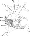
- FIG. 3Ais a top perspective view of an organ model made with a simulated tissue structure according to the present invention.
- FIG. 3Bis a top perspective view of an organ model made with a simulated tissue structure according to the present invention.
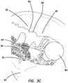
- FIG. 3Cis a top perspective, sectional view of an organ model made with a simulated tissue structure according to the present invention.
- FIG. 4is a top perspective, sectional view of an organ model made with simulated tissue structure according to the present invention.
- FIG. 5is a top perspective view of a surgical training device according to the present invention.
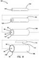
- FIG. 6is an exploded view of a simulated rectum model according to the present invention.
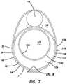
- FIG. 7is a cross-sectional view of a simulated rectum model according to the present invention.
- FIG. 8is a sectional view of a cross-section of a simulated rectum model according to the present invention.
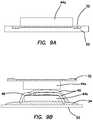
- FIG. 9Ais cross-sectional view of a casting dish, a first layer of silicone and a first layer of fiber according to the present invention.
- FIG. 9Bis a cross-sectional view of the first layer of silicone and first layer of fiber of FIG. 9A located above a casting dish, a second layer of silicone, a second layer of fiber and simulated vessels according to the present invention.
- FIG. 10Ais a top view of a casting dish according to the present invention.
- FIG. 10Bis a side elevational view of the casting dish of FIG. 10A according to the present invention.
- FIG. 11Ais a side elevational view of a casting dish, a layer of wet foam and a layer of fiber according to the present invention.
- FIG. 11Bis a side elevational view of the first layer of fiber and layer of foam from FIG. 11A located above a second layer of silicone according to the present invention.
- FIG. 11Cis a side elevational view of the first layer of fiber and layer of foam and second layer of silicone of FIG. 11B located beneath a second layer of fiber, third layer of silicone and artificial vessels according to the present invention.
- FIG. 1A simulated tissue structure 30 according to the present invention is shown in FIG. 1 .
- the structure 30includes a first layer 32 and a second layer 34 having an upper surface 36 , 38 and lower surface 40 , 42 , respectively.
- the first layer 32 and the second layer 34are interconnected by a third layer 44 defining a gap 46 therebetween.
- the simulated tissue structure 30may further optionally include inclusions 48 located between the first and second layers 32 , 34 .
- the inclusions 48include simulated vessels, veins, tumors, ducts, vasculature, nerves, fat deposits, pathologies or other anatomical structures.
- the inclusions 48are typically made of silicone but may also be made of other polymers or other suitable material and realistically shaped, colored and configured.
- the third layer 44comprises a plurality of one or more non-aligned, randomly arranged, nonwoven fiber 50 connected to the first layer 32 and/or second layer 34 at one or more location along the length of the fiber(s) 50 .
- the fiber 50is connected to one or more of the first layer 32 and the second layer 34 by being embedded into the one or more of the first layer 32 and the second layer 34 during the manufacturing process which will be described in greater detail below.
- Each fibermay be in the form of a strand, filament, yarn, micro-fiber and the like and has a length and a first free end and a second free end. Adhesive is not used to connect the fiber.
- the fiber of the third layer 44is resident within the gap 46 in a randomly arranged fashion.
- One strand of fiber 50may be connected to the first layer 32 at one location and then connected to the first layer 32 again at another location along the length of the fiber or to the second layer 34 and its free ends may or may not be embedded in the first or second layer. Some strands of fiber 50 may not be connected to the first layer 32 or second layer 34 and are freely disposed between the first layer 32 and the second layer 34 . Some strands of fiber 50 are entangled and intertwined with other strands in a loose fashion such that the strands may move relative to other strands. The fiber may span the gap 46 to be connected to the opposite or second layer 34 at one or more location along the length of the fiber.
- the fibersare selected from any suitable material such as polyester, polyamide, acrylic, acetate, polyolefin, cotton, fiberfill, batting, polyethylene terephthalate, polyethylene naphthalate, nylon, polyfill, fiberfill, polymer, plastic, spandex or other suitable fiber, natural fiber, non-absorbent fiber, synthetic fiber or fiber-like material.
- the materialmay be woven, not woven or partially woven.
- Fiberfillis typically made by garnetting in which a garnet machine takes fibers and combs them into a batt form. The garnet machine may then fold and chop the fibers to make strands that are shorter and clumped together. The fibers mat together entangle and bunch.
- first layer 32 and second layer 34has a substantially uniform thickness between its upper surface 36 , 38 and its lower surface 40 , 42 defining a substantially planar configuration.
- first layer 32 and the second layer 34have a substantially uniform thickness between its upper surface 36 , 38 and its lower surface 40 , 42 .
- the lower surface 42 of the second layer 34faces the upper surface 36 of the first layer 32 .
- the layer 32 , 34has a reduced thickness because part of the thickness is taken by the thickness of the fiber itself.
- the first and second layers 32 , 34are made of any suitable elastomeric material such as silicone. Room temperature vulcanization silicone is used in one variation.
- the second layer 34is avoided and the simulated tissue structure 30 includes only the first layer 32 and the third layer 44 of fiber connected to the first layer 32 .
- a casting dish 52 having a textured surface 54is provided.
- the casting dish 52has a smooth surface.
- Uncured room temperature vulcanization siliconeis provided and applied evenly onto the textured surface 54 of the casting dish 52 as shown in FIG. 2B to form a thin first layer 32 .
- a spatulamay be used to calendar the silicone evenly into a thin first layer 32 .
- the third layer 44is applied.
- a layer of polyester fibers 50is placed onto the upper surface 36 of the first layer 32 while the first layer 32 is still wet.
- the polyester fibers 50are arranged in a desired shape, thickness and density.
- the fibers 50are then tamped down into the first layer 32 to help embed the fibers into the first layer 32 in a random fashion. Some parts of the fibers 50 are embedded in the silicone and most are exposed to the air and remain available to embed in a subsequent silicone casting.
- any optional inclusions 48are placed onto or in juxtaposition with the upper surface 36 of the first layer 32 .
- the inclusions 48are placed before the polyester fibers 50 are applied.
- the inclusions 48are placed after the polyester fibers 50 are applied. If the inclusions 48 are placed before the polyester fibers 50 , the inclusions 48 will become adhered to the first layer 32 as the silicone cures. If the inclusions 48 are placed after the polyester fibers 50 , only portions of the inclusions 48 that are in direct contact with the wet silicone of the first layer 32 will become adhered to the first layer 32 as the silicone cures.
- the inclusionsmay be selectively adhered to either the first layer and/or the second layer to provide a realistic scenario for practicing the removal of an inclusion in a simulated surgical excision of the inclusion 48 with the surgeon employing careful and selective dissection. Also, only portions of the fibers 50 that are in contact with the wet silicone of the first layer 32 will become adhered to the first layer 32 . The silicone of the first layer 32 is allowed to cure fully embedding parts of the fibers into the first layer 32 .
- the inclusions 48are placed onto the first layer 32 after the first layer 32 has cured, thereby, not being embedded therein.
- the fiber third layer 44is placed onto a cured first layer 32 and, thereby, not becoming bonded thereto.
- the textured first layer 32is removed from the casting dish 52 .
- very thin sheets of siliconeare difficult to remove from a casting dish 52 even with a layer of mold release coating the casting dish.
- the presence of fibers 50 that are attached to the first layer 32 upon curing of the siliconeenable extremely thin layers of silicone to be removed from a casting dish without resulting in the layer tearing or being damaged.
- the interconnected embedded fibers 50help to gently pull the thin layer away from the casting dish.
- the fiber layer 44makes the tissue structure 30 more resilient to tearing and advantageously enables extremely thin layers of silicone to be casted and safely removed without tearing from the casting dish.
- the textured casting dish 52advantageously provides locations of reduced thickness as wet silicone will pool in the locations where the casting dish is deeper.
- the texture of casting dish 52creates a multitude of small holes throughout the layer.
- the holesare relatively unrecognizable because advantageously the fiber layer provides a visual of glistening tissue as light is reflected in many directions from the shiny fiber mimicking wet live tissue.
- the holesact as points of origin for tears in the first layer 32 of silicone which is advantageous for simulating dissection, because, as mentioned previously, defects in the silicone help overcome the large and often unrealistic resistance to tearing of silicone.
- the first layer 32 of siliconeis made thinner, it become more difficult to de-mold and remove.
- the added fibers 50which are placed on top of the uncured silicone while in the casting dish 52 , form a composite with the silicone and make it possible to de-mold extremely thin sheets. Furthermore, advantageously, the presence of fibers 50 atop and in connection with the first layer 32 while the silicone of the first layer 32 is still uncured creates a capillary action or absorbency depending upon the type of material used in making the fiber that pulls silicone into the fibers 50 and away from the casting dish 52 . This capillary action results in extremely thin spots and even small holes in the casting of the first and second layers 32 , 34 which are easy and realistic to dissect using surgical instruments.
- This capillary actionallows for the formation of sheets on un-textured, smooth casting dishes with the same desirable end results wherein the layers 32 , 34 have locations of reduced thickness of silicone.
- the isolated spots of reduced thickness in the silicone layer 32 , 34act as points of origin for tears that mimic real dissection with a scalpel.
- the capillary-like actiontakes place when the fibers 50 are placed on the silicone when it is in an uncured state and results in at least part of the fiber strand becoming coated with the polymer or silicone polymer.
- all of the fibersare coated before being embedded in one or more of the first and second layers.
- the fibers 50 of the third layer 44are not ordered or aligned but randomly tangled. This tangled configuration resist the silicone's natural rebound, greatly enhancing the realistic feel of the tissue structure 30 , especially when performing blunt dissection as in laparoscopic surgery, as the fibers can slide/move relative to each other dampening the resiliency of the silicone. Also, the tangled configuration of the fibers 50 make separation of the first layer 32 and the second layer 34 a function of pulling tangled fibers instead of pulling layers that are adhered with silicone or other adhesive. In a sense, the fibers act as an adhesive layer or mechanical linkage between the first layer 32 and the second layer 34 .
- the adhesionbeing defined by the tangled fibers of the third layer 44 and the degree of their adhesion to the layers 32 , 34 . Separating the tangled fibers when pulling the first and second layers apart permits the surgeon to employ and practice respect for tissue techniques instead of using larger forces merely because the model is made of silicone, with adjoining layers firmly adhered with adhesive and the like. Therefore, the present invention is highly effective for making dissectible tissue models.
- the method of manufacturing the simulated tissue structure 30includes providing a second layer 34 of silicone.
- the second layer 34 of siliconeis applied to a smooth or textured casting dish to create a thin layer of silicone.
- a spatulamay be used to calendar the silicone evenly into a thin second layer 34 .
- the combination of the first layer 32 and the third layer 44 previously madeis applied onto the lower surface 42 of the second layer 34 while the silicone of the second layer 34 is in an uncured state.
- the third layer 44 of polyester fibers 50is placed onto the lower surface 42 of the second layer 34 .
- the fibers 50are then tamped down onto the second layer 34 to help embed the fibers 50 into the second layer 34 .
- any optional inclusions 48are may be optionally provided onto the lower surface 42 of the second layer 32 .
- the inclusions 48are placed before the polyester fibers 50 are applied.
- the inclusions 48 together with the fiber layermay become adhered to the second layer 34 as the silicone cures.
- the second layer 34is allowed to cure before the first layer 32 and third layer 44 are overlaid onto the second layer 34 if adhesion of fiber only to the first layer 32 is desired.
- a framehaving a central window of a desired shape.
- the frame(not shown) is applied against the lower surface 40 of the first layer 32 and pressed down toward the second layer 34 to bring the perimeter of the first layer 32 into sealing contact with the uncured silicone of the second layer 34 capturing the third layer 44 in between creating a pocket of fibers 50 with or without inclusions 48 .
- the perimeter areas of the first and second layers 32 , 34are without fibers, in one variation, ensuring that the first and second layers 32 , 34 come into direct contact with each other to create and substantially seal the pocket.
- the pocketis not created and the sides of the simulated tissue structure 30 are left open as shown in FIG. 1 .
- the silicone of the second layer 34is allowed to cure fully resulting in the third layer being attached and embedded in the upper surface 36 of the first layer 32 and the lower surface 42 of the second layer 34 in sandwich-like fashion.
- One of the first layer 32 and second layer 34may have a greater thickness than the other. In another variation, both the first layer 32 and the second layer 34 have the same thickness.
- the most basic variation of the simulated tissue structure 30is a first layer 32 sheet of silicone with fibers 50 on one side.
- This basic variationcan be combined with other processes to create models of increasing complexity having additional layers of silicone, fiber and inclusions provided on outer or inner surfaces.
- the second layer 34 of siliconecan be applied to the same casting dish 52 and the previously made first layer 32 together with attached third layer 44 can be placed fiber-side down onto the uncured second layer 34 .
- This assemblycan then be used alone or as a component to a larger and more complex model.
- the thickness of the first and second layersis approximately between 1.0 millimeter and 7.0 millimeters and, preferably, between 0.5 millimeters and 3 millimeters.
- the third layeris approximately between 2.5 millimeters to 25.0 millimeters.
- FIGS. 3A-3Cillustrates a pelvic model 56 with the simulated tissue structure 30 of the present invention.
- the pelvic model 56includes a portion of a simulated pelvis 58 .
- the simulated tissue structure 30includes only a first layer 32 and a third layer 44 of fibers 50 without a second layer 34 of silicone.
- the upper surface 36 of the first layer 32faces toward the simulated pelvis 58 such that the fibers 50 are located between the first layer 32 and the simulated pelvis.
- the simulated pelvis 58serves as an armature on which the simulated tissue structure of the present invention is attached.
- the simulated tissue structure 30 of the present inventionis placed over the simulated pelvis 58 that is shown to include other anatomical features including but not limited to ducts 59 and a defect 60 interior to the first layer 32 .
- the edges of the first layer 32are adhered to the backside of the simulated pelvis 58 as shown in FIG. 3B and optionally at other selected areas along the first layer 32 .
- the pelvic model 56is approached by a surgeon employing a laparoscope, the lower surface 40 of the first layer 32 will be visualized first. Because of the textured surface of the first layer 32 and because of the varying placement and arrangement of the third layer 44 beneath the thin first layer 32 , the model 56 will appear more realistic than a uniform layer of silicone without texturing or without the underlying fiber layer 44 .
- the fiber layer 44will advantageously serve to obscure portions of the structures/inclusions making them more difficult to discern making the dissection practice more realistic and difficult for the practitioner. Thicker areas of the third layer 44 from having more fiber will obscure underlying structures/inclusions 48 more than thinner areas of the third layer 44 having less fiber thickness.
- the first layer 32may vary in thickness itself permitting different degrees of visualization of the underlying structures/tissues. The first layer 32 may be dyed red or pink. The light-colored or white fibers 50 will make the overlaying first layer 32 appear lighter in color in certain locations. With the underlying third layer 44 of fiber, the first layer 32 will appear lighter red or lighter pink in certain areas relative to other locations where there is no fiber or less fiber.
- FIGS. 3A and 3CAn incision 62 is shown in FIGS. 3A and 3C .
- the first layer 32will not rebound due to the elasticity of the silicone itself which would resulting in the incision 62 appearing to close at an unrealistically fast rate or response. Instead, the incision 62 will remain substantially open as shown as a result of the fiber layer 44 dampening or holding back resiliency of the silicone itself. Also, the ability to mold very thin layers of silicone with the help of the fiber layer, the resulting thinner layer of silicone will have less thickness and reboundability.
- the polyester fibers 50appear to glisten as the fibers 50 reflect light in various directions advantageously making the simulated tissue structure 30 appear wet or moist as real tissue without the help of any liquid being present in the model.
- the simulated tissue structuresmay appear unrealistic outside of a simulator or outside of a laparoscopic simulation environment and when observed with the naked eye, but because visualization takes place via a scope in a cavernous trainer that is artificially illuminated, certain liberties can be taken to achieve realistic advantages that could not be achieved for organs suitable for open procedures used outside the a laparoscopic simulation environment.
- the fibers 50 of the third layer 44may appear very unrealistic as an organ or tissue simulation when observed with the naked eye but appear and behave very realistically in a laparoscopic training environment which will be described in greater detail below.
- the inclusions 48 including the ducts 59 and underlying artificial tissue structures 60are exposed.
- FIG. 4illustrates an abdominal organ model 64 that includes simulated bowels 66 atop a simulated mesentery or omentum layer 68 that comprises the simulated tissue structure 30 according to the present invention.
- the bottom surface 40 of the structure 30is facing up and vasculature 70 is included as an inclusion 48 attached to the first layer 32 .
- the vasculature 70was attached to the first layer 32 before the third layer 44 of fiber 50 was embedded. Hence, the vasculature is clearly visible through the first layer 32 .
- the simulated mesentery layer 68is made of silicone that is dyed yellow and the vasculature is red in color and made of silicone.
- Uncured siliconeis provided and applied evenly to a rotating mandrel to create the first layer 32 . While the silicone of the first layer 32 is still wet, the polyester fiber layer is applied to form a third layer 44 of fibers 50 .
- the fibersmay be randomly or evenly applied or strategically applied forming areas where more or less fiber is intentionally located to effect a desired simulation outcome.
- the first layer 32 of siliconeis allowed to cure to embed the fibers 50 into the first layer 32 .
- the cured first layer 32is taken off the mandrel and has a cylindrical shape with the lower surface 40 of the first layer 32 forming the interior of the cylinder and defining the cylinder lumen.
- the cylindrical shape of the first layer 32 and the third layer 44may be inverted to place the fiber layer 44 inwardly and the lower surface 40 of the first layer 32 forming a smooth outer surface of the cylinder.
- Inclusions 48may be applied to the outer surface of the cylinder either after inversion or prior to forming the first layer 32 .
- the cylinderis not inverted.
- a first strip of uncured siliconeis applied onto a surface. The first strip has a length approximately equal to the length of the tubular first layer 32 .
- the tubular first layer 32 and third layer 44is aligned with the first strip and laid down onto the first strip with the fiber side of the combination facing the uncured first strip and tamped down to embed fibers 50 into the first strip.
- the first stripis allowed to cure to embed the fibers 50 of the third layer 44 into the first strip.
- a second strip of uncured siliconeis applied to a surface.
- the second striphas a length approximately equal to the length of the tubular first layer 32 .
- the tubular first layer 32 , third layer 44 and first stripis laid onto the second strip while the silicone of the second strip is still wet to embed the fibers 50 of the third layer 44 .
- the tubular first layer 32is applied to the second strip offset from the first strip so that an adjacent portion of exposed fibers of the third layer 44 come in contact with the wet second strip, preferably adjacent to the first strip and slightly overlaying the first strip to form an almost continuous second layer 34 .
- This processis repeated to form the second layer 34 from a plurality or any number of silicone sections or strips.
- the stripsmay be rectangular, triangular or any other shape to suitably cover the cylindrical surface and embed the third layer into the second layer 34 .
- Different organ modelssuch as bowels can be formed with the simulated tissue structure 30 having a tubular shape and any inclusions 48 can be provided directly to either side of the first layer 32 prior to the application of the fiber layer 44 or after the fiber layer 44 or directly to the second layer 34 .
- the second layer 34is not applied and the simulated tissue structure includes the first and second third layer and any inclusions 48 .
- the simulated tissue structure 30by itself or formed as part of another larger model or tissue structure such as the abdominal organ model 64 or pelvic model 56 described above with respect to FIGS. 3A-3C and FIG. 4 is sized and configured to be placed inside a simulated laparoscopic environment such as a surgical training device 10 of the like shown in FIG. 5 .
- the simulated tissue structuremay also be used to practice open surgical procedures.
- a surgical training device 10that is configured to mimic the torso of a patient such as the abdominal region is shown in FIG. 5 .
- the surgical training device 10provides a body cavity 12 substantially obscured from the user for receiving simulated or live tissue or model organs or training models of the like described in this invention.
- the body cavity 12is accessed via a tissue simulation region 14 that is penetrated by the user employing devices to practice surgical techniques on the tissue or practice model found located in the body cavity 12 .
- the body cavity 12is shown to be accessible through a tissue simulation region, a hand-assisted access device or single-site port device may be alternatively employed to access the body cavity 12 .
- An exemplary surgical training deviceis described in U.S. patent application Ser. No. 13/248,449 entitled “Portable Laparoscopic Trainer” filed on Sep. 29, 2011 and incorporated herein by reference in its entirety.
- the surgical training device 10is particularly well suited for practicing laparoscopic or other minimally invasive surgical procedures.
- the surgical training device 10includes a top cover 16 connected to and spaced apart from a base 18 by at least one leg 20 .
- FIG. 5shows a plurality of legs 20 .
- the surgical training device 10is configured to mimic the torso of a patient such as the abdominal region.
- the top cover 16is representative of the anterior surface of the patient and the space 12 between the top cover 16 and the base 18 is representative of an interior of the patient or body cavity where organs reside.
- the surgical trainer 10is a useful tool for teaching, practicing and demonstrating various surgical procedures and their related instruments in simulation of a patient undergoing a surgical procedure. Surgical instruments are inserted into the cavity 12 through the tissue simulation region 14 as well as through pre-established apertures 22 in the top cover 16 .
- the base 18includes a model-receiving area 24 or tray for staging or holding a simulated tissue model or live tissue.
- the model-receiving area 24 of the base 18includes frame-like elements for holding the model (not shown) in place.
- a clip attached to a retractable wireis provided at locations 26 . The retractable wire is extended and then clipped to hold the tissue model in position substantially beneath the tissue simulation region 14 .
- tissue modelOther means for retaining the tissue model include a patch of hook-and-loop type fastening material affixed to the base 18 in the model receiving area 24 such that it is removably connectable to a complementary piece of hook-and-loop type fastening material affixed to the model.
- a video display monitor 28 that is hinged to the top cover 16is shown in a closed orientation in FIG. 5 .
- the video monitor 28is connectable to a variety of visual systems for delivering an image to the monitor.
- a laparoscope inserted through one of the pre-established apertures 22 or a webcam located in the cavity and used to observe the simulated procedurecan be connected to the video monitor 28 and/or a mobile computing device to provide an image to the user.
- audio recording or delivery meansmay also be provided and integrated with the trainer 10 to provide audio and visual capabilities.
- Means for connecting a portable memory storage device such as a flash drive, smart phone, digital audio or video player, or other digital mobile deviceis also provided, to record training procedures and/or play back pre-recorded videos on the monitor for demonstration purposes.
- connection means for providing an audio visual output to a screen larger than the monitoris provided.
- the top cover 16does not include a video display but includes means for connecting with a laptop computer, a mobile digital device or tablet and connecting it by wire or wirelessly to the trainer.
- the top cover 16When assembled, the top cover 16 is positioned directly above the base 18 with the legs 20 located substantially around the periphery and interconnected between the top cover 16 and base 18 .
- the top cover 16 and base 18are substantially the same shape and size and have substantially the same peripheral outline.
- the internal cavityis partially or entirely obscured from view.
- the legsinclude openings to allow ambient light to illuminate the internal cavity as much as possible and also to advantageously provide as much weight reduction as possible for convenient portability.
- the top cover 16is removable from the legs 20 which in turn are removable or collapsible via hinges or the like with respect to the base 18 . Therefore, the unassembled trainer 10 has a reduced height that makes for easier portability.
- the surgical trainer 10provides a simulated body cavity 12 that is obscured from the user.
- the body cavity 12is configured to receive at least one surgical model accessible via at least one tissue simulation region 14 and/or apertures 22 in the top cover 16 through which the user may access the models to practice laparoscopic or endoscopic minimally invasive surgical techniques.
- the simulated rectum model 100includes a first tube 102 made of silicone.
- the first tube 102may include an embedded mesh material such that the first tube 102 is capable of retaining sutures such that they do not pull out or tear through the silicone.
- the first tube 102defines a first lumen 103 extending between a proximal end and a distal end.
- the simulated rectum model 100further includes a second tube 104 defining a second lumen 105 and extending between a proximal end and a distal end.
- the second tube 104is made of yellow urethane foam. A layer of foam is formed and then folded into a cylindrical shape and the ends adhered to form a tube. The anterior end of the urethane foam second tube 104 is thinner as shown in FIG. 6 .
- the second lumen 105is dimensioned to receive the first tube 102 inside the second lumen 105 in a concentric-like fashion.
- the second tube 104is adhered to the first tube 102 using cyanoacrylate glue.
- the model 100further includes a third tube 106 .
- the third tube 106is simulated tissue structure 30 of the like described above having a first layer 32 , a second layer 34 and a third layer 44 of polyfill fiber 50 that is formed into a cylindrical tube to define a third lumen 107 .
- the first layer 32 of the third tube 106is yellow in color and the second layer 34 is white in color.
- the third layer 44is made of white polyfill fiber.
- the diameter of the third lumen 107is dimensioned to receive the second tube 104 inside the third lumen 107 in an eccentric fashion.
- the third tube 106is adhered to the second tube 104 with adhesive such as cyanoacrylate glue.
- the simulated rectum model 100further includes a fourth tube 108 .
- the fourth tube 108is simulated tissue structure 30 of the like described above having a first layer 32 and a third layer 44 of polyfill fiber 50 but does not have a second layer 34 that is formed into a cylindrical tube to define a fourth lumen 109 such that the third layer 44 of free polyfill fibers faces the fourth lumen 109 .
- the second layer 34is pink in color.
- the third layer 44is made of white polyfill fiber.
- the fourth tube 108includes a second layer 34 that is white in color.
- the diameter of the fourth lumen 109is dimensioned to receive the third tube 106 inside the fourth lumen 109 in a concentric-like fashion.
- the fourth tube 108is adhered to the third tube 106 with adhesive in select areas.
- the simulated rectum model 100further includes a simulated prostate system 110 located between the third tube 106 and the fourth tube 108 .
- the simulated prostate system 110is located at the anterior side of the model 100 .
- the simulated prostate system 110includes a simulated prostate, simulated seminal vesicles, simulated bladder, simulated urethra, and simulated vas deferens.
- the simulated urethra and simulated vas deferensare made of silicone formed into a solid tube.
- the simulated seminal vesiclesare made of urethane foam over molded onto the simulated vas deferens.
- the simulated prostateis made of urethane foam over molded onto the simulated urethra.
- the simulated rectum model 100further includes additional polyfill material located between the fourth tube 108 and the third tube 106 at the anterior side of the model 100 and surrounding the simulated prostate system 110 .
- the simulated rectum model 100is fantastically suited for practicing transanal total mesorectal excision (TaTME) for cancer located in the lower rectum.
- TaTMEtransanal total mesorectal excision
- the cancerous simulated rectumis approached through the anus via a sealable port connected to a channel that is inserted into the simulated rectum.
- a purse-string sutureseals off the cancerous portion of the rectum.
- the purse-string sutureis a type of suture technique that the user of the model 100 can practice. It involves suturing around the circumference of the rectum and pulling it tight to seal off the area of the rectum that includes the tumor.
- the first tube 102includes mesh embedded in the silicone layer of the tube to hold the purse-string suture in place.
- the silicone layer of the first tube 102allows the purse-string suture to be pulled tight. Then, the surgeon will cut down posteriorly through the second tube 104 which represents the mesorectum. The surgeon will continue to dissect through the first layer 32 of the third tube 106 and then dissect circumferentially around in the third layer 44 of the third tube 106 being careful not to penetrate the second layer 34 of the third tube 106 because doing so would endanger the adjacent simulated prostate system 110 .
- the first layer 32 of the third tube 106is yellow, which is the same color as the simulated mesorectum, second tube 104 , making it hard to distinguish apart from the second tube 104 .
- the fourth tube 108and in particular, the second layer 34 of the fourth tube 108 is red, representing the muscle and the pelvic floor.
- Accidental dissection into the second layer 34 of the fourth tube 108 and circumferential progression of dissection in this locationwould possibly lead to intersection with the simulated prostate system 110 which this model 100 teaches the surgeon to avoid.
- Dissection within the third layer 44 of the third tube 106leads to a safe excision of the simulated prostate system 110 .
- anterior dissectionbegins by dissecting through the thinner section of the simulated mesorectum (second tube 104 ) until the third tube 106 is reached.
- dissectionproceeds circumferentially until the dissection meets the posterior dissection.
- the simulated mesorectum (second tube 104 )has an area of reduced thickness and the third tube 106 is attached to the second tube 104 and indistinguishably colored when comparing the yellow first layer 32 with the yellow second tube 104 .
- the simulated prostate system 110is located on top of the third tube 106 as shown in FIG. 7 and it is surrounded with polyfill fiber 112 which makes it harder to distinguish from the polyfill fiber of the third layer 44 of the third tube 106 while dissecting in the third tube 106 . Dissection proceeds until the pelvic cavity is breached.
- the proximal end of the simulated rectum model 100may be attached to a transanal adapter.
- the transanal adapteris a leg 20 used to space apart the top cover 16 from the base 18 of a surgical trainer 10 to provide access into the model 100 from the side of the surgical trainer.
- the transanal adapterincludes an opening that is connected to the first lumen 103 of the first tube 102 .
- soft siliconeSurrounding the opening of the transanal adapter, soft silicone is provided to simulate an anus.
- the practice of the surgical TaTME procedureis performed through the opening of the transanal adapter with a circumferential purse string suture placed proximal to the transanal adapter with the simulated prostate system located distal to the transanal adapter.
- the simulated rectum model 100is manufactured by first placing a mesh sheath on a mandrel and paining uncured silicone over the mesh.
- the second tube 104(simulated mesorectum) is made of urethane foam that is cast into a flat sheet. The foam is cast to have a thinner section. The simulated mesorectum is wrapped into a cylinder around the first tube 102 to create the second tube 104 . Cyanoacrylate glue is used together with a primer to adhere the thicker portions of the second tube 104 together on the posterior side of the simulated rectum 100 .
- a thin planar sheet of yellow siliconeis cast onto foam to create the first layer 32 .
- a layer of polyfillis evenly placed on top to create the third layer 44 of polyfill.
- the first layer 32cures, it is de-molded.
- a new layer of white-colored or clear siliconeis cast on the foam to form the second layer 34 .
- the previously-cured first layer 32 together with the polyfill third layer 44is placed on top with the polyfill third layer 44 touching the wet silicone of the second layer 34 .
- the assemblyis demolded and wrapped around the second tube 104 to form a cylindrical third tube 106 which is adhered to the second tube 104 using cyanoacrylate glue.
- the fourth tube 108is formed in a similar fashion as the third tube 106 .
- a thin planar sheet of white or clear siliconeis cast onto foam to create the first layer 32 . While the silicone of the first layer 32 is still wet, a layer of polyfill is evenly placed on top to create the third layer 44 of polyfill. More polyfill fiber is added to create an area where the third layer 44 is thicker as shown in FIG. 7 . After the first layer 32 cures, the third layer 44 is adhered and the combination of the first layer 32 and the third layer 44 is de-molded. A new layer of red-colored silicone is cast on the foam to form the second layer 34 of the fourth tube 108 . The previously-cured first layer 32 together with the polyfill third layer 44 is placed on top with the polyfill third layer 44 touching the wet silicone of the second layer 34 .
- the assemblyis demolded and wrapped around the third tube 106 to form a cylindrical fourth tube 108 which is adhered to the third tube 106 using cyanoacrylate glue or silicone dots.
- the simulated prostate system 110is previously formed and located between the third tube 106 and the fourth tube 108 .
- FIG. 9Athere is shown a casting dish 52 that has a textured molding surface.
- the surfacemay vary in thickness.
- a first layer 32 of uncured siliconeis poured into the casting dish 52 .
- a third layer 44 a of fiberis placed on top of the first layer 32 such that fibers on one side of the third layer 44 a are embedded into the first layer 32 .
- the first layer 32is allowed to cure.
- the first layer 32is removed from the casting dish 52 with the help of the third layer 44 a .
- the third layer 44 a and the first layer 32are pulled up from the casting dish 52 .
- the third layer 44 aadhered to the first layer 32 , pulling up on the third layer 44 a allows the fibers of the third layer 44 a to distribute the removal force and advantageously prevent the thin first layer 32 from tearing during removal.
- the combination of the first layer 32 and the third layer 44 ais removed, it is inverted and placed in juxtaposition to a second casting dish 52 with a second layer 34 of wet silicone as shown in FIG. 9B .
- Another third layer 44 b of fiberis placed onto the second layer 32 of wet silicone.
- Inclusions 48are placed over the third layer 44 b . Part of the inclusions 48 are placed in contact with the second layer 34 and remains embedded in the second layer 34 when the second layer 34 finishes curing.
- the third layer 44 balso is embedded into the second layer 34 .
- the inclusions 48are not embedded in the second layer 34 and are located between the third layers 44 a , 44 b .
- the constructis completed.
- the first layer 32is placed into contact with the second layer 34 while the second layer 34 is still uncured to adhere the first layer 32 and create a pocket that includes the inclusions 48 and third layers 44 a , 44 b .
- portions of the third layer 44 aare also embedded into the second layer 34 while the silicone is still wet to embed the third layer 44 a into the second layer 34 .
- the inclusions 48 shown in FIG. 9Bare vasculature made of silicone; however, the invention is not so limited and the inclusions 48 may be any inclusion, anatomical structure, landmark, organ, nerves, tissue, tumors and the like.
- FIGS. 10-11with particular reference to FIGS. 10A-10B there is shown a casting dish 52 having two channels 72 for receiving uncured silicone.
- the materialmay include uncured silicone, uncured urethane foam, uncured silicone foam and the like.
- wet uncured urethane foamis poured into the channels 72 to create a first inclusion 48 a as shown in FIG. 11A .
- a first fiber layer 44 ais placed on top of the uncured foam 48 a inside the channels 72 to embed the fiber layer 44 a into the uncured foam.
- the uncured silicone inside the channels 72is allowed to cure and as a result become attached to the first fiber layer 44 a .
- the first fiber layer 44 a together with the formed inclusions 48 aare removed from the casting dish 52 and placed in juxtaposition to a first layer 32 of uncured silicone as shown in FIG. 11B .
- the first fiber layer 44 a with the attached first inclusions 48 aare pressed into the first layer 32 while the silicone is still wet to embed the first inclusions 48 a and the first fiber layer 44 a into the first layer 32 as shown in FIG. 11C .
- the first inclusions 48 aare configured to depict and mimic nerves; however, the first inclusions 48 a can be any type of inclusion suitable for the simulated tissue structure.
- a second fiber layer 44 b made of fiberis provided together with one or more second inclusion 48 b .
- the second inclusions 48 bare attached to the second fiber layer 44 b in the same manner as described above with respect to the first fiber layer 44 a and first inclusions 48 a and may be made of silicone, silicone foam, urethane foam and the like.
- a casting dishis provided that is configured with a pattern for molding the one or more second inclusion 48 b .
- the patternis filled with wet silicone, for example, and while uncured, the second fiber layer 44 b is overlaid onto the casting dish and wet silicone of the second inclusions 48 b to embed and attach the second inclusions 48 b to the second fiber layer 44 b along a first side of the second fiber layer 44 b .
- the second side of the second fiber layer 44 bis embedded into a second layer 34 while the second layer 34 is still uncured.
- the second inclusions 48 b and the second layer 34are cured the second fiber layer 44 b and the second layer 34 together with the second inclusions 48 b are removed from the respective casting dishes and placed on the first fiber layer 44 a , first layer 32 and first inclusions 48 a to create the sandwich-like simulated tissue construct.
- the second inclusions 48 bare configured to mimic vasculature or any other anatomical structure, tissue, organ, nerve, tumor and the like.
- One or more of the first fiber layer 44 a and second fiber layer 44 bcreate ideal dissection pathways for skeletonizing any one or more of the inclusions 48 a , 48 b wherein the dissection pathway through the fiber creates a realistic look and feel with the fibers being capable of being cut and/or spread apart to separate and expose the layers and inclusions for removal.
- Any portion of the model 30can be made of one or more organic base polymer including but not limited to hydrogel, single-polymer hydrogel, multi-polymer hydrogel, rubber, latex, nitrile, protein, gelatin, collagen, soy, non-organic base polymer such as thermo plastic elastomer, Kraton, silicone, foam, silicone-based foam, urethane-based foam and ethylene vinyl acetate foam and the like.
- organic base polymerincluding but not limited to hydrogel, single-polymer hydrogel, multi-polymer hydrogel, rubber, latex, nitrile, protein, gelatin, collagen, soy, non-organic base polymer such as thermo plastic elastomer, Kraton, silicone, foam, silicone-based foam, urethane-based foam and ethylene vinyl acetate foam and the like.
- any base polymerone or more filler may be employed such as a fabric, woven or non-woven fiber, polyester, non-absorbent fiber, nylon, mesh, cotton and silk, conductive filler material such as graphite, platinum, silver, gold, copper, miscellaneous additives, gels, oil, cornstarch, glass, dolomite, carbonate mineral, alcohol, deadener, silicone oil, pigment, foam, poloxamer, collagen, gelatin and the like.
- the adhesives employedmay include but are not limited to cyanoacrylate, silicone, epoxy, spray adhesive, rubber adhesive and the like.
Landscapes
- Engineering & Computer Science (AREA)
- General Physics & Mathematics (AREA)
- Physics & Mathematics (AREA)
- Health & Medical Sciences (AREA)
- General Health & Medical Sciences (AREA)
- Mathematical Analysis (AREA)
- Mathematical Physics (AREA)
- Medicinal Chemistry (AREA)
- Chemical & Material Sciences (AREA)
- Algebra (AREA)
- Computational Mathematics (AREA)
- Theoretical Computer Science (AREA)
- Educational Technology (AREA)
- Mathematical Optimization (AREA)
- Medical Informatics (AREA)
- Pure & Applied Mathematics (AREA)
- Business, Economics & Management (AREA)
- Educational Administration (AREA)
- Pulmonology (AREA)
- Radiology & Medical Imaging (AREA)
- Life Sciences & Earth Sciences (AREA)
- Biomedical Technology (AREA)
- Molecular Biology (AREA)
- Instructional Devices (AREA)
- Laminated Bodies (AREA)
Abstract
Description
Claims (20)
Priority Applications (4)
| Application Number | Priority Date | Filing Date | Title |
|---|---|---|---|
| US16/431,118US10755602B2 (en) | 2015-07-16 | 2019-06-04 | Simulated dissectible tissue |
| US17/002,033US11587466B2 (en) | 2015-07-16 | 2020-08-25 | Simulated dissectible tissue |
| US18/172,070US12087179B2 (en) | 2015-07-16 | 2023-02-21 | Simulated dissectible tissue |
| US18/830,198US20240428708A1 (en) | 2015-07-16 | 2024-09-10 | Simulated dissectible tissue |
Applications Claiming Priority (5)
| Application Number | Priority Date | Filing Date | Title |
|---|---|---|---|
| US201562193143P | 2015-07-16 | 2015-07-16 | |
| US201562257847P | 2015-11-20 | 2015-11-20 | |
| PCT/US2016/041852WO2017011436A1 (en) | 2015-07-16 | 2016-07-12 | Simulated dissectable tissue |
| US15/209,565US10332425B2 (en) | 2015-07-16 | 2016-07-13 | Simulated dissectible tissue |
| US16/431,118US10755602B2 (en) | 2015-07-16 | 2019-06-04 | Simulated dissectible tissue |
Related Parent Applications (1)
| Application Number | Title | Priority Date | Filing Date |
|---|---|---|---|
| US15/209,565ContinuationUS10332425B2 (en) | 2015-07-16 | 2016-07-13 | Simulated dissectible tissue |
Related Child Applications (2)
| Application Number | Title | Priority Date | Filing Date |
|---|---|---|---|
| US17/002,033ContinuationUS11587466B2 (en) | 2015-07-16 | 2020-08-25 | Simulated dissectible tissue |
| US18/172,070ContinuationUS12087179B2 (en) | 2015-07-16 | 2023-02-21 | Simulated dissectible tissue |
Publications (2)
| Publication Number | Publication Date |
|---|---|
| US20190287425A1 US20190287425A1 (en) | 2019-09-19 |
| US10755602B2true US10755602B2 (en) | 2020-08-25 |
Family
ID=56497909
Family Applications (5)
| Application Number | Title | Priority Date | Filing Date |
|---|---|---|---|
| US15/209,565Active2037-02-06US10332425B2 (en) | 2015-07-16 | 2016-07-13 | Simulated dissectible tissue |
| US16/431,118ActiveUS10755602B2 (en) | 2015-07-16 | 2019-06-04 | Simulated dissectible tissue |
| US17/002,033Active2037-01-18US11587466B2 (en) | 2015-07-16 | 2020-08-25 | Simulated dissectible tissue |
| US18/172,070ActiveUS12087179B2 (en) | 2015-07-16 | 2023-02-21 | Simulated dissectible tissue |
| US18/830,198PendingUS20240428708A1 (en) | 2015-07-16 | 2024-09-10 | Simulated dissectible tissue |
Family Applications Before (1)
| Application Number | Title | Priority Date | Filing Date |
|---|---|---|---|
| US15/209,565Active2037-02-06US10332425B2 (en) | 2015-07-16 | 2016-07-13 | Simulated dissectible tissue |
Family Applications After (3)
| Application Number | Title | Priority Date | Filing Date |
|---|---|---|---|
| US17/002,033Active2037-01-18US11587466B2 (en) | 2015-07-16 | 2020-08-25 | Simulated dissectible tissue |
| US18/172,070ActiveUS12087179B2 (en) | 2015-07-16 | 2023-02-21 | Simulated dissectible tissue |
| US18/830,198PendingUS20240428708A1 (en) | 2015-07-16 | 2024-09-10 | Simulated dissectible tissue |
Country Status (8)
| Country | Link |
|---|---|
| US (5) | US10332425B2 (en) |
| EP (3) | EP3323122B1 (en) |
| JP (4) | JP7009355B2 (en) |
| KR (2) | KR102697097B1 (en) |
| AU (3) | AU2016291726B2 (en) |
| CA (1) | CA2992552A1 (en) |
| ES (1) | ES2824529T3 (en) |
| WO (1) | WO2017011436A1 (en) |
Cited By (1)
| Publication number | Priority date | Publication date | Assignee | Title |
|---|---|---|---|---|
| US20230317258A1 (en)* | 2020-12-03 | 2023-10-05 | Intuitive Surgical Operations, Inc. | Systems and methods for assessing surgical ability |
Families Citing this family (13)
| Publication number | Priority date | Publication date | Assignee | Title |
|---|---|---|---|---|
| US11776428B1 (en)* | 2015-10-21 | 2023-10-03 | University Of Rochester | Systems, models, and methods for simulating surgery on anatomical organs |
| JP6886975B2 (en)* | 2015-11-20 | 2021-06-16 | アプライド メディカル リソーシーズ コーポレイション | Simulated incisable tissue |
| EP3288009A1 (en)* | 2016-08-23 | 2018-02-28 | Virtamed AG | Low-resistance anatomical tissue model |
| US11501662B2 (en)* | 2017-11-15 | 2022-11-15 | Applied Medical Resources Corporation | Suturing skills surgical training model |
| EP3719779B1 (en)* | 2017-11-30 | 2022-08-31 | Mitsui Chemicals, Inc. | Simulated eyeball, ocular surgery training device, and ocular surgery training method |
| EP3727172A1 (en)* | 2017-12-19 | 2020-10-28 | Applied Medical Resources Corporation | Total mesorectal excision surgical simulator |
| EP3803837A1 (en)* | 2018-06-01 | 2021-04-14 | Applied Medical Resources Corporation | Renal hilum surgical simulation system |
| SG11202102411WA (en)* | 2018-09-28 | 2021-04-29 | Mitsui Chemicals Inc | Simulated sclera and simulated eyeball |
| CN108986621B (en)* | 2018-10-12 | 2023-06-30 | 北京大学人民医院 | Simulation trainer for anorectal NOTES endoscopic surgery |
| US11984046B2 (en) | 2019-08-16 | 2024-05-14 | University Of Kentucky Research Foundation | Surgical skills training model |
| CA3209856A1 (en)* | 2021-03-01 | 2022-09-09 | Gwendolyn Mary-jean MORGAN | Tactile tissue simulating structures |
| CA3234698A1 (en)* | 2021-10-22 | 2023-04-27 | Jacqueline FOSS | Colpotomy model |
| WO2023069782A1 (en) | 2021-10-23 | 2023-04-27 | Simulated Inanimate Models, LLC | Procedure guidance and training apparatus, methods and systems |
Citations (438)
| Publication number | Priority date | Publication date | Assignee | Title |
|---|---|---|---|---|
| US184573A (en) | 1876-11-21 | Improvement in gas-cocks | ||
| US2127774A (en) | 1936-04-27 | 1938-08-23 | Jacobs Julian Bay | Apparatus for teaching obstetrics |
| US2284888A (en) | 1941-04-14 | 1942-06-02 | Arc Diaphragm & Drug Co | Demonstrating device for vaginal diaphragms |
| US2324702A (en) | 1938-11-30 | 1943-07-20 | Karl F Hoffmann | Surgical simulacra and process of preparing same |
| US2345489A (en) | 1943-04-10 | 1944-03-28 | Frederic P Lord | Anatomical model |
| US2495568A (en) | 1948-12-30 | 1950-01-24 | Holland Rantos Company Inc | Clinical model |
| US3766666A (en) | 1971-10-13 | 1973-10-23 | Robins Co Inc A H | Uterine simulator trainer |
| US3775865A (en) | 1972-07-24 | 1973-12-04 | R Rowan | Simulator for teaching suturing techniques |
| US3789518A (en) | 1972-04-12 | 1974-02-05 | Weatherby Nasco Inc | Simulated human limb |
| US3921311A (en) | 1975-01-06 | 1975-11-25 | Pathfinder Fund | Clinical demonstration model |
| US3991490A (en) | 1973-04-09 | 1976-11-16 | Markman H David | Teaching aid for sigmoidoscope and the like |
| US4001952A (en) | 1975-10-24 | 1977-01-11 | Kleppinger Trygve M | Hysteroscopy teaching aid |
| US4001951A (en) | 1975-03-25 | 1977-01-11 | Fasse Wolfgang G | Breast cancer detection training device |
| US4321047A (en) | 1980-06-05 | 1982-03-23 | Bradley Landis | Simulator and process for teaching surgical knot tying techniques |
| US4323350A (en) | 1980-09-22 | 1982-04-06 | Bowden Jr Robert L | Anatomical model |
| US4332569A (en) | 1981-03-16 | 1982-06-01 | University Of Kentucky Research Foundation | Instructional device for use of a bronchoscope |
| US4371345A (en) | 1980-10-17 | 1983-02-01 | National Research Development Corporation | Multi-dimensional display equipment |
| US4386917A (en) | 1981-09-16 | 1983-06-07 | Forrest Leonard E | Suturing training device and method |
| US4459113A (en) | 1981-08-31 | 1984-07-10 | Boscaro Gatti Antonino E | Device for specializing in the use of endoscopes |
| US4481001A (en) | 1983-05-26 | 1984-11-06 | Collagen Corporation | Human skin model for intradermal injection demonstration or training |
| US4596528A (en) | 1984-07-02 | 1986-06-24 | Lewis Leonard A | Simulated skin and method |
| US4726772A (en) | 1986-12-01 | 1988-02-23 | Kurt Amplatz | Medical simulator |
| US4737109A (en) | 1987-03-03 | 1988-04-12 | Abramson Daniel J | Breast cancer detection training device |
| US4789340A (en) | 1987-08-18 | 1988-12-06 | Zikria Bashir A | Surgical student teaching aid |
| US4832978A (en) | 1987-04-24 | 1989-05-23 | Lesser Jary M | Simulated connective tissue for construction of models and prostheses |
| US4867686A (en) | 1989-02-09 | 1989-09-19 | Goldstein Mark K | Breast cancer detection model and method for using same |
| US4907973A (en) | 1988-11-14 | 1990-03-13 | Hon David C | Expert system simulator for modeling realistic internal environments and performance |
| US4938696A (en) | 1989-07-25 | 1990-07-03 | Foster-Pickard International, Inc. | Model demonstrating human organ systems |
| US4940412A (en) | 1987-12-08 | 1990-07-10 | Elscint Ltd. | Method of manufacturing anatomical models |
| DE9102218U1 (en) | 1991-02-14 | 1991-05-16 | Pier, Arnold, Dipl.-Ing. Dr.med., 5138 Heinsberg | Training device for laparoscopic surgical technique |
| US5061187A (en) | 1990-04-12 | 1991-10-29 | Ravinder Jerath | Ultrasound training apparatus |
| US5083962A (en) | 1987-08-24 | 1992-01-28 | Pracas Victor M | Doll capable of bodily functions |
| US5104328A (en) | 1990-04-18 | 1992-04-14 | Lounsbury Katherine L | Anatomical model |
| DE4105892A1 (en) | 1991-02-14 | 1992-08-27 | Arnold Dipl Ing Dr Med Pier | Simulation training appts. for surgery - comprises sealed housing and cover releasably connected to housing, excess pressure being formable in housing, and having flat insert in cover top side opening |
| US5149270A (en) | 1990-10-29 | 1992-09-22 | Mckeown M J | Apparatus for practicing surgical procedures |
| US5180308A (en) | 1992-01-06 | 1993-01-19 | Garito Jon C | Medical demonstration model |
| US5230630A (en) | 1992-07-20 | 1993-07-27 | Richard Burgett | Suture training device |
| FR2691826A1 (en) | 1992-06-01 | 1993-12-03 | Allal Hossein | Coelio-surgery simulator for teaching and training of surgeons - uses semi-rigid envelope in form of human body, with coeloscope connected to video camera and to video screen |
| US5273435A (en) | 1992-07-16 | 1993-12-28 | The Mcw Research Foundation, Inc. | Tumor localization phantom |
| WO1994006109A1 (en) | 1992-09-07 | 1994-03-17 | Diethelm Wallwiener | Medical training apparatus |
| US5295694A (en) | 1992-10-27 | 1994-03-22 | Levin John M | Laparoscopic surgery simulating game |
| US5310348A (en) | 1992-10-09 | 1994-05-10 | United States Surgical Corporation | Suture demonstration portfolio |
| US5318448A (en) | 1992-01-06 | 1994-06-07 | Garito Jon C | Demonstration model for gynecological procedure |
| US5320537A (en) | 1993-03-16 | 1994-06-14 | Triangle Research And Development Corporation | Microsurgical training apparatus |
| US5358408A (en) | 1993-03-09 | 1994-10-25 | Marelyn Medina | Tissue specimen suspension device |
| US5368487A (en) | 1992-07-31 | 1994-11-29 | Medina; Marelyn | Laparoscopic training device and method of use |
| US5380207A (en) | 1993-12-27 | 1995-01-10 | Siepser; Steven B. | Slip suture practice kit |
| US5403191A (en) | 1991-10-21 | 1995-04-04 | Tuason; Leo B. | Laparoscopic surgery simulator and method of use |
| US5425731A (en) | 1991-04-05 | 1995-06-20 | Metcal, Inc. | Instrument for cutting, coagulating and ablating tissue |
| US5425644A (en) | 1993-05-13 | 1995-06-20 | Gerhard Szinicz | Surgical training apparatus and method |
| DE4414832A1 (en) | 1994-04-28 | 1995-11-02 | Laerdal Asmund S As | Teaching model for practising blood taking or injection of blood vessels |
| US5472345A (en) | 1993-04-14 | 1995-12-05 | Gaumard Scientific Company, Inc. | Gynecological simulator |
| US5518406A (en) | 1993-11-24 | 1996-05-21 | Waters; Tammie C. | Percutaneous endoscopic gastrostomy teaching device |
| US5518407A (en) | 1993-11-02 | 1996-05-21 | Greenfield; Cathy L. | Anatomically correct artificial organ replicas for use as teaching aids |
| US5520633A (en) | 1991-01-03 | 1996-05-28 | Costin; John A. | Computer controlled smart phacoemulsification method and apparatus |
| US5541304A (en) | 1994-05-02 | 1996-07-30 | Hercules Incorporated | Crosslinked hydrogel compositions with improved mechanical performance |
| WO1996042076A1 (en) | 1995-06-09 | 1996-12-27 | Simulab Corporation | Anatomical simulator for videoendoscopic surgical training |
| US5720742A (en) | 1994-10-11 | 1998-02-24 | Zacharias; Jaime | Controller and actuating system for surgical instrument |
| US5722836A (en) | 1996-05-21 | 1998-03-03 | Simulab Corporation | Reflected-image videoendoscopic surgical trainer and method of training |
| US5727948A (en) | 1996-09-05 | 1998-03-17 | Jordan; Lynette S. | Syringe injection practice device |
| US5743730A (en) | 1996-05-07 | 1998-04-28 | Clester; Kenneth E. | Dental porcelain shading guide and method of use therefore |
| US5762458A (en) | 1996-02-20 | 1998-06-09 | Computer Motion, Inc. | Method and apparatus for performing minimally invasive cardiac procedures |
| US5769640A (en) | 1992-12-02 | 1998-06-23 | Cybernet Systems Corporation | Method and system for simulating medical procedures including virtual reality and control method and system for use therein |
| US5775916A (en) | 1992-01-15 | 1998-07-07 | Limbs & Things Limited | Method of making a surgical and/or clinical apparatus |
| US5785531A (en) | 1996-06-06 | 1998-07-28 | Wilson-Cook Medical Incorporated | Cuttable papilla and sphincterotomy training apparatus |
| JPH10211160A (en) | 1997-01-29 | 1998-08-11 | Olympus Optical Co Ltd | Colon endoscope insertion training device |
| US5800178A (en) | 1995-03-29 | 1998-09-01 | Gillio; Robert G. | Virtual surgery input device |
| US5803746A (en) | 1996-01-23 | 1998-09-08 | Medisim Corporation | Body part model and method of making same |
| US5807378A (en) | 1995-06-07 | 1998-09-15 | Sri International | Surgical manipulator for a telerobotic system |
| US5810880A (en) | 1995-06-07 | 1998-09-22 | Sri International | System and method for releasably holding a surgical instrument |
| DE19716341A1 (en) | 1997-03-19 | 1998-10-15 | Erbe Elektromedizin | Model of human torso with simulated organs for surgical training |
| US5850033A (en) | 1994-09-30 | 1998-12-15 | Mirzeabasov; Timur Akhmedbekovich | Target for simulating biological subjects |
| WO1998058358A1 (en) | 1997-06-19 | 1998-12-23 | Limbs & Things Limited | Clinical and/or surgical training apparatus |
| US5855583A (en) | 1996-02-20 | 1999-01-05 | Computer Motion, Inc. | Method and apparatus for performing minimally invasive cardiac procedures |
| WO1999001074A1 (en) | 1997-07-03 | 1999-01-14 | Neothermia Corporation | Methods and apparatus for therapeutic cauterization of predetermined volumes of biological tissue |
| US5873863A (en) | 1997-08-29 | 1999-02-23 | United States Surgical Corporation | Vascular surgery demonstration/training kit |
| US5873732A (en) | 1997-09-26 | 1999-02-23 | Hasson; Harrith M. | Apparatus for training for the performance of a medical procedure |
| US5908302A (en) | 1998-06-12 | 1999-06-01 | Goldfarb; Michael A. | Inguinal hernia model |
| US5947743A (en) | 1997-09-26 | 1999-09-07 | Hasson; Harrith M. | Apparatus for training for the performance of a medical procedure |
| WO2000036577A1 (en) | 1998-12-14 | 2000-06-22 | Pharmabotics Limited | Simulated body tissue |
| US6083008A (en) | 1997-09-01 | 2000-07-04 | Agency Of Industrial Science & Technology, Ministry Of International Trade & Industry | Optical phantom of living body and method for producing it |
| EP1024173A1 (en) | 1999-01-14 | 2000-08-02 | Dow Corning Corporation | Silicone gel composition and silicone gel produced therefrom |
| US6113395A (en) | 1998-08-18 | 2000-09-05 | Hon; David C. | Selectable instruments with homing devices for haptic virtual reality medical simulation |
| JP2001005378A (en) | 1999-06-24 | 2001-01-12 | Yasuhisa Koki:Kk | Simulator for training of operation technique |
| CN2421706Y (en) | 2000-04-26 | 2001-02-28 | 佟玉章 | Multifunctional exerciser for operating tying and suturing |
| US6234804B1 (en) | 1999-03-02 | 2001-05-22 | Peter Yong | Thoracic training model for endoscopic cardiac surgery |
| US6271278B1 (en) | 1997-05-13 | 2001-08-07 | Purdue Research Foundation | Hydrogel composites and superporous hydrogel composites having fast swelling, high mechanical strength, and superabsorbent properties |
| WO2002038039A2 (en) | 2000-10-23 | 2002-05-16 | Toly Christopher C | Human surgical trainer and methods for training |
| US6398557B1 (en) | 1999-09-17 | 2002-06-04 | The University Of Iowa Research Foundation | Devices, methods and kits for training in surgical techniques |
| US6474993B1 (en) | 1996-12-04 | 2002-11-05 | Erbe Elektromedizin Gmbh | Artificial tissue |
| US20020168619A1 (en) | 1999-12-29 | 2002-11-14 | Provenza Thadeu Rezende | Simulator device for human feminine mammary gland |
| US6485308B1 (en) | 2001-07-09 | 2002-11-26 | Mark K. Goldstein | Training aid for needle biopsy |
| US6488507B1 (en) | 1999-11-29 | 2002-12-03 | Ethicon, Inc. | Portable surgical trainer |
| US6497902B1 (en) | 1999-12-01 | 2002-12-24 | The Regents Of The University Of Michigan | Ionically crosslinked hydrogels with adjustable gelation time |
| US6511325B1 (en) | 1998-05-04 | 2003-01-28 | Advanced Research & Technology Institute | Aortic stent-graft calibration and training model |
| US6517354B1 (en) | 2000-11-17 | 2003-02-11 | David Levy | Medical simulation apparatus and related method |
| US20030031993A1 (en) | 1999-08-30 | 2003-02-13 | Carla Pugh | Medical examination teaching and measurement system |
| US20030091967A1 (en) | 2000-04-12 | 2003-05-15 | Edna Chosack | Endoscopic tutorial system for urology |
| US6589057B1 (en) | 2000-09-27 | 2003-07-08 | Becton, Dickinson & Company | Incision trainer for ophthalmological surgery |
| US20030176770A1 (en) | 2000-03-16 | 2003-09-18 | Merril Gregory L. | System and method for controlling force applied to and manipulation of medical instruments |
| MXPA02004422A (en) | 2002-05-03 | 2003-11-07 | Univ Mexico Nacional Autonoma | Training device for surgery of minimal invasion. |
| US6654000B2 (en) | 1994-07-14 | 2003-11-25 | Immersion Corporation | Physically realistic computer simulation of medical procedures |
| US6659776B1 (en) | 2000-12-28 | 2003-12-09 | 3-D Technical Services, Inc. | Portable laparoscopic trainer |
| US20040005423A1 (en) | 2000-05-12 | 2004-01-08 | Dalton Paul D. | Method of producing structures using centrifugal forces |
| WO2004032095A1 (en) | 2002-10-07 | 2004-04-15 | Xitact S.A. | Interactive medical training system and method |
| US20040126746A1 (en) | 2000-10-23 | 2004-07-01 | Toly Christopher C. | Medical physiological simulator including a conductive elastomer layer |
| US6773263B2 (en) | 2001-10-09 | 2004-08-10 | Robert J. Nicholls | Medical simulator |
| WO2004082486A1 (en) | 2003-03-18 | 2004-09-30 | Anke Gasche | Apparatus and method for colonoscopic appendectomy |
| US6820025B2 (en) | 2000-10-30 | 2004-11-16 | The United States Of America As Represented By The Secretary Of The Navy | Method and apparatus for motion tracking of an articulated rigid body |
| US20040248072A1 (en) | 2001-10-02 | 2004-12-09 | Gray Roger Leslie | Method and apparatus for simulation of an endoscopy operation |
| US20050008997A1 (en) | 2003-07-08 | 2005-01-13 | Mayo Foundation For Medical Education And Research | Portable endoscopic training and research device and methods of use |
| US20050026125A1 (en) | 2000-10-23 | 2005-02-03 | Toly Christopher C. | Simulated anatomical structures incorporating an embedded image layer |
| US6854976B1 (en) | 2002-11-02 | 2005-02-15 | John S. Suhr | Breast model teaching aid and method |
| US6857878B1 (en) | 1998-01-26 | 2005-02-22 | Simbionix Ltd. | Endoscopic tutorial system |
| US6866514B2 (en) | 2003-01-31 | 2005-03-15 | Von Enterprises, Inc. | Gel electrophoresis training aid and training kit |
| US20050084833A1 (en) | 2002-05-10 | 2005-04-21 | Gerard Lacey | Surgical training simulator |
| US6887082B2 (en) | 2000-08-23 | 2005-05-03 | The Royal Alexandra Hospital For Children | Laparoscopic trainer |
| US20050131390A1 (en) | 2002-04-25 | 2005-06-16 | Russell Heinrich | Surgical instruments including mems devices |
| US20050142525A1 (en) | 2003-03-10 | 2005-06-30 | Stephane Cotin | Surgical training system for laparoscopic procedures |
| WO2005071639A1 (en) | 2004-01-09 | 2005-08-04 | Board Of Regents, The University Of Texas System | Models imitating internal organs and the real anatomy |
| US6929481B1 (en) | 1996-09-04 | 2005-08-16 | Immersion Medical, Inc. | Interface device and method for interfacing instruments to medical procedure simulation systems |
| US20050196739A1 (en) | 2004-03-08 | 2005-09-08 | Olympus Corporation | Endoscopic simulator system and training method for endoscopic manipulation using endoscopic simulator |
| US20050196740A1 (en) | 2004-03-08 | 2005-09-08 | Olympus Corporation | Simulator system and training method for endoscopic manipulation using simulator |
| US6950025B1 (en) | 2002-05-17 | 2005-09-27 | Li Nguyen | Medical surgery safety device |
| US20050214727A1 (en) | 2004-03-08 | 2005-09-29 | The Johns Hopkins University | Device and method for medical training and evaluation |
| US6960617B2 (en) | 2002-04-22 | 2005-11-01 | Purdue Research Foundation | Hydrogels having enhanced elasticity and mechanical strength properties |
| CN2751372Y (en) | 2004-12-15 | 2006-01-11 | 武彪 | Laparoscope simulated training table |
| US6997719B2 (en) | 2002-06-26 | 2006-02-14 | Ethicon, Inc. | Training model for endoscopic vessel harvesting |
| US7008232B2 (en) | 2001-09-29 | 2006-03-07 | Friedhelm Brassel | Method for producing a modeling system for vessel deformations |
| US7018327B1 (en) | 2002-08-27 | 2006-03-28 | Conti James C | Test apparatus providing pulsatile flow service for test samples |
| US7056123B2 (en) | 2001-07-16 | 2006-06-06 | Immersion Corporation | Interface apparatus with cable-driven force feedback and grounded actuators |
| US7080984B1 (en) | 2002-04-29 | 2006-07-25 | Bonnie Cohen | Simulated disposable foreskin for training surgical procedure of infant circumcision |
| WO2006083963A2 (en) | 2005-02-03 | 2006-08-10 | Christopher Sakezles | Models and methods of using same for testing medical devices |
| US20060252019A1 (en) | 2005-05-06 | 2006-11-09 | David Burkitt | Knot tying training apparatus |
| US20060275741A1 (en) | 2005-06-02 | 2006-12-07 | Depuy Spine, Inc. | Spine simulator system |
| US20070078484A1 (en) | 2005-10-03 | 2007-04-05 | Joseph Talarico | Gentle touch surgical instrument and method of using same |
| US20070077544A1 (en) | 2005-06-16 | 2007-04-05 | Gottfried Lemperle | Life-like anatomic feature for testing injection of soft tissue fillers |
| US20070074584A1 (en) | 2005-10-03 | 2007-04-05 | Joseph Talarico | Gentle touch surgical instrument and method of using same |
| CN2909427Y (en) | 2006-03-21 | 2007-06-06 | 南方医科大学珠江医院 | Surgical basic skill training box |
| WO2007068360A1 (en) | 2005-12-13 | 2007-06-21 | Erbe Elektromedizin Gmbh | Training model for the endoscopic investigation and treatment of hollow organs |
| US20070148626A1 (en) | 2003-10-16 | 2007-06-28 | Seiichi Ikeda | Three-dimensional model |
| US20070166682A1 (en) | 2003-01-22 | 2007-07-19 | Realsim Systems, Llc. | Medical training apparatus |
| US7255565B2 (en) | 2004-03-15 | 2007-08-14 | Brian Keegan | Anthropomorphic phantoms and method |
| US20070197895A1 (en) | 2006-02-17 | 2007-08-23 | Sdgi Holdings, Inc. | Surgical instrument to assess tissue characteristics |
| US7269532B2 (en) | 2002-04-05 | 2007-09-11 | Commissariat A L'energie Atomique | Device and method for measuring orientation of a solid with measurement correction means |
| US7272766B2 (en) | 2005-04-04 | 2007-09-18 | Christopher Sakezles | Method of making tissue simulating analog materials and models made from same |
| US20070225734A1 (en) | 2006-03-22 | 2007-09-27 | Minos Medical | Systems and methods for less invasive resolution of maladies of tissue including the appendix, gall bladder, and hemorrhoids |
| US7300450B2 (en) | 2001-09-03 | 2007-11-27 | Vleugels Holding B.V. | Surgical instrument |
| US20070275359A1 (en) | 2004-06-22 | 2007-11-29 | Rotnes Jan S | Kit, operating element and haptic device for use in surgical simulation systems |
| US20080032272A1 (en) | 2006-08-01 | 2008-02-07 | Palakodeti Ratna K | Surgery Practice Kit |
| US20080032273A1 (en) | 2006-06-21 | 2008-02-07 | Boston Scientific Scimed, Inc. | Anatomical model |
| WO2008021720A2 (en) | 2006-08-14 | 2008-02-21 | Artann Laboratories, Inc. | Human tissue phantoms and methods for manufacturing thereof |
| US20080064017A1 (en) | 2006-08-29 | 2008-03-13 | Grundmeyer Ramond Iii | Suture training device |
| US20080076101A1 (en) | 2006-05-12 | 2008-03-27 | Abbott Laboratories | Forming vascular diseases within anatomical models |
| US20080097501A1 (en) | 2006-06-22 | 2008-04-24 | Tyco Healthcare Group Lp | Ultrasonic probe deflection sensor |
| US7364582B2 (en) | 2003-10-30 | 2008-04-29 | Cambridge Endoscopic Devices, Inc. | Surgical instrument |
| US20080108869A1 (en) | 2006-10-20 | 2008-05-08 | Femsuite Llc | Optical surgical device and methods of use |
| US20080188948A1 (en) | 2007-02-05 | 2008-08-07 | Flatt Terry J | Liner system and liner for prosthetics and method for using and making |
| WO2008103383A1 (en) | 2007-02-20 | 2008-08-28 | Gildenberg Philip L | Videotactic and audiotactic assisted surgical methods and procedures |
| US7427199B2 (en) | 2005-02-03 | 2008-09-23 | Christopher Sakezles | Models and methods of using same for testing medical devices |
| US7431189B2 (en) | 2006-08-02 | 2008-10-07 | Ethicon Endo-Surgery, Inc. | Pneumatically powered surgical cutting and fastening instrument with mechanical linkage coupling end effector and trigger motion |
| CN101313842A (en) | 2007-05-29 | 2008-12-03 | 高永东 | Peritoneoscope vermiform appendix ablation lancing retractor |
| US7467075B2 (en) | 2004-12-23 | 2008-12-16 | Covidien Ag | Three-dimensional finite-element code for electrosurgery and thermal ablation simulations |
| US7465168B2 (en) | 2004-09-03 | 2008-12-16 | Birth Injury Prevention, Llc | Birthing simulator |
| US20080317818A1 (en) | 2005-09-09 | 2008-12-25 | May Griffith | Interpenetrating Networks, and Related Methods and Compositions |
| FR2917876A1 (en) | 2007-06-22 | 2008-12-26 | Michel Bams | Anatomical pedagogical device for use in e.g. healthcare establishment, has abdominal muscles with strap connecting lower part of sternum to pubis to shorten/extend muscles length for inducing hyperpressure and amplitude limitation |
| WO2009000939A1 (en) | 2007-06-22 | 2008-12-31 | Gmv, S.A. | Laparoscopic surgical simulator |
| US20090142739A1 (en) | 2006-10-18 | 2009-06-04 | Shyh-Jen Wang | Laparoscopic trainer and method of training |
| US20090142741A1 (en) | 2007-11-29 | 2009-06-04 | Cedars-Sinai Medical Center | Medical training methods and devices |
| US20090143642A1 (en) | 2007-11-29 | 2009-06-04 | Kazuhiko Takahashi | Therapeutic device system and manipulator system |
| US7544062B1 (en) | 2005-08-02 | 2009-06-09 | Ams Research Corporation | Abdominopelvic region male anatomic model |
| US7549866B2 (en) | 2005-12-15 | 2009-06-23 | Kimberly-Clark Worldwide, Inc. | Mannequin with more skin-like properties |
| US7553159B1 (en) | 2006-05-12 | 2009-06-30 | Ams Research Corporation | Abdominopelvic region surgical training model |
| US20090176196A1 (en) | 2007-12-03 | 2009-07-09 | Endosim Limited | Laparoscopic apparatus |
| US20090187079A1 (en) | 2008-01-22 | 2009-07-23 | Applied Medical Resources Corporation | Surgical instrument access device |
| WO2009089614A1 (en) | 2008-01-14 | 2009-07-23 | The University Of Western Ontario | Sensorized medical instrument |
| CN101528780A (en) | 2006-09-11 | 2009-09-09 | 费迪亚医药股份公司 | Hyaluronic acid derivatives obtained via 'click chemistry' crosslinking |
| US7594815B2 (en) | 2003-09-24 | 2009-09-29 | Toly Christopher C | Laparoscopic and endoscopic trainer including a digital camera |
| US20090246747A1 (en) | 2008-03-25 | 2009-10-01 | Operative Experience, Inc. | Simulator for major surgical operations |
| JP2009236963A (en) | 2008-03-25 | 2009-10-15 | Panasonic Electric Works Co Ltd | Training device for endoscopic surgery, and skill evaluation method for endoscopic surgery |
| US7621749B2 (en) | 2006-05-05 | 2009-11-24 | Wallcur, Inc. | Kit, injectable object, aids and a method of using them for practicing hypodermic needle insertion techniques |
| US20090298034A1 (en) | 2008-06-03 | 2009-12-03 | Techline Technologies, Inc. Dba Mps Techline Of Pennsylvania, Inc. | Wearable Wound Simulant |
| CN201364679Y (en) | 2008-12-05 | 2009-12-16 | 天津市天堰医教科技开发有限公司 | Genital cutting demonstration model |
| US20090314550A1 (en) | 2008-06-18 | 2009-12-24 | Layton Michael D | Touchpad designed in a planar configuration that can be molded to conform to a non-planar object |
| US7646901B2 (en) | 2001-04-30 | 2010-01-12 | Chase Medical, L.P. | System and method for facilitating cardiac intervention |
| US7648367B1 (en) | 2005-09-23 | 2010-01-19 | Acclarent, Inc. | Anatomical models and methods for training and demonstration of medical procedures |
| US7651332B2 (en) | 2003-08-01 | 2010-01-26 | Centre National De La Recherche Scientifique | Functional and anatomical delivery simulator |
| US20100047752A1 (en) | 2006-12-21 | 2010-02-25 | Koninklijke Philips Electronics N.V. | Anatomically and functionally accurate soft tissue phantoms and method for generating same |
| US20100094312A1 (en) | 2006-10-25 | 2010-04-15 | The European Atomic Energy Community (Euratom), Represented By The European Commission | Force estimation for a minimally invasive robotic surgery system |
| US20100099067A1 (en) | 2008-10-21 | 2010-04-22 | Felice Eugenio Agro' | Mannequin for Medical Training |
| US20100167254A1 (en) | 2008-12-23 | 2010-07-01 | Dermsurg Scientific, Llc | Cutaneous surgical training model of the head, neck and shoulders |
| US20100167250A1 (en) | 2008-12-31 | 2010-07-01 | Haptica Ltd. | Surgical training simulator having multiple tracking systems |
| US20100167249A1 (en) | 2008-12-31 | 2010-07-01 | Haptica Ltd. | Surgical training simulator having augmented reality |
| US20100167253A1 (en) | 2008-12-31 | 2010-07-01 | Haptica Ltd. | Surgical training simulator |
| US20100167248A1 (en) | 2008-12-31 | 2010-07-01 | Haptica Ltd. | Tracking and training system for medical procedures |
| US20100196867A1 (en) | 2007-07-13 | 2010-08-05 | Koninklijke Philips Electronics N.V. | Phantom for ultrasound guided needle insertion and method for making the phantom |
| US20100204713A1 (en) | 2006-02-03 | 2010-08-12 | The European Atomic Energy Community (Euratom) | Medical robotic system |
| US7775916B1 (en) | 2005-08-05 | 2010-08-17 | Thomas Henry Mahoney | Soccer goal structure |
| EP2218570A1 (en) | 2009-01-26 | 2010-08-18 | VKR Holding A/S | Roofing components having vacuum-formed thermoset materials and related manufacturing methods |
| US20100209899A1 (en) | 2009-02-18 | 2010-08-19 | Park Adrian E | Simulated Abdominal Wall |
| US7780451B2 (en) | 2006-11-07 | 2010-08-24 | Arthrex, Inc. | Shoulder model for shoulder arthroscopy |
| WO2010094730A1 (en) | 2009-02-20 | 2010-08-26 | Commissariat A L'energie Atomique Et Aux Energies Alternatives | Device for connecting two hollow biological tissues by sutures |
| JP3162161U (en) | 2009-11-26 | 2010-08-26 | 株式会社ワインレッド | Endoscopic surgery / inspection training organ placement device and pulsation device |
| US7803151B2 (en) | 2001-12-04 | 2010-09-28 | Power Medical Interventions, Llc | System and method for calibrating a surgical instrument |
| US7802990B2 (en) | 2004-01-23 | 2010-09-28 | Korndorffer James R Jr | Laparoscopic camera navigation trainer |
| US20100248200A1 (en) | 2008-09-26 | 2010-09-30 | Ladak Hanif M | System, Method and Computer Program for Virtual Reality Simulation for Medical Procedure Skills Training |
| US7806696B2 (en) | 1998-01-28 | 2010-10-05 | Immersion Corporation | Interface device and method for interfacing instruments to medical procedure simulation systems |
| US20100258611A1 (en) | 2006-05-19 | 2010-10-14 | Smith Kevin W | Electrical Surgical Stapling Instrument with Tissue Compressive Force Control |
| US7819799B2 (en) | 2000-03-16 | 2010-10-26 | Immersion Medical, Inc. | System and method for controlling force applied to and manipulation of medical instruments |
| US20100273136A1 (en) | 2009-04-24 | 2010-10-28 | Sangampalyam Vedanayagam Kandasami | Svk's real time turp simulator |
| US20100279263A1 (en) | 2009-04-29 | 2010-11-04 | Scott Duryea | Polysomnography Training Apparatus |
| US20100285094A1 (en) | 2006-04-20 | 2010-11-11 | University Of Utah Research Foundation | Polymeric compositions and methods of making and using thereof |
| US7837473B2 (en) | 2006-04-11 | 2010-11-23 | Koh Charles H | Surgical training device and method |
| US7850456B2 (en) | 2003-07-15 | 2010-12-14 | Simbionix Ltd. | Surgical simulation device, system and method |
| US7854612B2 (en) | 2006-05-19 | 2010-12-21 | Spirus Medical, Inc. | Anatomical model |
| US7866983B2 (en) | 2006-01-13 | 2011-01-11 | East Tennessee State University Research Foundation | Surgical simulator system |
| US20110020779A1 (en) | 2005-04-25 | 2011-01-27 | University Of Washington | Skill evaluation using spherical motion mechanism |
| US20110046659A1 (en) | 2007-07-09 | 2011-02-24 | Immersion Corporation | Minimally Invasive Surgical Tools With Haptic Feedback |
| WO2011035410A1 (en) | 2009-09-22 | 2011-03-31 | The University Of Western Ontario | Surgical training aids and methods of fabrication thereof |
| WO2011046606A1 (en) | 2009-10-15 | 2011-04-21 | Miyazaki Douglas W | Pelvic surgery training model |
| US7931471B2 (en) | 2007-05-24 | 2011-04-26 | Anthony Senagore | Surgical training aid apparatus |
| US20110137337A1 (en) | 2008-05-30 | 2011-06-09 | Vieugels Holding B.V. | Instrument for Minimally Invasive Surgery |
| US7988992B2 (en) | 2006-07-06 | 2011-08-02 | KOS Life Sciences Abbott Laboratories | Superporous hydrogels for heavy-duty applications |
| US7997903B2 (en) | 2003-01-22 | 2011-08-16 | Realsim Systems, Llc | Medical training apparatus |
| US20110200976A1 (en) | 2010-02-12 | 2011-08-18 | Mari Hou | Method and apparatus for in vitro testing for medical devices |
| US20110207104A1 (en) | 2010-02-19 | 2011-08-25 | Gaumard Scientific Company, Inc. | Breast tissue models, materials, and methods |
| US8007281B2 (en) | 2003-09-24 | 2011-08-30 | Toly Christopher C | Laparoscopic and endoscopic trainer including a digital camera with multiple camera angles |
| CN201955979U (en) | 2010-11-17 | 2011-08-31 | 天津市医学堂科技有限公司 | Abdominal operation opening-suturing model |
| US20110218550A1 (en) | 2010-03-08 | 2011-09-08 | Tyco Healthcare Group Lp | System and method for determining and adjusting positioning and orientation of a surgical device |
| US8016818B2 (en) | 2005-09-23 | 2011-09-13 | Mcgill University | Tactile amplification instrument and method of use |
| US8017107B2 (en) | 2005-12-22 | 2011-09-13 | Zimmer, Inc. | Perfluorocyclobutane crosslinked hydrogels |
| US8021162B2 (en) | 2004-08-06 | 2011-09-20 | The Chinese University Of Hong Kong | Navigation surgical training model, apparatus having the same and method thereof |
| US20110244436A1 (en) | 2010-04-01 | 2011-10-06 | Campo Theresa M | Incision and drainage simulator |
| WO2011127379A2 (en) | 2010-04-09 | 2011-10-13 | University Of Florida Research Foundation Inc. | Interactive mixed reality system and uses thereof |
| US20110281251A1 (en) | 2009-01-27 | 2011-11-17 | Thierry Mousques | Pedagogical device for incisions and sutures |
| WO2011151304A1 (en) | 2010-05-31 | 2011-12-08 | Laerdal Medical As | Iv training system |
| US8083691B2 (en) | 2008-11-12 | 2011-12-27 | Hansen Medical, Inc. | Apparatus and method for sensing force |
| US20120015339A1 (en) | 2010-07-15 | 2012-01-19 | Hendrickson Dean A | Surgical simulator, simulated organs and method of making same |
| US20120028231A1 (en) | 2009-02-17 | 2012-02-02 | Terumo Kabushiki Kaisha | Biological model for training and production method of biological model for training |
| US8116847B2 (en) | 2006-10-19 | 2012-02-14 | Stryker Corporation | System and method for determining an optimal surgical trajectory |
| US20120045743A1 (en) | 2009-04-28 | 2012-02-23 | Yuugengaisha Seiwadental | Organ model |
| US20120065632A1 (en) | 1998-03-27 | 2012-03-15 | Tsunami Medtech, Llc | Medical instrument and method of use |
| US8137110B2 (en) | 2005-02-03 | 2012-03-20 | Christopher Sakezles | Dielectric properties models and methods of using same |
| US20120082970A1 (en) | 2010-10-01 | 2012-04-05 | Applied Medical Resources Corporation | Portable laparoscopic trainer |
| US8157145B2 (en) | 2007-05-31 | 2012-04-17 | Ethicon Endo-Surgery, Inc. | Pneumatically powered surgical cutting and fastening instrument with electrical feedback |
| US20120100217A1 (en) | 2010-10-22 | 2012-04-26 | Newsouth Innovations Pty Limited | Polymeric material |
| US20120115117A1 (en) | 2010-11-08 | 2012-05-10 | Marshall M Blair | Suture training device |
| US20120116391A1 (en) | 2010-11-05 | 2012-05-10 | Houser Kevin L | Surgical instrument with sensor and powered control |
| US20120115118A1 (en) | 2010-11-08 | 2012-05-10 | Marshall M Blair | Suture training device |
| CN102458496A (en) | 2009-05-15 | 2012-05-16 | 新加坡南洋理工大学 | Composition for manufacturing scaffold for tissue engineering and production method thereof |
| US8197464B2 (en) | 2007-10-19 | 2012-06-12 | Cordis Corporation | Deflecting guide catheter for use in a minimally invasive medical procedure for the treatment of mitral valve regurgitation |
| US20120148994A1 (en) | 2010-12-14 | 2012-06-14 | Sini Inc. | Human body partial manikin |
| US8205779B2 (en) | 2009-07-23 | 2012-06-26 | Tyco Healthcare Group Lp | Surgical stapler with tactile feedback system |
| US20120165866A1 (en) | 2006-02-03 | 2012-06-28 | Biomet Sports Medicine, Llc | Method and Apparatus for Coupling Soft Tissue to Bone |
| US20120164616A1 (en) | 2009-09-07 | 2012-06-28 | Koken Co., Ltd. | Exercise Mode For Small Intestine Endoscope |
| US20120172873A1 (en) | 2010-10-04 | 2012-07-05 | Tyco Healthcare Group Lp | Vessel Sealing Instrument |
| US20120179072A1 (en) | 2009-09-22 | 2012-07-12 | Marc Kegreiss | Surgical device |
| CN102596275A (en) | 2009-09-04 | 2012-07-18 | 亚洲大学校产学协力团 | In situ-forming hydrogel for tissue adhesives and biomedical use thereof |
| US20120202180A1 (en) | 2008-10-14 | 2012-08-09 | Pyng Medical Corp. | Training Device For Medical Procedures |
| CN202443680U (en) | 2012-01-19 | 2012-09-19 | 德州学院 | Teaching model for abdominal operation |
| GB2488994A (en) | 2011-03-14 | 2012-09-19 | Marek Stefan Cynk | Surgical Training Model |
| US20120264096A1 (en) | 2011-04-15 | 2012-10-18 | Taylor Christopher J | Bph laser ablation simulation |
| US20120264097A1 (en) | 2010-12-15 | 2012-10-18 | Allergan, Inc. | Anatomical model |
| US20120282583A1 (en) | 2011-05-02 | 2012-11-08 | Ofer Thaler | System and method for performing a hybrid simulation of a medical procedure |
| US20120283707A1 (en) | 2011-03-01 | 2012-11-08 | Giordano James R | Surgical instrument with wireless communication between control unit and remote sensor |
| WO2012149606A1 (en) | 2011-05-05 | 2012-11-08 | University Of New England | Artificial bowel model |
| US8308817B2 (en) | 2007-08-02 | 2012-11-13 | Ossur Hf | Liner for prosthetic and orthopedic systems |
| US20120288839A1 (en) | 2010-05-12 | 2012-11-15 | Traves Dean Crabtree | Surgical simulation model and methods of practicing surgical procedures using the same |
| CN202563792U (en) | 2012-05-17 | 2012-11-28 | 北京日正华瑞科技发展有限公司 | Base laparoscope simulator |
| US8323028B2 (en) | 2006-10-05 | 2012-12-04 | Mudit Matanhelia | Endotrainer |
| US20120308977A1 (en) | 2010-08-24 | 2012-12-06 | Angelo Tortola | Apparatus and method for laparoscopic skills training |
| CN202601055U (en) | 2012-05-17 | 2012-12-12 | 谢梅芳 | Perineum cutting and suturing simulation teaching model |
| WO2012168287A1 (en) | 2011-06-06 | 2012-12-13 | Lapskill Medical As | Artificial organs for surgical simulation training and method of producing artificial organs |
| WO2012175993A1 (en) | 2011-06-22 | 2012-12-27 | Royal Brompton & Harefield Nhs Foundation | Simulation apparatus |
| US8342851B1 (en) | 2008-09-19 | 2013-01-01 | Devicor Medical Products, Inc. | Tissue model for testing biopsy needles |
| CN202694651U (en) | 2012-03-15 | 2013-01-23 | 中国人民解放军第二军医大学 | Laparoscopic surgery puncture operation training device |
| KR101231565B1 (en) | 2009-09-04 | 2013-02-08 | 한양대학교 산학협력단 | Preparation method of dna-carbon nanotube hydrogel fiber and dna-carbon nanotube hydrogel fiber thereof |
| US8403674B2 (en) | 2004-03-23 | 2013-03-26 | Laerdal Medical As | Vascular-access simulation system with ergonomic features |
| US8403676B2 (en) | 2006-05-19 | 2013-03-26 | Olympus Endo Technology America Inc. | Anatomical model |
| US8408920B2 (en) | 2006-11-10 | 2013-04-02 | Bayer Healthcare Llc | Training aid |
| WO2013048978A1 (en) | 2011-09-26 | 2013-04-04 | Allergan, Inc. | Silicone implant with imprinted texture |
| US20130087597A1 (en) | 2006-01-31 | 2013-04-11 | Ethicon Endo-Surgery, Inc. | Surgical instrument |
| CN103050040A (en) | 2011-10-11 | 2013-04-17 | 天津艾劢奇科技有限公司 | Surgical planar model for use in simulation teaching of laparoscope gynecological tumor surgery |
| US8425234B2 (en) | 2005-02-03 | 2013-04-23 | Christopher Sakezles | Joint replica models and methods of using same for testing medical devices |
| US20130101973A1 (en) | 2011-10-21 | 2013-04-25 | Applied Medical Resources Corporation | Simulated tissue structure for surgical training |
| US20130105552A1 (en) | 2011-10-26 | 2013-05-02 | Intuitive Surgical Operations, Inc. | Cartridge Status and Presence Detection |
| US8439687B1 (en) | 2006-12-29 | 2013-05-14 | Acclarent, Inc. | Apparatus and method for simulated insertion and positioning of guidewares and other interventional devices |
| US8442621B2 (en) | 2006-05-17 | 2013-05-14 | Nuvasive, Inc. | Surgical trajectory monitoring system and related methods |
| US8459094B2 (en) | 2009-01-30 | 2013-06-11 | Research In Motion Limited | Method for calibrating an accelerometer of an electronic device, an accelerometer, and an electronic device having an accelerometer with improved calibration features |
| US8459520B2 (en) | 2007-01-10 | 2013-06-11 | Ethicon Endo-Surgery, Inc. | Surgical instrument with wireless communication between control unit and remote sensor |
| US8465771B2 (en) | 2005-03-30 | 2013-06-18 | The University Of Western Ontario | Anisotropic hydrogels |
| CN203013103U (en) | 2013-01-16 | 2013-06-19 | 黄磊 | A uterus operation teaching and training model |
| US20130157240A1 (en) | 2011-12-20 | 2013-06-20 | Applied Medical Resources Corporation | Advanced surgical simulation |
| US8469715B2 (en) | 2007-09-26 | 2013-06-25 | Rose Marie Ambrozio | Dynamic human model |
| US8469716B2 (en) | 2010-04-19 | 2013-06-25 | Covidien Lp | Laparoscopic surgery simulator |
| JP2013127496A (en) | 2011-12-16 | 2013-06-27 | Tanac Co Ltd | Simulated internal organ installing base and surgical operation training device |
| CN203038549U (en) | 2012-12-12 | 2013-07-03 | 内蒙古自治区人民医院医学工程处 | Endoscopic surgery operation training device |
| US20130171288A1 (en) | 2011-12-29 | 2013-07-04 | Allergan, Inc. | Device for facilitating molding of breast implant shells |
| US8480408B2 (en) | 2005-02-09 | 2013-07-09 | Koken Co., Ltd. | Medical training model device |
| US8480407B2 (en) | 2008-08-13 | 2013-07-09 | National Research Council Of Canada | Tissue-mimicking phantom for prostate cancer brachytherapy |
| WO2013103956A1 (en) | 2012-01-05 | 2013-07-11 | President And Fellows Of Harvard College | Interpenetrating networks with covalent and ionic crosslinks |
| US20130177890A1 (en) | 2011-11-23 | 2013-07-11 | Christopher Sakezles | Artificial anatomic model |
| US8491309B2 (en) | 2008-06-03 | 2013-07-23 | Techline Technologies, Inc. | Wearable wound simulant |
| US20130192741A1 (en) | 2012-01-27 | 2013-08-01 | Gaumard Scientific Company, Inc. | Human Tissue Models, Materials, and Methods |
| US20130218166A1 (en) | 2012-02-21 | 2013-08-22 | Ranell Elmore | Surgical Angulation Measurement Instrument for Orthopedic Instumentation System |
| US8521252B2 (en) | 2006-01-13 | 2013-08-27 | Siemens Aktiengesellschaft | Method for displaying a hollow space in an object under investigation |
| US20130224709A1 (en) | 2012-02-24 | 2013-08-29 | Arizona Board Of Regents, On Behalf Of The University Of Arizona | Portable Low Cost Computer Assisted Surgical Trainer and Assessment System |
| US20130245681A1 (en) | 2012-03-16 | 2013-09-19 | Ethicon, Inc. | Devices for dispensing surgical fasteners into tissue while simultaneously generating external marks that mirror the number and location of the dispensed surgical fasteners |
| US20130253480A1 (en) | 2012-03-22 | 2013-09-26 | Cory G. Kimball | Surgical instrument usage data management |
| PT106230A (en) | 2012-03-27 | 2013-09-27 | David Serrano Faustino Angelo | SURGICAL TRAINING PLATFORM |
| US20130267876A1 (en) | 2010-12-14 | 2013-10-10 | Aslam Khan | Stylus and treatment head for use with a medical device |
| US8556635B2 (en) | 2000-10-23 | 2013-10-15 | Christopher C. Toly | Physiological simulator for use as a brachial plexus nerve block trainer |
| US20130282038A1 (en) | 2012-04-18 | 2013-10-24 | William D. Dannaher | Surgical instrument with tissue density sensing |
| US20130288216A1 (en) | 2008-06-03 | 2013-10-31 | Techline Technologies, Inc. | Simulant with Vascular Element Mechanically Responsive to a Tourniquet |
| US20130302771A1 (en) | 2012-04-17 | 2013-11-14 | Suzanne Renee Alderete | Three-dimensional muscle and fascial pieces |
| CN103396562A (en) | 2013-07-09 | 2013-11-20 | 西安交通大学 | Preparation method for sodium alginate-acrylamide-based hydrogel |
| US20130324999A1 (en) | 2012-05-31 | 2013-12-05 | Daniel W. Price | Surgical instrument with orientation sensing |
| US20130324991A1 (en) | 2012-05-31 | 2013-12-05 | William E. Clem | Surgical instrument with stress sensor |
| CN203338651U (en) | 2013-07-09 | 2013-12-11 | 金黑鹰 | Laparoscope exercising machine |
| US20140011172A1 (en) | 2012-01-28 | 2014-01-09 | Gaumard Scientific Company, Inc. | Surgical Simulation Models, Materials, and Methods |
| CN203397593U (en) | 2013-08-22 | 2014-01-15 | 马常兰 | Obstetric perineum cutting and stitching skill training model |
| US20140017651A1 (en) | 2011-03-31 | 2014-01-16 | Fasotec Co., Ltd. | Method for Manufacturing Three-Dimensional Molded Model and Support Tool for Medical Treatment, Medical Training, Research, and Education |
| US8636520B2 (en) | 2008-07-16 | 2014-01-28 | Waseda University | Mold for producing simulated blood vessel, method of producing simulated blood vessel and simulated blood vessel |
| US20140030682A1 (en) | 2012-07-26 | 2014-01-30 | William Jackson THILENIUS | Training device and method for spaying and/or suturing animals |
| US8641423B2 (en) | 2009-08-14 | 2014-02-04 | Covidien Lp | Circumcision testing and training model |
| US20140038151A1 (en) | 2012-08-03 | 2014-02-06 | Applied Medical Resources Corporation | Simulated stapling and energy based ligation for surgical training |
| US8647125B2 (en) | 2007-03-30 | 2014-02-11 | Emory University | Apparatuses and methods for simulating microlaryngeal surgery |
| USD699297S1 (en) | 2012-03-30 | 2014-02-11 | Ali Nehme Bahsoun | Laparoscopic trainer |
| US20140051049A1 (en) | 2012-08-17 | 2014-02-20 | Intuitive Surgical Operations, Inc. | Anatomical model and method for surgical training |
| US8679279B2 (en) | 2010-11-16 | 2014-03-25 | Allergan, Inc. | Methods for creating foam-like texture |
| US20140087345A1 (en) | 2012-09-26 | 2014-03-27 | Applied Medical Resources Corporation | Surgical training model for laparoscopic procedures |
| US20140088413A1 (en) | 2011-06-10 | 2014-03-27 | Koninklijke Philips N.V. | Optical fiber sensing for determining real time changes in applicator geometry for interventional therapy |
| US20140087347A1 (en) | 2012-09-27 | 2014-03-27 | Applied Medical Resources Corporation | Surgical training model for laparoscopic procedures |
| US20140087348A1 (en) | 2012-09-27 | 2014-03-27 | Applied Medical Resources Corporation | Surgical training model for laparoscopic procedures |
| US20140093852A1 (en) | 2012-09-28 | 2014-04-03 | Applied Medical Resources Corporation | Surgical training model for laparoscopic procedures |
| US20140093854A1 (en) | 2012-09-28 | 2014-04-03 | Applied Medical Resources Corporation | Surgical training model for transluminal laparoscopic procedures |
| US20140099858A1 (en) | 2012-10-05 | 2014-04-10 | Wsm Investment Llc | Model dolls and methods for making the same |
| US20140106328A1 (en) | 2012-10-17 | 2014-04-17 | The Cleveland Clinic Foundation | Surgical training apparatus |
| US20140107471A1 (en) | 2011-06-27 | 2014-04-17 | Hani Haider | On-board tool tracking system and methods of computer assisted surgery |
| CN203562128U (en) | 2013-11-29 | 2014-04-23 | 刘兰峰 | Porous laparoscope simulation teaching aid |
| US8708213B2 (en) | 2006-01-31 | 2014-04-29 | Ethicon Endo-Surgery, Inc. | Surgical instrument having a feedback system |
| CN103845757A (en) | 2013-12-13 | 2014-06-11 | 天津大学 | Artificial articular cartilage material and preparation method thereof |
| US20140162016A1 (en) | 2012-12-06 | 2014-06-12 | Sony Corporation | Molded article producing method and molded article |
| WO2014093669A1 (en) | 2012-12-13 | 2014-06-19 | Allergan, Inc. | Device and method for making a variable surface breast implant |
| US20140170623A1 (en) | 2012-12-19 | 2014-06-19 | John S. Jarstad | Cataract surgical trainer |
| CN103886797A (en) | 2014-03-13 | 2014-06-25 | 西安交通大学 | High-simulation laparoscopic surgery simulated training device |
| US8764449B2 (en) | 2012-10-30 | 2014-07-01 | Trulnject Medical Corp. | System for cosmetic and therapeutic training |
| US20140187855A1 (en) | 2012-12-28 | 2014-07-03 | Boston Scientific Scimed, Inc. | Methods, compositions and kits for surgical repair |
| US20140200561A1 (en) | 2013-01-16 | 2014-07-17 | Covidien Lp | Hand held electromechanical surgical system including battery compartment diagnostic display |
| US20140212861A1 (en) | 2013-01-29 | 2014-07-31 | Peter Joseph Romano | Educational suturing apparatus |
| US20140220527A1 (en) | 2013-02-07 | 2014-08-07 | AZ Board of Regents, a body corporate of the State of AZ, acting for & on behalf of AZ State | Video-Based System for Improving Surgical Training by Providing Corrective Feedback on a Trainee's Movement |
| US20140220530A1 (en) | 2013-02-07 | 2014-08-07 | The Johns Hopkins University | Human Surrogate Neck Model |
| US8800839B2 (en) | 2005-06-03 | 2014-08-12 | Covidien Lp | Surgical instruments employing sensors |
| US8807414B2 (en) | 2006-10-06 | 2014-08-19 | Covidien Lp | System and method for non-contact electronic articulation sensing |
| US20140248596A1 (en) | 2013-03-01 | 2014-09-04 | Applied Medical Resources Corporation | Advanced surgical simulation constructions and methods |
| US20140246479A1 (en) | 2013-03-01 | 2014-09-04 | Ethicon Endo-Surgery, Inc. | Sensor straightened end effector during removal through trocar |
| US8827988B2 (en) | 2009-02-27 | 2014-09-09 | Modular Surgical, Inc. | Apparatus and methods for hybrid endoscopic and laparoscopic surgery |
| US20140275795A1 (en) | 2013-03-14 | 2014-09-18 | 7-Sigma, Inc. | Access device with variable lumen |
| US20140263538A1 (en) | 2013-03-14 | 2014-09-18 | Ethicon Endo-Surgery, Inc. | Sensor arrangements for absolute positioning system for surgical instruments |
| US20140272879A1 (en) | 2013-03-15 | 2014-09-18 | Smsrtummy Llc | Dynamically-changeable abdominal simulator system |
| US20140275981A1 (en) | 2013-03-13 | 2014-09-18 | Sean P. Selover | Methods, systems, and devices for guiding surgical instruments using radio frequency technology |
| US20140272878A1 (en) | 2013-03-15 | 2014-09-18 | Smartummy Llc | Dynamically-changeable abdominal simulator system |
| US20140303646A1 (en) | 2006-01-31 | 2014-10-09 | Ethicon Endo-Surgery, Inc. | Robotically-controlled end effector |
| US20140303643A1 (en) | 2013-04-08 | 2014-10-09 | Samsung Electronics Co., Ltd. | Surgical robot system |
| US20140303660A1 (en) | 2013-04-04 | 2014-10-09 | Elwha Llc | Active tremor control in surgical instruments |
| US8888498B2 (en) | 2009-06-02 | 2014-11-18 | National Research Council Of Canada | Multilayered tissue phantoms, fabrication methods, and use |
| US20140342334A1 (en) | 2013-05-15 | 2014-11-20 | Applied Medical Resources Corporation | Hernia model |
| US8893946B2 (en) | 2007-03-28 | 2014-11-25 | Ethicon Endo-Surgery, Inc. | Laparoscopic tissue thickness and clamp load measuring devices |
| US20140349266A1 (en) | 2011-12-06 | 2014-11-27 | Ohio University | Active colonoscopy training model and method of using the same |
| US20140357977A1 (en) | 2011-05-12 | 2014-12-04 | William Beaumont Hospital | Catheter Placement Detection System and Method for Surgical Procedures |
| WO2014197793A1 (en) | 2013-06-06 | 2014-12-11 | The Board Of Regents Of The University Of Nebraska | Camera aided simulator for minimally invasive surgical training |
| US8911238B2 (en) | 2011-11-28 | 2014-12-16 | BrachyTech LLC | Prostate brachytherapy simulator |
| US20140370477A1 (en) | 2013-06-18 | 2014-12-18 | Applied Medical Resources Corporation | Gallbladder model |
| US20140371761A1 (en) | 2010-12-17 | 2014-12-18 | Jesus Hernandez Juanpera | Portable stand-alone device, particularly suitable for use in surgery, micro-component handling and the like |
| US20140378995A1 (en) | 2011-05-05 | 2014-12-25 | Intuitive Surgical Operations, Inc. | Method and system for analyzing a task trajectory |
| US20150031008A1 (en) | 2013-07-24 | 2015-01-29 | Applied Medical Resources | First entry model |
| US8945095B2 (en) | 2005-03-30 | 2015-02-03 | Intuitive Surgical Operations, Inc. | Force and torque sensing for surgical instruments |
| US20150037773A1 (en) | 2011-10-06 | 2015-02-05 | Cesar Quirarte Catano | Tissue-Simulation Device for Learning and Training in Basic Techniques of Laparoscopic, Endoscopic or Minimally-Invasive Surgery |
| US8966954B2 (en) | 2011-10-31 | 2015-03-03 | Institute Of Nuclear Energy Research Atomic Energy Council, Executive Yuan | Anthropomorphic phantom for medical imaging systems |
| US20150086955A1 (en) | 2012-05-03 | 2015-03-26 | Lauren H. Poniatowski | Systems and methods for analyzing surgical techniques |
| US9008989B2 (en) | 2012-05-02 | 2015-04-14 | Microsoft Technology Licensing, Llc | Wireless controller |
| US9017080B1 (en) | 2008-08-29 | 2015-04-28 | Otto J. Placik | System and method for teaching injection techniques of the human head and face |
| US9026247B2 (en) | 2011-03-30 | 2015-05-05 | University of Washington through its Center for Communication | Motion and video capture for tracking and evaluating robotic surgery and associated systems and methods |
| US20150132733A1 (en) | 2012-04-30 | 2015-05-14 | Laerdal Global Health As | Postpartum uterus model |
| US20150135832A1 (en) | 2013-11-13 | 2015-05-21 | Intuitive Surgical Operations, Inc. | Integrated fiber bragg grating accelerometer in a surgical instrument |
| US20150148660A1 (en) | 2012-06-28 | 2015-05-28 | Koninklijke Philips N.V. | Dedicated user interface for mr-guided interstitial interventions |
| US9056126B2 (en) | 2004-07-05 | 2015-06-16 | Ascendis Pharma A/S | Hydrogel formulations |
| US9070306B2 (en) | 2012-11-02 | 2015-06-30 | Digital Surgicals Pte. Ltd. | Apparatus, method and system for microsurgical suture training |
| US20150187229A1 (en) | 2013-07-24 | 2015-07-02 | Applied Medical Resources Corporation | Advanced first entry model for surgical simulation |
| US20150202299A1 (en) | 2012-08-14 | 2015-07-23 | The Trustees Of The University Of Pennsylvania | Stabilizing shear-thinning hydrogels |
| US20150209035A1 (en) | 2007-10-05 | 2015-07-30 | Covidien Lp | Methods to shorten calibration times for powered devices |
| US20150209573A1 (en) | 2014-01-28 | 2015-07-30 | Ethicon Endo-Surgery, Inc. | Surgical devices having controlled tissue cutting and sealing |
| US20150209059A1 (en) | 2014-01-28 | 2015-07-30 | Ethicon Endo-Surgery, Inc. | Methods and devices for controlling motorized surgical devices |
| US9119572B2 (en) | 2007-10-24 | 2015-09-01 | Josef Gorek | Monitoring trajectory of surgical instrument during the placement of a pedicle screw |
| US9129054B2 (en) | 2012-09-17 | 2015-09-08 | DePuy Synthes Products, Inc. | Systems and methods for surgical and interventional planning, support, post-operative follow-up, and, functional recovery tracking |
| US20150262511A1 (en) | 2014-03-17 | 2015-09-17 | Henry Lin | Systems and methods for medical device simulator scoring |
| US20150272574A1 (en) | 2014-03-26 | 2015-10-01 | Ethicon Endo-Surgery, Inc. | Power management through sleep options of segmented circuit and wake up control |
| WO2015148817A1 (en) | 2014-03-26 | 2015-10-01 | Applied Medical Resources Corporation | Simulated dissectible tissue |
| US20150272580A1 (en) | 2014-03-26 | 2015-10-01 | Ethicon Endo-Surgery, Inc. | Verification of number of battery exchanges/procedure count |
| US20150272604A1 (en) | 2014-03-31 | 2015-10-01 | Covidien Lp | Apparatus and method for tissue thickness sensing |
| US20150332609A1 (en) | 2014-05-16 | 2015-11-19 | Kimberly Jean Alexander | Kit for simulated animal spaying |
| US9196176B2 (en) | 2009-03-20 | 2015-11-24 | The Johns Hopkins University | Systems and methods for training one or more training users |
| CN105194740A (en) | 2015-09-20 | 2015-12-30 | 哈尔滨工业大学 | Postoperation anti-adhesion hydrogel and preparing method thereof |
| US20150374378A1 (en) | 2007-01-10 | 2015-12-31 | Ethicon Endo-Surgery, Inc. | Surgical instrument with wireless communication between control unit and remote sensor |
| US20150374449A1 (en) | 2014-06-26 | 2015-12-31 | Covidien Lp | Adapter assemblies for interconnecting electromechanical handle assemblies and surgical loading units |
| US9226799B2 (en) | 2010-06-23 | 2016-01-05 | Mako Surgical Corp. | Inertially tracked objects |
| US20160022374A1 (en) | 2013-03-15 | 2016-01-28 | Board Of Regents Of The University Of Nebraska | On-board tool tracking system and methods of computer assisted surgery |
| US20160030240A1 (en) | 2014-07-29 | 2016-02-04 | The Johns Hopkins University | Micromanipulation systems and methods |
| US20160031091A1 (en) | 2013-03-29 | 2016-02-04 | Koninklijke Philips N.V. | Force feedback gripping device with magnetorheological based actuator |
| US9265587B2 (en) | 2010-05-03 | 2016-02-23 | General Electric Company | Method for determining an insertion trajectory of a tool in a deformable tissular matrix and robotic system executing the method |
| US20160058534A1 (en) | 2014-08-27 | 2016-03-03 | The Cleveland Clinic Foundation | Biocompatible tissue graft |
| US20160070436A1 (en) | 2013-03-15 | 2016-03-10 | Monroe M. Thomas | Planning, navigation and simulation systems and methods for minimally invasive therapy |
| US20160066909A1 (en) | 2014-09-05 | 2016-03-10 | Ethicon Endo-Surgery, Inc. | Multiple sensors with one sensor affecting a second sensor's output or interpretation |
| US20160074103A1 (en) | 2014-09-15 | 2016-03-17 | Covidien Lp | Vessel-sealing device including force-balance interface and electrosurgical system including same |
| US20160073928A1 (en) | 2003-12-12 | 2016-03-17 | University Of Washington | Catheterscope 3d guidance and interface system |
| US20160104394A1 (en) | 2014-10-09 | 2016-04-14 | Douglas Miyazaki | Pelvic Model |
| CN105504166A (en) | 2016-01-20 | 2016-04-20 | 武汉理工大学 | Sodium alginate-acrylamide composite aquagel, and preparation method and application thereof |
| US20160117956A1 (en) | 2013-06-07 | 2016-04-28 | Surgical Science Sweden Ab | A user interface for a surgical simulation system |
| US20160125762A1 (en) | 2014-11-05 | 2016-05-05 | Illinois Tool Works Inc. | System and method for welding system clamp assembly |
| US20160140876A1 (en) | 2014-11-18 | 2016-05-19 | Ibrahim Ihsan Jabbour | Collapsible Surgical Training Apparatus and Method for Laparoscopic Procedures |
| US9364279B2 (en) | 2010-11-05 | 2016-06-14 | Ethicon Endo-Surgery, Llc | User feedback through handpiece of surgical instrument |
| US9364224B2 (en) | 2010-11-19 | 2016-06-14 | Covidien Lp | Surgical device |
| US9370361B2 (en) | 2005-06-03 | 2016-06-21 | Covidien Lp | Surgical stapler with timer and feedback display |
| US20160194378A1 (en) | 2013-03-18 | 2016-07-07 | Heart Biotech Limited | Peptides and uses thereof |
| US20160220150A1 (en) | 2013-09-25 | 2016-08-04 | Covidien Lp | Surgical instrument with magnetic sensor |
| US20160225288A1 (en) | 2013-07-18 | 2016-08-04 | Biotras Holdings, Llc | Spinal injection trainer and methods therefor |
| US20160220314A1 (en) | 2014-03-12 | 2016-08-04 | Justin Huelman | Surgical guidance systems, devices, and methods |
| US20160232819A1 (en) | 2014-11-13 | 2016-08-11 | Applied Medical Resources Corporation | Simulated tissue models and methods |
| US9427496B2 (en) | 2005-02-18 | 2016-08-30 | Drexel University | Method for creating an internal transport system within tissue scaffolds using computer-aided tissue engineering |
| WO2016138528A1 (en) | 2015-02-27 | 2016-09-01 | Wayne State University | Methods and compositions relating to biocompatible implants |
| US20160256187A1 (en) | 2015-03-06 | 2016-09-08 | Ethicon Endo-Surgery, Llc | Time dependent evaluation of sensor data to determine stability, creep, and viscoelastic elements of measures |
| US20160293055A1 (en) | 2015-02-19 | 2016-10-06 | Applied Medical Resources Corporation | Simulated tissue structures and methods |
| US20160296144A1 (en) | 2014-04-29 | 2016-10-13 | Nxp B.V. | Time and frequency domain based activity tracking system |
| WO2016183412A1 (en) | 2015-05-14 | 2016-11-17 | Applied Medical Resources Corporation | Synthetic tissue structures for electrosurgical training and simulation |
| WO2016201085A1 (en) | 2015-06-09 | 2016-12-15 | Applied Medical Resources Corporation | Hysterectomy model |
| WO2016198238A1 (en) | 2015-06-11 | 2016-12-15 | Commissariat à l'énergie atomique et aux énergies alternatives | Material comprising a polymer capable of forming a hydrogel and nanoparticles |
| WO2017031214A1 (en) | 2015-08-19 | 2017-02-23 | University Of Iowa Research Foundation | Preventative therapy for post-traumatic osteoarthritis |
| WO2017042301A1 (en) | 2015-09-09 | 2017-03-16 | ETH Zürich | Injectable macroporous hydrogels |
Family Cites Families (13)
| Publication number | Priority date | Publication date | Assignee | Title |
|---|---|---|---|---|
| DE9320422U1 (en) | 1992-09-07 | 1994-06-30 | Bastert, G., Prof. Dr., 69115 Heidelberg | Medical training device |
| TW550428B (en) | 1999-07-12 | 2003-09-01 | Nec Lcd Technologies Ltd | Flat panel display device and manufacturing method thereof |
| WO2005083653A1 (en) | 2004-02-24 | 2005-09-09 | Cedars-Sinai Medical Center | Laparoscopic surgery training device with adjustable instrument placement |
| JP4388012B2 (en)* | 2004-12-13 | 2009-12-24 | 明美 野村 | Pseudo-arm for learning wearable needle |
| JP2006187566A (en) | 2005-01-05 | 2006-07-20 | Tamotsu Sato | Table for injection and blood collection |
| JP4330555B2 (en) | 2005-05-27 | 2009-09-16 | 日本エー・シー・ピー株式会社 | Artificial blood vessel manufacturing method |
| JP4866816B2 (en) | 2007-09-06 | 2012-02-01 | 株式会社八光 | Endoscopic surgical training instrument |
| JP5290103B2 (en) | 2009-09-16 | 2013-09-18 | テルモ株式会社 | Biological model for training |
| JP5390241B2 (en)* | 2009-04-08 | 2014-01-15 | 富双ゴム工業株式会社 | Injection training device and method of manufacturing the same |
| JP5397898B2 (en) | 2009-10-14 | 2014-01-22 | 学校法人関西医科大学 | Prostatic hypertrophy model and prostate surgery simulation model |
| JP5505927B2 (en) | 2009-11-30 | 2014-05-28 | 株式会社 鹿児島Tlo | Endoscopic surgery simulation device |
| WO2011125136A1 (en) | 2010-04-09 | 2011-10-13 | 株式会社Frontier Vision | Simulated lens for cataract surgery training |
| JP5759055B1 (en)* | 2014-05-26 | 2015-08-05 | サンアロー株式会社 | Organ model |
- 2016
- 2016-07-12KRKR1020187004616Apatent/KR102697097B1/enactiveActive
- 2016-07-12KRKR1020247027396Apatent/KR20240128138A/enactivePending
- 2016-07-12EPEP16741497.8Apatent/EP3323122B1/enactiveActive
- 2016-07-12AUAU2016291726Apatent/AU2016291726B2/enactiveActive
- 2016-07-12WOPCT/US2016/041852patent/WO2017011436A1/ennot_activeCeased
- 2016-07-12EPEP25182359.7Apatent/EP4618061A1/enactivePending
- 2016-07-12EPEP20186713.2Apatent/EP3748610B1/enactiveActive
- 2016-07-12CACA2992552Apatent/CA2992552A1/enactivePending
- 2016-07-12JPJP2018501269Apatent/JP7009355B2/enactiveActive
- 2016-07-12ESES16741497Tpatent/ES2824529T3/enactiveActive
- 2016-07-13USUS15/209,565patent/US10332425B2/enactiveActive
- 2019
- 2019-06-04USUS16/431,118patent/US10755602B2/enactiveActive
- 2020
- 2020-08-25USUS17/002,033patent/US11587466B2/enactiveActive
- 2022
- 2022-01-12JPJP2022002796Apatent/JP7324881B2/enactiveActive
- 2022-04-28AUAU2022202830Apatent/AU2022202830B2/enactiveActive
- 2023
- 2023-02-21USUS18/172,070patent/US12087179B2/enactiveActive
- 2023-07-31JPJP2023124432Apatent/JP7566991B2/enactiveActive
- 2024
- 2024-07-31AUAU2024205283Apatent/AU2024205283A1/enactivePending
- 2024-09-10USUS18/830,198patent/US20240428708A1/enactivePending
- 2024-10-02JPJP2024173250Apatent/JP2025003450A/enactivePending
Patent Citations (560)
| Publication number | Priority date | Publication date | Assignee | Title |
|---|---|---|---|---|
| US184573A (en) | 1876-11-21 | Improvement in gas-cocks | ||
| US2127774A (en) | 1936-04-27 | 1938-08-23 | Jacobs Julian Bay | Apparatus for teaching obstetrics |
| US2324702A (en) | 1938-11-30 | 1943-07-20 | Karl F Hoffmann | Surgical simulacra and process of preparing same |
| US2284888A (en) | 1941-04-14 | 1942-06-02 | Arc Diaphragm & Drug Co | Demonstrating device for vaginal diaphragms |
| US2345489A (en) | 1943-04-10 | 1944-03-28 | Frederic P Lord | Anatomical model |
| US2495568A (en) | 1948-12-30 | 1950-01-24 | Holland Rantos Company Inc | Clinical model |
| US3766666A (en) | 1971-10-13 | 1973-10-23 | Robins Co Inc A H | Uterine simulator trainer |
| US3789518A (en) | 1972-04-12 | 1974-02-05 | Weatherby Nasco Inc | Simulated human limb |
| US3775865A (en) | 1972-07-24 | 1973-12-04 | R Rowan | Simulator for teaching suturing techniques |
| US3991490A (en) | 1973-04-09 | 1976-11-16 | Markman H David | Teaching aid for sigmoidoscope and the like |
| US3921311A (en) | 1975-01-06 | 1975-11-25 | Pathfinder Fund | Clinical demonstration model |
| US4001951A (en) | 1975-03-25 | 1977-01-11 | Fasse Wolfgang G | Breast cancer detection training device |
| US4001952A (en) | 1975-10-24 | 1977-01-11 | Kleppinger Trygve M | Hysteroscopy teaching aid |
| US4321047A (en) | 1980-06-05 | 1982-03-23 | Bradley Landis | Simulator and process for teaching surgical knot tying techniques |
| US4323350A (en) | 1980-09-22 | 1982-04-06 | Bowden Jr Robert L | Anatomical model |
| US4371345A (en) | 1980-10-17 | 1983-02-01 | National Research Development Corporation | Multi-dimensional display equipment |
| US4332569A (en) | 1981-03-16 | 1982-06-01 | University Of Kentucky Research Foundation | Instructional device for use of a bronchoscope |
| US4459113A (en) | 1981-08-31 | 1984-07-10 | Boscaro Gatti Antonino E | Device for specializing in the use of endoscopes |
| US4386917A (en) | 1981-09-16 | 1983-06-07 | Forrest Leonard E | Suturing training device and method |
| US4481001A (en) | 1983-05-26 | 1984-11-06 | Collagen Corporation | Human skin model for intradermal injection demonstration or training |
| US4596528A (en) | 1984-07-02 | 1986-06-24 | Lewis Leonard A | Simulated skin and method |
| US4726772A (en) | 1986-12-01 | 1988-02-23 | Kurt Amplatz | Medical simulator |
| US4737109A (en) | 1987-03-03 | 1988-04-12 | Abramson Daniel J | Breast cancer detection training device |
| US4832978A (en) | 1987-04-24 | 1989-05-23 | Lesser Jary M | Simulated connective tissue for construction of models and prostheses |
| US4789340A (en) | 1987-08-18 | 1988-12-06 | Zikria Bashir A | Surgical student teaching aid |
| US5083962A (en) | 1987-08-24 | 1992-01-28 | Pracas Victor M | Doll capable of bodily functions |
| US4940412A (en) | 1987-12-08 | 1990-07-10 | Elscint Ltd. | Method of manufacturing anatomical models |
| US4907973A (en) | 1988-11-14 | 1990-03-13 | Hon David C | Expert system simulator for modeling realistic internal environments and performance |
| US4867686A (en) | 1989-02-09 | 1989-09-19 | Goldstein Mark K | Breast cancer detection model and method for using same |
| US4938696A (en) | 1989-07-25 | 1990-07-03 | Foster-Pickard International, Inc. | Model demonstrating human organ systems |
| US5061187A (en) | 1990-04-12 | 1991-10-29 | Ravinder Jerath | Ultrasound training apparatus |
| US5104328A (en) | 1990-04-18 | 1992-04-14 | Lounsbury Katherine L | Anatomical model |
| US5149270A (en) | 1990-10-29 | 1992-09-22 | Mckeown M J | Apparatus for practicing surgical procedures |
| US5520633A (en) | 1991-01-03 | 1996-05-28 | Costin; John A. | Computer controlled smart phacoemulsification method and apparatus |
| DE4105892A1 (en) | 1991-02-14 | 1992-08-27 | Arnold Dipl Ing Dr Med Pier | Simulation training appts. for surgery - comprises sealed housing and cover releasably connected to housing, excess pressure being formable in housing, and having flat insert in cover top side opening |
| DE9102218U1 (en) | 1991-02-14 | 1991-05-16 | Pier, Arnold, Dipl.-Ing. Dr.med., 5138 Heinsberg | Training device for laparoscopic surgical technique |
| US5425731A (en) | 1991-04-05 | 1995-06-20 | Metcal, Inc. | Instrument for cutting, coagulating and ablating tissue |
| US5403191A (en) | 1991-10-21 | 1995-04-04 | Tuason; Leo B. | Laparoscopic surgery simulator and method of use |
| US5180308A (en) | 1992-01-06 | 1993-01-19 | Garito Jon C | Medical demonstration model |
| US5318448A (en) | 1992-01-06 | 1994-06-07 | Garito Jon C | Demonstration model for gynecological procedure |
| US5775916A (en) | 1992-01-15 | 1998-07-07 | Limbs & Things Limited | Method of making a surgical and/or clinical apparatus |
| FR2691826A1 (en) | 1992-06-01 | 1993-12-03 | Allal Hossein | Coelio-surgery simulator for teaching and training of surgeons - uses semi-rigid envelope in form of human body, with coeloscope connected to video camera and to video screen |
| US5273435A (en) | 1992-07-16 | 1993-12-28 | The Mcw Research Foundation, Inc. | Tumor localization phantom |
| US5273435B1 (en) | 1992-07-16 | 1995-12-05 | Wisconsin Med College Inc | Tumor localization phantom |
| US5230630A (en) | 1992-07-20 | 1993-07-27 | Richard Burgett | Suture training device |
| US5368487A (en) | 1992-07-31 | 1994-11-29 | Medina; Marelyn | Laparoscopic training device and method of use |
| WO1994006109A1 (en) | 1992-09-07 | 1994-03-17 | Diethelm Wallwiener | Medical training apparatus |
| US5310348A (en) | 1992-10-09 | 1994-05-10 | United States Surgical Corporation | Suture demonstration portfolio |
| US5295694A (en) | 1992-10-27 | 1994-03-22 | Levin John M | Laparoscopic surgery simulating game |
| US5769640A (en) | 1992-12-02 | 1998-06-23 | Cybernet Systems Corporation | Method and system for simulating medical procedures including virtual reality and control method and system for use therein |
| US5358408A (en) | 1993-03-09 | 1994-10-25 | Marelyn Medina | Tissue specimen suspension device |
| US5320537A (en) | 1993-03-16 | 1994-06-14 | Triangle Research And Development Corporation | Microsurgical training apparatus |
| US5472345A (en) | 1993-04-14 | 1995-12-05 | Gaumard Scientific Company, Inc. | Gynecological simulator |
| US5425644A (en) | 1993-05-13 | 1995-06-20 | Gerhard Szinicz | Surgical training apparatus and method |
| US5518407A (en) | 1993-11-02 | 1996-05-21 | Greenfield; Cathy L. | Anatomically correct artificial organ replicas for use as teaching aids |
| US5518406A (en) | 1993-11-24 | 1996-05-21 | Waters; Tammie C. | Percutaneous endoscopic gastrostomy teaching device |
| US5380207A (en) | 1993-12-27 | 1995-01-10 | Siepser; Steven B. | Slip suture practice kit |
| DE4414832A1 (en) | 1994-04-28 | 1995-11-02 | Laerdal Asmund S As | Teaching model for practising blood taking or injection of blood vessels |
| US5541304A (en) | 1994-05-02 | 1996-07-30 | Hercules Incorporated | Crosslinked hydrogel compositions with improved mechanical performance |
| US6654000B2 (en) | 1994-07-14 | 2003-11-25 | Immersion Corporation | Physically realistic computer simulation of medical procedures |
| US5850033A (en) | 1994-09-30 | 1998-12-15 | Mirzeabasov; Timur Akhmedbekovich | Target for simulating biological subjects |
| US5720742A (en) | 1994-10-11 | 1998-02-24 | Zacharias; Jaime | Controller and actuating system for surgical instrument |
| US5800178A (en) | 1995-03-29 | 1998-09-01 | Gillio; Robert G. | Virtual surgery input device |
| US8048088B2 (en) | 1995-06-07 | 2011-11-01 | Sri International | Surgical manipulator for a telerobotic system |
| US8500753B2 (en) | 1995-06-07 | 2013-08-06 | Sri International | Surgical manipulator for a telerobotic system |
| US20050192595A1 (en) | 1995-06-07 | 2005-09-01 | Sri International | Surgical manipulator for a telerobotic system |
| US8840628B2 (en) | 1995-06-07 | 2014-09-23 | Intuitive Surgical Operations, Inc. | Surgical manipulator for a telerobotic system |
| US6413264B1 (en) | 1995-06-07 | 2002-07-02 | Sri International | Surgical manipulator for a telerobotic system |
| US6620174B2 (en) | 1995-06-07 | 2003-09-16 | Sri International | Surgical manipulator for a telerobotic system |
| US7648513B2 (en) | 1995-06-07 | 2010-01-19 | Sri International | Surgical manipulator for a telerobotic system |
| US5807378A (en) | 1995-06-07 | 1998-09-15 | Sri International | Surgical manipulator for a telerobotic system |
| US5810880A (en) | 1995-06-07 | 1998-09-22 | Sri International | System and method for releasably holding a surgical instrument |
| US5814038A (en) | 1995-06-07 | 1998-09-29 | Sri International | Surgical manipulator for a telerobotic system |
| US6080181A (en) | 1995-06-07 | 2000-06-27 | Sri International | System and method for releasably holding a surgical instrument |
| WO1996042076A1 (en) | 1995-06-09 | 1996-12-27 | Simulab Corporation | Anatomical simulator for videoendoscopic surgical training |
| US5951301A (en) | 1995-06-09 | 1999-09-14 | Simulab Corporation | Anatomical simulator for videoendoscopic surgical training |
| US5620326A (en) | 1995-06-09 | 1997-04-15 | Simulab Corporation | Anatomical simulator for videoendoscopic surgical training |
| US5803746A (en) | 1996-01-23 | 1998-09-08 | Medisim Corporation | Body part model and method of making same |
| US7118582B1 (en) | 1996-02-20 | 2006-10-10 | Computer Motion, Inc. | Method and apparatus for performing minimally invasive cardiac procedures |
| US5855583A (en) | 1996-02-20 | 1999-01-05 | Computer Motion, Inc. | Method and apparatus for performing minimally invasive cardiac procedures |
| US5762458A (en) | 1996-02-20 | 1998-06-09 | Computer Motion, Inc. | Method and apparatus for performing minimally invasive cardiac procedures |
| US20110087238A1 (en) | 1996-02-20 | 2011-04-14 | Intuitive Surgical Operations, Inc. | Method and apparatus for performing minimally invasive cardiac procedures |
| US7025064B2 (en) | 1996-02-20 | 2006-04-11 | Intuitive Surgical Inc | Method and apparatus for performing minimally invasive cardiac procedures |
| US5743730A (en) | 1996-05-07 | 1998-04-28 | Clester; Kenneth E. | Dental porcelain shading guide and method of use therefore |
| US5722836A (en) | 1996-05-21 | 1998-03-03 | Simulab Corporation | Reflected-image videoendoscopic surgical trainer and method of training |
| US5785531A (en) | 1996-06-06 | 1998-07-28 | Wilson-Cook Medical Incorporated | Cuttable papilla and sphincterotomy training apparatus |
| US7931470B2 (en) | 1996-09-04 | 2011-04-26 | Immersion Medical, Inc. | Interface device and method for interfacing instruments to medical procedure simulation systems |
| US20060046235A1 (en) | 1996-09-04 | 2006-03-02 | David Alexander | Interface device and method for interfacing instruments to medical procedure simulation systems |
| US6929481B1 (en) | 1996-09-04 | 2005-08-16 | Immersion Medical, Inc. | Interface device and method for interfacing instruments to medical procedure simulation systems |
| US7833018B2 (en) | 1996-09-04 | 2010-11-16 | Immersion Corporation | Interface device and method for interfacing instruments to medical procedure simulation systems |
| US5727948A (en) | 1996-09-05 | 1998-03-17 | Jordan; Lynette S. | Syringe injection practice device |
| US6474993B1 (en) | 1996-12-04 | 2002-11-05 | Erbe Elektromedizin Gmbh | Artificial tissue |
| JPH10211160A (en) | 1997-01-29 | 1998-08-11 | Olympus Optical Co Ltd | Colon endoscope insertion training device |
| DE19716341A1 (en) | 1997-03-19 | 1998-10-15 | Erbe Elektromedizin | Model of human torso with simulated organs for surgical training |
| US6271278B1 (en) | 1997-05-13 | 2001-08-07 | Purdue Research Foundation | Hydrogel composites and superporous hydrogel composites having fast swelling, high mechanical strength, and superabsorbent properties |
| US6336812B1 (en) | 1997-06-19 | 2002-01-08 | Limbs & Things Limited | Clinical and/or surgical training apparatus |
| WO1998058358A1 (en) | 1997-06-19 | 1998-12-23 | Limbs & Things Limited | Clinical and/or surgical training apparatus |
| WO1999001074A1 (en) | 1997-07-03 | 1999-01-14 | Neothermia Corporation | Methods and apparatus for therapeutic cauterization of predetermined volumes of biological tissue |
| US5873863A (en) | 1997-08-29 | 1999-02-23 | United States Surgical Corporation | Vascular surgery demonstration/training kit |
| US6083008A (en) | 1997-09-01 | 2000-07-04 | Agency Of Industrial Science & Technology, Ministry Of International Trade & Industry | Optical phantom of living body and method for producing it |
| US5947743A (en) | 1997-09-26 | 1999-09-07 | Hasson; Harrith M. | Apparatus for training for the performance of a medical procedure |
| US5873732A (en) | 1997-09-26 | 1999-02-23 | Hasson; Harrith M. | Apparatus for training for the performance of a medical procedure |
| US6857878B1 (en) | 1998-01-26 | 2005-02-22 | Simbionix Ltd. | Endoscopic tutorial system |
| US6863536B1 (en) | 1998-01-26 | 2005-03-08 | Simbionix Ltd. | Endoscopic tutorial system with a bleeding complication |
| US7806696B2 (en) | 1998-01-28 | 2010-10-05 | Immersion Corporation | Interface device and method for interfacing instruments to medical procedure simulation systems |
| US20120065632A1 (en) | 1998-03-27 | 2012-03-15 | Tsunami Medtech, Llc | Medical instrument and method of use |
| US6511325B1 (en) | 1998-05-04 | 2003-01-28 | Advanced Research & Technology Institute | Aortic stent-graft calibration and training model |
| US5908302A (en) | 1998-06-12 | 1999-06-01 | Goldfarb; Michael A. | Inguinal hernia model |
| US6113395A (en) | 1998-08-18 | 2000-09-05 | Hon; David C. | Selectable instruments with homing devices for haptic virtual reality medical simulation |
| WO2000036577A1 (en) | 1998-12-14 | 2000-06-22 | Pharmabotics Limited | Simulated body tissue |
| EP1024173A1 (en) | 1999-01-14 | 2000-08-02 | Dow Corning Corporation | Silicone gel composition and silicone gel produced therefrom |
| US20010019818A1 (en) | 1999-03-02 | 2001-09-06 | Peter Yong | Method of endoscopic cardiac surgery training |
| US6234804B1 (en) | 1999-03-02 | 2001-05-22 | Peter Yong | Thoracic training model for endoscopic cardiac surgery |
| JP2001005378A (en) | 1999-06-24 | 2001-01-12 | Yasuhisa Koki:Kk | Simulator for training of operation technique |
| US20030031993A1 (en) | 1999-08-30 | 2003-02-13 | Carla Pugh | Medical examination teaching and measurement system |
| US6398557B1 (en) | 1999-09-17 | 2002-06-04 | The University Of Iowa Research Foundation | Devices, methods and kits for training in surgical techniques |
| US6488507B1 (en) | 1999-11-29 | 2002-12-03 | Ethicon, Inc. | Portable surgical trainer |
| US6497902B1 (en) | 1999-12-01 | 2002-12-24 | The Regents Of The University Of Michigan | Ionically crosslinked hydrogels with adjustable gelation time |
| US20020168619A1 (en) | 1999-12-29 | 2002-11-14 | Provenza Thadeu Rezende | Simulator device for human feminine mammary gland |
| US6817973B2 (en) | 2000-03-16 | 2004-11-16 | Immersion Medical, Inc. | Apparatus for controlling force for manipulation of medical instruments |
| US20030176770A1 (en) | 2000-03-16 | 2003-09-18 | Merril Gregory L. | System and method for controlling force applied to and manipulation of medical instruments |
| US7819799B2 (en) | 2000-03-16 | 2010-10-26 | Immersion Medical, Inc. | System and method for controlling force applied to and manipulation of medical instruments |
| US20030091967A1 (en) | 2000-04-12 | 2003-05-15 | Edna Chosack | Endoscopic tutorial system for urology |
| US6939138B2 (en) | 2000-04-12 | 2005-09-06 | Simbionix Ltd. | Endoscopic tutorial system for urology |
| CN2421706Y (en) | 2000-04-26 | 2001-02-28 | 佟玉章 | Multifunctional exerciser for operating tying and suturing |
| US20040005423A1 (en) | 2000-05-12 | 2004-01-08 | Dalton Paul D. | Method of producing structures using centrifugal forces |
| US6887082B2 (en) | 2000-08-23 | 2005-05-03 | The Royal Alexandra Hospital For Children | Laparoscopic trainer |
| US6589057B1 (en) | 2000-09-27 | 2003-07-08 | Becton, Dickinson & Company | Incision trainer for ophthalmological surgery |
| US20050026125A1 (en) | 2000-10-23 | 2005-02-03 | Toly Christopher C. | Simulated anatomical structures incorporating an embedded image layer |
| WO2002038039A3 (en) | 2000-10-23 | 2002-10-10 | Christopher C Toly | Human surgical trainer and methods for training |
| US6780016B1 (en) | 2000-10-23 | 2004-08-24 | Christopher C. Toly | Human surgical trainer and methods for training |
| US8323029B2 (en) | 2000-10-23 | 2012-12-04 | Toly Christopher C | Medical physiological simulator including a conductive elastomer layer |
| WO2002038039A2 (en) | 2000-10-23 | 2002-05-16 | Toly Christopher C | Human surgical trainer and methods for training |
| US20090068627A1 (en) | 2000-10-23 | 2009-03-12 | Toly Christopher C | Medical physiological simulator including a conductive elastomer layer |
| US20040126746A1 (en) | 2000-10-23 | 2004-07-01 | Toly Christopher C. | Medical physiological simulator including a conductive elastomer layer |
| US8556635B2 (en) | 2000-10-23 | 2013-10-15 | Christopher C. Toly | Physiological simulator for use as a brachial plexus nerve block trainer |
| US7857626B2 (en) | 2000-10-23 | 2010-12-28 | Toly Christopher C | Medical physiological simulator including a conductive elastomer layer |
| US7850454B2 (en) | 2000-10-23 | 2010-12-14 | Toly Christopher C | Simulated anatomical structures incorporating an embedded image layer |
| US6820025B2 (en) | 2000-10-30 | 2004-11-16 | The United States Of America As Represented By The Secretary Of The Navy | Method and apparatus for motion tracking of an articulated rigid body |
| US6517354B1 (en) | 2000-11-17 | 2003-02-11 | David Levy | Medical simulation apparatus and related method |
| US6659776B1 (en) | 2000-12-28 | 2003-12-09 | 3-D Technical Services, Inc. | Portable laparoscopic trainer |
| US7646901B2 (en) | 2001-04-30 | 2010-01-12 | Chase Medical, L.P. | System and method for facilitating cardiac intervention |
| US6568941B1 (en) | 2001-07-09 | 2003-05-27 | Mark K. Goldstein | Training aids and methods for needle biopsy |
| US6485308B1 (en) | 2001-07-09 | 2002-11-26 | Mark K. Goldstein | Training aid for needle biopsy |
| US8007282B2 (en) | 2001-07-16 | 2011-08-30 | Immersion Corporation | Medical simulation interface apparatus and method |
| US7056123B2 (en) | 2001-07-16 | 2006-06-06 | Immersion Corporation | Interface apparatus with cable-driven force feedback and grounded actuators |
| US7404716B2 (en) | 2001-07-16 | 2008-07-29 | Immersion Corporation | Interface apparatus with cable-driven force feedback and four grounded actuators |
| US7300450B2 (en) | 2001-09-03 | 2007-11-27 | Vleugels Holding B.V. | Surgical instrument |
| US7008232B2 (en) | 2001-09-29 | 2006-03-07 | Friedhelm Brassel | Method for producing a modeling system for vessel deformations |
| US20040248072A1 (en) | 2001-10-02 | 2004-12-09 | Gray Roger Leslie | Method and apparatus for simulation of an endoscopy operation |
| US8696363B2 (en) | 2001-10-02 | 2014-04-15 | Keymed (Medical & Industrial Equipment) Limited | Method and apparatus for simulation of an endoscopy operation |
| US6773263B2 (en) | 2001-10-09 | 2004-08-10 | Robert J. Nicholls | Medical simulator |
| US20100324541A1 (en) | 2001-12-04 | 2010-12-23 | Power Medical Interventions, Llc | System and method for calibrating a surgical instrument |
| US7803151B2 (en) | 2001-12-04 | 2010-09-28 | Power Medical Interventions, Llc | System and method for calibrating a surgical instrument |
| US20080052034A1 (en) | 2002-04-05 | 2008-02-28 | Commissariat A L'energie Atomique | System and method for rotational motion capture of a solid |
| US7269532B2 (en) | 2002-04-05 | 2007-09-11 | Commissariat A L'energie Atomique | Device and method for measuring orientation of a solid with measurement correction means |
| US6960617B2 (en) | 2002-04-22 | 2005-11-01 | Purdue Research Foundation | Hydrogels having enhanced elasticity and mechanical strength properties |
| US8808311B2 (en) | 2002-04-25 | 2014-08-19 | Covidien Lp | Surgical instruments including MEMS devices |
| US9295468B2 (en) | 2002-04-25 | 2016-03-29 | Covidien Lp | Surgical instruments including MEMS devices |
| US20120016362A1 (en) | 2002-04-25 | 2012-01-19 | Tyco Healthcare Group Lp | Surgical Instrument Including MEMS Devices |
| US20050131390A1 (en) | 2002-04-25 | 2005-06-16 | Russell Heinrich | Surgical instruments including mems devices |
| US7080984B1 (en) | 2002-04-29 | 2006-07-25 | Bonnie Cohen | Simulated disposable foreskin for training surgical procedure of infant circumcision |
| MXPA02004422A (en) | 2002-05-03 | 2003-11-07 | Univ Mexico Nacional Autonoma | Training device for surgery of minimal invasion. |
| US20050084833A1 (en) | 2002-05-10 | 2005-04-21 | Gerard Lacey | Surgical training simulator |
| US6950025B1 (en) | 2002-05-17 | 2005-09-27 | Li Nguyen | Medical surgery safety device |
| US6997719B2 (en) | 2002-06-26 | 2006-02-14 | Ethicon, Inc. | Training model for endoscopic vessel harvesting |
| US7018327B1 (en) | 2002-08-27 | 2006-03-28 | Conti James C | Test apparatus providing pulsatile flow service for test samples |
| WO2004032095A1 (en) | 2002-10-07 | 2004-04-15 | Xitact S.A. | Interactive medical training system and method |
| US6854976B1 (en) | 2002-11-02 | 2005-02-15 | John S. Suhr | Breast model teaching aid and method |
| US20070166682A1 (en) | 2003-01-22 | 2007-07-19 | Realsim Systems, Llc. | Medical training apparatus |
| US7997903B2 (en) | 2003-01-22 | 2011-08-16 | Realsim Systems, Llc | Medical training apparatus |
| US6866514B2 (en) | 2003-01-31 | 2005-03-15 | Von Enterprises, Inc. | Gel electrophoresis training aid and training kit |
| US20050142525A1 (en) | 2003-03-10 | 2005-06-30 | Stephane Cotin | Surgical training system for laparoscopic procedures |
| WO2004082486A1 (en) | 2003-03-18 | 2004-09-30 | Anke Gasche | Apparatus and method for colonoscopic appendectomy |
| US20050008997A1 (en) | 2003-07-08 | 2005-01-13 | Mayo Foundation For Medical Education And Research | Portable endoscopic training and research device and methods of use |
| US7850456B2 (en) | 2003-07-15 | 2010-12-14 | Simbionix Ltd. | Surgical simulation device, system and method |
| US7651332B2 (en) | 2003-08-01 | 2010-01-26 | Centre National De La Recherche Scientifique | Functional and anatomical delivery simulator |
| US8007281B2 (en) | 2003-09-24 | 2011-08-30 | Toly Christopher C | Laparoscopic and endoscopic trainer including a digital camera with multiple camera angles |
| US7594815B2 (en) | 2003-09-24 | 2009-09-29 | Toly Christopher C | Laparoscopic and endoscopic trainer including a digital camera |
| US20070148626A1 (en) | 2003-10-16 | 2007-06-28 | Seiichi Ikeda | Three-dimensional model |
| US7364582B2 (en) | 2003-10-30 | 2008-04-29 | Cambridge Endoscopic Devices, Inc. | Surgical instrument |
| US20160073928A1 (en) | 2003-12-12 | 2016-03-17 | University Of Washington | Catheterscope 3d guidance and interface system |
| WO2005071639A1 (en) | 2004-01-09 | 2005-08-04 | Board Of Regents, The University Of Texas System | Models imitating internal organs and the real anatomy |
| US7802990B2 (en) | 2004-01-23 | 2010-09-28 | Korndorffer James R Jr | Laparoscopic camera navigation trainer |
| US20050196739A1 (en) | 2004-03-08 | 2005-09-08 | Olympus Corporation | Endoscopic simulator system and training method for endoscopic manipulation using endoscopic simulator |
| US8403675B2 (en) | 2004-03-08 | 2013-03-26 | The Johns Hopkins University | Device and method for medical training and evaluation |
| US20050214727A1 (en) | 2004-03-08 | 2005-09-29 | The Johns Hopkins University | Device and method for medical training and evaluation |
| US20050196740A1 (en) | 2004-03-08 | 2005-09-08 | Olympus Corporation | Simulator system and training method for endoscopic manipulation using simulator |
| US7255565B2 (en) | 2004-03-15 | 2007-08-14 | Brian Keegan | Anthropomorphic phantoms and method |
| US8403674B2 (en) | 2004-03-23 | 2013-03-26 | Laerdal Medical As | Vascular-access simulation system with ergonomic features |
| US20070275359A1 (en) | 2004-06-22 | 2007-11-29 | Rotnes Jan S | Kit, operating element and haptic device for use in surgical simulation systems |
| US9056126B2 (en) | 2004-07-05 | 2015-06-16 | Ascendis Pharma A/S | Hydrogel formulations |
| US8021162B2 (en) | 2004-08-06 | 2011-09-20 | The Chinese University Of Hong Kong | Navigation surgical training model, apparatus having the same and method thereof |
| US7465168B2 (en) | 2004-09-03 | 2008-12-16 | Birth Injury Prevention, Llc | Birthing simulator |
| CN2751372Y (en) | 2004-12-15 | 2006-01-11 | 武彪 | Laparoscope simulated training table |
| US7467075B2 (en) | 2004-12-23 | 2008-12-16 | Covidien Ag | Three-dimensional finite-element code for electrosurgery and thermal ablation simulations |
| US8137110B2 (en) | 2005-02-03 | 2012-03-20 | Christopher Sakezles | Dielectric properties models and methods of using same |
| US8425234B2 (en) | 2005-02-03 | 2013-04-23 | Christopher Sakezles | Joint replica models and methods of using same for testing medical devices |
| WO2006083963A2 (en) | 2005-02-03 | 2006-08-10 | Christopher Sakezles | Models and methods of using same for testing medical devices |
| US7677897B2 (en) | 2005-02-03 | 2010-03-16 | Christopher Sakezles | Models and methods of using same for testing medical devices |
| US20080187895A1 (en) | 2005-02-03 | 2008-08-07 | Christopher Sakezles | Models And Methods Of Using Same For Testing Medical Devices |
| US7993140B2 (en) | 2005-02-03 | 2011-08-09 | Christopher Sakezles | Models and methods of using same for testing medical devices |
| US7427199B2 (en) | 2005-02-03 | 2008-09-23 | Christopher Sakezles | Models and methods of using same for testing medical devices |
| US8512044B2 (en) | 2005-02-03 | 2013-08-20 | Christopher Sakezles | Dielectric properties models and methods of using same |
| US8480408B2 (en) | 2005-02-09 | 2013-07-09 | Koken Co., Ltd. | Medical training model device |
| US9427496B2 (en) | 2005-02-18 | 2016-08-30 | Drexel University | Method for creating an internal transport system within tissue scaffolds using computer-aided tissue engineering |
| US9096744B2 (en) | 2005-03-30 | 2015-08-04 | The University Of Western Ontario | Anisotropic hydrogels |
| US8945095B2 (en) | 2005-03-30 | 2015-02-03 | Intuitive Surgical Operations, Inc. | Force and torque sensing for surgical instruments |
| US8465771B2 (en) | 2005-03-30 | 2013-06-18 | The University Of Western Ontario | Anisotropic hydrogels |
| US20150164598A1 (en) | 2005-03-30 | 2015-06-18 | Intuitive Surgical Operations, Inc. | Force and torque sensing for surgical instruments |
| US7272766B2 (en) | 2005-04-04 | 2007-09-18 | Christopher Sakezles | Method of making tissue simulating analog materials and models made from same |
| US20110020779A1 (en) | 2005-04-25 | 2011-01-27 | University Of Washington | Skill evaluation using spherical motion mechanism |
| US20060252019A1 (en) | 2005-05-06 | 2006-11-09 | David Burkitt | Knot tying training apparatus |
| US20060275741A1 (en) | 2005-06-02 | 2006-12-07 | Depuy Spine, Inc. | Spine simulator system |
| US8800839B2 (en) | 2005-06-03 | 2014-08-12 | Covidien Lp | Surgical instruments employing sensors |
| US9370361B2 (en) | 2005-06-03 | 2016-06-21 | Covidien Lp | Surgical stapler with timer and feedback display |
| US20070077544A1 (en) | 2005-06-16 | 2007-04-05 | Gottfried Lemperle | Life-like anatomic feature for testing injection of soft tissue fillers |
| US7544062B1 (en) | 2005-08-02 | 2009-06-09 | Ams Research Corporation | Abdominopelvic region male anatomic model |
| US7775916B1 (en) | 2005-08-05 | 2010-08-17 | Thomas Henry Mahoney | Soccer goal structure |
| US20080317818A1 (en) | 2005-09-09 | 2008-12-25 | May Griffith | Interpenetrating Networks, and Related Methods and Compositions |
| US7648367B1 (en) | 2005-09-23 | 2010-01-19 | Acclarent, Inc. | Anatomical models and methods for training and demonstration of medical procedures |
| US8016818B2 (en) | 2005-09-23 | 2011-09-13 | Mcgill University | Tactile amplification instrument and method of use |
| US20070074584A1 (en) | 2005-10-03 | 2007-04-05 | Joseph Talarico | Gentle touch surgical instrument and method of using same |
| US20070078484A1 (en) | 2005-10-03 | 2007-04-05 | Joseph Talarico | Gentle touch surgical instrument and method of using same |
| WO2007068360A1 (en) | 2005-12-13 | 2007-06-21 | Erbe Elektromedizin Gmbh | Training model for the endoscopic investigation and treatment of hollow organs |
| US20080299529A1 (en) | 2005-12-13 | 2008-12-04 | Daniel Schaller | Training Model for the Endoscopic Investigation and Treatment of Hollow Organs |
| US7549866B2 (en) | 2005-12-15 | 2009-06-23 | Kimberly-Clark Worldwide, Inc. | Mannequin with more skin-like properties |
| US8017107B2 (en) | 2005-12-22 | 2011-09-13 | Zimmer, Inc. | Perfluorocyclobutane crosslinked hydrogels |
| US8521252B2 (en) | 2006-01-13 | 2013-08-27 | Siemens Aktiengesellschaft | Method for displaying a hollow space in an object under investigation |
| US7866983B2 (en) | 2006-01-13 | 2011-01-11 | East Tennessee State University Research Foundation | Surgical simulator system |
| US20160256229A1 (en) | 2006-01-31 | 2016-09-08 | Ethicon Endo-Surgery, Llc | Robotically-controlled end effector |
| US20160262745A1 (en) | 2006-01-31 | 2016-09-15 | Ethicon Endo-Surgery, Llc | Robotically-controlled end effector |
| US9439649B2 (en) | 2006-01-31 | 2016-09-13 | Ethicon Endo-Surgery, Llc | Surgical instrument having force feedback capabilities |
| US8708213B2 (en) | 2006-01-31 | 2014-04-29 | Ethicon Endo-Surgery, Inc. | Surgical instrument having a feedback system |
| US20160199059A1 (en) | 2006-01-31 | 2016-07-14 | Ethicon Endo-Surgery, Llc | Surgical instrument having a feedback system |
| US20130116668A1 (en) | 2006-01-31 | 2013-05-09 | Ethicon Endo-Surgery, Inc. | Surgical instrument having force feedback capabilities. |
| US20130087597A1 (en) | 2006-01-31 | 2013-04-11 | Ethicon Endo-Surgery, Inc. | Surgical instrument |
| US20140303646A1 (en) | 2006-01-31 | 2014-10-09 | Ethicon Endo-Surgery, Inc. | Robotically-controlled end effector |
| US20100204713A1 (en) | 2006-02-03 | 2010-08-12 | The European Atomic Energy Community (Euratom) | Medical robotic system |
| US9358682B2 (en) | 2006-02-03 | 2016-06-07 | The European Atomic Energy Community (Euratom), Represented By The European Commission | Medical robotic system |
| US20120165866A1 (en) | 2006-02-03 | 2012-06-28 | Biomet Sports Medicine, Llc | Method and Apparatus for Coupling Soft Tissue to Bone |
| US20070197895A1 (en) | 2006-02-17 | 2007-08-23 | Sdgi Holdings, Inc. | Surgical instrument to assess tissue characteristics |
| CN2909427Y (en) | 2006-03-21 | 2007-06-06 | 南方医科大学珠江医院 | Surgical basic skill training box |
| US20070225734A1 (en) | 2006-03-22 | 2007-09-27 | Minos Medical | Systems and methods for less invasive resolution of maladies of tissue including the appendix, gall bladder, and hemorrhoids |
| US7837473B2 (en) | 2006-04-11 | 2010-11-23 | Koh Charles H | Surgical training device and method |
| US20100285094A1 (en) | 2006-04-20 | 2010-11-11 | University Of Utah Research Foundation | Polymeric compositions and methods of making and using thereof |
| US7621749B2 (en) | 2006-05-05 | 2009-11-24 | Wallcur, Inc. | Kit, injectable object, aids and a method of using them for practicing hypodermic needle insertion techniques |
| US7553159B1 (en) | 2006-05-12 | 2009-06-30 | Ams Research Corporation | Abdominopelvic region surgical training model |
| US20080076101A1 (en) | 2006-05-12 | 2008-03-27 | Abbott Laboratories | Forming vascular diseases within anatomical models |
| US8442621B2 (en) | 2006-05-17 | 2013-05-14 | Nuvasive, Inc. | Surgical trajectory monitoring system and related methods |
| US8403676B2 (en) | 2006-05-19 | 2013-03-26 | Olympus Endo Technology America Inc. | Anatomical model |
| US7854612B2 (en) | 2006-05-19 | 2010-12-21 | Spirus Medical, Inc. | Anatomical model |
| US20100258611A1 (en) | 2006-05-19 | 2010-10-14 | Smith Kevin W | Electrical Surgical Stapling Instrument with Tissue Compressive Force Control |
| US20080032273A1 (en) | 2006-06-21 | 2008-02-07 | Boston Scientific Scimed, Inc. | Anatomical model |
| US20080097501A1 (en) | 2006-06-22 | 2008-04-24 | Tyco Healthcare Group Lp | Ultrasonic probe deflection sensor |
| US7988992B2 (en) | 2006-07-06 | 2011-08-02 | KOS Life Sciences Abbott Laboratories | Superporous hydrogels for heavy-duty applications |
| US7575434B2 (en) | 2006-08-01 | 2009-08-18 | Palakodeti Ratna K | Surgery practice kit |
| US20080032272A1 (en) | 2006-08-01 | 2008-02-07 | Palakodeti Ratna K | Surgery Practice Kit |
| US7441684B2 (en) | 2006-08-02 | 2008-10-28 | Ethicon Endo-Surgery, Inc. | Pneumatically powered surgical cutting and fastening instrument with audible and visual feedback features |
| US7431189B2 (en) | 2006-08-02 | 2008-10-07 | Ethicon Endo-Surgery, Inc. | Pneumatically powered surgical cutting and fastening instrument with mechanical linkage coupling end effector and trigger motion |
| WO2008021720A2 (en) | 2006-08-14 | 2008-02-21 | Artann Laboratories, Inc. | Human tissue phantoms and methods for manufacturing thereof |
| US7419376B2 (en) | 2006-08-14 | 2008-09-02 | Artahn Laboratories, Inc. | Human tissue phantoms and methods for manufacturing thereof |
| US20080064017A1 (en) | 2006-08-29 | 2008-03-13 | Grundmeyer Ramond Iii | Suture training device |
| CN101528780A (en) | 2006-09-11 | 2009-09-09 | 费迪亚医药股份公司 | Hyaluronic acid derivatives obtained via 'click chemistry' crosslinking |
| US8323028B2 (en) | 2006-10-05 | 2012-12-04 | Mudit Matanhelia | Endotrainer |
| US9351714B2 (en) | 2006-10-06 | 2016-05-31 | Covidien Lp | System and method for non-contact electronic articulation sensing |
| US8807414B2 (en) | 2006-10-06 | 2014-08-19 | Covidien Lp | System and method for non-contact electronic articulation sensing |
| US20160262736A1 (en) | 2006-10-06 | 2016-09-15 | Covidien Lp | System and method for non-contact electronic articulation sensing |
| US20140350530A1 (en) | 2006-10-06 | 2014-11-27 | Covidien Lp | System and method for non-contact electronic articulation sensing |
| US8460002B2 (en) | 2006-10-18 | 2013-06-11 | Shyh-Jen Wang | Laparoscopic trainer and method of training |
| US20090142739A1 (en) | 2006-10-18 | 2009-06-04 | Shyh-Jen Wang | Laparoscopic trainer and method of training |
| US8116847B2 (en) | 2006-10-19 | 2012-02-14 | Stryker Corporation | System and method for determining an optimal surgical trajectory |
| US20080108869A1 (en) | 2006-10-20 | 2008-05-08 | Femsuite Llc | Optical surgical device and methods of use |
| US20100094312A1 (en) | 2006-10-25 | 2010-04-15 | The European Atomic Energy Community (Euratom), Represented By The European Commission | Force estimation for a minimally invasive robotic surgery system |
| US7780451B2 (en) | 2006-11-07 | 2010-08-24 | Arthrex, Inc. | Shoulder model for shoulder arthroscopy |
| US8408920B2 (en) | 2006-11-10 | 2013-04-02 | Bayer Healthcare Llc | Training aid |
| US20100047752A1 (en) | 2006-12-21 | 2010-02-25 | Koninklijke Philips Electronics N.V. | Anatomically and functionally accurate soft tissue phantoms and method for generating same |
| US8439687B1 (en) | 2006-12-29 | 2013-05-14 | Acclarent, Inc. | Apparatus and method for simulated insertion and positioning of guidewares and other interventional devices |
| US8459520B2 (en) | 2007-01-10 | 2013-06-11 | Ethicon Endo-Surgery, Inc. | Surgical instrument with wireless communication between control unit and remote sensor |
| US8517243B2 (en) | 2007-01-10 | 2013-08-27 | Ethicon Endo-Surgery, Inc. | Surgical instrument with wireless communication between control unit and remote sensor |
| US20150374378A1 (en) | 2007-01-10 | 2015-12-31 | Ethicon Endo-Surgery, Inc. | Surgical instrument with wireless communication between control unit and remote sensor |
| US20160000437A1 (en) | 2007-01-10 | 2016-01-07 | Ethicon Endo-Surgery, Inc. | Surgical instrument with wireless communication between control unit and remote sensor |
| US20160235494A1 (en) | 2007-01-10 | 2016-08-18 | Ethicon Endo-Surgery, Llc | Surgical instrument with wireless communication between a control unit of a robotic system and remote sensor |
| US20080188948A1 (en) | 2007-02-05 | 2008-08-07 | Flatt Terry J | Liner system and liner for prosthetics and method for using and making |
| WO2008103383A1 (en) | 2007-02-20 | 2008-08-28 | Gildenberg Philip L | Videotactic and audiotactic assisted surgical methods and procedures |
| US20150076207A1 (en) | 2007-03-28 | 2015-03-19 | Ethicon Endo-Surgery, Inc. | Laparoscopic tissue thickness and clamp load measuring devices |
| US8893946B2 (en) | 2007-03-28 | 2014-11-25 | Ethicon Endo-Surgery, Inc. | Laparoscopic tissue thickness and clamp load measuring devices |
| US8647125B2 (en) | 2007-03-30 | 2014-02-11 | Emory University | Apparatuses and methods for simulating microlaryngeal surgery |
| US7931471B2 (en) | 2007-05-24 | 2011-04-26 | Anthony Senagore | Surgical training aid apparatus |
| CN101313842A (en) | 2007-05-29 | 2008-12-03 | 高永东 | Peritoneoscope vermiform appendix ablation lancing retractor |
| US8157145B2 (en) | 2007-05-31 | 2012-04-17 | Ethicon Endo-Surgery, Inc. | Pneumatically powered surgical cutting and fastening instrument with electrical feedback |
| FR2917876A1 (en) | 2007-06-22 | 2008-12-26 | Michel Bams | Anatomical pedagogical device for use in e.g. healthcare establishment, has abdominal muscles with strap connecting lower part of sternum to pubis to shorten/extend muscles length for inducing hyperpressure and amplitude limitation |
| WO2009000939A1 (en) | 2007-06-22 | 2008-12-31 | Gmv, S.A. | Laparoscopic surgical simulator |
| US20110046659A1 (en) | 2007-07-09 | 2011-02-24 | Immersion Corporation | Minimally Invasive Surgical Tools With Haptic Feedback |
| US20100196867A1 (en) | 2007-07-13 | 2010-08-05 | Koninklijke Philips Electronics N.V. | Phantom for ultrasound guided needle insertion and method for making the phantom |
| US8308817B2 (en) | 2007-08-02 | 2012-11-13 | Ossur Hf | Liner for prosthetic and orthopedic systems |
| US9050201B2 (en) | 2007-08-02 | 2015-06-09 | Ossur Hf | Liner for prosthetic and orthopedic systems |
| US20150265431A1 (en) | 2007-08-02 | 2015-09-24 | Ossur Hf | Liner for prosthetic and orthopedic systems |
| US8469715B2 (en) | 2007-09-26 | 2013-06-25 | Rose Marie Ambrozio | Dynamic human model |
| US20150209035A1 (en) | 2007-10-05 | 2015-07-30 | Covidien Lp | Methods to shorten calibration times for powered devices |
| US8197464B2 (en) | 2007-10-19 | 2012-06-12 | Cordis Corporation | Deflecting guide catheter for use in a minimally invasive medical procedure for the treatment of mitral valve regurgitation |
| US9119572B2 (en) | 2007-10-24 | 2015-09-01 | Josef Gorek | Monitoring trajectory of surgical instrument during the placement of a pedicle screw |
| US8454368B2 (en) | 2007-11-29 | 2013-06-04 | Cedars-Sinai Medical Center | Medical training methods and devices |
| US20090142741A1 (en) | 2007-11-29 | 2009-06-04 | Cedars-Sinai Medical Center | Medical training methods and devices |
| US20090143642A1 (en) | 2007-11-29 | 2009-06-04 | Kazuhiko Takahashi | Therapeutic device system and manipulator system |
| US20090176196A1 (en) | 2007-12-03 | 2009-07-09 | Endosim Limited | Laparoscopic apparatus |
| US8328560B2 (en) | 2007-12-03 | 2012-12-11 | Endosim Limited | Laparoscopic apparatus |
| WO2009089614A1 (en) | 2008-01-14 | 2009-07-23 | The University Of Western Ontario | Sensorized medical instrument |
| US20110046637A1 (en) | 2008-01-14 | 2011-02-24 | The University Of Western Ontario | Sensorized medical instrument |
| US20090187079A1 (en) | 2008-01-22 | 2009-07-23 | Applied Medical Resources Corporation | Surgical instrument access device |
| JP2009236963A (en) | 2008-03-25 | 2009-10-15 | Panasonic Electric Works Co Ltd | Training device for endoscopic surgery, and skill evaluation method for endoscopic surgery |
| US20090246747A1 (en) | 2008-03-25 | 2009-10-01 | Operative Experience, Inc. | Simulator for major surgical operations |
| US20110137337A1 (en) | 2008-05-30 | 2011-06-09 | Vieugels Holding B.V. | Instrument for Minimally Invasive Surgery |
| US8491309B2 (en) | 2008-06-03 | 2013-07-23 | Techline Technologies, Inc. | Wearable wound simulant |
| US20130288216A1 (en) | 2008-06-03 | 2013-10-31 | Techline Technologies, Inc. | Simulant with Vascular Element Mechanically Responsive to a Tourniquet |
| US20090298034A1 (en) | 2008-06-03 | 2009-12-03 | Techline Technologies, Inc. Dba Mps Techline Of Pennsylvania, Inc. | Wearable Wound Simulant |
| US8221129B2 (en) | 2008-06-03 | 2012-07-17 | Techline Technologies, Inc. | Wearable wound simulant |
| US20090314550A1 (en) | 2008-06-18 | 2009-12-24 | Layton Michael D | Touchpad designed in a planar configuration that can be molded to conform to a non-planar object |
| US8636520B2 (en) | 2008-07-16 | 2014-01-28 | Waseda University | Mold for producing simulated blood vessel, method of producing simulated blood vessel and simulated blood vessel |
| US8480407B2 (en) | 2008-08-13 | 2013-07-09 | National Research Council Of Canada | Tissue-mimicking phantom for prostate cancer brachytherapy |
| US9017080B1 (en) | 2008-08-29 | 2015-04-28 | Otto J. Placik | System and method for teaching injection techniques of the human head and face |
| US8342851B1 (en) | 2008-09-19 | 2013-01-01 | Devicor Medical Products, Inc. | Tissue model for testing biopsy needles |
| US20100248200A1 (en) | 2008-09-26 | 2010-09-30 | Ladak Hanif M | System, Method and Computer Program for Virtual Reality Simulation for Medical Procedure Skills Training |
| US20120202180A1 (en) | 2008-10-14 | 2012-08-09 | Pyng Medical Corp. | Training Device For Medical Procedures |
| US20100099067A1 (en) | 2008-10-21 | 2010-04-22 | Felice Eugenio Agro' | Mannequin for Medical Training |
| US8083691B2 (en) | 2008-11-12 | 2011-12-27 | Hansen Medical, Inc. | Apparatus and method for sensing force |
| CN201364679Y (en) | 2008-12-05 | 2009-12-16 | 天津市天堰医教科技开发有限公司 | Genital cutting demonstration model |
| US8535062B2 (en) | 2008-12-23 | 2013-09-17 | Simskin, Llc | Cutaneous surgical training model of the head, neck and shoulders |
| US8814573B2 (en) | 2008-12-23 | 2014-08-26 | Simskin, Llc | Cutaneous surgical training model of the head, neck and shoulders |
| US20100167254A1 (en) | 2008-12-23 | 2010-07-01 | Dermsurg Scientific, Llc | Cutaneous surgical training model of the head, neck and shoulders |
| US20100167253A1 (en) | 2008-12-31 | 2010-07-01 | Haptica Ltd. | Surgical training simulator |
| US20100167249A1 (en) | 2008-12-31 | 2010-07-01 | Haptica Ltd. | Surgical training simulator having augmented reality |
| US20100167250A1 (en) | 2008-12-31 | 2010-07-01 | Haptica Ltd. | Surgical training simulator having multiple tracking systems |
| US20100167248A1 (en) | 2008-12-31 | 2010-07-01 | Haptica Ltd. | Tracking and training system for medical procedures |
| EP2218570A1 (en) | 2009-01-26 | 2010-08-18 | VKR Holding A/S | Roofing components having vacuum-formed thermoset materials and related manufacturing methods |
| US20110281251A1 (en) | 2009-01-27 | 2011-11-17 | Thierry Mousques | Pedagogical device for incisions and sutures |
| US8801437B2 (en) | 2009-01-27 | 2014-08-12 | Thierry Mousques | Pedagogical device for incisions and sutures |
| US8459094B2 (en) | 2009-01-30 | 2013-06-11 | Research In Motion Limited | Method for calibrating an accelerometer of an electronic device, an accelerometer, and an electronic device having an accelerometer with improved calibration features |
| US8808004B2 (en) | 2009-02-17 | 2014-08-19 | Terumo Kabushiki Kaisha | Biological model for training and production method of biological model for training |
| US20120028231A1 (en) | 2009-02-17 | 2012-02-02 | Terumo Kabushiki Kaisha | Biological model for training and production method of biological model for training |
| US8297982B2 (en) | 2009-02-18 | 2012-10-30 | University Of Maryland, Baltimore | Simulated abdominal wall |
| US20100209899A1 (en) | 2009-02-18 | 2010-08-19 | Park Adrian E | Simulated Abdominal Wall |
| US20110301620A1 (en) | 2009-02-20 | 2011-12-08 | Commissariat A L'energie Atomique Et Aux Energies Alternatives | Device for suturing two hollow biological tissues |
| WO2010094730A1 (en) | 2009-02-20 | 2010-08-26 | Commissariat A L'energie Atomique Et Aux Energies Alternatives | Device for connecting two hollow biological tissues by sutures |
| US8827988B2 (en) | 2009-02-27 | 2014-09-09 | Modular Surgical, Inc. | Apparatus and methods for hybrid endoscopic and laparoscopic surgery |
| US9196176B2 (en) | 2009-03-20 | 2015-11-24 | The Johns Hopkins University | Systems and methods for training one or more training users |
| US20160098933A1 (en) | 2009-03-20 | 2016-04-07 | The Johns Hopkins University | Method and system for quantifying technical skill |
| US20100273136A1 (en) | 2009-04-24 | 2010-10-28 | Sangampalyam Vedanayagam Kandasami | Svk's real time turp simulator |
| US20120045743A1 (en) | 2009-04-28 | 2012-02-23 | Yuugengaisha Seiwadental | Organ model |
| US20100279263A1 (en) | 2009-04-29 | 2010-11-04 | Scott Duryea | Polysomnography Training Apparatus |
| CN102458496A (en) | 2009-05-15 | 2012-05-16 | 新加坡南洋理工大学 | Composition for manufacturing scaffold for tissue engineering and production method thereof |
| US8888498B2 (en) | 2009-06-02 | 2014-11-18 | National Research Council Of Canada | Multilayered tissue phantoms, fabrication methods, and use |
| US8205779B2 (en) | 2009-07-23 | 2012-06-26 | Tyco Healthcare Group Lp | Surgical stapler with tactile feedback system |
| US8544711B2 (en) | 2009-07-23 | 2013-10-01 | Covidien Lp | Surgical stapler with tactile feedback system |
| US8641423B2 (en) | 2009-08-14 | 2014-02-04 | Covidien Lp | Circumcision testing and training model |
| CN102596275A (en) | 2009-09-04 | 2012-07-18 | 亚洲大学校产学协力团 | In situ-forming hydrogel for tissue adhesives and biomedical use thereof |
| KR101231565B1 (en) | 2009-09-04 | 2013-02-08 | 한양대학교 산학협력단 | Preparation method of dna-carbon nanotube hydrogel fiber and dna-carbon nanotube hydrogel fiber thereof |
| CN102596275B (en) | 2009-09-04 | 2014-06-04 | 亚洲大学校产学协力团 | In situ forming hydrogels for tissue adhesives and their biomedical applications |
| US20120164616A1 (en) | 2009-09-07 | 2012-06-28 | Koken Co., Ltd. | Exercise Mode For Small Intestine Endoscope |
| US9257055B2 (en) | 2009-09-07 | 2016-02-09 | Showa University | Small intestine endoscope training simulator |
| US8870576B2 (en) | 2009-09-22 | 2014-10-28 | The University Of Western Ontario | Surgical training aids and methods of fabrication thereof |
| US20120179072A1 (en) | 2009-09-22 | 2012-07-12 | Marc Kegreiss | Surgical device |
| US20120282584A1 (en) | 2009-09-22 | 2012-11-08 | The University Of Western Ontario | Surgical training aids and methods of fabrication thereof |
| WO2011035410A1 (en) | 2009-09-22 | 2011-03-31 | The University Of Western Ontario | Surgical training aids and methods of fabrication thereof |
| US20110269109A2 (en) | 2009-10-15 | 2011-11-03 | Douglas Miyazaki | Pelvic surgery training model |
| US9373270B2 (en) | 2009-10-15 | 2016-06-21 | Douglas Wayne Miyazaki | Pelvic surgery training model |
| WO2011046606A1 (en) | 2009-10-15 | 2011-04-21 | Miyazaki Douglas W | Pelvic surgery training model |
| US20110091855A1 (en) | 2009-10-15 | 2011-04-21 | Evans, Harrison & Hackett, PLLC | Pelvic surgery training model |
| JP3162161U (en) | 2009-11-26 | 2010-08-26 | 株式会社ワインレッド | Endoscopic surgery / inspection training organ placement device and pulsation device |
| US20110200976A1 (en) | 2010-02-12 | 2011-08-18 | Mari Hou | Method and apparatus for in vitro testing for medical devices |
| US8608483B2 (en) | 2010-02-19 | 2013-12-17 | Gaumard Scientific Company, Inc. | Breast tissue models, materials, and methods |
| US8678831B2 (en) | 2010-02-19 | 2014-03-25 | Gaumard Scientific Company, Inc. | Ultrasound phantom models, materials, and methods |
| US20110207104A1 (en) | 2010-02-19 | 2011-08-25 | Gaumard Scientific Company, Inc. | Breast tissue models, materials, and methods |
| US20140308643A1 (en) | 2010-02-19 | 2014-10-16 | Gaumard Scientific Company, Inc. | Ultrasound Phantom Models, Materials, and Methods |
| US20110218550A1 (en) | 2010-03-08 | 2011-09-08 | Tyco Healthcare Group Lp | System and method for determining and adjusting positioning and orientation of a surgical device |
| US20110244436A1 (en) | 2010-04-01 | 2011-10-06 | Campo Theresa M | Incision and drainage simulator |
| WO2011127379A2 (en) | 2010-04-09 | 2011-10-13 | University Of Florida Research Foundation Inc. | Interactive mixed reality system and uses thereof |
| US8469716B2 (en) | 2010-04-19 | 2013-06-25 | Covidien Lp | Laparoscopic surgery simulator |
| US9265587B2 (en) | 2010-05-03 | 2016-02-23 | General Electric Company | Method for determining an insertion trajectory of a tool in a deformable tissular matrix and robotic system executing the method |
| US20120288839A1 (en) | 2010-05-12 | 2012-11-15 | Traves Dean Crabtree | Surgical simulation model and methods of practicing surgical procedures using the same |
| WO2011151304A1 (en) | 2010-05-31 | 2011-12-08 | Laerdal Medical As | Iv training system |
| US9226799B2 (en) | 2010-06-23 | 2016-01-05 | Mako Surgical Corp. | Inertially tracked objects |
| US20120015339A1 (en) | 2010-07-15 | 2012-01-19 | Hendrickson Dean A | Surgical simulator, simulated organs and method of making same |
| US20140186809A1 (en) | 2010-07-15 | 2014-07-03 | Colorado State University Research Foundation | Surgical simulator, simulated organs and methods of making same |
| US8968003B2 (en) | 2010-07-15 | 2015-03-03 | Colorado State University Research Foundation | Surgical simulator, simulated organs and methods of making same |
| US20120015337A1 (en) | 2010-07-15 | 2012-01-19 | Hendrickson Dean A | Simulated tissue, body lumens and body wall and methods of making same |
| US8613621B2 (en) | 2010-07-15 | 2013-12-24 | Colorado State University Research Foundation | Simulated tissue, body lumens and body wall and methods of making same |
| US8915742B2 (en) | 2010-07-15 | 2014-12-23 | Colorado State University Research Foundation | Simulated tissue, body lumens and body wall and methods of making same |
| US8708707B2 (en) | 2010-07-15 | 2014-04-29 | Colorado State University Research Foundation | Surgical simulator, simulated organs and method of making same |
| US20140072941A1 (en) | 2010-07-15 | 2014-03-13 | Colorado State University Research Foundation | Simulated tissue, body lumens and body wall and methods of making same |
| US20120308977A1 (en) | 2010-08-24 | 2012-12-06 | Angelo Tortola | Apparatus and method for laparoscopic skills training |
| US20120082970A1 (en) | 2010-10-01 | 2012-04-05 | Applied Medical Resources Corporation | Portable laparoscopic trainer |
| US20140242564A1 (en) | 2010-10-01 | 2014-08-28 | Applied Medical Resources Corporation | Portable laparoscopic trainer |
| US8764452B2 (en) | 2010-10-01 | 2014-07-01 | Applied Medical Resources Corporation | Portable laparoscopic trainer |
| US20120172873A1 (en) | 2010-10-04 | 2012-07-05 | Tyco Healthcare Group Lp | Vessel Sealing Instrument |
| US20120100217A1 (en) | 2010-10-22 | 2012-04-26 | Newsouth Innovations Pty Limited | Polymeric material |
| US20120116391A1 (en) | 2010-11-05 | 2012-05-10 | Houser Kevin L | Surgical instrument with sensor and powered control |
| US9364279B2 (en) | 2010-11-05 | 2016-06-14 | Ethicon Endo-Surgery, Llc | User feedback through handpiece of surgical instrument |
| US20120115117A1 (en) | 2010-11-08 | 2012-05-10 | Marshall M Blair | Suture training device |
| US20120115118A1 (en) | 2010-11-08 | 2012-05-10 | Marshall M Blair | Suture training device |
| US8679279B2 (en) | 2010-11-16 | 2014-03-25 | Allergan, Inc. | Methods for creating foam-like texture |
| US20140156002A1 (en) | 2010-11-16 | 2014-06-05 | Allergan, Inc. | Methods for creating foam-like texture |
| CN201955979U (en) | 2010-11-17 | 2011-08-31 | 天津市医学堂科技有限公司 | Abdominal operation opening-suturing model |
| US9364224B2 (en) | 2010-11-19 | 2016-06-14 | Covidien Lp | Surgical device |
| US20120148994A1 (en) | 2010-12-14 | 2012-06-14 | Sini Inc. | Human body partial manikin |
| US20130267876A1 (en) | 2010-12-14 | 2013-10-10 | Aslam Khan | Stylus and treatment head for use with a medical device |
| US20120264097A1 (en) | 2010-12-15 | 2012-10-18 | Allergan, Inc. | Anatomical model |
| US20140371761A1 (en) | 2010-12-17 | 2014-12-18 | Jesus Hernandez Juanpera | Portable stand-alone device, particularly suitable for use in surgery, micro-component handling and the like |
| US20120283707A1 (en) | 2011-03-01 | 2012-11-08 | Giordano James R | Surgical instrument with wireless communication between control unit and remote sensor |
| GB2488994A (en) | 2011-03-14 | 2012-09-19 | Marek Stefan Cynk | Surgical Training Model |
| US9026247B2 (en) | 2011-03-30 | 2015-05-05 | University of Washington through its Center for Communication | Motion and video capture for tracking and evaluating robotic surgery and associated systems and methods |
| US20140017651A1 (en) | 2011-03-31 | 2014-01-16 | Fasotec Co., Ltd. | Method for Manufacturing Three-Dimensional Molded Model and Support Tool for Medical Treatment, Medical Training, Research, and Education |
| US20120264096A1 (en) | 2011-04-15 | 2012-10-18 | Taylor Christopher J | Bph laser ablation simulation |
| US20120282583A1 (en) | 2011-05-02 | 2012-11-08 | Ofer Thaler | System and method for performing a hybrid simulation of a medical procedure |
| WO2012149606A1 (en) | 2011-05-05 | 2012-11-08 | University Of New England | Artificial bowel model |
| US20140378995A1 (en) | 2011-05-05 | 2014-12-25 | Intuitive Surgical Operations, Inc. | Method and system for analyzing a task trajectory |
| US20140357977A1 (en) | 2011-05-12 | 2014-12-04 | William Beaumont Hospital | Catheter Placement Detection System and Method for Surgical Procedures |
| WO2012168287A1 (en) | 2011-06-06 | 2012-12-13 | Lapskill Medical As | Artificial organs for surgical simulation training and method of producing artificial organs |
| US20140088413A1 (en) | 2011-06-10 | 2014-03-27 | Koninklijke Philips N.V. | Optical fiber sensing for determining real time changes in applicator geometry for interventional therapy |
| WO2012175993A1 (en) | 2011-06-22 | 2012-12-27 | Royal Brompton & Harefield Nhs Foundation | Simulation apparatus |
| US20140220532A1 (en) | 2011-06-22 | 2014-08-07 | Royal Brompton & Harefield Nhs Foundation | Simulation apparatus |
| US20140107471A1 (en) | 2011-06-27 | 2014-04-17 | Hani Haider | On-board tool tracking system and methods of computer assisted surgery |
| WO2013048978A1 (en) | 2011-09-26 | 2013-04-04 | Allergan, Inc. | Silicone implant with imprinted texture |
| US20150037773A1 (en) | 2011-10-06 | 2015-02-05 | Cesar Quirarte Catano | Tissue-Simulation Device for Learning and Training in Basic Techniques of Laparoscopic, Endoscopic or Minimally-Invasive Surgery |
| CN103050040A (en) | 2011-10-11 | 2013-04-17 | 天津艾劢奇科技有限公司 | Surgical planar model for use in simulation teaching of laparoscope gynecological tumor surgery |
| US20130101973A1 (en) | 2011-10-21 | 2013-04-25 | Applied Medical Resources Corporation | Simulated tissue structure for surgical training |
| US20130105552A1 (en) | 2011-10-26 | 2013-05-02 | Intuitive Surgical Operations, Inc. | Cartridge Status and Presence Detection |
| US8966954B2 (en) | 2011-10-31 | 2015-03-03 | Institute Of Nuclear Energy Research Atomic Energy Council, Executive Yuan | Anthropomorphic phantom for medical imaging systems |
| US8801438B2 (en) | 2011-11-23 | 2014-08-12 | Christopher Sakezles | Artificial anatomic model |
| US20130177890A1 (en) | 2011-11-23 | 2013-07-11 | Christopher Sakezles | Artificial anatomic model |
| US8911238B2 (en) | 2011-11-28 | 2014-12-16 | BrachyTech LLC | Prostate brachytherapy simulator |
| US20140349266A1 (en) | 2011-12-06 | 2014-11-27 | Ohio University | Active colonoscopy training model and method of using the same |
| JP2013127496A (en) | 2011-12-16 | 2013-06-27 | Tanac Co Ltd | Simulated internal organ installing base and surgical operation training device |
| US20150132732A1 (en) | 2011-12-20 | 2015-05-14 | Applied Medical Resources Corporation | Advanced surgical simulation |
| US20130157240A1 (en) | 2011-12-20 | 2013-06-20 | Applied Medical Resources Corporation | Advanced surgical simulation |
| US8961190B2 (en) | 2011-12-20 | 2015-02-24 | Applied Medical Resources Corporation | Advanced surgical simulation |
| US20130171288A1 (en) | 2011-12-29 | 2013-07-04 | Allergan, Inc. | Device for facilitating molding of breast implant shells |
| WO2013103956A1 (en) | 2012-01-05 | 2013-07-11 | President And Fellows Of Harvard College | Interpenetrating networks with covalent and ionic crosslinks |
| US20150038613A1 (en) | 2012-01-05 | 2015-02-05 | President And Fellows Of Harvard College | Interpenetrating Networks With Covalent and Ionic Crosslinks |
| US9387276B2 (en) | 2012-01-05 | 2016-07-12 | President And Fellows Of Harvard College | Interpenetrating networks with covalent and Ionic Crosslinks |
| CN202443680U (en) | 2012-01-19 | 2012-09-19 | 德州学院 | Teaching model for abdominal operation |
| US20130192741A1 (en) | 2012-01-27 | 2013-08-01 | Gaumard Scientific Company, Inc. | Human Tissue Models, Materials, and Methods |
| US20140011172A1 (en) | 2012-01-28 | 2014-01-09 | Gaumard Scientific Company, Inc. | Surgical Simulation Models, Materials, and Methods |
| US20150371560A1 (en) | 2012-01-28 | 2015-12-24 | Gaumard Scientific Company, Inc. | Surgical simulation models, materials, and methods |
| US9123261B2 (en) | 2012-01-28 | 2015-09-01 | Gaumard Scientific Company, Inc. | Surgical simulation models, materials, and methods |
| US20130218166A1 (en) | 2012-02-21 | 2013-08-22 | Ranell Elmore | Surgical Angulation Measurement Instrument for Orthopedic Instumentation System |
| US20130224709A1 (en) | 2012-02-24 | 2013-08-29 | Arizona Board Of Regents, On Behalf Of The University Of Arizona | Portable Low Cost Computer Assisted Surgical Trainer and Assessment System |
| CN202694651U (en) | 2012-03-15 | 2013-01-23 | 中国人民解放军第二军医大学 | Laparoscopic surgery puncture operation training device |
| US20130245681A1 (en) | 2012-03-16 | 2013-09-19 | Ethicon, Inc. | Devices for dispensing surgical fasteners into tissue while simultaneously generating external marks that mirror the number and location of the dispensed surgical fasteners |
| US20130253480A1 (en) | 2012-03-22 | 2013-09-26 | Cory G. Kimball | Surgical instrument usage data management |
| US20150358426A1 (en) | 2012-03-22 | 2015-12-10 | Ethicon Endo-Surgery, Inc. | Surgical instrument usage data management |
| PT106230A (en) | 2012-03-27 | 2013-09-27 | David Serrano Faustino Angelo | SURGICAL TRAINING PLATFORM |
| USD699297S1 (en) | 2012-03-30 | 2014-02-11 | Ali Nehme Bahsoun | Laparoscopic trainer |
| US20130302771A1 (en) | 2012-04-17 | 2013-11-14 | Suzanne Renee Alderete | Three-dimensional muscle and fascial pieces |
| US20130282038A1 (en) | 2012-04-18 | 2013-10-24 | William D. Dannaher | Surgical instrument with tissue density sensing |
| US20150132733A1 (en) | 2012-04-30 | 2015-05-14 | Laerdal Global Health As | Postpartum uterus model |
| US9008989B2 (en) | 2012-05-02 | 2015-04-14 | Microsoft Technology Licensing, Llc | Wireless controller |
| US20150086955A1 (en) | 2012-05-03 | 2015-03-26 | Lauren H. Poniatowski | Systems and methods for analyzing surgical techniques |
| CN202601055U (en) | 2012-05-17 | 2012-12-12 | 谢梅芳 | Perineum cutting and suturing simulation teaching model |
| CN202563792U (en) | 2012-05-17 | 2012-11-28 | 北京日正华瑞科技发展有限公司 | Base laparoscope simulator |
| US20130324999A1 (en) | 2012-05-31 | 2013-12-05 | Daniel W. Price | Surgical instrument with orientation sensing |
| US20130324991A1 (en) | 2012-05-31 | 2013-12-05 | William E. Clem | Surgical instrument with stress sensor |
| US20150148660A1 (en) | 2012-06-28 | 2015-05-28 | Koninklijke Philips N.V. | Dedicated user interface for mr-guided interstitial interventions |
| US20140030682A1 (en) | 2012-07-26 | 2014-01-30 | William Jackson THILENIUS | Training device and method for spaying and/or suturing animals |
| US20140038151A1 (en) | 2012-08-03 | 2014-02-06 | Applied Medical Resources Corporation | Simulated stapling and energy based ligation for surgical training |
| WO2014022815A1 (en) | 2012-08-03 | 2014-02-06 | Applied Medical Resources Corporation | Simulated stapling and energy based ligation for surgical training |
| US20150202299A1 (en) | 2012-08-14 | 2015-07-23 | The Trustees Of The University Of Pennsylvania | Stabilizing shear-thinning hydrogels |
| US20140051049A1 (en) | 2012-08-17 | 2014-02-20 | Intuitive Surgical Operations, Inc. | Anatomical model and method for surgical training |
| US9129054B2 (en) | 2012-09-17 | 2015-09-08 | DePuy Synthes Products, Inc. | Systems and methods for surgical and interventional planning, support, post-operative follow-up, and, functional recovery tracking |
| US20140087345A1 (en) | 2012-09-26 | 2014-03-27 | Applied Medical Resources Corporation | Surgical training model for laparoscopic procedures |
| US20140087346A1 (en) | 2012-09-26 | 2014-03-27 | Applied Medical Resources Corporation | Surgical training model for laparoscopic procedures |
| US20140087347A1 (en) | 2012-09-27 | 2014-03-27 | Applied Medical Resources Corporation | Surgical training model for laparoscopic procedures |
| US20140087348A1 (en) | 2012-09-27 | 2014-03-27 | Applied Medical Resources Corporation | Surgical training model for laparoscopic procedures |
| US20140093854A1 (en) | 2012-09-28 | 2014-04-03 | Applied Medical Resources Corporation | Surgical training model for transluminal laparoscopic procedures |
| US20140093852A1 (en) | 2012-09-28 | 2014-04-03 | Applied Medical Resources Corporation | Surgical training model for laparoscopic procedures |
| US20140099858A1 (en) | 2012-10-05 | 2014-04-10 | Wsm Investment Llc | Model dolls and methods for making the same |
| US20140106328A1 (en) | 2012-10-17 | 2014-04-17 | The Cleveland Clinic Foundation | Surgical training apparatus |
| US8764449B2 (en) | 2012-10-30 | 2014-07-01 | Trulnject Medical Corp. | System for cosmetic and therapeutic training |
| US20150194075A1 (en) | 2012-11-02 | 2015-07-09 | Digital Surgicals Pte. Ltd. | Apparatus, Method and System for Microsurgical Suture Training |
| US9070306B2 (en) | 2012-11-02 | 2015-06-30 | Digital Surgicals Pte. Ltd. | Apparatus, method and system for microsurgical suture training |
| US20140162016A1 (en) | 2012-12-06 | 2014-06-12 | Sony Corporation | Molded article producing method and molded article |
| CN203038549U (en) | 2012-12-12 | 2013-07-03 | 内蒙古自治区人民医院医学工程处 | Endoscopic surgery operation training device |
| WO2014093669A1 (en) | 2012-12-13 | 2014-06-19 | Allergan, Inc. | Device and method for making a variable surface breast implant |
| US20140170623A1 (en) | 2012-12-19 | 2014-06-19 | John S. Jarstad | Cataract surgical trainer |
| US20140187855A1 (en) | 2012-12-28 | 2014-07-03 | Boston Scientific Scimed, Inc. | Methods, compositions and kits for surgical repair |
| CN203013103U (en) | 2013-01-16 | 2013-06-19 | 黄磊 | A uterus operation teaching and training model |
| US20140200561A1 (en) | 2013-01-16 | 2014-07-17 | Covidien Lp | Hand held electromechanical surgical system including battery compartment diagnostic display |
| US20140212861A1 (en) | 2013-01-29 | 2014-07-31 | Peter Joseph Romano | Educational suturing apparatus |
| US20140220527A1 (en) | 2013-02-07 | 2014-08-07 | AZ Board of Regents, a body corporate of the State of AZ, acting for & on behalf of AZ State | Video-Based System for Improving Surgical Training by Providing Corrective Feedback on a Trainee's Movement |
| US20140220530A1 (en) | 2013-02-07 | 2014-08-07 | The Johns Hopkins University | Human Surrogate Neck Model |
| US20140248596A1 (en) | 2013-03-01 | 2014-09-04 | Applied Medical Resources Corporation | Advanced surgical simulation constructions and methods |
| US9468438B2 (en) | 2013-03-01 | 2016-10-18 | Eticon Endo-Surgery, LLC | Sensor straightened end effector during removal through trocar |
| US20140246479A1 (en) | 2013-03-01 | 2014-09-04 | Ethicon Endo-Surgery, Inc. | Sensor straightened end effector during removal through trocar |
| US20140275981A1 (en) | 2013-03-13 | 2014-09-18 | Sean P. Selover | Methods, systems, and devices for guiding surgical instruments using radio frequency technology |
| US20140263538A1 (en) | 2013-03-14 | 2014-09-18 | Ethicon Endo-Surgery, Inc. | Sensor arrangements for absolute positioning system for surgical instruments |
| US20140277017A1 (en) | 2013-03-14 | 2014-09-18 | Ethicon Endo-Surgery, Inc. | Method and system for operating a surgical instrument |
| US20140275795A1 (en) | 2013-03-14 | 2014-09-18 | 7-Sigma, Inc. | Access device with variable lumen |
| US9087458B2 (en) | 2013-03-15 | 2015-07-21 | Smartummy Llc | Dynamically-changeable abdominal simulator system |
| US20160022374A1 (en) | 2013-03-15 | 2016-01-28 | Board Of Regents Of The University Of Nebraska | On-board tool tracking system and methods of computer assisted surgery |
| US20140272878A1 (en) | 2013-03-15 | 2014-09-18 | Smartummy Llc | Dynamically-changeable abdominal simulator system |
| US20140272879A1 (en) | 2013-03-15 | 2014-09-18 | Smsrtummy Llc | Dynamically-changeable abdominal simulator system |
| US9336694B2 (en) | 2013-03-15 | 2016-05-10 | Smartummy Llc | Dynamically programmable abdominal simulator system |
| US20160070436A1 (en) | 2013-03-15 | 2016-03-10 | Monroe M. Thomas | Planning, navigation and simulation systems and methods for minimally invasive therapy |
| US20150228206A1 (en) | 2013-03-15 | 2015-08-13 | Smartummy Llc | Dynamically programmable abdominal simulator system |
| US9117377B2 (en) | 2013-03-15 | 2015-08-25 | SmarTummy, LLC | Dynamically-changeable abdominal simulator system |
| US20160194378A1 (en) | 2013-03-18 | 2016-07-07 | Heart Biotech Limited | Peptides and uses thereof |
| US20160031091A1 (en) | 2013-03-29 | 2016-02-04 | Koninklijke Philips N.V. | Force feedback gripping device with magnetorheological based actuator |
| US20140303660A1 (en) | 2013-04-04 | 2014-10-09 | Elwha Llc | Active tremor control in surgical instruments |
| US20140303643A1 (en) | 2013-04-08 | 2014-10-09 | Samsung Electronics Co., Ltd. | Surgical robot system |
| US9439733B2 (en) | 2013-04-08 | 2016-09-13 | Samsung Electronics Co., Ltd. | Surgical robot system |
| US9449532B2 (en) | 2013-05-15 | 2016-09-20 | Applied Medical Resources Corporation | Hernia model |
| US20140342334A1 (en) | 2013-05-15 | 2014-11-20 | Applied Medical Resources Corporation | Hernia model |
| US20160133158A1 (en) | 2013-06-06 | 2016-05-12 | The Board Of Regents Of The University Of Nebraska | PORTABLE CAMERA AIDED SIMULATOR (PortCAS) FOR MINIMALLY INVASIVE SURGICAL TRAINING |
| WO2014197793A1 (en) | 2013-06-06 | 2014-12-11 | The Board Of Regents Of The University Of Nebraska | Camera aided simulator for minimally invasive surgical training |
| US20160117956A1 (en) | 2013-06-07 | 2016-04-28 | Surgical Science Sweden Ab | A user interface for a surgical simulation system |
| US20140370477A1 (en) | 2013-06-18 | 2014-12-18 | Applied Medical Resources Corporation | Gallbladder model |
| CN103396562B (en) | 2013-07-09 | 2015-07-08 | 西安交通大学 | A kind of preparation method based on sodium alginate-polyacrylamide hydrogel |
| CN103396562A (en) | 2013-07-09 | 2013-11-20 | 西安交通大学 | Preparation method for sodium alginate-acrylamide-based hydrogel |
| CN203338651U (en) | 2013-07-09 | 2013-12-11 | 金黑鹰 | Laparoscope exercising machine |
| US20160225288A1 (en) | 2013-07-18 | 2016-08-04 | Biotras Holdings, Llc | Spinal injection trainer and methods therefor |
| US20150187229A1 (en) | 2013-07-24 | 2015-07-02 | Applied Medical Resources Corporation | Advanced first entry model for surgical simulation |
| US20150031008A1 (en) | 2013-07-24 | 2015-01-29 | Applied Medical Resources | First entry model |
| CN203397593U (en) | 2013-08-22 | 2014-01-15 | 马常兰 | Obstetric perineum cutting and stitching skill training model |
| US20160220150A1 (en) | 2013-09-25 | 2016-08-04 | Covidien Lp | Surgical instrument with magnetic sensor |
| US20150135832A1 (en) | 2013-11-13 | 2015-05-21 | Intuitive Surgical Operations, Inc. | Integrated fiber bragg grating accelerometer in a surgical instrument |
| CN203562128U (en) | 2013-11-29 | 2014-04-23 | 刘兰峰 | Porous laparoscope simulation teaching aid |
| CN103845757A (en) | 2013-12-13 | 2014-06-11 | 天津大学 | Artificial articular cartilage material and preparation method thereof |
| US20150209573A1 (en) | 2014-01-28 | 2015-07-30 | Ethicon Endo-Surgery, Inc. | Surgical devices having controlled tissue cutting and sealing |
| US20150209059A1 (en) | 2014-01-28 | 2015-07-30 | Ethicon Endo-Surgery, Inc. | Methods and devices for controlling motorized surgical devices |
| US20160220314A1 (en) | 2014-03-12 | 2016-08-04 | Justin Huelman | Surgical guidance systems, devices, and methods |
| CN103886797A (en) | 2014-03-13 | 2014-06-25 | 西安交通大学 | High-simulation laparoscopic surgery simulated training device |
| US20150262511A1 (en) | 2014-03-17 | 2015-09-17 | Henry Lin | Systems and methods for medical device simulator scoring |
| US20150272583A1 (en) | 2014-03-26 | 2015-10-01 | Ethicon Endo-Surgery, Inc. | Surgical stapling instrument system |
| US20150272580A1 (en) | 2014-03-26 | 2015-10-01 | Ethicon Endo-Surgery, Inc. | Verification of number of battery exchanges/procedure count |
| US20150272574A1 (en) | 2014-03-26 | 2015-10-01 | Ethicon Endo-Surgery, Inc. | Power management through sleep options of segmented circuit and wake up control |
| US20150272571A1 (en) | 2014-03-26 | 2015-10-01 | Ethicon Endo-Surgery, Inc. | Surgical instrument utilizing sensor adaptation |
| US20150272581A1 (en) | 2014-03-26 | 2015-10-01 | Ethicon Endo-Surgery, Inc. | Sterilization verification circuit |
| WO2015148817A1 (en) | 2014-03-26 | 2015-10-01 | Applied Medical Resources Corporation | Simulated dissectible tissue |
| US20150272604A1 (en) | 2014-03-31 | 2015-10-01 | Covidien Lp | Apparatus and method for tissue thickness sensing |
| US20160296144A1 (en) | 2014-04-29 | 2016-10-13 | Nxp B.V. | Time and frequency domain based activity tracking system |
| US20150332609A1 (en) | 2014-05-16 | 2015-11-19 | Kimberly Jean Alexander | Kit for simulated animal spaying |
| US20150374449A1 (en) | 2014-06-26 | 2015-12-31 | Covidien Lp | Adapter assemblies for interconnecting electromechanical handle assemblies and surgical loading units |
| US20160030240A1 (en) | 2014-07-29 | 2016-02-04 | The Johns Hopkins University | Micromanipulation systems and methods |
| US20160058534A1 (en) | 2014-08-27 | 2016-03-03 | The Cleveland Clinic Foundation | Biocompatible tissue graft |
| US20160066909A1 (en) | 2014-09-05 | 2016-03-10 | Ethicon Endo-Surgery, Inc. | Multiple sensors with one sensor affecting a second sensor's output or interpretation |
| US20160074103A1 (en) | 2014-09-15 | 2016-03-17 | Covidien Lp | Vessel-sealing device including force-balance interface and electrosurgical system including same |
| US20160104394A1 (en) | 2014-10-09 | 2016-04-14 | Douglas Miyazaki | Pelvic Model |
| US20160125762A1 (en) | 2014-11-05 | 2016-05-05 | Illinois Tool Works Inc. | System and method for welding system clamp assembly |
| US20160232819A1 (en) | 2014-11-13 | 2016-08-11 | Applied Medical Resources Corporation | Simulated tissue models and methods |
| US20160140876A1 (en) | 2014-11-18 | 2016-05-19 | Ibrahim Ihsan Jabbour | Collapsible Surgical Training Apparatus and Method for Laparoscopic Procedures |
| US20160293055A1 (en) | 2015-02-19 | 2016-10-06 | Applied Medical Resources Corporation | Simulated tissue structures and methods |
| WO2016138528A1 (en) | 2015-02-27 | 2016-09-01 | Wayne State University | Methods and compositions relating to biocompatible implants |
| US20160256187A1 (en) | 2015-03-06 | 2016-09-08 | Ethicon Endo-Surgery, Llc | Time dependent evaluation of sensor data to determine stability, creep, and viscoelastic elements of measures |
| WO2016183412A1 (en) | 2015-05-14 | 2016-11-17 | Applied Medical Resources Corporation | Synthetic tissue structures for electrosurgical training and simulation |
| WO2016201085A1 (en) | 2015-06-09 | 2016-12-15 | Applied Medical Resources Corporation | Hysterectomy model |
| WO2016198238A1 (en) | 2015-06-11 | 2016-12-15 | Commissariat à l'énergie atomique et aux énergies alternatives | Material comprising a polymer capable of forming a hydrogel and nanoparticles |
| WO2017031214A1 (en) | 2015-08-19 | 2017-02-23 | University Of Iowa Research Foundation | Preventative therapy for post-traumatic osteoarthritis |
| WO2017042301A1 (en) | 2015-09-09 | 2017-03-16 | ETH Zürich | Injectable macroporous hydrogels |
| CN105194740A (en) | 2015-09-20 | 2015-12-30 | 哈尔滨工业大学 | Postoperation anti-adhesion hydrogel and preparing method thereof |
| CN105504166A (en) | 2016-01-20 | 2016-04-20 | 武汉理工大学 | Sodium alginate-acrylamide composite aquagel, and preparation method and application thereof |
Non-Patent Citations (83)
| Title |
|---|
| 3D-MED Corporation, "Loops and Wire #1," https://www.3-dmed.com/product/loops-and-wire-1 , printed Aug. 23, 2016, 4 pgs. |
| 3D-MED Corporation, "Validated Training Course for Laparoscopic Skills," https://www.3-dmed.com/sites/default/files/product-additional/product-spec/Validated%20Training%20Course%20for%20Laparoscopic%20Skills.docx_3.pdf , printed Aug. 23, 2016, pp. 1-6. |
| Anonymous: Realsim Systems—LTS2000, Sep. 4, 2005, pp. 1-2, XP055096193, Retrieved from the Internet: URL:https://web.archive.org/web/2005090403; 3030/http://www.realsimsystems.com/exersizes.htm (retrieved on Jan. 14, 2014). |
| Anonymous: Silicone rubber-from Wikipedia, the free encyclopedia, pp. 1-6, XP055192375, Retrieved from the Internet: URL:http://en.wikipedia.org/w.index.php?title=Silicone_rubber&oldid=596456058 (retrieved on May 29, 2015). |
| Australian Patent Office, Patent Examination Report No. 1 for Australian Application No. 2012358851, titled "Advanced Surgical Simulation," dated May 26, 2016, 3 pgs. |
| Barrier, et al., "A Novel and Inexpensive Vaginal Hysterectomy Simulatory," Simulation in Healthcare: The Journal of the Society for Simulation in Healthcare, vol. 7, No. 6, Dec. 1, 2012, pp. 374-379. |
| European Patent Office, Examination Report for European Application No. 14733949.3 titled "Gallbladder Model," dated Dec. 21, 2016, 6 pgs. |
| European Patent Office, Extended European Search Report for European Patent Application No. EP 17202365.7, titled "Gallbladder Model", dated Jan. 31, 2018, 8 pgs. |
| European Patent Office, Extended European Search Report for European Patent Application No. EP 18177751.7, titled "Portable Laparoscopic Trainer," dated Jul. 13, 2018, 8 pgs. |
| European Patent Office, Extended European Search Report for European Patent Application No. EP 18184147.9, titled "First Entry Model," dated Nov. 7, 2018, 7 pgs. |
| European Patent Office, Extended European Search Report for European Patent Application No. EP 18207214.0, titled "Synthetic Tissue Structures for Electrosurgical Training and Simulation," dated Mar. 28, 2019, 6 pgs. |
| European Patent Office, Extended European Search Report for European Patent Application No. EP 18210006.5, titled "Surgical Training Model for Laparoscopic Procedures," dated Jan. 21, 2019, 7 pgs. |
| European Patent Office, Extended European Search Report for European Patent Application No. EP 18216002.8, titled "Surgical Training Model for Laparoscopic Procedures," dated Apr. 2, 2019, 6 pgs. |
| European Patent Office, Extended European Search Report for European Patent Application No. EP 18216005.1, titled "Surgical Training Model for Laparoscopic Procedures," dated Apr. 2, 2019, 7 pgs. |
| European Patent Office, Extended European Search Report for European Patent Application No. EP 19159065.2, titled "Simulated Tissue Structures and Methods," dated May 29, 2019, 8 pgs. |
| European Patent Office, International Search Report and Written Opinion for International Application No. PCT/US2013/053497, titled "Simulated Stapling and Energy Based Ligation for Surgical Training," dated Nov. 5, 2013, 8 pgs. |
| European Patent Office, International Search Report and Written Opinion for International Application No. PCT/US2015/059668, entitled "Simulated Tissue Models and Methods," dated Apr. 26, 2016, 20 pgs. |
| European Patent Office, International Search Report and Written Opinion for International Application No. PCT/US2016/018697, entitled "Simulated Tissue Structures and Methods," dated Jul. 14, 2016, 21 pgs. |
| European Patent Office, International Search Report and Written Opinion for International Application No. PCT/US2016/032292, entitled "Synthetic Tissue Structures for Electrosurgical Training and Simulation," dated Jul. 14, 2016, 11 pgs. |
| European Patent Office, International Search Report and Written Opinion for International Application No. PCT/US2016/034591, entitled "Surgical Training Model for Laparoscopic Procedures," dated Aug. 8, 2016, 18 pgs. |
| European Patent Office, International Search Report for International Application No. PCT/US2011/053859 A3, dated Apr. 5, 2012, entitled "Portable Laparoscopic Trainer," 8 pgs. |
| European Patent Office, Invitation to Pay Additional Fees for International Application No. PCT/US2016/062669, titled "Simulated Dissectible Tissue", dated Feb. 10, 2017, 8 pgs. |
| European Patent Office, The International Search Report and Written Opinion for International Application No. PCT/US2012/070971, entitled "Advanced Surgical Simulation," dated Mar. 18, 2013, 10 pgs. |
| European Patent Office, The International Search Report and Written Opinion for International Application No. PCT/US2012/60997, entitled "Simulated Tissue Structure for Surgical Training," dated Mar. 7, 2013, 8 pgs. |
| European Patent Office, The International Search Report and Written Opinion for International Application No. PCT/US2013/061557, entitled "Surgical Training Model for Laparoscopic Procedures," dated Feb. 10, 2014, 9 pgs. |
| European Patent Office, The International Search Report and Written Opinion for International Application No. PCT/US2013/061728, entitled "Surgical Training Model for Laparoscopic Procedures," dated Oct. 18, 2013, 9 pgs. |
| European Patent Office, The International Search Report and Written Opinion for International Application No. PCT/US2013/061949, entitled "Surgical Training Model for Laparoscopic Procedures," dated Feb. 17, 2014, 7 pgs. |
| European Patent Office, The International Search Report and Written Opinion for International Application No. PCT/US2013/062269, entitled "Surgical Training Model for Transluminal Procedures," dated Feb. 17, 2014, 8 pgs. |
| European Patent Office, The International Search Report and Written Opinion for International Application No. PCT/US2013/062363, entitled "Surgical Training Model for Laparoscopic Procedures," dated Jan. 22, 2014, 11 pgs. |
| European Patent Office, The International Search Report and Written Opinion for International Application No. PCT/US2014/019840, entitled "Advanced Surgical Simulation Constructions and Methods," dated Jul. 4, 2014, 8 pgs. |
| European Patent Office, The International Search Report and Written Opinion for International Application No. PCT/US2014/042998, entitled "Gallbladder Model," dated Jan. 7, 2015, 20 pgs. |
| European Patent Office, The International Search Report and Written Opinion for International Application No. PCT/US2015/020574, entitled "Advanced First Entry Model for Surgical Simulation," dated Jun. 1, 2015, 12 pgs. |
| European Patent Office, The International Search Report and Written Opinion for International Application No. PCT/US2015/022774, entitled "Simulated Dissectible Tissue," dated Jun. 11, 2015, 13 pgs. |
| European Patent Office, The International Search Report and Written Opinion for International Application No. PCT/US2017/039113, entitled "Simulated Abdominal Wall," dated Aug. 7, 2017, 13 pgs. |
| European Patent Office, The International Search Report and Written Opinion for International Application No. PCT/US2018/018036, entitled "Laparoscopic Training System," dated Jun. 8, 2018, 13 pgs. |
| European Patent Office, The International Search Report and Written Opinion for International Application No. PCT/US2018/018895, entitled "Synthetic Tissue Structures for Electrosurgical Training and Simulation," dated May 17, 2018, 12 pgs. |
| European Patent Office, The International Search Report and Written Opinion for International Application No. PCT/US2018/034705, entitled "Laparoscopic Training System," dated Aug. 20, 2018, 14 pgs. |
| European Patent Office, The International Search Report and Written Opinion of the International Searching Authority for International Application No. PCT/US2014/038195, entitled "Hernia Model", dated Oct. 15, 2014, 20 pgs. |
| European Patent Office, The International Search Report and Written Opinion of the International Searching Authority for International Application No. PCT/US2014/048027, entitled "First Entry Model", dated Oct. 17, 2014, 10 pgs. |
| European Patent Office, The International Search Report and Written Opinion of the International Searching Authority for International Application No. PCT/US2016/0043277 titled "Appendectomy Model", dated Oct. 4, 2016, 12 pgs. |
| European Patent Office, The International Search Report and Written Opinion of the International Searching Authority for International Application No. PCT/US2016/036664, entitled "Hysterectomy Model", dated Aug. 19, 2016, 15 pgs. |
| European Patent Office, The International Search Report and Written Opinion of the International Searching Authority for International Application No. PCT/US2016/041852 titled "Simulated Dissectible Tissue", dated Oct. 13, 2016, 12 pgs. |
| European Patent Office, The International Search Report and Written Opinion of the International Searching Authority for International Application No. PCT/US2016/055148 titled "Hysterectomy Model", dated Feb. 28, 2017, 12 pgs. |
| European Patent Office, The International Search Report and Written Opinion of the International Searching Authority for International Application No. PCT/US2016/062669 titled "Simulated Dissectible Tissue," dated Apr. 5, 2017, 19 pgs. |
| European Patent Office, The International Search Report and Written Opinion of the International Searching Authority for International Application No. PCT/US2017/020389 titled "Simulated Tissue Cartridge", dated May 24, 2017, 13 pgs. |
| Human Patient Simulator, Medical Education Technologies, Inc., http://www.meti.com (1999) all, printed Apr. 12, 2013, 24 pgs. |
| Kurashima, et al, "A tool for training and evaluation of Laparoscopic inguinal hernia repair; the Global Operative Assessment of Laparoscopic Skills-Groin Hernia" American Journal of Surgery, Paul Hoeber, New York, NY, US vol. 201, No. 1, Jan. 1, 2011, pp. 54-61 XP027558745. |
| KURASHIMA, Y. ; FELDMAN, L.S. ; AL-SABAH, S. ; KANEVA, P.A. ; FRIED, G.M. ; VASSILIOU, M.C.: "A tool for training and evaluation of laparoscopic inguinal hernia repair: the Global Operative Assessment of Laparoscopic Skills-Groin Hernia (GOALS-GH)", AMERICAN JOURNAL OF SURGERY, PAUL HOEBER, NEW YORK, NY, US, vol. 201, no. 1, 1 January 2011 (2011-01-01), US, pages 54 - 61, XP027558745, ISSN: 0002-9610 |
| Lamouche, et al., "Review of tissue simulating phantoms with controllable optical, mechanical and structural properties for use in optical coherence tomography," Biomedical Optics Express, Jun. 1, 2012, 18 pgs., vol. 3, No. 6. |
| Limps and Things, EP Guildford MATTU Hernia Trainer, http://limbsandthings.com/us/products/tep-guildford-mattu-hernia-trainer/, printed May 29, 2014, 11 pgs. |
| McGill Laparoscopic Inguinal Hernia Simulator, Novel Low-Cost Simulator for Laparoscopic Inguinal Hernia Repair, Feb. 8, 2011, 1 pg. |
| Miyazaki Enterprises, "Miya Model Pelvic Surgery Training Model and Video," www.miyazakienterprises, printed Jul. 1, 2016, 1 pg. |
| Simulab, Hernia Model, http://www.simulab.com/product/surgery/open/hernia model, printed printed May 29, 2014, 4 pgs. |
| Society of Laparoendoscopic Surgeons, "Future Technology Session: The Edge of Innovation in Surgery, Space, and Business," http://www.laparoscopytoday.com/endourology/page/2/ , Figure 1B: http://laparoscopy.blogs.com/laparoscopy_today/images/6-1/6-1VlaovicPicB.jpg , Sep. 5-8, 2007, 10 pgs. |
| The International Bureau of WIPO, International Preliminary Report on Patentability and Written Opinion for International Application No. PCT/US2011/053859, entitled "Portable Laparoscopic Trainer," dated Apr. 2, 2013, 9 pgs. |
| The International Bureau of WIPO, International Preliminary Report on Patentability and Written Opinion for International Application No. PCT/US2015/059668, entitled "Simulated Tissue Models and Methods," dated May 26, 2017, 16 pgs. |
| The International Bureau of WIPO, International Preliminary Report on Patentability for International Application No. PCT/US2012/060997, entitled "Simulated Tissue Structure for Surgical Training" dated Apr. 22, 2014, 6 pgs. |
| The International Bureau of WIPO, International Preliminary Report on Patentability for International Application No. PCT/US2012/070971, entitled "Advanced Surgical Simulation," dated Jun. 24, 2014, 7 pgs. |
| The International Bureau of WIPO, International Preliminary Report on Patentability for International Application No. PCT/US2013/061557, entitled "Surgical Training Model for Laparoscopic Procedures," dated Apr. 9, 2015, 6 pgs. |
| The International Bureau of WIPO, International Preliminary Report on Patentability for International Application No. PCT/US2013/061728, entitled "Surgical Training Model for Laparoscopic Procedures," dated Apr. 9, 2015, 7 pgs. |
| The International Bureau of WIPO, International Preliminary Report on Patentability for International Application No. PCT/US2013/061949, entitled "Surgical Training Model for Laparoscopic Procedures," dated Apr. 9, 2015, 6 pgs. |
| The International Bureau of WIPO, International Preliminary Report on Patentability for International Application No. PCT/US2013/062269, entitled "Surgical Training Model for Laparoscopic Procedures," dated Apr. 9, 2015, 6 pgs. |
| The International Bureau of WIPO, International Preliminary Report on Patentability for International Application No. PCT/US2013/062363, entitled "Surgical Training Model for Laparoscopic Procedures," dated Apr. 9, 2015, 9 pgs. |
| The International Bureau of WIPO, International Preliminary Report on Patentability for International Application No. PCT/US2014/019840, entitled "Simulated Tissue Structure for Surgical Training," dated Sep. 11, 2015, 8 pgs. |
| The International Bureau of WIPO, International Preliminary Report on Patentability for International Application No. PCT/US2014/038195, entitled "Hernia Model," dated Nov. 26, 2015, 16 pgs. |
| The International Bureau of WIPO, International Preliminary Report on Patentability for International Application No. PCT/US2014/042998, entitled "Gallbladder Model," dated Dec. 30, 2015, 15 pgs. |
| The International Bureau of WIPO, International Preliminary Report on Patentability for International Application No. PCT/US2014/048027, entitled "First Entry Model," dated Feb. 4, 2016, 8 pgs. |
| The International Bureau of WIPO, International Preliminary Report on Patentability for International Application No. PCT/US2015/020574, entitled "Advanced First Entry Model for Surgical Simulation," dated Sep. 22, 2016, 9 pgs. |
| The International Bureau of WIPO, International Preliminary Report on Patentability for International Application No. PCT/US2015/022774, titled "Simulated Dissectible Tissue," dated Oct. 6, 2016, 9 pgs. |
| The International Bureau of WIPO, International Preliminary Report on Patentability for International Application No. PCT/US2016/0032292, entitled "Synthetic Tissue Structures for Electrosurgical Training and Simulation," dated Nov. 23, 2017, 2017, 8 pgs. |
| The International Bureau of WIPO, International Preliminary Report on Patentability for International Application No. PCT/US2016/018697, entitled "Simulated Tissue Structures and Methods," dated Aug. 31, 2017, 14 pgs. |
| The International Bureau of WIPO, International Preliminary Report on Patentability for International Application No. PCT/US2016/034591, entitled "Surgical Training Model for Laparoscopic Procedures," dated Dec. 7, 2017, 2017, 14 pgs. |
| The International Bureau of WIPO, International Preliminary Report on Patentability for International Application No. PCT/US2016/036664, entitled "Hysterectomy Model," dated Dec. 21, 2017, 10 pgs. |
| The International Bureau of WIPO, International Preliminary Report on Patentability for International Application No. PCT/US2016/041852, entitled "Simulated Dissectible Tissue," dated Jan. 25, 2018, 12 pgs. |
| The International Bureau of WIPO, International Preliminary Report on Patentability for International Application No. PCT/US2016/043277, entitled "Appendectomy Model," dated Feb. 1, 2018, 9 pgs. |
| The International Bureau of WIPO, International Preliminary Report on Patentability for International Application No. PCT/US2016/055148, entitled "Hysterectomy Model," dated Apr. 12, 2018, 12 pgs. |
| The International Bureau of WIPO, International Preliminary Report on Patentability for International Application No. PCT/US2016/062669, entitled "Simulated Dissectible Tissue," dated May 31, 2018, 11 pgs. |
| The International Bureau of WIPO, International Preliminary Report on Patentability for International Application No. PCT/US2017/020389, entitled "Simulated Tissue Cartridge," dated Sep. 13, 2018, 8 pgs. |
| The International Bureau of WIPO, International Preliminary Report on Patentability for International Application No. PCT/US2017/039113, entitled "Simulated Abdominal Wall," dated Jan. 10, 2019, 8 pgs. |
| The International Bureau of WIPO, International Preliminary Report on Patentability for International Application No. PCT/US2018/018036, entitled "Laparoscopic Training System," dated Aug. 29, 2019, 8 pgs. |
| The International Bureau of WIPO, International Preliminary Report on Patentability for International Application No. PCT/US2018/018895, entitled "Synthetic Tissue Structures for Electrosurgical Training and Simulation," dated Sep. 6, 2019, 7 pgs. |
| The International Bureau of WIPO, International Preliminary Report on Patentability, for PCT application No. PCT/US2013/053497, entitled Simulated Stapling and Energy Based Ligation for Surgical Training, dated Feb. 12, 2015, 6 pgs. |
| University of Wisconsin-Madison Biomedical Engineering, Inguinal Hernia Model, http://bmedesign.engr.wisc.edu/projects/s10/hernia_model/, printed May 29, 2014, 62 pgs. |
Cited By (1)
| Publication number | Priority date | Publication date | Assignee | Title |
|---|---|---|---|---|
| US20230317258A1 (en)* | 2020-12-03 | 2023-10-05 | Intuitive Surgical Operations, Inc. | Systems and methods for assessing surgical ability |
Also Published As
| Publication number | Publication date |
|---|---|
| CA2992552A1 (en) | 2017-01-19 |
| KR102697097B1 (en) | 2024-08-21 |
| JP7324881B2 (en) | 2023-08-10 |
| US20230196945A1 (en) | 2023-06-22 |
| US20200388195A1 (en) | 2020-12-10 |
| EP3748610B1 (en) | 2025-09-03 |
| WO2017011436A1 (en) | 2017-01-19 |
| JP2018524635A (en) | 2018-08-30 |
| ES2824529T3 (en) | 2021-05-12 |
| AU2022202830B2 (en) | 2024-05-09 |
| EP3748610A1 (en) | 2020-12-09 |
| EP3323122A1 (en) | 2018-05-23 |
| KR20240128138A (en) | 2024-08-23 |
| AU2016291726B2 (en) | 2022-02-03 |
| AU2024205283A1 (en) | 2024-08-22 |
| US20170018206A1 (en) | 2017-01-19 |
| US20190287425A1 (en) | 2019-09-19 |
| US20240428708A1 (en) | 2024-12-26 |
| US10332425B2 (en) | 2019-06-25 |
| AU2016291726A1 (en) | 2018-01-04 |
| JP2025003450A (en) | 2025-01-09 |
| JP7566991B2 (en) | 2024-10-15 |
| EP4618061A1 (en) | 2025-09-17 |
| JP2023155239A (en) | 2023-10-20 |
| EP3323122B1 (en) | 2020-09-02 |
| US11587466B2 (en) | 2023-02-21 |
| JP7009355B2 (en) | 2022-01-25 |
| KR20180030178A (en) | 2018-03-21 |
| US12087179B2 (en) | 2024-09-10 |
| JP2022051751A (en) | 2022-04-01 |
| AU2022202830A1 (en) | 2022-05-19 |
Similar Documents
| Publication | Publication Date | Title |
|---|---|---|
| US12087179B2 (en) | Simulated dissectible tissue | |
| US12217625B2 (en) | Simulated dissectible tissue | |
| JP2016510138A (en) | Surgical simulation system and method | |
| JP7730354B2 (en) | Hysterectomy model | |
| KR102646090B1 (en) | Appendectomy model |
Legal Events
| Date | Code | Title | Description |
|---|---|---|---|
| FEPP | Fee payment procedure | Free format text:ENTITY STATUS SET TO UNDISCOUNTED (ORIGINAL EVENT CODE: BIG.); ENTITY STATUS OF PATENT OWNER: LARGE ENTITY | |
| STPP | Information on status: patent application and granting procedure in general | Free format text:DOCKETED NEW CASE - READY FOR EXAMINATION | |
| STPP | Information on status: patent application and granting procedure in general | Free format text:NON FINAL ACTION MAILED | |
| STPP | Information on status: patent application and granting procedure in general | Free format text:NOTICE OF ALLOWANCE MAILED -- APPLICATION RECEIVED IN OFFICE OF PUBLICATIONS | |
| STPP | Information on status: patent application and granting procedure in general | Free format text:PUBLICATIONS -- ISSUE FEE PAYMENT VERIFIED | |
| STCF | Information on status: patent grant | Free format text:PATENTED CASE | |
| AS | Assignment | Owner name:CITIBANK, N.A., TEXAS Free format text:SECURITY INTEREST;ASSIGNOR:APPLIED MEDICAL RESOURCES CORPORATION;REEL/FRAME:056683/0001 Effective date:20210625 | |
| MAFP | Maintenance fee payment | Free format text:PAYMENT OF MAINTENANCE FEE, 4TH YEAR, LARGE ENTITY (ORIGINAL EVENT CODE: M1551); ENTITY STATUS OF PATENT OWNER: LARGE ENTITY Year of fee payment:4 | |
| AS | Assignment | Owner name:BMO BANK N.A., AS ADMINISTRATIVE AGENT, CALIFORNIA Free format text:SECURITY INTEREST;ASSIGNOR:APPLIED MEDICAL RESOURCES CORPORATION;REEL/FRAME:066702/0123 Effective date:20240227 | |
| AS | Assignment | Owner name:APPLIED MEDICAL RESOURCES CORPORATION, CALIFORNIA Free format text:RELEASE BY SECURED PARTY;ASSIGNOR:CITIBANK N.A., AS ADMINISTRATIVE AGENT;REEL/FRAME:066796/0262 Effective date:20240129 |