US10641767B2 - Multiple hybrid immunoassay - Google Patents
Multiple hybrid immunoassayDownload PDFInfo
- Publication number
- US10641767B2 US10641767B2US15/975,145US201815975145AUS10641767B2US 10641767 B2US10641767 B2US 10641767B2US 201815975145 AUS201815975145 AUS 201815975145AUS 10641767 B2US10641767 B2US 10641767B2
- Authority
- US
- United States
- Prior art keywords
- analyte
- antibody
- antibodies
- signal
- binding
- Prior art date
- Legal status (The legal status is an assumption and is not a legal conclusion. Google has not performed a legal analysis and makes no representation as to the accuracy of the status listed.)
- Expired - Fee Related
Links
Images


Classifications
- G—PHYSICS
- G01—MEASURING; TESTING
- G01N—INVESTIGATING OR ANALYSING MATERIALS BY DETERMINING THEIR CHEMICAL OR PHYSICAL PROPERTIES
- G01N33/00—Investigating or analysing materials by specific methods not covered by groups G01N1/00 - G01N31/00
- G01N33/48—Biological material, e.g. blood, urine; Haemocytometers
- G01N33/50—Chemical analysis of biological material, e.g. blood, urine; Testing involving biospecific ligand binding methods; Immunological testing
- G01N33/53—Immunoassay; Biospecific binding assay; Materials therefor
- G01N33/543—Immunoassay; Biospecific binding assay; Materials therefor with an insoluble carrier for immobilising immunochemicals
- G01N33/54366—Apparatus specially adapted for solid-phase testing
- G01N33/54386—Analytical elements
- B—PERFORMING OPERATIONS; TRANSPORTING
- B01—PHYSICAL OR CHEMICAL PROCESSES OR APPARATUS IN GENERAL
- B01L—CHEMICAL OR PHYSICAL LABORATORY APPARATUS FOR GENERAL USE
- B01L3/00—Containers or dishes for laboratory use, e.g. laboratory glassware; Droppers
- B01L3/50—Containers for the purpose of retaining a material to be analysed, e.g. test tubes
- B01L3/502—Containers for the purpose of retaining a material to be analysed, e.g. test tubes with fluid transport, e.g. in multi-compartment structures
- B01L3/5027—Containers for the purpose of retaining a material to be analysed, e.g. test tubes with fluid transport, e.g. in multi-compartment structures by integrated microfluidic structures, i.e. dimensions of channels and chambers are such that surface tension forces are important, e.g. lab-on-a-chip
- B01L3/50273—Containers for the purpose of retaining a material to be analysed, e.g. test tubes with fluid transport, e.g. in multi-compartment structures by integrated microfluidic structures, i.e. dimensions of channels and chambers are such that surface tension forces are important, e.g. lab-on-a-chip characterised by the means or forces applied to move the fluids
- G—PHYSICS
- G01—MEASURING; TESTING
- G01N—INVESTIGATING OR ANALYSING MATERIALS BY DETERMINING THEIR CHEMICAL OR PHYSICAL PROPERTIES
- G01N33/00—Investigating or analysing materials by specific methods not covered by groups G01N1/00 - G01N31/00
- G01N33/48—Biological material, e.g. blood, urine; Haemocytometers
- G01N33/50—Chemical analysis of biological material, e.g. blood, urine; Testing involving biospecific ligand binding methods; Immunological testing
- G01N33/53—Immunoassay; Biospecific binding assay; Materials therefor
- G01N33/5302—Apparatus specially adapted for immunological test procedures
- G—PHYSICS
- G01—MEASURING; TESTING
- G01N—INVESTIGATING OR ANALYSING MATERIALS BY DETERMINING THEIR CHEMICAL OR PHYSICAL PROPERTIES
- G01N33/00—Investigating or analysing materials by specific methods not covered by groups G01N1/00 - G01N31/00
- G01N33/48—Biological material, e.g. blood, urine; Haemocytometers
- G01N33/50—Chemical analysis of biological material, e.g. blood, urine; Testing involving biospecific ligand binding methods; Immunological testing
- G01N33/53—Immunoassay; Biospecific binding assay; Materials therefor
- G01N33/543—Immunoassay; Biospecific binding assay; Materials therefor with an insoluble carrier for immobilising immunochemicals
- G01N33/54313—Immunoassay; Biospecific binding assay; Materials therefor with an insoluble carrier for immobilising immunochemicals the carrier being characterised by its particulate form
- G—PHYSICS
- G01—MEASURING; TESTING
- G01N—INVESTIGATING OR ANALYSING MATERIALS BY DETERMINING THEIR CHEMICAL OR PHYSICAL PROPERTIES
- G01N33/00—Investigating or analysing materials by specific methods not covered by groups G01N1/00 - G01N31/00
- G01N33/48—Biological material, e.g. blood, urine; Haemocytometers
- G01N33/50—Chemical analysis of biological material, e.g. blood, urine; Testing involving biospecific ligand binding methods; Immunological testing
- G01N33/53—Immunoassay; Biospecific binding assay; Materials therefor
- G01N33/543—Immunoassay; Biospecific binding assay; Materials therefor with an insoluble carrier for immobilising immunochemicals
- G01N33/54353—Immunoassay; Biospecific binding assay; Materials therefor with an insoluble carrier for immobilising immunochemicals with ligand attached to the carrier via a chemical coupling agent
- G—PHYSICS
- G01—MEASURING; TESTING
- G01N—INVESTIGATING OR ANALYSING MATERIALS BY DETERMINING THEIR CHEMICAL OR PHYSICAL PROPERTIES
- G01N33/00—Investigating or analysing materials by specific methods not covered by groups G01N1/00 - G01N31/00
- G01N33/48—Biological material, e.g. blood, urine; Haemocytometers
- G01N33/50—Chemical analysis of biological material, e.g. blood, urine; Testing involving biospecific ligand binding methods; Immunological testing
- G01N33/53—Immunoassay; Biospecific binding assay; Materials therefor
- G01N33/543—Immunoassay; Biospecific binding assay; Materials therefor with an insoluble carrier for immobilising immunochemicals
- G01N33/544—Immunoassay; Biospecific binding assay; Materials therefor with an insoluble carrier for immobilising immunochemicals the carrier being organic
- G01N33/545—Synthetic resin
- G—PHYSICS
- G01—MEASURING; TESTING
- G01N—INVESTIGATING OR ANALYSING MATERIALS BY DETERMINING THEIR CHEMICAL OR PHYSICAL PROPERTIES
- G01N33/00—Investigating or analysing materials by specific methods not covered by groups G01N1/00 - G01N31/00
- G01N33/48—Biological material, e.g. blood, urine; Haemocytometers
- G01N33/50—Chemical analysis of biological material, e.g. blood, urine; Testing involving biospecific ligand binding methods; Immunological testing
- G01N33/68—Chemical analysis of biological material, e.g. blood, urine; Testing involving biospecific ligand binding methods; Immunological testing involving proteins, peptides or amino acids
- G01N33/6887—Chemical analysis of biological material, e.g. blood, urine; Testing involving biospecific ligand binding methods; Immunological testing involving proteins, peptides or amino acids from muscle, cartilage or connective tissue
- G—PHYSICS
- G01—MEASURING; TESTING
- G01N—INVESTIGATING OR ANALYSING MATERIALS BY DETERMINING THEIR CHEMICAL OR PHYSICAL PROPERTIES
- G01N33/00—Investigating or analysing materials by specific methods not covered by groups G01N1/00 - G01N31/00
- G01N33/48—Biological material, e.g. blood, urine; Haemocytometers
- G01N33/50—Chemical analysis of biological material, e.g. blood, urine; Testing involving biospecific ligand binding methods; Immunological testing
- G01N33/68—Chemical analysis of biological material, e.g. blood, urine; Testing involving biospecific ligand binding methods; Immunological testing involving proteins, peptides or amino acids
- G01N33/6893—Chemical analysis of biological material, e.g. blood, urine; Testing involving biospecific ligand binding methods; Immunological testing involving proteins, peptides or amino acids related to diseases not provided for elsewhere
- B—PERFORMING OPERATIONS; TRANSPORTING
- B01—PHYSICAL OR CHEMICAL PROCESSES OR APPARATUS IN GENERAL
- B01L—CHEMICAL OR PHYSICAL LABORATORY APPARATUS FOR GENERAL USE
- B01L2400/00—Moving or stopping fluids
- B01L2400/06—Valves, specific forms thereof
- B01L2400/0677—Valves, specific forms thereof phase change valves; Meltable, freezing, dissolvable plugs; Destructible barriers
- B01L2400/0683—Valves, specific forms thereof phase change valves; Meltable, freezing, dissolvable plugs; Destructible barriers mechanically breaking a wall or membrane within a channel or chamber
- G—PHYSICS
- G01—MEASURING; TESTING
- G01N—INVESTIGATING OR ANALYSING MATERIALS BY DETERMINING THEIR CHEMICAL OR PHYSICAL PROPERTIES
- G01N2333/00—Assays involving biological materials from specific organisms or of a specific nature
- G01N2333/435—Assays involving biological materials from specific organisms or of a specific nature from animals; from humans
- G01N2333/46—Assays involving biological materials from specific organisms or of a specific nature from animals; from humans from vertebrates
- G01N2333/47—Assays involving proteins of known structure or function as defined in the subgroups
- G01N2333/4701—Details
- G01N2333/4712—Muscle proteins, e.g. myosin, actin, protein
- G—PHYSICS
- G01—MEASURING; TESTING
- G01N—INVESTIGATING OR ANALYSING MATERIALS BY DETERMINING THEIR CHEMICAL OR PHYSICAL PROPERTIES
- G01N2800/00—Detection or diagnosis of diseases
- G01N2800/32—Cardiovascular disorders
- G01N2800/324—Coronary artery diseases, e.g. angina pectoris, myocardial infarction
Definitions
- the present inventionrelates to the diagnosis of diseases and medical events, notably myocardial infarction (“MI”). More specifically, the present invention relates to the detection of a clinical marker of a disease or medical event, in particular MI, using multiple antibodies, each antibody having a different specificity for the clinical marker. The present invention also relates to reagents and apparatuses used in the diagnosis of a disease or medical event, in particular MI.
- MImyocardial infarction
- ELISAenzyme-linked immunosorbent assay
- LDHlactate dehydrogenase
- GHTglutamic oxaloacetic transaminase
- problems with LDH and GDHled to the use of the MB isoenzyme of creatine kinase (“CK-MB”) as a clinical marker for the diagnosis of MI.
- CK-MBcan also be found in skeletal muscle and in blood after skeletal muscle injury. Thus, CK-MB is not completely specific for cardiac muscle.
- CK-MBas a clinical marker of MI is that the level of CK-MB in the skeletal muscle varies with the degree of skeletal muscle regeneration, information which may often not be known when administering a test or analyzing a test result for MI.
- Another disadvantage of testing for CK-MBis that CK-MB levels remain elevated for only 2-3 days after the onset of chest pain. For patients admitted after that time, the CK-MB test will be of limited value. Thus, due to the lack of specificity when assaying CK-MB, and the limited time frame for its use as a diagnostic tool, CK-MB is not an ideal clinical marker for diagnosing MI.
- Cardiac troponin 1(“cTnI”) is now used as an accurate cardiac-specific biological parameter detectable in serum very soon after MI and remaining present for more than 2-3 days after the onset of MI.
- Troponinis present in cardiac tissue as a complex of three subunits: Troponin T (“TnT”), the tropomyosin binding subunit, Troponin C (“TnC”), the Ca 2+ binding subunit; and Troponin I (“TnI”), the sub-unit which inhibits the actomyosin Mg 2+ ATPase.
- TnIis a thin filament regulatory protein complex, which confers calcium sensitivity to the cardiac and striated muscle.
- Cardiac TnIis uniquely located in the myocardium where it is the only isoform present.
- Cardiac TnIrapidly appears in human serum (within approximately 4 to 6 hours) following a MI. It reaches a peak level after 18-24 hours and remains at elevated levels in the blood stream for up to 6-10 days. As a result, cTnI released into the circulation from the myocardium is very specific for myocardial injury.
- Elevated cTnI levels in bloodmay be used to diagnose MI and distinguish other heart related events and diseases.
- Immunoassay systems capable of accurately detecting human cTnIwould be valuable to the medical community for diagnosing the occurrence of MI.
- Apple and WuMyocardial infarction redefined: Role of cardiac troponin testing.
- Cardiac TnIexists in multiple subforms in blood as a result of modifications such as proteolytic cleavage, phosphorylation, chemical oxidation, chemical reduction, cleavage of amino acid residues, and chemical modification of amino acid moieties.
- amino acids 1 to 25 and 150 to 209are generally not found on cTnI subforms in serum as a result of proteolysis.
- the many different subforms of cTnI circulating in the bloodstreamare predominantly found in complexes with other proteins. See Katrukha (1997).
- cTnImay be complexed with cTnT and cTnC (“cITC”).
- ELISA assayshave been traditionally developed by pairwise testing of available antibodies for use either as the capture reagent or signal reagent.
- the present inventionsatisfies this need and provides reagents, kits, and apparatuses for detecting a clinical marker and subforms thereof, in particular cTnI.
- an immunoassay composition for detecting an analyte of interestmay comprise three or more antibodies, wherein each antibody is capable of binding to a different epitope on the analyte. At least two of the three different antibodies which bind to the analyte are also capable of binding to at least two different epitopes on a subform of the analyte. At least one of the epitopes on the analyte of interest is unavailable for binding on the subform.
- an immunoassay device for detecting an analyte of interestmay comprise two or more antibodies, wherein each antibody binds to a different epitope on the analyte and wherein the two or more antibodies are bound to at least one surface. At least one of the two different antibodies which bind to the analyte is also capable of binding to at least one different epitope on a subform of the analyte. At least one of the epitopes on the analyte of interest is unavailable for binding on the subform.
- an immunoassay kit for detecting an analyte of interestmay comprise three or more antibodies, wherein each antibody is capable of binding to a different epitope on the analyte. At least two of the three different antibodies which bind to the analyte are also capable of binding to at least two different epitopes on a subform of the analyte. At least one of the epitopes on the analyte of interest is unavailable for binding on the subform.
- a sandwich immunoassay productmay comprise an analyte of interest and a subform of the analyte. At least three different epitopes on the analyte are available for binding by at least three different antibodies. At least three different antibodies are bound to different epitopes on the analyte. At least two of the three epitopes on the analyte are available for binding on the subform. At least two of the at least three antibodies are bound to different epitopes on the subform. At least one of the epitopes on the analyte of interest is unavailable for binding on the subform.
- a patientmay be diagnosed for the occurrence of an acute disease, such as myocardial infarction, by applying a sample obtained from the patient to a surface comprising at least two antibodies which bind to different epitopes on cTnI, wherein at least one of the two antibodies is capable of binding to a different epitope on a subform of cTnI, and wherein at least one epitope of the at least two antibodies is unavailable for binding on the subform.
- a third antibodyis then added which binds to yet another epitope on cTnI and the subform. The extent of binding of the third antibody to cTnI and the subform is then measured.
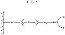
- FIG. 1shows the standard sandwich assay for detecting an analyte of interest, wherein a capture antibody (Ab 1 ) and signal antibody (Ab 2 ) are directed to a first and second epitope (e 1 and e 2 ), respectively, of an analyte of interest (An).
- the signal antibodyis labeled with an enzyme (Enz), which is used for signal generation by converting substrate (S) to product (P).
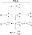
- FIG. 2is a representation of an analyte of interest (An) with three epitopes (e 1 , e 2 and e 3 ).
- analyte of interestAn
- three epitopese 1 , e 2 and e 3
- a subformis produced with one or more of the epitopes unavailable for binding by antibodies.
- Each epitope that is unavailable for bidingis identified by a circle, whereas each epitope that is available for binding is identified without a circle.
- Each subformmay be further modified so that additional epitopes are unavailable for binding by antibodies.
- FIG. 3comprises FIGS. 3A-3C , which show various views of a modified sandwich assay for detecting an analyte of interest (An) and subforms thereof using one capture antibody (Ab 1 ) and more than one signal antibody (Ab 2 , Ab 3 ).
- the assayis able to detect the presence of the analyte and all subforms thereof that have at least one epitope for the capture antibody (e 1 ) and at least one epitope for a signal antibody (e 2 or e 3 ). All epitopes for binding are shown bound by an antibody; however, the analyte or subforms thereof need to be bound by only one capture antibody and one signal antibody to be detected.
- FIG. 4comprises FIGS. 4A-4C , which show various views of a modified sandwich assay for detecting an analyte of interest (An) and subforms thereof using more than one capture antibody (Ab 1 and Ab 2 ) and one signal antibody (Ab 3 ).
- the assayis able to detect the presence of the analyte and all subforms thereof that have at least one epitope for the signal antibody (e 3 ) and at least one epitope for a capture antibody (e 1 or e 2 ). All epitopes for binding are shown bound by an antibody; however, the analyte or subforms thereof need to be bound by only one capture antibody and one signal antibody to be detected.
- FIG. 5comprises FIGS. 5A-5I , which show various views of a modified sandwich assay for detecting an analyte of interest (An) suing more than one capture antibody (Ab 1 and Ab 2 ) and more than one signal antibody (Ab 3 and Ab 4 ).
- the assayis able to detect the presence of the analyte and all subforms thereof that have at least one epitope for a capture antibody (e 1 or e 2 ) and at least one epitope for a signal antibody (e 3 or e 4 ). All eptitopes for binding are shown bound by an antibody; however, the analyte or subforms thereof need to be bound by only one capture antibody and one signal antibody to be detected.
- FIG. 6shows one antibody (Ab 1 ) or more than one antibody (Ab 1 and Ab 2 ) binding to a different epilope (e 1 or e 2 ) on the analyte of interest (An) and conjugated to a common member, wherein the common member is a surface ( FIG. 6A ), microparticle ( FIG. 6B ), and a polypeptide, such as an enzyme ( FIG. 6C ).
- the common memberis a surface ( FIG. 6A ), microparticle ( FIG. 6B ), and a polypeptide, such as an enzyme ( FIG. 6C ).
- FIG. 7shows a mixture of common members individually conjugated with a different antibody (Ab 1 or Ab 2 ), each antibody binding to a different epitope (e 1 or e 2 ) on the analyte being assayed (An), wherein the common member is a microparticle ( FIG. 7A ) or polypeptide, such as an enzyme ( FIG. 7B ).
- FIG. 8shows a common member, such as a microparticle, conjugated to two or more antibodies (Ab 1 and Ab 2 ), conjugated to another common member, such as a surface, wherein two or more capture antibodies are either (i) individually conjugated to different microparticles ( FIG. 8A ), or (ii) conjugated together on a microparticle ( FIG. 8B ).
- FIG. 9depicts the primary structure of subform of cardiac troponin I (cTnI) without amino acids 1 to 25 and 150 to 209, and shows the location for epitopes of the cTnI antibodies.
- abbreviated namesare utilized for at least some antibodies.
- the abbreviated names 3472, 34500, 3444, 3449, 34502, and 3473are equivalent to antibodies A34720, A34500, A34440, A34490, A34502, and A34730, respectively.
- FIG. 10shows the amperometric signals from different single-use assays prepared with a first capture antibody (CBI), a second capture antibody (CB2), or a combination of first and second capture antibodies (CB12) to a whole blood sample spiked with free cTnI and two levels of cITC complex.
- CBIfirst capture antibody
- CB2second capture antibody
- CB12combination of first and second capture antibodies
- FIG. 11shows a plot of the average signal obtained from a whole blood sample spiked with 6.0 ng/mL cITC complex immediately after preparation and after one-day incubation at room temperature using cartridges prepared with either CBI or CB12 coaled sensors and with a signal producing enzyme conjugated to a first antibody (EC1), a second antibody (EC2) or a mixture of EC1 and EC2 (ECI2).
- EC1first antibody
- EC2a second antibody
- ECI2a mixture of EC1 and EC2
- FIG. 12shows elution profiles for Fab-ALP conjugates with different ratios of ALP to Fab.
- alytemeans a biological or chemical substance that is capable of being bound by at least three different antibodies, and includes subforms thereof that may be bound by at least three different antibodies.
- Capture antibodyor “capture reagent” means an antibody that specifically binds to an analyte of interest or subform thereof, wherein said antibody is immobilized on a surface either (i) before, (ii) after or (iii) during binding to said analyte or subform thereof.
- Detectwhen used to describe a signal of a signal-generating element, means a signal capable of being distinguished from background.
- Epitopemeans a binding site for an antibody or other binding member on an analyte of interest or subform thereof.
- An epitopeis “unavailable for binding” if the epitope is either not present or inaccessible for binding by an antibody.
- “Subform,” when used to describe an analyte,means the product of a chemical reaction, a member of a complex with other biological or chemical substances, alternative conformations, or combinations thereof that may be bound by at least two different antibodies.
- Signal antibodyor “signal reagent” means an antibody that specifically binds to an analyte of interest or subform thereof, wherein said antibody is attached to a signal-generating element via a covalent linkage, hydrophobic interactions, hydrophilic interaction, ionic interactions, Van der Waal forces, or a combination thereof.
- the present inventionrelates to the detection of an analyte and subforms thereof.
- the analytemay be a clinical marker of a disease state from a patient believed to have suffered an acute disease event, wherein the clinical marker is also present in one or more subforms.
- the clinical markermay be a transiently elevated substance in the blood which is released in a significant quantity at, after, or before the time of the acute disease event of interest.
- the elevated concentration of the clinical marker analytedecreases as endogenous conversion factors act upon the clinical marker to produce subforms of the analyte.
- Subforms derived from the clinical markermay be transiently elevated in a serial manner, as each is first created then metabolized by endogenous conversion factors. The period of transient elevation may be for as short as a few hours to as long as several weeks.
- the analytemay be a protein including, but not limited to, TnI, TnT, CK-M, CK-B, myoglobin, hCG, TSH, FSH, pneumococcal PCA, apolipoproteins, C-reactive protein (CRP), brain natriuretic protein (BNP) and its pro-form pro-BNP, human leukocyte antigen and human apolipoproteins.
- a preferred analyteis cTnI.
- the analytehas at least three different epitopes that are available for binding by an antibody ( FIG. 2 ).
- a subform of the analytehas at least two different epitopes that are available for binding by an antibody and the epitopes of the subform are also present on the analyte ( FIG. 2 ).
- At least one epitope of the analyteis unavailable for binding on the subform.
- An epitope of the analyte unavailable for binding on the subformmay be as a result of complexation or alternative conformations, as well as the endogenous conversion factors described above.
- the analytemay also be any modified form of cTnI that has one or more epitopes that are unavailable for binding on any precursor of the analyte.
- the modified or derivative form of cTnImust have at least three epitopes available for binding by three different antibodies.
- the present inventionmay also be used to detect an analyte and subforms thereof that are present at low concentration where high sensitivity is required.
- Analyteshave traditionally been detected using a system based on the sandwich assay depicted in FIG. 1 wherein a first monoclonal or polyclonal antibody for capture is attached to a surface, and a second monoclonal or polyclonal antibody is labeled with a signal-generating element (e.g., an enzyme).
- a signal-generating elemente.g., an enzyme
- the analyte of interest and one or more subforms thereofmay be detected by performing an immunoassay using antibodies specific for more than one epitope on the analyte either (i) on the capture side of the sandwich ( FIG. 4 ), (ii) signal side of the sandwich ( FIG. 3 ), (iii) or both ( FIG. 5 ).
- the multiple sandwich assay of the present inventionmay detect the presence of the analyte and subforms thereof even if certain epitopes are unavailable for binding on the subform, so long as there is at least one epitope capable of binding by a capture antibody and at least one epitope available for binding by a signal antibody.
- the present inventionis therefore able to detect analytes and multiple subforms thereof that appear within a sample.
- Binding membersinclude, but are not limited to, extracellular or intracellular receptors, polynucleotides, peptide nucleic acids, and derivatives thereof.
- the use of multiple antibodies in the practice of the present inventionis contrasted with the use of polyclonal antibodies in traditional sandwich assays; in addition, the present invention is superior.
- the specific epitopes recognized and the ratio of various antibodies in the preparationis generally unknown.
- the epitopes recognized and the ratio of various antibodiesvary between polyclonal preparations.
- Another deficiency in the polyclonal approachis that the binding of individual antibodies within the polyclonal preparation will have diverse binding affinities.
- a significant portion of polyclonal preparationsare antibodies which have sub-standard analytical performances or which bind epitopes that compete with other antibodies in the preparation.
- the present inventionallows the preparation of controlled multi-epitopic reagents with more efficient assay characteristics because of better binding affinities and less reagent required for a given assay signal.
- the immunoassayis based on a first antibody attached to a surface (i.e., “capture antibody” or “capture reagent”) and at least a second and third antibody, wherein each antibody binds to a different epitope on the analyte.
- the antibodies not attached to a surfacemay be labeled with a signal-generating element (i.e., “signal antibody” or “signal reagent”). See FIG. 3 .
- the immunoassayis based on at least a first and second capture antibody and a third antibody.
- the antibody not attached to a surfacemay be a signal antibody. See FIG. 4 .
- the immunoassayis based on at least a first and second capture antibody and at least a third and fourth antibody.
- the antibodies not attached to a surfacemay be signal antibodies. See FIG. 5 .
- the antibodies of the present inventionmay be any antibody that specifically binds to an epitope available for binding on an analyte of interest or a subform thereof.
- the antibodies of the present inventioninclude antibodies of classes IgG, IgM, IgA, IgD, and IgE, and fragments and derivatives thereof including Fab, F(ab′) 2 and single chain antibodies.
- the antibodies of the present inventioninclude monoclonal antibodies, polyclonal antibodies, affinity purified antibodies, or mixtures thereof which exhibit sufficient binding specificity to an epitope of the analyte or subform thereof. Monoclonal antibodies and fragments and derivatives thereof are preferred in the practice of the invention.
- the antibodies of the present inventionpreferably bind to epitopes of the analyte sufficiently removed from each other such that the antibodies do not mutually interfere with binding to the analyte or subform thereof.
- Appropriate antibodies for an analyte of interestmay be chosen by means of mapping combinations of available antibodies to known epitope sites on the analyte using methods known in the art.
- the antibodies of the present inventionmay be specific for human cTnI.
- the antibodiesmay specifically recognize epitopes located on the primary sequence of cTnI as indicated in FIG. 9 .
- the antibodiesmay also be chosen from those antibodies indicated in FIG. 9 .
- Capture antibodies of the present inventionmay be immobilized by conjugating one or more antibodies to a surface. See, e.g., FIG. 6A and FIG. 6B .
- a wide variety of compoundsmay be employed as the surface, the primary consideration being the binding of the antibody to the surface, the absence of interference with the signal generating element, and the absence of interference with the examination of the label. In particular, if a fluorescence or chromogenic spectrum is being measured, the surface should not provide interference.
- Organic and inorganic polymersmay be employed as the surface.
- suitable polymersinclude polyethylene, polypropylene, polybutylene, poly(4-methylbutylene), butyl rubber and other synthetic rubbers, silicone rubbers and silastic polymers, polyesters, polyimides, cellulose and cellulose derivatives (such as cellulose acetate, nitrocellulose and the like), acrylates, methacrylates, vinyl polymers (such as polyvinyl acetate, polyvinyl chloride, polyvinylidene chloride, polyvinyl fluoride, and the like), polystyrene and styrene graft copolymers, styrene-acrylonitrile copolymers, rayon, nylon, polyvinylbutyrate, polyformaldehyde, etc.
- silica gelsilicon wafers, glass, paper, insoluble protein, metals, metalloids, metal oxides, magnetic materials, semi-conductive materials, cements or the like.
- substances that form gelsi.e., proteins such as gelatins, lipopolysaccharides, silicates, agarose, polyacrylamides or polymers which form several aqueous phases such as dextrans, polyalkylene glycols (alkylene with 2 to 3 carbon atoms) or surfactants, e.g. amphophilic compounds such as phospholipids, long chain (12-24 carbon atoms) alkyl ammonium salts and the like.
- the surfacemay comprise polystyrene, styrene copolymers including styrene-(vinyl monomer) copolymers such as styrene-acrylonitrile copolymers, polyolefins such as polyethylene and polypropylene, and acrylate and methacrylate polymers and copolymers and mixtures thereof.
- the surfacemay also comprise magnetizable materials in particulate form. Traditional interference by such magnetizable materials may be minimized by adding magnetizable particles to each of the reaction steps. Magnetic interference produced at each step may be made nearly equal, and thus is effectively cancelled. Magnetizable particles may be easily separated from the serum or other solution by application of a magnetic field to concentrate the particles.
- the capture antibodymay be bound to the surface by any method of bonding which does not significantly reduce the antibody binding sites and which binds sufficiently to permit separation of the surface from the liquids and rinse solutions without significant detachment of antibody from the surface.
- Non-covalent bondingmay be achieved by adsorption, ionic bonding, van der Waals adsorption, electrostatic bonding, and other non-covalent bonding.
- the antibodymay also be bound to the surface by covalent bonding.
- the antibodymay be coupled to a hydroxylated material by means of cyanogen bromide as described in U.S. Pat. No. 3,720,760.
- Staphylococcus Protein Amay be bound to the surface, and the F c chain of the antibody can be conjugated with the Protein A.
- Capture antibodiesmay be attached to a surface by adhesion followed by chemical crosslinking.
- Crosslinking agentswhich may be used include, but are not limited to, 1-ethyl-3-(3-dimethylaminopropyl)-carbodiimide (EDC), glutaraldehyde, adipic acid dihydrazide, bis-diazotized benzidine, 1,4-butane diglycidyl ether, bis-maleimido hexane, sulfosuccinimidyl 4-(N-maleimidomethyl)-cyclohexane-1-carboxylate, and N-hydroxysuccinimidyl 4-azidosalicylic acid.
- EDCmay be used for any surface that has free carboxyl groups. These and many other similar reagents are well known in the art.
- the same or different antibodiesmay be conjugated to a common member, using any of the conjugation methods described above.
- Common membersinclude, but are not limited to, a particle, microparticle ( FIG. 6B ), polypeptide ( FIG. 6C ), chemical linker, or a surface ( FIG. 6A ) as described above.
- a common membermay be conjugated with more than one antibody, each antibody binding to a different epitope on the analyte being assayed. See FIG. 6 .
- the multiple antibodiesmay be conjugated to the common member at controlled molar ratios, for example using methods where the stoichiometry of the conjugation reaction is controlled.
- multiple common membersmay be individually conjugated with a different antibody, each antibody binding to a different epitope on the analyte being assayed. See FIG. 7 .
- Molar ratios of the different antibodiesmay be controlled, using methods such as the mixing of multiple common members with conjugated antibodies.
- Antibodiesmay be attached to microparticles using any of the conjugation methods described above. Various factors may be considered when choosing the coupling method including, but not limited to type and size of particle, coupling reagents, concentration of reactants, single or multi-step reaction, reaction buffer and pH, storage buffer, and blocking agents.
- the coupling methodincluding, but not limited to type and size of particle, coupling reagents, concentration of reactants, single or multi-step reaction, reaction buffer and pH, storage buffer, and blocking agents.
- Signal reagentsmay be prepared by conjugating an antibody to a signal-generating element by any of a number of common methods.
- Conjugatesmay be prepared, for example, by utilizing free sulfhydryl groups, generating sulfhydryl groups from available disulfide bonds, or introducing additional sulfhydryls onto the antibody.
- Linking agentssuch as succinimydyi 4-(N-maleimidomethyl)cyclohexane-1-carboxylate (SMCC) may then be used to conjugate the activated antibody to the signal-generating element.
- SMCCsuccinimydyi 4-(N-maleimidomethyl)cyclohexane-1-carboxylate
- Signal-generating elements of the present inventioninclude, but are not limited to, radiolabels, metal particles, chromogens, fluorescent dyes, labeled proteins and enzymes.
- the enzymemay catalyze a reaction wherein the depletion of substrate or the production of product can be detected.
- Exemplary enzymesinclude, but are not limited to, amylases, polynucleotidase, arginase, adenase, aminopolypeptidase, pepsin, lipases, catalase, tyrosinases, alcohol dehydrogenase, succinic dehydrogenase, diaphorase, glyoxalase, aldolase, glucose oxidase, horseradish peroxidase, beta-galactosidase, phosphatases, phosphorylases and hexokinases.
- Exemplary enzymesalso include alkaline phosphatase, glucose oxidase, horseradish peroxidase, ⁇ -galactosidase and phenol oxidase.
- the enzymatic conversion of a substrate to a productmay be measured by for example optical and electrochemical means.
- the sensitivity of a sandwich assayis limited largely by the level of non-specific-adsorption of the signal reagent to the capture reagent and surfaces within the detection region of the analysis device.
- Such surfacesmay include the walls of a cuvette, a wicking element, an electrode and the like.
- the propensity of the signal reagent to non-specifically adsorb to surfaces within the analysis devicemay be a function of the inherent properties of the antibody and signal-generating element used to form the signal antibody, as well as the crosslinker.
- the materials used in the analysis devicee.g., types of plastics
- the composition of the wash and substrate-containing solutionsare generally optimized to minimize such non-specific protein adsorption.
- various surfactantse.g. TWEEN® 20, BRIJ® 35, TRITON® X100
- Non-enzymatic proteinssuch as serum albumin, denatured proteins such as gelatins, and deactivated enzymes may also be added as surface blocking agents in order to lower the level of non-specific adsorption of the signal reagent.
- the signal reagentmay comprise fragments of antibodies, such as Fab fragments, which may contribute to lowering the level of background signal. By lowering the molecular weight of the signal reagent, the time required for the binding step may be reduced due to diffusional considerations.
- the signal reagentmay also comprise more than one antibody, which may also lower background signal levels by minimizing the overall size of the signal reagent while maximizing its ability to generate a signal in response to the presence of the analyte of interest.
- the more than one antibody of the signal reagentmay bind to two or more different epitopes of the analyte, which may lead to stabilization of the signal antibody-analyte complex when more than one epitope of the analyte is bound by more than one antibody of the signal reagent.
- the signal reagentcomprises more than one antibody
- said antibodiesmay bind to one or more different epitopes.
- a signal-generating elementmay be conjugated with more than one antibody, each antibody binding to a different epitope on the analyte of interest. See FIG. 6C .
- the molar ratio of antibodies conjugated to the signal-generating elementmay be controlled by selecting the reaction conditions and stoichiometry for the conjugation reaction.
- a first antibodymay be conjugated to a first signal-generating element and a second antibody may be conjugated to a second signal-generating element, each at a ratio greater than 1:1, and then mixed, wherein the first and second antibodies each bind to a different epitope on the analyte of interest. See FIG. 7B .
- the molar ratio of the first and second antibodiesmay also be controlled.
- the signal-generating elementmay be conjugated to a single antibody, or two or more antibodies to different epitopes on the same analyte, wherein the signal antibody comprises a ratio of signal-generating element to a population of antibodies from about 1:100 to about 1:1.001.
- the signal-generating elementmay also be conjugated to a population of antibodies at a ratio from about 1:90 to about 1:1.001.
- the signal-generating elementmay also be conjugated to a population of antibodies at a ratio from about 1:80 to about 1:1.001.
- the signal-generating elementmay also be conjugated to a population of antibodies at a ratio from about 1:70 to about 1:1.001.
- the signal-generating elementmay also be conjugated to a population of antibodies at a ratio from about 1:60 to about 1:1.001.
- the signal-generating elementmay also be conjugated to a population of antibodies at a ratio from about 1:50 to about 1:1.001.
- the signal-generating elementmay also be conjugated to a population of antibodies at a ratio from about 1:40 to about 1:1.001.
- the signal-generating elementmay also be conjugated to a population of antibodies at a ratio from about 1:30 to about 1:1.001.
- the signal-generating elementmay also be conjugated to a population of antibodies at a ratio from about 1:20 to about 1:1.001.
- the signal-generating elementmay also be conjugated to a population of antibodies at a ratio from about 1:10 to about 1:1.001, about 1:20 to about 1:10, about 1:30 to about 1:20, about 1:40 to about 1:30, about 1:50 to about 1:40, about 1:60 to about 1:50, about 1:70 to about 1:60, about 1:80 to about 1:70, about 1:90 to about 1:80, and about 1:100 to about 1:90.
- the signal-generating elementmay also be conjugated to a population of antibodies at a ratio from about 1:5 to about 1:1.001, about 1:10 to about 1:5, about 1:15 to about 1:10, about 1:20 to about 1:15, about 1:25 to about 1:20, about 1:30 to about 1:25, about 1:35 to about 1:30, about 1:40 to about 1:35, about 1:45 to about 1:40, and about 1:50 to about 1:45.
- the signal-generating elementmay also be conjugated to a population of antibodies at a ratio of about 1:3.3.
- the signal-generating elementmay be conjugated to a first and second antibody, each capable of binding to different epitopes on an analyte or subforms thereof, wherein the ratio of signal-generating element to total antibody is as disclosed above, and wherein the ratio of the first antibody to the second antibody is from about 1:50 to about 50:1.
- the ration of the first antibody to the second antibodymay also be from about 1:10 to about 10:1, from about 1:5 to about 5:1, from about 1:3 to about 3:1, and from about 1:2 to about 2:1.
- a signalmay be produced by the signal-generating element, which may be measurable by a sensing element using methods which include, but are not limited to, radiation, optically, electrochemically, or by some other transduction means known in the art.
- a sensing elementmay be an electrode, biosensor, field-effect transistor, surface acoustic wave device, optical wave-guide, fiber optic, cuvette, radioactivity detector, reagents that facilitate, physical, nuclear, chemical, biochemical, electrical, or optical detection, or combinations thereof.
- the immunoassay of an analyte of interestmay be conducted in an immunoassay device comprising one or more sensing elements and a surface on which a modified sandwich immunoassay is conducted as described above.
- the device surfacemay comprise two or more antibodies that are each capable of binding to different epitopes on the analyte. At least one of the two antibodies is also capable of binding to the same epitope on a subform of the analyte. At least one of the epitopes on the analyte is unavailable for binding on the subform.
- Devices of the present inventioninclude a cartridge, columns, syringe, cuvette, or other analytical device or system known in the art.
- the cartridgemay be of a type as described in U.S. Pat. No. 5,096,669. Examples of other cartridge configurations are found in U.S. Pat. Nos. 5,416,026, 5,593,638, 5,447,440, 5,628,961, 5,514,253, 5,609,824, 5,605,664, 5,614,416, 5,789,253, 6,030,827, and 6,379,883. Still other cartridge configurations are described in PCT/US00/31158, PCT/US01/04345 and U.S. Pat. No. 7,419,821. The disclosures of the foregoing are hereby incorporated by reference.
- the surface of the immunoassay devicemaybe of any appropriate surface known to those of skill in the art including, but not limited to, glass, semiconductor, plastic, silicon dioxide, photoformable PVA, photoformable gelatin, film forming latex, and conductive metal.
- a conductive metal surfacemay be silver, iridium, gold, platinum, and alloys thereof.
- Antibodiesmay be absorbed, adsorbed or covalently attached to the surface of the immunoassay device. Antibodies may be absorbed into PVA, gelatin and latex layers or absorbed to their surfaces. Antibodies may be adsorbed to glass, metal, semiconductor, plastic, silicon dioxide, photoformable PVA, and conductive metal. The two or more antibodies may also be attached to a microparticle as discussed above, wherein the microparticle is attached to the surface as discussed above.
- the immunoassay devicemay also contain a third antibody which binds to a different epitope on the analyte.
- the sensing element of the immunoassay devicemay be of any type known in the art including, but not limited to, an electrode, biosensor, field-effect transistor, surface acoustic wave device, optical wave-guide, fiber optic, cuvette, radioactivity detector, immunechromatographic device, reagents that facilitate, physical, nuclear, chemical, biochemical, electrical, optical detection, or combinations thereof.
- An immunoassay kitmay be used to detect an analyte of interest and subforms thereof in a modified sandwich immunoassay as described above.
- the kitmay comprise three or more antibodies that are each capable of binding to different epitopes on the analyte. At least two of the three antibodies are also capable of binding to the same epitopes on a subform of the analyte. At least one of the epitopes on the analyte is unavailable for binding on the subform.
- a sandwich immunoassay productmay comprise an analyte of interest and a subform thereof. At least three epitopes on the analyte are available for binding by at least three different antibodies.
- the analytemay be bound by at least three different antibodies with each antibody binding to a different epitope on the analyte. At least two of the three epitopes on the analyte are available for binding on the subform by at least two of the at least three different antibodies.
- the subformmay be bound by at least two of the three different antibodies that bind to the analyte with each antibody binding to a different epitope on the subform. At least one epitope of the analyte is unavailable for binding on the subform. At least one of the antibodies bound to the analyte and the subform of the analyte may be a signal antibody.
- a samplemay be immunoassayed for the presence of an analyte and subforms thereof by performing a modified sandwich immunoassay as described above.
- At least three different antibodiesare used, wherein each antibody is capable of binding to a different epitope on the analyte.
- At least two of the antibodies capable of binding to the analyteare capture antibodies or signal antibodies, or a combination thereof.
- At least one of the epitopes on the analyteis unavailable for binding on the subform.
- the sample to be immunoassayedis added to a surface comprising one or more capture antibodies.
- the one or more signal antibodiesare then added.
- one or more signal antibodiesmay be added to the sample before or during application of the sample to the surface comprising one or more capture antibodies. After removing unbound signal antibodies, the extent of binding of the one or more signal antibodies is determined as described above.
- a signalwill be produced by analyte as well as all subforms of the analyte that have at least one epitope that is present or capable of being bound by a capture antibody and at least a second epitope that is present or capable of being bound by a signal antibody.
- the analyte of interest and subforms thereofmay be immunoassayed to determine whether a patient has suffered an acute medical event.
- a samplemay be obtained from the patient and immunoassayed for the presence of the analyte of interest and subforms thereof by using the method of assaying described above.
- Signal produced by signal antibodiesmay be compared to control values.
- a signal, or a sequence of signals from a series of assays over a period of hours or days, detectable above controlmay indicate that the patient has suffered an acute medical event.
- the acute medical eventmay be any disease state that is associated with a clinical marker, wherein the clinical marker is an analyte in the modified immunoassay described herein.
- the medical eventmay be for example a myocardial infarction.
- the analyte of interestmay be for example TnI, TnT, TnC, CK-M, CK-B, CK-MB, myoglobin, TSH, FSH, CRP, BNP, pro-BNP, PSA, PCA, apolipoprotein, and combinations thereof.
- the analyte of interest and subforms thereofmay be immunoassayed to determine when a patient suffered an acute medical event.
- a sample, or set of samples obtained at different times,may be obtained from the patient and immunoassayed for the presence of the analyte of interest and subforms thereof by using the method of assaying described above.
- the amount of signal produced by signal antibodiesmay be correlated with a standard curve of amount of analyte vs. time to determine when the patient suffered the acute medical event.
- One of ordinary skillmay produce the standard curve by using the methods described herein.
- the analyte of interest and subforms thereofmay be immunoassayed to determine the severity of an acute medical event suffered by a patient.
- a sample, or set of samples obtained at different times,may be obtained from the patient and immunoassayed for the presence of the analyte of interest and subforms thereof by using the method of assaying described above.
- the amount of signal produced by signal antibodiesmay correlate to the severity of a medical event suffered by the patient.
- Bead particles conjugated with antibodies specific for cTnIwere prepared by first exchanging polystyrene/acrylic acid latex 0.2 ⁇ m diameter spheres (Seradyn) into 0.05 M 2-(N-morpholino)ethanesulfonic acid (MES, Sigma Aldrich) buffer at pH 6.2. To a 2% w/w solution of the latex spheres was added cTnI antibodies (buffer exchanged into the same 0.05 M MES buffer) at a controlled mass ratio relative to the mass of the latex spheres. The solution was stirred for 15 minutes at 4° C. and the antibody coated beads separated via controlled centrifugation.
- MES2-(N-morpholino)ethanesulfonic acid
- alkaline phosphatase (ALP) labeled Fab conjugateswas used as signal reagents.
- Alkaline phosphataseBiozyme
- PBSphosphate buffered saline
- a 1 mg/ml to 15 mg/ml solution of the ALPwas reacted with 25 weight percent (relative to the ALP weight in the solution) of the heterobifunctional crosslinker succinimidyl-4-[N-maleimidomethyl]-cyclohexane-1-carboxy[6-amidocaproate]
- LCSMCCphosphate buffered saline
- Piercethe heterobifunctional crosslinker succinimidyl-4-[N-maleimidomethyl]-cyclohexane-1-carboxy[6-amidocaproate]
- the crosslinkerwas added as a solution in dimethyl sulphoxide (DMSO, Sigma). This solution was allowed to react at room temperature for 45 minutes, after which it was centrifuged to remove
- Fab fragments of the antibodies to be incorporated into the signal reagentwere formed via pepsin (Sigma) digestion of the whole antibodies and separated using a S200 sephacryl size exclusion column (Amersham Pharmacia). The resulting F(ab) 2 fragments were chemically reduced in 60 mM mercaptoethylamine (MEA, Sigma Aldrich) for 45 minutes at 37° C. to produce two Fab fragments, which were separated from excess MEA by desalting using a sephadex G50 column. The Fab fragments were then added to activated ALP and allowed to react at 4° C. for 2 to 12 hours.
- MUAmercaptoethylamine
- FIG. 12shows the elution profile for ALP-Fab signal reagents with ALP:Fab ratios of 1:1, 1:3, 1:6 and 1:10.
- ALP-labeled signal reagentswere made comprising either EC1 (G129C, Biospacific Inc.) or EC2 (G130C, Biospacific Inc.).
- EC12 signal reagentscomprising both EC1 and EC2 were prepared using the above method by mixing the two fab fragments at a molar ratio of 1:1 before adding the mixture to activated ALP.
- the enzyme labeled signal reagent of Example 2is printed onto the same ELISA sensor as the capture antibodies.
- the signal reagentis formulated in a 1% to 50% sugar solution containing PBS and a preservative. This composition was found to provide rapid dissolution of the signal reagent into a blood sample.
- the assembled cartridgeis pressed to yield the disposable cartridge, which may be used in a portable electrochemical analyzer.
- FIG. 10shows high signal levels for cartridges comprising the CB1 capture reagent for both free cTnI (diamond points) and the two levels of ITC complex (square and triangle points).
- cartridges comprising the CB2 capture reagentyield lower signals, especially for the ITC complex.
- Cartridges using the CB12 hybrid capture reagentshow improved signal generation for both forms of cTnI relative to either CB1 or CB2 capture reagents.
- the improved performance of the CB12 capture reagentis surprising, because the hybrid reagent comprises the CB2 antibody that by itself has markedly lower analytical performance than the CB1 antibody.
- cTnIwas assayed in a whole blood sample spiked with a 6.0 ng/mL cITC complex immediately after preparation and after a one-day incubation at room temperature using cartridges with sensors coated with latex microsphere comprising CB1 and CB12 capture reagents, as described in Example 3, and ECI, EC2 or EC12 signal reagents as described in Example 2.
- FIG. 11demonstrates that the EC1 signal reagent shows a higher signal generation than the EC2 signal reagent. After a one day incubation of the sample to simulate aging of cTnI (e.g., proteolytic degradation), the signal level for the EC1 signal reagent drops significantly. In contrast, the EC2 signal reagent shows only a modest drop in the signal level.
- the signal generation of the hybrid signal reagent EC12is surprisingly superior to each of the two single antibody signal reagents, especially after incubating the sample for one day.
- Whole blood samplesare obtained from patients suspected of suffering a myocardial infarction, as well as from control individuals.
- cTnIis assayed in the whole blood samples using cartridges with sensors coated with latex microsphere comprising CB12 capture reagents, as described in Example 3, and EC12 signal reagents as described in Example 2.
- the signal generated in samples derived from patients suspected of suffering an MIis compared to the level of signal from controls. Levels of signal greater than control indicate that the patient has suffered a MI.
Landscapes
- Health & Medical Sciences (AREA)
- Life Sciences & Earth Sciences (AREA)
- Immunology (AREA)
- Engineering & Computer Science (AREA)
- Chemical & Material Sciences (AREA)
- Biomedical Technology (AREA)
- Hematology (AREA)
- Molecular Biology (AREA)
- Urology & Nephrology (AREA)
- General Health & Medical Sciences (AREA)
- Analytical Chemistry (AREA)
- Food Science & Technology (AREA)
- Pathology (AREA)
- Microbiology (AREA)
- Medicinal Chemistry (AREA)
- Physics & Mathematics (AREA)
- Cell Biology (AREA)
- Biochemistry (AREA)
- Biotechnology (AREA)
- General Physics & Mathematics (AREA)
- Proteomics, Peptides & Aminoacids (AREA)
- Clinical Laboratory Science (AREA)
- Chemical Kinetics & Catalysis (AREA)
- Dispersion Chemistry (AREA)
- Peptides Or Proteins (AREA)
Abstract
Description
Claims (4)
Priority Applications (1)
| Application Number | Priority Date | Filing Date | Title |
|---|---|---|---|
| US15/975,145US10641767B2 (en) | 2002-07-29 | 2018-05-09 | Multiple hybrid immunoassay |
Applications Claiming Priority (4)
| Application Number | Priority Date | Filing Date | Title |
|---|---|---|---|
| US10/208,560US20040018577A1 (en) | 2002-07-29 | 2002-07-29 | Multiple hybrid immunoassay |
| US14/081,309US9267939B2 (en) | 2002-07-29 | 2013-11-15 | Multiple hybrid immunoassay |
| US14/994,713US9995744B2 (en) | 2002-07-29 | 2016-01-13 | Multiple hybrid immunoassay |
| US15/975,145US10641767B2 (en) | 2002-07-29 | 2018-05-09 | Multiple hybrid immunoassay |
Related Parent Applications (1)
| Application Number | Title | Priority Date | Filing Date |
|---|---|---|---|
| US14/994,713DivisionUS9995744B2 (en) | 2002-07-29 | 2016-01-13 | Multiple hybrid immunoassay |
Publications (2)
| Publication Number | Publication Date |
|---|---|
| US20180259511A1 US20180259511A1 (en) | 2018-09-13 |
| US10641767B2true US10641767B2 (en) | 2020-05-05 |
Family
ID=30770571
Family Applications (4)
| Application Number | Title | Priority Date | Filing Date |
|---|---|---|---|
| US10/208,560AbandonedUS20040018577A1 (en) | 2002-07-29 | 2002-07-29 | Multiple hybrid immunoassay |
| US14/081,309Expired - Fee RelatedUS9267939B2 (en) | 2002-07-29 | 2013-11-15 | Multiple hybrid immunoassay |
| US14/994,713Expired - LifetimeUS9995744B2 (en) | 2002-07-29 | 2016-01-13 | Multiple hybrid immunoassay |
| US15/975,145Expired - Fee RelatedUS10641767B2 (en) | 2002-07-29 | 2018-05-09 | Multiple hybrid immunoassay |
Family Applications Before (3)
| Application Number | Title | Priority Date | Filing Date |
|---|---|---|---|
| US10/208,560AbandonedUS20040018577A1 (en) | 2002-07-29 | 2002-07-29 | Multiple hybrid immunoassay |
| US14/081,309Expired - Fee RelatedUS9267939B2 (en) | 2002-07-29 | 2013-11-15 | Multiple hybrid immunoassay |
| US14/994,713Expired - LifetimeUS9995744B2 (en) | 2002-07-29 | 2016-01-13 | Multiple hybrid immunoassay |
Country Status (4)
| Country | Link |
|---|---|
| US (4) | US20040018577A1 (en) |
| EP (1) | EP1525480B1 (en) |
| JP (1) | JP4507879B2 (en) |
| WO (1) | WO2004011947A1 (en) |
Families Citing this family (224)
| Publication number | Priority date | Publication date | Assignee | Title |
|---|---|---|---|---|
| US7981420B2 (en) | 2000-12-22 | 2011-07-19 | Max-Planck-Gesellschaft Zur Foederung Der Wissenschaften E.V. | Therapeutic use of antibodies directed against repulsive guidance molecule (RGM) |
| US6896778B2 (en)* | 2001-06-04 | 2005-05-24 | Epocal Inc. | Electrode module |
| US7214300B2 (en)* | 2001-06-04 | 2007-05-08 | Epocal Inc. | Integrated electrokinetic devices and methods of manufacture |
| US20040018577A1 (en) | 2002-07-29 | 2004-01-29 | Emerson Campbell John Lewis | Multiple hybrid immunoassay |
| US7767068B2 (en) | 2002-12-02 | 2010-08-03 | Epocal Inc. | Heterogeneous membrane electrodes |
| US7842234B2 (en)* | 2002-12-02 | 2010-11-30 | Epocal Inc. | Diagnostic devices incorporating fluidics and methods of manufacture |
| US20040197841A1 (en)* | 2003-04-02 | 2004-10-07 | Apffel James Alexander | Methods and reagents for multiplexed analyses |
| FI20030652A0 (en)* | 2003-04-30 | 2003-04-30 | Susann Eriksson | Improved immune determination |
| US7722817B2 (en)* | 2003-08-28 | 2010-05-25 | Epocal Inc. | Lateral flow diagnostic devices with instrument controlled fluidics |
| JP6080163B2 (en) | 2013-10-02 | 2017-02-15 | 古河電気工業株式会社 | Target substance detection method |
| GB0329288D0 (en)* | 2003-12-18 | 2004-01-21 | Inverness Medical Switzerland | Monitoring method and apparatus |
| US8563328B2 (en)* | 2004-07-26 | 2013-10-22 | University Of Louisville Research Foundation, Inc. | Fiber-optic biosensor and biosensing methods |
| AT500800B1 (en)* | 2004-09-08 | 2008-07-15 | Biomedica Medizinprodukte Gmbh | PROCESS FOR DETERMINING PROBNP |
| US20060204999A1 (en)* | 2005-03-14 | 2006-09-14 | Stephen Macevicz | Detecting molecular complexes |
| US20090054741A1 (en)* | 2005-03-29 | 2009-02-26 | Inverness Medical Switzerland Gmbh | Device and method of monitoring a patient |
| US8624027B2 (en) | 2005-05-12 | 2014-01-07 | Abbvie Inc. | Combination therapy for treating cancer and diagnostic assays for use therein |
| ES2542501T3 (en)* | 2005-09-30 | 2015-08-06 | Abbvie Deutschland Gmbh & Co Kg | Protein binding domains of the protein family of repulsive targeting molecules (RGM) and functional fragments thereof, as well as their use |
| US7874007B2 (en)* | 2006-04-28 | 2011-01-18 | Microsoft Corporation | Providing guest users access to network resources through an enterprise network |
| WO2008105814A2 (en)* | 2006-08-22 | 2008-09-04 | Los Alamos National Security, Llc | Miniturized lateral flow device for rapid and sensitive detection of proteins or nucleic acids |
| US8865398B2 (en) | 2006-09-01 | 2014-10-21 | Abbott Laboratories | Combination hepatitis C virus antigen and antibody detection method |
| US20080220448A1 (en)* | 2006-09-01 | 2008-09-11 | Blincko Stuart J | Antigenic protein conjugates and process for preparing same |
| US20080305512A1 (en)* | 2006-10-26 | 2008-12-11 | Mattingly Phillip G | Assay for cardiac troponin autoantibodies |
| JP5057402B2 (en)* | 2006-10-26 | 2012-10-24 | アボット・ラボラトリーズ | Immunoassay of analytes in samples containing endogenous anti-analyte antibodies |
| US7776605B2 (en) | 2006-10-26 | 2010-08-17 | Abbott Laboratories | Assay for cardiac troponin autoantibodies |
| WO2008140581A2 (en)* | 2006-11-22 | 2008-11-20 | 3M Innovative Properties Company | Systems and methods for preparing and analyzing samples |
| WO2008082984A2 (en) | 2006-12-29 | 2008-07-10 | Abbott Laboratories | Non-denaturing lysis reagent for use with capture-in-solution immunoassay |
| US7914999B2 (en) | 2006-12-29 | 2011-03-29 | Abbott Laboratories | Non-denaturing lysis reagent |
| AT504942B1 (en)* | 2007-03-07 | 2009-08-15 | Biomedica Medizinprodukte Gmbh | PROCESS FOR DETERMINING PROBNP |
| WO2008137991A1 (en) | 2007-05-08 | 2008-11-13 | Abbott Laboratories | Human b-type natriuretic peptide assay having reduced cross-reactivity with other peptide forms |
| ATE535813T1 (en)* | 2007-05-24 | 2011-12-15 | Abbott Lab | IMMUNE TESTING PROCEDURES SHOWING REDUCED CROSS-REACTIVITY WITH HYDROPHOBIC DRUG ANALYTE METABOLISM PRODUCTS |
| US20090053736A1 (en)* | 2007-08-21 | 2009-02-26 | Mattingly Phillip G | Homogeneous Chemiluminescent Immunoassay for Analysis of Iron Metalloproteins |
| US7608361B2 (en)* | 2007-08-22 | 2009-10-27 | Stauffer John E | Alkali metal battery |
| EP2033971A1 (en)* | 2007-09-06 | 2009-03-11 | Abbott GmbH & Co. KG | Bone Morphogenetic Protein (BMP) binding domains of proteins of the Repulsive Guidance Molecule (RGM) protein family and functional fragments thereof and their application |
| US20100136520A1 (en)* | 2007-09-13 | 2010-06-03 | Abbott Laboratories | Detecting hepatitis b virus |
| US8138318B2 (en) | 2007-09-13 | 2012-03-20 | Abbott Laboratories | Hepatitis B pre-S2 nucleic acid |
| US20090098531A1 (en)* | 2007-09-13 | 2009-04-16 | Abbott Laboratories | Detecting hepatitis b virus |
| US20100267055A1 (en)* | 2007-10-21 | 2010-10-21 | Abbott Laboratories | One-step immunoassays exhibiting increased sensitivity and specificity |
| US8283124B2 (en)* | 2007-10-25 | 2012-10-09 | Abbott Laboratories | Compositions for detecting antibodies to Babesia microti and methods of uses thereof |
| EP2232264B1 (en) | 2007-12-19 | 2015-12-02 | Abbott Laboratories | Immunosuppressant drug extraction reagent for immunoassays |
| US20090203037A1 (en) | 2007-12-27 | 2009-08-13 | Abbott Laboratories | Anti-T. Cruzi Antibodies and Methods of Use |
| US8962803B2 (en) | 2008-02-29 | 2015-02-24 | AbbVie Deutschland GmbH & Co. KG | Antibodies against the RGM A protein and uses thereof |
| US8030471B2 (en)* | 2008-03-06 | 2011-10-04 | Abbott Laboratories | Plasmodium malariae and Plasmodium ovale genes and uses thereof |
| US8268981B2 (en) | 2008-03-06 | 2012-09-18 | Abbott Laboratories | Plasmodium malariae and plasmodium ovale genes and uses thereof |
| US11730407B2 (en) | 2008-03-28 | 2023-08-22 | Dexcom, Inc. | Polymer membranes for continuous analyte sensors |
| US8682408B2 (en) | 2008-03-28 | 2014-03-25 | Dexcom, Inc. | Polymer membranes for continuous analyte sensors |
| US8583204B2 (en) | 2008-03-28 | 2013-11-12 | Dexcom, Inc. | Polymer membranes for continuous analyte sensors |
| CA2722466A1 (en) | 2008-04-29 | 2009-11-05 | Tariq Ghayur | Dual variable domain immunoglobulins and uses thereof |
| EP2279403B1 (en) | 2008-05-05 | 2016-03-16 | Los Alamos National Security, LLC | Highly simplified lateral flow-based nucleic acid sample preparation and passive fluid flow control |
| US20090286256A1 (en)* | 2008-05-19 | 2009-11-19 | Abbott Laboratories | Isolated human autoantibodies to natriuretic peptides and methods and kits for detecting human autoantibodies to natriuretic peptides |
| US20090286329A1 (en)* | 2008-05-19 | 2009-11-19 | Abbott Laboratoires | Isolated human autoantibodies to natriuretic peptides and methods and kits for detecting human autoantibodies to natriuretic peptides |
| NZ589436A (en) | 2008-06-03 | 2012-12-21 | Abbott Lab | Dual variable domain immunoglobulins and uses thereof |
| RU2010153580A (en) | 2008-06-03 | 2012-07-20 | Эбботт Лэборетриз (Us) | IMMUNOGLOBULINS WITH TWO VARIABLE DOMAINS AND THEIR APPLICATION |
| US8741287B2 (en) | 2008-06-18 | 2014-06-03 | Abbott Laboratories | PlGF-1 assay and kits and components thereof |
| US20100015655A1 (en)* | 2008-07-17 | 2010-01-21 | Abbott Laboratories | Methods and Kits for Detecting and Quantifying Damage Caused by a Parasite |
| JP2010122205A (en)* | 2008-08-29 | 2010-06-03 | Sysmex Corp | Method for detecting measles virus, membrane assay test device, and membrane assay test kit |
| WO2010028014A1 (en) | 2008-09-03 | 2010-03-11 | Abbott Laboratories | Assays and kits for determining hiv-1 tropism |
| US20100105150A1 (en)* | 2008-10-24 | 2010-04-29 | Abbott Laboratories | Isolated human autoantibodies to neutrophil gelatinase-associated lipocalin (ngal) and methods and kits for the detection of human autoantibodies to ngal |
| US9720003B2 (en)* | 2008-12-22 | 2017-08-01 | Koninklijke Philips Electronics N.V. | Assay for Troponin I using magnetic labels |
| US8168165B2 (en) | 2008-12-23 | 2012-05-01 | Abbott Laboratories | Alkylated interleukin-18 compositions |
| US8349325B2 (en) | 2008-12-23 | 2013-01-08 | Abbott Laboratories | Soluble FMS-like tyrosine kinase-1 (sFLT-1) antibody and related composition, kit, methods of using, and materials and method for making |
| AU2009334505B2 (en)* | 2008-12-31 | 2013-05-09 | Abbott Point Of Care Inc. | Method and device for immunoassay using nucleotide conjugates |
| US8241915B2 (en)* | 2009-01-14 | 2012-08-14 | Abbott Laboratories | Methods and kits for detecting hemoglobin in test samples |
| CN102365372B (en) | 2009-01-31 | 2016-05-25 | Abbvie公司 | Prediction and monitoring are for the mark of replying of Aurora A B inhibitor therapy |
| CN102388151A (en)* | 2009-02-11 | 2012-03-21 | 雅培制药有限公司 | Methods and compositions for identifying, classifying and monitoring subject having bcl-2 family inhibitor-resistant tumors and cancers |
| US8030026B2 (en) | 2009-02-24 | 2011-10-04 | Abbott Laboratories | Antibodies to troponin I and methods of use thereof |
| MX2011009362A (en) | 2009-03-05 | 2011-09-26 | Abbott Lab | Il-17 binding proteins. |
| US8283162B2 (en) | 2009-03-10 | 2012-10-09 | Abbott Laboratories | Antibodies relating to PIVKAII and uses thereof |
| US8084272B2 (en) | 2009-03-25 | 2011-12-27 | Abbott Point Of Care Inc. | Amelioration of heterophile antibody immunosensor interference |
| US8063193B2 (en) | 2009-03-27 | 2011-11-22 | Abbott Laboratories | Nucleotide and amino acid sequences encoding an exported protein 1 derived from Plasmodium vivax and uses thereof |
| US20110076697A1 (en)* | 2009-04-28 | 2011-03-31 | Innovative Laboratory Technologies, Inc. | Lateral-flow immuno-chromatographic assay devices |
| US20100291709A1 (en)* | 2009-05-13 | 2010-11-18 | Abbott Laboratories | Human nt-pro b-type natriuretic peptide assay having reduced cross-reactivity with other peptide forms |
| CA2766794A1 (en) | 2009-06-30 | 2011-01-06 | Abbott Laboratories | Markers of xmrv infection and uses thereof |
| CN102741288B (en) | 2009-08-29 | 2015-08-19 | Abbvie公司 | Therapeutic DLL4-binding protein |
| US8501420B2 (en)* | 2009-08-31 | 2013-08-06 | Abbott Laboratories | Biomarkers for prediction of major adverse cardiac events and uses thereof |
| US11493507B2 (en) | 2009-09-14 | 2022-11-08 | Siemens Healthineers Nederland B.V. | Highly sensitive immunoassay with large particle labels |
| US8377669B2 (en) | 2009-11-17 | 2013-02-19 | Abbott Point Of Care Inc. | Reducing leukocyte interference in non-competitive immunoassays |
| US8389293B2 (en) | 2009-11-17 | 2013-03-05 | Abbott Point Of Care Inc. | Reducing leukocyte interference in competitive immunoassays |
| US8455212B2 (en) | 2009-11-21 | 2013-06-04 | Abbott Laboratories | Assays for human NT-pro B-type natriuretic peptide, human pro B-type natriuretic peptide and human B-type natriuretic peptide |
| EP2504466B1 (en) | 2009-11-23 | 2019-11-20 | Proxim Diagnostics Corporation | Controlled electrochemical activation of carbon-based electrodes |
| US8835120B2 (en)* | 2009-12-02 | 2014-09-16 | Abbott Laboratories | Assay for cardiac troponin-T (cTnT) |
| US8652788B2 (en)* | 2009-12-02 | 2014-02-18 | Abbott Laboratories | Assay for diagnosis of cardiac myocyte damage |
| US20110136141A1 (en) | 2009-12-03 | 2011-06-09 | Abbott Laboratories | Peptide reagents and method for inhibiting autoantibody antigen binding |
| US8183002B2 (en) | 2009-12-03 | 2012-05-22 | Abbott Laboratories | Autoantibody enhanced immunoassays and kits |
| DK2510001T3 (en)* | 2009-12-08 | 2016-02-29 | Abbvie Deutschland | MONOCLONAL ANTIBODIES AGAINST RGM A PROTEIN USED TO TREAT DEGENERATION OF THE RETINAL NERVE FIBER LAYER |
| CN105037543B (en) | 2010-03-02 | 2020-11-03 | Abbvie 公司 | Therapeutic DLL4 binding proteins |
| US20110229921A1 (en)* | 2010-03-18 | 2011-09-22 | Abbott Laboratories | METHODS OF ASSAYING URINARY NEUTROPHIL GELATINASE-ASSOCIATED LIPOCALIN (uNGAL) IN THE PROGNOSIS OF CADAVERIC KIDNEY TRANSPLANT FUNCTION IN A PATIENT, INCLUDING A PATIENT DIAGNOSED WITH DELAYED GRAFT FUNCTION (DGF), A METHOD OF ASSAYING uNGAL IN THE ASSESSMENT OF RISK OF DGF IN A PATIENT DIAGNOSED WITH EARLY GRAFT FUNCTION (EGF), AND RELATED KITS |
| JP5786020B2 (en) | 2010-04-16 | 2015-09-30 | アボットジャパン株式会社 | Methods and reagents for diagnosing rheumatoid arthritis |
| US8476079B2 (en) | 2010-04-30 | 2013-07-02 | Abbott Point Of Care Inc. | Reagents for reducing leukocyte interference in immunoassays |
| PE20130205A1 (en) | 2010-05-14 | 2013-03-24 | Abbvie Inc | IL-1 BINDING PROTEINS |
| US8394325B2 (en) | 2010-06-14 | 2013-03-12 | Abbott Point Of Care Inc. | Magnetic beads for reducing leukocyte interference in immunoassays |
| US9551714B2 (en) | 2010-06-25 | 2017-01-24 | Abbott Laboratories | Materials and methods for assay of anti-hepatitis C virus (HCV) antibodies |
| US20120009196A1 (en) | 2010-07-08 | 2012-01-12 | Abbott Laboratories | Monoclonal antibodies against hepatitis c virus core protein |
| UY33492A (en) | 2010-07-09 | 2012-01-31 | Abbott Lab | IMMUNOGLOBULINS WITH DUAL VARIABLE DOMAIN AND USES OF THE SAME |
| US9120862B2 (en) | 2010-07-26 | 2015-09-01 | Abbott Laboratories | Antibodies relating to PIVKA-II and uses thereof |
| KR20130100118A (en) | 2010-08-03 | 2013-09-09 | 아비에 인코포레이티드 | Dual variable domain immunoglobulins and uses therof |
| AU2011285694B2 (en) | 2010-08-04 | 2014-12-18 | Idexx Laboratories, Inc. | Detection of degradation products of canine NT-proBNP |
| US20120058462A1 (en) | 2010-08-18 | 2012-03-08 | Abbott Laboratories | Molecular detection of xmrv infection |
| US20120058461A1 (en) | 2010-08-18 | 2012-03-08 | Abbott Laboratories | Molecular detection of xmrv infection |
| RU2013113225A (en) | 2010-08-26 | 2014-10-10 | Эббви Инк. | IMMUNOGLOBULINS WITH TWO VARIABLE DOMAINS AND THEIR APPLICATION |
| US20120115244A1 (en) | 2010-11-09 | 2012-05-10 | Abbott Laboratories | Materials and methods for immunoassay of pterins |
| WO2012121775A2 (en) | 2010-12-21 | 2012-09-13 | Abbott Laboratories | Dual variable domain immunoglobulins and uses thereof |
| CN102103143B (en)* | 2011-02-24 | 2013-09-25 | 南京基蛋生物科技有限公司 | Colloidal gold test strip for double-index detection and preparation method thereof |
| CN102279269A (en)* | 2011-04-12 | 2011-12-14 | 王贤俊 | Preparation method of cystatin C detection kit |
| JP5731066B2 (en) | 2011-04-20 | 2015-06-10 | メサ テック インターナショナル,インク. | Cycle-variable amplification reactions for nucleic acids |
| CN103620409B (en) | 2011-05-20 | 2017-04-12 | 阿波特日本有限公司 | Immunoassay methods and reagents for reducing non-specific binding |
| US8617826B2 (en) | 2011-05-27 | 2013-12-31 | Abbott Point Of Care Inc. | TSH immunoassays employing scavenging reagents for cross-reacting endocrine glycoprotein hormone analogues |
| JP5938471B2 (en) | 2011-05-27 | 2016-06-22 | アボット ポイント オブ ケア インコーポレイテッド | Methods for performing TSH immunoassays and TSH immunoassays in the presence of endogenous contaminants in a limited wash format |
| WO2012166198A1 (en) | 2011-05-27 | 2012-12-06 | Abbott Point Of Care Inc. | Tsh antibodies for point-of-care immunoassay formats |
| AU2012262154B2 (en) | 2011-05-31 | 2016-08-04 | Idexx Laboratories, Inc. | Detection of degradation products of feline NT-proBNP |
| EP2737315B1 (en) | 2011-07-25 | 2022-03-16 | Proxim Diagnostics Corporation | Cartridge for diagnostic testing |
| CA2853258A1 (en) | 2011-10-24 | 2013-05-02 | Abbvie Inc. | Immunobinders directed against sclerostin |
| SG11201401796SA (en) | 2011-10-24 | 2014-10-30 | Abbvie Inc | Bispecific immunobinders directed against tnf and il-17 |
| CA2855840C (en) | 2011-12-14 | 2023-08-29 | AbbVie Deutschland GmbH & Co. KG | Composition and method for the diagnosis and treatment of iron-related disorders |
| WO2013090633A2 (en) | 2011-12-14 | 2013-06-20 | AbbVie Deutschland GmbH & Co. KG | Composition and method for the diagnosis and treatment of iron-related disorders |
| CN102539784B (en)* | 2011-12-26 | 2014-06-25 | 宁波美康生物科技股份有限公司 | Method for detecting cardiac troponin I and application thereof |
| EP2915818A3 (en) | 2011-12-30 | 2015-11-11 | AbbVie Inc. | Dual variable domain immunoglobulins and uses thereof |
| CN104159920A (en) | 2011-12-30 | 2014-11-19 | 艾伯维公司 | Dual specific binding proteins directed against il-13 and/or il-17 |
| US8993248B2 (en) | 2011-12-31 | 2015-03-31 | Abbott Laboratories | Truncated human vitamin D binding protein and mutation and fusion thereof and related materials and methods of use |
| MX352772B (en) | 2012-01-27 | 2017-12-07 | Abbvie Deutschland | Composition and method for diagnosis and treatment of diseases associated with neurite degeneration. |
| TW201402608A (en) | 2012-07-12 | 2014-01-16 | Abbvie Inc | IL-1 binding proteins |
| CN102749460A (en)* | 2012-07-27 | 2012-10-24 | 北京恩济和生物科技有限公司 | Troponin detection kit and preparing method thereof |
| WO2014071074A2 (en) | 2012-11-01 | 2014-05-08 | Abbvie Inc. | Anti-vegf/dll4 dual variable domain immunoglobulins and uses thereof |
| WO2014089209A2 (en) | 2012-12-04 | 2014-06-12 | Abbvie, Inc. | Blood-brain barrier (bbb) penetrating dual specific binding proteins |
| US9856319B2 (en) | 2012-12-28 | 2018-01-02 | Abbvie Inc. | Monovalent binding proteins |
| WO2014106001A2 (en) | 2012-12-28 | 2014-07-03 | Abbvie, Inc. | Dual specific binding proteins having a receptor sequence |
| WO2014116846A2 (en) | 2013-01-23 | 2014-07-31 | Abbvie, Inc. | Methods and compositions for modulating an immune response |
| US20140257833A1 (en)* | 2013-03-08 | 2014-09-11 | Adventive Ipbank | Cloud Based System For Remote Medical Checkup And Physician Managed Biometric Data |
| JP6505076B2 (en) | 2013-03-14 | 2019-04-24 | アボット・ラボラトリーズAbbott Laboratories | HCV antigen-antibody combination assay and methods and compositions for use therein |
| EP3564384A1 (en) | 2013-03-14 | 2019-11-06 | Abbott Laboratories | Hcv core lipid binding domain monoclonal antibodies |
| MX2015012824A (en) | 2013-03-14 | 2016-06-24 | Abbott Lab | Hcv ns3 recombinant antigens and mutants thereof for improved antibody detection. |
| US20140271453A1 (en) | 2013-03-14 | 2014-09-18 | Abbott Laboratories | Methods for the early detection of lung cancer |
| US9488663B2 (en) | 2013-03-14 | 2016-11-08 | Abbott Point Of Care Inc. | Electrochemical methods and devices for amending urine samples for immunosensor detection |
| US9651547B2 (en) | 2013-03-14 | 2017-05-16 | Abbott Point Of Care Inc. | Electrochemical methods and devices for amending urine samples for immunosensor detection |
| US9469686B2 (en) | 2013-03-15 | 2016-10-18 | Abbott Laboratories | Anti-GP73 monoclonal antibodies and methods of obtaining the same |
| US9005901B2 (en) | 2013-03-15 | 2015-04-14 | Abbott Laboratories | Assay with internal calibration |
| US9157910B2 (en) | 2013-03-15 | 2015-10-13 | Abbott Laboratories | Assay with increased dynamic range |
| TW201512219A (en) | 2013-03-15 | 2015-04-01 | Abbvie Inc | Dual specific binding proteins directed against IL-1[beta] and/or IL-17 |
| WO2014144299A2 (en) | 2013-03-15 | 2014-09-18 | Abbvie Inc. | DUAL SPECIFIC BINDING PROTEINS DIRECTED AGAINST TNFα |
| CN103217535A (en)* | 2013-03-18 | 2013-07-24 | 杭州德安奇生物工程有限公司 | Immunochromatography detection card for detecting troponin I |
| CN103267855A (en)* | 2013-05-06 | 2013-08-28 | 南京凯基生物科技发展有限公司 | CTnI, Myo and CK-MB three-in-one colloidal gold test strip, kit thereof, and making methods of test strip and kit |
| WO2015031626A1 (en) | 2013-08-28 | 2015-03-05 | Abbvie Inc. | Soluble cmet assay |
| JP6080164B2 (en) | 2013-10-02 | 2017-02-15 | 古河電気工業株式会社 | Fluorescently labeled particles |
| BR112016007592A2 (en) | 2013-10-06 | 2018-01-23 | Abbvie Inc | bispecific binding protein binding to at least two targets, bispecific binding protein conjugate, pharmaceutical composition, isolated nucleic acid, vector, host cell, method for producing a bispecific binding protein, method for determining a patient's reactivity to a therapeutic agent that is capable of modulating tlr activity, a method of activating or inhibiting a tlr9 responsive cell, a method of treating a patient in need of tlr9 activation or a tlr inhibition, and a method for identifying a tlr signaling inhibitor or enhancer. tlr |
| JP6706578B2 (en) | 2013-12-30 | 2020-06-10 | エピムアブ バイオセラピューティクス インコーポレイテッド | Tandem Fab immunoglobulins and uses thereof |
| CN103940986B (en)* | 2014-03-24 | 2015-10-21 | 安徽省煦棠医疗科技有限公司 | The preparation of Troponin I specific site antibody and detection kit thereof |
| CN103913580A (en)* | 2014-04-23 | 2014-07-09 | 齐鲁工业大学 | Nanotechnology for myocardial infarction diagnosis and device adopting same |
| US20160002343A1 (en) | 2014-06-11 | 2016-01-07 | Abbvie Inc. | Blood-brain barrier (bbb) penetrating dual specific binding proteins for treating brain and neurological diseases |
| ES2753573T3 (en) | 2014-06-27 | 2020-04-13 | Abbott Lab | Compositions and methods for detecting human pegivirus 2 (HPGV-2) |
| US10670611B2 (en) | 2014-09-26 | 2020-06-02 | Somalogic, Inc. | Cardiovascular risk event prediction and uses thereof |
| WO2016069762A2 (en) | 2014-10-29 | 2016-05-06 | Abbott Laboratories | Subject anti-hcv antibody detection assays employing ns3 capture peptides |
| JP2017533733A (en) | 2014-11-11 | 2017-11-16 | アボツト・モレキユラー・インコーポレイテツド | Hybridization probes and methods |
| US10093733B2 (en) | 2014-12-11 | 2018-10-09 | Abbvie Inc. | LRP-8 binding dual variable domain immunoglobulin proteins |
| US20160202250A1 (en)* | 2015-01-09 | 2016-07-14 | Council Of Scientific & Industrial Research | Chemiresistive Biosensor for the Quantitative Detection of Human Cardiac Biomarker and a Process Thereof |
| WO2016160976A2 (en) | 2015-03-30 | 2016-10-06 | Abbvie Inc. | Monovalent tnf binding proteins |
| EP4282532A3 (en) | 2015-04-24 | 2024-02-28 | Mesa Biotech, Inc. | Fluidic test cassette |
| TW201710286A (en) | 2015-06-15 | 2017-03-16 | 艾伯維有限公司 | Binding proteins against VEGF, PDGF, and/or their receptors |
| CN105137066A (en)* | 2015-07-31 | 2015-12-09 | 江苏巨珩新材料科技有限公司 | Polystyrene latex based cancer marker immuno-chromatography kit and application method thereof |
| US20170089905A1 (en) | 2015-09-28 | 2017-03-30 | Abbott Japan Co., Ltd. | Methods of diagnosing hepatocellular carcinoma and pancreatic cancer |
| IL260937B2 (en) | 2016-02-06 | 2024-07-01 | Epimab Biotherapeutics Inc | Fabs-in-tandem immunoglobulin and uses thereof |
| CN105842440B (en)* | 2016-04-12 | 2017-08-04 | 江苏晶红生物医药科技股份有限公司 | People's C reactive protein fluorogenic quantitative detection test cards |
| WO2017218911A1 (en) | 2016-06-17 | 2017-12-21 | Abbott Laboratories | BIOMARKERS TO PREDICT NEW ONSET HEART FAILURE WITH PRESERVED EJECTION FRACTION (HFpEF) |
| PL3482401T3 (en) | 2016-07-06 | 2021-10-25 | Clemson University Research Foundation | Radiation delivery devices |
| CN105954523A (en)* | 2016-07-14 | 2016-09-21 | 宏葵生物(中国)有限公司 | Cardiac troponin I detection kit |
| EP3279667A1 (en) | 2016-08-02 | 2018-02-07 | Abbott Laboratories | Cardiac troponin i and copeptin as biomarkers for short-term mortality in acute exacerbation of chronic obstructive pulmonary disease (aecopd) |
| AU2017339809B2 (en) | 2016-10-03 | 2022-02-17 | Abbott Laboratories | Improved methods of assessing UCH-L1 status in patient samples |
| JP7346300B2 (en) | 2017-03-23 | 2023-09-19 | アボット・ラボラトリーズ | Methods for aiding in the diagnosis and determination of the extent of traumatic brain injury in human subjects using the early biomarker ubiquitin carboxy-terminal hydrolase L1 |
| CN110546513A (en) | 2017-04-15 | 2019-12-06 | 雅培实验室 | Method for aiding hyperacute diagnosis and determination of traumatic brain injury in human subjects using early biomarkers |
| CN110603449A (en) | 2017-04-28 | 2019-12-20 | 雅培实验室 | Method for determining traumatic brain injury using early biomarkers from at least two samples of the same human subject for aiding hyperacute diagnosis |
| CN110651190A (en) | 2017-05-25 | 2020-01-03 | 雅培实验室 | Method for using early biomarkers to help determine whether to perform imaging on a human subject who has suffered or may have suffered a head injury |
| AU2018275236B2 (en) | 2017-05-30 | 2024-11-07 | Abbott Laboratories | Methods for aiding in diagnosing and evaluating a mild traumatic brain injury in a human subject using cardiac troponin I |
| EP3649474A1 (en) | 2017-07-03 | 2020-05-13 | Abbott Laboratories | Improved methods for measuring ubiquitin carboxy-terminal hydrolase l1 levels in blood |
| EP3655160A4 (en) | 2017-07-19 | 2021-04-07 | Evanostics, LLC | ORAL LIQUID ANALYSIS CARTRIDGES AND METHOD OF USE |
| JP7379165B2 (en) | 2017-12-09 | 2023-11-14 | アボット・ラボラトリーズ | Methods for aiding in diagnosing and assessing traumatic brain injury in human subjects using a combination of GFAP and UCH-L1 |
| CN111094983A (en) | 2017-12-09 | 2020-05-01 | 雅培实验室 | Methods of using Glial Fibrillary Acidic Protein (GFAP) and/or ubiquitin carboxy-terminal hydrolase L1(UCH-L1) to aid in the diagnosis and evaluation of patients who have suffered orthopedic injury and who have suffered or may have suffered a head injury such as mild Traumatic Brain Injury (TBI) |
| CA3086538A1 (en) | 2017-12-15 | 2019-06-20 | Evanostics Llc | Optical reader for analyte testing |
| BR112020010430A2 (en) | 2017-12-29 | 2020-11-24 | Abbott Laboratories | biomarkers and innovative methods to diagnose and evaluate traumatic brain injury |
| JP7434144B2 (en)* | 2018-02-27 | 2024-02-20 | 株式会社島津製作所 | Antibody that specifically recognizes the N-terminus of APP669-x and immunoassay |
| CN108535491B (en)* | 2018-03-22 | 2019-11-08 | 北京九强生物技术股份有限公司 | A kind of latex enhancing immune of Troponin I is than turbid detection kit |
| WO2019200326A1 (en) | 2018-04-13 | 2019-10-17 | Rarecyte, Inc. | Kits for labeling of biomarkers and methods of using the same |
| AU2019264029A1 (en) | 2018-04-30 | 2020-11-19 | Takeda Pharmaceutical Company Limited | Cannabinoid receptor type 1 (CB1) binding proteins and uses thereof |
| CA3097916A1 (en) | 2018-05-03 | 2019-11-07 | Shanghai Epimab Biotherapeutics Co., Ltd. | High affinity antibodies to pd-1 and lag-3 and bispecific binding proteins made therefrom |
| US12235273B2 (en) | 2018-05-04 | 2025-02-25 | Abbott Laboratories | HBV diagnostic, prognostic, and therapeutic methods and products |
| CN108918883A (en)* | 2018-05-18 | 2018-11-30 | 瑞莱生物科技江苏有限公司 | A kind of immunofluorescent reagent box of rapid quantitative detection cardiac muscle troponin I, creatine kinase isozyme and myoglobins |
| WO2020132146A1 (en) | 2018-12-18 | 2020-06-25 | Evanostics Llc | Optical analyte detection |
| WO2020180695A1 (en) | 2019-03-01 | 2020-09-10 | Abbott Laboratories | Methods for predicting major adverse cardiovascular events in subjects with coronary artery disease |
| US11567068B2 (en)* | 2019-03-28 | 2023-01-31 | Autonomous Medical Devices Inc. | Detection of cardiac troponin or biological markers via shear horizontal surface acoustic wave biosensor using a wet-dry bioanalytical technique |
| KR102788544B1 (en)* | 2019-04-16 | 2025-03-31 | (주)얼라인드제네틱스 | Analyte collecting device, analyte collecting method using same and system for examining analyte |
| JP7485755B2 (en) | 2019-08-07 | 2024-05-16 | アボット・ラボラトリーズ | Methods for detecting assay interferents and increasing dynamic range - Patents.com |
| US11776684B2 (en) | 2019-09-26 | 2023-10-03 | Pacesetter, Inc | Method and device for managing energy usage by a medical device |
| CN111308084B (en)* | 2019-12-30 | 2021-04-13 | 菲鹏生物股份有限公司 | Detection method and kit for hypersensitive cardiac troponin I |
| CN111308085B (en)* | 2019-12-30 | 2021-02-23 | 菲鹏生物股份有限公司 | A kind of high-sensitivity cardiac troponin I detection method and kit |
| EP4136459A1 (en) | 2020-04-13 | 2023-02-22 | Abbott Laboratories | Methods, complexes and kits for detecting or determining an amount of a ss-coronavirus antibody in a sample |
| EP3906843B1 (en) | 2020-05-08 | 2022-12-14 | Pacesetter, Inc. | System for verifying a pathologic episode using an accelerometer |
| BR112022022986A2 (en) | 2020-05-13 | 2023-01-17 | Disc Medicine Inc | ANTI-HEMOJUVELIN (HJV) ANTIBODIES TO TREAT MYELOFIBROSIS |
| WO2022029494A1 (en) | 2020-08-04 | 2022-02-10 | Abbott Rapid Diagnostics International Unlimited Company | Assays for detecting sars-cov-2 |
| KR20230042301A (en) | 2020-08-04 | 2023-03-28 | 애벗트 라보라토리이즈 | Improved methods and kits for detecting SARS-COV-2 proteins in samples |
| CN111781385B (en)* | 2020-08-19 | 2023-07-18 | 武汉生之源生物科技股份有限公司 | NT-proBNP detection kit and preparation method thereof |
| US12192285B2 (en) | 2020-10-30 | 2025-01-07 | Abbott Diabetes Care Inc. | Method for managing a physical layer utilized during a wireless connection with medical devices |
| US12021555B2 (en) | 2020-10-30 | 2024-06-25 | Pacesetter, Inc. | Implantable medical device and method for managing a physical layer utilized during a wireless connection |
| US20220170948A1 (en) | 2020-12-01 | 2022-06-02 | Abbott Laboratories | Use of one or more biomarkers to determine traumatic brain injury (tbi) in a human subject having received a head computerized tomography scan that is negative for a tbi |
| WO2023102384A1 (en) | 2021-11-30 | 2023-06-08 | Abbott Laboratories | Use of one or more biomarkers to determine traumatic brain injury (tbi) in a subject having received a head computerized tomography scan that is negative for a tbi |
| CN112710855A (en)* | 2020-12-15 | 2021-04-27 | 深圳天辰医疗科技有限公司 | BNP detection kit and BNP detection method |
| WO2022147147A1 (en) | 2020-12-30 | 2022-07-07 | Abbott Laboratories | Methods for determining sars-cov-2 antigen and anti-sars-cov-2 antibody in a sample |
| WO2022147178A1 (en) | 2020-12-30 | 2022-07-07 | Abbott Laboratories | Improved methods, reagents and kits for detergent-based inactivation of betacoronavirus prior to and/or while assessing a biological sample for sars-cov-2 antigen or antibody |
| JP2024519858A (en) | 2021-05-18 | 2024-05-21 | アボット・ラボラトリーズ | Methods for assessing brain injury in pediatric subjects |
| JP2024521476A (en) | 2021-06-14 | 2024-05-31 | アボット・ラボラトリーズ | Methods for diagnosing or aiding in the diagnosis of brain injury caused by acoustic energy, electromagnetic energy, overpressure waves and/or blast waves |
| US12329544B2 (en) | 2021-06-28 | 2025-06-17 | Pacesetter, Inc. | Method and system for optimizing filter settings of an implantable medical device |
| EP4392783A1 (en) | 2021-08-27 | 2024-07-03 | Abbott Laboratories | Methods for detecting immunoglobulin g, subclass 4 (igg4) in a biological sample |
| CA3230038A1 (en) | 2021-08-31 | 2023-03-09 | Hongwei Zhang | Methods and systems of diagnosing brain injury |
| CN118715440A (en) | 2021-08-31 | 2024-09-27 | 雅培实验室 | Method and system for diagnosing brain injury |
| WO2023056268A1 (en) | 2021-09-30 | 2023-04-06 | Abbott Laboratories | Methods and systems of diagnosing brain injury |
| CN118076895A (en)* | 2021-10-08 | 2024-05-24 | 万迈医疗仪器有限公司 | Method for testing solid surface repeatedly in biochemical determination |
| CN114544978A (en)* | 2022-03-04 | 2022-05-27 | 美康生物科技股份有限公司 | IGF-1 detection kit, preparation method and detection method thereof |
| US20230346258A1 (en) | 2022-04-28 | 2023-11-02 | Pacesetter, Inc. | System for determining change in position of an implanted medical device within an implant pocket |
| WO2024044288A1 (en) | 2022-08-26 | 2024-02-29 | Abbott Laboratories | Use of cardiac troponin and galectin-3 to differentiate myocardial infarction type i and type ii |
| KR20250068724A (en) | 2022-09-15 | 2025-05-16 | 아보트 러보러터리즈 | HBV diagnostic, prognostic and therapeutic methods and products |
| EP4587842A1 (en) | 2022-09-15 | 2025-07-23 | Abbott Laboratories | Biomarkers and methods for differentiating between mild and supermild traumatic brain injury |
| EP4616201A1 (en) | 2022-11-10 | 2025-09-17 | Abbott Laboratories | Methods of identifying macrotroponin in biological samples |
| WO2024211475A1 (en) | 2023-04-04 | 2024-10-10 | Abbott Laboratories | Use of biomarkers to determine whether a subject has sustained, may have sustained or is suspected of sustaining a subacute acquired brain injury (abi) |
| WO2024226969A1 (en) | 2023-04-28 | 2024-10-31 | Abbott Point Of Care Inc. | Improved assays, cartridges, and kits for detection of biomarkers, including brain injury biomarkers |
| WO2024226899A1 (en) | 2023-04-28 | 2024-10-31 | Abbott Laboratories | Diagnosis of late-stage hepatocellular carcinoma |
| WO2025085348A1 (en) | 2023-10-16 | 2025-04-24 | Abbott Laboratories | Methods and systems for diagnosing orthopoxvirus infection |
| WO2025136783A1 (en) | 2023-12-21 | 2025-06-26 | Tc1 Llc | Methods and systems for displaying healthcare metrics associated with patient populations |
| WO2025193825A1 (en) | 2024-03-13 | 2025-09-18 | Abbott Laboratories | Diagnosis and monitoring of liver disease |
Citations (41)
| Publication number | Priority date | Publication date | Assignee | Title |
|---|---|---|---|---|
| US687851A (en) | 1897-11-05 | 1901-12-03 | Frederick C Austin | Impact-tool. |
| US3720760A (en) | 1968-09-06 | 1973-03-13 | Pharmacia Ab | Method for determining the presence of reagin-immunoglobulins(reagin-ig)directed against certain allergens,in aqueous samples |
| US3857931A (en) | 1971-02-01 | 1974-12-31 | Hoffmann La Roche | Latex polymer reagents for diagnostic tests |
| US4210418A (en) | 1976-08-30 | 1980-07-01 | Mallinckrodt, Inc. | Container for immunochemical and enzymatical determinations or procedures |
| US4353982A (en) | 1980-04-10 | 1982-10-12 | Hoffmann-La Roche Inc. | Immunochemical assay for creatine kinase-MB isoenzyme |
| US4433059A (en) | 1981-09-08 | 1984-02-21 | Ortho Diagnostic Systems Inc. | Double antibody conjugate |
| US4514505A (en) | 1982-05-21 | 1985-04-30 | The Trustees Of Columbia University In The City Of New York | Monoclonal antibody mixtures and use thereof for enhanced sensitivity immunoassays |
| US4595661A (en) | 1983-11-18 | 1986-06-17 | Beckman Instruments, Inc. | Immunoassays and kits for use therein which include low affinity antibodies for reducing the hook effect |
| US4624930A (en) | 1982-07-05 | 1986-11-25 | Boehringer Mannheim Gmbh | Immunochemical process |
| US4737456A (en) | 1985-05-09 | 1988-04-12 | Syntex (U.S.A.) Inc. | Reducing interference in ligand-receptor binding assays |
| US4804626A (en) | 1986-10-22 | 1989-02-14 | The General Hospital Corporation | Immunometric assay for the detection of human chorionic gonadotropin |
| US4808521A (en) | 1985-01-23 | 1989-02-28 | Serone Diagnostics Partners | Double-labeled enzymeimmunoassay methods |
| EP0310132A2 (en) | 1987-10-02 | 1989-04-05 | E.I. Du Pont De Nemours And Company | Immunoassay utilizing IgG capture antibody and multiple monoclonal antibody detection system |
| US4880731A (en) | 1986-03-19 | 1989-11-14 | Boehringer Mannheim Gmbh | Process and reagent for the determination of a reaction partner of an immunological reaction |
| US4885255A (en) | 1987-04-28 | 1989-12-05 | Boehringer Mannheim Gmbh | Immunochemical process and reagent for the determination of a polyvalent antigen in a liquid sample |
| US5011771A (en) | 1984-04-12 | 1991-04-30 | The General Hospital Corporation | Multiepitopic immunometric assay |
| US5096669A (en) | 1988-09-15 | 1992-03-17 | I-Stat Corporation | Disposable sensing device for real time fluid analysis |
| US5187067A (en) | 1986-12-15 | 1993-02-16 | Teijin Limited | Immunological determination of free human protein S and C4bp-protein S complex |
| US5202234A (en) | 1987-07-21 | 1993-04-13 | Internationl Immunoassay Laboratories, Inc. | Myocardial infarction immunoassay |
| US5324650A (en) | 1990-03-20 | 1994-06-28 | E. I. Du Pont De Nemours And Company | Situ process for production of conjugates |
| US5416026A (en) | 1993-10-04 | 1995-05-16 | I-Stat Corporation | Method for detecting the change in an analyte due to hemolysis in a fluid sample |
| US5447440A (en) | 1993-10-28 | 1995-09-05 | I-Stat Corporation | Apparatus for assaying viscosity changes in fluid samples and method of conducting same |
| US5514253A (en) | 1994-07-13 | 1996-05-07 | I-Stat Corporation | Method of measuring gas concentrations and microfabricated sensing device for practicing same |
| US5554339A (en) | 1988-11-14 | 1996-09-10 | I-Stat Corporation | Process for the manufacture of wholly microfabricated biosensors |
| US5599677A (en)* | 1993-12-29 | 1997-02-04 | Abbott Laboratories | Immunoassays for prostate specific antigen |
| US5605664A (en) | 1994-07-13 | 1997-02-25 | I-Stat Corporation | Methods and apparatus for rapid equilibration of dissolved gas composition |
| WO1997019955A1 (en) | 1995-11-29 | 1997-06-05 | Dade International Inc. | HUMAN HEART CNBr TROPONIN I ISOFORM AND USE OF SAME |
| US5795725A (en)* | 1995-04-18 | 1998-08-18 | Biosite Diagnostics Incorporated | Methods for the assay of troponin I and T and selection of antibodies for use in immunoassays |
| US5821061A (en) | 1995-01-12 | 1998-10-13 | Toyo Boseki Kabushiki Kaisha | Method and reagent for detecting a ligand in a sample |
| US6030827A (en) | 1998-01-23 | 2000-02-29 | I-Stat Corporation | Microfabricated aperture-based sensor |
| US6114180A (en) | 1995-07-06 | 2000-09-05 | Bayer Aktiengesellschaft | Synthetic calibrators for use in immunoassays, comprising the analytes or partial sequences thereof which are conjugated to inert carrier molecules |
| US6140474A (en) | 1995-05-02 | 2000-10-31 | Japan Tobacco, Inc. | Monoclonal antibody reactive with human-origin CETP and method of quantifying human-origin CETP |
| US6180340B1 (en) | 1997-10-31 | 2001-01-30 | Gen-Probe Incorporated | Extended dynamic range assays |
| WO2001036666A1 (en) | 1999-11-15 | 2001-05-25 | I-Stat Corporation | Apparatus and method for assaying coagulation in fluid samples |
| WO2001059425A1 (en) | 2000-02-10 | 2001-08-16 | I-Stat Corporation | System, method and computer implemented process for assaying coagulation in fluid samples |
| US20040018577A1 (en) | 2002-07-29 | 2004-01-29 | Emerson Campbell John Lewis | Multiple hybrid immunoassay |
| US6942771B1 (en) | 1999-04-21 | 2005-09-13 | Clinical Micro Sensors, Inc. | Microfluidic systems in the electrochemical detection of target analytes |
| US7419821B2 (en) | 2002-03-05 | 2008-09-02 | I-Stat Corporation | Apparatus and methods for analyte measurement and immunoassay |
| US7638292B2 (en) | 2003-04-30 | 2009-12-29 | University Of Turku | Immunoassay |
| US20100167301A1 (en) | 2008-12-31 | 2010-07-01 | Abbott Point Of Care Inc. | Method and device for immunoassay using nucleotide conjugates |
| US20100311185A1 (en) | 2000-12-22 | 2010-12-09 | Carsten Schelp | Detection methods |
Family Cites Families (14)
| Publication number | Priority date | Publication date | Assignee | Title |
|---|---|---|---|---|
| US3720960A (en)* | 1971-05-21 | 1973-03-20 | J Bond | Spring attachment clip |
| US4411868A (en)* | 1981-12-11 | 1983-10-25 | Becton, Dickinson And Company | Multiple tube rack |
| US4657853A (en)* | 1984-09-14 | 1987-04-14 | E. I. Du Pont De Nemours And Company | Immunoassays utilizing covalent conjugates of polymerized enzyme and antibody |
| US5026653A (en)* | 1985-04-02 | 1991-06-25 | Leeco Diagnostic, Inc. | Scavenger antibody mixture and its use for immunometric assay |
| GB8607101D0 (en)* | 1986-03-21 | 1986-04-30 | Serono Diagnostics Ltd | Immunoassay |
| US5108933A (en)* | 1988-09-16 | 1992-04-28 | Immunicon Corporation | Manipulation of colloids for facilitating magnetic separations |
| US5120660A (en)* | 1989-12-05 | 1992-06-09 | International Canine Genetics, Inc. | Method for canine fertility detection |
| EP0699306B1 (en)* | 1993-05-17 | 1999-09-08 | Fortron Bioscience, Inc. | Assay for cardiac troponin i |
| JP3441763B2 (en)* | 1993-06-30 | 2003-09-02 | キリン−アムジエン・インコーポレーテツド | Monoclonal antibody and method for measuring human G-CSF using the monoclonal antibody |
| US5661040A (en)* | 1993-07-13 | 1997-08-26 | Abbott Laboratories | Fluorescent polymer labeled conjugates and intermediates |
| SE504798C2 (en)* | 1995-06-16 | 1997-04-28 | Ulf Landegren | Immunoassay and test kits with two reagents that can be cross-linked if adsorbed to the analyte |
| US5869348A (en)* | 1996-05-15 | 1999-02-09 | Medinox, Inc. | Methods for the preparation of antibodies directed against dithiocarbamates and the use thereof for detection of nitric oxide in body fluids |
| US5871905A (en)* | 1996-09-04 | 1999-02-16 | Epitope, Inc. | Reduction of false positives in oral-fluid based immunoassays |
| US5947124A (en)* | 1997-03-11 | 1999-09-07 | Biosite Diagnostics Incorporated | Diagnostic for determining the time of a heart attack |
- 2002
- 2002-07-29USUS10/208,560patent/US20040018577A1/ennot_activeAbandoned
- 2003
- 2003-07-29WOPCT/US2003/023483patent/WO2004011947A1/enactiveApplication Filing
- 2003-07-29EPEP03771940.8Apatent/EP1525480B1/ennot_activeExpired - Lifetime
- 2003-07-29JPJP2004524914Apatent/JP4507879B2/ennot_activeExpired - Fee Related
- 2013
- 2013-11-15USUS14/081,309patent/US9267939B2/ennot_activeExpired - Fee Related
- 2016
- 2016-01-13USUS14/994,713patent/US9995744B2/ennot_activeExpired - Lifetime
- 2018
- 2018-05-09USUS15/975,145patent/US10641767B2/ennot_activeExpired - Fee Related
Patent Citations (51)
| Publication number | Priority date | Publication date | Assignee | Title |
|---|---|---|---|---|
| US687851A (en) | 1897-11-05 | 1901-12-03 | Frederick C Austin | Impact-tool. |
| US3720760A (en) | 1968-09-06 | 1973-03-13 | Pharmacia Ab | Method for determining the presence of reagin-immunoglobulins(reagin-ig)directed against certain allergens,in aqueous samples |
| US3720760B1 (en) | 1968-09-06 | 1984-02-07 | Pharmacia Ab | |
| US3857931A (en) | 1971-02-01 | 1974-12-31 | Hoffmann La Roche | Latex polymer reagents for diagnostic tests |
| US4210418A (en) | 1976-08-30 | 1980-07-01 | Mallinckrodt, Inc. | Container for immunochemical and enzymatical determinations or procedures |
| US4353982A (en) | 1980-04-10 | 1982-10-12 | Hoffmann-La Roche Inc. | Immunochemical assay for creatine kinase-MB isoenzyme |
| US4433059A (en) | 1981-09-08 | 1984-02-21 | Ortho Diagnostic Systems Inc. | Double antibody conjugate |
| US4514505A (en) | 1982-05-21 | 1985-04-30 | The Trustees Of Columbia University In The City Of New York | Monoclonal antibody mixtures and use thereof for enhanced sensitivity immunoassays |
| US4624930A (en) | 1982-07-05 | 1986-11-25 | Boehringer Mannheim Gmbh | Immunochemical process |
| US4595661A (en) | 1983-11-18 | 1986-06-17 | Beckman Instruments, Inc. | Immunoassays and kits for use therein which include low affinity antibodies for reducing the hook effect |
| US5011771A (en) | 1984-04-12 | 1991-04-30 | The General Hospital Corporation | Multiepitopic immunometric assay |
| US4808521A (en) | 1985-01-23 | 1989-02-28 | Serone Diagnostics Partners | Double-labeled enzymeimmunoassay methods |
| US4737456A (en) | 1985-05-09 | 1988-04-12 | Syntex (U.S.A.) Inc. | Reducing interference in ligand-receptor binding assays |
| US4880731A (en) | 1986-03-19 | 1989-11-14 | Boehringer Mannheim Gmbh | Process and reagent for the determination of a reaction partner of an immunological reaction |
| US4804626A (en) | 1986-10-22 | 1989-02-14 | The General Hospital Corporation | Immunometric assay for the detection of human chorionic gonadotropin |
| US5187067A (en) | 1986-12-15 | 1993-02-16 | Teijin Limited | Immunological determination of free human protein S and C4bp-protein S complex |
| US4885255A (en) | 1987-04-28 | 1989-12-05 | Boehringer Mannheim Gmbh | Immunochemical process and reagent for the determination of a polyvalent antigen in a liquid sample |
| US5202234A (en) | 1987-07-21 | 1993-04-13 | Internationl Immunoassay Laboratories, Inc. | Myocardial infarction immunoassay |
| EP0310132A2 (en) | 1987-10-02 | 1989-04-05 | E.I. Du Pont De Nemours And Company | Immunoassay utilizing IgG capture antibody and multiple monoclonal antibody detection system |
| US5096669A (en) | 1988-09-15 | 1992-03-17 | I-Stat Corporation | Disposable sensing device for real time fluid analysis |
| US5554339A (en) | 1988-11-14 | 1996-09-10 | I-Stat Corporation | Process for the manufacture of wholly microfabricated biosensors |
| US5324650A (en) | 1990-03-20 | 1994-06-28 | E. I. Du Pont De Nemours And Company | Situ process for production of conjugates |
| US5593638A (en) | 1993-10-04 | 1997-01-14 | I-Stat Corporation | Apparatus for estimating the change in an analyte from hemolysis in a fluid sample |
| US5416026A (en) | 1993-10-04 | 1995-05-16 | I-Stat Corporation | Method for detecting the change in an analyte due to hemolysis in a fluid sample |
| US5628961A (en) | 1993-10-28 | 1997-05-13 | I-Stat Corporation | Apparatus for assaying viscosity changes in fluid samples and method of conducting same |
| US5447440A (en) | 1993-10-28 | 1995-09-05 | I-Stat Corporation | Apparatus for assaying viscosity changes in fluid samples and method of conducting same |
| US5599677A (en)* | 1993-12-29 | 1997-02-04 | Abbott Laboratories | Immunoassays for prostate specific antigen |
| US5614416A (en) | 1994-07-13 | 1997-03-25 | I-Stat Corporation | Methods and apparatus for rapid equilibration of dissolved gas composition |
| US5609824A (en) | 1994-07-13 | 1997-03-11 | I-Stat Corporation | Methods and apparatus for rapid equilibration of dissolved gas composition |
| US5514253A (en) | 1994-07-13 | 1996-05-07 | I-Stat Corporation | Method of measuring gas concentrations and microfabricated sensing device for practicing same |
| US5605664A (en) | 1994-07-13 | 1997-02-25 | I-Stat Corporation | Methods and apparatus for rapid equilibration of dissolved gas composition |
| US5789253A (en) | 1994-07-13 | 1998-08-04 | I-Stat Corporation | Methods for rapid equalibration of dissolved gas composition |
| US5821061A (en) | 1995-01-12 | 1998-10-13 | Toyo Boseki Kabushiki Kaisha | Method and reagent for detecting a ligand in a sample |
| US5821095A (en) | 1995-01-12 | 1998-10-13 | Toyo Boseki Kabushiki Kaisha | Alkaline phosphatase |
| US5795725A (en)* | 1995-04-18 | 1998-08-18 | Biosite Diagnostics Incorporated | Methods for the assay of troponin I and T and selection of antibodies for use in immunoassays |
| US6140474A (en) | 1995-05-02 | 2000-10-31 | Japan Tobacco, Inc. | Monoclonal antibody reactive with human-origin CETP and method of quantifying human-origin CETP |
| US6114180A (en) | 1995-07-06 | 2000-09-05 | Bayer Aktiengesellschaft | Synthetic calibrators for use in immunoassays, comprising the analytes or partial sequences thereof which are conjugated to inert carrier molecules |
| WO1997019955A1 (en) | 1995-11-29 | 1997-06-05 | Dade International Inc. | HUMAN HEART CNBr TROPONIN I ISOFORM AND USE OF SAME |
| US6180340B1 (en) | 1997-10-31 | 2001-01-30 | Gen-Probe Incorporated | Extended dynamic range assays |
| US6379883B2 (en) | 1998-01-23 | 2002-04-30 | I-Stat Corporation | Microfabricated aperture-based sensor |
| US6030827A (en) | 1998-01-23 | 2000-02-29 | I-Stat Corporation | Microfabricated aperture-based sensor |
| US6942771B1 (en) | 1999-04-21 | 2005-09-13 | Clinical Micro Sensors, Inc. | Microfluidic systems in the electrochemical detection of target analytes |
| WO2001036666A1 (en) | 1999-11-15 | 2001-05-25 | I-Stat Corporation | Apparatus and method for assaying coagulation in fluid samples |
| WO2001059425A1 (en) | 2000-02-10 | 2001-08-16 | I-Stat Corporation | System, method and computer implemented process for assaying coagulation in fluid samples |
| US20100311185A1 (en) | 2000-12-22 | 2010-12-09 | Carsten Schelp | Detection methods |
| US7419821B2 (en) | 2002-03-05 | 2008-09-02 | I-Stat Corporation | Apparatus and methods for analyte measurement and immunoassay |
| US20040018577A1 (en) | 2002-07-29 | 2004-01-29 | Emerson Campbell John Lewis | Multiple hybrid immunoassay |
| US20140141484A1 (en) | 2002-07-29 | 2014-05-22 | Abbott Point Of Care Inc. | Multiple Hybrid Immunoassay |
| US20160123976A1 (en) | 2002-07-29 | 2016-05-05 | Abbott Point Of Care Inc. | Multiple hybrid immunoassay |
| US7638292B2 (en) | 2003-04-30 | 2009-12-29 | University Of Turku | Immunoassay |
| US20100167301A1 (en) | 2008-12-31 | 2010-07-01 | Abbott Point Of Care Inc. | Method and device for immunoassay using nucleotide conjugates |
Non-Patent Citations (41)
| Title |
|---|
| "Non-Final Office Action" issued in U.S. Appl. No. 14/081,309, dated Jul. 28, 2015, 9 pages. |
| "Non-Final Office Action" issued in U.S. Appl. No. 14/994,713, dated Nov. 24, 2017, 12 pages. |
| "Notice of Allowance" issued in U.S. Appl. No. 14/081,309, dated Nov. 6, 2015, 7 pages. |
| "Notice of Allowance", issued in U.S. Appl. No. 14/994,713 dated Mar. 27, 2018, 11 pages. |
| Alpert, et al., "Myocardial Infarction Redefined—A consensus Document of the Joint European Society of Cardiology/American College of Cardiology Committee for the Redefinition of Myocardial Infarction," Journal of American College Cardiology, Sep. 2000, vol. 36, No. 3, pp. 959-969. |
| Apple, et al., "Myocardial Infarction Redefined: Role of Cardiac Troponin Testing," Clinical Chemistry 47, 2001, No. 3, pp. 377-379. |
| Bangs TechNote #201, "Working with Microspheres", © 2002, Bangs Laboratories, Inc. (20 Pages). |
| Bangs TechNote #204, "Adsorption to Microspheres", © 1999, Bangs Laboratories, Inc. (6 Pages). |
| Bangs TechNote #205, "Covalent Coupling", © 2002, Bangs Laboratories, Inc. (11 Pages). |
| Brinklay, MA (1992), A survey of methods for preparing protein conjugates with dyes, haptens and crosslinking reagents, Bioconj Chem 3, 2-13. |
| Cuatrecasas, "Protein Purification by Affinity Chromatography, Derivatizations of Agarose and Polyacrylamide Beads", The Journal of Biological Chemistry, vol. 245, No. 12, Issue of Jun. 25, pp. 3059-3065, 1970 (7 Pages). |
| D. Beale, 1987, Molecular Framentation: Some Applications in immunology, Exp Comp Immunology 11, 287-296. |
| Dominguez, et al., "Detection of Streptococcus pneumonia antigen by a rapid immunochromatographic assay in urine samples," Chest., Jan. 2001, vol. 119(1), pp. 243-249. |
| E. Lamoyi, 1986, Preparation of F(ab′)2 fragments from mouse IgG of various subclasses, Meth Enz 121, 652-663. |
| Eriksson, S. et al., "Negative Interference in Cardiac Troponin I Immunoassay from a Frequently Occurring Serum and Plasma Component" Clinical Chemistry, 2003, vol. 49:7, pp. 1095-1104. |
| European Office Action dated May 11, 2012 in corresponding European Application No. 03 771 940.8-2404. |
| European Office Action for EP Application No. 03 771 940.8 dated Jul. 9, 2007 (6 Pages). |
| European Office Action for EP Application No. 03 771 940.8 dated Mar. 3, 2011 (4 Pages). |
| European Office Action for EP Application No. 03 771 940.8 dated Oct. 7, 2009 (3 Pages). |
| Ferrieres, et al., "Human Cardiac Troponin I: Precise Identification of Antigenic Epitopes and Prediction of Secondary Structure," Clinical Chemistry, vol. 44:3, 1998, pp. 487-493. |
| FILATOV V. L., ET AL.: "EPITOPE MAPPING OF ANTI-TROPONIN I MONOCLONAL ANTIBODIES.", BIOCHEMISTRY AND MOLECULAR BIOLOGY INTERNATIONAL., ACADEMIC PRESS, LONDON., GB, vol. 45., no. 06., 1 September 1998 (1998-09-01), GB, pages 1179 - 1187., XP000941112, ISSN: 1039-9712 |
| Filatov, V. L., et al., "Epitope Mapping of Anti-Troponin I Monoclonal Antibodies," Biochemistry and Molecular Biology International, Academic Press, London, GB, vol. 45, No. 6, Sep. 1998,pp. 1179-1187, XP 000941112, ISSN: 1039-9712. |
| Harlow, et al., Antibodies, a laboratory manual, Cold Spring Harbor Laboratory, NY, 1998. |
| Hermanson, G., Bioconjugation techniques, Academic Press, NY 1996. |
| I. Chibata, Immobilized Enzymes, Halsted Press, New York, 1978, pp. 1-284. |
| Katrukha, et al., "Degradation of cardiac Troponin I: implication for reliable immunodetection," Clin. Chem., Dec. 1998, vol. 44 (12), pp. 2433-2440. |
| Katrukha, et al., "Troponin I is released in bloodstream of patients with acute myocardial infarction not in free form but as complex," Clin. Chem., Aug. 1997, vol. 43, 8 Pt1, pp. 1379-1385. |
| King, TP, Kochoumian, L., 1979, A comparison of different enzyme-antibody conjugates for enzyme-linked immunosorbent assay, J Immun Meth 28, 201-210. |
| MORJANA N A: "DEGRADATION OF HUMAN CARDIAC TROPONIN I AFTER MYOCARDIAL INFARCTION", BIOTECHNOLOGY AND APPLIED BIOCHEMISTRY, ACADEMIC PRESS, US, vol. 28, no. 02, 1 October 1998 (1998-10-01), US, pages 105 - 111, XP001080545, ISSN: 0885-4513 |
| Morjana, N., "Degradation of human cardiac troponin I after myocardial infarction," Biotechnol. Appl. Biochem., Oct. 1998, vol. 28, Pt. 2, pp. 105-111. |
| Morjana, N.A., "Degradation of Human Cardiac Troponin I After Myocardial Infarction," Biotechnology and Applied Biochemistry, Academy Press, US, vol. 28, No. 2, Oct. 1998, pp. 105-111, XP 001080545, ISSN: 0885-4513. |
| Ngo, T., (Ed) Electrochemical sensors in immunological analysis, Plenum Press NY, 1987. |
| OH S-K, ET AL.: "USE OF A DUAL MONOCLONAL SOLID PHASE AND A POLYCLONAL DETECTOR TO CREATE AN IMMUNOASSAY FOR THE DETECTION OF HUMAN CARDIAC TROPONIN I", CLINICAL BIOCHEMISTRY, ELSEVIER INC, US, CA, vol. 33, no. 04, 1 June 2000 (2000-06-01), US, CA, pages 255 - 262, XP001182264, ISSN: 0009-9120, DOI: 10.1016/S0009-9120(00)00069-2 |
| Pierce Instructions, EZ-Link Maleimide Activated Phosphatase Kit, #31493, © Pierce Biotechnology, Inc., Jan. 2006 (5 Pages). |
| Pierce Instructions, ImmunoPure IgG1 Fab and F(ab′)2 Preparation Kit, #44880, © Pierce Biotechnology, Inc., Jan. 2007 (3 Pages). |
| Pierce Instructions, SMCC, Sulfo-SMCC, #22360, #22322, © 2011 Thermo Fisher Scientific Inc. (4 Pages). |
| Se-Kyung Oh et al., "Use of a Dual Monoclonal Solid Phase and a Polyclonal Detector to Create an Immunoassay for the Detection of Human Cardiac Troponin I", Clinical Biochemistry, vol. 33, No. 4, Jun. 1, 2000, pp. 255-262, XP 001182264. |
| Seradyn Technical Method Bulletin, "Recommended Adsorption and Covalent Coupling Procedure", 1999. |
| Tobias, R. B., "Quantitative ELISA for measuring serum Troponin I," Abstract for poster presented at symposium, C-70, American Association for Clinical Chemistry (2002). |
| Wild, D., (Ed) The immunoassay handbook, Stockton Press NY, 1994. |
| Wong, Chemistry of Protein Conjugation and Cross-Linking, 1991, CRC Press, Boca Raton, Fl. |
Also Published As
| Publication number | Publication date |
|---|---|
| US20040018577A1 (en) | 2004-01-29 |
| US9995744B2 (en) | 2018-06-12 |
| WO2004011947A1 (en) | 2004-02-05 |
| US20160123976A1 (en) | 2016-05-05 |
| US9267939B2 (en) | 2016-02-23 |
| EP1525480A1 (en) | 2005-04-27 |
| EP1525480B1 (en) | 2017-09-13 |
| US20140141484A1 (en) | 2014-05-22 |
| US20180259511A1 (en) | 2018-09-13 |
| JP4507879B2 (en) | 2010-07-21 |
| JP2005534907A (en) | 2005-11-17 |
Similar Documents
| Publication | Publication Date | Title |
|---|---|---|
| US10641767B2 (en) | Multiple hybrid immunoassay | |
| EP1591789B1 (en) | Method for improving the recovery of Troponin I and T | |
| US4894347A (en) | Erythrocyte agglutination assay | |
| JPS6367661B2 (en) | ||
| US5362655A (en) | Process for the determination of a specifically bindable substance | |
| CN204989197U (en) | Troponin I, myoglobin , creatine kinase isoenzyme ration joint inspection test paper strip | |
| US5814461A (en) | Method for the determination of anti-TSH receptor autoantibodies | |
| JP2603422B2 (en) | Analyte measuring method and measuring means | |
| JPH07325083A (en) | Method for measuring ratio of specific sugar chain of glycoprotein | |
| EP0919811A1 (en) | Immunoassay method and kit | |
| US5620861A (en) | Method and kit for pyridinium crosslink assay | |
| US5639627A (en) | Method for assaying specific antibody | |
| JPH02124462A (en) | Improved immunoassay | |
| EP0125368A1 (en) | A method of immunoassay detection by reaction-rate potentiometry using fluoride ion-selective electrode | |
| EP0184701B1 (en) | A method for determining a ligand | |
| JP4556605B2 (en) | Target substance measurement method and reagent | |
| JPH10319017A (en) | Measuring method for substance utilizing fluorescent energy transfer and reagent therefor | |
| AU7721094A (en) | Immobilization of chemically cross-linked proteins on solid supports | |
| JPH09304386A (en) | Manufacture of immunity diagnostic drug and immunity diagnostic drug obtained | |
| JP2025006951A (en) | Hapten measurement method | |
| JP2000235032A (en) | Immunoassey and dry immunoassey reagent | |
| Ricchiuti | IMMUNOASSAY-BASED TECHNOL-OGIES F OR THE MEASUREMENT OF BIOLOGICAL MATERIALS USED FOR BIOMARKERS DISCOVERY AND TRANSLATIONAL RESEARCH | |
| JPH04232861A (en) | Simplified enzyme immunoassay | |
| JPH07209300A (en) | Method for measuring factor of rheumatism | |
| JPH0694708A (en) | Method for detecting antiglutamate decarboxylase antibody |
Legal Events
| Date | Code | Title | Description |
|---|---|---|---|
| FEPP | Fee payment procedure | Free format text:ENTITY STATUS SET TO UNDISCOUNTED (ORIGINAL EVENT CODE: BIG.); ENTITY STATUS OF PATENT OWNER: LARGE ENTITY | |
| STPP | Information on status: patent application and granting procedure in general | Free format text:DOCKETED NEW CASE - READY FOR EXAMINATION | |
| AS | Assignment | Owner name:I-STAT CORP0RATION, NEW JERSEY Free format text:ASSIGNMENT OF ASSIGNORS INTEREST;ASSIGNORS:CAMPBELL, JOHN LEWIS EMERSON;FRANK, PAMELA ANNE;HAWKES, MEGHAN ELIZABETH;AND OTHERS;SIGNING DATES FROM 20030623 TO 20030625;REEL/FRAME:047590/0825 Owner name:ABBOTT POINT OF CARE INC., NEW JERSEY Free format text:CHANGE OF NAME;ASSIGNOR:I-STAT CORP0RATION;REEL/FRAME:047646/0898 Effective date:20060914 | |
| STPP | Information on status: patent application and granting procedure in general | Free format text:NON FINAL ACTION MAILED | |
| STPP | Information on status: patent application and granting procedure in general | Free format text:RESPONSE TO NON-FINAL OFFICE ACTION ENTERED AND FORWARDED TO EXAMINER | |
| STPP | Information on status: patent application and granting procedure in general | Free format text:NOTICE OF ALLOWANCE MAILED -- APPLICATION RECEIVED IN OFFICE OF PUBLICATIONS | |
| STCF | Information on status: patent grant | Free format text:PATENTED CASE | |
| FEPP | Fee payment procedure | Free format text:MAINTENANCE FEE REMINDER MAILED (ORIGINAL EVENT CODE: REM.); ENTITY STATUS OF PATENT OWNER: LARGE ENTITY | |
| LAPS | Lapse for failure to pay maintenance fees | Free format text:PATENT EXPIRED FOR FAILURE TO PAY MAINTENANCE FEES (ORIGINAL EVENT CODE: EXP.); ENTITY STATUS OF PATENT OWNER: LARGE ENTITY | |
| STCH | Information on status: patent discontinuation | Free format text:PATENT EXPIRED DUE TO NONPAYMENT OF MAINTENANCE FEES UNDER 37 CFR 1.362 | |
| FP | Lapsed due to failure to pay maintenance fee | Effective date:20240505 |