US10617854B2 - Trans-jugular carotid artery access methods - Google Patents
Trans-jugular carotid artery access methodsDownload PDFInfo
- Publication number
- US10617854B2 US10617854B2US15/835,114US201715835114AUS10617854B2US 10617854 B2US10617854 B2US 10617854B2US 201715835114 AUS201715835114 AUS 201715835114AUS 10617854 B2US10617854 B2US 10617854B2
- Authority
- US
- United States
- Prior art keywords
- carotid artery
- jugular vein
- stylet
- advancing
- vessel wall
- Prior art date
- Legal status (The legal status is an assumption and is not a legal conclusion. Google has not performed a legal analysis and makes no representation as to the accuracy of the status listed.)
- Active, expires
Links
- 210000001715carotid arteryAnatomy0.000titleclaimsabstractdescription99
- 238000000034methodMethods0.000titleclaimsabstractdescription52
- 210000004731jugular veinAnatomy0.000claimsabstractdescription66
- 210000003462veinAnatomy0.000claimsdescription19
- 238000011282treatmentMethods0.000claimsdescription14
- 210000003191femoral veinAnatomy0.000claimsdescription10
- 230000017531blood circulationEffects0.000claimsdescription6
- 239000003550markerSubstances0.000claimsdescription6
- 238000002399angioplastyMethods0.000claimsdescription5
- 238000002594fluoroscopyMethods0.000claimsdescription4
- 210000001631vena cava inferiorAnatomy0.000claimsdescription3
- 238000002604ultrasonographyMethods0.000claimsdescription2
- 230000002792vascularEffects0.000description29
- 210000002376aorta thoracicAnatomy0.000description6
- 210000001367arteryAnatomy0.000description6
- 230000009471actionEffects0.000description5
- 239000008280bloodSubstances0.000description5
- 210000004369bloodAnatomy0.000description5
- 239000012530fluidSubstances0.000description4
- 206010002329AneurysmDiseases0.000description3
- 238000004891communicationMethods0.000description3
- 210000001105femoral arteryAnatomy0.000description3
- 238000002355open surgical procedureMethods0.000description3
- 238000001356surgical procedureMethods0.000description3
- 210000004204blood vesselAnatomy0.000description2
- 210000000269carotid artery externalAnatomy0.000description2
- 210000004004carotid artery internalAnatomy0.000description2
- 238000003780insertionMethods0.000description2
- 230000037431insertionEffects0.000description2
- 239000000463materialSubstances0.000description2
- HLXZNVUGXRDIFK-UHFFFAOYSA-Nnickel titaniumChemical compound[Ti].[Ti].[Ti].[Ti].[Ti].[Ti].[Ti].[Ti].[Ti].[Ti].[Ti].[Ni].[Ni].[Ni].[Ni].[Ni].[Ni].[Ni].[Ni].[Ni].[Ni].[Ni].[Ni].[Ni].[Ni]HLXZNVUGXRDIFK-UHFFFAOYSA-N0.000description2
- 229910001000nickel titaniumInorganic materials0.000description2
- BASFCYQUMIYNBI-UHFFFAOYSA-NplatinumChemical compound[Pt]BASFCYQUMIYNBI-UHFFFAOYSA-N0.000description2
- 208000005189EmbolismDiseases0.000description1
- 230000008901benefitEffects0.000description1
- 210000004556brainAnatomy0.000description1
- 230000002308calcificationEffects0.000description1
- 238000013172carotid endarterectomyMethods0.000description1
- 230000008878couplingEffects0.000description1
- 238000010168coupling processMethods0.000description1
- 238000005859coupling reactionMethods0.000description1
- 230000001419dependent effectEffects0.000description1
- 230000000881depressing effectEffects0.000description1
- 238000002695general anesthesiaMethods0.000description1
- PCHJSUWPFVWCPO-UHFFFAOYSA-NgoldChemical compound[Au]PCHJSUWPFVWCPO-UHFFFAOYSA-N0.000description1
- 229910052737goldInorganic materials0.000description1
- 239000010931goldSubstances0.000description1
- 230000003993interactionEffects0.000description1
- 239000007788liquidSubstances0.000description1
- 238000002690local anesthesiaMethods0.000description1
- 230000007246mechanismEffects0.000description1
- 210000003205muscleAnatomy0.000description1
- 229910052697platinumInorganic materials0.000description1
- HWLDNSXPUQTBOD-UHFFFAOYSA-Nplatinum-iridium alloyChemical compound[Ir].[Pt]HWLDNSXPUQTBOD-UHFFFAOYSA-N0.000description1
- 210000003491skinAnatomy0.000description1
- 210000001519tissueAnatomy0.000description1
- 210000005166vasculatureAnatomy0.000description1
Images


Classifications
- A—HUMAN NECESSITIES
- A61—MEDICAL OR VETERINARY SCIENCE; HYGIENE
- A61M—DEVICES FOR INTRODUCING MEDIA INTO, OR ONTO, THE BODY; DEVICES FOR TRANSDUCING BODY MEDIA OR FOR TAKING MEDIA FROM THE BODY; DEVICES FOR PRODUCING OR ENDING SLEEP OR STUPOR
- A61M25/00—Catheters; Hollow probes
- A61M25/10—Balloon catheters
- A61M25/104—Balloon catheters used for angioplasty
- A—HUMAN NECESSITIES
- A61—MEDICAL OR VETERINARY SCIENCE; HYGIENE
- A61M—DEVICES FOR INTRODUCING MEDIA INTO, OR ONTO, THE BODY; DEVICES FOR TRANSDUCING BODY MEDIA OR FOR TAKING MEDIA FROM THE BODY; DEVICES FOR PRODUCING OR ENDING SLEEP OR STUPOR
- A61M25/00—Catheters; Hollow probes
- A61M25/0067—Catheters; Hollow probes characterised by the distal end, e.g. tips
- A61M25/0082—Catheter tip comprising a tool
- A61M2025/0095—Catheter tip comprising a tool being one or more needles protruding from the distal tip and which are not used for injection nor for electro-stimulation, e.g. for fixation purposes
- A—HUMAN NECESSITIES
- A61—MEDICAL OR VETERINARY SCIENCE; HYGIENE
- A61M—DEVICES FOR INTRODUCING MEDIA INTO, OR ONTO, THE BODY; DEVICES FOR TRANSDUCING BODY MEDIA OR FOR TAKING MEDIA FROM THE BODY; DEVICES FOR PRODUCING OR ENDING SLEEP OR STUPOR
- A61M25/00—Catheters; Hollow probes
- A61M25/01—Introducing, guiding, advancing, emplacing or holding catheters
- A61M2025/0183—Rapid exchange or monorail catheters
- A—HUMAN NECESSITIES
- A61—MEDICAL OR VETERINARY SCIENCE; HYGIENE
- A61M—DEVICES FOR INTRODUCING MEDIA INTO, OR ONTO, THE BODY; DEVICES FOR TRANSDUCING BODY MEDIA OR FOR TAKING MEDIA FROM THE BODY; DEVICES FOR PRODUCING OR ENDING SLEEP OR STUPOR
- A61M25/00—Catheters; Hollow probes
- A61M25/0021—Catheters; Hollow probes characterised by the form of the tubing
- A61M25/0023—Catheters; Hollow probes characterised by the form of the tubing by the form of the lumen, e.g. cross-section, variable diameter
- A61M25/0026—Multi-lumen catheters with stationary elements
- A61M25/0032—Multi-lumen catheters with stationary elements characterized by at least one unconventionally shaped lumen, e.g. polygons, ellipsoids, wedges or shapes comprising concave and convex parts
- A—HUMAN NECESSITIES
- A61—MEDICAL OR VETERINARY SCIENCE; HYGIENE
- A61M—DEVICES FOR INTRODUCING MEDIA INTO, OR ONTO, THE BODY; DEVICES FOR TRANSDUCING BODY MEDIA OR FOR TAKING MEDIA FROM THE BODY; DEVICES FOR PRODUCING OR ENDING SLEEP OR STUPOR
- A61M25/00—Catheters; Hollow probes
- A61M25/01—Introducing, guiding, advancing, emplacing or holding catheters
- A61M25/0102—Insertion or introduction using an inner stiffening member, e.g. stylet or push-rod
- A—HUMAN NECESSITIES
- A61—MEDICAL OR VETERINARY SCIENCE; HYGIENE
- A61M—DEVICES FOR INTRODUCING MEDIA INTO, OR ONTO, THE BODY; DEVICES FOR TRANSDUCING BODY MEDIA OR FOR TAKING MEDIA FROM THE BODY; DEVICES FOR PRODUCING OR ENDING SLEEP OR STUPOR
- A61M25/00—Catheters; Hollow probes
- A61M25/01—Introducing, guiding, advancing, emplacing or holding catheters
- A61M25/09—Guide wires
- A61M25/09041—Mechanisms for insertion of guide wires
Definitions
- the present disclosurerelates generally to the field of medical devices. More particularly, some embodiments relate to methods of accessing the carotid artery via the jugular vein to perform treatments on a medical condition.
- FIG. 1Ais a schematic drawing illustrating the insertion of a guidewire extending from a patient's femoral vein to the jugular vein.
- FIG. 1Bis a schematic drawing illustrating the insertion of a guidewire extending from a patient's vein in their arm to the jugular vein.
- FIG. 2illustrates a perspective view of a transvascular access device accordingly to an embodiment.
- FIG. 3illustrates a side view of the device of FIG. 2 coupled to a guidewire.
- FIG. 4Aillustrates a perspective view of the device of FIG. 2 .
- FIG. 4Billustrates a bottom view of the device of FIG. 2 with a top portion removed to expose interior components.
- FIG. 4Cillustrates a perspective view a bottom portion of the housing of the device of FIG. 2 , with other components removed.
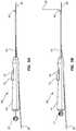
- FIG. 5Ais a side view of the device of FIG. 2 in a primed configuration with the stylet cover tube advanced and the stylet actuator loaded.
- FIG. 5Billustrates a side view of the device of FIG. 2 in an actuated configuration with the stylet deployed.
- FIG. 6Aillustrates a cross-sectional schematic view of a vascular catheter of the device of FIG. 2 in a configuration with the stylet deployed.
- FIG. 6Billustrates a cross-sectional schematic view of the vascular catheter of the device of FIG. 2 , in a retracted configuration with the stylet retracted, being advanced over a guidewire in a vessel, such as a vein.
- FIG. 7Aillustrates a cross-sectional schematic view of a device in accordance with an embodiment with the stylet deployed in an actuated configuration.
- FIG. 7Billustrates a cross-sectional schematic view of a device in accordance with an embodiment, in a retracted configuration with the stylet retracted, being advanced over a guidewire in a vessel, such as a vein.
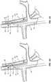
- FIG. 8illustrates a schematic view of the device of FIG. 2 in a retracted configuration, with a vascular catheter in the jugular vein close to an access point for the carotid artery.
- FIG. 9illustrates a schematic view of the device of FIG. 2 in a primed configuration with the stylet cover tube advanced to a desired access point for the carotid artery.
- FIG. 10illustrates a schematic view of the device of FIG. 2 in an actuated configuration with the stylet advanced through the vessel wall of the jugular vein and into the carotid artery.
- FIG. 11illustrates a schematic view of the device of FIG. 2 with an access catheter advanced over the stylet.
- FIG. 12illustrates a schematic view of a stent to close the opening between the jugular vein and the carotid artery.
- FIG. 13illustrates a schematic view of a guidewire being introduced into the carotid artery via the puncture needle.
- FIG. 14illustrates a schematic view of a guidewire being introduced into the carotid artery via the puncture needle.
- FIG. 15illustrates a schematic view of an interventional sheath being advanced over the guidewire into the carotid artery.
- FIG. 16illustrates a schematic view of a closure device being introduced into the jugular vein to close the opening between the jugular vein and the carotid artery.
- FIG. 17illustrates a flowchart of a method of accessing a patient's carotid artery through their jugular vein.
- Certain medical proceduresrequire access to the carotid artery in order to treat a patient for certain medical conditions.
- the carotid arteryis accessed through open surgical procedure.
- An exemplary procedure that includes open surgeryis carotid endarterectomy in which the carotid artery is clamped and cut open and then scraped to remove fatty plaque to treat a blockage in the carotid artery.
- Very ill and/or elderly patientsmay have a difficult time tolerating an open surgical procedure under general anesthesia.
- Another method of accessing the carotid arteryis to access the carotid artery percutaneously via the femoral artery and advancing treatment devices such as catheters through the aortic arch to the carotid artery.
- treatment devicessuch as catheters through the aortic arch to the carotid artery.
- carotid artery inventionby advancing a filter and an angioplasty balloon catheter percutaneously (via a sheath) form the femoral artery into the aortic arch and then into the blocked carotid artery under local anesthesia.
- the aortic archmay be difficult to navigate due to age-related calcification and/or tortuosity.
- the present disclosureincludes methods and devices for accessing a patient's carotid artery via the patient's jugular vein, without open surgery or having to pass a treatment device through the patient's aortic arch. It is within the scope of this disclosure to access the carotid artery at a remote access point, e.g., femoral vein, brachial vein, basilic vein, cephalic vein, median antecubital, median antebrachial, etc., or directly though the patient's neck.
- a remote access pointe.g., femoral vein, brachial vein, basilic vein, cephalic vein, median antecubital, median antebrachial, etc.
- Coupled tois broad enough to refer to any suitable coupling or other form of interaction between two or more entities.
- Two componentsmay be coupled to each other even though they are not in direct contact with each other.
- two componentsmay be coupled to each other through an intermediate component.
- fluid communicationis used in its ordinary sense, and is broad enough to refer to arrangements in which a fluid (e.g., a gas or a liquid) can flow from one element to another element when the elements are in fluid communication with each other.
- proximal and distalare opposite directional terms.
- distal end of a device or componentis the end of the component that is furthest from the practitioner during ordinary use.
- proximal endrefers to the opposite end or the end nearest the practitioner during ordinary use.
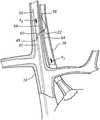
- FIG. 1Aschematically illustrates the vasculature of a patient wherein the femoral vein 12 has been accessed, such as by the Modified Seldinger Technique.
- a general use guidewire 30(such as a guidewire measuring about 0.0035 inches in diameter) may be passed through a vascular sheath 20 and positioned in a right or left jugular vein 14 of the patient via the inferior vena cava.
- the guidewire 30may ultimately be positioned in the vicinity of a desired access point to a carotid artery 18 .
- the desired access pointmay be located at any point along the jugular vein 14 where the jugular vein also runs along the carotid artery 18 .
- the desired access pointmay be just below the bifurcation of the carotid artery 18 into the internal carotid artery 19 and the external carotid artery 17 .
- a medical practitionermay be able to access the carotid artery 18 without having to do an open surgical procedure or to advance percutaneously from the patient's femoral artery and past the patient's aortic arch.
- the remote entry pointmay be a vein 16 within the patient's arm (e.g., brachial vein, basilic vein, cephalic vein, median antecubital, median antebrachial, etc.) and the vascular catheter 42 may have a length of about half a meter and a diameter around 7 french, as illustrated in FIG. 1B .
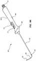
- FIGS. 2-6Billustrate an embodiments of an access device 40 .
- a vascular catheter 42extends distally from a handle 44 .
- the length and diameter of the vascular catheter 42depends on the distance between a remote entry point and the desired access point to the carotid artery 18 .
- the remote entry pointmay be the femoral vein 12 and the vascular catheter 42 may have a length of about 1 meter and may have a diameter of around 7 french (0.092 inches).
- the vascular catheter 42may comprise a catheter tip 47 at the distal end of the vascular catheter 42 .
- the catheter tip 47may be tapered, beveled, conical or comprise other shapes or structures.
- the catheter tip 47 in FIG. 3is illustrated as conical.
- the vascular catheter 42may have a guidewire lumen 46 , as illustrated in FIGS. 2-6B .
- the guidewire 30 lumen 46may be configured to be a rapid exchange (RX) guidewire lumen for receiving the guidewire 30 .
- RXrapid exchange
- the guidewire 30may be a 0.035 inch guidewire.
- the guidewire 30is advanced through the handle 44 of the access device 40 .
- the guidewire 30may be introduced into the guidewire lumen 46 using an introducer kit (not shown).
- the guidewire 30may be positioned adjacent to the desired access point in the patient's jugular vein 14 .
- the vascular catheter 42may be advanced over the guidewire 30 before or after the guidewire 30 is positioned adjacent to the desired access point in the jugular vein 14 .
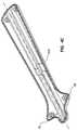
- the handle 44may include a top portion 45 A and a bottom portion 45 B.
- FIG. 4Ais a top perspective view of the top portion 45 A of the handle 44 along with other components.
- FIG. 4Bis a bottom view of the top portion 45 A of the handle 44 , along with other components of the device, with the bottom portion 45 B removed to expose internal components.
- FIG. 4Cillustrates a bottom portion 45 B of the handle 44 .
- the top portion 45 A and the bottom portion 45 Bmay engage to from the handle 44 .
- the handle 44also includes wings 48 on opposing sides of the handle 44 . The wings 48 may be used to apply a distal force to the vascular catheter 42 from the handle 44 .
- FIGS. 6A-7Bwhich illustrate a portion of the access device 40 comprising a distal portion of the vascular catheter 42 in FIGS. 6A and 6B and an analogous portion of an alternative embodiment of an access catheter 42 ′ in FIGS. 7A and 7B .
- the vascular catheter 42 and 42 ′are shown in cross-section, while the elements disposed within the access catheters are not in cross-section for clarity.
- the vascular catheter 42 ′ of FIGS. 7A and 7Bis identical to vascular catheter 42 of FIGS. 6A and 6B except that vascular catheter 42 ′ does not comprise a ramped camming surface 56 as detailed below. Accordingly, other elements of the access device 40 of FIG. 2 as shown in FIGS.
- FIGS. 7A and 7B(such as guidewire 30 ) retain the same numerals as the embodiment of FIGS. 2, 6A, and 6B . Disclosure recited in connection with vascular catheter 42 of FIGS. 6A and 6B may be analogously applied to vascular catheter 42 ′ of FIGS. 7A and 7B .
- the device 40may have a stylet lumen 50 extending from the handle 44 to an opening toward the distal end of the vascular catheter 42 .
- the stylet lumen 50curves at its distal end to form a camming surface 56 , as illustrated in FIGS. 6A and 6B .
- the camming surface 56may provide additional structural support to a guide tube or a cover tube 60 when it is in an advanced position.
- a stylet 58may be slideably disposed within cover tube 60 .
- the stylet lumen 50does not have a curved camming surface 56 .
- the stylet lumen 50may be substantially cylindrical as illustrated in FIGS. 7A and 7B .
- the stylet 58(formed, e.g., from Nitinol with a diameter of 0.014 inches) enclosed by the cover tube 60 (such as a 0.025 inch diameter Nitinol hypotube) extends from an actuator 51 , 52 , and 53 , in the handle 44 toward the distal end of the device 40 .
- the cover tube 60has a preformed curved or deflected tip at its distal end.
- the performed curvemay be between 30° and 60° of the longitudinal axis of vascular catheter 42 .
- the optional camming surface 56may promote the curvature of the cover tube 60 .
- Stylet 58may have a sharp distal point 62 adapted to penetrate tissue, such as blood vessels, muscle, and skin.
- the sharp distal point 62may be part of a tip designed to have various dimensions and shapes.
- the tip 62may be faceted.
- the tip 62may include three flat surfaces that intersect to form a sharp distal point.
- tip 62may be a conical tip. The conical tip may form an angle of about 10° with the shaft of the stylet 58 .
- vascular catheter 40may be inserted into the femoral vein 12 over the guidewire 30 (with the guidewire disposed as shown in FIG. 1A ) and may be advanced adjacent to the desired access point in the jugular vein 14 under fluoroscopic guidance. During this advancement, the stylet 58 and cover tube 60 may be in a retracted configuration, as illustrated in FIG. 8 . The distal opening of the stylet lumen 50 may then be oriented toward the desired carotid artery access point in the vein wall 66 .
- the arrows (F 1 and F 2 ) in FIGS. 8-11illustrate the direction of blood flow in the jugular vein 14 and the carotid artery 18 .
- the blood in the carotid artery 18flows away from the heart in direction F 1 and the blood flow in the jugular vein 14 flows toward to heart in direction F 2 .
- the catheter tip 47may include a radiopaque marker 49 visible under the fluoroscopy as illustrated in FIGS. 8-10 .
- the radiopaque marker 49may be embedded in the catheter tip 47 .
- the radiopaque marker 49is illustrated as a ring in FIGS. 9 and 10 ; however, other shapes and geometries may be used.
- the shape of the radiopaque marker 49may be selected to facilitate fluoroscopic identification of the location and orientation of the catheter tip 47 .
- radiopaque marker materialsinclude gold, platinum, platinum-iridium, and other biocompatible radiopaque materials.
- the cover tube 60may be advanced out of opening into an primed configuration by moving a slide button 51 proximally in handle 44 , at which point the cover tube 60 assumes its curved shape, as shown in FIGS. 5A and 9 .
- moving the slide button 51 proximally in handle 44pushes the cover tube distally from the vascular catheter 42 .
- moving the slide button 51 proximally in the handle 44moves the catheter proximally to expose the distal end of the cover tube 60 .
- the slide button 51may engage with and be operatively connected to the vascular catheter 42 with catheter slide 57 .
- the cover tube 60is advanced until its distal end is adjacent to the vein wall 66 at the desired carotid artery access point. In some embodiments the cover tube 60 advances until its distal end abuts the vein wall 66 at the desired carotid artery access point. The stylet 58 remains in cover tube 60 in the primed configuration.
- the orientation of the extended cover tube 60may be determined based on the orientation of the handle 44 .
- the slide button 51engages with the top portion 45 A of the handle 44 .
- the slide button 51is disposed substantially perpendicular to a plane defined by the opposing wings 48 that extends along a length of the handle 44 e.g., the plane defined by the curve where the top portion 45 A of the handle 44 contacts the bottom portion 45 B of the handle 44 .
- the cover tube 60extends upwards from the stylet lumen 50 and is also substantially perpendicular to the plane defined by the opposing wings 48 extending along a length of the handle 44 .
- the positioning and orientation of the cover tube 60 after it has been advancedmay also be visually verified, for example using fluoroscopy prior to deploying the stylet 58 .
- the stylet actuatoris loaded by pulling proximally on a spring load mechanism 52 in the handle 44 , as illustrated in FIG. 5A .
- a stylet release button 53is exposed. Depressing release button 53 advances the stylet 58 distally under the action of a spring 55 to an actuated configuration, as illustrated in FIGS. 5B, 6A, 7A, and 10 .
- the sharp distal tippenetrates the vein wall 66 of the jugular vein 14 and the artery wall 64 of the carotid artery 18 , as illustrated in FIG. 10 .
- the blood flow F 1 of the carotid arterymay push the stylet 58 downstream so that it does not damage or perforate the opposing artery wall of the carotid artery 18 .
- the puncture of the artery wall 64may create a flap 22 .
- the distance traveled by the stylet 58depends on the application. For example, when the stylet 58 is moving from the jugular vein wall to the carotid artery 18 , the stylet 58 may move less than half a centimeter. In other embodiments, the stylet 50 may move more or less than this amount, including 0.25 centimeters or less and 1, 2, 3, 4, or more centimeters.
- cover tube 60Various sizes may be used for the cover tube 60 .
- the configuration of the pre-formed cover tube 60may be varied to use different shapes.
- the length and pre-formed configuration of the cover tube 60may be selected based on the size of the vessel to be accessed.
- the cover tube 60is not fully extended from the vascular catheter 42 .
- the cover tube 60may not be fully extended to accommodate the specific geometry and size of the vessel. In smaller diameter blood vessels there may not be enough space to fully extend the cover tube; however, the cover tube 60 and stylet 58 may properly function partially extended from the vascular catheter 42 .
- Access catheter 70may be introduced and advanced along stylet 58 through the femoral vein 12 to the jugular vein 14 and into the carotid artery 18 . Once the access catheter 70 has been introduced into the carotid artery 18 , it may be used to perform a number of different medical procedures via the carotid artery 18 . Access catheter 70 may advance a particular treatment device or tool 72 , for treating the patient. Exemplary treatment devices 72 may include angioplasty balloons, stents, filters, aneurysm stents, etc. These devices 72 may be used to treat a blockage in the carotid artery, treat an embolism in the brain (stroke), treat an aneurysm, place a filter, remove fatty plaque, etc.
- the stylet 58 , access catheter 70 , and any treatment devices 72may be withdrawn from the carotid artery 18 and the jugular vein 14 .
- a closure device 80may be introduced into the jugular vein 14 adjacent to the opening to close the opening between the jugular vein 14 and the carotid artery 18 , as illustrated in FIG. 12 .
- the closure device 80may be a bioabsorable, tightly meshed/woven venous stent 80 .
- the opening in artery wall 64 of carotid artery 18may be closed when the flap 22 is pushed back toward the opening by the blood flow F 1 in the carotid artery 18 .
- FIGS. 13-16illustrate an alternative method to access the carotid artery 18 of a patient without open surgery and without passing through the aortic arch.
- the carotid artery 18may be accessed directly from the patient's neck.
- a puncture needle 10e.g., 18 gauge for larger patients, 21 gauge for smaller patients
- the puncture needle 10may be advanced through the skin 90 into the jugular vein 14 and then through the other side of the jugular vein 14 and into the carotid artery 18 at a point below the bifurcation of the carotid artery 18 into the internal carotid artery 19 and the external carotid artery 17 .
- the puncture needle 10thus creates an opening between the jugular vein 14 and the carotid artery 18 for fluid communication.
- the puncture needle 10may also create a flap 22 in the artery wall of the carotid artery 18 .
- the puncture needle 10may be inserted at a number of different orientations, for example, at an upward angle, a downward angel, etc.
- FIG. 13illustrates the puncture needle 10 being inserted at an upward angle between 30° and 60° off the longitudinal axis of the jugular vein 14 .
- the blood that flows out of the proximal end of the puncture needle 10may indicate the location of the distal tip 21 of the puncture needle 10 .
- Venous blood from the jugular vein 14is darker and flows slowly and the higher pressure and more oxygenated arterial blood from the carotid artery 18 flows quicker and is redder than the blood from the jugular vein. This provides another indication to the practitioner that the puncture needle 10 has accessed the carotid artery 18 .
- a guidewire 30may be advanced through the puncture needle 10 into the carotid artery 18 , as illustrated in FIG. 14 .
- the puncture needle 10may be removed and an interventional sheath 26 (e.g., with a diameter of 5 F or 6 F) may be advanced over the guidewire 30 , as illustrated in FIG. 15 .
- the interventional sheath 26may provide access for a catheter or other treatment devices or tools to be advanced into the carotid artery 18 to perform a desired procedure, such as an angioplasty, plaque removal, treat aneurysms, etc.
- a number of different types of tools, such as angioplasty balloons, filters, stents, etc.may be used.
- a closure device 80may be introduced into the jugular vein 14 adjacent to the opening to close the opening between the jugular vein 14 and the carotid artery 18 , as illustrated in FIG. 16 .
- the closure device 80may be a bioabsorable, tightly meshed/woven venous stent.
- the opening in artery wall 64 of carotid artery 18may be closed when the flap 22 is pushed back toward the opening by the blood flow F 1 in the carotid artery 18 .
- the closure device 80may be inserted via the interventional sheath 26 (after it has been withdrawn from the carotid artery 18 , but before withdrawing it from the internal jugular vein 14 ).
- the closure device 80may be implanted using a separate catheter (not shown) that has been advanced from a remote entry point in the femoral vein 12 into the jugular vein 14 .
- FIG. 17illustrates a flowchart of a method 1700 of accessing a patient's carotid artery 18 via their jugular vein 14 , as previously discussed.
- the practitionermay access the patient's jugular vein 14 in a variety of different manners.
- the practitionermay access the jugular vein via a remote access point, such as a femoral vein 12 or a vein 16 in the patient's arm (e.g., brachial vein, basilic vein, cephalic vein, median antecubital, median antebrachial, etc.).
- a remote access pointsuch as a femoral vein 12 or a vein 16 in the patient's arm (e.g., brachial vein, basilic vein, cephalic vein, median antecubital, median antebrachial, etc.).
- the practitionermay access the jugular vein 14 directly by inserting a puncture needle 10 through the patient's neck.
- the practitionermay access the carotid artery 18 via the jugular vein 14 .
- the practitionermay access the carotid artery 18 by piercing the carotid artery 18 after piercing the jugular vein 14 with the puncture needle 10 .
- the practitionermay advance a guidewire 30 and a vascular catheter 42 to the jugular vein 14 and pierce the carotid artery 18 with the stylet 58 .
- step 1706once the practitioner has gained access to the carotid artery 18 , the practitioner may introduce or advance a treatment device for treating a medical condition via the carotid artery 18 .
- the treatment device 72may be removed and the practitioner may introduce a closure device 80 for closing the opening between the jugular vein 14 and the carotid artery 18 .
- Any methods disclosed hereininclude one or more steps or actions for performing the described method.
- the method steps and/or actionsmay be interchanged with one another.
- the order and/or use of specific steps and/or actionsmay be modified.
- sub-routines or only a portion of a method described hereinmay be a separate method within the scope of this disclosure. Stated otherwise, some methods may include only a portion of the steps described in a more detailed method.
Landscapes
- Health & Medical Sciences (AREA)
- Life Sciences & Earth Sciences (AREA)
- Heart & Thoracic Surgery (AREA)
- Anesthesiology (AREA)
- Biophysics (AREA)
- Pulmonology (AREA)
- Engineering & Computer Science (AREA)
- Biomedical Technology (AREA)
- Hematology (AREA)
- Animal Behavior & Ethology (AREA)
- General Health & Medical Sciences (AREA)
- Public Health (AREA)
- Veterinary Medicine (AREA)
- Child & Adolescent Psychology (AREA)
- Vascular Medicine (AREA)
- Surgical Instruments (AREA)
Abstract
Description
Claims (19)
Priority Applications (3)
| Application Number | Priority Date | Filing Date | Title |
|---|---|---|---|
| US15/835,114US10617854B2 (en) | 2016-12-09 | 2017-12-07 | Trans-jugular carotid artery access methods |
| US16/847,281US11554256B2 (en) | 2016-12-09 | 2020-04-13 | Trans-jugular carotid artery access methods |
| US18/065,450US20230218869A1 (en) | 2016-12-09 | 2022-12-13 | Trans-jugular corotid artery access methods |
Applications Claiming Priority (4)
| Application Number | Priority Date | Filing Date | Title |
|---|---|---|---|
| US201662432369P | 2016-12-09 | 2016-12-09 | |
| US201662433634P | 2016-12-13 | 2016-12-13 | |
| US201662440735P | 2016-12-30 | 2016-12-30 | |
| US15/835,114US10617854B2 (en) | 2016-12-09 | 2017-12-07 | Trans-jugular carotid artery access methods |
Related Child Applications (1)
| Application Number | Title | Priority Date | Filing Date |
|---|---|---|---|
| US16/847,281DivisionUS11554256B2 (en) | 2016-12-09 | 2020-04-13 | Trans-jugular carotid artery access methods |
Publications (2)
| Publication Number | Publication Date |
|---|---|
| US20180161551A1 US20180161551A1 (en) | 2018-06-14 |
| US10617854B2true US10617854B2 (en) | 2020-04-14 |
Family
ID=62488228
Family Applications (3)
| Application Number | Title | Priority Date | Filing Date |
|---|---|---|---|
| US15/835,114Active2038-06-28US10617854B2 (en) | 2016-12-09 | 2017-12-07 | Trans-jugular carotid artery access methods |
| US16/847,281Active2038-05-28US11554256B2 (en) | 2016-12-09 | 2020-04-13 | Trans-jugular carotid artery access methods |
| US18/065,450AbandonedUS20230218869A1 (en) | 2016-12-09 | 2022-12-13 | Trans-jugular corotid artery access methods |
Family Applications After (2)
| Application Number | Title | Priority Date | Filing Date |
|---|---|---|---|
| US16/847,281Active2038-05-28US11554256B2 (en) | 2016-12-09 | 2020-04-13 | Trans-jugular carotid artery access methods |
| US18/065,450AbandonedUS20230218869A1 (en) | 2016-12-09 | 2022-12-13 | Trans-jugular corotid artery access methods |
Country Status (3)
| Country | Link |
|---|---|
| US (3) | US10617854B2 (en) |
| AU (1) | AU2017272319A1 (en) |
| CA (1) | CA2988274A1 (en) |
Families Citing this family (13)
| Publication number | Priority date | Publication date | Assignee | Title |
|---|---|---|---|---|
| US9511214B2 (en) | 2006-05-02 | 2016-12-06 | Vascular Access Technologies, Inc. | Methods of transvascular retrograde access placement and devices for facilitating therein |
| US9623217B2 (en) | 2012-05-30 | 2017-04-18 | Vascular Access Techonlogies, Inc. | Transvascular access methods |
| US10617854B2 (en) | 2016-12-09 | 2020-04-14 | Vascular Access Technologies, Inc. | Trans-jugular carotid artery access methods |
| US11654224B2 (en) | 2016-12-30 | 2023-05-23 | Vascular Access Technologies, Inc. | Methods and devices for percutaneous implantation of arterio-venous grafts |
| US10470797B1 (en) | 2018-07-17 | 2019-11-12 | SlipStream, LLC | Systems and methods for vascular access |
| US10828038B2 (en) | 2018-10-05 | 2020-11-10 | Lakshmikumar Pillai, MD | Carotid artery embolic protection method using percutaneous transjugular carotid flow reversal |
| US11426173B2 (en) | 2019-01-04 | 2022-08-30 | Lakshmikumar Pillai | Devices and methods using percutaneous transjugular carotid flow reversal |
| CN109663172B (en)* | 2019-01-15 | 2024-03-26 | 苏州林华医疗器械股份有限公司 | Jugular vein puncture indwelling needle |
| US11497552B2 (en) | 2019-07-09 | 2022-11-15 | Juad, Inc. | Apparatus, systems and methods for transvascular access to the brain |
| US10835258B1 (en) | 2019-08-21 | 2020-11-17 | Lakshmikumar Pillai | Systems and methods for retrograde perfusion and clearance of emboli |
| US12042152B2 (en) | 2019-08-21 | 2024-07-23 | Lakshmikumar Pillai | Systems and methods for retrograde perfusion and clearance of emboli |
| EP4171723B1 (en) | 2020-06-30 | 2025-09-17 | Vonova Inc. | Transcatheter electroctode array |
| WO2025145101A1 (en)* | 2023-12-29 | 2025-07-03 | Cvrx, Inc. | Visualization devices and techniques for implanting medical devices |
Citations (70)
| Publication number | Priority date | Publication date | Assignee | Title |
|---|---|---|---|---|
| US4559039A (en) | 1983-12-05 | 1985-12-17 | Purdue Research Foundation | Permanently placed transcutaneous access device to blood vessels |
| US4790825A (en) | 1986-09-05 | 1988-12-13 | Electro Catheter Corporation | Closed chest cannulation method and device for atrial-major artery bypass |
| US4966163A (en) | 1989-02-14 | 1990-10-30 | Advanced Cardiovascular Systems, Inc. | Extendable guidewire for vascular procedures |
| US5421348A (en) | 1993-11-29 | 1995-06-06 | Cordis Corporation | Rotating guidewire extension system with mechanically locking extension wire |
| US5492530A (en) | 1994-02-07 | 1996-02-20 | Cathco, Inc. | Method for accessing the coronary arteries from the radial or brachial artery in the arm |
| US5685820A (en) | 1990-11-06 | 1997-11-11 | Partomed Medizintechnik Gmbh | Instrument for the penetration of body tissue |
| US5733248A (en) | 1995-11-29 | 1998-03-31 | Scimed Life Systems, Inc. | Universal guide catheter |
| US6047700A (en) | 1998-03-30 | 2000-04-11 | Arthrocare Corporation | Systems and methods for electrosurgical removal of calcified deposits |
| US6102926A (en) | 1996-12-02 | 2000-08-15 | Angiotrax, Inc. | Apparatus for percutaneously performing myocardial revascularization having means for sensing tissue parameters and methods of use |
| US6190353B1 (en) | 1995-10-13 | 2001-02-20 | Transvascular, Inc. | Methods and apparatus for bypassing arterial obstructions and/or performing other transvascular procedures |
| US6217527B1 (en) | 1998-09-30 | 2001-04-17 | Lumend, Inc. | Methods and apparatus for crossing vascular occlusions |
| US20010012924A1 (en) | 1998-01-13 | 2001-08-09 | Milo Charles F. | Methods and apparatus for crossing total occlusions in blood vessels |
| US20010023346A1 (en) | 1999-05-04 | 2001-09-20 | Cardiodyne, Inc. | Method and devices for creating a trap for confining therapeutic drugs and/or genes in the myocardium |
| US20020004666A1 (en) | 2000-05-22 | 2002-01-10 | Michael Schwager | Catheter arrangement |
| US20020029060A1 (en) | 1998-07-29 | 2002-03-07 | Michael Hogendijk | Surgical cutting instrument and method of use |
| US20020120250A1 (en) | 1998-08-11 | 2002-08-29 | Altman Peter A. | Catheter drug delivery system and method for use |
| US20020122877A1 (en)* | 2000-12-28 | 2002-09-05 | Sameer Harish | Methods of forming a coating for a prosthesis |
| US20020133168A1 (en) | 2001-03-16 | 2002-09-19 | Smedley Gregory T. | Applicator and methods for placing a trabecular shunt for glaucoma treatment |
| US6475226B1 (en) | 1999-02-03 | 2002-11-05 | Scimed Life Systems, Inc. | Percutaneous bypass apparatus and method |
| US20020169377A1 (en) | 2000-04-13 | 2002-11-14 | Khairkhahan Alexander K. | Method and apparatus for accessing the left atrial appendage |
| US6508777B1 (en) | 1998-05-08 | 2003-01-21 | Cardeon Corporation | Circulatory support system and method of use for isolated segmental perfusion |
| US20030040771A1 (en)* | 1999-02-01 | 2003-02-27 | Hideki Hyodoh | Methods for creating woven devices |
| US6554794B1 (en) | 1997-09-24 | 2003-04-29 | Richard L. Mueller | Non-deforming deflectable multi-lumen catheter |
| US6623480B1 (en) | 1998-07-24 | 2003-09-23 | University Of Kentucky Research Foundation | Flexible recording/high energy electrode catheter with anchor for ablation of atrial flutter by radio frequency energy |
| US20040039371A1 (en) | 2002-08-23 | 2004-02-26 | Bruce Tockman | Coronary vein navigator |
| US6709444B1 (en) | 1996-02-02 | 2004-03-23 | Transvascular, Inc. | Methods for bypassing total or near-total obstructions in arteries or other anatomical conduits |
| US6726677B1 (en) | 1995-10-13 | 2004-04-27 | Transvascular, Inc. | Stabilized tissue penetrating catheters |
| US20040082850A1 (en) | 2002-10-23 | 2004-04-29 | Medtonic, Inc. | Methods and apparatus for locating body vessels and occlusions in body vessels |
| US20040133168A1 (en) | 2002-12-23 | 2004-07-08 | Salcudean Septimiu E. | Steerable needle |
| US20040181238A1 (en) | 2003-03-14 | 2004-09-16 | David Zarbatany | Mitral valve repair system and method for use |
| US20040181150A1 (en) | 1999-09-01 | 2004-09-16 | Bacchus Vascular, Inc. | Methods and apparatus for accessing and treating body lumens |
| US20050101984A1 (en) | 2003-11-06 | 2005-05-12 | Nmt Medical, Inc. | Transseptal puncture apparatus |
| WO2005053547A2 (en) | 2003-11-28 | 2005-06-16 | Cook Incorporated | Vascular occlusion methods, systems and devices |
| US20050149097A1 (en) | 2003-12-30 | 2005-07-07 | Regnell Sandra J. | Transseptal needle |
| US20050209579A1 (en) | 2004-03-22 | 2005-09-22 | Yacoubian Vahe S | System, methods and apparatus for cerebral protection |
| US6955657B1 (en) | 2001-12-31 | 2005-10-18 | Advanced Cardiovascular Systems, Inc. | Intra-ventricular substance delivery catheter system |
| US20060009737A1 (en) | 2004-07-12 | 2006-01-12 | Whiting James S | Methods and devices for transseptal access |
| US7008979B2 (en) | 2002-04-30 | 2006-03-07 | Hydromer, Inc. | Coating composition for multiple hydrophilic applications |
| US20060135962A1 (en) | 2004-09-09 | 2006-06-22 | Kick George F | Expandable trans-septal sheath |
| US20060173440A1 (en) | 2001-01-17 | 2006-08-03 | Medtronic Vascular, Inc. | Microcatheter Devices and Methods for Targeted Substance Delivery |
| US20060247750A1 (en) | 2005-04-28 | 2006-11-02 | Seifert Kevin R | Guide catheters for accessing cardiac sites |
| US20070021767A1 (en) | 2005-07-25 | 2007-01-25 | Breznock Eugene M | Steerable endoluminal punch |
| US20070203515A1 (en) | 2006-01-25 | 2007-08-30 | Heuser Richard R | Catheter system for connecting adjacent blood vessels |
| US20080082136A1 (en) | 2006-10-03 | 2008-04-03 | Gaudiani Vincent A | Transcoronary Sinus Pacing System, LV Summit Pacing, Early Mitral Closure Pacing, and Methods Therefor |
| US7374567B2 (en) | 2006-01-25 | 2008-05-20 | Heuser Richard R | Catheter system for connecting adjacent blood vessels |
| US20080125748A1 (en) | 2006-09-25 | 2008-05-29 | Medtronic Vascular, Inc. | High Torque, Low Profile Catheters and Methods for Transluminal Interventions |
| US20080154172A1 (en) | 2006-12-20 | 2008-06-26 | Medtronic Vascular, Inc. | Low Profile Catheters and Methods for Treatment of Chronic Total Occlusions and Other Disorders |
| US20080171944A1 (en) | 2005-07-26 | 2008-07-17 | Rox Medical, Inc. | Devices, systems, and methods for peripheral arteriovenous fistula creation |
| US20080215008A1 (en) | 2006-12-20 | 2008-09-04 | Nance Edward J | Expandable trans-septal sheath |
| US20080249565A1 (en) | 1996-08-22 | 2008-10-09 | The Trustees Of Columbia University In The City Of New York | Endovascular Flexible Stapling Device |
| US20090112050A1 (en) | 2007-10-24 | 2009-04-30 | Circulite, Inc. | Transseptal cannula, tip, delivery system, and method |
| US20090240122A1 (en) | 2006-05-04 | 2009-09-24 | The Cleveland Clinic Foundation | Intrajugular catheter |
| US7648517B2 (en) | 1995-10-13 | 2010-01-19 | Medtronic Vascular, Inc. | Catheters and related devices for forming passageways between blood vessels or other anatomical structures |
| US20100249491A1 (en) | 2009-03-27 | 2010-09-30 | Circulite, Inc. | Two-piece transseptal cannula, delivery system, and method of delivery |
| WO2011068540A1 (en) | 2009-12-03 | 2011-06-09 | Therix Medical Development, Ltd. | Central venous access system |
| US20110178530A1 (en) | 2007-01-30 | 2011-07-21 | Bly Mark J | Direct delivery system for transvascular lead |
| US8019420B2 (en) | 2003-08-21 | 2011-09-13 | Medtronic, Inc. | Medical lead connector systems with adapters |
| US20120136366A1 (en) | 2006-05-02 | 2012-05-31 | Lakshmikumar Pillai | Methods of Transvascular Retrograde Access Placement and Devices For Facilitating Therein |
| US20120136247A1 (en) | 2006-05-02 | 2012-05-31 | Lakshmikumar Pillai | Methods of Transvascular Retrograde Access Placement and Devices for Facilitating the Placement |
| US8241311B2 (en) | 2009-12-15 | 2012-08-14 | Medtronic Vascular, Inc. | Methods and systems for bypassing an occlusion in a blood vessel |
| US20130006282A1 (en) | 2011-06-29 | 2013-01-03 | Matthew Wilkinson | System and method for re-entering a vessel lumen |
| US8374680B2 (en) | 2008-04-21 | 2013-02-12 | Medtronic Vascular, Inc. | Needleless catheters and methods for true lumen re-entry in treatment of chronic total occlusions and other disorders |
| US20130072957A1 (en)* | 2011-09-19 | 2013-03-21 | Boston Scientific Scimed, Inc. | Subintimal re-entry catheter and retrograde recanalization |
| US8409236B2 (en) | 2009-08-21 | 2013-04-02 | Vascular Access Technologies, Inc. | Methods of transvascular retrograde access placement and devices for facilitating the placement |
| WO2013119547A1 (en) | 2012-02-09 | 2013-08-15 | Therix Medical Development, Ltd. | Occlusion access system |
| US20130324901A1 (en) | 2012-05-30 | 2013-12-05 | Lakshmikumar Pillai | Transvascular Access Methods |
| US20130324967A1 (en) | 2012-05-30 | 2013-12-05 | Lakshmikumar Pillai | Transvascular access device and method |
| US20140142418A1 (en) | 2012-02-09 | 2014-05-22 | Therix Medical Development, Ltd. | Occlusion access system |
| US20140142677A1 (en)* | 2012-04-23 | 2014-05-22 | Pq Bypass, Inc. | Methods and systems for bypassing occlusions in a femoral artery |
| US20150320357A1 (en)* | 2014-02-20 | 2015-11-12 | GraftWorx, LLC | Methods for assessing fluid flow through a conduit |
Family Cites Families (13)
| Publication number | Priority date | Publication date | Assignee | Title |
|---|---|---|---|---|
| US6123725A (en)* | 1997-07-11 | 2000-09-26 | A-Med Systems, Inc. | Single port cardiac support apparatus |
| WO2001026562A1 (en) | 1999-10-08 | 2001-04-19 | The General Hospital Corporation D/B/A Massachusetts General Hospital | Percutaneous stent graft and method for vascular bypass |
| EP2497520B1 (en)* | 2007-07-18 | 2022-04-13 | Silk Road Medical, Inc. | Systems for establishing retrograde carotid arterial blood flow |
| US8574245B2 (en)* | 2008-08-13 | 2013-11-05 | Silk Road Medical, Inc. | Suture delivery device |
| US8568471B2 (en)* | 2010-01-30 | 2013-10-29 | Abbott Cardiovascular Systems Inc. | Crush recoverable polymer scaffolds |
| EP2598044B1 (en)* | 2010-07-27 | 2019-03-13 | Incept, LLC | Apparatus for treating neurovascular venous outflow obstruction |
| US9446172B2 (en)* | 2011-05-10 | 2016-09-20 | Abbott Cardiovascular Systems Inc. | Modification of bioabsorbable stent to reduce thrombogenecity |
| US9174032B2 (en) | 2012-07-13 | 2015-11-03 | Boston Scientific Scimed, Inc. | Subintimal reentry system |
| US8968387B2 (en)* | 2012-07-23 | 2015-03-03 | Abbott Cardiovascular Systems Inc. | Shape memory bioresorbable polymer peripheral scaffolds |
| US10159479B2 (en)* | 2012-08-09 | 2018-12-25 | Silk Road Medical, Inc. | Suture delivery device |
| EP2929859A1 (en) | 2014-04-10 | 2015-10-14 | Carag AG | A kit for placing a bypass |
| US11027104B2 (en)* | 2014-09-04 | 2021-06-08 | Silk Road Medical, Inc. | Methods and devices for transcarotid access |
| US10617854B2 (en) | 2016-12-09 | 2020-04-14 | Vascular Access Technologies, Inc. | Trans-jugular carotid artery access methods |
- 2017
- 2017-12-07USUS15/835,114patent/US10617854B2/enactiveActive
- 2017-12-08CACA2988274Apatent/CA2988274A1/enactivePending
- 2017-12-08AUAU2017272319Apatent/AU2017272319A1/ennot_activeAbandoned
- 2020
- 2020-04-13USUS16/847,281patent/US11554256B2/enactiveActive
- 2022
- 2022-12-13USUS18/065,450patent/US20230218869A1/ennot_activeAbandoned
Patent Citations (77)
| Publication number | Priority date | Publication date | Assignee | Title |
|---|---|---|---|---|
| US4559039A (en) | 1983-12-05 | 1985-12-17 | Purdue Research Foundation | Permanently placed transcutaneous access device to blood vessels |
| US4790825A (en) | 1986-09-05 | 1988-12-13 | Electro Catheter Corporation | Closed chest cannulation method and device for atrial-major artery bypass |
| US4966163A (en) | 1989-02-14 | 1990-10-30 | Advanced Cardiovascular Systems, Inc. | Extendable guidewire for vascular procedures |
| US5685820A (en) | 1990-11-06 | 1997-11-11 | Partomed Medizintechnik Gmbh | Instrument for the penetration of body tissue |
| US5421348A (en) | 1993-11-29 | 1995-06-06 | Cordis Corporation | Rotating guidewire extension system with mechanically locking extension wire |
| US5492530A (en) | 1994-02-07 | 1996-02-20 | Cathco, Inc. | Method for accessing the coronary arteries from the radial or brachial artery in the arm |
| US6190353B1 (en) | 1995-10-13 | 2001-02-20 | Transvascular, Inc. | Methods and apparatus for bypassing arterial obstructions and/or performing other transvascular procedures |
| US6726677B1 (en) | 1995-10-13 | 2004-04-27 | Transvascular, Inc. | Stabilized tissue penetrating catheters |
| US7648517B2 (en) | 1995-10-13 | 2010-01-19 | Medtronic Vascular, Inc. | Catheters and related devices for forming passageways between blood vessels or other anatomical structures |
| US20040059280A1 (en) | 1995-10-13 | 2004-03-25 | Trans Vascular, Inc. | Methods and apparatus for bypassing arterial obstructions and/or performing other transvascular procedures |
| US5733248A (en) | 1995-11-29 | 1998-03-31 | Scimed Life Systems, Inc. | Universal guide catheter |
| US6709444B1 (en) | 1996-02-02 | 2004-03-23 | Transvascular, Inc. | Methods for bypassing total or near-total obstructions in arteries or other anatomical conduits |
| US20080249565A1 (en) | 1996-08-22 | 2008-10-09 | The Trustees Of Columbia University In The City Of New York | Endovascular Flexible Stapling Device |
| US6102926A (en) | 1996-12-02 | 2000-08-15 | Angiotrax, Inc. | Apparatus for percutaneously performing myocardial revascularization having means for sensing tissue parameters and methods of use |
| US6554794B1 (en) | 1997-09-24 | 2003-04-29 | Richard L. Mueller | Non-deforming deflectable multi-lumen catheter |
| US20010012924A1 (en) | 1998-01-13 | 2001-08-09 | Milo Charles F. | Methods and apparatus for crossing total occlusions in blood vessels |
| US6047700A (en) | 1998-03-30 | 2000-04-11 | Arthrocare Corporation | Systems and methods for electrosurgical removal of calcified deposits |
| US6508777B1 (en) | 1998-05-08 | 2003-01-21 | Cardeon Corporation | Circulatory support system and method of use for isolated segmental perfusion |
| US6623480B1 (en) | 1998-07-24 | 2003-09-23 | University Of Kentucky Research Foundation | Flexible recording/high energy electrode catheter with anchor for ablation of atrial flutter by radio frequency energy |
| US20020029060A1 (en) | 1998-07-29 | 2002-03-07 | Michael Hogendijk | Surgical cutting instrument and method of use |
| US20020120250A1 (en) | 1998-08-11 | 2002-08-29 | Altman Peter A. | Catheter drug delivery system and method for use |
| US6217527B1 (en) | 1998-09-30 | 2001-04-17 | Lumend, Inc. | Methods and apparatus for crossing vascular occlusions |
| US20030040771A1 (en)* | 1999-02-01 | 2003-02-27 | Hideki Hyodoh | Methods for creating woven devices |
| US6475226B1 (en) | 1999-02-03 | 2002-11-05 | Scimed Life Systems, Inc. | Percutaneous bypass apparatus and method |
| US20010023346A1 (en) | 1999-05-04 | 2001-09-20 | Cardiodyne, Inc. | Method and devices for creating a trap for confining therapeutic drugs and/or genes in the myocardium |
| US20040181150A1 (en) | 1999-09-01 | 2004-09-16 | Bacchus Vascular, Inc. | Methods and apparatus for accessing and treating body lumens |
| US20020169377A1 (en) | 2000-04-13 | 2002-11-14 | Khairkhahan Alexander K. | Method and apparatus for accessing the left atrial appendage |
| US20020004666A1 (en) | 2000-05-22 | 2002-01-10 | Michael Schwager | Catheter arrangement |
| US20020122877A1 (en)* | 2000-12-28 | 2002-09-05 | Sameer Harish | Methods of forming a coating for a prosthesis |
| US20060173440A1 (en) | 2001-01-17 | 2006-08-03 | Medtronic Vascular, Inc. | Microcatheter Devices and Methods for Targeted Substance Delivery |
| US20020133168A1 (en) | 2001-03-16 | 2002-09-19 | Smedley Gregory T. | Applicator and methods for placing a trabecular shunt for glaucoma treatment |
| US6955657B1 (en) | 2001-12-31 | 2005-10-18 | Advanced Cardiovascular Systems, Inc. | Intra-ventricular substance delivery catheter system |
| US7008979B2 (en) | 2002-04-30 | 2006-03-07 | Hydromer, Inc. | Coating composition for multiple hydrophilic applications |
| WO2004018029A2 (en) | 2002-08-23 | 2004-03-04 | Cardiac Pacemakers, Inc. | Coronary vein navigator |
| US20040039371A1 (en) | 2002-08-23 | 2004-02-26 | Bruce Tockman | Coronary vein navigator |
| US20040082850A1 (en) | 2002-10-23 | 2004-04-29 | Medtonic, Inc. | Methods and apparatus for locating body vessels and occlusions in body vessels |
| US20040133168A1 (en) | 2002-12-23 | 2004-07-08 | Salcudean Septimiu E. | Steerable needle |
| US20040181238A1 (en) | 2003-03-14 | 2004-09-16 | David Zarbatany | Mitral valve repair system and method for use |
| US8019420B2 (en) | 2003-08-21 | 2011-09-13 | Medtronic, Inc. | Medical lead connector systems with adapters |
| US20050101984A1 (en) | 2003-11-06 | 2005-05-12 | Nmt Medical, Inc. | Transseptal puncture apparatus |
| WO2005053547A2 (en) | 2003-11-28 | 2005-06-16 | Cook Incorporated | Vascular occlusion methods, systems and devices |
| US20050149097A1 (en) | 2003-12-30 | 2005-07-07 | Regnell Sandra J. | Transseptal needle |
| US20050209579A1 (en) | 2004-03-22 | 2005-09-22 | Yacoubian Vahe S | System, methods and apparatus for cerebral protection |
| US20060009737A1 (en) | 2004-07-12 | 2006-01-12 | Whiting James S | Methods and devices for transseptal access |
| US20060135962A1 (en) | 2004-09-09 | 2006-06-22 | Kick George F | Expandable trans-septal sheath |
| US20060247750A1 (en) | 2005-04-28 | 2006-11-02 | Seifert Kevin R | Guide catheters for accessing cardiac sites |
| US20070021767A1 (en) | 2005-07-25 | 2007-01-25 | Breznock Eugene M | Steerable endoluminal punch |
| US20080171944A1 (en) | 2005-07-26 | 2008-07-17 | Rox Medical, Inc. | Devices, systems, and methods for peripheral arteriovenous fistula creation |
| US7374567B2 (en) | 2006-01-25 | 2008-05-20 | Heuser Richard R | Catheter system for connecting adjacent blood vessels |
| US20070203515A1 (en) | 2006-01-25 | 2007-08-30 | Heuser Richard R | Catheter system for connecting adjacent blood vessels |
| US20120136366A1 (en) | 2006-05-02 | 2012-05-31 | Lakshmikumar Pillai | Methods of Transvascular Retrograde Access Placement and Devices For Facilitating Therein |
| US20120136247A1 (en) | 2006-05-02 | 2012-05-31 | Lakshmikumar Pillai | Methods of Transvascular Retrograde Access Placement and Devices for Facilitating the Placement |
| US20170056625A1 (en) | 2006-05-02 | 2017-03-02 | Lakshmikumar Pillai | Devices for transvascular retrograde access placement |
| US20090240122A1 (en) | 2006-05-04 | 2009-09-24 | The Cleveland Clinic Foundation | Intrajugular catheter |
| US20080125748A1 (en) | 2006-09-25 | 2008-05-29 | Medtronic Vascular, Inc. | High Torque, Low Profile Catheters and Methods for Transluminal Interventions |
| US20080082136A1 (en) | 2006-10-03 | 2008-04-03 | Gaudiani Vincent A | Transcoronary Sinus Pacing System, LV Summit Pacing, Early Mitral Closure Pacing, and Methods Therefor |
| US20080215008A1 (en) | 2006-12-20 | 2008-09-04 | Nance Edward J | Expandable trans-septal sheath |
| US20080154172A1 (en) | 2006-12-20 | 2008-06-26 | Medtronic Vascular, Inc. | Low Profile Catheters and Methods for Treatment of Chronic Total Occlusions and Other Disorders |
| US20110178530A1 (en) | 2007-01-30 | 2011-07-21 | Bly Mark J | Direct delivery system for transvascular lead |
| US20090112050A1 (en) | 2007-10-24 | 2009-04-30 | Circulite, Inc. | Transseptal cannula, tip, delivery system, and method |
| US8374680B2 (en) | 2008-04-21 | 2013-02-12 | Medtronic Vascular, Inc. | Needleless catheters and methods for true lumen re-entry in treatment of chronic total occlusions and other disorders |
| US20100249491A1 (en) | 2009-03-27 | 2010-09-30 | Circulite, Inc. | Two-piece transseptal cannula, delivery system, and method of delivery |
| US8568435B2 (en) | 2009-08-21 | 2013-10-29 | Vascular Access Technologies, Inc. | Transvascular retrograde access devices |
| US8409236B2 (en) | 2009-08-21 | 2013-04-02 | Vascular Access Technologies, Inc. | Methods of transvascular retrograde access placement and devices for facilitating the placement |
| US20110295206A1 (en) | 2009-12-03 | 2011-12-01 | John Gurley | Central venous access system |
| WO2011068540A1 (en) | 2009-12-03 | 2011-06-09 | Therix Medical Development, Ltd. | Central venous access system |
| US8241311B2 (en) | 2009-12-15 | 2012-08-14 | Medtronic Vascular, Inc. | Methods and systems for bypassing an occlusion in a blood vessel |
| US20130006282A1 (en) | 2011-06-29 | 2013-01-03 | Matthew Wilkinson | System and method for re-entering a vessel lumen |
| US20130072957A1 (en)* | 2011-09-19 | 2013-03-21 | Boston Scientific Scimed, Inc. | Subintimal re-entry catheter and retrograde recanalization |
| US20140142418A1 (en) | 2012-02-09 | 2014-05-22 | Therix Medical Development, Ltd. | Occlusion access system |
| WO2013119547A1 (en) | 2012-02-09 | 2013-08-15 | Therix Medical Development, Ltd. | Occlusion access system |
| US20140142677A1 (en)* | 2012-04-23 | 2014-05-22 | Pq Bypass, Inc. | Methods and systems for bypassing occlusions in a femoral artery |
| US20130324901A1 (en) | 2012-05-30 | 2013-12-05 | Lakshmikumar Pillai | Transvascular Access Methods |
| US20130324967A1 (en) | 2012-05-30 | 2013-12-05 | Lakshmikumar Pillai | Transvascular access device and method |
| US9220874B2 (en) | 2012-05-30 | 2015-12-29 | Vascular Access Technologies, Inc. | Transvascular access device and method |
| US20190321600A1 (en) | 2012-05-30 | 2019-10-24 | Vascular Access Technologies, Inc. | Transvascular access methods |
| US20150320357A1 (en)* | 2014-02-20 | 2015-11-12 | GraftWorx, LLC | Methods for assessing fluid flow through a conduit |
Non-Patent Citations (16)
| Title |
|---|
| Faul, et al., Vascular Disease Management, vol. 5 No. 5 ,Sep./Oct. 2008 ,128-133. |
| Huang, et al.,Evaluation of the Needle Technique for Producing an Arteriovenous Fistula, Journal of Applied Physiology, vol. 77(6) ,Dec. 1994 ,2907-2911. |
| Khanna, et al.,sharpening of Hollow Silicon Microneedles to Reduce Skin Penetration Force, ,Mar. 15, 2010 ,045011. |
| Lumend Inc., et al.,Outback LTD Re-Entry Catheter; Product Resources (http://www.lumend.com/Images/Technology/Products/brochure.pdf), ,Jul. 19, 2006. |
| Mewissen, et al.,Revascularization of Long FP Arterial Occlusions, Endovascular Today ,Mar. 2004 ,2-4. |
| Notice of Allowance dated Jan. 28, 2019 for U.S. Appl. No. 14/949,243. |
| Notice of Allowance dated Mar. 19, 2019 for U.S. Appl. No. 15/464,055. |
| Notice of Allowance dated May 1, 2019 for U.S. Appl. No. 15/347,478. |
| O'Callaghan, et al.,Dynamics of Stab Wounds: Force Required for Penetration of Various Cadaveric Himan Tissues, Forensic Sci. Int'l., vol. 104 ,Oct. 11, 1999 ,173-178. |
| Office Action dated Jan. 30, 2018 for U.S. Appl. No. 14/949,243. |
| Office Action dated Jun. 14, 2018 for U.S. Appl. No. 14/949,243. |
| Office Action dated May 30, 2017 for U.S. Appl. No. 14/949,243. |
| Office Action dated Oct. 2, 2018 for U.S. Appl. No. 14/949,243. |
| Office Action dated Oct. 23, 2019 for U.S. Appl. No. 15/834,998. |
| Office Action dated Sep. 27, 2018 for U.S. Appl. No. 15/464,055. |
| Office Action dated Sep. 7, 2018 for U.S. Appl. No. 15/347,478. |
Also Published As
| Publication number | Publication date |
|---|---|
| AU2017272319A1 (en) | 2018-06-28 |
| US11554256B2 (en) | 2023-01-17 |
| US20180161551A1 (en) | 2018-06-14 |
| CA2988274A1 (en) | 2018-06-09 |
| US20200338320A1 (en) | 2020-10-29 |
| US20230218869A1 (en) | 2023-07-13 |
Similar Documents
| Publication | Publication Date | Title |
|---|---|---|
| US11554256B2 (en) | Trans-jugular carotid artery access methods | |
| US12318564B2 (en) | Catheter systems and methods for medical procedures using catheters | |
| JP6882275B2 (en) | Sheathless guide catheter assembly | |
| US11426173B2 (en) | Devices and methods using percutaneous transjugular carotid flow reversal | |
| US12102329B2 (en) | Carotid artery embolic protection method using percutaneous transjugular carotid flow reversal | |
| US20230381395A1 (en) | Methods and devices for percutaneous implantation of arterio-venous grafts | |
| EP4631442A2 (en) | Securing a guidewire delivery catheter in the coronary sinus using a mechanically releasing arm | |
| US20080058839A1 (en) | Reverse tapered guidewire and method of use | |
| JP5508479B2 (en) | Catheter-type therapeutic / diagnostic instrument with stylet and catheter tube using stylet | |
| US10668253B2 (en) | Methods for exchanging devices | |
| US11020224B2 (en) | Methods for exchanging devices | |
| US12053602B2 (en) | Methods and devices for vascular access | |
| CN111629696A (en) | Vascular access device and method for lower extremity intervention | |
| US9220872B2 (en) | Bidirectional vascular introducer sheath | |
| US11471140B2 (en) | Verivas rapid vein harvester | |
| US10939898B2 (en) | Vascular closure device | |
| WO2009108164A1 (en) | Method and apparatus for vascular access | |
| WO2022271176A1 (en) | Verivas rapid vein harvester | |
| JP5651257B2 (en) | Catheter-type therapeutic / diagnostic instrument with stylet and catheter tube using stylet | |
| IES85716Y1 (en) | An angioplasty assembly | |
| IE20100331U1 (en) | An angioplasty assembly | |
| IE86053B1 (en) | An angioplasty assembly |
Legal Events
| Date | Code | Title | Description |
|---|---|---|---|
| FEPP | Fee payment procedure | Free format text:ENTITY STATUS SET TO UNDISCOUNTED (ORIGINAL EVENT CODE: BIG.); ENTITY STATUS OF PATENT OWNER: LARGE ENTITY | |
| AS | Assignment | Owner name:VASCULAR ACCESS TECHNOLOGIES, INC., UTAH Free format text:ASSIGNMENT OF ASSIGNORS INTEREST;ASSIGNOR:PILLAI, LAKSHMIKUMAR;REEL/FRAME:044583/0823 Effective date:20180108 | |
| STPP | Information on status: patent application and granting procedure in general | Free format text:DOCKETED NEW CASE - READY FOR EXAMINATION | |
| AS | Assignment | Owner name:WELLS FARGO BANK, NATIONAL ASSOCIATION, AS ADMINISTRATIVE AGENT, NORTH CAROLINA Free format text:SECURITY INTEREST;ASSIGNOR:MERIT MEDICAL SYSTEMS, INC.;REEL/FRAME:046957/0264 Effective date:20180727 Owner name:WELLS FARGO BANK, NATIONAL ASSOCIATION, AS ADMINIS Free format text:SECURITY INTEREST;ASSIGNOR:MERIT MEDICAL SYSTEMS, INC.;REEL/FRAME:046957/0264 Effective date:20180727 | |
| STPP | Information on status: patent application and granting procedure in general | Free format text:NON FINAL ACTION MAILED | |
| STPP | Information on status: patent application and granting procedure in general | Free format text:RESPONSE TO NON-FINAL OFFICE ACTION ENTERED AND FORWARDED TO EXAMINER | |
| STPP | Information on status: patent application and granting procedure in general | Free format text:NOTICE OF ALLOWANCE MAILED -- APPLICATION RECEIVED IN OFFICE OF PUBLICATIONS | |
| STCF | Information on status: patent grant | Free format text:PATENTED CASE | |
| MAFP | Maintenance fee payment | Free format text:PAYMENT OF MAINTENANCE FEE, 4TH YEAR, LARGE ENTITY (ORIGINAL EVENT CODE: M1551); ENTITY STATUS OF PATENT OWNER: LARGE ENTITY Year of fee payment:4 |