US10617558B2 - Apparatus for delivering ocular implants into an anterior chamber of the eye - Google Patents
Apparatus for delivering ocular implants into an anterior chamber of the eyeDownload PDFInfo
- Publication number
- US10617558B2 US10617558B2US14/440,610US201314440610AUS10617558B2US 10617558 B2US10617558 B2US 10617558B2US 201314440610 AUS201314440610 AUS 201314440610AUS 10617558 B2US10617558 B2US 10617558B2
- Authority
- US
- United States
- Prior art keywords
- canal
- schlemm
- cannula
- eye
- distal
- Prior art date
- Legal status (The legal status is an assumption and is not a legal conclusion. Google has not performed a legal analysis and makes no representation as to the accuracy of the status listed.)
- Active, expires
Links
Images


Classifications
- A—HUMAN NECESSITIES
- A61—MEDICAL OR VETERINARY SCIENCE; HYGIENE
- A61F—FILTERS IMPLANTABLE INTO BLOOD VESSELS; PROSTHESES; DEVICES PROVIDING PATENCY TO, OR PREVENTING COLLAPSING OF, TUBULAR STRUCTURES OF THE BODY, e.g. STENTS; ORTHOPAEDIC, NURSING OR CONTRACEPTIVE DEVICES; FOMENTATION; TREATMENT OR PROTECTION OF EYES OR EARS; BANDAGES, DRESSINGS OR ABSORBENT PADS; FIRST-AID KITS
- A61F9/00—Methods or devices for treatment of the eyes; Devices for putting in contact-lenses; Devices to correct squinting; Apparatus to guide the blind; Protective devices for the eyes, carried on the body or in the hand
- A61F9/0008—Introducing ophthalmic products into the ocular cavity or retaining products therein
- A61F9/0017—Introducing ophthalmic products into the ocular cavity or retaining products therein implantable in, or in contact with, the eye, e.g. ocular inserts
- A—HUMAN NECESSITIES
- A61—MEDICAL OR VETERINARY SCIENCE; HYGIENE
- A61F—FILTERS IMPLANTABLE INTO BLOOD VESSELS; PROSTHESES; DEVICES PROVIDING PATENCY TO, OR PREVENTING COLLAPSING OF, TUBULAR STRUCTURES OF THE BODY, e.g. STENTS; ORTHOPAEDIC, NURSING OR CONTRACEPTIVE DEVICES; FOMENTATION; TREATMENT OR PROTECTION OF EYES OR EARS; BANDAGES, DRESSINGS OR ABSORBENT PADS; FIRST-AID KITS
- A61F9/00—Methods or devices for treatment of the eyes; Devices for putting in contact-lenses; Devices to correct squinting; Apparatus to guide the blind; Protective devices for the eyes, carried on the body or in the hand
- A61F9/007—Methods or devices for eye surgery
- A61F9/00781—Apparatus for modifying intraocular pressure, e.g. for glaucoma treatment
Definitions
- the present disclosurerelates generally to devices that are implanted within the eye. More particularly, the present disclosure relates to systems, devices and methods for delivering ocular implants into the eye.
- glaucomais now the leading cause of irreversible blindness worldwide and the second leading cause of blindness, behind cataract, in the world.
- NHIHNational Eye Institute
- Glaucoma researchershave found a strong correlation between high intraocular pressure and glaucoma. For this reason, eye care professionals routinely screen patients for glaucoma by measuring intraocular pressure using a device known as a tonometer. Many modern tonometers make this measurement by blowing a sudden puff of air against the outer surface of the eye.
- the eyecan be conceptualized as a ball filled with fluid.
- fluidThere are two types of fluid inside the eye.
- the cavity behind the lensis filled with a viscous fluid known as vitreous humor.
- the cavities in front of the lensare filled with a fluid know as aqueous humor. Whenever a person views an object, he or she is viewing that object through both the vitreous humor and the aqueous humor.
- the cornea and the lenscan include no blood vessels. Accordingly, no blood flows through the cornea and the lens to provide nutrition to these tissues and to remove wastes from these tissues. Instead, these functions are performed by the aqueous humor.
- a continuous flow of aqueous humor through the eyeprovides nutrition to portions of the eye (e.g., the cornea and the lens) that have no blood vessels. This flow of aqueous humor also removes waste from these tissues.
- Aqueous humoris produced by an organ known as the ciliary body.
- the ciliary bodyincludes epithelial cells that continuously secrete aqueous humor.
- a stream of aqueous humorflows out of the anterior chamber of the eye through the trabecular meshwork and into Schlemm's canal as new aqueous humor is secreted by the epithelial cells of the ciliary body.
- This excess aqueous humorenters the venous blood stream from Schlemm's canal and is carried along with the venous blood leaving the eye.
- shuntswere implanted to direct aqueous humor from the anterior chamber to the extraocular vein (Lee and Scheppens, “Aqueous-venous shunt and intraocular pressure,” Investigative Ophthalmology (February 1966)).
- Other early glaucoma treatment implantsled from the anterior chamber to a sub-conjunctival bleb (e.g., U.S. Pat. Nos. 4,968,296 and 5,180,362).
- a cannula for delivering an ocular implant into Schlemm's canal of an eyecomprising a rigid curved tube adapted to extend through an anterior chamber of the eye to achieve tangential entry into Schlemm's canal, a trough portion formed by an opening extending along a distal portion of the rigid curved tube, and an asymmetric tip disposed at a distal end of the trough portion, the asymmetric tip being located at an intersection between an upper camming surface and a lower camming surface, the upper camming surface being configured to contact scleral tissue of the eye to guide the trough portion into Schlemm's canal, the lower camming surface being configured to contact a scleral spur of the eye to guide the trough portion into Schlemm's canal.
- the asymmetric tipis configured to not pierce the scleral tissue. In other embodiments, the asymmetric tip is configured to pierce the trabecular meshwork. In some embodiments, the asymmetric tip is formed by the upper camming surface being shorter than the lower camming surface.
- the rigid curved tube and the trough portiondefine a path for directing the ocular implant from a location outside of the eye to a location within Schlemm's canal of the eye.
- the asymmetric tipis sufficiently blunt to slide along an outer wall of Schlemm's canal without cutting the scleral tissue underlying the outer wall of Schlemm's canal.
- the asymmetric tiphas an asymmetric V-shape.
- the cannulais shaped and dimensioned so that at least part some of the trough portion can be advanced into Schlemm's canal while a first portion of the rigid curved tube is disposed inside the anterior chamber and a second portion of the rigid curved tube is extended through an incision in the eye to a location outside of the eye.
- An ocular implant and delivery systemcomprising a rigid curved cannula adapted to extend through an anterior chamber of an eye to achieve tangential entry into Schlemm's canal of the eye, a trough portion formed by an opening extending along a distal portion of the rigid curved cannula, an ocular implant configured to be carried inside the rigid curved cannula and advanced distally through the rigid curved cannula and along the trough portion into Schlemm's canal, and an asymmetric tip disposed at a distal end of the trough portion, the asymmetric tip being located at an intersection between an upper camming surface and a lower camming surface, the upper camming surface being configured to contact scleral tissue of the eye to guide the trough portion into Schlemm's canal, the lower camming surface being configured to contact a scleral spur of the eye to guide the trough portion into Schlemm's canal.
- the asymmetric tipis configured to not pierce the scleral tissue. In other embodiments, the asymmetric tip is configured to pierce the trabecular meshwork. In some embodiments, the asymmetric tip is formed by the upper camming surface being shorter than the lower camming surface.
- the rigid curved tube and the trough portiondefine a path for directing the ocular implant from a location outside of the eye to a location within Schlemm's canal of the eye.
- the asymmetric tipis sufficiently blunt to slide along an outer wall of Schlemm's canal without cutting the scleral tissue underlying the outer wall of Schlemm's canal.
- the asymmetric tiphas an asymmetric V-shape.
- the cannulais shaped and dimensioned so that at least part some of the trough portion can be advanced into Schlemm's canal while a first portion of the rigid curved tube is disposed inside the anterior chamber and a second portion of the rigid curved tube is extended through an incision in the eye to a location outside of the eye.
- the rigid curved cannula and the trough portiondefine a path for directing the ocular implant from a location outside of the eye to a location within Schlemm's canal of the eye.
- the asymmetric tipis sufficiently blunt to slide along an outer wall of Schlemm's canal without cutting the scleral tissue underlying the outer wall of Schlemm's canal.
- the asymmetric tiphas an asymmetric V-shape.
- the rigid curved cannulais shaped and dimensioned so that at least part some of the trough portion can be advanced into Schlemm's canal while a first portion of the rigid curved cannula is disposed inside the anterior chamber and a second portion of the rigid curved cannula is extended through an incision in the eye to a location outside of the eye.
- a cannula for delivering an ocular implant into Schlemm's canal of an eyecomprising a rigid body having a distal curved portion adapted to gain tangential entry into Schlemm's canal, a lumen extending from a proximal end of the body through at least part of the distal curved portion, the lumen being adapted to contain the ocular implant, a trough formed in the distal curved portion, the trough being defined by an opening along the body that provides access to a concave inner surface, and a distal tip at a distal end of the trough, the distal tip being in a position offset from a central axis of the trough.
- the distal tipis formed at an intersection between an upper camming surface and a lower camming surface. In one embodiment, the upper camming surface is smaller than the lower camming surface.
- the distal tipis sufficiently blunt to slide along an outer wall of Schlemm's canal without cutting scleral tissue underlying the outer wall of Schlemm's canal.
- a method of inserting an ocular implant into Schlemm's canal of an eyecomprising inserting a curved cannula having a distal trough portion through an anterior chamber of the eye to gain tangential entry of the trough portion into Schlemm's canal, allowing an upper camming surface of a distal tip of the distal trough portion to contact scleral tissue of the eye to guide the distal trough portion into Schlemm's canal, allowing a lower camming surface of the distal tip of the distal trough portion to contact a scleral spur of the eye to guide the distal trough portion into Schlemm's canal, and advancing an ocular implant through the curved cannula and along the distal trough portion into Schlemm's canal.
- a diameter of the rigid curved tubeis larger than a width of Schlemm's canal. In one embodiment, the diameter of the rigid curved tube is approximately 400-500 microns. In another embodiment, the diameter of the rigid curved tube is approximately 350-550 microns.
- FIG. 1is a stylized representation of a medical procedure in accordance with this detailed description.
- FIG. 2is an enlarged perspective view further illustrating the delivery system and the eye shown in the previous figure.
- FIG. 3Ais a perspective view further illustrating the eye and cannula shown in the previous figure.
- FIG. 3Bis a section view further illustrating the eye shown in FIG. 3A .
- FIG. 3Cis perspective view further illustrating the anatomy of the eye shown in FIG. 3B .
- FIG. 3Dis a perspective view showing a portion of eye shown in FIG. 3C .
- FIG. 3Eis an additional perspective view showing the ocular implant and the cannula shown in FIG. 3D .
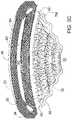
- FIG. 4is a photographic image showing a histology slide HS. Histology slide HS of FIG. 4 was created by sectioning and staining tissue from a cadaveric eye. An ocular implant was implanted in Schlemm's canal of the cadaveric eye prior to sectioning.
- FIG. 5Ais a stylized line drawing illustrating histology slide HS shown in the previous figure.
- FIG. 5Bis a simplified cross-sectional view illustrating the eye from which the histology sample was taken.
- FIG. 6is a stylized perspective view illustrating the anatomy of an eye.
- FIG. 7is a stylized perspective view depicting the surface that defines the anterior chamber of the eye shown in FIG. 6 .
- FIG. 8is a stylized perspective view further illustrating Schlemm's canal SC and iris 30 shown in FIG. 6 .
- FIGS. 9A-9Care plan views of the surface that defines anterior chamber of the eye shown in FIG. 6 .
- FIG. 10is an enlarged side view showing a cannula extending into anterior chamber defined by an inner surface of a dome shaped wall.
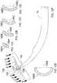
- FIGS. 11A-11Care plan views of a cannula created using multiview projection.
- FIG. 11Dis an axial view further illustrating the cannula shown in FIG. 11A .
- FIGS. 12A-12Dare lateral cross-sectional views of the tip portion of a cannula.
- FIG. 12Eis a lateral cross-sectional view of a trough portion of the cannula.
- FIG. 12Fis a plan view of the cannula including a plurality of section lines.
- FIGS. 13A-13Dform a sequence of stylized section views illustrating the insertion of the tip portion of a cannula into Schlemm's canal located in the anterior chamber of an eye.
- FIGS. 13E-13Hform a sequence stylized side plan views further illustrating the insertion of the tip portion into Schlemm's canal.
- FIG. 14is an abstract graphical representation further illustrating the insertion of the tip portion of a cannula into Schlemm's canal.
- FIG. 1is a stylized representation of a medical procedure in accordance with this detailed description.
- a physicianis treating an eye 20 of a patient P.
- the physicianis holding a hand piece of a delivery system 70 in his or her right hand RH.
- the physician's left hand LHis holding the handle H of a gonio lens 23 in the procedure of FIG. 1 .
- Some physiciansmay prefer holding the delivery system hand piece in the right hand and the gonio lens handle in the left hand.
- the physicianmay view the interior of the anterior chamber using gonio lens 23 and a microscope 25 .
- Detail A of FIG. 1is a stylized simulation of the image viewed by the physician.
- a distal portion of a cannula 72is visible in Detail A.
- a shadow-like lineindicates the location of Schlemm's canal SC, which is a tube-like structure that encircling the iris and lying under various tissue (e.g., the trabecular meshwork) that surround the anterior chamber.
- a distal opening 74 of cannula 72is positioned near Schlemm's canal SC of eye 20 .
- Methods in accordance with this detailed descriptionmay include the step of advancing the distal end of cannula 72 through the cornea of eye 20 so that a distal portion of cannula 72 is disposed in the anterior chamber of the eye.
- Cannula 72may then be used to access Schlemm's canal of the eye, for example, by piercing the wall of Schlemm's canal with the distal end of cannula 72 .
- Distal opening 74 of cannula 72may be placed in fluid communication with a lumen defined by Schlemm's canal.
- An ocular implant carried by the cannulamay be advanced out of distal opening 74 and into Schlemm's canal.
- Insertion of the ocular implant into Schlemm's canalmay facilitate the flow of aqueous humor out of the anterior chamber of the eye.
- ocular implantsthat may be delivered through the cannula of this invention may be found, e.g., in U.S. Pat. Nos. 7,740,604; 8,267,882; 8,425,449; US Patent Publ. No. 2009/0082860; and US Patent Publ. No. 2009/0082862.
- FIG. 2is an enlarged perspective view further illustrating delivery system 70 and eye 20 shown in the previous figure.
- cannula 72 of delivery system 70is shown being advanced and extending through a dome-shaped wall 90 of eye 20 .
- Dome shaped wall 90includes the cornea 36 of eye 20 and scleral tissue that meets the cornea at a limbus of the eye.
- a distal portion of cannula 72is disposed inside the anterior chamber AC defined by dome-shaped wall 90 .
- cannula 72is sized and configured so that a distal opening of cannula 72 can be placed in fluid communication with Schlemm's canal while a proximal portion of cannula 72 is extending through an incision in cornea 36 .
- an ocular implant(not shown) is disposed in a lumen or passageway within cannula 72 .
- Delivery system 70includes a mechanism that is capable of advancing and retracting the ocular implant along the length of cannula 72 . Suitable delivery systems are described in more detail in, e.g., U.S. Pat. Nos. 8,512,404; 8,337,509; US Patent Publ. No. 2011/0009874; and US Patent Publ. No. 2013/0158462.
- the ocular implantmay be placed in Schlemm's canal of eye 20 by advancing the ocular implant through the distal opening of cannula 72 while the distal opening is in fluid communication with Schlemm's canal.
- FIG. 3Ais a perspective view further illustrating eye 20 shown in the previous figure.
- cannula 72is shown extending through a cornea 36 of eye 20 .
- a distal opening 74 of cannula 72is shown disposed inside an anterior chamber AC of eye 20 .
- a cutting plane PPis shown extending across eye 20 .
- FIG. 3Bis a stylized cross-sectional view taken along cutting plane PP shown in FIG. 3A .
- the cutting plane of FIG. 3Aextends laterally across Schlemm's canal SC and the trabecular meshwork TM of the eye.
- Eye 20includes an iris 30 that defines a pupil 32 of the eye.
- Schlemm's canal SCforms a ring around iris 30 with pupil 32 disposed in the center of that ring.
- Schlemm's canal SChas a first major side 50 , a second major side 52 , a first minor side 54 , and a second minor side 56 .
- First major side 50is on the outside of the ring formed by Schlemm's canal SC and second major side 52 is on the inside of the ring formed by Schlemm's canal SC. Accordingly, first major side 50 may be referred to as an outer major side of Schlemm's canal SC and second major side 52 may be referred to as an inner major side of Schlemm's canal SC.
- first major side 50is further from pupil 32 than second major side 52 .
- first major side 50is an outer major side of Schlemm's canal SC and second major side 52 is an inner major side of Schlemm's canal SC.
- a scleral spur 80extends around minor side 56 toward the trabecular meshwork TM.
- FIG. 3Cis perspective view further illustrating the anatomy of eye 20 shown in FIG. 3B .
- Eye 20includes a dome-shaped wall 90 that defines and encloses the anterior chamber AC.
- Dome-shaped wall 90comprises a cornea 36 and scleral tissue 34 .
- the scleral tissue 34meets the cornea 36 at a limbus of eye 20 .
- Dome-shaped wall 90includes a scleral spur 80 that encircles anterior chamber AC.
- Schlemm's canal SCresides in a shallow depression in the scleral tissue located near scleral spur 80 .
- the trabecular meshwork TMis fixed to scleral spur 80 and extends over Schlemm's canal.
- Schlemm's canal SC, trabecular meshwork TM, and scleral spur 80encircle anterior chamber AC along dome-shaped wall 90 .
- Iris 30 of eye 20is disposed inside the anterior chamber AC. Iris 30 defines a pupil 32 .
- Schwalbe's line 82is disposed at the end of Descemet's membrane 84 .
- Descemet's membrane 84is one of the inner-most layers of cornea 36 . Descemet's membrane extends across cornea 36 toward Schlemm's canal SC and terminates near the upper edge of Schlemm's canal SC.
- FIG. 3Dis a perspective view showing a portion of eye shown in the previous figure.
- the tip portion of a cannula 72can be seen extending into trabecular meshwork TM.
- cannula 72can be curved to achieve substantially tangential entry into Schlemm's canal SC.
- a curved distal portion of cannula 72is dimensioned to be disposed within the anterior chamber of the eye.
- an ocular implant 86can be seen extending from a lumen in cannula 72 into a trough 140 defined by cannula 72 .
- Ocular implant 86can be advanced through a distal opening of cannula 72 along the trough 140 and into Schlemm's canal SC. Scleral spur 80 and Schwalbe's line 82 are also visible in FIG. 3D .
- FIG. 3Eis an additional perspective view showing ocular implant 86 and cannula 72 shown in the previous figure.
- ocular implant 86has been advanced in a distal direction D while cannula 72 has remained stationary so the distal end of ocular implant 86 is disposed inside Schlemm's canal SC and the remainder of the implant is disposed in trough 140 and inside the lumen of the cannula.
- Trough 140opens into an elongate opening extending through the side wall of cannula 72 .
- the elongate opening defined by the cannulaprovides direct visualization of the ocular implant as it is advanced into Schlemm's canal.
- a configuration allowing direct visualization of the ocular implanthas a number of clinical advantages.
- blood refluxmay push blood into Schlemm's canal obstructing a physician's view the portion of the implant that has entered Schlemm's canal.
- ocular implant 86tracks along trough 140 as it is advanced distally along cannula 72 into Schlemm's canal.
- the trough openingallows the physician to monitor the progress of the implant by viewing the implant structures as they advance through the trough prior to entering Schlemm's canal.
- the trough openingalso allows the physician to identify the position of the proximal end of the ocular implant with respect to the incision made by the cannula to access Schlemm's canal.
- the ocular implants referenced aboveare intended to reside partially or wholly within Schlemm's canal.
- One function of the cannulais to deliver a leading edge of the ocular implant into Schlemm's canal so that the ocular implant can be advanced circumferentially into Schlemm's canal.
- the cannula of this inventionprovides features to help the user guide the distal end of the cannula into Schlemm's canal. These cannula features take advantage of the shapes and properties of the various tissue structures of and around Schlemm's canal to achieve this goal.
- the physicianmay use anatomical landmarks to guide the cannula placement and advancement.
- One convenient landmarkis scleral spur 80 which has the appearance of a white line encircling the anterior chamber AC.
- Another convenient landmarkis a pigment line centered on Schlemm's canal SC.
- An additional convenient landmarkis Schwalbe's line 82 .
- FIG. 4is a photographic image showing a histology slide HS. Histology slide HS of FIG. 4 was created by implanting the ocular implant into Schlemm's canal of the eye, then sectioning and staining a portion of the eye. The photograph of FIG. 4 was created while examining the section of tissue using a light microscope.
- FIG. 5Ais a stylized line drawing illustrating histology slide HS shown in the previous figure.
- FIG. 5Bis a simplified cross-sectional view illustrating the eye from which the histology sample was taken.
- FIG. 5A and FIG. 5Bare presented on a single page to illustrate the location of the histology sample relative to other portions of the eye 20 .
- Eye 20includes a dome-shaped wall 90 having a surface 92 defining an anterior chamber AC. Dome-shaped wall 90 of eye 20 comprises a cornea 36 and scleral tissue 34 .
- the scleral tissue 34meets the cornea 36 at a limbus of the eye.
- surface 92is shown having a generally hemispherical shape.
- FIG. 6is a stylized perspective view illustrating a portion of eye 20 discussed above.
- Eye 20includes an iris 30 defining a pupil 32 .
- eye 20is illustrated in a cross-sectional view created by a cutting plane passing through the center of pupil 32 .
- Eye 20includes a dome-shaped wall 90 having a surface 92 defining an anterior chamber AC.
- surface 92is shown having a generally hemispherical shape.
- Dome-shaped wall 90 of eye 20comprises a cornea 36 and scleral tissue 34 .
- the scleral tissue 34meets the cornea 36 at a limbus 38 of eye 20 .
- Additional scleral tissue 34 of eye 20surrounds a posterior chamber PC filled with a viscous fluid known as vitreous humor.
- a lens 40 of eye 20is located between anterior chamber AC and posterior chamber PC. Lens 40 is held in place by a number of ciliary zonules 42 .
- the cornea and the lenscan include no blood vessels. Accordingly, no blood flows through the cornea and the lens to provide nutrition to these tissues and to remove wastes from these tissues. Instead, these functions are performed by the aqueous humor.
- a continuous flow of aqueous humor through the eyeprovides nutrition to portions of the eye (e.g., the cornea and the lens) that have no blood vessels. This flow of aqueous humor also removes waste from these tissues.
- Aqueous humoris produced by an organ known as the ciliary body.
- the ciliary bodyincludes epithelial cells that continuously secrete aqueous humor.
- a stream of aqueous humorflows out of the eye as new aqueous humor is secreted by the epithelial cells of the ciliary body. This excess aqueous humor enters the blood stream and is carried away by venous blood leaving the eye.
- FIG. 6the cutting plane passing through the center of pupil 32 has also passed through Schlemm's canal. Accordingly, two laterally cut ends of Schlemm's canal SC are visible in the cross-sectional view of FIG. 6 .
- aqueous humorflows out of anterior chamber AC and into Schlemm's canal SC.
- Aqueous humorexits Schlemm's canal SC and flows into a number of collector channels.
- aqueous humoris absorbed into the venous blood stream and carried out of the eye.
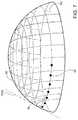
- FIG. 7is a stylized perspective view depicting the surface 92 that defines anterior chamber AC of the eye shown in FIG. 6 .
- surface 92is shown having a generally hemispherical shape.
- FIG. 7may be used to illustrate some fundamental geometric concepts that will be used below to describe the various ocular implant delivery cannula structures. Geometry is a branch of mathematics concerned with the properties of space and the shape, size, and relative position of objects within that space. In geometry, a sphere is a round object in three-dimensional space. All points on the surface of a sphere are located the same distance r from a center point so that the sphere is completely symmetrical about the center point. In geometry, a point represents an exact location.
- a pointis a zero-dimensional entity (i.e., it has no length, area, or volume).
- Geometrically speakingat any point on a spherical surface, one can find a normal direction which is at right angles to the surface. For a spherical surface all normal directions intersect the center point of the sphere. Each normal direction will also be perpendicular to a line that is tangent to the spherical surface.
- a normal line Nis illustrated using dashed lines. Normal line N is at right angles to spherical surface 92 . Normal line N is also perpendicular to a reference line TAN. Reference line TAN is tangent to spherical surface 92 in FIG. 7 .
- a method in accordance with this detailed descriptionmay include the step of advancing a distal portion of a cannula into the anterior chamber of the eye.
- the cannulamay then be used to access Schlemm's canal, for example, by piercing the wall of Schlemm's canal with the distal end of the cannula.
- An ocular implantmay be advanced out of the distal opening of the cannula and into Schlemm's canal.
- a path 94 taken by an ocular implant as it follows Schlemm's canal along surface 92is illustrated using a row of dots in FIG. 7 .
- the ocular implantmay press against the scleral tissue supporting the outer major wall of Schlemm's canal and the scleral tissue of the dome-shaped wall that defines the anterior chamber of the eye.
- the dome-shaped wallprovides support for Schlemm's canal and the ocular implant.
- the support provided by the dome-shaped wallmay be represented by force vectors. The direction of these force vectors may be at right angles to points on the spherical surface that defines the anterior chamber. Accordingly, the outer major wall of Schlemm's canal may be supported by the dome shaped wall as the ocular implant advances circumferentially into Schlemm's canal.
- the ocular implantfollow the lumen of Schlemm's canal as it is advanced out the distal opening of the cannula.
- the ability of the ocular implant to be advanced into and follow the lumen of Schlemm's canalmay be referred to as trackability.
- Characteristics of an ocular implant that effect trackabilityinclude axial pushability and lateral flexibility.
- Axial pushabilitygenerally concerns the ability of an ocular implant to transmit to the distal end of the ocular implant an axial force applied to the proximal end of the ocular implant.
- Lateral flexibilityconcerns the ease with which the ocular implant body can bend to conform to the shape of the lumen. Trackability may be adversely affected when twisting forces are applied to a curved body. For example, twisting the body of a curved ocular implant about its longitudinal axis may cause the curved body to steer away from a desired path.
- FIG. 8is a stylized perspective view further illustrating Schlemm's canal SC and iris 30 shown in FIG. 6 .
- the surface 92 that defines the anterior chamber AC of eye 20is depicted using dashed lines in FIG. 8 .
- Schlemm's canal SC and iris 30are shown in cross-section, with a cutting plane passing through the center of a pupil 32 defined by iris 30 .
- Schlemm's canal SCcomprises a first major side 50 , a second major side 52 , a first minor side 54 , and a second minor side 56 .
- Schlemm's canal SCforms a ring around iris 30 with pupil 32 disposed in the center of that ring.
- first major side 50is on the outside of the ring formed by Schlemm's canal SC and second major side 52 is on the inside of the ring formed by Schlemm's canal SC. Accordingly, first major side 50 may be referred to as an outer major side of Schlemm's canal SC and second major side 52 may be referred to as an inner major side of Schlemm's canal SC. With reference to FIG. 8 , it will be appreciated that first major side 50 is further from pupil 32 than second major side 52 .
- a path 94 taken by an ocular implant as it follows Schlemm's canal along surface 92is illustrated using a row of dots in FIG. 8 .
- the ocular implantmay press against the outer major wall of Schlemm's canal and the dome-shaped wall that defines the anterior chamber.
- Some embodimentsinclude an ocular implant delivery cannula with a distal tip that is offset from the longitudinal center line of the cannula. This arrangement facilitates the intuitive use of anatomical landmarks that can be easy observed using gonioscopic visualization.
- the tip portion of the cannulawill pierce the trabecular meshwork and the wall of Schlemm's canal at a point slightly above the center of Schlemm's canal.
- the offset distal tipalso provides the distal end of the cannula with a lower camming surface for guiding the cannula distal end over the scleral spur and an optional upper camming surface for guiding the cannula distal end into Schlemm's canal when the cannula has a diameter larger than a width of Schlemm's canal.
- the camming surfacesare configured to direct the cannula into Schlemm's canal when the cannula is wider or oversized with respect to a width of the canal.
- FIGS. 9A-9Care plan views of the surface 92 that defines anterior chamber AC of the eye shown in FIG. 6 .
- FIG. 9Amay be referred to as a front view of surface 92
- FIG. 9Bmay be referred to as a top view of surface 92
- FIG. 9Cmay be referred to as a side view of surface 92 .
- a cannula 72is shown extending into anterior chamber AC.
- Cannula 72may be used to deliver an ocular implant to a target location within anterior chamber AC.
- target locationsthat may be suitable in some applications include areas in and around Schlemm's canal, the trabecular meshwork, and the suprachoroidal space of an eye.
- a path 94 that may be taken by an ocular implant as it follows Schlemm's canal along surface 92is illustrated using a row of dots in FIGS. 9A-9C .
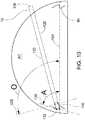
- FIG. 10is an enlarged side view showing cannula 72 extending into anterior chamber AC defined by surface 92 .
- Cannula 72may be used, for example, to deliver an ocular implant to a target location within Schlemm canal SC.
- a scleral spur 80is disposed in anterior chamber AC. Scleral spur 80 is fixed to surface 92 and encircles anterior chamber AC. Scleral spur 80 defines a spur plane 104 .
- cannula 72can include a body member 120 extending along a longitudinal axis.
- Body member 120can include a proximal end 126 and a tubular portion 130 extending distally from the proximal end.
- Body member 120can also include a tip portion 132 disposed at a distal end thereof.
- a trough portion 140 of body memberextends between tip portion 132 and tubular portion 130 .
- tip portion 132has a semi-circular transverse cross-section including a tip chord line segment.
- a secant 136 extending beyond the tip chordis shown in FIG. 10 .
- Trough portion 140 of body member 120has a semi-circular transverse cross-section including a trough chord line segment.
- FIG. 10includes a secant 138 extending beyond the trough cord.
- tip portion 132 and trough portion 140are adapted and configured such that, when tubular portion 130 is extending through an incision in the dome shaped wall defining anterior chamber AC and tip portion 132 is extending into Schlemm's canal of the eye, secant 136 intersects spur plane 104 at an acute angle A and secant 138 intersects spur plane 104 at an obtuse angle O.
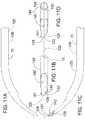
- FIGS. 11A-11Care plan views of cannula 72 created using multiview projection.
- FIG. 11Dis an axial view further illustrating cannula 72 .
- Cannula 72 of FIGS. 11A-11Dmay be used to deliver an ocular implant into Schlemm's canal of an eye.
- FIG. 11Amay be referred to as a top view of cannula 72
- FIG. 11Bmay be referred to as a side view of cannula 72
- FIG. 11Cmay be referred to as a bottom view of cannula 72 .
- cannula 72comprises a body member 120 extending along a medial plane 122 .
- Body member 120can include a proximal end 126 and a tubular portion 130 extending distally from the proximal end.
- Body member 120can also include a tip portion 132 disposed at a distal tip 128 thereof.
- the distal tip 128can be offset from the medial plane 122 of body member 120 .
- the distal tip 128can form a point at the intersection of lower camming surface 129 and upper camming surface 131 .
- the distal tipmay be at one side of the cannula, in which case there will be no upper camming surface.
- distal tip 128can be sharpened enough to pierce trabecular meshwork tissue but not sharp enough to easily pierce scleral tissue.
- Body member 120also includes a trough portion 140 extending between distal tip 128 and tubular portion 130 .
- Trough portion 140is configured to fluidly communicates with a lumen 144 defined by tubular portion 130 and a distal opening 142 defined by tip portion 132 . Because of the offset position of distal tip 128 , tip portion 132 is asymmetric about medial plane 122 and trough portion 140 is symmetric about medial plane 124 .
- FIG. 12A through FIG. 12Dare lateral cross-sectional views of tip portion 132 of cannula 72 .
- FIG. 12Eis a lateral cross-sectional view of trough portion 140 of cannula 72 .
- FIG. 12Fis an enlarged plan view showing a portion of cannula 72 shown in the previous figure.
- the cannulais formed from a tube (such as a hypotube) with material removed from the distal end to form the trough portion and the distal tip 129 .
- the cannulamay have a non-tubular shape.
- FIG. 12Fshows the cannula 72 including the tip portion 132 , distal tip 128 , camming surfaces 129 and 131 , and trough portion 140 .
- a number of section linescan be seen traversing crossing cannula 72 . These section lines have been used to create a number of lateral cross-sections illustrating the shape of cannula 72 .
- Section 146 A of FIG. 12Awas created by cutting tip portion 132 along section line A-A shown in FIG. 12F .
- Section 146 B, section 146 C, and section 146 D, of FIGS. 12B, 12C , and 12 D, respectively,were made by cutting tip portion 132 along section line B-B, section line C-C, and section line D-D, respectively.
- tip portion 132can have a semi-circular transverse cross-section.
- section 146 Ahas a chord 136 A.
- Section 146 B, section 146 C, and section 146 Dhave a chord 136 B, a chord 136 C and a chord 136 D, respectively.
- chord 136 A, chord 136 B, chord 136 C and chord 136 Dit will be appreciated that the chord length of tip portion 132 increases as tip portion 132 extends proximally away from its distal point.
- Section 146 Ewas created by cutting through portion 140 along section line E-E shown in FIG. 12F . In the embodiment of FIG. 12E , section 146 E has a chord 136 E.
- the camming surfaces 129 and 131 and the cannula's tip portion 132are configured to guide an oversized cannula relative to the width of Schlemm's canal into Schlemm's canal.
- a diameter of the cannulacan be between approximately 350-550 microns, or alternatively, between 400-500 microns.
- Schlemm's canaltypically has a width of approximately 300 microns, so it can be a challenge to guide a conventional cannula that is wider than Schlemm's canal into the canal.
- the upper camming surface 131 of the cannulawill engage scleral tissue above the meshwork. Since the distal tip 128 is not sharp enough to easily pierce scleral tissue, upper camming surface 131 is configured to contact the scleral tissue and guide the distal tip 128 into Schlemm's canal.
- the lower camming surface 129is configured to contact the scleral spur below the meshwork to guide the tip 128 into the Schlemm's canal.
- the distal tip's offsetplacing it above the cannula's longitudinal center axis, along with the physician's use of the anatomical landmarks, helps ensure that the cannula is not positioned so low with respect to the meshwork that the upper camming surface engages the scleral spur to push the cannula tip downward away from the meshwork.
- FIGS. 13A-13Dform a sequence of stylized section views illustrating the insertion of tip portion 132 of cannula 72 into Schlemm's canal SC located in the anterior chamber AC of an eye.
- FIGS. 13E-13Hform a sequence stylized side plan views further illustrating the insertion of the tip portion into Schlemm's canal.
- tip portion 132 of cannula 72has been advanced into Schlemm's canal so that section 146 A (shown in FIG. 12A ) of tip portion 132 is aligned with the incision in Schlemm's canal created by the cannula's distal tip 128 .
- Section 146 Aincludes a chord 136 A. Referring to FIG. 13A , chord 136 A defines a line that intersects a spur plane 104 of the eye at a chord angle 148 A.
- Spur plane 104is defined by a scleral spur 102 that encircles the anterior chamber AC of the eye.
- tip portion 132 of cannula 72has been advanced into Schlemm's canal so that section 146 B of tip portion 132 is aligned with the incision in Schlemm's canal.
- Section 146 Bincludes a chord 136 B.
- chord 136 Bdefines a line that intersects spur plane 104 at a chord angle 148 B.
- tip portion 132 of cannula 72has been advanced into Schlemm's canal so that section 146 C of tip portion 132 is aligned with the incision in Schlemm's canal.
- Section 146 Cincludes a chord 136 C.
- chord 136 Cdefines a line that intersects spur plane 104 at a chord angle 148 C.
- tip portion 132 of cannula 72has been advanced into Schlemm's canal so that section 146 D of tip portion 132 is aligned with the incision in Schlemm's canal.
- Section 146 Dincludes a chord 136 D.
- chord 136 Ddefines a line that intersects spur plane 104 at a chord angle 148 D.
- FIG. 14is an abstract graphical representation further illustrating the insertion of tip portion 132 of a cannula into Schlemm's canal SC.
- the profile of each section view illustrated in FIGS. 12A-12Dis included in FIG. 14 .
- These profilesform contour lines illustrating the tapered shape of tip portion 132 and trough portion 140 .
- the profiles associated with section 146 A, section 146 B, section 146 C, section 146 D, and section 146 Eare labeled in FIG. 14 .
- inner major wall 52 of Schlemm's canalrides along a first leading edge of tip portion 132 .
- the insertion of tip portion 132 into Schlemm's canal SCcauses inner major wall 52 to separate from outer major wall 50 .
- the changing shape of Schlemm's canalis illustrated with a plurality of Schlemm's canal profiles shown using dashed lines in FIG. 14 .
- tip portion 132 and trough portion 140are adapted and configured such that, when tubular portion 130 is extending through an incision in the dome shaped wall defining anterior chamber AC and tip portion 132 is extending into Schlemm's canal of the eye, secant 136 intersects spur plane 104 at an acute angle A and secant 138 intersects spur plane 104 at an obtuse angle O.
Landscapes
- Health & Medical Sciences (AREA)
- Ophthalmology & Optometry (AREA)
- Engineering & Computer Science (AREA)
- Biomedical Technology (AREA)
- Heart & Thoracic Surgery (AREA)
- Vascular Medicine (AREA)
- Life Sciences & Earth Sciences (AREA)
- Animal Behavior & Ethology (AREA)
- General Health & Medical Sciences (AREA)
- Public Health (AREA)
- Veterinary Medicine (AREA)
- Nuclear Medicine, Radiotherapy & Molecular Imaging (AREA)
- Surgery (AREA)
- Prostheses (AREA)
Abstract
Description
Claims (32)
Priority Applications (1)
| Application Number | Priority Date | Filing Date | Title |
|---|---|---|---|
| US14/440,610US10617558B2 (en) | 2012-11-28 | 2013-11-26 | Apparatus for delivering ocular implants into an anterior chamber of the eye |
Applications Claiming Priority (3)
| Application Number | Priority Date | Filing Date | Title |
|---|---|---|---|
| US201261730895P | 2012-11-28 | 2012-11-28 | |
| PCT/US2013/072001WO2014085450A1 (en) | 2012-11-28 | 2013-11-26 | Apparatus for delivering ocular implants into an anterior chamber of the eye |
| US14/440,610US10617558B2 (en) | 2012-11-28 | 2013-11-26 | Apparatus for delivering ocular implants into an anterior chamber of the eye |
Related Parent Applications (2)
| Application Number | Title | Priority Date | Filing Date |
|---|---|---|---|
| USPCT/US2016/072001A-371-Of-International | 2013-11-26 | ||
| PCT/US2013/072001A-371-Of-InternationalWO2014085450A1 (en) | 2012-11-28 | 2013-11-26 | Apparatus for delivering ocular implants into an anterior chamber of the eye |
Related Child Applications (1)
| Application Number | Title | Priority Date | Filing Date |
|---|---|---|---|
| US16/805,217ContinuationUS11712369B2 (en) | 2012-11-28 | 2020-02-28 | Apparatus for delivering ocular implants into an anterior chamber of the eye |
Publications (2)
| Publication Number | Publication Date |
|---|---|
| US20150282982A1 US20150282982A1 (en) | 2015-10-08 |
| US10617558B2true US10617558B2 (en) | 2020-04-14 |
Family
ID=50828415
Family Applications (3)
| Application Number | Title | Priority Date | Filing Date |
|---|---|---|---|
| US14/440,610Active2034-06-22US10617558B2 (en) | 2012-11-28 | 2013-11-26 | Apparatus for delivering ocular implants into an anterior chamber of the eye |
| US16/805,217Active2035-05-13US11712369B2 (en) | 2012-11-28 | 2020-02-28 | Apparatus for delivering ocular implants into an anterior chamber of the eye |
| US18/334,256Active2034-04-29US12376988B2 (en) | 2012-11-28 | 2023-06-13 | Apparatus for delivering ocular implants into an anterior chamber of the eye |
Family Applications After (2)
| Application Number | Title | Priority Date | Filing Date |
|---|---|---|---|
| US16/805,217Active2035-05-13US11712369B2 (en) | 2012-11-28 | 2020-02-28 | Apparatus for delivering ocular implants into an anterior chamber of the eye |
| US18/334,256Active2034-04-29US12376988B2 (en) | 2012-11-28 | 2023-06-13 | Apparatus for delivering ocular implants into an anterior chamber of the eye |
Country Status (2)
| Country | Link |
|---|---|
| US (3) | US10617558B2 (en) |
| WO (1) | WO2014085450A1 (en) |
Cited By (15)
| Publication number | Priority date | Publication date | Assignee | Title |
|---|---|---|---|---|
| US11540940B2 (en) | 2021-01-11 | 2023-01-03 | Alcon Inc. | Systems and methods for viscoelastic delivery |
| US11712369B2 (en) | 2012-11-28 | 2023-08-01 | Alcon Inc. | Apparatus for delivering ocular implants into an anterior chamber of the eye |
| US11918514B2 (en) | 2009-07-09 | 2024-03-05 | Alcon Inc. | Single operator device for delivering an ocular implant |
| US11992437B2 (en) | 2012-04-18 | 2024-05-28 | Alcon Inc. | Ocular implants for delivery into an anterior chamber of the eye |
| US12016796B2 (en) | 2007-09-24 | 2024-06-25 | Alcon Inc. | Methods and devices for increasing aqueous humor outflow |
| US12029683B2 (en) | 2018-02-22 | 2024-07-09 | Alcon Inc. | Ocular implant and delivery system |
| US12076273B2 (en) | 2011-12-19 | 2024-09-03 | Alcon Inc. | Delivering ocular implants into the eye |
| US12083044B2 (en) | 2019-07-10 | 2024-09-10 | Aquea Health, Inc. | Eye stents and delivery systems |
| US12226309B2 (en) | 2013-03-15 | 2025-02-18 | Alcon Inc. | Intraocular lens storage and loading devices and methods of use |
| US12245930B2 (en) | 2023-06-30 | 2025-03-11 | Alcon Inc. | System and methods for compensating for intraocular lens tilt |
| US12257188B2 (en) | 2019-06-14 | 2025-03-25 | Iantrek, Inc. | Implantable biologic stent and system for biologic material shaping, preparation, and intraocular stenting for increased aqueous outflow and lowering of intraocular pressure |
| US12318328B2 (en) | 2020-05-20 | 2025-06-03 | Iantrek, Inc. | System for shaping and implanting biologic intraocular stent for increased aqueous outflow and lowering of intraocular pressure |
| US12318279B2 (en) | 2007-07-23 | 2025-06-03 | Alcon Inc. | Lens delivery system |
| US12419738B2 (en) | 2010-02-23 | 2025-09-23 | Alcon Inc. | Fluid for accommodating intraocular lenses |
| US12440377B2 (en) | 2024-11-13 | 2025-10-14 | Aquea Health, Inc. | Eye stents and delivery systems and methods |
Families Citing this family (15)
| Publication number | Priority date | Publication date | Assignee | Title |
|---|---|---|---|---|
| US9603741B2 (en) | 2000-05-19 | 2017-03-28 | Michael S. Berlin | Delivery system and method of use for the eye |
| US7909789B2 (en) | 2006-06-26 | 2011-03-22 | Sight Sciences, Inc. | Intraocular implants and methods and kits therefor |
| US8425449B2 (en) | 2009-07-09 | 2013-04-23 | Ivantis, Inc. | Ocular implants and methods for delivering ocular implants into the eye |
| JP2011513002A (en) | 2008-03-05 | 2011-04-28 | イバンティス インコーポレイテッド | Method and apparatus for treating glaucoma |
| US8529622B2 (en) | 2010-02-05 | 2013-09-10 | Sight Sciences, Inc. | Intraocular implants and related kits and methods |
| US8657776B2 (en) | 2011-06-14 | 2014-02-25 | Ivantis, Inc. | Ocular implants for delivery into the eye |
| ES2842454T3 (en) | 2012-03-20 | 2021-07-14 | Sight Sciences Inc | Eye delivery systems |
| WO2016011056A1 (en) | 2014-07-14 | 2016-01-21 | Ivantis, Inc. | Ocular implant delivery system and method |
| US10299958B2 (en) | 2015-03-31 | 2019-05-28 | Sight Sciences, Inc. | Ocular delivery systems and methods |
| AU2016307951B2 (en) | 2015-08-14 | 2021-04-01 | Alcon Inc. | Ocular implant with pressure sensor and delivery system |
| WO2017106517A1 (en) | 2015-12-15 | 2017-06-22 | Ivantis, Inc. | Ocular implant and delivery system |
| JP2022538906A (en) | 2019-07-01 | 2022-09-06 | マイケル エス. バーリン, | Image guided method and apparatus for glaucoma surgery |
| US11504270B1 (en) | 2019-09-27 | 2022-11-22 | Sight Sciences, Inc. | Ocular delivery systems and methods |
| WO2021202313A1 (en) | 2020-03-31 | 2021-10-07 | Berlin Michael S | Endoscopic instrument for ophthalmic surgery |
| USD1037439S1 (en) | 2022-01-17 | 2024-07-30 | EyePoint Pharamaceuticals, Inc. | Ocular injector |
Citations (312)
| Publication number | Priority date | Publication date | Assignee | Title |
|---|---|---|---|---|
| US703296A (en) | 1901-06-15 | 1902-06-24 | Arnold Nueesch | Cattle-probe. |
| US1601709A (en) | 1924-01-28 | 1926-10-05 | Anderson Windom Edward | Retainable needle construction for syringes |
| US2716983A (en) | 1952-10-08 | 1955-09-06 | Abbott Lab | Piercing needle |
| US3071135A (en) | 1960-01-27 | 1963-01-01 | Mfg Process Lab Inc | Hollow needle |
| US3788327A (en) | 1971-03-30 | 1974-01-29 | H Donowitz | Surgical implant device |
| US3811442A (en) | 1972-03-23 | 1974-05-21 | A Maroth | Hypodermic syringe holder and applicator |
| US3948271A (en) | 1972-11-07 | 1976-04-06 | Taichiro Akiyama | Drain for the eardrum and apparatus for introducing the same |
| US4037604A (en) | 1976-01-05 | 1977-07-26 | Newkirk John B | Artifical biological drainage device |
| US4134405A (en) | 1977-01-10 | 1979-01-16 | Smit Julie A | Catheter and intestine tube and method of using the same |
| US4428746A (en) | 1981-07-29 | 1984-01-31 | Antonio Mendez | Glaucoma treatment device |
| US4457757A (en) | 1981-07-20 | 1984-07-03 | Molteno Anthony C B | Device for draining aqueous humour |
| US4601713A (en) | 1985-06-11 | 1986-07-22 | Genus Catheter Technologies, Inc. | Variable diameter catheter |
| US4604087A (en) | 1985-02-26 | 1986-08-05 | Joseph Neil H | Aqueous humor drainage device |
| US4689040A (en) | 1985-04-29 | 1987-08-25 | Thompson Robert J | Tip for a phacoemulsification needle |
| US4699140A (en) | 1985-07-10 | 1987-10-13 | Iolab Corporation | Instrument for inserting an intraocular lens |
| US4706669A (en) | 1984-01-30 | 1987-11-17 | Schlegel Hans Joachim | Device for perforating the lens capsule front wall in the eye of living beings |
| US4722724A (en) | 1986-06-23 | 1988-02-02 | Stanley Schocket | Anterior chamber tube shunt to an encircling band, and related surgical procedure |
| US4733665A (en) | 1985-11-07 | 1988-03-29 | Expandable Grafts Partnership | Expandable intraluminal graft, and method and apparatus for implanting an expandable intraluminal graft |
| US4750901A (en) | 1986-03-07 | 1988-06-14 | Molteno Anthony C B | Implant for drainage of aqueous humour |
| EP0168201B1 (en) | 1984-06-28 | 1988-06-22 | Neil Howard Joseph | Aqueous humour drainage device |
| US4826478A (en) | 1986-06-23 | 1989-05-02 | Stanley Schocket | Anterior chamber tube shunt to an encircling band, and related surgical procedure |
| US4861341A (en) | 1988-07-18 | 1989-08-29 | Woodburn Robert T | Subcutaneous venous access device and needle system |
| US4880000A (en) | 1987-12-15 | 1989-11-14 | Iolab Corporation | Lens insertion instrument |
| US4886488A (en) | 1987-08-06 | 1989-12-12 | White Thomas C | Glaucoma drainage the lacrimal system and method |
| US4919130A (en) | 1986-11-07 | 1990-04-24 | Nestle S.A. | Tool for inserting compressible intraocular lenses into the eye and method |
| US4934363A (en) | 1987-12-15 | 1990-06-19 | Iolab Corporation | Lens insertion instrument |
| US4934809A (en) | 1988-06-24 | 1990-06-19 | Volk Donald A | Lens positioning device for indirect biomicroscopy of the eye |
| US4936825A (en) | 1988-04-11 | 1990-06-26 | Ungerleider Bruce A | Method for reducing intraocular pressure caused by glaucoma |
| US4946436A (en) | 1989-11-17 | 1990-08-07 | Smith Stewart G | Pressure-relieving device and process for implanting |
| US4968296A (en) | 1989-12-20 | 1990-11-06 | Robert Ritch | Transscleral drainage implant device for the treatment of glaucoma |
| US5092837A (en) | 1989-12-20 | 1992-03-03 | Robert Ritch | Method for the treatment of glaucoma |
| US5127901A (en) | 1990-05-18 | 1992-07-07 | Odrich Ronald B | Implant with subconjunctival arch |
| US5178604A (en) | 1990-05-31 | 1993-01-12 | Iovision, Inc. | Glaucoma implant |
| US5180362A (en) | 1990-04-03 | 1993-01-19 | Worst J G F | Gonio seton |
| US5190552A (en) | 1992-02-04 | 1993-03-02 | Kelman Charles D | Slotted tube injector for an intraocular lens |
| US5213569A (en) | 1992-03-31 | 1993-05-25 | Davis Peter L | Tip for a tissue phacoemulsification device |
| DE4226476C1 (en) | 1992-08-10 | 1993-08-12 | Hans Dr.Med. 3015 Wennigsen De Haindl | |
| US5246452A (en) | 1992-04-13 | 1993-09-21 | Impra, Inc. | Vascular graft with removable sheath |
| US5290267A (en) | 1991-01-17 | 1994-03-01 | Fresenius Ag | Hypodermic needle |
| US5360399A (en) | 1992-01-10 | 1994-11-01 | Robert Stegmann | Method and apparatus for maintaining the normal intraocular pressure |
| US5445637A (en) | 1993-12-06 | 1995-08-29 | American Cyanamid Company | Method and apparatus for preventing posterior capsular opacification |
| US5454796A (en) | 1991-04-09 | 1995-10-03 | Hood Laboratories | Device and method for controlling intraocular fluid pressure |
| US5458615A (en) | 1993-07-06 | 1995-10-17 | Advanced Cardiovascular Systems, Inc. | Stent delivery system |
| US5536259A (en) | 1995-07-28 | 1996-07-16 | Medisystems Technology Corp | Hypodermic cannula |
| US5575780A (en) | 1995-04-28 | 1996-11-19 | Saito; Yoshikuni | Medical hollow needle and a method of producing thereof |
| US5591223A (en) | 1992-11-23 | 1997-01-07 | Children's Medical Center Corporation | Re-expandable endoprosthesis |
| US5613972A (en) | 1992-07-15 | 1997-03-25 | The University Of Miami | Surgical cutting heads with curled cutting wings |
| US5626558A (en) | 1995-05-05 | 1997-05-06 | Suson; John | Adjustable flow rate glaucoma shunt and method of using same |
| US5653753A (en) | 1994-04-29 | 1997-08-05 | Allergan | Method and apparatus for folding of intraocular lenses |
| US5676669A (en) | 1993-04-30 | 1997-10-14 | Colvard; Michael | Intraocular capsular shield |
| EP0766544B1 (en) | 1994-06-22 | 1998-05-13 | Chauvin Opsia | Sclerotomy implant |
| JPH10504978A (en) | 1994-09-01 | 1998-05-19 | ユニバーシティ オブ マイアミ | Adjustable keratoplasty syringe with gel injection |
| US5792099A (en) | 1995-02-14 | 1998-08-11 | Decamp; Dennis | Syringe and cannula for insertion of viscoelastic material into an eye and method of using same |
| US5807302A (en) | 1996-04-01 | 1998-09-15 | Wandel; Thaddeus | Treatment of glaucoma |
| US5865831A (en) | 1996-04-17 | 1999-02-02 | Premier Laser Systems, Inc. | Laser surgical procedures for treatment of glaucoma |
| US5868697A (en) | 1995-05-14 | 1999-02-09 | Optonol Ltd. | Intraocular implant |
| AU7619798A (en) | 1997-08-15 | 1999-02-25 | Grieshaber & Co. Ag Schaffhausen | Method and device to improve aqueous humor drainage in an eye |
| US5893837A (en) | 1997-02-28 | 1999-04-13 | Staar Surgical Company, Inc. | Glaucoma drain implanting device and method |
| US5919171A (en) | 1994-08-03 | 1999-07-06 | Kanegafuchi Kagaku Kogyo Kabushiki Kaisha | Microcatheter |
| US5948427A (en) | 1996-04-25 | 1999-09-07 | Point Medical Corporation | Microparticulate surgical adhesive |
| US5968058A (en) | 1996-03-27 | 1999-10-19 | Optonol Ltd. | Device for and method of implanting an intraocular implant |
| EP0957949A1 (en) | 1995-05-14 | 1999-11-24 | Optonol Ltd. | Intraocular implant, delivery device, and method of implantation |
| US6007511A (en) | 1991-05-08 | 1999-12-28 | Prywes; Arnold S. | Shunt valve and therapeutic delivery system for treatment of glaucoma and methods and apparatus for its installation |
| WO2000007525A1 (en) | 1998-08-05 | 2000-02-17 | Keravision, Inc. | Corneal implant with migration preventer |
| US6050970A (en) | 1997-05-08 | 2000-04-18 | Pharmacia & Upjohn Company | Method and apparatus for inserting a glaucoma implant in an anterior and posterior segment of the eye |
| US6102045A (en) | 1994-07-22 | 2000-08-15 | Premier Laser Systems, Inc. | Method and apparatus for lowering the intraocular pressure of an eye |
| WO2000064393A1 (en) | 1999-04-26 | 2000-11-02 | Lynch Mary G | Shunt device and method for treating glaucoma |
| US6186974B1 (en) | 1997-01-10 | 2001-02-13 | University College London And Moorfields Eye Hospital Nhs Trust | Device for use in the eye |
| US6217584B1 (en) | 1996-05-09 | 2001-04-17 | Aharon Lehrer | Method and a system for performing cataract surgery |
| US6221078B1 (en) | 1999-06-25 | 2001-04-24 | Stephen S. Bylsma | Surgical implantation apparatus |
| US6238409B1 (en) | 1997-03-10 | 2001-05-29 | Johnson & Johnson Interventional Systems Co. | Articulated expandable intraluminal stent |
| US20010002438A1 (en) | 1994-12-22 | 2001-05-31 | Ivan Sepetka | Implant delivery assembly with expandable coupling/decoupling mechanism |
| US6241721B1 (en) | 1998-10-09 | 2001-06-05 | Colette Cozean | Laser surgical procedures for treatment of glaucoma |
| USD444874S1 (en) | 2000-07-31 | 2001-07-10 | Allergan Sales, Inc. | Self instill twist housing eye drop dispenser |
| US6328747B1 (en) | 1996-05-09 | 2001-12-11 | Itos Innovative Technology In Ocular Surgery, Ltd. | Method and a system for performing cataract surgery |
| WO2001097727A1 (en) | 2000-06-19 | 2001-12-27 | Glaukos Corporation | Stented trabecular shunt and methods thereof |
| US20020003546A1 (en) | 2000-05-31 | 2002-01-10 | Agency Of Industrial Science And Technology | Virtual shape generation method and device using the same |
| US20020013572A1 (en) | 2000-05-19 | 2002-01-31 | Berlin Michael S. | Delivery system and method of use for the eye |
| US6375642B1 (en) | 2000-02-15 | 2002-04-23 | Grieshaber & Co. Ag Schaffhausen | Method of and device for improving a drainage of aqueous humor within the eye |
| US20020052653A1 (en) | 1998-07-06 | 2002-05-02 | Russell Durgin | Implant system and method for bulking tissue |
| WO2002036052A1 (en) | 2000-11-01 | 2002-05-10 | Glaukos Corporation | Glaucoma treatment device |
| US20020072673A1 (en) | 1999-12-10 | 2002-06-13 | Yamamoto Ronald K. | Treatment of ocular disease |
| US6409752B1 (en) | 1993-07-23 | 2002-06-25 | Cook Incorporated | Flexible stent having a pattern formed from a sheet of material |
| US20020133168A1 (en) | 2001-03-16 | 2002-09-19 | Smedley Gregory T. | Applicator and methods for placing a trabecular shunt for glaucoma treatment |
| US20020143284A1 (en) | 2001-04-03 | 2002-10-03 | Hosheng Tu | Drug-releasing trabecular implant for glaucoma treatment |
| WO2002080811A2 (en) | 2001-04-07 | 2002-10-17 | Glaukos Corporation | Glaucoma stent and methods thereof for glaucoma treatment |
| US6471666B1 (en) | 2000-02-24 | 2002-10-29 | Steven A. Odrich | Injectable glaucoma device |
| US20020165504A1 (en) | 2000-06-09 | 2002-11-07 | Inviro Medical Devices Ltd. | Cannula for use with a medical syringe |
| JP2002542872A (en) | 1999-05-03 | 2002-12-17 | ベントリカ, インコーポレイテッド | Methods and apparatus for placing a conduit in fluid communication with a target vessel |
| US6494857B1 (en) | 1998-09-02 | 2002-12-17 | Thomas Neuhann | Device for improving in a targeted manner and/or permanently ensuring the ability of the aqueous humor to pass through the trabecular meshwork |
| US20020193805A1 (en) | 2001-03-19 | 2002-12-19 | Allergan Sales, Inc. | IOL insertion apparatus with IOL engagement structure and method for using same |
| US20030004457A1 (en) | 2001-06-26 | 2003-01-02 | Andersson Stig O. | Hypodermic implant device |
| US6517523B1 (en) | 1999-03-15 | 2003-02-11 | Kaneko Kogyo Inc. | Needle for injection syringe and method for manufacturing the same |
| US20030040754A1 (en) | 1999-03-18 | 2003-02-27 | Michael Mitchell | Radially expanding stents |
| WO2003015659A2 (en) | 2001-08-16 | 2003-02-27 | Gmp Vision Solutions, Inc. | Improved shunt device and method for treating glaucoma |
| US6533768B1 (en) | 2000-04-14 | 2003-03-18 | The Regents Of The University Of California | Device for glaucoma treatment and methods thereof |
| US6533764B1 (en) | 2000-11-06 | 2003-03-18 | Allergan, Inc. | Twist housing apparatus for instilling a medication into an eye |
| US20030055372A1 (en) | 1999-04-26 | 2003-03-20 | Lynch Mary G. | Shunt device and method for treating glaucoma |
| US20030060748A1 (en) | 2001-01-19 | 2003-03-27 | Georges Baikoff | Techniques and implants for correcting presbyopia |
| US20030060752A1 (en) | 2000-04-14 | 2003-03-27 | Olav Bergheim | Glaucoma device and methods thereof |
| US20030060784A1 (en) | 1999-02-04 | 2003-03-27 | Hilgers Michael Edward | Needle for body fluid tester |
| US6544208B2 (en) | 2000-12-29 | 2003-04-08 | C. Ross Ethier | Implantable shunt device |
| US6544249B1 (en) | 1996-11-29 | 2003-04-08 | The Lions Eye Institute Of Western Australia Incorporated | Biological microfistula tube and implantation method and apparatus |
| US6551289B1 (en) | 1999-09-14 | 2003-04-22 | Dr. Japan Co., Ltd. | Outer needle of anesthetic needle assembly for epidural |
| US20030093084A1 (en) | 2001-11-13 | 2003-05-15 | Optonol Ltd. | Delivery devices for flow regulating implants |
| US20030097151A1 (en) | 2001-10-25 | 2003-05-22 | Smedley Gregory T. | Apparatus and mitochondrial treatment for glaucoma |
| WO2003045290A1 (en) | 2001-11-21 | 2003-06-05 | Iscience Corporation | Ophthalmic microsurgical system |
| US20030125351A1 (en) | 1998-08-17 | 2003-07-03 | Mitsuyoshi Azuma | Agent for prophylaxis and treatment of glaucoma |
| US20030181848A1 (en) | 2000-04-14 | 2003-09-25 | Bergheim Olav B. | Implant with drug coating |
| US20030229303A1 (en) | 2002-03-22 | 2003-12-11 | Haffner David S. | Expandable glaucoma implant and methods of use |
| US6666841B2 (en) | 2001-05-02 | 2003-12-23 | Glaukos Corporation | Bifurcatable trabecular shunt for glaucoma treatment |
| US20030236483A1 (en) | 2002-06-25 | 2003-12-25 | Ren David H | Dual drainage ocular shunt for glaucoma |
| US20040024345A1 (en) | 2002-04-19 | 2004-02-05 | Morteza Gharib | Glaucoma implant with valveless flow bias |
| US20040024453A1 (en) | 2001-08-03 | 2004-02-05 | Glaucoma Research Technologies, Inc. | Method and intra sclera implant for treatment of glaucoma and presbyopia |
| US20040030302A1 (en) | 2001-03-28 | 2004-02-12 | Miyako Kamata | Medical syringe, and method of producing the same |
| US6699211B2 (en) | 2000-08-22 | 2004-03-02 | James A. Savage | Method and apparatus for treatment of glaucoma |
| US6699210B2 (en) | 1999-04-27 | 2004-03-02 | The Arizona Board Of Regents | Glaucoma shunt and a method of making and surgically implanting the same |
| US6702790B1 (en)* | 2002-10-31 | 2004-03-09 | Chauncey F. Ross | Hypodermic needle |
| US6726676B2 (en) | 2000-01-05 | 2004-04-27 | Grieshaber & Co. Ag Schaffhausen | Method of and device for improving the flow of aqueous humor within the eye |
| US6730056B1 (en) | 2000-09-21 | 2004-05-04 | Motorola, Inc. | Eye implant for treating glaucoma and method for manufacturing same |
| USD490152S1 (en) | 2003-02-28 | 2004-05-18 | Glaukos Corporation | Surgical handpiece |
| US20040098124A1 (en) | 2002-11-19 | 2004-05-20 | Freeman Jerre M. | Elongate scleral implants for the treatment of eye disorders such as presbyopia and glaucoma |
| US20040102729A1 (en) | 2002-04-08 | 2004-05-27 | David Haffner | Devices and methods for glaucoma treatment |
| US20040106975A1 (en) | 2001-03-20 | 2004-06-03 | Gmp/Cardiac Care, Inc. | Rail stent |
| US20040111050A1 (en) | 2000-04-14 | 2004-06-10 | Gregory Smedley | Implantable ocular pump to reduce intraocular pressure |
| US20040122380A1 (en) | 2002-12-19 | 2004-06-24 | Utterberg David S. | Blunt cannula with bent tip |
| US20040127843A1 (en) | 2000-04-14 | 2004-07-01 | Hosheng Tu | Glaucoma implant with therapeutic agents |
| WO2004054643A1 (en) | 2002-12-13 | 2004-07-01 | Terumo Kabushiki Kaisha | Needle body for medical use and liquid-introducing tool |
| US20040147870A1 (en) | 2002-04-08 | 2004-07-29 | Burns Thomas W. | Glaucoma treatment kit |
| US20040193262A1 (en) | 2003-03-29 | 2004-09-30 | Shadduck John H. | Implants for treating ocular hypertension, methods of use and methods of fabrication |
| US20040193095A1 (en) | 2003-03-29 | 2004-09-30 | Shadduck John H. | Implants for treating ocular hypertension, methods of use and methods of fabrication |
| US20040199171A1 (en) | 2003-04-04 | 2004-10-07 | Takayuki Akahoshi | Phacoemulsification needle |
| US20040210181A1 (en) | 2001-04-26 | 2004-10-21 | Clemens Vass | Drainage implant for draining aqueous humour from the anterior aqueous chamber of the eye into schlemm's canal |
| WO2004093761A1 (en) | 2003-04-16 | 2004-11-04 | Iscience Surgical Corporation | Opthalmic microsurgical instruments |
| US20040216749A1 (en) | 2003-01-23 | 2004-11-04 | Hosheng Tu | Vasomodulation during glaucoma surgery |
| US20040225357A1 (en) | 2002-01-23 | 2004-11-11 | Ophtec B.V. | Fixation of an intraocular implant to the iris |
| US20040254517A1 (en) | 2003-02-18 | 2004-12-16 | Hugo Quiroz-Mercado | Methods and devices for draining fluids and lowering intraocular pressure |
| US20050041200A1 (en) | 2001-11-07 | 2005-02-24 | Darren Rich | Gonioscopy assembly |
| US20050049578A1 (en) | 2000-04-14 | 2005-03-03 | Hosheng Tu | Implantable ocular pump to reduce intraocular pressure |
| US6881198B2 (en) | 2001-01-09 | 2005-04-19 | J. David Brown | Glaucoma treatment device and method |
| US20050101967A1 (en) | 2002-09-18 | 2005-05-12 | Weber David A. | Methods and apparatus for delivery of ocular implants |
| US20050107734A1 (en) | 2003-11-14 | 2005-05-19 | Coroneo Minas T. | Ocular pressure regulation |
| US6899717B2 (en) | 2002-09-18 | 2005-05-31 | Allergan, Inc. | Methods and apparatus for delivery of ocular implants |
| US20050119636A1 (en) | 2001-05-02 | 2005-06-02 | David Haffner | Implant with intraocular pressure sensor for glaucoma treatment |
| US20050125003A1 (en) | 2003-12-05 | 2005-06-09 | Leonard Pinchuk | Glaucoma implant device |
| US20050131514A1 (en) | 1999-05-20 | 2005-06-16 | Hijlkema Lukas J. | Delivery system for endoluminal implant |
| US20050149114A1 (en) | 2002-08-29 | 2005-07-07 | Cartledge Richard G. | Apparatus for implanting surgical devices for controlling the internal circumference of an anatomic orifice or lumen |
| US20050154443A1 (en) | 2004-01-09 | 2005-07-14 | Rubicon Medical, Inc. | Stent delivery device |
| US20050165385A1 (en) | 2004-01-22 | 2005-07-28 | Solx, Inc. | Glaucoma treatment method |
| US20050192527A1 (en) | 2001-05-02 | 2005-09-01 | Morteza Gharib | Glaucoma implant with extending members |
| US6939298B2 (en) | 2002-02-28 | 2005-09-06 | Gmp Vision Solutions, Inc | Device and method for monitoring aqueous flow within the eye |
| US20050197667A1 (en) | 2004-03-02 | 2005-09-08 | Scimed Life Systems, Inc. | Occlusion balloon catheter with external inflation lumen |
| US20050203542A1 (en) | 2002-09-18 | 2005-09-15 | Allergan, Inc. | Apparatus for delivery of ocular implants with reduced incidence of ocular adverse events |
| US20050244464A1 (en) | 2004-04-30 | 2005-11-03 | Allergan, Inc. | Hypotensive lipid-containing biodegradable intraocular implants and related methods |
| US6962573B1 (en) | 2000-10-18 | 2005-11-08 | Wilcox Michael J | C-shaped cross section tubular ophthalmic implant for reduction of intraocular pressure in glaucomatous eyes and method of use |
| US20050250788A1 (en) | 2004-01-30 | 2005-11-10 | Hosheng Tu | Aqueous outflow enhancement with vasodilated aqueous cavity |
| WO2005105197A2 (en) | 2003-02-28 | 2005-11-10 | Gmp Vision Solutions, Inc. | Indwelling shunt device and methods for treating glaucoma |
| US20050260186A1 (en) | 2003-03-05 | 2005-11-24 | Halozyme, Inc. | Soluble glycosaminoglycanases and methods of preparing and using soluble glycosaminoglycanases |
| US20050266047A1 (en) | 2002-04-08 | 2005-12-01 | Hosheng Tu | Injectable glaucoma implants with multiple openings |
| US20050271704A1 (en) | 2002-04-08 | 2005-12-08 | Hosheng Tu | Injectable glaucoma implants with multiple openings |
| US20050273033A1 (en) | 2002-05-29 | 2005-12-08 | Grahn Bruce H | Shunt and method treatment of glaucoma |
| US20050277864A1 (en) | 2000-04-14 | 2005-12-15 | David Haffner | Injectable gel implant for glaucoma treatment |
| US20050288745A1 (en) | 2004-06-28 | 2005-12-29 | Andersen Dan E | Method and device for optical ophthalmic therapy |
| US6989007B2 (en) | 2001-02-21 | 2006-01-24 | Solx, Inc. | Devices and techniques for treating glaucoma |
| US20060020247A1 (en) | 2002-11-01 | 2006-01-26 | Jonathan Kagan | Devices and methods for attaching an endolumenal gastrointestinal implant |
| US20060032507A1 (en) | 2004-08-11 | 2006-02-16 | Hosheng Tu | Contrast-enhanced ocular imaging |
| US20060052879A1 (en) | 2003-12-05 | 2006-03-09 | Fossa Medical, Inc. | Open lumen stents |
| US20060069340A1 (en) | 2003-06-16 | 2006-03-30 | Solx, Inc. | Shunt for the treatment of glaucoma |
| US20060106370A1 (en) | 2001-01-18 | 2006-05-18 | The Regents Of The University Of California | Minimally invasive glaucoma surgical instrument and method |
| US20060116626A1 (en) | 2002-03-07 | 2006-06-01 | Gregory Smedley | Fluid infusion methods for glaucoma treatment |
| WO2006066103A2 (en) | 2004-12-16 | 2006-06-22 | Iscience Interventional Corporation | Ophthalmic implant for treatment of glaucoma |
| US20060155300A1 (en) | 2002-09-17 | 2006-07-13 | Iscience Surgical, Inc. | Apparatus and method for surgical bypass of aqueous humor |
| US20060154981A1 (en) | 2005-01-12 | 2006-07-13 | Alcon, Inc. | Method of reducing intraocular pressure and treating glaucoma |
| US20060155238A1 (en) | 2002-07-19 | 2006-07-13 | Shields Milton B | Uveoscleral drainage device |
| US20060167421A1 (en) | 2005-01-21 | 2006-07-27 | Radius International Ltd. Partnership | Catheter with insert-molded tip |
| US20060167466A1 (en) | 2005-01-21 | 2006-07-27 | Vaclav Dusek | Intraocular lens inserter system components |
| US20060173397A1 (en) | 2004-11-23 | 2006-08-03 | Hosheng Tu | Ophthalmology implants and methods of manufacture |
| US20060178674A1 (en) | 2005-02-08 | 2006-08-10 | Mcintyre John | Surgical apparatus having configurable portions |
| US7094225B2 (en) | 2001-05-03 | 2006-08-22 | Glaukos Corporation | Medical device and methods of use of glaucoma treatment |
| US20060189917A1 (en) | 2003-01-23 | 2006-08-24 | Austria Wirtsservice Gellschaft Mit | Implant for draining chamber water from the front eye chamber into the episcleral veins |
| US20060189915A1 (en) | 2005-02-23 | 2006-08-24 | Camras Carl B | Method and apparatus for reducing intraocular pressure |
| US20060189916A1 (en) | 2005-02-21 | 2006-08-24 | Bas Arturo M A | Device for draining aqueous humor in cases of glaucoma |
| US20060200113A1 (en) | 2002-03-07 | 2006-09-07 | David Haffner | Liquid jet for glaucoma treatment |
| JP2006289075A (en) | 2005-03-28 | 2006-10-26 | Alcon Inc | Tip for cleaning |
| US20060241749A1 (en) | 2001-08-28 | 2006-10-26 | Hosheng Tu | Glaucoma stent system |
| US20060264971A1 (en) | 2005-05-18 | 2006-11-23 | Takayuki Akahoshi | Intraocular lens injection nozzle |
| US20060276759A1 (en) | 2003-01-21 | 2006-12-07 | Peter Kinast | Needle for penetrating a membrane |
| US7147650B2 (en) | 2003-10-30 | 2006-12-12 | Woojin Lee | Surgical instrument |
| US7163543B2 (en) | 2001-11-08 | 2007-01-16 | Glaukos Corporation | Combined treatment for cataract and glaucoma treatment |
| US20070021725A1 (en)* | 2005-06-20 | 2007-01-25 | Alain Villette | Penetrating injection needle |
| US20070027452A1 (en) | 2005-05-18 | 2007-02-01 | Varner Signe E | Insertion instrument for non-linear medical devices |
| US7192412B1 (en) | 2002-09-14 | 2007-03-20 | Glaukos Corporation | Targeted stent placement and multi-stent therapy |
| WO2007035356A2 (en) | 2005-09-16 | 2007-03-29 | Bg Implant, Inc. | Glaucoma treatment devices and methods |
| US7207980B2 (en) | 2004-01-23 | 2007-04-24 | Iscience Surgical Corporation | Composite ophthalmic microcannula |
| US7207965B2 (en) | 2003-06-16 | 2007-04-24 | Solx, Inc. | Shunt for the treatment of glaucoma |
| WO2007047744A2 (en) | 2005-10-14 | 2007-04-26 | Alcon, Inc. | Method for treating primary and secondary forms of glaucoma |
| US20070106200A1 (en) | 2005-11-08 | 2007-05-10 | Brian Levy | Intraocular shunt device and method |
| US20070118147A1 (en) | 2002-03-15 | 2007-05-24 | Smedley Gregory T | Combined treatment for cataract and glaucoma treatment |
| US20070135681A1 (en) | 2005-12-08 | 2007-06-14 | Yem Chin | Flexible needle |
| US20070179520A1 (en) | 2006-01-31 | 2007-08-02 | Stephen West | Embolic device delivery system |
| WO2007087061A2 (en) | 2006-01-17 | 2007-08-02 | Transcend Medical, Inc. | Glaucoma treatment device |
| US20070202186A1 (en) | 2006-02-22 | 2007-08-30 | Iscience Interventional Corporation | Apparatus and formulations for suprachoroidal drug delivery |
| US20070219509A1 (en) | 2006-03-16 | 2007-09-20 | Fujifilm Corporation | Blood collecting needle, syringe needle, winged needle, test kit and blood collecting kit |
| US20070265582A1 (en) | 2002-06-12 | 2007-11-15 | University Of Southern California | Injection Devices for Unimpeded Target Location Testing |
| US20070270945A1 (en) | 2006-05-18 | 2007-11-22 | Kenichi Kobayashi | Insertion device for intraocular lens |
| US20070293872A1 (en) | 2006-06-20 | 2007-12-20 | Minu, L.L.C. | Ocular Drainage Device |
| US20070293807A1 (en) | 2006-05-01 | 2007-12-20 | Lynch Mary G | Dual drainage pathway shunt device and method for treating glaucoma |
| US20070298068A1 (en) | 2006-06-26 | 2007-12-27 | Badawi David Y | Intraocular implants and methods and kits therefor |
| WO2008005873A2 (en) | 2006-06-30 | 2008-01-10 | Aquesys Inc. | Methods, systems and apparatus for relieving pressure in an organ |
| US20080058704A1 (en) | 2004-04-29 | 2008-03-06 | Michael Hee | Apparatus and Method for Ocular Treatment |
| US20080228127A1 (en) | 2006-11-10 | 2008-09-18 | Glaukos Corporation | Uveoscleral shunt and methods for implanting same |
| US20080288082A1 (en) | 2007-05-14 | 2008-11-20 | Travis Deal | Open lumen stent |
| US20080312661A1 (en) | 2007-06-12 | 2008-12-18 | Downer David A | Lens Injector Lumen Tip for Wound Assisted Delivery |
| US20090005852A1 (en) | 1999-01-15 | 2009-01-01 | Gittings Darin C | Methods and Devices for Placing a Conduit in Fluid Communication with a Target Vessel |
| US20090028953A1 (en) | 1999-12-10 | 2009-01-29 | Yamamoto Ronald K | Method of treatment using microparticulate biomaterial composition |
| US20090030363A1 (en) | 2002-10-30 | 2009-01-29 | Gellman Barry N | Linearly expandable ureteral stent |
| US20090030381A1 (en) | 2007-07-23 | 2009-01-29 | Lind Casey J | Arced Hypodermic Needle |
| US20090036843A1 (en) | 2005-09-30 | 2009-02-05 | Erskine Timothy J | Needle-based medical device including needle guide and method for constructing |
| US7488303B1 (en) | 2002-09-21 | 2009-02-10 | Glaukos Corporation | Ocular implant with anchor and multiple openings |
| US20090043321A1 (en) | 2004-04-29 | 2009-02-12 | Iscience Interventional Corporation | Apparatus And Method For Surgical Enhancement Of Aqueous Humor Drainage |
| US20090054723A1 (en) | 1999-08-09 | 2009-02-26 | Alexander Khairkhahan | Retrievable devices for improving cardiac function |
| US20090069786A1 (en) | 2006-07-05 | 2009-03-12 | Medical Research Products-B, Inc. | Medical apparatus and method for facilitating the management of long term tunneled conduits |
| US20090082862A1 (en) | 2007-09-24 | 2009-03-26 | Schieber Andrew T | Ocular Implant Architectures |
| US20090104248A1 (en) | 2007-09-07 | 2009-04-23 | Qlt Plug Delivery, Inc. -Qpdi | Lacrimal implants and related methods |
| US20090182421A1 (en) | 2007-07-17 | 2009-07-16 | Tom Silvestrini | Ocular implant with hydrogel expansion capabilities |
| US20090198248A1 (en) | 2008-02-04 | 2009-08-06 | Yeung Jeffrey E | Device for disc shunt implantation and peri-shunt injection |
| US20090204053A1 (en) | 2008-02-11 | 2009-08-13 | Optonol Ltd. | Devices and methods for opening fluid passageways |
| WO2009120960A2 (en) | 2008-03-27 | 2009-10-01 | Iscience Interventional Corporation | Microliter injector |
| US20090259126A1 (en) | 2008-04-02 | 2009-10-15 | Laurimed, Llc | Methods and devices for delivering injections |
| US20090281520A1 (en) | 2007-11-08 | 2009-11-12 | Brian Highley | Ocular Implantation Device |
| US20100057072A1 (en) | 2008-09-02 | 2010-03-04 | Medtronic, Inc. | Irrigated Ablation Catheter System and Methods |
| US20100114309A1 (en) | 2006-12-26 | 2010-05-06 | De Juan Jr Eugene | Drug delivery implants for inhibition of optical defects |
| US20100121342A1 (en)* | 2007-11-20 | 2010-05-13 | Schieber Andrew T | Methods and Apparatus for Delivering Ocular Implants Into the Eye |
| US20100137981A1 (en) | 2008-06-25 | 2010-06-03 | Silvestrini Thomas A | Ocular implant with shape change capabilities |
| EP2193821A1 (en) | 2004-04-29 | 2010-06-09 | iScience Interventional Corporation | Apparatus for ocular treatment |
| US7740604B2 (en) | 2007-09-24 | 2010-06-22 | Ivantis, Inc. | Ocular implants for placement in schlemm's canal |
| US20100173866A1 (en) | 2004-04-29 | 2010-07-08 | Iscience Interventional Corporation | Apparatus and method for ocular treatment |
| US20100191177A1 (en) | 2009-01-23 | 2010-07-29 | Iscience Interventional Corporation | Device for aspirating fluids |
| US20100191176A1 (en) | 2009-01-23 | 2010-07-29 | Iscience Interventional Corporation | Subretinal access device |
| US20100234726A1 (en) | 1998-12-24 | 2010-09-16 | Sirimanne D Laksen | Device and method for safe location and marking of a biopsy cavity |
| US20100262174A1 (en) | 2006-08-11 | 2010-10-14 | The Regents Of The University Of California | Microsurgery for Treatment of Glaucoma |
| US20100274258A1 (en)* | 2009-01-28 | 2010-10-28 | Silvestrini Thomas A | Ocular implant with stiffness qualities, methods of implantation and system |
| US20110009874A1 (en) | 2009-07-09 | 2011-01-13 | John Wardle | Single Operator Device for Delivering an Ocular Implant |
| US7931596B2 (en) | 2002-07-12 | 2011-04-26 | Iscience Interventional Corporation | Ultrasound interfacing device for tissue imaging |
| US20110098809A1 (en) | 2009-10-23 | 2011-04-28 | John Wardle | Ocular Implant System and Method |
| WO2011057283A1 (en) | 2009-11-09 | 2011-05-12 | Iscience Interventional Corporation | Expandable cannula for infusion of fluids |
| US7967772B2 (en) | 2004-01-12 | 2011-06-28 | Iscience Interventional Corporation | Injector for viscous materials |
| US20110196487A1 (en) | 2010-02-05 | 2011-08-11 | Sight Sciences, Inc. | Intraocular implants and related kits and methods |
| WO2011106781A1 (en) | 2010-02-26 | 2011-09-01 | Iscience Interventional Corporation | Apparatus for enhancement of aqueous humor drainage from the eye |
| US8012115B2 (en) | 2003-02-18 | 2011-09-06 | S.K. Pharmaceuticals, Inc. | Optic nerve implants |
| US20110218523A1 (en) | 2010-03-03 | 2011-09-08 | Wavelight Ag | Apparatus for Movable and Weight-Compensating Suspension of a Focusing Objective of a Laser System |
| US20110224597A1 (en) | 2008-12-22 | 2011-09-15 | GRIESHABAR OPHTHALMIC RESARCH FOUNDATION C/O PRICEWATERHOUSE COOPERS AG NEUMARKET 4/KorNHAUSST | Implant for inserting into the schlemm's canal of an eye |
| WO2011150045A1 (en) | 2010-05-27 | 2011-12-01 | Iscience International Corporation | Device for placing circumferential implant in schlemm's canal |
| US20110319806A1 (en) | 2010-06-23 | 2011-12-29 | John Wardle | Ocular Implants Deployed in Schlemm's Canal of the Eye |
| US20120010702A1 (en) | 2004-12-16 | 2012-01-12 | Iscience Interventional Corporation | Ophthalmic implant for treatment of glaucoma |
| US20120035524A1 (en) | 2010-06-09 | 2012-02-09 | Silvestrini Thomas A | Expandable ocular devices |
| WO2012051575A2 (en) | 2010-10-15 | 2012-04-19 | Iscience Interventional Corporation | Device for ocular access |
| US20120191064A1 (en) | 2009-05-15 | 2012-07-26 | Iscience Interventional Corporation | Methods and apparatus for sub-retinal catheterization |
| US8267882B2 (en) | 2008-03-05 | 2012-09-18 | Ivantis, Inc. | Methods and apparatus for treating glaucoma |
| US8308701B2 (en) | 2010-11-15 | 2012-11-13 | Aquesys, Inc. | Methods for deploying intraocular shunts |
| US20130023837A1 (en) | 2011-07-20 | 2013-01-24 | Becker Bruce B | Punctal plug inserter and method |
| US8425449B2 (en) | 2009-07-09 | 2013-04-23 | Ivantis, Inc. | Ocular implants and methods for delivering ocular implants into the eye |
| US20130182223A1 (en) | 2012-01-17 | 2013-07-18 | John Wardle | Suspended goniolens system |
| US20130184631A1 (en) | 2012-01-12 | 2013-07-18 | Leonard Pinchuk | Method, Surgical Kit and Device for Treating Glaucoma |
| US8512404B2 (en) | 2007-11-20 | 2013-08-20 | Ivantis, Inc. | Ocular implant delivery system and method |
| US20130253528A1 (en) | 2012-03-26 | 2013-09-26 | Glaukos Corporation | System and method for delivering multiple ocular implants |
| US20130253438A1 (en) | 2012-03-20 | 2013-09-26 | Sight Sciences, Inc. | Ocular delivery systems and methods |
| US20130267887A1 (en) | 2010-09-21 | 2013-10-10 | The Regents Of The University Of Colorado | Aqueous humor micro-bypass shunts |
| US20130281908A1 (en) | 2012-04-24 | 2013-10-24 | Transcend Medical, Inc. | Delivery System for Ocular Implant |
| US20130281907A1 (en) | 2012-04-18 | 2013-10-24 | John Wardle | Ocular Implants for Delivery into an Anterior Chamber of the Eye |
| US8629161B2 (en) | 2005-06-21 | 2014-01-14 | Kowa Co., Ltd. | Preventive or remedy for glaucoma |
| US20140018720A1 (en) | 2010-11-15 | 2014-01-16 | Aquesys, Inc. | Shunt placement through the sclera |
| US8636647B2 (en) | 2009-04-03 | 2014-01-28 | Transcend Medical, Inc. | Ocular implant delivery systems and methods |
| US8647659B2 (en) | 2010-01-22 | 2014-02-11 | Allergan, Inc. | Intracameral sustained release therapeutic agent implants |
| US8657776B2 (en) | 2011-06-14 | 2014-02-25 | Ivantis, Inc. | Ocular implants for delivery into the eye |
| US8663150B2 (en) | 2011-12-19 | 2014-03-04 | Ivantis, Inc. | Delivering ocular implants into the eye |
| US8663303B2 (en) | 2010-11-15 | 2014-03-04 | Aquesys, Inc. | Methods for deploying an intraocular shunt from a deployment device and into an eye |
| US20140066821A1 (en) | 2005-07-18 | 2014-03-06 | Tearscience, Inc. | Methods, apparatuses, and systems for reducing intraocular pressure as a means of preventing or treating open-angle glaucoma |
| US20140081195A1 (en) | 2012-09-17 | 2014-03-20 | Transcend Medical, Inc. | Expanding ocular implant devices and methods |
| US8734377B2 (en) | 2007-09-24 | 2014-05-27 | Ivantis, Inc. | Ocular implants with asymmetric flexibility |
| US8808222B2 (en) | 2007-11-20 | 2014-08-19 | Ivantis, Inc. | Methods and apparatus for delivering ocular implants into the eye |
| US20150018746A1 (en) | 2012-01-12 | 2015-01-15 | Geuder Ag | Device for use in glaucoma surgery |
| US20150022780A1 (en) | 2011-10-21 | 2015-01-22 | Transcend Medical, Inc. | Gonio Lens System With Stabilization Mechanism |
| US8939948B2 (en) | 2010-06-01 | 2015-01-27 | Forsight Vision5, Inc. | Ocular insert apparatus and methods |
| US8939906B2 (en) | 2013-03-18 | 2015-01-27 | National Chiao Tung University | Wireless intraocular pressure monitoring device, and detecting module thereof |
| US8945038B2 (en) | 2003-05-05 | 2015-02-03 | Transcend Medical, Inc. | Internal shunt and method for treating glaucoma |
| US8951221B2 (en) | 2009-08-20 | 2015-02-10 | Grieshaber Ophthalmic Research Foundation | Method and device for the treatment of glaucoma |
| US20150045714A1 (en) | 2011-12-08 | 2015-02-12 | Aquesys, Inc. | Intrascleral shunt placement |
| US20150057583A1 (en) | 2013-08-24 | 2015-02-26 | Alcon Research, Ltd. | Trabecular meshwork stimulation device |
| US20150057591A1 (en) | 2010-11-15 | 2015-02-26 | Aquesys, Inc. | Methods for intraocular shunt placement |
| US20150065940A1 (en) | 2013-03-15 | 2015-03-05 | Glaukos Corporation | Systems and methods for delivering an ocular implant to the suprachoroidal space within an eye |
| US8974511B2 (en) | 2010-11-15 | 2015-03-10 | Aquesys, Inc. | Methods for treating closed angle glaucoma |
| US20150080783A1 (en) | 2000-05-19 | 2015-03-19 | Michael S. Berlin | Delivery System and Method of Use for the Eye |
| US9642746B2 (en) | 2001-05-21 | 2017-05-09 | Michael Berlin | Glaucoma surgery methods and systems |
| US20170172798A1 (en) | 2013-11-14 | 2017-06-22 | Aquesys, Inc. | Intraocular shunt inserter |
| US20170172795A1 (en) | 2012-04-02 | 2017-06-22 | Ocuject, Llc | Injection devices with a twist-lock safety mechanism |
| US20170172799A1 (en) | 2013-02-27 | 2017-06-22 | Aquesys, Inc. | Intraocular shunt implantation methods and devices |
| US20170172794A1 (en) | 2001-06-12 | 2017-06-22 | The Johns Hopkins University | Reservoir Device for Intraocular Drug Delivery |
| US20170172800A1 (en) | 2011-12-08 | 2017-06-22 | Aquesys, Inc. | Intraocular shunt manufacture |
| EP2996648B1 (en) | 2013-05-13 | 2017-06-28 | Ith T3 Plus S.R.L. | Drain valve implantable in the eye of a patient for the treatment of glaucoma |
| EP1740153B2 (en) | 2004-04-30 | 2017-08-09 | Allergan, Inc. | Biodegradable ocular implants with long-term release characteristics |
| US9730638B2 (en) | 2013-03-13 | 2017-08-15 | Glaukos Corporation | Intraocular physiological sensor |
| EP3205333A1 (en) | 2010-04-16 | 2017-08-16 | Allergan, Inc. | Intraocular pressure reduction with intracameral bimatoprost implants |
| EP1732484B1 (en) | 2004-03-26 | 2017-08-16 | Molteno Ophthalmic Ltd. | Ophthalmic implant for treating glaucoma |
| US20170239272A1 (en) | 2008-05-12 | 2017-08-24 | University Of Utah Research Foundation | Intraocular drug delivery device and associated methods |
| US9757276B2 (en) | 2011-11-11 | 2017-09-12 | Opr Group Ltd. | Ocular implant with intraocular fluid pressure regulation |
| US9775729B2 (en) | 2007-04-17 | 2017-10-03 | Micell Technologies, Inc. | Stents having controlled elution |
| US20170281409A1 (en) | 2009-05-18 | 2017-10-05 | Dose Medical Corporation | Drug eluting ocular implant |
| US9782293B2 (en) | 2012-09-28 | 2017-10-10 | Doci Innovations GmbH | Implant for treating glaucoma |
| US9788999B2 (en) | 2013-05-21 | 2017-10-17 | Novartis Ag | Flow promoting ocular implant device and methods |
| US9795503B2 (en) | 2013-10-18 | 2017-10-24 | Rodolfo Alfredo PEREZ GROSSMANN | Method and apparatus for trabeculectomy and suprachoroidal shunt surgery |
| US9808373B2 (en) | 2013-06-28 | 2017-11-07 | Aquesys, Inc. | Intraocular shunt implantation |
Family Cites Families (144)
| Publication number | Priority date | Publication date | Assignee | Title |
|---|---|---|---|---|
| US3884236A (en) | 1971-10-28 | 1975-05-20 | Mikhail M Krasnov | Method of glaucoma treatment |
| US3858577A (en) | 1974-04-05 | 1975-01-07 | Univ Southern California | Fiber optic laser light delivery system |
| US3982541A (en) | 1974-07-29 | 1976-09-28 | Esperance Jr Francis A L | Eye surgical instrument |
| US4273109A (en) | 1976-07-06 | 1981-06-16 | Cavitron Corporation | Fiber optic light delivery apparatus and medical instrument utilizing same |
| DE3069080D1 (en) | 1979-11-28 | 1984-10-04 | Lasag Ag | Observation device for eye-treatment |
| NO147900C (en) | 1981-03-12 | 1983-07-06 | Finn Skjaerpe | MICROSURGICAL INSTRUMENT. |
| JPS589111U (en) | 1981-07-07 | 1983-01-21 | 住友電気工業株式会社 | laser scalpel |
| US4497319A (en) | 1981-10-28 | 1985-02-05 | Nippon Infrared Industries Co., Ltd. | Laser irradiating apparatus |
| US4633866A (en) | 1981-11-23 | 1987-01-06 | Gholam Peyman | Ophthalmic laser surgical method |
| US4583539A (en) | 1982-01-12 | 1986-04-22 | Cornell Research Foundation, Inc. | Laser surgical system |
| US4461294A (en) | 1982-01-20 | 1984-07-24 | Baron Neville A | Apparatus and process for recurving the cornea of an eye |
| US4470407A (en) | 1982-03-11 | 1984-09-11 | Laserscope, Inc. | Endoscopic device |
| US4548205A (en) | 1982-10-27 | 1985-10-22 | Armeniades C D | Ophthalmic instrument for measuring intraocular fluid pressure |
| US4551129A (en) | 1983-04-08 | 1985-11-05 | Coleman D Jackson | Technique and apparatus for intraocular and microsurgery including lighter-irrigator hypodermic tube |
| US4559942A (en) | 1984-02-29 | 1985-12-24 | William Eisenberg | Method utilizing a laser for eye surgery |
| US4558698A (en) | 1984-03-01 | 1985-12-17 | Dell Lawrence W O | Laser canaliculostomy eye-treatment |
| US4671273A (en) | 1984-03-19 | 1987-06-09 | Lindsey Ernest J | Laser hand piece, for use in opthalmic, plastic, and ear, nose, and throat surgery |
| US4538608A (en) | 1984-03-23 | 1985-09-03 | Esperance Jr Francis A L | Method and apparatus for removing cataractous lens tissue by laser radiation |
| US4580559A (en) | 1984-07-24 | 1986-04-08 | Esperance Francis A L | Indirect ophthalmoscopic photocoagulation delivery system for retinal surgery |
| US4994060A (en) | 1984-09-17 | 1991-02-19 | Xintec Corporation | Laser heated cautery cap with transparent substrate |
| US4722350A (en) | 1984-09-21 | 1988-02-02 | Armeniades C D | Ophthalmic instrument for measuring intraocular fluid pressure |
| US4566438A (en) | 1984-10-05 | 1986-01-28 | Liese Grover J | Fiber-optic stylet for needle tip localization |
| US4660546A (en) | 1984-11-07 | 1987-04-28 | Robert S. Herrick | Method for treating for deficiency of tears |
| US4658816A (en) | 1984-11-14 | 1987-04-21 | Concept Incorporated | Lighted canaliculus intubation sets |
| US5034010A (en) | 1985-03-22 | 1991-07-23 | Massachusetts Institute Of Technology | Optical shield for a laser catheter |
| US4770654A (en) | 1985-09-26 | 1988-09-13 | Alcon Laboratories Inc. | Multimedia apparatus for driving powered surgical instruments |
| US4791927A (en) | 1985-12-26 | 1988-12-20 | Allied Corporation | Dual-wavelength laser scalpel background of the invention |
| US4729373A (en) | 1986-12-18 | 1988-03-08 | Peyman Gholam A | Laser-powered surgical device with a vibrating crystalline tip |
| US4846172A (en) | 1987-05-26 | 1989-07-11 | Berlin Michael S | Laser-delivery eye-treatment method |
| DE3726524A1 (en) | 1987-08-10 | 1989-02-23 | Fresenius Ag | HAEMOGLOBIN DETECTOR |
| US4876250A (en) | 1988-10-31 | 1989-10-24 | Alcon Laboratories, Inc. | Methods for controlling ocular hypertension with angiostatic steroids |
| DE3831141A1 (en) | 1988-09-13 | 1990-03-22 | Zeiss Carl Fa | METHOD AND DEVICE FOR MICROSURGERY ON EYE BY LASER RADIATION |
| US5990099A (en) | 1988-10-31 | 1999-11-23 | Alcon Laboratories, Inc. | Angiostatic agents and methods and compositions for controlling ocular hypertension |
| US5371078A (en) | 1988-10-31 | 1994-12-06 | Alcon Laboratories, Inc. | Angiostatic steroids and methods and compositions for controlling ocular hypertension |
| US5129895A (en) | 1990-05-16 | 1992-07-14 | Sunrise Technologies, Inc. | Laser sclerostomy procedure |
| US5254112A (en) | 1990-10-29 | 1993-10-19 | C. R. Bard, Inc. | Device for use in laser angioplasty |
| US5722970A (en) | 1991-04-04 | 1998-03-03 | Premier Laser Systems, Inc. | Laser surgical method using transparent probe |
| US5300020A (en) | 1991-05-31 | 1994-04-05 | Medflex Corporation | Surgically implantable device for glaucoma relief |
| US5359685A (en) | 1991-06-21 | 1994-10-25 | The United States Of America As Represented By The Department Of Health And Human Services | Focusing tips for optical fibers |
| US5770592A (en) | 1991-11-22 | 1998-06-23 | Alcon Laboratories, Inc. | Prevention and treatment of ocular neovascularization using angiostatic steroids |
| AU678967B2 (en) | 1992-04-10 | 1997-06-19 | Premier Laser Systems, Inc. | Apparatus and method for performing eye surgery |
| US5273056A (en) | 1992-06-12 | 1993-12-28 | Alcon Laboratories, Inc. | Use of combinations of viscoelastics during surgery |
| US5643250A (en) | 1992-08-07 | 1997-07-01 | O'donnell, Jr.; Francis E. | Laser probe hand piece |
| US5772597A (en) | 1992-09-14 | 1998-06-30 | Sextant Medical Corporation | Surgical tool end effector |
| DE4235483A1 (en)* | 1992-10-21 | 1994-04-28 | Rolf Dipl Ing Starec | Hollow cannular with asymmetric tip - to reduce piercing force and reduce pain |
| WO1994021196A2 (en) | 1993-03-18 | 1994-09-29 | C.R. Bard, Inc. | Endovascular stents |
| US5993438A (en) | 1993-11-12 | 1999-11-30 | Escalon Medical Corporation | Intrastromal photorefractive keratectomy |
| AU2373695A (en) | 1994-05-03 | 1995-11-29 | Board Of Regents, The University Of Texas System | Apparatus and method for noninvasive doppler ultrasound-guided real-time control of tissue damage in thermal therapy |
| US5704907A (en) | 1994-07-22 | 1998-01-06 | Wound Healing Of Oklahoma | Method and apparatus for lowering the intraocular pressure of an eye |
| US5811453A (en) | 1994-12-23 | 1998-09-22 | Alcon Laboratories, Inc. | Viscoelastic compositions and methods of use |
| US5607966A (en) | 1994-12-23 | 1997-03-04 | Alcon Laboratories, Inc. | Esters and amides of non-steroidal anti-inflammatory carboxylic acids which may be used as anti-oxidants, 5-lipoxygenase inhibitors and non-steroidal anti-inflammatory prodrugs |
| US5738676A (en) | 1995-01-03 | 1998-04-14 | Hammer; Daniel X. | Laser surgical probe for use in intraocular surgery |
| WO1996020742A1 (en) | 1995-01-06 | 1996-07-11 | Wong Vernon G | Improve eye implant for relief of glaucoma |
| US5792103A (en) | 1995-02-03 | 1998-08-11 | Schwartz; Daniel M. | Viscosurgical method and apparatus |
| US7892226B2 (en) | 1995-03-20 | 2011-02-22 | Amo Development, Llc. | Method of corneal surgery by laser incising a contoured corneal flap |
| US5501274A (en) | 1995-03-29 | 1996-03-26 | Halliburton Company | Control of particulate flowback in subterranean wells |
| US7655002B2 (en) | 1996-03-21 | 2010-02-02 | Second Sight Laser Technologies, Inc. | Lenticular refractive surgery of presbyopia, other refractive errors, and cataract retardation |
| US5895831A (en) | 1996-12-04 | 1999-04-20 | Uop Llc | Solid catalyst alkylation process |
| US5713844A (en) | 1997-01-10 | 1998-02-03 | Peyman; Gholam A. | Device and method for regulating intraocular pressure |
| DE19705815C2 (en) | 1997-02-15 | 1999-02-11 | Heidelberg Engineering Optisch | Medical device for microsurgery on the eye |
| US6002480A (en) | 1997-06-02 | 1999-12-14 | Izatt; Joseph A. | Depth-resolved spectroscopic optical coherence tomography |
| DE19728069C1 (en) | 1997-07-01 | 1999-02-11 | Acritec Gmbh | Device for measuring intraocular pressure |
| JPH1156897A (en) | 1997-08-18 | 1999-03-02 | Koken Co Ltd | Collagen eye surgery aid |
| US5997531A (en) | 1998-01-29 | 1999-12-07 | Cardiodyne, Inc. | User actuated laser energy device and procedure for forming a channel within tissue |
| US6083193A (en) | 1998-03-10 | 2000-07-04 | Allergan Sales, Inc. | Thermal mode phaco apparatus and method |
| US6099521A (en) | 1998-05-26 | 2000-08-08 | Shadduck; John H. | Semiconductor contact lens cooling system and technique for light-mediated eye therapies |
| US6319274B1 (en) | 1998-06-22 | 2001-11-20 | John H. Shadduck | Devices and techniques for light-mediated stimulation of trabecular meshwork in glaucoma therapy |
| US6146375A (en) | 1998-12-02 | 2000-11-14 | The University Of Michigan | Device and method for internal surface sclerostomy |
| US6709465B2 (en) | 1999-03-18 | 2004-03-23 | Fossa Medical, Inc. | Radially expanding ureteral device |
| DE19920615A1 (en) | 1999-05-05 | 2000-12-07 | Tui Laser Ag | Device for treating glaucorn of the eye |
| US6398809B1 (en) | 2000-04-12 | 2002-06-04 | Bausch & Lomb Incorporated | Intraocular lens |
| DE10064202A1 (en)* | 2000-05-25 | 2001-11-29 | Pajunk Gmbh | Device for applying bone cement and cannula for such a device |
| FR2813521B1 (en) | 2000-09-01 | 2003-06-13 | Ioltechnologie Production | GLAUCOME DRAIN |
| DE10062478A1 (en) | 2000-12-14 | 2002-07-04 | Glautec Ag | Glaucoma treatment device |
| US6863667B2 (en) | 2001-01-29 | 2005-03-08 | Intralase Corp. | Ocular fixation and stabilization device for ophthalmic surgical applications |
| US6713081B2 (en) | 2001-03-15 | 2004-03-30 | The United States Of America As Represented By The Department Of Health And Human Services | Ocular therapeutic agent delivery devices and methods for making and using such devices |
| DE10118933A1 (en) | 2001-04-18 | 2002-11-14 | Glautec Ag | Glaucoma treatment device |
| US6533769B2 (en) | 2001-05-03 | 2003-03-18 | Holmen Joergen | Method for use in cataract surgery |
| US7027233B2 (en) | 2001-10-12 | 2006-04-11 | Intralase Corp. | Closed-loop focal positioning system and method |
| US20030105456A1 (en) | 2001-12-04 | 2003-06-05 | J.T. Lin | Apparatus and methods for prevention of age-related macular degeneration and other eye diseases |
| ATE365511T1 (en) | 2002-03-23 | 2007-07-15 | Intralase Corp | SYSTEM FOR IMPROVED MATERIAL PROCESSING USING A LASER BEAM |
| US7133137B2 (en) | 2002-06-27 | 2006-11-07 | Visx, Incorporated | Integrated scanning and ocular tomography system and method |
| US6992765B2 (en) | 2002-10-11 | 2006-01-31 | Intralase Corp. | Method and system for determining the alignment of a surface of a material in relation to a laser beam |
| US20040116909A1 (en) | 2002-12-11 | 2004-06-17 | Ceramoptec Industries Inc. | Multipurpose diode laser system for ophthalmic laser treatments |
| US20050043722A1 (en) | 2003-08-22 | 2005-02-24 | Lin J. T. | Methods and apparatus for treatment of eye disorders using articulated-arm-coupled ultraviolet lasers |
| US6942343B2 (en) | 2003-04-07 | 2005-09-13 | Arkadiy Farberov | Optical device for intraocular observation |
| US20060173399A1 (en) | 2005-02-01 | 2006-08-03 | Rodgers M S | MEMS flow module with pivoting-type baffle |
| US7704246B2 (en) | 2004-04-30 | 2010-04-27 | Connor Christopher S | Shielded intraocular probe for improved illumination or therapeutic application of light |
| US20050279369A1 (en) | 2004-06-21 | 2005-12-22 | Lin J T | Method and apparatus for the treatment of presbyopia and glaucoma by ciliary body ablation |
| US20060110428A1 (en) | 2004-07-02 | 2006-05-25 | Eugene Dejuan | Methods and devices for the treatment of ocular conditions |
| US20060021623A1 (en) | 2004-07-30 | 2006-02-02 | Miller Joan W | Methods and compositions for treating ocular glaucoma |
| US7584756B2 (en) | 2004-08-17 | 2009-09-08 | Amo Development, Llc | Apparatus and method for correction of aberrations in laser system optics |
| US20060129141A1 (en) | 2004-12-10 | 2006-06-15 | Lin J T | Treatment of eye disorders using articulated-arm coupled ultraviolet lasers |
| US20060224146A1 (en) | 2005-03-30 | 2006-10-05 | Lin J T | Method and system for non-invasive treatment of hyperopia, presbyopia and glaucoma |
| US20060259021A1 (en) | 2005-05-12 | 2006-11-16 | Lin J T | Diode-laser-pumped ultraviolet and infrared lasers for ablation and coagulation of soft tissue |
| DE102005027355A1 (en) | 2005-06-13 | 2006-12-14 | Femtotechnologies Gmbh | Method for processing an organic material |
| US20070093794A1 (en) | 2005-10-14 | 2007-04-26 | Qi Wang | Device, system and method for dual-path ophthalmic device |
| US7611507B2 (en) | 2005-10-24 | 2009-11-03 | Amo Development Llc | Disposable patient interface |
| WO2007059292A2 (en) | 2005-11-16 | 2007-05-24 | Schachar Ronald A | Apparatus and method for measuring scleral curvature and velocity of tissues of the eye |
| WO2007084582A2 (en) | 2006-01-17 | 2007-07-26 | Forsight Labs, Llc | Drug delivery treatment device |
| US20070173791A1 (en) | 2006-01-20 | 2007-07-26 | Intralase Corp. | System for ophthalmic laser surgery |
| US9402714B2 (en) | 2006-03-06 | 2016-08-02 | Amo Development, Llc | Method of transplanting a cornea |
| US20070219541A1 (en) | 2006-03-14 | 2007-09-20 | Intralase Corp. | System and method for ophthalmic laser surgery on a cornea |
| US7522642B2 (en) | 2006-03-29 | 2009-04-21 | Amo Development Llc | Method and system for laser amplification using a dual crystal Pockels cell |
| US8057463B2 (en) | 2006-04-07 | 2011-11-15 | Amo Development, Llc. | Adaptive pattern correction for laser scanners |
| MX364494B (en) | 2006-07-11 | 2019-04-29 | Refocus Group Inc Star | Apparatus for securing ocular tissue. |
| US20080027519A1 (en) | 2006-07-28 | 2008-01-31 | Guerrero John M | Method of treatment of ocular compartment syndromes |
| US20080082088A1 (en) | 2006-09-05 | 2008-04-03 | Intralase Corp. | System and method for resecting corneal tissue |
| US7887532B2 (en) | 2006-09-05 | 2011-02-15 | Amo Development, Llc. | System and method for resecting corneal tissue using non-continuous initial incisions |
| CA2664396A1 (en) | 2006-09-29 | 2008-04-10 | Idenix Pharmaceuticals, Inc. | Enantiomerically pure phosphoindoles as hiv inhibitors |
| US20090131885A1 (en)* | 2006-11-08 | 2009-05-21 | Takayuki Akahoshi | Curved Irrigation/Aspiration Needle |
| US8075552B2 (en) | 2006-11-20 | 2011-12-13 | Amo Development Llc. | System and method for preparation of donor corneal tissue |
| US7575322B2 (en) | 2007-05-11 | 2009-08-18 | Amo Development Llc. | Auto-alignment and auto-focus system and method |
| US8475374B2 (en) | 2007-08-23 | 2013-07-02 | Purdue Research Foundation | Intra-occular pressure sensor |
| US9456925B2 (en) | 2007-09-06 | 2016-10-04 | Alcon Lensx, Inc. | Photodisruptive laser treatment of the crystalline lens |
| DE112008002383T5 (en) | 2007-09-06 | 2010-06-24 | LenSx Lasers, Inc., Aliso Viejo | Precise targeting of surgical photodisruption |
| US20100324543A1 (en) | 2007-09-18 | 2010-12-23 | Kurtz Ronald M | Method And Apparatus For Integrating Cataract Surgery With Glaucoma Or Astigmatism Surgery |
| US20170360609A9 (en) | 2007-09-24 | 2017-12-21 | Ivantis, Inc. | Methods and devices for increasing aqueous humor outflow |
| EP2211802B1 (en) | 2007-11-02 | 2012-06-27 | Alcon LenSx, Inc. | Apparatus for improved post-operative ocular optical peformance |
| US8142423B2 (en) | 2007-11-07 | 2012-03-27 | Amo Development, Llc. | System and method for incising material |
| US20090118716A1 (en) | 2007-11-07 | 2009-05-07 | Intralase, Inc. | System and method for scanning a pulsed laser beam |
| US8632526B2 (en) | 2007-11-07 | 2014-01-21 | Amo Development, Llc | System and method of interfacing a surgical laser with an eye |
| US8230866B2 (en) | 2007-12-13 | 2012-07-31 | Carl Zeiss Meditec Ag | Systems and methods for treating glaucoma and systems and methods for imaging a portion of an eye |
| EP2244784A2 (en) | 2008-01-30 | 2010-11-03 | The Trustees of Columbia University in the City of New York | Systems, devices, and methods for robot-assisted micro-surgical stenting |
| KR101683042B1 (en) | 2008-04-04 | 2016-12-06 | 포사이트 비젼4, 인크. | Therapeutic device for pain management and vision |
| US8157568B2 (en) | 2008-05-22 | 2012-04-17 | Tsutomu Hara | Ophthalmologic model |
| WO2010135369A1 (en) | 2009-05-18 | 2010-11-25 | Dose Medical Corporation | Drug eluting ocular implant |
| US9504608B2 (en) | 2009-07-29 | 2016-11-29 | Alcon Lensx, Inc. | Optical system with movable lens for ophthalmic surgical laser |
| US8262647B2 (en) | 2009-07-29 | 2012-09-11 | Alcon Lensx, Inc. | Optical system for ophthalmic surgical laser |
| US8267925B2 (en) | 2009-07-29 | 2012-09-18 | Alcon Lensx, Inc. | Optical system for ophthalmic surgical laser |
| US20110028948A1 (en) | 2009-07-29 | 2011-02-03 | Lensx Lasers, Inc. | Optical System for Ophthalmic Surgical Laser |
| JP2011123205A (en) | 2009-12-09 | 2011-06-23 | Fuji Xerox Co Ltd | Display device |
| US8684743B2 (en) | 2010-07-23 | 2014-04-01 | Eye Care And Cure Pte. Ltd | Model human eye and face manikin for use therewith |
| WO2012083143A1 (en) | 2010-12-17 | 2012-06-21 | Abbott Medical Optics Inc. | Ophthalmic lens, systems and methods having at least one rotationally asymmetric diffractive structure |
| WO2013095695A1 (en) | 2011-04-10 | 2013-06-27 | Vanessa Vera | Systems and methods to deliver laser pulses into the eye |
| EP2517619B1 (en) | 2011-04-27 | 2013-05-22 | Istar Medical | Improvements in or relating to glaucoma management and treatment |
| US20120283557A1 (en) | 2011-05-05 | 2012-11-08 | Berlin Michael S | Methods and Apparatuses for the Treatment of Glaucoma using visible and infrared ultrashort laser pulses |
| DE102012221350A1 (en) | 2012-11-22 | 2014-05-22 | Universität Rostock | Eye implant of eye implant arrangement for treating and monitoring glaucoma disease in eye, has pressure sensors that are provided in casing to detect intraocular pressure at eye, such that supply or disconnection of pump is effected |
| US10617558B2 (en) | 2012-11-28 | 2020-04-14 | Ivantis, Inc. | Apparatus for delivering ocular implants into an anterior chamber of the eye |
| US10137034B2 (en) | 2013-11-26 | 2018-11-27 | Novartis Ag | Pressure-sensing vitrectomy surgical systems and methods |
| WO2016011056A1 (en) | 2014-07-14 | 2016-01-21 | Ivantis, Inc. | Ocular implant delivery system and method |
| AU2016307951B2 (en) | 2015-08-14 | 2021-04-01 | Alcon Inc. | Ocular implant with pressure sensor and delivery system |
| WO2017106517A1 (en) | 2015-12-15 | 2017-06-22 | Ivantis, Inc. | Ocular implant and delivery system |
| CA3207829A1 (en) | 2018-02-22 | 2019-08-29 | Alcon Inc. | Ocular implant and delivery system |
- 2013
- 2013-11-26USUS14/440,610patent/US10617558B2/enactiveActive
- 2013-11-26WOPCT/US2013/072001patent/WO2014085450A1/enactiveApplication Filing
- 2020
- 2020-02-28USUS16/805,217patent/US11712369B2/enactiveActive
- 2023
- 2023-06-13USUS18/334,256patent/US12376988B2/enactiveActive
Patent Citations (424)
| Publication number | Priority date | Publication date | Assignee | Title |
|---|---|---|---|---|
| US703296A (en) | 1901-06-15 | 1902-06-24 | Arnold Nueesch | Cattle-probe. |
| US1601709A (en) | 1924-01-28 | 1926-10-05 | Anderson Windom Edward | Retainable needle construction for syringes |
| US2716983A (en) | 1952-10-08 | 1955-09-06 | Abbott Lab | Piercing needle |
| US3071135A (en) | 1960-01-27 | 1963-01-01 | Mfg Process Lab Inc | Hollow needle |
| US3788327A (en) | 1971-03-30 | 1974-01-29 | H Donowitz | Surgical implant device |
| US3811442A (en) | 1972-03-23 | 1974-05-21 | A Maroth | Hypodermic syringe holder and applicator |
| US3948271A (en) | 1972-11-07 | 1976-04-06 | Taichiro Akiyama | Drain for the eardrum and apparatus for introducing the same |
| US4037604A (en) | 1976-01-05 | 1977-07-26 | Newkirk John B | Artifical biological drainage device |
| US4134405A (en) | 1977-01-10 | 1979-01-16 | Smit Julie A | Catheter and intestine tube and method of using the same |
| US4457757A (en) | 1981-07-20 | 1984-07-03 | Molteno Anthony C B | Device for draining aqueous humour |
| US4428746A (en) | 1981-07-29 | 1984-01-31 | Antonio Mendez | Glaucoma treatment device |
| US4706669A (en) | 1984-01-30 | 1987-11-17 | Schlegel Hans Joachim | Device for perforating the lens capsule front wall in the eye of living beings |
| EP0168201B1 (en) | 1984-06-28 | 1988-06-22 | Neil Howard Joseph | Aqueous humour drainage device |
| US4604087A (en) | 1985-02-26 | 1986-08-05 | Joseph Neil H | Aqueous humor drainage device |
| US4689040A (en) | 1985-04-29 | 1987-08-25 | Thompson Robert J | Tip for a phacoemulsification needle |
| US4601713A (en) | 1985-06-11 | 1986-07-22 | Genus Catheter Technologies, Inc. | Variable diameter catheter |
| US4699140A (en) | 1985-07-10 | 1987-10-13 | Iolab Corporation | Instrument for inserting an intraocular lens |
| US4733665B1 (en) | 1985-11-07 | 1994-01-11 | Expandable Grafts Partnership | Expandable intraluminal graft,and method and apparatus for implanting an expandable intraluminal graft |
| US4733665A (en) | 1985-11-07 | 1988-03-29 | Expandable Grafts Partnership | Expandable intraluminal graft, and method and apparatus for implanting an expandable intraluminal graft |
| US4733665C2 (en) | 1985-11-07 | 2002-01-29 | Expandable Grafts Partnership | Expandable intraluminal graft and method and apparatus for implanting an expandable intraluminal graft |
| US4750901A (en) | 1986-03-07 | 1988-06-14 | Molteno Anthony C B | Implant for drainage of aqueous humour |
| US4826478A (en) | 1986-06-23 | 1989-05-02 | Stanley Schocket | Anterior chamber tube shunt to an encircling band, and related surgical procedure |
| US4722724A (en) | 1986-06-23 | 1988-02-02 | Stanley Schocket | Anterior chamber tube shunt to an encircling band, and related surgical procedure |
| US4919130A (en) | 1986-11-07 | 1990-04-24 | Nestle S.A. | Tool for inserting compressible intraocular lenses into the eye and method |
| US4886488A (en) | 1987-08-06 | 1989-12-12 | White Thomas C | Glaucoma drainage the lacrimal system and method |
| US4880000A (en) | 1987-12-15 | 1989-11-14 | Iolab Corporation | Lens insertion instrument |
| US4934363A (en) | 1987-12-15 | 1990-06-19 | Iolab Corporation | Lens insertion instrument |
| US4936825A (en) | 1988-04-11 | 1990-06-26 | Ungerleider Bruce A | Method for reducing intraocular pressure caused by glaucoma |
| US5372577A (en) | 1988-04-11 | 1994-12-13 | Ungerleider; Bruce A. | Apparatus for reducing intraocular pressure |
| US4934809A (en) | 1988-06-24 | 1990-06-19 | Volk Donald A | Lens positioning device for indirect biomicroscopy of the eye |
| US4861341A (en) | 1988-07-18 | 1989-08-29 | Woodburn Robert T | Subcutaneous venous access device and needle system |
| US4946436A (en) | 1989-11-17 | 1990-08-07 | Smith Stewart G | Pressure-relieving device and process for implanting |
| US5092837A (en) | 1989-12-20 | 1992-03-03 | Robert Ritch | Method for the treatment of glaucoma |
| US4968296A (en) | 1989-12-20 | 1990-11-06 | Robert Ritch | Transscleral drainage implant device for the treatment of glaucoma |
| US5180362A (en) | 1990-04-03 | 1993-01-19 | Worst J G F | Gonio seton |
| US5127901A (en) | 1990-05-18 | 1992-07-07 | Odrich Ronald B | Implant with subconjunctival arch |
| US5178604A (en) | 1990-05-31 | 1993-01-12 | Iovision, Inc. | Glaucoma implant |
| US5290267A (en) | 1991-01-17 | 1994-03-01 | Fresenius Ag | Hypodermic needle |
| US5454796A (en) | 1991-04-09 | 1995-10-03 | Hood Laboratories | Device and method for controlling intraocular fluid pressure |
| US6007511A (en) | 1991-05-08 | 1999-12-28 | Prywes; Arnold S. | Shunt valve and therapeutic delivery system for treatment of glaucoma and methods and apparatus for its installation |
| US5360399A (en) | 1992-01-10 | 1994-11-01 | Robert Stegmann | Method and apparatus for maintaining the normal intraocular pressure |
| US5190552A (en) | 1992-02-04 | 1993-03-02 | Kelman Charles D | Slotted tube injector for an intraocular lens |
| US5213569A (en) | 1992-03-31 | 1993-05-25 | Davis Peter L | Tip for a tissue phacoemulsification device |
| US5246452A (en) | 1992-04-13 | 1993-09-21 | Impra, Inc. | Vascular graft with removable sheath |
| US5613972A (en) | 1992-07-15 | 1997-03-25 | The University Of Miami | Surgical cutting heads with curled cutting wings |
| DE4226476C1 (en) | 1992-08-10 | 1993-08-12 | Hans Dr.Med. 3015 Wennigsen De Haindl | |
| US5591223A (en) | 1992-11-23 | 1997-01-07 | Children's Medical Center Corporation | Re-expandable endoprosthesis |
| US5676669A (en) | 1993-04-30 | 1997-10-14 | Colvard; Michael | Intraocular capsular shield |
| US5458615A (en) | 1993-07-06 | 1995-10-17 | Advanced Cardiovascular Systems, Inc. | Stent delivery system |
| US6409752B1 (en) | 1993-07-23 | 2002-06-25 | Cook Incorporated | Flexible stent having a pattern formed from a sheet of material |
| US5445637A (en) | 1993-12-06 | 1995-08-29 | American Cyanamid Company | Method and apparatus for preventing posterior capsular opacification |
| US5653753A (en) | 1994-04-29 | 1997-08-05 | Allergan | Method and apparatus for folding of intraocular lenses |
| EP0766544B1 (en) | 1994-06-22 | 1998-05-13 | Chauvin Opsia | Sclerotomy implant |
| US5879319A (en) | 1994-06-22 | 1999-03-09 | Chauvin Opsia | Sclerotomy implant |
| US6102045A (en) | 1994-07-22 | 2000-08-15 | Premier Laser Systems, Inc. | Method and apparatus for lowering the intraocular pressure of an eye |
| US5919171A (en) | 1994-08-03 | 1999-07-06 | Kanegafuchi Kagaku Kogyo Kabushiki Kaisha | Microcatheter |
| JPH10504978A (en) | 1994-09-01 | 1998-05-19 | ユニバーシティ オブ マイアミ | Adjustable keratoplasty syringe with gel injection |
| US20010002438A1 (en) | 1994-12-22 | 2001-05-31 | Ivan Sepetka | Implant delivery assembly with expandable coupling/decoupling mechanism |
| US5792099A (en) | 1995-02-14 | 1998-08-11 | Decamp; Dennis | Syringe and cannula for insertion of viscoelastic material into an eye and method of using same |
| US5575780A (en) | 1995-04-28 | 1996-11-19 | Saito; Yoshikuni | Medical hollow needle and a method of producing thereof |
| US5626558A (en) | 1995-05-05 | 1997-05-06 | Suson; John | Adjustable flow rate glaucoma shunt and method of using same |
| US6508779B1 (en) | 1995-05-05 | 2003-01-21 | John Suson | Adjustable flow rate glaucoma shunt and method of using same |
| EP0957949A1 (en) | 1995-05-14 | 1999-11-24 | Optonol Ltd. | Intraocular implant, delivery device, and method of implantation |
| US20040088048A1 (en) | 1995-05-14 | 2004-05-06 | Jacob Richter | Intraocular implant, delivery device, and method of implantation |
| US5868697A (en) | 1995-05-14 | 1999-02-09 | Optonol Ltd. | Intraocular implant |
| US5536259A (en) | 1995-07-28 | 1996-07-16 | Medisystems Technology Corp | Hypodermic cannula |
| US5968058A (en) | 1996-03-27 | 1999-10-19 | Optonol Ltd. | Device for and method of implanting an intraocular implant |
| US5807302A (en) | 1996-04-01 | 1998-09-15 | Wandel; Thaddeus | Treatment of glaucoma |
| US5865831A (en) | 1996-04-17 | 1999-02-02 | Premier Laser Systems, Inc. | Laser surgical procedures for treatment of glaucoma |
| US5948427A (en) | 1996-04-25 | 1999-09-07 | Point Medical Corporation | Microparticulate surgical adhesive |
| US6328747B1 (en) | 1996-05-09 | 2001-12-11 | Itos Innovative Technology In Ocular Surgery, Ltd. | Method and a system for performing cataract surgery |
| US6217584B1 (en) | 1996-05-09 | 2001-04-17 | Aharon Lehrer | Method and a system for performing cataract surgery |
| US6544249B1 (en) | 1996-11-29 | 2003-04-08 | The Lions Eye Institute Of Western Australia Incorporated | Biological microfistula tube and implantation method and apparatus |
| US6186974B1 (en) | 1997-01-10 | 2001-02-13 | University College London And Moorfields Eye Hospital Nhs Trust | Device for use in the eye |
| US5893837A (en) | 1997-02-28 | 1999-04-13 | Staar Surgical Company, Inc. | Glaucoma drain implanting device and method |
| US6238409B1 (en) | 1997-03-10 | 2001-05-29 | Johnson & Johnson Interventional Systems Co. | Articulated expandable intraluminal stent |
| US6050970A (en) | 1997-05-08 | 2000-04-18 | Pharmacia & Upjohn Company | Method and apparatus for inserting a glaucoma implant in an anterior and posterior segment of the eye |
| AU7619798A (en) | 1997-08-15 | 1999-02-25 | Grieshaber & Co. Ag Schaffhausen | Method and device to improve aqueous humor drainage in an eye |
| JPH11123205A (en) | 1997-08-15 | 1999-05-11 | Grieshaber & Co Ag Schatzhausen | Device for improving drainage of aqueous humor in eyeball |
| US20020013546A1 (en) | 1997-08-15 | 2002-01-31 | Grieshaber & Co. Ag Schaffhausen | Method and device to improve aqueous humor drainage in an eye |
| US20020052653A1 (en) | 1998-07-06 | 2002-05-02 | Russell Durgin | Implant system and method for bulking tissue |
| WO2000007525A1 (en) | 1998-08-05 | 2000-02-17 | Keravision, Inc. | Corneal implant with migration preventer |
| US20030125351A1 (en) | 1998-08-17 | 2003-07-03 | Mitsuyoshi Azuma | Agent for prophylaxis and treatment of glaucoma |
| US6494857B1 (en) | 1998-09-02 | 2002-12-17 | Thomas Neuhann | Device for improving in a targeted manner and/or permanently ensuring the ability of the aqueous humor to pass through the trabecular meshwork |
| US6241721B1 (en) | 1998-10-09 | 2001-06-05 | Colette Cozean | Laser surgical procedures for treatment of glaucoma |
| US20100234726A1 (en) | 1998-12-24 | 2010-09-16 | Sirimanne D Laksen | Device and method for safe location and marking of a biopsy cavity |
| US20090005852A1 (en) | 1999-01-15 | 2009-01-01 | Gittings Darin C | Methods and Devices for Placing a Conduit in Fluid Communication with a Target Vessel |
| US20030060784A1 (en) | 1999-02-04 | 2003-03-27 | Hilgers Michael Edward | Needle for body fluid tester |
| US6517523B1 (en) | 1999-03-15 | 2003-02-11 | Kaneko Kogyo Inc. | Needle for injection syringe and method for manufacturing the same |
| US20030040754A1 (en) | 1999-03-18 | 2003-02-27 | Michael Mitchell | Radially expanding stents |
| US20070088432A1 (en) | 1999-04-26 | 2007-04-19 | Kenneth Solovay | Indwelling shunt device and methods for treating glaucoma |
| US7220238B2 (en) | 1999-04-26 | 2007-05-22 | Gmp Vision Solutions, Inc. | Shunt device and method for treating glaucoma |
| US20030236484A1 (en) | 1999-04-26 | 2003-12-25 | Gmp Vision Solutions, Inc. | Inflatable device and method for treating glaucoma |
| WO2000064393A1 (en) | 1999-04-26 | 2000-11-02 | Lynch Mary G | Shunt device and method for treating glaucoma |
| US6464724B1 (en) | 1999-04-26 | 2002-10-15 | Gmp Vision Solutions, Inc. | Stent device and method for treating glaucoma |
| US20050090806A1 (en) | 1999-04-26 | 2005-04-28 | Gmp Vision Solutions Inc. | Shunt device and method for treating glaucoma |
| WO2000064389A1 (en) | 1999-04-26 | 2000-11-02 | Lynch Mary G | Trabeculotomy device and method for treating glaucoma |
| US20050090807A1 (en) | 1999-04-26 | 2005-04-28 | Gmp Vision Solutions, Inc. | Shunt device and method for treating glaucoma |
| US6450984B1 (en) | 1999-04-26 | 2002-09-17 | Gmp Vision Solutions, Inc. | Shunt device and method for treating glaucoma |
| US20030055372A1 (en) | 1999-04-26 | 2003-03-20 | Lynch Mary G. | Shunt device and method for treating glaucoma |
| US20040260228A1 (en) | 1999-04-26 | 2004-12-23 | Lynch Mary G. | Stent device and method for treating glaucoma |
| US6827700B2 (en) | 1999-04-26 | 2004-12-07 | Gmp Vision Solutions, Inc. | Shunt device and method for treating glaucoma |
| US6626858B2 (en) | 1999-04-26 | 2003-09-30 | Gmp Vision Solutions, Inc. | Shunt device and method for treating glaucoma |
| US20050119601A9 (en) | 1999-04-26 | 2005-06-02 | Lynch Mary G. | Shunt device and method for treating glaucoma |
| US6524275B1 (en) | 1999-04-26 | 2003-02-25 | Gmp Vision Solutions, Inc. | Inflatable device and method for treating glaucoma |
| US6783544B2 (en) | 1999-04-26 | 2004-08-31 | Gmp Vision Solutions, Inc. | Stent device and method for treating glaucoma |
| US6827699B2 (en) | 1999-04-26 | 2004-12-07 | Gmp Vision Solutions, Inc. | Shunt device and method for treating glaucoma |
| US20100004580A1 (en) | 1999-04-26 | 2010-01-07 | Glaukos Corporation | Shunt device and method for treating ocular disorders |
| US6699210B2 (en) | 1999-04-27 | 2004-03-02 | The Arizona Board Of Regents | Glaucoma shunt and a method of making and surgically implanting the same |
| JP2002542872A (en) | 1999-05-03 | 2002-12-17 | ベントリカ, インコーポレイテッド | Methods and apparatus for placing a conduit in fluid communication with a target vessel |
| US20050131514A1 (en) | 1999-05-20 | 2005-06-16 | Hijlkema Lukas J. | Delivery system for endoluminal implant |
| US6221078B1 (en) | 1999-06-25 | 2001-04-24 | Stephen S. Bylsma | Surgical implantation apparatus |
| US20090054723A1 (en) | 1999-08-09 | 2009-02-26 | Alexander Khairkhahan | Retrievable devices for improving cardiac function |
| US6551289B1 (en) | 1999-09-14 | 2003-04-22 | Dr. Japan Co., Ltd. | Outer needle of anesthetic needle assembly for epidural |
| US8123729B2 (en) | 1999-12-10 | 2012-02-28 | Iscience Interventional Corporation | Treatment of ocular disease |
| US20020072673A1 (en) | 1999-12-10 | 2002-06-13 | Yamamoto Ronald K. | Treatment of ocular disease |
| US20090028953A1 (en) | 1999-12-10 | 2009-01-29 | Yamamoto Ronald K | Method of treatment using microparticulate biomaterial composition |
| US6726676B2 (en) | 2000-01-05 | 2004-04-27 | Grieshaber & Co. Ag Schaffhausen | Method of and device for improving the flow of aqueous humor within the eye |
| US6375642B1 (en) | 2000-02-15 | 2002-04-23 | Grieshaber & Co. Ag Schaffhausen | Method of and device for improving a drainage of aqueous humor within the eye |
| US6471666B1 (en) | 2000-02-24 | 2002-10-29 | Steven A. Odrich | Injectable glaucoma device |
| US20030060752A1 (en) | 2000-04-14 | 2003-03-27 | Olav Bergheim | Glaucoma device and methods thereof |
| US20040210185A1 (en) | 2000-04-14 | 2004-10-21 | Hosheng Tu | Glaucoma implant kit |
| US20030187384A1 (en) | 2000-04-14 | 2003-10-02 | Bergheim Olav B. | Implant with a micropump |
| US6638239B1 (en) | 2000-04-14 | 2003-10-28 | Glaukos Corporation | Apparatus and method for treating glaucoma |
| US20060195055A1 (en) | 2000-04-14 | 2006-08-31 | Bergheim Olav B | Shunt device and method for treating glaucoma |
| US20040254519A1 (en) | 2000-04-14 | 2004-12-16 | Hosheng Tu | Glaucoma treatment device |
| US20030181848A1 (en) | 2000-04-14 | 2003-09-25 | Bergheim Olav B. | Implant with drug coating |
| US20060084907A1 (en) | 2000-04-14 | 2006-04-20 | Bergheim Olav B | Shunt device and method for treating glaucoma |
| US20090138081A1 (en) | 2000-04-14 | 2009-05-28 | Glaukos Copporation | Ocular implant with anchor and methods thereof |
| US20060074375A1 (en) | 2000-04-14 | 2006-04-06 | Bergheim Olav B | Shunt device and method for treating glaucoma |
| US20040249333A1 (en) | 2000-04-14 | 2004-12-09 | Bergheim Olav B. | Glaucoma implant with bi-directional flow |
| US20080045878A1 (en) | 2000-04-14 | 2008-02-21 | Glaukos Corporation | Therapeutic shunt device and method for treating glaucoma |
| US6533768B1 (en) | 2000-04-14 | 2003-03-18 | The Regents Of The University Of California | Device for glaucoma treatment and methods thereof |
| US20050049578A1 (en) | 2000-04-14 | 2005-03-03 | Hosheng Tu | Implantable ocular pump to reduce intraocular pressure |
| US20070282245A1 (en) | 2000-04-14 | 2007-12-06 | Glaukos Corporation | Glaucoma implant with valve |
| US20100234790A1 (en) | 2000-04-14 | 2010-09-16 | Glaukos Corporation | Ocular implant with therapeutic agents and methods thereof |
| US20070282244A1 (en) | 2000-04-14 | 2007-12-06 | Glaukos Corporation | Glaucoma implant with anchor |
| US20060195056A1 (en) | 2000-04-14 | 2006-08-31 | Bergheim Olav B | Shunt device and method for treating glaucoma |
| US6736791B1 (en) | 2000-04-14 | 2004-05-18 | Glaukos Corporation | Glaucoma treatment device |
| US20050277864A1 (en) | 2000-04-14 | 2005-12-15 | David Haffner | Injectable gel implant for glaucoma treatment |
| US7297130B2 (en) | 2000-04-14 | 2007-11-20 | Glaukos Corporation | Implant with anchor |
| US6780164B2 (en) | 2000-04-14 | 2004-08-24 | Glaukos Corporation | L-shaped implant with bi-directional flow |
| US6955656B2 (en) | 2000-04-14 | 2005-10-18 | Glaukos Corporation | Apparatus and method for treating glaucoma |
| US20040111050A1 (en) | 2000-04-14 | 2004-06-10 | Gregory Smedley | Implantable ocular pump to reduce intraocular pressure |
| US20050209549A1 (en) | 2000-04-14 | 2005-09-22 | Bergheim Olav B | Glaucoma implant with multiple openings |
| US20040127843A1 (en) | 2000-04-14 | 2004-07-01 | Hosheng Tu | Glaucoma implant with therapeutic agents |
| US20050209550A1 (en) | 2000-04-14 | 2005-09-22 | Bergheim Olav B | Method of treating glaucoma using an implant having a uniform diameter between the anterior chamber and Schlemm's canal |
| US20040082939A1 (en) | 2000-05-19 | 2004-04-29 | Berlin Michael S. | Delivery system and method of use for the eye |
| US20170202708A1 (en) | 2000-05-19 | 2017-07-20 | Michael S. Berlin | Glaucoma Surgery Methods and Systems |
| US20150080783A1 (en) | 2000-05-19 | 2015-03-19 | Michael S. Berlin | Delivery System and Method of Use for the Eye |
| US20020013572A1 (en) | 2000-05-19 | 2002-01-31 | Berlin Michael S. | Delivery system and method of use for the eye |
| US20020003546A1 (en) | 2000-05-31 | 2002-01-10 | Agency Of Industrial Science And Technology | Virtual shape generation method and device using the same |
| US20020165504A1 (en) | 2000-06-09 | 2002-11-07 | Inviro Medical Devices Ltd. | Cannula for use with a medical syringe |
| WO2001097727A1 (en) | 2000-06-19 | 2001-12-27 | Glaukos Corporation | Stented trabecular shunt and methods thereof |
| USD444874S1 (en) | 2000-07-31 | 2001-07-10 | Allergan Sales, Inc. | Self instill twist housing eye drop dispenser |
| US6699211B2 (en) | 2000-08-22 | 2004-03-02 | James A. Savage | Method and apparatus for treatment of glaucoma |
| US6730056B1 (en) | 2000-09-21 | 2004-05-04 | Motorola, Inc. | Eye implant for treating glaucoma and method for manufacturing same |
| US6962573B1 (en) | 2000-10-18 | 2005-11-08 | Wilcox Michael J | C-shaped cross section tubular ophthalmic implant for reduction of intraocular pressure in glaucomatous eyes and method of use |
| WO2002036052A1 (en) | 2000-11-01 | 2002-05-10 | Glaukos Corporation | Glaucoma treatment device |
| US6533764B1 (en) | 2000-11-06 | 2003-03-18 | Allergan, Inc. | Twist housing apparatus for instilling a medication into an eye |
| US6544208B2 (en) | 2000-12-29 | 2003-04-08 | C. Ross Ethier | Implantable shunt device |
| US20060079828A1 (en) | 2001-01-09 | 2006-04-13 | Brown J D | Glaucoma treatment device and method |
| US6881198B2 (en) | 2001-01-09 | 2005-04-19 | J. David Brown | Glaucoma treatment device and method |
| US20060106370A1 (en) | 2001-01-18 | 2006-05-18 | The Regents Of The University Of California | Minimally invasive glaucoma surgical instrument and method |
| US20030060748A1 (en) | 2001-01-19 | 2003-03-27 | Georges Baikoff | Techniques and implants for correcting presbyopia |
| US6989007B2 (en) | 2001-02-21 | 2006-01-24 | Solx, Inc. | Devices and techniques for treating glaucoma |
| US20020133168A1 (en) | 2001-03-16 | 2002-09-19 | Smedley Gregory T. | Applicator and methods for placing a trabecular shunt for glaucoma treatment |
| WO2002074052A2 (en) | 2001-03-16 | 2002-09-26 | Glaukos Corporation | Applicator and methods for placing a trabecular shunt for glaucoma treatment |
| US20020193805A1 (en) | 2001-03-19 | 2002-12-19 | Allergan Sales, Inc. | IOL insertion apparatus with IOL engagement structure and method for using same |
| US20040106975A1 (en) | 2001-03-20 | 2004-06-03 | Gmp/Cardiac Care, Inc. | Rail stent |
| US20040030302A1 (en) | 2001-03-28 | 2004-02-12 | Miyako Kamata | Medical syringe, and method of producing the same |
| US20020143284A1 (en) | 2001-04-03 | 2002-10-03 | Hosheng Tu | Drug-releasing trabecular implant for glaucoma treatment |
| WO2002080811A2 (en) | 2001-04-07 | 2002-10-17 | Glaukos Corporation | Glaucoma stent and methods thereof for glaucoma treatment |
| US20040254520A1 (en) | 2001-04-07 | 2004-12-16 | Eric Porteous | Coil implant for glaucoma treatment |
| US7135009B2 (en) | 2001-04-07 | 2006-11-14 | Glaukos Corporation | Glaucoma stent and methods thereof for glaucoma treatment |
| US20070112292A1 (en) | 2001-04-07 | 2007-05-17 | Hosheng Tu | Glaucoma stent and methods thereof for glaucoma treatment |
| US20040210181A1 (en) | 2001-04-26 | 2004-10-21 | Clemens Vass | Drainage implant for draining aqueous humour from the anterior aqueous chamber of the eye into schlemm's canal |
| US20050119636A1 (en) | 2001-05-02 | 2005-06-02 | David Haffner | Implant with intraocular pressure sensor for glaucoma treatment |
| US20050288619A1 (en) | 2001-05-02 | 2005-12-29 | Morteza Gharib | Biodegradable glaucoma implant |
| US20050192527A1 (en) | 2001-05-02 | 2005-09-01 | Morteza Gharib | Glaucoma implant with extending members |
| US6981958B1 (en) | 2001-05-02 | 2006-01-03 | Glaukos Corporation | Implant with pressure sensor for glaucoma treatment |
| US6666841B2 (en) | 2001-05-02 | 2003-12-23 | Glaukos Corporation | Bifurcatable trabecular shunt for glaucoma treatment |
| US20080015488A1 (en) | 2001-05-03 | 2008-01-17 | Glaukos Corporation | Glaucoma implant with double anchor mechanism |
| US7273475B2 (en) | 2001-05-03 | 2007-09-25 | Glaukos Corporation | Medical device and methods of use for glaucoma treatment |
| US7094225B2 (en) | 2001-05-03 | 2006-08-22 | Glaukos Corporation | Medical device and methods of use of glaucoma treatment |
| US9642746B2 (en) | 2001-05-21 | 2017-05-09 | Michael Berlin | Glaucoma surgery methods and systems |
| US20170172794A1 (en) | 2001-06-12 | 2017-06-22 | The Johns Hopkins University | Reservoir Device for Intraocular Drug Delivery |
| US20030004457A1 (en) | 2001-06-26 | 2003-01-02 | Andersson Stig O. | Hypodermic implant device |
| US20040024453A1 (en) | 2001-08-03 | 2004-02-05 | Glaucoma Research Technologies, Inc. | Method and intra sclera implant for treatment of glaucoma and presbyopia |
| WO2003015659A2 (en) | 2001-08-16 | 2003-02-27 | Gmp Vision Solutions, Inc. | Improved shunt device and method for treating glaucoma |
| US20060241749A1 (en) | 2001-08-28 | 2006-10-26 | Hosheng Tu | Glaucoma stent system |
| US20070010827A1 (en) | 2001-08-28 | 2007-01-11 | Hosheng Tu | Glaucoma stent system |
| US7331984B2 (en) | 2001-08-28 | 2008-02-19 | Glaukos Corporation | Glaucoma stent for treating glaucoma and methods of use |
| US20030097151A1 (en) | 2001-10-25 | 2003-05-22 | Smedley Gregory T. | Apparatus and mitochondrial treatment for glaucoma |
| US20050041200A1 (en) | 2001-11-07 | 2005-02-24 | Darren Rich | Gonioscopy assembly |
| US7163543B2 (en) | 2001-11-08 | 2007-01-16 | Glaukos Corporation | Combined treatment for cataract and glaucoma treatment |
| US20030093084A1 (en) | 2001-11-13 | 2003-05-15 | Optonol Ltd. | Delivery devices for flow regulating implants |
| US20060149194A1 (en) | 2001-11-21 | 2006-07-06 | Conston Stanley R | Ophthalmic microsurgical system |
| WO2003045290A1 (en) | 2001-11-21 | 2003-06-05 | Iscience Corporation | Ophthalmic microsurgical system |
| US20040225357A1 (en) | 2002-01-23 | 2004-11-11 | Ophtec B.V. | Fixation of an intraocular implant to the iris |
| US6939298B2 (en) | 2002-02-28 | 2005-09-06 | Gmp Vision Solutions, Inc | Device and method for monitoring aqueous flow within the eye |
| US7186232B1 (en) | 2002-03-07 | 2007-03-06 | Glaukoa Corporation | Fluid infusion methods for glaucoma treatment |
| US20060200113A1 (en) | 2002-03-07 | 2006-09-07 | David Haffner | Liquid jet for glaucoma treatment |
| US20060116626A1 (en) | 2002-03-07 | 2006-06-01 | Gregory Smedley | Fluid infusion methods for glaucoma treatment |
| US20070118147A1 (en) | 2002-03-15 | 2007-05-24 | Smedley Gregory T | Combined treatment for cataract and glaucoma treatment |
| US20030229303A1 (en) | 2002-03-22 | 2003-12-11 | Haffner David S. | Expandable glaucoma implant and methods of use |
| US20050271704A1 (en) | 2002-04-08 | 2005-12-08 | Hosheng Tu | Injectable glaucoma implants with multiple openings |
| US20070276315A1 (en) | 2002-04-08 | 2007-11-29 | Glaukos Corporation | Devices and methods for glaucoma treatment |
| US20040147870A1 (en) | 2002-04-08 | 2004-07-29 | Burns Thomas W. | Glaucoma treatment kit |
| US9301875B2 (en) | 2002-04-08 | 2016-04-05 | Glaukos Corporation | Ocular disorder treatment implants with multiple opening |
| US20050266047A1 (en) | 2002-04-08 | 2005-12-01 | Hosheng Tu | Injectable glaucoma implants with multiple openings |
| US20070276316A1 (en) | 2002-04-08 | 2007-11-29 | Glaukos Corporation | Devices and methods for glaucoma treatment |
| US20040102729A1 (en) | 2002-04-08 | 2004-05-27 | David Haffner | Devices and methods for glaucoma treatment |
| US20040024345A1 (en) | 2002-04-19 | 2004-02-05 | Morteza Gharib | Glaucoma implant with valveless flow bias |
| US20050273033A1 (en) | 2002-05-29 | 2005-12-08 | Grahn Bruce H | Shunt and method treatment of glaucoma |
| US20070265582A1 (en) | 2002-06-12 | 2007-11-15 | University Of Southern California | Injection Devices for Unimpeded Target Location Testing |
| US20030236483A1 (en) | 2002-06-25 | 2003-12-25 | Ren David H | Dual drainage ocular shunt for glaucoma |
| US7931596B2 (en) | 2002-07-12 | 2011-04-26 | Iscience Interventional Corporation | Ultrasound interfacing device for tissue imaging |
| US20060155238A1 (en) | 2002-07-19 | 2006-07-13 | Shields Milton B | Uveoscleral drainage device |
| US20050149114A1 (en) | 2002-08-29 | 2005-07-07 | Cartledge Richard G. | Apparatus for implanting surgical devices for controlling the internal circumference of an anatomic orifice or lumen |
| US7192412B1 (en) | 2002-09-14 | 2007-03-20 | Glaukos Corporation | Targeted stent placement and multi-stent therapy |
| US20060155300A1 (en) | 2002-09-17 | 2006-07-13 | Iscience Surgical, Inc. | Apparatus and method for surgical bypass of aqueous humor |
| US7699882B2 (en) | 2002-09-17 | 2010-04-20 | Iscience Interventional Corporation | Apparatus and method for surgical bypass of aqueous humor |
| US20050203542A1 (en) | 2002-09-18 | 2005-09-15 | Allergan, Inc. | Apparatus for delivery of ocular implants with reduced incidence of ocular adverse events |
| US6899717B2 (en) | 2002-09-18 | 2005-05-31 | Allergan, Inc. | Methods and apparatus for delivery of ocular implants |
| US20050101967A1 (en) | 2002-09-18 | 2005-05-12 | Weber David A. | Methods and apparatus for delivery of ocular implants |
| US7488303B1 (en) | 2002-09-21 | 2009-02-10 | Glaukos Corporation | Ocular implant with anchor and multiple openings |
| US20090030363A1 (en) | 2002-10-30 | 2009-01-29 | Gellman Barry N | Linearly expandable ureteral stent |
| US6702790B1 (en)* | 2002-10-31 | 2004-03-09 | Chauncey F. Ross | Hypodermic needle |
| US20060020247A1 (en) | 2002-11-01 | 2006-01-26 | Jonathan Kagan | Devices and methods for attaching an endolumenal gastrointestinal implant |
| US20040098124A1 (en) | 2002-11-19 | 2004-05-20 | Freeman Jerre M. | Elongate scleral implants for the treatment of eye disorders such as presbyopia and glaucoma |
| WO2004054643A1 (en) | 2002-12-13 | 2004-07-01 | Terumo Kabushiki Kaisha | Needle body for medical use and liquid-introducing tool |
| US20040122380A1 (en) | 2002-12-19 | 2004-06-24 | Utterberg David S. | Blunt cannula with bent tip |
| US20060276759A1 (en) | 2003-01-21 | 2006-12-07 | Peter Kinast | Needle for penetrating a membrane |
| US20060189917A1 (en) | 2003-01-23 | 2006-08-24 | Austria Wirtsservice Gellschaft Mit | Implant for draining chamber water from the front eye chamber into the episcleral veins |
| US20040216749A1 (en) | 2003-01-23 | 2004-11-04 | Hosheng Tu | Vasomodulation during glaucoma surgery |
| US20040254517A1 (en) | 2003-02-18 | 2004-12-16 | Hugo Quiroz-Mercado | Methods and devices for draining fluids and lowering intraocular pressure |
| JP2006517848A (en) | 2003-02-18 | 2006-08-03 | カラジョージアン、ハンパー | Method and apparatus for draining fluid and reducing intraocular pressure |
| US8012115B2 (en) | 2003-02-18 | 2011-09-06 | S.K. Pharmaceuticals, Inc. | Optic nerve implants |
| WO2005105197A2 (en) | 2003-02-28 | 2005-11-10 | Gmp Vision Solutions, Inc. | Indwelling shunt device and methods for treating glaucoma |
| USD490152S1 (en) | 2003-02-28 | 2004-05-18 | Glaukos Corporation | Surgical handpiece |
| US20050260186A1 (en) | 2003-03-05 | 2005-11-24 | Halozyme, Inc. | Soluble glycosaminoglycanases and methods of preparing and using soluble glycosaminoglycanases |
| US20040193095A1 (en) | 2003-03-29 | 2004-09-30 | Shadduck John H. | Implants for treating ocular hypertension, methods of use and methods of fabrication |
| US20040193262A1 (en) | 2003-03-29 | 2004-09-30 | Shadduck John H. | Implants for treating ocular hypertension, methods of use and methods of fabrication |
| US20040199171A1 (en) | 2003-04-04 | 2004-10-07 | Takayuki Akahoshi | Phacoemulsification needle |
| WO2004093761A1 (en) | 2003-04-16 | 2004-11-04 | Iscience Surgical Corporation | Opthalmic microsurgical instruments |
| EP1615604B1 (en) | 2003-04-16 | 2009-08-12 | iScience Interventional Corporation | Opthalmic microsurgical instruments |
| US20070073275A1 (en) | 2003-04-16 | 2007-03-29 | Conston Stanley R | Ophthalmic microsurgical instruments |
| US8945038B2 (en) | 2003-05-05 | 2015-02-03 | Transcend Medical, Inc. | Internal shunt and method for treating glaucoma |
| US20060069340A1 (en) | 2003-06-16 | 2006-03-30 | Solx, Inc. | Shunt for the treatment of glaucoma |
| US7207965B2 (en) | 2003-06-16 | 2007-04-24 | Solx, Inc. | Shunt for the treatment of glaucoma |
| US7147650B2 (en) | 2003-10-30 | 2006-12-12 | Woojin Lee | Surgical instrument |
| US20050107734A1 (en) | 2003-11-14 | 2005-05-19 | Coroneo Minas T. | Ocular pressure regulation |
| US20070106236A1 (en) | 2003-11-14 | 2007-05-10 | Coroneo Minas T | Ocular Pressure Regulation |
| US20050125003A1 (en) | 2003-12-05 | 2005-06-09 | Leonard Pinchuk | Glaucoma implant device |
| US20060052879A1 (en) | 2003-12-05 | 2006-03-09 | Fossa Medical, Inc. | Open lumen stents |
| US20050154443A1 (en) | 2004-01-09 | 2005-07-14 | Rubicon Medical, Inc. | Stent delivery device |
| US7967772B2 (en) | 2004-01-12 | 2011-06-28 | Iscience Interventional Corporation | Injector for viscous materials |
| US20050165385A1 (en) | 2004-01-22 | 2005-07-28 | Solx, Inc. | Glaucoma treatment method |
| EP1715827B1 (en) | 2004-01-23 | 2010-12-29 | iScience Interventional Corporation | Composite ophthalmic microcannula |
| US7207980B2 (en) | 2004-01-23 | 2007-04-24 | Iscience Surgical Corporation | Composite ophthalmic microcannula |
| US20050250788A1 (en) | 2004-01-30 | 2005-11-10 | Hosheng Tu | Aqueous outflow enhancement with vasodilated aqueous cavity |
| US20050197667A1 (en) | 2004-03-02 | 2005-09-08 | Scimed Life Systems, Inc. | Occlusion balloon catheter with external inflation lumen |
| EP1732484B1 (en) | 2004-03-26 | 2017-08-16 | Molteno Ophthalmic Ltd. | Ophthalmic implant for treating glaucoma |
| US20100173866A1 (en) | 2004-04-29 | 2010-07-08 | Iscience Interventional Corporation | Apparatus and method for ocular treatment |
| EP2468327A1 (en) | 2004-04-29 | 2012-06-27 | iScience Interventional Corporation | Apparatus and method for surgical enhancement of aqueous humor drainage |
| EP2193821A1 (en) | 2004-04-29 | 2010-06-09 | iScience Interventional Corporation | Apparatus for ocular treatment |
| EP2471563A1 (en) | 2004-04-29 | 2012-07-04 | iScience Interventional Corporation | Apparatus and method for surgical enhancement of aqueous humor drainage |
| US20090043321A1 (en) | 2004-04-29 | 2009-02-12 | Iscience Interventional Corporation | Apparatus And Method For Surgical Enhancement Of Aqueous Humor Drainage |
| US20080058704A1 (en) | 2004-04-29 | 2008-03-06 | Michael Hee | Apparatus and Method for Ocular Treatment |
| EP2380622A1 (en) | 2004-04-29 | 2011-10-26 | iScience Interventional Corporation | Method for ocular treatment |
| CN1950091A (en) | 2004-04-30 | 2007-04-18 | 阿勒根公司 | Biodegradable intraocular implants containing prostamides |
| EP1740153B2 (en) | 2004-04-30 | 2017-08-09 | Allergan, Inc. | Biodegradable ocular implants with long-term release characteristics |
| US20050244464A1 (en) | 2004-04-30 | 2005-11-03 | Allergan, Inc. | Hypotensive lipid-containing biodegradable intraocular implants and related methods |
| US20050288745A1 (en) | 2004-06-28 | 2005-12-29 | Andersen Dan E | Method and device for optical ophthalmic therapy |
| US20060032507A1 (en) | 2004-08-11 | 2006-02-16 | Hosheng Tu | Contrast-enhanced ocular imaging |
| US20060173397A1 (en) | 2004-11-23 | 2006-08-03 | Hosheng Tu | Ophthalmology implants and methods of manufacture |
| WO2006066103A2 (en) | 2004-12-16 | 2006-06-22 | Iscience Interventional Corporation | Ophthalmic implant for treatment of glaucoma |
| EP1833440B1 (en) | 2004-12-16 | 2012-08-22 | Iscience Interventional Corporation | Ophthalmic implant for treatment of glaucoma |
| US20120010702A1 (en) | 2004-12-16 | 2012-01-12 | Iscience Interventional Corporation | Ophthalmic implant for treatment of glaucoma |
| US20060195187A1 (en) | 2004-12-16 | 2006-08-31 | Iscience Surgical Corporation | Ophthalmic implant for treatment of glaucoma |
| US20060154981A1 (en) | 2005-01-12 | 2006-07-13 | Alcon, Inc. | Method of reducing intraocular pressure and treating glaucoma |
| US20060167421A1 (en) | 2005-01-21 | 2006-07-27 | Radius International Ltd. Partnership | Catheter with insert-molded tip |
| US20060167466A1 (en) | 2005-01-21 | 2006-07-27 | Vaclav Dusek | Intraocular lens inserter system components |
| US20060178674A1 (en) | 2005-02-08 | 2006-08-10 | Mcintyre John | Surgical apparatus having configurable portions |
| US20060189916A1 (en) | 2005-02-21 | 2006-08-24 | Bas Arturo M A | Device for draining aqueous humor in cases of glaucoma |
| US20060189915A1 (en) | 2005-02-23 | 2006-08-24 | Camras Carl B | Method and apparatus for reducing intraocular pressure |
| JP2006289075A (en) | 2005-03-28 | 2006-10-26 | Alcon Inc | Tip for cleaning |
| US20070027452A1 (en) | 2005-05-18 | 2007-02-01 | Varner Signe E | Insertion instrument for non-linear medical devices |
| US20060264971A1 (en) | 2005-05-18 | 2006-11-23 | Takayuki Akahoshi | Intraocular lens injection nozzle |
| US20070021725A1 (en)* | 2005-06-20 | 2007-01-25 | Alain Villette | Penetrating injection needle |
| US8629161B2 (en) | 2005-06-21 | 2014-01-14 | Kowa Co., Ltd. | Preventive or remedy for glaucoma |
| US20140066821A1 (en) | 2005-07-18 | 2014-03-06 | Tearscience, Inc. | Methods, apparatuses, and systems for reducing intraocular pressure as a means of preventing or treating open-angle glaucoma |
| WO2007035356A2 (en) | 2005-09-16 | 2007-03-29 | Bg Implant, Inc. | Glaucoma treatment devices and methods |
| US20090036843A1 (en) | 2005-09-30 | 2009-02-05 | Erskine Timothy J | Needle-based medical device including needle guide and method for constructing |
| WO2007047744A2 (en) | 2005-10-14 | 2007-04-26 | Alcon, Inc. | Method for treating primary and secondary forms of glaucoma |
| US20070106200A1 (en) | 2005-11-08 | 2007-05-10 | Brian Levy | Intraocular shunt device and method |
| US20070135681A1 (en) | 2005-12-08 | 2007-06-14 | Yem Chin | Flexible needle |
| US20070191863A1 (en) | 2006-01-17 | 2007-08-16 | De Juan Eugene Jr | Glaucoma Treatment Device |
| WO2007087061A2 (en) | 2006-01-17 | 2007-08-02 | Transcend Medical, Inc. | Glaucoma treatment device |
| US20070179520A1 (en) | 2006-01-31 | 2007-08-02 | Stephen West | Embolic device delivery system |
| US20070202186A1 (en) | 2006-02-22 | 2007-08-30 | Iscience Interventional Corporation | Apparatus and formulations for suprachoroidal drug delivery |
| US20070219509A1 (en) | 2006-03-16 | 2007-09-20 | Fujifilm Corporation | Blood collecting needle, syringe needle, winged needle, test kit and blood collecting kit |
| US20070293807A1 (en) | 2006-05-01 | 2007-12-20 | Lynch Mary G | Dual drainage pathway shunt device and method for treating glaucoma |
| US20070270945A1 (en) | 2006-05-18 | 2007-11-22 | Kenichi Kobayashi | Insertion device for intraocular lens |
| US20070293872A1 (en) | 2006-06-20 | 2007-12-20 | Minu, L.L.C. | Ocular Drainage Device |
| WO2008002377A1 (en) | 2006-06-26 | 2008-01-03 | Badawi David Y | Intraocular implants and methods and kits therefor |
| US20170143541A1 (en) | 2006-06-26 | 2017-05-25 | Sight Sciences, Inc. | Intraocular implants and methods and kits therefor |
| US20070298068A1 (en) | 2006-06-26 | 2007-12-27 | Badawi David Y | Intraocular implants and methods and kits therefor |
| US9636254B2 (en) | 2006-06-30 | 2017-05-02 | Aquesys, Inc. | Systems for reducing pressure in an organ |
| WO2008005873A2 (en) | 2006-06-30 | 2008-01-10 | Aquesys Inc. | Methods, systems and apparatus for relieving pressure in an organ |
| US20090069786A1 (en) | 2006-07-05 | 2009-03-12 | Medical Research Products-B, Inc. | Medical apparatus and method for facilitating the management of long term tunneled conduits |
| US20100262174A1 (en) | 2006-08-11 | 2010-10-14 | The Regents Of The University Of California | Microsurgery for Treatment of Glaucoma |
| US20080228127A1 (en) | 2006-11-10 | 2008-09-18 | Glaukos Corporation | Uveoscleral shunt and methods for implanting same |
| JP2010509003A (en) | 2006-11-10 | 2010-03-25 | グローコス コーポレーション | Uveal sclera shunt and method for transplanting the shunt |
| US20100114309A1 (en) | 2006-12-26 | 2010-05-06 | De Juan Jr Eugene | Drug delivery implants for inhibition of optical defects |
| US9775729B2 (en) | 2007-04-17 | 2017-10-03 | Micell Technologies, Inc. | Stents having controlled elution |
| US20080288082A1 (en) | 2007-05-14 | 2008-11-20 | Travis Deal | Open lumen stent |
| US20080312661A1 (en) | 2007-06-12 | 2008-12-18 | Downer David A | Lens Injector Lumen Tip for Wound Assisted Delivery |
| US20090182421A1 (en) | 2007-07-17 | 2009-07-16 | Tom Silvestrini | Ocular implant with hydrogel expansion capabilities |
| US20090030381A1 (en) | 2007-07-23 | 2009-01-29 | Lind Casey J | Arced Hypodermic Needle |
| US20090104248A1 (en) | 2007-09-07 | 2009-04-23 | Qlt Plug Delivery, Inc. -Qpdi | Lacrimal implants and related methods |
| US8734377B2 (en) | 2007-09-24 | 2014-05-27 | Ivantis, Inc. | Ocular implants with asymmetric flexibility |
| US8414518B2 (en) | 2007-09-24 | 2013-04-09 | Ivantis, Inc. | Glaucoma treatment method |
| US8372026B2 (en) | 2007-09-24 | 2013-02-12 | Ivantis, Inc. | Ocular implant architectures |
| US20090082862A1 (en) | 2007-09-24 | 2009-03-26 | Schieber Andrew T | Ocular Implant Architectures |
| US9039650B2 (en) | 2007-09-24 | 2015-05-26 | Ivantis, Inc. | Ocular implants with asymmetric flexibility |
| US20130150959A1 (en) | 2007-09-24 | 2013-06-13 | Andrew T. Schieber | Ocular Implant Architectures |
| US8282592B2 (en) | 2007-09-24 | 2012-10-09 | Ivantis, Inc. | Glaucoma treatment method |
| US8961447B2 (en) | 2007-09-24 | 2015-02-24 | Ivantis, Inc. | Glaucoma treatment method |
| US7740604B2 (en) | 2007-09-24 | 2010-06-22 | Ivantis, Inc. | Ocular implants for placement in schlemm's canal |
| JP2011502649A (en) | 2007-11-08 | 2011-01-27 | アリメラ サイエンス、インコーポレイテッド | Implant device for the eye |
| US20090281520A1 (en) | 2007-11-08 | 2009-11-12 | Brian Highley | Ocular Implantation Device |
| US8512404B2 (en) | 2007-11-20 | 2013-08-20 | Ivantis, Inc. | Ocular implant delivery system and method |
| US8337509B2 (en) | 2007-11-20 | 2012-12-25 | Ivantis, Inc. | Methods and apparatus for delivering ocular implants into the eye |
| US8551166B2 (en) | 2007-11-20 | 2013-10-08 | Ivantis, Inc. | Methods and apparatus for delivering ocular implants into the eye |
| US20100121342A1 (en)* | 2007-11-20 | 2010-05-13 | Schieber Andrew T | Methods and Apparatus for Delivering Ocular Implants Into the Eye |
| US9050169B2 (en) | 2007-11-20 | 2015-06-09 | Ivantis, Inc. | Methods and apparatus for delivering ocular implants into the eye |
| US8808222B2 (en) | 2007-11-20 | 2014-08-19 | Ivantis, Inc. | Methods and apparatus for delivering ocular implants into the eye |
| US20090198248A1 (en) | 2008-02-04 | 2009-08-06 | Yeung Jeffrey E | Device for disc shunt implantation and peri-shunt injection |
| US20090204053A1 (en) | 2008-02-11 | 2009-08-13 | Optonol Ltd. | Devices and methods for opening fluid passageways |
| US8267882B2 (en) | 2008-03-05 | 2012-09-18 | Ivantis, Inc. | Methods and apparatus for treating glaucoma |
| US9066783B2 (en) | 2008-03-05 | 2015-06-30 | Ivantis, Inc. | Methods and apparatus for treating glaucoma |
| US8529494B2 (en) | 2008-03-05 | 2013-09-10 | Ivantis, Inc. | Methods and apparatus for treating glaucoma |
| US20090247955A1 (en) | 2008-03-27 | 2009-10-01 | Iscience Interventional Corporation | Microliter injector |
| WO2009120960A2 (en) | 2008-03-27 | 2009-10-01 | Iscience Interventional Corporation | Microliter injector |
| US20090259126A1 (en) | 2008-04-02 | 2009-10-15 | Laurimed, Llc | Methods and devices for delivering injections |
| US20170239272A1 (en) | 2008-05-12 | 2017-08-24 | University Of Utah Research Foundation | Intraocular drug delivery device and associated methods |
| US20100137981A1 (en) | 2008-06-25 | 2010-06-03 | Silvestrini Thomas A | Ocular implant with shape change capabilities |
| US20100057072A1 (en) | 2008-09-02 | 2010-03-04 | Medtronic, Inc. | Irrigated Ablation Catheter System and Methods |
| US20110224597A1 (en) | 2008-12-22 | 2011-09-15 | GRIESHABAR OPHTHALMIC RESARCH FOUNDATION C/O PRICEWATERHOUSE COOPERS AG NEUMARKET 4/KorNHAUSST | Implant for inserting into the schlemm's canal of an eye |
| US20100191177A1 (en) | 2009-01-23 | 2010-07-29 | Iscience Interventional Corporation | Device for aspirating fluids |
| US20100191176A1 (en) | 2009-01-23 | 2010-07-29 | Iscience Interventional Corporation | Subretinal access device |
| US20140066831A1 (en) | 2009-01-28 | 2014-03-06 | Transcend Medical, Inc. | Ocular implant with stiffness qualities, methods of implantation and system |
| US20100274258A1 (en)* | 2009-01-28 | 2010-10-28 | Silvestrini Thomas A | Ocular implant with stiffness qualities, methods of implantation and system |
| US8172899B2 (en) | 2009-01-28 | 2012-05-08 | Transcend Medical, Inc. | Ocular implant with stiffness qualities, methods of implantation and system |
| US8636647B2 (en) | 2009-04-03 | 2014-01-28 | Transcend Medical, Inc. | Ocular implant delivery systems and methods |
| US20120191064A1 (en) | 2009-05-15 | 2012-07-26 | Iscience Interventional Corporation | Methods and apparatus for sub-retinal catheterization |
| US20170281409A1 (en) | 2009-05-18 | 2017-10-05 | Dose Medical Corporation | Drug eluting ocular implant |
| US20130231603A1 (en) | 2009-07-09 | 2013-09-05 | John Wardle | Ocular Implants and Methods for Delivering Ocular Implants Into the Eye |
| US20110009874A1 (en) | 2009-07-09 | 2011-01-13 | John Wardle | Single Operator Device for Delivering an Ocular Implant |
| US20160051406A1 (en) | 2009-07-09 | 2016-02-25 | John Wardle | Ocular implants and methods for delivering ocular implants into the eye |
| US20170290705A1 (en) | 2009-07-09 | 2017-10-12 | John Wardle | Single operator device for delivering an ocular implant |
| US8425449B2 (en) | 2009-07-09 | 2013-04-23 | Ivantis, Inc. | Ocular implants and methods for delivering ocular implants into the eye |
| US8951221B2 (en) | 2009-08-20 | 2015-02-10 | Grieshaber Ophthalmic Research Foundation | Method and device for the treatment of glaucoma |
| US20110098809A1 (en) | 2009-10-23 | 2011-04-28 | John Wardle | Ocular Implant System and Method |
| US20140249463A1 (en) | 2009-10-23 | 2014-09-04 | John Wardle | Ocular implant system and method |
| WO2011053512A1 (en) | 2009-10-30 | 2011-05-05 | Iscience Interventional Corporation | Apparatus and method for ocular treatment |
| WO2011057283A1 (en) | 2009-11-09 | 2011-05-12 | Iscience Interventional Corporation | Expandable cannula for infusion of fluids |
| US8647659B2 (en) | 2010-01-22 | 2014-02-11 | Allergan, Inc. | Intracameral sustained release therapeutic agent implants |
| US20110196487A1 (en) | 2010-02-05 | 2011-08-11 | Sight Sciences, Inc. | Intraocular implants and related kits and methods |
| WO2011106781A1 (en) | 2010-02-26 | 2011-09-01 | Iscience Interventional Corporation | Apparatus for enhancement of aqueous humor drainage from the eye |
| US20110218523A1 (en) | 2010-03-03 | 2011-09-08 | Wavelight Ag | Apparatus for Movable and Weight-Compensating Suspension of a Focusing Objective of a Laser System |
| EP3205333A1 (en) | 2010-04-16 | 2017-08-16 | Allergan, Inc. | Intraocular pressure reduction with intracameral bimatoprost implants |
| US20120022424A1 (en) | 2010-05-27 | 2012-01-26 | Iscience Interventional Corporation | Device for placing circumferential implant in schlemm's canal |
| WO2011150045A1 (en) | 2010-05-27 | 2011-12-01 | Iscience International Corporation | Device for placing circumferential implant in schlemm's canal |
| US8939948B2 (en) | 2010-06-01 | 2015-01-27 | Forsight Vision5, Inc. | Ocular insert apparatus and methods |
| US20120035524A1 (en) | 2010-06-09 | 2012-02-09 | Silvestrini Thomas A | Expandable ocular devices |
| US20110319806A1 (en) | 2010-06-23 | 2011-12-29 | John Wardle | Ocular Implants Deployed in Schlemm's Canal of the Eye |
| US20130267887A1 (en) | 2010-09-21 | 2013-10-10 | The Regents Of The University Of Colorado | Aqueous humor micro-bypass shunts |
| US20120271272A1 (en) | 2010-10-15 | 2012-10-25 | Iscience Interventional Corporation | Device for ocular access |
| WO2012051575A2 (en) | 2010-10-15 | 2012-04-19 | Iscience Interventional Corporation | Device for ocular access |
| US20150057591A1 (en) | 2010-11-15 | 2015-02-26 | Aquesys, Inc. | Methods for intraocular shunt placement |
| US20140018720A1 (en) | 2010-11-15 | 2014-01-16 | Aquesys, Inc. | Shunt placement through the sclera |
| US8663303B2 (en) | 2010-11-15 | 2014-03-04 | Aquesys, Inc. | Methods for deploying an intraocular shunt from a deployment device and into an eye |
| US8308701B2 (en) | 2010-11-15 | 2012-11-13 | Aquesys, Inc. | Methods for deploying intraocular shunts |
| US9693901B2 (en) | 2010-11-15 | 2017-07-04 | Aquesys, Inc. | Shunt placement through the sclera |
| US8974511B2 (en) | 2010-11-15 | 2015-03-10 | Aquesys, Inc. | Methods for treating closed angle glaucoma |
| US8657776B2 (en) | 2011-06-14 | 2014-02-25 | Ivantis, Inc. | Ocular implants for delivery into the eye |
| US20140114229A1 (en) | 2011-06-14 | 2014-04-24 | John Wardle | Ocular implants for delivery into the eye |
| US20130023837A1 (en) | 2011-07-20 | 2013-01-24 | Becker Bruce B | Punctal plug inserter and method |
| US20150022780A1 (en) | 2011-10-21 | 2015-01-22 | Transcend Medical, Inc. | Gonio Lens System With Stabilization Mechanism |
| US9757276B2 (en) | 2011-11-11 | 2017-09-12 | Opr Group Ltd. | Ocular implant with intraocular fluid pressure regulation |
| US20170172800A1 (en) | 2011-12-08 | 2017-06-22 | Aquesys, Inc. | Intraocular shunt manufacture |
| US20150045714A1 (en) | 2011-12-08 | 2015-02-12 | Aquesys, Inc. | Intrascleral shunt placement |
| US9066750B2 (en) | 2011-12-19 | 2015-06-30 | Ivantis, Inc. | Delivering ocular implants into the eye |
| US20150119787A1 (en) | 2011-12-19 | 2015-04-30 | John Wardle | Delivering ocular implants into the eye |
| US8663150B2 (en) | 2011-12-19 | 2014-03-04 | Ivantis, Inc. | Delivering ocular implants into the eye |
| WO2013147978A2 (en) | 2012-01-12 | 2013-10-03 | Innfocus, Inc. | Method, surgical kit and device for treating glaucoma |
| US20130184631A1 (en) | 2012-01-12 | 2013-07-18 | Leonard Pinchuk | Method, Surgical Kit and Device for Treating Glaucoma |
| US20150018746A1 (en) | 2012-01-12 | 2015-01-15 | Geuder Ag | Device for use in glaucoma surgery |
| US20130182223A1 (en) | 2012-01-17 | 2013-07-18 | John Wardle | Suspended goniolens system |
| US20130253437A1 (en) | 2012-03-20 | 2013-09-26 | Sight Sciences, Inc. | Ocular delivery systems and methods |
| US20130253438A1 (en) | 2012-03-20 | 2013-09-26 | Sight Sciences, Inc. | Ocular delivery systems and methods |
| US20130253402A1 (en) | 2012-03-20 | 2013-09-26 | Sight Sciences, Inc. | Ocular delivery systems and methods |
| US20130253403A1 (en) | 2012-03-20 | 2013-09-26 | Sight Sciences, Inc. | Ocular delivery systems and methods |
| US20130253528A1 (en) | 2012-03-26 | 2013-09-26 | Glaukos Corporation | System and method for delivering multiple ocular implants |
| US20150038893A1 (en) | 2012-03-26 | 2015-02-05 | Glaukos Corporation | System for delivering multiple ocular implants |
| US20170172795A1 (en) | 2012-04-02 | 2017-06-22 | Ocuject, Llc | Injection devices with a twist-lock safety mechanism |
| US20130281907A1 (en) | 2012-04-18 | 2013-10-24 | John Wardle | Ocular Implants for Delivery into an Anterior Chamber of the Eye |
| US20130281908A1 (en) | 2012-04-24 | 2013-10-24 | Transcend Medical, Inc. | Delivery System for Ocular Implant |
| US20140081195A1 (en) | 2012-09-17 | 2014-03-20 | Transcend Medical, Inc. | Expanding ocular implant devices and methods |
| US9782293B2 (en) | 2012-09-28 | 2017-10-10 | Doci Innovations GmbH | Implant for treating glaucoma |
| US20170172799A1 (en) | 2013-02-27 | 2017-06-22 | Aquesys, Inc. | Intraocular shunt implantation methods and devices |
| US9730638B2 (en) | 2013-03-13 | 2017-08-15 | Glaukos Corporation | Intraocular physiological sensor |
| US20150065940A1 (en) | 2013-03-15 | 2015-03-05 | Glaukos Corporation | Systems and methods for delivering an ocular implant to the suprachoroidal space within an eye |
| US8939906B2 (en) | 2013-03-18 | 2015-01-27 | National Chiao Tung University | Wireless intraocular pressure monitoring device, and detecting module thereof |
| EP2996648B1 (en) | 2013-05-13 | 2017-06-28 | Ith T3 Plus S.R.L. | Drain valve implantable in the eye of a patient for the treatment of glaucoma |
| US9788999B2 (en) | 2013-05-21 | 2017-10-17 | Novartis Ag | Flow promoting ocular implant device and methods |
| US9808373B2 (en) | 2013-06-28 | 2017-11-07 | Aquesys, Inc. | Intraocular shunt implantation |
| US20150057583A1 (en) | 2013-08-24 | 2015-02-26 | Alcon Research, Ltd. | Trabecular meshwork stimulation device |
| US9795503B2 (en) | 2013-10-18 | 2017-10-24 | Rodolfo Alfredo PEREZ GROSSMANN | Method and apparatus for trabeculectomy and suprachoroidal shunt surgery |
| US20170172797A1 (en) | 2013-11-14 | 2017-06-22 | Aquesys, Inc. | Intraocular shunt insertion techniques |
| US20170172798A1 (en) | 2013-11-14 | 2017-06-22 | Aquesys, Inc. | Intraocular shunt inserter |
Non-Patent Citations (30)
| Title |
|---|
| Bahler, et al.; Trabecular bypass stents decrease intraocular pressure in cultured human anterior segments; Amer. Journal of Ophthalmology; vol. 138, No. 6; pp. 988-994.e2; Dec. 2004. |
| Camras et al.; A novel schlemm's canal scaffold increases outflow facility in a human anterior segment perfusion model; Invest. Opthalmol. Vis. Sci. ; 53(10); pp. 6115-6121; Sep. 1, 2012. |
| D'Ermo, et al.; Our results with the operation of ab externo trabeculotomy; Ophthalmologica; vol. 163; pp. 347-355; Feb. 1971. |
| Ellingsen et al.; Trabeculotomy and sinusotomy in enucleated human eyes; Investigative Ophthalmology; vol. 11; pp. 21-28; Jan. 1972. |
| Euteneuer et al.; U.S. Appl. No. 14/717,744 entitled "Methods and apparatus for treating glaucoma," filed May 20, 2015. |
| Grant; Experimental aqueous perfusion in enucleated human eyes; Archives of Ophthalmology; vol. 69; pp. 783-801; Jun. 1963. |
| Gulati et al; A novel 8-mm schlemm's canal scaffold reduces outflow resistance in a human anterior segment perfusion model; Invest. Ophthalmol. Vis. Sci.; 54(3); pp. 1698-1704; Mar. 5, 2013. |
| Johnstone et al.; "Microsurgery of Schlemm's Canal and the Human Aqueous Outflow System;" American Journal of Ophthalmology, vol. 76 (6): 906-917; Dec. 1973. |
| Kirkness et al,; The Use of Silicone Drainage Tubing to Control Post-Keratoplasty Glaucoma; Eye; 2 (pt 5); pp. 583-590; Apr. 1988. |
| Lee et al.; Aqueous-venous shunt and intraocular pressure. Preliminary report of animal studies; Investigative Ophthalmology; vol. 5; No. 1; pp. 59-64; Feb. 1966. |
| Lynch, Mary G.; U.S. Appl. No. 60/131,030 entitled "Devices and methods for treating glaucoma by enhancing aqueous outflow through schlemm's canal and anterior chamber angle ," filed Apr. 26, 1999. |
| Maepea et al.; The pressures in the episcleral veins, schlemm's canal and the trabecular meshwork in monkeys: effects of changes in intraocular pressure; Exp. Eye Res.; vol. 49; pp. 645-663; Oct. 1989. |
| Molteno et al.; Long Tube Implants in the Management of Glaucoma; SA Medical Journal; 26; pp. 1062-1066; Jun. 1976. |
| Molteno; New implant for drainage in glaucoma; Brit. J. Ophthal; 53; pp. 606-615; Sep. 1969. |
| Moses, Robert; The effect of intraocular pressure on resistance to outflow; Survey of Ophthalmology; vol. 22; No. 2; pp. 88-100; Sep.-Oct. 1977. |
| Rosenquist et al.; Outflow resistance of enucleated human eyes at two different perfusion pressures and different extents of trabeculotomy; Current Eye Res.; vol. 8; No. 12; pp. 1233-1240; Dec. 1989. |
| Savage, James; Gonioscopy in the management of glaucoma; Am. Academy of Ophthalmology; Focal Points; vol. XXIV; No. 3; pp. 1-14; Mar. 2006. |
| Schieber et al.; U.S. Appl. No. 14/691,267 entitled "Ocular implants with asymmetric flexibility," filed Apr. 20, 2015. |
| Schieber et al.; U.S. Appl. No. 14/692,442 entitled "Methods and apparatus for delivering ocular implants into the eye," filed Apr. 21, 2015. |
| Schieber et al.; U.S. Appl. No. 14/693,582 entitled "Methods and apparatus for delivering ocular implants into the eye," filed Apr. 22, 2015. |
| Schieber et al.; U.S. Appl. No. 14/843,563 entitled "Ocular implants for delivery into the eye," filed Sep. 2, 2015. |
| Schieber et al.; U.S. Appl. No. 15/012,544 entitled "Methods and devices for increasing aqueous humor outflow," filed Feb. 1, 2016. |
| Schieber; U.S. Appl. No. 15/325,628 entitled "Ocular implant delivery system and method," filed Jan. 11, 2017. |
| Schocket et al.; Anterior Chamber Tube Shunt to an Encircling Band in the Treatment of Neovascular Glaucoma and other Refractory Glaucomas; Ophthalmology; 92; pp. 553-562; Apr. 1985. |
| Schultz, Jared; Canaloplasty procedure shows promise for open-angle glaucoma in European study; Ocular Surgery News; vol. 34; Mar. 1, 2007. |
| Smit et al.; Effects of viscoelastic injection into schlemm's canal in primate and human eyes; J. Am. Academy of Ophthalmology; vol. 109; No. 4; pp. 786-792; Apr. 2002. |
| Spiegel et al.; Schlemm's canal implant: a new method to lower intraocular pressure in patients with POAG?; Ophthalmic Surgery and Lasers; vol. 30; No. 6; pp. 492-494; Jun. 1999. |
| Wardle et al.; U.S. Appl. No. 14/363,409 entitled "Delivering ocular implants into the eye," filed Jun. 6, 2014. |
| Wardle et al.; U.S. Appl. No. 15/150,175 entitled "Ocular implants for delivery into an anterior chamber of the eye," filed May 9, 2016. |
| Wilcox et al.; Hypothesis for Improving Accessory Filtration by Using Geometry; Journal of Glaucoma; 3; pp. 244-247; Fall 1994. |
Cited By (19)
| Publication number | Priority date | Publication date | Assignee | Title |
|---|---|---|---|---|
| US12318279B2 (en) | 2007-07-23 | 2025-06-03 | Alcon Inc. | Lens delivery system |
| US12016796B2 (en) | 2007-09-24 | 2024-06-25 | Alcon Inc. | Methods and devices for increasing aqueous humor outflow |
| US11918514B2 (en) | 2009-07-09 | 2024-03-05 | Alcon Inc. | Single operator device for delivering an ocular implant |
| US12409067B2 (en) | 2009-07-09 | 2025-09-09 | Alcon Inc. | Single operator device for delivering an ocular implant |
| US12419738B2 (en) | 2010-02-23 | 2025-09-23 | Alcon Inc. | Fluid for accommodating intraocular lenses |
| US12076273B2 (en) | 2011-12-19 | 2024-09-03 | Alcon Inc. | Delivering ocular implants into the eye |
| US11992437B2 (en) | 2012-04-18 | 2024-05-28 | Alcon Inc. | Ocular implants for delivery into an anterior chamber of the eye |
| US12376988B2 (en) | 2012-11-28 | 2025-08-05 | Alcon Inc. | Apparatus for delivering ocular implants into an anterior chamber of the eye |
| US11712369B2 (en) | 2012-11-28 | 2023-08-01 | Alcon Inc. | Apparatus for delivering ocular implants into an anterior chamber of the eye |
| US12226309B2 (en) | 2013-03-15 | 2025-02-18 | Alcon Inc. | Intraocular lens storage and loading devices and methods of use |
| US12029683B2 (en) | 2018-02-22 | 2024-07-09 | Alcon Inc. | Ocular implant and delivery system |
| US12257188B2 (en) | 2019-06-14 | 2025-03-25 | Iantrek, Inc. | Implantable biologic stent and system for biologic material shaping, preparation, and intraocular stenting for increased aqueous outflow and lowering of intraocular pressure |
| US12083044B2 (en) | 2019-07-10 | 2024-09-10 | Aquea Health, Inc. | Eye stents and delivery systems |
| US12318328B2 (en) | 2020-05-20 | 2025-06-03 | Iantrek, Inc. | System for shaping and implanting biologic intraocular stent for increased aqueous outflow and lowering of intraocular pressure |
| US12336933B2 (en) | 2021-01-11 | 2025-06-24 | Alcon Inc. | Systems and methods for viscoelastic delivery |
| US11540940B2 (en) | 2021-01-11 | 2023-01-03 | Alcon Inc. | Systems and methods for viscoelastic delivery |
| US12245930B2 (en) | 2023-06-30 | 2025-03-11 | Alcon Inc. | System and methods for compensating for intraocular lens tilt |
| US12440377B2 (en) | 2024-11-13 | 2025-10-14 | Aquea Health, Inc. | Eye stents and delivery systems and methods |
| US12440378B1 (en) | 2025-02-13 | 2025-10-14 | Iantrek, Inc. | Devices and systems for cutting, loading, and delivering biologic intraocular implants for increased aqueous outflow and lowering of intraocular pressure |
Also Published As
| Publication number | Publication date |
|---|---|
| US11712369B2 (en) | 2023-08-01 |
| US20150282982A1 (en) | 2015-10-08 |
| WO2014085450A1 (en) | 2014-06-05 |
| US20200197221A1 (en) | 2020-06-25 |
| US20230398021A1 (en) | 2023-12-14 |
| US12376988B2 (en) | 2025-08-05 |
Similar Documents
| Publication | Publication Date | Title |
|---|---|---|
| US12376988B2 (en) | Apparatus for delivering ocular implants into an anterior chamber of the eye | |
| US20230165711A1 (en) | Ocular implants and methods for delivering ocular implants into the eye | |
| US12336936B2 (en) | Ocular implants for delivery into an anterior chamber of the eye | |
| US9351874B2 (en) | Methods and apparatus for delivering ocular implants into the eye | |
| US8551166B2 (en) | Methods and apparatus for delivering ocular implants into the eye | |
| US8267882B2 (en) | Methods and apparatus for treating glaucoma |
Legal Events
| Date | Code | Title | Description |
|---|---|---|---|
| AS | Assignment | Owner name:IVANTIS, INC., CALIFORNIA Free format text:ASSIGNMENT OF ASSIGNORS INTEREST;ASSIGNORS:SCHIEBER, ANDREW T.;AHMED, IQBAL K.;SIGNING DATES FROM 20150805 TO 20150806;REEL/FRAME:037170/0522 | |
| STCV | Information on status: appeal procedure | Free format text:ON APPEAL -- AWAITING DECISION BY THE BOARD OF APPEALS | |
| STCV | Information on status: appeal procedure | Free format text:BOARD OF APPEALS DECISION RENDERED | |
| STPP | Information on status: patent application and granting procedure in general | Free format text:NOTICE OF ALLOWANCE MAILED -- APPLICATION RECEIVED IN OFFICE OF PUBLICATIONS | |
| STPP | Information on status: patent application and granting procedure in general | Free format text:DOCKETED NEW CASE - READY FOR EXAMINATION | |
| STPP | Information on status: patent application and granting procedure in general | Free format text:PUBLICATIONS -- ISSUE FEE PAYMENT RECEIVED | |
| STCF | Information on status: patent grant | Free format text:PATENTED CASE | |
| AS | Assignment | Owner name:ALCON INC., SWITZERLAND Free format text:ASSIGNMENT OF ASSIGNORS INTEREST;ASSIGNOR:IVANTIS, INC.;REEL/FRAME:061161/0697 Effective date:20220719 | |
| FEPP | Fee payment procedure | Free format text:ENTITY STATUS SET TO UNDISCOUNTED (ORIGINAL EVENT CODE: BIG.); ENTITY STATUS OF PATENT OWNER: LARGE ENTITY | |
| MAFP | Maintenance fee payment | Free format text:PAYMENT OF MAINTENANCE FEE, 4TH YEAR, LARGE ENTITY (ORIGINAL EVENT CODE: M1551); ENTITY STATUS OF PATENT OWNER: LARGE ENTITY Year of fee payment:4 |