US10582936B1 - Devices and techniques for performing an osteotomy procedure on a first metatarsal to correct a bone misalignment - Google Patents
Devices and techniques for performing an osteotomy procedure on a first metatarsal to correct a bone misalignmentDownload PDFInfo
- Publication number
- US10582936B1 US10582936B1US15/809,298US201715809298AUS10582936B1US 10582936 B1US10582936 B1US 10582936B1US 201715809298 AUS201715809298 AUS 201715809298AUS 10582936 B1US10582936 B1US 10582936B1
- Authority
- US
- United States
- Prior art keywords
- metatarsal
- bone
- longitudinal cut
- cut
- longitudinal
- Prior art date
- Legal status (The legal status is an assumption and is not a legal conclusion. Google has not performed a legal analysis and makes no representation as to the accuracy of the status listed.)
- Active, expires
Links
Images


Classifications
- A—HUMAN NECESSITIES
- A61—MEDICAL OR VETERINARY SCIENCE; HYGIENE
- A61B—DIAGNOSIS; SURGERY; IDENTIFICATION
- A61B17/00—Surgical instruments, devices or methods
- A61B17/14—Surgical saws
- A61B17/15—Guides therefor
- A61B17/151—Guides therefor for corrective osteotomy
- A—HUMAN NECESSITIES
- A61—MEDICAL OR VETERINARY SCIENCE; HYGIENE
- A61B—DIAGNOSIS; SURGERY; IDENTIFICATION
- A61B17/00—Surgical instruments, devices or methods
- A61B17/14—Surgical saws
- A61B17/15—Guides therefor
- A61B17/151—Guides therefor for corrective osteotomy
- A61B17/152—Guides therefor for corrective osteotomy for removing a wedge-shaped piece of bone
- A—HUMAN NECESSITIES
- A61—MEDICAL OR VETERINARY SCIENCE; HYGIENE
- A61B—DIAGNOSIS; SURGERY; IDENTIFICATION
- A61B17/00—Surgical instruments, devices or methods
- A61B17/16—Instruments for performing osteoclasis; Drills or chisels for bones; Trepans
- A61B17/17—Guides or aligning means for drills, mills, pins or wires
- A61B17/1739—Guides or aligning means for drills, mills, pins or wires specially adapted for particular parts of the body
- A61B17/1775—Guides or aligning means for drills, mills, pins or wires specially adapted for particular parts of the body for the foot or ankle
- A—HUMAN NECESSITIES
- A61—MEDICAL OR VETERINARY SCIENCE; HYGIENE
- A61B—DIAGNOSIS; SURGERY; IDENTIFICATION
- A61B17/00—Surgical instruments, devices or methods
- A61B17/14—Surgical saws
- A—HUMAN NECESSITIES
- A61—MEDICAL OR VETERINARY SCIENCE; HYGIENE
- A61B—DIAGNOSIS; SURGERY; IDENTIFICATION
- A61B17/00—Surgical instruments, devices or methods
- A61B17/16—Instruments for performing osteoclasis; Drills or chisels for bones; Trepans
- A61B17/1662—Instruments for performing osteoclasis; Drills or chisels for bones; Trepans for particular parts of the body
- A61B17/1682—Instruments for performing osteoclasis; Drills or chisels for bones; Trepans for particular parts of the body for the foot or ankle
- A—HUMAN NECESSITIES
- A61—MEDICAL OR VETERINARY SCIENCE; HYGIENE
- A61B—DIAGNOSIS; SURGERY; IDENTIFICATION
- A61B17/00—Surgical instruments, devices or methods
- A61B17/56—Surgical instruments or methods for treatment of bones or joints; Devices specially adapted therefor
- A61B2017/564—Methods for bone or joint treatment
- A61B2017/565—Methods for bone or joint treatment for surgical correction of axial deviation, e.g. hallux valgus or genu valgus
Definitions
- This disclosurerelates to devices and techniques for correcting bones and, more particularly, to osteotomy techniques for correcting bone misalignment.
- Bonessuch as the bones of a foot, may be anatomically misaligned. In certain circumstances, surgical intervention is required to correctly align the bones to reduce patient discomfort and improve patient quality of life.
- this disclosureis directed to devices and techniques for correcting an anatomical misalignment of one or more bones.
- the devices and techniquesare utilized to correct a misalignment of a first metatarsal relative to a medial cuneiform and/or an adjacent second metatarsal, such as bunion correction procedure.
- a clinicianmay make two parallel but offset cuts along the length of the first metatarsal. The two cuts may be angled relative to each other across the thickness of the bone, causing the cuts to intersect to form a wedged-shaped section of bone.
- the cliniciancan further make two transverse cuts on opposite ends of the longitudinal cuts, e.g., adjacent the proximal and distal ends of the first metatarsal, to release the wedge-shaped section of bone from a remainder of the metatarsal.
- the transverse cutscan be made before, after, or between making each of the two longitudinal cuts.
- the first metatarsalmay be divided into two independently movable portions.
- the metatarsalmay be divided into a proximal portion connected to the medial cuneiform and a distal portion connected to the proximal phalanx.
- the metatarsalmay be divided into a plantar portion and a dorsal portion.
- cliniciancan move the two portions of the first metatarsal relative to each other in multiple planes to help correct the anatomical misalignment of the first metatarsal (e.g., the distal portion of the first metatarsal).
- the clinicianmay rotate one portion of the first metatarsal in the frontal plane, pivot the portion of the first metatarsal in the transverse plane, and/or pivot the portion of the first metatarsal in the sagittal plane to adjust the anatomical alignment of the one portion of the first metatarsal relative to the other portion of the first metatarsal.
- the clinicianremoves the wedge portion cut from the first metatarsal to provide clearance for realignment. This can enable one portion of the metatarsal (e.g., the distal portion) to be rotated relative to the other portion of the first metatarsal (e.g., the proximal portion).
- a clinicianmay also trim around the perimeter of the cut end of the distal portion and/or proximal portion of the first metatarsal, e.g., to avoid causing the rotated bone portion to create an interfering lip or edge where it is rotated out of plane.
- the clinicianmay or may not use the wedge-shaped bone portion resected from the first metatarsal as an autograft to fill a space created during realignment of one portion of the cut first metatarsal relative to another portion of the cut metatarsal.
- the wedge-shaped bone portionmay be resected from the medial side of the first metatarsal to create a space that allows for realignment of a first portion of the metatarsal relative to a second portion of the metatarsal.
- a distal portion of the first metatarsalmay be moved in one or more planes (e.g., the frontal plane) relative to the proximal portion, closing the space created by removal of the wedge-shaped bone portion.
- a corresponding gapmay be created on the lateral side of the first metatarsal through realignment.
- the wedge-shaped bone portioncan be inserted as an autograft in this gap.
- the clinicianmay or may not trim or otherwise resize the wedge-shaped bone portion to fit within the space created through realignment.
- the clinicianmay use a different bone construct to fill the gap created through realignment, such as an allograft harvested from another person, a xenograft harvested from a different species, or synthetic bone.
- a bone cutting guideis used that has a longitudinal cutting slot.
- the bone cutting guidehas a longitudinal cutting slot and a plurality of fixation apertures to allow the guide to be fixated at different rotational and angular positions about the first metatarsal to be cut.
- the bone cuttingmay have one or more transverse cutting slots that extend upwardly and/or downwardly from the longitudinal cutting slot.
- the bone cutting guidemay include a longitudinal cutting slot, a first transverse cutting slot that intersects one end of the longitudinal cutting slot, and a second transverse cutting slot that intersects the opposite end of the longitudinal cutting slot.
- a clinicianmay use the longitudinal cutting slot to cut along the length of the first metatarsal and, after rotating the cutting guide, use the longitudinal cutting slot to make a second, intersecting cut along the length of the first metatarsal.
- the clinicianmay further use the two transverse cutting slots to make intersecting cuts to the two longitudinal cuts (e.g., in the dorsal to plantar direction, or vice versa). This may cleave the first metatarsal into adjacent proximal and distal portions for realignment.
- a methodin one example, includes making a first longitudinal cut through a first metatarsal between a proximal end of the first metatarsal and a distal end of the first metatarsal. The method also includes making a second longitudinal cut through the first metatarsal between a proximal end of the first metatarsal and a distal end of the first metatarsal, where the second longitudinal cut is radially offset from the first longitudinal cut and intersects the first longitudinal cut. The method further involves making a transverse cut adjacent the proximal end of the first metatarsal that intersects the first longitudinal cut and the second longitudinal cut and making a transverse cut adjacent the distal end of the first metatarsal that intersects the first longitudinal cut and the second longitudinal cut.
- the methodincludes removing a bone wedge from the first metatarsal and moving a distal portion of the first metatarsal relative to a proximal portion of the first metatarsal in at least two planes, thereby adjusting an anatomical alignment of the distal portion of the first metatarsal relative to the proximal portion of the first metatarsal.
- a bone cutting guidein another example, includes a body configured to be positioned against a bone to be cut.
- the bodyincludes a longitudinal cutting slot having a first end and a second end, a first transverse cutting slot extending downwardly from the first end of the longitudinal cutting slot, and a second transverse cutting slot extending upwardly from the second end of the longitudinal cutting slot.
- a bone cutting guidein another example, includes a body having a length extending from a first end to a second end and that is sized to be positioned against a first metatarsal.
- the examplespecifies that the body includes a longitudinal cutting slot extending between the first end and the second end, a first securing projection extending substantially orthogonally relative to the length in one direction and containing at least one fixation aperture, and a second securing projection extending substantially orthogonally relative to the length in an opposite direction and containing at least one fixation aperture.
- a methodin another example, includes making a first longitudinal cut through a first metatarsal extending from a proximal end of the first metatarsal to a distal end of the first metatarsal and making a second longitudinal cut extending from the proximal end of the first metatarsal to the distal end of the first metatarsal.
- the examplespecifies that the second longitudinal cut is radially offset from the first longitudinal cut and intersects the first longitudinal cut, thereby separating the first metatarsal into a first portion, a second portion, and a bone wedge.
- the techniqueinvolves removing the bone wedge from the first metatarsal and moving the first portion of the first metatarsal relative to the second portion of the first metatarsal, thereby adjusting an anatomical alignment of the first portion of the first metatarsal relative to the second portion of the first metatarsal.
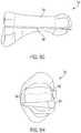
- FIGS. 1A and 1Bare front views of a foot showing a normal first metatarsal position and an example frontal plane rotational misalignment position, respectively.
- FIGS. 2A and 2Bare top views of a foot showing a normal first metatarsal position and an example transverse plane misalignment position, respectively.
- FIGS. 3A and 3Bare side views of a foot showing a normal first metatarsal position and an example sagittal plane misalignment position, respectively.
- FIG. 4is a flow diagram illustrating an example osteotomy technique for correcting an anatomical misalignment.
- FIG. 5Ais sectional image of a first metatarsal taken from the frontal plane showing example cut lines that can be made while performing the technique of FIG. 4 .
- FIG. 5Bis a perspective image of the example first metatarsal from FIG. 5A .
- FIG. 5Cis a sectional view of an example first metatarsal showing an example use of a wedge-shaped bone portion to close a gap created during realignment.
- FIG. 5Dis a dorsal perspective view of the example first metatarsal with wedge-shaped bone portion from FIG. 5C .
- FIGS. 5E and 5Fare a medial side view and a frontal plane view, respectively, of a first metatarsal illustrating an example technique in which the metatarsal is separated into two portions by making two cuts along the length of the metatarsal.
- FIGS. 5H and 5Gare a medial side view and a frontal plane view, respectively, showing an example use of wedge-shaped bone portion in a metatarsal realignment technique.
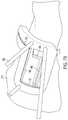
- FIG. 6Ais a perspective view of one example cutting guide that can be used to cut a first metatarsal to realign a distal portion of the metatarsal relative to a proximal portion of the metatarsal.
- FIG. 6Bis a perspective view of the example cutting guide of FIG. 6A shown attached to a first metatarsal.
- FIG. 7Ais a perspective illustration of another example configuration of a bone cutting guide that can be used to cut a first metatarsal to realign a distal portion of the metatarsal relative to a proximal portion of the metatarsal.
- FIG. 7Bis a perspective illustration of the example cutting guide of FIG. 7A shown attached to a first metatarsal.
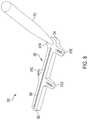
- FIG. 8is a perspective illustration of another example bone cutting guide that can be used according to the disclosure.
- FIGS. 9A-9Dare conceptual illustrations showing how the example bone cutting guide of FIG. 8 can be manipulated relative to a bone to be cut to execute a bone realignment technique.
- the present disclosureis directed to devices and techniques for correcting a misalignment of one or more bones.
- the disclosed devices and techniquescan be implemented in an osteotomy procedure in which a bone is surgically cut and/or a piece of bone is surgically removed.
- the techniqueis performed on one or more bones in the foot or hand, where bones are relatively small compared to bones in other parts of the human anatomy.
- the foregoing descriptiongenerally refers to example techniques performed on the foot and, more particularly a metatarsal of the foot.
- the disclosed techniquesmay be performed on other bones, such as the tibia, fibula, ulna, humerus, femur, or yet other bone, and the disclosure is not limited in this respect unless otherwise specifically indicated.
- the disclosed techniquesare used to correct a misalignment between a metatarsal (e.g., a first metatarsal) and a second metatarsal and/or a cuneiform (e.g., a medial, or first, cuneiform), such as in a bunion correction surgery.
- a metatarsale.g., a first metatarsal
- a second metatarsale.g., a medial, or first, cuneiform
- FIGS. 1-3are different views of a foot 200 showing example anatomical misalignments that may occur and be corrected according to the present disclosure. Such misalignment may be caused by a hallux valgus (bunion), natural growth deformity, or other condition causing anatomical misalignment.
- FIGS. 1A and 1Bare front views of foot 200 showing a normal first metatarsal position and an example frontal plane rotational misalignment position, respectively.
- FIGS. 2A and 2Bare top views of foot 200 showing a normal first metatarsal position and an example transverse plane misalignment position, respectively.
- FIGS. 3A and 3Bare side views of foot 200 showing a normal first metatarsal position and an example sagittal plane misalignment position, respectively.
- FIGS. 1B, 2B, and 3Bshow each respective planar misalignment in isolation
- a metatarsalmay be misaligned in any two of the three planes or even all three planes. Accordingly, it should be appreciated that the depiction of a single plane misalignment in each of FIGS. 1B , 2 B, and 3 B is for purposes of illustration and a metatarsal may be misaligned in multiple planes that is desirably corrected.
- foot 200is composed of multiple bones including a first metatarsal 210 , a second metatarsal 212 , a third metatarsal 214 , a fourth metatarsal 216 , and a fifth metatarsal 218 .
- the metatarsalsare connected distally to phalanges 220 and, more particularly, each to a respective proximal phalanx.
- the first metatarsal 210is connected proximally to a medial cuneiform 222
- the second metatarsal 212is connected proximally to an intermediate cuneiform 224
- the third metatarsalis connected proximally to lateral cuneiform 226 .
- the fourth and fifth metatarsals 216 , 218are connected proximally to the cuboid bone 228 .
- the joint 230 between a metatarsal and respective cuneiform(e.g., first metatarsal 210 and medial cuneiform 222 ) is referred to as the tarsometatarsal (“TMT”) joint.
- the joint 232 between a metatarsal and respective proximal phalanxis referred to as a metatarsophalangeal joint.
- the angle 234 between adjacent metatarsalse.g., first metatarsal 210 and second metatarsal 212
- IMAintermetatarsal angle
- FIG. 1Ais a frontal plane view of foot 200 showing a typical position for first metatarsal 210 .
- the frontal planewhich is also known as the coronal plane, is generally considered any vertical plane that divides the body into anterior and posterior sections.
- the frontal planeis a plane that extends vertically and is perpendicular to an axis extending proximally to distally along the length of the foot.
- FIG. 1Ashows first metatarsal 210 in a typical rotational position in the frontal plane.
- FIG. 1Bshows first metatarsal 210 with a frontal plane rotational deformity characterized by a rotational angle 236 relative to ground, as indicated by line 238 .
- FIG. 2Ais a top view of foot 200 showing a typical position of first metatarsal 210 in the transverse plane.
- the transverse planewhich is also known as the horizontal plane, axial plane, or transaxial plane, is considered any plane that divides the body into superior and inferior parts.
- the transverse planeis a plane that extends horizontally and is perpendicular to an axis extending dorsally to plantarly (top to bottom) across the foot.
- FIG. 2Ashows first metatarsal 210 with a typical IMA 234 in the transverse plane.
- FIG. 2Bshows first metatarsal 210 with a transverse plane rotational deformity characterized by a greater IMA caused by the distal end of first metatarsal 210 being pivoted medially relative to the second metatarsal 212 .
- FIG. 3Ais a side view of foot 200 showing a typical position of first metatarsal 210 in the sagittal plane.
- the sagittal planeis a plane parallel to the sagittal structure which divides the body into right and left halves.
- the sagittal planeis a plane that extends vertically and is perpendicular to an axis extending medially to laterally along the length of the foot.
- FIG. 3Ashows first metatarsal 210 with a typical rotational position in the sagittal plane.
- FIG. 3Bshows first metatarsal 210 with a sagittal plane rotational deformity characterized by a rotational angle 240 relative to ground, as indicated by line 238 .
- Bone positioning techniques and instruments according to the disclosurecan be useful to correct an anatomical misalignment of a bones or bones.
- the techniqueinvolves realigning a portion of a metatarsal relative to an adjacent metatarsal portion.
- the metatarsal undergoing realignmentmay be anatomically misaligned in the frontal plane, transverse plane, and/or sagittal plane, as illustrated and discussed with respect to FIGS. 1-3 above.
- realignmentmay involve cutting the metatarsal into two independently movable portions and thereafter realigning one metatarsal portion relative to the other metatarsal portion in one or more planes, two or more planes, or all three planes. After suitably realigning the metatarsal portions, the metatarsal portions can be fixated to hold and maintain the realigned positioned.
- anatomically aligned positionmeans that an angle of a long axis of first metatarsal 210 or portion thereof relative to the long axis of second metatarsal 212 is about 10 degrees or less in the transverse plane and/or sagittal plane.
- anatomical misalignmentcan be corrected in both the transverse plane and the frontal plane. In the transverse plane, a normal IMA 234 between first metatarsal 210 or portion thereof and second metatarsal 212 is less than about 9 degrees.
- An IMA 234 of between about 9 degrees and about 13 degreesis considered a mild misalignment of the first metatarsal and the second metatarsal.
- An IMA 234 of greater than about 16 degreesis considered a severe misalignment of the first metatarsal and the second metatarsal.
- methods according to the disclosureare utilized to anatomically align first metatarsal 210 or a portion thereof by reducing the IMA from over 10 degrees to about 10 degrees or less (e.g., to an IMA of about 1-5 degrees), including to negative angles of about ⁇ 5 degrees or until interference with the second metatarsal, by positioning the first metatarsal or portion thereof at a different angle with respect to the second metatarsal.
- a normal first metatarsalWith respect to the frontal plane, a normal first metatarsal will be positioned such that its crista prominence is generally perpendicular to the ground and/or its sesamoid bones are generally parallel to the ground and positioned under the metatarsal. This position can be defined as a metatarsal rotation of 0 degrees. In a misaligned first metatarsal, the metatarsal may be axially rotated between about 4 degrees to about 30 degrees or more.
- methods according to the disclosureare utilized to anatomically align the metatarsal or portion thereof by reducing the metatarsal rotation from about 4 degrees or more to less than 4 degrees (e.g., to about 0 to 2 degrees) by rotating the metatarsal or portion thereof with respect to the medial cuneiform.
- FIG. 4is a flow diagram illustrating an example osteotomy technique for correcting an anatomical alignment.
- the techniquewill be described with respect to first metatarsal 210 although can be performed on other bones, as discussed above.
- the technique of FIG. 4will be discussed with respect to two different images of first metatarsal 210 illustrated in FIGS. 5A and 5B to show how different cuts can be made along the first metatarsal during a bone correction procedure.
- FIG. 5Ais sectional image of first metatarsal 210 from the frontal plane of the bone showing example cut lines that can be made while performing the technique of FIG. 4 .
- FIG. 5Bis a perspective image of first metatarsal 210 from FIG. 5A .
- the example techniqueinvolves making a first longitudinal cut 350 into first metatarsal 210 ( 300 ) and also making a second longitudinal cut 352 into the first metatarsal that intersects the first longitudinal cut ( 302 ).
- the first longitudinal cut 350may be made along the length of first metatarsal 210 between the proximal end 354 and distal end 356 .
- the first longitudinal cut 350may be made through the medial side 358 and the lateral side 360 of first metatarsal 210 , thereby transecting the metatarsal along the lateral plane.
- first longitudinal cut 350is made from the medial side 358 of first metatarsal 210 toward the lateral side 360 of the metatarsal.
- the clinicianmay insert a cutting instrument from the medial side 358 of the bone and guide the cutting instrument towards the lateral side 360 of the bone to cut in this direction.
- the clinicianmay insert the cutting instrument from the lateral side 360 of the bone and guide the cutting instrument towards the medial side 358 of the bone.
- This alternative cutting directionmay be useful when performing the procedure on, for example, the fifth metatarsal.
- first longitudinal cut 350can be made from the proximal end 354 of first metatarsal 210 toward the distal end 356 of the metatarsal.
- first longitudinal cut 350can be made from the distal end 356 of the first metatarsal 210 toward the proximal end 354 of the bone.
- first longitudinal cut 350does not extend the entire length of first metatarsal 210 but instead extends only along a portion of the length of the metatarsal.
- the proximal end of first longitudinal cut 350may be offset from the proximal-most end 354 of first metatarsal 210 a separation distance 361 .
- the distal end of first longitudinal cut 350may also be offset from the distal-most end 356 of first metatarsal 210 a separation distance 362 .
- separation distance 361 and separation distance 362are substantially equal, e.g., such that first longitudinal cut 350 is substantially centered along the length of first metatarsal 210 . In other examples, separation distance 361 and separation distance 362 are different from each other, e.g., such that first longitudinal cut 350 extends more proximately along the length of first metatarsal 210 then distally along the length. In some examples, first longitudinal cut 350 extends along at least 50% of the overall length of first metatarsal 210 between the proximal-most end 354 and the distal-most end 356 such as, e.g., at least 60% of the overall length, or from 50% to 80% of the overall length.
- the technique of FIG. 4also includes making second longitudinal cut 352 into first metatarsal 210 ( 302 ).
- the second longitudinal cut 352may be made along the length of first metatarsal 210 between the proximal end 354 and the distal end 356 .
- the second longitudinal cut 352may extend the same length along first metatarsal 210 as first longitudinal cut 350 or may have a different length.
- the second longitudinal cut 352may be made from the medial side 358 of first metatarsal 210 or the lateral side 360 of the metatarsal, as discussed above with respect to the first longitudinal cut 350 . In either case, the second longitudinal cut 352 may be made at a converging angle with respect to the first longitudinal cut 350 to form a wedge-shaped cut.
- second longitudinal cut 352is shown as being radially offset from first longitudinal cut 350 (e.g., on the medial side 358 of first metatarsal 210 ) about an angle 364 .
- first longitudinal cut 350extends in a common transverse plane across first metatarsal 210 (in the medial to lateral direction) without angling in a plantar or dorsal direction across the bone.
- second longitudinal cut 352extends across first metatarsal 210 (in the medial to lateral direction) at an angle such that the medial end of second longitudinal cut 352 is positioned closer to the dorsal side of the metatarsal than the lateral side of the cut and, correspondingly, the lateral side of the cut is positioned closer to the plantar side of the metatarsal than the medial side of the cut.
- second longitudinal cut 352intersects first longitudinal cut 350 at the lateral side 360 of the first metatarsal 210 , e.g., such that the apex of the resulting wedge is the lateral side of the bone.
- second longitudinal cut 352may not intersect first longitudinal cut 350 at the lateral side 360 of the first metatarsal 210 but may instead intersect the first longitudinal cut closer to the medial side 358 of the bone and/or may extend through the lateral side 360 of the first metatarsal without intersecting the first longitudinal cut.
- first longitudinal cut 350 into first metatarsal 210 and/or the second longitudinal cut 352 into first metatarsal 210may or may not extend all the way through the metatarsal (e.g., in the transverse plane).
- first longitudinal cut 350 and second longitudinal cut 352are illustrated as both extending through first metatarsal 210 such that the cuts intersect at or outside of the lateral side 360 of first metatarsal 210 .
- one of the cutse.g., which may be designated the first longitudinal cut
- the other of the cutsextends into the first metatarsal and intersects the through cut within the cross-section of the bone.
- first longitudinal cut 350 and second longitudinal cut 352are illustrated as both extending through first metatarsal 210
- one of the cutsmay be an intersecting cut within the bone without extending through the entire cross-section of the bone.
- first longitudinal cut 350is radially offset from second longitudinal cut 352 by an angle 364 ranging from 10° to 50°, such as from 15° to 35°.
- FIG. 5Aillustrates first longitudinal cut 350 extending across first metatarsal 210 in a single transverse plane and second longitudinal cut 352 extending across the first metatarsal at an angle in the dorsal to plantar direction from the medial to lateral side
- one or both of the longitudinal cutsmay be angled (e.g., in the plantar to dorsal direction) across one or more planes (e.g., moving proximally to distally along the first metatarsal and/or moving medially to laterally across the first metatarsal). Therefore, the example cut configuration of FIG. 5A is for purposes of illustration, and the disclosure is not limited in this respect.
- FIG. 4also includes making a first transverse cut ( 304 ) and a second transverse cut ( 306 ).
- FIG. 5Billustrates a bone wedge 365 formed by making a first transverse cut 366 and a second transverse cut 368 .
- One or both of the first transverse cut 366 and the second transverse cut 368may be made before or after making the first longitudinal cut 350 and/or making the second longitudinal cut 352 .
- the specific cutting order in which the first longitudinal cut 350 , the second longitudinal cut 352 , the first transverse cut 366 , and the second transverse cut 368 are madecan be rearranged at the discretion of the clinician performing the procedure.
- the first transverse cut 366may cut first metatarsal 210 in a frontal plane adjacent the proximal end 354 of the metatarsal.
- the second transverse cut 368may cut first metatarsal 210 in a different frontal plane adjacent the distal end 356 of the metatarsal.
- the first transverse cut 366extends downwardly, e.g., from a dorsal to plantar direction, from second longitudinal cut 352 and intersects both the first longitudinal cut 350 and the second longitudinal cut 352 .
- the second transverse cut 368can extend upwardly, e.g., from a plantar to dorsal direction, from first longitudinal cut 350 and intersect both the first longitudinal cut 350 and the second longitudinal cut 352 .
- This configuration of cutscan form a distal first metatarsal portion 370 whose length is defined by the dorsal portion of first metatarsal 210 and a proximal first metatarsal portion 372 whose length is defined by the proximal portion of the first metatarsal 210 .
- first transverse cut 366 and the second transverse cut 368can be reversed such that the first transverse cut extends upwardly from the first longitudinal cut 350 and the second transverse cut 368 extends downwardly from the second longitudinal cut 352 .
- the distal first metatarsal portion 370can have a length defined by the plantar portion of the first metatarsal 210 while the proximal first metatarsal portion 372 can have a length defined by the dorsal portion of the metatarsal.
- first transverse cut 366 and the second transverse cut 368may be within a single frontal plane or may be angled along the length of the first metatarsal across multiple frontal planes.
- first transverse cut 366may extend distally away from the end of second longitudinal cut 352 .
- Second transverse cut 368may also extend distally away from the end of the second longitudinal cut 352 .
- This configuration of angled transverse cutscan provide a lock and key configuration, such as an interlocking “Z” shape, to help restrict relative movement between the distal first metatarsal portion 370 and the proximal first metatarsal portion 372 following realignment of the two bone portions.
- first transverse cut 366may be angled proximally from the end of the second longitudinal cut 352
- second transverse cut 368may be angled proximally from the end of the second longitudinal cut 352
- first transverse cut 366 or the second transverse cut 368can be angled proximally while the other transverse cut is orthogonal or angled distally.
- the example technique of FIG. 4also involves moving the distal portion 370 of the first metatarsal 210 relative to the proximal portion 372 of the metatarsal in one or more planes to realign the distal portion of the bone relative the proximal portion ( 308 ).
- Thiscan adjust the anatomical alignment of the distal portion 370 of the metatarsal relative to the proximal portion of the metatarsal, e.g., to help correct an anatomical misalignment.
- the distal potion 370 of the first metatarsal 210is rotated relative to the proximal portion 372 of the metatarsal.
- the distal portion 370 of the first metatarsal 210may be rotated in the frontal plane and/or pivoted in the transverse plane and/or pivoted in the sagittal plane to help correct an anatomical misalignment of the distal portion 370 .
- the boneis pivoted by translating and/or sliding the bone.
- the distal portion 370 of the first metatarsal 210is rotated about an axis extending through the frontal plane so the medial side is moved dorsally and/or the distal portion 370 of the first metatarsal 210 is moved laterally in the transverse plane and/or plantarly in the sagittal plane.
- the distal portion 370 of the first metatarsal 210may be moved from an anatomically misaligned position relative to the proximal portion 372 of the first metatarsal and/or the second metatarsal 212 and/or the medial cuneiform 222 to an anatomically aligned position.
- the wedge 365 formed by cutting the metatarsalcan be removed from the space between the two bone portions.
- the wedge-shaped bone portion 365may be removed from between the two bone portions and the bone portions distracted or separated from each other to facilitate relative realignment.
- the wedge-shaped bone portion 365may be removed from between the two bone portions and the bone portions realigned relative to each other without distracting the bone portions. In either case, the bone portions can shift and move relative to each other during realignment.
- Wedge 365 removed from first metatarsal 210may or may not be reused as an autograft to fill space created between distal portion 370 and proximal portion 372 of first metatarsal 210 during realignment.
- FIG. 5Cis a sectional view of first metatarsal 210 from the frontal plane showing an example use of wedge-shaped bone portion 365 to close a gap created during realignment of distal portion 370 relative to proximal portion 372 .
- FIG. 5Dis a dorsal perspective view of the first metatarsal 210 with wedge-shaped bone portion 365 from FIG. 5C .
- wedge-shaped bone portion 365has been removed from the medial side 350 of first metatarsal 210 . This creates a wedge-shaped gap on the medial side between distal portion 370 and proximal portion 372 .
- the gap or void space created by removing wedge-shaped bone portion 365can be closed.
- the gap created by removing wedge-shaped bone portion 365may be partially or fully closed.
- a second gapmay be created on an opposite side of the first metatarsal.
- This gap or void space created by realignment of the two bone portionsmay be left open for self-healing or, as illustrated, a bone implant may be inserted into the gap to promote accelerated and efficacious recovery.
- wedge-shaped bone portion 365defines a wedge extending from a base 359 to an apex 369 .
- base 359may be on the medial half of the medial half of the metatarsal while apex 369 is on the lateral half of the metatarsal.
- wedge-shaped bone portion 365may be rotated 180 degrees (e.g., in the frontal plane and/or transverse plane) so apex 369 is oriented medially and base 359 is oriented laterally.
- the rotated wedgecan then be inserted into the gap created through realignment between the distal portion 370 and proximal portion 372 .
- the clinicianmay trim or otherwise resize the wedge-shaped bone portion 365 to be configured (e.g., sized and/or shaped) to fit within the opening created during realignment between the distal portion 370 and proximal portion 372 .
- the bone portion 365 inserted into the opening created by realignment between distal portion 370 and proximal portion 372may or may not have a wedge shape (e.g., taping from a wider base to a narrower apex).
- the bone portion 365may be inserted while the gap is opening and/or after distracting distal portion 370 and proximal portion 372 . Accordingly, realignment between distal portion 370 and proximal portion 372 need not be performed or complete before inserting bone portion 365 on an opposite side of the metatarsal from which the bone portion was extracted.
- the bone portion inserted in the opening created by realignment between distal portion 370 and proximal portion 372may extend partially or fully across the cross-section of first metatarsal 210 in the transverse plane.
- bone portion 365is illustrated as extending across the entire width of first metatarsal 210 , e.g., such that the bone portion 365 is positioned between distal portion 370 and proximal portion 372 on both the medial side 350 of the metatarsal and the lateral side 360 of the metatarsal.
- bone portion 365may be inserted only part way across the cross-section of first metatarsal 210 , e.g., such that distal portion 370 and proximal portion 372 contact each other on the medial side 350 of the metatarsal.
- the bone portion 365 resected from first metatarsal 210When the wedge-shaped bone portion 365 resected from first metatarsal 210 is reused as a bone implant, the bone portion becomes an autograft within the surgical technique.
- the clinicianmay use a different bone construct to fill the gap created through realignment, such as an allograft harvested from another person, a xenograft harvested from a different species, or synthetic bone.
- the bone portions (and optional inserted bone member)can be fixated to each other to secure and hold the new realigned position achieved through movement ( 310 ).
- the distal and proximal bone portions (and optional inserted bone member)are provisionally fixated relative to each other before permanently fixating the bone portions relative to each other.
- Provisional fixationcan temporarily hold the proximal bone portion 372 and distal bone portion 370 in fixed alignment relative to each other while one or more permanent fixation devices are applied to the bones and across the joints formed therebetween.
- a fixation wire and/or a compression pin, such as a threaded olive pin,may be used as provisional fixation instruments.
- the clinicianmay apply a permanent fixation device to the bone portions and across the joint between the bone portions (and optional inserted bone member).
- the permanent fixation devicecan hold the bone portions in fixed alignment relative to each other, e.g., to promote healing between the bone portions in their aligned positions.
- one or more bone plates, pins, screws, staples, or other fixation mechanismscan be used to fixate the bones relative to each other.
- the clinicianmay resect bone around the perimeter of the cut end of the distal portion 370 of the first metatarsal and/or the proximal portion 372 of the first metatarsal. Rotating the distal portion 370 of the first metatarsal relative to the proximal portion 372 of the first metatarsal may cause the end of one of the bone portions to rotate out of plane, creating a projecting lip or edge.
- the clinicianmay use a cutting instrument to resect the lip or edge of the out-of-plane bone, helping to create a flush surface at the joint between the bone portions.
- the clinicianmay resect the protruding lip or edge of the cut end of the bone portion after moving the proximal and distal bone portions relative to each other.
- the cliniciancan perform such resection before fixating the bone portions relative to each other or after fixating the bone portions relative to each other (e.g., either provisionally or permanently).
- first metatarsal 210may be separated into a first portion and second portion by making two longitudinal cuts extending along the length of the metatarsal.
- the longitudinal cutscan extend along the length of the first metatarsal through the proximal end and distal end of the metatarsal. This can separate the first metatarsal 210 into two portions and release a removable bone wedge.
- FIGS. 5E and 5Fare a medial side view and a frontal plane view, respectively, of first metatarsal 210 illustrating an example technique in which the metatarsal is separated into two portions by making two cuts along the length of the metatarsal.
- first longitudinal cut 350extends through the proximal end 354 and the distal end 356 of the first metatarsal 210 and also through the medial side 358 and lateral side 360 of the metatarsal.
- Second longitudinal cut 352is radially offset from first longitudinal cut 350 (e.g., on the medial side 358 of first metatarsal 210 ) about angle 364 .
- Second longitudinal cut 352also extends through the proximal end 354 and the distal end 356 of the first metatarsal 210 as well as through the medial side 358 and lateral side 360 of the metatarsal.
- first longitudinal cut 350 and second longitudinal cut 352may be within the ranges discussed above with respect to FIGS. 5A and 5B .
- first longitudinal cut 350may be radially offset from second longitudinal cut 352 by an angle 364 ranging from 10° to 50°, such as from 15° to 35°.
- first longitudinal cut 350 and second longitudinal cut 352each extend transversely across first metatarsal 210 (in the medial to lateral direction) without angling in a plantar or dorsal direction across the bone.
- one or both of the longitudinal cutsmay angle, e.g., in a planar to dorsal direction or vice versa across the length of the bone, as discussed above.
- a cliniciancan use a cutting instrument to cut first metatarsal 210 along first longitudinal cut 350 and second longitudinal cut 352 . This can separate the first metatarsal into a first portion 363 , a second portion 367 , and wedge-shaped bone portion 365 .
- the wedge-shaped bone portion 365 formed by cutting the metatarsalcan be removed from the space between the two bone portions.
- the wedge-shaped bone portion 365may be removed from between the two bone portions and the bone portions distracted or separated from each other to facilitate relative realignment.
- the wedge-shaped bone portion 365may be removed from between the two bone portions and the bone portions realigned relative to each other without distracting the bone portions. In either execution, the bone portions can be realigned relative to each other, as discussed above.
- the wedge-shaped bone portion 365 removed from between the first bone portion 363 and the second bone portion 367is used as a bone implant in a gap during realignment of the first bone portion 363 relative to the second bone portion 367 .
- a gap created by removing wedge-shaped bone portion 365may be closed while realigning the first bone portion 363 relative to the second bone portion 367 .
- a second gapmay be created on an opposite side of the first metatarsal.
- the wedge-shaped bone portion 365 resected from first metatarsal 210can be reused as a bone implant in this second gap.
- FIGS. 5H and 5Gare a medial side view and a frontal plane view, respectively, showing an example use of wedge-shaped bone portion 365 in a metatarsal realignment technique.
- wedge-shaped bone portion 365may be rotated 180 degrees (e.g., in the frontal plane and/or transverse plane) and inserted into the gap created through realignment between the first bone portion 363 and the second bone portion 367 .
- the clinicianmay use a different bone construct to fill the gap created through realignment, such as an allograft harvested from another person, a xenograft harvested from a different species, or synthetic bone.
- the bone portions (and optional inserted bone member)can be fixated to each other to secure and hold the new realigned position achieved through movement.
- the bone portions (and optional inserted bone member)are provisionally fixated relative followed by permanent fixation.
- FIG. 6Ais a perspective view of one example cutting guide 400 that can be used to cut a first metatarsal 210 to realign a distal portion 370 of the metatarsal relative to a proximal portion 372 .
- cutting guidemay be defined by a body 402 that is configured (e.g., sized and/or shaped) to be positioned against a bone to be cut.
- body 402may define a front side 404 A and a backside 404 B that is opposite the front side.
- Body 402may further extend from a first longitudinal side edge 406 A to a second longitudinal side edge 406 B.
- body 402may be positioned against a first metatarsal to be cut such that backside 404 B is in contact with the metatarsal while the front side 404 A faces outwardly away from the metatarsal.
- first longitudinal side edge 406 Acan be positioned closer to the proximal end of the first metatarsal from the distal end
- second longitudinal side edge 406 Bcan be positioned closer to the distal end of the first metatarsal and the proximal end.
- Body 402 of cutting guide 400may have a variety of different shapes.
- body 402is shaped to conform to the curvature of a bone to be cut.
- body 402may have a “V” shape or a radius of curvature that conforms to the radius of curvature of a bone to be cut.
- body 402may wrap at least partially about the curved external surface of the bone to be cut.
- cutting guide 400can have at least one longitudinal cutting slot 408 .
- the longitudinal cutting slot 408may extend at least partially, and in some examples fully, along the length of the cutting guide body 402 between the first longitudinal side edge 406 A and the second longitudinal side edge 406 B.
- the longitudinal cutting slot 408may extend across body 402 at a constant vertical location on the body (e.g., as illustrated in FIG. 6A ) or may slope upwardly or downwardly across the length of the body.
- cutting guide 400may further include at least one transverse cutting slot which, in the example of FIG. 6A , is illustrated as two transverse cutting slots: first transverse cutting slot 410 and second transverse cutting slot 412 .
- First transverse cutting slot 410may extend downwardly from the longitudinal cutting slot 408
- the second transverse cutting slot 412may extend upwardly from the longitudinal cutting slot.
- longitudinal cutting slot 408may run from a first terminal end 414 to a second terminal end 416 .
- First transverse cutting slot 410can extend downwardly from the first terminal end 414 of the longitudinal cutting slot, e.g., such that the first transverse cutting slot and the longitudinal cutting slot share a common terminal end 414 .
- Second transverse cutting slot 412can extend upwardly from the second terminal end 416 of the longitudinal cutting slot, e.g., such that the second transverse cutting slot in longitudinal should cutting slot share a common terminal end 416 .
- the first transverse cutting slot 410can intersect the longitudinal cutting slot 408 at a first intersection angle 418 .
- the second transverse cutting slot 412can intersect the longitudinal cutting slot 408 at a second intersection angle 420 .
- the first and second intersection angles 418 , 420are illustrated as being approximately orthogonal or 90°.
- the first and second intersection angles 418 , 420may range from 20° to 135°, such as less than 90°, from 20° to 80°, from 30° to 75° , or yet other angles.
- the first and second intersection angles 418 , 420may be offset from orthogonal from 1° to 45°.
- the first and second intersection angles 418 , 420may be the same or may be different from each other.
- first transverse cutting slot 410is sloped proximately moving away from longitudinal cutting slot 408 .
- terminal end 414 of the first transverse cutting slotmay be positioned more distally along the length of the bone then the opposite end of the cutting slot.
- terminal end 416 of the second transverse cutting slotmay be positioned more proximately along the length of the bone than the opposite end of the cutting slot.
- Bone cutting guide 400can have any suitable dimensions.
- longitudinal cutting slot 408 , first transverse cutting slot 410 , and second transverse cutting slot 412each have a width ranging from 0.1 mm to 3 mm.
- the dimensions of each of the different cutting slotsmay be the same or may be different from each other.
- the dimensions of each of the cutting slotsmay be the same and may be sized based on the particular cutting instrument (e.g., saw blade, rotary bur, osteotome) intended to be used for the surgical procedure.
- Longitudinal cutting slot 408 , first transverse cutting slot 410 , and/or second transverse cutting slot 412may extend perpendicularly through the thickness of body 402 or may be angled as the slot extends through the thickness of the body, e.g., to provide an angled cutting slot.
- body 402may include one or more fixation apertures configured to receive a fixation member, such as a wire, pin, screw, or other mechanical fixation element intended to temporarily secure and hold the cutting guide to the bone during the surgical procedure.
- a fixation membersuch as a wire, pin, screw, or other mechanical fixation element intended to temporarily secure and hold the cutting guide to the bone during the surgical procedure.
- body 402includes a first slot 422 and a second slot 424 that function as fixation apertures.
- First slot 422is positioned between the first longitudinal edge 406 A of body 402 and first transverse cutting slot 410 .
- Second slot 424is positioned between the second longitudinal side edge 406 B of body 402 and second transverse cutting slot 412 .
- First slot 422 and second slot 424are illustrated as being parallel to first transverse cutting slot 410 and second transverse cutting slot 412 , respectively, but may not be parallel in other configurations.
- slots 422 and 424may be sufficiently long to allow cutting guide 400 to be positioned at different radial or angular locations about the first metatarsal 210 being cut, e.g., to allow different cuts at different radial or angular positions along the bone to be made using longitudinal cutting slot 408 .
- FIG. 6Bis a perspective view of cutting guide 400 attached to a first metatarsal 210 using slots 422 and 424 .
- fixation pins 426are inserted through each of slots 422 and 424 to position bone cutting guide 400 at a first radial position on the first metatarsal.
- Bone cutting guide 400can be rotated radially about first metatarsal 210 to a second radial position while fixation pins 426 remain in slots 422 and 424 , e.g., to reposition the location of longitudinal cutting slot 408 . This can be useful to position bone cutting guide 400 at one location to make first longitudinal cut 350 ( FIG. 5A ) and subsequently rotate the cutting guide to a second location to make second longitudinal cut 352 .
- Bone cutting guide 400may include one or more additional fixation apertures 428 A, 428 B to secure the bone cutting guide at a particular rotational orientation about first metatarsal 210 .
- FIG. 7Ais a perspective illustration of another example configuration of bone cutting guide 400 where like features discussed above with respect to FIGS. 6A and 6B refer to like elements.
- bone cutting guide 400includes the previously described longitudinal cutting slot 408 , first transverse cutting slot 410 , and second transverse cutting slot 412 .
- bone cutting guide 400includes a second longitudinal cutting slot 440 .
- Second longitudinal cutting slot 440may extend parallel to the first longitudinal cutting slot 408 and may be offset relative to the first longitudinal cutting slot.
- the first longitudinal cutting slot 408 and the second longitudinal cutting slot 440may be offset from their centerlines a distance 442 ranging from 2 mm to 35 mm, such as from 5 mm to 20 mm.
- Second longitudinal cutting slot 440can extend parallel to first longitudinal cutting slot 408 and intersect first transverse cutting slot 410 .
- second longitudinal cutting slot 440may intersect first transverse cutting slot 410 at a location between the terminal ends of the transverse cutting slot.
- a clinicianmay use first longitudinal cutting slot 408 and second longitudinal cutting slot 440 to make the two longitudinal cuts along the length of first metatarsal 210 discussed above with respect to FIGS. 4 and 5 .
- first longitudinal cutting slot 408 and second longitudinal cutting slot 440may be angled relative to each other to form converging cut lines that intersect on first metatarsal 210 .
- first longitudinal cutting slot 408 and second longitudinal cutting slot 440may be angled relative to each other to form cutting lines that converge at an angle ranging from 5° to 45°.
- cutting guide 400 in FIG. 7Amay include a third transverse cutting slot 444 that is co-linear with a second transverse cutting slot 412 .
- second longitudinal cutting slot 440may extend from a first end 445 to a second end 448 .
- the first end 445 of the second longitudinal cutting slot 440can intersect first transverse cutting slot 410 .
- Third transverse cutting slot 444can extend upwardly from the second end 448 of the second longitudinal cutting slot 440 .
- a clinicianmay insert a cutting instrument through both second transverse cutting slot 412 and third transverse cutting slot 444 to form a joined, single transverse cut line 368 ( FIG. 5B ) to help separate a distal metatarsal portion 370 from a proximal metatarsal portion 372 .
- the cutting guidemay include a depth limiter.
- the depth limitermay be a surface against which the clinician can position a cutting instrument and along which the clinician can translate the cutting instrument through a cutting slot in order to set the depth of cut made through the slot.
- the depth limitermay be permanently joined/integrally formed with the body 402 or may be a separate component attachable to or used in conjunction with body 402 .
- bone cutting guide 400includes a depth limiter 446 , which is defined by a surface projecting outwardly from the first side 404 A of body 402 .
- depth limiter 446includes one surface or rail projecting outwardly from body 402 on one side of second longitudinal cutting slot 440 and a second surface or rail projecting outwardly from the body on an opposite side of the longitudinal cutting slot.
- depth limiter 446projects outwardly from the first side 404 A of body 402 a distance ranging from 0.25 mm to 10 mm.
- a clinicianmay place their cutting instrument against depth limiter 446 , e.g., with a cutting blade or member inserted through the slot, and guide the cutting instrument along the depth limiter to form a cut of controlled depth.
- bone cutting guide 400 in the example of FIG. 7may include one or more fixation apertures to temporarily fixate the bone cutting guide against a bone to be cut during a procedure.
- bone cutting guide 400includes three fixation apertures 428 , although a different number of fixation apertures may be used.
- FIG. 7Bis a perspective illustration of bone cutting guide 400 from FIG. 7A showing the cutting guide attached to an example first metatarsal 210 .
- FIG. 8is a perspective illustration of another example bone cutting guide 500 that can be used according to the disclosure.
- bone cutting guide 500has a body 502 that extends from a first end 504 to a second end 506 .
- a longitudinal cutting slot 508is formed through the body 502 and extends along the length of the body between the first end 504 and the second end 506 .
- a clinicianmay manipulate cutting guide 500 to position longitudinal cutting slot 508 at different positions along a first metatarsal 210 to be cut.
- the cutting guidemay include securing projections.
- cutting guide 500may include a first securing projection 510 A and a second securing projection 510 B extending substantially orthogonally (or at a different angle) relative to the length of the body.
- Each securing projectioncan have at least one, and in some examples multiple, fixation apertures configured to receive a mechanical fixation element for securing the body to a bone to be cut.
- each securing projectionmay include multiple fixation apertures that are linearly aligned (e.g., in an orthogonal direction away from the length of the body).
- the different securing projectionsmay allow bone cutting guide 500 to be positioned at different degrees of rotation about the bone to be cut, e.g., by selecting different rotational positions corresponding to different sets of fixation apertures.
- bone cutting guide 500may include more than two securing projections, such as three, four or more securing projections.
- bone cutting guide 500includes four securing projections 510 A- 510 D.
- First and second securing projections 510 A and 510 Bmay be located adjacent the first end 504 of body 502
- the third and fourth securing projections 510 C and 510 Dmay be located adjacent the second end 506 of the body.
- the bone cutting guidemay include a handle 512 .
- the handlemay extend from the body 502 and angle ranging from 10° to 90°.
- the handlecan be permanently attached to body 502 or can be removably coupled to the body, e.g., threadingly coupled to the body.
- a clinicianmay hold bone cutting guide 500 against a bone to be cut while performing a cutting operation without securing the cutting guide to the bone with a mechanical fixation instrument using a securing projection.
- the clinicianmay manipulate bone cutting guide 500 against the bone using handle 512 but may provisionally fixate the bone cutting guide against the bone using a mechanical fixation element inserted through one or more securing projections before performing a cutting procedure.
- FIGS. 9A-9Dare conceptual illustrations showing how bone cutting guide 500 can be manipulated relative to a bone to be cut to execute a bone realignment technique as discussed herein.
Landscapes
- Health & Medical Sciences (AREA)
- Surgery (AREA)
- Life Sciences & Earth Sciences (AREA)
- Heart & Thoracic Surgery (AREA)
- Molecular Biology (AREA)
- Oral & Maxillofacial Surgery (AREA)
- Engineering & Computer Science (AREA)
- Biomedical Technology (AREA)
- Dentistry (AREA)
- Medical Informatics (AREA)
- Nuclear Medicine, Radiotherapy & Molecular Imaging (AREA)
- Animal Behavior & Ethology (AREA)
- General Health & Medical Sciences (AREA)
- Public Health (AREA)
- Veterinary Medicine (AREA)
- Orthopedic Medicine & Surgery (AREA)
- Surgical Instruments (AREA)
Abstract
Description
This application claims priority to U.S. Provisional Patent Application No. 62/421,027, filed Nov. 11, 2016, the entire contents of which are incorporated herein by reference.
This disclosure relates to devices and techniques for correcting bones and, more particularly, to osteotomy techniques for correcting bone misalignment.
Bones, such as the bones of a foot, may be anatomically misaligned. In certain circumstances, surgical intervention is required to correctly align the bones to reduce patient discomfort and improve patient quality of life.
In general, this disclosure is directed to devices and techniques for correcting an anatomical misalignment of one or more bones. In some examples, the devices and techniques are utilized to correct a misalignment of a first metatarsal relative to a medial cuneiform and/or an adjacent second metatarsal, such as bunion correction procedure. To perform a corrective procedure, a clinician may make two parallel but offset cuts along the length of the first metatarsal. The two cuts may be angled relative to each other across the thickness of the bone, causing the cuts to intersect to form a wedged-shaped section of bone. If the cuts do not extend the entire length of the bone, the clinician can further make two transverse cuts on opposite ends of the longitudinal cuts, e.g., adjacent the proximal and distal ends of the first metatarsal, to release the wedge-shaped section of bone from a remainder of the metatarsal. The transverse cuts can be made before, after, or between making each of the two longitudinal cuts.
Upon resecting the wedge-shaped section of bone from the first metatarsal, the first metatarsal may be divided into two independently movable portions. In cases where the first metatarsal is cut with longitudinal and transverse cuts, the metatarsal may be divided into a proximal portion connected to the medial cuneiform and a distal portion connected to the proximal phalanx. In cases where the first metatarsal is along the longitudinal axis but without transverse cuts, the metatarsal may be divided into a plantar portion and a dorsal portion. In either case, clinician can move the two portions of the first metatarsal relative to each other in multiple planes to help correct the anatomical misalignment of the first metatarsal (e.g., the distal portion of the first metatarsal). For example, the clinician may rotate one portion of the first metatarsal in the frontal plane, pivot the portion of the first metatarsal in the transverse plane, and/or pivot the portion of the first metatarsal in the sagittal plane to adjust the anatomical alignment of the one portion of the first metatarsal relative to the other portion of the first metatarsal.
In some applications, the clinician removes the wedge portion cut from the first metatarsal to provide clearance for realignment. This can enable one portion of the metatarsal (e.g., the distal portion) to be rotated relative to the other portion of the first metatarsal (e.g., the proximal portion). In some examples, a clinician may also trim around the perimeter of the cut end of the distal portion and/or proximal portion of the first metatarsal, e.g., to avoid causing the rotated bone portion to create an interfering lip or edge where it is rotated out of plane.
The clinician may or may not use the wedge-shaped bone portion resected from the first metatarsal as an autograft to fill a space created during realignment of one portion of the cut first metatarsal relative to another portion of the cut metatarsal. For example, the wedge-shaped bone portion may be resected from the medial side of the first metatarsal to create a space that allows for realignment of a first portion of the metatarsal relative to a second portion of the metatarsal. For example, a distal portion of the first metatarsal may be moved in one or more planes (e.g., the frontal plane) relative to the proximal portion, closing the space created by removal of the wedge-shaped bone portion. A corresponding gap may be created on the lateral side of the first metatarsal through realignment. The wedge-shaped bone portion can be inserted as an autograft in this gap. The clinician may or may not trim or otherwise resize the wedge-shaped bone portion to fit within the space created through realignment. Instead of reusing the bone portion harvested from the first metatarsal as an autograft, the clinician may use a different bone construct to fill the gap created through realignment, such as an allograft harvested from another person, a xenograft harvested from a different species, or synthetic bone.
Although different instruments may be used to perform the bone correction procedure, in some examples, a bone cutting guide is used that has a longitudinal cutting slot. In one example, the bone cutting guide has a longitudinal cutting slot and a plurality of fixation apertures to allow the guide to be fixated at different rotational and angular positions about the first metatarsal to be cut. Additionally or alternatively, the bone cutting may have one or more transverse cutting slots that extend upwardly and/or downwardly from the longitudinal cutting slot. For example, the bone cutting guide may include a longitudinal cutting slot, a first transverse cutting slot that intersects one end of the longitudinal cutting slot, and a second transverse cutting slot that intersects the opposite end of the longitudinal cutting slot. In use, a clinician may use the longitudinal cutting slot to cut along the length of the first metatarsal and, after rotating the cutting guide, use the longitudinal cutting slot to make a second, intersecting cut along the length of the first metatarsal. The clinician may further use the two transverse cutting slots to make intersecting cuts to the two longitudinal cuts (e.g., in the dorsal to plantar direction, or vice versa). This may cleave the first metatarsal into adjacent proximal and distal portions for realignment.
In one example, a method is described that includes making a first longitudinal cut through a first metatarsal between a proximal end of the first metatarsal and a distal end of the first metatarsal. The method also includes making a second longitudinal cut through the first metatarsal between a proximal end of the first metatarsal and a distal end of the first metatarsal, where the second longitudinal cut is radially offset from the first longitudinal cut and intersects the first longitudinal cut. The method further involves making a transverse cut adjacent the proximal end of the first metatarsal that intersects the first longitudinal cut and the second longitudinal cut and making a transverse cut adjacent the distal end of the first metatarsal that intersects the first longitudinal cut and the second longitudinal cut. In addition, the method includes removing a bone wedge from the first metatarsal and moving a distal portion of the first metatarsal relative to a proximal portion of the first metatarsal in at least two planes, thereby adjusting an anatomical alignment of the distal portion of the first metatarsal relative to the proximal portion of the first metatarsal.
In another example, a bone cutting guide is described that includes a body configured to be positioned against a bone to be cut. The body includes a longitudinal cutting slot having a first end and a second end, a first transverse cutting slot extending downwardly from the first end of the longitudinal cutting slot, and a second transverse cutting slot extending upwardly from the second end of the longitudinal cutting slot.
In another example, a bone cutting guide is described that includes a body having a length extending from a first end to a second end and that is sized to be positioned against a first metatarsal. The example specifies that the body includes a longitudinal cutting slot extending between the first end and the second end, a first securing projection extending substantially orthogonally relative to the length in one direction and containing at least one fixation aperture, and a second securing projection extending substantially orthogonally relative to the length in an opposite direction and containing at least one fixation aperture.
In another example, a method is described that includes making a first longitudinal cut through a first metatarsal extending from a proximal end of the first metatarsal to a distal end of the first metatarsal and making a second longitudinal cut extending from the proximal end of the first metatarsal to the distal end of the first metatarsal. The example specifies that the second longitudinal cut is radially offset from the first longitudinal cut and intersects the first longitudinal cut, thereby separating the first metatarsal into a first portion, a second portion, and a bone wedge. The technique involves removing the bone wedge from the first metatarsal and moving the first portion of the first metatarsal relative to the second portion of the first metatarsal, thereby adjusting an anatomical alignment of the first portion of the first metatarsal relative to the second portion of the first metatarsal.
The details of one or more examples are set forth in the accompanying drawings and the description below. Other features, objects, and advantages will be apparent from the description and drawings, and from the claims.
In general, the present disclosure is directed to devices and techniques for correcting a misalignment of one or more bones. The disclosed devices and techniques can be implemented in an osteotomy procedure in which a bone is surgically cut and/or a piece of bone is surgically removed. In some examples, the technique is performed on one or more bones in the foot or hand, where bones are relatively small compared to bones in other parts of the human anatomy. For example, the foregoing description generally refers to example techniques performed on the foot and, more particularly a metatarsal of the foot. However, the disclosed techniques may be performed on other bones, such as the tibia, fibula, ulna, humerus, femur, or yet other bone, and the disclosure is not limited in this respect unless otherwise specifically indicated. In some applications, however, the disclosed techniques are used to correct a misalignment between a metatarsal (e.g., a first metatarsal) and a second metatarsal and/or a cuneiform (e.g., a medial, or first, cuneiform), such as in a bunion correction surgery.
With reference toFIGS. 1A and 2A ,foot 200 is composed of multiple bones including afirst metatarsal 210, asecond metatarsal 212, athird metatarsal 214, afourth metatarsal 216, and afifth metatarsal 218. The metatarsals are connected distally tophalanges 220 and, more particularly, each to a respective proximal phalanx. Thefirst metatarsal 210 is connected proximally to amedial cuneiform 222, while thesecond metatarsal 212 is connected proximally to anintermediate cuneiform 224 and the third metatarsal is connected proximally tolateral cuneiform 226. The fourth andfifth metatarsals cuboid bone 228. The joint230 between a metatarsal and respective cuneiform (e.g.,first metatarsal 210 and medial cuneiform222) is referred to as the tarsometatarsal (“TMT”) joint. The joint232 between a metatarsal and respective proximal phalanx is referred to as a metatarsophalangeal joint. Theangle 234 between adjacent metatarsals (e.g.,first metatarsal 210 and second metatarsal212) is referred to as the intermetatarsal angle (“IMA”).
As noted,FIG. 1A is a frontal plane view offoot 200 showing a typical position forfirst metatarsal 210. The frontal plane, which is also known as the coronal plane, is generally considered any vertical plane that divides the body into anterior and posterior sections. Onfoot 200, the frontal plane is a plane that extends vertically and is perpendicular to an axis extending proximally to distally along the length of the foot.FIG. 1A showsfirst metatarsal 210 in a typical rotational position in the frontal plane.FIG. 1B showsfirst metatarsal 210 with a frontal plane rotational deformity characterized by arotational angle 236 relative to ground, as indicated byline 238.
Bone positioning techniques and instruments according to the disclosure can be useful to correct an anatomical misalignment of a bones or bones. In some applications, the technique involves realigning a portion of a metatarsal relative to an adjacent metatarsal portion. The metatarsal undergoing realignment may be anatomically misaligned in the frontal plane, transverse plane, and/or sagittal plane, as illustrated and discussed with respect toFIGS. 1-3 above. Accordingly, realignment may involve cutting the metatarsal into two independently movable portions and thereafter realigning one metatarsal portion relative to the other metatarsal portion in one or more planes, two or more planes, or all three planes. After suitably realigning the metatarsal portions, the metatarsal portions can be fixated to hold and maintain the realigned positioned.
While a metatarsal can have a variety of anatomically aligned and misaligned positions, in some examples, the term “anatomically aligned position” means that an angle of a long axis offirst metatarsal 210 or portion thereof relative to the long axis ofsecond metatarsal 212 is about 10 degrees or less in the transverse plane and/or sagittal plane. In certain embodiments, anatomical misalignment can be corrected in both the transverse plane and the frontal plane. In the transverse plane, anormal IMA 234 betweenfirst metatarsal 210 or portion thereof andsecond metatarsal 212 is less than about 9 degrees. AnIMA 234 of between about 9 degrees and about 13 degrees is considered a mild misalignment of the first metatarsal and the second metatarsal. AnIMA 234 of greater than about 16 degrees is considered a severe misalignment of the first metatarsal and the second metatarsal. In some embodiments, methods according to the disclosure are utilized to anatomically alignfirst metatarsal 210 or a portion thereof by reducing the IMA from over 10 degrees to about 10 degrees or less (e.g., to an IMA of about 1-5 degrees), including to negative angles of about −5 degrees or until interference with the second metatarsal, by positioning the first metatarsal or portion thereof at a different angle with respect to the second metatarsal.
With respect to the frontal plane, a normal first metatarsal will be positioned such that its crista prominence is generally perpendicular to the ground and/or its sesamoid bones are generally parallel to the ground and positioned under the metatarsal. This position can be defined as a metatarsal rotation of 0 degrees. In a misaligned first metatarsal, the metatarsal may be axially rotated between about 4 degrees to about 30 degrees or more. In some embodiments, methods according to the disclosure are utilized to anatomically align the metatarsal or portion thereof by reducing the metatarsal rotation from about 4 degrees or more to less than 4 degrees (e.g., to about 0 to 2 degrees) by rotating the metatarsal or portion thereof with respect to the medial cuneiform.
With reference toFIGS. 4 and 5 , the example technique involves making a firstlongitudinal cut 350 into first metatarsal210 (300) and also making a secondlongitudinal cut 352 into the first metatarsal that intersects the first longitudinal cut (302). The firstlongitudinal cut 350 may be made along the length offirst metatarsal 210 between theproximal end 354 anddistal end 356. In addition, the firstlongitudinal cut 350 may be made through themedial side 358 and thelateral side 360 offirst metatarsal 210, thereby transecting the metatarsal along the lateral plane. In some examples, firstlongitudinal cut 350 is made from themedial side 358 offirst metatarsal 210 toward thelateral side 360 of the metatarsal. The clinician may insert a cutting instrument from themedial side 358 of the bone and guide the cutting instrument towards thelateral side 360 of the bone to cut in this direction. Alternatively, the clinician may insert the cutting instrument from thelateral side 360 of the bone and guide the cutting instrument towards themedial side 358 of the bone. This alternative cutting direction may be useful when performing the procedure on, for example, the fifth metatarsal.
The clinician can make the firstlongitudinal cut 350 from theproximal end 354 offirst metatarsal 210 toward thedistal end 356 of the metatarsal. Alternatively, firstlongitudinal cut 350 can be made from thedistal end 356 of thefirst metatarsal 210 toward theproximal end 354 of the bone. In some examples, firstlongitudinal cut 350 does not extend the entire length offirst metatarsal 210 but instead extends only along a portion of the length of the metatarsal. For example, the proximal end of firstlongitudinal cut 350 may be offset from theproximal-most end 354 of first metatarsal210 aseparation distance 361. The distal end of firstlongitudinal cut 350 may also be offset from thedistal-most end 356 of first metatarsal210 aseparation distance 362.
In some examples,separation distance 361 andseparation distance 362 are substantially equal, e.g., such that firstlongitudinal cut 350 is substantially centered along the length offirst metatarsal 210. In other examples,separation distance 361 andseparation distance 362 are different from each other, e.g., such that firstlongitudinal cut 350 extends more proximately along the length offirst metatarsal 210 then distally along the length. In some examples, firstlongitudinal cut 350 extends along at least 50% of the overall length offirst metatarsal 210 between theproximal-most end 354 and thedistal-most end 356 such as, e.g., at least 60% of the overall length, or from 50% to 80% of the overall length.
The technique ofFIG. 4 also includes making secondlongitudinal cut 352 into first metatarsal210 (302). The secondlongitudinal cut 352 may be made along the length offirst metatarsal 210 between theproximal end 354 and thedistal end 356. The secondlongitudinal cut 352 may extend the same length alongfirst metatarsal 210 as firstlongitudinal cut 350 or may have a different length. Similarly, the secondlongitudinal cut 352 may be made from themedial side 358 offirst metatarsal 210 or thelateral side 360 of the metatarsal, as discussed above with respect to the firstlongitudinal cut 350. In either case, the secondlongitudinal cut 352 may be made at a converging angle with respect to the firstlongitudinal cut 350 to form a wedge-shaped cut.
In the example ofFIGS. 5A and 5B , secondlongitudinal cut 352 is shown as being radially offset from first longitudinal cut350 (e.g., on themedial side 358 of first metatarsal210) about anangle 364. In particular, in the illustrated example, firstlongitudinal cut 350 extends in a common transverse plane across first metatarsal210 (in the medial to lateral direction) without angling in a plantar or dorsal direction across the bone. By contrast, secondlongitudinal cut 352 extends across first metatarsal210 (in the medial to lateral direction) at an angle such that the medial end of secondlongitudinal cut 352 is positioned closer to the dorsal side of the metatarsal than the lateral side of the cut and, correspondingly, the lateral side of the cut is positioned closer to the plantar side of the metatarsal than the medial side of the cut. In the illustrated arrangement, secondlongitudinal cut 352 intersects firstlongitudinal cut 350 at thelateral side 360 of thefirst metatarsal 210, e.g., such that the apex of the resulting wedge is the lateral side of the bone. In other applications, secondlongitudinal cut 352 may not intersect firstlongitudinal cut 350 at thelateral side 360 of thefirst metatarsal 210 but may instead intersect the first longitudinal cut closer to themedial side 358 of the bone and/or may extend through thelateral side 360 of the first metatarsal without intersecting the first longitudinal cut.
Accordingly, in various applications, the firstlongitudinal cut 350 intofirst metatarsal 210 and/or the secondlongitudinal cut 352 intofirst metatarsal 210 may or may not extend all the way through the metatarsal (e.g., in the transverse plane). InFIGS. 5A and 5B , firstlongitudinal cut 350 and secondlongitudinal cut 352 are illustrated as both extending throughfirst metatarsal 210 such that the cuts intersect at or outside of thelateral side 360 offirst metatarsal 210. However, one of the cuts (e.g., which may be designated the first longitudinal cut) may extend throughfirst metatarsal 210 while the other of the cuts extends into the first metatarsal and intersects the through cut within the cross-section of the bone. Thus, it should be appreciated that while firstlongitudinal cut 350 and secondlongitudinal cut 352 are illustrated as both extending throughfirst metatarsal 210, one of the cuts may be an intersecting cut within the bone without extending through the entire cross-section of the bone.
In some examples, firstlongitudinal cut 350 is radially offset from secondlongitudinal cut 352 by anangle 364 ranging from 10° to 50°, such as from 15° to 35°. In addition, whileFIG. 5A illustrates firstlongitudinal cut 350 extending acrossfirst metatarsal 210 in a single transverse plane and secondlongitudinal cut 352 extending across the first metatarsal at an angle in the dorsal to plantar direction from the medial to lateral side, it should be appreciated that one or both of the longitudinal cuts may be angled (e.g., in the plantar to dorsal direction) across one or more planes (e.g., moving proximally to distally along the first metatarsal and/or moving medially to laterally across the first metatarsal). Therefore, the example cut configuration ofFIG. 5A is for purposes of illustration, and the disclosure is not limited in this respect.
To release the bone wedge formed by firstlongitudinal cut 350 and secondlongitudinal cut 352, the example technique ofFIG. 4 also includes making a first transverse cut (304) and a second transverse cut (306). For example,FIG. 5B illustrates abone wedge 365 formed by making a firsttransverse cut 366 and a secondtransverse cut 368. One or both of the firsttransverse cut 366 and the secondtransverse cut 368 may be made before or after making the firstlongitudinal cut 350 and/or making the secondlongitudinal cut 352. In general, the specific cutting order in which the firstlongitudinal cut 350, the secondlongitudinal cut 352, the firsttransverse cut 366, and the secondtransverse cut 368 are made can be rearranged at the discretion of the clinician performing the procedure.
The firsttransverse cut 366 may cutfirst metatarsal 210 in a frontal plane adjacent theproximal end 354 of the metatarsal. The secondtransverse cut 368 may cutfirst metatarsal 210 in a different frontal plane adjacent thedistal end 356 of the metatarsal. In some examples, such as the example illustrated inFIG. 5B , the firsttransverse cut 366 extends downwardly, e.g., from a dorsal to plantar direction, from secondlongitudinal cut 352 and intersects both the firstlongitudinal cut 350 and the secondlongitudinal cut 352. Similarly, the secondtransverse cut 368 can extend upwardly, e.g., from a plantar to dorsal direction, from firstlongitudinal cut 350 and intersect both the firstlongitudinal cut 350 and the secondlongitudinal cut 352. This configuration of cuts can form a distalfirst metatarsal portion 370 whose length is defined by the dorsal portion offirst metatarsal 210 and a proximalfirst metatarsal portion 372 whose length is defined by the proximal portion of thefirst metatarsal 210. Alternatively, the configuration of the firsttransverse cut 366 and the secondtransverse cut 368 can be reversed such that the first transverse cut extends upwardly from the firstlongitudinal cut 350 and the secondtransverse cut 368 extends downwardly from the secondlongitudinal cut 352. In this alternative configuration, the distalfirst metatarsal portion 370 can have a length defined by the plantar portion of thefirst metatarsal 210 while the proximalfirst metatarsal portion 372 can have a length defined by the dorsal portion of the metatarsal.
Independent of the direction in which the firsttransverse cut 366 and the secondtransverse cut 368 are made, one or both of the cuts may be within a single frontal plane or may be angled along the length of the first metatarsal across multiple frontal planes. For example, firsttransverse cut 366 may extend distally away from the end of secondlongitudinal cut 352. Secondtransverse cut 368 may also extend distally away from the end of the secondlongitudinal cut 352. This configuration of angled transverse cuts can provide a lock and key configuration, such as an interlocking “Z” shape, to help restrict relative movement between the distalfirst metatarsal portion 370 and the proximalfirst metatarsal portion 372 following realignment of the two bone portions. In alternative configurations, firsttransverse cut 366 may be angled proximally from the end of the secondlongitudinal cut 352, and secondtransverse cut 368 may be angled proximally from the end of the secondlongitudinal cut 352. In yet further configurations, the firsttransverse cut 366 or the secondtransverse cut 368 can be angled proximally while the other transverse cut is orthogonal or angled distally.
The example technique ofFIG. 4 also involves moving thedistal portion 370 of thefirst metatarsal 210 relative to theproximal portion 372 of the metatarsal in one or more planes to realign the distal portion of the bone relative the proximal portion (308). This can adjust the anatomical alignment of thedistal portion 370 of the metatarsal relative to the proximal portion of the metatarsal, e.g., to help correct an anatomical misalignment. In some examples, thedistal potion 370 of thefirst metatarsal 210 is rotated relative to theproximal portion 372 of the metatarsal. Thedistal portion 370 of thefirst metatarsal 210 may be rotated in the frontal plane and/or pivoted in the transverse plane and/or pivoted in the sagittal plane to help correct an anatomical misalignment of thedistal portion 370. In some applications, the bone is pivoted by translating and/or sliding the bone. In some examples, thedistal portion 370 of thefirst metatarsal 210 is rotated about an axis extending through the frontal plane so the medial side is moved dorsally and/or thedistal portion 370 of thefirst metatarsal 210 is moved laterally in the transverse plane and/or plantarly in the sagittal plane. For example, thedistal portion 370 of thefirst metatarsal 210 may be moved from an anatomically misaligned position relative to theproximal portion 372 of the first metatarsal and/or thesecond metatarsal 212 and/or themedial cuneiform 222 to an anatomically aligned position.
To facilitate movement of thedistal portion 370 of the first metatarsal relative to theproximal portion 372 of the metatarsal, thewedge 365 formed by cutting the metatarsal can be removed from the space between the two bone portions. For example, the wedge-shapedbone portion 365 may be removed from between the two bone portions and the bone portions distracted or separated from each other to facilitate relative realignment. Alternatively, the wedge-shapedbone portion 365 may be removed from between the two bone portions and the bone portions realigned relative to each other without distracting the bone portions. In either case, the bone portions can shift and move relative to each other during realignment.
In the example shown inFIGS. 5C and 5D , wedge-shapedbone portion 365 has been removed from themedial side 350 offirst metatarsal 210. This creates a wedge-shaped gap on the medial side betweendistal portion 370 andproximal portion 372. During subsequent realignment ofdistal portion 370 relative toproximal portion 372, the gap or void space created by removing wedge-shapedbone portion 365 can be closed. For example, asdistal portion 370 is rotated in the frontal plane and/or translated in the transverse plane, the gap created by removing wedge-shapedbone portion 365 may be partially or fully closed. As the gap created by removing wedge-shapedbone portion 365 is closed, a second gap may be created on an opposite side of the first metatarsal. This gap or void space created by realignment of the two bone portions may be left open for self-healing or, as illustrated, a bone implant may be inserted into the gap to promote accelerated and efficacious recovery.
In some examples, wedge-shapedbone portion 365 defines a wedge extending from a base359 to an apex369. When resected from the medial side offirst metatarsal 210,base 359 may be on the medial half of the medial half of the metatarsal whileapex 369 is on the lateral half of the metatarsal. After being resected from first metatarsal, wedge-shapedbone portion 365 may be rotated 180 degrees (e.g., in the frontal plane and/or transverse plane) soapex 369 is oriented medially andbase 359 is oriented laterally. The rotated wedge can then be inserted into the gap created through realignment between thedistal portion 370 andproximal portion 372. The clinician may trim or otherwise resize the wedge-shapedbone portion 365 to be configured (e.g., sized and/or shaped) to fit within the opening created during realignment between thedistal portion 370 andproximal portion 372. After trimming, thebone portion 365 inserted into the opening created by realignment betweendistal portion 370 andproximal portion 372 may or may not have a wedge shape (e.g., taping from a wider base to a narrower apex). It should be appreciated that thebone portion 365 may be inserted while the gap is opening and/or after distractingdistal portion 370 andproximal portion 372. Accordingly, realignment betweendistal portion 370 andproximal portion 372 need not be performed or complete before insertingbone portion 365 on an opposite side of the metatarsal from which the bone portion was extracted.
The bone portion inserted in the opening created by realignment betweendistal portion 370 andproximal portion 372 may extend partially or fully across the cross-section offirst metatarsal 210 in the transverse plane. In the example ofFIG. 5C ,bone portion 365 is illustrated as extending across the entire width offirst metatarsal 210, e.g., such that thebone portion 365 is positioned betweendistal portion 370 andproximal portion 372 on both themedial side 350 of the metatarsal and thelateral side 360 of the metatarsal. However,bone portion 365 may be inserted only part way across the cross-section offirst metatarsal 210, e.g., such thatdistal portion 370 andproximal portion 372 contact each other on themedial side 350 of the metatarsal.
When the wedge-shapedbone portion 365 resected fromfirst metatarsal 210 is reused as a bone implant, the bone portion becomes an autograft within the surgical technique. Instead of reusing the bone portion harvested from the first metatarsal as an autograph, the clinician may use a different bone construct to fill the gap created through realignment, such as an allograft harvested from another person, a xenograft harvested from a different species, or synthetic bone.
After suitably moving the two transected bone portions relative to each other and, if desired, inserting a bone construct between the two bone portions, the bone portions (and optional inserted bone member) can be fixated to each other to secure and hold the new realigned position achieved through movement (310). In some examples, the distal and proximal bone portions (and optional inserted bone member) are provisionally fixated relative to each other before permanently fixating the bone portions relative to each other. Provisional fixation can temporarily hold theproximal bone portion 372 anddistal bone portion 370 in fixed alignment relative to each other while one or more permanent fixation devices are applied to the bones and across the joints formed therebetween. A fixation wire and/or a compression pin, such as a threaded olive pin, may be used as provisional fixation instruments.
Independent of whether thedistal bone portion 370 andproximal bone portion 372 are provisionally fixated together, the clinician may apply a permanent fixation device to the bone portions and across the joint between the bone portions (and optional inserted bone member). The permanent fixation device can hold the bone portions in fixed alignment relative to each other, e.g., to promote healing between the bone portions in their aligned positions. In different examples, one or more bone plates, pins, screws, staples, or other fixation mechanisms can be used to fixate the bones relative to each other.
In some examples, the clinician may resect bone around the perimeter of the cut end of thedistal portion 370 of the first metatarsal and/or theproximal portion 372 of the first metatarsal. Rotating thedistal portion 370 of the first metatarsal relative to theproximal portion 372 of the first metatarsal may cause the end of one of the bone portions to rotate out of plane, creating a projecting lip or edge. The clinician may use a cutting instrument to resect the lip or edge of the out-of-plane bone, helping to create a flush surface at the joint between the bone portions. The clinician may resect the protruding lip or edge of the cut end of the bone portion after moving the proximal and distal bone portions relative to each other. In different examples, the clinician can perform such resection before fixating the bone portions relative to each other or after fixating the bone portions relative to each other (e.g., either provisionally or permanently).
While the foregoing description of an example technique involves making a firstlongitudinal cut 350, a secondlongitudinal cut 352, a firsttransverse cut 366, and a secondtransverse cut 368, alternative implementations of the technique may be performed by making a lesser number of cuts. As one example,first metatarsal 210 may be separated into a first portion and second portion by making two longitudinal cuts extending along the length of the metatarsal. The longitudinal cuts can extend along the length of the first metatarsal through the proximal end and distal end of the metatarsal. This can separate thefirst metatarsal 210 into two portions and release a removable bone wedge.
Theangle 364 of offset between firstlongitudinal cut 350 and secondlongitudinal cut 352 may be within the ranges discussed above with respect toFIGS. 5A and 5B . For example, firstlongitudinal cut 350 may be radially offset from secondlongitudinal cut 352 by anangle 364 ranging from 10° to 50°, such as from 15° to 35°. In addition, in the illustrated example, firstlongitudinal cut 350 and secondlongitudinal cut 352 each extend transversely across first metatarsal210 (in the medial to lateral direction) without angling in a plantar or dorsal direction across the bone. However, one or both of the longitudinal cuts may angle, e.g., in a planar to dorsal direction or vice versa across the length of the bone, as discussed above.
In practice, a clinician can use a cutting instrument to cutfirst metatarsal 210 along firstlongitudinal cut 350 and secondlongitudinal cut 352. This can separate the first metatarsal into afirst portion 363, asecond portion 367, and wedge-shapedbone portion 365. The wedge-shapedbone portion 365 formed by cutting the metatarsal can be removed from the space between the two bone portions. For example, the wedge-shapedbone portion 365 may be removed from between the two bone portions and the bone portions distracted or separated from each other to facilitate relative realignment. Alternatively, the wedge-shapedbone portion 365 may be removed from between the two bone portions and the bone portions realigned relative to each other without distracting the bone portions. In either execution, the bone portions can be realigned relative to each other, as discussed above.
In some examples, the wedge-shapedbone portion 365 removed from between thefirst bone portion 363 and thesecond bone portion 367 is used as a bone implant in a gap during realignment of thefirst bone portion 363 relative to thesecond bone portion 367. For example, as discussed with respect toFIGS. 5C and 5D , a gap created by removing wedge-shapedbone portion 365 may be closed while realigning thefirst bone portion 363 relative to thesecond bone portion 367. As this gap is closed, a second gap may be created on an opposite side of the first metatarsal. The wedge-shapedbone portion 365 resected fromfirst metatarsal 210 can be reused as a bone implant in this second gap. For example,FIGS. 5H and 5G are a medial side view and a frontal plane view, respectively, showing an example use of wedge-shapedbone portion 365 in a metatarsal realignment technique. As discussed above with respect toFIGS. 5C and 5D , wedge-shapedbone portion 365 may be rotated 180 degrees (e.g., in the frontal plane and/or transverse plane) and inserted into the gap created through realignment between thefirst bone portion 363 and thesecond bone portion 367. Instead of reusing the bone portion harvested from the first metatarsal as an autograph in the technique ofFIGS. 5E and 5H , the clinician may use a different bone construct to fill the gap created through realignment, such as an allograft harvested from another person, a xenograft harvested from a different species, or synthetic bone.
After suitably moving the two bone portions relative to each other and, if desired, inserting a bone construct between the two bone portions, the bone portions (and optional inserted bone member) can be fixated to each other to secure and hold the new realigned position achieved through movement. In some examples, the bone portions (and optional inserted bone member) are provisionally fixated relative followed by permanent fixation.
A variety of different cutting instruments and cutting guides can be used to implement the technique discussed above with respect toFIGS. 4 and 5 .FIG. 6A is a perspective view of oneexample cutting guide 400 that can be used to cut afirst metatarsal 210 to realign adistal portion 370 of the metatarsal relative to aproximal portion 372. As shown in the illustrated example ofFIG. 6A , cutting guide may be defined by abody 402 that is configured (e.g., sized and/or shaped) to be positioned against a bone to be cut. For example,body 402 may define afront side 404A and abackside 404B that is opposite the front side.Body 402 may further extend from a firstlongitudinal side edge 406A to a secondlongitudinal side edge 406B. In use,body 402 may be positioned against a first metatarsal to be cut such thatbackside 404B is in contact with the metatarsal while thefront side 404A faces outwardly away from the metatarsal. When so positioned, firstlongitudinal side edge 406A can be positioned closer to the proximal end of the first metatarsal from the distal end, while secondlongitudinal side edge 406B can be positioned closer to the distal end of the first metatarsal and the proximal end.
To cut a first metatarsal along the length of the bone, cuttingguide 400 can have at least onelongitudinal cutting slot 408. Thelongitudinal cutting slot 408 may extend at least partially, and in some examples fully, along the length of the cuttingguide body 402 between the firstlongitudinal side edge 406A and the secondlongitudinal side edge 406B. Thelongitudinal cutting slot 408 may extend acrossbody 402 at a constant vertical location on the body (e.g., as illustrated inFIG. 6A ) or may slope upwardly or downwardly across the length of the body.
To create transverse cuts on the first metatarsal, cuttingguide 400 may further include at least one transverse cutting slot which, in the example ofFIG. 6A , is illustrated as two transverse cutting slots: firsttransverse cutting slot 410 and secondtransverse cutting slot 412. Firsttransverse cutting slot 410 may extend downwardly from thelongitudinal cutting slot 408, and the secondtransverse cutting slot 412 may extend upwardly from the longitudinal cutting slot. For example,longitudinal cutting slot 408 may run from a firstterminal end 414 to a secondterminal end 416. Firsttransverse cutting slot 410 can extend downwardly from the firstterminal end 414 of the longitudinal cutting slot, e.g., such that the first transverse cutting slot and the longitudinal cutting slot share a commonterminal end 414. Secondtransverse cutting slot 412 can extend upwardly from the secondterminal end 416 of the longitudinal cutting slot, e.g., such that the second transverse cutting slot in longitudinal should cutting slot share a commonterminal end 416.
The firsttransverse cutting slot 410 can intersect thelongitudinal cutting slot 408 at afirst intersection angle 418. The secondtransverse cutting slot 412 can intersect thelongitudinal cutting slot 408 at asecond intersection angle 420. In the illustrated example, the first and second intersection angles418,420 are illustrated as being approximately orthogonal or 90°. In different examples, the first and second intersection angles418,420 may range from 20° to 135°, such as less than 90°, from 20° to 80°, from 30° to 75° , or yet other angles. In some examples, the first and second intersection angles418,420 may be offset from orthogonal from 1° to 45°. The first and second intersection angles418,420 may be the same or may be different from each other.
In some examples, firsttransverse cutting slot 410 is sloped proximately moving away fromlongitudinal cutting slot 408. For example, when cuttingguide 400 is applied to a first metatarsal,terminal end 414 of the first transverse cutting slot may be positioned more distally along the length of the bone then the opposite end of the cutting slot. Where secondtransverse cutting slot 412 is sloped at a corresponding angle,terminal end 416 of the second transverse cutting slot may be positioned more proximately along the length of the bone than the opposite end of the cutting slot.
To attachbone cutting guide 400 to a bone to be cut during a surgical procedure,body 402 may include one or more fixation apertures configured to receive a fixation member, such as a wire, pin, screw, or other mechanical fixation element intended to temporarily secure and hold the cutting guide to the bone during the surgical procedure. In the example ofFIG. 6A ,body 402 includes afirst slot 422 and asecond slot 424 that function as fixation apertures.First slot 422 is positioned between the firstlongitudinal edge 406A ofbody 402 and firsttransverse cutting slot 410.Second slot 424 is positioned between the secondlongitudinal side edge 406B ofbody 402 and secondtransverse cutting slot 412.First slot 422 andsecond slot 424 are illustrated as being parallel to firsttransverse cutting slot 410 and secondtransverse cutting slot 412, respectively, but may not be parallel in other configurations. In general,slots guide 400 to be positioned at different radial or angular locations about thefirst metatarsal 210 being cut, e.g., to allow different cuts at different radial or angular positions along the bone to be made usinglongitudinal cutting slot 408.
Secondlongitudinal cutting slot 440 can extend parallel to firstlongitudinal cutting slot 408 and intersect firsttransverse cutting slot 410. For example, secondlongitudinal cutting slot 440 may intersect firsttransverse cutting slot 410 at a location between the terminal ends of the transverse cutting slot. In use, a clinician may use firstlongitudinal cutting slot 408 and secondlongitudinal cutting slot 440 to make the two longitudinal cuts along the length offirst metatarsal 210 discussed above with respect toFIGS. 4 and 5 . In such applications, firstlongitudinal cutting slot 408 and secondlongitudinal cutting slot 440 may be angled relative to each other to form converging cut lines that intersect onfirst metatarsal 210. For example, firstlongitudinal cutting slot 408 and secondlongitudinal cutting slot 440 may be angled relative to each other to form cutting lines that converge at an angle ranging from 5° to 45°.
To help form second transverse cut368 (FIG. 5B ), cuttingguide 400 inFIG. 7A may include a thirdtransverse cutting slot 444 that is co-linear with a secondtransverse cutting slot 412. For example, secondlongitudinal cutting slot 440 may extend from afirst end 445 to asecond end 448. Thefirst end 445 of the secondlongitudinal cutting slot 440 can intersect firsttransverse cutting slot 410. Thirdtransverse cutting slot 444 can extend upwardly from thesecond end 448 of the secondlongitudinal cutting slot 440. In practice, a clinician may insert a cutting instrument through both secondtransverse cutting slot 412 and thirdtransverse cutting slot 444 to form a joined, single transverse cut line368 (FIG. 5B ) to help separate adistal metatarsal portion 370 from aproximal metatarsal portion 372.
To help control the depth of cut being made by a clinician usingbone cutting guide 400, the cutting guide may include a depth limiter. The depth limiter may be a surface against which the clinician can position a cutting instrument and along which the clinician can translate the cutting instrument through a cutting slot in order to set the depth of cut made through the slot. The depth limiter may be permanently joined/integrally formed with thebody 402 or may be a separate component attachable to or used in conjunction withbody 402.
In the example ofFIG. 7A ,bone cutting guide 400 includes adepth limiter 446, which is defined by a surface projecting outwardly from thefirst side 404A ofbody 402. In particular, in the illustrated configuration,depth limiter 446 includes one surface or rail projecting outwardly frombody 402 on one side of secondlongitudinal cutting slot 440 and a second surface or rail projecting outwardly from the body on an opposite side of the longitudinal cutting slot. In some examples,depth limiter 446 projects outwardly from thefirst side 404A of body402 a distance ranging from 0.25 mm to 10 mm. A clinician may place their cutting instrument againstdepth limiter 446, e.g., with a cutting blade or member inserted through the slot, and guide the cutting instrument along the depth limiter to form a cut of controlled depth.
As discussed above with respect to the example configuration ofbone cutting guide 400 inFIGS. 6A and 6B ,bone cutting guide 400 in the example ofFIG. 7 may include one or more fixation apertures to temporarily fixate the bone cutting guide against a bone to be cut during a procedure. In the illustrated configuration,bone cutting guide 400 includes threefixation apertures 428, although a different number of fixation apertures may be used.FIG. 7B is a perspective illustration ofbone cutting guide 400 fromFIG. 7A showing the cutting guide attached to an examplefirst metatarsal 210.
As mentioned, a variety of different cutting guide configurations can be used during a bone realignment procedure according to the present disclosure.FIG. 8 is a perspective illustration of another examplebone cutting guide 500 that can be used according to the disclosure. As shown in this example,bone cutting guide 500 has abody 502 that extends from afirst end 504 to asecond end 506. Alongitudinal cutting slot 508 is formed through thebody 502 and extends along the length of the body between thefirst end 504 and thesecond end 506. A clinician may manipulate cuttingguide 500 to positionlongitudinal cutting slot 508 at different positions along afirst metatarsal 210 to be cut.
To temporarily secure cuttingguide 500 at one or more different positions against the bone to be cut, the cutting guide may include securing projections. For example, cuttingguide 500 may include afirst securing projection 510A and asecond securing projection 510B extending substantially orthogonally (or at a different angle) relative to the length of the body. Each securing projection can have at least one, and in some examples multiple, fixation apertures configured to receive a mechanical fixation element for securing the body to a bone to be cut. For example, each securing projection may include multiple fixation apertures that are linearly aligned (e.g., in an orthogonal direction away from the length of the body). The different securing projections may allowbone cutting guide 500 to be positioned at different degrees of rotation about the bone to be cut, e.g., by selecting different rotational positions corresponding to different sets of fixation apertures.
In different examples,bone cutting guide 500 may include more than two securing projections, such as three, four or more securing projections. In the illustrated configuration,bone cutting guide 500 includes four securingprojections 510A-510D. First andsecond securing projections first end 504 ofbody 502, while the third and fourth securingprojections second end 506 of the body.
To manipulatebone cutting guide 500 during use, the bone cutting guide may include ahandle 512. The handle may extend from thebody 502 and angle ranging from 10° to 90°. The handle can be permanently attached tobody 502 or can be removably coupled to the body, e.g., threadingly coupled to the body. In some applications, a clinician may holdbone cutting guide 500 against a bone to be cut while performing a cutting operation without securing the cutting guide to the bone with a mechanical fixation instrument using a securing projection. In other applications, the clinician may manipulatebone cutting guide 500 against thebone using handle 512 but may provisionally fixate the bone cutting guide against the bone using a mechanical fixation element inserted through one or more securing projections before performing a cutting procedure.FIGS. 9A-9D are conceptual illustrations showing howbone cutting guide 500 can be manipulated relative to a bone to be cut to execute a bone realignment technique as discussed herein.
In the foregoing description, certain features, elements, and techniques have been described with respect to relative references such as “upwardly” and “downwardly.” It should be appreciated that such relative references are used for purposes of illustration based on the orientation in the figures. More generally, the features, elements, and techniques may change orientation in three-dimensional space. Accordingly, references to “upwardly” and “downwardly” should be considered relative and not confined to a specific orientation with respect to gravity.
Various examples have been described. These and other examples are within the scope of the following claims.
Claims (21)
1. A method comprising:
making a first longitudinal cut through a first metatarsal between a proximal end of the first metatarsal and a distal end of the first metatarsal;
making a second longitudinal cut between the proximal end of the first metatarsal and the distal end of the first metatarsal, wherein the second longitudinal cut is radially offset from the first longitudinal cut and intersects the first longitudinal cut;
making a transverse cut adjacent the proximal end of the first metatarsal that intersects the first longitudinal cut and the second longitudinal cut;
making a transverse cut adjacent the distal end of the first metatarsal that intersects the first longitudinal cut and the second longitudinal cut;
removing a bone wedge from the first metatarsal; and
moving a distal portion of the first metatarsal relative to a proximal portion of the first metatarsal in at least two planes, thereby adjusting an anatomical alignment of the distal portion of the first metatarsal relative to the proximal portion of the first metatarsal.
2. The method ofclaim 1 , wherein the second longitudinal cut is radially offset from the first longitudinal cut from 10 degrees to 50 degrees.
3. The method ofclaim 1 , wherein making the first longitudinal cut and making the second longitudinal cut comprises making the second longitudinal cut prior to making the first longitudinal cut.
4. The method ofclaim 1 , further comprising attaching a bone cutting guide having a body defining a longitudinal cutting slot to the first metatarsal prior to making at least the first longitudinal cut and the second longitudinal cut.
5. The method ofclaim 4 , wherein attaching the bone cutting guide to the first metatarsal comprising inserting fixation members through at least one fixation aperture defined by the body.
6. The method ofclaim 1 , wherein
making the first longitudinal cut through the first metatarsal comprises making the first longitudinal cut from a medial side of the first metatarsal; and
making the second longitudinal cut through the first metatarsal comprises making the second longitudinal cut from a medial side of the first metatarsal.
7. The method ofclaim 1 , wherein moving the distal portion of the first metatarsal relative to the proximal portion of the first metatarsal comprises rotating the distal portion of the first metatarsal in a frontal plane.
8. The method ofclaim 7 , wherein rotating the distal portion of the first metatarsal portion in the frontal plane comprises rotating the distal portion until a tibial sesamoid bone and a fibular sesamoid bone are on opposite sides of a sagittal plane when viewed from the frontal plane.
9. The method ofclaim 1 , further comprising, subsequent to moving the distal portion relative to the proximal portion, fixing the distal portion with respect to the proximal portion.
10. The method ofclaim 9 , wherein fixing the distal portion with respect to the proximal portion comprises applying at least one of a bone plate, a pin, a screw, and a staple across a joint separating the distal portion from the proximal portion.
11. The method ofclaim 1 ,
wherein removing the bone wedge from the first metatarsal comprises opening a gap in the first metatarsal, and
moving the distal portion of the first metatarsal relative to the proximal portion of the first metatarsal in at least two planes comprises closing the gap and opening a second gap, and
further comprising inserting a bone member into the second gap.
12. The method ofclaim 11 , wherein the bone member is selected from the group consisting of the bone wedge removed from the first metatarsal, an allograft, a xenograft, and synthetic bone.
13. The method ofclaim 11 , wherein the second gap is on an opposite side of the first metatarsal from the gap.
14. A method comprising:
making a first longitudinal cut through a first metatarsal extending from a proximal end of the first metatarsal to a distal end of the first metatarsal;
making a second longitudinal cut extending from the proximal end of the first metatarsal to the distal end of the first metatarsal, wherein the second longitudinal cut is radially offset from the first longitudinal cut and intersects the first longitudinal cut, thereby separating the first metatarsal into a first portion, a second portion, and a bone wedge;
removing the bone wedge from the first metatarsal; and
moving the first portion of the first metatarsal relative to the second portion of the first metatarsal, thereby adjusting an anatomical alignment of the first portion of the first metatarsal relative to the second portion of the first metatarsal.
15. The method ofclaim 14 , wherein the second longitudinal cut is radially offset from the first longitudinal cut from 10 degrees to 50 degrees.
16. The method ofclaim 14 , wherein the first portion comprises a dorsal portion of the first metatarsal and the second portion comprises a plantar portion of the first metatarsal.
17. The method ofclaim 14 , wherein moving the first portion of the first metatarsal relative to the second portion of the first metatarsal comprises rotating the first portion in a frontal plane.
18. The method ofclaim 14 , wherein moving the first portion of the first metatarsal relative to the second portion of the first metatarsal comprises moving the first portion relative to the second portion in at least two planes.
19. The method ofclaim 14 ,
wherein removing the bone wedge from the first metatarsal comprises opening a gap in the first metatarsal, and
moving the first portion of the first metatarsal relative to the second portion of the first metatarsal comprises closing the gap and opening a second gap, and
further comprising inserting a bone member into the second gap.
20. The method ofclaim 19 , wherein the bone member is selected from the group consisting of the bone wedge removed from the first metatarsal, an allograft, a xenograft, and synthetic bone.
21. The method ofclaim 14 , further comprising, subsequent to moving the first portion relative to the second portion, fixing the first portion with respect to the second portion.
Priority Applications (3)
| Application Number | Priority Date | Filing Date | Title |
|---|---|---|---|
| US15/809,298US10582936B1 (en) | 2016-11-11 | 2017-11-10 | Devices and techniques for performing an osteotomy procedure on a first metatarsal to correct a bone misalignment |
| US16/812,487US11364037B2 (en) | 2016-11-11 | 2020-03-09 | Techniques for performing an osteotomy procedure on bone to correct a bone misalignment |
| US17/844,675US12414779B2 (en) | 2016-11-11 | 2022-06-20 | Devices and techniques for performing an osteotomy procedure on a first metatarsal to correct a bone misalignment |
Applications Claiming Priority (2)
| Application Number | Priority Date | Filing Date | Title |
|---|---|---|---|
| US201662421027P | 2016-11-11 | 2016-11-11 | |
| US15/809,298US10582936B1 (en) | 2016-11-11 | 2017-11-10 | Devices and techniques for performing an osteotomy procedure on a first metatarsal to correct a bone misalignment |
Related Child Applications (1)
| Application Number | Title | Priority Date | Filing Date |
|---|---|---|---|
| US16/812,487ContinuationUS11364037B2 (en) | 2016-11-11 | 2020-03-09 | Techniques for performing an osteotomy procedure on bone to correct a bone misalignment |
Publications (1)
| Publication Number | Publication Date |
|---|---|
| US10582936B1true US10582936B1 (en) | 2020-03-10 |
Family
ID=69058435
Family Applications (4)
| Application Number | Title | Priority Date | Filing Date |
|---|---|---|---|
| US15/809,276Active2038-05-10US10524808B1 (en) | 2016-11-11 | 2017-11-10 | Devices and techniques for performing an osteotomy procedure on a first metatarsal to correct a bone misalignment |
| US15/809,298Active2038-05-10US10582936B1 (en) | 2016-11-11 | 2017-11-10 | Devices and techniques for performing an osteotomy procedure on a first metatarsal to correct a bone misalignment |
| US16/812,487Active2038-08-21US11364037B2 (en) | 2016-11-11 | 2020-03-09 | Techniques for performing an osteotomy procedure on bone to correct a bone misalignment |
| US17/844,675Active2038-09-17US12414779B2 (en) | 2016-11-11 | 2022-06-20 | Devices and techniques for performing an osteotomy procedure on a first metatarsal to correct a bone misalignment |
Family Applications Before (1)
| Application Number | Title | Priority Date | Filing Date |
|---|---|---|---|
| US15/809,276Active2038-05-10US10524808B1 (en) | 2016-11-11 | 2017-11-10 | Devices and techniques for performing an osteotomy procedure on a first metatarsal to correct a bone misalignment |
Family Applications After (2)
| Application Number | Title | Priority Date | Filing Date |
|---|---|---|---|
| US16/812,487Active2038-08-21US11364037B2 (en) | 2016-11-11 | 2020-03-09 | Techniques for performing an osteotomy procedure on bone to correct a bone misalignment |
| US17/844,675Active2038-09-17US12414779B2 (en) | 2016-11-11 | 2022-06-20 | Devices and techniques for performing an osteotomy procedure on a first metatarsal to correct a bone misalignment |
Country Status (1)
| Country | Link |
|---|---|
| US (4) | US10524808B1 (en) |
Cited By (13)
| Publication number | Priority date | Publication date | Assignee | Title |
|---|---|---|---|---|
| US11439415B2 (en) | 2020-11-18 | 2022-09-13 | Fusion Orthopedics USA, LLC | Osteotomy cutting systems and surgical techniques |
| US20220313277A1 (en)* | 2016-11-11 | 2022-10-06 | Treace Medical Concepts, Inc. | Devices and techniques for performing an osteotomy procedure on a first metatarsal to correct a bone misalignment |
| US11547425B1 (en) | 2022-05-19 | 2023-01-10 | Gramercy Extremity Orthopedics Llc | Tarsometatarsal joint arthrodesis tools and related method for bunion correction |
| US11672549B2 (en) | 2020-11-18 | 2023-06-13 | Fusion Orthopedics USA, LLC | Surgical jig |
| US11883075B2 (en) | 2021-11-08 | 2024-01-30 | Relja Innovations, Llc | Device and surgical technique for foot surgery |
| US12064127B2 (en) | 2020-11-18 | 2024-08-20 | Fusion Orthopedics USA, LLC | Surgical cutting block including multiple cut guides |
| US12232790B2 (en) | 2022-12-30 | 2025-02-25 | IvyTech Design LLC | Adjustable angle orthopedic distractor, compressor, and distractor-compressor |
| USD1065530S1 (en) | 2019-12-12 | 2025-03-04 | Relja Innovations Llc | Surgical apparatus for minimally invasive surgical procedures |
| US12310603B2 (en) | 2021-02-18 | 2025-05-27 | Treace Medical Concepts, Inc. | System and technique for metatarsal realignment with reduced incision length |
| US12364528B2 (en) | 2021-06-17 | 2025-07-22 | Wright Medical Technology, Inc. | Minimally invasive surgery osteotomy fragment shifter, stabilizer, and targeter |
| US12364523B2 (en) | 2017-10-27 | 2025-07-22 | Wright Medical Technology, Inc. | Implant with intramedullary portion and offset extramedullary portion |
| US12419673B2 (en) | 2012-03-01 | 2025-09-23 | Wright Medical Technology, Inc. | Surgical staple |
| US12433606B2 (en) | 2019-05-13 | 2025-10-07 | Wright Medical Technology, Inc. | Surgical tools and methods of use |
Families Citing this family (23)
| Publication number | Priority date | Publication date | Assignee | Title |
|---|---|---|---|---|
| US9316722B2 (en) | 2008-04-17 | 2016-04-19 | Aviation Communication & Surveillance Systems, Llc | Systems and methods for providing ATC overlay protocols |
| US20120041620A1 (en) | 2010-08-10 | 2012-02-16 | Stayton Gregory T | Systems and methods for providing spoof detection |
| EP3583905A1 (en)* | 2018-06-20 | 2019-12-25 | Arthrex Inc | Percutaneous targeting device |
| WO2020014320A1 (en)* | 2018-07-10 | 2020-01-16 | Paragon 28, Inc. | Guides, instruments, systems and methods of use |
| USD865173S1 (en)* | 2018-07-09 | 2019-10-29 | Paragon 28, Inc. | Cut guide |
| US11596443B2 (en)* | 2018-07-11 | 2023-03-07 | Treace Medical Concepts, Inc. | Compressor-distractor for angularly realigning bone portions |
| CA3117491A1 (en) | 2018-09-25 | 2020-04-02 | Aviation Communication & Surveillance Systems, Llc | Systems and methods for providing wake situational awareness displays |
| US11607250B2 (en) | 2019-02-13 | 2023-03-21 | Treace Medical Concepts, Inc. | Tarsal-metatarsal joint procedure utilizing compressor-distractor and instrument providing sliding surface |
| US11889998B1 (en) | 2019-09-12 | 2024-02-06 | Treace Medical Concepts, Inc. | Surgical pin positioning lock |
| EP4027922A4 (en) | 2019-09-13 | 2023-10-04 | MIOS Marketing LLC, DBA RedPoint Medical 3D | Patient-specific surgical methods and instrumentation |
| US11986251B2 (en) | 2019-09-13 | 2024-05-21 | Treace Medical Concepts, Inc. | Patient-specific osteotomy instrumentation |
| WO2021222415A1 (en) | 2020-04-28 | 2021-11-04 | Dmo Llc | Osteotomy fixation device and related systems and methods |
| CN113081161B (en)* | 2021-04-28 | 2022-11-01 | 北京力达康科技有限公司 | Ankle joint shin bone lower extreme cuts bone baffle |
| EP4337112A1 (en)* | 2021-05-10 | 2024-03-20 | Crossroads Extremity Systems, LLC | Capital fragment guide and related methods |
| US20230027816A1 (en)* | 2021-07-22 | 2023-01-26 | Inverted Ortho, LLC | Oblique diaphyseal osteotomy system for metatarsal shortening |
| CN113598874B (en)* | 2021-08-25 | 2022-07-15 | 周君琳 | Hallux valgus osteotomy guide plate for orthopedic operation |
| CA3131443A1 (en)* | 2021-09-21 | 2023-03-21 | Services Medicaux Georges Sioufi Inc. | Operating equipment for knee surgery, operating room with such equipment, assembly kit, and corresponding assembly, operation and use methods |
| USD1057155S1 (en) | 2022-02-23 | 2025-01-07 | Treace Medical Concepts, Inc. | Lesser metatarsal cut guide with parallel cut faces |
| AU2023223468A1 (en)* | 2022-02-23 | 2024-09-12 | Treace Medical Concepts, Inc. | First metatarsal lateral release instrument and technique |
| USD1051382S1 (en) | 2022-02-23 | 2024-11-12 | Treace Medical Concepts, Inc. | Lesser metatarsal cut guide |
| USD1011524S1 (en) | 2022-02-23 | 2024-01-16 | Treace Medical Concepts, Inc. | Compressor-distractor for the foot |
| USD1079011S1 (en) | 2022-02-23 | 2025-06-10 | Treace Medical Concepts, Inc. | Metatarsal cut guide with parallel cut faces |
| US20240358518A1 (en)* | 2023-02-08 | 2024-10-31 | Apex Orthopedics Holdings, LLC | Orthopedic Arthrodesis Devices and Methods for Treating Orthopedic Disorders |
Citations (432)
| Publication number | Priority date | Publication date | Assignee | Title |
|---|---|---|---|---|
| US2557364A (en) | 1948-06-15 | 1951-06-19 | Orthopedic Frame Co | Surgical saw blade |
| US3159952A (en) | 1960-02-26 | 1964-12-08 | Morton S Lipkins | Spherical cutting apparatus |
| DE2255312A1 (en) | 1972-11-11 | 1974-05-22 | Guenter Wendt | Rotary cutter assembly - for cutting out spherical cap type articles in one step |
| FR2304322A1 (en) | 1975-03-17 | 1976-10-15 | Cohen Patrick | Bone distortion correction instrument - has blade with partly spherical toothed portion coupled to oscillating drive unit |
| US4069824A (en) | 1976-07-12 | 1978-01-24 | Weinstock Robert E | Method of and apparatus for forming a crescentic joint in a bone |
| FR2362616A1 (en) | 1976-08-26 | 1978-03-24 | Duyck Jean | Metatarsal osteotomy anchor plate - has curved support with hooks, and holes for anchoring screws |
| US4159716A (en) | 1977-10-17 | 1979-07-03 | Borchers Clinton H | Method of compressing and realigning bone structures to correct splay foot |
| US4187840A (en) | 1978-08-14 | 1980-02-12 | Watanabe Robert S | Bone plate clamp |
| US4335715A (en) | 1980-06-20 | 1982-06-22 | Kirkley William H | Osteotomy guide |
| US4338927A (en) | 1981-03-16 | 1982-07-13 | Volkov Mstislav V | Device for restoring the function of the lower extremities |
| US4349018A (en) | 1980-12-29 | 1982-09-14 | Chambers Gary R | Osteotomy apparatus |
| US4409973A (en) | 1981-01-29 | 1983-10-18 | Neufeld John A | Method and apparatus for corrective osteotomy |
| US4440168A (en) | 1981-08-31 | 1984-04-03 | Warren Mark G | Surgical device |
| US4501268A (en) | 1981-08-20 | 1985-02-26 | Comparetto John E | Bone wedge guidance system |
| US4502474A (en) | 1981-08-20 | 1985-03-05 | Comparetto John E | Bone wedge guidance system |
| US4509511A (en) | 1983-06-30 | 1985-04-09 | Neufeld John A | Method and apparatus for corrective osteotomy |
| GB2154144A (en) | 1984-02-13 | 1985-09-04 | Orthofix Srl | Bone fixation device |
| GB2154143A (en) | 1984-02-13 | 1985-09-04 | Orthofix Srl | Bone fixation device |
| US4565191A (en) | 1984-01-12 | 1986-01-21 | Slocum D Barclay | Apparatus and method for performing cuneiform osteotomy |
| US4570624A (en) | 1983-08-10 | 1986-02-18 | Henry Ford Hospital | Universal guide for inserting parallel pins |
| US4627425A (en)* | 1983-09-28 | 1986-12-09 | Reese H William | Osteotomy appliances and method |
| US4628919A (en) | 1983-09-09 | 1986-12-16 | Clyburn Terry | Dynamic external fixator and method of use |
| US4632102A (en) | 1981-08-20 | 1986-12-30 | Comparetto John E | Bone wedge osteotomy method |
| US4664102A (en) | 1985-04-10 | 1987-05-12 | Comparetto John E | Electronic guidance for bone wedge excision |
| US4677973A (en) | 1985-05-28 | 1987-07-07 | Barclay Slocum | Proximal, tibial osteotomy for leveling a tibial plateau |
| SU1333328A2 (en) | 1986-03-05 | 1987-08-30 | .В,М.Надгериев, Р,В,Никогос н, А.Я,Ахмед-заде, Б,Н.Балашев и С.П,Гордеев | Arrangement for reposition and fixing of fractures of metallurgical and metacarpal bones of foot and hand |
| US4708133A (en) | 1985-04-10 | 1987-11-24 | Comparetto John E | Arcuate bone cutter and wedge guide system |
| US4750481A (en) | 1984-04-16 | 1988-06-14 | Reese H William | Osteotomy appliances and method |
| US4757810A (en) | 1986-07-23 | 1988-07-19 | Reese Hewitt W | Osteotomy apparatus and method |
| US4787908A (en) | 1987-04-30 | 1988-11-29 | Queen's University At Kingston | Metatarsal-phalangeal replacement joint |
| US4895141A (en) | 1984-04-26 | 1990-01-23 | Harrington Arthritis Research Center | Unilateral external fixation device |
| US4952214A (en)* | 1981-08-20 | 1990-08-28 | Ohio Medical Instrument Co., Inc. | Arcuate osteotomy blade, blade guide, and cutting method |
| US4978347A (en) | 1988-07-26 | 1990-12-18 | Ilizarov Gavrill A | Distraction apparatus for osteosynthesis of short tubular bones |
| US4988349A (en) | 1987-01-21 | 1991-01-29 | Orthofix S.R.L. | Device for osteosynthesis |
| US4995875A (en) | 1988-05-27 | 1991-02-26 | Cecil Coes | Femoral elevating tool |
| US5021056A (en) | 1989-09-14 | 1991-06-04 | Intermedics Orthopedics, Inc. | Upper tibial osteotomy system |
| US5035698A (en) | 1985-04-10 | 1991-07-30 | Ohio Medical Instrument Co., Inc. | Arcuate osteotomy blade |
| US5042983A (en) | 1989-10-30 | 1991-08-27 | Rayhack John M | Precision bone cutting guide |
| US5049149A (en) | 1988-12-14 | 1991-09-17 | Joachim Schmidt | Sawing gauge system |
| US5053039A (en) | 1989-09-14 | 1991-10-01 | Intermedics Orthopedics | Upper tibial osteotomy system |
| US5078719A (en) | 1990-01-08 | 1992-01-07 | Schreiber Saul N | Osteotomy device and method therefor |
| US5112334A (en) | 1990-10-25 | 1992-05-12 | Alchermes Stephen L | Surgical instrument for facilitating accurate osteotomy cuts in bone and method for utilizing same |
| US5147364A (en) | 1981-08-20 | 1992-09-15 | Ohio Medical Instrument Company | Osteotomy saw/file, cutting guide and method |
| US5176685A (en) | 1989-10-30 | 1993-01-05 | Rayhack John M | Precision bone cutting guide |
| US5207676A (en) | 1989-02-27 | 1993-05-04 | Jaquet Orthopedie S.A. | External fixator with controllable damping |
| US5246444A (en) | 1990-01-08 | 1993-09-21 | Schreiber Saul N | Osteotomy device and method |
| US5254119A (en) | 1991-08-23 | 1993-10-19 | Schreiber Saul N | Osteotomy device and method therefor |
| US5312412A (en) | 1993-02-03 | 1994-05-17 | Whipple Terry L | Fixation alignment guide for surgical use |
| US5358504A (en) | 1993-05-05 | 1994-10-25 | Smith & Nephew Richards, Inc. | Fixation brace with focal hinge |
| US5364402A (en) | 1993-07-29 | 1994-11-15 | Intermedics Orthopedics, Inc. | Tibial spacer saw guide |
| US5413579A (en) | 1992-05-03 | 1995-05-09 | Technology Finance Corporation (Proprietary) Limited | Surgical saw guide and drill guide |
| US5417694A (en) | 1993-11-08 | 1995-05-23 | Smith & Nephew Richards Inc. | Distal femoral cutting guide apparatus with anterior or posterior referencing for use in knee joint replacement surgery |
| US5449360A (en) | 1991-08-23 | 1995-09-12 | Schreiber; Saul N. | Osteotomy device and method |
| US5490854A (en) | 1992-02-20 | 1996-02-13 | Synvasive Technology, Inc. | Surgical cutting block and method of use |
| DE4425456A1 (en) | 1994-09-07 | 1996-03-21 | Matthias Dr Med Honl | Bone saw for concentric cylindrical or part spherical cuts |
| US5529075A (en) | 1994-09-12 | 1996-06-25 | Clark; David | Fixation device and method for repair of pronounced hallux valgus |
| US5540695A (en) | 1994-02-18 | 1996-07-30 | Howmedica Inc. | Osteotomy cutting guide |
| US5578038A (en) | 1992-06-18 | 1996-11-26 | Slocum; D. Barclay | Jig for use in osteotomies |
| US5601565A (en) | 1995-06-02 | 1997-02-11 | Huebner; Randall J. | Osteotomy method and apparatus |
| RU2074810C1 (en) | 1992-01-21 | 1997-03-10 | Александр Сергеевич Турин | Method and aggregate for working of material |
| US5613969A (en) | 1995-02-07 | 1997-03-25 | Jenkins, Jr.; Joseph R. | Tibial osteotomy system |
| US5620448A (en) | 1995-03-24 | 1997-04-15 | Arthrex, Inc. | Bone plate system for opening wedge proximal tibial osteotomy |
| US5620442A (en) | 1995-05-12 | 1997-04-15 | Bailey; Kirk J. | Method and apparatus for external fixation of small bones |
| US5643270A (en) | 1995-08-03 | 1997-07-01 | Combs; C. Robert | Multi-plane curvilinear saw, guide and method |
| US5667510A (en) | 1995-08-03 | 1997-09-16 | Combs; C. Robert | Joint fixation system and method |
| RU2098036C1 (en) | 1994-12-28 | 1997-12-10 | Научно-исследовательский центр Татарстана "Восстановительная травматология и ортопедия" | Method for elongating foot's metatarsal bones and toes phalanges |
| USH1706H (en) | 1996-07-08 | 1998-01-06 | The United States Of America As Represented By The Secretary Of The Air Force | Osteotomy guide |
| US5722978A (en) | 1996-03-13 | 1998-03-03 | Jenkins, Jr.; Joseph Robert | Osteotomy system |
| US5765648A (en) | 1996-08-06 | 1998-06-16 | Garden Works, Inc. | Multipurpose garden tool |
| US5779709A (en) | 1997-02-12 | 1998-07-14 | Wright Medical Technology, Inc. | Ulnar cut guide alignment system |
| US5788695A (en) | 1993-12-15 | 1998-08-04 | Richardson; James Bruce | Patient-operated orthopedic devices |
| US5803924A (en) | 1993-08-05 | 1998-09-08 | Hi-Shear Fastners Europe Limited | External fixator |
| US5810822A (en) | 1994-04-27 | 1998-09-22 | Mortier; Jean-Pierre | Apparatus for correcting long bone deformation |
| US5843085A (en) | 1997-10-14 | 1998-12-01 | Graser; Robert E. | Device for repair of hallux valgus |
| US5893553A (en) | 1997-10-17 | 1999-04-13 | Pinkous; Stephen L. | Portable C-clamp with non-rotatable clamping pad means |
| US5911724A (en) | 1995-05-26 | 1999-06-15 | Mathys Medizinaltechnik Ag | Instrument for adjustment osteotomy of a lower extremity |
| US5928234A (en) | 1997-10-10 | 1999-07-27 | Manspeizer; Sheldon | External fixture for tracking motion of a joint |
| US5935128A (en) | 1997-04-18 | 1999-08-10 | Bristol-Myers Squibb Co. | Orthopaedic template system including a joint locator |
| US5941877A (en) | 1998-01-14 | 1999-08-24 | The Board Of Regents Of The University Of Texas System | Hand external fixation and joint mobilization and distraction device |
| US5951556A (en) | 1996-05-15 | 1999-09-14 | Orthofix S.R.L. | Compact external fixation device |
| FR2764183B1 (en) | 1997-06-09 | 1999-11-05 | Jacques Afriat | MATERIAL ALLOWING THE CORRECTION OF "HALLUX VALGUS" |
| US5980526A (en) | 1997-02-12 | 1999-11-09 | Orthopaedic Innovations, Inc. | Wedge osteotomy device including a guide for controlling osteotomy depth |
| US5984931A (en) | 1997-03-18 | 1999-11-16 | Greenfield; Bruce G. | Diagnostic measurement transfer apparatus |
| US6007535A (en) | 1996-01-03 | 1999-12-28 | John M. Rayhack | Multi-plane bone distraction system |
| US6027504A (en) | 1996-12-06 | 2000-02-22 | Mcguire; David A. | Device and method for producing osteotomies |
| US6030391A (en) | 1998-10-26 | 2000-02-29 | Micropure Medical, Inc. | Alignment gauge for metatarsophalangeal fusion surgery |
| EP0685206B1 (en) | 1994-05-26 | 2000-09-06 | Waldemar Link (GmbH & Co.) | Finger bone fixator |
| US6162223A (en) | 1999-04-09 | 2000-12-19 | Smith & Nephew, Inc. | Dynamic wrist fixation apparatus for early joint motion in distal radius fractures |
| US6171309B1 (en) | 1995-02-15 | 2001-01-09 | Acumed, Inc. | External fixator for repairing fractures of distal radius and wrist |
| US6203545B1 (en) | 1996-03-26 | 2001-03-20 | Waldemar Link (Gmbh & Co.) | Implant for fixing bone fragments after an osteotomy |
| US6248109B1 (en) | 1998-07-30 | 2001-06-19 | Waldemar Link (Gmbh & Co.) | Implant for interconnecting two bone fragments |
| WO2001066022A1 (en) | 2000-03-10 | 2001-09-13 | Smith & Nephew, Inc | Apparatus for use in arthroplasty of the knees |
| US6391031B1 (en) | 2001-05-17 | 2002-05-21 | Eugene P. Toomey | Device for the repair of a hallux valgus deformity |
| US20020099381A1 (en) | 2001-01-23 | 2002-07-25 | Maroney Brian J. | Method and apparatus for resecting a greater tubercle from a humerus of a patient during performance of a shoulder replacement procedure |
| US20020107519A1 (en) | 2001-02-05 | 2002-08-08 | Dixon Robert A. | Dual spreader flange-tube vertebral stabilizer |
| US20020165552A1 (en) | 2001-03-29 | 2002-11-07 | Duffner David W. | Adjustable tibial osteotomy jig and method |
| US6478799B1 (en) | 2000-06-29 | 2002-11-12 | Richard V. Williamson | Instruments and methods for use in performing knee surgery |
| US20020198531A1 (en) | 2001-06-25 | 2002-12-26 | Thierry Millard | Apparatus for positioning the angle of a bone cutting guide |
| RU2195892C2 (en) | 2000-07-19 | 2003-01-10 | Самарский государственный медицинский университет | Method for surgical treatment of cross-sectional foot spreading and varus defect of the great toe |
| GB2334214B (en) | 1998-02-12 | 2003-01-22 | John Knowles Stanley | Cutting guide |
| US6511481B2 (en) | 2001-03-30 | 2003-01-28 | Triage Medical, Inc. | Method and apparatus for fixation of proximal femoral fractures |
| WO2003075775A1 (en) | 2002-03-12 | 2003-09-18 | Kupa Präzisionsmaschinen Gesellschaft M.B.H. | Implant for the dynamic fixation of a corrective osteotomy |
| CN1457968A (en) | 2003-05-26 | 2003-11-26 | 庄晓晖 | Spherical saw |
| US6676662B1 (en) | 1999-10-20 | 2004-01-13 | Sulzer Spine-Tech Inc. | Bone instruments and methods |
| US20040010259A1 (en) | 2002-03-12 | 2004-01-15 | Waldemar Link Gmbh & Co. | Cervical prosthesis and instrumentation therefor |
| US20040039394A1 (en) | 2002-08-26 | 2004-02-26 | Conti Stephen F. | Ankle fusion guide and method |
| US20040065117A1 (en) | 2002-07-25 | 2004-04-08 | Carl Zeiss Smt Ag | Method and device for producing optical elements with at least one curved surface |
| US6719773B1 (en) | 1998-06-01 | 2004-04-13 | Kyphon Inc. | Expandable structures for deployment in interior body regions |
| US20040097946A1 (en) | 2002-07-25 | 2004-05-20 | Richard Wolf Gmbh | Device for cutting bones to size |
| US6743233B1 (en) | 2000-08-02 | 2004-06-01 | Orthopaedic Biosystems, Ltd., Inc. | Medical screw and method of installation |
| US6755838B2 (en) | 2001-04-04 | 2004-06-29 | Newdeal S.A. | Arthrodesis guide for angularly positioning joint bones |
| US20040138669A1 (en) | 2002-02-15 | 2004-07-15 | Horn Paul C. | Long oblique ulna shortening osteotomy jig |
| WO2004089227A2 (en) | 2003-04-11 | 2004-10-21 | Harald Kuhn | Osteotome and surgical instrument used in osteotomy |
| US20050004676A1 (en) | 2002-06-27 | 2005-01-06 | Schon Lew C. | Semi-constrained ankle joint prosthesis and its method of implantation |
| US6859661B2 (en) | 2001-01-25 | 2005-02-22 | Finsbury (Development) Limited | Surgical system for use in the course of a knee replacement operation |
| US20050059978A1 (en) | 2001-11-02 | 2005-03-17 | Eugene Sherry | Apparatus and methods for bone surgery |
| US20050070909A1 (en) | 2002-02-13 | 2005-03-31 | Berthold Egger | Saw jig for medical purposes |
| US20050075641A1 (en) | 2003-10-03 | 2005-04-07 | Linvatec Corporation | Osteotomy system |
| US20050101961A1 (en) | 2003-11-12 | 2005-05-12 | Huebner Randall J. | Bone screws |
| US20050149042A1 (en) | 2003-01-15 | 2005-07-07 | Robert Metzger | Instrumentation for knee resection |
| CA2491824A1 (en) | 2004-03-08 | 2005-09-08 | James E. Grimm | Navigated orthopaedic guide and method |
| US20050228389A1 (en) | 2002-05-23 | 2005-10-13 | Claes-Olof Stiernborg | Mitre instrument, as an example for hallux surgery |
| US20050251147A1 (en) | 2004-05-07 | 2005-11-10 | Novak Vincent P | Open wedge osteotomy system and surgical method |
| US20050273112A1 (en) | 2004-05-26 | 2005-12-08 | Mcnamara Michael G | Three-dimensional osteotomy device and method for treating bone deformities |
| US20060015102A1 (en) | 2004-07-15 | 2006-01-19 | Toullec Eric R M | Fastener implant for osteosynthesis of fragments of a first metatarsal bone that is broken or osteotomized in its proximal portion and a corresponding osteosynthesis method |
| US7033361B2 (en) | 2004-02-19 | 2006-04-25 | Howmedica Osteonics Corp. | Tibial cutting guide having variable adjustment |
| US20060206044A1 (en) | 2002-08-10 | 2006-09-14 | Simon Willliam H | Method and apparatus for repairing the mid-food region via an intramedullary nail |
| US7112204B2 (en) | 2003-02-06 | 2006-09-26 | Medicinelodge, Inc. | Tibial tubercle osteotomy for total knee arthroplasty and instruments and implants therefor |
| US20060217733A1 (en) | 2005-02-15 | 2006-09-28 | Norman Plassky | User guidance in adjusting bone incision blocks |
| CH695846A5 (en) | 2002-02-05 | 2006-09-29 | Marc Lottenbach | Surgical instrument set for scarf osteotomy of metatarsal of bone, has osteotomy pad including two guiding channels, in which one channel forming acute angle is attached at one of two drilling wires, which are anchored at bones |
| US20060229621A1 (en) | 2004-12-14 | 2006-10-12 | Cadmus Calvin M | Apparatus and methods for tibial plateau leveling osteotomy |
| US20060241607A1 (en) | 2005-03-31 | 2006-10-26 | Mark Myerson | Metatarsal fixation plate |
| US20060241608A1 (en) | 2005-03-31 | 2006-10-26 | Mark Myerson | Plate for fusion of the metatarso-phalangeal joint |
| US20060264961A1 (en) | 2003-10-25 | 2006-11-23 | Murray-Brown James H | Surgical jig |
| US20070010818A1 (en) | 2005-07-06 | 2007-01-11 | Stone Howard A | Surgical system for joints |
| DE202006010241U1 (en) | 2006-07-01 | 2007-02-22 | Rhee, Ryszard van, Dr.med. | Surgical instrument, especially for an osteotomy, acts as a gage for the saw to guide the blade at a precise and controlled angle |
| US7182766B1 (en) | 2003-08-08 | 2007-02-27 | Stuart Mogul | Adjustable osteotomy guide |
| EP1508316B1 (en) | 2003-08-06 | 2007-05-16 | Depuy (Ireland) Limited | Total metatarsalphalangeal prothesis and ancillary for setting same. |
| US20070123857A1 (en) | 2005-10-27 | 2007-05-31 | Deffenbaugh Daren L | Orthopaedic joint, device and associated method |
| US7241298B2 (en) | 2003-01-31 | 2007-07-10 | Howmedica Osteonics Corp. | Universal alignment guide |
| CN2930668Y (en) | 2006-06-15 | 2007-08-08 | 林艳 | Drop foot reposition fixer |
| US20070233138A1 (en) | 2006-01-27 | 2007-10-04 | Zimmer Technology, Inc. | Apparatuses and methods for arthroplastic surgery |
| US7282054B2 (en) | 2003-12-26 | 2007-10-16 | Zimmer Technology, Inc. | Adjustable cut block |
| US20070265634A1 (en) | 2006-03-31 | 2007-11-15 | Ermi Corporation | Osteotomy guide |
| US20070276383A1 (en) | 2006-05-11 | 2007-11-29 | Rayhack L.L.C. | Osteotomy system |
| US20080009863A1 (en) | 2006-06-23 | 2008-01-10 | Zimmer Spine, Inc. | Pedicle screw distractor and associated method of use |
| US20080039850A1 (en) | 2004-03-10 | 2008-02-14 | Liam Rowley | Apparatus For Guiding A Surgical Instrument |
| RU2320287C1 (en) | 2006-11-02 | 2008-03-27 | Федеральное государственное учреждение "Нижегородский научно-исследовательский институт травматологии и ортопедии Федерального агентства по здравоохранению и социальному развитию" | Method for surgical therapy of transverse platypodia |
| RU2321369C1 (en) | 2006-11-02 | 2008-04-10 | Научно-исследовательский центр Татарстана "Восстановительная травматология и ортопедия" | Surgical method for treating calcaneus bone fracture with wire-and-rod apparatus |
| RU2321366C2 (en) | 2006-01-26 | 2008-04-10 | Государственное образовательное учреждение Высшего профессионального образования "Самарский государственный медицинский университет Росздрава" | Surgical correction method for treating flat valgus foot deformity in children and adolescents |
| US20080091197A1 (en) | 2001-09-26 | 2008-04-17 | Newdeal S.A. | Plate for fixing the bones of a joint, in particular a metatarso-phalangeal joint |
| WO2008051064A1 (en) | 2006-10-26 | 2008-05-02 | Anguiano Quijada Juan Jose | Device for securing and elongating human fingers |
| US7377924B2 (en) | 2004-09-09 | 2008-05-27 | Howmedica Osteonics Corp. | Navigated drill guided resection block |
| US20080140081A1 (en) | 2006-12-04 | 2008-06-12 | Zimmer, Inc. | Cut guides |
| US20080147073A1 (en) | 2005-01-31 | 2008-06-19 | Ammann Kelly G | Method and apparatus for performing an open wedge, high tibial osteotomy |
| US20080172054A1 (en) | 2007-01-16 | 2008-07-17 | Zimmer Technology, Inc. | Orthopedic device for securing to tissue |
| CN101237835A (en) | 2005-04-01 | 2008-08-06 | I平衡医疗公司 | Method and apparatus for performing an open wedge, high tibial osteotomy |
| US20080195215A1 (en) | 2007-02-09 | 2008-08-14 | Morton Troy N | Artificial toe joint |
| JP4134243B2 (en) | 2001-07-26 | 2008-08-20 | グンゼ株式会社 | Tools for processing osteosynthesis plates |
| US20080208252A1 (en) | 2007-01-17 | 2008-08-28 | Arthrex, Inc. | Bunion repair using suture-button construct |
| JP4162380B2 (en) | 1998-02-24 | 2008-10-08 | ジンテーズ ゲゼルシャフト ミト ベシュレンクテル ハフツング | Bone fixation device introducer |
| US20080262500A1 (en) | 2007-04-19 | 2008-10-23 | Howmedica Osteonics Corp. | Cutting guide with internal distraction |
| US20080269908A1 (en) | 2007-04-27 | 2008-10-30 | Piper Medical, Inc. | Carpometacarpal (cmc) joint arthoplasty implants and related jigs, medical kits and methods |
| US7465303B2 (en) | 2004-04-22 | 2008-12-16 | Wright Medical Technology, Inc. | External fixation assembly |
| US20090036893A1 (en) | 2007-08-02 | 2009-02-05 | Proactive Orthopedic, Llc | Fixation and alignment device and method used in orthopaedic surgery |
| US20090036931A1 (en) | 2007-08-04 | 2009-02-05 | Normed Medizin-Technik Vertriebs-Gmbh | Foot surgery bone plate, and system comprising bone plate and insertion aid |
| RU2346663C2 (en) | 2006-09-06 | 2009-02-20 | Государственное учреждение "Волгоградский научный центр Российской академии медицинских наук и Администрации Волгоградской области" | Method of operative treatment of forefoot deformation |
| US20090054899A1 (en) | 2005-01-31 | 2009-02-26 | Ammann Kelly G | Method and apparatus for performing an open wedge, high tibial osteotomy |
| WO2009029798A1 (en) | 2007-08-31 | 2009-03-05 | Acumed Llc | Rod-based system for bone fixation |
| WO2009032101A2 (en) | 2007-08-29 | 2009-03-12 | Simon William H | Tibia-talus-calcaneus (t-t-c) locking plate |
| US20090093849A1 (en) | 2007-10-03 | 2009-04-09 | Greg Grabowski | Metatarsal fixation system |
| DE102007053058B3 (en) | 2007-11-05 | 2009-04-09 | Normed Medizin-Technik Vertriebs-Gmbh | Surgical saw jig, has lower contact surface extending transverse to side surfaces for contacting bones, and wedge edge running at angle to longitudinal extension of contact surface, where angle deviates from specific degrees |
| US20090105767A1 (en) | 2007-10-18 | 2009-04-23 | Inbone Technologies, Inc. | Total joint subsidence protector |
| US20090118733A1 (en) | 2007-10-31 | 2009-05-07 | Wright Medical Technology, Inc. | Orthopedic device |
| US7540874B2 (en) | 2004-05-27 | 2009-06-02 | Trimed Inc. | Method and device for use in osteotomy |
| KR100904142B1 (en) | 2004-07-15 | 2009-06-24 | 라이트 메디컬 테크놀로지 아이엔씨. | Intramedullary fixation assembly and devices and methods for installing the same |
| EP1897509B1 (en) | 2006-09-11 | 2009-07-08 | Surge Foot | Arthrodesis plate for a metatarsal-phalanges joint |
| US20090198279A1 (en) | 2008-02-02 | 2009-08-06 | Texas Scottish Rite Hospital For Children | Spinal Rod Link Reducer |
| US20090198244A1 (en) | 2005-11-18 | 2009-08-06 | Leibel David A | Instrument for Implanting a Wrist Prosthesis |
| US20090222047A1 (en) | 2008-03-03 | 2009-09-03 | Michael Graham | Implant for correcting skeletal mechanics |
| US20090254126A1 (en) | 2008-04-04 | 2009-10-08 | Skeletal Dynamics Llc | Compression/distraction osteotomy system, plate, method, drill guide and saw guide |
| US20090254092A1 (en) | 2008-04-03 | 2009-10-08 | Albiol Llorach Agusti | Cutting apparatus for performing osteotomy |
| US20090287309A1 (en) | 2007-01-30 | 2009-11-19 | Tornier Sas | Intra-articular joint replacement |
| EP2124772A1 (en) | 2007-02-02 | 2009-12-02 | Ascension Orthopedics, Inc. | Wrist preparation system and method |
| US7641660B2 (en) | 2004-03-08 | 2010-01-05 | Biomet Manufacturing Corporation | Method, apparatus, and system for image guided bone cutting |
| USD610257S1 (en) | 2008-12-17 | 2010-02-16 | Horton Kenneth L | Surgical drill guide |
| US20100069910A1 (en) | 2006-12-15 | 2010-03-18 | Synthes Usa, Llc | Osteotomy Guide and Method of Cutting the Medial Distal Tibia Employing the Same |
| US7686811B2 (en) | 2006-04-17 | 2010-03-30 | Zimmer Technology, Inc. | Surgical component fixation and method |
| US7691108B2 (en) | 2003-03-11 | 2010-04-06 | Perception Raisonnement Action En Medecine | Instrument for locating the position of a cutting plane |
| US20100121334A1 (en) | 2007-07-09 | 2010-05-13 | Pierre Couture | Universal positioning device for orthopedic surgery and method of use thereof |
| US20100152782A1 (en) | 2006-02-27 | 2010-06-17 | Biomet Manufactring Corp. | Patient Specific High Tibia Osteotomy |
| US20100168799A1 (en) | 2008-12-29 | 2010-07-01 | Schumer Evan D | Ulnar osteotomy plate including increased compression |
| US20100185245A1 (en) | 2009-01-14 | 2010-07-22 | Paul David C | Devices and Methods for Treating Vertebral Fractures |
| CN201558162U (en) | 2009-06-19 | 2010-08-25 | 杜晓健 | Minisize bone plate for fracture dislocation of fourth and fifth tarsometatarsal joints |
| CN201572172U (en) | 2009-06-19 | 2010-09-08 | 杜晓健 | Minitype bone fracture plate for fracture and dislocation of second and third tarsometatarsal joints |
| CN201586060U (en) | 2009-06-19 | 2010-09-22 | 杜晓健 | Minitype bone fixation plate for fracture dislocation at first tarsometatarsal articulation |
| US20100249779A1 (en) | 2009-02-24 | 2010-09-30 | The Hospital For Special Surgery | External fixation devices and methods of use |
| US20100256687A1 (en) | 2009-04-01 | 2010-10-07 | Merete Medical Gmbh | Fixation Device and Method of Use for a Ludloff Osteotomy Procedure |
| US20100274293A1 (en) | 2009-04-28 | 2010-10-28 | Osteomed L.P. | Bone Plate with a Transfixation Screw Hole |
| US20100318088A1 (en) | 2007-07-20 | 2010-12-16 | Talus Medical, Inc. | Methods and devices for deploying biological implants |
| US20100324556A1 (en) | 2008-06-24 | 2010-12-23 | Jeff Tyber | Fixation system, an intramedullary fixation assembly and method of use |
| USD629900S1 (en) | 2009-04-20 | 2010-12-28 | Synvasive Technology, Inc. | Surgical cutting guide device |
| RU2412662C1 (en) | 2009-08-31 | 2011-02-27 | Сергей Яковлевич Зырянов | Method of surgical management of patients with subtotal metatarsal bone defect and platypodia |
| WO2011037885A1 (en) | 2009-09-25 | 2011-03-31 | Tarsus Medical, Inc. | Devices for treating a structural bone and joint deformity |
| US20110093084A1 (en) | 2007-02-09 | 2011-04-21 | Morton Troy N | Artificial joint preserving tendon and/or sesamoid bone structure |
| JP2011092405A (en) | 2009-10-29 | 2011-05-12 | Lexi:Kk | Program for preoperative plan of artificial knee joint replacement operation |
| US7967823B2 (en) | 2005-01-31 | 2011-06-28 | Arthrex, Inc. | Method and apparatus for performing an open wedge, high tibial osteotomy |
| US7972338B2 (en) | 2007-05-23 | 2011-07-05 | O'brien Todd | Self-supporting osteotomy guide and retraction device and method of use |
| CN201912210U (en) | 2010-11-02 | 2011-08-03 | 西安交通大学 | Bone plate for treating fracture and deformity of metatarsal bone |
| JP2011523889A (en) | 2008-06-10 | 2011-08-25 | ソノマ・オーソペディック・プロダクツ・インコーポレーテッド | Device, tool and method for fixing fractures |
| USD646389S1 (en) | 2010-06-01 | 2011-10-04 | Zimmer, Inc. | Patella osteotomy guide |
| US20110245835A1 (en) | 2007-06-25 | 2011-10-06 | Depuy Orthopadie Gmbh | Surgical Instrument |
| JP4796943B2 (en) | 2006-11-20 | 2011-10-19 | 日本メディカルマテリアル株式会社 | Osteotomy guide instrument for proximal tibia |
| US8057478B2 (en) | 2005-04-18 | 2011-11-15 | Arthrex, Inc. | Unicondylar knee instrument system |
| US8062301B2 (en) | 2005-01-31 | 2011-11-22 | Arthrex, Inc. | Method and apparatus for performing a high tibial, dome osteotomy |
| US20110288550A1 (en) | 2010-05-24 | 2011-11-24 | Skeletal Dynamics Llc | Devices, implements and methods for the treatment of a multi-axis joint |
| US20110301648A1 (en) | 2010-06-03 | 2011-12-08 | Trevor Lofthouse | Methods and Devices for Treating Hallux Valgus |
| US8080010B2 (en) | 2004-03-25 | 2011-12-20 | Greatbatch Medical S.A. | Device and template for canine humeral slide osteotomy |
| USD651316S1 (en) | 2010-06-04 | 2011-12-27 | Zimmer, Inc. | Femoral cut guide |
| USD651315S1 (en) | 2010-05-28 | 2011-12-27 | Zimmer, Inc. | Femoral provisional prosthesis with cut guide |
| US20120016426A1 (en) | 2009-03-17 | 2012-01-19 | Bonfix Ltd. | Hallux abducto valgus assemblies |
| FR2953120B1 (en) | 2009-11-27 | 2012-01-20 | Patrick Hechard | SYSTEM FOR ASSISTING FEMORAL AND TIBIAL CUTS |
| US8123753B2 (en) | 2008-04-28 | 2012-02-28 | Depuy (Ireland) Ltd. | Cutting guide assembly |
| WO2012029008A1 (en) | 2010-08-29 | 2012-03-08 | Bonfix Ltd. | Orthopedic implant for treatment of bone deformities |
| US20120065689A1 (en) | 2010-09-10 | 2012-03-15 | Priya Prasad | Joint Fusion Construct And Method |
| US8137406B2 (en) | 2006-09-27 | 2012-03-20 | Arthrex, Inc. | Method and apparatus for performing an open wedge, high tibial osteotomy |
| US20120078258A1 (en) | 2010-03-31 | 2012-03-29 | Darrick Lo | Shoulder arthroplasty instrumentation |
| US8147530B2 (en) | 2006-03-07 | 2012-04-03 | Orthohelix Surgical Designs, Inc. | Variable axis locking mechanism for use in orthopedic implants |
| US8167918B2 (en) | 2008-02-19 | 2012-05-01 | Orthohelix Surgical Designs, Inc. | Orthopedic plate for use in the MTP joint |
| US8172848B2 (en) | 2007-04-27 | 2012-05-08 | Spinemedica, Llc | Surgical instruments for spinal disc implants and related methods |
| US20120123420A1 (en) | 2009-06-24 | 2012-05-17 | John Robert Honiball | Positioning guide and bone cutting guide system |
| US20120123484A1 (en) | 2010-03-04 | 2012-05-17 | Eva Lietz | Ulna Osteotomy System |
| US20120130376A1 (en) | 2008-06-25 | 2012-05-24 | Small Bone Innovations, Inc. | Surgical instrumentation and methods of use for implanting a prosthesis |
| US20120130383A1 (en) | 2010-11-18 | 2012-05-24 | Budoff Jeffrey E | Method of performing osteotomy |
| US8192441B2 (en) | 2008-10-03 | 2012-06-05 | Howmedica Osteonics Corp. | High tibial osteotomy instrumentation |
| US8197487B2 (en) | 2008-04-28 | 2012-06-12 | Depuy (Ireland) Ltd. | Reaming guide alignment instrument |
| US20120184961A1 (en) | 2011-01-19 | 2012-07-19 | Synvasive Inc. | Knee arthroplasty apparatus and method |
| US20120191199A1 (en) | 2010-12-17 | 2012-07-26 | Michael Raemisch | Method and apparatus for distal radioulnar joint (druj) arthroplasty |
| US8231663B2 (en) | 2005-01-28 | 2012-07-31 | Orthohelix Surgical Designs, Inc. | Orthopedic plate |
| US8231623B1 (en) | 2009-11-23 | 2012-07-31 | Christopher Jordan | Bone reduction and plate clamp assembly |
| US8236000B2 (en) | 2005-01-31 | 2012-08-07 | Arthrex, Inc. | Method and apparatus for performing an open wedge, high tibial osteotomy |
| EP2124832B1 (en) | 2006-12-23 | 2012-08-08 | Corin Limited | Improvements in and relating to an ankle prosthesis |
| US8246561B1 (en) | 2007-08-07 | 2012-08-21 | John M. Agee | Systems, devices and methods for treating acute dorsal fracture dislocations of the PIP joint |
| USD666721S1 (en) | 2011-06-30 | 2012-09-04 | Depuy Products, Inc. | Patella resection guide |
| US20120239045A1 (en) | 2011-03-17 | 2012-09-20 | Zimmer, Inc. | Patient-specific instruments for total ankle arthroplasty |
| US8277459B2 (en) | 2009-09-25 | 2012-10-02 | Tarsus Medical Inc. | Methods and devices for treating a structural bone and joint deformity |
| US20120253350A1 (en) | 2011-03-31 | 2012-10-04 | Depuy Products, Inc. | Bone graft shaper |
| US8282644B2 (en) | 2007-01-17 | 2012-10-09 | Edwards Scott G | System and method for bone shortening |
| US8282645B2 (en) | 2010-01-18 | 2012-10-09 | Solana Surgical, Llc | Metatarsal bone implant cutting guide |
| US20120265301A1 (en) | 2011-04-16 | 2012-10-18 | Matt Demers | Intraosseous fixation assembly for an osteotomy and method of use |
| US20120277745A1 (en) | 2009-04-21 | 2012-11-01 | Emmanuel Lizee | Systems and methods for positioning a foot in ankle arthrodesis |
| US8303596B2 (en) | 2008-07-07 | 2012-11-06 | Brainlab Ag | Method and device for positioning or attaching a medical operating instrument, especially an incision block or a cutting block |
| US8313492B2 (en) | 2008-05-30 | 2012-11-20 | Wright Medical Technology, Inc. | Drill guide assembly |
| US8323289B2 (en) | 2008-02-21 | 2012-12-04 | Covidien Lp | Tibial guide for ACL repair having left/right docking configuration |
| US8337503B2 (en) | 2009-04-13 | 2012-12-25 | George John Lian | Custom radiographically designed cutting guides and instruments for use in total ankle replacement surgery |
| US20120330135A1 (en) | 2010-03-03 | 2012-12-27 | Manuel Millahn | Method for enabling medical navigation with minimised invasiveness |
| US8343159B2 (en) | 2007-09-30 | 2013-01-01 | Depuy Products, Inc. | Orthopaedic bone saw and method of use thereof |
| CN102860860A (en) | 2012-10-17 | 2013-01-09 | 夏和桃 | Longitudinal minimal-invasion bone cutter for tubular bones |
| US20130012949A1 (en) | 2011-07-08 | 2013-01-10 | Mtp Solutions, Llc | Osteotomy guide and method |
| US20130035694A1 (en) | 2011-08-01 | 2013-02-07 | Zimmer, Inc. | Combination ligament tensioner and alignment device |
| US8377105B2 (en) | 2008-10-23 | 2013-02-19 | Stryker Leibinger Gmbh & Co., Kg | Bone plate for use in a surgical procedure |
| CN202801773U (en) | 2012-06-07 | 2013-03-20 | 汤福刚 | One-way double-track combined type external skeletal fixing device |
| USD679395S1 (en) | 2011-06-30 | 2013-04-02 | Depuy (Ireland) | Combination patella clamp and drill guide |
| US8409209B2 (en) | 2006-11-22 | 2013-04-02 | Arthrex, Inc. | Method and apparatus for performing an open wedge, high tibial osteotomy |
| US20130096563A1 (en) | 2011-10-13 | 2013-04-18 | Arthrex, Inc. | Total joint instrumentation and method for use |
| US8435246B2 (en) | 2009-11-02 | 2013-05-07 | Synvasive Technology, Inc. | Knee arthroplasty apparatus and method |
| CA2854997A1 (en) | 2011-11-11 | 2013-05-16 | Depuy (Ireland) | A bone sizing guide |
| US20130131821A1 (en) | 2004-03-03 | 2013-05-23 | Victor V. Cachia | Catheter deliverable foot implant and method of delivering the same |
| US20130150903A1 (en) | 2011-12-12 | 2013-06-13 | Extremity Medical, Llc | Devices and methods for bone fixation using axial implants |
| US20130158556A1 (en) | 2011-12-19 | 2013-06-20 | Zimmer, Inc. | Surgical cutting guide |
| US20130165938A1 (en) | 2011-12-16 | 2013-06-27 | Imds Corporation | Prosthetic femoral stem for use in high impact hip replacement |
| US20130165936A1 (en) | 2011-11-23 | 2013-06-27 | Symmetry Medical, Inc | System and method for a modular resection guide |
| US8475462B2 (en) | 2008-11-27 | 2013-07-02 | Gareth Thomas | Adjustable excision device for bones |
| US20130172942A1 (en) | 2011-12-28 | 2013-07-04 | Orthohelix Surgical Designs, Inc. | Orthopedic compression plate and method of surgery |
| US20130184714A1 (en) | 2010-09-13 | 2013-07-18 | Ryutaku | Bone Resection Jig Used in Artificial Knee Joint Replacement Surgery |
| US20130190766A1 (en) | 2010-06-03 | 2013-07-25 | Biomet Uk Healthcare Limited | Guiding tool |
| US20130190765A1 (en) | 2010-04-28 | 2013-07-25 | Biomet Uk Healthcare Limited | Alignment tool |
| US8496662B2 (en) | 2005-01-31 | 2013-07-30 | Arthrex, Inc. | Method and apparatus for forming a wedge-like opening in a bone for an open wedge osteotomy |
| US20130204259A1 (en) | 2012-02-06 | 2013-08-08 | Arthrex, Inc. | Surgical instrumentation set and surgical technique |
| US8518045B2 (en) | 2007-09-13 | 2013-08-27 | Zsigmond Szanto | Spherical osteotomy device and method |
| US20130226252A1 (en) | 2010-09-30 | 2013-08-29 | Woodwelding Ag | Method and implant for stabilizing two bone portions separated by a cut or fracture |
| US20130226248A1 (en) | 2012-03-23 | 2013-08-29 | Osteomed Llc | Locking Plate with Screw Fixation from Opposite Cortex |
| US8523870B2 (en) | 2009-08-20 | 2013-09-03 | Wright Medical Technology, Inc. | Adjustable femoral resection guide |
| EP2632349A1 (en) | 2010-10-29 | 2013-09-04 | The Cleveland Clinic Foundation | System for assisting with attachment of a stock implant to a patient tissue |
| US20130231668A1 (en) | 2011-08-30 | 2013-09-05 | Russell G. Olsen | Bone treatment system |
| US8529571B2 (en) | 2009-01-23 | 2013-09-10 | DePuy Synthes Products, LLC | Jig and saw guides for use in osteotomies |
| US20130237989A1 (en) | 2000-01-14 | 2013-09-12 | Bonutti Skeletal Innovations Llc | Tibial guide for knee surgery |
| WO2013134387A1 (en) | 2012-03-08 | 2013-09-12 | Biomet Manufacturing Corp. | Lockable intramedullary fixation device |
| US20130237987A1 (en) | 2012-03-08 | 2013-09-12 | The Cleveland Clinic Foundation | Arthrodesis apparatus and method |
| US8540777B2 (en) | 2005-01-31 | 2013-09-24 | Arthrex, Inc. | Method and apparatus for performing an open wedge, high tibial osteotomy |
| US20130267956A1 (en) | 2012-03-26 | 2013-10-10 | Imds Corporation | Blade anchor for foot and ankle |
| WO2013169475A1 (en) | 2012-05-08 | 2013-11-14 | Neal David J | Porous spacers, instruments, and methods for foot and ankle fusion |
| US20130310836A1 (en) | 2012-05-18 | 2013-11-21 | Zimmer, Inc. | Pivoting cut guides |
| USD694884S1 (en) | 2012-02-06 | 2013-12-03 | Zimmer, Inc. | Cut guide |
| US20130325019A1 (en) | 2012-05-30 | 2013-12-05 | Kyle B. Thomas | Method of surgically preparing a patient's tibia |
| US20130325076A1 (en) | 2011-01-26 | 2013-12-05 | Del Palma Orthopedics, LLC | Lower extremity fusion devices and methods |
| USD695402S1 (en) | 2012-07-26 | 2013-12-10 | Paragon 28, Inc. | Lapidus cut guide |
| US20130338785A1 (en) | 2008-10-22 | 2013-12-19 | Wright Medical Technology, Inc. | Instruments for preparing bone implants |
| CN103462675A (en) | 2012-06-07 | 2013-12-25 | 汤福刚 | Single-side double-rail combined skeletal external fixation device |
| US20140005672A1 (en) | 2012-06-30 | 2014-01-02 | Jon M. Edwards | Cutting block including modular mounting systems |
| CN103505276A (en) | 2013-09-16 | 2014-01-15 | 王伟 | Calcaneus pre-operation returning frame |
| US20140025127A1 (en) | 2009-12-11 | 2014-01-23 | Small Bone Innovations, Inc. | Ankle Fusion Device, Instrumentation and Methods |
| US20140039501A1 (en) | 2012-08-06 | 2014-02-06 | The Cleveland Clinic Foundation | Method and apparatus for providing a soft-tissue transplant to a receiving bone |
| WO2014022055A1 (en) | 2012-08-01 | 2014-02-06 | Moximed, Inc. | Surgical methods and instruments for implanting a joint unloading system |
| US20140039561A1 (en) | 2012-08-03 | 2014-02-06 | Nextremity Solutions, Llc | Bone fixation device and method |
| WO2014020561A1 (en) | 2012-08-01 | 2014-02-06 | Custom Med Orthopaedics Proprietary Limited | A surgical tool guide |
| US20140046387A1 (en) | 2011-03-02 | 2014-02-13 | Hipp Medical Ag | Clamping element for setting a bone fracture and fixation device comprising same |
| US8652142B2 (en) | 2006-04-28 | 2014-02-18 | Acumed Llc | Osteotomy systems |
| US8657820B2 (en) | 2009-10-12 | 2014-02-25 | Tornier, Inc. | Bone plate and keel systems |
| CN203458450U (en) | 2013-09-16 | 2014-03-05 | 王伟 | Preoperative restoration frame for heel bone |
| WO2014035991A1 (en) | 2012-08-27 | 2014-03-06 | Conformis, Inc. | Methods, devices and techniques for improved placement and fixation of shoulder implant components |
| US20140074099A1 (en) | 2011-05-16 | 2014-03-13 | University Of Zurich | Surgical guides and methods for manufacturing thereof |
| US20140074101A1 (en) | 2006-06-30 | 2014-03-13 | Howmedica Osteonics Corp. | High tibial osteotomy guide |
| USD701303S1 (en) | 2011-11-09 | 2014-03-18 | Weston Area Health NHS Trust Weston General Hospital | Clip for securing a medical guide wire |
| US8672945B2 (en) | 2004-12-08 | 2014-03-18 | Perception Raisonnement Action En Medecine | Device for positioning a bone cutting guide |
| US20140094861A1 (en) | 2012-10-01 | 2014-04-03 | Smith & Nephew, Inc. | Surgical locator |
| US20140094924A1 (en) | 2007-12-18 | 2014-04-03 | The Royal Institution For The Advancement Of Learning / Mcgill University | Orthopaedic implants |
| JP5466647B2 (en) | 2007-10-10 | 2014-04-09 | バイオメット マニュファクチャリング,エルエルシー | Artificial knee joint system and implant method |
| US8702715B2 (en) | 2005-01-31 | 2014-04-22 | Arthrex, Inc. | Method and apparatus for performing an open wedge, high tibial osteotomy |
| US8715363B2 (en) | 2006-01-24 | 2014-05-06 | Tornier Sas | Surgical instrumentation kit for inserting an ankle prosthesis |
| CN203576647U (en) | 2013-11-27 | 2014-05-07 | 黄若昆 | Bone cutting guide template for treating strephexopodia |
| US20140135775A1 (en) | 2010-02-26 | 2014-05-15 | Biomet Sports Medicine, Llc | Patient-Specific Osteotomy Devices And Methods |
| JP2014511207A (en) | 2011-01-28 | 2014-05-15 | シンセス・ゲーエムベーハー | Reamer guide system |
| US8728084B2 (en) | 2011-06-27 | 2014-05-20 | Biomet Sports Medicine, Llc | Apparatus for repairing bone defects |
| USD705929S1 (en) | 2011-06-29 | 2014-05-27 | George A. Frey | Surgical guide |
| WO2014085882A1 (en) | 2012-12-07 | 2014-06-12 | Angeli José Alberto | Device with protection and guide for cutting bone tissue |
| US20140163563A1 (en) | 2012-12-12 | 2014-06-12 | Wright Medical Technology, Inc. | Instrument for intra-operative implant templating using fluoroscopy |
| EP2742878A1 (en) | 2012-12-12 | 2014-06-18 | Wright Medical Technology, Inc. | Alignment guide with embedded features for intra-operative fluoro-checks |
| US20140171953A1 (en) | 2012-10-26 | 2014-06-19 | Aston Medical | Jig for placing a shoulder prosthesis joint implant on a humeral head |
| US8758354B2 (en) | 2010-10-22 | 2014-06-24 | Zimmer, Inc. | Flexible attachment for an extramedullary surgical instrument |
| US20140180342A1 (en) | 2012-08-30 | 2014-06-26 | Wright Medical Technology, Inc. | Implant for osteotomy, tool for inserting the implant, and method of inserting the implant using the tool |
| US8764760B2 (en) | 2011-07-01 | 2014-07-01 | Biomet Manufacturing, Llc | Patient-specific bone-cutting guidance instruments and methods |
| US20140188139A1 (en) | 2011-07-08 | 2014-07-03 | Smith & Nephew, Inc. | Osteotomy guide and method |
| US8771279B2 (en) | 2005-01-31 | 2014-07-08 | Arthrex, Inc. | Method and apparatus for performing an osteotomy in bone |
| EP2750617A1 (en) | 2011-08-31 | 2014-07-09 | Merete Medical GmbH | Anatomically customized plantar bone plate and bone plate system |
| US20140194884A1 (en) | 2006-03-09 | 2014-07-10 | Microport Orthopedics Holdings Inc. | Instruments for total knee arthroplasty |
| AU2009227957B2 (en) | 2008-03-25 | 2014-07-10 | Orthosoft Ulc | Method and system for planning/guiding alterations to a bone |
| US20140194999A1 (en) | 2010-05-24 | 2014-07-10 | Skeletal Dynamics Llc | Devices, implements and methods for the treatment of a multi-axis joint |
| US8777948B2 (en) | 2008-11-27 | 2014-07-15 | I.T.S. Gmbh | Device for shortening an elongated bone |
| US8784457B2 (en) | 2010-10-14 | 2014-07-22 | Michael E Graham | Implant for correcting skeletal mechanics |
| US8784427B2 (en) | 2011-07-08 | 2014-07-22 | Smith & Nephew, Inc. | Orthopedic guide and method |
| US20140207144A1 (en) | 2013-01-24 | 2014-07-24 | Michael S. Lee | Adjustable allograft templates and methods of use |
| US8801727B2 (en) | 2011-07-08 | 2014-08-12 | Smith & Nephew, Inc. | Orthopedic suture passer and method |
| US8808303B2 (en) | 2009-02-24 | 2014-08-19 | Microport Orthopedics Holdings Inc. | Orthopedic surgical guide |
| JP2014521384A (en) | 2011-05-19 | 2014-08-28 | ザ クリーブランド クリニック ファウンデイション | Apparatus and method for providing a reference index to a patient's tissue |
| US8828012B2 (en) | 2009-03-02 | 2014-09-09 | Zimmer, Inc. | Anterior cortex referencing extramedullary femoral cut guide |
| US20140257308A1 (en) | 2013-03-07 | 2014-09-11 | Zimmer, Inc. | Intramedullary resection guide and methods |
| US20140257509A1 (en) | 2011-04-08 | 2014-09-11 | Paragon 28, Inc. | Bone implants and cutting apparatuses and methods |
| US20140276853A1 (en) | 2013-03-12 | 2014-09-18 | Jack F. Long | Instruments for use in the implantation of an ankle prosthesis and method of using the same |
| US20140276815A1 (en) | 2013-03-15 | 2014-09-18 | Nicholas Riccione | External Bone Fixation System |
| US20140277176A1 (en) | 2013-03-15 | 2014-09-18 | Merete Medical Gmbh | Fixation device and method of use for a lapidus-type plantar hallux valgus procedure |
| US20140277214A1 (en) | 2013-03-13 | 2014-09-18 | Arthrex, Inc. | Osteotomy opening jack |
| WO2014147099A1 (en) | 2013-03-18 | 2014-09-25 | Claes Antoon | Surgical device for proper orientation during osteotomy |
| WO2014152219A2 (en) | 2013-03-15 | 2014-09-25 | Paragon 28, Inc. | Intramedullary nail fixation guides, devices, and methods of use |
| US20140288562A1 (en) | 2013-03-21 | 2014-09-25 | Robert Von Zabern | System and method for performing measurable and controled osteotomy |
| WO2014152535A1 (en) | 2013-03-14 | 2014-09-25 | Wright Medical Technology, Inc. | Ankle replacement system and method |
| US20140296995A1 (en) | 2005-03-14 | 2014-10-02 | Inbone Technologies, Inc. | Ankle replacement system |
| US20140303621A1 (en) | 2013-01-23 | 2014-10-09 | Fixx Orthopedics, LLC | Orthopedic external fixation device |
| US8858602B2 (en) | 2011-02-01 | 2014-10-14 | Nextremity Solutions, Inc. | Bone defect repair device and method |
| WO2014177783A1 (en) | 2013-04-29 | 2014-11-06 | Biotech Ortho | Osteotomy device, in particular for performing extreme scarf osteotomy in the treatment of severe hallux valgus |
| US8882778B2 (en) | 2007-11-10 | 2014-11-11 | Waldemar Link Gmbh & Co. Kg | Instruments for carrying out an operating procedure on a joint |
| US8882816B2 (en) | 2007-08-02 | 2014-11-11 | Proactive Orthopedics, Llc | Fixation and alignment device and method used in orthopaedic surgery |
| US20140336658A1 (en) | 2012-12-27 | 2014-11-13 | Wright Medical Technology, Inc. | Ankle replacement system and method |
| JP5628875B2 (en) | 2006-02-08 | 2014-11-19 | ジンテス ゲゼルシャフト ミット ベシュレンクテル ハフツング | Bone plate |
| US20140343555A1 (en) | 2013-05-15 | 2014-11-20 | Martin Russi | Minimally invasive osteotomy device with protection and cutting guide |
| US8900247B2 (en) | 2012-05-11 | 2014-12-02 | National Central University | Measuring and guiding device for reconstruction surgery |
| US8906026B2 (en) | 2005-01-31 | 2014-12-09 | Arthrex, Inc. | Method and apparatus for performing an open wedge, high tibial osteotomy |
| WO2014200017A1 (en) | 2013-06-11 | 2014-12-18 | Tanji Atsushi | Bone cutting assistance system, information processing device, image processing method, and image processing program |
| US8915922B2 (en) | 2007-09-13 | 2014-12-23 | Zsigmond Szanto | Method of planning and performing a spherical osteotomy using the 3-dimensional center of rotation of angulation (CORA) |
| USD720456S1 (en) | 2012-07-26 | 2014-12-30 | Paragon 28, Inc. | Lapidus bone wedge |
| US20150032168A1 (en) | 2012-02-16 | 2015-01-29 | Paragon 28, Inc. | Charco-resis implant, alignment instrument, system and method of use |
| US8945132B2 (en) | 2007-02-13 | 2015-02-03 | Brainlab Ag | Device, system and method for positioning or preparing the positioning of a medical operating instrument |
| US20150045801A1 (en) | 2010-05-21 | 2015-02-12 | Howmedica Osteonics Corp. | Natural alignment knee instruments |
| US20150045839A1 (en) | 2013-08-12 | 2015-02-12 | Paragon 28, Inc. | Devices and method of achieving bone fusion |
| US20150051650A1 (en) | 2012-04-18 | 2015-02-19 | Materialise N.V. | Orthopedic bone fixation systems and methods |
| US20150066094A1 (en) | 2008-10-02 | 2015-03-05 | Memometal Technologies | Orthopedic implant in the form of a plate to be fixed between two bone parts |
| US8998904B2 (en) | 2012-07-17 | 2015-04-07 | Fastforward Surgical Inc. | Winged tether plate and method of use for reducing angular bone deformity |
| US8998903B2 (en) | 2010-03-10 | 2015-04-07 | Orthohelix Surgical Designs, Inc. | Wedge opening osteotomy plate |
| CN104490460A (en) | 2015-01-13 | 2015-04-08 | 上海斯地德商务咨询中心 | First tread wedge joint plate |
| CN104510523A (en) | 2015-01-12 | 2015-04-15 | 上海斯地德商务咨询中心 | Cross-navicular cuneiform second tarsometatarsal joint back side plate |
| CN104523327A (en) | 2015-01-12 | 2015-04-22 | 常州华森医疗器械有限公司 | Talonavicular fusion plate |
| US20150112446A1 (en) | 2012-04-30 | 2015-04-23 | Eyal Aharon Melamed | Device and method for hallux valgus repair by intermedullary spring clip |
| CN104546102A (en) | 2015-01-12 | 2015-04-29 | 常州华森医疗器械有限公司 | Lisfrance joint injury fusion plate |
| US20150119944A1 (en) | 2012-04-12 | 2015-04-30 | Josef J. Geldwert | Surgical implant for correction of hallux valgus or tailor's bunion |
| US20150142064A1 (en) | 2013-11-18 | 2015-05-21 | Biomet C.V. | Plate Benders and System |
| CN204379413U (en) | 2015-01-13 | 2015-06-10 | 上海斯地德商务咨询中心 | First sole of the foot wedge joint dorsal plate |
| CN204410951U (en) | 2014-12-12 | 2015-06-24 | 上海斯地德商务咨询中心 | A kind of first metatarsal osteotomy of distal plate |
| WO2015094409A1 (en) | 2013-12-20 | 2015-06-25 | Paragon 28, Inc. | Alignment guide apparatus, methods and systems |
| CN204428144U (en) | 2015-01-12 | 2015-07-01 | 上海斯地德商务咨询中心 | Across boat wedge second sole of the foot wedge joint dorsal plate |
| CN204428143U (en) | 2014-12-12 | 2015-07-01 | 上海斯地德商务咨询中心 | Hollow shape osteotomy plate |
| CN204428145U (en) | 2015-01-13 | 2015-07-01 | 上海斯地德商务咨询中心 | First sole of the foot wedge intraarticular side plate |
| US20150182273A1 (en) | 2012-12-27 | 2015-07-02 | Wright Medical Technology, Inc. | Ankle replacement system and method |
| CN204446081U (en) | 2015-01-12 | 2015-07-08 | 常州华森医疗器械有限公司 | Treatment of Tarsometatarsal Joint Injury merges plate |
| WO2015105880A1 (en) | 2014-01-07 | 2015-07-16 | Nextremity Solutions, Inc. | Resection guides, implants and methods |
| US9089376B2 (en) | 2008-05-05 | 2015-07-28 | Trimed, Incorporated | Combination holder/impactor and bone plate for fracture fixation |
| US9101421B2 (en) | 2011-12-12 | 2015-08-11 | Solana Surgical, Llc | Metatarsal fixation device, system and method |
| US20150223851A1 (en) | 2014-02-12 | 2015-08-13 | Centric Medical, LLC | Foot Bone Plate Providing Fixation and Compression |
| US9107715B2 (en) | 2008-11-19 | 2015-08-18 | Amei Technologies, Inc. | Fixation plate for use in the lapidus approach |
| WO2015127515A2 (en) | 2014-02-26 | 2015-09-03 | Biomet Manufacturing, Llc | Accessory for osteotomy |
| US20150245858A1 (en) | 2012-09-12 | 2015-09-03 | Nextremity Solutions, Inc. | Bone shortening device and method |
| USD740424S1 (en) | 2012-07-26 | 2015-10-06 | Paragon 28, Inc. | Metatarsal phalangeal length restoration disc |
| EP2624764B1 (en) | 2010-10-06 | 2015-12-30 | CeramTec GmbH | Ceramic cutting template |
| US20160015426A1 (en) | 2014-07-15 | 2016-01-21 | Treace Medical Concepts, Inc. | Bone positioning and cutting system and method |
| US20160022315A1 (en) | 2013-01-21 | 2016-01-28 | Tecres S.P.A. | External fixing device, for treating bone fractures |
| US20160151165A1 (en) | 2014-12-01 | 2016-06-02 | First Ray, LLC | Correction of first ray deformity |
| US20160175089A1 (en) | 2014-12-22 | 2016-06-23 | First Ray, LLC | Correction of first ray deformity via peroneus longus tendon modification |
| FR3030221A1 (en) | 2014-12-19 | 2016-06-24 | Novastep | INSTRUMENT AND METHOD FOR GUIDING A GUIDE PIN |
| US20160192950A1 (en) | 2015-01-07 | 2016-07-07 | Treace Medical Concepts, Inc. | Bone cutting guide systems and methods |
| US20160192970A1 (en) | 2015-01-07 | 2016-07-07 | Treace Medical Concepts, Inc. | Bone plating system and method |
| US20160199076A1 (en) | 2015-01-14 | 2016-07-14 | First Ray, LLC | Opening and closing wedge osteotomy guide and method |
| US20160213384A1 (en) | 2015-01-28 | 2016-07-28 | First Ray, LLC | Freeform tri-planar osteotomy guide and method |
| US20160235414A1 (en) | 2015-02-18 | 2016-08-18 | Treace Medical Concepts, Inc. | Pivotable bone cutting guide useful for bone realignment and compression techniques |
| US20160242791A1 (en) | 2015-02-19 | 2016-08-25 | First Ray, LLC | Indexed tri-planar osteotomy guide and method |
| WO2016134160A1 (en) | 2015-02-18 | 2016-08-25 | Treace Medical Concepts, Inc. | Bone plating kit for foot and ankle applications |
| USD765844S1 (en) | 2014-10-23 | 2016-09-06 | Paragon 28, Inc. | Bone plate |
| US20160256204A1 (en) | 2014-09-11 | 2016-09-08 | Wright Medical Technology, Inc. | Medial-plantar plate for medial column arthrodesis |
| USD766439S1 (en) | 2015-03-13 | 2016-09-13 | Paragon 28, Inc. | Bone plate |
| USD766434S1 (en) | 2014-10-23 | 2016-09-13 | Paragon 28, Inc. | Bone plate |
| USD766437S1 (en) | 2015-03-13 | 2016-09-13 | Paragon 28, Inc. | Bone plate |
| USD766438S1 (en) | 2015-03-13 | 2016-09-13 | Paragon 28, Inc. | Single ray bone plate |
| US20160354127A1 (en) | 2015-06-08 | 2016-12-08 | Andrew Lundquist | Combined Intramedullary - Extramedullary Bone Stabilization and Alignment System |
| US20170014173A1 (en) | 2015-07-14 | 2017-01-19 | Treace Medical Concepts, Inc. | Bone positioning guide |
| US20170042599A1 (en) | 2015-08-14 | 2017-02-16 | Treace Medical Concepts, Inc. | Tarsal-metatarsal joint procedure utilizing fulcrum |
| US20170079669A1 (en) | 2015-09-18 | 2017-03-23 | Treace Medical Concepts, Inc. | Joint spacer systems and methods |
| US20170143511A1 (en) | 2004-03-03 | 2017-05-25 | Victor V. Cachia | Catheter deliverable foot implant and method of delivering the same |
| US20170164989A1 (en) | 2014-08-28 | 2017-06-15 | Nextremity Solutions, Inc. | Proximal bunion resection guides and plates and methods of use |
| US9750551B1 (en) | 2014-02-26 | 2017-09-05 | Donald Nichols | Bunion repair method and tool assembly |
| US9785747B2 (en) | 2012-12-17 | 2017-10-10 | Materialise Nv | Method for designing a surgical guide |
| US20180110530A1 (en) | 2016-10-24 | 2018-04-26 | Paragon 28, Inc. | Osteotomy systems, devices and methods |
| US20180344334A1 (en) | 2016-03-29 | 2018-12-06 | Corentec Co., Ltd. | Coupler assembly for tibial cutting guide |
| US10159499B2 (en) | 2011-04-08 | 2018-12-25 | Paragon 26, Inc. | Bone implants and cutting apparatuses and methods |
| US10327829B2 (en) | 2012-12-28 | 2019-06-25 | Paragon 28, Inc. | Alignment guide apparatus, methods and systems |
Family Cites Families (13)
| Publication number | Priority date | Publication date | Assignee | Title |
|---|---|---|---|---|
| US2737835A (en)* | 1951-12-12 | 1956-03-13 | James R Herz | Tool for bending plates in bone surgery |
| US4754746A (en) | 1986-09-25 | 1988-07-05 | Cox Kenneth L | Self-retaining metatarsal spreader |
| US5586564A (en) | 1994-02-04 | 1996-12-24 | Instratek, Inc. | Instrumentation and surgical procedure for decompression for Morton's neuroma |
| US7153310B2 (en) | 2001-07-16 | 2006-12-26 | Spinecore, Inc. | Vertebral bone distraction instruments |
| US7097647B2 (en) | 2003-01-25 | 2006-08-29 | Christopher Paige Segler | Tarsal joint space distractor |
| US20050267482A1 (en) | 2004-04-22 | 2005-12-01 | Hyde Edward R Jr | Bone treatment method with implants and instrumentation |
| EP2563250B1 (en) | 2010-04-27 | 2017-11-15 | Synthes GmbH | Bone fixation system including k-wire compression |
| US9522023B2 (en) | 2011-12-09 | 2016-12-20 | Zimmer Gmbh | Orthopedic plate, orthopedic device, method of coupling bone segments, and method of assembling an orthopedic plate |
| IN2012DE00140A (en) | 2012-01-17 | 2015-08-21 | Council Scient Ind Res | |
| MD756Z (en) | 2013-10-11 | 2014-11-30 | Серджиу ЧОБАНУ | Method for concomitant surgical correction of metatarsus varus and hallux-valgus |
| US9980760B2 (en) | 2014-11-19 | 2018-05-29 | Paragon 28, Inc. | Step off bone plates, systems, and methods of use |
| US10039559B2 (en) | 2015-09-02 | 2018-08-07 | Wright Medical Technology, Inc. | Method and cut guide for biplanar wedge osteotomy |
| US10524808B1 (en)* | 2016-11-11 | 2020-01-07 | Treace Medical Concepts, Inc. | Devices and techniques for performing an osteotomy procedure on a first metatarsal to correct a bone misalignment |
- 2017
- 2017-11-10USUS15/809,276patent/US10524808B1/enactiveActive
- 2017-11-10USUS15/809,298patent/US10582936B1/enactiveActive
- 2020
- 2020-03-09USUS16/812,487patent/US11364037B2/enactiveActive
- 2022
- 2022-06-20USUS17/844,675patent/US12414779B2/enactiveActive
Patent Citations (472)
| Publication number | Priority date | Publication date | Assignee | Title |
|---|---|---|---|---|
| US2557364A (en) | 1948-06-15 | 1951-06-19 | Orthopedic Frame Co | Surgical saw blade |
| US3159952A (en) | 1960-02-26 | 1964-12-08 | Morton S Lipkins | Spherical cutting apparatus |
| DE2255312A1 (en) | 1972-11-11 | 1974-05-22 | Guenter Wendt | Rotary cutter assembly - for cutting out spherical cap type articles in one step |
| FR2304322A1 (en) | 1975-03-17 | 1976-10-15 | Cohen Patrick | Bone distortion correction instrument - has blade with partly spherical toothed portion coupled to oscillating drive unit |
| US4069824A (en) | 1976-07-12 | 1978-01-24 | Weinstock Robert E | Method of and apparatus for forming a crescentic joint in a bone |
| FR2362616A1 (en) | 1976-08-26 | 1978-03-24 | Duyck Jean | Metatarsal osteotomy anchor plate - has curved support with hooks, and holes for anchoring screws |
| US4159716A (en) | 1977-10-17 | 1979-07-03 | Borchers Clinton H | Method of compressing and realigning bone structures to correct splay foot |
| US4187840A (en) | 1978-08-14 | 1980-02-12 | Watanabe Robert S | Bone plate clamp |
| US4335715A (en) | 1980-06-20 | 1982-06-22 | Kirkley William H | Osteotomy guide |
| US4349018A (en) | 1980-12-29 | 1982-09-14 | Chambers Gary R | Osteotomy apparatus |
| US4409973A (en) | 1981-01-29 | 1983-10-18 | Neufeld John A | Method and apparatus for corrective osteotomy |
| US4338927A (en) | 1981-03-16 | 1982-07-13 | Volkov Mstislav V | Device for restoring the function of the lower extremities |
| US4952214A (en)* | 1981-08-20 | 1990-08-28 | Ohio Medical Instrument Co., Inc. | Arcuate osteotomy blade, blade guide, and cutting method |
| US4501268A (en) | 1981-08-20 | 1985-02-26 | Comparetto John E | Bone wedge guidance system |
| US4502474A (en) | 1981-08-20 | 1985-03-05 | Comparetto John E | Bone wedge guidance system |
| US5147364A (en) | 1981-08-20 | 1992-09-15 | Ohio Medical Instrument Company | Osteotomy saw/file, cutting guide and method |
| US4632102A (en) | 1981-08-20 | 1986-12-30 | Comparetto John E | Bone wedge osteotomy method |
| US4440168A (en) | 1981-08-31 | 1984-04-03 | Warren Mark G | Surgical device |
| US4509511A (en) | 1983-06-30 | 1985-04-09 | Neufeld John A | Method and apparatus for corrective osteotomy |
| US4570624A (en) | 1983-08-10 | 1986-02-18 | Henry Ford Hospital | Universal guide for inserting parallel pins |
| US4628919A (en) | 1983-09-09 | 1986-12-16 | Clyburn Terry | Dynamic external fixator and method of use |
| US4627425A (en)* | 1983-09-28 | 1986-12-09 | Reese H William | Osteotomy appliances and method |
| US4565191A (en) | 1984-01-12 | 1986-01-21 | Slocum D Barclay | Apparatus and method for performing cuneiform osteotomy |
| GB2154143A (en) | 1984-02-13 | 1985-09-04 | Orthofix Srl | Bone fixation device |
| GB2154144A (en) | 1984-02-13 | 1985-09-04 | Orthofix Srl | Bone fixation device |
| US4750481A (en) | 1984-04-16 | 1988-06-14 | Reese H William | Osteotomy appliances and method |
| US4895141A (en) | 1984-04-26 | 1990-01-23 | Harrington Arthritis Research Center | Unilateral external fixation device |
| US4708133A (en) | 1985-04-10 | 1987-11-24 | Comparetto John E | Arcuate bone cutter and wedge guide system |
| US5035698A (en) | 1985-04-10 | 1991-07-30 | Ohio Medical Instrument Co., Inc. | Arcuate osteotomy blade |
| US4664102A (en) | 1985-04-10 | 1987-05-12 | Comparetto John E | Electronic guidance for bone wedge excision |
| US4677973A (en) | 1985-05-28 | 1987-07-07 | Barclay Slocum | Proximal, tibial osteotomy for leveling a tibial plateau |
| SU1333328A2 (en) | 1986-03-05 | 1987-08-30 | .В,М.Надгериев, Р,В,Никогос н, А.Я,Ахмед-заде, Б,Н.Балашев и С.П,Гордеев | Arrangement for reposition and fixing of fractures of metallurgical and metacarpal bones of foot and hand |
| US4757810A (en) | 1986-07-23 | 1988-07-19 | Reese Hewitt W | Osteotomy apparatus and method |
| US4988349A (en) | 1987-01-21 | 1991-01-29 | Orthofix S.R.L. | Device for osteosynthesis |
| US4787908A (en) | 1987-04-30 | 1988-11-29 | Queen's University At Kingston | Metatarsal-phalangeal replacement joint |
| US4995875A (en) | 1988-05-27 | 1991-02-26 | Cecil Coes | Femoral elevating tool |
| US4978347A (en) | 1988-07-26 | 1990-12-18 | Ilizarov Gavrill A | Distraction apparatus for osteosynthesis of short tubular bones |
| US5049149A (en) | 1988-12-14 | 1991-09-17 | Joachim Schmidt | Sawing gauge system |
| US5207676A (en) | 1989-02-27 | 1993-05-04 | Jaquet Orthopedie S.A. | External fixator with controllable damping |
| US5021056A (en) | 1989-09-14 | 1991-06-04 | Intermedics Orthopedics, Inc. | Upper tibial osteotomy system |
| US5053039A (en) | 1989-09-14 | 1991-10-01 | Intermedics Orthopedics | Upper tibial osteotomy system |
| US5042983A (en) | 1989-10-30 | 1991-08-27 | Rayhack John M | Precision bone cutting guide |
| US5176685A (en) | 1989-10-30 | 1993-01-05 | Rayhack John M | Precision bone cutting guide |
| US5078719A (en) | 1990-01-08 | 1992-01-07 | Schreiber Saul N | Osteotomy device and method therefor |
| US5246444A (en) | 1990-01-08 | 1993-09-21 | Schreiber Saul N | Osteotomy device and method |
| US5112334A (en) | 1990-10-25 | 1992-05-12 | Alchermes Stephen L | Surgical instrument for facilitating accurate osteotomy cuts in bone and method for utilizing same |
| US5254119A (en) | 1991-08-23 | 1993-10-19 | Schreiber Saul N | Osteotomy device and method therefor |
| US5449360A (en) | 1991-08-23 | 1995-09-12 | Schreiber; Saul N. | Osteotomy device and method |
| RU2074810C1 (en) | 1992-01-21 | 1997-03-10 | Александр Сергеевич Турин | Method and aggregate for working of material |
| US5490854A (en) | 1992-02-20 | 1996-02-13 | Synvasive Technology, Inc. | Surgical cutting block and method of use |
| US5413579A (en) | 1992-05-03 | 1995-05-09 | Technology Finance Corporation (Proprietary) Limited | Surgical saw guide and drill guide |
| US5470335A (en) | 1992-05-03 | 1995-11-28 | Technology Finance Corporation (Proprietary) Limited | Method for carrying out an osteotomy procedure |
| US5578038A (en) | 1992-06-18 | 1996-11-26 | Slocum; D. Barclay | Jig for use in osteotomies |
| US5312412A (en) | 1993-02-03 | 1994-05-17 | Whipple Terry L | Fixation alignment guide for surgical use |
| US5358504A (en) | 1993-05-05 | 1994-10-25 | Smith & Nephew Richards, Inc. | Fixation brace with focal hinge |
| US5364402A (en) | 1993-07-29 | 1994-11-15 | Intermedics Orthopedics, Inc. | Tibial spacer saw guide |
| US5803924A (en) | 1993-08-05 | 1998-09-08 | Hi-Shear Fastners Europe Limited | External fixator |
| US5417694A (en) | 1993-11-08 | 1995-05-23 | Smith & Nephew Richards Inc. | Distal femoral cutting guide apparatus with anterior or posterior referencing for use in knee joint replacement surgery |
| US5788695A (en) | 1993-12-15 | 1998-08-04 | Richardson; James Bruce | Patient-operated orthopedic devices |
| US5540695A (en) | 1994-02-18 | 1996-07-30 | Howmedica Inc. | Osteotomy cutting guide |
| US5810822A (en) | 1994-04-27 | 1998-09-22 | Mortier; Jean-Pierre | Apparatus for correcting long bone deformation |
| EP0685206B1 (en) | 1994-05-26 | 2000-09-06 | Waldemar Link (GmbH & Co.) | Finger bone fixator |
| DE4425456A1 (en) | 1994-09-07 | 1996-03-21 | Matthias Dr Med Honl | Bone saw for concentric cylindrical or part spherical cuts |
| US5529075A (en) | 1994-09-12 | 1996-06-25 | Clark; David | Fixation device and method for repair of pronounced hallux valgus |
| RU2098036C1 (en) | 1994-12-28 | 1997-12-10 | Научно-исследовательский центр Татарстана "Восстановительная травматология и ортопедия" | Method for elongating foot's metatarsal bones and toes phalanges |
| US5613969A (en) | 1995-02-07 | 1997-03-25 | Jenkins, Jr.; Joseph R. | Tibial osteotomy system |
| US6171309B1 (en) | 1995-02-15 | 2001-01-09 | Acumed, Inc. | External fixator for repairing fractures of distal radius and wrist |
| US5749875A (en) | 1995-03-24 | 1998-05-12 | Arthrex, Inc. | Bone plate system for proximal tibial osteotomy |
| US5620448A (en) | 1995-03-24 | 1997-04-15 | Arthrex, Inc. | Bone plate system for opening wedge proximal tibial osteotomy |
| US5620442A (en) | 1995-05-12 | 1997-04-15 | Bailey; Kirk J. | Method and apparatus for external fixation of small bones |
| US5911724A (en) | 1995-05-26 | 1999-06-15 | Mathys Medizinaltechnik Ag | Instrument for adjustment osteotomy of a lower extremity |
| US5601565A (en) | 1995-06-02 | 1997-02-11 | Huebner; Randall J. | Osteotomy method and apparatus |
| US5643270A (en) | 1995-08-03 | 1997-07-01 | Combs; C. Robert | Multi-plane curvilinear saw, guide and method |
| US5667510A (en) | 1995-08-03 | 1997-09-16 | Combs; C. Robert | Joint fixation system and method |
| US6007535A (en) | 1996-01-03 | 1999-12-28 | John M. Rayhack | Multi-plane bone distraction system |
| US5722978A (en) | 1996-03-13 | 1998-03-03 | Jenkins, Jr.; Joseph Robert | Osteotomy system |
| US6203545B1 (en) | 1996-03-26 | 2001-03-20 | Waldemar Link (Gmbh & Co.) | Implant for fixing bone fragments after an osteotomy |
| US5951556A (en) | 1996-05-15 | 1999-09-14 | Orthofix S.R.L. | Compact external fixation device |
| USH1706H (en) | 1996-07-08 | 1998-01-06 | The United States Of America As Represented By The Secretary Of The Air Force | Osteotomy guide |
| US5765648A (en) | 1996-08-06 | 1998-06-16 | Garden Works, Inc. | Multipurpose garden tool |
| US20060129163A1 (en) | 1996-12-06 | 2006-06-15 | Mcguire David A | Systems and methods for producing osteotomies |
| US6547793B1 (en) | 1996-12-06 | 2003-04-15 | David A. McGuire | Systems and methods for producing osteotomies |
| US7018383B2 (en) | 1996-12-06 | 2006-03-28 | David A. McGuire | Systems and methods for producing osteotomies |
| US6027504A (en) | 1996-12-06 | 2000-02-22 | Mcguire; David A. | Device and method for producing osteotomies |
| US5779709A (en) | 1997-02-12 | 1998-07-14 | Wright Medical Technology, Inc. | Ulnar cut guide alignment system |
| US5980526A (en) | 1997-02-12 | 1999-11-09 | Orthopaedic Innovations, Inc. | Wedge osteotomy device including a guide for controlling osteotomy depth |
| US5984931A (en) | 1997-03-18 | 1999-11-16 | Greenfield; Bruce G. | Diagnostic measurement transfer apparatus |
| US5935128A (en) | 1997-04-18 | 1999-08-10 | Bristol-Myers Squibb Co. | Orthopaedic template system including a joint locator |
| FR2764183B1 (en) | 1997-06-09 | 1999-11-05 | Jacques Afriat | MATERIAL ALLOWING THE CORRECTION OF "HALLUX VALGUS" |
| US5928234A (en) | 1997-10-10 | 1999-07-27 | Manspeizer; Sheldon | External fixture for tracking motion of a joint |
| US5843085A (en) | 1997-10-14 | 1998-12-01 | Graser; Robert E. | Device for repair of hallux valgus |
| US5893553A (en) | 1997-10-17 | 1999-04-13 | Pinkous; Stephen L. | Portable C-clamp with non-rotatable clamping pad means |
| US5941877A (en) | 1998-01-14 | 1999-08-24 | The Board Of Regents Of The University Of Texas System | Hand external fixation and joint mobilization and distraction device |
| GB2334214B (en) | 1998-02-12 | 2003-01-22 | John Knowles Stanley | Cutting guide |
| JP4162380B2 (en) | 1998-02-24 | 2008-10-08 | ジンテーズ ゲゼルシャフト ミト ベシュレンクテル ハフツング | Bone fixation device introducer |
| US6719773B1 (en) | 1998-06-01 | 2004-04-13 | Kyphon Inc. | Expandable structures for deployment in interior body regions |
| US6248109B1 (en) | 1998-07-30 | 2001-06-19 | Waldemar Link (Gmbh & Co.) | Implant for interconnecting two bone fragments |
| US6030391A (en) | 1998-10-26 | 2000-02-29 | Micropure Medical, Inc. | Alignment gauge for metatarsophalangeal fusion surgery |
| US6162223A (en) | 1999-04-09 | 2000-12-19 | Smith & Nephew, Inc. | Dynamic wrist fixation apparatus for early joint motion in distal radius fractures |
| US6676662B1 (en) | 1999-10-20 | 2004-01-13 | Sulzer Spine-Tech Inc. | Bone instruments and methods |
| US20130237989A1 (en) | 2000-01-14 | 2013-09-12 | Bonutti Skeletal Innovations Llc | Tibial guide for knee surgery |
| WO2001066022A1 (en) | 2000-03-10 | 2001-09-13 | Smith & Nephew, Inc | Apparatus for use in arthroplasty of the knees |
| US6478799B1 (en) | 2000-06-29 | 2002-11-12 | Richard V. Williamson | Instruments and methods for use in performing knee surgery |
| RU2195892C2 (en) | 2000-07-19 | 2003-01-10 | Самарский государственный медицинский университет | Method for surgical treatment of cross-sectional foot spreading and varus defect of the great toe |
| US6743233B1 (en) | 2000-08-02 | 2004-06-01 | Orthopaedic Biosystems, Ltd., Inc. | Medical screw and method of installation |
| US20020099381A1 (en) | 2001-01-23 | 2002-07-25 | Maroney Brian J. | Method and apparatus for resecting a greater tubercle from a humerus of a patient during performance of a shoulder replacement procedure |
| US6859661B2 (en) | 2001-01-25 | 2005-02-22 | Finsbury (Development) Limited | Surgical system for use in the course of a knee replacement operation |
| US20020107519A1 (en) | 2001-02-05 | 2002-08-08 | Dixon Robert A. | Dual spreader flange-tube vertebral stabilizer |
| US20020165552A1 (en) | 2001-03-29 | 2002-11-07 | Duffner David W. | Adjustable tibial osteotomy jig and method |
| US6796986B2 (en) | 2001-03-29 | 2004-09-28 | David W. Duffner | Adjustable tibial osteotomy jig and method |
| US6511481B2 (en) | 2001-03-30 | 2003-01-28 | Triage Medical, Inc. | Method and apparatus for fixation of proximal femoral fractures |
| US6755838B2 (en) | 2001-04-04 | 2004-06-29 | Newdeal S.A. | Arthrodesis guide for angularly positioning joint bones |
| US6391031B1 (en) | 2001-05-17 | 2002-05-21 | Eugene P. Toomey | Device for the repair of a hallux valgus deformity |
| US20020198531A1 (en) | 2001-06-25 | 2002-12-26 | Thierry Millard | Apparatus for positioning the angle of a bone cutting guide |
| JP4134243B2 (en) | 2001-07-26 | 2008-08-20 | グンゼ株式会社 | Tools for processing osteosynthesis plates |
| US20080091197A1 (en) | 2001-09-26 | 2008-04-17 | Newdeal S.A. | Plate for fixing the bones of a joint, in particular a metatarso-phalangeal joint |
| US20050059978A1 (en) | 2001-11-02 | 2005-03-17 | Eugene Sherry | Apparatus and methods for bone surgery |
| CH695846A5 (en) | 2002-02-05 | 2006-09-29 | Marc Lottenbach | Surgical instrument set for scarf osteotomy of metatarsal of bone, has osteotomy pad including two guiding channels, in which one channel forming acute angle is attached at one of two drilling wires, which are anchored at bones |
| US20050070909A1 (en) | 2002-02-13 | 2005-03-31 | Berthold Egger | Saw jig for medical purposes |
| US7763026B2 (en) | 2002-02-13 | 2010-07-27 | Karl Storz Gmbh & Co. Kg | Saw jig for medical purposes |
| US20040138669A1 (en) | 2002-02-15 | 2004-07-15 | Horn Paul C. | Long oblique ulna shortening osteotomy jig |
| US20040010259A1 (en) | 2002-03-12 | 2004-01-15 | Waldemar Link Gmbh & Co. | Cervical prosthesis and instrumentation therefor |
| WO2003075775A1 (en) | 2002-03-12 | 2003-09-18 | Kupa Präzisionsmaschinen Gesellschaft M.B.H. | Implant for the dynamic fixation of a corrective osteotomy |
| US20050228389A1 (en) | 2002-05-23 | 2005-10-13 | Claes-Olof Stiernborg | Mitre instrument, as an example for hallux surgery |
| US7572258B2 (en) | 2002-05-23 | 2009-08-11 | Claes-Olof Stiernborg | Mitre instrument, as an example for hallux surgery |
| US20050004676A1 (en) | 2002-06-27 | 2005-01-06 | Schon Lew C. | Semi-constrained ankle joint prosthesis and its method of implantation |
| US20040097946A1 (en) | 2002-07-25 | 2004-05-20 | Richard Wolf Gmbh | Device for cutting bones to size |
| US20040065117A1 (en) | 2002-07-25 | 2004-04-08 | Carl Zeiss Smt Ag | Method and device for producing optical elements with at least one curved surface |
| US20060206044A1 (en) | 2002-08-10 | 2006-09-14 | Simon Willliam H | Method and apparatus for repairing the mid-food region via an intramedullary nail |
| US20040039394A1 (en) | 2002-08-26 | 2004-02-26 | Conti Stephen F. | Ankle fusion guide and method |
| US20050149042A1 (en) | 2003-01-15 | 2005-07-07 | Robert Metzger | Instrumentation for knee resection |
| US7241298B2 (en) | 2003-01-31 | 2007-07-10 | Howmedica Osteonics Corp. | Universal alignment guide |
| US8262664B2 (en) | 2003-02-06 | 2012-09-11 | Zimmer, Inc. | Methods for performing a tibial tubercle osteotomy |
| US7112204B2 (en) | 2003-02-06 | 2006-09-26 | Medicinelodge, Inc. | Tibial tubercle osteotomy for total knee arthroplasty and instruments and implants therefor |
| US7691108B2 (en) | 2003-03-11 | 2010-04-06 | Perception Raisonnement Action En Medecine | Instrument for locating the position of a cutting plane |
| WO2004089227A2 (en) | 2003-04-11 | 2004-10-21 | Harald Kuhn | Osteotome and surgical instrument used in osteotomy |
| CN1457968A (en) | 2003-05-26 | 2003-11-26 | 庄晓晖 | Spherical saw |
| EP1508316B1 (en) | 2003-08-06 | 2007-05-16 | Depuy (Ireland) Limited | Total metatarsalphalangeal prothesis and ancillary for setting same. |
| US7182766B1 (en) | 2003-08-08 | 2007-02-27 | Stuart Mogul | Adjustable osteotomy guide |
| US20050075641A1 (en) | 2003-10-03 | 2005-04-07 | Linvatec Corporation | Osteotomy system |
| US20060264961A1 (en) | 2003-10-25 | 2006-11-23 | Murray-Brown James H | Surgical jig |
| US20050101961A1 (en) | 2003-11-12 | 2005-05-12 | Huebner Randall J. | Bone screws |
| US7282054B2 (en) | 2003-12-26 | 2007-10-16 | Zimmer Technology, Inc. | Adjustable cut block |
| US7033361B2 (en) | 2004-02-19 | 2006-04-25 | Howmedica Osteonics Corp. | Tibial cutting guide having variable adjustment |
| US20130131821A1 (en) | 2004-03-03 | 2013-05-23 | Victor V. Cachia | Catheter deliverable foot implant and method of delivering the same |
| US20170143511A1 (en) | 2004-03-03 | 2017-05-25 | Victor V. Cachia | Catheter deliverable foot implant and method of delivering the same |
| CA2491824A1 (en) | 2004-03-08 | 2005-09-08 | James E. Grimm | Navigated orthopaedic guide and method |
| US7641660B2 (en) | 2004-03-08 | 2010-01-05 | Biomet Manufacturing Corporation | Method, apparatus, and system for image guided bone cutting |
| US20080039850A1 (en) | 2004-03-10 | 2008-02-14 | Liam Rowley | Apparatus For Guiding A Surgical Instrument |
| US8080010B2 (en) | 2004-03-25 | 2011-12-20 | Greatbatch Medical S.A. | Device and template for canine humeral slide osteotomy |
| US7465303B2 (en) | 2004-04-22 | 2008-12-16 | Wright Medical Technology, Inc. | External fixation assembly |
| US8083746B2 (en) | 2004-05-07 | 2011-12-27 | Arthrex, Inc. | Open wedge osteotomy system and surgical method |
| US20050251147A1 (en) | 2004-05-07 | 2005-11-10 | Novak Vincent P | Open wedge osteotomy system and surgical method |
| US20050273112A1 (en) | 2004-05-26 | 2005-12-08 | Mcnamara Michael G | Three-dimensional osteotomy device and method for treating bone deformities |
| US7540874B2 (en) | 2004-05-27 | 2009-06-02 | Trimed Inc. | Method and device for use in osteotomy |
| US20060015102A1 (en) | 2004-07-15 | 2006-01-19 | Toullec Eric R M | Fastener implant for osteosynthesis of fragments of a first metatarsal bone that is broken or osteotomized in its proximal portion and a corresponding osteosynthesis method |
| KR100904142B1 (en) | 2004-07-15 | 2009-06-24 | 라이트 메디컬 테크놀로지 아이엔씨. | Intramedullary fixation assembly and devices and methods for installing the same |
| US7377924B2 (en) | 2004-09-09 | 2008-05-27 | Howmedica Osteonics Corp. | Navigated drill guided resection block |
| US8672945B2 (en) | 2004-12-08 | 2014-03-18 | Perception Raisonnement Action En Medecine | Device for positioning a bone cutting guide |
| US20060229621A1 (en) | 2004-12-14 | 2006-10-12 | Cadmus Calvin M | Apparatus and methods for tibial plateau leveling osteotomy |
| US8231663B2 (en) | 2005-01-28 | 2012-07-31 | Orthohelix Surgical Designs, Inc. | Orthopedic plate |
| US20080147073A1 (en) | 2005-01-31 | 2008-06-19 | Ammann Kelly G | Method and apparatus for performing an open wedge, high tibial osteotomy |
| US8702715B2 (en) | 2005-01-31 | 2014-04-22 | Arthrex, Inc. | Method and apparatus for performing an open wedge, high tibial osteotomy |
| US8888785B2 (en) | 2005-01-31 | 2014-11-18 | Arthrex, Inc. | Method and apparatus for performing an open wedge, high tibial osteotomy |
| US8062301B2 (en) | 2005-01-31 | 2011-11-22 | Arthrex, Inc. | Method and apparatus for performing a high tibial, dome osteotomy |
| US8236000B2 (en) | 2005-01-31 | 2012-08-07 | Arthrex, Inc. | Method and apparatus for performing an open wedge, high tibial osteotomy |
| US8540777B2 (en) | 2005-01-31 | 2013-09-24 | Arthrex, Inc. | Method and apparatus for performing an open wedge, high tibial osteotomy |
| US8906026B2 (en) | 2005-01-31 | 2014-12-09 | Arthrex, Inc. | Method and apparatus for performing an open wedge, high tibial osteotomy |
| US8771279B2 (en) | 2005-01-31 | 2014-07-08 | Arthrex, Inc. | Method and apparatus for performing an osteotomy in bone |
| US7967823B2 (en) | 2005-01-31 | 2011-06-28 | Arthrex, Inc. | Method and apparatus for performing an open wedge, high tibial osteotomy |
| US8496662B2 (en) | 2005-01-31 | 2013-07-30 | Arthrex, Inc. | Method and apparatus for forming a wedge-like opening in a bone for an open wedge osteotomy |
| US9113920B2 (en) | 2005-01-31 | 2015-08-25 | Arthrex, Inc. | Method and apparatus for performing an open wedge osteotomy |
| US20150057667A1 (en) | 2005-01-31 | 2015-02-26 | Arthrex, Inc. | Apparatus for performing an open wedge, high tibial osteotomy |
| US20090054899A1 (en) | 2005-01-31 | 2009-02-26 | Ammann Kelly G | Method and apparatus for performing an open wedge, high tibial osteotomy |
| US20060217733A1 (en) | 2005-02-15 | 2006-09-28 | Norman Plassky | User guidance in adjusting bone incision blocks |
| US20140296995A1 (en) | 2005-03-14 | 2014-10-02 | Inbone Technologies, Inc. | Ankle replacement system |
| US20060241608A1 (en) | 2005-03-31 | 2006-10-26 | Mark Myerson | Plate for fusion of the metatarso-phalangeal joint |
| US20060241607A1 (en) | 2005-03-31 | 2006-10-26 | Mark Myerson | Metatarsal fixation plate |
| CN101237835B (en) | 2005-04-01 | 2012-11-07 | 阿特雷克斯公司 | Apparatus for performing an open wedge, high tibial osteotomy |
| CN101237835A (en) | 2005-04-01 | 2008-08-06 | I平衡医疗公司 | Method and apparatus for performing an open wedge, high tibial osteotomy |
| US8057478B2 (en) | 2005-04-18 | 2011-11-15 | Arthrex, Inc. | Unicondylar knee instrument system |
| US20070010818A1 (en) | 2005-07-06 | 2007-01-11 | Stone Howard A | Surgical system for joints |
| US20070123857A1 (en) | 2005-10-27 | 2007-05-31 | Deffenbaugh Daren L | Orthopaedic joint, device and associated method |
| US20090198244A1 (en) | 2005-11-18 | 2009-08-06 | Leibel David A | Instrument for Implanting a Wrist Prosthesis |
| US8715363B2 (en) | 2006-01-24 | 2014-05-06 | Tornier Sas | Surgical instrumentation kit for inserting an ankle prosthesis |
| RU2321366C2 (en) | 2006-01-26 | 2008-04-10 | Государственное образовательное учреждение Высшего профессионального образования "Самарский государственный медицинский университет Росздрава" | Surgical correction method for treating flat valgus foot deformity in children and adolescents |
| US20070233138A1 (en) | 2006-01-27 | 2007-10-04 | Zimmer Technology, Inc. | Apparatuses and methods for arthroplastic surgery |
| JP5628875B2 (en) | 2006-02-08 | 2014-11-19 | ジンテス ゲゼルシャフト ミット ベシュレンクテル ハフツング | Bone plate |
| US20100152782A1 (en) | 2006-02-27 | 2010-06-17 | Biomet Manufactring Corp. | Patient Specific High Tibia Osteotomy |
| US8147530B2 (en) | 2006-03-07 | 2012-04-03 | Orthohelix Surgical Designs, Inc. | Variable axis locking mechanism for use in orthopedic implants |
| US20140194884A1 (en) | 2006-03-09 | 2014-07-10 | Microport Orthopedics Holdings Inc. | Instruments for total knee arthroplasty |
| US20070265634A1 (en) | 2006-03-31 | 2007-11-15 | Ermi Corporation | Osteotomy guide |
| US7686811B2 (en) | 2006-04-17 | 2010-03-30 | Zimmer Technology, Inc. | Surgical component fixation and method |
| US8652142B2 (en) | 2006-04-28 | 2014-02-18 | Acumed Llc | Osteotomy systems |
| US20070276383A1 (en) | 2006-05-11 | 2007-11-29 | Rayhack L.L.C. | Osteotomy system |
| CN2930668Y (en) | 2006-06-15 | 2007-08-08 | 林艳 | Drop foot reposition fixer |
| US20080009863A1 (en) | 2006-06-23 | 2008-01-10 | Zimmer Spine, Inc. | Pedicle screw distractor and associated method of use |
| US20140074101A1 (en) | 2006-06-30 | 2014-03-13 | Howmedica Osteonics Corp. | High tibial osteotomy guide |
| DE202006010241U1 (en) | 2006-07-01 | 2007-02-22 | Rhee, Ryszard van, Dr.med. | Surgical instrument, especially for an osteotomy, acts as a gage for the saw to guide the blade at a precise and controlled angle |
| RU2346663C2 (en) | 2006-09-06 | 2009-02-20 | Государственное учреждение "Волгоградский научный центр Российской академии медицинских наук и Администрации Волгоградской области" | Method of operative treatment of forefoot deformation |
| EP1897509B1 (en) | 2006-09-11 | 2009-07-08 | Surge Foot | Arthrodesis plate for a metatarsal-phalanges joint |
| US8137406B2 (en) | 2006-09-27 | 2012-03-20 | Arthrex, Inc. | Method and apparatus for performing an open wedge, high tibial osteotomy |
| WO2008051064A1 (en) | 2006-10-26 | 2008-05-02 | Anguiano Quijada Juan Jose | Device for securing and elongating human fingers |
| RU2320287C1 (en) | 2006-11-02 | 2008-03-27 | Федеральное государственное учреждение "Нижегородский научно-исследовательский институт травматологии и ортопедии Федерального агентства по здравоохранению и социальному развитию" | Method for surgical therapy of transverse platypodia |
| RU2321369C1 (en) | 2006-11-02 | 2008-04-10 | Научно-исследовательский центр Татарстана "Восстановительная травматология и ортопедия" | Surgical method for treating calcaneus bone fracture with wire-and-rod apparatus |
| JP4796943B2 (en) | 2006-11-20 | 2011-10-19 | 日本メディカルマテリアル株式会社 | Osteotomy guide instrument for proximal tibia |
| US8409209B2 (en) | 2006-11-22 | 2013-04-02 | Arthrex, Inc. | Method and apparatus for performing an open wedge, high tibial osteotomy |
| US20080140081A1 (en) | 2006-12-04 | 2008-06-12 | Zimmer, Inc. | Cut guides |
| US20100069910A1 (en) | 2006-12-15 | 2010-03-18 | Synthes Usa, Llc | Osteotomy Guide and Method of Cutting the Medial Distal Tibia Employing the Same |
| EP2124832B1 (en) | 2006-12-23 | 2012-08-08 | Corin Limited | Improvements in and relating to an ankle prosthesis |
| US20080172054A1 (en) | 2007-01-16 | 2008-07-17 | Zimmer Technology, Inc. | Orthopedic device for securing to tissue |
| US8282644B2 (en) | 2007-01-17 | 2012-10-09 | Edwards Scott G | System and method for bone shortening |
| US20080208252A1 (en) | 2007-01-17 | 2008-08-28 | Arthrex, Inc. | Bunion repair using suture-button construct |
| US20090287309A1 (en) | 2007-01-30 | 2009-11-19 | Tornier Sas | Intra-articular joint replacement |
| EP2124772A1 (en) | 2007-02-02 | 2009-12-02 | Ascension Orthopedics, Inc. | Wrist preparation system and method |
| US20100130981A1 (en) | 2007-02-02 | 2010-05-27 | Ascension Orthopedics, Inc. | Wrist preparation system and method |
| US20080195215A1 (en) | 2007-02-09 | 2008-08-14 | Morton Troy N | Artificial toe joint |
| US20110093084A1 (en) | 2007-02-09 | 2011-04-21 | Morton Troy N | Artificial joint preserving tendon and/or sesamoid bone structure |
| US8292966B2 (en) | 2007-02-09 | 2012-10-23 | Morton Ballard Arthrotechnology, LLC. | Artificial toe joint |
| US8945132B2 (en) | 2007-02-13 | 2015-02-03 | Brainlab Ag | Device, system and method for positioning or preparing the positioning of a medical operating instrument |
| US20080262500A1 (en) | 2007-04-19 | 2008-10-23 | Howmedica Osteonics Corp. | Cutting guide with internal distraction |
| US8172848B2 (en) | 2007-04-27 | 2012-05-08 | Spinemedica, Llc | Surgical instruments for spinal disc implants and related methods |
| US20080269908A1 (en) | 2007-04-27 | 2008-10-30 | Piper Medical, Inc. | Carpometacarpal (cmc) joint arthoplasty implants and related jigs, medical kits and methods |
| US20120185056A1 (en) | 2007-04-27 | 2012-07-19 | Warburton Mark J | Carpometacarpal (cmc) joint arthroplasty implants and related jigs and methods |
| US7972338B2 (en) | 2007-05-23 | 2011-07-05 | O'brien Todd | Self-supporting osteotomy guide and retraction device and method of use |
| US20110245835A1 (en) | 2007-06-25 | 2011-10-06 | Depuy Orthopadie Gmbh | Surgical Instrument |
| US20100121334A1 (en) | 2007-07-09 | 2010-05-13 | Pierre Couture | Universal positioning device for orthopedic surgery and method of use thereof |
| US20100318088A1 (en) | 2007-07-20 | 2010-12-16 | Talus Medical, Inc. | Methods and devices for deploying biological implants |
| US20090036893A1 (en) | 2007-08-02 | 2009-02-05 | Proactive Orthopedic, Llc | Fixation and alignment device and method used in orthopaedic surgery |
| US8696716B2 (en) | 2007-08-02 | 2014-04-15 | Proactive Orthopedics, Llc | Fixation and alignment device and method used in orthopaedic surgery |
| US8882816B2 (en) | 2007-08-02 | 2014-11-11 | Proactive Orthopedics, Llc | Fixation and alignment device and method used in orthopaedic surgery |
| US20090036931A1 (en) | 2007-08-04 | 2009-02-05 | Normed Medizin-Technik Vertriebs-Gmbh | Foot surgery bone plate, and system comprising bone plate and insertion aid |
| US8246561B1 (en) | 2007-08-07 | 2012-08-21 | John M. Agee | Systems, devices and methods for treating acute dorsal fracture dislocations of the PIP joint |
| WO2009032101A2 (en) | 2007-08-29 | 2009-03-12 | Simon William H | Tibia-talus-calcaneus (t-t-c) locking plate |
| WO2009029798A1 (en) | 2007-08-31 | 2009-03-05 | Acumed Llc | Rod-based system for bone fixation |
| USD730132S1 (en) | 2007-09-13 | 2015-05-26 | Zsigmond Szanto | Spherical saw blade |
| US8915922B2 (en) | 2007-09-13 | 2014-12-23 | Zsigmond Szanto | Method of planning and performing a spherical osteotomy using the 3-dimensional center of rotation of angulation (CORA) |
| US8518045B2 (en) | 2007-09-13 | 2013-08-27 | Zsigmond Szanto | Spherical osteotomy device and method |
| US8535317B2 (en) | 2007-09-13 | 2013-09-17 | Zsigmond Szanto | Spherical osteotomy device and method |
| US8343159B2 (en) | 2007-09-30 | 2013-01-01 | Depuy Products, Inc. | Orthopaedic bone saw and method of use thereof |
| US10028750B2 (en) | 2007-09-30 | 2018-07-24 | DePuy Synthes Products, Inc. | Apparatus and method for fabricating a customized patient-specific orthopaedic instrument |
| US20090093849A1 (en) | 2007-10-03 | 2009-04-09 | Greg Grabowski | Metatarsal fixation system |
| JP5466647B2 (en) | 2007-10-10 | 2014-04-09 | バイオメット マニュファクチャリング,エルエルシー | Artificial knee joint system and implant method |
| US20090105767A1 (en) | 2007-10-18 | 2009-04-23 | Inbone Technologies, Inc. | Total joint subsidence protector |
| US20090118733A1 (en) | 2007-10-31 | 2009-05-07 | Wright Medical Technology, Inc. | Orthopedic device |
| DE102007053058B3 (en) | 2007-11-05 | 2009-04-09 | Normed Medizin-Technik Vertriebs-Gmbh | Surgical saw jig, has lower contact surface extending transverse to side surfaces for contacting bones, and wedge edge running at angle to longitudinal extension of contact surface, where angle deviates from specific degrees |
| US8882778B2 (en) | 2007-11-10 | 2014-11-11 | Waldemar Link Gmbh & Co. Kg | Instruments for carrying out an operating procedure on a joint |
| US20140094924A1 (en) | 2007-12-18 | 2014-04-03 | The Royal Institution For The Advancement Of Learning / Mcgill University | Orthopaedic implants |
| US20090198279A1 (en) | 2008-02-02 | 2009-08-06 | Texas Scottish Rite Hospital For Children | Spinal Rod Link Reducer |
| US8167918B2 (en) | 2008-02-19 | 2012-05-01 | Orthohelix Surgical Designs, Inc. | Orthopedic plate for use in the MTP joint |
| US8323289B2 (en) | 2008-02-21 | 2012-12-04 | Covidien Lp | Tibial guide for ACL repair having left/right docking configuration |
| US20090222047A1 (en) | 2008-03-03 | 2009-09-03 | Michael Graham | Implant for correcting skeletal mechanics |
| AU2009227957B2 (en) | 2008-03-25 | 2014-07-10 | Orthosoft Ulc | Method and system for planning/guiding alterations to a bone |
| US20090254092A1 (en) | 2008-04-03 | 2009-10-08 | Albiol Llorach Agusti | Cutting apparatus for performing osteotomy |
| US20090254126A1 (en) | 2008-04-04 | 2009-10-08 | Skeletal Dynamics Llc | Compression/distraction osteotomy system, plate, method, drill guide and saw guide |
| US8197487B2 (en) | 2008-04-28 | 2012-06-12 | Depuy (Ireland) Ltd. | Reaming guide alignment instrument |
| US8123753B2 (en) | 2008-04-28 | 2012-02-28 | Depuy (Ireland) Ltd. | Cutting guide assembly |
| US9089376B2 (en) | 2008-05-05 | 2015-07-28 | Trimed, Incorporated | Combination holder/impactor and bone plate for fracture fixation |
| US20140249537A1 (en) | 2008-05-30 | 2014-09-04 | Wright Medical Technology, Inc. | Procedure for repairing foot injury |
| US8764763B2 (en) | 2008-05-30 | 2014-07-01 | Wright Medical Technology, Inc. | Procedure for repairing foot injury |
| US8313492B2 (en) | 2008-05-30 | 2012-11-20 | Wright Medical Technology, Inc. | Drill guide assembly |
| JP2011523889A (en) | 2008-06-10 | 2011-08-25 | ソノマ・オーソペディック・プロダクツ・インコーポレーテッド | Device, tool and method for fixing fractures |
| US20100324556A1 (en) | 2008-06-24 | 2010-12-23 | Jeff Tyber | Fixation system, an intramedullary fixation assembly and method of use |
| US20120130376A1 (en) | 2008-06-25 | 2012-05-24 | Small Bone Innovations, Inc. | Surgical instrumentation and methods of use for implanting a prosthesis |
| US8303596B2 (en) | 2008-07-07 | 2012-11-06 | Brainlab Ag | Method and device for positioning or attaching a medical operating instrument, especially an incision block or a cutting block |
| US20150066094A1 (en) | 2008-10-02 | 2015-03-05 | Memometal Technologies | Orthopedic implant in the form of a plate to be fixed between two bone parts |
| US8192441B2 (en) | 2008-10-03 | 2012-06-05 | Howmedica Osteonics Corp. | High tibial osteotomy instrumentation |
| US20130338785A1 (en) | 2008-10-22 | 2013-12-19 | Wright Medical Technology, Inc. | Instruments for preparing bone implants |
| US8377105B2 (en) | 2008-10-23 | 2013-02-19 | Stryker Leibinger Gmbh & Co., Kg | Bone plate for use in a surgical procedure |
| US9107715B2 (en) | 2008-11-19 | 2015-08-18 | Amei Technologies, Inc. | Fixation plate for use in the lapidus approach |
| US8777948B2 (en) | 2008-11-27 | 2014-07-15 | I.T.S. Gmbh | Device for shortening an elongated bone |
| US8475462B2 (en) | 2008-11-27 | 2013-07-02 | Gareth Thomas | Adjustable excision device for bones |
| USD610257S1 (en) | 2008-12-17 | 2010-02-16 | Horton Kenneth L | Surgical drill guide |
| US20100168799A1 (en) | 2008-12-29 | 2010-07-01 | Schumer Evan D | Ulnar osteotomy plate including increased compression |
| US20100185245A1 (en) | 2009-01-14 | 2010-07-22 | Paul David C | Devices and Methods for Treating Vertebral Fractures |
| US20130331845A1 (en) | 2009-01-23 | 2013-12-12 | DePuy Synthes Products, LLC | Jig and Saw Guides for Use in Osteotomies |
| US8529571B2 (en) | 2009-01-23 | 2013-09-10 | DePuy Synthes Products, LLC | Jig and saw guides for use in osteotomies |
| US20100249779A1 (en) | 2009-02-24 | 2010-09-30 | The Hospital For Special Surgery | External fixation devices and methods of use |
| US8808303B2 (en) | 2009-02-24 | 2014-08-19 | Microport Orthopedics Holdings Inc. | Orthopedic surgical guide |
| US8828012B2 (en) | 2009-03-02 | 2014-09-09 | Zimmer, Inc. | Anterior cortex referencing extramedullary femoral cut guide |
| US20120016426A1 (en) | 2009-03-17 | 2012-01-19 | Bonfix Ltd. | Hallux abducto valgus assemblies |
| US20100256687A1 (en) | 2009-04-01 | 2010-10-07 | Merete Medical Gmbh | Fixation Device and Method of Use for a Ludloff Osteotomy Procedure |
| US8337503B2 (en) | 2009-04-13 | 2012-12-25 | George John Lian | Custom radiographically designed cutting guides and instruments for use in total ankle replacement surgery |
| US20130085499A1 (en) | 2009-04-13 | 2013-04-04 | George John Lian | Custom Radiographically Designed Cutting Guides and Instruments for Use in Total Ankle Replacement Surgery |
| USD629900S1 (en) | 2009-04-20 | 2010-12-28 | Synvasive Technology, Inc. | Surgical cutting guide device |
| US20120277745A1 (en) | 2009-04-21 | 2012-11-01 | Emmanuel Lizee | Systems and methods for positioning a foot in ankle arthrodesis |
| US20100274293A1 (en) | 2009-04-28 | 2010-10-28 | Osteomed L.P. | Bone Plate with a Transfixation Screw Hole |
| CN201572172U (en) | 2009-06-19 | 2010-09-08 | 杜晓健 | Minitype bone fracture plate for fracture and dislocation of second and third tarsometatarsal joints |
| CN201586060U (en) | 2009-06-19 | 2010-09-22 | 杜晓健 | Minitype bone fixation plate for fracture dislocation at first tarsometatarsal articulation |
| CN201558162U (en) | 2009-06-19 | 2010-08-25 | 杜晓健 | Minisize bone plate for fracture dislocation of fourth and fifth tarsometatarsal joints |
| US20120123420A1 (en) | 2009-06-24 | 2012-05-17 | John Robert Honiball | Positioning guide and bone cutting guide system |
| US8523870B2 (en) | 2009-08-20 | 2013-09-03 | Wright Medical Technology, Inc. | Adjustable femoral resection guide |
| RU2412662C1 (en) | 2009-08-31 | 2011-02-27 | Сергей Яковлевич Зырянов | Method of surgical management of patients with subtotal metatarsal bone defect and platypodia |
| US8795286B2 (en) | 2009-09-25 | 2014-08-05 | Tarsus Medical Inc. | Methods and devices for treating a structural bone and joint deformity |
| WO2011037885A1 (en) | 2009-09-25 | 2011-03-31 | Tarsus Medical, Inc. | Devices for treating a structural bone and joint deformity |
| US8277459B2 (en) | 2009-09-25 | 2012-10-02 | Tarsus Medical Inc. | Methods and devices for treating a structural bone and joint deformity |
| US8657820B2 (en) | 2009-10-12 | 2014-02-25 | Tornier, Inc. | Bone plate and keel systems |
| JP2011092405A (en) | 2009-10-29 | 2011-05-12 | Lexi:Kk | Program for preoperative plan of artificial knee joint replacement operation |
| US8435246B2 (en) | 2009-11-02 | 2013-05-07 | Synvasive Technology, Inc. | Knee arthroplasty apparatus and method |
| US8231623B1 (en) | 2009-11-23 | 2012-07-31 | Christopher Jordan | Bone reduction and plate clamp assembly |
| FR2953120B1 (en) | 2009-11-27 | 2012-01-20 | Patrick Hechard | SYSTEM FOR ASSISTING FEMORAL AND TIBIAL CUTS |
| US20140025127A1 (en) | 2009-12-11 | 2014-01-23 | Small Bone Innovations, Inc. | Ankle Fusion Device, Instrumentation and Methods |
| US8282645B2 (en) | 2010-01-18 | 2012-10-09 | Solana Surgical, Llc | Metatarsal bone implant cutting guide |
| US20140135775A1 (en) | 2010-02-26 | 2014-05-15 | Biomet Sports Medicine, Llc | Patient-Specific Osteotomy Devices And Methods |
| US20120330135A1 (en) | 2010-03-03 | 2012-12-27 | Manuel Millahn | Method for enabling medical navigation with minimised invasiveness |
| US20120123484A1 (en) | 2010-03-04 | 2012-05-17 | Eva Lietz | Ulna Osteotomy System |
| US9023052B2 (en) | 2010-03-04 | 2015-05-05 | DePuy Synthes Products, Inc. | Ulna osteotomy system |
| US8998903B2 (en) | 2010-03-10 | 2015-04-07 | Orthohelix Surgical Designs, Inc. | Wedge opening osteotomy plate |
| US20120078258A1 (en) | 2010-03-31 | 2012-03-29 | Darrick Lo | Shoulder arthroplasty instrumentation |
| US20130190765A1 (en) | 2010-04-28 | 2013-07-25 | Biomet Uk Healthcare Limited | Alignment tool |
| US20150045801A1 (en) | 2010-05-21 | 2015-02-12 | Howmedica Osteonics Corp. | Natural alignment knee instruments |
| US20140194999A1 (en) | 2010-05-24 | 2014-07-10 | Skeletal Dynamics Llc | Devices, implements and methods for the treatment of a multi-axis joint |
| US20110288550A1 (en) | 2010-05-24 | 2011-11-24 | Skeletal Dynamics Llc | Devices, implements and methods for the treatment of a multi-axis joint |
| USD651315S1 (en) | 2010-05-28 | 2011-12-27 | Zimmer, Inc. | Femoral provisional prosthesis with cut guide |
| USD646389S1 (en) | 2010-06-01 | 2011-10-04 | Zimmer, Inc. | Patella osteotomy guide |
| US20110301648A1 (en) | 2010-06-03 | 2011-12-08 | Trevor Lofthouse | Methods and Devices for Treating Hallux Valgus |
| US20130190766A1 (en) | 2010-06-03 | 2013-07-25 | Biomet Uk Healthcare Limited | Guiding tool |
| USD651316S1 (en) | 2010-06-04 | 2011-12-27 | Zimmer, Inc. | Femoral cut guide |
| WO2012029008A1 (en) | 2010-08-29 | 2012-03-08 | Bonfix Ltd. | Orthopedic implant for treatment of bone deformities |
| US20120065689A1 (en) | 2010-09-10 | 2012-03-15 | Priya Prasad | Joint Fusion Construct And Method |
| US20130184714A1 (en) | 2010-09-13 | 2013-07-18 | Ryutaku | Bone Resection Jig Used in Artificial Knee Joint Replacement Surgery |
| US20130226252A1 (en) | 2010-09-30 | 2013-08-29 | Woodwelding Ag | Method and implant for stabilizing two bone portions separated by a cut or fracture |
| EP2624764B1 (en) | 2010-10-06 | 2015-12-30 | CeramTec GmbH | Ceramic cutting template |
| US8784457B2 (en) | 2010-10-14 | 2014-07-22 | Michael E Graham | Implant for correcting skeletal mechanics |
| US8758354B2 (en) | 2010-10-22 | 2014-06-24 | Zimmer, Inc. | Flexible attachment for an extramedullary surgical instrument |
| EP2632349A1 (en) | 2010-10-29 | 2013-09-04 | The Cleveland Clinic Foundation | System for assisting with attachment of a stock implant to a patient tissue |
| CN201912210U (en) | 2010-11-02 | 2011-08-03 | 西安交通大学 | Bone plate for treating fracture and deformity of metatarsal bone |
| US20120130383A1 (en) | 2010-11-18 | 2012-05-24 | Budoff Jeffrey E | Method of performing osteotomy |
| US20120191199A1 (en) | 2010-12-17 | 2012-07-26 | Michael Raemisch | Method and apparatus for distal radioulnar joint (druj) arthroplasty |
| US20120184961A1 (en) | 2011-01-19 | 2012-07-19 | Synvasive Inc. | Knee arthroplasty apparatus and method |
| EP2665428A1 (en) | 2011-01-19 | 2013-11-27 | Synvasive Technology, Inc. | Knee arthroplasty apparatus and method |
| US20130325076A1 (en) | 2011-01-26 | 2013-12-05 | Del Palma Orthopedics, LLC | Lower extremity fusion devices and methods |
| JP2014511207A (en) | 2011-01-28 | 2014-05-15 | シンセス・ゲーエムベーハー | Reamer guide system |
| US8858602B2 (en) | 2011-02-01 | 2014-10-14 | Nextremity Solutions, Inc. | Bone defect repair device and method |
| US20140046387A1 (en) | 2011-03-02 | 2014-02-13 | Hipp Medical Ag | Clamping element for setting a bone fracture and fixation device comprising same |
| US20120239045A1 (en) | 2011-03-17 | 2012-09-20 | Zimmer, Inc. | Patient-specific instruments for total ankle arthroplasty |
| US20120253350A1 (en) | 2011-03-31 | 2012-10-04 | Depuy Products, Inc. | Bone graft shaper |
| US10064631B2 (en) | 2011-04-08 | 2018-09-04 | Paragon 28, Inc. | Bone implants and cutting apparatuses and methods |
| US10159499B2 (en) | 2011-04-08 | 2018-12-25 | Paragon 26, Inc. | Bone implants and cutting apparatuses and methods |
| US20140257509A1 (en) | 2011-04-08 | 2014-09-11 | Paragon 28, Inc. | Bone implants and cutting apparatuses and methods |
| US20120265301A1 (en) | 2011-04-16 | 2012-10-18 | Matt Demers | Intraosseous fixation assembly for an osteotomy and method of use |
| US20140074099A1 (en) | 2011-05-16 | 2014-03-13 | University Of Zurich | Surgical guides and methods for manufacturing thereof |
| JP2014521384A (en) | 2011-05-19 | 2014-08-28 | ザ クリーブランド クリニック ファウンデイション | Apparatus and method for providing a reference index to a patient's tissue |
| US8728084B2 (en) | 2011-06-27 | 2014-05-20 | Biomet Sports Medicine, Llc | Apparatus for repairing bone defects |
| USD705929S1 (en) | 2011-06-29 | 2014-05-27 | George A. Frey | Surgical guide |
| USD679395S1 (en) | 2011-06-30 | 2013-04-02 | Depuy (Ireland) | Combination patella clamp and drill guide |
| USD666721S1 (en) | 2011-06-30 | 2012-09-04 | Depuy Products, Inc. | Patella resection guide |
| US8764760B2 (en) | 2011-07-01 | 2014-07-01 | Biomet Manufacturing, Llc | Patient-specific bone-cutting guidance instruments and methods |
| US8784427B2 (en) | 2011-07-08 | 2014-07-22 | Smith & Nephew, Inc. | Orthopedic guide and method |
| US20130012949A1 (en) | 2011-07-08 | 2013-01-10 | Mtp Solutions, Llc | Osteotomy guide and method |
| US20140188139A1 (en) | 2011-07-08 | 2014-07-03 | Smith & Nephew, Inc. | Osteotomy guide and method |
| US8801727B2 (en) | 2011-07-08 | 2014-08-12 | Smith & Nephew, Inc. | Orthopedic suture passer and method |
| US20130035694A1 (en) | 2011-08-01 | 2013-02-07 | Zimmer, Inc. | Combination ligament tensioner and alignment device |
| US20130231668A1 (en) | 2011-08-30 | 2013-09-05 | Russell G. Olsen | Bone treatment system |
| US9044250B2 (en) | 2011-08-30 | 2015-06-02 | Russell G. Olsen | Bone treatment system |
| EP2750617A1 (en) | 2011-08-31 | 2014-07-09 | Merete Medical GmbH | Anatomically customized plantar bone plate and bone plate system |
| US20130096563A1 (en) | 2011-10-13 | 2013-04-18 | Arthrex, Inc. | Total joint instrumentation and method for use |
| USD701303S1 (en) | 2011-11-09 | 2014-03-18 | Weston Area Health NHS Trust Weston General Hospital | Clip for securing a medical guide wire |
| CA2854997A1 (en) | 2011-11-11 | 2013-05-16 | Depuy (Ireland) | A bone sizing guide |
| US20130165936A1 (en) | 2011-11-23 | 2013-06-27 | Symmetry Medical, Inc | System and method for a modular resection guide |
| US20130150903A1 (en) | 2011-12-12 | 2013-06-13 | Extremity Medical, Llc | Devices and methods for bone fixation using axial implants |
| US9101421B2 (en) | 2011-12-12 | 2015-08-11 | Solana Surgical, Llc | Metatarsal fixation device, system and method |
| WO2013090392A1 (en) | 2011-12-12 | 2013-06-20 | Extremity Medical, Llc | Devices and methods for bone fixation using axial implants |
| US20150150608A1 (en) | 2011-12-12 | 2015-06-04 | Extremity Medical, Llc | Devices and methods for bone fixation using axial implants |
| US20130165938A1 (en) | 2011-12-16 | 2013-06-27 | Imds Corporation | Prosthetic femoral stem for use in high impact hip replacement |
| US20130158556A1 (en) | 2011-12-19 | 2013-06-20 | Zimmer, Inc. | Surgical cutting guide |
| US20130172942A1 (en) | 2011-12-28 | 2013-07-04 | Orthohelix Surgical Designs, Inc. | Orthopedic compression plate and method of surgery |
| US9060822B2 (en) | 2011-12-28 | 2015-06-23 | Orthohelix Surgical Designs, Inc. | Orthopedic compression plate and method of surgery |
| US20130204259A1 (en) | 2012-02-06 | 2013-08-08 | Arthrex, Inc. | Surgical instrumentation set and surgical technique |
| USD694884S1 (en) | 2012-02-06 | 2013-12-03 | Zimmer, Inc. | Cut guide |
| US20150032168A1 (en) | 2012-02-16 | 2015-01-29 | Paragon 28, Inc. | Charco-resis implant, alignment instrument, system and method of use |
| US20130237987A1 (en) | 2012-03-08 | 2013-09-12 | The Cleveland Clinic Foundation | Arthrodesis apparatus and method |
| WO2013134387A1 (en) | 2012-03-08 | 2013-09-12 | Biomet Manufacturing Corp. | Lockable intramedullary fixation device |
| US20130226248A1 (en) | 2012-03-23 | 2013-08-29 | Osteomed Llc | Locking Plate with Screw Fixation from Opposite Cortex |
| US20130267956A1 (en) | 2012-03-26 | 2013-10-10 | Imds Corporation | Blade anchor for foot and ankle |
| US20150119944A1 (en) | 2012-04-12 | 2015-04-30 | Josef J. Geldwert | Surgical implant for correction of hallux valgus or tailor's bunion |
| US20150051650A1 (en) | 2012-04-18 | 2015-02-19 | Materialise N.V. | Orthopedic bone fixation systems and methods |
| US20150112446A1 (en) | 2012-04-30 | 2015-04-23 | Eyal Aharon Melamed | Device and method for hallux valgus repair by intermedullary spring clip |
| EP2849684A1 (en) | 2012-05-08 | 2015-03-25 | Zimmer, Inc. | Porous spacers, instruments, and methods for foot and ankle fusion |
| WO2013169475A1 (en) | 2012-05-08 | 2013-11-14 | Neal David J | Porous spacers, instruments, and methods for foot and ankle fusion |
| US8900247B2 (en) | 2012-05-11 | 2014-12-02 | National Central University | Measuring and guiding device for reconstruction surgery |
| US20130310836A1 (en) | 2012-05-18 | 2013-11-21 | Zimmer, Inc. | Pivoting cut guides |
| US20130325019A1 (en) | 2012-05-30 | 2013-12-05 | Kyle B. Thomas | Method of surgically preparing a patient's tibia |
| CN202801773U (en) | 2012-06-07 | 2013-03-20 | 汤福刚 | One-way double-track combined type external skeletal fixing device |
| CN103462675A (en) | 2012-06-07 | 2013-12-25 | 汤福刚 | Single-side double-rail combined skeletal external fixation device |
| US20140005672A1 (en) | 2012-06-30 | 2014-01-02 | Jon M. Edwards | Cutting block including modular mounting systems |
| US8998904B2 (en) | 2012-07-17 | 2015-04-07 | Fastforward Surgical Inc. | Winged tether plate and method of use for reducing angular bone deformity |
| USD720456S1 (en) | 2012-07-26 | 2014-12-30 | Paragon 28, Inc. | Lapidus bone wedge |
| USD695402S1 (en) | 2012-07-26 | 2013-12-10 | Paragon 28, Inc. | Lapidus cut guide |
| USD740424S1 (en) | 2012-07-26 | 2015-10-06 | Paragon 28, Inc. | Metatarsal phalangeal length restoration disc |
| WO2014022055A1 (en) | 2012-08-01 | 2014-02-06 | Moximed, Inc. | Surgical methods and instruments for implanting a joint unloading system |
| WO2014020561A1 (en) | 2012-08-01 | 2014-02-06 | Custom Med Orthopaedics Proprietary Limited | A surgical tool guide |
| US20140039561A1 (en) | 2012-08-03 | 2014-02-06 | Nextremity Solutions, Llc | Bone fixation device and method |
| US20140039501A1 (en) | 2012-08-06 | 2014-02-06 | The Cleveland Clinic Foundation | Method and apparatus for providing a soft-tissue transplant to a receiving bone |
| WO2014035991A1 (en) | 2012-08-27 | 2014-03-06 | Conformis, Inc. | Methods, devices and techniques for improved placement and fixation of shoulder implant components |
| US20140180342A1 (en) | 2012-08-30 | 2014-06-26 | Wright Medical Technology, Inc. | Implant for osteotomy, tool for inserting the implant, and method of inserting the implant using the tool |
| US20150245858A1 (en) | 2012-09-12 | 2015-09-03 | Nextremity Solutions, Inc. | Bone shortening device and method |
| US20140094861A1 (en) | 2012-10-01 | 2014-04-03 | Smith & Nephew, Inc. | Surgical locator |
| CN102860860A (en) | 2012-10-17 | 2013-01-09 | 夏和桃 | Longitudinal minimal-invasion bone cutter for tubular bones |
| CN102860860B (en) | 2012-10-17 | 2014-05-21 | 夏和桃 | Longitudinal minimal-invasion bone cutter for tubular bones |
| US20140171953A1 (en) | 2012-10-26 | 2014-06-19 | Aston Medical | Jig for placing a shoulder prosthesis joint implant on a humeral head |
| WO2014085882A1 (en) | 2012-12-07 | 2014-06-12 | Angeli José Alberto | Device with protection and guide for cutting bone tissue |
| US20140163563A1 (en) | 2012-12-12 | 2014-06-12 | Wright Medical Technology, Inc. | Instrument for intra-operative implant templating using fluoroscopy |
| EP2742878A1 (en) | 2012-12-12 | 2014-06-18 | Wright Medical Technology, Inc. | Alignment guide with embedded features for intra-operative fluoro-checks |
| US9785747B2 (en) | 2012-12-17 | 2017-10-10 | Materialise Nv | Method for designing a surgical guide |
| US20140336658A1 (en) | 2012-12-27 | 2014-11-13 | Wright Medical Technology, Inc. | Ankle replacement system and method |
| US20150182273A1 (en) | 2012-12-27 | 2015-07-02 | Wright Medical Technology, Inc. | Ankle replacement system and method |
| US10327829B2 (en) | 2012-12-28 | 2019-06-25 | Paragon 28, Inc. | Alignment guide apparatus, methods and systems |
| US20160022315A1 (en) | 2013-01-21 | 2016-01-28 | Tecres S.P.A. | External fixing device, for treating bone fractures |
| US9750538B2 (en) | 2013-01-21 | 2017-09-05 | Tecres S.P.A. | External fixing device, for treating bone fractures |
| US20140303621A1 (en) | 2013-01-23 | 2014-10-09 | Fixx Orthopedics, LLC | Orthopedic external fixation device |
| US20140207144A1 (en) | 2013-01-24 | 2014-07-24 | Michael S. Lee | Adjustable allograft templates and methods of use |
| US20140257308A1 (en) | 2013-03-07 | 2014-09-11 | Zimmer, Inc. | Intramedullary resection guide and methods |
| US20140276853A1 (en) | 2013-03-12 | 2014-09-18 | Jack F. Long | Instruments for use in the implantation of an ankle prosthesis and method of using the same |
| US20140277214A1 (en) | 2013-03-13 | 2014-09-18 | Arthrex, Inc. | Osteotomy opening jack |
| WO2014152535A1 (en) | 2013-03-14 | 2014-09-25 | Wright Medical Technology, Inc. | Ankle replacement system and method |
| WO2014152219A2 (en) | 2013-03-15 | 2014-09-25 | Paragon 28, Inc. | Intramedullary nail fixation guides, devices, and methods of use |
| US20140276815A1 (en) | 2013-03-15 | 2014-09-18 | Nicholas Riccione | External Bone Fixation System |
| US20140277176A1 (en) | 2013-03-15 | 2014-09-18 | Merete Medical Gmbh | Fixation device and method of use for a lapidus-type plantar hallux valgus procedure |
| WO2014147099A1 (en) | 2013-03-18 | 2014-09-25 | Claes Antoon | Surgical device for proper orientation during osteotomy |
| US20140288562A1 (en) | 2013-03-21 | 2014-09-25 | Robert Von Zabern | System and method for performing measurable and controled osteotomy |
| US9427240B2 (en) | 2013-03-21 | 2016-08-30 | Von Zabern Surgical | System and method for performing measurable and controled osteotomy |
| WO2014177783A1 (en) | 2013-04-29 | 2014-11-06 | Biotech Ortho | Osteotomy device, in particular for performing extreme scarf osteotomy in the treatment of severe hallux valgus |
| US20140343555A1 (en) | 2013-05-15 | 2014-11-20 | Martin Russi | Minimally invasive osteotomy device with protection and cutting guide |
| WO2014200017A1 (en) | 2013-06-11 | 2014-12-18 | Tanji Atsushi | Bone cutting assistance system, information processing device, image processing method, and image processing program |
| US20150045839A1 (en) | 2013-08-12 | 2015-02-12 | Paragon 28, Inc. | Devices and method of achieving bone fusion |
| CN203458450U (en) | 2013-09-16 | 2014-03-05 | 王伟 | Preoperative restoration frame for heel bone |
| CN103505276A (en) | 2013-09-16 | 2014-01-15 | 王伟 | Calcaneus pre-operation returning frame |
| US20150142064A1 (en) | 2013-11-18 | 2015-05-21 | Biomet C.V. | Plate Benders and System |
| CN203576647U (en) | 2013-11-27 | 2014-05-07 | 黄若昆 | Bone cutting guide template for treating strephexopodia |
| WO2015094409A1 (en) | 2013-12-20 | 2015-06-25 | Paragon 28, Inc. | Alignment guide apparatus, methods and systems |
| WO2015105880A1 (en) | 2014-01-07 | 2015-07-16 | Nextremity Solutions, Inc. | Resection guides, implants and methods |
| US20160324532A1 (en)* | 2014-01-07 | 2016-11-10 | Nextremity Solutions, Inc. | Resection guides, implants and methods |
| US20150223851A1 (en) | 2014-02-12 | 2015-08-13 | Centric Medical, LLC | Foot Bone Plate Providing Fixation and Compression |
| WO2015127515A2 (en) | 2014-02-26 | 2015-09-03 | Biomet Manufacturing, Llc | Accessory for osteotomy |
| US9750551B1 (en) | 2014-02-26 | 2017-09-05 | Donald Nichols | Bunion repair method and tool assembly |
| US20160015426A1 (en) | 2014-07-15 | 2016-01-21 | Treace Medical Concepts, Inc. | Bone positioning and cutting system and method |
| US20170164989A1 (en) | 2014-08-28 | 2017-06-15 | Nextremity Solutions, Inc. | Proximal bunion resection guides and plates and methods of use |
| US20160256204A1 (en) | 2014-09-11 | 2016-09-08 | Wright Medical Technology, Inc. | Medial-plantar plate for medial column arthrodesis |
| USD765844S1 (en) | 2014-10-23 | 2016-09-06 | Paragon 28, Inc. | Bone plate |
| USD766434S1 (en) | 2014-10-23 | 2016-09-13 | Paragon 28, Inc. | Bone plate |
| US20160151165A1 (en) | 2014-12-01 | 2016-06-02 | First Ray, LLC | Correction of first ray deformity |
| CN204428143U (en) | 2014-12-12 | 2015-07-01 | 上海斯地德商务咨询中心 | Hollow shape osteotomy plate |
| CN204410951U (en) | 2014-12-12 | 2015-06-24 | 上海斯地德商务咨询中心 | A kind of first metatarsal osteotomy of distal plate |
| FR3030221A1 (en) | 2014-12-19 | 2016-06-24 | Novastep | INSTRUMENT AND METHOD FOR GUIDING A GUIDE PIN |
| US20160175089A1 (en) | 2014-12-22 | 2016-06-23 | First Ray, LLC | Correction of first ray deformity via peroneus longus tendon modification |
| US20160192970A1 (en) | 2015-01-07 | 2016-07-07 | Treace Medical Concepts, Inc. | Bone plating system and method |
| US20160192950A1 (en) | 2015-01-07 | 2016-07-07 | Treace Medical Concepts, Inc. | Bone cutting guide systems and methods |
| CN104523327A (en) | 2015-01-12 | 2015-04-22 | 常州华森医疗器械有限公司 | Talonavicular fusion plate |
| CN204428144U (en) | 2015-01-12 | 2015-07-01 | 上海斯地德商务咨询中心 | Across boat wedge second sole of the foot wedge joint dorsal plate |
| CN104510523A (en) | 2015-01-12 | 2015-04-15 | 上海斯地德商务咨询中心 | Cross-navicular cuneiform second tarsometatarsal joint back side plate |
| CN204446081U (en) | 2015-01-12 | 2015-07-08 | 常州华森医疗器械有限公司 | Treatment of Tarsometatarsal Joint Injury merges plate |
| CN104546102A (en) | 2015-01-12 | 2015-04-29 | 常州华森医疗器械有限公司 | Lisfrance joint injury fusion plate |
| CN204379413U (en) | 2015-01-13 | 2015-06-10 | 上海斯地德商务咨询中心 | First sole of the foot wedge joint dorsal plate |
| CN204428145U (en) | 2015-01-13 | 2015-07-01 | 上海斯地德商务咨询中心 | First sole of the foot wedge intraarticular side plate |
| CN104490460A (en) | 2015-01-13 | 2015-04-08 | 上海斯地德商务咨询中心 | First tread wedge joint plate |
| US20160199076A1 (en) | 2015-01-14 | 2016-07-14 | First Ray, LLC | Opening and closing wedge osteotomy guide and method |
| US10292713B2 (en) | 2015-01-28 | 2019-05-21 | First Ray, LLC | Freeform tri-planar osteotomy guide and method |
| US20160213384A1 (en) | 2015-01-28 | 2016-07-28 | First Ray, LLC | Freeform tri-planar osteotomy guide and method |
| WO2016134160A1 (en) | 2015-02-18 | 2016-08-25 | Treace Medical Concepts, Inc. | Bone plating kit for foot and ankle applications |
| US20160235414A1 (en) | 2015-02-18 | 2016-08-18 | Treace Medical Concepts, Inc. | Pivotable bone cutting guide useful for bone realignment and compression techniques |
| US20160242791A1 (en) | 2015-02-19 | 2016-08-25 | First Ray, LLC | Indexed tri-planar osteotomy guide and method |
| USD766438S1 (en) | 2015-03-13 | 2016-09-13 | Paragon 28, Inc. | Single ray bone plate |
| USD766437S1 (en) | 2015-03-13 | 2016-09-13 | Paragon 28, Inc. | Bone plate |
| USD766439S1 (en) | 2015-03-13 | 2016-09-13 | Paragon 28, Inc. | Bone plate |
| US20160354127A1 (en) | 2015-06-08 | 2016-12-08 | Andrew Lundquist | Combined Intramedullary - Extramedullary Bone Stabilization and Alignment System |
| US20170014173A1 (en) | 2015-07-14 | 2017-01-19 | Treace Medical Concepts, Inc. | Bone positioning guide |
| US20170042598A1 (en) | 2015-08-14 | 2017-02-16 | Treace Medical Concepts, Inc. | Bone positioning and preparing guide systems and methods |
| US20170042599A1 (en) | 2015-08-14 | 2017-02-16 | Treace Medical Concepts, Inc. | Tarsal-metatarsal joint procedure utilizing fulcrum |
| US20170079669A1 (en) | 2015-09-18 | 2017-03-23 | Treace Medical Concepts, Inc. | Joint spacer systems and methods |
| US20180344334A1 (en) | 2016-03-29 | 2018-12-06 | Corentec Co., Ltd. | Coupler assembly for tibial cutting guide |
| US20180110530A1 (en) | 2016-10-24 | 2018-04-26 | Paragon 28, Inc. | Osteotomy systems, devices and methods |
Non-Patent Citations (77)
| Title |
|---|
| "Accu-Cut Osteotomy Guide System," BioPro, Brochure, Oct. 2018, 2 pages. |
| "Acumed Osteotomiesystem Operationstechnik," Acumed, 2014, 19 pages (including 3 pages English translation). |
| "Futura Forefoot Implant Arthroplasty Products," Tornier, Inc., 2008, 14 pages. |
| "Hat-Trick Lesser Toe Repair System, Foot and Ankle Technique Guide, Metatarsal Shortening Osteotomy Surgical Technique," Smith & Nephew, 2014, 16 pages. |
| "HAT-TRICK Lesser Toe Repair System," Smith & Nephew, Brochure, Aug. 2014, 12 pages. |
| "Hoffmann II Compact External Fixation System," Stryker, Brochure, Literature No. 5075-1-500, 2006, 12 pages. |
| "Hoffmann II Micro Lengthener," Stryker, Operative Technique, Literature No. 5075-2-002, 2008, 12 pages. |
| "Hoffmann Small System External Fixator Orthopedic Instruments," Stryker, retrieved Dec. 19, 2014, from the Internet: <http://www.alibaba.com/product-detail/Stryker-Hoffmann-Small-System-External-Fixator_1438850129.html>, 3 pages. |
| "Lag Screw Target Bow," Stryker Leibinger GmbH & Co. KG, Germany 2004, 8 pages. |
| "Patient to Patient Precision, Accu-Cut, Osteotomy Guide System," BioPro, Foot & Ankle International Journal, vol. 23, No. 8, Aug. 2002, 2 pages. |
| "Prophecy Inbone Preoperative Navigation Guides," Wright Medical Technology, Inc., Nov. 2013, 6 pages. |
| "Rayhack Ulnar Shortening Generation II Low-Profile Locking System Surgical Technique," Wright Medical Technology, Inc., Dec. 2013, 20 pages. |
| "Smith & Nephew scores a HAT-TRICK with its entry into the high-growth hammer toe repair market," Smith & Nephew, Jul. 31, 2014, 2 pages. |
| Albano et al., "Biomechanical Study of Transcortical or Transtrabecular Bone Fixation of Patellar Tendon Graft wih Bioabsorbable Pins in ACL Reconstruction in Sheep," Revista Brasileira de Ortopedia (Rev Bras Ortop.) vol. 47, No. 1, 2012, pp. 43-49. |
| Anderson et al., "Uncemented STAR Total Ankle Prostheses," The Journal of Bone and Joint Surgery, vol. 86(1, Suppl 2), Sep. 2004, pp. 103-111, (Abstract Only). |
| Blomer, "Knieendoprothetik—Herstellerische Probleme und technologische Entwicklungen," Orthopade, vol. 29, 2000, pp. 688-696, including English Abstract on p. 689. |
| Bouaicha et al., "Fixation of Maximal Shift Scarf Osteotomy with Inside-Out Plating: Technique Tip," Foot & Ankle International, vol. 32, No. 5, May 2011, pp. 567-569. |
| Dayton et al., "Is Our Current Paradigm for Evaluation and Management of the Bunion Deformity Flawed? A Discussion of Procedure Philosophy Relative to Anatomy," The Journal of Foot and Ankle Surgery, vol. 54, 2015, pp. 102-111. |
| Dayton et al., "Observed Changes in Radiographic Measurements of the First Ray after Frontal and Transverse Plane Rotation of the Hallux: Does the Hallux Drive the Metatarsal in a Bunion Deformity?," The Journal of Foot and Ankle Surgery, vol. 53, 2014, pp. 584-587. |
| Dayton et al., "Quantitative Analysis of the Degree of Frontal Rotation Required to Anatomically Align the First Metatarsal Phalangeal Joint During Modified Tarsal-Metatarsal Arthrodesis Without Capsular Balancing," The Journal of Foot and Ankle Surgery, 2015, pp. 1-6. |
| Dayton et al., "Relationship of Frontal Plane Rotation of First Metatarsal to Proximal Articular Set Angle and Hallux Alignment in Patients Undergoing Tarsometatarsal Arthrodesis for Hallux Abducto Valgus: A Case Series and Critical Review of the Literature," The Journal of Foot and Ankle Surgery, vol. 52, No. 3, May/Jun. 2013, pp. 348-354. |
| Dayton et al."Evidence-Based Bunion Surgery: A Critical Examination of Current and Emerging Concepts and Techniques," Springer International Publishing, 2017, 254 pages. |
| De Geer et al., "A New Measure of Tibial Sesamoid Position in Hallux Valgus in Relation to the Coronal Rotation of the First Metatarsal in CT Scans," Foot and Ankle International, Mar. 26, 2015, 9 pages. |
| DiDomenico et al., "Correction of Frontal Plane Rotation of Sesamoid Apparatus during the Lapidus Procedure: A Novel Approach," The Journal of Foot and Ankle Surgery, vol. 53, 2014, pp. 248-251. |
| Dobbe et al. "Patient-Tailored Plate for Bone Fixation and Accurate 3D Positioning in Corrective Osteotomy," Medical and Biological Engineering and Computing, vol. 51, No. 1-2, Feb. 2013, pp. 19-27, (Abstract Only). |
| EBI Extra Small Rail Fixator, Biomet Trauma, retrieved Dec. 19, 2014, from the Internet: <http://footandanklefixation.com/product/biomet-trauma-ebi-extra-small-rail-fixator>, 7 pages. |
| Garthwait, "Accu-Cut System Facilitates Enhanced Precision," Podiatry Today, vol. 18, No. 6, Jun. 2005, 6 pages. |
| Gonzalez Del Pino et al., "Variable Angle Locking Intercarpal Fusion System for Four-Corner Arthrodesis: Indications and Surgical Technique," Journal of Wrist Surgery, vol. 1, No. 1, Aug. 2012, pp. 73-78. |
| Gotte, "Entwicklung eines Assistenzrobotersystems für die Knieendoprothetik," Forschungsberichte, Technische Universitat Munchen, 165, 2002, 11 pages, including partial English Translation. |
| Gregg et al., "Plantar plate repair and Weil osteotomy for metatarsophalangeal joint instability," Foot and Ankle Surgery, vol. 13, 2007, pp. 116-121. |
| Grondal et al., "A Guide Plate for Accurate Positioning of First Metatarsophalangeal Joint during Fusion," Operative Orthopädie Und Traumatologie, vol. 16, No. 2, 2004, pp. 167-178 (Abstract Only). |
| Hetherington et al., "Evaluation of surgical experience and the use of an osteotomy guide on the apical angle of an Austin osteotomy," The Foot, vol. 18, 2008, pp. 159-164. |
| Hirao et al., "Computer assisted planning and custom-made surgical guide for malunited pronation deformity after first metatarsophalangeal joint arthrodesis in rheumatoid arthritis: A case report," Computer Aided Surgery, vol. 19, Nos. 1-3, 2014, pp. 13-19. |
| Joung et al., "A spherical bone cutting system for Rotational Acetabular Osteotomy," World Congress on Medical Physics and Biomedical Engineering, 2006, pp. 3130-3133 (Abstract Only). |
| Kim et al., "A New Measure of Tibial Sesamoid Position in Hallux Valgus in Relation to the Coronal Rotation of the Firsl Metatarsal in CT Scans," Foot and Ankle International, vol. 36, No. 8, 2015, pp. 944-952. |
| Koyama et al., "Computer-assisted spherical osteotomy with a curved-bladed Tuke Saw®," Computer Aided Surgery, vol. 11, No. 4, Jul. 2006, pp. 202-208. |
| Lieske et al., "Implantation einer Sprunggelenktotalendo-prothese vom Typ Salto 2," Operative Orthopädie und Traumatologie, vol. 26, No. 4, 2014, pp. 401-413, including English Abstract on p. 403. |
| MAC (Multi Axial Correction) Fixation System, Biomet Trauma, retrieved Dec. 19, 2014, from the Internet: <http://footandanklefixation.com/product/biomet-trauma-mac-multi-axial-correction-fixation-system>, 7 pages. |
| Magin, "Computemavigierter Gelenkersatz am Knie mit dem Orthopilot," Operative Orthopädie und Traumatologie, vol. 22, No. 1, 2010, pp. 63-80, including English Abstract on p. 64. |
| Magin, "Die belastungsstabile Lapidus-Arthrodese bei Hallux-valgus-Deformität mittels IVP-Plattenfixateur (V-TEK-System)," Operative Orthopädie und Traumatologie, vol. 26, No. 2, 2014, pp. 184-195, including English Abstract on p. 186. |
| Michelangelo Bunion System, Surgical Technique, Instratek Incorporated, publication date unknown, 4 pages. |
| Mini Joint Distractor, Arthrex, retrieved Dec. 19, 2014, from the Internet: <http://www.arthrex.com/foot-ankle/mini-joint-distractor/products>, 2 pages. |
| MiniRail System, Small Bone Innovations, Surgical Technique, 2010, 24 pages. |
| Miyake et al., "Three-Dimensional Corrective Osteotomy for Malunited Diaphyseal Forearm Fractures Using Custom-Made Surgical Guides Based on Computer Simulation," JBJS Essential Surgical Techniques, vol. 2, No. 4, 2012, 11 pages. |
| Modular Rail System: External Fixator, Smith & Nephew, Surgical Technique, 2013, 44 pages. |
| Monnich et al., "A Hand Guided Robotic Planning System for Laser Osteotomy in Surgery," World Congress on Medical Physics and Biomedical Engineering vol. 25/6: Surgery, Nimimal Invasive Interventions, Endoscopy and Image Guided Therapy, Sep. 7-12, 2009, pp. 59-62, (Abstract Only). |
| Moore et al., "Effect of Ankle Flexion Angle on Axial Alignment of Total Ankle Replacement," Foot and Ankle International, vol. 31, No. 12, Dec. 2010, pp. 1093-1098, (Abstract Only). |
| Mortier et al., "Axial Rotation of the First Metatarsal Head in a Normal Population and Hallux Valgus Patients," Orthopaedics and Traumatology: Surgery and Research, vol. 98, 2012, pp. 677-683. |
| Nagy et al., "The AO Ulnar Shortening Osteotomy System Indications and Surgical Technique," Journal of Wrist Surgery, vol. 3, No. 2, 2014, pp. 91-97. |
| NexFix from Nexa Orthopedics, MetaFix I from Merete Medical, Inc. and the BioPro Lower Extremities from BioPro, found in Foot & Ankle International Journal, vol. 28, No. 1, Jan. 2007, 4 pages. |
| Odenbring et al., "A guide instrument for high tibial osteotomy," Acta Orthopaedica Scandinavica, vol. 60, No. 4, 1989, pp. 449-451. |
| Okuda et al., "Postoperative Incomplete Reduction of the Sesamoids as a Risk Factor for Recurrence of Hallux Valgus," The Journal of Bone and Joint Surgery, vol. 91-A, No. 1, Jul. 2009, pp. 1637-1645. |
| Otsuki et al., "Developing a novel custom cutting guide for curved per-acetabular osteotomy," International Orthopaedics (SICOT), vol. 37, 2013, pp. 1033-1038. |
| Peters et al., "Flexor Hallucis Longus Tendon Laceration as a Complication of Total Ankle Arthroplasty," Foot & Ankle International, vol. 34, No. 1, 2013, pp. 148-149. |
| Rx-Fix Mini Rail External Fixator, Wright Medical Technology, Brochure, Aug. 15, 2014, 2 pages. |
| Sakuma et al., "A bone cutting device for rotational acetabular osteotomy (RAO) with a curved oscillating saw," International Congress Series, vol. 1268, 2004, pp. 632-637. |
| Saltzman et al., "Prospective Controlled Trial of STAR Total Ankle Replacement Versus Ankle Fusion: Initial Results," Foot & Ankle International, vol. 30, No. 7, Jul. 2009, pp. 579-596. |
| Scanlan et al. "Technique Tip: Subtalar Joint Fusion Using a Parallel Guide and Double Screw Fixation," The Journal of Foot and Ankle Surgery, vol. 49, Issue 3, May-Jun. 2010, pp. 305-309, (Abstract Only). |
| Scranton Jr. et al, "Anatomic Variations in the First Ray: Part I. Anatomic Aspects Related to Bunion Surgery," Clinical Orthopaedics and Related Research, vol. 151, Sep. 1980, pp. 244-255. |
| Siddiqui et al. "Fixation of Metatarsal Fracture With Bone Plate in a Dromedary Heifer," Open Veterinary Journal, vol. 3, No. 1, 2013, pp. 17-20. |
| Sidekick Stealth Rearfoot Fixator, Wright Medical Technology, Surgical Technique, Dec. 2, 2013, 20 pages. |
| Simpson et al., "Computer-Assisted Distraction Ostegogenesis by Ilizarov's Method," International Journal of Medical Robots and Computer Assisted Surgery, vol. 4, No. 4, Dec. 2008, pp. 310-320, (Abstract Only). |
| Small Bone External Fixation System, Acumed, Surgical Technique, Effective date Sep. 2014, 8 pages. |
| Stableloc External Fixation System, Acumed, Product Overview, Effective date Sep. 2015, 4 pages. |
| Stahl et al., "Derotation of Post-Traumatic Femoral Deformities by Closed Intramedullary Sawing," Injury, vol. 37, No. 2, Feb. 2006, pp. 145-151, (Abstract Only). |
| Talbot et al., "Assessing Sesamoid Subluxation: How Good is the AP Radiograph?," Foot and Ankle International, vol. 19, No. 8, Aug. 1998, pp. 547-554. |
| TempFix Spanning the Ankle Joint Half Pin and Transfixing Pin Techniques, Biomet Orthopedics, Surgical Technique, 2012, 16 pages. |
| Tricot et al., "3D-corrective osteotomy using surgical guides for posttraumatic distal humeral deformity," Acta Orthopaedica Belgica, vol. 78, No. 4, 2012, pp. 538-542. |
| Vitek et al., "Die Behandlung des Hallux rigidus mit Cheilektomie und Akin-Moberg-Osteotomie unter Verwendung einer neuen Schnittlehre und eines neuen Schraubensystems," Orthopadische Praxis, vol. 44, Nov. 2008, pp. 563-566, including English Abstract on p. 564. |
| Vitek, "Neue Techniken in der Fußchirurgie Das V-tek-System," ABW Wissenschaftsverlag GmbH, 2009, 11 pages, including English Abstract. |
| Weber et al., "A Simple System for Navigation of Bone Alignment Osteotomies of the Tibia," International Congress Series, vol. 1268, Jan. 2004, pp. 608-613, (Abstract Only). |
| Weil et al., "Anatomic Plantar Plate Repair Using the Weil Metatarsal Osteotomy Approach," Foot & Ankle Specialist, vol. 4, No. 3, 2011, pp. 145-150. |
| Weil Foot & Ankle Institute, "Scarf Akin Procedure for Bunion Correction," YouTube video, published on Jul. 29, 2008 to https://www.youtube.com/watch?v=Sh2V8QvbaGc, 4 pages of example screen shots. |
| Wendl et al., "Navigation in der Knieendoprothetik," OP-Journal, vol. 17, 2002, pp. 22-27, including English Abstract. |
| Whipple et al., "Zimmer Herbert Whipple Bone Screw System: Surgical Techniques for Fixation of Scaphoid and Other Small Bone Fractures," Zimmer, 2003, 59 pages. |
| Yakacki et al. "Compression Forces of Internal and External Ankle Fixation Devices with Simulated Bone Resorption," Foot and Ankle International, vol. 31, No. 1, Jan. 2010, pp. 76-85, (Abstract Only). |
| Yasuda et al., "Proximal Supination Osteotomy of the First Metatarsal for Hallux Valgus," Foot and Ankle International, vol. 36, No. 6, Jun. 2015, pp. 696-704. |
Cited By (18)
| Publication number | Priority date | Publication date | Assignee | Title |
|---|---|---|---|---|
| US12419673B2 (en) | 2012-03-01 | 2025-09-23 | Wright Medical Technology, Inc. | Surgical staple |
| US20220313277A1 (en)* | 2016-11-11 | 2022-10-06 | Treace Medical Concepts, Inc. | Devices and techniques for performing an osteotomy procedure on a first metatarsal to correct a bone misalignment |
| US12414779B2 (en)* | 2016-11-11 | 2025-09-16 | Treace Medical Concepts, Inc. | Devices and techniques for performing an osteotomy procedure on a first metatarsal to correct a bone misalignment |
| US12364523B2 (en) | 2017-10-27 | 2025-07-22 | Wright Medical Technology, Inc. | Implant with intramedullary portion and offset extramedullary portion |
| US12419672B2 (en) | 2017-10-27 | 2025-09-23 | Wright Medical Technology, Inc. | Implant with intramedullary portion and offset extramedullary portion |
| US12433606B2 (en) | 2019-05-13 | 2025-10-07 | Wright Medical Technology, Inc. | Surgical tools and methods of use |
| US12295589B2 (en) | 2019-12-12 | 2025-05-13 | Relja Innovations, Llc | Method, surgical apparatus, and surgical implant for minimally invasive surgical procedures |
| USD1065530S1 (en) | 2019-12-12 | 2025-03-04 | Relja Innovations Llc | Surgical apparatus for minimally invasive surgical procedures |
| US12064127B2 (en) | 2020-11-18 | 2024-08-20 | Fusion Orthopedics USA, LLC | Surgical cutting block including multiple cut guides |
| US11439415B2 (en) | 2020-11-18 | 2022-09-13 | Fusion Orthopedics USA, LLC | Osteotomy cutting systems and surgical techniques |
| US11672549B2 (en) | 2020-11-18 | 2023-06-13 | Fusion Orthopedics USA, LLC | Surgical jig |
| US12310603B2 (en) | 2021-02-18 | 2025-05-27 | Treace Medical Concepts, Inc. | System and technique for metatarsal realignment with reduced incision length |
| US12419675B2 (en) | 2021-06-17 | 2025-09-23 | Wright Medical Technology, Inc. | Minimally invasive surgery osteotomy fragment shifter, stabilizer, and targeter |
| US12364528B2 (en) | 2021-06-17 | 2025-07-22 | Wright Medical Technology, Inc. | Minimally invasive surgery osteotomy fragment shifter, stabilizer, and targeter |
| US11883075B2 (en) | 2021-11-08 | 2024-01-30 | Relja Innovations, Llc | Device and surgical technique for foot surgery |
| US12262901B2 (en) | 2022-05-19 | 2025-04-01 | Gramercy Extremity Orthopedics Llc | Tarsometatarsal joint arthrodesis tools and related method for bunion correction |
| US11547425B1 (en) | 2022-05-19 | 2023-01-10 | Gramercy Extremity Orthopedics Llc | Tarsometatarsal joint arthrodesis tools and related method for bunion correction |
| US12232790B2 (en) | 2022-12-30 | 2025-02-25 | IvyTech Design LLC | Adjustable angle orthopedic distractor, compressor, and distractor-compressor |
Also Published As
| Publication number | Publication date |
|---|---|
| US12414779B2 (en) | 2025-09-16 |
| US20200205844A1 (en) | 2020-07-02 |
| US20220313277A1 (en) | 2022-10-06 |
| US11364037B2 (en) | 2022-06-21 |
| US10524808B1 (en) | 2020-01-07 |
Similar Documents
| Publication | Publication Date | Title |
|---|---|---|
| US12414779B2 (en) | Devices and techniques for performing an osteotomy procedure on a first metatarsal to correct a bone misalignment | |
| JP7691465B2 (en) | Joint spacer system and method | |
| US20230200855A1 (en) | Compressor-distractor for angularly realigning bone portions | |
| US12251091B2 (en) | Bi-planar instrument for bone cutting and joint realignment procedure | |
| US11931047B2 (en) | Osteotomy procedure for correcting bone misalignment | |
| US20240164793A1 (en) | Implants, devices, systems, kits and methods of implanting | |
| US20070055268A1 (en) | Cutting blocks for a surgical procedure and methods for using cutting blocks | |
| EP3082633A1 (en) | Orthopedic bone plate and locking tab apparatus and method of use | |
| US12193683B2 (en) | Cut guide with integrated joint realignment features | |
| US20230263536A1 (en) | Devices and techniques for treating lesser metatarsals of the foot | |
| US11889998B1 (en) | Surgical pin positioning lock | |
| CA2976063C (en) | Bone plates, systems, and methods of use | |
| US20230263557A1 (en) | Anatomically-fitted tarsometatarsal bone plate | |
| US20250049449A1 (en) | Devices and less invasive techniques for treating lesser metatarsals of the foot |
Legal Events
| Date | Code | Title | Description |
|---|---|---|---|
| FEPP | Fee payment procedure | Free format text:ENTITY STATUS SET TO UNDISCOUNTED (ORIGINAL EVENT CODE: BIG.); ENTITY STATUS OF PATENT OWNER: SMALL ENTITY | |
| FEPP | Fee payment procedure | Free format text:ENTITY STATUS SET TO SMALL (ORIGINAL EVENT CODE: SMAL); ENTITY STATUS OF PATENT OWNER: SMALL ENTITY | |
| STCF | Information on status: patent grant | Free format text:PATENTED CASE | |
| MAFP | Maintenance fee payment | Free format text:PAYMENT OF MAINTENANCE FEE, 4TH YR, SMALL ENTITY (ORIGINAL EVENT CODE: M2551); ENTITY STATUS OF PATENT OWNER: SMALL ENTITY Year of fee payment:4 | |
| FEPP | Fee payment procedure | Free format text:ENTITY STATUS SET TO UNDISCOUNTED (ORIGINAL EVENT CODE: BIG.); ENTITY STATUS OF PATENT OWNER: LARGE ENTITY | |
| FEPP | Fee payment procedure | Free format text:ENTITY STATUS SET TO SMALL (ORIGINAL EVENT CODE: SMAL); ENTITY STATUS OF PATENT OWNER: SMALL ENTITY |