US10573276B2 - System and method for generating a 2D image using mammography and/or tomosynthesis image data - Google Patents
System and method for generating a 2D image using mammography and/or tomosynthesis image dataDownload PDFInfo
- Publication number
- US10573276B2 US10573276B2US16/013,701US201816013701AUS10573276B2US 10573276 B2US10573276 B2US 10573276B2US 201816013701 AUS201816013701 AUS 201816013701AUS 10573276 B2US10573276 B2US 10573276B2
- Authority
- US
- United States
- Prior art keywords
- images
- image
- synthesized
- display
- user
- Prior art date
- Legal status (The legal status is an assumption and is not a legal conclusion. Google has not performed a legal analysis and makes no representation as to the accuracy of the status listed.)
- Active
Links
Images


Classifications
- G—PHYSICS
- G09—EDUCATION; CRYPTOGRAPHY; DISPLAY; ADVERTISING; SEALS
- G09G—ARRANGEMENTS OR CIRCUITS FOR CONTROL OF INDICATING DEVICES USING STATIC MEANS TO PRESENT VARIABLE INFORMATION
- G09G5/00—Control arrangements or circuits for visual indicators common to cathode-ray tube indicators and other visual indicators
- G09G5/36—Control arrangements or circuits for visual indicators common to cathode-ray tube indicators and other visual indicators characterised by the display of a graphic pattern, e.g. using an all-points-addressable [APA] memory
- G09G5/37—Details of the operation on graphic patterns
- G09G5/377—Details of the operation on graphic patterns for mixing or overlaying two or more graphic patterns
- A—HUMAN NECESSITIES
- A61—MEDICAL OR VETERINARY SCIENCE; HYGIENE
- A61B—DIAGNOSIS; SURGERY; IDENTIFICATION
- A61B6/00—Apparatus or devices for radiation diagnosis; Apparatus or devices for radiation diagnosis combined with radiation therapy equipment
- A61B6/02—Arrangements for diagnosis sequentially in different planes; Stereoscopic radiation diagnosis
- A61B6/025—Tomosynthesis
- A—HUMAN NECESSITIES
- A61—MEDICAL OR VETERINARY SCIENCE; HYGIENE
- A61B—DIAGNOSIS; SURGERY; IDENTIFICATION
- A61B6/00—Apparatus or devices for radiation diagnosis; Apparatus or devices for radiation diagnosis combined with radiation therapy equipment
- A61B6/50—Apparatus or devices for radiation diagnosis; Apparatus or devices for radiation diagnosis combined with radiation therapy equipment specially adapted for specific body parts; specially adapted for specific clinical applications
- A61B6/502—Apparatus or devices for radiation diagnosis; Apparatus or devices for radiation diagnosis combined with radiation therapy equipment specially adapted for specific body parts; specially adapted for specific clinical applications for diagnosis of breast, i.e. mammography
- A—HUMAN NECESSITIES
- A61—MEDICAL OR VETERINARY SCIENCE; HYGIENE
- A61B—DIAGNOSIS; SURGERY; IDENTIFICATION
- A61B6/00—Apparatus or devices for radiation diagnosis; Apparatus or devices for radiation diagnosis combined with radiation therapy equipment
- A61B6/52—Devices using data or image processing specially adapted for radiation diagnosis
- A61B6/5205—Devices using data or image processing specially adapted for radiation diagnosis involving processing of raw data to produce diagnostic data
- A—HUMAN NECESSITIES
- A61—MEDICAL OR VETERINARY SCIENCE; HYGIENE
- A61B—DIAGNOSIS; SURGERY; IDENTIFICATION
- A61B6/00—Apparatus or devices for radiation diagnosis; Apparatus or devices for radiation diagnosis combined with radiation therapy equipment
- A61B6/52—Devices using data or image processing specially adapted for radiation diagnosis
- A61B6/5211—Devices using data or image processing specially adapted for radiation diagnosis involving processing of medical diagnostic data
- A61B6/5229—Devices using data or image processing specially adapted for radiation diagnosis involving processing of medical diagnostic data combining image data of a patient, e.g. combining a functional image with an anatomical image
- A61B6/5235—Devices using data or image processing specially adapted for radiation diagnosis involving processing of medical diagnostic data combining image data of a patient, e.g. combining a functional image with an anatomical image combining images from the same or different ionising radiation imaging techniques, e.g. PET and CT
- A61B6/5241—Devices using data or image processing specially adapted for radiation diagnosis involving processing of medical diagnostic data combining image data of a patient, e.g. combining a functional image with an anatomical image combining images from the same or different ionising radiation imaging techniques, e.g. PET and CT combining overlapping images of the same imaging modality, e.g. by stitching
- G—PHYSICS
- G06—COMPUTING OR CALCULATING; COUNTING
- G06T—IMAGE DATA PROCESSING OR GENERATION, IN GENERAL
- G06T11/00—2D [Two Dimensional] image generation
- G06T11/003—Reconstruction from projections, e.g. tomography
- G06T11/006—Inverse problem, transformation from projection-space into object-space, e.g. transform methods, back-projection, algebraic methods
- G—PHYSICS
- G06—COMPUTING OR CALCULATING; COUNTING
- G06T—IMAGE DATA PROCESSING OR GENERATION, IN GENERAL
- G06T11/00—2D [Two Dimensional] image generation
- G06T11/003—Reconstruction from projections, e.g. tomography
- G06T11/008—Specific post-processing after tomographic reconstruction, e.g. voxelisation, metal artifact correction
- G—PHYSICS
- G06—COMPUTING OR CALCULATING; COUNTING
- G06T—IMAGE DATA PROCESSING OR GENERATION, IN GENERAL
- G06T7/00—Image analysis
- G06T7/30—Determination of transform parameters for the alignment of images, i.e. image registration
- G—PHYSICS
- G06—COMPUTING OR CALCULATING; COUNTING
- G06T—IMAGE DATA PROCESSING OR GENERATION, IN GENERAL
- G06T2207/00—Indexing scheme for image analysis or image enhancement
- G06T2207/10—Image acquisition modality
- G06T2207/10072—Tomographic images
- G06T2207/10081—Computed x-ray tomography [CT]
- G—PHYSICS
- G06—COMPUTING OR CALCULATING; COUNTING
- G06T—IMAGE DATA PROCESSING OR GENERATION, IN GENERAL
- G06T2207/00—Indexing scheme for image analysis or image enhancement
- G06T2207/20—Special algorithmic details
- G06T2207/20212—Image combination
- G06T2207/20221—Image fusion; Image merging
- G—PHYSICS
- G06—COMPUTING OR CALCULATING; COUNTING
- G06T—IMAGE DATA PROCESSING OR GENERATION, IN GENERAL
- G06T2207/00—Indexing scheme for image analysis or image enhancement
- G06T2207/30—Subject of image; Context of image processing
- G06T2207/30004—Biomedical image processing
- G06T2207/30068—Mammography; Breast
- G—PHYSICS
- G06—COMPUTING OR CALCULATING; COUNTING
- G06T—IMAGE DATA PROCESSING OR GENERATION, IN GENERAL
- G06T2211/00—Image generation
- G06T2211/40—Computed tomography
- G06T2211/436—Limited angle
Definitions
- This patent specificationpertains to x-ray mammography and tomosynthesis, and more specifically to techniques and equipment for acquiring and/or synthesizing, processing, storing and displaying mammograms, tomosynthesis projection images, synthesized two-dimensional (2D) images and/or tomosynthesis reconstructed images, and to medical image softcopy reading systems, to hanging protocols and to other medical image display features.
- Mammographyhas long been used to screen for breast cancer and other abnormalities and for diagnostics.
- mammogramswere formed on X-ray film, but more recently flat panel digital imagers have been introduced that acquire a mammogram in digital form and thereby facilitate analysis and storage and provide other benefits as well.
- substantial attention and technological developmenthas been dedicated towards obtaining a three-dimensional image of the breast, using methods such as breast tomosynthesis.
- breast tomosynthesis systemsconstruct a 3D image volume from a series of 2D projection images, each projection image obtained at a different angular displacement of the x-ray source relative to the image detector as the x-ray source is scanned over the detector.
- the constructed 3D image volumeis typically presented as a plurality of slabs or slices of image data, the slabs geometrically reconstructed on planes parallel to the imaging detector.
- the reconstructed tomosynthesis slicesreduce or eliminate the problems caused by tissue overlap and structure noise in single slice two-dimensional mammography imaging by permitting a radiologist to scroll through the slabs and view underlying structures.
- Restricting systems to tomosynthesis acquisition and image displaymay present an obstacle to acceptance of the tomosynthesis imaging technology, as medical professionals have grown accustomed to screening and analysis of mammogram images.
- Mammogramsoffer good visualization of micro-calcifications, and can offer higher spatial resolution when compared with tomosynthesis images.
- tomosynthesis images provided by dedicated breast tomosynthesis systems in the arthave other desirable characteristics (i.e., better visualization of structures), such systems do not leverage the existing interpretation expertise of medical professionals.
- an improved synthesized 2D imagemay be obtained by merging the most relevant data from a plurality of data sources.
- the mergingis performed using a combination of 2D and 3D image data, wherein the 2D image may include either an acquired mammogram, a synthesized mammogram, or a tomosynthesis projection image, and the 3D image data may comprises a reconstructed 3D data set.
- the merged datais formed using 3D projection images and/or reconstruction images.
- the improved synthesized imageincorporates the most relevant information from all acquired and computer generated data sets into one ‘supreme’ 2D image for display on a workstation.
- regions of pixels in the displayed merged imagemay be sourced by different images in a data set including but not limited to one or more of an acquired 2D image (mammogram or tomosynthesis projection image), a synthesized 2D image, or a reconstructed tomosynthesis slice or slab.
- the particular regionsmay be identified statically (i.e., within a particular grid), or dynamically, and may range in granularity from one pixel to all pixels in the image. With such an arrangement, the radiologist may quickly view a large number of regions of interest within a breast while referencing only a single 2D image, thereby increasing the performance and efficiency of breast cancer screening and diagnosis.
- a mapis automatically generated for each region in the merged image, identifying the particular image that sourced the region in the merged image.
- An interface featuremay be provided that enables the origin source image to be displayed when the region is selected by the user.
- the origin source imagemay be displayed in a variety of manners, e.g., overlaid over the merged image to allow toggling between the two images or display using cine mode, or displayed adjacent to the merged image, etc.
- particular types of imagesmay include different types of relevant information. For example, calcifications are best visualized in 2D mammograms, while masses are best visualized using 3D reconstructed images.
- different filtersare applied to each of the different types of images (i.e., 2D and 3D), where the filters are selected to highlight the particular characteristics of the images which are best displayed in the respective imaging mode. Appropriate filtering of the respective types of images prior to the merge ensures that the final merged image includes the most relevant information that can be obtained from all image types.
- the type of filtering that is performed for the various imagesmay be defined via user input, which permits a user to select a ‘merge mode’, for example, geared towards highlighting masses, calcifications, or making the merged image appear to be a particular image type, such as a 3D reconstructed slice, or a 2D mammogram.
- a ‘merge mode’for example, geared towards highlighting masses, calcifications, or making the merged image appear to be a particular image type, such as a 3D reconstructed slice, or a 2D mammogram.
- CADComputer Assisted Diagnosis
- the various featuresare assigned weights, and the merged image is built by selecting the image having the region with the most significant weight.
- the size of the regionmay vary in granularity from one pixel to many (or even all) pixels, and may be statically pre-defined, or may have margins which vary in accordance with the varying thresholds of the source images.
- the merged imagemay be pre-processed and stored following tomosynthesis acquisition, or dynamically generated in response to a request for a merged image at a radiologist work station.
- a cluster spread indicatoris a graphical indicator provided with the merged image, and visually indicates the distribution of calcifications along the z-plane, allowing the medical professional to quickly assess whether a group of calcifications comprise a calcification cluster.
- One aspect of the inventionincludes a method including the steps of obtaining a plurality of images, each of the images in the plurality having at least one corresponding region, generating a merged image, the merged image also having the corresponding region, the step of generating including: selecting an image source from the plurality of images to source image data for the corresponding region in the merged image by comparing attributes of the corresponding regions of the plurality of images to identify the image source having preferred attributes.
- the foregoing aspectcan include any one or more of the following embodiments.
- the preferred attributesincludes attributes indicative of regions of interest, such as cancers, or alternatively such as more accurate representation of breast density or breast anatomy (e.g. truthful breast-border/nipple appearance). In general, any attribute capable of delivering a high/better-quality image can be relevant here.
- the methodcan further include filtering the plurality of images to magnify attributes of the plurality of images. The filtering can apply different filters to images of different types.
- the methodcan further include visually indicating a distribution of the preferred attributes in the corresponding region by providing a histogram to illustrate a depth of the preferred attributes.
- the plurality of imagescan comprise a 2D image and a 3D image.
- the plurality of imagescan be selected from a group consisting of tomosynthesis projection images, reconstructed tomosynthesis slices, mammograms, and synthesized two dimensional images.
- the methodcan further include generating a map for a region in the merged image, whereby the map identifies at least one of the plurality of images that sourced the region in the merged image.
- the methodcan further include displaying the at least one of the plurality of images that sourced the region when the region is selected by a user.
- the at least one of the plurality of images that sourced the regioncan overlay the merged image such that a user can view both types of images at the same time or by toggling between the types.
- a display workstationcomprises an interface, the interface including a mechanism which, upon selection, results in the generation and/or display of a merged image, the merged image comprising a two dimensional synthesized image comprising a plurality of regions, wherein at least two of the regions are sourced by different images selected from a group of images, and wherein the group of images include tomosynthesis projection images, tomosynthesis reconstruction slices, mammograms and synthesized 2D images.
- the interfacecan further include a mode selection mechanism enabling a user to select a mode of either generation or display of the merged image.
- the interfacecan further include a mechanism for selecting a region of the merged image and a mechanism for displaying three-dimensional information related to the selected region on a display station. Additionally or alternatively, the generation of the merged display does not take place at the workstation, but can happen in another part of the system.
- the interfacecan display a map for at least one of the plurality of regions in the merged image such that a user can select at least one of the plurality of regions to display one of the group of images that sourced the at least one of the plurality of regions.
- the interfacecan allow a user to view both of or toggle between the merged image and the one of the group of images that sourced the at least one of the plurality of regions.
- the interfacecan provide a graphical indicator along with a display of the merged image such that the graphical indicator illustrates a depth of attributes within the regions.
- the interfacecan display simultaneously, sequentially, or in toggle mode two or more of the merged image, the tomosynthesis projection image, the tomosynthesis reconstruction slice, the mammogram and the synthesized 2D image.
- the present inventioncan include any one or more of the foregoing aspects and/or embodiments, in any combination, and can include any one or more of any of the details described herein.
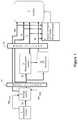
- FIG. 1is a block diagram illustrating flow of data through a system which includes a combination mammography/tomosynthesis acquisition station or a tomosynthesis only acquisition station to acquire tomosynthesis and/or mammography images, and includes image merge technology of the present invention for providing a supreme synthesized two dimensional image which merges the most relevant data from all available data sources into one 2D image;
- FIG. 2is diagram illustrating the data flow of a series of tomosynthesis slices, a synthesized 2D mammogram through the image merge technology of the present invention to generate at least one of a merged image I MERGE and a merge map;
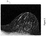
- FIG. 3is a diagram illustrating an alternate form of merged image, wherein region boundaries are dynamically identified during merge image build;
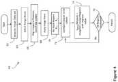
- FIG. 4is flow diagram illustrating exemplary steps that may be performed during an image merge process of the present invention
- FIGS. 5A and 5Billustrate a display of a merged image, and a resultant display of a source image when a region is selected by a user
- FIG. 6is a diagram of an exemplary merged image display, which incorporates an optional cluster spread indicator of the present invention.
- FIG. 7is an x-ray image acquisition, processing and display system.
- the notation Mprefers to a conventional mammogram, which is a two-dimensional projection image of a breast and encompasses both a digital image as acquired by a flat panel detector or another imaging device and the image after conventional processing to prepare it for display to a health professional or for storage, e.g. in the PACS system of a hospital or another institution.
- Tprefers to an image that is similarly two-dimensional but is taken at a respective tomosynthesis angle between the breast and the origin of the imaging X-rays (typically the focal spot of an X-ray tube), and also encompasses the image as acquired as well as the image after being processed for display or for some other use.
- Trrefers to an image that is reconstructed from images Tp, for example in the manner described in said earlier-filed patent applications, and represents a slice of the breast as it would appear in a projection X-ray image of that slice at any desired angle, not only at an angle used for Tp or Mp images.
- Msrefers to synthesized 2D projection images which simulate mammography images, such as a craniocaudal (CC) or mediolateral oblique (MLO) images, and are constructed using tomosynthesis projection images Tp, tomosynthesis reconstructed images Tr or a combination thereof.
- Ms imagesmay be provided for display to a health professional or for storage in the PACS system of a hospital or another institution.
- An example of methods that may be used to generate synthesized 2D projection imagesare described in U.S. patent application Ser. No. 12/471,981, filed May 26, 2009, as well as U.S. Pat. No. 7,760,924, filed Nov. 21, 2008, both incorporated herein by reference in their entireties.
- I MERGErefers to a 2D image generated by merging together any two or more of Mp, Ms, Tp or Tr images.
- I MERGE , Tp, Tr, Ms and Mpalso encompasses information, in whatever form, that is sufficient to describe such an image for display, further processing, or storage.
- the images I MERGE , Mp, Ms, Tp and Trtypically are in digital form before being displayed, and are defined by information identifying properties of each pixel in a two-dimensional array of pixels.
- the pixel valuestypically relate to respective measured or estimated or computed responses to X-rays of corresponding volumes in the breast (voxels or columns of tissue).
- the geometry of the tomosynthesis images (Tr and Tp), mammography images (Ms, Mp) and the merged image I MERGEare matched to a common coordinate system as described in U.S.
- FIG. 1illustrates flow of data in one example of an image generation and display system which may incorporate the merged image generation and display features of the present invention. It should be understood that FIG. 1 illustrates a particular embodiment of a flow diagram, with certain processes happening in a particular serial order or in parallel, the present invention is not generally limited to the performance of image processing in any particular order.
- FIG. 1illustrates an image data acquisition system 1 that acquires tomosynthesis image data for Tp images of patients' breasts using the three dimensional and/or tomosynthesis acquisition methods of any of the systems available in the art. If the acquisition system is a combo system, Mp images may also be generated. Some dedicated tomosynthesis systems or combo systems may be adapted to accept and store legacy mammogram images (meaning pre-acquired mammogram systems indicated via dashed line and legend Mp legacy in FIG. 1 ) in a Picture Archiving and Communication System (PACS) storage device 2 , although it is not a requirement that any Mp images be acquired by the acquisition system or pre-stored.
- PPSPicture Archiving and Communication System
- the projection images Tpare sent to storage device 2 , which is preferably a DICOM-compliant PACS.
- the Tp imagesare sent (from either acquisition system 1 or from storage device 2 ) to a computer system 3 configured as a reconstruction engine that reconstructs the Tp images into reconstructed image slabs Tr representing breast slices of selected thickness and at selected orientations, as disclosed in said earlier-filed patent applications and detailed below.
- the computer systemmay be further configured with 2D synthesis functionality 4 , which may operate substantially in parallel with reconstruction engine 3 to generate a synthesized 2D image (interchangeably referenced as T2d or Ms).
- the reconstructed slice images Trare then sent to a display system 5 so that they can be viewed.
- Tr slicescan be returned to the storage device. If the reconstruction engine 3 is connected to display 5 via a fast link, then large datasets can be transmitted quickly. Other images, such as the Ms, Mp and/or Tp images may also be forwarded to the display unit for concurrent or toggled viewing.
- the imaging and display system of the present inventionincludes a 2D synthesizer for generating 2D images simulating mammograms taken at both a CC and MLO orientation using a combination of one or more Tp and/or Tr images.
- the synthesized 2D imagesmay be generated dynamically prior to display, as shown in FIG. 1 , and/or may be stored in storage system 2 for use by other processes.
- a set of mode filters 7 a , 7 bare disposed between image acquisition and image display.
- Each of the filters 7 a and 7 bmay additionally include customized filters for each type of image (i.e., Tp, Mp, Tr) arranged to highlight certain aspects of the particular types of images.
- Tp, Mp, Trtype of image
- each modecan be tuned/configured in a optimal way for a specific purpose.
- the tuning or configurationmay be automatic, based on the type of the image, or may be defined by manual input, for example through a user interface coupled to a display.
- filterscould be provided to define a mass/calc-emphasis mode, 3D-tomo-slice-look mode, 2D-mammo-look mode, etc.
- an image merge processor 6merges relevant image data obtained from a set of available images to provide a merged image I MERGE for display.
- the set of available imagesincludes at least filtered and/or unfiltered Ms, Mp, Tr and/or Tp images.
- FIG. 1illustrates that all types of images are input into the image merge processor 6 it is envisioned that the form of merged image shall be manually configurable.
- a user interfacecomprised of a keypad, touch pad, mouse, pull down menu, button, switch, etc., may be configured to allow a user to select a particular group of two or more images or image types for merging to produce a supreme 2D image for display.
- a radiologistmay wish to merge two or more reconstructed tomosynthesis slabs to provide a merged image showing the most discernable structures in a single image.
- the radiologistmay combine the 2D mammogram image, Mp or Ms, with 3D projection or reconstructed images to obtain a merged image that highlights both calcifications and structures.
- Filters applied to each type of imagefurther highlight the types of structures or features which are generally most prevalent or discernable in the image type; thus a certain filter may be applied to mammography images to highlight calcifications, while a different filter may be applied to tomosynthesis slices to highlight masses. Filters may also be provided to give the particular image a desired look and feel; i.e., make the merged image appear more like a tomosynthesis image, or a mammography image.
- the display 5may be the display of an acquisition workstation, or a technologists review station, or a display that is physically remote from the acquisition system or storage device, i.e., connected via the network.
- a display of the systempreferably should be able to display I MERGE , Ms, Mp and Tr (and/or Tp) images concurrently (either in separate windows on the display, on separate monitors of a technology workstation, or overlaid) or sequentially or in toggled mode, wherein the I MERGE Ms, Mp, Tp and Tr images may be those currently acquired, or those that were acquired in previous studies.
- the displaycan simultaneously or sequentially or in toggled mode display merged images I MERGE , mammograms (Ms, Mp) and tomosynthesis images Tr (and/or Tp) from the current and previous studies.
- Tr slicescan be reconstructed all to the same size, which can be the same as the size of an Mp or Ms image of the breast, or they can be initially reconstructed to sizes determined by the fan shape of the x-ray beam used in the acquisition and later converted to that same size by appropriate interpolate]on/extrapolation.
- Images of different types and from different sourcescan be displayed in desirable size and resolution.
- an imagecan be displayed in (1) Fit To View Port mode, in which the size of the displayed image size is maximized such that the entire imaged breast tissue is visible, (2) True Size mode, in which a display pixel on the screen corresponds to a pixel of the image, or (3) Right Size mode, in which the size of a displayed image is adjusted so that it matches that of another image that is concurrently displayed or with which the displayed image is or can be toggled.
- the system described as a non-limiting example in this patent specificationis thus capable of receiving and displaying selectively the tomosynthesis projection images Tp, the tomosynthesis reconstruction images Tr, the synthesized mammogram image Ms and/or the mammogram images Mp, or a single type, or any sub combination of types.
- the systemhas software to perform reconstruction of tomosynthesis image data for images Tp into images Tr, software for synthesizing mammogram images Ms and software for merging a set of images to provide a supreme image that displays, for every region of the merged image, the most relevant feature in that region among all images in the image set.
- a featureis the ‘most relevant’ based upon the application of one or more a computer assisted detection (CAD) algorithms to the image, wherein the CAD algorithms assign numerical values, weights or thresholds, to pixels or regions based upon detected features within the region or between features.
- the featuresmay include, for example, speculated lesions, calcifications and the like.
- CADcomputer assisted detection
- Various systems and methodsare currently known for computerized detection of abnormalities in radiographic images, such as those disclosed by Giger et al. in RadioGraphics, May 1993, pp. 647 656; Giger et al. in Proceedings of SPIE, Vol. 1445 (1991), pp. 101 103; U.S. Pat. No.
- FIG. 2is a diagram which pictorially illustrates the merging of image data from a tomosynthesis image data set Tr, comprising tomosynthesis slices 10 A to 10 N, with image data from a mammogram 20 , in this case a synthesized mammogram Ms.
- the image datais forwarded to the region compare and image merge processor 6 which compares the images on a region by region basis, searching for that image with the most desirable display data for that region.
- the image with the most desirable display datamay be that image with a highest pixel value, the lowest pixel value, or which has been assigned a threshold value or weight based on the application of a CAD algorithm to the image.
- the pixels of that regionare copied over to the corresponding region of the merged image. For example, as shown in FIG. 2 , region 36 M from image Ms is written to region 361 of the merged image, and region 35 Tr of tomosynthesis slice 10 A is copied to region 351 of the merged image.
- a merge map 40is constructed.
- the merge mapstores, for each region of the merged image, an identifier of the image which sourced the region. Therefore, as shown in FIG. 2 , the Ms identifier is stored in region 361 , while the 10 A TR slice identifier is stored in region 351 .
- the merged mapmay be used during merge image display to permit fast viewing of the source image for a selected region.
- FIG. 3illustrates a merged image 50 which has been constructed via the combinations of numerous regions of different source images, at arbitrary region boundaries 52 .
- the boundaries 52may be identified according to the detection of particular features within the slices.
- FIG. 4is a flow diagram provided to illustrate exemplary steps that may be performed in an image merge process of the present invention.
- an image data setis acquired.
- the image data setmay be acquired by a tomosynthesis acquisition system, a combo tomosynthesis/mammography system, or acquired merely by retrieving pre-existing data from a storage device, either locally or remotely located relative to an image display device.
- a usermay optionally select a merge mode. As part of selecting a merge mode, the user may select which images to use for the merge, whether to highlight certain features, such as calcifications, speculated lesions or masses, whether to display the image as a lower resolution tomosynthesis image, etc.
- the images that are to be merged to build the supreme vieware mapped to a common coordinate system, for example as described in U.S. Pat. No. 7,702,142 entitled Matching Geometry Generation and Display of Tomosynthesis Images , incorporated herein by reference. Other methods of matching images of different coordinate systems may alternatively be used.
- step 68image filters are applied and, for each of the regions (indicated by “step” 70 ), the process of comparing regions among the different images begins, indicated by step 72 .
- step 74each I MERGE region is populated with the pixels of the region of the image in the image set having the most desirable pixels, value or pattern. The process of populating regions continues until it is determined, at step 76 , that all regions have been evaluated, at which point the merged image is ready for display.
- FIGS. 5A and 5Billustrate two views of a display 80 .
- the first view of display 80 shown in FIG. 5Aillustrates a merged image 82 , having regions sourced by different ones of an acquired or synthesized image set.
- FIG. 5Billustrates one feature enabled by the present invention, whereby a user may select a region or area 83 within the merged image 82 , and the resulting image source 84 for that area is displayed together with the merged image.
- the imageneed not be displayed proximate to the merged image; in one embodiment, selection of a desired region replaces the merged image with the source image, or alternatively overlays the source image on the merged image, allowing the two to be viewed in cine mode or toggle mode.
- Multiple source imagesmay be displayed concurrently in this manner, i.e., when a user selects multiple regions within the merged image a ‘stack’, pull down menu or other means is used for concurrent display of the multiple images.
- the merged imagemay automatically be shrunk to an icon, or moved to a task bar.
- the merged imagemay also be dynamically modified by the selection of different filters, modes or sources at a user interface of the display.
- a merged imagemay obfuscate data presented to the reviewer by essentially removing depth information provided by the tomosynthesis reconstruction.
- merged image 100appears to include multiple clusters of calcifications 102 a and 102 b , which are potential predictors of cancer.
- the respective clusters of calcifications 102 a and 102 bwhich appear proximate when evaluating only in the x-y axis, may actually be spread apart throughout the breast.
- the present inventionincludes a histogram 104 , 106 , which pictorially illustrates the depth of the clusters of calcifications 102 a and 102 b within the respective magnified regions. Histogram 104 illustrates that the cluster of calcifications 102 a are actually a real calcification cluster, while Histogram 106 illustrates that the visualized cluster of calcifications 102 b are actually distributed throughout the breast.
- FIG. 7is schematic illustration of an x-ray image acquisition, processing and display system 108 .
- the system 108includes an x-ray source 110 that projects x-rays 112 at a detector 116 in order to acquire x-ray images of breast tissue under the control of an acquisition control 118 .
- the acquired imagesare transmitted to a processing unit 120 , which generates a reconstructed 3D image set 122 , a 2D synthesized image 124 , and a synthesized merged image 126 , as described herein.
- These constructed/generated imagesmay be stored in a storage memory 130 coupled to the processor 120 and/or displayed on a viewing interface 152 of a display unit 150 .
- the merged imagemay combine regions of any combination of 2D and 3D image data, including an acquired mammogram, a synthesized mammogram, or a tomosynthesis projection image and a reconstructed 3D data set, thereby allowing the radiologist to quickly view a large number of regions of interest within a breast while referencing only a single 2D image and increasing the performance and efficiency of breast cancer screening and diagnosis.
Landscapes
- Engineering & Computer Science (AREA)
- Health & Medical Sciences (AREA)
- Life Sciences & Earth Sciences (AREA)
- Medical Informatics (AREA)
- Physics & Mathematics (AREA)
- Public Health (AREA)
- Biomedical Technology (AREA)
- Veterinary Medicine (AREA)
- General Health & Medical Sciences (AREA)
- Animal Behavior & Ethology (AREA)
- Surgery (AREA)
- Biophysics (AREA)
- High Energy & Nuclear Physics (AREA)
- Molecular Biology (AREA)
- Nuclear Medicine, Radiotherapy & Molecular Imaging (AREA)
- Optics & Photonics (AREA)
- Pathology (AREA)
- Radiology & Medical Imaging (AREA)
- Heart & Thoracic Surgery (AREA)
- Theoretical Computer Science (AREA)
- General Physics & Mathematics (AREA)
- Computer Vision & Pattern Recognition (AREA)
- Pure & Applied Mathematics (AREA)
- Mathematical Optimization (AREA)
- Algebra (AREA)
- Mathematical Physics (AREA)
- Mathematical Analysis (AREA)
- Dentistry (AREA)
- Oral & Maxillofacial Surgery (AREA)
- Computer Hardware Design (AREA)
- Apparatus For Radiation Diagnosis (AREA)
- Image Processing (AREA)
- Image Analysis (AREA)
Abstract
Description
Claims (13)
Priority Applications (6)
| Application Number | Priority Date | Filing Date | Title |
|---|---|---|---|
| US16/013,701US10573276B2 (en) | 2011-11-27 | 2018-06-20 | System and method for generating a 2D image using mammography and/or tomosynthesis image data |
| US16/792,182US10978026B2 (en) | 2011-11-27 | 2020-02-14 | System and method for generating a 2D image using mammography and/or tomosynthesis image data |
| US17/220,913US11508340B2 (en) | 2011-11-27 | 2021-04-01 | System and method for generating a 2D image using mammography and/or tomosynthesis image data |
| US17/869,235US11837197B2 (en) | 2011-11-27 | 2022-07-20 | System and method for generating a 2D image using mammography and/or tomosynthesis image data |
| US18/493,433US12183309B2 (en) | 2011-11-27 | 2023-10-24 | System and method for generating a 2D image using mammography and/or tomosynthesis image data |
| US18/950,896US20250140219A1 (en) | 2011-11-27 | 2024-11-18 | System and method for generating a 2d image using mammography and/or tomosynthesis image data |
Applications Claiming Priority (4)
| Application Number | Priority Date | Filing Date | Title |
|---|---|---|---|
| US201161563785P | 2011-11-27 | 2011-11-27 | |
| PCT/US2012/066526WO2013078476A1 (en) | 2011-11-27 | 2012-11-26 | System and method for generating a 2d image using mammography and/or tomosynthesis image data |
| US201414360389A | 2014-05-23 | 2014-05-23 | |
| US16/013,701US10573276B2 (en) | 2011-11-27 | 2018-06-20 | System and method for generating a 2D image using mammography and/or tomosynthesis image data |
Related Parent Applications (2)
| Application Number | Title | Priority Date | Filing Date |
|---|---|---|---|
| PCT/US2012/066526ContinuationWO2013078476A1 (en) | 2005-11-10 | 2012-11-26 | System and method for generating a 2d image using mammography and/or tomosynthesis image data |
| US14/360,389ContinuationUS10008184B2 (en) | 2005-11-10 | 2012-11-26 | System and method for generating a 2D image using mammography and/or tomosynthesis image data |
Related Child Applications (1)
| Application Number | Title | Priority Date | Filing Date |
|---|---|---|---|
| US16/792,182ContinuationUS10978026B2 (en) | 2011-11-27 | 2020-02-14 | System and method for generating a 2D image using mammography and/or tomosynthesis image data |
Publications (2)
| Publication Number | Publication Date |
|---|---|
| US20190043456A1 US20190043456A1 (en) | 2019-02-07 |
| US10573276B2true US10573276B2 (en) | 2020-02-25 |
Family
ID=48470355
Family Applications (6)
| Application Number | Title | Priority Date | Filing Date |
|---|---|---|---|
| US16/013,701ActiveUS10573276B2 (en) | 2011-11-27 | 2018-06-20 | System and method for generating a 2D image using mammography and/or tomosynthesis image data |
| US16/792,182ActiveUS10978026B2 (en) | 2011-11-27 | 2020-02-14 | System and method for generating a 2D image using mammography and/or tomosynthesis image data |
| US17/220,913ActiveUS11508340B2 (en) | 2011-11-27 | 2021-04-01 | System and method for generating a 2D image using mammography and/or tomosynthesis image data |
| US17/869,235ActiveUS11837197B2 (en) | 2011-11-27 | 2022-07-20 | System and method for generating a 2D image using mammography and/or tomosynthesis image data |
| US18/493,433ActiveUS12183309B2 (en) | 2011-11-27 | 2023-10-24 | System and method for generating a 2D image using mammography and/or tomosynthesis image data |
| US18/950,896PendingUS20250140219A1 (en) | 2011-11-27 | 2024-11-18 | System and method for generating a 2d image using mammography and/or tomosynthesis image data |
Family Applications After (5)
| Application Number | Title | Priority Date | Filing Date |
|---|---|---|---|
| US16/792,182ActiveUS10978026B2 (en) | 2011-11-27 | 2020-02-14 | System and method for generating a 2D image using mammography and/or tomosynthesis image data |
| US17/220,913ActiveUS11508340B2 (en) | 2011-11-27 | 2021-04-01 | System and method for generating a 2D image using mammography and/or tomosynthesis image data |
| US17/869,235ActiveUS11837197B2 (en) | 2011-11-27 | 2022-07-20 | System and method for generating a 2D image using mammography and/or tomosynthesis image data |
| US18/493,433ActiveUS12183309B2 (en) | 2011-11-27 | 2023-10-24 | System and method for generating a 2D image using mammography and/or tomosynthesis image data |
| US18/950,896PendingUS20250140219A1 (en) | 2011-11-27 | 2024-11-18 | System and method for generating a 2d image using mammography and/or tomosynthesis image data |
Country Status (5)
| Country | Link |
|---|---|
| US (6) | US10573276B2 (en) |
| EP (1) | EP2782505B1 (en) |
| JP (3) | JP2014534042A (en) |
| KR (1) | KR102109588B1 (en) |
| WO (1) | WO2013078476A1 (en) |
Cited By (19)
| Publication number | Priority date | Publication date | Assignee | Title |
|---|---|---|---|---|
| US10978026B2 (en)* | 2011-11-27 | 2021-04-13 | Hologic, Inc. | System and method for generating a 2D image using mammography and/or tomosynthesis image data |
| US11403483B2 (en) | 2017-06-20 | 2022-08-02 | Hologic, Inc. | Dynamic self-learning medical image method and system |
| US11406332B2 (en) | 2011-03-08 | 2022-08-09 | Hologic, Inc. | System and method for dual energy and/or contrast enhanced breast imaging for screening, diagnosis and biopsy |
| US11419565B2 (en) | 2014-02-28 | 2022-08-23 | IIologic, Inc. | System and method for generating and displaying tomosynthesis image slabs |
| US11445993B2 (en) | 2017-03-30 | 2022-09-20 | Hologic, Inc. | System and method for targeted object enhancement to generate synthetic breast tissue images |
| US11452486B2 (en) | 2006-02-15 | 2022-09-27 | Hologic, Inc. | Breast biopsy and needle localization using tomosynthesis systems |
| US11455754B2 (en) | 2017-03-30 | 2022-09-27 | Hologic, Inc. | System and method for synthesizing low-dimensional image data from high-dimensional image data using an object grid enhancement |
| US11589944B2 (en) | 2013-03-15 | 2023-02-28 | Hologic, Inc. | Tomosynthesis-guided biopsy apparatus and method |
| US11663780B2 (en) | 2012-02-13 | 2023-05-30 | Hologic Inc. | System and method for navigating a tomosynthesis stack using synthesized image data |
| US11701199B2 (en) | 2009-10-08 | 2023-07-18 | Hologic, Inc. | Needle breast biopsy system and method of use |
| US11775156B2 (en) | 2010-11-26 | 2023-10-03 | Hologic, Inc. | User interface for medical image review workstation |
| US11783476B2 (en) | 2019-10-25 | 2023-10-10 | DeepHealth, Inc. | System and method for analyzing three-dimensional image data |
| US11957497B2 (en) | 2017-03-30 | 2024-04-16 | Hologic, Inc | System and method for hierarchical multi-level feature image synthesis and representation |
| US12029602B2 (en) | 2013-10-24 | 2024-07-09 | Hologic, Inc. | System and method for navigating x-ray guided breast biopsy |
| US12211608B2 (en) | 2013-03-15 | 2025-01-28 | Hologic, Inc. | System and method for navigating a tomosynthesis stack including automatic focusing |
| US12236582B2 (en) | 2018-09-24 | 2025-02-25 | Hologic, Inc. | Breast mapping and abnormality localization |
| US12236597B2 (en) | 2021-11-29 | 2025-02-25 | Hologic, Inc. | Systems and methods for correlating objects of interest |
| US12254586B2 (en) | 2021-10-25 | 2025-03-18 | Hologic, Inc. | Auto-focus tool for multimodality image review |
| US12367574B2 (en) | 2019-12-23 | 2025-07-22 | DeepHealth, Inc. | Systems and methods for analyzing two-dimensional and three-dimensional image data |
Families Citing this family (20)
| Publication number | Priority date | Publication date | Assignee | Title |
|---|---|---|---|---|
| US8571289B2 (en) | 2002-11-27 | 2013-10-29 | Hologic, Inc. | System and method for generating a 2D image from a tomosynthesis data set |
| EP3221847B1 (en) | 2014-11-20 | 2021-01-06 | Koninklijke Philips N.V. | Method for generation of synthetic mammograms from tomosynthesis data |
| WO2016142492A1 (en) | 2015-03-10 | 2016-09-15 | Koninklijke Philips N.V. | Retrieval of corresponding structures in pairs of medical images |
| JP6456550B2 (en) | 2015-09-01 | 2019-01-23 | コーニンクレッカ フィリップス エヌ ヴェKoninklijke Philips N.V. | Device for displaying medical image data of a body part |
| JP6502509B2 (en) | 2015-09-10 | 2019-04-17 | 富士フイルム株式会社 | Image processing apparatus, radiographic imaging system, image processing method, and image processing program |
| JP6580963B2 (en)* | 2015-11-30 | 2019-09-25 | キヤノンメディカルシステムズ株式会社 | Image processing apparatus, image processing method, and X-ray diagnostic apparatus |
| KR102527017B1 (en)* | 2017-01-23 | 2023-05-02 | 한국전자통신연구원 | Apparatus and method for generating 2d medical image based on plate interpolation |
| JP7033931B2 (en) | 2018-01-17 | 2022-03-11 | 富士フイルム株式会社 | Image processing device, image processing method, and image processing program |
| US12121304B2 (en) | 2018-05-04 | 2024-10-22 | Hologic, Inc. | Introducer and localization wire visualization |
| EP3787520B1 (en) | 2018-05-04 | 2024-09-25 | Hologic, Inc. | Biopsy needle visualization |
| JP7242436B2 (en) | 2019-06-13 | 2023-03-20 | 富士フイルム株式会社 | Image interpretation support device, image interpretation support method, and image interpretation support program |
| US11883206B2 (en) | 2019-07-29 | 2024-01-30 | Hologic, Inc. | Personalized breast imaging system |
| EP4439580A3 (en) | 2019-09-27 | 2024-12-25 | Hologic, Inc. | Ai system for predicting reading time and reading complexity for reviewing 2d/3d breast images |
| EP4101386A4 (en) | 2020-02-04 | 2023-07-12 | FUJIFILM Corporation | IMAGE ADJUSTMENT DEVICE, METHOD AND PROGRAM |
| WO2021157180A1 (en)* | 2020-02-04 | 2021-08-12 | 富士フイルム株式会社 | Image setting device, method, and program |
| EP4119055B1 (en) | 2020-03-13 | 2024-10-30 | FUJIFILM Corporation | Image generation device and program, learning device and program, and image processing device and program |
| JP7446410B2 (en) | 2020-03-18 | 2024-03-08 | 富士フイルム株式会社 | Image processing device, method and program |
| CN115297778B (en) | 2020-03-18 | 2025-08-08 | 富士胶片株式会社 | Image processing device, method, and recording medium |
| US11481038B2 (en) | 2020-03-27 | 2022-10-25 | Hologic, Inc. | Gesture recognition in controlling medical hardware or software |
| JP2023059633A (en)* | 2021-10-15 | 2023-04-27 | キヤノンメディカルシステムズ株式会社 | X-ray diagnostic apparatus and control method of x-ray diagnostic apparatus |
Citations (188)
| Publication number | Priority date | Publication date | Assignee | Title |
|---|---|---|---|---|
| US1000818A (en) | 1910-12-17 | 1911-08-15 | John W Kempf | Core for making culverts. |
| US1001030A (en) | 1910-06-18 | 1911-08-22 | Chicago Door Company | Combined track and hanger for car-doors. |
| US1041326A (en) | 1911-07-13 | 1912-10-15 | Orville V Monroe | Pen-wiper. |
| US1041041A (en) | 1908-07-10 | 1912-10-15 | Charles Hall Davis | Coupon certificate of deposit. |
| US3502878A (en) | 1967-09-22 | 1970-03-24 | Us Health Education & Welfare | Automatic x-ray apparatus for limiting the field size of a projected x-ray beam in response to film size and to source-to-film distance |
| US3863073A (en) | 1973-04-26 | 1975-01-28 | Machlett Lab Inc | Automatic system for precise collimation of radiation |
| US3971950A (en) | 1975-04-14 | 1976-07-27 | Xerox Corporation | Independent compression and positioning device for use in mammography |
| US4160906A (en) | 1977-06-23 | 1979-07-10 | General Electric Company | Anatomically coordinated user dominated programmer for diagnostic x-ray apparatus |
| US4310766A (en) | 1978-09-06 | 1982-01-12 | Siemens Aktiengesellschaft | Motor driven x-ray grid and film-holder assembly |
| US4496557A (en) | 1981-08-27 | 1985-01-29 | Adir | Tricyclic ethers, their preparation and the pharmaceutical compositions containing them |
| US4559641A (en) | 1983-06-24 | 1985-12-17 | Thomson-Cgr | Retractable cassette holder for a radiological and radiographic examination apparatus |
| US4706269A (en) | 1985-03-11 | 1987-11-10 | Reina Leo J | Anti-scatter grid structure |
| US4744099A (en) | 1983-11-03 | 1988-05-10 | Siemens Aktiengesellschaft | X-ray diagnostic apparatus comprising radiation filters |
| US4773086A (en) | 1983-12-16 | 1988-09-20 | Yokogawa Medical Systems, Limited | Operator console for X-ray tomographs |
| US4773087A (en) | 1986-04-14 | 1988-09-20 | University Of Rochester | Quality of shadowgraphic x-ray images |
| US4819258A (en) | 1986-11-28 | 1989-04-04 | Bennett X-Ray Corp. | Auto-setting of KV in an x-ray machine after selection of technic factors |
| US4821727A (en) | 1986-10-30 | 1989-04-18 | Elscint Ltd. | Mammographic biopsy needle holder system |
| US4907156A (en) | 1987-06-30 | 1990-03-06 | University Of Chicago | Method and system for enhancement and detection of abnormal anatomic regions in a digital image |
| WO1990005485A1 (en) | 1988-11-23 | 1990-05-31 | Nrt-Nordisk Roentgen Teknik A/S | X-ray apparatus |
| US4969174A (en) | 1989-09-06 | 1990-11-06 | General Electric Company | Scanning mammography system with reduced scatter radiation |
| US4989227A (en) | 1989-04-28 | 1991-01-29 | General Electric Cgr S.A. | Cassette carrier adaptable in size and position for mammography |
| US5018176A (en) | 1989-03-29 | 1991-05-21 | General Electric Cgr S.A. | Mammograph equipped with an integrated device for taking stereotaxic photographs and a method of utilization of said mammograph |
| US5029193A (en) | 1989-07-03 | 1991-07-02 | Siemens Aktiengesellschaft | X-ray diagnostic installation for mammography exposures |
| USRE33634E (en) | 1986-09-23 | 1991-07-09 | Method and structure for optimizing radiographic quality by controlling X-ray tube voltage, current focal spot size and exposure time | |
| US5051904A (en) | 1988-03-24 | 1991-09-24 | Olganix Corporation | Computerized dynamic tomography system |
| US5078142A (en) | 1989-11-21 | 1992-01-07 | Fischer Imaging Corporation | Precision mammographic needle biopsy system |
| US5133020A (en) | 1989-07-21 | 1992-07-21 | Arch Development Corporation | Automated method and system for the detection and classification of abnormal lesions and parenchymal distortions in digital medical images |
| US5163075A (en) | 1991-08-08 | 1992-11-10 | Eastman Kodak Company | Contrast enhancement of electrographic imaging |
| US5164976A (en) | 1989-09-06 | 1992-11-17 | General Electric Company | Scanning mammography system with improved skin line viewing |
| US5199056A (en) | 1989-11-28 | 1993-03-30 | Darrah Carol J | Mammography compression paddle |
| US5240011A (en) | 1991-11-27 | 1993-08-31 | Fischer Imaging Corporation | Motorized biopsy needle positioner |
| US5289520A (en) | 1991-11-27 | 1994-02-22 | Lorad Corporation | Stereotactic mammography imaging system with prone position examination table and CCD camera |
| US5343390A (en) | 1992-02-28 | 1994-08-30 | Arch Development Corporation | Method and system for automated selection of regions of interest and detection of septal lines in digital chest radiographs |
| US5359637A (en) | 1992-04-28 | 1994-10-25 | Wake Forest University | Self-calibrated tomosynthetic, radiographic-imaging system, method, and device |
| US5365562A (en) | 1993-09-20 | 1994-11-15 | Fischer Imaging Corporation | Digital imaging apparatus |
| US5415169A (en) | 1989-11-21 | 1995-05-16 | Fischer Imaging Corporation | Motorized mammographic biopsy apparatus |
| US5452367A (en) | 1993-11-29 | 1995-09-19 | Arch Development Corporation | Automated method and system for the segmentation of medical images |
| US5491627A (en) | 1993-05-13 | 1996-02-13 | Arch Development Corporation | Method and system for the detection of microcalcifications in digital mammograms |
| US5506877A (en) | 1994-11-23 | 1996-04-09 | The General Hospital Corporation | Mammography breast compression device and method |
| US5526394A (en) | 1993-11-26 | 1996-06-11 | Fischer Imaging Corporation | Digital scan mammography apparatus |
| US5539797A (en) | 1993-03-29 | 1996-07-23 | Ge Medical Systems Sa | Method and apparatus for digital stereotaxic mammography |
| US5553111A (en) | 1994-10-26 | 1996-09-03 | The General Hospital Corporation | Apparatus and method for improved tissue imaging |
| US5592562A (en) | 1994-01-19 | 1997-01-07 | International Business Machines Corporation | Inspection system for cross-sectional imaging |
| US5594769A (en) | 1991-11-27 | 1997-01-14 | Thermotrex Corporation | Method and apparatus for obtaining stereotactic mammographic guided needle breast biopsies |
| US5596200A (en) | 1992-10-14 | 1997-01-21 | Primex | Low dose mammography system |
| US5598454A (en) | 1994-04-26 | 1997-01-28 | Siemens Aktiengesellschaft | X-ray diagnostics installation |
| US5627869A (en) | 1995-11-22 | 1997-05-06 | Thermotrex Corporation | Mammography apparatus with proportional collimation |
| EP0775467A1 (en) | 1995-11-23 | 1997-05-28 | Planmed Oy | Method and system for controlling the functions of a mammography apparatus |
| US5657362A (en) | 1995-02-24 | 1997-08-12 | Arch Development Corporation | Automated method and system for computerized detection of masses and parenchymal distortions in medical images |
| US5668889A (en) | 1990-04-19 | 1997-09-16 | Fuji Photo Film Co., Ltd. | Apparatus for determining an image position, and method for adjusting read-out conditions and/or image processing conditions for a radiation image |
| US5712890A (en) | 1994-11-23 | 1998-01-27 | Thermotrex Corp. | Full breast digital mammography device |
| WO1998016903A1 (en) | 1996-10-16 | 1998-04-23 | Vital Images, Inc. | Advanced diagnostic viewer |
| US5769086A (en) | 1995-12-06 | 1998-06-23 | Biopsys Medical, Inc. | Control system and method for automated biopsy device |
| US5818898A (en) | 1995-11-07 | 1998-10-06 | Kabushiki Kaisha Toshiba | X-ray imaging apparatus using X-ray planar detector |
| US5828722A (en) | 1996-05-17 | 1998-10-27 | Sirona Dental Systems Gmbh & Co., Kg | X-ray diagnostic apparatus for tomosynthesis having a detector that detects positional relationships |
| US5872828A (en) | 1996-07-23 | 1999-02-16 | The General Hospital Corporation | Tomosynthesis system for breast imaging |
| US5878104A (en) | 1996-05-17 | 1999-03-02 | Sirona Dental Systems Gmbh & Co. Kg | Method for producing tomosynthesis exposures employing a reference object formed by a region of the examination subject |
| US5878746A (en) | 1993-08-25 | 1999-03-09 | Lemelson; Jerome H. | Computerized medical diagnostic system |
| US5896437A (en) | 1996-05-17 | 1999-04-20 | Sirona Dental Systems Gmbh & Co. Kg | X-ray diagnostics apparatus for tomosynthesis having a reference object in fixed relationship to a radiation emitter |
| US5941832A (en) | 1991-09-27 | 1999-08-24 | Tumey; David M. | Method and apparatus for detection of cancerous and precancerous conditions in a breast |
| US6005907A (en) | 1996-05-17 | 1999-12-21 | Sirona Dental Systems Gmbh & Co. Kg | Method and apparatus for producing tomosynthesis exposures employing a reference object composed of a number of sub-objects |
| EP0982001A1 (en) | 1998-08-25 | 2000-03-01 | General Electric Company | Protocol driven image reconstruction, display, and processing in a multislice imaging system |
| US6075879A (en) | 1993-09-29 | 2000-06-13 | R2 Technology, Inc. | Method and system for computer-aided lesion detection using information from multiple images |
| US6091841A (en) | 1997-09-04 | 2000-07-18 | Qualia Computing, Inc. | Method and system for segmenting desired regions in digital mammograms |
| WO2000051484A2 (en) | 1998-11-25 | 2000-09-08 | Fischer Imaging Corporation | User interface system for mammographic imager |
| US6137527A (en) | 1996-12-23 | 2000-10-24 | General Electric Company | System and method for prompt-radiology image screening service via satellite |
| US6149301A (en) | 1998-12-30 | 2000-11-21 | General Electric Company | X-ray target centering apparatus for radiographic imaging system |
| US6175117B1 (en) | 1998-01-23 | 2001-01-16 | Quanta Vision, Inc. | Tissue analysis apparatus |
| US6196715B1 (en) | 1959-04-28 | 2001-03-06 | Kabushiki Kaisha Toshiba | X-ray diagnostic system preferable to two dimensional x-ray detection |
| US6216540B1 (en) | 1995-06-06 | 2001-04-17 | Robert S. Nelson | High resolution device and method for imaging concealed objects within an obscuring medium |
| US6233473B1 (en) | 1999-02-16 | 2001-05-15 | Hologic, Inc. | Determining body composition using fan beam dual-energy x-ray absorptiometry |
| US6243441B1 (en) | 1999-07-13 | 2001-06-05 | Edge Medical Devices | Active matrix detector for X-ray imaging |
| US6256370B1 (en) | 2000-01-24 | 2001-07-03 | General Electric Company | Method and apparatus for performing tomosynthesis |
| US6272207B1 (en) | 1999-02-18 | 2001-08-07 | Creatv Microtech, Inc. | Method and apparatus for obtaining high-resolution digital X-ray and gamma ray images |
| US6289235B1 (en) | 1998-03-05 | 2001-09-11 | Wake Forest University | Method and system for creating three-dimensional images using tomosynthetic computed tomography |
| US6292530B1 (en) | 1999-04-29 | 2001-09-18 | General Electric Company | Method and apparatus for reconstructing image data acquired by a tomosynthesis x-ray imaging system |
| US20010038861A1 (en) | 1999-12-16 | 2001-11-08 | Tsung-Min Hsu | Transdermal administration of nonsteroidal anti-inflammatory drugs using hydroxide-releasing agents as permeation enhancers |
| US6327336B1 (en) | 2000-06-05 | 2001-12-04 | Direct Radiography Corp. | Radiogram showing location of automatic exposure control sensor |
| US6341156B1 (en) | 1999-05-14 | 2002-01-22 | Siemens Aktiengesellschaft | X-ray diagnostic apparatus with relatively moved x-ray source and detector |
| US20020012450A1 (en) | 1998-01-09 | 2002-01-31 | Osamu Tsujii | Image processing apparatus and method |
| US6375352B1 (en) | 1999-10-01 | 2002-04-23 | General Electric Company | Apparatus and method for obtaining x-ray tomosynthesis data for mammography |
| US20020050986A1 (en) | 2000-08-11 | 2002-05-02 | Hitoshi Inoue | Image display apparatus and method, and storage medium |
| US6389104B1 (en) | 2000-06-30 | 2002-05-14 | Siemens Corporate Research, Inc. | Fluoroscopy based 3-D neural navigation based on 3-D angiography reconstruction data |
| US20020075997A1 (en) | 2000-12-18 | 2002-06-20 | Unger Christopher David | Medical diagnostic method and apparatus to control dual energy exposure techniques based on image information |
| US6411836B1 (en) | 1999-12-30 | 2002-06-25 | General Electric Company | Method and apparatus for user preferences configuring in an image handling system |
| US6415015B2 (en) | 1999-12-28 | 2002-07-02 | Ge Medical Systems Sa | Method and system of compensation of thickness of an organ |
| US6442288B1 (en) | 1997-12-17 | 2002-08-27 | Siemens Aktiengesellschaft | Method for reconstructing a three-dimensional image of an object scanned in the context of a tomosynthesis, and apparatus for tomosynthesis |
| US6463181B2 (en) | 2000-12-22 | 2002-10-08 | The United States Of America As Represented By The Secretary Of The Navy | Method for optimizing visual display of enhanced digital images |
| US20030007598A1 (en) | 2000-11-24 | 2003-01-09 | U-Systems, Inc. | Breast cancer screening with adjunctive ultrasound mammography |
| US20030018272A1 (en) | 2001-06-28 | 2003-01-23 | Treado Patrick J. | Method for Raman chemical imaging and characterization of calcification in tissue |
| US20030026386A1 (en) | 2001-02-01 | 2003-02-06 | Cha-Mei Tang | Anti-scatter grids and collimator designs, and their motion, fabrication and assembly |
| WO2003020114A2 (en) | 2001-08-31 | 2003-03-13 | Analogic Corporation | Image positioning method and system for tomosynthesis in a digital x-ray radiography system |
| US6556655B1 (en) | 1998-11-27 | 2003-04-29 | Ge Medical Systems Sa | Method for automatic detection of glandular tissue |
| US20030095624A1 (en) | 2001-11-21 | 2003-05-22 | Eberhard Jeffrey Wayne | Dose management system for mammographic tomosynthesis |
| US6574304B1 (en) | 2002-09-13 | 2003-06-03 | Ge Medical Systems Global Technology Company, Llc | Computer aided acquisition of medical images |
| JP2003189179A (en) | 2001-12-14 | 2003-07-04 | Konica Corp | Abnormal shade detector and image output device |
| US20030128893A1 (en) | 2001-11-19 | 2003-07-10 | Alfio Castorina | Method for merging digital images to obtain a high dynamic range digital image |
| US6597762B1 (en) | 2002-11-27 | 2003-07-22 | Ge Medical Systems Global Technology Co., Llc | Method and apparatus of lesion detection and validation based on multiple reviews of a CT image |
| US6611575B1 (en) | 2001-07-27 | 2003-08-26 | General Electric Company | Method and system for high resolution 3D visualization of mammography images |
| US20030169847A1 (en) | 2001-11-21 | 2003-09-11 | University Of Massachusetts Medical Center | System and method for x-ray fluoroscopic imaging |
| US6620111B2 (en) | 2001-04-20 | 2003-09-16 | Ethicon Endo-Surgery, Inc. | Surgical biopsy device having automatic rotation of the probe for taking multiple samples |
| US6626849B2 (en) | 2001-11-01 | 2003-09-30 | Ethicon Endo-Surgery, Inc. | MRI compatible surgical biopsy device |
| US6633674B1 (en) | 1999-11-24 | 2003-10-14 | General Electric Company | Picture archiving and communication system employing improved data compression |
| US20030194050A1 (en) | 2002-04-15 | 2003-10-16 | General Electric Company | Multi modality X-ray and nuclear medicine mammography imaging system and method |
| US20030194121A1 (en) | 2002-04-15 | 2003-10-16 | General Electric Company | Computer aided detection (CAD) for 3D digital mammography |
| US6638235B2 (en) | 2000-11-06 | 2003-10-28 | Suros Surgical Systems, Inc. | Biopsy apparatus |
| US6647092B2 (en) | 2002-01-18 | 2003-11-11 | General Electric Company | Radiation imaging system and method of collimation |
| US20030210254A1 (en) | 2002-05-13 | 2003-11-13 | Doan William D. | Method, system and computer product for displaying axial images |
| US20030212327A1 (en) | 2000-11-24 | 2003-11-13 | U-Systems Inc. | Adjunctive ultrasound processing and display for breast cancer screening |
| US20030215120A1 (en) | 2002-05-15 | 2003-11-20 | Renuka Uppaluri | Computer aided diagnosis of an image set |
| US20040008901A1 (en) | 2002-07-11 | 2004-01-15 | Avinash Gopal B. | Interpolated image filtering method and apparatus |
| US20040008809A1 (en) | 1998-07-24 | 2004-01-15 | Webber Richard L. | Method and system for creating task-dependent three-dimensional images |
| US20040047518A1 (en) | 2002-08-28 | 2004-03-11 | Carlo Tiana | Image fusion system and method |
| US20040052328A1 (en) | 2002-09-13 | 2004-03-18 | Sabol John M. | Computer assisted analysis of tomographic mammography data |
| US20040066884A1 (en) | 2002-10-07 | 2004-04-08 | Hermann Claus Bernhard Erich | Continuous scan tomosynthesis system and method |
| US20040070582A1 (en) | 2002-10-11 | 2004-04-15 | Matthew Warren Smith To Sonocine, Inc. | 3D modeling system |
| US20040094167A1 (en) | 2000-03-17 | 2004-05-20 | Brady John Michael | Three-dimensional reconstructions of a breast from two x-ray mammographics |
| US20040101095A1 (en) | 2002-11-27 | 2004-05-27 | Hologic Inc. | Full field mammography with tissue exposure control, tomosynthesis, and dynamic field of view processing |
| US6744848B2 (en) | 2000-02-11 | 2004-06-01 | Brandeis University | Method and system for low-dose three-dimensional imaging of a scene |
| US20040109529A1 (en) | 2002-12-10 | 2004-06-10 | General Electric Company | Full field digital tomosynthesis method and apparatus |
| US20040171986A1 (en) | 1999-04-26 | 2004-09-02 | Scimed Life System, Inc. | Apparatus and methods for guiding a needle |
| US6813334B2 (en) | 2000-10-20 | 2004-11-02 | Koninklijke Philips Electronics N.V. | Tomosynthesis in a limited angular range |
| US20050063509A1 (en) | 2001-10-19 | 2005-03-24 | Defreitas Kenneth F | Mammography system and method employing offset compression paddles automatic collimation and retractable anti-scatter grid |
| US20050078797A1 (en) | 2002-03-01 | 2005-04-14 | Mats Danielsson | X-ray protection device |
| US6882700B2 (en) | 2002-04-15 | 2005-04-19 | General Electric Company | Tomosynthesis X-ray mammogram system and method with automatic drive system |
| US6885724B2 (en) | 2003-08-22 | 2005-04-26 | Ge Medical Systems Global Technology Company, Llc | Radiographic tomosynthesis image acquisition utilizing asymmetric geometry |
| US20050105679A1 (en) | 2003-02-12 | 2005-05-19 | Tao Wu | Tomosynthesis imaging system and method |
| US20050113715A1 (en) | 2000-11-06 | 2005-05-26 | Jeffrey Schwindt | Biopsy apparatus |
| US20050113681A1 (en) | 2002-11-27 | 2005-05-26 | Defreitas Kenneth F. | X-ray mammography with tomosynthesis |
| WO2005051197A2 (en) | 2003-11-26 | 2005-06-09 | Koninklijke Philips Electronics, N.V. | Workflow optimization for high throughput imaging environment |
| US20050135555A1 (en) | 2003-12-23 | 2005-06-23 | Claus Bernhard Erich H. | Method and system for simultaneously viewing rendered volumes |
| US20050135664A1 (en) | 2003-12-23 | 2005-06-23 | Kaufhold John P. | Methods and apparatus for reconstruction of volume data from projection data |
| US20050226375A1 (en) | 2004-03-31 | 2005-10-13 | Eberhard Jeffrey W | Enhanced X-ray imaging system and method |
| WO2005110230A1 (en) | 2004-05-14 | 2005-11-24 | Philips Intellectual Property & Standards Gmbh | System and method for diagnosing breast cancer |
| WO2005112767A1 (en) | 2004-05-21 | 2005-12-01 | Tissuomics Limited | Apparatus and method for penetrating radiation measurements |
| US6978040B2 (en) | 2001-12-19 | 2005-12-20 | Canon Kabushiki Kaisha | Optical recovery of radiographic geometry |
| US20060018526A1 (en) | 2004-07-23 | 2006-01-26 | Avinash Gopal B | Methods and apparatus for noise reduction filtering of images |
| US6999554B2 (en) | 2003-11-17 | 2006-02-14 | Siemens Aktiengesellschaft | X-ray diagnostic apparatus for mammography examinations |
| US20060074288A1 (en) | 2004-10-04 | 2006-04-06 | Thomas Kelly | Estimating visceral fat by dual-energy x-ray absorptiometry |
| US7025725B2 (en) | 2002-03-28 | 2006-04-11 | Ultrasound Detection Systems, Llc | Three-dimensional ultrasound computed tomography imaging system |
| US20060098855A1 (en) | 2002-11-27 | 2006-05-11 | Gkanatsios Nikolaos A | Image handling and display in X-ray mammography and tomosynthesis |
| WO2006055830A2 (en) | 2004-11-15 | 2006-05-26 | Hologic, Inc. | Matching geometry generation and display of mammograms and tomosynthesis images |
| WO2006058160A2 (en) | 2004-11-26 | 2006-06-01 | Hologic, Inc. | Integrated multi-mode mammography/tomosynthesis x-ray system and method |
| US7110502B2 (en) | 2003-05-12 | 2006-09-19 | Canon Kabushiki Kaisha | Radiographic apparatus and method for switching a grid |
| US7127091B2 (en) | 2000-12-22 | 2006-10-24 | Koninklijke Philips Electronics, N.V. | Method and apparatus for visualizing a limited part of a 3D medical image-point-related data set, through basing a rendered image on an intermediate region between first and second clipping planes, and including spectroscopic viewing of such region |
| US20070036265A1 (en) | 2005-08-15 | 2007-02-15 | Zhenxue Jing | X-ray mammography/tomosynthesis of patient's breast |
| US20070052700A1 (en) | 2005-09-07 | 2007-03-08 | Wheeler Frederick W | System and method for 3D CAD using projection images |
| US20070223651A1 (en) | 2006-03-21 | 2007-09-27 | Wagenaar Douglas J | Dual modality mammography device |
| US7315607B2 (en) | 2005-09-02 | 2008-01-01 | Siemens Aktiengesellschaft | Mammograph system with a face shield |
| US7323692B2 (en) | 2004-08-10 | 2008-01-29 | Research Foundation Of State University Of New York | Flat-panel detector with avalanche gain |
| US20080045833A1 (en) | 2006-02-15 | 2008-02-21 | Defreitas Kenneth F | Breast biopsy and needle localization using tomosynthesis systems |
| US7346381B2 (en) | 2002-11-01 | 2008-03-18 | Ge Medical Systems Global Technology Company Llc | Method and apparatus for medical intervention procedure planning |
| WO2008047270A1 (en) | 2006-10-17 | 2008-04-24 | Koninklijke Philips Electronics N.V. | Visualization of 3d images in combination with 2d projection images |
| US20080187095A1 (en) | 2005-05-03 | 2008-08-07 | The Regents Of The University Of California | Biopsy Systems For Breast Computed Tomography |
| US20090005668A1 (en) | 2007-06-30 | 2009-01-01 | West Jay B | Non-invasive method for using 2D angiographic images for radiosurgical target definition |
| US20090034684A1 (en) | 2007-08-02 | 2009-02-05 | Sylvain Bernard | Method and system for displaying tomosynthesis images |
| US20090080594A1 (en) | 2006-08-03 | 2009-03-26 | Kenneth Brooks | Dedicated breast radiation imaging/therapy system |
| US20090080765A1 (en) | 2007-09-20 | 2009-03-26 | General Electric Company | System and method to generate a selected visualization of a radiological image of an imaged subject |
| US20090123052A1 (en) | 2002-11-27 | 2009-05-14 | Chris Ruth | System and Method for Generating a 2D Image from a Tomosynthesis Data Set |
| US20090135997A1 (en) | 2006-03-27 | 2009-05-28 | Hologic, Inc. | Breast Compression For Digital Mammography, Tomosynthesis And Other Modalities |
| JP2009207545A (en) | 2008-02-29 | 2009-09-17 | Fujifilm Corp | Image display method and apparatus |
| US20090238424A1 (en) | 2008-03-19 | 2009-09-24 | Kazumasa Arakita | Image processing apparatus and image processing method |
| US7606801B2 (en) | 2005-06-07 | 2009-10-20 | Varonis Inc. | Automatic management of storage access control |
| US20090268865A1 (en) | 2003-11-26 | 2009-10-29 | Baorui Ren | X-ray imaging with X-ray markers that provide adjunct information but preserve image quality |
| US7616801B2 (en) | 2002-11-27 | 2009-11-10 | Hologic, Inc. | Image handling and display in x-ray mammography and tomosynthesis |
| US7630533B2 (en) | 2007-09-20 | 2009-12-08 | Hologic, Inc. | Breast tomosynthesis with display of highlighted suspected calcifications |
| US20100054400A1 (en) | 2008-08-29 | 2010-03-04 | Hologic, Inc. | Multi-mode tomosynthesis/mammography gain calibration and image correction using gain map information from selected projection angles |
| US20100135558A1 (en) | 2002-11-27 | 2010-06-03 | Chris Ruth | System and Method for Generating a 2D Image from a Tomosynthesis Data Set |
| US20100259645A1 (en) | 2009-04-13 | 2010-10-14 | Pure Digital Technologies | Method and system for still image capture from video footage |
| WO2011008239A1 (en) | 2009-06-29 | 2011-01-20 | Thomson Licensing | Contrast enhancement |
| US20110069906A1 (en) | 2009-09-23 | 2011-03-24 | Samsung Electronics Co., Ltd. | Method and apparatus for blending images |
| EP2301432A1 (en) | 2008-06-03 | 2011-03-30 | FUJIFILM Corporation | Projection image creation device, method, and program |
| US20110110576A1 (en) | 2009-10-07 | 2011-05-12 | Hologic, Inc. | Selective Display Of Computer-Aided Detection Findings With Associated Breast X-Ray Mammogram and/or Tomosynthesis Image Information |
| WO2011065950A1 (en) | 2009-11-27 | 2011-06-03 | Qview Medical, Inc. | Interactive display of computer aided detection radiological screening results combined with quantitative prompts |
| WO2011073864A2 (en) | 2009-12-17 | 2011-06-23 | Koninklijke Philips Electronics N.V. | Reconstructing an object of interest |
| US20110178389A1 (en) | 2008-05-02 | 2011-07-21 | Eigen, Inc. | Fused image moldalities guidance |
| WO2011091300A2 (en) | 2010-01-24 | 2011-07-28 | Mistretta Medical, Llc | System and method for implementation of 4d time-energy subtraction computed tomography |
| DE102010009295A1 (en) | 2010-02-25 | 2011-08-25 | Siemens Aktiengesellschaft, 80333 | Method for displaying a region to be examined and / or treated |
| US20110234630A1 (en) | 2008-11-28 | 2011-09-29 | Fujifilm Medical Systems USA, Inc | Active Overlay System and Method for Accessing and Manipulating Imaging Displays |
| US20110242092A1 (en) | 2010-03-30 | 2011-10-06 | Fujifilm Corporation | Image display system |
| US8044972B2 (en) | 2006-12-21 | 2011-10-25 | Sectra Mamea Ab | Synchronized viewing of tomosynthesis and/or mammograms |
| US8051386B2 (en) | 2006-12-21 | 2011-11-01 | Sectra Ab | CAD-based navigation of views of medical image data stacks or volumes |
| WO2012063653A1 (en) | 2010-11-12 | 2012-05-18 | 株式会社 日立メディコ | Medical image display device and medical image display method |
| US20130121618A1 (en)* | 2011-05-27 | 2013-05-16 | Vikas Yadav | Seamless Image Composition |
| US20130121569A1 (en)* | 2009-09-14 | 2013-05-16 | Vikas Yadav | Methods and Apparatus for Blending Images |
| US20140327702A1 (en) | 2005-11-10 | 2014-11-06 | Hologic, Inc | System and method for generating a 2d image using mammography and/or tomosynthesis image data |
| US8983156B2 (en) | 2012-11-23 | 2015-03-17 | Icad, Inc. | System and method for improving workflow efficiences in reading tomosynthesis medical image data |
| US9805507B2 (en) | 2012-02-13 | 2017-10-31 | Hologic, Inc | System and method for navigating a tomosynthesis stack using synthesized image data |
Family Cites Families (317)
| Publication number | Priority date | Publication date | Assignee | Title |
|---|---|---|---|---|
| US7050611B2 (en) | 2001-05-29 | 2006-05-23 | Mevis Breastcare Gmbh Co. Kg | Method and computer system for screening of medical cases |
| SE8306243L (en) | 1983-11-14 | 1985-05-15 | Cytex Medicinteknik Ab | LOCATION METHODOLOGY |
| US4559557A (en) | 1984-06-01 | 1985-12-17 | General Electric Company | Region-of-interest digital subtraction angiography |
| US5740270A (en) | 1988-04-08 | 1998-04-14 | Neuromedical Systems, Inc. | Automated cytological specimen classification system and method |
| US5099846A (en) | 1988-12-23 | 1992-03-31 | Hardy Tyrone L | Method and apparatus for video presentation from a variety of scanner imaging sources |
| US5280427A (en) | 1989-11-27 | 1994-01-18 | Bard International, Inc. | Puncture guide for computer tomography |
| FR2668359B1 (en) | 1990-10-24 | 1998-02-20 | Gen Electric Cgr | MAMMOGRAPH PROVIDED WITH A PERFECTED NEEDLE HOLDER. |
| US5129911A (en) | 1991-03-11 | 1992-07-14 | Siczek Bernard W | Orbital aiming device |
| US5409497A (en) | 1991-03-11 | 1995-04-25 | Fischer Imaging Corporation | Orbital aiming device for mammo biopsy |
| US5279309A (en) | 1991-06-13 | 1994-01-18 | International Business Machines Corporation | Signaling device and method for monitoring positions in a surgical operation |
| US5386447A (en) | 1992-09-23 | 1995-01-31 | Fischer Imaging Corporation | Mammographic screening and biopsy apparatus |
| US5499097A (en) | 1994-09-19 | 1996-03-12 | Neopath, Inc. | Method and apparatus for checking automated optical system performance repeatability |
| AU3371395A (en) | 1994-09-20 | 1996-04-19 | Neopath, Inc. | Biological specimen analysis system processing integrity checking apparatus |
| US5647025A (en) | 1994-09-20 | 1997-07-08 | Neopath, Inc. | Automatic focusing of biomedical specimens apparatus |
| US5557097A (en) | 1994-09-20 | 1996-09-17 | Neopath, Inc. | Cytological system autofocus integrity checking apparatus |
| US5660185A (en) | 1995-04-13 | 1997-08-26 | Neovision Corporation | Image-guided biopsy apparatus with enhanced imaging and methods |
| US5671288A (en) | 1995-05-31 | 1997-09-23 | Neopath, Inc. | Method and apparatus for assessing slide and specimen preparation quality |
| JPH10505286A (en) | 1995-06-20 | 1998-05-26 | シン ング、ワン | Articulated arm for medical procedures |
| JP3617698B2 (en) | 1995-07-17 | 2005-02-09 | 東芝医用システムエンジニアリング株式会社 | Diagnosis support device |
| US5642433A (en) | 1995-07-31 | 1997-06-24 | Neopath, Inc. | Method and apparatus for image contrast quality evaluation |
| US5642441A (en) | 1995-10-24 | 1997-06-24 | Neopath, Inc. | Separation apparatus and method for measuring focal plane |
| US5693948A (en) | 1995-11-21 | 1997-12-02 | Loral Fairchild Corporation | Advanced CCD-based x-ray image sensor system |
| US5709206A (en) | 1995-11-27 | 1998-01-20 | Teboul; Michel | Imaging system for breast sonography |
| EP1300713A3 (en) | 1995-11-30 | 2004-11-03 | Chromavision Medical Systems, Inc. | Method and apparatus for automated image analysis of biological specimens |
| JPH09198490A (en) | 1996-01-22 | 1997-07-31 | Hitachi Medical Corp | Three-dimensional discrete data projector |
| JPH09238934A (en) | 1996-03-11 | 1997-09-16 | Toshiba Medical Eng Co Ltd | Image display system |
| US6067079A (en) | 1996-06-13 | 2000-05-23 | International Business Machines Corporation | Virtual pointing device for touchscreens |
| US5835079A (en) | 1996-06-13 | 1998-11-10 | International Business Machines Corporation | Virtual pointing device for touchscreens |
| US5841124A (en) | 1996-06-19 | 1998-11-24 | Neopath, Inc. | Cytological system autofocus integrity checking apparatus |
| JPH1033523A (en) | 1996-07-24 | 1998-02-10 | Hitachi Medical Corp | X-ray ct device |
| US5776062A (en) | 1996-10-15 | 1998-07-07 | Fischer Imaging Corporation | Enhanced breast imaging/biopsy system employing targeted ultrasound |
| US6293282B1 (en) | 1996-11-05 | 2001-09-25 | Jerome Lemelson | System and method for treating select tissue in living being |
| JP3878259B2 (en) | 1996-11-13 | 2007-02-07 | 東芝医用システムエンジニアリング株式会社 | Medical image processing device |
| US5757880A (en) | 1997-01-08 | 1998-05-26 | Colomb; Denis | Apparatus, article of manufacture, and method for creation of an uncompressed image of compressed matter |
| US7117098B1 (en) | 1997-02-27 | 2006-10-03 | Cellomics, Inc. | Machine-readable storage medium for analyzing distribution of macromolecules between the cell membrane and the cell cytoplasm |
| US6091981A (en) | 1997-09-16 | 2000-07-18 | Assurance Medical Inc. | Clinical tissue examination |
| US20030135115A1 (en) | 1997-11-24 | 2003-07-17 | Burdette Everette C. | Method and apparatus for spatial registration and mapping of a biopsy needle during a tissue biopsy |
| US6101236A (en) | 1998-10-02 | 2000-08-08 | University Of Iowa Research Foundation | Iterative method and apparatus for x-ray computed tomographic fluoroscopy |
| JP2000200340A (en) | 1999-01-06 | 2000-07-18 | Ge Yokogawa Medical Systems Ltd | Method and device for displaying image and ct system |
| US6424332B1 (en) | 1999-01-29 | 2002-07-23 | Hunter Innovations, Inc. | Image comparison apparatus and method |
| WO2000048286A1 (en) | 1999-01-29 | 2000-08-17 | American Superconductor Corporation | Electric utility system with superconducting magnetic energy storage |
| US20020173721A1 (en) | 1999-08-20 | 2002-11-21 | Novasonics, Inc. | User interface for handheld imaging devices |
| US6987831B2 (en) | 1999-11-18 | 2006-01-17 | University Of Rochester | Apparatus and method for cone beam volume computed tomography breast imaging |
| US6480565B1 (en) | 1999-11-18 | 2002-11-12 | University Of Rochester | Apparatus and method for cone beam volume computed tomography breast imaging |
| US6245028B1 (en) | 1999-11-24 | 2001-06-12 | Marconi Medical Systems, Inc. | Needle biopsy system |
| US8352289B2 (en) | 1999-12-30 | 2013-01-08 | Dhi Computing, Inc. | Systems and methods for providing and maintaining electronic medical records |
| US6901156B2 (en) | 2000-02-04 | 2005-05-31 | Arch Development Corporation | Method, system and computer readable medium for an intelligent search workstation for computer assisted interpretation of medical images |
| US6351660B1 (en) | 2000-04-18 | 2002-02-26 | Litton Systems, Inc. | Enhanced visualization of in-vivo breast biopsy location for medical documentation |
| US6683934B1 (en) | 2000-06-05 | 2004-01-27 | General Electric Company | Dual energy x-ray imaging system and method for radiography and mammography |
| JP2002109510A (en) | 2000-09-27 | 2002-04-12 | Fuji Photo Film Co Ltd | Possible abnormal shadow detecting and processing system |
| US6468226B1 (en) | 2000-11-22 | 2002-10-22 | Mcintyre, Iv John J. | Remote tissue biopsy apparatus and associated methods |
| US7615008B2 (en) | 2000-11-24 | 2009-11-10 | U-Systems, Inc. | Processing and displaying breast ultrasound information |
| US7103205B2 (en) | 2000-11-24 | 2006-09-05 | U-Systems, Inc. | Breast cancer screening with ultrasound image overlays |
| US6650928B1 (en) | 2000-11-27 | 2003-11-18 | Ge Medical Systems Global Technology Company, Llc | Color parametric and composite maps for CT perfusion |
| FR2818116B1 (en) | 2000-12-19 | 2004-08-27 | Ge Med Sys Global Tech Co Llc | MAMMOGRAPHY APPARATUS |
| US7030861B1 (en) | 2001-02-10 | 2006-04-18 | Wayne Carl Westerman | System and method for packing multi-touch gestures onto a hand |
| US6486764B2 (en) | 2001-02-16 | 2002-11-26 | Delphi Technologies, Inc. | Rotary position sensor |
| US6964025B2 (en)* | 2001-03-20 | 2005-11-08 | Microsoft Corporation | Auto thumbnail gallery |
| US20020188466A1 (en) | 2001-04-18 | 2002-12-12 | Barrette Pierre Philip | Secure digital medical intellectual property (IP) distribution, market applications, and mobile devices |
| WO2003017244A1 (en) | 2001-08-17 | 2003-02-27 | Multidigit, Inc. | System and method for selecting actions based on the identification of user's fingers |
| FR2829918A1 (en) | 2001-09-25 | 2003-03-28 | Ge Med Sys Global Tech Co Llc | Imaging method in mammography apparatus, involves moving compression paddle in compression position, in projection to different directions perpendicular to X-ray propagation direction |
| US20030072478A1 (en) | 2001-10-12 | 2003-04-17 | Claus Bernhard Erich Hermann | Reconstruction method for tomosynthesis |
| US20030097055A1 (en) | 2001-11-21 | 2003-05-22 | Philips Medical Systems(Cleveland), Inc. | Method of reviewing tomographic scans with a large number of images |
| WO2003077202A1 (en) | 2002-03-06 | 2003-09-18 | Siemens Corporate Research, Inc. | Visualization of volume-volume fusion |
| US6819790B2 (en) | 2002-04-12 | 2004-11-16 | The University Of Chicago | Massive training artificial neural network (MTANN) for detecting abnormalities in medical images |
| US6707878B2 (en) | 2002-04-15 | 2004-03-16 | General Electric Company | Generalized filtered back-projection reconstruction in digital tomosynthesis |
| US6752767B2 (en) | 2002-04-16 | 2004-06-22 | Vivant Medical, Inc. | Localization element with energized tip |
| US11275405B2 (en) | 2005-03-04 | 2022-03-15 | Apple Inc. | Multi-functional hand-held device |
| US7450747B2 (en) | 2002-07-12 | 2008-11-11 | Ge Medical Systems Global Technology Company, Llc | System and method for efficiently customizing an imaging system |
| US7406666B2 (en) | 2002-08-26 | 2008-07-29 | Palm, Inc. | User-interface features for computers with contact-sensitive displays |
| US7260249B2 (en) | 2002-09-27 | 2007-08-21 | Confirma Incorporated | Rules-based approach for processing medical images |
| US7347829B2 (en) | 2002-10-07 | 2008-03-25 | Suros Surgical Systems, Inc. | Introduction system for minimally invasive surgical instruments |
| US20040171933A1 (en) | 2002-11-25 | 2004-09-02 | Milton Stoller | Mammography needle biopsy system and method |
| US7406150B2 (en) | 2002-11-29 | 2008-07-29 | Hologic, Inc. | Distributed architecture for mammographic image acquisition and processing |
| US7904824B2 (en) | 2002-12-10 | 2011-03-08 | Siemens Medical Solutions Usa, Inc. | Medical imaging programmable custom user interface system and method |
| EP1430835B1 (en) | 2002-12-17 | 2011-11-16 | Kabushiki Kaisha Toshiba | System for peripheral X-ray angiography |
| US7286634B2 (en) | 2002-12-23 | 2007-10-23 | Select Technologies, Llc | Method and apparatus for improving baggage screening examination |
| JP4604451B2 (en) | 2003-02-24 | 2011-01-05 | コニカミノルタホールディングス株式会社 | Medical image processing apparatus and malignancy determination method |
| US6999553B2 (en) | 2003-03-04 | 2006-02-14 | Livingston Products, Inc. | Method and apparatus for x-ray mammography imaging |
| US7333644B2 (en) | 2003-03-11 | 2008-02-19 | Siemens Medical Solutions Usa, Inc. | Systems and methods for providing automatic 3D lesion segmentation and measurements |
| EP1620003B1 (en) | 2003-05-06 | 2015-04-15 | Victor John Yannacone, Jr. | System and method for identifying and classifying dynamic thermodynamic processes in mammals and discriminating between and among such processes |
| WO2005001740A2 (en) | 2003-06-25 | 2005-01-06 | Siemens Medical Solutions Usa, Inc. | Systems and methods for automated diagnosis and decision support for breast imaging |
| US8090164B2 (en) | 2003-08-25 | 2012-01-03 | The University Of North Carolina At Chapel Hill | Systems, methods, and computer program products for analysis of vessel attributes for diagnosis, disease staging, and surgical planning |
| US7424141B2 (en) | 2003-08-29 | 2008-09-09 | Agilent Technologies, Inc. | System and method for performing auto-focused tomosynthesis |
| US7578781B2 (en) | 2003-09-18 | 2009-08-25 | Wisconsin Alumni Research Foundation | Device for placement of needles and radioactive seeds in radiotherapy |
| WO2005028681A1 (en) | 2003-09-19 | 2005-03-31 | Arcturus Bioscience, Inc. | Predicting breast cancer treatment outcome |
| JP2005110843A (en) | 2003-10-06 | 2005-04-28 | Canon Inc | Radiation image processing apparatus and processing method |
| US7869862B2 (en) | 2003-10-15 | 2011-01-11 | Varian Medical Systems, Inc. | Systems and methods for functional imaging using contrast-enhanced multiple-energy computed tomography |
| US20050089205A1 (en) | 2003-10-23 | 2005-04-28 | Ajay Kapur | Systems and methods for viewing an abnormality in different kinds of images |
| US20050113680A1 (en) | 2003-10-29 | 2005-05-26 | Yoshihiro Ikeda | Cerebral ischemia diagnosis assisting apparatus, X-ray computer tomography apparatus, and apparatus for aiding diagnosis and treatment of acute cerebral infarct |
| JP2005149107A (en) | 2003-11-14 | 2005-06-09 | Konica Minolta Medical & Graphic Inc | Medical image management system |
| US8265728B2 (en) | 2003-11-26 | 2012-09-11 | University Of Chicago | Automated method and system for the evaluation of disease and registration accuracy in the subtraction of temporally sequential medical images |
| US7727151B2 (en) | 2003-11-28 | 2010-06-01 | U-Systems Inc. | Navigation among multiple breast ultrasound volumes |
| US7773721B2 (en) | 2003-12-03 | 2010-08-10 | The General Hospital Corporation | Multi-segment cone-beam reconstruction system and method for tomosynthesis imaging |
| CN1933787A (en) | 2004-01-23 | 2007-03-21 | 特拉克斯医疗有限公司 | Methods and apparatus for performing procedures on target locations in the body |
| JP2005227350A (en) | 2004-02-10 | 2005-08-25 | Olympus Corp | Electric revolver controller and program for controlling electric revolver |
| US7298881B2 (en) | 2004-02-13 | 2007-11-20 | University Of Chicago | Method, system, and computer software product for feature-based correlation of lesions from multiple images |
| US7289825B2 (en) | 2004-03-15 | 2007-10-30 | General Electric Company | Method and system for utilizing wireless voice technology within a radiology workflow |
| US7699783B2 (en) | 2004-04-08 | 2010-04-20 | Techniscan, Inc. | Method for imaging and treating a breast |
| AU2005266901B2 (en) | 2004-07-23 | 2010-04-08 | Learning Tree International | System and method for electronic presentations |
| FR2873835A1 (en) | 2004-07-29 | 2006-02-03 | Gen Electric | METHOD AND DEVICE FOR X-RAY IMAGING WITH CONTRAST PRODUCT FOR ENHANCED VISUALIZATION |
| WO2006042077A2 (en) | 2004-10-09 | 2006-04-20 | Viatronix Incorporated | Sampling medical images for virtual histology |
| WO2006055251A2 (en) | 2004-11-02 | 2006-05-26 | Metrohealth System | Method and apparatus for determining correlation between spatial coordinates in breast |
| US20060132508A1 (en) | 2004-12-16 | 2006-06-22 | Navid Sadikali | Multi-planar image viewing system and method |
| US20090005693A1 (en) | 2004-12-22 | 2009-01-01 | Biotree Systems, Inc. | Medical Imaging Methods and Apparatus for Diagnosis and Monitoring of Diseases and Uses Therefor |
| US7616793B2 (en) | 2004-12-30 | 2009-11-10 | Hologic, Inc. | Medical image review workstation with integrated content-based resource retrieval |
| US9760214B2 (en) | 2005-02-23 | 2017-09-12 | Zienon, Llc | Method and apparatus for data entry input |
| US7859549B2 (en) | 2005-03-08 | 2010-12-28 | Agfa Inc. | Comparative image review system and method |
| US20060210131A1 (en) | 2005-03-15 | 2006-09-21 | Wheeler Frederick W Jr | Tomographic computer aided diagnosis (CAD) with multiple reconstructions |
| JP5038643B2 (en) | 2005-04-06 | 2012-10-03 | 株式会社東芝 | Image display device |
| US8373652B2 (en) | 2005-04-06 | 2013-02-12 | Kabushiki Kaisha Toshiba | Image display apparatus and image display method |
| DE102005022543A1 (en) | 2005-05-17 | 2006-11-23 | Siemens Ag | Mammography procedure and mammography device |
| JP3891442B2 (en) | 2005-05-25 | 2007-03-14 | 株式会社日立メディコ | 3D image processing method |
| EP1893077A4 (en) | 2005-06-02 | 2011-02-09 | Medipattern Corp | SYSTEM AND METHOD FOR COMPUTER-ASSISTED DETECTION |
| US7809175B2 (en) | 2005-07-01 | 2010-10-05 | Hologic, Inc. | Displaying and navigating computer-aided detection results on a review workstation |
| US20070014468A1 (en) | 2005-07-12 | 2007-01-18 | Gines David L | System and method for confidence measures for mult-resolution auto-focused tomosynthesis |
| US7764820B2 (en) | 2005-08-24 | 2010-07-27 | The General Hospital Corporation | Multi-threshold peripheral equalization method and apparatus for digital mammography and breast tomosynthesis |
| WO2007027610A2 (en) | 2005-08-30 | 2007-03-08 | Bruce Reiner | Multi-functional navigational device and method |
| US7342233B2 (en) | 2005-11-18 | 2008-03-11 | Sectra Mamea Ab | Method and arrangement relating to x-ray imaging |
| US20070118400A1 (en) | 2005-11-22 | 2007-05-24 | General Electric Company | Method and system for gesture recognition to drive healthcare applications |
| US20070236490A1 (en) | 2005-11-25 | 2007-10-11 | Agfa-Gevaert | Medical image display and review system |
| US20150258271A1 (en) | 2005-12-01 | 2015-09-17 | Dr. Susan Love Research Foundation | Methods of assessing intraductal disease |
| JP2009522005A (en) | 2005-12-29 | 2009-06-11 | ケアストリーム ヘルス インク | Cross-time and cross-modality medical diagnosis |
| US7581399B2 (en) | 2006-01-05 | 2009-09-01 | United Technologies Corporation | Damped coil pin for attachment hanger hinge |
| US20070156451A1 (en) | 2006-01-05 | 2007-07-05 | Gering David T | System and method for portable display of relevant healthcare information |
| FR2897461A1 (en) | 2006-02-16 | 2007-08-17 | Gen Electric | X-RAY DEVICE AND IMAGE PROCESSING METHOD |
| DE602007012886D1 (en) | 2006-04-12 | 2011-04-14 | Nassir Navab | VIRTUAL PENETRATING MIRROR FOR VISUALIZING VIRTUAL OBJECTS IN ANGIOGRAPHIC APPLICATIONS |
| US8471866B2 (en) | 2006-05-05 | 2013-06-25 | General Electric Company | User interface and method for identifying related information displayed in an ultrasound system |
| US7945083B2 (en) | 2006-05-25 | 2011-05-17 | Carestream Health, Inc. | Method for supporting diagnostic workflow from a medical imaging apparatus |
| FR2902218A1 (en) | 2006-06-07 | 2007-12-14 | Gen Electric | METHOD FOR PROCESSING TOMOSYNTHESIS IMAGES FOR DETECTION OF RADIOLOGICAL SIGNS |
| JP2007330334A (en) | 2006-06-12 | 2007-12-27 | Toshiba Corp | X-ray imaging apparatus and method thereof |
| US7974924B2 (en) | 2006-07-19 | 2011-07-05 | Mvisum, Inc. | Medical data encryption for communication over a vulnerable system |
| CN100444800C (en) | 2006-07-25 | 2008-12-24 | 倪湘申 | X-ray puncture positioning device and method for microtrauma operation |
| US20080043905A1 (en) | 2006-08-21 | 2008-02-21 | Bamdad Hassanpourgol | Portable Prone Stereotactic Mammography System for Biopsies, Image Guided Lumpectomies, and Radiation Treatment |
| JP2008068032A (en) | 2006-09-15 | 2008-03-27 | Toshiba Corp | Image display device |
| US20080089584A1 (en)* | 2006-10-13 | 2008-04-17 | Vanmetter Richard L | Viewing glass display for multi-component images |
| US20080139896A1 (en) | 2006-10-13 | 2008-06-12 | Siemens Medical Solutions Usa, Inc. | System and Method for Graphical Annotation of Anatomical Images Using a Touch Screen Display |
| JP4851296B2 (en) | 2006-10-26 | 2012-01-11 | 富士フイルム株式会社 | Radiation tomographic image acquisition apparatus and radiation tomographic image acquisition method |
| WO2008050823A1 (en) | 2006-10-26 | 2008-05-02 | Hitachi Medical Corporation | Medical image display device |
| US20080114614A1 (en) | 2006-11-15 | 2008-05-15 | General Electric Company | Methods and systems for healthcare application interaction using gesture-based interaction enhanced with pressure sensitivity |
| US8280488B2 (en) | 2006-11-24 | 2012-10-02 | Huisman Henkjan J | Processing and displaying dynamic contrast-enhanced magnetic resonance imaging information |
| US7769219B2 (en) | 2006-12-11 | 2010-08-03 | Cytyc Corporation | Method for assessing image focus quality |
| US8091045B2 (en) | 2007-01-07 | 2012-01-03 | Apple Inc. | System and method for managing lists |
| US10682107B2 (en) | 2007-01-31 | 2020-06-16 | Philips Digital Mammography Sweden Ab | Method and arrangement relating to x-ray imaging |
| US20080221478A1 (en) | 2007-03-07 | 2008-09-11 | Ritchie Paul G | Integrated Imaging and Biopsy System with Integrated Control Interface |
| JP4888165B2 (en) | 2007-03-12 | 2012-02-29 | 富士ゼロックス株式会社 | Image processing apparatus and program |
| US8155417B2 (en) | 2007-03-27 | 2012-04-10 | Hologic, Inc. | Post-acquisition adaptive reconstruction of MRI data |
| JP5656339B2 (en) | 2007-03-28 | 2015-01-21 | Jsr株式会社 | Protein-immobilized carrier and method for producing the same |
| JP2008253401A (en) | 2007-04-02 | 2008-10-23 | Toshiba Corp | Data management system |
| US7936341B2 (en) | 2007-05-30 | 2011-05-03 | Microsoft Corporation | Recognizing selection regions from multiple simultaneous inputs |
| US7889175B2 (en) | 2007-06-28 | 2011-02-15 | Panasonic Corporation | Touchpad-enabled remote controller and user interaction methods |
| WO2009026587A1 (en) | 2007-08-23 | 2009-02-26 | Fischer Medical Technologies, Inc. | Improved computed tomography breast imaging and biopsy system |
| WO2009036375A1 (en) | 2007-09-14 | 2009-03-19 | Panasonic Avionics Corporation | Portable user control device and method for vehicle information systems |
| US7929743B2 (en) | 2007-10-02 | 2011-04-19 | Hologic, Inc. | Displaying breast tomosynthesis computer-aided detection results |
| US8107700B2 (en) | 2007-11-21 | 2012-01-31 | Merge Cad Inc. | System and method for efficient workflow in reading medical image data |
| FR2924010B1 (en) | 2007-11-23 | 2010-11-26 | Gen Electric | IMPROVEMENTS ON MAMMOGRAPHIC DEVICES |
| US20090138280A1 (en) | 2007-11-26 | 2009-05-28 | The General Electric Company | Multi-stepped default display protocols |
| EP2231015A4 (en)* | 2007-12-07 | 2013-10-23 | Univ Maryland | COMPOSITE IMAGES FOR MEDICAL PROCEDURES |
| WO2009086332A1 (en) | 2007-12-21 | 2009-07-09 | Koning Corporation | The methods and apparatus of cone beam ct imaging and image-guided procedures |
| US20090167702A1 (en) | 2008-01-02 | 2009-07-02 | Nokia Corporation | Pointing device detection |
| KR100977385B1 (en) | 2008-04-10 | 2010-08-20 | 주식회사 팬택 | Mobile terminal capable of controlling a widget-type idle screen and a standby screen control method using the same |
| US20100177053A2 (en) | 2008-05-09 | 2010-07-15 | Taizo Yasutake | Method and apparatus for control of multiple degrees of freedom of a display |
| US8031835B2 (en) | 2008-08-07 | 2011-10-04 | Xcision Medical Systems Llc | Method and system for translational digital tomosynthesis mammography |
| US9848849B2 (en) | 2008-08-21 | 2017-12-26 | General Electric Company | System and method for touch screen control of an ultrasound system |
| CA2735935C (en) | 2008-09-04 | 2017-07-25 | Hologic Inc. | Integrated multi-mode mammography/tomosynthesis x-ray system and method |
| JP2010068929A (en) | 2008-09-17 | 2010-04-02 | Fujifilm Corp | Mamma x-ray transmission plane image and tomographic image radiographing apparatus |
| US8284170B2 (en) | 2008-09-30 | 2012-10-09 | Apple Inc. | Touch screen device, method, and graphical user interface for moving on-screen objects without using a cursor |
| US20100088346A1 (en) | 2008-10-08 | 2010-04-08 | General Electric Company | Method and system for attaching objects to a data repository |
| US7940891B2 (en) | 2008-10-22 | 2011-05-10 | Varian Medical Systems, Inc. | Methods and systems for treating breast cancer using external beam radiation |
| US20100131482A1 (en) | 2008-11-26 | 2010-05-27 | General Electric Company | Adaptive user interface systems and methods for healthcare applications |
| US8543415B2 (en) | 2008-11-26 | 2013-09-24 | General Electric Company | Mobile medical device image and series navigation |
| US9146663B2 (en) | 2008-12-08 | 2015-09-29 | Hologic, Inc. | Displaying computer-aided detection information with associated breast tomosynthesis image information |
| JP2010137004A (en) | 2008-12-15 | 2010-06-24 | Fujifilm Corp | Radiation image processing system and processing method |
| US8184890B2 (en) | 2008-12-26 | 2012-05-22 | Three Palm Software | Computer-aided diagnosis and visualization of tomosynthesis mammography data |
| US8942342B2 (en) | 2008-12-29 | 2015-01-27 | Analogic Corporation | Multi-modality image acquisition |
| CN101783129B (en) | 2009-01-04 | 2012-12-05 | 虹软(杭州)科技有限公司 | Image scaling device and image scaling method |
| DE102009004898A1 (en)* | 2009-01-16 | 2010-08-19 | Siemens Aktiengesellschaft | Method for displaying two different images of a fusion image and device therefor |
| JP5355110B2 (en) | 2009-01-27 | 2013-11-27 | キヤノン株式会社 | Diagnosis support apparatus and diagnosis support method |
| JP2010188003A (en) | 2009-02-19 | 2010-09-02 | Fujifilm Corp | Image displaying system and image capturing and displaying system |
| DE102009015007A1 (en) | 2009-03-26 | 2010-10-21 | Siemens Aktiengesellschaft | Method for evaluating a time series of two-dimensional images of a test bolus measurement and medical image recording device |
| US8290227B2 (en) | 2009-03-27 | 2012-10-16 | Carestream Health, Inc. | Method and system for diagnostics support |
| JP5373450B2 (en) | 2009-03-31 | 2013-12-18 | 富士フイルム株式会社 | Biopsy device and method of operating biopsy device |
| US8300023B2 (en) | 2009-04-10 | 2012-10-30 | Qualcomm Incorporated | Virtual keypad generator with learning capabilities |
| US8217357B2 (en) | 2009-04-13 | 2012-07-10 | Hologic, Inc. | Integrated breast X-ray and molecular imaging system |
| US8677282B2 (en) | 2009-05-13 | 2014-03-18 | International Business Machines Corporation | Multi-finger touch adaptations for medical imaging systems |
| WO2011006257A1 (en) | 2009-07-16 | 2011-01-20 | Karim Karim S | Multi-layer flat panel x-ray detector |
| JP5632913B2 (en) | 2009-07-17 | 2014-11-26 | コーニンクレッカ フィリップス エヌ ヴェ | Multi-modality chest imaging system, method and program |
| FR2948481B1 (en) | 2009-07-27 | 2012-01-20 | Gen Electric | IMAGING METHOD FOR THE PRODUCTION OF A TRIPLE ENERGY MODELING, AND DEVICE FOR IMPLEMENTING SUCH A METHOD |
| JP5572440B2 (en) | 2009-09-15 | 2014-08-13 | 富士フイルム株式会社 | Diagnosis support system, diagnosis support program, and diagnosis support method |
| WO2011042833A1 (en) | 2009-10-05 | 2011-04-14 | Koninklijke Philips Electronics N.V. | Interactive selection of a volume of interest in an image |
| ES2862525T3 (en) | 2009-10-08 | 2021-10-07 | Hologic Inc | Needle Breast Biopsy System and Method of Use |
| US20110110570A1 (en) | 2009-11-10 | 2011-05-12 | Avi Bar-Shalev | Apparatus and methods for generating a planar image |
| US8687860B2 (en) | 2009-11-24 | 2014-04-01 | Penrad Technologies, Inc. | Mammography statistical diagnostic profiler and prediction system |
| JP2011110175A (en) | 2009-11-26 | 2011-06-09 | Konica Minolta Medical & Graphic Inc | Medical image display system and program |
| US9019262B2 (en) | 2009-11-27 | 2015-04-28 | Hologic, Inc. | Systems and methods for tracking positions between imaging modalities and transforming a displayed three-dimensional image corresponding to a position and orientation of a probe |
| US20140082542A1 (en) | 2010-07-19 | 2014-03-20 | Qview, Inc. | Viewing and correlating between breast ultrasound and mammogram or breast tomosynthesis images |
| US20120014578A1 (en) | 2010-07-19 | 2012-01-19 | Qview Medical, Inc. | Computer Aided Detection Of Abnormalities In Volumetric Breast Ultrasound Scans And User Interface |
| US8027582B2 (en) | 2009-12-21 | 2011-09-27 | Sony Corporation | Autofocus with confidence measure |
| US9451924B2 (en)* | 2009-12-30 | 2016-09-27 | General Electric Company | Single screen multi-modality imaging displays |
| US9201627B2 (en) | 2010-01-05 | 2015-12-01 | Rovi Guides, Inc. | Systems and methods for transferring content between user equipment and a wireless communications device |
| US8559590B2 (en) | 2010-01-28 | 2013-10-15 | Varian Medical Systems, Inc. | Imaging breast cancerous lesions with microcalcifications |
| US8433120B2 (en) | 2010-02-25 | 2013-04-30 | Carestream Health, Inc. | Method for image processing of mammographic images |
| US9678059B2 (en) | 2010-05-23 | 2017-06-13 | Technion Research & Development Foundation Ltd. | Detection, staging and grading of benign and malignant tumors |
| US20110310126A1 (en) | 2010-06-22 | 2011-12-22 | Emil Markov Georgiev | Method and system for interacting with datasets for display |
| JP5986994B2 (en) | 2010-06-28 | 2016-09-06 | コーニンクレッカ フィリップス エヌ ヴェKoninklijke Philips N.V. | Medical tomosynthesis system |
| JP5654787B2 (en) | 2010-06-30 | 2015-01-14 | 富士フイルム株式会社 | Radiographic imaging display method and system |
| WO2012004742A1 (en) | 2010-07-09 | 2012-01-12 | Koninklijke Philips Electronics N.V. | Automatic point-wise validation of respiratory motion estimation |
| US9125570B2 (en) | 2010-07-16 | 2015-09-08 | The Board Of Trustees Of The Leland Stanford Junior University | Real-time tomosynthesis guidance for radiation therapy |
| KR101687971B1 (en) | 2010-07-19 | 2016-12-21 | 삼성전자주식회사 | Apparatus and method for checking breast cancer |
| US10282840B2 (en) | 2010-07-21 | 2019-05-07 | Armin Moehrle | Image reporting method |
| JP5650467B2 (en) | 2010-08-27 | 2015-01-07 | 富士フイルム株式会社 | Radiation imaging system |
| JP2012061196A (en) | 2010-09-17 | 2012-03-29 | Fujifilm Corp | Tomographic image displaying method and apparatus |
| DE102010041920A1 (en) | 2010-10-04 | 2012-04-05 | Siemens Aktiengesellschaft | Method for representing concentration of contrast agent in predetermined volume portion of female breast, involves subtracting two dimensional low-energy image of female breast from two dimensional high-energy image |
| CA2813591C (en) | 2010-10-05 | 2020-09-22 | Hologic, Inc. | Upright x-ray breast imaging with a ct mode, multiple tomosynthesis modes, and a mammography mode |
| AU2011329836B2 (en) | 2010-11-18 | 2016-05-12 | Hologic Inc. | Table for performing medical procedures |
| US9146674B2 (en) | 2010-11-23 | 2015-09-29 | Sectra Ab | GUI controls with movable touch-control objects for alternate interactions |
| US20120127297A1 (en) | 2010-11-24 | 2012-05-24 | Baxi Vipul A | Digital microscopy with focus grading in zones distinguished for comparable image structures |
| US20120133600A1 (en) | 2010-11-26 | 2012-05-31 | Hologic, Inc. | User interface for medical image review workstation |
| JP5170226B2 (en) | 2010-12-10 | 2013-03-27 | カシオ計算機株式会社 | Image processing apparatus, image processing method, and program |
| US9901320B2 (en) | 2010-12-14 | 2018-02-27 | Hologic, Inc. | System and method for fusing three dimensional image data from a plurality of different imaging systems for use in diagnostic imaging |
| DE102011003137A1 (en) | 2011-01-25 | 2012-07-26 | Siemens Aktiengesellschaft | Imaging method with an improved representation of a tissue area |
| CN103582455B (en) | 2011-02-14 | 2016-12-28 | 罗切斯特大学 | Computer aided detection based on cone beam breast CT image and the method and apparatus of diagnosis |
| FR2971412B1 (en) | 2011-02-15 | 2014-01-17 | Gen Electric | METHOD OF ACQUIRING THE MORPHOLOGY OF A BREAST. |
| IT1404617B1 (en) | 2011-02-25 | 2013-11-29 | I M S Internaz Medicoscientifica S R L | EQUIPMENT FOR TOMOSYNTHESIS AND MAMMOGRAPHY. |
| JP6057922B2 (en) | 2011-03-08 | 2017-01-11 | ホロジック, インコーポレイテッドHologic, Inc. | System and method for dual energy and / or contrast enhanced breast imaging for screening, diagnosis and biopsy |
| DE202011004071U1 (en) | 2011-03-16 | 2011-07-27 | Siemens Aktiengesellschaft | Compression plate for tomosynthesis |
| JP5355619B2 (en) | 2011-04-27 | 2013-11-27 | 富士フイルム株式会社 | Radiation imaging equipment |
| CN103649961B (en) | 2011-06-27 | 2018-06-26 | 皇家飞利浦有限公司 | To the method for the anatomic landmarks of the discovery in image data |
| US9715751B2 (en) | 2011-07-13 | 2017-07-25 | Apple Inc. | Zooming to faces depicted in images |
| KR101477543B1 (en) | 2011-07-22 | 2014-12-31 | 삼성전자주식회사 | APPARATUS AND METHOD OF PHOTOGRAPHING USING X-ray |
| US9269141B2 (en) | 2011-09-07 | 2016-02-23 | Koninklijke Philips N.V. | Interactive live segmentation with automatic selection of optimal tomography slice |
| CA2851590A1 (en) | 2011-10-10 | 2013-04-18 | Tractus Corporation | Method, apparatus and system for complete examination of tissue with hand-held imaging devices |
| JP5439453B2 (en) | 2011-10-20 | 2014-03-12 | 株式会社東芝 | Image display device |
| DE102011087127B4 (en) | 2011-11-25 | 2015-11-19 | Siemens Aktiengesellschaft | Determination of acquisition parameters in a dual-energy tomosynthesis |
| EP2782505B1 (en)* | 2011-11-27 | 2020-04-22 | Hologic, Inc. | System and method for generating a 2d image using mammography and/or tomosynthesis image data |
| WO2013082289A1 (en) | 2011-11-30 | 2013-06-06 | Rush University Medical Center | System and methods for identification of implanted medical devices and/or detection of retained surgical foreign objects from medical images |
| US9743893B2 (en) | 2011-12-21 | 2017-08-29 | Carestream Health, Inc. | Dental imaging with photon-counting detector |
| US8594407B2 (en) | 2012-02-03 | 2013-11-26 | Siemens Aktiengesellschaft | Plane-by-plane iterative reconstruction for digital breast tomosynthesis |
| JP5745444B2 (en) | 2012-03-05 | 2015-07-08 | 富士フイルム株式会社 | MEDICAL IMAGE DISPLAY DEVICE, MEDICAL IMAGE DISPLAY METHOD, AND MEDICAL IMAGE DISPLAY PROGRAM |
| JP6059261B2 (en) | 2012-03-08 | 2017-01-11 | コーニンクレッカ フィリップス エヌ ヴェKoninklijke Philips N.V. | Intelligent landmark selection to improve registration accuracy in multimodal image integration |
| US20150042658A1 (en) | 2012-03-12 | 2015-02-12 | Koninklijke Philips N.V. | Providing image information of an object |
| JP5244250B1 (en) | 2012-03-28 | 2013-07-24 | パナソニック株式会社 | Power supply device |
| US8842806B2 (en) | 2012-04-03 | 2014-09-23 | Carestream Health, Inc. | Apparatus and method for breast imaging |
| ITBO20120227A1 (en) | 2012-04-24 | 2013-10-25 | I M S Internaz Medico Scient Ifica S R L | EQUIPMENT TO CARRY OUT AN EXAMINATION ON THE BREAST OF A PATIENT. |
| JP2013244211A (en) | 2012-05-25 | 2013-12-09 | Toshiba Corp | Medical image processor, medical image processing method and control program |
| DE102012213910A1 (en) | 2012-08-06 | 2014-02-06 | Siemens Aktiengesellschaft | Control module and method for perspective determination in the rendering of medical image data sets |
| KR101479212B1 (en) | 2012-09-05 | 2015-01-06 | 삼성전자 주식회사 | X-ray image apparatus and x-ray image forming method |
| GB201221069D0 (en) | 2012-11-23 | 2013-01-09 | Univ Reading | Control method |
| KR102154733B1 (en) | 2013-01-16 | 2020-09-11 | 삼성전자주식회사 | Apparatus and method for estimating whether malignant tumor is in object by using medical image |
| US9113781B2 (en) | 2013-02-07 | 2015-08-25 | Siemens Aktiengesellschaft | Method and system for on-site learning of landmark detection models for end user-specific diagnostic medical image reading |
| US20140276061A1 (en) | 2013-03-15 | 2014-09-18 | Cubuus, Inc. | System and Method for Identification of Breast Ductal Network |
| US10092358B2 (en) | 2013-03-15 | 2018-10-09 | Hologic, Inc. | Tomosynthesis-guided biopsy apparatus and method |
| CN105451657A (en) | 2013-03-15 | 2016-03-30 | 霍罗吉克公司 | System and method for navigating tomosynthesis stack including automatic focusing |
| US9129362B2 (en) | 2013-05-22 | 2015-09-08 | Siemens Aktiengesellschaft | Semantic navigation and lesion mapping from digital breast tomosynthesis |
| WO2014210430A1 (en) | 2013-06-27 | 2014-12-31 | Tractus Corporation | Systems and methods for tissue mapping |
| US9836872B2 (en) | 2013-06-28 | 2017-12-05 | Koninklijke Philips N.V. | Methods for generation of edge=preserving synthetic mammograms from tomosynthesis data |
| KR20150010515A (en) | 2013-07-19 | 2015-01-28 | 삼성전자주식회사 | Apparatus and method for photographing a medical image |
| US9668699B2 (en) | 2013-10-17 | 2017-06-06 | Siemens Healthcare Gmbh | Method and system for anatomical object detection using marginal space deep neural networks |
| EP3060132B1 (en) | 2013-10-24 | 2019-12-04 | Hologic, Inc. | System and method for navigating x-ray guided breast biopsy |
| US10978184B2 (en) | 2013-11-04 | 2021-04-13 | Terarecon, Inc. | Evolving contextual clinical data engine for medical information |
| KR20150074304A (en) | 2013-12-23 | 2015-07-02 | 삼성전자주식회사 | Method for Providing Medical Image and Apparatus Thereof |
| US10610182B2 (en) | 2014-01-15 | 2020-04-07 | Alara Systems, Inc | Converting low-dose to higher dose 3D tomosynthesis images through machine-learning processes |
| JP6283850B2 (en) | 2014-01-31 | 2018-02-28 | 国立大学法人 熊本大学 | Image processing method, image processing apparatus, and program |
| JP6495003B2 (en) | 2014-02-04 | 2019-04-03 | キヤノンメディカルシステムズ株式会社 | Medical image processing apparatus, medical image diagnostic apparatus, and medical image processing method |
| JP6506769B2 (en) | 2014-02-28 | 2019-04-24 | ホロジック, インコーポレイテッドHologic, Inc. | System and method for generating and displaying tomosynthesis image slabs |
| US20150331995A1 (en) | 2014-05-14 | 2015-11-19 | Tiecheng Zhao | Evolving contextual clinical data engine for medical data processing |
| US9460499B2 (en) | 2014-05-30 | 2016-10-04 | Shenzhen Mindray Bio-Medical Electronics Co., Ltd. | Systems and methods for selective enhancement of a region of interest in an image |
| US20160000399A1 (en) | 2014-07-02 | 2016-01-07 | General Electric Company | Method and apparatus for ultrasound needle guidance |
| US9569864B2 (en) | 2014-09-12 | 2017-02-14 | Siemens Aktiengesellschaft | Method and apparatus for projection image generation from tomographic images |
| JP6331922B2 (en) | 2014-09-22 | 2018-05-30 | コニカミノルタ株式会社 | Medical image system and program |
| EP3236859B1 (en) | 2014-12-24 | 2021-03-31 | Koninklijke Philips N.V. | Needle trajectory prediction for target biopsy |
| US20160210774A1 (en) | 2015-01-15 | 2016-07-21 | QT Ultrasound LLC | Breast density estimation |
| JP6383321B2 (en) | 2015-04-08 | 2018-08-29 | 株式会社エクスメディオ | Diagnosis support system |
| JP6843073B2 (en) | 2015-05-18 | 2021-03-17 | コーニンクレッカ フィリップス エヌ ヴェKoninklijke Philips N.V. | Accuracy feedback during procedure for image-guided biopsy |
| US9922433B2 (en) | 2015-05-29 | 2018-03-20 | Moira F. Schieke | Method and system for identifying biomarkers using a probability map |
| JP2017000364A (en) | 2015-06-09 | 2017-01-05 | コニカミノルタ株式会社 | Ultrasonograph and ultrasonic image processing method |
| US10169863B2 (en) | 2015-06-12 | 2019-01-01 | International Business Machines Corporation | Methods and systems for automatically determining a clinical image or portion thereof for display to a diagnosing physician |
| CN107787203B (en) | 2015-06-25 | 2021-04-27 | 皇家飞利浦有限公司 | Image registration |
| US20180008236A1 (en) | 2015-10-08 | 2018-01-11 | Zmk Medical Technologies Inc. | 3d multi-parametric ultrasound imaging |
| CN105193447A (en) | 2015-10-09 | 2015-12-30 | 汕头市超声仪器研究所有限公司 | X-ray and ultrasound combined breast examination device and fusion imaging method thereof |
| DE102015221998B4 (en) | 2015-11-09 | 2019-01-17 | Siemens Healthcare Gmbh | A method of assisting a finder in locating a target structure in a breast, apparatus and computer program |
| KR101822404B1 (en) | 2015-11-30 | 2018-01-26 | 임욱빈 | diagnostics system for cell using Deep Neural Network learning |
| EP3416560A4 (en) | 2015-12-28 | 2019-12-25 | Metritrack, Inc. | SYSTEM AND METHOD FOR CO-RECORDING MEDICAL IMAGE DATA |
| JP6748434B2 (en) | 2016-01-18 | 2020-09-02 | キヤノン株式会社 | Image processing apparatus, estimation method, system and program |
| KR20170091913A (en) | 2016-02-02 | 2017-08-10 | 삼성전자주식회사 | Method and apparatus for providing video service |
| KR20180115725A (en) | 2016-02-08 | 2018-10-23 | 이마고 시스템즈, 인크. | System and method for visualization and characterization of objects in an image |
| CN115345278A (en) | 2016-03-11 | 2022-11-15 | 奇跃公司 | Structural learning of convolutional neural networks |
| TWI609674B (en) | 2016-05-12 | 2018-01-01 | 太豪生醫股份有限公司 | Breast image matching method and image processing apparatus |
| US10984528B2 (en) | 2016-07-19 | 2021-04-20 | Volpara Health Technologies Limited | System and apparatus for clinical decision optimisation |
| US9589374B1 (en) | 2016-08-01 | 2017-03-07 | 12 Sigma Technologies | Computer-aided diagnosis system for medical images using deep convolutional neural networks |
| US10276265B2 (en) | 2016-08-31 | 2019-04-30 | International Business Machines Corporation | Automated anatomically-based reporting of medical images via image annotation |
| US20180144244A1 (en) | 2016-11-23 | 2018-05-24 | Vital Images, Inc. | Distributed clinical workflow training of deep learning neural networks |
| EP3326535B1 (en) | 2016-11-25 | 2019-06-12 | ScreenPoint Medical | Displaying system for displaying digital breast tomosynthesis data |
| EP3600047A1 (en) | 2017-03-30 | 2020-02-05 | Hologic, Inc. | System and method for hierarchical multi-level feature image synthesis and representation |
| CN110621233B (en) | 2017-03-30 | 2023-12-12 | 豪洛捷公司 | Method for processing breast tissue image data |
| EP3600052A1 (en) | 2017-03-30 | 2020-02-05 | Hologic, Inc. | System and method for targeted object enhancement to generate synthetic breast tissue images |
| WO2018236565A1 (en) | 2017-06-20 | 2018-12-27 | Hologic, Inc. | METHOD AND SYSTEM FOR MEDICAL IMAGING WITH DYNAMIC SELF-LEARNING |
| EP3665620A4 (en) | 2017-08-07 | 2021-04-21 | Imago Systems, Inc. | SYSTEM AND PROCESS FOR VISUALIZATION AND CHARACTERIZATION OF OBJECTS IN PICTURES |
| US11361868B2 (en) | 2017-08-16 | 2022-06-14 | The Johns Hopkins University | Abnormal tissue detection via modal upstream data fusion |
| JP7431729B2 (en) | 2017-11-08 | 2024-02-15 | コーニンクレッカ フィリップス エヌ ヴェ | Ultrasound system and method for correlating ultrasound chest images with chest images from other imaging modalities |
| US10796430B2 (en) | 2018-04-24 | 2020-10-06 | General Electric Company | Multimodality 2D to 3D imaging navigation |
| US12121304B2 (en) | 2018-05-04 | 2024-10-22 | Hologic, Inc. | Introducer and localization wire visualization |
| EP3787520B1 (en) | 2018-05-04 | 2024-09-25 | Hologic, Inc. | Biopsy needle visualization |
| WO2020068851A1 (en) | 2018-09-24 | 2020-04-02 | Hologic, Inc. | Breast mapping and abnormality localization |
| WO2021021329A1 (en) | 2019-07-31 | 2021-02-04 | Google Llc | System and method for interpretation of multiple medical images using deep learning |
| WO2021084060A1 (en) | 2019-11-01 | 2021-05-06 | Koninklijke Philips N.V. | Systems and methods for vascular imaging |
| US11817204B2 (en) | 2019-12-09 | 2023-11-14 | Case Western Reserve University | Specialized computer-aided diagnosis and disease characterization with a multi-focal ensemble of convolutional neural networks |
| AU2021206666A1 (en) | 2020-01-07 | 2022-07-21 | The Regents Of The University Of Michigan | Systems and methods for robotically-assisted histotripsy targeting based on MRI/CT scans taken prior to treatment |
| US11890142B2 (en) | 2020-01-13 | 2024-02-06 | GE Precision Healthcare LLC | System and methods for automatic lesion characterization |
| EP4107752A1 (en) | 2020-02-21 | 2022-12-28 | Hologic, Inc. | Real-time ai for physical biopsy marker detection |
| AU2021244562A1 (en) | 2020-03-27 | 2022-09-01 | Hologic, Inc. | Systems and methods to produce tissue imaging biomarkers |
| JP7671776B2 (en) | 2020-03-27 | 2025-05-02 | ホロジック, インコーポレイテッド | System and method for identifying regions of interest in multiple imaging modalities - Patents.com |
| JP2023519878A (en) | 2020-03-27 | 2023-05-15 | ホロジック, インコーポレイテッド | Systems and methods for correlating regions of interest in multiple imaging modalities |
| CN112561908B (en) | 2020-12-24 | 2021-11-23 | 北京医准智能科技有限公司 | Mammary gland image focus matching method, device and storage medium |
| US12254586B2 (en) | 2021-10-25 | 2025-03-18 | Hologic, Inc. | Auto-focus tool for multimodality image review |
| WO2023097279A1 (en) | 2021-11-29 | 2023-06-01 | Hologic, Inc. | Systems and methods for correlating objects of interest |
| US12191895B2 (en) | 2022-04-25 | 2025-01-07 | Airoha Technology Corp. | Bluetooth transmitter, bluetooth device, and transmitter capable of improving success rate of broadcast operation |
- 2012
- 2012-11-26EPEP12851085.6Apatent/EP2782505B1/enactiveActive
- 2012-11-26KRKR1020147013733Apatent/KR102109588B1/enactiveActive
- 2012-11-26WOPCT/US2012/066526patent/WO2013078476A1/enactiveApplication Filing
- 2012-11-26JPJP2014543604Apatent/JP2014534042A/ennot_activeWithdrawn
- 2017
- 2017-01-05JPJP2017000345Apatent/JP2017056358A/ennot_activeWithdrawn
- 2018
- 2018-06-20USUS16/013,701patent/US10573276B2/enactiveActive
- 2019
- 2019-04-16JPJP2019077918Apatent/JP6816200B2/enactiveActive
- 2020
- 2020-02-14USUS16/792,182patent/US10978026B2/enactiveActive
- 2021
- 2021-04-01USUS17/220,913patent/US11508340B2/enactiveActive
- 2022
- 2022-07-20USUS17/869,235patent/US11837197B2/enactiveActive
- 2023
- 2023-10-24USUS18/493,433patent/US12183309B2/enactiveActive
- 2024
- 2024-11-18USUS18/950,896patent/US20250140219A1/enactivePending
Patent Citations (255)
| Publication number | Priority date | Publication date | Assignee | Title |
|---|---|---|---|---|
| US1041041A (en) | 1908-07-10 | 1912-10-15 | Charles Hall Davis | Coupon certificate of deposit. |
| US1001030A (en) | 1910-06-18 | 1911-08-22 | Chicago Door Company | Combined track and hanger for car-doors. |
| US1000818A (en) | 1910-12-17 | 1911-08-15 | John W Kempf | Core for making culverts. |
| US1041326A (en) | 1911-07-13 | 1912-10-15 | Orville V Monroe | Pen-wiper. |
| US6196715B1 (en) | 1959-04-28 | 2001-03-06 | Kabushiki Kaisha Toshiba | X-ray diagnostic system preferable to two dimensional x-ray detection |
| US3502878A (en) | 1967-09-22 | 1970-03-24 | Us Health Education & Welfare | Automatic x-ray apparatus for limiting the field size of a projected x-ray beam in response to film size and to source-to-film distance |
| US3863073A (en) | 1973-04-26 | 1975-01-28 | Machlett Lab Inc | Automatic system for precise collimation of radiation |
| US3971950A (en) | 1975-04-14 | 1976-07-27 | Xerox Corporation | Independent compression and positioning device for use in mammography |
| US4160906A (en) | 1977-06-23 | 1979-07-10 | General Electric Company | Anatomically coordinated user dominated programmer for diagnostic x-ray apparatus |
| US4310766A (en) | 1978-09-06 | 1982-01-12 | Siemens Aktiengesellschaft | Motor driven x-ray grid and film-holder assembly |
| US4496557A (en) | 1981-08-27 | 1985-01-29 | Adir | Tricyclic ethers, their preparation and the pharmaceutical compositions containing them |
| US4559641A (en) | 1983-06-24 | 1985-12-17 | Thomson-Cgr | Retractable cassette holder for a radiological and radiographic examination apparatus |
| US4744099A (en) | 1983-11-03 | 1988-05-10 | Siemens Aktiengesellschaft | X-ray diagnostic apparatus comprising radiation filters |
| US4773086A (en) | 1983-12-16 | 1988-09-20 | Yokogawa Medical Systems, Limited | Operator console for X-ray tomographs |
| US4706269A (en) | 1985-03-11 | 1987-11-10 | Reina Leo J | Anti-scatter grid structure |
| US4773087A (en) | 1986-04-14 | 1988-09-20 | University Of Rochester | Quality of shadowgraphic x-ray images |
| USRE33634E (en) | 1986-09-23 | 1991-07-09 | Method and structure for optimizing radiographic quality by controlling X-ray tube voltage, current focal spot size and exposure time | |
| US4821727A (en) | 1986-10-30 | 1989-04-18 | Elscint Ltd. | Mammographic biopsy needle holder system |
| US4819258A (en) | 1986-11-28 | 1989-04-04 | Bennett X-Ray Corp. | Auto-setting of KV in an x-ray machine after selection of technic factors |
| US4907156A (en) | 1987-06-30 | 1990-03-06 | University Of Chicago | Method and system for enhancement and detection of abnormal anatomic regions in a digital image |
| US5051904A (en) | 1988-03-24 | 1991-09-24 | Olganix Corporation | Computerized dynamic tomography system |
| WO1990005485A1 (en) | 1988-11-23 | 1990-05-31 | Nrt-Nordisk Roentgen Teknik A/S | X-ray apparatus |
| US5018176A (en) | 1989-03-29 | 1991-05-21 | General Electric Cgr S.A. | Mammograph equipped with an integrated device for taking stereotaxic photographs and a method of utilization of said mammograph |
| US4989227A (en) | 1989-04-28 | 1991-01-29 | General Electric Cgr S.A. | Cassette carrier adaptable in size and position for mammography |
| US5029193A (en) | 1989-07-03 | 1991-07-02 | Siemens Aktiengesellschaft | X-ray diagnostic installation for mammography exposures |
| US5133020A (en) | 1989-07-21 | 1992-07-21 | Arch Development Corporation | Automated method and system for the detection and classification of abnormal lesions and parenchymal distortions in digital medical images |
| US5164976A (en) | 1989-09-06 | 1992-11-17 | General Electric Company | Scanning mammography system with improved skin line viewing |
| US4969174A (en) | 1989-09-06 | 1990-11-06 | General Electric Company | Scanning mammography system with reduced scatter radiation |
| US5415169A (en) | 1989-11-21 | 1995-05-16 | Fischer Imaging Corporation | Motorized mammographic biopsy apparatus |
| US5078142A (en) | 1989-11-21 | 1992-01-07 | Fischer Imaging Corporation | Precision mammographic needle biopsy system |
| US6022325A (en) | 1989-11-21 | 2000-02-08 | Fischer Imaging Corporation | Mammographic biopsy apparatus |
| US5735264A (en) | 1989-11-21 | 1998-04-07 | Fischer Imaging Corporation | Motorized mammographic biopsy apparatus |
| US5803912A (en) | 1989-11-21 | 1998-09-08 | Fischer Imaging Corporation | Positioning function mammographic biopsy function system with offset |
| US5199056A (en) | 1989-11-28 | 1993-03-30 | Darrah Carol J | Mammography compression paddle |
| US5668889A (en) | 1990-04-19 | 1997-09-16 | Fuji Photo Film Co., Ltd. | Apparatus for determining an image position, and method for adjusting read-out conditions and/or image processing conditions for a radiation image |
| US5163075A (en) | 1991-08-08 | 1992-11-10 | Eastman Kodak Company | Contrast enhancement of electrographic imaging |
| US5941832A (en) | 1991-09-27 | 1999-08-24 | Tumey; David M. | Method and apparatus for detection of cancerous and precancerous conditions in a breast |
| US5609152A (en) | 1991-11-27 | 1997-03-11 | Thermotrex Corporation | Prone position stereotactic mammography needle biopsy apparatus and method for using the same |
| US5426685A (en) | 1991-11-27 | 1995-06-20 | Thermotrex Corporation | Stereotactic mammography system imaging |
| US5289520A (en) | 1991-11-27 | 1994-02-22 | Lorad Corporation | Stereotactic mammography imaging system with prone position examination table and CCD camera |
| US5594769A (en) | 1991-11-27 | 1997-01-14 | Thermotrex Corporation | Method and apparatus for obtaining stereotactic mammographic guided needle breast biopsies |
| US5240011A (en) | 1991-11-27 | 1993-08-31 | Fischer Imaging Corporation | Motorized biopsy needle positioner |
| US5343390A (en) | 1992-02-28 | 1994-08-30 | Arch Development Corporation | Method and system for automated selection of regions of interest and detection of septal lines in digital chest radiographs |
| US5359637A (en) | 1992-04-28 | 1994-10-25 | Wake Forest University | Self-calibrated tomosynthetic, radiographic-imaging system, method, and device |
| US5596200A (en) | 1992-10-14 | 1997-01-21 | Primex | Low dose mammography system |
| US5539797A (en) | 1993-03-29 | 1996-07-23 | Ge Medical Systems Sa | Method and apparatus for digital stereotaxic mammography |
| US5491627A (en) | 1993-05-13 | 1996-02-13 | Arch Development Corporation | Method and system for the detection of microcalcifications in digital mammograms |
| US5878746A (en) | 1993-08-25 | 1999-03-09 | Lemelson; Jerome H. | Computerized medical diagnostic system |
| US5365562A (en) | 1993-09-20 | 1994-11-15 | Fischer Imaging Corporation | Digital imaging apparatus |
| US6075879A (en) | 1993-09-29 | 2000-06-13 | R2 Technology, Inc. | Method and system for computer-aided lesion detection using information from multiple images |
| US5526394A (en) | 1993-11-26 | 1996-06-11 | Fischer Imaging Corporation | Digital scan mammography apparatus |
| US5452367A (en) | 1993-11-29 | 1995-09-19 | Arch Development Corporation | Automated method and system for the segmentation of medical images |
| US5719952A (en) | 1994-01-19 | 1998-02-17 | International Business Machines Corporation | Inspection system for cross-sectional imaging |
| US5592562A (en) | 1994-01-19 | 1997-01-07 | International Business Machines Corporation | Inspection system for cross-sectional imaging |
| US5598454A (en) | 1994-04-26 | 1997-01-28 | Siemens Aktiengesellschaft | X-ray diagnostics installation |
| US5553111A (en) | 1994-10-26 | 1996-09-03 | The General Hospital Corporation | Apparatus and method for improved tissue imaging |
| US5712890A (en) | 1994-11-23 | 1998-01-27 | Thermotrex Corp. | Full breast digital mammography device |
| US5506877A (en) | 1994-11-23 | 1996-04-09 | The General Hospital Corporation | Mammography breast compression device and method |
| US5657362A (en) | 1995-02-24 | 1997-08-12 | Arch Development Corporation | Automated method and system for computerized detection of masses and parenchymal distortions in medical images |
| US6216540B1 (en) | 1995-06-06 | 2001-04-17 | Robert S. Nelson | High resolution device and method for imaging concealed objects within an obscuring medium |
| US5818898A (en) | 1995-11-07 | 1998-10-06 | Kabushiki Kaisha Toshiba | X-ray imaging apparatus using X-ray planar detector |
| US5627869A (en) | 1995-11-22 | 1997-05-06 | Thermotrex Corporation | Mammography apparatus with proportional collimation |
| EP0775467A1 (en) | 1995-11-23 | 1997-05-28 | Planmed Oy | Method and system for controlling the functions of a mammography apparatus |
| US5769086A (en) | 1995-12-06 | 1998-06-23 | Biopsys Medical, Inc. | Control system and method for automated biopsy device |
| US5828722A (en) | 1996-05-17 | 1998-10-27 | Sirona Dental Systems Gmbh & Co., Kg | X-ray diagnostic apparatus for tomosynthesis having a detector that detects positional relationships |
| US5896437A (en) | 1996-05-17 | 1999-04-20 | Sirona Dental Systems Gmbh & Co. Kg | X-ray diagnostics apparatus for tomosynthesis having a reference object in fixed relationship to a radiation emitter |
| US6005907A (en) | 1996-05-17 | 1999-12-21 | Sirona Dental Systems Gmbh & Co. Kg | Method and apparatus for producing tomosynthesis exposures employing a reference object composed of a number of sub-objects |
| US5878104A (en) | 1996-05-17 | 1999-03-02 | Sirona Dental Systems Gmbh & Co. Kg | Method for producing tomosynthesis exposures employing a reference object formed by a region of the examination subject |
| US5872828A (en) | 1996-07-23 | 1999-02-16 | The General Hospital Corporation | Tomosynthesis system for breast imaging |
| US20030073895A1 (en) | 1996-10-15 | 2003-04-17 | Nields Morgan W. | User interface system for mammographic imager |
| US5986662A (en) | 1996-10-16 | 1999-11-16 | Vital Images, Inc. | Advanced diagnostic viewer employing automated protocol selection for volume-rendered imaging |
| WO1998016903A1 (en) | 1996-10-16 | 1998-04-23 | Vital Images, Inc. | Advanced diagnostic viewer |
| US6219059B1 (en) | 1996-10-16 | 2001-04-17 | Vital Images, Inc. | Interactive control of voxel attributes using selectable characteristics |
| US6137527A (en) | 1996-12-23 | 2000-10-24 | General Electric Company | System and method for prompt-radiology image screening service via satellite |
| US6091841A (en) | 1997-09-04 | 2000-07-18 | Qualia Computing, Inc. | Method and system for segmenting desired regions in digital mammograms |
| US6442288B1 (en) | 1997-12-17 | 2002-08-27 | Siemens Aktiengesellschaft | Method for reconstructing a three-dimensional image of an object scanned in the context of a tomosynthesis, and apparatus for tomosynthesis |
| US20020012450A1 (en) | 1998-01-09 | 2002-01-31 | Osamu Tsujii | Image processing apparatus and method |
| US6175117B1 (en) | 1998-01-23 | 2001-01-16 | Quanta Vision, Inc. | Tissue analysis apparatus |
| US6289235B1 (en) | 1998-03-05 | 2001-09-11 | Wake Forest University | Method and system for creating three-dimensional images using tomosynthetic computed tomography |
| US20040008809A1 (en) | 1998-07-24 | 2004-01-15 | Webber Richard L. | Method and system for creating task-dependent three-dimensional images |
| US6141398A (en) | 1998-08-25 | 2000-10-31 | General Electric Company | Protocol driven image reconstruction, display, and processing in a multislice imaging system |
| EP0982001A1 (en) | 1998-08-25 | 2000-03-01 | General Electric Company | Protocol driven image reconstruction, display, and processing in a multislice imaging system |
| WO2000051484A2 (en) | 1998-11-25 | 2000-09-08 | Fischer Imaging Corporation | User interface system for mammographic imager |
| US6459925B1 (en) | 1998-11-25 | 2002-10-01 | Fischer Imaging Corporation | User interface system for mammographic imager |
| US6556655B1 (en) | 1998-11-27 | 2003-04-29 | Ge Medical Systems Sa | Method for automatic detection of glandular tissue |
| US6149301A (en) | 1998-12-30 | 2000-11-21 | General Electric Company | X-ray target centering apparatus for radiographic imaging system |
| US6233473B1 (en) | 1999-02-16 | 2001-05-15 | Hologic, Inc. | Determining body composition using fan beam dual-energy x-ray absorptiometry |
| US6272207B1 (en) | 1999-02-18 | 2001-08-07 | Creatv Microtech, Inc. | Method and apparatus for obtaining high-resolution digital X-ray and gamma ray images |
| US20040171986A1 (en) | 1999-04-26 | 2004-09-02 | Scimed Life System, Inc. | Apparatus and methods for guiding a needle |
| US6292530B1 (en) | 1999-04-29 | 2001-09-18 | General Electric Company | Method and apparatus for reconstructing image data acquired by a tomosynthesis x-ray imaging system |
| US6341156B1 (en) | 1999-05-14 | 2002-01-22 | Siemens Aktiengesellschaft | X-ray diagnostic apparatus with relatively moved x-ray source and detector |
| US6243441B1 (en) | 1999-07-13 | 2001-06-05 | Edge Medical Devices | Active matrix detector for X-ray imaging |
| US6375352B1 (en) | 1999-10-01 | 2002-04-23 | General Electric Company | Apparatus and method for obtaining x-ray tomosynthesis data for mammography |
| US6912319B1 (en) | 1999-11-24 | 2005-06-28 | Ge Medical Systems Information Technologies, Inc. | Method and system for lossless wavelet decomposition, compression and decompression of data |
| US6633674B1 (en) | 1999-11-24 | 2003-10-14 | General Electric Company | Picture archiving and communication system employing improved data compression |
| US20010038861A1 (en) | 1999-12-16 | 2001-11-08 | Tsung-Min Hsu | Transdermal administration of nonsteroidal anti-inflammatory drugs using hydroxide-releasing agents as permeation enhancers |
| US6415015B2 (en) | 1999-12-28 | 2002-07-02 | Ge Medical Systems Sa | Method and system of compensation of thickness of an organ |
| US6411836B1 (en) | 1999-12-30 | 2002-06-25 | General Electric Company | Method and apparatus for user preferences configuring in an image handling system |
| US6256370B1 (en) | 2000-01-24 | 2001-07-03 | General Electric Company | Method and apparatus for performing tomosynthesis |
| US6744848B2 (en) | 2000-02-11 | 2004-06-01 | Brandeis University | Method and system for low-dose three-dimensional imaging of a scene |
| US20040094167A1 (en) | 2000-03-17 | 2004-05-20 | Brady John Michael | Three-dimensional reconstructions of a breast from two x-ray mammographics |
| US6327336B1 (en) | 2000-06-05 | 2001-12-04 | Direct Radiography Corp. | Radiogram showing location of automatic exposure control sensor |
| US6389104B1 (en) | 2000-06-30 | 2002-05-14 | Siemens Corporate Research, Inc. | Fluoroscopy based 3-D neural navigation based on 3-D angiography reconstruction data |
| US20020050986A1 (en) | 2000-08-11 | 2002-05-02 | Hitoshi Inoue | Image display apparatus and method, and storage medium |
| US6813334B2 (en) | 2000-10-20 | 2004-11-02 | Koninklijke Philips Electronics N.V. | Tomosynthesis in a limited angular range |
| US20050049521A1 (en) | 2000-11-06 | 2005-03-03 | Suros Surgical Systems, Inc. | Collection filter for biopsy apparatus |
| US20060030784A1 (en) | 2000-11-06 | 2006-02-09 | Miller Michael E | Collection filter |
| US20060129062A1 (en) | 2000-11-06 | 2006-06-15 | Nicoson Zachary R | Fluid control element for biopsy apparatus |
| US6758824B1 (en) | 2000-11-06 | 2004-07-06 | Suros Surgical Systems, Inc. | Biopsy apparatus |
| US20060155209A1 (en) | 2000-11-06 | 2006-07-13 | Miller Michael E | Selectively detachable outer cannula hub |
| US20050113715A1 (en) | 2000-11-06 | 2005-05-26 | Jeffrey Schwindt | Biopsy apparatus |
| US20040267157A1 (en) | 2000-11-06 | 2004-12-30 | Miller Michael E | Biopsy apparatus |
| US6638235B2 (en) | 2000-11-06 | 2003-10-28 | Suros Surgical Systems, Inc. | Biopsy apparatus |
| US20030007598A1 (en) | 2000-11-24 | 2003-01-09 | U-Systems, Inc. | Breast cancer screening with adjunctive ultrasound mammography |
| US20030212327A1 (en) | 2000-11-24 | 2003-11-13 | U-Systems Inc. | Adjunctive ultrasound processing and display for breast cancer screening |
| US6501819B2 (en) | 2000-12-18 | 2002-12-31 | Ge Medical Systems Global Technology Company, Llc | Medical diagnostic method and apparatus to control dual energy exposure techniques based on image information |
| US20020075997A1 (en) | 2000-12-18 | 2002-06-20 | Unger Christopher David | Medical diagnostic method and apparatus to control dual energy exposure techniques based on image information |
| US6463181B2 (en) | 2000-12-22 | 2002-10-08 | The United States Of America As Represented By The Secretary Of The Navy | Method for optimizing visual display of enhanced digital images |
| US7127091B2 (en) | 2000-12-22 | 2006-10-24 | Koninklijke Philips Electronics, N.V. | Method and apparatus for visualizing a limited part of a 3D medical image-point-related data set, through basing a rendered image on an intermediate region between first and second clipping planes, and including spectroscopic viewing of such region |
| US20030026386A1 (en) | 2001-02-01 | 2003-02-06 | Cha-Mei Tang | Anti-scatter grids and collimator designs, and their motion, fabrication and assembly |
| US6620111B2 (en) | 2001-04-20 | 2003-09-16 | Ethicon Endo-Surgery, Inc. | Surgical biopsy device having automatic rotation of the probe for taking multiple samples |
| US20030018272A1 (en) | 2001-06-28 | 2003-01-23 | Treado Patrick J. | Method for Raman chemical imaging and characterization of calcification in tissue |
| US6611575B1 (en) | 2001-07-27 | 2003-08-26 | General Electric Company | Method and system for high resolution 3D visualization of mammography images |
| WO2003020114A2 (en) | 2001-08-31 | 2003-03-13 | Analogic Corporation | Image positioning method and system for tomosynthesis in a digital x-ray radiography system |
| US20050063509A1 (en) | 2001-10-19 | 2005-03-24 | Defreitas Kenneth F | Mammography system and method employing offset compression paddles automatic collimation and retractable anti-scatter grid |
| US20070076844A1 (en) | 2001-10-19 | 2007-04-05 | Defreitas Kenneth F | Mammography system and method employing offset compression paddles, automatic collimation, and retractable anti-scatter grid |
| US7319735B2 (en) | 2001-10-19 | 2008-01-15 | Hologic, Inc. | Mammography system and method employing offset compression paddles, automatic collimation, and retractable anti-scatter grid |
| US7443949B2 (en) | 2001-10-19 | 2008-10-28 | Hologic, Inc. | Mammography system and method employing offset compression paddles, automatic collimation, and retractable anti-scatter grid |
| US6626849B2 (en) | 2001-11-01 | 2003-09-30 | Ethicon Endo-Surgery, Inc. | MRI compatible surgical biopsy device |
| US20030128893A1 (en) | 2001-11-19 | 2003-07-10 | Alfio Castorina | Method for merging digital images to obtain a high dynamic range digital image |
| US6751285B2 (en) | 2001-11-21 | 2004-06-15 | General Electric Company | Dose management system for mammographic tomosynthesis |
| US20030169847A1 (en) | 2001-11-21 | 2003-09-11 | University Of Massachusetts Medical Center | System and method for x-ray fluoroscopic imaging |
| US20030095624A1 (en) | 2001-11-21 | 2003-05-22 | Eberhard Jeffrey Wayne | Dose management system for mammographic tomosynthesis |
| JP2003189179A (en) | 2001-12-14 | 2003-07-04 | Konica Corp | Abnormal shade detector and image output device |
| US6978040B2 (en) | 2001-12-19 | 2005-12-20 | Canon Kabushiki Kaisha | Optical recovery of radiographic geometry |
| US6647092B2 (en) | 2002-01-18 | 2003-11-11 | General Electric Company | Radiation imaging system and method of collimation |
| US20040066904A1 (en) | 2002-01-18 | 2004-04-08 | Eberhard Jeffrey Wayne | Radiation imaging system and method of collimation |
| US20050078797A1 (en) | 2002-03-01 | 2005-04-14 | Mats Danielsson | X-ray protection device |
| US7025725B2 (en) | 2002-03-28 | 2006-04-11 | Ultrasound Detection Systems, Llc | Three-dimensional ultrasound computed tomography imaging system |
| US20030194121A1 (en) | 2002-04-15 | 2003-10-16 | General Electric Company | Computer aided detection (CAD) for 3D digital mammography |
| US20030194050A1 (en) | 2002-04-15 | 2003-10-16 | General Electric Company | Multi modality X-ray and nuclear medicine mammography imaging system and method |
| US6882700B2 (en) | 2002-04-15 | 2005-04-19 | General Electric Company | Tomosynthesis X-ray mammogram system and method with automatic drive system |
| US20030210254A1 (en) | 2002-05-13 | 2003-11-13 | Doan William D. | Method, system and computer product for displaying axial images |
| US20030215120A1 (en) | 2002-05-15 | 2003-11-20 | Renuka Uppaluri | Computer aided diagnosis of an image set |
| US20040008901A1 (en) | 2002-07-11 | 2004-01-15 | Avinash Gopal B. | Interpolated image filtering method and apparatus |
| US20040047518A1 (en) | 2002-08-28 | 2004-03-11 | Carlo Tiana | Image fusion system and method |
| US20040052328A1 (en) | 2002-09-13 | 2004-03-18 | Sabol John M. | Computer assisted analysis of tomographic mammography data |
| US6574304B1 (en) | 2002-09-13 | 2003-06-03 | Ge Medical Systems Global Technology Company, Llc | Computer aided acquisition of medical images |
| US6748044B2 (en) | 2002-09-13 | 2004-06-08 | Ge Medical Systems Global Technology Company, Llc | Computer assisted analysis of tomographic mammography data |
| US20040066884A1 (en) | 2002-10-07 | 2004-04-08 | Hermann Claus Bernhard Erich | Continuous scan tomosynthesis system and method |
| US6940943B2 (en) | 2002-10-07 | 2005-09-06 | General Electric Company | Continuous scan tomosynthesis system and method |
| US20040070582A1 (en) | 2002-10-11 | 2004-04-15 | Matthew Warren Smith To Sonocine, Inc. | 3D modeling system |
| US7346381B2 (en) | 2002-11-01 | 2008-03-18 | Ge Medical Systems Global Technology Company Llc | Method and apparatus for medical intervention procedure planning |
| US20090123052A1 (en) | 2002-11-27 | 2009-05-14 | Chris Ruth | System and Method for Generating a 2D Image from a Tomosynthesis Data Set |
| US20060098855A1 (en) | 2002-11-27 | 2006-05-11 | Gkanatsios Nikolaos A | Image handling and display in X-ray mammography and tomosynthesis |
| US20070030949A1 (en) | 2002-11-27 | 2007-02-08 | Zhenxue Jing | Full field mammography with tissue exposure control, tomosynthesis, and dynamic field of view processing |
| US7577282B2 (en) | 2002-11-27 | 2009-08-18 | Hologic, Inc. | Image handling and display in X-ray mammography and tomosynthesis |
| US6597762B1 (en) | 2002-11-27 | 2003-07-22 | Ge Medical Systems Global Technology Co., Llc | Method and apparatus of lesion detection and validation based on multiple reviews of a CT image |
| US20090010384A1 (en) | 2002-11-27 | 2009-01-08 | Hologic, Inc. | Full field mammography with tissue exposure control, tomosynthesis, and dynamic field of view processing |
| US8897535B2 (en) | 2002-11-27 | 2014-11-25 | Hologic, Inc. | System and method for generating a 2D image from a tomosynthesis data set |
| US7616801B2 (en) | 2002-11-27 | 2009-11-10 | Hologic, Inc. | Image handling and display in x-ray mammography and tomosynthesis |
| US20090296882A1 (en) | 2002-11-27 | 2009-12-03 | Hologic, Inc. | Image Handling And Display In X-Ray Mammography And Tomosynthess |
| US20050113681A1 (en) | 2002-11-27 | 2005-05-26 | Defreitas Kenneth F. | X-ray mammography with tomosynthesis |
| US7831296B2 (en) | 2002-11-27 | 2010-11-09 | Hologic, Inc. | X-ray mammography with tomosynthesis |
| US7760924B2 (en) | 2002-11-27 | 2010-07-20 | Hologic, Inc. | System and method for generating a 2D image from a tomosynthesis data set |
| US9456797B2 (en) | 2002-11-27 | 2016-10-04 | Hologic, Inc. | System and method for generating a 2D image from a tomosynthesis data set |
| US20040101095A1 (en) | 2002-11-27 | 2004-05-27 | Hologic Inc. | Full field mammography with tissue exposure control, tomosynthesis, and dynamic field of view processing |
| US7430272B2 (en) | 2002-11-27 | 2008-09-30 | Hologic, Inc. | Full field mammography with tissue exposure control, tomosynthesis, and dynamic field of view processing |
| US8571289B2 (en) | 2002-11-27 | 2013-10-29 | Hologic, Inc. | System and method for generating a 2D image from a tomosynthesis data set |
| US7123684B2 (en) | 2002-11-27 | 2006-10-17 | Hologic, Inc. | Full field mammography with tissue exposure control, tomosynthesis, and dynamic field of view processing |
| US9808215B2 (en) | 2002-11-27 | 2017-11-07 | Hologic, Inc. | System and method for generating a 2D image from a tomosynthesis data set |
| US20100135558A1 (en) | 2002-11-27 | 2010-06-03 | Chris Ruth | System and Method for Generating a 2D Image from a Tomosynthesis Data Set |
| EP1428473A2 (en) | 2002-12-10 | 2004-06-16 | General Electric Company | Full field digital tomosynthesis method and apparatus |
| US20060291618A1 (en) | 2002-12-10 | 2006-12-28 | General Electric Company | Tomographic mammography method |
| US7110490B2 (en) | 2002-12-10 | 2006-09-19 | General Electric Company | Full field digital tomosynthesis method and apparatus |
| US20040109529A1 (en) | 2002-12-10 | 2004-06-10 | General Electric Company | Full field digital tomosynthesis method and apparatus |
| US20050105679A1 (en) | 2003-02-12 | 2005-05-19 | Tao Wu | Tomosynthesis imaging system and method |
| US7110502B2 (en) | 2003-05-12 | 2006-09-19 | Canon Kabushiki Kaisha | Radiographic apparatus and method for switching a grid |
| US6885724B2 (en) | 2003-08-22 | 2005-04-26 | Ge Medical Systems Global Technology Company, Llc | Radiographic tomosynthesis image acquisition utilizing asymmetric geometry |
| US6999554B2 (en) | 2003-11-17 | 2006-02-14 | Siemens Aktiengesellschaft | X-ray diagnostic apparatus for mammography examinations |
| US20090268865A1 (en) | 2003-11-26 | 2009-10-29 | Baorui Ren | X-ray imaging with X-ray markers that provide adjunct information but preserve image quality |
| WO2005051197A2 (en) | 2003-11-26 | 2005-06-09 | Koninklijke Philips Electronics, N.V. | Workflow optimization for high throughput imaging environment |
| US20050135555A1 (en) | 2003-12-23 | 2005-06-23 | Claus Bernhard Erich H. | Method and system for simultaneously viewing rendered volumes |
| US20050135664A1 (en) | 2003-12-23 | 2005-06-23 | Kaufhold John P. | Methods and apparatus for reconstruction of volume data from projection data |
| US20050226375A1 (en) | 2004-03-31 | 2005-10-13 | Eberhard Jeffrey W | Enhanced X-ray imaging system and method |
| US7142633B2 (en) | 2004-03-31 | 2006-11-28 | General Electric Company | Enhanced X-ray imaging system and method |
| US20070225600A1 (en) | 2004-05-14 | 2007-09-27 | Koninklijke Philips Electronics, N.V. | System And Method For Diagnosing Breast Cancer |
| JP2007536968A (en) | 2004-05-14 | 2007-12-20 | コーニンクレッカ フィリップス エレクトロニクス エヌ ヴィ | Breast cancer diagnosis system and method |
| WO2005110230A1 (en) | 2004-05-14 | 2005-11-24 | Philips Intellectual Property & Standards Gmbh | System and method for diagnosing breast cancer |
| WO2005112767A1 (en) | 2004-05-21 | 2005-12-01 | Tissuomics Limited | Apparatus and method for penetrating radiation measurements |
| US20060018526A1 (en) | 2004-07-23 | 2006-01-26 | Avinash Gopal B | Methods and apparatus for noise reduction filtering of images |
| US7323692B2 (en) | 2004-08-10 | 2008-01-29 | Research Foundation Of State University Of New York | Flat-panel detector with avalanche gain |
| US20060074288A1 (en) | 2004-10-04 | 2006-04-06 | Thomas Kelly | Estimating visceral fat by dual-energy x-ray absorptiometry |
| WO2006055830A2 (en) | 2004-11-15 | 2006-05-26 | Hologic, Inc. | Matching geometry generation and display of mammograms and tomosynthesis images |
| US8712127B2 (en) | 2004-11-15 | 2014-04-29 | Hologic, Inc. | Matching geometry generation and display of mammograms and tomosynthesis images |
| US9811758B2 (en) | 2004-11-15 | 2017-11-07 | Hologic, Inc. | Matching geometry generation and display of mammograms and tomosynthesis |
| US20150317538A1 (en) | 2004-11-15 | 2015-11-05 | Hologic, Inc. | Matching geometry generation and display of mammograpms and tomosynthesis |
| US20080130979A1 (en) | 2004-11-15 | 2008-06-05 | Baorui Ren | Matching Geometry Generation and Display of Mammograms and Tomosynthesis Images |
| US20100195882A1 (en) | 2004-11-15 | 2010-08-05 | Hologic, Inc. | Matching Geometry Generation And Display Of Mammograms And Tomosynthesis Images |
| US9084579B2 (en) | 2004-11-15 | 2015-07-21 | Hologic, Inc. | Matching geometry generation and display of mammograms and tomosynthesis |
| US8155421B2 (en) | 2004-11-15 | 2012-04-10 | Hologic, Inc. | Matching geometry generation and display of mammograms and tomosynthesis images |
| US20180137385A1 (en) | 2004-11-15 | 2018-05-17 | Hologic, Inc. | Matching geometry generation and display of mammograms and tomosynthesis images |
| US7702142B2 (en) | 2004-11-15 | 2010-04-20 | Hologic, Inc. | Matching geometry generation and display of mammograms and tomosynthesis images |
| US20090003519A1 (en) | 2004-11-26 | 2009-01-01 | Kenneth Defreitas | Integrated Multi-Mode Mammography/Tomosynthesis X-Ray System And Method |
| WO2006058160A2 (en) | 2004-11-26 | 2006-06-01 | Hologic, Inc. | Integrated multi-mode mammography/tomosynthesis x-ray system and method |
| US20080187095A1 (en) | 2005-05-03 | 2008-08-07 | The Regents Of The University Of California | Biopsy Systems For Breast Computed Tomography |
| US7606801B2 (en) | 2005-06-07 | 2009-10-20 | Varonis Inc. | Automatic management of storage access control |
| US7245694B2 (en) | 2005-08-15 | 2007-07-17 | Hologic, Inc. | X-ray mammography/tomosynthesis of patient's breast |
| US20090304147A1 (en) | 2005-08-15 | 2009-12-10 | Hologic, Inc. | X-ray mammography/tomosynthesis of patient's breast |
| US20070242800A1 (en) | 2005-08-15 | 2007-10-18 | Zhenxue Jing | X-ray mammography/tomosynthesis of patient's breast |
| US20070036265A1 (en) | 2005-08-15 | 2007-02-15 | Zhenxue Jing | X-ray mammography/tomosynthesis of patient's breast |
| US7315607B2 (en) | 2005-09-02 | 2008-01-01 | Siemens Aktiengesellschaft | Mammograph system with a face shield |
| US20070052700A1 (en) | 2005-09-07 | 2007-03-08 | Wheeler Frederick W | System and method for 3D CAD using projection images |
| US20140327702A1 (en) | 2005-11-10 | 2014-11-06 | Hologic, Inc | System and method for generating a 2d image using mammography and/or tomosynthesis image data |
| US10008184B2 (en)* | 2005-11-10 | 2018-06-26 | Hologic, Inc. | System and method for generating a 2D image using mammography and/or tomosynthesis image data |
| US20080045833A1 (en) | 2006-02-15 | 2008-02-21 | Defreitas Kenneth F | Breast biopsy and needle localization using tomosynthesis systems |
| US20070223651A1 (en) | 2006-03-21 | 2007-09-27 | Wagenaar Douglas J | Dual modality mammography device |
| US20090135997A1 (en) | 2006-03-27 | 2009-05-28 | Hologic, Inc. | Breast Compression For Digital Mammography, Tomosynthesis And Other Modalities |
| US20090080602A1 (en) | 2006-08-03 | 2009-03-26 | Kenneth Brooks | Dedicated breast radiation imaging/therapy system |
| US20090080594A1 (en) | 2006-08-03 | 2009-03-26 | Kenneth Brooks | Dedicated breast radiation imaging/therapy system |
| WO2008047270A1 (en) | 2006-10-17 | 2008-04-24 | Koninklijke Philips Electronics N.V. | Visualization of 3d images in combination with 2d projection images |
| US8044972B2 (en) | 2006-12-21 | 2011-10-25 | Sectra Mamea Ab | Synchronized viewing of tomosynthesis and/or mammograms |
| US8051386B2 (en) | 2006-12-21 | 2011-11-01 | Sectra Ab | CAD-based navigation of views of medical image data stacks or volumes |
| US20090005668A1 (en) | 2007-06-30 | 2009-01-01 | West Jay B | Non-invasive method for using 2D angiographic images for radiosurgical target definition |
| US20090034684A1 (en) | 2007-08-02 | 2009-02-05 | Sylvain Bernard | Method and system for displaying tomosynthesis images |
| US8165365B2 (en) | 2007-08-02 | 2012-04-24 | General Electric Company | Method and system for displaying tomosynthesis images |
| US7630533B2 (en) | 2007-09-20 | 2009-12-08 | Hologic, Inc. | Breast tomosynthesis with display of highlighted suspected calcifications |
| US20100086188A1 (en) | 2007-09-20 | 2010-04-08 | Hologic, Inc. | Breast Tomosynthesis With Display Of Highlighted Suspected Calcifications |
| US8126226B2 (en) | 2007-09-20 | 2012-02-28 | General Electric Company | System and method to generate a selected visualization of a radiological image of an imaged subject |
| US20090080765A1 (en) | 2007-09-20 | 2009-03-26 | General Electric Company | System and method to generate a selected visualization of a radiological image of an imaged subject |
| JP2009207545A (en) | 2008-02-29 | 2009-09-17 | Fujifilm Corp | Image display method and apparatus |
| US20090238424A1 (en) | 2008-03-19 | 2009-09-24 | Kazumasa Arakita | Image processing apparatus and image processing method |
| US20110178389A1 (en) | 2008-05-02 | 2011-07-21 | Eigen, Inc. | Fused image moldalities guidance |
| EP2301432A1 (en) | 2008-06-03 | 2011-03-30 | FUJIFILM Corporation | Projection image creation device, method, and program |
| US20100054400A1 (en) | 2008-08-29 | 2010-03-04 | Hologic, Inc. | Multi-mode tomosynthesis/mammography gain calibration and image correction using gain map information from selected projection angles |
| WO2010059920A2 (en) | 2008-11-21 | 2010-05-27 | Hologic, Inc. | System and method for generating a 2d image from a tomosynthesis data set |
| EP2215600A2 (en) | 2008-11-21 | 2010-08-11 | Hologic Inc. | System and method for generating a 2d image from a tomosynthesis data set |
| US20110234630A1 (en) | 2008-11-28 | 2011-09-29 | Fujifilm Medical Systems USA, Inc | Active Overlay System and Method for Accessing and Manipulating Imaging Displays |
| US20100259645A1 (en) | 2009-04-13 | 2010-10-14 | Pure Digital Technologies | Method and system for still image capture from video footage |
| WO2011008239A1 (en) | 2009-06-29 | 2011-01-20 | Thomson Licensing | Contrast enhancement |
| US20130121569A1 (en)* | 2009-09-14 | 2013-05-16 | Vikas Yadav | Methods and Apparatus for Blending Images |
| US20110069906A1 (en) | 2009-09-23 | 2011-03-24 | Samsung Electronics Co., Ltd. | Method and apparatus for blending images |
| US20110110576A1 (en) | 2009-10-07 | 2011-05-12 | Hologic, Inc. | Selective Display Of Computer-Aided Detection Findings With Associated Breast X-Ray Mammogram and/or Tomosynthesis Image Information |
| US20110109650A1 (en) | 2009-10-07 | 2011-05-12 | Hologic, Inc. | Displaying Computer-Aided Detection Information With Associated Breast Tomosynthesis Image Information |
| WO2011065950A1 (en) | 2009-11-27 | 2011-06-03 | Qview Medical, Inc. | Interactive display of computer aided detection radiological screening results combined with quantitative prompts |
| WO2011073864A2 (en) | 2009-12-17 | 2011-06-23 | Koninklijke Philips Electronics N.V. | Reconstructing an object of interest |
| WO2011091300A2 (en) | 2010-01-24 | 2011-07-28 | Mistretta Medical, Llc | System and method for implementation of 4d time-energy subtraction computed tomography |
| US20120293511A1 (en) | 2010-02-25 | 2012-11-22 | Siemens Aktiengesellschaft | Method for displaying an area to be medically examined and/or treated |
| DE102010009295A1 (en) | 2010-02-25 | 2011-08-25 | Siemens Aktiengesellschaft, 80333 | Method for displaying a region to be examined and / or treated |
| US20110242092A1 (en) | 2010-03-30 | 2011-10-06 | Fujifilm Corporation | Image display system |
| WO2012063653A1 (en) | 2010-11-12 | 2012-05-18 | 株式会社 日立メディコ | Medical image display device and medical image display method |
| US20130121618A1 (en)* | 2011-05-27 | 2013-05-16 | Vikas Yadav | Seamless Image Composition |
| US9805507B2 (en) | 2012-02-13 | 2017-10-31 | Hologic, Inc | System and method for navigating a tomosynthesis stack using synthesized image data |
| US20180047211A1 (en) | 2012-02-13 | 2018-02-15 | Hologic, Inc. | System and method for navigating a tomosynthesis stack using synthesized image data |
| US8983156B2 (en) | 2012-11-23 | 2015-03-17 | Icad, Inc. | System and method for improving workflow efficiences in reading tomosynthesis medical image data |
Non-Patent Citations (68)
| Title |
|---|
| "Biomechanical 3-D Finite Element Modeling of the Human Breast Using MRI Data" by A. Samani et al. IEEE Transactions on Medical Imaing, vol. 20, No. 4, pp. 271-279. 2001. |
| "ImageParser: a tool for finite element generation from three-dimensional medical images" by H. M. Yin et al., BioMedical Engineering OnLine. 3:31, pp. 1-9, Oct. 1, 2004. |
| "Mammogram synthesis using a 3D simulation. I. breast tissue model and image acquisition simulation" by Bakic et al., Medical Physics. 29, pp. 2131-2139 (2002). |
| "Predicting tumour location by simulating large deformations of the breast using a 3D finite element model and nonlinear elasticity" by P. Pathmanathan et al., Medical Image Computing and Computer-Assisted Intervention, pp. 217-224, vol. 3217 (2004). |
| Advisory Action dated Aug. 24, 2016 for U.S. Appl. No. 14/360,389. |
| Amendment After Non-Final Office Action filed Apr. 12, 2019 for U.S. Appl. No. 16/013,782. |
| Amendment Resonse after Final Office Action for U.S. Appl. No. 12/471,981 dated Apr. 3, 2013 (6 pages). |
| Amendment Response to Final Office Action for U.S. Appl. No. 12/276,006 dated Mar. 24, 2010 (6 pages). |
| Amendment Response to Non-Final Office Action for U.S. Appl. No. 12/276,006 dated Sep. 28, 2009 (8 pages). |
| Amendment Response to Non-Final Office Action for U.S. Appl. No. 12/471,981 dated Dec. 10, 2012 (6 pages). |
| Amendment Response to Non-Final Office Action for U.S. Appl. No. 14/044,959 dated May 13, 2014 (8 pages). |
| Amendment response to office action filed Nov. 14, 2018 for U.S. Appl. No. 15/794,635. |
| Appeal Brief dated Dec. 4, 2016 for U.S. Appl. No. 14/360,389. |
| Computer generated translation of Foreign Patent Reference JP 2003-189179 A, published Jul. 4, 2003,16 pages. |
| Decision on Appeal dated Nov. 8, 2017 for U.S. Appl. No. 14/360,389. |
| European search report in connection with corresponding European patent application No. EP 06255790, dated Aug. 17, 2007. |
| European search report in connection with counterpart European Patent Application No. 05824734, dated May 9, 2011. |
| Examiner's Answer to Appeal Brief dated Jan. 31, 2017 for U.S. Appl. No. 14/360,389. |
| Extended EP Search Report for EP Application No. 12851085.6, dated Jan. 6, 2015, 6 pages. |
| Extended EP Search Report for EP Application No. 13749870.5 dated Oct. 7, 2015, 7 pages. |
| Extended EP Search Report for EP Application No. 17176956.5 dated Apr. 3, 2018, 7 pages. |
| Final Office Action dated Jan. 20, 2010 for U.S. Appl. No. 12/276,006. |
| Final Office Action dated Jul. 5, 2016 for U.S. Appl. No. 14/360,389. |
| Final Office Action dated Mar. 31, 2017 for U.S. Appl. No. 14/744,930. |
| Foreign Office Action for CN Application No. 200980101409.X dated Jun. 26, 2014. |
| Foreign Office Action for EP Patent Application No. 09796173.4 dated Apr. 11, 2014. |
| Foreign Office Action for JP Patent Application No. 2011-537644 dated Jul. 29, 2013. |
| Foreign Office Action for JP Patent Application No. 2014-047021 dated Jan. 21, 2015. |
| Foreign office action from JP 2014-543604 dated Oct. 4, 2016. |
| Giger et al. "Development of a smart workstation for use in mammography", in Proceedings of SPIE, vol. 1445 (1991), pp. 101 103; 4 pages. |
| Giger et al., "An Intelligent Workstation for Computer-aided Diagnosis", in RadioGraphics, May 1993, 13:3 pp. 647 656; 10 pages. |
| Heang-Ping, ROC "Study of the effect of stereoscopic imaging on assessment of breast lesions," Medical Physics, vol. 32, No. 4, Apr. 2005. |
| International Preliminary Report on Patentability for International Publication No. PCT/US2009/065288 dated Feb. 18, 2014. |
| International Preliminary Report on Patentability for International Publication No. PCT/US2012/066526 dated May 27, 2014. |
| International Preliminary Report on Patentability for International Publication No. PCT/US2013/025993 dated Aug. 19, 2014. |
| International Search Report and Written Opinion for International Publication No. PCT/US2013/025993 dated Apr. 26, 2013. |
| International Search Report for International Publication No. PCT/US2009/065288 dated Jan. 29, 2014. |
| Mikko Lilja, "Fast and accurate voxel projection technique in free-form cone-beam geometry with application to algebraic reconstruction," Applies Sciences on Biomedical and Communication Technologies, 2008, Isabel '08, first international symposium on, IEEE, Piscataway NJ, Oct. 25, 2008. |
| Non final office action dated Jan. 22, 2016 for U.S. Appl. No. 14/360,389. |
| Non Final office Action dated Jan. 25, 2019 for U.S. Appl. No. 16/013,782. |
| Non Final Office action dated May 18, 2018 for U.S. Appl. No. 15/804,915. |
| Non Final Office action dated Nov. 20, 2015 for U.S. Appl. No. 14/549,604. |
| Non Final Office Action dated Sep. 21, 2016 for U.S. Appl. No. 14/744,930. |
| Non-Final Office Action dated Aug. 10, 2012 for U.S. Appl. No. 12/471,981. |
| Non-Final Office Action dated Feb. 13, 2013 for U.S. Appl. No. 12/471,981. |
| Non-Final Office Action dated Feb. 13, 2014 for U.S. Appl. No. 14/044,959. |
| Non-Final Office Action dated Jun. 25, 2009 for U.S. Appl. No. 12/276,006. |
| Non-Final Office Action dated Mar. 9, 2017 for U.S. Appl. No. 15/088,844. |
| Notice of Allowance dated Jan. 17, 2019 for U.S. Appl. No. 15/794,635. |
| Notice of Allowance dated Jan. 22, 2018 for U.S. Appl. No. 14/360,389. |
| Notice of Allowance dated May 21, 2018 for U.S. Appl. No. 14/360,389. |
| Notice of Allowance for U.S. Appl. No. 15/088,844 dated Jun. 29, 2017. |
| Notice of Allowance for U.S. Appl. No. 15/088,844 dated Mar. 28, 2017. |
| Office action dated Aug. 27, 2018 for U.S. Appl. No. 15/794,635. |
| Office action dated Feb. 1, 2018 for U.S. Appl. No. 15/802,225. |
| Office Action dated Feb. 19, 2018 for EP Application 12851085.6, Applicant Hologic, Inc. 5 pp. |
| Office Action dated Jan. 11, 2017 for Japanese Patent Application No. 2014-556824, Applicant Hologic, Inc., including English Translation provided by Japanese associate, 12 pages. |
| Office Action dated Mar. 10, 2017 for Canadian Application No. 2,702,782, Owner Hologic, Inc., based on PCT/US2009/065288, 3 pages. |
| PCT International Search Report and Written Opinion dated Sep. 25, 2008, for International Application No. PCT/US2005/041941, Applicant Hologic, Inc., 8 pages. |
| PCT Notification of International Search Report and Written Opinion for PCT/US2012/066526, Applicant Hologic, Inc., dated Feb. 6, 2013 (7 pages). |
| Pediconi, "Color-coded automated signal intensity-curve for detection and characterization of breast lesions: Preliminary evaluation of new software for MR-based breast imaging," International Congress Series 1281 (2005) 1081-1086. |
| Pre-Appeal Brief Request for Review dated Oct. 4, 2016 for U.S. Appl. No. 14/360,389. |
| Reply Brief dated Mar. 6, 2017 for U.S. Appl. No. 14/360,389. |
| Response to Final Office Action dated Aug. 15, 2016 for U.S. Appl. No. 14/360,389. |
| Response to Final Office action filed Aug. 2, 2017 for U.S. Appl. No. 14/744,930. |
| Response to Non Final Office action dated May 23, 2016 for U.S. Appl. No. 14/360,389. |
| Response to Non Final Office Action filed Aug. 20, 2018 for U.S. Appl. No. 15/804,915. |
| Response to Non Final Office action filed Dec. 14, 2016 for U.S. Appl. No. 14/744,930. |
Cited By (34)
| Publication number | Priority date | Publication date | Assignee | Title |
|---|---|---|---|---|
| US11452486B2 (en) | 2006-02-15 | 2022-09-27 | Hologic, Inc. | Breast biopsy and needle localization using tomosynthesis systems |
| US12193853B2 (en) | 2006-02-15 | 2025-01-14 | Hologic, Inc. | Breast biopsy and needle localization using tomosynthesis systems |
| US11918389B2 (en) | 2006-02-15 | 2024-03-05 | Hologic, Inc. | Breast biopsy and needle localization using tomosynthesis systems |
| US12193886B2 (en) | 2009-10-08 | 2025-01-14 | Hologic, Inc. | Needle breast biopsy system and method of use |
| US11701199B2 (en) | 2009-10-08 | 2023-07-18 | Hologic, Inc. | Needle breast biopsy system and method of use |
| US11775156B2 (en) | 2010-11-26 | 2023-10-03 | Hologic, Inc. | User interface for medical image review workstation |
| US12239471B2 (en) | 2011-03-08 | 2025-03-04 | Hologic, Inc. | System and method for dual energy and/or contrast enhanced breast imaging for screening, diagnosis and biopsy |
| US11406332B2 (en) | 2011-03-08 | 2022-08-09 | Hologic, Inc. | System and method for dual energy and/or contrast enhanced breast imaging for screening, diagnosis and biopsy |
| US11837197B2 (en) | 2011-11-27 | 2023-12-05 | Hologic, Inc. | System and method for generating a 2D image using mammography and/or tomosynthesis image data |
| US11508340B2 (en) | 2011-11-27 | 2022-11-22 | Hologic, Inc. | System and method for generating a 2D image using mammography and/or tomosynthesis image data |
| US10978026B2 (en)* | 2011-11-27 | 2021-04-13 | Hologic, Inc. | System and method for generating a 2D image using mammography and/or tomosynthesis image data |
| US12183309B2 (en) | 2011-11-27 | 2024-12-31 | Hologic, Inc. | System and method for generating a 2D image using mammography and/or tomosynthesis image data |
| US12307604B2 (en) | 2012-02-13 | 2025-05-20 | Hologic, Inc. | System and method for navigating a tomosynthesis stack using synthesized image data |
| US11663780B2 (en) | 2012-02-13 | 2023-05-30 | Hologic Inc. | System and method for navigating a tomosynthesis stack using synthesized image data |
| US12211608B2 (en) | 2013-03-15 | 2025-01-28 | Hologic, Inc. | System and method for navigating a tomosynthesis stack including automatic focusing |
| US12064291B2 (en) | 2013-03-15 | 2024-08-20 | Hologic, Inc. | Tomosynthesis-guided biopsy in prone |
| US12324707B2 (en) | 2013-03-15 | 2025-06-10 | Hologic, Inc. | Tomosynthesis-guided biopsy in prone |
| US11589944B2 (en) | 2013-03-15 | 2023-02-28 | Hologic, Inc. | Tomosynthesis-guided biopsy apparatus and method |
| US12029602B2 (en) | 2013-10-24 | 2024-07-09 | Hologic, Inc. | System and method for navigating x-ray guided breast biopsy |
| US11801025B2 (en) | 2014-02-28 | 2023-10-31 | Hologic, Inc. | System and method for generating and displaying tomosynthesis image slabs |
| US11419565B2 (en) | 2014-02-28 | 2022-08-23 | IIologic, Inc. | System and method for generating and displaying tomosynthesis image slabs |
| US11455754B2 (en) | 2017-03-30 | 2022-09-27 | Hologic, Inc. | System and method for synthesizing low-dimensional image data from high-dimensional image data using an object grid enhancement |
| US11445993B2 (en) | 2017-03-30 | 2022-09-20 | Hologic, Inc. | System and method for targeted object enhancement to generate synthetic breast tissue images |
| US12070349B2 (en) | 2017-03-30 | 2024-08-27 | Hologic, Inc. | System and method for targeted object enhancement to generate synthetic breast tissue images |
| US11983799B2 (en) | 2017-03-30 | 2024-05-14 | Hologic, Inc. | System and method for synthesizing low-dimensional image data from high-dimensional image data using an object grid enhancement |
| US11957497B2 (en) | 2017-03-30 | 2024-04-16 | Hologic, Inc | System and method for hierarchical multi-level feature image synthesis and representation |
| US12211124B2 (en) | 2017-03-30 | 2025-01-28 | Hologic, Inc. | System and method for synthesizing low-dimensional image data from high-dimensional image data using an object grid enhancement |
| US11403483B2 (en) | 2017-06-20 | 2022-08-02 | Hologic, Inc. | Dynamic self-learning medical image method and system |
| US11850021B2 (en) | 2017-06-20 | 2023-12-26 | Hologic, Inc. | Dynamic self-learning medical image method and system |
| US12236582B2 (en) | 2018-09-24 | 2025-02-25 | Hologic, Inc. | Breast mapping and abnormality localization |
| US11783476B2 (en) | 2019-10-25 | 2023-10-10 | DeepHealth, Inc. | System and method for analyzing three-dimensional image data |
| US12367574B2 (en) | 2019-12-23 | 2025-07-22 | DeepHealth, Inc. | Systems and methods for analyzing two-dimensional and three-dimensional image data |
| US12254586B2 (en) | 2021-10-25 | 2025-03-18 | Hologic, Inc. | Auto-focus tool for multimodality image review |
| US12236597B2 (en) | 2021-11-29 | 2025-02-25 | Hologic, Inc. | Systems and methods for correlating objects of interest |
Also Published As
| Publication number | Publication date |
|---|---|
| JP2014534042A (en) | 2014-12-18 |
| JP2017056358A (en) | 2017-03-23 |
| US20230053489A1 (en) | 2023-02-23 |
| JP6816200B2 (en) | 2021-01-20 |
| US10978026B2 (en) | 2021-04-13 |
| US20190043456A1 (en) | 2019-02-07 |
| US11837197B2 (en) | 2023-12-05 |
| US20200258479A1 (en) | 2020-08-13 |
| WO2013078476A1 (en) | 2013-05-30 |
| US11508340B2 (en) | 2022-11-22 |
| EP2782505B1 (en) | 2020-04-22 |
| US12183309B2 (en) | 2024-12-31 |
| KR102109588B1 (en) | 2020-05-12 |
| KR20140098087A (en) | 2014-08-07 |
| US20240169958A1 (en) | 2024-05-23 |
| JP2019130388A (en) | 2019-08-08 |
| EP2782505A4 (en) | 2015-07-01 |
| EP2782505A1 (en) | 2014-10-01 |
| US20250140219A1 (en) | 2025-05-01 |
| US20220013089A1 (en) | 2022-01-13 |
Similar Documents
| Publication | Publication Date | Title |
|---|---|---|
| US12183309B2 (en) | System and method for generating a 2D image using mammography and/or tomosynthesis image data | |
| US10008184B2 (en) | System and method for generating a 2D image using mammography and/or tomosynthesis image data | |
| US11801025B2 (en) | System and method for generating and displaying tomosynthesis image slabs | |
| US11663780B2 (en) | System and method for navigating a tomosynthesis stack using synthesized image data |
Legal Events
| Date | Code | Title | Description |
|---|---|---|---|
| FEPP | Fee payment procedure | Free format text:ENTITY STATUS SET TO UNDISCOUNTED (ORIGINAL EVENT CODE: BIG.); ENTITY STATUS OF PATENT OWNER: LARGE ENTITY | |
| AS | Assignment | Owner name:HOLOGIC, INC., MASSACHUSETTS Free format text:ASSIGNMENT OF ASSIGNORS INTEREST;ASSIGNORS:KREEGER, KEVIN;SMITH, ANDREW P.;KSHIRSAGAR, ASHWINI;AND OTHERS;SIGNING DATES FROM 20120629 TO 20120726;REEL/FRAME:046513/0792 | |
| AS | Assignment | Owner name:BANK OF AMERICA, N.A., AS COLLATERAL AGENT, NORTH Free format text:SECURITY INTEREST;ASSIGNORS:HOLOGIC, INC.;GEN-PROBE INCORPORATED;CYNOSURE, INC.;AND OTHERS;REEL/FRAME:047272/0347 Effective date:20181012 Owner name:BANK OF AMERICA, N.A., AS COLLATERAL AGENT, NORTH CAROLINA Free format text:SECURITY INTEREST;ASSIGNORS:HOLOGIC, INC.;GEN-PROBE INCORPORATED;CYNOSURE, INC.;AND OTHERS;REEL/FRAME:047272/0347 Effective date:20181012 | |
| STPP | Information on status: patent application and granting procedure in general | Free format text:DOCKETED NEW CASE - READY FOR EXAMINATION | |
| STPP | Information on status: patent application and granting procedure in general | Free format text:NON FINAL ACTION MAILED | |
| STPP | Information on status: patent application and granting procedure in general | Free format text:RESPONSE TO NON-FINAL OFFICE ACTION ENTERED AND FORWARDED TO EXAMINER | |
| STPP | Information on status: patent application and granting procedure in general | Free format text:NOTICE OF ALLOWANCE MAILED -- APPLICATION RECEIVED IN OFFICE OF PUBLICATIONS | |
| STPP | Information on status: patent application and granting procedure in general | Free format text:PUBLICATIONS -- ISSUE FEE PAYMENT VERIFIED | |
| STCF | Information on status: patent grant | Free format text:PATENTED CASE | |
| AS | Assignment | Owner name:BANK OF AMERICA, N.A., AS COLLATERAL AGENT, NORTH CAROLINA Free format text:SECURITY INTEREST;ASSIGNORS:HOLOGIC, INC.;FAXITRON BIOPTICS, LLC;FOCAL THERAPEUTICS, INC.;AND OTHERS;REEL/FRAME:054089/0804 Effective date:20201013 | |
| MAFP | Maintenance fee payment | Free format text:PAYMENT OF MAINTENANCE FEE, 4TH YEAR, LARGE ENTITY (ORIGINAL EVENT CODE: M1551); ENTITY STATUS OF PATENT OWNER: LARGE ENTITY Year of fee payment:4 |