US10555746B2 - Devices and methods for treating conditions caused by affarent nerve signals - Google Patents
Devices and methods for treating conditions caused by affarent nerve signalsDownload PDFInfo
- Publication number
- US10555746B2 US10555746B2US14/614,303US201514614303AUS10555746B2US 10555746 B2US10555746 B2US 10555746B2US 201514614303 AUS201514614303 AUS 201514614303AUS 10555746 B2US10555746 B2US 10555746B2
- Authority
- US
- United States
- Prior art keywords
- bladder
- trigone
- tissue
- blade
- balloon
- Prior art date
- Legal status (The legal status is an assumption and is not a legal conclusion. Google has not performed a legal analysis and makes no representation as to the accuracy of the status listed.)
- Active, expires
Links
Images


Classifications
- A—HUMAN NECESSITIES
- A61—MEDICAL OR VETERINARY SCIENCE; HYGIENE
- A61B—DIAGNOSIS; SURGERY; IDENTIFICATION
- A61B17/00—Surgical instruments, devices or methods
- A61B17/32—Surgical cutting instruments
- A61B17/320016—Endoscopic cutting instruments, e.g. arthroscopes, resectoscopes
- A—HUMAN NECESSITIES
- A61—MEDICAL OR VETERINARY SCIENCE; HYGIENE
- A61B—DIAGNOSIS; SURGERY; IDENTIFICATION
- A61B1/00—Instruments for performing medical examinations of the interior of cavities or tubes of the body by visual or photographical inspection, e.g. endoscopes; Illuminating arrangements therefor
- A61B1/00064—Constructional details of the endoscope body
- A61B1/00071—Insertion part of the endoscope body
- A61B1/0008—Insertion part of the endoscope body characterised by distal tip features
- A61B1/00087—Tools
- A—HUMAN NECESSITIES
- A61—MEDICAL OR VETERINARY SCIENCE; HYGIENE
- A61B—DIAGNOSIS; SURGERY; IDENTIFICATION
- A61B1/00—Instruments for performing medical examinations of the interior of cavities or tubes of the body by visual or photographical inspection, e.g. endoscopes; Illuminating arrangements therefor
- A61B1/307—Instruments for performing medical examinations of the interior of cavities or tubes of the body by visual or photographical inspection, e.g. endoscopes; Illuminating arrangements therefor for the urinary organs, e.g. urethroscopes, cystoscopes
- A—HUMAN NECESSITIES
- A61—MEDICAL OR VETERINARY SCIENCE; HYGIENE
- A61B—DIAGNOSIS; SURGERY; IDENTIFICATION
- A61B17/00—Surgical instruments, devices or methods
- A61B17/32—Surgical cutting instruments
- A61B17/3201—Scissors
- A—HUMAN NECESSITIES
- A61—MEDICAL OR VETERINARY SCIENCE; HYGIENE
- A61B—DIAGNOSIS; SURGERY; IDENTIFICATION
- A61B17/00—Surgical instruments, devices or methods
- A61B17/00234—Surgical instruments, devices or methods for minimally invasive surgery
- A61B2017/00353—Surgical instruments, devices or methods for minimally invasive surgery one mechanical instrument performing multiple functions, e.g. cutting and grasping
- A—HUMAN NECESSITIES
- A61—MEDICAL OR VETERINARY SCIENCE; HYGIENE
- A61B—DIAGNOSIS; SURGERY; IDENTIFICATION
- A61B17/00—Surgical instruments, devices or methods
- A61B2017/00743—Type of operation; Specification of treatment sites
- A61B2017/00805—Treatment of female stress urinary incontinence
- A—HUMAN NECESSITIES
- A61—MEDICAL OR VETERINARY SCIENCE; HYGIENE
- A61B—DIAGNOSIS; SURGERY; IDENTIFICATION
- A61B17/00—Surgical instruments, devices or methods
- A61B17/28—Surgical forceps
- A61B17/29—Forceps for use in minimally invasive surgery
- A61B2017/2926—Details of heads or jaws
- A61B2017/2932—Transmission of forces to jaw members
- A—HUMAN NECESSITIES
- A61—MEDICAL OR VETERINARY SCIENCE; HYGIENE
- A61B—DIAGNOSIS; SURGERY; IDENTIFICATION
- A61B17/00—Surgical instruments, devices or methods
- A61B17/30—Surgical pincettes, i.e. surgical tweezers without pivotal connections
- A61B2017/306—Surgical pincettes, i.e. surgical tweezers without pivotal connections holding by means of suction
- A—HUMAN NECESSITIES
- A61—MEDICAL OR VETERINARY SCIENCE; HYGIENE
- A61B—DIAGNOSIS; SURGERY; IDENTIFICATION
- A61B2217/00—General characteristics of surgical instruments
- A61B2217/002—Auxiliary appliance
- A61B2217/005—Auxiliary appliance with suction drainage system
Definitions
- the modified Inglemann-Sundberg (IS) procedurecreated a submucosal dissection plane in the trigone region of the bladder, for purposes of interrupting the afferent nerves emanating from the bladder trigone, as a treatment for OAB (Over active bladder).
- This dissection planewas created with basic surgical technique, via the superior portion of the vagina.
- a flapwas created then re-approximated to result in a dissection layer within the congruent tissue of the vagina and trigone of the bladder. While apparently successful clinically, few physicians have adopted this. Even early study authors apparently do not continue to perform this procedure.
- FIG. 1is a diagram depicting the female urogenital anatomy
- FIG. 2is a diagram of a female bladder
- FIG. 3is a diagram depicting the location of the trigone region of a female bladder in relation to the rest of the female urogenital anatomy
- FIG. 4is a diagram of an embodiment of a device of the invention being inserted into a female urogenital region
- FIG. 5is a diagram of a dissection layer formed by an embodiment of a method of the invention.
- FIG. 6is a diagram of multiple dissection layers formed by an embodiment of a method of the invention.
- FIG. 7is an elevation of an embodiment of a visualization scope of the invention.
- FIG. 8is a section of the embodiment of FIG. 7 taken along section lines A-A;
- FIGS. 9-10are elevations of an embodiment of a catheter of the invention.
- FIG. 11is a diagram of an embodiment of a visualization scope of the invention being used in an embodiment of a method of the invention.
- FIG. 12is a diagram of an embodiment of a guidewire of the invention placed in a female urogenital region by an embodiment of a method of the invention
- FIG. 13is a diagram of an embodiment of a catheter of the invention being used in an embodiment of a method of the invention
- FIGS. 14-15depict an embodiment of a cutting device of the invention being used in an embodiment of a method of the invention
- FIGS. 16-17are elevations of an embodiment of a cutting device of the invention with cutaways showing internal components
- FIGS. 18-20depict an embodiment of a cutting device of the invention being used in an embodiment of a method of the invention
- FIG. 21is a perspective view of an embodiment of a cutting device of the invention.
- FIG. 22is an elevation of an embodiment of a cutting device of the invention.
- FIG. 23is a bottom view of an embodiment of a cutting device of the invention.
- FIG. 24is an end view showing lumens of an embodiment of a cutting device of the invention.
- FIG. 25is an end view of an embodiment of a cutting device of the invention.
- FIGS. 26-28are perspective views of an embodiment of a cutting device of the invention.
- FIG. 29is a perspective view of an embodiment of a cutting device of the invention.
- FIG. 1illustrates in sagittal section, the female urogenital organs. Bladder B, urethra UR, vagina V, and uterus UT.
- FIG. 2shows the bladder B in frontal-axial section (parallel to urethra), illustrating the trigonal area or region T on the surface of the bladder, defined approximately by the triangle of the ureteral ostia UO, and bladder neck BN.
- the bladder anatomy of the maleis similar.
- Afferent nervesemanate from various locations within the bladder, but afferent nerves that emanate from the trigonal region T (dashed line in FIG. 3 ), are believed to play a particularly important role in modulating the urge to urinate. These afferent nerves travel from the bladder surface (mucosal layer) into the submucosal tissue and potentially into the vaginal layer coextant with the bladder trigonal tissue. Disruption of some or all of these nerves is believed to interfere with the pathological hyperactive voiding urge mechanism in people with over active bladder (OAB).
- OFApathological hyperactive voiding urge mechanism
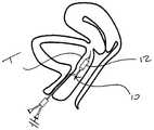
- FIG. 4A first embodiment of a method of the invention shown in FIG. 4 .
- a balloon catheter 10is placed in the submucosal tissue in the trigone region T.
- Upon inflationusing suitable inflation medium such as liquid or air
- a dilated balloon 12forms a dissection plane within the tissue, thus disrupting the afferent nerves therein.
- the balloonis then deflated and removed, resulting in the dissection layer 14 shown in FIG. 5 .
- the balloon catheter 10may be positioned in one or multiple locations.
- FIG. 6shows the resultant dissected regions 14 of multiple placements and dilations. Multiple regions may be parallel, or “fanned out”, as shown.
- the dissected regions or planes 14may overlap, or be discontinuous (as shown).
- the size of the dissected plane(s)may be influenced by the balloon size, as well the number and position of the placements.
- the dissected plane(s)may be completely within the trigone region T, or may extend beyond the trigone region T.
- One or more dissected planes 14may also be outside the trigone region T.
- the dissection plane(s)may be anywhere within the tissue of the bladder trigone, or in the vaginal wall.
- the plane(s)may be just under the mucosal layer, or at the junction of the vagina and bladder tissue. These layers may have a natural “separability” to facilitate relatively controlled dissectioning of the tissue.
- the dissection plane(s) 14may be stabilized following the dissecting procedure by temporary placement of a Fogarty balloon in the bladder to keep the dissected tissues in approximation.
- other suitable devicesmay also be used from either the bladder side and/or the vaginal side.
- the dilation catheter 10(which may also be an expandable mechanical dilator), may be placed with the aid of various devices, as described below.
- a representative visualization scope such as a cystoscope 20is shown in FIGS. 7 and 8 .
- This device 20may include a channel 22 for an endoscope 24 (or could have built-in visualization), one or more channels 26 for infusion/aspiration, and one or more channels (lumens) for delivery of “working devices” (working channel) 28 .
- the dilation catheter 10seen in FIG. 9 (uninflated balloon 12 ) and 10 (inflated balloon 12 ) may include a lumen for use with a guide wire 16 .
- the guide wire lumenmay extend the entire length of the balloon catheter, as is shown in the figures, or may emerge alongside, some distance from the tip, as is often the case with balloon catheters used in vascular procedures (aka “monorail style”). This shorter wire lumen allows for easier installation of the balloon catheter over the proximal end of the guide wire after the guide wire has been placed at a desired target location.
- the guide wire 16may be flexible and steerable, with a pre-formed curve 18 at the distal end.
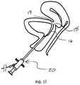
- the guide wiremay be initially placed transurethrally with the aid of the cystoscope 20 , as shown in FIG. 11 .
- the cystoscope 20is placed in the urethra to a site where entry of the guide wire 16 is desired to be placed submucosally.
- the guide wiremay have a sharpened tip 19 to aid in penetrating the surface and advancing submucosally.
- a cautery device or other tissue penetratormay also be used to initially access the submucosal space. Imaging may also be used to facilitate navigation and placement, such as fluoroscopy or ultrasound.
- the cystoscope 20may be removed, or may be left in place and the balloon catheter 10 advanced over the guide wire 16 and within the working channel 28 .
- FIG. 12shows the cystoscope 20 removed.
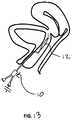
- FIG. 13shows the balloon catheter 10 advanced over the guide wire to the target site for dilation. Positioning of the balloon 12 may be aided with markers (not shown) on the guide wire that at a known distance from the tip, coupled with a known length of balloon catheter, resulting in the tip of the balloon catheter being positioned a known distance from the guide wire.
- FIG. 4shows the balloon in an inflated condition.
- FIG. 5is a side view showing the dissection plane 14 within the tissue between the bladder trigone region and the vagina. And, as mentioned above, FIG. 6 shows the areas in an exemplary treatment with three dissection regions 14 following three balloon placements and inflations.
- FIGS. 14 and 15illustrate an alternative embodiment of the invention.
- a cutting device 30is positioned in the submucosal space of the bladder trigone T.
- Cutting device 30may be positioned in similar locations as described above in connection with the balloon embodiments.
- Cutting device 30may be one of the cutting devices used in other minimally invasive surgical procedures, such as those used through working channels of cyctoscopes for other urological or gynecological procedures, or other scope devices used in other human minimally invasive surgical procedures. Such devices may be steerable and/or deflectable. Furthermore they may also incorporate cautery or other energy combined with cutting to achieve cutting while minimizing bleeding.
- the cutting device 30may be positioned with the aid of a cystoscope 20 , an exemplary version of which was illustrated in FIGS. 7 and 8 , via a working channel 28 .
- Cutting device 30may be further positioned with the aid of a guide wire 16 , similar to that described in connection with the balloon catheter embodiments.
- the cutting device 30may also be advanced in a tissue plane that has a natural “separability”, such as between the bladder mucosa and submucosa, or between the bladder trigone submucosa and the vaginal submucosa.
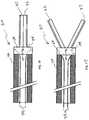
- FIGS. 16 and 17illustrate a novel and particularly useful embodiment of a cutting device 30 for making submucosal dissection planes.
- Cutting device 30includes 2 laterally extendable blades 32 , with cutting surfaces on their outer (lateral) aspects.
- the cutting deviceWhen the cutting device is placed in a target position, it is initially “closed”, as shown in FIG. 16 . Then the device is “opened”, by relative movement between the activation tethers 34 and the body, creating a dissection plane wider than the device.
- the activation tetherspull on the lever arms 38 of the cutting blades, and the cutting blades rotate outward about a pivot 40 .
- the cutting devicemay be further advanced to make a more elongate dissection plane.
- the cutting devicemay be repositioned to make multiple laterally displaced cutting planes 14 , as shown in FIG. 6 .
- Such dissection planes 14may ultimately be conjoined to form one relatively large dissection plane in the submucosal region of the bladder trigone.
- FIGS. 18-19show another embodiment 50 of a cutting device.
- the blades 52may be relatively long, and as such may create a larger dissection plane 14 in the submucosal surface when the blades are opened laterally, as shown in FIGS. 18-20 .
- these cutting device embodimentsmay also be used from the vaginal side of the bladder trigone to create one or more submucosal dissection planes.
- FIGS. 21-25illustrate yet another alternative embodiment 60 for a cutting tool.
- cutting tool 60may include a positioning guide 62 .
- Positioning guide 62may include a suction paddle 64 for engagement with the bladder mucosa, and a cutting blade 66 (shown retracted) within a cutting blade lumen 68 , and a visualization device 70 such as an endoscope.
- the positioning guide 62is placed into the bladder transurethrally to a site of interest, such as adjacent the trigone region. If a dissection plane is to be made, the suction paddle 64 is placed against the bladder mucosa in the region to be submucosally dissected.
- the endoscope 70may be movable longitudinally within the scope channel 72 to help with precise placement of the distal tip of the suction paddle 64 , as seen in FIG. 26 . Advancing the endoscope 70 to near the tip can be particularly advantageous to accurately position the tip relative to the ureteral orifices in order to avoid damage to them.
- the suction paddle 64is activated by applying suction to the suction lumen 74 ( FIG. 24 ). Valves (not shown) operative with the lumen 74 may be incorporated. Apertures 76 in the tissue face of the suction paddle 64 then securely engage the mucosal tissue. The blade is then advanced, as seen in FIG. 28 . Because the cutting blade lumen is parallel to but offset from the tissue face, as shown in FIG. P3 , the cutting blade cuts a dissection plane at a predetermined depth submucosally.
- a handle mechanism(not shown) may be incorporated to manage and control the positions of the proximal ends of the cutting blade and/or the endoscope.
- the positioning guide 62can be placed in multiple locations by repeatedly removing suction, manipulating the guide, re-applying suction, and re-advancing the blade. In this manner, multiple submucosal dissection planes can be generated in the bladder trigone area.
- the cutting blade 66may incorporate cautery, such as monopolar RF applied to the blade, or bipolar RF energy applied from the blade to the suction head.
- cauterysuch as monopolar RF applied to the blade, or bipolar RF energy applied from the blade to the suction head.
- portions of the cutting blade and suction headare appropriately conductive, and adjoining surfaces are appropriately electrically insulated.
- the positioning guide embodimentsmay be utilized via a trans-vaginal approach.
- the positioning guide 62 described above, with the suction paddle 64may be used with other cutting mechanisms that are placed submucosally via the cutting channel.
- An appropriately sized cutting channelis incorporated, depending on the size and shape of the cutting device to be used.
- FIG. 29shows the cutting device 60 employing a translatable cutting mechanism 80 .
- the cutting mechanism 80In addition to being able to be advanced and retracted longitudinally, as shown by arrow 82 , the cutting mechanism 80 as able to be translated from side to side as shown in arrow 84 .
- a pivot point(not shown) is located as near the distal opening of the lumen 68 when the mechanism 80 is advanced longitudinally to its distal extent.
- the sides of the lumen 68may act as translation limits. In this way, the desired degree of translation may be controlled by the amount the mechanism 80 is advanced distally. Retracting the mechanism 80 proximally reduces the degree to which the mechanism 80 may be translated side-to-side.
- mechanism 80may comprise a blade or needle and may be energized for ablation, to form a lesion, or cauterization. If a needle is used as the mechanism 80 , the translation feature is used prior to advancing the needle out of the lumen 68 , and is thus used to control the angular direction the needle travels out of the lumen. In this way, several injection lines may be effected without repositioning the positioning guide 62 .
- one alternative to the methods of the inventionmay include trans-urethral trigonal resection—whether by mechanical or energy delivery (including cryo) means. Resection may involve removal or destruction of a layer of desired thickness (ranging from the 1 mm mucosal thickness to the 5-6 mm complete bladder wall thickness) which would include nerves and nerve endings involved in OAB.
- the devices described abovemay be modified (e.g. by making longer and/or flexible) for use in the male anatomy.
Landscapes
- Health & Medical Sciences (AREA)
- Life Sciences & Earth Sciences (AREA)
- Surgery (AREA)
- Engineering & Computer Science (AREA)
- General Health & Medical Sciences (AREA)
- Nuclear Medicine, Radiotherapy & Molecular Imaging (AREA)
- Veterinary Medicine (AREA)
- Public Health (AREA)
- Animal Behavior & Ethology (AREA)
- Molecular Biology (AREA)
- Medical Informatics (AREA)
- Biomedical Technology (AREA)
- Heart & Thoracic Surgery (AREA)
- Physics & Mathematics (AREA)
- Radiology & Medical Imaging (AREA)
- Biophysics (AREA)
- Pathology (AREA)
- Optics & Photonics (AREA)
- Urology & Nephrology (AREA)
- Orthopedic Medicine & Surgery (AREA)
- Surgical Instruments (AREA)
- Reproductive Health (AREA)
- Vascular Medicine (AREA)
Abstract
Description
Claims (18)
Priority Applications (2)
| Application Number | Priority Date | Filing Date | Title |
|---|---|---|---|
| US14/614,303US10555746B2 (en) | 2014-02-04 | 2015-02-04 | Devices and methods for treating conditions caused by affarent nerve signals |
| US16/751,096US11471180B2 (en) | 2014-02-04 | 2020-01-23 | Devices and methods for treating conditions caused by afferent nerve signals |
Applications Claiming Priority (2)
| Application Number | Priority Date | Filing Date | Title |
|---|---|---|---|
| US201461935753P | 2014-02-04 | 2014-02-04 | |
| US14/614,303US10555746B2 (en) | 2014-02-04 | 2015-02-04 | Devices and methods for treating conditions caused by affarent nerve signals |
Related Child Applications (1)
| Application Number | Title | Priority Date | Filing Date |
|---|---|---|---|
| US16/751,096ContinuationUS11471180B2 (en) | 2014-02-04 | 2020-01-23 | Devices and methods for treating conditions caused by afferent nerve signals |
Publications (2)
| Publication Number | Publication Date |
|---|---|
| US20150216532A1 US20150216532A1 (en) | 2015-08-06 |
| US10555746B2true US10555746B2 (en) | 2020-02-11 |
Family
ID=53753822
Family Applications (2)
| Application Number | Title | Priority Date | Filing Date |
|---|---|---|---|
| US14/614,303Active2036-05-12US10555746B2 (en) | 2014-02-04 | 2015-02-04 | Devices and methods for treating conditions caused by affarent nerve signals |
| US16/751,096Active2036-05-10US11471180B2 (en) | 2014-02-04 | 2020-01-23 | Devices and methods for treating conditions caused by afferent nerve signals |
Family Applications After (1)
| Application Number | Title | Priority Date | Filing Date |
|---|---|---|---|
| US16/751,096Active2036-05-10US11471180B2 (en) | 2014-02-04 | 2020-01-23 | Devices and methods for treating conditions caused by afferent nerve signals |
Country Status (2)
| Country | Link |
|---|---|
| US (2) | US10555746B2 (en) |
| WO (1) | WO2015120079A1 (en) |
Families Citing this family (2)
| Publication number | Priority date | Publication date | Assignee | Title |
|---|---|---|---|---|
| US11033290B2 (en)* | 2015-12-21 | 2021-06-15 | Boston Scientific Scimed, Inc | Medical device and methods of use |
| US11766298B2 (en)* | 2019-05-03 | 2023-09-26 | Neil Glossop | Systems, methods, and devices for registering and tracking organs during interventional procedures |
Citations (72)
| Publication number | Priority date | Publication date | Assignee | Title |
|---|---|---|---|---|
| US5342353A (en) | 1990-11-05 | 1994-08-30 | Allen Paul M | System for laser treatment of the female urethra and bladder |
| US5370675A (en) | 1992-08-12 | 1994-12-06 | Vidamed, Inc. | Medical probe device and method |
| US5378675A (en) | 1991-11-05 | 1995-01-03 | Konica Corporation | Thermal transfer recording image receiving sheet |
| US5667488A (en) | 1992-08-12 | 1997-09-16 | Vidamed, Inc. | Transurethral needle ablation device and method for the treatment of the prostate |
| US5672153A (en) | 1992-08-12 | 1997-09-30 | Vidamed, Inc. | Medical probe device and method |
| US6190353B1 (en) | 1995-10-13 | 2001-02-20 | Transvascular, Inc. | Methods and apparatus for bypassing arterial obstructions and/or performing other transvascular procedures |
| US6216704B1 (en) | 1997-08-13 | 2001-04-17 | Surx, Inc. | Noninvasive devices, methods, and systems for shrinking of tissues |
| US6231518B1 (en) | 1998-05-26 | 2001-05-15 | Comedicus Incorporated | Intrapericardial electrophysiological procedures |
| US6325798B1 (en) | 1998-02-19 | 2001-12-04 | Curon Medical, Inc. | Vacuum-assisted systems and methods for treating sphincters and adjoining tissue regions |
| US20020002372A1 (en) | 2000-04-27 | 2002-01-03 | Medtronic, Inc. | Suction stabilized epicardial ablation devices |
| US6425877B1 (en) | 1999-04-02 | 2002-07-30 | Novasys Medical, Inc. | Treatment of tissue in the digestive circulatory respiratory urinary and reproductive systems |
| US20020193851A1 (en) | 2001-06-14 | 2002-12-19 | Silverman David E. | Energy treatment apparatus for treating gastrointestinal tract and method for using same |
| US20030032860A1 (en) | 1997-11-04 | 2003-02-13 | Arie Avni | Video rectoscope |
| US6626930B1 (en) | 1999-10-21 | 2003-09-30 | Edwards Lifesciences Corporation | Minimally invasive mitral valve repair method and apparatus |
| US6632193B1 (en) | 1995-06-07 | 2003-10-14 | Arthrocare Corporation | Systems and methods for electrosurgical tissue treatment |
| US6692490B1 (en) | 1999-05-18 | 2004-02-17 | Novasys Medical, Inc. | Treatment of urinary incontinence and other disorders by application of energy and drugs |
| US20040059389A1 (en) | 2002-08-13 | 2004-03-25 | Chornenky Victor I. | Apparatus and method for the treatment of benign prostatic hyperplasia |
| US20040186468A1 (en) | 1999-09-28 | 2004-09-23 | Edwards Stuart D. | Treatment of urinary incontinence and other disorders by application of energy and drugs |
| US20040215179A1 (en) | 2003-04-25 | 2004-10-28 | Medtronic, Inc. | Device and Method for transurethral prostate treatment |
| US6960205B2 (en) | 2000-04-27 | 2005-11-01 | Medtronic, Inc. | Suction stabilized epicardial ablation devices |
| US20070112340A1 (en) | 2000-10-02 | 2007-05-17 | Novasys Medical, Inc. | Apparatus and methods for treating female urinary incontinence |
| US20090069803A1 (en) | 2007-09-10 | 2009-03-12 | Medtronic, Inc. | Selective depth electrode deployment for electrical stimulation |
| US20090171315A1 (en) | 2007-03-05 | 2009-07-02 | Ebrahim Versi | Method and device for delivering drug to the trigone of the bladder |
| US20100174306A1 (en) | 2007-07-11 | 2010-07-08 | Vladimir Mitelberg | Methods and Systems for Performing Submucosal Medical Procedures |
| US20100256446A1 (en) | 2007-05-11 | 2010-10-07 | Board Of Regents, The University Of Texas System | Medical scope carrier and scope as system and method |
| US20110112434A1 (en) | 2009-11-06 | 2011-05-12 | Ethicon Endo-Surgery, Inc. | Kits and procedures for natural orifice translumenal endoscopic surgery |
| EP2155093B1 (en) | 2007-04-30 | 2011-05-25 | Medtronic, Inc. | Extension and retraction mechanism for a hand-held surgical ablation device |
| CN102256560A (en) | 2008-11-10 | 2011-11-23 | 微立方有限责任公司 | Method and apparatus for applying energy to body tissue |
| US20120048417A1 (en) | 2003-06-13 | 2012-03-01 | Underground Solutions Technologies Group, Inc. | Polyvinyl Chloride Formulations |
| US20120048419A1 (en) | 2010-08-30 | 2012-03-01 | Health Robotics S.r.I. | Method for the Production of Pharmaceutical Products |
| US20120123411A1 (en) | 2010-11-12 | 2012-05-17 | Estech, Inc. (Endoscopic Technologies, Inc.) | Stabilized ablation systems and methods |
| US20130018281A1 (en) | 2011-07-15 | 2013-01-17 | Sandra Nagale | Systems and Methods for Monitoring Organ Activity |
| US20130030249A1 (en) | 2009-02-06 | 2013-01-31 | Endoclear Llc | Visualized endotracheal tube placement systems |
| WO2013016588A1 (en) | 2011-07-26 | 2013-01-31 | Dan Sachs | Apparatus and methods to modulate pelvic nervous tissue |
| US20130066308A1 (en) | 2011-08-31 | 2013-03-14 | Jaime Landman | Ablation-based therapy for bladder pathologies |
| US20130072855A1 (en) | 2011-09-16 | 2013-03-21 | Boston Scientific Scimed, Inc. | Apparatus for treating an organ and related methods of use |
| US20130090648A1 (en) | 2011-10-07 | 2013-04-11 | Boston Scientific Scimed, Inc. | Methods and systems for detection and thermal treatment of lower urinary tract conditions |
| US20130090640A1 (en) | 2011-10-07 | 2013-04-11 | University Of Surrey | Methods and systems for detection and thermal treatment of lower urinary tract conditions |
| US20130172864A1 (en) | 2008-05-21 | 2013-07-04 | Estech, Inc. (Endoscopic Technologies, Inc.) | Stabilized ablation systems and methods |
| WO2013160772A2 (en) | 2012-04-22 | 2013-10-31 | Omry Ben-Ezra | Bladder tissue modification for overactive bladder disorders |
| WO2014004698A1 (en) | 2012-06-27 | 2014-01-03 | Acoustic Medsystems, Inc. | Noninvasive transvaginal acoustic thermal treatment of female stress urinary incontinence |
| US20140012247A1 (en) | 2012-07-03 | 2014-01-09 | Ethicon Endo-Surgery, Inc. | Endoscopic cap electrode and method for using the same |
| US20140012256A1 (en) | 2005-07-22 | 2014-01-09 | Medtronic Ardian Luxembourg S.A.R.L. | Systems and Methods for Neuromodulation for Treatment of Pain and Other Disorders Associated with Nerve Conduction |
| US20140018786A1 (en) | 2007-02-21 | 2014-01-16 | Electromedical Associates Llc | Instruments and Methods for Thermal Tissue Treatment |
| US20140025055A1 (en) | 2011-02-01 | 2014-01-23 | Channel Medsystems, Inc. | Treatments using cyrogenic ablation systems |
| US20140031810A1 (en) | 2012-07-30 | 2014-01-30 | Northwestern University | Radiofrequency Probe for Circumferential Ablation of a Hollow Cavity |
| US8641711B2 (en) | 2007-05-04 | 2014-02-04 | Covidien Lp | Method and apparatus for gastrointestinal tract ablation for treatment of obesity |
| WO2014022436A1 (en) | 2012-07-30 | 2014-02-06 | Fractyl Laboratories Inc. | Electrical energy ablation systems, devices and methods for the treatment of tissue |
| US20140039491A1 (en) | 2012-08-02 | 2014-02-06 | Ethicon Endo-Surgery, Inc. | Flexible expandable electrode and method of intraluminal delivery of pulsed power |
| WO2014026028A1 (en) | 2012-08-10 | 2014-02-13 | Attenuex Technologies, Inc. | Systems for performing a medical procedure |
| WO2014025394A1 (en) | 2012-08-09 | 2014-02-13 | University Of Iowa Research Foundation | Catheters, catheter systems, and methods for puncturing through a tissue structure |
| AU2010319333B2 (en) | 2009-11-13 | 2014-02-20 | St. Jude Medical, Inc. | Assembly of staggered ablation elements |
| US8657814B2 (en) | 2005-08-22 | 2014-02-25 | Medtronic Ablation Frontiers Llc | User interface for tissue ablation system |
| US20140081257A1 (en) | 2012-09-17 | 2014-03-20 | The Regents Of The University Of California | Bladder denervation for treating overactive bladder |
| US8684998B2 (en) | 2002-04-08 | 2014-04-01 | Medtronic Ardian Luxembourg S.A.R.L. | Methods for inhibiting renal nerve activity |
| CN103764225A (en) | 2011-03-04 | 2014-04-30 | 彩虹医疗公司 | Tissue treatment and monitoring by application of energy |
| US8721632B2 (en) | 2008-09-09 | 2014-05-13 | Tsunami Medtech, Llc | Methods for delivering energy into a target tissue of a body |
| US8740896B2 (en) | 2002-04-08 | 2014-06-03 | Medtronic Ardian Luxembourg S.A.R.L. | Methods and apparatus for performing renal neuromodulation via catheter apparatuses having inflatable balloons |
| US20140163548A1 (en) | 2012-12-06 | 2014-06-12 | Steven C. Christian | Irrigant distribution system for electrodes |
| US8758337B2 (en) | 2006-09-29 | 2014-06-24 | Medtronic, Inc. | User interface for ablation therapy |
| US8771269B2 (en) | 2007-05-11 | 2014-07-08 | Medtronic Ablation Frontiers Llc | RF energy delivery system and method |
| US8771267B2 (en) | 2005-06-20 | 2014-07-08 | Medtronic Ablation Frontiers Llc | Ablation catheter |
| EP2349045B1 (en) | 2008-10-21 | 2014-07-16 | Microcube, LLC | Devices for applying energy to bodily tissues |
| US20140200568A1 (en) | 2008-10-06 | 2014-07-17 | Virender K. Sharma | Method and Apparatus for Tissue Ablation |
| US20140207136A1 (en) | 2012-05-04 | 2014-07-24 | St. Jude Medical, Inc. | Multiple staggered electrodes connected via flexible joints |
| US8790281B2 (en) | 2006-04-20 | 2014-07-29 | The Regents Of The University Of California | Method of thermal treatment of myolysis and destruction of benign uterine tumors |
| US20140257272A1 (en) | 2009-08-07 | 2014-09-11 | Ulthera, Inc. | Handpiece and methods for performing subcutaneous surgery |
| US20140276726A1 (en) | 2013-03-13 | 2014-09-18 | Hologic, Inc. | Intrauterine treatment device with articulating array |
| US20140276593A1 (en) | 2013-03-15 | 2014-09-18 | Boston Scientific Scimed, Inc. | Methods and systems for treatment of a bladder |
| US8840625B2 (en) | 2006-10-18 | 2014-09-23 | Hologic, Inc. | Systems for performing gynecological procedures with closed visualization lumen |
| EP2813192A2 (en) | 2008-10-21 | 2014-12-17 | Microcube, LLC | Methods and devices for applying energy to bodily tissues |
| US20160030107A1 (en) | 2014-07-31 | 2016-02-04 | Boston Scientific Scimed, Inc. | Bladder treatment by ablative denervation |
- 2015
- 2015-02-04WOPCT/US2015/014500patent/WO2015120079A1/enactiveApplication Filing
- 2015-02-04USUS14/614,303patent/US10555746B2/enactiveActive
- 2020
- 2020-01-23USUS16/751,096patent/US11471180B2/enactiveActive
Patent Citations (86)
| Publication number | Priority date | Publication date | Assignee | Title |
|---|---|---|---|---|
| US5342353A (en) | 1990-11-05 | 1994-08-30 | Allen Paul M | System for laser treatment of the female urethra and bladder |
| US5378675A (en) | 1991-11-05 | 1995-01-03 | Konica Corporation | Thermal transfer recording image receiving sheet |
| US6419653B2 (en) | 1992-08-12 | 2002-07-16 | Vidamed, Inc. | Medical probe device and method |
| US5370675A (en) | 1992-08-12 | 1994-12-06 | Vidamed, Inc. | Medical probe device and method |
| US5667488A (en) | 1992-08-12 | 1997-09-16 | Vidamed, Inc. | Transurethral needle ablation device and method for the treatment of the prostate |
| US5672153A (en) | 1992-08-12 | 1997-09-30 | Vidamed, Inc. | Medical probe device and method |
| US5964727A (en) | 1992-08-12 | 1999-10-12 | Vidamed, Inc. | Medical probe device and method |
| US6129726A (en) | 1992-08-12 | 2000-10-10 | Vidamed, Inc. | Medical probe device and method |
| US6852091B2 (en) | 1992-08-12 | 2005-02-08 | Medtronic Vidamed, Inc. | Medical probe device and method |
| US6610054B1 (en) | 1992-08-12 | 2003-08-26 | Vidamed, Inc. | Medical probe device and method |
| US6632193B1 (en) | 1995-06-07 | 2003-10-14 | Arthrocare Corporation | Systems and methods for electrosurgical tissue treatment |
| US6190353B1 (en) | 1995-10-13 | 2001-02-20 | Transvascular, Inc. | Methods and apparatus for bypassing arterial obstructions and/or performing other transvascular procedures |
| US6216704B1 (en) | 1997-08-13 | 2001-04-17 | Surx, Inc. | Noninvasive devices, methods, and systems for shrinking of tissues |
| US20030032860A1 (en) | 1997-11-04 | 2003-02-13 | Arie Avni | Video rectoscope |
| US6325798B1 (en) | 1998-02-19 | 2001-12-04 | Curon Medical, Inc. | Vacuum-assisted systems and methods for treating sphincters and adjoining tissue regions |
| US6231518B1 (en) | 1998-05-26 | 2001-05-15 | Comedicus Incorporated | Intrapericardial electrophysiological procedures |
| US6425877B1 (en) | 1999-04-02 | 2002-07-30 | Novasys Medical, Inc. | Treatment of tissue in the digestive circulatory respiratory urinary and reproductive systems |
| US6692490B1 (en) | 1999-05-18 | 2004-02-17 | Novasys Medical, Inc. | Treatment of urinary incontinence and other disorders by application of energy and drugs |
| US20040186468A1 (en) | 1999-09-28 | 2004-09-23 | Edwards Stuart D. | Treatment of urinary incontinence and other disorders by application of energy and drugs |
| US6626930B1 (en) | 1999-10-21 | 2003-09-30 | Edwards Lifesciences Corporation | Minimally invasive mitral valve repair method and apparatus |
| US20020002372A1 (en) | 2000-04-27 | 2002-01-03 | Medtronic, Inc. | Suction stabilized epicardial ablation devices |
| US7818039B2 (en) | 2000-04-27 | 2010-10-19 | Medtronic, Inc. | Suction stabilized epicardial ablation devices |
| US6960205B2 (en) | 2000-04-27 | 2005-11-01 | Medtronic, Inc. | Suction stabilized epicardial ablation devices |
| US20070112340A1 (en) | 2000-10-02 | 2007-05-17 | Novasys Medical, Inc. | Apparatus and methods for treating female urinary incontinence |
| US20020193851A1 (en) | 2001-06-14 | 2002-12-19 | Silverman David E. | Energy treatment apparatus for treating gastrointestinal tract and method for using same |
| US8684998B2 (en) | 2002-04-08 | 2014-04-01 | Medtronic Ardian Luxembourg S.A.R.L. | Methods for inhibiting renal nerve activity |
| US8740896B2 (en) | 2002-04-08 | 2014-06-03 | Medtronic Ardian Luxembourg S.A.R.L. | Methods and apparatus for performing renal neuromodulation via catheter apparatuses having inflatable balloons |
| US20040059389A1 (en) | 2002-08-13 | 2004-03-25 | Chornenky Victor I. | Apparatus and method for the treatment of benign prostatic hyperplasia |
| US20040215179A1 (en) | 2003-04-25 | 2004-10-28 | Medtronic, Inc. | Device and Method for transurethral prostate treatment |
| US20120048417A1 (en) | 2003-06-13 | 2012-03-01 | Underground Solutions Technologies Group, Inc. | Polyvinyl Chloride Formulations |
| US8771267B2 (en) | 2005-06-20 | 2014-07-08 | Medtronic Ablation Frontiers Llc | Ablation catheter |
| EP2759276A1 (en) | 2005-06-20 | 2014-07-30 | Medtronic Ablation Frontiers LLC | Ablation catheter |
| US20140012256A1 (en) | 2005-07-22 | 2014-01-09 | Medtronic Ardian Luxembourg S.A.R.L. | Systems and Methods for Neuromodulation for Treatment of Pain and Other Disorders Associated with Nerve Conduction |
| US8657814B2 (en) | 2005-08-22 | 2014-02-25 | Medtronic Ablation Frontiers Llc | User interface for tissue ablation system |
| US8790281B2 (en) | 2006-04-20 | 2014-07-29 | The Regents Of The University Of California | Method of thermal treatment of myolysis and destruction of benign uterine tumors |
| US8758337B2 (en) | 2006-09-29 | 2014-06-24 | Medtronic, Inc. | User interface for ablation therapy |
| US8840625B2 (en) | 2006-10-18 | 2014-09-23 | Hologic, Inc. | Systems for performing gynecological procedures with closed visualization lumen |
| US20140018786A1 (en) | 2007-02-21 | 2014-01-16 | Electromedical Associates Llc | Instruments and Methods for Thermal Tissue Treatment |
| US20090171315A1 (en) | 2007-03-05 | 2009-07-02 | Ebrahim Versi | Method and device for delivering drug to the trigone of the bladder |
| EP2155093B1 (en) | 2007-04-30 | 2011-05-25 | Medtronic, Inc. | Extension and retraction mechanism for a hand-held surgical ablation device |
| US8641711B2 (en) | 2007-05-04 | 2014-02-04 | Covidien Lp | Method and apparatus for gastrointestinal tract ablation for treatment of obesity |
| US8771269B2 (en) | 2007-05-11 | 2014-07-08 | Medtronic Ablation Frontiers Llc | RF energy delivery system and method |
| US20100256446A1 (en) | 2007-05-11 | 2010-10-07 | Board Of Regents, The University Of Texas System | Medical scope carrier and scope as system and method |
| US20100174306A1 (en) | 2007-07-11 | 2010-07-08 | Vladimir Mitelberg | Methods and Systems for Performing Submucosal Medical Procedures |
| US20090069803A1 (en) | 2007-09-10 | 2009-03-12 | Medtronic, Inc. | Selective depth electrode deployment for electrical stimulation |
| US20130172864A1 (en) | 2008-05-21 | 2013-07-04 | Estech, Inc. (Endoscopic Technologies, Inc.) | Stabilized ablation systems and methods |
| US8721632B2 (en) | 2008-09-09 | 2014-05-13 | Tsunami Medtech, Llc | Methods for delivering energy into a target tissue of a body |
| US20140200568A1 (en) | 2008-10-06 | 2014-07-17 | Virender K. Sharma | Method and Apparatus for Tissue Ablation |
| EP2813192A2 (en) | 2008-10-21 | 2014-12-17 | Microcube, LLC | Methods and devices for applying energy to bodily tissues |
| EP2349045B1 (en) | 2008-10-21 | 2014-07-16 | Microcube, LLC | Devices for applying energy to bodily tissues |
| CN102256560B (en) | 2008-11-10 | 2014-07-09 | 微立方有限责任公司 | Method and apparatus for applying energy to body tissue |
| CN102256560A (en) | 2008-11-10 | 2011-11-23 | 微立方有限责任公司 | Method and apparatus for applying energy to body tissue |
| US20130030249A1 (en) | 2009-02-06 | 2013-01-31 | Endoclear Llc | Visualized endotracheal tube placement systems |
| US20140257272A1 (en) | 2009-08-07 | 2014-09-11 | Ulthera, Inc. | Handpiece and methods for performing subcutaneous surgery |
| US20110112434A1 (en) | 2009-11-06 | 2011-05-12 | Ethicon Endo-Surgery, Inc. | Kits and procedures for natural orifice translumenal endoscopic surgery |
| AU2010319333B2 (en) | 2009-11-13 | 2014-02-20 | St. Jude Medical, Inc. | Assembly of staggered ablation elements |
| US20120048419A1 (en) | 2010-08-30 | 2012-03-01 | Health Robotics S.r.I. | Method for the Production of Pharmaceutical Products |
| US20120123411A1 (en) | 2010-11-12 | 2012-05-17 | Estech, Inc. (Endoscopic Technologies, Inc.) | Stabilized ablation systems and methods |
| US20140025055A1 (en) | 2011-02-01 | 2014-01-23 | Channel Medsystems, Inc. | Treatments using cyrogenic ablation systems |
| CN103764225A (en) | 2011-03-04 | 2014-04-30 | 彩虹医疗公司 | Tissue treatment and monitoring by application of energy |
| US20130018281A1 (en) | 2011-07-15 | 2013-01-17 | Sandra Nagale | Systems and Methods for Monitoring Organ Activity |
| US20140148798A1 (en) | 2011-07-26 | 2014-05-29 | Amphora Medical, Inc. | Apparatus and methods to modulate bladder function |
| CN104080418A (en) | 2011-07-26 | 2014-10-01 | 安福拉医药公司 | Apparatus and methods to modulate pelvic nervous tissue |
| US20140039356A1 (en) | 2011-07-26 | 2014-02-06 | Amphora Medical, Inc. | Apparatus and methods to modulate bladder function |
| WO2013016588A1 (en) | 2011-07-26 | 2013-01-31 | Dan Sachs | Apparatus and methods to modulate pelvic nervous tissue |
| WO2013016590A1 (en)* | 2011-07-26 | 2013-01-31 | Dan Sachs | Apparatus and methods to modulate pelvic nervous tissue |
| US20130066308A1 (en) | 2011-08-31 | 2013-03-14 | Jaime Landman | Ablation-based therapy for bladder pathologies |
| US20130072855A1 (en) | 2011-09-16 | 2013-03-21 | Boston Scientific Scimed, Inc. | Apparatus for treating an organ and related methods of use |
| US20130090648A1 (en) | 2011-10-07 | 2013-04-11 | Boston Scientific Scimed, Inc. | Methods and systems for detection and thermal treatment of lower urinary tract conditions |
| US20130090640A1 (en) | 2011-10-07 | 2013-04-11 | University Of Surrey | Methods and systems for detection and thermal treatment of lower urinary tract conditions |
| WO2013160772A2 (en) | 2012-04-22 | 2013-10-31 | Omry Ben-Ezra | Bladder tissue modification for overactive bladder disorders |
| US20140207136A1 (en) | 2012-05-04 | 2014-07-24 | St. Jude Medical, Inc. | Multiple staggered electrodes connected via flexible joints |
| WO2014004698A1 (en) | 2012-06-27 | 2014-01-03 | Acoustic Medsystems, Inc. | Noninvasive transvaginal acoustic thermal treatment of female stress urinary incontinence |
| US20140012247A1 (en) | 2012-07-03 | 2014-01-09 | Ethicon Endo-Surgery, Inc. | Endoscopic cap electrode and method for using the same |
| WO2014022379A1 (en) | 2012-07-30 | 2014-02-06 | Northwestern University | Radiofrequency probe for circumferential ablation of a hollow cavity |
| US20140031810A1 (en) | 2012-07-30 | 2014-01-30 | Northwestern University | Radiofrequency Probe for Circumferential Ablation of a Hollow Cavity |
| WO2014022436A1 (en) | 2012-07-30 | 2014-02-06 | Fractyl Laboratories Inc. | Electrical energy ablation systems, devices and methods for the treatment of tissue |
| US20140039491A1 (en) | 2012-08-02 | 2014-02-06 | Ethicon Endo-Surgery, Inc. | Flexible expandable electrode and method of intraluminal delivery of pulsed power |
| WO2014025394A1 (en) | 2012-08-09 | 2014-02-13 | University Of Iowa Research Foundation | Catheters, catheter systems, and methods for puncturing through a tissue structure |
| WO2014026028A1 (en) | 2012-08-10 | 2014-02-13 | Attenuex Technologies, Inc. | Systems for performing a medical procedure |
| US20140081257A1 (en) | 2012-09-17 | 2014-03-20 | The Regents Of The University Of California | Bladder denervation for treating overactive bladder |
| US20140163548A1 (en) | 2012-12-06 | 2014-06-12 | Steven C. Christian | Irrigant distribution system for electrodes |
| WO2014113724A2 (en) | 2013-01-17 | 2014-07-24 | Sharma Virender K | Method and apparatus for tissue ablation |
| US20140276726A1 (en) | 2013-03-13 | 2014-09-18 | Hologic, Inc. | Intrauterine treatment device with articulating array |
| US20140276593A1 (en) | 2013-03-15 | 2014-09-18 | Boston Scientific Scimed, Inc. | Methods and systems for treatment of a bladder |
| US20160030107A1 (en) | 2014-07-31 | 2016-02-04 | Boston Scientific Scimed, Inc. | Bladder treatment by ablative denervation |
Non-Patent Citations (5)
| Title |
|---|
| Chinese Patent Office, Office Action dated Jan. 4, 2016 in Chinese Patent Application No. 201280046659.X, 21 pages. |
| WIPO International Searching Authority, International Search Report and Written Opinion dated Aug. 25, 2015 in International Patent Application No. PCT/US2015/0032298, 10 pages. |
| WIPO International Searching Authority, International Search Report and Written Opinion dated Jun. 24, 2015 in International Patent Application No. PCT/US2015/014500, 11 pages. |
| WIPO International Searching Authority, International Search Report and Written Opinion dated Nov. 21, 2012 in International Patent Application No. PCT/US2012/048417, 16 pages. |
| WIPO International Searching Authority, International Search Report and Written Opinion dated Nov. 26, 2012 in International Patent Application No. PCT/US2012/048419, 17 pages. |
Also Published As
| Publication number | Publication date |
|---|---|
| US20150216532A1 (en) | 2015-08-06 |
| US20200155189A1 (en) | 2020-05-21 |
| WO2015120079A1 (en) | 2015-08-13 |
| US11471180B2 (en) | 2022-10-18 |
Similar Documents
| Publication | Publication Date | Title |
|---|---|---|
| AU2007238753B2 (en) | Apparatus and methods for endoscopic resection of tissue | |
| US9011433B2 (en) | Bipolar colpotomy device | |
| JP5336377B2 (en) | Hood member used with endoscope | |
| US6510854B2 (en) | Method of treatment of prostatic adenoma | |
| US7604648B2 (en) | Direct vision port site dissector | |
| US7398781B1 (en) | Method for subxiphoid endoscopic access | |
| US8951226B2 (en) | Mediastinoscopy access, sampling, and visualization kit featuring toroidal balloons and exotracheal method of using | |
| EP3785654B1 (en) | Multifunctional high-frequency electric knife | |
| US20170258521A1 (en) | Pericardial modification devices and methods | |
| US20100312054A1 (en) | Prostatic tissue removal and/or prostatic capsulotomy for treatment of conditions | |
| US20110270239A1 (en) | Transseptal crossing device | |
| US20240180611A1 (en) | Bladder treatment by ablative denervation | |
| JP2010502381A5 (en) | ||
| JPH1024049A (en) | Electrosurgical unit | |
| WO2009072131A2 (en) | Prostatic capsulotomy for treatment of conditions | |
| JP2016512090A (en) | Apparatus for tissue separation and related uses | |
| US11471180B2 (en) | Devices and methods for treating conditions caused by afferent nerve signals | |
| US10363087B2 (en) | Tissue resection device | |
| EP1553882A1 (en) | Direct vision port site dissector | |
| CN114302756A (en) | Device for performing interventional endoscopic ultrasound surgery | |
| US20250009421A1 (en) | Devices, systems, and methods for tunneling between tissue layers | |
| US20090093808A1 (en) | Partial (non-apical) prostate ablation procedure and device | |
| HK40037515B (en) | Multifunctional high-frequency electric knife | |
| HK40037515A (en) | Multifunctional high-frequency electric knife | |
| Nam | Total Laparoscopic Hysterectomy using POWERSEAL |
Legal Events
| Date | Code | Title | Description |
|---|---|---|---|
| AS | Assignment | Owner name:AMPHORA MEDICAL, INC., MINNESOTA Free format text:ASSIGNMENT OF ASSIGNORS INTEREST;ASSIGNORS:HLAVKA, EDWIN J.;WHITBROOK, ERIC;KEITH, PETER T.;AND OTHERS;SIGNING DATES FROM 20150205 TO 20150211;REEL/FRAME:041103/0875 | |
| STPP | Information on status: patent application and granting procedure in general | Free format text:NON FINAL ACTION MAILED | |
| AS | Assignment | Owner name:HOLOGIC, INC.,, MASSACHUSETTS Free format text:ASSIGNMENT OF ASSIGNORS INTEREST;ASSIGNOR:AMPHORA MEDICAL, INC.;REEL/FRAME:049786/0138 Effective date:20190624 | |
| STPP | Information on status: patent application and granting procedure in general | Free format text:NOTICE OF ALLOWANCE MAILED -- APPLICATION RECEIVED IN OFFICE OF PUBLICATIONS | |
| AS | Assignment | Owner name:BANK OF AMERICA, N.A., AS COLLATERAL AGENT, NORTH Free format text:SECURITY INTEREST;ASSIGNORS:HOLOGIC, INC.;CYNOSURE, LLC;CYTYC CORPORATION;AND OTHERS;REEL/FRAME:050719/0701 Effective date:20191011 Owner name:BANK OF AMERICA, N.A., AS COLLATERAL AGENT, NORTH CAROLINA Free format text:SECURITY INTEREST;ASSIGNORS:HOLOGIC, INC.;CYNOSURE, LLC;CYTYC CORPORATION;AND OTHERS;REEL/FRAME:050719/0701 Effective date:20191011 | |
| FEPP | Fee payment procedure | Free format text:ENTITY STATUS SET TO UNDISCOUNTED (ORIGINAL EVENT CODE: BIG.); ENTITY STATUS OF PATENT OWNER: LARGE ENTITY | |
| STPP | Information on status: patent application and granting procedure in general | Free format text:PUBLICATIONS -- ISSUE FEE PAYMENT VERIFIED | |
| STCF | Information on status: patent grant | Free format text:PATENTED CASE | |
| AS | Assignment | Owner name:BANK OF AMERICA, N.A., AS COLLATERAL AGENT, NORTH CAROLINA Free format text:SECURITY INTEREST;ASSIGNORS:HOLOGIC, INC.;FAXITRON BIOPTICS, LLC;FOCAL THERAPEUTICS, INC.;AND OTHERS;REEL/FRAME:054089/0804 Effective date:20201013 | |
| MAFP | Maintenance fee payment | Free format text:PAYMENT OF MAINTENANCE FEE, 4TH YEAR, LARGE ENTITY (ORIGINAL EVENT CODE: M1551); ENTITY STATUS OF PATENT OWNER: LARGE ENTITY Year of fee payment:4 |