US10534129B2 - System and method providing intracoronary laser speckle imaging for the detection of vulnerable plaque - Google Patents
System and method providing intracoronary laser speckle imaging for the detection of vulnerable plaqueDownload PDFInfo
- Publication number
- US10534129B2 US10534129B2US12/058,279US5827908AUS10534129B2US 10534129 B2US10534129 B2US 10534129B2US 5827908 AUS5827908 AUS 5827908AUS 10534129 B2US10534129 B2US 10534129B2
- Authority
- US
- United States
- Prior art keywords
- light
- fiber bundle
- catheter
- exemplary
- imaging
- Prior art date
- Legal status (The legal status is an assumption and is not a legal conclusion. Google has not performed a legal analysis and makes no representation as to the accuracy of the status listed.)
- Active, expires
Links
Images


Classifications
- G—PHYSICS
- G02—OPTICS
- G02B—OPTICAL ELEMENTS, SYSTEMS OR APPARATUS
- G02B6/00—Light guides; Structural details of arrangements comprising light guides and other optical elements, e.g. couplings
- G02B6/04—Light guides; Structural details of arrangements comprising light guides and other optical elements, e.g. couplings formed by bundles of fibres
- G02B6/06—Light guides; Structural details of arrangements comprising light guides and other optical elements, e.g. couplings formed by bundles of fibres the relative position of the fibres being the same at both ends, e.g. for transporting images
- A—HUMAN NECESSITIES
- A61—MEDICAL OR VETERINARY SCIENCE; HYGIENE
- A61B—DIAGNOSIS; SURGERY; IDENTIFICATION
- A61B1/00—Instruments for performing medical examinations of the interior of cavities or tubes of the body by visual or photographical inspection, e.g. endoscopes; Illuminating arrangements therefor
- A61B1/00064—Constructional details of the endoscope body
- A61B1/00071—Insertion part of the endoscope body
- A61B1/0008—Insertion part of the endoscope body characterised by distal tip features
- A61B1/00096—Optical elements
- A—HUMAN NECESSITIES
- A61—MEDICAL OR VETERINARY SCIENCE; HYGIENE
- A61B—DIAGNOSIS; SURGERY; IDENTIFICATION
- A61B1/00—Instruments for performing medical examinations of the interior of cavities or tubes of the body by visual or photographical inspection, e.g. endoscopes; Illuminating arrangements therefor
- A61B1/00163—Optical arrangements
- A61B1/00165—Optical arrangements with light-conductive means, e.g. fibre optics
- A61B1/00167—Details of optical fibre bundles, e.g. shape or fibre distribution
- A—HUMAN NECESSITIES
- A61—MEDICAL OR VETERINARY SCIENCE; HYGIENE
- A61B—DIAGNOSIS; SURGERY; IDENTIFICATION
- A61B5/00—Measuring for diagnostic purposes; Identification of persons
- A61B5/0059—Measuring for diagnostic purposes; Identification of persons using light, e.g. diagnosis by transillumination, diascopy, fluorescence
- A61B5/0062—Arrangements for scanning
- A61B5/0066—Optical coherence imaging
- A—HUMAN NECESSITIES
- A61—MEDICAL OR VETERINARY SCIENCE; HYGIENE
- A61B—DIAGNOSIS; SURGERY; IDENTIFICATION
- A61B5/00—Measuring for diagnostic purposes; Identification of persons
- A61B5/0059—Measuring for diagnostic purposes; Identification of persons using light, e.g. diagnosis by transillumination, diascopy, fluorescence
- A61B5/0082—Measuring for diagnostic purposes; Identification of persons using light, e.g. diagnosis by transillumination, diascopy, fluorescence adapted for particular medical purposes
- A61B5/0084—Measuring for diagnostic purposes; Identification of persons using light, e.g. diagnosis by transillumination, diascopy, fluorescence adapted for particular medical purposes for introduction into the body, e.g. by catheters
- A—HUMAN NECESSITIES
- A61—MEDICAL OR VETERINARY SCIENCE; HYGIENE
- A61B—DIAGNOSIS; SURGERY; IDENTIFICATION
- A61B5/00—Measuring for diagnostic purposes; Identification of persons
- A61B5/72—Signal processing specially adapted for physiological signals or for diagnostic purposes
- A61B5/7271—Specific aspects of physiological measurement analysis
- A61B5/7285—Specific aspects of physiological measurement analysis for synchronizing or triggering a physiological measurement or image acquisition with a physiological event or waveform, e.g. an ECG signal
- G—PHYSICS
- G02—OPTICS
- G02B—OPTICAL ELEMENTS, SYSTEMS OR APPARATUS
- G02B6/00—Light guides; Structural details of arrangements comprising light guides and other optical elements, e.g. couplings
- G02B6/24—Coupling light guides
- G02B6/26—Optical coupling means
- G02B6/32—Optical coupling means having lens focusing means positioned between opposed fibre ends
Definitions
- the present inventionrelates to imaging at least one portion of a sample, and more particularly to system and method providing intracoronary laser speckle imaging for the detection of vulnerable plaque.
- Ischemic cardiovascular diseasethe leading cause of death in industrialized societies, can be frequently preceded by the rupture of unstable atherosclerotic plaque.
- the intricate interplay between biomechanical, compositional and morphological factorsmay influence plaque stability.
- Certain exemplary techniques that facilitate a composite understanding of the link between these factorscan assist in identifying rupture-prone plaques, guiding treatment and for investigating mechanisms associated with plaque stabilization therapies.
- Laser Speckle Imagingis a granular pattern formed by the interference of coherent laser light scattered from tissue.
- the speckle patternis dynamically modulated by Brownian motion of endogenous particles within tissue, which is governed by the viscoelasticity of tissue.
- the extent of Brownian motioncan be quantified by the cross-correlation of speckle images obtained as a function of time.
- the exemplary techniques of using arterial specimens ex vivohave demonstrated that the index of viscoelasticity measured by LSI can be related to plaque type, structure and composition.
- the exemplary intracoronary LSI system and techniquecan, e.g., (a) facilitate a rapid screening of long coronary segments (e.g., ⁇ 5 cm) to identify high-risk plaques, (b) obtain diagnostic information in the presence of coronary blood flow, and (c) retain an adequate motion stability over the cardiac cycle.
- NCFAnecrotic-core fibroatheroma
- MMP-1matrix metalloproteinase-1
- Detecting Unstable Coronary PlaquesA variety of catheter-based imaging methods such as IVUS, magnetic resonance imaging (MRI), angioscopy, thermography, infrared and Raman spectroscopy, and optical coherence tomography (OCT) have been investigated for identifying unstable plaque. 19,20,21,22,23,24,25,26,27,28,29 These exemplary methods are complementary to techniques that measure biomechanical properties, since they provide important structural and compositional information associated with plaque stability. To address the likely need for evaluating plaque biomechanical properties, IVUS-based elastography has been developed to compute local strain in atherosclerotic plaque in response to intra-luminal pressure differentials exerted on the arterial wall.
- IVUS-based elastographyhas been developed to compute local strain in atherosclerotic plaque in response to intra-luminal pressure differentials exerted on the arterial wall.
- IVUS elastographyarterial tissue deformation may be estimated using cross-correlation analysis and strains are computed from the tissue velocity gradient.
- Exemplary approaches utilized for IVUS elastographycan be applied to OCT to provide higher spatial resolution of strain estimation and enhanced tissue contrast relative to IVUS. 31
- Such exemplary methods for strain imaging using elastographyenable the measurement of arterial response to a dynamic external loading environment, providing an indirect evaluation of intrinsic tissue compliance, which depends on tissue viscoelasticity.
- a measurement of plaque viscoelasticity with these approachesmay be challenging, generally using a priori knowledge of the microscopic plaque morphology and loading conditions to solve the inverse problem.
- the passive dynamics of particles suspended in a viscoelastic materialmay be potentially of significant utility in evaluating the bulk mechanical properties of the medium.
- Robert Brownobserved and noted that small particles suspended in a viscous medium ceaselessly move about following a random path. This effect, termed as Brownian motion, can be caused due to the thermal motion of molecules which incessantly bombard suspended particles within the medium, causing random particular motion.
- Mason and Weitzdemonstrated that the Brownian motion of suspended particles is intimately related to the structure and viscoelastic properties of the suspending medium, and suspended particles exhibit larger range of motions when their local environment is less rigid.
- laser speckle 35can occur in two situations, e.g., (i) when coherent light is reflected from a surface which is rough on the scale of an optical wavelength, and (ii) when coherent light propagates through and is scattered by a medium with random refractive index fluctuations such as in tissue.
- the interference of light returning from the random surface or mediumgenerally causes laser speckle.
- Laser speckle formed from scattering within tissueis extremely sensitive to Brownian motion.
- the Brownian motion of endogenous light scattering particles in tissuemay cause scatterer locations and optical path lengths to dynamically change resulting in time dependent intensity modulations of laser speckle.
- the rate of laser speckle modulationis dependent on the extent of motion of suspended scatterers, which is in turn influenced by viscoelasticity of the medium. Consequently, in a NCFA, due to the relatively low viscosity of lipid, endogenous scatterers within the compliant necrotic core exhibit more rapid Brownian motion compared to the stiffer fibrous regions of the plaque.
- exemplary systems and methodscan be provided for providing intracoronary laser speckle imaging for a detection of vulnerable plaque. Such deficiencies can be addressed using the exemplary embodiments of the present invention.
- an apparatus and methodfor analyzing tissue.
- the apparatuscan include at least one first arrangement configured to illuminate at least one anatomical structure with at least one of at least one electro-magnetic radiation.
- the apparatuscan also include at least one second arrangement that may include at least two wave-guiding arrangements associated with one another that are configured to receive a further electro-magnetic radiation reflected from the tissue and transmit at least one speckle pattern associated with the further electro-magnetic radiation.
- the wave-guiding arrangementsmay be structured so as to reduce crosstalk there between.
- the wave-guiding arrangementscan include at least two fibers which can be provided in a fiber bundle.
- the first arrangementmay include at least one section of at least one of the wave-guiding arrangements.
- the first arrangementcan also include at least two wave-guiding further arrangements and/or a further single fiber.
- the wave-guiding arrangementsmay be separated from one another by a predetermined distance which can be selected so as to reduce the crosstalk.
- the wave-guiding arrangementsmay include respective cores, and the predetermined distance can be approximately at least 3 times a width of at least one of the cores.
- At least one section of at least one of the wave-guiding arrangementscan be covered by a cladding material which has characteristics so as to reduce the crosstalk.
- the wave-guiding arrangementsmay be provided in a leached fiber bundle.
- at least one of the wave-guiding arrangementscan include a wave-guide region that has an angle for transceiving the electro-magnetic radiation that has at least one characteristic so as to reduce the crosstalk.
- the first arrangementcan include a plurality of first arrangements, and the second arrangement may include a plurality of second arrangements.
- At least one third arrangementcan be provided which may be configured to move the first arrangement and/or the second arrangement.
- the first arrangement and/or the second arrangementcan be structured to be provided in a particular proximity to an anatomical structure (e.g., a blood vessel).
- At least one fourth arrangementmay be provided that can be structured to (i) partially occlude the blood vessel, and/or (ii) flush a fluid from or within the blood vessel.
- the second arrangementmay transmit at least one angioscopy image.
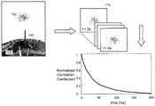
- FIG. 1are illustration of various exemplary results of Laser Speckle Imaging (“LSI”) according to an exemplary embodiment of the present invention
- FIG. 2is perspective side view of a schematic diagram of a wave-guiding arrangement for performing intravascular LSI according to an exemplary embodiment of the present invention
- FIG. 3is a schematic diagram of an arrangement for evaluating optical fiber bundles for LSI according to an exemplary embodiment of the present invention
- FIG. 4are exemplary laser speckle images of static Teflon obtained using two high cross talk optical fiber bundles according to an exemplary embodiment of the present invention
- FIG. 5are exemplary laser speckle images static Teflon obtained using a low cross talk optical fiber bundle according to an exemplary embodiment of the present invention
- FIG. 6Ais a graph showing exemplary speckle decorrelation curves measured from atherosclerotic plaque using a low cross talk optical fiber bundle according to an exemplary embodiment of the present invention
- FIG. 6Bis a graph showing exemplary speckle decorrelation curves measured from atherosclerotic plaque using a high cross talk optical fiber bundle according to an exemplary embodiment of the present invention
- FIG. 7Ais an image of a comparison of exemplary decorrelation time constants of atherosclerotic plaques using a low cross talk optical fiber bundle measured during moving and stationary conditions of the fiber bundle according to an exemplary embodiment of the present invention
- FIG. 7Bis an image of a comparison of exemplary decorrelation time constants of atherosclerotic plaques using a high cross talk optical fiber bundle measured during moving and stationary conditions of the fiber bundle according to an exemplary embodiment of the present invention
- FIG. 8is an exemplary graph of average decorrelation time constants of human atherosclerotic plaques obtained using a low cross talk optical fiber bundle during stationary and moving conditions of the fiber bundle according to an exemplary embodiment of the present invention
- FIG. 9is an exemplary graph of speckle decorrelation curves of an atherosclerotic plaque measured through different thicknesses of blood according to an exemplary embodiment of the present invention.
- FIG. 10Ais a side perspective view of a schematic diagram of a wave-guiding arrangement to conduct intravascular LSI according to an exemplary embodiment of the present invention
- FIG. 10Bis a side perspective view of a schematic diagram of a wave-guiding arrangement to conduct intravascular LSI according to another exemplary embodiment of the present invention.
- FIG. 10Cis a side perspective view of a schematic diagram of a wave-guiding arrangement to conduct intravascular LSI according to yet another exemplary embodiment of the present invention.
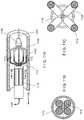
- FIG. 11Ais a side view of a diagram of an intracoronary LSI catheter arrangement according to an exemplary embodiment of the present invention.
- FIG. 11Bis a front cut-away view of a schematic diagram of the cross-sectional view of the exemplary embodiment shown in FIG. 11A at a particular exemplary location;
- FIG. 11Cis a rear cut-away view of a schematic diagram of the cross-sectional view of the exemplary embodiment shown in FIG. 11A at another particular exemplary location;
- FIG. 12is a diagram of the proximal end of the intracoronary LSI catheter (in a cut-away view and a side view) according to an exemplary embodiment of the present invention
- FIG. 13is a side perspective view of the distal end of the intracoronary LSI catheter according to an exemplary embodiment of the present invention.
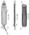
- FIG. 14a side view of an intracoronary LSI catheter arrangement according to another exemplary embodiment of the present invention.
- FIG. 15a side view of the intracoronary LSI catheter arrangement according to yet another exemplary embodiment of the present invention.
- FIG. 16a side view of the intracoronary LSI catheter arrangement according to still another exemplary embodiment of the present invention.
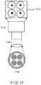
- FIG. 17is a side view of a schematic diagram of an exemplary embodiment of the LSI catheter at the proximal end for performing the LSI image detection according to an exemplary embodiment of the present invention
- FIG. 18is a schematic diagram of an exemplary arrangement of intracoronary LSI system and catheter in excised arteries for testing and/or evaluating the exemplary embodiments;
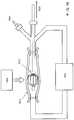
- FIG. 19is a diagram of an arrangement to test intracoronary LSI system and catheter using in vivo animal experiments according to the exemplary embodiment of the present invention.
- FIGS. 20A-20Care illustrations of exemplary implementations for providing computer controlled pull-back device in operation according to an exemplary embodiment of the present invention.
- FIGS. 21A-21Bare illustrations of exemplary implementations to conduct LSI in conjunction with other exemplary intra-coronary techniques.
- time-varying laser speckle images 110were analyzed using normalized two-dimensional cross-correlation techniques 38 to determine the speckle decorrelation time constant, ⁇ , which is inversely dependent on the rate of change of the speckle image 115 .
- the time constant, ⁇was computed by exponential fitting of the normalized speckle decorrelation data 115 .
- the laser speckle patternshould be transmitted from the coronary wall to the image detector.
- small-diameter, flexible exemplary optical fiber bundles 200 , 205e.g., which can be similar to those used in coronary angioscopy, 6 may be incorporated into an intravascular catheter for this purpose.
- the capability of fiber bundles to reliably transmit laser speckle data in the presence of motion such as that encountered in coronary arterieshas not been evaluated.
- exemplary LSI procedurescan be performed on thoracic, and abdominal aortic specimens may be obtained from human cadavers. Immediately After such collection, the aortas may be stored in phosphate buffered saline (PBS). For example, the time between autopsy and imaging possibly may not exceed about 48 hours.
- An exemplary bench-top system shown in FIG. 3can be constructed to acquire laser speckle images of aortic plaques using optical fiber bundles.
- the beam from a polarized Helium Neon laser (632 nm) 300can be expanded (5 ⁇ ) 310 , passed through a 50:50 beam splitter 315 and focused through a lens 320 to an approximately 50 ⁇ m diameter spot on the luminal surface of the plaque samples 325 .
- the characteristic speckle sizecan be approximately matched to the individual fiber size.
- a computer controlled motorized stage 345can be used to control the motion of the optical fiber bundle.
- the motorized stagemay be programmed (e.g., using a controller ESP 300 , Newport) to mimic the coronary wall motion waveform over the cardiac cycle with a maximum peak-to-peak velocity of about 12 mm/s perpendicular to the axis of the bundle.
- the proximal end of each bundlemay be imaged using an objective lens 350 and images can be acquired using a CMOS camera 355 (e.g., Model #PL-A741, Pixelink, Ottawa, Canada).
- Time-varying laser speckle images 365 of aortic plaquesmay be obtained using each optical fiber bundle at a rate of about 240 frames per second.
- each imaging sitecan be marked with two India ink spots which delineated the diameter of the imaged speckle pattern on the lesion.
- a set of 18 randomly selected aortic plaque samplescan be imaged by all three bundles under stationary conditions and during bundle motion.
- the fiber bundle that may provide the highest tolerance to motioncan be selected as described as follows. For example, using such exemplary bundle, about 74 aortic plaques were imaged during both stationary and moving conditions. Following imaging, all aortic plaques were fixed in 10% formalin, embedded and sectioned for histological processing. Sections were cut across the India ink spots and stained with Hematoxylin-Eosin and Trichrome stains. The histological sections were interpreted by a pathologist blinded to the LSI data.
- Plaqueswere classified into the following groups 1 : (i) intimal hyperplasia (IH), (ii) calcific (C), (iii) pathological intimal thickening (PIT), and (iv) necrotic-core fibroatheroma (NCFA). Morphometric measurements of fibrous cap thickness were obtained from the digitized histopathology slides. NCFA's with a minimum fibrous cap thickness ⁇ 100 ⁇ m were further classified as high-risk NCFA's. 8
- Time-varying laser speckle patterns obtained using the optical fiber bundles under stationary and moving conditionswere analyzed using exemplary cross-correlation techniques to determine the speckle decorrelation time constant, ⁇ , which is inversely proportional to the rate of change of the speckle pattern.
- ⁇speckle decorrelation time constant
- the average difference (error) in time constant measurements under stationary and moving conditionswas measured and expressed as a percentage of the time constant measured under stationary conditions.
- the fiber bundle with the highest tolerance to motionwas selected as having the highest correlation, lowest error and minimal statistically significant difference in measurement of plaque time constants under stationary and moving conditions.
- the efficacy of this bundle for identifying high-risk NCFA'swas tested during stationary conditions and bundle motion.
- the time constant value associated with each lesionwas assigned to one of five plaque groups for most or all of 74 aortic plaques, and the average time constant and standard error for each group were computed.
- the differences between average time constant measurements for all plaque groupswere compared using two-way (for plaque type and patient within each plaque group) ANOVA tests; the pair-wise comparisons between the high-risk NCFA group and other plaque groups were evaluated using the Dunnett's t-test. In most or all cases, a p-value ⁇ about 0.05 was considered statistically significant.
- FIG. 4shows exemplary laser speckle images of a card paper using two high cross talk fiber bundles 400 and 405 in which there is significant leakage of light between individual fiber cores.
- FIG. 5shows images of exemplary laser speckle patterns of a card paper obtained using a low cross talk leached fiber bundle, while the flexible shafts of the bundles were moved. Although the card paper exhibits a frozen speckle pattern when the system is stationary, rapid speckle motion is observed even with the slightest motion in the high cross talk fiber bundles as shown in FIG. 4 obtained about 100 ms after the onset of motion. The leached fiber bundle, on the other hand, showed high tolerance to bundle motion with negligible inter-fiber effect as seen in FIG. 5 .
- FIG. 6Agraphs of exemplary normalized speckle decorrelation curves obtained from a fibrous plaque using the low cross talk optical fiber bundle are shown in FIG. 6A
- FIG. 6Bgraphs of exemplary normalized speckle decorrelation curves obtained from a fibrous plaque using the low cross talk optical fiber bundle are shown in FIG. 6B .
- Normalized speckle decorrelation curves obtained using the high cross talk fiber bundle shown in FIG. 6B during stationary and moving conditionsindicate larger differences resulting from the significant temporal modulation of the speckle pattern during motion with an error of ⁇ 80% in measuring the plaque time constant.
- the normalized speckle decorrelation curves obtained from a low cross talk bundle as illustrated in FIG. 6Ashow a high correspondence during stationary and likely identical moving conditions of the fiber bundle. In such exemplary case, negligible inter-fiber effect during motion may result in, e.g., approximately a 7% error in measuring the plaque time constant.
- plaque time constants that can be measured while the bundles are stationarycan be plotted against those measured during bundle motion for two bundles, with the results of the linear regression analysis and paired t—tests presented.
- an acceptable correlationcan be found between plaque time constants measured during stationary and moving conditions, as illustrated in FIG. 7A .
- Such exemplary bundlemay provide, e.g., the highest error in measuring time constants during bundle motion compared to stationary conditions (e.g., 47%), and paired t-tests showed a statistically significant difference (e.g., p ⁇ 0.003).
- the efficacy of the low cross talk leached fiber bundle for identifying high-risk NCFA's with LSIcan then be evaluated using all 74 aortic specimens.
- the exemplary average time constants which can be determined for the different plaque groups under both stationary and moving conditionsare shown in a graph of FIG. 8 .
- the high-risk NCFA group with fibrous cap thickness ⁇ about 100 ⁇ mcan have the lowest average time constant and the calcific group had the highest time constant for both the stationary and moving bundle conditions.
- plaque time constants measured for the high-risk NCFA groupcan be significantly different from each of the other groups even during bundle motion (e.g., p ⁇ 0.05).
- the exemplary cathetercan be similar in design to an angioscope, which may utilize a small diameter optical fiber bundle to illuminate and obtain images of the arterial wall.
- conventional angioscopesmay be incapable of conducting LSI.
- Commercially available angioscopessimilar to certain angioscope, may often be designed to conduct white light endoscopy and provide high quality color images to evaluate coronary plaques in vivo.
- multi-core image fiber bundlesmay be used in commercial angioscopes in which multiple optical fiber cores are fused together in close proximity to achieve high fiber density in order to improve resolution. Due to the close proximity between fibers over the entire length of the image fiber, light may leak between the individual fiber cores.
- laser speckle patternsformed by interference of coherent light
- the high fiber density fused in close proximity over the length of the angioscopemay contribute to leakage of light (crosstalk) between individual fibers which may be exacerbated during cardiac motion potentially corrupting laser speckle patterns of the arterial wall.
- crosstalkleakage of light between individual fibers which may be exacerbated during cardiac motion potentially corrupting laser speckle patterns of the arterial wall.
- the ability to conduct LSI via an intracoronary devicemay be dependent on the reliability of the optical wave guide to transmit arterial laser speckle images, e.g., in the presence of cardiac motion.
- the exemplary leached fiber bundlesmay provide a high correlation in plaque time constants measured under stationary conditions and under conditions that simulated cardiac motion, likely indicating that these bundles may be suitable for intracoronary LSI.
- leached fiber bundlesmay be composed of multiple optical fibers each consisting of a primary acid resistant cladding and a secondary acid soluble cladding that bonds the individual fiber elements. The distal and proximal ends of the bundle are protected and the bundle can be immersed in an acid bath to leach away the secondary cladding producing a highly flexible fiber bundle.
- This exemplary processmay result in a fiber bundle which is highly flexible with larger separation between individual optical fibers along the length of the bundle. This larger inter-fiber separation over the flexible part of the bundle may result in reduced inter-fiber cross-talk potentially allowing these bundles to be more conducive to the transmission of laser speckle images under motion conditions as compared to multi-core image fibers used in angioscopes.
- the error in measuring plaque time constants under moving conditionsmay be significantly lower (e.g., ⁇ 16%) for the fiber bundle with the lowest partial core size (e.g., core area ⁇ individual fiber area) of about 0.36.
- exemplary results of the paired t-testsshowed no significant different in plaque time constants measured during stationary and moving conditions using this bundle, suggesting that the leached fiber bundle with low partial core size could be suitable for use in the intracoronary LSI device.
- high-risk NCFA'smay be identified from a randomly selected group of 74 aortic plaques, even in the presence of motion (e.g., p ⁇ 0.05 in all cases).
- the ability to conduct LSI using optical fiber bundlescan depend on the inter-fiber leakage of light which is related to the separation between fiber cores.
- a leached optical fiber bundle with a low partial core sizemay elicit reduced cross-talk between fiber cores, thus allowing the reliable transmission of laser speckle patterns during motion.
- another exemplary arrangement to reduce inter-core cross-talkcan include packing an absorbing material to increase separation between individual fiber cores.
- angioscopes with low pixilation artifactsmay be suitable for conducting white light angioscopy, it can be beneficial to optimize these angioscopes for LSI by incorporating low cross talk fiber bundles.
- the distal and proximal end of the fiber bundlesmay be maintained fixed while the fiber bundle shaft can be moved using a computer-controlled motorized stage to mimic coronary motion over the cardiac cycle.
- a motion of the distal end of the fiber bundle relative to the coronary wallmay also add an undesired speckle decorrelation that may reduce the diagnostic value of intracoronary LSI.
- One exemplary way of minimizing this effectcan be to provide an intracoronary LSI device that can maintain a constant contact with the coronary wall during the imaging period, which may typically be less than about 100 ms.
- Certain well-established catheter designs used for intracoronary temperature measurementhave been developed. 9,10 Thermography studies have demonstrated the utility and safety of contact-based intra-arterial catheters in which flexible thermosensors maintain contact with the vessel wall to measure local temperature changes associated with increased inflammation without requiring an interruption of coronary blood flow.
- cardiac gating methodsmay be employed which can provide for a sufficient temporal window during the resting phase of the cardiac cycle to obtain diagnostic quality speckle data.
- intracoronary saline flushingwhich has been implemented for optical coherence tomography and angioscopy procedures to temporarily displace blood by injecting a bolus of saline, 11 can be utilized in conjunction with LSI to enable unobstructed imaging of the coronary wall.
- Exemplary review described hereincan evaluate the feasibility of LSI in the presence of blood.
- a development of an intracoronary LSI catheter, similar in design to those used in thermography, 39,40 which contacts the arterial wallcan be effectuated, thereby diminishing the influence of blood and potentially obviating the need for saline flushing.
- the thicknesscan be varied by raising the fibroatheroma through blood layer thickness different heights within the flow cell, and speckle decorrelation curves may be obtained as shown in the exemplary graph of FIG. 9 .
- ⁇may be approximated to be that of whole blood (e.g., ⁇ approximately 4 ms).
- the exemplary results of the unpaired t-testshowed that the average ⁇ changed significantly in the presence of a blood layer (e.g., p ⁇ 0.05).
- exemplary resultscan indicate that, as long as reasonably close contact (a few hundred microns) is maintained between the LSI catheter and the arterial wall, LSI may potentially be conducted in the presence of blood.
- Exemplary contact catheterscan be provided for other optical imaging techniques and this level of close contact can be reliably achieved in coronary arteries.
- An alternative exemplary method according to the present inventionmay be to conduct an exemplary intracoronary LSI procedure in conjunction with saline flushing as in other optical imaging techniques. 42
- aortic plaques within the flow cellthrough, e.g., a 2 mm intervening layer of whole blood, serially diluted to different concentrations using phosphate buffered saline (PBS).
- PBSphosphate buffered saline
- time constants, ⁇of aortic plaques measured by diluting blood to HCT can be ⁇ about 0.1% maybe similar to ⁇ values measured without any intervening medium.
- the exemplary OCT imagingcan be performed, and it may be determined that at HCT>about 0.03%, backscattering from blood cells can be evident in the exemplary OCT images.
- Exemplary arrangements of optical waveguides to conduct intravascular LSIcan be provided according to the exemplary embodiments of the present invention:
- optical fiber bundle arrangements with low cross talkare capable of transmitting laser speckle images.
- Small diameter coherent optical fiber bundlescan be drawn consisting of multiple fibers.
- a central light delivery fiber core 1000 of the fiber bundle arrangementcan be a single mode or a multimode fiber to launch light which can be focused using a miniature lens towards the arterial wall.
- Surrounding single- or multimode fibers 1005can transmit the speckle image.
- two optical fiberscan be utilized to conduct intravascular LSI.
- One single mode or multimodecan be used to forward focused or broad area illumination onto the arterial wall 1000 and another single or multimode fiber can be used to transmit the reflected light from the arterial wall 1005 .
- multiple single mode or multimode optical fibers 1000can be used in an arrangement surrounding the collection fibers 1005 to conduct broad area illumination and collection of laser speckle patterns of the arterial wall.
- Exemplary considerations for developing an imaging catheter capable of reliably transmitting laser speckle images of the coronary wallcan be: (i) to enable evaluation of long (5.0 cm) segments of proximal main coronary arteries, (ii) to minimize the influence of blood on speckle modulation without interrupting blood flow, and (iii) to achieve sufficient motion stability during imaging.
- an exemplary approachcan be to construct an imaging catheter that maintains contact with the coronary wall during the LSI procedure, thereby obviating the influence of blood in the region of contact, and facilitating a sufficient coupling with the arterial wall to ensure adequate motion stability over the cardiac cycle.
- One exemplary catheter designcan be in view of an established design used for intracoronary temperature measurement. Thermography studies have demonstrated the utility and safety of contact-based intraarterial catheters in which flexible thermosensors maintain contact with the vessel wall to measure local temperature changes associated with increased inflammation without requiring interruption of coronary blood flow. 43 One exemplary design provides a catheter consisting of four nitinol prongs with dedicated thermistors which after engagement expand to maintain endoluminal surface during the procedure. 44 In another exemplary design, a contact basket catheter can be provided in which a nitinol basket equipped with flexible wires and thermosensors detects temperature changes of the vessel wall in the presence of blood flow without occluding the lumen designs have been successfully demonstrated in animal studies. 45 Clinical studies in several patients to date have exemplified the safety and efficacy of the contact-based design. 50
- One exemplary embodiment of the intravascular LSI cathetercan utilize an exemplary basket catheter design. It is possible to provide a contact imaging basket catheter to conduct intracoronary LSI. It is possible to fabricate miniature optical imaging probes using a combination of in-house micromachining systems and, e.g., outsourcing SolidWorks CAD (SolidWorks Corporation, Concord, Mass.) schematics for precision machining.
- SolidWorks CADSolidWorks Corporation, Concord, Mass.
- the target diameter of the intracoronary LSI catheter 1105can be, e.g., less than about 1.5 mm.
- the cathetercan be equipped with multiple (e.g., four) waveguide arrangements 1115 .
- Each waveguide arrangementmay be utilized to illuminate one quadrant of the coronary wall and to detect the speckle images that result.
- the waveguide arrangementscan terminate in an expandable e.g., nitinol basket 1125 at the distal end of the catheter (see FIG. 11 ).
- the basketcan contain four hollow expandable prongs, comprised of e.g., nitinol tubes with a central optical window 1120 .
- the waveguide arrangements 1115may be housed within the nitinol tubes of the basket and the imaging portion of each bundle may terminate at the central optical window.
- a tapered connector 1130can terminate the basket.
- the outer sheath 1110may be retracted, the basket may expand, and the imaging portions of the fiber bundles may come into contact with the coronary artery wall.
- a single wound, flexible cableProximal to the basket, a single wound, flexible cable, can envelop the four optical fiber bundles.
- a remote, motorized computer-controlled translational stage 1100 connected to the proximal end of the wound cablecan translate the deployed catheter along the coronary segment.
- the basketcan be retracted into the catheter sheath so that the catheter can be withdrawn from the artery.
- a port to house a guide wire 1135may be provided through the center of the catheter.
- Low cross talk optical fiber bundles for example leached fiber bundles capable of transmitting laser speckle imagescan be incorporated within each basket prong.
- a custom-designed fiber bundles with minimal cross talkcan be provided.
- Each prong of the basket cathetercan be incorporated with one of multiple exemplary waveguide arrangements shown in FIGS. 10A, 10B and/or FIG. 10C .
- the fiber bundles 1210can be configured so that the light delivery fiber 1205 of each bundle may be separated from the collection fibers 1210 .
- This exemplary arrangementcan facilitate the use of separate illumination and collection ports.
- FIG. 13shows a side view of an exemplary embodiment of the catheter according to the present invention
- the distal portion of each optical wave guide arrangementcan contain miniature optics for focusing the incident light and imaging the speckle pattern through the optical window.
- a distal end 1300 , 1305 of the exemplary cathetercan be affixed to the distal face of each wave guide arrangement to detect only cross-polarized light from the artery wall.
- An exemplary circular polarizer 1310can be used to minimize specular surface reflections of the arterial wall.
- a miniaturized lense.g., a gradient index (GRIN) microlens 1315 and reflective right angle prism 1320 may focus and direct light at ⁇ 90° from the catheter axis onto the vessel wall through the optical window 1325 .
- Light remitted from the tissuecan be imaged onto the fiber bundle face through the same optical components.
- the fiber bundle faces, polarizer, GRIN lens, and the prismmay be provided with antireflection coatings to avoid reflections from the optical components
- FIG. 14shows a side view of an exemplary embodiment of an intravascular LSI catheter according to the present invention in which the exemplary LSI catheter does not contact the vessel wall.
- a single wave-guide arrangement 1400includes one or multiple illumination and collection optical fibers (as shown in, e.g., FIGS. 10A, 10B and 10C ) can be incorporated within a catheter sheath 1412 .
- Focused or broad area illumination of the arterial wall 1425can be conducted and speckle patterns can be imaged on to the distal end of the collection wave guiding arrangement using a distal optical arrangement 1410 described above in FIG. 13 .
- An exemplary rotary junctioncan be used to rotate 1435 the intravascular catheter within the vessel lumen.
- the exemplary LSI procedure(s)can be conducted while a bolus of saline is flushed through the arterial wall.
- FIG. 15shows a side view of another exemplary embodiment of the intravascular LSI catheter according to the present invention in which a single wave guiding arrangement can be rotated within a balloon 1520 that is inflated to temporarily occlude coronary blood flow during the imaging duration.
- a single wave guiding arrangementcan be rotated within a balloon 1520 that is inflated to temporarily occlude coronary blood flow during the imaging duration.
- Similar element and features described above and labeled with numerals 1400 , 1410 , 1425 and 1430are labeled with numerals 1500 , 1510 , 1525 and 1530 , respectively, in FIG. 15 .
- FIG. 16shows a side view of still another exemplary embodiment of the intravascular LSI catheter according to the present invention in which multiple prong waveguiding arrangements 1635 are affixed within a clover shaped balloon 1620 which partially occludes blood flow during imaging and maintains contact with the arterial wall 1625 to minimize the effect of cardiac motion on the transmission of laser speckle patterns.
- Similar element and features described above and labeled with numerals 1400 , 1410 and 1430are labeled with numerals 1600 , 1610 and 1630 , respectively, in FIG. 16 .
- an exemplary embodiment of an LSI cart system and instrumentationcan be as follows.
- an exemplary LSI systemcan be assembled and incorporated within a portable medical cart for ease of use in the catheterization suite.
- the exemplary systemmay contain a coherent light source.
- light from the lasercan be coupled into a single-mode fiber, connected to a single mode fiber-optic star coupler that splits the input light equally into multiple optical fibers.
- Each output port of the star couplermay be connected to the illumination fiber of each waveguiding arrangement (see FIGS. 11A, 11B, 11C and 16 ).
- a high-speed, triggerable digital CCD camera 1710can be incorporated in the LSI cart.
- a relay lens 1705may image the collection port onto the CCD camera so that laser speckle images transmitted by the multiple waveguide arrangements may be simultaneously detected 1715 .
- a Camera Link interface capable of high data transfer rate ofmay be used to transfer image data to a computer in real time.
- each imaging location along the vesseltime-varying laser speckle images may be acquired for at least about 40 ms.
- This acquisition timecan provide sufficient data for NCFA characterization and may be sufficiently short to allow screening of large coronary segments.
- a custom-built pullback device using a computer-controlled stagemay be affixed to the wound cable at the proximal end of the catheter. Basket pullback can be conducted in exemplary discrete steps (e.g., 0.1-2 mm) at a rate of up to 5 mm/s and speckle data may be obtained over at least 40 ms at each step when the catheter is stationary.
- off-the-shelf bundles with different core:cladding ratioscan be obtained from Schott Inc. It is possible to test crosstalk by illuminating one of the fibers and detecting light leakage in the cladding and adjacent fibers.
- the testing environmentcan include an apparatus for bending the fiber bundle to evaluate crosstalk as a function of radii of curvature (e.g., 3-10 cm).
- optical elementsmay be purchased and coronary circulation assembled onto the fiber bundle. Light can be transmitted through the central phantom fiber onto static and dynamic scattering phantoms. As above, it is possible to i) compare time constants obtained with the bundle and optics, with the bundle straight and with coronary circulation phantom, and ii) compare time constant of phantoms measured through exemplary bundle and optics with that obtained by exemplary free-space system. Success can be defined as a deviation of ⁇ 10% between these measurements.
- the exemplary intracoronary basket or balloonmay be designed and constructed based on exemplary specifications (see FIGS. 11A, 11B, 11C, 15 and 16 ).
- an imaging windowcan be provided by a notch in the nitinol tubes at the center of the basket, where light can be transmitted.
- the distal opticsmay be fed through the hollow nitinol prongs so that they are located and oriented for optimal coupling upon contact with the vessel wall. Following correct positioning of the optics, such optics can be affixed to the prongs by applying a small drop of optically transparent epoxy in the notch.
- exemplary waveguide arrangementscan be affixed within the balloon to maintain arterial contact. After the optics are incorporated, it is possible to test the performance of the assembled catheter in the same manner as exemplary methods described above. As described herein, it is possible to define success as a deviation in time constant measurements of ⁇ about 10%.
- the surface contact of the basket at the imaging sitemay be evaluated in excised cadaveric arterial specimens. Contact can be measured using optical frequency domain imaging (OFDI), 44 a cross-sectional optical imaging technique with high speed, high-resolution (e.g., 10 ⁇ m), and a large ranging depth (e.g., about 10 mm).
- OFDIoptical frequency domain imaging
- the experimental exemplary arrangement according to the present invention to test catheter contactis depicted in a diagram of FIG. 18 .
- the intracoronary LSI catheter 1810may be introduced into the coronary artery 1815 and the exemplary distal basket or balloon can be engaged while OFDI monitoring 1805 is performed external to the vessel and coincident with LSI imaging location. Precise registration between the OFDI beam and the LSI site is possible since the LSI illumination beam may be visible through the adventitia of the artery. Catheter contact can be evaluated under at least three conditions: (a) during vessel deformation, (b) in the presence of blood, and (c) during catheter pullback. To evaluate the effect of vessel deformation, an automated syringe pump may inject saline 1800 to cause a 10% radial deformation of the arterial wall mimicking the extent of physiological coronary deformation.
- Exemplary pressure measurement 1825may be conducted during the experiment.
- the response time of the nitinol basket or balloon construct to conform to arterial deformationcan be evaluated by varying the saline infusion may be used to test intracoronary LSI system and rates from about 1-10 cc/s.
- whole porcine bloodcan be injected into the artery during OFDI monitoring 1805 and scattering due to blood in the region of basket contact may be evaluated.
- the custom-built motorized pullback devicecan be used to linearly translate the LSI intracoronary catheter to evaluate the effect of pullback rates on basket-to-wall contact. This testing may be performed, e.g., in two phases of catheter development, during design optimization of the nitinol basket and after completion of the catheter. Success can be defined as, e.g., ⁇ about 200 ⁇ m spacing between the basket prongs and the arterial wall for greater than 80% of the pullback length.
- Exemplary optical components for imaging the fiber bundle collection portmay be designed and optimized with ZEMAX and subsequently fabricated and assembled.
- the laser, star coupler, collection optics, CCD, pullback device, and computercan be integrated in a portable cart.
- the computer or another processing arrangementmay also include a data acquisition board for digitizing the EKG during speckle pattern acquisition.
- Softwarecan be developed for controlling the motors, reading and storing motor encoder positions, real-time speckle analysis, and displaying data in a various formats for ease of interpretation.
- Cadaver coronary arterial segmentsmay be excised during autopsy and prepared for LSI testing using the experimental configuration, as shown in the diagram of an exemplary embodiment illustrated in FIG. 18 .
- segmentscan be immersed in phosphate buffered saline and warmed to 37° C.
- Time-varying laser speckle imagesmay be obtained over 5 cm arterial segments with the LSI catheter at about 1.0 mm increments, using a measurement duration at each site of 40 ms.
- Intracoronary LSIcan be conducted (a) without blood, (b) in the presence of blood, and (c) during arterial deformation.
- the arterial specimensmay be marked at each quadrant along the adventitial aspect of the specimen using different colored ink spots for precise registration of each quadrant with histology. Marking can be facilitated by observation of the laser light through the arterial wall.
- the speckle decorrelation time constantmay be determined at each spatial location from the normalized cross-correlation of time-varying laser speckle images.
- the arterial specimenscan be fixed in Formalin and prepared for histological processing.
- Histopathologic diagnosisas the gold standard, the sensitivity and specificity of intracoronary LSI for identifying necrotic core fibroatheromas with thin fibrous caps ( ⁇ 65 ⁇ m) can be evaluated under all three experimental conditions. Diagnostic performance targets to determine success in identifying TCFA's may be based on a presumed intra-plaque and intra-patient correlation of approximately 50%, with an estimated TCFA prevalence of about 3%.
- Fiber bundle crosstalk⁇ 10% Based on certain preliminary studies, it is believed that cross talk of ⁇ 10% between fibers with a radius of curvature of 3 cm can be sufficient to satisfy exemplary requirements for intracoronary LSI. It is possible to measure fiber crosstalk by illuminating a single fiber and detecting light leakage in the cladding and adjacent fibers for different radii of curvature as described above in Specific method 1.
- Catheter size⁇ 1.5 mm (4.7 F)
- the cathetercan be designed with a target size of ⁇ 1.5 mm.
- Each of the four expandable prongs of the distal basketmay have a diameter of 350 ⁇ m, with a 250 ⁇ m guide wire through the center of the catheter.
- Imaging Rate 2000 frames/sA high-speed CCD camera (Mikrotron MC 1310) may be incorporated in the system capable of acquiring 512 ⁇ 512 pixel images at 2000 frames/s over 40 ms at each imaging site. Date storage rate 200 MB/s The speckle data from the CCD camera can be digitally stored in real-time. This data storage rate is more than sufficient to recover all images at video rate. Field of View (FOV) ⁇ 500 ⁇ m Using Monte Carlo simulations (FIG.
- this FOVcan be more than sufficient to allow evaluation of the critical superficial ⁇ 400 ⁇ m of the arterial wall Imaging time
- Tens of seconds Catheter pullbackmay be conducted in discrete steps (0.1-2 mm), and speckle data can be acquired at each step for 40 ms.
- a pullback rate of 5 mm/smay allow imaging of a typical 5 cm artery in tens of seconds.
- Basket catheter contact ⁇ 200 ⁇ m Catheter contactcan be measured using OFDI as described above in Specific method 4. Success may be defined as ⁇ 200 ⁇ m spacing between prongs and arterial wall for >80% of the pullback length.
- time constants of with free space system scattering phantoms measured through the LSI cathetercan be compared with the free space system (see FIG. 2). Based on exemplary preliminary experiments using low crosstalk fiber bundles, it is anticipated ⁇ 10% deviation between time constants measured in the two cases. Sensitivity for detecting ⁇ 95% ⁇ 7% (no Comparison of time constant measured by TCFA's.
- intracoronary LSImay be statistically compared with detecting TCFA's (with blood) Histological diagnoses to determine sensitivity and ⁇ 90% ⁇ 10% (no specificity for TCFA detection without blood and in the blood) ⁇ 85% ⁇ 12% presence of blood. These expected values are (with blood) determined based on exemplary sample size (1000 plaques) and TCFA prevalence (e.g., 3%) calculations, accounting for intra-plaque and intra-patient correlation of 50%. Success is defined by the feasibility of detecting TCFA's since this is clinically most significant.
- the exemplary intracoronary LSI catheter and system described hereincan be tested in a pilot animal study to determine feasibility of conducting LSI in vivo.
- Rationale for human-to-swine coronary xenograft modelA selection of animal model can be motivated by two important preferences. First, feasibility of intracoronary LSI can be preferably tested on evaluating human coronary atherosclerosis. Second, testing should be performed in a living animal model under conditions of hemodynamics and cardiac motion that closely approximate human coronary physiology. Based on these preferences, the exemplary approach is to use a human-to-swine coronary xenograft model that allows imaging of human coronaries under physiological conditions similar to those encountered in humans, the exemplary utilization being shown in FIG. 19 . The exemplary model shown in FIG. 19 has been successfully utilized with intracoronary optical techniques.
- swinecan be used in the study. Normal swine may provide controls and additional swine can be grafted with human cadaveric coronary artery xenografts.
- human cadaveric heartsmay be obtained at autopsy and screened using angiography to identify proximal coronary arteries with disease.
- exemplary candidate coronary segments 1905( ⁇ 5 cm) can be prosected off the cadaver heart, the side branches ligated, and luer locks sutured to the ends of the grafts.
- adventitial fatmay be removed and discrete imaging sites can be randomly selected along the length and marked with India ink spots on the artery.
- the swinemay be anesthetized and the heart surgically exposed.
- the cadaveric coronary graft(with ink marks facing upwards) can be surgically implanted to the ascending aorta at the proximal end and sutured to the left ventricle to ensure mechanical coupling with the beating swine heart 1915 as shown in FIG. 19 .
- a “Y” connector with a valve and guide-portmay be connected proximal to the coronary graft for saline administration and introduction of the exemplary LSI catheter 1910 .
- the intracoronary LSI proceduremay be conducted the anesthesized swine with simultaneous EKG monitoring and recording.
- FIG. 19shows an exemplary embodiment of a configuration of the LSI catheter to be used in the swine heart during the imaging procedure.
- a guide catheter and guide wirecan be introduced into the coronary graft through the guide port of the “Y” connector.
- the exemplary LSI cathetermay be inserted over the guide wire and advanced into the coronary graft. After the catheter is positioned within the coronary graft, the outer catheter sheath can be retracted and the distal imaging basket may expand to contact the coronary endoluminal surface.
- the exemplary LSI cathetercan be affixed to a computer controlled pull-back device incorporated in the portable LSI system. Imaging may be performed at each of the 10 discrete sites. At each LSI site, the catheter can be positioned such that the visible laser beam observed through the coronary wall at one basket prong overlaps with the fiducial mark and the pullback motor coordinates may be recorded.
- the exemplary intracoronary LSI procedurecan be conducted (a) without interruption of blood flow, and (b) with administration of saline flush.
- the EKGmay trigger the CCD camera to begin acquisition of the first frame on the R-wave, followed by asynchronous acquisition of subsequent frames at a rate of about 2000 frames/s for a duration of 5 seconds.
- a 5 cc saline flushcan be administered while the exemplary LSI procedure is repeated at the each site.
- the guide wire and LSI cathetermay be retracted through the guide-port and a second graft can be imaged.
- the influence of heart rate on the exemplary LSI procedure performancemay be evaluated by administering 10 ⁇ g/kg/min of dobutamine, which has been previously described in the swine animal model to cause ⁇ 50% increase in heart rate.
- 45 LSI imagingcan be repeated as above after heart rate is stabilized.
- the exemplary intracoronary LSI proceduremay be similarly repeated to image the native LAD and LCx arteries. In the control arteries, ink marking of discrete locations is difficult on the beating heart. Instead, the computer controlled pull-back device can pull back as shown in FIGS. 20A-20C .
- the exemplary cathetercan be operated, e.g., at a rate of 5 mm/s through 10 discrete steps spaced 5 mm apart during the performance of the exemplary LSI procedure in human-swine, while the exemplary intracoronary LSI procedure is performed, e.g., over 5 seconds at coronary xenograft model. each location.
- the linear translator position and the EKGmay be digitally recorded throughout the procedure.
- the swinecan be sacrificed by the intravenous administration of sodium pentobarbital (120 mg/kg) with heparin (100 U/kg).
- the coronary segmentsmay be fixed in 10% formalin, embedded and sectioned using standard Histology techniques.
- All coronary sectionsmay be broadly characterized into two groups: NCFA and non-necrotic core plaques. Fibrous cap thickness in the NCFA set and collagen content in all plaques can be determined from digitized histology sections. 46,47
- intracoronary LSIin identifying NCFA's may be determined. For example, a large number (e.g., 260) of coronary sites can be analyzed, including 100 human cadaveric coronary sites in the xenograft model and 160 sites in the control native coronaries (e.g., 10 sites/artery ⁇ 2 arteries ⁇ 4 quadrants ⁇ 2 control swine). Exemplary time constants may be measured by exponential fitting of the normalized cross-correlation data for each coronary site over a duration of 40 ms by previously described exemplary LSI techniques.
- a large numbere.g., 260
- Exemplary time constantsmay be measured by exponential fitting of the normalized cross-correlation data for each coronary site over a duration of 40 ms by previously described exemplary LSI techniques.
- the sensitivity and specificity for identifying NCFA'scan be evaluated for the following conditions: (a) in the presence of blood, (b) during saline administration, and (c) following dobutamine administration.
- Exemplary diagnostic performance targets to determine success in identifying NCFA'smay be based on a presumed intra-plaque and intra-patient correlation of approximately 50%, with an estimated prevalence of 10%.
- Statistical significance to achieve discrimination of NCFA's under all conditionscan be evaluated using two-way ANOVA tests: success may be defined by p ⁇ 0.05.
- the exemplary relationships between LSI time constants with NCFA cap thickness and collagen content in all plaques measured under the above conditionscan be determined using linear regression analysis. In all cases, success may be defined by a statistically significant (e.g., p ⁇ 0.05) good correlation (e.g., R>0.6) between measurements.
- the influence of cardiac phase over the cardiac cycle on LSI measurementscan be evaluated by measuring time constants within a windowed duration of 40 ms over 5 s ( ⁇ 5 cardiac cycles).
- the mean, ⁇ , and standard deviation, ⁇ , in time constants measured over the cardiac cyclemay be determined for all plaques.
- the cardiac motion over 40 msmay not influence a significant variation in time constants and it is anticipated an average variation in ⁇ of ⁇ 10% over the cardiac cycle. However, this variation may be dependent on tissue type, since the rate of Brownian motion during arterial deformation is dependent on composition. For example, plaques containing a compliant lipid core may present a higher ⁇ over the cardiac cycle as compared with stiffer fibrous plaques.
- the exemplary standard deviation, ⁇may potentially provide an additional diagnostic metric for plaque discrimination.
- Intracoronary LSImay be implemented in the catheterization suite as a stand alone technique as described in the embodiments above or as an adjunct to other intra-vascular techniques such as angioscopy.
- LSImay be conducted simultaneously with angioscopy through a single catheter.
- a cross-sectional view of an exemplary catheter to simultaneously perform angioscopy in conjunction with LSIis shown in FIG. 21A .
- An exemplary catheter 2100may consist of low cross talk optical waveguides consisting of multiple fiber cores 2102 for collecting both angioscopy and laser speckle patterns of the arterial wall may be incorporated.
- Optical wave guidesmay be incorporated for illumination using white light such as a xenon lamp source to conduct angioscopy 2105 and a waveguide for coherent light illumination 2110 to conduct LSI.
- the exemplary configuration of the waveguide arrangementmay include at least one of the exemplary embodiments described herein and shown in FIGS. 10A, 10B and/or 10C .
- the exemplary rotation 2115 and/or pullback 2120 of the cathetermay be facilitated either manually or utilizing a rotary junction and/or a motorized translational stage.
- An exemplary system to conduct angioscopy in conjunction with LSIis shown in FIG. 21B .
- a light source such as a xenon source 2125 and a coherent light source 2130may be utilized in the exemplary system for illumination through the catheter 2100 .
- LSI and angioscopy imagesmay be collected simultaneously via the collection port.
- a dichroic mirror 2135may be utilized in conjunction with a color camera to capture angioscopy images at video rate and a high speed camera 2145 to capture coherent laser speckle patterns at high frames rates.
- a Camera Link interface capable of high data transfer rate ofmay be used to transfer image data to a computer 2150 in real time.
- Performance Target Value Justification and Verification Method Variation in LSI time⁇ 10% It is anticipated that about ⁇ 10% deviation in time constants constant measurements comparing LSI conducted in the presence of blood to imaging during the administration of a saline flush. Additionally, since cardiac motion over 40 ms may not cause a significant variation in time constants, it is anticipated that ⁇ 10% deviation in time constants measured at resting heart rate compared with those measured at increased heart rate following dobutamine administration. In vivo detection of 90% ⁇ 10% Comparison of time constant measured by intracoronary LSI NCFA's: -sensitivity - 85% ⁇ 15% can be statistically compared with Histological diagnoses to specificity determine sensitivity and specificity for NCFA detection in vivo.
- an intracoronary LSI catheter and systemaccording to an exemplary embodiment of the present invention to enable evaluation of long coronary segments in vivo.
- Similar techniques using intravascular LSIcan be utilized for evaluating plaque in other vasculature such as the carotid arteries, peripheral arteries and renal arteries.
Landscapes
- Health & Medical Sciences (AREA)
- Life Sciences & Earth Sciences (AREA)
- Physics & Mathematics (AREA)
- Surgery (AREA)
- Biomedical Technology (AREA)
- Molecular Biology (AREA)
- Pathology (AREA)
- Veterinary Medicine (AREA)
- Public Health (AREA)
- Engineering & Computer Science (AREA)
- Biophysics (AREA)
- Heart & Thoracic Surgery (AREA)
- Medical Informatics (AREA)
- Optics & Photonics (AREA)
- Animal Behavior & Ethology (AREA)
- General Health & Medical Sciences (AREA)
- Nuclear Medicine, Radiotherapy & Molecular Imaging (AREA)
- Radiology & Medical Imaging (AREA)
- General Physics & Mathematics (AREA)
- Endoscopes (AREA)
- Investigating Or Analysing Materials By Optical Means (AREA)
Abstract
Description
| Performance Target | Value | Justification and Verification Method |
| Fiber bundle crosstalk | <10% | Based on certain preliminary studies, it is believed that |
| cross talk of <10% between fibers with a radius of | ||
| curvature of 3 cm can be sufficient to satisfy exemplary | ||
| requirements for intracoronary LSI. It is possible to | ||
| measure fiber crosstalk by illuminating a single fiber | ||
| and detecting light leakage in the cladding and adjacent | ||
| fibers for different radii of curvature as described | ||
| above in | ||
| Catheter size | <1.5 mm (4.7 F) | The catheter can be designed with a target size of |
| <1.5 mm. Each of the four expandable prongs of the | ||
| distal basket may have a diameter of 350 μm, with a | ||
| 250 μm guide wire through the center of the catheter. | ||
| When engaged, the distal basket can be designed to | ||
| expand and conform to the coronary wall. | ||
| Imaging Rate | 2000 frames/s | A high-speed CCD camera (Mikrotron MC 1310) may |
| be incorporated in the system capable of acquiring | ||
| 512 × 512 pixel images at 2000 frames/s over 40 ms at | ||
| each imaging site. | ||
| 200 MB/s | The speckle data from the CCD camera can be digitally | |
| stored in real-time. This data storage rate is more than | ||
| sufficient to recover all images at video rate. | ||
| Field of View (FOV) | ~500 μm | Using Monte Carlo simulations (FIG. 9), this FOV |
| can be more than sufficient to allow evaluation of the | ||
| critical superficial ~400 μm of the arterial wall | ||
| Imaging time | Tens of seconds | Catheter pullback may be conducted in discrete steps |
| (0.1-2 mm), and speckle data can be acquired at each | ||
| step for 40 ms. A pullback rate of 5 mm/s may allow | ||
| imaging of a typical 5 cm artery in tens of seconds. | ||
| Basket catheter contact | <200 μm | Catheter contact can be measured using OFDI as |
| described above in Specific method 4. Success may be | ||
| defined as <200 μm spacing between prongs and | ||
| arterial wall for >80% of the pullback length. | ||
| LSI catheter compared | ~10% deviation in τ | As described in the Specific methods, time constants of |
| with free space system | scattering phantoms measured through the LSI catheter | |
| can be compared with the free space system (see | ||
| FIG. 2). Based on exemplary preliminary | ||
| experiments using low crosstalk fiber bundles, it is | ||
| anticipated ~10% deviation between time constants | ||
| measured in the two cases. | ||
| Sensitivity for detecting | ~95% ± 7% (no | Comparison of time constant measured by |
| TCFA's. Specificity for | blood) ~90% ± 10% | intracoronary LSI may be statistically compared with |
| detecting TCFA's | (with blood) | Histological diagnoses to determine sensitivity and |
| ~90% ± 10% (no | specificity for TCFA detection without blood and in the | |
| blood) ~85% ± 12% | presence of blood. These expected values are | |
| (with blood) | determined based on exemplary sample size (1000 | |
| plaques) and TCFA prevalence (e.g., 3%) calculations, | ||
| accounting for intra-plaque and intra-patient correlation | ||
| of 50%. Success is defined by the feasibility of | ||
| detecting TCFA's since this is clinically most | ||
| significant. | ||
Exemplary Intracoronary LSI Feasibility Review in Living Swine Coronary Xenograft Model
| Performance Target | Value | Justification and Verification Method |
| Variation in LSI time | ~10% | It is anticipated that about ~10% deviation in time constants |
| constant measurements | comparing LSI conducted in the presence of blood to imaging | |
| during the administration of a saline flush. Additionally, | ||
| since cardiac motion over 40 ms may not cause a significant | ||
| variation in time constants, it is anticipated that ~10% | ||
| deviation in time constants measured at resting heart rate | ||
| compared with those measured at increased heart rate | ||
| following dobutamine administration. | ||
| In vivo detection of | 90% ± 10% | Comparison of time constant measured by intracoronary LSI |
| NCFA's: -sensitivity - | 85% ± 15% | can be statistically compared with Histological diagnoses to |
| specificity | determine sensitivity and specificity for NCFA detection in | |
| vivo. These expected values are determined based on | ||
| exemplary sample size (260 plaques) and NCFA prevalence | ||
| (10%) calculations. Success may be defined by the feasibility | ||
| to attain these values in the presence of blood or during | ||
| administration of a saline flush. | ||
| Intracoronary LSI | R > 0.6 | Collagen content and NCFA cap thickness measured from |
| relationships with | (p < 0.05) | Histological sections can be compared with time constant |
| collagen content and | measurements. | |
| NCFA cap thickness | ||
- 1Virmani R, Kolodgie F D, Burke A P, Farb A, Schwartz S M. Lessons from sudden coronary death: a comprehensive morphological classification scheme for atherosclerotic lesions. Arterioscler Thromb Vasc Biol. 2000; 20:1262-75.
- 2Schroeder A P, Falk E. Vulnerable and dangerous coronary plaques. Atherosclerosis. 1995; 118 Suppl:S141-9.
- 3Bauriedel G, Hutter R, Welsch U, Bach R, Sievert H, Luderitz B. Role of smooth muscle cell death in advanced coronary primary lesions: implications for plaque instability. Cardiovasc Res. 1999; 41:480-8.
- 4Newby A C, Zaltsman A B. Fibrous cap formation or destruction—the critical importance of vascular smooth muscle cell proliferation, migration and matrix formation. Cardiovasc Res. 1999; 41:345-60.
- 5Rekhter M D, Hicks G W, Brammer D W, Hallak H, Kindt E, Chen J, Rosebury W S, Anderson M K, Kuipers P J, Ryan M J. Hypercholesterolemia causes mechanical weakening of rabbit atheroma: local collagen loss as a prerequisite of plaque rupture. Circ Res. 2000; 86:101-8.
- 6Slager C J, Wentzel J J, Gijsen F J, Thury A, van der Wal A C, Schaar J A, Serruys P W. The role of shear stress in the destabilization of vulnerable plaques and related therapeutic implications. Nat Clin Pract Cardiovasc Med. 2005; 2:456-64.
- 7Schartl M, Bocksch W, Koschyk D H, Voelker W, Karsch K R, Kreuzer J, Hausmann D, Beckmann S, Gross M. Use of intravascular ultrasound to compare effects of different strategies of lipid-lowering therapy on plaque volume and composition in patients with coronary artery disease.Circulation.2001; 104:387-92.
- 8Aikawa M, Rabkin E, Okada Y, Voglic S J, Clinton S K, Brinckerhoff C E, Sukhova G K, Libby P. Lipid lowering by diet reduces matrix metalloproteinase activity and increases collagen content of rabbit atheroma: a potential mechanism of lesion stabilization. Circulation. 1998; 97:2433-44.
- 9Libby P, Aikawa M. Mechanisms of plaque stabilization with statins. Am J. Cardiol. 2003; 91:4B-8B.
- 10Aikawa M, Libby P. Lipid lowering reduces proteolytic and prothrombotic potential in rabbit atheroma. Ann NY Acad Sci. 2000; 902:140-52.
- 11Fukumoto Y, Libby P, Rabkin E, Hill C C, Enomoto M, Hirouchi Y, Shiomi M, Aikawa M. Statins alter smooth muscle cell accumulation and collagen content in established atheroma of watanabe heritable hyperlipidemic rabbits. Circulation. 2001; 103:993-9.
- 12Richardson P D, Davies M J, Born G V. Influence of plaque configuration and stress distribution on fissuring of coronary atherosclerotic plaques. Lancet. 1989; 2:941-4.
- 13Loree H M, Kamm R D, Stringfellow R G, Lee R T. Effects of fibrous cap thickness on peak circumferential stress in model atherosclerotic vessels. Circ Res. 1992; 71:850-8.
- 14Ohayon J, Teppaz P, Finet G, Rioufol G. In-vivo prediction of human coronary plaque rupture location using intravascular ultrasound and the finite element method. Coron Artery Dis. 2001; 12:655-63.
- 15Tang D, Yang C, Kobayashi S, Ku D N. Effect of a lipid pool on stress/strain distributions in stenotic arteries: 3-D fluid-structure interactions (FSI) models. J Biomech Eng. 2004; 126:363-70.
- 16Schaar J A, De Korte C L, Mastik F, Strijder C, Pasterkamp G, Boersma E, Serruys P W, Van Der Steen A F. Characterizing vulnerable plaque features with intravascular elastography. Circulation. 2003; 108:2636-41.
- 17Lee R T, Yamamoto C, Feng Y, Potter-Perigo S, Briggs W H, Landschulz K T, Turi T G, Thompson J F, Libby P, Wight T N. Mechanical strain induces specific changes in the synthesis and organization of proteoglycans by vascular smooth muscle cells. J Biol Chem. 2001; 276:13847-51.
- 18Lee R T, Berditchevski F, Cheng G C, Hemler M E. Integrin-mediated collagen matrix reorganization by cultured human vascular smooth muscle cells. Circ Res. 1995; 76:209-14.
- 19Liebson P R, Klein L W. Intravascular ultrasound in coronary atherosclerosis: a new approach to clinical assessment. Am Heart J. 1992; 123:1643-60.
- 20Rogers W J, Prichard J W, Hu Y L, Olson P R, Benckart D H, Kramer C M, Vido D A, Reichek N. Characterization of signal properties in atherosclerotic plaque components by intravascular MRI. Arterioscler Thromb Vasc Biol. 2000; 20: 1824-30.
- 21Yabushita H, Bouma B E, Houser S L, Aretz H T, Jang I K, Schlendorf K H, Kauffman C R, Shishkov M, Kang D H, Halpern E F, Tearney G J. Characterization of human atherosclerosis by optical coherence tomography. Circulation. 2002; 106: 1640-5.
- 22Brezinski M E, Tearney G J, Bouma B E, Izatt J A, Hee M R, Swanson E A, Southern J F, Fujimoto J G. Optical coherence tomography for optical biopsy. Properties and demonstration of vascular pathology. Circulation. 1996; 93:1206-13.
- 23Jang I K, Bouma B E, Kang D H, Park S J, Park S W, Seung K B, Choi K B, Shishkov M, Schlendorf K, Pomerantsev E, Houser S L, Aretz H T, Tearney G J. Visualization of coronary atherosclerotic plaques in patients using optical coherence tomography: comparison with intravascular ultrasound. J Am Coll Cardiol. 2002; 39:604-9.
- 24Tearney G, J., Bouma B E. Atherosclerotic plaque characterization by spatial and temporal speckle pattern analysis. Optics Letters. 2002; 27:533-535.
- 25Schmermund A, Rodermann J, Erbel R. Intracoronary thermography. Herz. 2003; 28:505-12.
- 26Stefanadis C, Toutouzas K, Tsiamis E, Pitsavos C, Papadimitriou L, Toutouzas P. Identification and stabilization of vulnerable atherosclerotic plaques: the role of coronary thermography and external heat delivery. Indian Heart J. 2001; 53:104-9.
- 27Uchida Y, Fujimori Y, Hirose J, Oshima T. Percutaneous coronary angioscopy. Jpn Heart J. 1992; 33:271-94.
- 28Casscells W, Hathorn B, David M, Krabach T, Vaughn W K, McAllister H A, Bearman G, Willerson J T. Thermal detection of cellular infiltrates in living atherosclerotic plaques: possible implications for plaque rupture and thrombosis. Lancet. 1996; 347:1447-51.
- 29Moreno P R, Lodder R A, Purushothaman K R, Charash W E, O'Connor W N, Muller J E. Detection of lipid pool, thin fibrous cap, and inflammatory cells in human aortic atherosclerotic plaques by near-infrared spectroscopy. Circulation. 2002; 105:923-7.
- 30de Korte C L, van der Steen A F, Cespedes E I, Pasterkamp G. Intravascular ultrasound elastography in human arteries: initial experience in vitro. Ultrasound Med Biol. 1998; 24:401-8.
- 31Schmitt J M. OCT elastography: imaging microscopic deformation and strain of tissue. Opt. Express. 1998; 3:199-211.
- 32Mason T G, Weitz D A. Optical measurements of frequency-dependent linear viscoelasticity moduli of complex fluids. Physical Review Letters. 1995; 74:1250-1253.
- 33Palmer A, Xu J, Kuo S C, Wirtz D. Diffusing wave spectroscopy microrheology of actin filament networks. Biophys J. 1999; 76:1063-71.
- 34Yamada S, Wirtz D, Kuo S C. Mechanics of living cells measured by laser tracking microrheology. Biophysical Journal. 2000; 78:1736-1747.
- 35Goodman J W. In: Statistical Optics: Wiley Interscience; 2000: 347-356.
- 36Nadkarni S K, Bouma B E, Helg T, Chan R C, Halpern E, Chau A, Minsky M, Motz J, Houser S L, Tearney G, J. Characterization of atherosclerotic plaques by laser speckle analysis. Circulation. 2005; (in press).
- 37Nadkarni S K, Bouma B E, Helg T, Chan R, Halpern E, Chau A, Minsky M S, Motz J T, Houser S L, Tearney G J. Characterization of atherosclerotic plaques by laser speckle imaging. Circulation. 2005; 112:885-92.
- 38Lewis J P. Fast Template Matching. Vision Interface. 1995: 120-123.
- 39Verheye S, De Meyer G R, Van Langenhove G, Knaapen M W, Kockx M M. In vivo temperature heterogeneity of atherosclerotic plaques is determined by plaque composition. Circulation. 2002; 105:1596-601.
- 40Naghavi M, Madjid M, Gul K, Siadaty M S, Litovsky S, Willerson J T, Casscells S W. Thermography basket catheter: in vivo measurement of the temperature of atherosclerotic plaques for detection of vulnerable plaques. Catheter Cardiovasc Interv. 2003; 59:52-9.
- 41Motz, J T, Puppels G J, Waxman S, Bakker Schut T C, Marple E. Nazemi J. Chau A. Gardecki J A<Brennan J F, Teamey G J. Percutaneous intracoronary Raman spectroscopy. In: Cardiovascular Revascularization Therapies. Washington, D.C.; 2007: 813
- 42Jang I K, Teamey G J, MacNeill B, Takano M, Moselewski F, Iftima N, Shishkov M, Houser S, Aretz H T, Halpern E F, Bouma B E. In vivo characterization of coronary atherosclerotic plaque by use of optical coherence tomography. Circulation. 2005; 111:1551-5.
- 43Madjid M, Willerson J T, Casscells S W. Intracoronary thermography for detection of high-risk vulnerable plaques. J Am Coll Cardiol. 2006; 47:C80-5.
- 44Yun S H, Teamey G J, de Boer J F, Iftima N, Bouma B E. High-speed optical frequency domain imaging. Optics Express. 2003; 11:2953-2963.
- 45Handke M, Heinrichs G, Magosaki E, Lutter G, Bode C, Geibel A. Three-dimensional echocardiographic determination of cardiac output at rest and under dobutamine stress: comparison with thermodilution measurements in the ischemic pig model. Echocardiography. 2003; 20:47-55.
- 46Whitaker P. Polarized light microscopy in biomedical research. Microscopy and Analysis. 1995: 15-17.
- 47Nadkarni S K, Pierce M C, Park B H, de Boer J F, Whitaker P, Bouma B E, Bressner J E, Halpern E, Houser S, Teamey G J. Measurement of collagen and smooth muscle cell content in atherosclerotic plaques using polarization-sensitive optical coherence tomography. J Am Coll Cardiol. 2007 (in press).
Claims (35)
Priority Applications (1)
| Application Number | Priority Date | Filing Date | Title |
|---|---|---|---|
| US12/058,279US10534129B2 (en) | 2007-03-30 | 2008-03-28 | System and method providing intracoronary laser speckle imaging for the detection of vulnerable plaque |
Applications Claiming Priority (2)
| Application Number | Priority Date | Filing Date | Title |
|---|---|---|---|
| US90928807P | 2007-03-30 | 2007-03-30 | |
| US12/058,279US10534129B2 (en) | 2007-03-30 | 2008-03-28 | System and method providing intracoronary laser speckle imaging for the detection of vulnerable plaque |
Publications (2)
| Publication Number | Publication Date |
|---|---|
| US20080262359A1 US20080262359A1 (en) | 2008-10-23 |
| US10534129B2true US10534129B2 (en) | 2020-01-14 |
Family
ID=39595619
Family Applications (1)
| Application Number | Title | Priority Date | Filing Date |
|---|---|---|---|
| US12/058,279Active2029-01-01US10534129B2 (en) | 2007-03-30 | 2008-03-28 | System and method providing intracoronary laser speckle imaging for the detection of vulnerable plaque |
Country Status (2)
| Country | Link |
|---|---|
| US (1) | US10534129B2 (en) |
| WO (1) | WO2008121844A1 (en) |
Cited By (2)
| Publication number | Priority date | Publication date | Assignee | Title |
|---|---|---|---|---|
| US20210270735A1 (en)* | 2020-03-02 | 2021-09-02 | The Wave Talk, Inc. | Microorganism detection apparatus |
| US12231613B2 (en) | 2019-11-06 | 2025-02-18 | Hes Ip Holdings, Llc | System and method for displaying an object with depths |
Families Citing this family (47)
| Publication number | Priority date | Publication date | Assignee | Title |
|---|---|---|---|---|
| EP1952003B1 (en)* | 2005-11-04 | 2014-07-16 | Zolo Technologies, Inc. | Method and apparatus for spectroscopic measurements in the combustion zone of a gas turbine engine |
| US9176276B2 (en)* | 2006-05-09 | 2015-11-03 | Koninklijke Philips N.V. | Imaging system for three-dimensional imaging of the interior of an object |
| US8795628B2 (en)* | 2007-05-04 | 2014-08-05 | The Board Of Trustees Of The Leland Stanford Junior University | Molecular imaging of living subjects using Raman spectroscopy and labeled Raman nanoparticles |
| US20100171956A1 (en)* | 2009-01-07 | 2010-07-08 | Zolo Technologies, Inc. | Alignment Free Single-Ended Optical Probe and Methods for Spectroscopic Measurements in a Gas Turbine Engine |
| WO2010096453A1 (en)* | 2009-02-17 | 2010-08-26 | Board Of Regents, The University Of Texas System | Methods of producing laser speckle contrast images |
| WO2010105197A2 (en)* | 2009-03-12 | 2010-09-16 | The General Hospital Corporation | Non-contact optical system, computer-accessible medium and method for measuring at least one mechanical property of tissue using coherent speckle techniques(s) |
| JP5819823B2 (en)* | 2009-07-14 | 2015-11-24 | ザ ジェネラル ホスピタル コーポレイション | Device for measuring the flow and pressure inside a blood vessel and method of operating the device |
| US8786857B2 (en) | 2009-08-10 | 2014-07-22 | Zolo Technologies, Inc. | Mitigation of optical signal noise using a multimode transmit fiber |
| IN2012DN01884A (en)* | 2009-09-04 | 2015-08-21 | Univ Johns Hopkins | |
| US9226673B2 (en) | 2011-01-10 | 2016-01-05 | East Carolina University | Methods, systems and computer program products for non-invasive determination of blood flow distribution using speckle imaging techniques and hemodynamic modeling |
| EP2663222B1 (en) | 2011-01-10 | 2021-10-27 | East Carolina University | Methods and systems for non-invasive determination of blood flow distribution using speckle imaging techniques and hemodynamic modeling |
| US9636041B2 (en)* | 2011-01-28 | 2017-05-02 | Bar Ilan University | Method and system for non-invasively monitoring biological or biochemical parameters of individual |
| WO2012101644A2 (en) | 2011-01-28 | 2012-08-02 | Bar Ilan University | Method and system for non-invasively monitoring biological or biochemical parameters of individual |
| US10359361B2 (en)* | 2011-02-18 | 2019-07-23 | The General Hospital Corporation | Laser speckle micro-rheology in characterization of biomechanical properties of tissues |
| EP2676123B1 (en)* | 2011-02-18 | 2024-07-17 | The General Hospital Corporation | Method for measuring mechanical properties of biological tissue via a laser speckle interferometric measurement |
| WO2012143883A2 (en)* | 2011-04-20 | 2012-10-26 | Koninklijke Philips Electronics N.V. | Visible optical fiber for medical imaging applications |
| CN104471376B (en) | 2012-04-19 | 2017-05-17 | 佐勒技术公司 | Furnace Retroreflector Incorporating Steerable and Tunable Diode Laser Absorption Spectrometer |
| WO2013188520A2 (en)* | 2012-06-12 | 2013-12-19 | Yale University | Multimode optical fiber spectrometer |
| US10219864B2 (en) | 2013-04-16 | 2019-03-05 | Calcula Technologies, Inc. | Basket and everting balloon with simplified design and control |
| US10188411B2 (en) | 2013-04-16 | 2019-01-29 | Calcula Technologies, Inc. | Everting balloon for medical devices |
| US8974472B2 (en) | 2013-04-16 | 2015-03-10 | Calcula Technologies, Inc. | Method for removing kidney stones |
| JP6470273B2 (en)* | 2013-06-19 | 2019-02-13 | ザ ジェネラル ホスピタル コーポレイション | Omnidirectional visual device |
| US20140378845A1 (en) | 2013-06-19 | 2014-12-25 | The General Hospital Corporation | Apparatus, devices and methods for obtaining omnidirectional viewing by a catheter |
| WO2015094411A1 (en) | 2013-12-18 | 2015-06-25 | Massachusetts Institute Of Technology | Methods and apparatus for spectrometry |
| US20150305603A1 (en)* | 2014-04-23 | 2015-10-29 | Calcula Technologies, Inc. | Integrated medical imaging system |
| WO2016019458A1 (en)* | 2014-08-05 | 2016-02-11 | Neurescence Inc. | Miniature multi-target optical imaging apparatus |
| JP6813245B2 (en) | 2014-10-14 | 2021-01-13 | イースト カロライナ ユニバーシティ | How to operate a system to combine anatomical and physiological data on a single image, a computer system, and a program to combine anatomical and physiological data on a single image. Recording medium on which |
| WO2016061041A1 (en) | 2014-10-14 | 2016-04-21 | East Carolina University | Methods, systems and computer program products for determining hemodynamic status parameters using signals derived from multispectral blood flow and perfusion imaging |
| US11553844B2 (en) | 2014-10-14 | 2023-01-17 | East Carolina University | Methods, systems and computer program products for calculating MetaKG signals for regions having multiple sets of optical characteristics |
| US10058256B2 (en) | 2015-03-20 | 2018-08-28 | East Carolina University | Multi-spectral laser imaging (MSLI) methods and systems for blood flow and perfusion imaging and quantification |
| US10390718B2 (en) | 2015-03-20 | 2019-08-27 | East Carolina University | Multi-spectral physiologic visualization (MSPV) using laser imaging methods and systems for blood flow and perfusion imaging and quantification in an endoscopic design |
| EP3260037A4 (en)* | 2015-03-31 | 2018-11-07 | Sony Corporation | Illumination light transmission device and illumination light transmission method |
| IL239113A (en) | 2015-06-01 | 2016-12-29 | Elbit Systems Land & C4I Ltd | System and method for determining audio characteristics from within a body |
| US10686520B2 (en)* | 2015-11-26 | 2020-06-16 | Nippon Telegraph And Telephone Corporation | Transmission quality estimation system, transmission quality estimation device, and transmission quality estimation method |
| WO2017103643A1 (en)* | 2015-12-17 | 2017-06-22 | Université D'aix-Marseille (Amu) | Systems and methods for high resolution imaging using a bundle of optical fibers |
| CN108463154B (en)* | 2016-01-13 | 2021-02-26 | 奥林巴斯株式会社 | Endoscope with a detachable handle |
| US11150173B2 (en) | 2016-02-12 | 2021-10-19 | The General Hospital Corporation | Laser speckle micro-rheology in characterization of biomechanical properties of tissues |
| US11478150B2 (en) | 2016-03-28 | 2022-10-25 | Becton, Dickinson And Company | Optical fiber sensor |
| US10850046B2 (en)* | 2016-03-28 | 2020-12-01 | Becton, Dickinson And Company | Cannula locator device |
| US10835718B2 (en) | 2016-03-28 | 2020-11-17 | Becton, Dickinson And Company | Cannula with light-emitting optical fiber |
| WO2017197165A1 (en)* | 2016-05-12 | 2017-11-16 | The Board Of Trustees Of The Leland Stanford Junior University | Probe for detecting atherosclerosis |
| US11602270B2 (en) | 2017-02-01 | 2023-03-14 | University Of Utah Research Foundation | Devices and methods for mapping cardiac tissue |
| US10323926B2 (en) | 2017-06-21 | 2019-06-18 | Canon U.S.A., Inc. | Crosstalk elimination or mitigation in optical coherence tomography |
| CN108387974B (en)* | 2018-05-02 | 2023-11-07 | 中国人民解放军国防科技大学 | A high-power fiber laser transceiver integrated end cap |
| JP7529676B2 (en) | 2019-01-28 | 2024-08-06 | ザ ジェネラル ホスピタル コーポレイション | Speckle-based image distortion correction for laser scanning microscopy |
| CN114981695B (en)* | 2019-12-17 | 2024-11-12 | 犹他大学研究基金会 | Characterizing Cardiac Tissue Using Catheterized Light Scattering Spectroscopy |
| WO2025021671A1 (en)* | 2023-07-26 | 2025-01-30 | Schott Ag | Flexible optical waveguides |
Citations (568)
| Publication number | Priority date | Publication date | Assignee | Title |
|---|---|---|---|---|
| US2339754A (en) | 1941-03-04 | 1944-01-25 | Westinghouse Electric & Mfg Co | Supervisory apparatus |
| US3090753A (en) | 1960-08-02 | 1963-05-21 | Exxon Research Engineering Co | Ester oil compositions containing acid anhydride |
| US3601480A (en) | 1968-07-10 | 1971-08-24 | Physics Int Co | Optical tunnel high-speed camera system |
| GB1257778A (en) | 1967-12-07 | 1971-12-22 | ||
| US3674452A (en)* | 1970-06-25 | 1972-07-04 | American Optical Corp | Method of fabricating illuminated fiber optics |
| US3856000A (en) | 1972-06-19 | 1974-12-24 | Machido Seisakusho Kk | Endoscope |
| US3872407A (en) | 1972-09-01 | 1975-03-18 | Us Navy | Rapidly tunable laser |
| US3941121A (en) | 1974-12-20 | 1976-03-02 | The University Of Cincinnati | Focusing fiber-optic needle endoscope |
| US3973219A (en) | 1975-04-24 | 1976-08-03 | Cornell Research Foundation, Inc. | Very rapidly tuned cw dye laser |
| US3983507A (en) | 1975-01-06 | 1976-09-28 | Research Corporation | Tunable laser systems and method |
| US4030827A (en) | 1973-12-03 | 1977-06-21 | Institut National De La Sante Et De La Recherche Medicale (Inserm) | Apparatus for the non-destructive examination of heterogeneous samples |
| US4030831A (en) | 1976-03-22 | 1977-06-21 | The United States Of America As Represented By The Secretary Of The Navy | Phase detector for optical figure sensing |
| US4140364A (en) | 1973-06-23 | 1979-02-20 | Olympus Optical Co., Ltd. | Variable field optical system for endoscopes |
| US4141362A (en) | 1977-05-23 | 1979-02-27 | Richard Wolf Gmbh | Laser endoscope |
| WO1979000841A1 (en) | 1978-03-09 | 1979-10-18 | Nat Res Dev | Speckle interferometric measurement of small oscillatory movements |
| JPS559417A (en) | 1978-07-05 | 1980-01-23 | Seiko Epson Corp | Semiconductor integrated circuit |
| GB2030313A (en) | 1978-06-29 | 1980-04-02 | Wolf Gmbh Richard | Endoscopes |
| US4224929A (en) | 1977-11-08 | 1980-09-30 | Olympus Optical Co., Ltd. | Endoscope with expansible cuff member and operation section |
| US4295738A (en) | 1979-08-30 | 1981-10-20 | United Technologies Corporation | Fiber optic strain sensor |
| US4300816A (en) | 1979-08-30 | 1981-11-17 | United Technologies Corporation | Wide band multicore optical fiber |
| US4303300A (en) | 1979-02-07 | 1981-12-01 | Thomson-Csf | Rotary-joint device providing for an optical waveguide transmission |
| US4428643A (en) | 1981-04-08 | 1984-01-31 | Xerox Corporation | Optical scanning system with wavelength shift correction |
| EP0110201A2 (en) | 1982-11-25 | 1984-06-13 | Központi Elelmiszeripari Kutato Intezet | Apparatus for providing radiation of controlled spectral composition |
| US4479499A (en) | 1982-01-29 | 1984-10-30 | Alfano Robert R | Method and apparatus for detecting the presence of caries in teeth using visible light |
| JPS6073405A (en) | 1983-09-12 | 1985-04-25 | バテル メモリアル インステイチユート | Method and device for measuring position of surface element |
| US4533247A (en) | 1981-09-03 | 1985-08-06 | International Standard Electric Corporation | Optical transmission system |
| US4601036A (en) | 1982-09-30 | 1986-07-15 | Honeywell Inc. | Rapidly tunable laser |
| US4607622A (en) | 1985-04-11 | 1986-08-26 | Charles D. Fritch | Fiber optic ocular endoscope |
| US4631498A (en) | 1985-04-26 | 1986-12-23 | Hewlett-Packard Company | CW Laser wavemeter/frequency locking technique |
| US4639999A (en) | 1984-11-02 | 1987-02-03 | Xerox Corporation | High resolution, high efficiency I.R. LED printing array fabrication method |
| US4650327A (en) | 1985-10-28 | 1987-03-17 | Oximetrix, Inc. | Optical catheter calibrating assembly |
| JPS62188001A (en) | 1986-02-13 | 1987-08-17 | Matsushita Electric Ind Co Ltd | Rotating magnetic head recording device |
| EP0251062A2 (en) | 1986-06-20 | 1988-01-07 | Fujitsu Limited | Dual balanced optical signal receiver |
| US4734578A (en) | 1985-03-27 | 1988-03-29 | Olympus Optical Co., Ltd. | Two-dimensional scanning photo-electric microscope |
| US4744656A (en) | 1986-12-08 | 1988-05-17 | Spectramed, Inc. | Disposable calibration boot for optical-type cardiovascular catheter |
| US4751706A (en) | 1986-12-31 | 1988-06-14 | The United States Of America As Represented By The Secretary Of The Army | Laser for providing rapid sequence of different wavelengths |
| US4763977A (en) | 1985-01-09 | 1988-08-16 | Canadian Patents And Development Limited-Societe | Optical fiber coupler with tunable coupling ratio and method of making |
| US4770492A (en) | 1986-10-28 | 1988-09-13 | Spectran Corporation | Pressure or strain sensitive optical fiber |
| GB2209221A (en) | 1987-09-01 | 1989-05-04 | Litton Systems Inc | A demodulator circuit for an interferometer type of hydrophone |
| US4827907A (en) | 1986-11-28 | 1989-05-09 | Teac Optical Co., Ltd. | Intra-observation apparatus |
| US4834111A (en) | 1987-01-12 | 1989-05-30 | The Trustees Of Columbia University In The City Of New York | Heterodyne interferometer |
| US4868834A (en) | 1988-09-14 | 1989-09-19 | The United States Of America As Represented By The Secretary Of The Army | System for rapidly tuning a low pressure pulsed laser |
| US4890901A (en) | 1987-12-22 | 1990-01-02 | Hughes Aircraft Company | Color corrector for embedded prisms |
| US4892406A (en) | 1988-01-11 | 1990-01-09 | United Technologies Corporation | Method of and arrangement for measuring vibrations |
| US4905169A (en) | 1988-06-02 | 1990-02-27 | The United States Of America As Represented By The United States Department Of Energy | Method and apparatus for simultaneously measuring a plurality of spectral wavelengths present in electromagnetic radiation |
| US4909631A (en) | 1987-12-18 | 1990-03-20 | Tan Raul Y | Method for film thickness and refractive index determination |
| US4925302A (en) | 1988-04-13 | 1990-05-15 | Hewlett-Packard Company | Frequency locking device |
| US4928005A (en) | 1988-01-25 | 1990-05-22 | Thomson-Csf | Multiple-point temperature sensor using optic fibers |
| US4940328A (en) | 1988-11-04 | 1990-07-10 | Georgia Tech Research Corporation | Optical sensing apparatus and method |
| US4965441A (en) | 1988-01-27 | 1990-10-23 | Commissariat A L'energie Atomique | Method for the scanning confocal light-optical microscopic and indepth examination of an extended field and devices for implementing said method |
| US4965599A (en) | 1989-11-13 | 1990-10-23 | Eastman Kodak Company | Scanning apparatus for halftone image screen writing |
| US4966589A (en) | 1988-11-14 | 1990-10-30 | Hemedix International, Inc. | Intravenous catheter placement device |
| US4984888A (en) | 1989-12-13 | 1991-01-15 | Imo Industries, Inc. | Two-dimensional spectrometer |
| US4993834A (en) | 1988-10-03 | 1991-02-19 | Fried. Krupp Gmbh | Spectrometer for the simultaneous measurement of intensity in various spectral regions |
| US4998972A (en) | 1988-04-28 | 1991-03-12 | Thomas J. Fogarty | Real time angioscopy imaging system |
| US5039193A (en) | 1990-04-03 | 1991-08-13 | Focal Technologies Incorporated | Fibre optic single mode rotary joint |
| US5040889A (en) | 1986-05-30 | 1991-08-20 | Pacific Scientific Company | Spectrometer with combined visible and ultraviolet sample illumination |
| US5045936A (en) | 1988-07-25 | 1991-09-03 | Keymed (Medical And Industrial Equipment) Limited | Laser scanning imaging apparatus and method of ranging |
| US5046501A (en) | 1989-01-18 | 1991-09-10 | Wayne State University | Atherosclerotic identification |
| DE4105221A1 (en) | 1990-03-05 | 1991-09-12 | Jenoptik Jena Gmbh | LIGHT GUIDE FOR A MEDICAL OBSERVATION DEVICE |
| US5065331A (en) | 1981-05-18 | 1991-11-12 | Vachon Reginald I | Apparatus and method for determining the stress and strain in pipes, pressure vessels, structural members and other deformable bodies |
| US5085496A (en) | 1989-03-31 | 1992-02-04 | Sharp Kabushiki Kaisha | Optical element and optical pickup device comprising it |
| WO1992001966A1 (en) | 1990-07-18 | 1992-02-06 | Medical Research Council | Confocal scanning optical microscope |
| JPH0456907A (en) | 1990-06-26 | 1992-02-24 | Fujikura Ltd | Optical fiber coupler |
| JPH04135550A (en) | 1990-09-27 | 1992-05-11 | Olympus Optical Co Ltd | Optical scanner for observing tomographic image |
| JPH04135551A (en) | 1990-09-27 | 1992-05-11 | Olympus Optical Co Ltd | Optical three-dimensional image observing device |
| US5120953A (en) | 1988-07-13 | 1992-06-09 | Harris Martin R | Scanning confocal microscope including a single fibre for transmitting light to and receiving light from an object |
| US5121983A (en) | 1989-12-14 | 1992-06-16 | Goldstar Co., Ltd. | Stereoscopic projector |
| US5127730A (en) | 1990-08-10 | 1992-07-07 | Regents Of The University Of Minnesota | Multi-color laser scanning confocal imaging system |
| WO1992016865A1 (en) | 1991-03-22 | 1992-10-01 | Hicks John W | Multifiber endoscope with multiple scanning modes to produce an image free of fixed pattern noise |
| WO1992019930A1 (en) | 1991-04-29 | 1992-11-12 | Massachusetts Institute Of Technology | Method and apparatus for optical imaging and measurement |
| WO1993003672A1 (en) | 1991-08-20 | 1993-03-04 | Redd Douglas C B | Optical histochemical analysis, in vivo detection and real-time guidance for ablation of abnormal tissues using a raman spectroscopic detection system |
| US5197470A (en) | 1990-07-16 | 1993-03-30 | Eastman Kodak Company | Near infrared diagnostic method and instrument |
| US5202745A (en) | 1990-11-07 | 1993-04-13 | Hewlett-Packard Company | Polarization independent optical coherence-domain reflectometry |
| US5202931A (en) | 1987-10-06 | 1993-04-13 | Cell Analysis Systems, Inc. | Methods and apparatus for the quantitation of nuclear protein |
| US5208651A (en) | 1991-07-16 | 1993-05-04 | The Regents Of The University Of California | Apparatus and method for measuring fluorescence intensities at a plurality of wavelengths and lifetimes |
| US5212667A (en) | 1992-02-03 | 1993-05-18 | General Electric Company | Light imaging in a scattering medium, using ultrasonic probing and speckle image differencing |
| US5214538A (en) | 1988-07-25 | 1993-05-25 | Keymed (Medical And Industrial Equipment) Limited | Optical apparatus |
| US5217456A (en) | 1992-02-24 | 1993-06-08 | Pdt Cardiovascular, Inc. | Device and method for intra-vascular optical radial imaging |
| US5228001A (en) | 1991-01-23 | 1993-07-13 | Syracuse University | Optical random access memory |
| US5241364A (en) | 1990-10-19 | 1993-08-31 | Fuji Photo Film Co., Ltd. | Confocal scanning type of phase contrast microscope and scanning microscope |
| US5248876A (en) | 1992-04-21 | 1993-09-28 | International Business Machines Corporation | Tandem linear scanning confocal imaging system with focal volumes at different heights |
| US5251009A (en) | 1990-01-22 | 1993-10-05 | Ciba-Geigy Corporation | Interferometric measuring arrangement for refractive index measurements in capillary tubes |
| US5250186A (en) | 1990-10-23 | 1993-10-05 | Cetus Corporation | HPLC light scattering detector for biopolymers |
| US5262644A (en) | 1990-06-29 | 1993-11-16 | Southwest Research Institute | Remote spectroscopy for raman and brillouin scattering |
| US5275594A (en) | 1990-11-09 | 1994-01-04 | C. R. Bard, Inc. | Angioplasty system having means for identification of atherosclerotic plaque |
| US5281811A (en) | 1991-06-17 | 1994-01-25 | Litton Systems, Inc. | Digital wavelength division multiplex optical transducer having an improved decoder |
| US5283795A (en) | 1992-04-21 | 1994-02-01 | Hughes Aircraft Company | Diffraction grating driven linear frequency chirped laser |
| US5291885A (en) | 1990-11-27 | 1994-03-08 | Kowa Company Ltd. | Apparatus for measuring blood flow |
| US5293872A (en)* | 1991-04-03 | 1994-03-15 | Alfano Robert R | Method for distinguishing between calcified atherosclerotic tissue and fibrous atherosclerotic tissue or normal cardiovascular tissue using Raman spectroscopy |
| US5293873A (en) | 1991-08-29 | 1994-03-15 | Siemens Aktiengesellschaft | Measuring arrangement for tissue-optical examination of a subject with visible, NIR or IR light |
| EP0590268A1 (en) | 1985-03-22 | 1994-04-06 | Massachusetts Institute Of Technology | Catheter for Laser angiosurgery |
| US5302025A (en) | 1982-08-06 | 1994-04-12 | Kleinerman Marcos Y | Optical systems for sensing temperature and other physical parameters |
| US5304173A (en)* | 1985-03-22 | 1994-04-19 | Massachusetts Institute Of Technology | Spectral diagonostic and treatment system |
| US5305759A (en) | 1990-09-26 | 1994-04-26 | Olympus Optical Co., Ltd. | Examined body interior information observing apparatus by using photo-pulses controlling gains for depths |
| US5317389A (en) | 1989-06-12 | 1994-05-31 | California Institute Of Technology | Method and apparatus for white-light dispersed-fringe interferometric measurement of corneal topography |
| US5333144A (en) | 1991-12-30 | 1994-07-26 | U.S. Philips Corporation | Diode laser device having a reflecting feedback element, and apparatus using the device |
| US5348003A (en) | 1992-09-03 | 1994-09-20 | Sirraya, Inc. | Method and apparatus for chemical analysis |
| DE4309056A1 (en) | 1993-03-20 | 1994-09-22 | Haeusler Gerd | Method for determining the distance and intensity of scattering of the scattering points |
| EP0617286A2 (en) | 1993-03-18 | 1994-09-28 | Wallac Oy | Biospecific solid phase carrier |
| US5353790A (en) | 1992-01-17 | 1994-10-11 | Board Of Regents, The University Of Texas System | Method and apparatus for optical measurement of bilirubin in tissue |
| US5383467A (en) | 1992-11-18 | 1995-01-24 | Spectrascience, Inc. | Guidewire catheter and apparatus for diagnostic imaging |
| US5394235A (en) | 1993-03-17 | 1995-02-28 | Ando Electric Co., Ltd. | Apparatus for measuring distortion position of optical fiber |
| US5404415A (en) | 1993-01-27 | 1995-04-04 | Shin-Etsu Chemical Co., Ltd. | Optical fiber coupler and method for preparing same |
| US5411016A (en) | 1994-02-22 | 1995-05-02 | Scimed Life Systems, Inc. | Intravascular balloon catheter for use in combination with an angioscope |
| US5419323A (en) | 1988-12-21 | 1995-05-30 | Massachusetts Institute Of Technology | Method for laser induced fluorescence of tissue |
| US5424827A (en) | 1993-04-30 | 1995-06-13 | Litton Systems, Inc. | Optical system and method for eliminating overlap of diffraction spectra |
| US5439000A (en) | 1992-11-18 | 1995-08-08 | Spectrascience, Inc. | Method of diagnosing tissue with guidewire |
| US5441053A (en) | 1991-05-03 | 1995-08-15 | University Of Kentucky Research Foundation | Apparatus and method for multiple wavelength of tissue |
| US5450203A (en) | 1993-12-22 | 1995-09-12 | Electroglas, Inc. | Method and apparatus for determining an objects position, topography and for imaging |
| US5454807A (en) | 1993-05-14 | 1995-10-03 | Boston Scientific Corporation | Medical treatment of deeply seated tissue using optical radiation |
| US5459325A (en) | 1994-07-19 | 1995-10-17 | Molecular Dynamics, Inc. | High-speed fluorescence scanner |
| US5465147A (en) | 1991-04-29 | 1995-11-07 | Massachusetts Institute Of Technology | Method and apparatus for acquiring images using a ccd detector array and no transverse scanner |
| US5486701A (en) | 1992-06-16 | 1996-01-23 | Prometrix Corporation | Method and apparatus for measuring reflectance in two wavelength bands to enable determination of thin film thickness |
| US5491552A (en) | 1993-03-29 | 1996-02-13 | Bruker Medizintechnik | Optical interferometer employing mutually coherent light source and an array detector for imaging in strongly scattered media |
| US5491524A (en) | 1994-10-05 | 1996-02-13 | Carl Zeiss, Inc. | Optical coherence tomography corneal mapping apparatus |
| US5522004A (en) | 1993-04-30 | 1996-05-28 | Telefonaktiebolaget Lm Ericsson | Device and method for dispersion compensation in a fiber optic transmission system |
| US5526338A (en) | 1995-03-10 | 1996-06-11 | Yeda Research & Development Co. Ltd. | Method and apparatus for storage and retrieval with multilayer optical disks |
| GB2298054A (en) | 1995-02-01 | 1996-08-21 | Nikon Precision Inc | Orthogonally polarised light beam(s) for interferometer |
| EP0728440A2 (en) | 1995-02-24 | 1996-08-28 | Ott, Lutz | Method and device for deep, selective, non-invasive detection of muscle activity |
| US5555087A (en) | 1993-06-15 | 1996-09-10 | Fuji Photo Film Co., Ltd. | Method and apparatus for employing a light source and heterodyne interferometer for obtaining information representing the microstructure of a medium at various depths therein |
| WO1996028212A1 (en) | 1995-03-09 | 1996-09-19 | Innotech Usa, Inc. | Laser surgical device and method of its use |
| US5565986A (en) | 1994-03-30 | 1996-10-15 | Kn+E,Uml U+Ee Ttel; Alexander | Stationary optical spectroscopic imaging in turbid objects by special light focusing and signal detection of light with various optical wavelengths |
| US5565983A (en) | 1995-05-26 | 1996-10-15 | The Perkin-Elmer Corporation | Optical spectrometer for detecting spectra in separate ranges |
| US5566267A (en) | 1994-12-15 | 1996-10-15 | Ceram Optec Industries Inc. | Flat surfaced optical fibers and diode laser medical delivery devices |
| US5583342A (en) | 1993-06-03 | 1996-12-10 | Hamamatsu Photonics K.K. | Laser scanning optical system and laser scanning optical apparatus |
| US5590660A (en) | 1994-03-28 | 1997-01-07 | Xillix Technologies Corp. | Apparatus and method for imaging diseased tissue using integrated autofluorescence |
| US5600486A (en) | 1995-01-30 | 1997-02-04 | Lockheed Missiles And Space Company, Inc. | Color separation microlens |
| FR2738343A1 (en) | 1995-08-30 | 1997-03-07 | Cohen Sabban Joseph | OPTICAL MICROSTRATIGRAPHY DEVICE |
| US5621830A (en) | 1995-06-07 | 1997-04-15 | Smith & Nephew Dyonics Inc. | Rotatable fiber optic joint |
| US5623336A (en) | 1993-04-30 | 1997-04-22 | Raab; Michael | Method and apparatus for analyzing optical fibers by inducing Brillouin spectroscopy |
| DE19542955A1 (en) | 1995-11-17 | 1997-05-22 | Schwind Gmbh & Co Kg Herbert | Endoscope with cannula for medical use |
| US5635830A (en) | 1993-03-29 | 1997-06-03 | Matsushita Electric Industrial Co., Ltd. | Optical magnetic field sensor employing differently sized transmission lines |
| US5649924A (en) | 1988-06-10 | 1997-07-22 | Trimedyne, Inc. | Medical device for irradiation of tissue |
| WO1997032182A1 (en) | 1996-02-27 | 1997-09-04 | Massachusetts Institute Of Technology | Method and apparatus for performing optical measurements using a fiber optic imaging guidewire, catheter or endoscope |
| JPH09230248A (en) | 1995-11-07 | 1997-09-05 | Univ Leland Stanford Jr | Small scanning confocal microscope |
| US5698397A (en) | 1995-06-07 | 1997-12-16 | Sri International | Up-converting reporters for biological and other assays using laser excitation techniques |
| US5697373A (en) | 1995-03-14 | 1997-12-16 | Board Of Regents, The University Of Texas System | Optical method and apparatus for the diagnosis of cervical precancers using raman and fluorescence spectroscopies |
| WO1998000057A1 (en) | 1996-06-28 | 1998-01-08 | Board Of Regents, The University Of Texas System | Spectroscopic probe for in vivo measurement of raman signals |
| WO1998001074A1 (en) | 1996-07-08 | 1998-01-15 | Boston Scientific Corporation | Diagnosing and performing interventional procedures on tissue in vivo |
| US5710630A (en) | 1994-05-05 | 1998-01-20 | Boehringer Mannheim Gmbh | Method and apparatus for determining glucose concentration in a biological sample |
| US5716324A (en) | 1992-08-25 | 1998-02-10 | Fuji Photo Film Co., Ltd. | Endoscope with surface and deep portion imaging systems |
| US5719399A (en) | 1995-12-18 | 1998-02-17 | The Research Foundation Of City College Of New York | Imaging and characterization of tissue based upon the preservation of polarized light transmitted therethrough |
| US5730731A (en) | 1988-04-28 | 1998-03-24 | Thomas J. Fogarty | Pressure-based irrigation accumulator |
| US5735276A (en) | 1995-03-21 | 1998-04-07 | Lemelson; Jerome | Method and apparatus for scanning and evaluating matter |
| WO1998014132A1 (en) | 1996-10-01 | 1998-04-09 | Leica Lasertechnik Gmbh | Confocal surface-measuring device |
| US5740808A (en) | 1996-10-28 | 1998-04-21 | Ep Technologies, Inc | Systems and methods for guilding diagnostic or therapeutic devices in interior tissue regions |
| US5748598A (en) | 1995-12-22 | 1998-05-05 | Massachusetts Institute Of Technology | Apparatus and methods for reading multilayer storage media using short coherence length sources |
| US5748318A (en) | 1996-01-23 | 1998-05-05 | Brown University Research Foundation | Optical stress generator and detector |
| US5752518A (en) | 1996-10-28 | 1998-05-19 | Ep Technologies, Inc. | Systems and methods for visualizing interior regions of the body |
| US5784352A (en) | 1995-07-21 | 1998-07-21 | Massachusetts Institute Of Technology | Apparatus and method for accessing data on multilayered optical media |
| US5785651A (en) | 1995-06-07 | 1998-07-28 | Keravision, Inc. | Distance measuring confocal microscope |
| WO1998035203A2 (en) | 1997-02-07 | 1998-08-13 | Massachusetts Institute Of Technology | Method and apparatus for performing optical measurements using a rapidly frequency-tuned laser |
| US5795295A (en) | 1996-06-25 | 1998-08-18 | Carl Zeiss, Inc. | OCT-assisted surgical microscope with multi-coordinate manipulator |
| US5801831A (en) | 1996-09-20 | 1998-09-01 | Institute For Space And Terrestrial Science | Fabry-Perot spectrometer for detecting a spatially varying spectral signature of an extended source |
| US5801826A (en) | 1997-02-18 | 1998-09-01 | Williams Family Trust B | Spectrometric device and method for recognizing atomic and molecular signatures |
| US5803082A (en) | 1993-11-09 | 1998-09-08 | Staplevision Inc. | Omnispectramammography |
| WO1998038907A1 (en) | 1997-03-06 | 1998-09-11 | Massachusetts Institute Of Technology | Instrument for optically scanning of living tissue |
| US5807264A (en)* | 1995-04-11 | 1998-09-15 | Paltieli; Yoav | Endoscope apparatus and method for detecting cilia motion using multimode detection fibers to collect back-scattered light |
| US5807261A (en) | 1992-09-14 | 1998-09-15 | Sextant Medical Corporation | Noninvasive system for characterizing tissue in vivo |
| US5817144A (en) | 1994-10-25 | 1998-10-06 | Latis, Inc. | Method for contemporaneous application OF laser energy and localized pharmacologic therapy |
| JPH10267631A (en) | 1997-03-26 | 1998-10-09 | Kowa Co | Optical measuring device |
| WO1998046123A1 (en) | 1997-04-11 | 1998-10-22 | Imperial College Of Science, Technology And Medicine | Anatomical probe |
| WO1998048846A1 (en) | 1997-04-29 | 1998-11-05 | Nycomed Imaging As | Light imaging contrast agents |
| WO1998048838A1 (en) | 1997-04-29 | 1998-11-05 | Nycomed Imaging As | Compounds |
| US5836877A (en) | 1997-02-24 | 1998-11-17 | Lucid Inc | System for facilitating pathological examination of a lesion in tissue |
| US5840031A (en) | 1993-07-01 | 1998-11-24 | Boston Scientific Corporation | Catheters for imaging, sensing electrical potentials and ablating tissue |
| US5840023A (en) | 1996-01-31 | 1998-11-24 | Oraevsky; Alexander A. | Optoacoustic imaging for medical diagnosis |
| US5840075A (en) | 1996-08-23 | 1998-11-24 | Eclipse Surgical Technologies, Inc. | Dual laser device for transmyocardial revascularization procedures |
| US5843000A (en) | 1996-05-07 | 1998-12-01 | The General Hospital Corporation | Optical biopsy forceps and method of diagnosing tissue |
| US5843052A (en) | 1996-10-04 | 1998-12-01 | Benja-Athon; Anuthep | Irrigation kit for application of fluids and chemicals for cleansing and sterilizing wounds |
| US5847827A (en) | 1995-06-23 | 1998-12-08 | Carl Zeiss Jena Gmbh | Coherence biometry and coherence tomography with dynamic coherent |
| US5862273A (en) | 1996-02-23 | 1999-01-19 | Kaiser Optical Systems, Inc. | Fiber optic probe with integral optical filtering |
| US5867268A (en) | 1995-03-01 | 1999-02-02 | Optical Coherence Technologies, Inc. | Optical fiber interferometer with PZT scanning of interferometer arm optical length |
| US5865754A (en) | 1995-08-24 | 1999-02-02 | Purdue Research Foundation Office Of Technology Transfer | Fluorescence imaging system and method |
| WO1999005487A1 (en) | 1997-07-25 | 1999-02-04 | Physical Optics Corporation | Accurate tissue injury assessment using hybrid neural network analysis |
| US5871449A (en) | 1996-12-27 | 1999-02-16 | Brown; David Lloyd | Device and method for locating inflamed plaque in an artery |
| US5872879A (en) | 1996-11-25 | 1999-02-16 | Boston Scientific Corporation | Rotatable connecting optical fibers |
| US5877856A (en) | 1996-05-14 | 1999-03-02 | Carl Zeiss Jena Gmbh | Methods and arrangement for increasing contrast in optical coherence tomography by means of scanning an object with a dual beam |
| US5887009A (en) | 1997-05-22 | 1999-03-23 | Optical Biopsy Technologies, Inc. | Confocal optical scanning system employing a fiber laser |
| US5892583A (en) | 1997-08-21 | 1999-04-06 | Li; Ming-Chiang | High speed inspection of a sample using superbroad radiation coherent interferometer |
| US5910839A (en) | 1996-02-05 | 1999-06-08 | The Regents Of The University Of California | White light velocity interferometer |
| US5912764A (en) | 1996-09-18 | 1999-06-15 | Olympus Optical Co., Ltd. | Endoscope optical system and image pickup apparatus |
| US5920373A (en) | 1997-09-24 | 1999-07-06 | Heidelberg Engineering Optische Messysteme Gmbh | Method and apparatus for determining optical characteristics of a cornea |
| US5920390A (en) | 1997-06-26 | 1999-07-06 | University Of North Carolina | Fiberoptic interferometer and associated method for analyzing tissue |
| US5921926A (en) | 1997-07-28 | 1999-07-13 | University Of Central Florida | Three dimensional optical imaging colposcopy |
| US5926592A (en) | 1995-03-24 | 1999-07-20 | Optiscan Pty Ltd | Optical fibre confocal imager with variable near-confocal control |
| EP0933096A2 (en) | 1998-01-29 | 1999-08-04 | International Business Machines Corporation | Laser for dermal ablation |
| WO1999044089A1 (en) | 1998-02-26 | 1999-09-02 | The General Hospital Corporation | Confocal microscopy with multi-spectral encoding |
| WO1999045338A1 (en) | 1998-03-06 | 1999-09-10 | Optical Coherence Technologies, Inc. | Optical coherent tomography apparatus, fiberoptic lateral scanner and method for studying biological tissues in vivo |
| US5951482A (en) | 1997-10-03 | 1999-09-14 | Intraluminal Therapeutics, Inc. | Assemblies and methods for advancing a guide wire through body tissue |
| US5955737A (en) | 1997-10-27 | 1999-09-21 | Systems & Processes Engineering Corporation | Chemometric analysis for extraction of individual fluorescence spectrum and lifetimes from a target mixture |
| US5968064A (en) | 1997-02-28 | 1999-10-19 | Lumend, Inc. | Catheter system for treating a vascular occlusion |
| US5975697A (en) | 1998-11-25 | 1999-11-02 | Oti Ophthalmic Technologies, Inc. | Optical mapping apparatus with adjustable depth resolution |
| US5983125A (en) | 1993-12-13 | 1999-11-09 | The Research Foundation Of City College Of New York | Method and apparatus for in vivo examination of subcutaneous tissues inside an organ of a body using optical spectroscopy |
| WO1999057507A1 (en) | 1998-05-01 | 1999-11-11 | Board Of Regents, The University Of Texas System | Method and apparatus for subsurface imaging |
| US5987346A (en) | 1993-02-26 | 1999-11-16 | Benaron; David A. | Device and method for classification of tissue |
| US5991697A (en) | 1996-12-31 | 1999-11-23 | The Regents Of The University Of California | Method and apparatus for optical Doppler tomographic imaging of fluid flow velocity in highly scattering media |
| US5994690A (en) | 1997-03-17 | 1999-11-30 | Kulkarni; Manish D. | Image enhancement in optical coherence tomography using deconvolution |
| US5995223A (en) | 1998-06-01 | 1999-11-30 | Power; Joan Fleurette | Apparatus for rapid phase imaging interferometry and method therefor |
| US6002480A (en) | 1997-06-02 | 1999-12-14 | Izatt; Joseph A. | Depth-resolved spectroscopic optical coherence tomography |
| US6006128A (en) | 1997-06-02 | 1999-12-21 | Izatt; Joseph A. | Doppler flow imaging using optical coherence tomography |
| US6004314A (en) | 1994-08-18 | 1999-12-21 | Carl Zeiss, Inc. | Optical coherence tomography assisted surgical apparatus |
| US6007996A (en) | 1995-12-12 | 1999-12-28 | Applied Spectral Imaging Ltd. | In situ method of analyzing cells |
| US6010449A (en) | 1997-02-28 | 2000-01-04 | Lumend, Inc. | Intravascular catheter system for treating a vascular occlusion |
| US6014214A (en) | 1997-08-21 | 2000-01-11 | Li; Ming-Chiang | High speed inspection of a sample using coherence processing of scattered superbroad radiation |
| US6016197A (en) | 1995-08-25 | 2000-01-18 | Ceramoptec Industries Inc. | Compact, all-optical spectrum analyzer for chemical and biological fiber optic sensors |
| US6020963A (en) | 1996-06-04 | 2000-02-01 | Northeastern University | Optical quadrature Interferometer |
| US6025956A (en) | 1995-12-26 | 2000-02-15 | Olympus Optical Co., Ltd. | Incident-light fluorescence microscope |
| JP2000046729A (en) | 1998-07-31 | 2000-02-18 | Takahisa Mitsui | Apparatus and method for high-speed measurement of optical topographic image by using wavelength dispersion |
| US6033721A (en) | 1994-10-26 | 2000-03-07 | Revise, Inc. | Image-based three-axis positioner for laser direct write microchemical reaction |
| US6037579A (en) | 1997-11-13 | 2000-03-14 | Biophotonics Information Laboratories, Ltd. | Optical interferometer employing multiple detectors to detect spatially distorted wavefront in imaging of scattering media |
| US6044288A (en) | 1996-11-08 | 2000-03-28 | Imaging Diagnostics Systems, Inc. | Apparatus and method for determining the perimeter of the surface of an object being scanned |
| JP2000504234A (en) | 1996-03-04 | 2000-04-11 | イノテック ユーエスエイ インコーポレイテッド | Laser surgical device and method of using the same |
| US6048742A (en) | 1998-02-26 | 2000-04-11 | The United States Of America As Represented By The Secretary Of The Air Force | Process for measuring the thickness and composition of thin semiconductor films deposited on semiconductor wafers |
| US6053613A (en) | 1998-05-15 | 2000-04-25 | Carl Zeiss, Inc. | Optical coherence tomography with new interferometer |
| JP2000121961A (en) | 1998-10-13 | 2000-04-28 | Olympus Optical Co Ltd | Confocal optical scanning probe system |
| US6069698A (en) | 1997-08-28 | 2000-05-30 | Olympus Optical Co., Ltd. | Optical imaging apparatus which radiates a low coherence light beam onto a test object, receives optical information from light scattered by the object, and constructs therefrom a cross-sectional image of the object |
| US6078047A (en) | 1997-03-14 | 2000-06-20 | Lucent Technologies Inc. | Method and apparatus for terahertz tomographic imaging |
| US6091984A (en) | 1997-10-10 | 2000-07-18 | Massachusetts Institute Of Technology | Measuring tissue morphology |
| US6091496A (en) | 1997-01-28 | 2000-07-18 | Zetetic Institute | Multiple layer, multiple track optical disk access by confocal interference microscopy using wavenumber domain reflectometry and background amplitude reduction and compensation |
| US6094274A (en) | 1998-06-05 | 2000-07-25 | Olympus Optical Co., Ltd. | Fluorescence detecting device |
| WO2000042906A2 (en) | 1999-01-22 | 2000-07-27 | Massachusetts Institute Of Technology | Fiber optic needle probes for optical coherence tomography imaging |
| US6107048A (en) | 1997-11-20 | 2000-08-22 | Medical College Of Georgia Research Institute, Inc. | Method of detecting and grading dysplasia in epithelial tissue |
| US6111645A (en) | 1991-04-29 | 2000-08-29 | Massachusetts Institute Of Technology | Grating based phase control optical delay line |
| US6117128A (en) | 1997-04-30 | 2000-09-12 | Kenton W. Gregory | Energy delivery catheter and method for the use thereof |
| US6120516A (en) | 1997-02-28 | 2000-09-19 | Lumend, Inc. | Method for treating vascular occlusion |
| WO2000058766A1 (en) | 1999-03-29 | 2000-10-05 | Scimed Life Systems, Inc. | Single mode optical fiber coupling systems |
| US6134010A (en) | 1997-11-07 | 2000-10-17 | Lucid, Inc. | Imaging system using polarization effects to enhance image quality |
| US6134033A (en) | 1998-02-26 | 2000-10-17 | Tyco Submarine Systems Ltd. | Method and apparatus for improving spectral efficiency in wavelength division multiplexed transmission systems |
| US6151522A (en) | 1998-03-16 | 2000-11-21 | The Research Foundation Of Cuny | Method and system for examining biological materials using low power CW excitation raman spectroscopy |
| US6159445A (en) | 1994-07-20 | 2000-12-12 | Nycomed Imaging As | Light imaging contrast agents |
| US6161031A (en) | 1990-08-10 | 2000-12-12 | Board Of Regents Of The University Of Washington | Optical imaging methods |
| US6166373A (en) | 1998-07-21 | 2000-12-26 | The Institute For Technology Development | Focal plane scanner with reciprocating spatial window |
| WO2001001111A1 (en) | 1999-06-28 | 2001-01-04 | University College London | Optical fibre probe for photoacoustic material analysis |
| US6175669B1 (en) | 1998-03-30 | 2001-01-16 | The Regents Of The Universtiy Of California | Optical coherence domain reflectometry guidewire |
| US6174291B1 (en) | 1998-03-09 | 2001-01-16 | Spectrascience, Inc. | Optical biopsy system and methods for tissue diagnosis |
| US6185271B1 (en) | 1999-02-16 | 2001-02-06 | Richard Estyn Kinsinger | Helical computed tomography with feedback scan control |
| WO2001008579A1 (en) | 1999-07-30 | 2001-02-08 | Ceramoptec Industries, Inc. | Dual wavelength medical diode laser system |
| US6191862B1 (en) | 1999-01-20 | 2001-02-20 | Lightlab Imaging, Llc | Methods and apparatus for high speed longitudinal scanning in imaging systems |
| US6193676B1 (en) | 1997-10-03 | 2001-02-27 | Intraluminal Therapeutics, Inc. | Guide wire assembly |
| US6198956B1 (en) | 1999-09-30 | 2001-03-06 | Oti Ophthalmic Technologies Inc. | High speed sector scanning apparatus having digital electronic control |
| US6201989B1 (en) | 1997-03-13 | 2001-03-13 | Biomax Technologies Inc. | Methods and apparatus for detecting the rejection of transplanted tissue |
| US6208887B1 (en)* | 1999-06-24 | 2001-03-27 | Richard H. Clarke | Catheter-delivered low resolution Raman scattering analyzing system for detecting lesions |
| US6208415B1 (en) | 1997-06-12 | 2001-03-27 | The Regents Of The University Of California | Birefringence imaging in biological tissue using polarization sensitive optical coherent tomography |
| WO2001027679A1 (en) | 1999-10-15 | 2001-04-19 | Cellavision Ab | Microscope and method for manufacturing a composite image with a high resolution |
| WO2001038820A1 (en) | 1999-11-24 | 2001-05-31 | Haag-Streit Ag | Method and device for measuring the optical properties of at least two regions located at a distance from one another in a transparent and/or diffuse object |
| US6245026B1 (en) | 1996-07-29 | 2001-06-12 | Farallon Medsystems, Inc. | Thermography catheter |
| WO2001042735A1 (en) | 1999-12-09 | 2001-06-14 | Oti Ophthalmic Technologies Inc. | Optical mapping apparatus with adjustable depth resolution |
| US6249630B1 (en) | 1996-12-13 | 2001-06-19 | Imra America, Inc. | Apparatus and method for delivery of dispersion-compensated ultrashort optical pulses with high peak power |
| US6249381B1 (en) | 1998-05-13 | 2001-06-19 | Sony Corporation | Illuminating method and illuminating device |
| US6249349B1 (en) | 1996-09-27 | 2001-06-19 | Vincent Lauer | Microscope generating a three-dimensional representation of an object |
| JP2001508340A (en) | 1997-01-13 | 2001-06-26 | メディスペクトラ インコーポレーテッド | Spatially resolved optical measurements |
| JP2001174404A (en) | 1999-12-15 | 2001-06-29 | Takahisa Mitsui | Apparatus and method for measuring optical tomographic image |
| JP2001174744A (en) | 1999-10-06 | 2001-06-29 | Olympus Optical Co Ltd | Optical scanning probe device |
| US6264610B1 (en) | 1999-05-05 | 2001-07-24 | The University Of Connecticut | Combined ultrasound and near infrared diffused light imaging system |
| JP2001212086A (en) | 2000-02-07 | 2001-08-07 | Japan Science & Technology Corp | Tomographic image forming method and apparatus therefor |
| US6272376B1 (en) | 1999-01-22 | 2001-08-07 | Cedars-Sinai Medical Center | Time-resolved, laser-induced fluorescence for the characterization of organic material |
| US6272268B1 (en) | 1996-12-31 | 2001-08-07 | Corning Incorporated | Optical couplers with multilayer fibers |
| US6274871B1 (en) | 1998-10-22 | 2001-08-14 | Vysis, Inc. | Method and system for performing infrared study on a biological sample |
| US20010020126A1 (en) | 1996-10-28 | 2001-09-06 | David K. Swanson | Systems and methods for visualizing tissue during diagnostic or therapeutic procedures |
| US6297018B1 (en) | 1998-04-17 | 2001-10-02 | Ljl Biosystems, Inc. | Methods and apparatus for detecting nucleic acid polymorphisms |
| US6301048B1 (en) | 2000-05-19 | 2001-10-09 | Avanex Corporation | Tunable chromatic dispersion and dispersion slope compensator utilizing a virtually imaged phased array |
| US6308092B1 (en) | 1999-10-13 | 2001-10-23 | C. R. Bard Inc. | Optical fiber tissue localization device |
| US6324419B1 (en) | 1998-10-27 | 2001-11-27 | Nejat Guzelsu | Apparatus and method for non-invasive measurement of stretch |
| US20010047137A1 (en) | 1998-10-08 | 2001-11-29 | University Of Kentucky Research Foundation, Kentucky Corporation | Methods and apparatus for in vivo identification and characterization of vulnerable atherosclerotic plaques |
| JP2001525580A (en) | 1997-12-04 | 2001-12-11 | アプライド スペクトラル イメージング リミテッド | Method for detecting cancer cells |
| US20020016533A1 (en) | 2000-05-03 | 2002-02-07 | Marchitto Kevin S. | Optical imaging of subsurface anatomical structures and biomolecules |
| US20020024015A1 (en) | 2000-08-30 | 2002-02-28 | Juergen Hoffmann | Device and method for the excitation of fluorescent labels and scanning microscope |
| US6353693B1 (en) | 1999-05-31 | 2002-03-05 | Sanyo Electric Co., Ltd. | Optical communication device and slip ring unit for an electronic component-mounting apparatus |
| US6359692B1 (en) | 1999-07-09 | 2002-03-19 | Zygo Corporation | Method and system for profiling objects having multiple reflective surfaces using wavelength-tuning phase-shifting interferometry |
| JP2002095663A (en) | 2000-09-26 | 2002-04-02 | Fuji Photo Film Co Ltd | Method of acquiring optical tomographic image of sentinel lymph node and its device |
| US6374128B1 (en) | 1998-11-20 | 2002-04-16 | Fuji Photo Film Co., Ltd. | Blood vessel imaging system |
| US6377349B1 (en) | 1998-03-30 | 2002-04-23 | Carl Zeiss Jena Gmbh | Arrangement for spectral interferometric optical tomography and surface profile measurement |
| US20020048026A1 (en) | 2000-03-28 | 2002-04-25 | Fumio Isshiki | Laser interferometer displacement measuring system, exposure apparatus, and elecron beam lithography apparatus |
| US20020048025A1 (en) | 2000-08-12 | 2002-04-25 | Hideyuki Takaoka | Optical system and optical apparatus |
| US20020052547A1 (en) | 2000-10-31 | 2002-05-02 | Fuji Photo Film Co., Ltd. | Endoscope apparatus |
| US6384915B1 (en)* | 1998-03-30 | 2002-05-07 | The Regents Of The University Of California | Catheter guided by optical coherence domain reflectometry |
| WO2002036015A1 (en) | 2000-10-30 | 2002-05-10 | The General Hospital Corporation | Optical methods and systems for tissue analysis |
| WO2002037075A2 (en) | 2000-10-31 | 2002-05-10 | Forskningscenter Risø | Optical amplification in coherent optical frequency modulated continuous wave reflectometry |
| US6389307B1 (en)* | 1999-04-05 | 2002-05-14 | George S. Abela | Fluorescence sensing of tissue |
| WO2002038040A2 (en) | 2000-11-10 | 2002-05-16 | The General Hospital Corporation | Spectrally encoded miniature endoscopic imaging probe |
| US20020057431A1 (en) | 1999-04-09 | 2002-05-16 | Fateley William G. | System and method for encoded spatio-spectral information processing |
| US6393312B1 (en) | 1999-10-13 | 2002-05-21 | C. R. Bard, Inc. | Connector for coupling an optical fiber tissue localization device to a light source |
| US6396941B1 (en) | 1996-08-23 | 2002-05-28 | Bacus Research Laboratories, Inc. | Method and apparatus for internet, intranet, and local viewing of virtual microscope slides |
| US6394964B1 (en) | 1998-03-09 | 2002-05-28 | Spectrascience, Inc. | Optical forceps system and method of diagnosing and treating tissue |
| US20020064341A1 (en) | 2000-11-27 | 2002-05-30 | Fauver Mark E. | Micro-fabricated optical waveguide for use in scanning fiber displays and scanned fiber image acquisition |
| US20020076152A1 (en) | 2000-12-14 | 2002-06-20 | Hughes Richard P. | Optical fiber termination |
| US20020085209A1 (en) | 2000-11-14 | 2002-07-04 | Mittleman Daniel M. | Interferometric imaging system and method |
| US20020086347A1 (en) | 1999-06-23 | 2002-07-04 | Johnson Peter C. | Method for quantitative analysis of blood vessel structure |
| WO2002054027A1 (en) | 2000-12-28 | 2002-07-11 | Coretek, Inc. | Wavelength monitoring system |
| WO2002053050A1 (en) | 2000-12-28 | 2002-07-11 | Palomar Medical Technologies, Inc. | Method and apparatus for therapeutic emr treatment of the skin |
| US20020091322A1 (en) | 1998-12-03 | 2002-07-11 | Joseph Chaiken | Method and apparatus for noninvasive assessment of skin condition and diagnosis of skin abnormalities |
| US20020093662A1 (en) | 2000-09-05 | 2002-07-18 | Chen Gang Paul | System and method for fabricating components of precise optical path length |
| JP2002205434A (en) | 2001-01-10 | 2002-07-23 | Seiko Epson Corp | Image output device and printing system |
| US20020109851A1 (en) | 2000-08-08 | 2002-08-15 | Deck Leslie L. | Frequency transform phase shifting interferometry |
| US6437867B2 (en) | 1996-12-04 | 2002-08-20 | The Research Foundation Of The City University Of New York | Performing selected optical measurements with optical coherence domain reflectometry |
| US20020113965A1 (en) | 2000-02-18 | 2002-08-22 | Roche John W. | High numerical aperture flow cytometer and method of using same |
| US6441959B1 (en) | 2000-05-19 | 2002-08-27 | Avanex Corporation | Method and system for testing a tunable chromatic dispersion, dispersion slope, and polarization mode dispersion compensator utilizing a virtually imaged phased array |
| US6441892B2 (en) | 1999-11-19 | 2002-08-27 | Jobin Yvon, Inc. | Compact spectrofluorometer |
| US6445485B1 (en) | 2000-01-21 | 2002-09-03 | At&T Corp. | Micro-machine polarization-state controller |
| US6445939B1 (en) | 1999-08-09 | 2002-09-03 | Lightlab Imaging, Llc | Ultra-small optical probes, imaging optics, and methods for using same |
| US6445944B1 (en) | 1999-02-01 | 2002-09-03 | Scimed Life Systems | Medical scanning system and related method of scanning |
| US20020122182A1 (en) | 2001-03-01 | 2002-09-05 | Carl Zeiss, Inc. | Optical delay line |
| US20020122246A1 (en) | 1998-02-26 | 2002-09-05 | Tearney Guillermo J. | Confocal microscopy with multi-spectral encoding and system and apparatus for spectroscopically encoded confocal microscopy |
| US20020140942A1 (en) | 2001-02-17 | 2002-10-03 | Fee Michale Sean | Acousto-optic monitoring and imaging in a depth sensitive manner |
| US6463313B1 (en) | 1997-07-09 | 2002-10-08 | Thomas R. Winston | Systems for guiding a medical instrument through a body |
| US6469846B2 (en) | 2000-06-29 | 2002-10-22 | Riken | Grism |
| WO2002084263A1 (en) | 2001-04-17 | 2002-10-24 | Medizinisches Laserzentrum Lübeck GmbH | Interferometric arrangement for determining the transit time of light in a sample |
| WO2002083003A1 (en) | 2001-04-11 | 2002-10-24 | Clarke Dana S | Tissue structure identification in advance of instrument |
| US20020158211A1 (en) | 2001-04-16 | 2002-10-31 | Dakota Technologies, Inc. | Multi-dimensional fluorescence apparatus and method for rapid and highly sensitive quantitative analysis of mixtures |
| US6477403B1 (en) | 1999-08-09 | 2002-11-05 | Asahi Kogaku Kogyo Kabushiki Kaisha | Endoscope system |
| US6475159B1 (en) | 1995-09-20 | 2002-11-05 | S. Ward Casscells | Method of detecting vulnerable atherosclerotic plaque |
| US6475210B1 (en) | 2000-02-11 | 2002-11-05 | Medventure Technology Corp | Light treatment of vulnerable atherosclerosis plaque |
| US20020163622A1 (en) | 2001-04-06 | 2002-11-07 | Paul Magnin | Apparatus and method for selective data collection and signal to noise ratio enhancement using optical coherence tomography |
| US20020168158A1 (en) | 1999-09-02 | 2002-11-14 | Asahi Kogaku Kogyo Kabushiki Kaisha | Fiber bundle and endoscope apparatus |
| US20020172485A1 (en) | 2001-04-02 | 2002-11-21 | Keaton Gregory L. | Optical wavelength filtering apparatus with depressed-index claddings |
| US6485413B1 (en) | 1991-04-29 | 2002-11-26 | The General Hospital Corporation | Methods and apparatus for forward-directed optical scanning instruments |
| US6485482B1 (en) | 1999-07-30 | 2002-11-26 | Scimed Life Systems, Inc. | Rotational and translational drive coupling for catheter assembly |
| US20020183623A1 (en) | 2001-05-31 | 2002-12-05 | Jing Tang | Multi-path optical catheter |
| US20020188204A1 (en) | 2001-06-07 | 2002-12-12 | Mcnamara Edward I. | Fiber optic endoscopic gastrointestinal probe |
| US20020196446A1 (en) | 2001-01-22 | 2002-12-26 | Roth Jonathan E. | Method and apparatus for polarization-sensitive optical coherence tomography |
| US20020198457A1 (en) | 2001-04-30 | 2002-12-26 | Tearney Guillermo J. | Method and apparatus for improving image clarity and sensitivity in optical coherence tomography using dynamic feedback to control focal properties and coherence gating |
| US6501551B1 (en) | 1991-04-29 | 2002-12-31 | Massachusetts Institute Of Technology | Fiber optic imaging endoscope interferometer with at least one faraday rotator |
| US20030001071A1 (en) | 2000-07-28 | 2003-01-02 | Mandella Michael J. | Fiber-coupled, high-speed, angled-dual-axis optical coherence scanning microscopes |
| JP2003014585A (en) | 2001-06-07 | 2003-01-15 | Agilent Technol Inc | Determination of optical device characteristics |
| US20030013973A1 (en) | 2001-01-19 | 2003-01-16 | Massachusetts Institute Of Technology | System and methods of fluorescence, reflectance and light scattering spectroscopy for measuring tissue characteristics |
| US6516014B1 (en) | 1998-11-13 | 2003-02-04 | The Research And Development Institute, Inc. | Programmable frequency reference for laser frequency stabilization, and arbitrary optical clock generator, using persistent spectral hole burning |
| JP2003504627A (en) | 1999-07-13 | 2003-02-04 | クロマビジョン メディカル システムズ インコーポレイテッド | Automatic detection of objects in biological samples |
| US20030026735A1 (en) | 2001-06-22 | 2003-02-06 | Nolte David D. | Bio-optical compact disk system |
| US20030028114A1 (en) | 1995-09-20 | 2003-02-06 | Texas Heart Institute | Method and apparatus for detecting vulnerable atherosclerotic plaque |
| JP2003035659A (en) | 2001-06-15 | 2003-02-07 | Carl Zeiss Jena Gmbh | Lateral numerical compensation by the stereo variable correlation kernel of dispersion in PCI measurement signal and OCT A scan signal |
| US6517532B1 (en) | 1997-05-15 | 2003-02-11 | Palomar Medical Technologies, Inc. | Light energy delivery head |
| US20030030816A1 (en) | 2001-08-11 | 2003-02-13 | Eom Tae Bong | Nonlinearity error correcting method and phase angle measuring method for displacement measurement in two-freqency laser interferometer and displacement measurement system using the same |
| WO2003012405A2 (en) | 2001-08-03 | 2003-02-13 | Rollins Andrew M | Aspects of basic oct engine technologies for high speed optical coherence tomography and light source and other improvements in oct |
| WO2003013624A2 (en) | 2001-08-10 | 2003-02-20 | C2Cure Inc. | Intra vascular imaging method and apparatus |
| US20030045778A1 (en)* | 2000-04-03 | 2003-03-06 | Ohline Robert M. | Tendon-driven endoscope and methods of insertion |
| WO2003020119A2 (en) | 2001-09-04 | 2003-03-13 | Bioluminate, Inc. | Multisensor probe for tissue identification |
| US20030053673A1 (en) | 2001-09-18 | 2003-03-20 | Piet Dewaele | Radiographic scoring method |
| US6538817B1 (en) | 1999-10-25 | 2003-03-25 | Aculight Corporation | Method and apparatus for optical coherence tomography with a multispectral laser source |
| US6540391B2 (en) | 2000-04-27 | 2003-04-01 | Iridex Corporation | Method and apparatus for real-time detection, control and recording of sub-clinical therapeutic laser lesions during ocular laser photocoagulation |
| JP2003512085A (en) | 1998-09-11 | 2003-04-02 | スペクトルックス・インコーポレイテッド | Multi-mode optical tissue diagnosis system |
| JP2003513278A (en) | 1999-11-01 | 2003-04-08 | ケレン メッカリム イチロブ, プニミット デー | System and method for generating a particulate component profile of a body fluid sample |
| JP2003102672A (en) | 2001-10-01 | 2003-04-08 | Japan Science & Technology Corp | Method and apparatus for automatically detecting and treating or collecting a target site such as a lesion |
| US20030067607A1 (en) | 2001-10-09 | 2003-04-10 | Ralf Wolleschensky | Method and arrangement for the depth-resolved detection of specimens |
| US6549801B1 (en) | 1998-06-11 | 2003-04-15 | The Regents Of The University Of California | Phase-resolved optical coherence tomography and optical doppler tomography for imaging fluid flow in tissue with fast scanning speed and high velocity sensitivity |
| US6556305B1 (en) | 2000-02-17 | 2003-04-29 | Veeco Instruments, Inc. | Pulsed source scanning interferometer |
| US6556853B1 (en) | 1995-12-12 | 2003-04-29 | Applied Spectral Imaging Ltd. | Spectral bio-imaging of the eye |
| US20030082105A1 (en) | 2001-06-04 | 2003-05-01 | Alan Fischman | Methods and devices for detection and therapy of atheromatous plaque |
| US6560259B1 (en) | 2000-05-31 | 2003-05-06 | Applied Optoelectronics, Inc. | Spatially coherent surface-emitting, grating coupled quantum cascade laser with unstable resonance cavity |
| US6558324B1 (en) | 2000-11-22 | 2003-05-06 | Siemens Medical Solutions, Inc., Usa | System and method for strain image display |
| US6564089B2 (en) | 1999-02-04 | 2003-05-13 | University Hospital Of Cleveland | Optical imaging device |
| US6567585B2 (en) | 2000-04-04 | 2003-05-20 | Optiscan Pty Ltd | Z sharpening for fibre confocal microscopes |
| US20030097048A1 (en) | 2001-05-31 | 2003-05-22 | Ryan S. Eric | Referencing optical catheters |
| WO2003046495A1 (en) | 2001-11-21 | 2003-06-05 | Delta E | Refractive-diffractive spectrometer |
| US20030108911A1 (en) | 2001-08-01 | 2003-06-12 | Chromeon Gmbh | Arrangement and method for multiple-fluorescence measurement |
| US20030120156A1 (en)* | 2001-12-26 | 2003-06-26 | Forrester Kevin R. | Motion measuring device |
| US20030120137A1 (en) | 2001-12-21 | 2003-06-26 | Romuald Pawluczyk | Raman spectroscopic system with integrating cavity |
| WO2003052478A1 (en) | 2001-12-18 | 2003-06-26 | Advanced Cardiovascular Systems Inc. | Rotatable ferrules and interfaces for use with an optical guidewire |
| EP1324051A1 (en) | 2001-12-26 | 2003-07-02 | Kevin R. Forrester | Motion measuring device |
| WO2003053226A2 (en) | 2001-12-11 | 2003-07-03 | C2Cure Inc. | Apparatus, method and system for intravascular photographic imaging |
| US6593101B2 (en) | 2000-03-28 | 2003-07-15 | Board Of Regents, The University Of Texas System | Enhancing contrast in biological imaging |
| US20030135101A1 (en) | 2001-12-21 | 2003-07-17 | Advanced Cardiovascular Systems, Inc. | System and methods for imaging within a body lumen |
| WO2003062802A2 (en) | 2002-01-24 | 2003-07-31 | The General Hospital Corporation | Apparatus and method for rangings and noise reduction of low coherence interferometry lci and optical coherence tomography (oct) signals by parallel detection of spectral bands |
| US6611833B1 (en) | 1999-06-23 | 2003-08-26 | Tissueinformatics, Inc. | Methods for profiling and classifying tissue using a database that includes indices representative of a tissue population |
| US6615071B1 (en) | 1995-09-20 | 2003-09-02 | Board Of Regents, The University Of Texas System | Method and apparatus for detecting vulnerable atherosclerotic plaque |
| US20030165263A1 (en) | 2002-02-19 | 2003-09-04 | Hamer Michael J. | Histological assessment |
| US20030164952A1 (en) | 2000-08-25 | 2003-09-04 | Nikolaj Deichmann | Method and apparatus for three-dimensional optical scanning of interior surfaces |
| US20030171691A1 (en) | 1999-06-25 | 2003-09-11 | Casscells S. Ward | Method and apparatus for detecting vulnerable atherosclerotic plaque |
| US20030174339A1 (en) | 2002-02-14 | 2003-09-18 | Feldchtein Felix I. | Method for studying a sample and optical interferometer for doing the same |
| US6622732B2 (en) | 1998-07-15 | 2003-09-23 | Corazon Technologies, Inc. | Methods and devices for reducing the mineral content of vascular calcified lesions |
| US20030199769A1 (en) | 2002-04-08 | 2003-10-23 | Adrian Podoleanu | Apparatus for high resolution imaging of moving organs |
| WO2003088826A1 (en) | 2001-10-24 | 2003-10-30 | Scimed Life Systems, Inc. | Optical catheter connector |
| US20030212394A1 (en)* | 2001-05-10 | 2003-11-13 | Rob Pearson | Tissue ablation apparatus and method |
| US20030216719A1 (en) | 2001-12-12 | 2003-11-20 | Len Debenedictis | Method and apparatus for treating skin using patterns of optical energy |
| US20030220749A1 (en) | 2002-04-09 | 2003-11-27 | Zhongping Chen | Phase-resolved functional optical coherence tomography: simultaneous imaging of the stokes vectors, structure, blood flow velocity, standard deviation and birefringence in biological samples |
| US20030218756A1 (en) | 2002-01-16 | 2003-11-27 | Zhongping Chen | High resolution optical coherence tomography with an improved depth range using an axicon lens |
| US6658278B2 (en) | 2001-10-17 | 2003-12-02 | Terumo Cardiovascular Systems Corporation | Steerable infrared imaging catheter having steering fins |
| US6657730B2 (en) | 2001-01-04 | 2003-12-02 | Tilman Pfau | Interferometer with angled beam entry |
| WO2003105678A2 (en) | 2002-06-12 | 2003-12-24 | Advanced Research And Technology Institute, Inc. | Method and apparatus for improving both lateral and axial resolution in ophthalmoscopy |
| US20030236443A1 (en) | 2002-04-19 | 2003-12-25 | Cespedes Eduardo Ignacio | Methods and apparatus for the identification and stabilization of vulnerable plaque |
| US20040001662A1 (en)* | 2002-06-27 | 2004-01-01 | Biotechplex Corporation | Method of and apparatus for measuring oscillatory motion |
| US20040002650A1 (en) | 2001-06-20 | 2004-01-01 | Evgenia Mandrusov | Agents that stimulate therapeutic angiogenesis and techniques and devices that enable their delivery |
| US6680780B1 (en) | 1999-12-23 | 2004-01-20 | Agere Systems, Inc. | Interferometric probe stabilization relative to subject movement |
| US6687036B2 (en) | 2000-11-03 | 2004-02-03 | Nuonics, Inc. | Multiplexed optical scanner technology |
| US6687010B1 (en) | 1999-09-09 | 2004-02-03 | Olympus Corporation | Rapid depth scanning optical imaging device |
| US6687007B1 (en) | 2000-12-14 | 2004-02-03 | Kestrel Corporation | Common path interferometer for spectral image generation |
| JP2004056907A (en) | 2002-07-19 | 2004-02-19 | Nippon Steel Corp | Synchronous machine |
| US20040039298A1 (en) | 1996-09-04 | 2004-02-26 | Abreu Marcio Marc | Noninvasive measurement of chemical substances |
| US20040054268A1 (en) | 2000-03-01 | 2004-03-18 | Rinat Esenaliev | Continuous optoacoustic monitoring of hemoglobin concentration and hematocrit |
| US6721094B1 (en) | 2001-03-05 | 2004-04-13 | Sandia Corporation | Long working distance interference microscope |
| US20040072200A1 (en) | 2000-11-13 | 2004-04-15 | Rudolf Rigler | Detection of nucleic acid polymorphisms |
| US6725073B1 (en) | 1999-08-17 | 2004-04-20 | Board Of Regents, The University Of Texas System | Methods for noninvasive analyte sensing |
| US20040076940A1 (en) | 1998-01-28 | 2004-04-22 | Immersion Medical, Inc. | Interface device and method for interfacing instruments to medical procedure simulation systems |
| US20040075841A1 (en) | 2002-10-16 | 2004-04-22 | Fiso Technologies, Inc. | System and method for measuring an optical path difference in a sensing interferometer |
| US20040077949A1 (en) | 2001-01-11 | 2004-04-22 | Blofgett David W. | Assessment of tooth structure using laser based ultrasonics |
| WO2004034869A2 (en) | 2002-10-18 | 2004-04-29 | Arieh Sher | Atherectomy system with imaging guidewire |
| US20040085540A1 (en) | 2000-12-28 | 2004-05-06 | Lapotko Dmitri Olegovich | Method and device for photothermal examination of microinhomogeneities |
| US20040086245A1 (en) | 2002-03-19 | 2004-05-06 | Farroni Julia A. | Optical fiber |
| US6738144B1 (en) | 1999-12-17 | 2004-05-18 | University Of Central Florida | Non-invasive method and low-coherence apparatus system analysis and process control |
| US20040095464A1 (en) | 2002-11-18 | 2004-05-20 | Kunihiko Miyagi | Vibrating object observing system and vocal cord observing processing apparatus |
| US6741355B2 (en) | 2000-11-20 | 2004-05-25 | Robert Bosch Gmbh | Short coherence fiber probe interferometric measuring device |
| WO2004043251A1 (en) | 2002-11-07 | 2004-05-27 | Infraredx, Inc. | Spectroscope with modified field-of-view |
| US20040100631A1 (en) | 2002-11-27 | 2004-05-27 | Mark Bashkansky | Method and apparatus for reducing speckle in optical coherence tomography images |
| US20040100681A1 (en) | 2000-08-11 | 2004-05-27 | Anders Bjarklev | Optical wavelength converter |
| EP1426799A2 (en) | 2002-11-29 | 2004-06-09 | Matsushita Electric Industrial Co., Ltd. | Optical demultiplexer, optical multi-/demultiplexer, and optical device |
| US20040110206A1 (en) | 2002-09-26 | 2004-06-10 | Bio Techplex Corporation | Waveform modulated light emitting diode (LED) light source for use in a method of and apparatus for screening to identify drug candidates |
| US6757467B1 (en) | 2000-07-25 | 2004-06-29 | Optical Air Data Systems, Lp | Optical fiber system |
| US20040126048A1 (en) | 2001-01-12 | 2004-07-01 | Dave Digant P. | Fiber-based optical low coherence tomography |
| US20040126120A1 (en) | 2001-03-08 | 2004-07-01 | Xtellus, Inc. | Dynamic gain equalizer |
| US20040133191A1 (en) | 2002-12-27 | 2004-07-08 | Masayuki Momiuchi | Laser device for medical treatment system |
| WO2004057266A2 (en) | 2002-12-20 | 2004-07-08 | Carl Zeiss | Interferometer system and measuring device |
| US20040152989A1 (en) | 2003-01-03 | 2004-08-05 | Jayanth Puttappa | Speckle pattern analysis method and system |
| US20040150830A1 (en) | 2003-01-29 | 2004-08-05 | Chan Winston Kong | Interferometer having a scanning mirror |
| WO2004066824A2 (en) | 2003-01-24 | 2004-08-12 | The General Hospital Corporation | System and method for identifying tissue using low-coherence interferometry |
| US20040165184A1 (en) | 2003-02-24 | 2004-08-26 | Pentax Corporation | Confocal probe |
| US20040166593A1 (en) | 2001-06-22 | 2004-08-26 | Nolte David D. | Adaptive interferometric multi-analyte high-speed biosensor |
| US6790175B1 (en) | 1999-10-28 | 2004-09-14 | Pentax Corporation | Endoscope system |
| US20040189999A1 (en) | 2003-03-06 | 2004-09-30 | De Groot Peter J. | Profiling complex surface structures using scanning interferometry |
| US20040190844A1 (en)* | 2003-03-25 | 2004-09-30 | Boyd David W. | Scanner transparent media adapter using fiber optic face plate |
| WO2004088361A2 (en) | 2003-03-31 | 2004-10-14 | The General Hospital Corporation | Speckle reduction in optical coherence tomography by path length encoded angular compounding |
| US20040212808A1 (en) | 2002-09-25 | 2004-10-28 | Olympus Optical Co., Ltd. | Optical probe system |
| CN1550203A (en) | 2003-05-12 | 2004-12-01 | ��ʿд������ʽ���� | Air bag type endoscope |
| US20040239938A1 (en) | 2003-05-28 | 2004-12-02 | Duke University | System for fourier domain optical coherence tomography |
| US20040246583A1 (en) | 2001-12-14 | 2004-12-09 | Emmerich Mueller | Retro-reflecting device in particular for tunable lasers |
| US20040246490A1 (en) | 2003-06-04 | 2004-12-09 | Feiling Wang | Measurements of substances using two different propagation modes of light through a common optical path |
| WO2004105598A1 (en) | 2003-05-22 | 2004-12-09 | Boston Scientific Limited | Systems and methods for dynamic optical imaging |
| US20040254474A1 (en) | 2001-05-07 | 2004-12-16 | Eric Seibel | Optical fiber scanner for performing multimodal optical imaging |
| US20040258106A1 (en) | 2003-06-19 | 2004-12-23 | Araujo Francisco M. | Laser source with configurable output beam characteristics |
| US20040263843A1 (en) | 2003-04-18 | 2004-12-30 | Knopp Kevin J. | Raman spectroscopy system and method and specimen holder therefor |
| WO2005000115A1 (en) | 2003-06-23 | 2005-01-06 | Infraredx, Inc. | Intraluminal spectroscope with wall-contacting probe |
| US20050018133A1 (en) | 2003-05-01 | 2005-01-27 | The Cleveland Clinic Foundation | Method and apparatus for measuring a retinal sublayer characteristic |
| US20050018201A1 (en) | 2002-01-24 | 2005-01-27 | De Boer Johannes F | Apparatus and method for ranging and noise reduction of low coherence interferometry lci and optical coherence tomography oct signals by parallel detection of spectral bands |
| US20050018200A1 (en) | 2002-01-11 | 2005-01-27 | Guillermo Tearney J. | Apparatus for low coherence ranging |
| US20050035295A1 (en) | 2003-06-06 | 2005-02-17 | Brett Bouma | Process and apparatus for a wavelength tuning source |
| US20050036150A1 (en) | 2003-01-24 | 2005-02-17 | Duke University | Method for optical coherence tomography imaging with molecular contrast |
| US20050046837A1 (en) | 2003-09-03 | 2005-03-03 | Fujitsu Limited | Spectroscopic apparatus |
| JP2005062850A (en) | 2003-07-29 | 2005-03-10 | Olympus Corp | Confocal microscope |
| US20050059894A1 (en) | 2003-09-16 | 2005-03-17 | Haishan Zeng | Automated endoscopy device, diagnostic method, and uses |
| US20050057680A1 (en) | 2003-09-16 | 2005-03-17 | Agan Martin J. | Method and apparatus for controlling integration time in imagers |
| US20050057756A1 (en) | 2001-12-18 | 2005-03-17 | Massachusetts Institute Of Technology | Systems and methods for phase measurements |
| US20050065421A1 (en) | 2003-09-19 | 2005-03-24 | Siemens Medical Solutions Usa, Inc. | System and method of measuring disease severity of a patient before, during and after treatment |
| US20050075547A1 (en) | 2003-06-04 | 2005-04-07 | Feiling Wang | Coherence-gated optical glucose monitor |
| JP2005110208A (en) | 2003-09-30 | 2005-04-21 | Lucent Technol Inc | High-speed modulation for optical subcarrier |
| US20050083534A1 (en) | 2003-08-28 | 2005-04-21 | Riza Nabeel A. | Agile high sensitivity optical sensor |
| WO2005047813A1 (en) | 2003-10-27 | 2005-05-26 | The General Hospital Corporation | Method and apparatus for performing optical imaging using frequency-domain interferometry |
| US6900899B2 (en) | 2001-08-20 | 2005-05-31 | Agilent Technologies, Inc. | Interferometers with coated polarizing beam splitters that are rotated to optimize extinction ratios |
| US20050119567A1 (en) | 2002-04-25 | 2005-06-02 | Cardiac Pacemakers, Inc. | Methods using a dual balloon telescoping guiding catheter |
| US20050128488A1 (en) | 2003-11-28 | 2005-06-16 | Dvir Yelin | Method and apparatus for three-dimensional spectrally encoded imaging |
| DE10351319A1 (en) | 2003-10-31 | 2005-06-16 | Medizinisches Laserzentrum Lübeck GmbH | Interferometer for optical coherence tomography, e.g. for use with a medical scanning OCT-enabled endoscope, has a deflection mirror for beam deflection in the sample arm of the interferometer |
| US6909105B1 (en) | 1999-03-02 | 2005-06-21 | Max-Planck-Gesellschaft Zur Forderung Der Wissenschaften E.V. | Method and device for representing an object |
| JP2005195485A (en) | 2004-01-08 | 2005-07-21 | Olympus Corp | Confocal microscopic spectroscope |
| US20050165303A1 (en) | 2003-12-15 | 2005-07-28 | Martin Kleen | Catheter device |
| US20050171438A1 (en) | 2003-12-09 | 2005-08-04 | Zhongping Chen | High speed spectral domain functional optical coherence tomography and optical doppler tomography for in vivo blood flow dynamics and tissue structure |
| US20050185192A1 (en)* | 2004-02-20 | 2005-08-25 | Kim Myung K. | Method of full-color optical coherence tomography |
| US20050190372A1 (en) | 2003-08-14 | 2005-09-01 | Aristide Dogariu | Interferometric sensor for characterizing materials |
| US20050197530A1 (en) | 2003-09-25 | 2005-09-08 | Wallace Daniel T. | Balloon visualization for traversing a tissue wall |
| WO2005082225A1 (en) | 2004-02-27 | 2005-09-09 | Optiscan Pty Ltd | Optical element |
| US6949072B2 (en) | 2003-09-22 | 2005-09-27 | Infraredx, Inc. | Devices for vulnerable plaque detection |
| US20050221270A1 (en) | 2001-03-07 | 2005-10-06 | Connelly Patrick R | Process for identifying and treating cells types within a living organism |
| US6961123B1 (en) | 2001-09-28 | 2005-11-01 | The Texas A&M University System | Method and apparatus for obtaining information from polarization-sensitive optical coherence tomography |
| US20050254061A1 (en) | 2004-05-14 | 2005-11-17 | Alphonse Gerard A | Low coherence interferometry for detecting and characterizing plaques |
| US20050259934A1 (en)* | 2004-04-08 | 2005-11-24 | Burak Temelkuran | Photonic crystal fibers and medical systems including photonic crystal fibers |
| US6980299B1 (en) | 2001-10-16 | 2005-12-27 | General Hospital Corporation | Systems and methods for imaging a sample |
| US20060020172A1 (en) | 2004-07-21 | 2006-01-26 | Rowiak Gmbh. | OCT laryngoscope |
| US6996549B2 (en) | 1998-05-01 | 2006-02-07 | Health Discovery Corporation | Computer-aided image analysis |
| WO2006014392A1 (en) | 2004-07-02 | 2006-02-09 | The General Hospital Corporation | Endoscopic imaging probe comprising dual clad fibre |
| US20060033923A1 (en) | 2002-05-17 | 2006-02-16 | Japan Science And Technology Agency | Autonomous ultra-short optical pulse compression, phase compensating and waveform shaping device |
| US20060039004A1 (en) | 2004-08-06 | 2006-02-23 | The General Hospital Corporation | Process, system and software arrangement for determining at least one location in a sample using an optical coherence tomography |
| US7006232B2 (en) | 2002-04-05 | 2006-02-28 | Case Western Reserve University | Phase-referenced doppler optical coherence tomography |
| US7006231B2 (en) | 2001-10-18 | 2006-02-28 | Scimed Life Systems, Inc. | Diffraction grating based interferometric systems and methods |
| US7019838B2 (en) | 2003-05-30 | 2006-03-28 | Duke University | System and method for low coherence broadband quadrature interferometry |
| US7027633B2 (en) | 2000-11-30 | 2006-04-11 | Foran David J | Collaborative diagnostic systems |
| WO2006039091A2 (en) | 2004-09-10 | 2006-04-13 | The General Hospital Corporation | System and method for optical coherence imaging |
| WO2006038876A1 (en) | 2004-10-08 | 2006-04-13 | Trajan Badju | A method and a system for generating three- or two-dimensional images |
| US20060093276A1 (en) | 2004-11-02 | 2006-05-04 | The General Hospital Corporation | Fiber-optic rotational device, optical system and method for imaging a sample |
| US20060106375A1 (en) | 2004-11-15 | 2006-05-18 | Werneth Randell L | Ablation system with feedback |
| US20060103850A1 (en) | 2004-11-12 | 2006-05-18 | Alphonse Gerard A | Single trace multi-channel low coherence interferometric sensor |
| WO2006059109A1 (en) | 2004-12-03 | 2006-06-08 | Ic Innovations Limited | Method for the analysis of cells |
| US7072047B2 (en) | 2002-07-12 | 2006-07-04 | Case Western Reserve University | Method and system for quantitative image correction for optical coherence tomography |
| US20060146339A1 (en) | 2004-12-06 | 2006-07-06 | Fujinon Corporation | Optical tomographic apparatus |
| US20060155193A1 (en) | 2000-01-27 | 2006-07-13 | National Research Center Of Canada | Visible-near infrared spectroscopy in burn injury assessment |
| US20060167363A1 (en) | 2001-10-11 | 2006-07-27 | Osypka Medical Gmbh | System and apparatus for determining the left-ventricular ejection time TLVE of a heart of a subject |
| US20060164639A1 (en) | 2005-01-21 | 2006-07-27 | Horn Jochen M M | Cross-dispersed spectrometer in a spectral domain optical coherence tomography system |
| US20060171503A1 (en) | 2005-01-21 | 2006-08-03 | O'hara Keith E | Method to suppress artifacts in frequency-domain optical coherence tomography |
| US20060184048A1 (en) | 2005-02-02 | 2006-08-17 | Vahid Saadat | Tissue visualization and manipulation system |
| US7099358B1 (en) | 2005-08-05 | 2006-08-29 | Santec Corporation | Tunable laser light source |
| US20060193352A1 (en) | 2005-02-25 | 2006-08-31 | Changho Chong | Tunable fiber laser light source |
| US7113625B2 (en) | 2004-10-01 | 2006-09-26 | U.S. Pathology Labs, Inc. | System and method for image analysis of slides |
| US20060224053A1 (en) | 2005-03-30 | 2006-10-05 | Skyline Biomedical, Inc. | Apparatus and method for non-invasive and minimally-invasive sensing of venous oxygen saturation and pH levels |
| US7130320B2 (en) | 2003-11-13 | 2006-10-31 | Mitutoyo Corporation | External cavity laser with rotary tuning element |
| US7139598B2 (en) | 2002-04-04 | 2006-11-21 | Veralight, Inc. | Determination of a measure of a glycation end-product or disease state using tissue fluorescence |
| WO2006124860A1 (en) | 2005-05-13 | 2006-11-23 | The General Hospital Corporation | Arrangements, systems and methods capable of providing spectral-domain optical coherence reflectometry for a sensitive detection of chemical and biological sample |
| US7142835B2 (en) | 2003-09-29 | 2006-11-28 | Silicon Laboratories, Inc. | Apparatus and method for digital image correction in a receiver |
| WO2006130797A2 (en) | 2005-05-31 | 2006-12-07 | The General Hospital Corporation | Spectral encoding heterodyne interferometry techniques for imaging |
| US20060279742A1 (en) | 2005-06-01 | 2006-12-14 | The General Hospital Corporation | Apparatus, method and system for performing phase-resolved optical frequency domain imaging |
| US20070002435A1 (en) | 2003-05-29 | 2007-01-04 | The Regents Of The University Of Michigan | Double-clad fiber scanning microscope |
| US20070019208A1 (en) | 2004-12-10 | 2007-01-25 | Fuji Photo Film Co., Ltd. | Optical tomography apparatus |
| US20070038040A1 (en) | 2005-04-22 | 2007-02-15 | The General Hospital Corporation | Arrangements, systems and methods capable of providing spectral-domain polarization-sensitive optical coherence tomography |
| WO2007028531A1 (en) | 2005-09-09 | 2007-03-15 | Carl Zeiss Meditec Ag | Method of bioimage data processing for revealing more meaningful anatomic features of diseased tissues |
| US20070070496A1 (en) | 2005-09-23 | 2007-03-29 | Gweon Dae G | Confocal self-interference microscopy from which side lobe has been removed |
| US20070078500A1 (en)* | 2005-09-30 | 2007-04-05 | Cornova, Inc. | Systems and methods for analysis and treatment of a body lumen |
| WO2007038787A1 (en) | 2005-09-29 | 2007-04-05 | General Hospital Corporation | Method and apparatus for optical imaging via spectral encoding |
| US20070076217A1 (en) | 2005-10-05 | 2007-04-05 | Chris Baker | Optical coherence tomography for eye-length measurement |
| US20070086017A1 (en) | 2005-10-07 | 2007-04-19 | Bioptigen, Inc. | Imaging Systems Using Unpolarized Light And Related Methods And Controllers |
| US20070086013A1 (en) | 2005-10-11 | 2007-04-19 | Zygo Corporation | Interferometry method and system including spectral decomposition |
| US20070091317A1 (en) | 2005-10-26 | 2007-04-26 | Klaus Freischlad | Method and apparatus for optically analyzing a surface |
| US20070133002A1 (en) | 2005-10-11 | 2007-06-14 | Duke University | Systems and methods for endoscopic angle-resolved low coherence interferometry |
| US7236637B2 (en) | 1999-11-24 | 2007-06-26 | Ge Medical Systems Information Technologies, Inc. | Method and apparatus for transmission and display of a compressed digitized image |
| US7242480B2 (en) | 2004-05-14 | 2007-07-10 | Medeikon Corporation | Low coherence interferometry for detecting and characterizing plaques |
| WO2007084995A2 (en) | 2006-01-19 | 2007-07-26 | The General Hospital Corporation | Methods and systems for optical imaging of epithelial luminal organs by beam scanning thereof |
| WO2007083138A1 (en) | 2006-01-20 | 2007-07-26 | Perkinelmer Singapore Pte Ltd. | Improvements in and relating to imaging of biological samples |
| US20070188855A1 (en) | 2006-01-19 | 2007-08-16 | The General Hospital Corporation | Apparatus for obtaining information for a structure using spectrally-encoded endoscopy teachniques and methods for producing one or more optical arrangements |
| US7263261B2 (en)* | 2002-09-03 | 2007-08-28 | Schott Ag | Process for producing a leached fiber bundle, and improved leached fiber bundle |
| US20070208225A1 (en) | 2006-02-23 | 2007-09-06 | Atmos Medizintechnik Gmbh & Co. Kg | Process and arrangement for the production of a signal that corresponds to the opening status of the vocal cords of the larynx |
| US20070208257A1 (en)* | 2006-03-03 | 2007-09-06 | Furnish Simon M | Lateral Viewing Optical Catheters |
| US7267494B2 (en) | 2005-02-01 | 2007-09-11 | Finisar Corporation | Fiber stub for cladding mode coupling reduction |
| US7272252B2 (en) | 2002-06-12 | 2007-09-18 | Clarient, Inc. | Automated system for combining bright field and fluorescent microscopy |
| US20070223006A1 (en) | 2006-01-19 | 2007-09-27 | The General Hospital Corporation | Systems and methods for performing rapid fluorescence lifetime, excitation and emission spectral measurements |
| US20070233056A1 (en) | 2006-02-08 | 2007-10-04 | The General Hospital Corporation | Methods, arrangements and systems for obtaining information associated with an anatomical sample using optical microscopy |
| US20070236700A1 (en) | 2006-04-05 | 2007-10-11 | The General Hospital Corporation | Methods, arrangements and systems for polarization-sensitive optical frequency domain imaging of a sample |
| JP2007271761A (en) | 2006-03-30 | 2007-10-18 | Fujitsu Ltd | Spectroscopic device and wavelength dispersion control device |
| US20070258094A1 (en) | 2006-04-28 | 2007-11-08 | Bioptigen, Inc. | Methods, systems and computer program products for optical coherence tomography (OCT) using automatic dispersion compensation |
| US20070291277A1 (en) | 2006-06-20 | 2007-12-20 | Everett Matthew J | Spectral domain optical coherence tomography system |
| US20080002197A1 (en) | 2006-06-19 | 2008-01-03 | Ke-Xun Sun | Grating angle magnification enhanced angular sensor and scanner |
| US20080007734A1 (en) | 2004-10-29 | 2008-01-10 | The General Hospital Corporation | System and method for providing Jones matrix-based analysis to determine non-depolarizing polarization parameters using polarization-sensitive optical coherence tomography |
| US7336366B2 (en) | 2005-01-20 | 2008-02-26 | Duke University | Methods and systems for reducing complex conjugate ambiguity in interferometric data |
| US20080049220A1 (en) | 2006-08-28 | 2008-02-28 | Federico Izzia | Spectroscopic microscopy with image-driven analysis |
| US7355721B2 (en) | 2003-05-05 | 2008-04-08 | D4D Technologies, Llc | Optical coherence tomography imaging |
| US20080094637A1 (en) | 2003-01-24 | 2008-04-24 | The General Hospital Corporation | Apparatus and method for ranging and noise reduction of low coherence interferometry lci and optical coherence tomography oct signals by parallel detection of spectral bands |
| US20080097225A1 (en) | 2006-10-19 | 2008-04-24 | The General Hospital Corporation | Apparatus and method for obtaining and providing imaging information associated with at least one portion of a sample, and effecting such portion(s) |
| US7366376B2 (en) | 2004-09-29 | 2008-04-29 | The General Hospital Corporation | System and method for optical coherence imaging |
| US7391520B2 (en) | 2005-07-01 | 2008-06-24 | Carl Zeiss Meditec, Inc. | Fourier domain optical coherence tomography employing a swept multi-wavelength laser and a multi-channel receiver |
| US20080154090A1 (en) | 2005-01-04 | 2008-06-26 | Dune Medical Devices Ltd. | Endoscopic System for In-Vivo Procedures |
| US20080192236A1 (en) | 2004-11-18 | 2008-08-14 | Michelson Diagnostics Limited | Interference Apparatus And Method And Probe |
| JP4135550B2 (en) | 2003-04-18 | 2008-08-20 | 日立電線株式会社 | Semiconductor light emitting device |
| JP4135551B2 (en) | 2002-05-07 | 2008-08-20 | 松下電工株式会社 | Position sensor |
| US20080204762A1 (en) | 2007-01-17 | 2008-08-28 | Duke University | Methods, systems, and computer program products for removing undesired artifacts in fourier domain optical coherence tomography (FDOCT) systems using integrating buckets |
| US20080214927A1 (en)* | 2005-04-29 | 2008-09-04 | The Regents Of The University Of California | Integrated Pet-Mri Scanner |
| US20080228086A1 (en) | 2006-02-15 | 2008-09-18 | Olusegun Johnson Ilegbusi | Systems and methods for evaluating vessels |
| US20080234560A1 (en) | 2007-03-23 | 2008-09-25 | Hitachi, Ltd. | Optical measurement instrument for living body semiconductor laser installation for living body light measuring device |
| US20080265130A1 (en) | 2005-02-23 | 2008-10-30 | Tristan Colomb | Wave Front Sensing Method and Apparatus |
| US7458683B2 (en) | 2003-06-16 | 2008-12-02 | Amo Manufacturing Usa, Llc | Methods and devices for registering optical measurement datasets of an optical system |
| US20080308730A1 (en) | 2005-01-27 | 2008-12-18 | Vizi Szilveszter E | Real-Time, 3D, Non-Linear Microscope Measuring System and Method for Application of the Same |
| US20090005691A1 (en) | 2007-04-10 | 2009-01-01 | University Of Southern California | Methods and systems for blood flow measurement using doppler optical coherence tomography |
| US20090011948A1 (en) | 2005-04-25 | 2009-01-08 | Unlu M Selim | Structured Substrates for Optical Surface Profiling |
| US20090051923A1 (en) | 2005-09-30 | 2009-02-26 | Infraredx, Inc. | Arterial probe for oct |
| US7530948B2 (en) | 2005-02-28 | 2009-05-12 | University Of Washington | Tethered capsule endoscope for Barrett's Esophagus screening |
| US20090131801A1 (en) | 2007-10-12 | 2009-05-21 | The General Hospital Corporation | Systems and processes for optical imaging of luminal anatomic structures |
| US7539530B2 (en) | 2003-08-22 | 2009-05-26 | Infraredx, Inc. | Method and system for spectral examination of vascular walls through blood during cardiac motion |
| US20090192358A1 (en) | 2008-01-28 | 2009-07-30 | The General Hospital Corporation | Systems, processes and computer-accessible medium for providing hybrid flourescence and optical coherence tomography imaging |
| US20090196477A1 (en) | 2004-05-29 | 2009-08-06 | The General Hospital Corporation | Process, System And Software Arrangement For A Chromatic Dispersion Compensation Using Reflective Layers In Optical Coherence Tomography (OCT) Imaging |
| US7609391B2 (en) | 2004-11-23 | 2009-10-27 | Robert Eric Betzig | Optical lattice microscopy |
| US20090273777A1 (en) | 2008-04-30 | 2009-11-05 | The General Hospital Corporation | Apparatus and method for cross axis parallel spectroscopy |
| US20090281390A1 (en) | 2006-05-26 | 2009-11-12 | Stichting Voor De Technische Wetenschappen | Optical Triggering System For Stroboscopy, And A Stroboscopic System |
| US20090290156A1 (en) | 2008-05-21 | 2009-11-26 | The Board Of Trustee Of The University Of Illinois | Spatial light interference microscopy and fourier transform light scattering for cell and tissue characterization |
| US20090305309A1 (en) | 2005-09-15 | 2009-12-10 | The Regents Of The University Of California | Methods and Compositions for Detecting Neoplastic Cells |
| WO2009153929A1 (en) | 2008-06-19 | 2009-12-23 | 株式会社トプコン | Optical image measuring device |
| US20100002241A1 (en) | 2008-07-07 | 2010-01-07 | Canon Kabushiki Kaisha | Optical coherence tomographic imaging apparatus and optical coherence tomographic imaging method |
| US7646905B2 (en) | 2002-12-23 | 2010-01-12 | Qinetiq Limited | Scoring estrogen and progesterone receptors expression based on image analysis |
| US7664300B2 (en) | 2005-02-03 | 2010-02-16 | Sti Medical Systems, Llc | Uterine cervical cancer computer-aided-diagnosis (CAD) |
| US20100086251A1 (en) | 2006-10-26 | 2010-04-08 | Chris Xu | Production of optical pulses at a desired wavelength using solition self-frequency shift in higher-order-mode fiber |
| US20100150467A1 (en) | 2008-07-21 | 2010-06-17 | Mingtao Zhao | Methods, systems, and computer readable media for synthetic wavelength-based phase unwrapping in optical coherence tomography and spectral domain phase microscopy |
| US7782464B2 (en) | 2006-05-12 | 2010-08-24 | The General Hospital Corporation | Processes, arrangements and systems for providing a fiber layer thickness map based on optical coherence tomography images |
| US7805034B2 (en) | 2008-01-29 | 2010-09-28 | Fujifilm Corporation | OCT probe for eliminating ghost images |
| WO2011055376A1 (en) | 2009-11-09 | 2011-05-12 | Tata Institute Of Fundamental Research | Biological laser plasma x-ray point source |
| JP5509417B2 (en) | 2007-03-26 | 2014-06-04 | 国立大学法人東京海洋大学 | Germ cell marker using fish Vasa gene |
| JP6073405B2 (en) | 2010-01-14 | 2017-02-01 | サムスン エレクトロニクス カンパニー リミテッド | Video decoding method and apparatus |
Family Cites Families (16)
| Publication number | Priority date | Publication date | Assignee | Title |
|---|---|---|---|---|
| US188855A (en)* | 1877-03-27 | Improvement in can-jackets | ||
| US273777A (en)* | 1883-03-13 | Purifying water | ||
| US86347A (en)* | 1869-02-02 | Improvement in the manufacture of tin-lined lead pipes | ||
| US1071A (en)* | 1839-01-21 | Mode of constructing liee-boats | ||
| US85540A (en)* | 1869-01-05 | Improvement in coffee-pots | ||
| US76217A (en)* | 1868-03-31 | Geobge w | ||
| US254061A (en)* | 1882-02-21 | Telegraphic receivi ng-instrum ent | ||
| US236700A (en)* | 1881-01-18 | Machine for cleaning cotton | ||
| US38040A (en)* | 1863-03-31 | Improvement in treating phosphatic guanos | ||
| US86017A (en)* | 1869-01-19 | Improvement in base-burning stoves | ||
| US100837A (en)* | 1870-03-15 | Improved railway-car coupling | ||
| US86251A (en)* | 1869-01-26 | robinson | ||
| US174339A (en)* | 1876-02-29 | Improvement in stockings | ||
| US691123A (en)* | 1901-03-05 | 1902-01-14 | George A Farwell | Pumping and dredging apparatus. |
| JPS54150393A (en)* | 1978-05-16 | 1979-11-26 | Mineral Process Licensing Corp | Recovery of chlorine |
| US4563247A (en)* | 1984-05-10 | 1986-01-07 | Phillips Petroleum Company | Retort with anti-bridging mechanical agitator |
- 2008
- 2008-03-28USUS12/058,279patent/US10534129B2/enactiveActive
- 2008-03-28WOPCT/US2008/058703patent/WO2008121844A1/enactiveApplication Filing
Patent Citations (643)
| Publication number | Priority date | Publication date | Assignee | Title |
|---|---|---|---|---|
| US2339754A (en) | 1941-03-04 | 1944-01-25 | Westinghouse Electric & Mfg Co | Supervisory apparatus |
| US3090753A (en) | 1960-08-02 | 1963-05-21 | Exxon Research Engineering Co | Ester oil compositions containing acid anhydride |
| GB1257778A (en) | 1967-12-07 | 1971-12-22 | ||
| US3601480A (en) | 1968-07-10 | 1971-08-24 | Physics Int Co | Optical tunnel high-speed camera system |
| US3674452A (en)* | 1970-06-25 | 1972-07-04 | American Optical Corp | Method of fabricating illuminated fiber optics |
| US3856000A (en) | 1972-06-19 | 1974-12-24 | Machido Seisakusho Kk | Endoscope |
| US3872407A (en) | 1972-09-01 | 1975-03-18 | Us Navy | Rapidly tunable laser |
| US4140364A (en) | 1973-06-23 | 1979-02-20 | Olympus Optical Co., Ltd. | Variable field optical system for endoscopes |
| US4030827A (en) | 1973-12-03 | 1977-06-21 | Institut National De La Sante Et De La Recherche Medicale (Inserm) | Apparatus for the non-destructive examination of heterogeneous samples |
| US3941121A (en) | 1974-12-20 | 1976-03-02 | The University Of Cincinnati | Focusing fiber-optic needle endoscope |
| US3983507A (en) | 1975-01-06 | 1976-09-28 | Research Corporation | Tunable laser systems and method |
| US3973219A (en) | 1975-04-24 | 1976-08-03 | Cornell Research Foundation, Inc. | Very rapidly tuned cw dye laser |
| US4030831A (en) | 1976-03-22 | 1977-06-21 | The United States Of America As Represented By The Secretary Of The Navy | Phase detector for optical figure sensing |
| US4141362A (en) | 1977-05-23 | 1979-02-27 | Richard Wolf Gmbh | Laser endoscope |
| US4224929A (en) | 1977-11-08 | 1980-09-30 | Olympus Optical Co., Ltd. | Endoscope with expansible cuff member and operation section |
| WO1979000841A1 (en) | 1978-03-09 | 1979-10-18 | Nat Res Dev | Speckle interferometric measurement of small oscillatory movements |
| GB2030313A (en) | 1978-06-29 | 1980-04-02 | Wolf Gmbh Richard | Endoscopes |
| JPS559417A (en) | 1978-07-05 | 1980-01-23 | Seiko Epson Corp | Semiconductor integrated circuit |
| US4303300A (en) | 1979-02-07 | 1981-12-01 | Thomson-Csf | Rotary-joint device providing for an optical waveguide transmission |
| US4295738A (en) | 1979-08-30 | 1981-10-20 | United Technologies Corporation | Fiber optic strain sensor |
| US4300816A (en) | 1979-08-30 | 1981-11-17 | United Technologies Corporation | Wide band multicore optical fiber |
| US4428643A (en) | 1981-04-08 | 1984-01-31 | Xerox Corporation | Optical scanning system with wavelength shift correction |
| US5065331A (en) | 1981-05-18 | 1991-11-12 | Vachon Reginald I | Apparatus and method for determining the stress and strain in pipes, pressure vessels, structural members and other deformable bodies |
| US4533247A (en) | 1981-09-03 | 1985-08-06 | International Standard Electric Corporation | Optical transmission system |
| US4479499A (en) | 1982-01-29 | 1984-10-30 | Alfano Robert R | Method and apparatus for detecting the presence of caries in teeth using visible light |
| US5302025A (en) | 1982-08-06 | 1994-04-12 | Kleinerman Marcos Y | Optical systems for sensing temperature and other physical parameters |
| US4601036A (en) | 1982-09-30 | 1986-07-15 | Honeywell Inc. | Rapidly tunable laser |
| EP0110201A2 (en) | 1982-11-25 | 1984-06-13 | Központi Elelmiszeripari Kutato Intezet | Apparatus for providing radiation of controlled spectral composition |
| US4585349A (en) | 1983-09-12 | 1986-04-29 | Battelle Memorial Institute | Method of and apparatus for determining the position of a device relative to a reference |
| JPS6073405A (en) | 1983-09-12 | 1985-04-25 | バテル メモリアル インステイチユート | Method and device for measuring position of surface element |
| US4639999A (en) | 1984-11-02 | 1987-02-03 | Xerox Corporation | High resolution, high efficiency I.R. LED printing array fabrication method |
| US4763977A (en) | 1985-01-09 | 1988-08-16 | Canadian Patents And Development Limited-Societe | Optical fiber coupler with tunable coupling ratio and method of making |
| US5318024A (en) | 1985-03-22 | 1994-06-07 | Massachusetts Institute Of Technology | Laser endoscope for spectroscopic imaging |
| US5304173A (en)* | 1985-03-22 | 1994-04-19 | Massachusetts Institute Of Technology | Spectral diagonostic and treatment system |
| EP0590268A1 (en) | 1985-03-22 | 1994-04-06 | Massachusetts Institute Of Technology | Catheter for Laser angiosurgery |
| US4734578A (en) | 1985-03-27 | 1988-03-29 | Olympus Optical Co., Ltd. | Two-dimensional scanning photo-electric microscope |
| US4607622A (en) | 1985-04-11 | 1986-08-26 | Charles D. Fritch | Fiber optic ocular endoscope |
| US4631498A (en) | 1985-04-26 | 1986-12-23 | Hewlett-Packard Company | CW Laser wavemeter/frequency locking technique |
| US4650327A (en) | 1985-10-28 | 1987-03-17 | Oximetrix, Inc. | Optical catheter calibrating assembly |
| JPS62188001A (en) | 1986-02-13 | 1987-08-17 | Matsushita Electric Ind Co Ltd | Rotating magnetic head recording device |
| US5040889A (en) | 1986-05-30 | 1991-08-20 | Pacific Scientific Company | Spectrometer with combined visible and ultraviolet sample illumination |
| EP0251062A2 (en) | 1986-06-20 | 1988-01-07 | Fujitsu Limited | Dual balanced optical signal receiver |
| US4770492A (en) | 1986-10-28 | 1988-09-13 | Spectran Corporation | Pressure or strain sensitive optical fiber |
| US4827907A (en) | 1986-11-28 | 1989-05-09 | Teac Optical Co., Ltd. | Intra-observation apparatus |
| US4744656A (en) | 1986-12-08 | 1988-05-17 | Spectramed, Inc. | Disposable calibration boot for optical-type cardiovascular catheter |
| US4751706A (en) | 1986-12-31 | 1988-06-14 | The United States Of America As Represented By The Secretary Of The Army | Laser for providing rapid sequence of different wavelengths |
| US4834111A (en) | 1987-01-12 | 1989-05-30 | The Trustees Of Columbia University In The City Of New York | Heterodyne interferometer |
| GB2209221A (en) | 1987-09-01 | 1989-05-04 | Litton Systems Inc | A demodulator circuit for an interferometer type of hydrophone |
| US5202931A (en) | 1987-10-06 | 1993-04-13 | Cell Analysis Systems, Inc. | Methods and apparatus for the quantitation of nuclear protein |
| US4909631A (en) | 1987-12-18 | 1990-03-20 | Tan Raul Y | Method for film thickness and refractive index determination |
| US4890901A (en) | 1987-12-22 | 1990-01-02 | Hughes Aircraft Company | Color corrector for embedded prisms |
| US4892406A (en) | 1988-01-11 | 1990-01-09 | United Technologies Corporation | Method of and arrangement for measuring vibrations |
| US4928005A (en) | 1988-01-25 | 1990-05-22 | Thomson-Csf | Multiple-point temperature sensor using optic fibers |
| US4965441A (en) | 1988-01-27 | 1990-10-23 | Commissariat A L'energie Atomique | Method for the scanning confocal light-optical microscopic and indepth examination of an extended field and devices for implementing said method |
| US4925302A (en) | 1988-04-13 | 1990-05-15 | Hewlett-Packard Company | Frequency locking device |
| US4998972A (en) | 1988-04-28 | 1991-03-12 | Thomas J. Fogarty | Real time angioscopy imaging system |
| US5730731A (en) | 1988-04-28 | 1998-03-24 | Thomas J. Fogarty | Pressure-based irrigation accumulator |
| US4905169A (en) | 1988-06-02 | 1990-02-27 | The United States Of America As Represented By The United States Department Of Energy | Method and apparatus for simultaneously measuring a plurality of spectral wavelengths present in electromagnetic radiation |
| US5649924A (en) | 1988-06-10 | 1997-07-22 | Trimedyne, Inc. | Medical device for irradiation of tissue |
| US5120953A (en) | 1988-07-13 | 1992-06-09 | Harris Martin R | Scanning confocal microscope including a single fibre for transmitting light to and receiving light from an object |
| US5045936A (en) | 1988-07-25 | 1991-09-03 | Keymed (Medical And Industrial Equipment) Limited | Laser scanning imaging apparatus and method of ranging |
| US5214538A (en) | 1988-07-25 | 1993-05-25 | Keymed (Medical And Industrial Equipment) Limited | Optical apparatus |
| US4868834A (en) | 1988-09-14 | 1989-09-19 | The United States Of America As Represented By The Secretary Of The Army | System for rapidly tuning a low pressure pulsed laser |
| US4993834A (en) | 1988-10-03 | 1991-02-19 | Fried. Krupp Gmbh | Spectrometer for the simultaneous measurement of intensity in various spectral regions |
| US4940328A (en) | 1988-11-04 | 1990-07-10 | Georgia Tech Research Corporation | Optical sensing apparatus and method |
| US4966589A (en) | 1988-11-14 | 1990-10-30 | Hemedix International, Inc. | Intravenous catheter placement device |
| US5419323A (en) | 1988-12-21 | 1995-05-30 | Massachusetts Institute Of Technology | Method for laser induced fluorescence of tissue |
| US5562100A (en) | 1988-12-21 | 1996-10-08 | Massachusetts Institute Of Technology | Method for laser induced fluorescence of tissue |
| US5046501A (en) | 1989-01-18 | 1991-09-10 | Wayne State University | Atherosclerotic identification |
| US5085496A (en) | 1989-03-31 | 1992-02-04 | Sharp Kabushiki Kaisha | Optical element and optical pickup device comprising it |
| US5317389A (en) | 1989-06-12 | 1994-05-31 | California Institute Of Technology | Method and apparatus for white-light dispersed-fringe interferometric measurement of corneal topography |
| US4965599A (en) | 1989-11-13 | 1990-10-23 | Eastman Kodak Company | Scanning apparatus for halftone image screen writing |
| US4984888A (en) | 1989-12-13 | 1991-01-15 | Imo Industries, Inc. | Two-dimensional spectrometer |
| US5121983A (en) | 1989-12-14 | 1992-06-16 | Goldstar Co., Ltd. | Stereoscopic projector |
| US5251009A (en) | 1990-01-22 | 1993-10-05 | Ciba-Geigy Corporation | Interferometric measuring arrangement for refractive index measurements in capillary tubes |
| DE4105221A1 (en) | 1990-03-05 | 1991-09-12 | Jenoptik Jena Gmbh | LIGHT GUIDE FOR A MEDICAL OBSERVATION DEVICE |
| US5039193A (en) | 1990-04-03 | 1991-08-13 | Focal Technologies Incorporated | Fibre optic single mode rotary joint |
| JPH0456907A (en) | 1990-06-26 | 1992-02-24 | Fujikura Ltd | Optical fiber coupler |
| US5262644A (en) | 1990-06-29 | 1993-11-16 | Southwest Research Institute | Remote spectroscopy for raman and brillouin scattering |
| US5197470A (en) | 1990-07-16 | 1993-03-30 | Eastman Kodak Company | Near infrared diagnostic method and instrument |
| US5304810A (en) | 1990-07-18 | 1994-04-19 | Medical Research Council | Confocal scanning optical microscope |
| WO1992001966A1 (en) | 1990-07-18 | 1992-02-06 | Medical Research Council | Confocal scanning optical microscope |
| US6161031A (en) | 1990-08-10 | 2000-12-12 | Board Of Regents Of The University Of Washington | Optical imaging methods |
| US5127730A (en) | 1990-08-10 | 1992-07-07 | Regents Of The University Of Minnesota | Multi-color laser scanning confocal imaging system |
| US5305759A (en) | 1990-09-26 | 1994-04-26 | Olympus Optical Co., Ltd. | Examined body interior information observing apparatus by using photo-pulses controlling gains for depths |
| JPH04135550A (en) | 1990-09-27 | 1992-05-11 | Olympus Optical Co Ltd | Optical scanner for observing tomographic image |
| JPH04135551A (en) | 1990-09-27 | 1992-05-11 | Olympus Optical Co Ltd | Optical three-dimensional image observing device |
| US5241364A (en) | 1990-10-19 | 1993-08-31 | Fuji Photo Film Co., Ltd. | Confocal scanning type of phase contrast microscope and scanning microscope |
| US5250186A (en) | 1990-10-23 | 1993-10-05 | Cetus Corporation | HPLC light scattering detector for biopolymers |
| US5202745A (en) | 1990-11-07 | 1993-04-13 | Hewlett-Packard Company | Polarization independent optical coherence-domain reflectometry |
| US5275594A (en) | 1990-11-09 | 1994-01-04 | C. R. Bard, Inc. | Angioplasty system having means for identification of atherosclerotic plaque |
| US5291885A (en) | 1990-11-27 | 1994-03-08 | Kowa Company Ltd. | Apparatus for measuring blood flow |
| US5228001A (en) | 1991-01-23 | 1993-07-13 | Syracuse University | Optical random access memory |
| WO1992016865A1 (en) | 1991-03-22 | 1992-10-01 | Hicks John W | Multifiber endoscope with multiple scanning modes to produce an image free of fixed pattern noise |
| US5293872A (en)* | 1991-04-03 | 1994-03-15 | Alfano Robert R | Method for distinguishing between calcified atherosclerotic tissue and fibrous atherosclerotic tissue or normal cardiovascular tissue using Raman spectroscopy |
| US6282011B1 (en) | 1991-04-29 | 2001-08-28 | Massachusetts Institute Of Technology | Grating based phase control optical delay line |
| US6501551B1 (en) | 1991-04-29 | 2002-12-31 | Massachusetts Institute Of Technology | Fiber optic imaging endoscope interferometer with at least one faraday rotator |
| US6111645A (en) | 1991-04-29 | 2000-08-29 | Massachusetts Institute Of Technology | Grating based phase control optical delay line |
| US5465147A (en) | 1991-04-29 | 1995-11-07 | Massachusetts Institute Of Technology | Method and apparatus for acquiring images using a ccd detector array and no transverse scanner |
| US5321501A (en) | 1991-04-29 | 1994-06-14 | Massachusetts Institute Of Technology | Method and apparatus for optical imaging with means for controlling the longitudinal range of the sample |
| US6421164B2 (en) | 1991-04-29 | 2002-07-16 | Massachusetts Institute Of Technology | Interferometeric imaging with a grating based phase control optical delay line |
| US6134003A (en) | 1991-04-29 | 2000-10-17 | Massachusetts Institute Of Technology | Method and apparatus for performing optical measurements using a fiber optic imaging guidewire, catheter or endoscope |
| WO1992019930A1 (en) | 1991-04-29 | 1992-11-12 | Massachusetts Institute Of Technology | Method and apparatus for optical imaging and measurement |
| US6160826A (en) | 1991-04-29 | 2000-12-12 | Massachusetts Institute Of Technology | Method and apparatus for performing optical frequency domain reflectometry |
| US6485413B1 (en) | 1991-04-29 | 2002-11-26 | The General Hospital Corporation | Methods and apparatus for forward-directed optical scanning instruments |
| US6564087B1 (en) | 1991-04-29 | 2003-05-13 | Massachusetts Institute Of Technology | Fiber optic needle probes for optical coherence tomography imaging |
| US20010036002A1 (en) | 1991-04-29 | 2001-11-01 | Guillermo Tearney | Grating based phase control optical delay line |
| US5956355A (en) | 1991-04-29 | 1999-09-21 | Massachusetts Institute Of Technology | Method and apparatus for performing optical measurements using a rapidly frequency-tuned laser |
| US5459570A (en) | 1991-04-29 | 1995-10-17 | Massachusetts Institute Of Technology | Method and apparatus for performing optical measurements |
| US5441053A (en) | 1991-05-03 | 1995-08-15 | University Of Kentucky Research Foundation | Apparatus and method for multiple wavelength of tissue |
| US5281811A (en) | 1991-06-17 | 1994-01-25 | Litton Systems, Inc. | Digital wavelength division multiplex optical transducer having an improved decoder |
| US5208651A (en) | 1991-07-16 | 1993-05-04 | The Regents Of The University Of California | Apparatus and method for measuring fluorescence intensities at a plurality of wavelengths and lifetimes |
| WO1993003672A1 (en) | 1991-08-20 | 1993-03-04 | Redd Douglas C B | Optical histochemical analysis, in vivo detection and real-time guidance for ablation of abnormal tissues using a raman spectroscopic detection system |
| US5293873A (en) | 1991-08-29 | 1994-03-15 | Siemens Aktiengesellschaft | Measuring arrangement for tissue-optical examination of a subject with visible, NIR or IR light |
| US5333144A (en) | 1991-12-30 | 1994-07-26 | U.S. Philips Corporation | Diode laser device having a reflecting feedback element, and apparatus using the device |
| US5353790A (en) | 1992-01-17 | 1994-10-11 | Board Of Regents, The University Of Texas System | Method and apparatus for optical measurement of bilirubin in tissue |
| US5212667A (en) | 1992-02-03 | 1993-05-18 | General Electric Company | Light imaging in a scattering medium, using ultrasonic probing and speckle image differencing |
| US5217456A (en) | 1992-02-24 | 1993-06-08 | Pdt Cardiovascular, Inc. | Device and method for intra-vascular optical radial imaging |
| US5283795A (en) | 1992-04-21 | 1994-02-01 | Hughes Aircraft Company | Diffraction grating driven linear frequency chirped laser |
| US5248876A (en) | 1992-04-21 | 1993-09-28 | International Business Machines Corporation | Tandem linear scanning confocal imaging system with focal volumes at different heights |
| US5486701A (en) | 1992-06-16 | 1996-01-23 | Prometrix Corporation | Method and apparatus for measuring reflectance in two wavelength bands to enable determination of thin film thickness |
| US5716324A (en) | 1992-08-25 | 1998-02-10 | Fuji Photo Film Co., Ltd. | Endoscope with surface and deep portion imaging systems |
| US5810719A (en) | 1992-08-25 | 1998-09-22 | Fuji Photo Film Co., Ltd. | Endoscope |
| US5348003A (en) | 1992-09-03 | 1994-09-20 | Sirraya, Inc. | Method and apparatus for chemical analysis |
| US5807261A (en) | 1992-09-14 | 1998-09-15 | Sextant Medical Corporation | Noninvasive system for characterizing tissue in vivo |
| US5439000A (en) | 1992-11-18 | 1995-08-08 | Spectrascience, Inc. | Method of diagnosing tissue with guidewire |
| US5383467A (en) | 1992-11-18 | 1995-01-24 | Spectrascience, Inc. | Guidewire catheter and apparatus for diagnostic imaging |
| US5601087A (en) | 1992-11-18 | 1997-02-11 | Spectrascience, Inc. | System for diagnosing tissue with guidewire |
| US5404415A (en) | 1993-01-27 | 1995-04-04 | Shin-Etsu Chemical Co., Ltd. | Optical fiber coupler and method for preparing same |
| US5987346A (en) | 1993-02-26 | 1999-11-16 | Benaron; David A. | Device and method for classification of tissue |
| US5394235A (en) | 1993-03-17 | 1995-02-28 | Ando Electric Co., Ltd. | Apparatus for measuring distortion position of optical fiber |
| EP0617286A2 (en) | 1993-03-18 | 1994-09-28 | Wallac Oy | Biospecific solid phase carrier |
| DE4309056A1 (en) | 1993-03-20 | 1994-09-22 | Haeusler Gerd | Method for determining the distance and intensity of scattering of the scattering points |
| US5491552A (en) | 1993-03-29 | 1996-02-13 | Bruker Medizintechnik | Optical interferometer employing mutually coherent light source and an array detector for imaging in strongly scattered media |
| US5635830A (en) | 1993-03-29 | 1997-06-03 | Matsushita Electric Industrial Co., Ltd. | Optical magnetic field sensor employing differently sized transmission lines |
| US5424827A (en) | 1993-04-30 | 1995-06-13 | Litton Systems, Inc. | Optical system and method for eliminating overlap of diffraction spectra |
| US5623336A (en) | 1993-04-30 | 1997-04-22 | Raab; Michael | Method and apparatus for analyzing optical fibers by inducing Brillouin spectroscopy |
| US5522004A (en) | 1993-04-30 | 1996-05-28 | Telefonaktiebolaget Lm Ericsson | Device and method for dispersion compensation in a fiber optic transmission system |
| US5454807A (en) | 1993-05-14 | 1995-10-03 | Boston Scientific Corporation | Medical treatment of deeply seated tissue using optical radiation |
| US5583342A (en) | 1993-06-03 | 1996-12-10 | Hamamatsu Photonics K.K. | Laser scanning optical system and laser scanning optical apparatus |
| US5555087A (en) | 1993-06-15 | 1996-09-10 | Fuji Photo Film Co., Ltd. | Method and apparatus for employing a light source and heterodyne interferometer for obtaining information representing the microstructure of a medium at various depths therein |
| US5840031A (en) | 1993-07-01 | 1998-11-24 | Boston Scientific Corporation | Catheters for imaging, sensing electrical potentials and ablating tissue |
| US5803082A (en) | 1993-11-09 | 1998-09-08 | Staplevision Inc. | Omnispectramammography |
| US5983125A (en) | 1993-12-13 | 1999-11-09 | The Research Foundation Of City College Of New York | Method and apparatus for in vivo examination of subcutaneous tissues inside an organ of a body using optical spectroscopy |
| US5450203A (en) | 1993-12-22 | 1995-09-12 | Electroglas, Inc. | Method and apparatus for determining an objects position, topography and for imaging |
| US5411016A (en) | 1994-02-22 | 1995-05-02 | Scimed Life Systems, Inc. | Intravascular balloon catheter for use in combination with an angioscope |
| US5590660A (en) | 1994-03-28 | 1997-01-07 | Xillix Technologies Corp. | Apparatus and method for imaging diseased tissue using integrated autofluorescence |
| US5565986A (en) | 1994-03-30 | 1996-10-15 | Kn+E,Uml U+Ee Ttel; Alexander | Stationary optical spectroscopic imaging in turbid objects by special light focusing and signal detection of light with various optical wavelengths |
| US5710630A (en) | 1994-05-05 | 1998-01-20 | Boehringer Mannheim Gmbh | Method and apparatus for determining glucose concentration in a biological sample |
| WO1995033971A1 (en) | 1994-06-02 | 1995-12-14 | Massachusetts Institute Of Technology | Method and apparatus for acquiring images |
| US5459325A (en) | 1994-07-19 | 1995-10-17 | Molecular Dynamics, Inc. | High-speed fluorescence scanner |
| US6159445A (en) | 1994-07-20 | 2000-12-12 | Nycomed Imaging As | Light imaging contrast agents |
| US6004314A (en) | 1994-08-18 | 1999-12-21 | Carl Zeiss, Inc. | Optical coherence tomography assisted surgical apparatus |
| US5491524A (en) | 1994-10-05 | 1996-02-13 | Carl Zeiss, Inc. | Optical coherence tomography corneal mapping apparatus |
| US5817144A (en) | 1994-10-25 | 1998-10-06 | Latis, Inc. | Method for contemporaneous application OF laser energy and localized pharmacologic therapy |
| US6033721A (en) | 1994-10-26 | 2000-03-07 | Revise, Inc. | Image-based three-axis positioner for laser direct write microchemical reaction |
| US5566267A (en) | 1994-12-15 | 1996-10-15 | Ceram Optec Industries Inc. | Flat surfaced optical fibers and diode laser medical delivery devices |
| US5600486A (en) | 1995-01-30 | 1997-02-04 | Lockheed Missiles And Space Company, Inc. | Color separation microlens |
| GB2298054A (en) | 1995-02-01 | 1996-08-21 | Nikon Precision Inc | Orthogonally polarised light beam(s) for interferometer |
| EP0728440A2 (en) | 1995-02-24 | 1996-08-28 | Ott, Lutz | Method and device for deep, selective, non-invasive detection of muscle activity |
| US6045511A (en) | 1995-02-24 | 2000-04-04 | Dipl-Ing. Lutz Ott | Device and evaluation procedure for the depth-selective, noninvasive detection of the blood flow and/or intra and/or extra-corporeally flowing liquids in biological tissue |
| US5867268A (en) | 1995-03-01 | 1999-02-02 | Optical Coherence Technologies, Inc. | Optical fiber interferometer with PZT scanning of interferometer arm optical length |
| WO1996028212A1 (en) | 1995-03-09 | 1996-09-19 | Innotech Usa, Inc. | Laser surgical device and method of its use |
| US5526338A (en) | 1995-03-10 | 1996-06-11 | Yeda Research & Development Co. Ltd. | Method and apparatus for storage and retrieval with multilayer optical disks |
| US5697373A (en) | 1995-03-14 | 1997-12-16 | Board Of Regents, The University Of Texas System | Optical method and apparatus for the diagnosis of cervical precancers using raman and fluorescence spectroscopies |
| US5735276A (en) | 1995-03-21 | 1998-04-07 | Lemelson; Jerome | Method and apparatus for scanning and evaluating matter |
| US5926592A (en) | 1995-03-24 | 1999-07-20 | Optiscan Pty Ltd | Optical fibre confocal imager with variable near-confocal control |
| US5807264A (en)* | 1995-04-11 | 1998-09-15 | Paltieli; Yoav | Endoscope apparatus and method for detecting cilia motion using multimode detection fibers to collect back-scattered light |
| US5565983A (en) | 1995-05-26 | 1996-10-15 | The Perkin-Elmer Corporation | Optical spectrometer for detecting spectra in separate ranges |
| US5785651A (en) | 1995-06-07 | 1998-07-28 | Keravision, Inc. | Distance measuring confocal microscope |
| US5698397A (en) | 1995-06-07 | 1997-12-16 | Sri International | Up-converting reporters for biological and other assays using laser excitation techniques |
| US5621830A (en) | 1995-06-07 | 1997-04-15 | Smith & Nephew Dyonics Inc. | Rotatable fiber optic joint |
| US5847827A (en) | 1995-06-23 | 1998-12-08 | Carl Zeiss Jena Gmbh | Coherence biometry and coherence tomography with dynamic coherent |
| US5784352A (en) | 1995-07-21 | 1998-07-21 | Massachusetts Institute Of Technology | Apparatus and method for accessing data on multilayered optical media |
| US5865754A (en) | 1995-08-24 | 1999-02-02 | Purdue Research Foundation Office Of Technology Transfer | Fluorescence imaging system and method |
| US6016197A (en) | 1995-08-25 | 2000-01-18 | Ceramoptec Industries Inc. | Compact, all-optical spectrum analyzer for chemical and biological fiber optic sensors |
| FR2738343A1 (en) | 1995-08-30 | 1997-03-07 | Cohen Sabban Joseph | OPTICAL MICROSTRATIGRAPHY DEVICE |
| US6475159B1 (en) | 1995-09-20 | 2002-11-05 | S. Ward Casscells | Method of detecting vulnerable atherosclerotic plaque |
| US20030028114A1 (en) | 1995-09-20 | 2003-02-06 | Texas Heart Institute | Method and apparatus for detecting vulnerable atherosclerotic plaque |
| US6615071B1 (en) | 1995-09-20 | 2003-09-02 | Board Of Regents, The University Of Texas System | Method and apparatus for detecting vulnerable atherosclerotic plaque |
| JPH09230248A (en) | 1995-11-07 | 1997-09-05 | Univ Leland Stanford Jr | Small scanning confocal microscope |
| DE19542955A1 (en) | 1995-11-17 | 1997-05-22 | Schwind Gmbh & Co Kg Herbert | Endoscope with cannula for medical use |
| US6007996A (en) | 1995-12-12 | 1999-12-28 | Applied Spectral Imaging Ltd. | In situ method of analyzing cells |
| US6556853B1 (en) | 1995-12-12 | 2003-04-29 | Applied Spectral Imaging Ltd. | Spectral bio-imaging of the eye |
| US5719399A (en) | 1995-12-18 | 1998-02-17 | The Research Foundation Of City College Of New York | Imaging and characterization of tissue based upon the preservation of polarized light transmitted therethrough |
| US5748598A (en) | 1995-12-22 | 1998-05-05 | Massachusetts Institute Of Technology | Apparatus and methods for reading multilayer storage media using short coherence length sources |
| US6025956A (en) | 1995-12-26 | 2000-02-15 | Olympus Optical Co., Ltd. | Incident-light fluorescence microscope |
| US5748318A (en) | 1996-01-23 | 1998-05-05 | Brown University Research Foundation | Optical stress generator and detector |
| US5840023A (en) | 1996-01-31 | 1998-11-24 | Oraevsky; Alexander A. | Optoacoustic imaging for medical diagnosis |
| US5910839A (en) | 1996-02-05 | 1999-06-08 | The Regents Of The University Of California | White light velocity interferometer |
| US5862273A (en) | 1996-02-23 | 1999-01-19 | Kaiser Optical Systems, Inc. | Fiber optic probe with integral optical filtering |
| WO1997032182A1 (en) | 1996-02-27 | 1997-09-04 | Massachusetts Institute Of Technology | Method and apparatus for performing optical measurements using a fiber optic imaging guidewire, catheter or endoscope |
| JP2002214127A (en) | 1996-02-27 | 2002-07-31 | Massachusetts Inst Of Technol <Mit> | Method and apparatus for making optical measurements using fiber optic imaging guidewires, catheters or endoscopes |
| JP2000504234A (en) | 1996-03-04 | 2000-04-11 | イノテック ユーエスエイ インコーポレイテッド | Laser surgical device and method of using the same |
| US5843000A (en) | 1996-05-07 | 1998-12-01 | The General Hospital Corporation | Optical biopsy forceps and method of diagnosing tissue |
| US5877856A (en) | 1996-05-14 | 1999-03-02 | Carl Zeiss Jena Gmbh | Methods and arrangement for increasing contrast in optical coherence tomography by means of scanning an object with a dual beam |
| US6020963A (en) | 1996-06-04 | 2000-02-01 | Northeastern University | Optical quadrature Interferometer |
| US5795295A (en) | 1996-06-25 | 1998-08-18 | Carl Zeiss, Inc. | OCT-assisted surgical microscope with multi-coordinate manipulator |
| US5842995A (en) | 1996-06-28 | 1998-12-01 | Board Of Regents, The Univerisity Of Texas System | Spectroscopic probe for in vivo measurement of raman signals |
| WO1998000057A1 (en) | 1996-06-28 | 1998-01-08 | Board Of Regents, The University Of Texas System | Spectroscopic probe for in vivo measurement of raman signals |
| WO1998001074A1 (en) | 1996-07-08 | 1998-01-15 | Boston Scientific Corporation | Diagnosing and performing interventional procedures on tissue in vivo |
| US6245026B1 (en) | 1996-07-29 | 2001-06-12 | Farallon Medsystems, Inc. | Thermography catheter |
| US5840075A (en) | 1996-08-23 | 1998-11-24 | Eclipse Surgical Technologies, Inc. | Dual laser device for transmyocardial revascularization procedures |
| US6396941B1 (en) | 1996-08-23 | 2002-05-28 | Bacus Research Laboratories, Inc. | Method and apparatus for internet, intranet, and local viewing of virtual microscope slides |
| US20040039298A1 (en) | 1996-09-04 | 2004-02-26 | Abreu Marcio Marc | Noninvasive measurement of chemical substances |
| US5912764A (en) | 1996-09-18 | 1999-06-15 | Olympus Optical Co., Ltd. | Endoscope optical system and image pickup apparatus |
| US5801831A (en) | 1996-09-20 | 1998-09-01 | Institute For Space And Terrestrial Science | Fabry-Perot spectrometer for detecting a spatially varying spectral signature of an extended source |
| US6249349B1 (en) | 1996-09-27 | 2001-06-19 | Vincent Lauer | Microscope generating a three-dimensional representation of an object |
| US6263234B1 (en) | 1996-10-01 | 2001-07-17 | Leica Microsystems Heidelberg Gmbh | Confocal surface-measuring device |
| WO1998014132A1 (en) | 1996-10-01 | 1998-04-09 | Leica Lasertechnik Gmbh | Confocal surface-measuring device |
| US5843052A (en) | 1996-10-04 | 1998-12-01 | Benja-Athon; Anuthep | Irrigation kit for application of fluids and chemicals for cleansing and sterilizing wounds |
| US20010020126A1 (en) | 1996-10-28 | 2001-09-06 | David K. Swanson | Systems and methods for visualizing tissue during diagnostic or therapeutic procedures |
| US5752518A (en) | 1996-10-28 | 1998-05-19 | Ep Technologies, Inc. | Systems and methods for visualizing interior regions of the body |
| US5740808A (en) | 1996-10-28 | 1998-04-21 | Ep Technologies, Inc | Systems and methods for guilding diagnostic or therapeutic devices in interior tissue regions |
| US6044288A (en) | 1996-11-08 | 2000-03-28 | Imaging Diagnostics Systems, Inc. | Apparatus and method for determining the perimeter of the surface of an object being scanned |
| US5872879A (en) | 1996-11-25 | 1999-02-16 | Boston Scientific Corporation | Rotatable connecting optical fibers |
| US5949929A (en) | 1996-11-25 | 1999-09-07 | Boston Scientific Corporation | Rotatably connecting optical fibers |
| US6437867B2 (en) | 1996-12-04 | 2002-08-20 | The Research Foundation Of The City University Of New York | Performing selected optical measurements with optical coherence domain reflectometry |
| US6249630B1 (en) | 1996-12-13 | 2001-06-19 | Imra America, Inc. | Apparatus and method for delivery of dispersion-compensated ultrashort optical pulses with high peak power |
| US5871449A (en) | 1996-12-27 | 1999-02-16 | Brown; David Lloyd | Device and method for locating inflamed plaque in an artery |
| US5991697A (en) | 1996-12-31 | 1999-11-23 | The Regents Of The University Of California | Method and apparatus for optical Doppler tomographic imaging of fluid flow velocity in highly scattering media |
| US6272268B1 (en) | 1996-12-31 | 2001-08-07 | Corning Incorporated | Optical couplers with multilayer fibers |
| JP2001508340A (en) | 1997-01-13 | 2001-06-26 | メディスペクトラ インコーポレーテッド | Spatially resolved optical measurements |
| US6091496A (en) | 1997-01-28 | 2000-07-18 | Zetetic Institute | Multiple layer, multiple track optical disk access by confocal interference microscopy using wavenumber domain reflectometry and background amplitude reduction and compensation |
| WO1998035203A2 (en) | 1997-02-07 | 1998-08-13 | Massachusetts Institute Of Technology | Method and apparatus for performing optical measurements using a rapidly frequency-tuned laser |
| US5801826A (en) | 1997-02-18 | 1998-09-01 | Williams Family Trust B | Spectrometric device and method for recognizing atomic and molecular signatures |
| US5836877A (en) | 1997-02-24 | 1998-11-17 | Lucid Inc | System for facilitating pathological examination of a lesion in tissue |
| US6010449A (en) | 1997-02-28 | 2000-01-04 | Lumend, Inc. | Intravascular catheter system for treating a vascular occlusion |
| US5968064A (en) | 1997-02-28 | 1999-10-19 | Lumend, Inc. | Catheter system for treating a vascular occlusion |
| US6120516A (en) | 1997-02-28 | 2000-09-19 | Lumend, Inc. | Method for treating vascular occlusion |
| WO1998038907A1 (en) | 1997-03-06 | 1998-09-11 | Massachusetts Institute Of Technology | Instrument for optically scanning of living tissue |
| US6201989B1 (en) | 1997-03-13 | 2001-03-13 | Biomax Technologies Inc. | Methods and apparatus for detecting the rejection of transplanted tissue |
| US6078047A (en) | 1997-03-14 | 2000-06-20 | Lucent Technologies Inc. | Method and apparatus for terahertz tomographic imaging |
| US5994690A (en) | 1997-03-17 | 1999-11-30 | Kulkarni; Manish D. | Image enhancement in optical coherence tomography using deconvolution |
| JPH10267631A (en) | 1997-03-26 | 1998-10-09 | Kowa Co | Optical measuring device |
| WO1998046123A1 (en) | 1997-04-11 | 1998-10-22 | Imperial College Of Science, Technology And Medicine | Anatomical probe |
| WO1998048846A1 (en) | 1997-04-29 | 1998-11-05 | Nycomed Imaging As | Light imaging contrast agents |
| WO1998048838A1 (en) | 1997-04-29 | 1998-11-05 | Nycomed Imaging As | Compounds |
| US6117128A (en) | 1997-04-30 | 2000-09-12 | Kenton W. Gregory | Energy delivery catheter and method for the use thereof |
| US6517532B1 (en) | 1997-05-15 | 2003-02-11 | Palomar Medical Technologies, Inc. | Light energy delivery head |
| US5887009A (en) | 1997-05-22 | 1999-03-23 | Optical Biopsy Technologies, Inc. | Confocal optical scanning system employing a fiber laser |
| US20030023153A1 (en) | 1997-06-02 | 2003-01-30 | Joseph A. Izatt | Doppler flow imaging using optical coherence tomography |
| US6006128A (en) | 1997-06-02 | 1999-12-21 | Izatt; Joseph A. | Doppler flow imaging using optical coherence tomography |
| US6002480A (en) | 1997-06-02 | 1999-12-14 | Izatt; Joseph A. | Depth-resolved spectroscopic optical coherence tomography |
| US6208415B1 (en) | 1997-06-12 | 2001-03-27 | The Regents Of The University Of California | Birefringence imaging in biological tissue using polarization sensitive optical coherent tomography |
| US5920390A (en) | 1997-06-26 | 1999-07-06 | University Of North Carolina | Fiberoptic interferometer and associated method for analyzing tissue |
| US6463313B1 (en) | 1997-07-09 | 2002-10-08 | Thomas R. Winston | Systems for guiding a medical instrument through a body |
| WO1999005487A1 (en) | 1997-07-25 | 1999-02-04 | Physical Optics Corporation | Accurate tissue injury assessment using hybrid neural network analysis |
| US5921926A (en) | 1997-07-28 | 1999-07-13 | University Of Central Florida | Three dimensional optical imaging colposcopy |
| US6141577A (en) | 1997-07-28 | 2000-10-31 | University Of Central Florida | Three dimensional optical imaging colposcopy |
| US5892583A (en) | 1997-08-21 | 1999-04-06 | Li; Ming-Chiang | High speed inspection of a sample using superbroad radiation coherent interferometer |
| US6014214A (en) | 1997-08-21 | 2000-01-11 | Li; Ming-Chiang | High speed inspection of a sample using coherence processing of scattered superbroad radiation |
| US6069698A (en) | 1997-08-28 | 2000-05-30 | Olympus Optical Co., Ltd. | Optical imaging apparatus which radiates a low coherence light beam onto a test object, receives optical information from light scattered by the object, and constructs therefrom a cross-sectional image of the object |
| US5920373A (en) | 1997-09-24 | 1999-07-06 | Heidelberg Engineering Optische Messysteme Gmbh | Method and apparatus for determining optical characteristics of a cornea |
| US6193676B1 (en) | 1997-10-03 | 2001-02-27 | Intraluminal Therapeutics, Inc. | Guide wire assembly |
| US5951482A (en) | 1997-10-03 | 1999-09-14 | Intraluminal Therapeutics, Inc. | Assemblies and methods for advancing a guide wire through body tissue |
| US6091984A (en) | 1997-10-10 | 2000-07-18 | Massachusetts Institute Of Technology | Measuring tissue morphology |
| US5955737A (en) | 1997-10-27 | 1999-09-21 | Systems & Processes Engineering Corporation | Chemometric analysis for extraction of individual fluorescence spectrum and lifetimes from a target mixture |
| US6134010A (en) | 1997-11-07 | 2000-10-17 | Lucid, Inc. | Imaging system using polarization effects to enhance image quality |
| US6037579A (en) | 1997-11-13 | 2000-03-14 | Biophotonics Information Laboratories, Ltd. | Optical interferometer employing multiple detectors to detect spatially distorted wavefront in imaging of scattering media |
| US6107048A (en) | 1997-11-20 | 2000-08-22 | Medical College Of Georgia Research Institute, Inc. | Method of detecting and grading dysplasia in epithelial tissue |
| JP2001525580A (en) | 1997-12-04 | 2001-12-11 | アプライド スペクトラル イメージング リミテッド | Method for detecting cancer cells |
| US20040076940A1 (en) | 1998-01-28 | 2004-04-22 | Immersion Medical, Inc. | Interface device and method for interfacing instruments to medical procedure simulation systems |
| EP0933096A2 (en) | 1998-01-29 | 1999-08-04 | International Business Machines Corporation | Laser for dermal ablation |
| US6048742A (en) | 1998-02-26 | 2000-04-11 | The United States Of America As Represented By The Secretary Of The Air Force | Process for measuring the thickness and composition of thin semiconductor films deposited on semiconductor wafers |
| US20020122246A1 (en) | 1998-02-26 | 2002-09-05 | Tearney Guillermo J. | Confocal microscopy with multi-spectral encoding and system and apparatus for spectroscopically encoded confocal microscopy |
| US6831781B2 (en) | 1998-02-26 | 2004-12-14 | The General Hospital Corporation | Confocal microscopy with multi-spectral encoding and system and apparatus for spectroscopically encoded confocal microscopy |
| WO1999044089A1 (en) | 1998-02-26 | 1999-09-02 | The General Hospital Corporation | Confocal microscopy with multi-spectral encoding |
| US6134033A (en) | 1998-02-26 | 2000-10-17 | Tyco Submarine Systems Ltd. | Method and apparatus for improving spectral efficiency in wavelength division multiplexed transmission systems |
| US6341036B1 (en) | 1998-02-26 | 2002-01-22 | The General Hospital Corporation | Confocal microscopy with multi-spectral encoding |
| WO1999045338A1 (en) | 1998-03-06 | 1999-09-10 | Optical Coherence Technologies, Inc. | Optical coherent tomography apparatus, fiberoptic lateral scanner and method for studying biological tissues in vivo |
| US6174291B1 (en) | 1998-03-09 | 2001-01-16 | Spectrascience, Inc. | Optical biopsy system and methods for tissue diagnosis |
| US6394964B1 (en) | 1998-03-09 | 2002-05-28 | Spectrascience, Inc. | Optical forceps system and method of diagnosing and treating tissue |
| US6151522A (en) | 1998-03-16 | 2000-11-21 | The Research Foundation Of Cuny | Method and system for examining biological materials using low power CW excitation raman spectroscopy |
| US6175669B1 (en) | 1998-03-30 | 2001-01-16 | The Regents Of The Universtiy Of California | Optical coherence domain reflectometry guidewire |
| US6384915B1 (en)* | 1998-03-30 | 2002-05-07 | The Regents Of The University Of California | Catheter guided by optical coherence domain reflectometry |
| US6377349B1 (en) | 1998-03-30 | 2002-04-23 | Carl Zeiss Jena Gmbh | Arrangement for spectral interferometric optical tomography and surface profile measurement |
| US6297018B1 (en) | 1998-04-17 | 2001-10-02 | Ljl Biosystems, Inc. | Methods and apparatus for detecting nucleic acid polymorphisms |
| WO1999057507A1 (en) | 1998-05-01 | 1999-11-11 | Board Of Regents, The University Of Texas System | Method and apparatus for subsurface imaging |
| US6996549B2 (en) | 1998-05-01 | 2006-02-07 | Health Discovery Corporation | Computer-aided image analysis |
| US6249381B1 (en) | 1998-05-13 | 2001-06-19 | Sony Corporation | Illuminating method and illuminating device |
| US6053613A (en) | 1998-05-15 | 2000-04-25 | Carl Zeiss, Inc. | Optical coherence tomography with new interferometer |
| US5995223A (en) | 1998-06-01 | 1999-11-30 | Power; Joan Fleurette | Apparatus for rapid phase imaging interferometry and method therefor |
| US6094274A (en) | 1998-06-05 | 2000-07-25 | Olympus Optical Co., Ltd. | Fluorescence detecting device |
| US6549801B1 (en) | 1998-06-11 | 2003-04-15 | The Regents Of The University Of California | Phase-resolved optical coherence tomography and optical doppler tomography for imaging fluid flow in tissue with fast scanning speed and high velocity sensitivity |
| US6622732B2 (en) | 1998-07-15 | 2003-09-23 | Corazon Technologies, Inc. | Methods and devices for reducing the mineral content of vascular calcified lesions |
| US6166373A (en) | 1998-07-21 | 2000-12-26 | The Institute For Technology Development | Focal plane scanner with reciprocating spatial window |
| JP2000046729A (en) | 1998-07-31 | 2000-02-18 | Takahisa Mitsui | Apparatus and method for high-speed measurement of optical topographic image by using wavelength dispersion |
| JP2003512085A (en) | 1998-09-11 | 2003-04-02 | スペクトルックス・インコーポレイテッド | Multi-mode optical tissue diagnosis system |
| US20010047137A1 (en) | 1998-10-08 | 2001-11-29 | University Of Kentucky Research Foundation, Kentucky Corporation | Methods and apparatus for in vivo identification and characterization of vulnerable atherosclerotic plaques |
| US6816743B2 (en) | 1998-10-08 | 2004-11-09 | University Of Kentucky Research Foundation | Methods and apparatus for in vivo identification and characterization of vulnerable atherosclerotic plaques |
| JP2000121961A (en) | 1998-10-13 | 2000-04-28 | Olympus Optical Co Ltd | Confocal optical scanning probe system |
| US6274871B1 (en) | 1998-10-22 | 2001-08-14 | Vysis, Inc. | Method and system for performing infrared study on a biological sample |
| US6324419B1 (en) | 1998-10-27 | 2001-11-27 | Nejat Guzelsu | Apparatus and method for non-invasive measurement of stretch |
| US6516014B1 (en) | 1998-11-13 | 2003-02-04 | The Research And Development Institute, Inc. | Programmable frequency reference for laser frequency stabilization, and arbitrary optical clock generator, using persistent spectral hole burning |
| US6374128B1 (en) | 1998-11-20 | 2002-04-16 | Fuji Photo Film Co., Ltd. | Blood vessel imaging system |
| US5975697A (en) | 1998-11-25 | 1999-11-02 | Oti Ophthalmic Technologies, Inc. | Optical mapping apparatus with adjustable depth resolution |
| US20020091322A1 (en) | 1998-12-03 | 2002-07-11 | Joseph Chaiken | Method and apparatus for noninvasive assessment of skin condition and diagnosis of skin abnormalities |
| US6191862B1 (en) | 1999-01-20 | 2001-02-20 | Lightlab Imaging, Llc | Methods and apparatus for high speed longitudinal scanning in imaging systems |
| WO2000042906A2 (en) | 1999-01-22 | 2000-07-27 | Massachusetts Institute Of Technology | Fiber optic needle probes for optical coherence tomography imaging |
| US6272376B1 (en) | 1999-01-22 | 2001-08-07 | Cedars-Sinai Medical Center | Time-resolved, laser-induced fluorescence for the characterization of organic material |
| US6445944B1 (en) | 1999-02-01 | 2002-09-03 | Scimed Life Systems | Medical scanning system and related method of scanning |
| US6564089B2 (en) | 1999-02-04 | 2003-05-13 | University Hospital Of Cleveland | Optical imaging device |
| US6185271B1 (en) | 1999-02-16 | 2001-02-06 | Richard Estyn Kinsinger | Helical computed tomography with feedback scan control |
| US6909105B1 (en) | 1999-03-02 | 2005-06-21 | Max-Planck-Gesellschaft Zur Forderung Der Wissenschaften E.V. | Method and device for representing an object |
| WO2000058766A1 (en) | 1999-03-29 | 2000-10-05 | Scimed Life Systems, Inc. | Single mode optical fiber coupling systems |
| US6389307B1 (en)* | 1999-04-05 | 2002-05-14 | George S. Abela | Fluorescence sensing of tissue |
| US20020057431A1 (en) | 1999-04-09 | 2002-05-16 | Fateley William G. | System and method for encoded spatio-spectral information processing |
| US6264610B1 (en) | 1999-05-05 | 2001-07-24 | The University Of Connecticut | Combined ultrasound and near infrared diffused light imaging system |
| US6353693B1 (en) | 1999-05-31 | 2002-03-05 | Sanyo Electric Co., Ltd. | Optical communication device and slip ring unit for an electronic component-mounting apparatus |
| US20020086347A1 (en) | 1999-06-23 | 2002-07-04 | Johnson Peter C. | Method for quantitative analysis of blood vessel structure |
| US6611833B1 (en) | 1999-06-23 | 2003-08-26 | Tissueinformatics, Inc. | Methods for profiling and classifying tissue using a database that includes indices representative of a tissue population |
| US6208887B1 (en)* | 1999-06-24 | 2001-03-27 | Richard H. Clarke | Catheter-delivered low resolution Raman scattering analyzing system for detecting lesions |
| US20030171691A1 (en) | 1999-06-25 | 2003-09-11 | Casscells S. Ward | Method and apparatus for detecting vulnerable atherosclerotic plaque |
| WO2001001111A1 (en) | 1999-06-28 | 2001-01-04 | University College London | Optical fibre probe for photoacoustic material analysis |
| US6839496B1 (en) | 1999-06-28 | 2005-01-04 | University College Of London | Optical fibre probe for photoacoustic material analysis |
| US6359692B1 (en) | 1999-07-09 | 2002-03-19 | Zygo Corporation | Method and system for profiling objects having multiple reflective surfaces using wavelength-tuning phase-shifting interferometry |
| JP2003504627A (en) | 1999-07-13 | 2003-02-04 | クロマビジョン メディカル システムズ インコーポレイテッド | Automatic detection of objects in biological samples |
| WO2001008579A1 (en) | 1999-07-30 | 2001-02-08 | Ceramoptec Industries, Inc. | Dual wavelength medical diode laser system |
| US6485482B1 (en) | 1999-07-30 | 2002-11-26 | Scimed Life Systems, Inc. | Rotational and translational drive coupling for catheter assembly |
| US6477403B1 (en) | 1999-08-09 | 2002-11-05 | Asahi Kogaku Kogyo Kabushiki Kaisha | Endoscope system |
| US6445939B1 (en) | 1999-08-09 | 2002-09-03 | Lightlab Imaging, Llc | Ultra-small optical probes, imaging optics, and methods for using same |
| US6725073B1 (en) | 1999-08-17 | 2004-04-20 | Board Of Regents, The University Of Texas System | Methods for noninvasive analyte sensing |
| US20020168158A1 (en) | 1999-09-02 | 2002-11-14 | Asahi Kogaku Kogyo Kabushiki Kaisha | Fiber bundle and endoscope apparatus |
| US6687010B1 (en) | 1999-09-09 | 2004-02-03 | Olympus Corporation | Rapid depth scanning optical imaging device |
| US6198956B1 (en) | 1999-09-30 | 2001-03-06 | Oti Ophthalmic Technologies Inc. | High speed sector scanning apparatus having digital electronic control |
| JP2001174744A (en) | 1999-10-06 | 2001-06-29 | Olympus Optical Co Ltd | Optical scanning probe device |
| US6393312B1 (en) | 1999-10-13 | 2002-05-21 | C. R. Bard, Inc. | Connector for coupling an optical fiber tissue localization device to a light source |
| US6308092B1 (en) | 1999-10-13 | 2001-10-23 | C. R. Bard Inc. | Optical fiber tissue localization device |
| WO2001027679A1 (en) | 1999-10-15 | 2001-04-19 | Cellavision Ab | Microscope and method for manufacturing a composite image with a high resolution |
| US6538817B1 (en) | 1999-10-25 | 2003-03-25 | Aculight Corporation | Method and apparatus for optical coherence tomography with a multispectral laser source |
| US6790175B1 (en) | 1999-10-28 | 2004-09-14 | Pentax Corporation | Endoscope system |
| JP2003513278A (en) | 1999-11-01 | 2003-04-08 | ケレン メッカリム イチロブ, プニミット デー | System and method for generating a particulate component profile of a body fluid sample |
| US6441892B2 (en) | 1999-11-19 | 2002-08-27 | Jobin Yvon, Inc. | Compact spectrofluorometer |
| WO2001038820A1 (en) | 1999-11-24 | 2001-05-31 | Haag-Streit Ag | Method and device for measuring the optical properties of at least two regions located at a distance from one another in a transparent and/or diffuse object |
| US6806963B1 (en) | 1999-11-24 | 2004-10-19 | Haag-Streit Ag | Method and device for measuring the optical properties of at least two regions located at a distance from one another in a transparent and/or diffuse object |
| US7236637B2 (en) | 1999-11-24 | 2007-06-26 | Ge Medical Systems Information Technologies, Inc. | Method and apparatus for transmission and display of a compressed digitized image |
| WO2001042735A1 (en) | 1999-12-09 | 2001-06-14 | Oti Ophthalmic Technologies Inc. | Optical mapping apparatus with adjustable depth resolution |
| JP2003516531A (en) | 1999-12-09 | 2003-05-13 | オーティーアイ オフサルミック テクノロジーズ インク | Optical mapping device with variable depth resolution |
| JP2001174404A (en) | 1999-12-15 | 2001-06-29 | Takahisa Mitsui | Apparatus and method for measuring optical tomographic image |
| US6738144B1 (en) | 1999-12-17 | 2004-05-18 | University Of Central Florida | Non-invasive method and low-coherence apparatus system analysis and process control |
| US6680780B1 (en) | 1999-12-23 | 2004-01-20 | Agere Systems, Inc. | Interferometric probe stabilization relative to subject movement |
| US6445485B1 (en) | 2000-01-21 | 2002-09-03 | At&T Corp. | Micro-machine polarization-state controller |
| US20060155193A1 (en) | 2000-01-27 | 2006-07-13 | National Research Center Of Canada | Visible-near infrared spectroscopy in burn injury assessment |
| JP2001212086A (en) | 2000-02-07 | 2001-08-07 | Japan Science & Technology Corp | Tomographic image forming method and apparatus therefor |
| US6475210B1 (en) | 2000-02-11 | 2002-11-05 | Medventure Technology Corp | Light treatment of vulnerable atherosclerosis plaque |
| US6556305B1 (en) | 2000-02-17 | 2003-04-29 | Veeco Instruments, Inc. | Pulsed source scanning interferometer |
| US20020113965A1 (en) | 2000-02-18 | 2002-08-22 | Roche John W. | High numerical aperture flow cytometer and method of using same |
| US20040054268A1 (en) | 2000-03-01 | 2004-03-18 | Rinat Esenaliev | Continuous optoacoustic monitoring of hemoglobin concentration and hematocrit |
| US6593101B2 (en) | 2000-03-28 | 2003-07-15 | Board Of Regents, The University Of Texas System | Enhancing contrast in biological imaging |
| US20020048026A1 (en) | 2000-03-28 | 2002-04-25 | Fumio Isshiki | Laser interferometer displacement measuring system, exposure apparatus, and elecron beam lithography apparatus |
| US20030045778A1 (en)* | 2000-04-03 | 2003-03-06 | Ohline Robert M. | Tendon-driven endoscope and methods of insertion |
| US6567585B2 (en) | 2000-04-04 | 2003-05-20 | Optiscan Pty Ltd | Z sharpening for fibre confocal microscopes |
| US6692430B2 (en) | 2000-04-10 | 2004-02-17 | C2Cure Inc. | Intra vascular imaging apparatus |
| US6540391B2 (en) | 2000-04-27 | 2003-04-01 | Iridex Corporation | Method and apparatus for real-time detection, control and recording of sub-clinical therapeutic laser lesions during ocular laser photocoagulation |
| US20020016533A1 (en) | 2000-05-03 | 2002-02-07 | Marchitto Kevin S. | Optical imaging of subsurface anatomical structures and biomolecules |
| US6301048B1 (en) | 2000-05-19 | 2001-10-09 | Avanex Corporation | Tunable chromatic dispersion and dispersion slope compensator utilizing a virtually imaged phased array |
| US6441959B1 (en) | 2000-05-19 | 2002-08-27 | Avanex Corporation | Method and system for testing a tunable chromatic dispersion, dispersion slope, and polarization mode dispersion compensator utilizing a virtually imaged phased array |
| US6560259B1 (en) | 2000-05-31 | 2003-05-06 | Applied Optoelectronics, Inc. | Spatially coherent surface-emitting, grating coupled quantum cascade laser with unstable resonance cavity |
| US6469846B2 (en) | 2000-06-29 | 2002-10-22 | Riken | Grism |
| US6757467B1 (en) | 2000-07-25 | 2004-06-29 | Optical Air Data Systems, Lp | Optical fiber system |
| US20030001071A1 (en) | 2000-07-28 | 2003-01-02 | Mandella Michael J. | Fiber-coupled, high-speed, angled-dual-axis optical coherence scanning microscopes |
| US6882432B2 (en) | 2000-08-08 | 2005-04-19 | Zygo Corporation | Frequency transform phase shifting interferometry |
| US20020109851A1 (en) | 2000-08-08 | 2002-08-15 | Deck Leslie L. | Frequency transform phase shifting interferometry |
| US20040100681A1 (en) | 2000-08-11 | 2004-05-27 | Anders Bjarklev | Optical wavelength converter |
| US20020048025A1 (en) | 2000-08-12 | 2002-04-25 | Hideyuki Takaoka | Optical system and optical apparatus |
| US20030164952A1 (en) | 2000-08-25 | 2003-09-04 | Nikolaj Deichmann | Method and apparatus for three-dimensional optical scanning of interior surfaces |
| US20020024015A1 (en) | 2000-08-30 | 2002-02-28 | Juergen Hoffmann | Device and method for the excitation of fluorescent labels and scanning microscope |
| US6459487B1 (en) | 2000-09-05 | 2002-10-01 | Gang Paul Chen | System and method for fabricating components of precise optical path length |
| US20020093662A1 (en) | 2000-09-05 | 2002-07-18 | Chen Gang Paul | System and method for fabricating components of precise optical path length |
| JP2002095663A (en) | 2000-09-26 | 2002-04-02 | Fuji Photo Film Co Ltd | Method of acquiring optical tomographic image of sentinel lymph node and its device |
| WO2002036015A1 (en) | 2000-10-30 | 2002-05-10 | The General Hospital Corporation | Optical methods and systems for tissue analysis |
| US7231243B2 (en) | 2000-10-30 | 2007-06-12 | The General Hospital Corporation | Optical methods for tissue analysis |
| US20020183601A1 (en)* | 2000-10-30 | 2002-12-05 | Tearney Guillermo J. | Optical methods and systems for tissue analysis |
| US20020052547A1 (en) | 2000-10-31 | 2002-05-02 | Fuji Photo Film Co., Ltd. | Endoscope apparatus |
| WO2002037075A2 (en) | 2000-10-31 | 2002-05-10 | Forskningscenter Risø | Optical amplification in coherent optical frequency modulated continuous wave reflectometry |
| US6687036B2 (en) | 2000-11-03 | 2004-02-03 | Nuonics, Inc. | Multiplexed optical scanner technology |
| WO2002038040A2 (en) | 2000-11-10 | 2002-05-16 | The General Hospital Corporation | Spectrally encoded miniature endoscopic imaging probe |
| US20040072200A1 (en) | 2000-11-13 | 2004-04-15 | Rudolf Rigler | Detection of nucleic acid polymorphisms |
| US20020085209A1 (en) | 2000-11-14 | 2002-07-04 | Mittleman Daniel M. | Interferometric imaging system and method |
| US6741355B2 (en) | 2000-11-20 | 2004-05-25 | Robert Bosch Gmbh | Short coherence fiber probe interferometric measuring device |
| US6558324B1 (en) | 2000-11-22 | 2003-05-06 | Siemens Medical Solutions, Inc., Usa | System and method for strain image display |
| US20020064341A1 (en) | 2000-11-27 | 2002-05-30 | Fauver Mark E. | Micro-fabricated optical waveguide for use in scanning fiber displays and scanned fiber image acquisition |
| US7027633B2 (en) | 2000-11-30 | 2006-04-11 | Foran David J | Collaborative diagnostic systems |
| US6687007B1 (en) | 2000-12-14 | 2004-02-03 | Kestrel Corporation | Common path interferometer for spectral image generation |
| US6501878B2 (en) | 2000-12-14 | 2002-12-31 | Nortel Networks Limited | Optical fiber termination |
| US20020076152A1 (en) | 2000-12-14 | 2002-06-20 | Hughes Richard P. | Optical fiber termination |
| WO2002053050A1 (en) | 2000-12-28 | 2002-07-11 | Palomar Medical Technologies, Inc. | Method and apparatus for therapeutic emr treatment of the skin |
| US20040085540A1 (en) | 2000-12-28 | 2004-05-06 | Lapotko Dmitri Olegovich | Method and device for photothermal examination of microinhomogeneities |
| US20020161357A1 (en) | 2000-12-28 | 2002-10-31 | Anderson R. Rox | Method and apparatus for EMR treatment |
| US7230708B2 (en) | 2000-12-28 | 2007-06-12 | Dmitri Olegovich Lapotko | Method and device for photothermal examination of microinhomogeneities |
| WO2002054027A1 (en) | 2000-12-28 | 2002-07-11 | Coretek, Inc. | Wavelength monitoring system |
| US6657730B2 (en) | 2001-01-04 | 2003-12-02 | Tilman Pfau | Interferometer with angled beam entry |
| JP2002205434A (en) | 2001-01-10 | 2002-07-23 | Seiko Epson Corp | Image output device and printing system |
| US20040077949A1 (en) | 2001-01-11 | 2004-04-22 | Blofgett David W. | Assessment of tooth structure using laser based ultrasonics |
| US20040126048A1 (en) | 2001-01-12 | 2004-07-01 | Dave Digant P. | Fiber-based optical low coherence tomography |
| US20030013973A1 (en) | 2001-01-19 | 2003-01-16 | Massachusetts Institute Of Technology | System and methods of fluorescence, reflectance and light scattering spectroscopy for measuring tissue characteristics |
| US20020196446A1 (en) | 2001-01-22 | 2002-12-26 | Roth Jonathan E. | Method and apparatus for polarization-sensitive optical coherence tomography |
| US20020140942A1 (en) | 2001-02-17 | 2002-10-03 | Fee Michale Sean | Acousto-optic monitoring and imaging in a depth sensitive manner |
| US20020122182A1 (en) | 2001-03-01 | 2002-09-05 | Carl Zeiss, Inc. | Optical delay line |
| US6654127B2 (en) | 2001-03-01 | 2003-11-25 | Carl Zeiss Ophthalmic Systems, Inc. | Optical delay line |
| US6721094B1 (en) | 2001-03-05 | 2004-04-13 | Sandia Corporation | Long working distance interference microscope |
| US20050221270A1 (en) | 2001-03-07 | 2005-10-06 | Connelly Patrick R | Process for identifying and treating cells types within a living organism |
| US20040126120A1 (en) | 2001-03-08 | 2004-07-01 | Xtellus, Inc. | Dynamic gain equalizer |
| US20020172485A1 (en) | 2001-04-02 | 2002-11-21 | Keaton Gregory L. | Optical wavelength filtering apparatus with depressed-index claddings |
| US20020163622A1 (en) | 2001-04-06 | 2002-11-07 | Paul Magnin | Apparatus and method for selective data collection and signal to noise ratio enhancement using optical coherence tomography |
| US6552796B2 (en) | 2001-04-06 | 2003-04-22 | Lightlab Imaging, Llc | Apparatus and method for selective data collection and signal to noise ratio enhancement using optical coherence tomography |
| WO2002083003A1 (en) | 2001-04-11 | 2002-10-24 | Clarke Dana S | Tissue structure identification in advance of instrument |
| US20020158211A1 (en) | 2001-04-16 | 2002-10-31 | Dakota Technologies, Inc. | Multi-dimensional fluorescence apparatus and method for rapid and highly sensitive quantitative analysis of mixtures |
| US20040150829A1 (en) | 2001-04-17 | 2004-08-05 | Peter Koch | Interferometric arrangement for determining the transit time of light in a sample |
| WO2002084263A1 (en) | 2001-04-17 | 2002-10-24 | Medizinisches Laserzentrum Lübeck GmbH | Interferometric arrangement for determining the transit time of light in a sample |
| US20020198457A1 (en) | 2001-04-30 | 2002-12-26 | Tearney Guillermo J. | Method and apparatus for improving image clarity and sensitivity in optical coherence tomography using dynamic feedback to control focal properties and coherence gating |
| US20040254474A1 (en) | 2001-05-07 | 2004-12-16 | Eric Seibel | Optical fiber scanner for performing multimodal optical imaging |
| US20030212394A1 (en)* | 2001-05-10 | 2003-11-13 | Rob Pearson | Tissue ablation apparatus and method |
| US20020183623A1 (en) | 2001-05-31 | 2002-12-05 | Jing Tang | Multi-path optical catheter |
| US20030097048A1 (en) | 2001-05-31 | 2003-05-22 | Ryan S. Eric | Referencing optical catheters |
| US6701181B2 (en) | 2001-05-31 | 2004-03-02 | Infraredx, Inc. | Multi-path optical catheter |
| US20030082105A1 (en) | 2001-06-04 | 2003-05-01 | Alan Fischman | Methods and devices for detection and therapy of atheromatous plaque |
| US20020188204A1 (en) | 2001-06-07 | 2002-12-12 | Mcnamara Edward I. | Fiber optic endoscopic gastrointestinal probe |
| JP2003014585A (en) | 2001-06-07 | 2003-01-15 | Agilent Technol Inc | Determination of optical device characteristics |
| JP2003035659A (en) | 2001-06-15 | 2003-02-07 | Carl Zeiss Jena Gmbh | Lateral numerical compensation by the stereo variable correlation kernel of dispersion in PCI measurement signal and OCT A scan signal |
| US7113288B2 (en) | 2001-06-15 | 2006-09-26 | Carl Zeiss Jena Gmbh | Numerical a posteriori dispersion compensation in PCI measurement signals and OCT A-scan signals with spatially variant correlation core |
| US20030043381A1 (en) | 2001-06-15 | 2003-03-06 | Carl Zeiss Jena Gmbh | Numerical a posteriori dispersion compensation in PCI measurement signals and OCT A-scan signals with spatially variant correlation core |
| US20040002650A1 (en) | 2001-06-20 | 2004-01-01 | Evgenia Mandrusov | Agents that stimulate therapeutic angiogenesis and techniques and devices that enable their delivery |
| US20030026735A1 (en) | 2001-06-22 | 2003-02-06 | Nolte David D. | Bio-optical compact disk system |
| US6685885B2 (en) | 2001-06-22 | 2004-02-03 | Purdue Research Foundation | Bio-optical compact dist system |
| US20040166593A1 (en) | 2001-06-22 | 2004-08-26 | Nolte David D. | Adaptive interferometric multi-analyte high-speed biosensor |
| US20030108911A1 (en) | 2001-08-01 | 2003-06-12 | Chromeon Gmbh | Arrangement and method for multiple-fluorescence measurement |
| WO2003012405A2 (en) | 2001-08-03 | 2003-02-13 | Rollins Andrew M | Aspects of basic oct engine technologies for high speed optical coherence tomography and light source and other improvements in oct |
| US7061622B2 (en) | 2001-08-03 | 2006-06-13 | Case Western Reserve University | Aspects of basic OCT engine technologies for high speed optical coherence tomography and light source and other improvements in optical coherence tomography |
| US20030137669A1 (en) | 2001-08-03 | 2003-07-24 | Rollins Andrew M. | Aspects of basic OCT engine technologies for high speed optical coherence tomography and light source and other improvements in optical coherence tomography |
| WO2003013624A2 (en) | 2001-08-10 | 2003-02-20 | C2Cure Inc. | Intra vascular imaging method and apparatus |
| US20030030816A1 (en) | 2001-08-11 | 2003-02-13 | Eom Tae Bong | Nonlinearity error correcting method and phase angle measuring method for displacement measurement in two-freqency laser interferometer and displacement measurement system using the same |
| US6900899B2 (en) | 2001-08-20 | 2005-05-31 | Agilent Technologies, Inc. | Interferometers with coated polarizing beam splitters that are rotated to optimize extinction ratios |
| WO2003020119A2 (en) | 2001-09-04 | 2003-03-13 | Bioluminate, Inc. | Multisensor probe for tissue identification |
| US20030053673A1 (en) | 2001-09-18 | 2003-03-20 | Piet Dewaele | Radiographic scoring method |
| US6961123B1 (en) | 2001-09-28 | 2005-11-01 | The Texas A&M University System | Method and apparatus for obtaining information from polarization-sensitive optical coherence tomography |
| JP2003102672A (en) | 2001-10-01 | 2003-04-08 | Japan Science & Technology Corp | Method and apparatus for automatically detecting and treating or collecting a target site such as a lesion |
| US20030067607A1 (en) | 2001-10-09 | 2003-04-10 | Ralf Wolleschensky | Method and arrangement for the depth-resolved detection of specimens |
| US20060167363A1 (en) | 2001-10-11 | 2006-07-27 | Osypka Medical Gmbh | System and apparatus for determining the left-ventricular ejection time TLVE of a heart of a subject |
| US7148970B2 (en) | 2001-10-16 | 2006-12-12 | The General Hospital Corporation | Systems and methods for imaging a sample |
| US6980299B1 (en) | 2001-10-16 | 2005-12-27 | General Hospital Corporation | Systems and methods for imaging a sample |
| US6658278B2 (en) | 2001-10-17 | 2003-12-02 | Terumo Cardiovascular Systems Corporation | Steerable infrared imaging catheter having steering fins |
| US7006231B2 (en) | 2001-10-18 | 2006-02-28 | Scimed Life Systems, Inc. | Diffraction grating based interferometric systems and methods |
| WO2003088826A1 (en) | 2001-10-24 | 2003-10-30 | Scimed Life Systems, Inc. | Optical catheter connector |
| WO2003046495A1 (en) | 2001-11-21 | 2003-06-05 | Delta E | Refractive-diffractive spectrometer |
| WO2003046636A1 (en) | 2001-11-29 | 2003-06-05 | The General Hospital Corporation | Confocal microscopy with multi-spectral encoding and system and apparatus for spectroscopically encoded confocal microscopy |
| WO2003053226A2 (en) | 2001-12-11 | 2003-07-03 | C2Cure Inc. | Apparatus, method and system for intravascular photographic imaging |
| US20030216719A1 (en) | 2001-12-12 | 2003-11-20 | Len Debenedictis | Method and apparatus for treating skin using patterns of optical energy |
| US20040246583A1 (en) | 2001-12-14 | 2004-12-09 | Emmerich Mueller | Retro-reflecting device in particular for tunable lasers |
| WO2003052478A1 (en) | 2001-12-18 | 2003-06-26 | Advanced Cardiovascular Systems Inc. | Rotatable ferrules and interfaces for use with an optical guidewire |
| US20050057756A1 (en) | 2001-12-18 | 2005-03-17 | Massachusetts Institute Of Technology | Systems and methods for phase measurements |
| US20030135101A1 (en) | 2001-12-21 | 2003-07-17 | Advanced Cardiovascular Systems, Inc. | System and methods for imaging within a body lumen |
| US20030120137A1 (en) | 2001-12-21 | 2003-06-26 | Romuald Pawluczyk | Raman spectroscopic system with integrating cavity |
| EP1324051A1 (en) | 2001-12-26 | 2003-07-02 | Kevin R. Forrester | Motion measuring device |
| US20030120156A1 (en)* | 2001-12-26 | 2003-06-26 | Forrester Kevin R. | Motion measuring device |
| US7310150B2 (en) | 2002-01-11 | 2007-12-18 | The General Hospital Corporation | Apparatus and method for low coherence ranging |
| US20050018200A1 (en) | 2002-01-11 | 2005-01-27 | Guillermo Tearney J. | Apparatus for low coherence ranging |
| US20030218756A1 (en) | 2002-01-16 | 2003-11-27 | Zhongping Chen | High resolution optical coherence tomography with an improved depth range using an axicon lens |
| US20080152353A1 (en) | 2002-01-24 | 2008-06-26 | The General Hospital Corporation | Apparatus and method for ranging and noise reduction of low coherence interferometry lci and optical coherence tomography oct signals by parallel detection of spectral bands |
| US20050018201A1 (en) | 2002-01-24 | 2005-01-27 | De Boer Johannes F | Apparatus and method for ranging and noise reduction of low coherence interferometry lci and optical coherence tomography oct signals by parallel detection of spectral bands |
| WO2003062802A2 (en) | 2002-01-24 | 2003-07-31 | The General Hospital Corporation | Apparatus and method for rangings and noise reduction of low coherence interferometry lci and optical coherence tomography (oct) signals by parallel detection of spectral bands |
| US7630083B2 (en) | 2002-01-24 | 2009-12-08 | The General Hospital Corporation | Apparatus and method for ranging and noise reduction of low coherence interferometry LCI and optical coherence tomography OCT signals by parallel detection of spectral bands |
| US7355716B2 (en) | 2002-01-24 | 2008-04-08 | The General Hospital Corporation | Apparatus and method for ranging and noise reduction of low coherence interferometry LCI and optical coherence tomography OCT signals by parallel detection of spectral bands |
| US20100094576A1 (en) | 2002-01-24 | 2010-04-15 | The General Hospital Corporation | Apparatus and method for ranging and noise reduction of low coherence interferometry lci and optical coherence tomography oct signals by parallel detection of spectral bands |
| US20080097709A1 (en) | 2002-01-24 | 2008-04-24 | The General Hospital Corporation | Apparatus and method for rangings and noise reduction of low coherence interferometry lci and optical coherence tomography oct signals by parallel detection of spectral bands |
| US7643152B2 (en) | 2002-01-24 | 2010-01-05 | The General Hospital Corporation | Apparatus and method for ranging and noise reduction of low coherence interferometry LCI and optical coherence tomography OCT signals by parallel detection of spectral bands |
| US20080100837A1 (en) | 2002-01-24 | 2008-05-01 | The General Hospital Corporation | Apparatus and method for ranging and noise reduction of low coherence interferometry lci and optical coherence tomography oct signals by parallel detection of spectral bands |
| US20030174339A1 (en) | 2002-02-14 | 2003-09-18 | Feldchtein Felix I. | Method for studying a sample and optical interferometer for doing the same |
| US20030165263A1 (en) | 2002-02-19 | 2003-09-04 | Hamer Michael J. | Histological assessment |
| US20040086245A1 (en) | 2002-03-19 | 2004-05-06 | Farroni Julia A. | Optical fiber |
| US7139598B2 (en) | 2002-04-04 | 2006-11-21 | Veralight, Inc. | Determination of a measure of a glycation end-product or disease state using tissue fluorescence |
| US7006232B2 (en) | 2002-04-05 | 2006-02-28 | Case Western Reserve University | Phase-referenced doppler optical coherence tomography |
| US20030199769A1 (en) | 2002-04-08 | 2003-10-23 | Adrian Podoleanu | Apparatus for high resolution imaging of moving organs |
| US20030220749A1 (en) | 2002-04-09 | 2003-11-27 | Zhongping Chen | Phase-resolved functional optical coherence tomography: simultaneous imaging of the stokes vectors, structure, blood flow velocity, standard deviation and birefringence in biological samples |
| US20030236443A1 (en) | 2002-04-19 | 2003-12-25 | Cespedes Eduardo Ignacio | Methods and apparatus for the identification and stabilization of vulnerable plaque |
| US20050119567A1 (en) | 2002-04-25 | 2005-06-02 | Cardiac Pacemakers, Inc. | Methods using a dual balloon telescoping guiding catheter |
| JP4135551B2 (en) | 2002-05-07 | 2008-08-20 | 松下電工株式会社 | Position sensor |
| US20060033923A1 (en) | 2002-05-17 | 2006-02-16 | Japan Science And Technology Agency | Autonomous ultra-short optical pulse compression, phase compensating and waveform shaping device |
| US7177027B2 (en) | 2002-05-17 | 2007-02-13 | Japan Science And Technology Agency | Autonomous ultra-short optical pulse compression, phase compensating and waveform shaping device |
| WO2003105678A2 (en) | 2002-06-12 | 2003-12-24 | Advanced Research And Technology Institute, Inc. | Method and apparatus for improving both lateral and axial resolution in ophthalmoscopy |
| US7272252B2 (en) | 2002-06-12 | 2007-09-18 | Clarient, Inc. | Automated system for combining bright field and fluorescent microscopy |
| US20040001662A1 (en)* | 2002-06-27 | 2004-01-01 | Biotechplex Corporation | Method of and apparatus for measuring oscillatory motion |
| US7072047B2 (en) | 2002-07-12 | 2006-07-04 | Case Western Reserve University | Method and system for quantitative image correction for optical coherence tomography |
| JP2004056907A (en) | 2002-07-19 | 2004-02-19 | Nippon Steel Corp | Synchronous machine |
| US7263261B2 (en)* | 2002-09-03 | 2007-08-28 | Schott Ag | Process for producing a leached fiber bundle, and improved leached fiber bundle |
| US20040212808A1 (en) | 2002-09-25 | 2004-10-28 | Olympus Optical Co., Ltd. | Optical probe system |
| US20040110206A1 (en) | 2002-09-26 | 2004-06-10 | Bio Techplex Corporation | Waveform modulated light emitting diode (LED) light source for use in a method of and apparatus for screening to identify drug candidates |
| US20040075841A1 (en) | 2002-10-16 | 2004-04-22 | Fiso Technologies, Inc. | System and method for measuring an optical path difference in a sensing interferometer |
| WO2004034869A2 (en) | 2002-10-18 | 2004-04-29 | Arieh Sher | Atherectomy system with imaging guidewire |
| WO2004043251A1 (en) | 2002-11-07 | 2004-05-27 | Infraredx, Inc. | Spectroscope with modified field-of-view |
| US20040095464A1 (en) | 2002-11-18 | 2004-05-20 | Kunihiko Miyagi | Vibrating object observing system and vocal cord observing processing apparatus |
| US20040100631A1 (en) | 2002-11-27 | 2004-05-27 | Mark Bashkansky | Method and apparatus for reducing speckle in optical coherence tomography images |
| EP1426799A2 (en) | 2002-11-29 | 2004-06-09 | Matsushita Electric Industrial Co., Ltd. | Optical demultiplexer, optical multi-/demultiplexer, and optical device |
| WO2004057266A2 (en) | 2002-12-20 | 2004-07-08 | Carl Zeiss | Interferometer system and measuring device |
| US7646905B2 (en) | 2002-12-23 | 2010-01-12 | Qinetiq Limited | Scoring estrogen and progesterone receptors expression based on image analysis |
| US20040133191A1 (en) | 2002-12-27 | 2004-07-08 | Masayuki Momiuchi | Laser device for medical treatment system |
| US20040152989A1 (en) | 2003-01-03 | 2004-08-05 | Jayanth Puttappa | Speckle pattern analysis method and system |
| US20080094613A1 (en) | 2003-01-24 | 2008-04-24 | The General Hospital Corporation | Apparatus and method for ranging and noise reduction of low coherence interferometry lci and optical coherence tomography oct signals by parallel detection of spectral bands |
| US7643153B2 (en) | 2003-01-24 | 2010-01-05 | The General Hospital Corporation | Apparatus and method for ranging and noise reduction of low coherence interferometry LCI and optical coherence tomography OCT signals by parallel detection of spectral bands |
| US20050036150A1 (en) | 2003-01-24 | 2005-02-17 | Duke University | Method for optical coherence tomography imaging with molecular contrast |
| WO2004066824A2 (en) | 2003-01-24 | 2004-08-12 | The General Hospital Corporation | System and method for identifying tissue using low-coherence interferometry |
| US20080094637A1 (en) | 2003-01-24 | 2008-04-24 | The General Hospital Corporation | Apparatus and method for ranging and noise reduction of low coherence interferometry lci and optical coherence tomography oct signals by parallel detection of spectral bands |
| US20050004453A1 (en) | 2003-01-24 | 2005-01-06 | Tearney Guillermo J. | System and method for identifying tissue using low-coherence interferometry |
| US7075658B2 (en) | 2003-01-24 | 2006-07-11 | Duke University | Method for optical coherence tomography imaging with molecular contrast |
| US20040150830A1 (en) | 2003-01-29 | 2004-08-05 | Chan Winston Kong | Interferometer having a scanning mirror |
| US20040165184A1 (en) | 2003-02-24 | 2004-08-26 | Pentax Corporation | Confocal probe |
| US20040189999A1 (en) | 2003-03-06 | 2004-09-30 | De Groot Peter J. | Profiling complex surface structures using scanning interferometry |
| US20040190844A1 (en)* | 2003-03-25 | 2004-09-30 | Boyd David W. | Scanner transparent media adapter using fiber optic face plate |
| WO2004088361A2 (en) | 2003-03-31 | 2004-10-14 | The General Hospital Corporation | Speckle reduction in optical coherence tomography by path length encoded angular compounding |
| JP4135550B2 (en) | 2003-04-18 | 2008-08-20 | 日立電線株式会社 | Semiconductor light emitting device |
| US20040263843A1 (en) | 2003-04-18 | 2004-12-30 | Knopp Kevin J. | Raman spectroscopy system and method and specimen holder therefor |
| US20050018133A1 (en) | 2003-05-01 | 2005-01-27 | The Cleveland Clinic Foundation | Method and apparatus for measuring a retinal sublayer characteristic |
| US7355721B2 (en) | 2003-05-05 | 2008-04-08 | D4D Technologies, Llc | Optical coherence tomography imaging |
| CN1550203A (en) | 2003-05-12 | 2004-12-01 | ��ʿд������ʽ���� | Air bag type endoscope |
| WO2004105598A1 (en) | 2003-05-22 | 2004-12-09 | Boston Scientific Limited | Systems and methods for dynamic optical imaging |
| US20040239938A1 (en) | 2003-05-28 | 2004-12-02 | Duke University | System for fourier domain optical coherence tomography |
| US20070002435A1 (en) | 2003-05-29 | 2007-01-04 | The Regents Of The University Of Michigan | Double-clad fiber scanning microscope |
| US7019838B2 (en) | 2003-05-30 | 2006-03-28 | Duke University | System and method for low coherence broadband quadrature interferometry |
| US20040246490A1 (en) | 2003-06-04 | 2004-12-09 | Feiling Wang | Measurements of substances using two different propagation modes of light through a common optical path |
| US20050075547A1 (en) | 2003-06-04 | 2005-04-07 | Feiling Wang | Coherence-gated optical glucose monitor |
| US6903820B2 (en) | 2003-06-04 | 2005-06-07 | Tomophase Corporation | Measurements of substances using two different propagation modes of light through a common optical path |
| US20050035295A1 (en) | 2003-06-06 | 2005-02-17 | Brett Bouma | Process and apparatus for a wavelength tuning source |
| US7458683B2 (en) | 2003-06-16 | 2008-12-02 | Amo Manufacturing Usa, Llc | Methods and devices for registering optical measurement datasets of an optical system |
| US20040258106A1 (en) | 2003-06-19 | 2004-12-23 | Araujo Francisco M. | Laser source with configurable output beam characteristics |
| WO2005000115A1 (en) | 2003-06-23 | 2005-01-06 | Infraredx, Inc. | Intraluminal spectroscope with wall-contacting probe |
| JP2005062850A (en) | 2003-07-29 | 2005-03-10 | Olympus Corp | Confocal microscope |
| US20050190372A1 (en) | 2003-08-14 | 2005-09-01 | Aristide Dogariu | Interferometric sensor for characterizing materials |
| US7539530B2 (en) | 2003-08-22 | 2009-05-26 | Infraredx, Inc. | Method and system for spectral examination of vascular walls through blood during cardiac motion |
| US20050083534A1 (en) | 2003-08-28 | 2005-04-21 | Riza Nabeel A. | Agile high sensitivity optical sensor |
| US7304798B2 (en) | 2003-09-03 | 2007-12-04 | Fujitsu Limited | Spectroscopic apparatus |
| US20050046837A1 (en) | 2003-09-03 | 2005-03-03 | Fujitsu Limited | Spectroscopic apparatus |
| US20050057680A1 (en) | 2003-09-16 | 2005-03-17 | Agan Martin J. | Method and apparatus for controlling integration time in imagers |
| US20050059894A1 (en) | 2003-09-16 | 2005-03-17 | Haishan Zeng | Automated endoscopy device, diagnostic method, and uses |
| US20050065421A1 (en) | 2003-09-19 | 2005-03-24 | Siemens Medical Solutions Usa, Inc. | System and method of measuring disease severity of a patient before, during and after treatment |
| US6949072B2 (en) | 2003-09-22 | 2005-09-27 | Infraredx, Inc. | Devices for vulnerable plaque detection |
| US20050197530A1 (en) | 2003-09-25 | 2005-09-08 | Wallace Daniel T. | Balloon visualization for traversing a tissue wall |
| US7142835B2 (en) | 2003-09-29 | 2006-11-28 | Silicon Laboratories, Inc. | Apparatus and method for digital image correction in a receiver |
| JP2005110208A (en) | 2003-09-30 | 2005-04-21 | Lucent Technol Inc | High-speed modulation for optical subcarrier |
| WO2005047813A1 (en) | 2003-10-27 | 2005-05-26 | The General Hospital Corporation | Method and apparatus for performing optical imaging using frequency-domain interferometry |
| US7733497B2 (en) | 2003-10-27 | 2010-06-08 | The General Hospital Corporation | Method and apparatus for performing optical imaging using frequency-domain interferometry |
| US20060244973A1 (en) | 2003-10-27 | 2006-11-02 | Seok-Hyun Yun | Method and apparatus for performing optical imaging using frequency-domain interferometry |
| DE10351319A1 (en) | 2003-10-31 | 2005-06-16 | Medizinisches Laserzentrum Lübeck GmbH | Interferometer for optical coherence tomography, e.g. for use with a medical scanning OCT-enabled endoscope, has a deflection mirror for beam deflection in the sample arm of the interferometer |
| US7130320B2 (en) | 2003-11-13 | 2006-10-31 | Mitutoyo Corporation | External cavity laser with rotary tuning element |
| US20050128488A1 (en) | 2003-11-28 | 2005-06-16 | Dvir Yelin | Method and apparatus for three-dimensional spectrally encoded imaging |
| WO2005054780A1 (en) | 2003-11-28 | 2005-06-16 | The General Hospital Corporation | Method and apparatus for three-dimensional spectrally encoded imaging |
| US7359062B2 (en) | 2003-12-09 | 2008-04-15 | The Regents Of The University Of California | High speed spectral domain functional optical coherence tomography and optical doppler tomography for in vivo blood flow dynamics and tissue structure |
| US20050171438A1 (en) | 2003-12-09 | 2005-08-04 | Zhongping Chen | High speed spectral domain functional optical coherence tomography and optical doppler tomography for in vivo blood flow dynamics and tissue structure |
| US20050165303A1 (en) | 2003-12-15 | 2005-07-28 | Martin Kleen | Catheter device |
| JP2005195485A (en) | 2004-01-08 | 2005-07-21 | Olympus Corp | Confocal microscopic spectroscope |
| US20050185192A1 (en)* | 2004-02-20 | 2005-08-25 | Kim Myung K. | Method of full-color optical coherence tomography |
| WO2005082225A1 (en) | 2004-02-27 | 2005-09-09 | Optiscan Pty Ltd | Optical element |
| US20050259934A1 (en)* | 2004-04-08 | 2005-11-24 | Burak Temelkuran | Photonic crystal fibers and medical systems including photonic crystal fibers |
| US7242480B2 (en) | 2004-05-14 | 2007-07-10 | Medeikon Corporation | Low coherence interferometry for detecting and characterizing plaques |
| US20050254061A1 (en) | 2004-05-14 | 2005-11-17 | Alphonse Gerard A | Low coherence interferometry for detecting and characterizing plaques |
| US7190464B2 (en) | 2004-05-14 | 2007-03-13 | Medeikon Corporation | Low coherence interferometry for detecting and characterizing plaques |
| US20090196477A1 (en) | 2004-05-29 | 2009-08-06 | The General Hospital Corporation | Process, System And Software Arrangement For A Chromatic Dispersion Compensation Using Reflective Layers In Optical Coherence Tomography (OCT) Imaging |
| WO2006004743A2 (en) | 2004-06-28 | 2006-01-12 | University Of Washington | Optical fiber scanner for performing multimodal optical imaging |
| WO2006014392A1 (en) | 2004-07-02 | 2006-02-09 | The General Hospital Corporation | Endoscopic imaging probe comprising dual clad fibre |
| US20060020172A1 (en) | 2004-07-21 | 2006-01-26 | Rowiak Gmbh. | OCT laryngoscope |
| US20060039004A1 (en) | 2004-08-06 | 2006-02-23 | The General Hospital Corporation | Process, system and software arrangement for determining at least one location in a sample using an optical coherence tomography |
| WO2006039091A2 (en) | 2004-09-10 | 2006-04-13 | The General Hospital Corporation | System and method for optical coherence imaging |
| US7366376B2 (en) | 2004-09-29 | 2008-04-29 | The General Hospital Corporation | System and method for optical coherence imaging |
| US7113625B2 (en) | 2004-10-01 | 2006-09-26 | U.S. Pathology Labs, Inc. | System and method for image analysis of slides |
| WO2006038876A1 (en) | 2004-10-08 | 2006-04-13 | Trajan Badju | A method and a system for generating three- or two-dimensional images |
| US20080007734A1 (en) | 2004-10-29 | 2008-01-10 | The General Hospital Corporation | System and method for providing Jones matrix-based analysis to determine non-depolarizing polarization parameters using polarization-sensitive optical coherence tomography |
| US20060093276A1 (en) | 2004-11-02 | 2006-05-04 | The General Hospital Corporation | Fiber-optic rotational device, optical system and method for imaging a sample |
| US20060103850A1 (en) | 2004-11-12 | 2006-05-18 | Alphonse Gerard A | Single trace multi-channel low coherence interferometric sensor |
| US20060106375A1 (en) | 2004-11-15 | 2006-05-18 | Werneth Randell L | Ablation system with feedback |
| US20080192236A1 (en) | 2004-11-18 | 2008-08-14 | Michelson Diagnostics Limited | Interference Apparatus And Method And Probe |
| US7609391B2 (en) | 2004-11-23 | 2009-10-27 | Robert Eric Betzig | Optical lattice microscopy |
| WO2006059109A1 (en) | 2004-12-03 | 2006-06-08 | Ic Innovations Limited | Method for the analysis of cells |
| US20060146339A1 (en) | 2004-12-06 | 2006-07-06 | Fujinon Corporation | Optical tomographic apparatus |
| US20070019208A1 (en) | 2004-12-10 | 2007-01-25 | Fuji Photo Film Co., Ltd. | Optical tomography apparatus |
| US20080154090A1 (en) | 2005-01-04 | 2008-06-26 | Dune Medical Devices Ltd. | Endoscopic System for In-Vivo Procedures |
| US7336366B2 (en) | 2005-01-20 | 2008-02-26 | Duke University | Methods and systems for reducing complex conjugate ambiguity in interferometric data |
| US20060164639A1 (en) | 2005-01-21 | 2006-07-27 | Horn Jochen M M | Cross-dispersed spectrometer in a spectral domain optical coherence tomography system |
| US20060171503A1 (en) | 2005-01-21 | 2006-08-03 | O'hara Keith E | Method to suppress artifacts in frequency-domain optical coherence tomography |
| US7342659B2 (en) | 2005-01-21 | 2008-03-11 | Carl Zeiss Meditec, Inc. | Cross-dispersed spectrometer in a spectral domain optical coherence tomography system |
| US7330270B2 (en) | 2005-01-21 | 2008-02-12 | Carl Zeiss Meditec, Inc. | Method to suppress artifacts in frequency-domain optical coherence tomography |
| US20080308730A1 (en) | 2005-01-27 | 2008-12-18 | Vizi Szilveszter E | Real-Time, 3D, Non-Linear Microscope Measuring System and Method for Application of the Same |
| US7267494B2 (en) | 2005-02-01 | 2007-09-11 | Finisar Corporation | Fiber stub for cladding mode coupling reduction |
| US20060184048A1 (en) | 2005-02-02 | 2006-08-17 | Vahid Saadat | Tissue visualization and manipulation system |
| US7664300B2 (en) | 2005-02-03 | 2010-02-16 | Sti Medical Systems, Llc | Uterine cervical cancer computer-aided-diagnosis (CAD) |
| US7649160B2 (en) | 2005-02-23 | 2010-01-19 | Lyncee Tec S.A. | Wave front sensing method and apparatus |
| US20080265130A1 (en) | 2005-02-23 | 2008-10-30 | Tristan Colomb | Wave Front Sensing Method and Apparatus |
| US20060193352A1 (en) | 2005-02-25 | 2006-08-31 | Changho Chong | Tunable fiber laser light source |
| US7382809B2 (en) | 2005-02-25 | 2008-06-03 | Santec Corporation | Tunable fiber laser light source |
| US7530948B2 (en) | 2005-02-28 | 2009-05-12 | University Of Washington | Tethered capsule endoscope for Barrett's Esophagus screening |
| US20060224053A1 (en) | 2005-03-30 | 2006-10-05 | Skyline Biomedical, Inc. | Apparatus and method for non-invasive and minimally-invasive sensing of venous oxygen saturation and pH levels |
| US20070038040A1 (en) | 2005-04-22 | 2007-02-15 | The General Hospital Corporation | Arrangements, systems and methods capable of providing spectral-domain polarization-sensitive optical coherence tomography |
| US20090011948A1 (en) | 2005-04-25 | 2009-01-08 | Unlu M Selim | Structured Substrates for Optical Surface Profiling |
| US20080214927A1 (en)* | 2005-04-29 | 2008-09-04 | The Regents Of The University Of California | Integrated Pet-Mri Scanner |
| WO2006124860A1 (en) | 2005-05-13 | 2006-11-23 | The General Hospital Corporation | Arrangements, systems and methods capable of providing spectral-domain optical coherence reflectometry for a sensitive detection of chemical and biological sample |
| WO2006130797A2 (en) | 2005-05-31 | 2006-12-07 | The General Hospital Corporation | Spectral encoding heterodyne interferometry techniques for imaging |
| US20060279742A1 (en) | 2005-06-01 | 2006-12-14 | The General Hospital Corporation | Apparatus, method and system for performing phase-resolved optical frequency domain imaging |
| US7391520B2 (en) | 2005-07-01 | 2008-06-24 | Carl Zeiss Meditec, Inc. | Fourier domain optical coherence tomography employing a swept multi-wavelength laser and a multi-channel receiver |
| US7099358B1 (en) | 2005-08-05 | 2006-08-29 | Santec Corporation | Tunable laser light source |
| WO2007028531A1 (en) | 2005-09-09 | 2007-03-15 | Carl Zeiss Meditec Ag | Method of bioimage data processing for revealing more meaningful anatomic features of diseased tissues |
| US20090305309A1 (en) | 2005-09-15 | 2009-12-10 | The Regents Of The University Of California | Methods and Compositions for Detecting Neoplastic Cells |
| US20070070496A1 (en) | 2005-09-23 | 2007-03-29 | Gweon Dae G | Confocal self-interference microscopy from which side lobe has been removed |
| US20070233396A1 (en) | 2005-09-29 | 2007-10-04 | The General Hospital Corporation | Method and apparatus for optical imaging via spectral encoding |
| WO2007038787A1 (en) | 2005-09-29 | 2007-04-05 | General Hospital Corporation | Method and apparatus for optical imaging via spectral encoding |
| US20070078500A1 (en)* | 2005-09-30 | 2007-04-05 | Cornova, Inc. | Systems and methods for analysis and treatment of a body lumen |
| US20090051923A1 (en) | 2005-09-30 | 2009-02-26 | Infraredx, Inc. | Arterial probe for oct |
| US20070076217A1 (en) | 2005-10-05 | 2007-04-05 | Chris Baker | Optical coherence tomography for eye-length measurement |
| US20070086017A1 (en) | 2005-10-07 | 2007-04-19 | Bioptigen, Inc. | Imaging Systems Using Unpolarized Light And Related Methods And Controllers |
| US20070133002A1 (en) | 2005-10-11 | 2007-06-14 | Duke University | Systems and methods for endoscopic angle-resolved low coherence interferometry |
| US20070086013A1 (en) | 2005-10-11 | 2007-04-19 | Zygo Corporation | Interferometry method and system including spectral decomposition |
| US20070091317A1 (en) | 2005-10-26 | 2007-04-26 | Klaus Freischlad | Method and apparatus for optically analyzing a surface |
| WO2007084995A2 (en) | 2006-01-19 | 2007-07-26 | The General Hospital Corporation | Methods and systems for optical imaging of epithelial luminal organs by beam scanning thereof |
| US20080021275A1 (en) | 2006-01-19 | 2008-01-24 | The General Hospital Corporation | Methods and systems for optical imaging or epithelial luminal organs by beam scanning thereof |
| US20070188855A1 (en) | 2006-01-19 | 2007-08-16 | The General Hospital Corporation | Apparatus for obtaining information for a structure using spectrally-encoded endoscopy teachniques and methods for producing one or more optical arrangements |
| US20070223006A1 (en) | 2006-01-19 | 2007-09-27 | The General Hospital Corporation | Systems and methods for performing rapid fluorescence lifetime, excitation and emission spectral measurements |
| WO2007083138A1 (en) | 2006-01-20 | 2007-07-26 | Perkinelmer Singapore Pte Ltd. | Improvements in and relating to imaging of biological samples |
| US20070233056A1 (en) | 2006-02-08 | 2007-10-04 | The General Hospital Corporation | Methods, arrangements and systems for obtaining information associated with an anatomical sample using optical microscopy |
| US20080228086A1 (en) | 2006-02-15 | 2008-09-18 | Olusegun Johnson Ilegbusi | Systems and methods for evaluating vessels |
| US20070208225A1 (en) | 2006-02-23 | 2007-09-06 | Atmos Medizintechnik Gmbh & Co. Kg | Process and arrangement for the production of a signal that corresponds to the opening status of the vocal cords of the larynx |
| US20070208257A1 (en)* | 2006-03-03 | 2007-09-06 | Furnish Simon M | Lateral Viewing Optical Catheters |
| JP2007271761A (en) | 2006-03-30 | 2007-10-18 | Fujitsu Ltd | Spectroscopic device and wavelength dispersion control device |
| US20070236700A1 (en) | 2006-04-05 | 2007-10-11 | The General Hospital Corporation | Methods, arrangements and systems for polarization-sensitive optical frequency domain imaging of a sample |
| US20070258094A1 (en) | 2006-04-28 | 2007-11-08 | Bioptigen, Inc. | Methods, systems and computer program products for optical coherence tomography (OCT) using automatic dispersion compensation |
| US7782464B2 (en) | 2006-05-12 | 2010-08-24 | The General Hospital Corporation | Processes, arrangements and systems for providing a fiber layer thickness map based on optical coherence tomography images |
| US20090281390A1 (en) | 2006-05-26 | 2009-11-12 | Stichting Voor De Technische Wetenschappen | Optical Triggering System For Stroboscopy, And A Stroboscopic System |
| US20080002197A1 (en) | 2006-06-19 | 2008-01-03 | Ke-Xun Sun | Grating angle magnification enhanced angular sensor and scanner |
| US20070291277A1 (en) | 2006-06-20 | 2007-12-20 | Everett Matthew J | Spectral domain optical coherence tomography system |
| US20080049220A1 (en) | 2006-08-28 | 2008-02-28 | Federico Izzia | Spectroscopic microscopy with image-driven analysis |
| US20080097225A1 (en) | 2006-10-19 | 2008-04-24 | The General Hospital Corporation | Apparatus and method for obtaining and providing imaging information associated with at least one portion of a sample, and effecting such portion(s) |
| US20100086251A1 (en) | 2006-10-26 | 2010-04-08 | Chris Xu | Production of optical pulses at a desired wavelength using solition self-frequency shift in higher-order-mode fiber |
| US20080204762A1 (en) | 2007-01-17 | 2008-08-28 | Duke University | Methods, systems, and computer program products for removing undesired artifacts in fourier domain optical coherence tomography (FDOCT) systems using integrating buckets |
| US20080234560A1 (en) | 2007-03-23 | 2008-09-25 | Hitachi, Ltd. | Optical measurement instrument for living body semiconductor laser installation for living body light measuring device |
| JP5509417B2 (en) | 2007-03-26 | 2014-06-04 | 国立大学法人東京海洋大学 | Germ cell marker using fish Vasa gene |
| US20090005691A1 (en) | 2007-04-10 | 2009-01-01 | University Of Southern California | Methods and systems for blood flow measurement using doppler optical coherence tomography |
| US20090131801A1 (en) | 2007-10-12 | 2009-05-21 | The General Hospital Corporation | Systems and processes for optical imaging of luminal anatomic structures |
| US20090192358A1 (en) | 2008-01-28 | 2009-07-30 | The General Hospital Corporation | Systems, processes and computer-accessible medium for providing hybrid flourescence and optical coherence tomography imaging |
| US7805034B2 (en) | 2008-01-29 | 2010-09-28 | Fujifilm Corporation | OCT probe for eliminating ghost images |
| US20090273777A1 (en) | 2008-04-30 | 2009-11-05 | The General Hospital Corporation | Apparatus and method for cross axis parallel spectroscopy |
| US20090290156A1 (en) | 2008-05-21 | 2009-11-26 | The Board Of Trustee Of The University Of Illinois | Spatial light interference microscopy and fourier transform light scattering for cell and tissue characterization |
| WO2009153929A1 (en) | 2008-06-19 | 2009-12-23 | 株式会社トプコン | Optical image measuring device |
| US20100002241A1 (en) | 2008-07-07 | 2010-01-07 | Canon Kabushiki Kaisha | Optical coherence tomographic imaging apparatus and optical coherence tomographic imaging method |
| US20100150467A1 (en) | 2008-07-21 | 2010-06-17 | Mingtao Zhao | Methods, systems, and computer readable media for synthetic wavelength-based phase unwrapping in optical coherence tomography and spectral domain phase microscopy |
| WO2011055376A1 (en) | 2009-11-09 | 2011-05-12 | Tata Institute Of Fundamental Research | Biological laser plasma x-ray point source |
| JP6073405B2 (en) | 2010-01-14 | 2017-02-01 | サムスン エレクトロニクス カンパニー リミテッド | Video decoding method and apparatus |
Non-Patent Citations (1031)
| Title |
|---|
| A. Ymeti et al., "Integration of microfluidics with a four-channel integrated optical Young interferometer immunosensor", Biosensors and Bioelectronics, Elsevier Science Publishers, 2005, pp. 1417-1421. |
| A.A. Bednov et al., "Investigation of Statistical Properties of Lymph Flow Dynamics Using Speckle-Microscopy," SPIE, 2981: 181-90 (1997). |
| Abbas, G.L., V.W.S. Chan et al., "Local-Oscillator Excess-Noise Suppression for Homodyne and Heterodyne-Detection," Optics Letters, vol. 8, pp. 419-421, Aug. 1983 issue. |
| Acioli, L. H., M. Ulman, et al. (1991). "Femtosecond Temporal Encoding in Barium-Titanate." Optics Letters 16(24): 1984-1986. |
| Adrain, Alyn L. et al. (1997) "High-Resolution Endoluminal Sonography is a Sensitive Modality for the Identification of Barrett's Meaplasia" Gastrointestinal Endoscopy vol. 46, No. 2, pp. 147-151. |
| Agrawal, G.P., "Population Pulsations and Nondegenerate 4-Wave Mixing in Semiconductor-Lasers and Amplifiers," Journal of the Optical Society of America B-Optical Physics, vol.5, pp. 147-159, Jan. 1998. |
| Agrawal, G.P., "Population Pulsations and Nondegenerate 4-Wave Mixing in Semiconductor—Lasers and Amplifiers," Journal of the Optical Society of America B—Optical Physics, vol.5, pp. 147-159, Jan. 1998. |
| Aigouy, L., A. Lahrech, et al. (1999). "Polarization effects in apertureless scanning near-field optical microscopy: an experimental study." Optics Letters 24(4): 187-189. |
| Aizu, Y et al. (1991) "Bio-Speckle Phenomena and Their Application to the Evaluation of Blood Flow" Optics and Laser Technology, vol. 23, No. 4, Aug. 1, 1991. |
| Akiba, M., K. P. Chan, et al. (2003). "Full-field optical coherence tomography by two-dimensional heterodyne detection with a pair of CCD cameras." Optics Letters 28(10): 816-818. |
| Akiba, Masahiro et al. "En-face optical coherence imaging for three-dimensional microscopy", SPIE, 2002, pp. 8-15. |
| Akkin, T., D. P. Dave, et al. (2003). "Imaging tissue response to electrical and photothermal stimulation with nanometer sensitivity." Lasers in Surgery and Medicine 33(4): 219-225. |
| Akkin, T., D. P. Dave, et al. (2003). "Surface analysis using phase sensitive optical low coherence reflectometry." Lasers in Surgery and Medicine: 4-4. |
| Akkin, T., D. P. Dave, et al. (2004). "Detection of neural activity using phase-sensitive optical low-coherence reflectometry." Optics Express 12(11): 2377-2386. |
| Akkin, T., T. E. Milner, et al. (2002). "Phase-sensitive measurement of birefringence change as an indication of neural functionality and diseases." Lasers in Surgery and Medicine: 6-6. |
| Anderson, R. Rox et al. (1983) "Selective Photothermolysis" Precise Microsurgery by Selective Absorption of Pulsed Radiation Science vol. 220, No. 4596, pp. 524-527. |
| Andreas Zumbusch et al. "Three-dimensional vibrational imaging by coherent anti-Stokes Raman scattering", Physical Review Letters 1999, 82 (20): 4142-4145. |
| Andretzky, P. et al., "Optical Coherence Tomography by Spectral Radar: Improvement of Signal-to-Noise Ratio," The International Society for Optical Engineering, USA, vol. 3915, 2000. |
| Andretzky, P., Lindner, M.W., Herrmann, J.M., Schultz, A., Konzog, M., Kiesewetter, F., Haeusler, G. (1999). "Optical coherence tomography by 'spectral radar': Dynamic range estimation and in vivo measurements of skin." Proceedings of SPIE-The International Society for Optical Engineering 3567: pp. 78-87. |
| Andretzky, P., Lindner, M.W., Herrmann, J.M., Schultz, A., Konzog, M., Kiesewetter, F., Haeusler, G. (1999). "Optical coherence tomography by ‘spectral radar’: Dynamic range estimation and in vivo measurements of skin." Proceedings of SPIE—The International Society for Optical Engineering 3567: pp. 78-87. |
| Antcliff, R. J., M. R. Stanford, et al. (2000). "Comparison between optical coherence tomography and fundus fluorescein angiography for the detection of cystoid macular edema in patients with uveitis." Ophthalmology 107(3): 593-9. |
| Antcliff, R. J., T. J. ffytche, et al. (2000). "Optical coherence tomography of melanocytoma." American Journal of Ophthalmology 130(6): 845-7. |
| Anvari, B., B. S. Tanenbaum, et al. (1995). "A Theoretical-Study of the Thermal Response of Skin to Cryogen Spray Cooling and Pulsed-Laser Irradiation-Implications for Treatment of Port-Wine Stain Birthmarks."Physics in Medicine and Biology 40(9): 1451-1465. |
| Anvari, B., B. S. Tanenbaum, et al. (1995). "A Theoretical-Study of the Thermal Response of Skin to Cryogen Spray Cooling and Pulsed-Laser Irradiation—Implications for Treatment of Port-Wine Stain Birthmarks."Physics in Medicine and Biology 40(9): 1451-1465. |
| Anvari, B., T. E. Milner, et al. (1995). "Selective Cooling of Biological Tissues-Application for Thermally Mediated Therapeutic Procedures." Physics in Medicine and Biology 40(2): 241-252. |
| Anvari, B., T. E. Milner, et al. (1995). "Selective Cooling of Biological Tissues—Application for Thermally Mediated Therapeutic Procedures." Physics in Medicine and Biology 40(2): 241-252. |
| Arend, O., M. Ruffer, et al. (2000). "Macular circulation in patients with diabetes mellitus with and without arterial hypertension." British Journal of Ophthalmology 84(12): 1392-1396. |
| Arimoto, H. and Y. Ohtsuka (1997). "Measurements of the complex degree of spectral coherence by use of a wave-front-folded interferometer."Optics Letters 22(13): 958-960. |
| Australian Examiner's Report dated May 27, 2008 for Australian patent application No. 2003210669. |
| Azzolini, C., F. Patelli, et al. (2001). "Correlation between optical coherence tomography data and biomicroscopic interpretation of idiopathic macular hole." American Journal of Ophthalmology 132(3): 348-55. |
| B. Bailey et al. "Enhancement of Axial Resolution in Fluorescence Microscopy by Standing-Wave Excitation," Nature 366:44 (1993). |
| Baba, T., K. Ohno-Matsui, et al. (2002). "Optical coherence tomography of choroidal neovascularization in high myopia." Acta Ophthalmologica Scandinavica 80(1): 82-7. |
| Bail, M. A. H., Gerd; Herrmann, Juergen M.; Lindner, Michael W.; Ringler, R. (1996). "Optical coherence tomography with the "spectral radar": fast optical analysis in volume scatterers by short-coherence interferometry." Proc. SPIE , 2925: p. 298-303. |
| Ballif, J. et al., "Rapid and Scalable Scans at 21 m/s in optical Low-Coherence Reflectometry," Optics Letters, vol. 22, pp. 757-759, Jun. 1997. |
| Baney, D. M. and W. V. Sorin (1993). "Extended-Range Optical Low-Coherence Reflectometry Using a Recirculating Delay Technique." Ieee Photonics Technology Letters 5(9): 1109-1112. |
| Baney, D. M., B. Szafraniec, et al. (2002). "Coherent optical spectrum analyzer." Ieee Photonics Technology Letters 14(3): 355-357. |
| Barakat, R. (1981). "Bilinear Constraints between Elements of the 4by4 Mueller-Jones Transfer-Matrix of Polarization Theory." Optics Communications 38(3): 159-161. |
| Barakat, R. (1993). "Analytic Proofs of the Arago-Fresnel Laws for the Interference of Polarized-Light." Journal of the Optical Society of America a-Optics Image Science and Vision 10(1): 180-185. |
| Barakat, R. (1993). "Analytic Proofs of the Arago-Fresnel Laws for the Interference of Polarized-Light." Journal of the Optical Society of America a—Optics Image Science and Vision 10(1): 180-185. |
| Barakat, Richard, "Statistics of the Stokes Parameters," J. Opt. Soc. Am. B., vol. 4, No. 7, Jul. 1987, pp. 1256-1263. |
| Barbastathis, G. and D. J. Brady (1999). "Multidimensional tomographic imaging using volume holography." Proceedings of the leee 87(12): 2098-2120. |
| Bardal, S., A. Kamal, et al. (1992). "Photoinduced Birefringence in Optical Fibers-a Comparative-Study of Low-Birefringence and High-Birefringence Fibers." Optics Letters 17(6): 411-413. |
| Bardal, S., A. Kamal, et al. (1992). "Photoinduced Birefringence in Optical Fibers—a Comparative-Study of Low-Birefringence and High-Birefringence Fibers." Optics Letters 17(6): 411-413. |
| Barfuss et al (1989) "Modified Optical Frequency Domain Reflectometry with High spatial Resolution for Components of integrated optic Systems", Journal of Lightwave Technology, IEEE vol. 7., No. 1. |
| Barfuss H. et al., "Modified Optical Frequency-Domain Reflectometry with High Spatial-Resolution for Components of Integrated Optic Systems," Journal of Lightwave Technology, vol. 7, pp. 3-10, Jan. 1989. |
| Barr, H et al. (2005) "Endoscopic Therapy for Barrett's Oesophaugs" Gut vol. 54:875-884. |
| Barry Cense et al., "Spectral-domain polarization-sensitive optical coherence tomography at 850nm", Coherence Domain Optical Methods and Optical Coherence Tomography in Biomedicine IX, 2005, pp. 159-162. |
| Barsky, S. H., S. Rosen, et al. (1980). "Nature and Evolution of Port Wine Stains-Computer-Assisted Study." Journal of Investigative Dermatology 74(3): 154-157. |
| Barsky, S. H., S. Rosen, et al. (1980). "Nature and Evolution of Port Wine Stains—Computer-Assisted Study." Journal of Investigative Dermatology 74(3): 154-157. |
| Barton, J. K., A. J. Welch, et al. (1998). "Investigating pulsed dye laser-blood vessel interaction with color Doppler optical coherence tomography." Optics Express 3. |
| Barton, J. K., A. Rollins, et al. (2001). "Photothermal coagulation of blood vessels: a comparison of high-speed optical coherence tomography and numerical modelling." Physics in Medicine and Biology 46. |
| Barton, J. K., J. A. Izatt, et al. (1999). "Three-dimensional reconstruction of blood vessels from in vivo color Doppler optical coherence tomography images." Dermatology 198(4): 355-361. |
| Bashkansky, et al., "Signal Processing for Improving Field Cross-Correlation Function in Optical Coherence Tomography," Optics & Photonics News, vol. 9, pp. 8137-8138, May 1998. |
| Bashkansky, M. and J. Reintjes (2000). "Statistics and reduction of speckle in optical coherence tomography." Optics Letters 25(8): 545-547. |
| Bashkansky, M., M. D. Duncan, et al. (1997). "Subsurface defect detection in ceramics by high-speed high-resolution optical coherent tomography." Optics Letters 22 (1): 61-63. |
| Baumgartner, A., C. K. Hitzenberger, et al. (1998). "Signal and resolution enhancements in dual beam optical coherence tomography of the human eye." Journal of Biomedical Optics 3(1): 45-54. |
| Baumgartner, A., C. K. Hitzenberger, et al. (2000). "Resolution-improved dual-beam and standard optical coherence tomography: a comparison." Graefes Archive for Clinical and Experimental Ophthalmology 238(5): 385-392. |
| Baumgartner, A., S. Dichtl, et al. (2000). "Polarization-sensitive optical coherence tomography of dental structures." Caries Research 34(1): 59-69. |
| Beaud, P. et al., "Optical Reflectometry with Micrometer Resolution for the Investigation of Integrated Optical-Devices," Leee Journal of Quantum Electronics, vol. 25, pp. 755-759, Apr. 1989. |
| Beaurepaire, E., L. Moreaux, et al. (1999). "Combined scanning optical coherence and two-photon-excited fluorescence microscopy." Optics Letters 24(14): 969-971. |
| Beaurepaire, E., P. Gleyzes, et at. (1998). Optical coherence microscopy for the in-depth study of biological structures: System based on a parallel detection scheme, Proceedings of SPIE-The International Society for Optical Engineering. |
| Beaurepaire, E., P. Gleyzes, et at. (1998). Optical coherence microscopy for the in-depth study of biological structures: System based on a parallel detection scheme, Proceedings of SPIE—The International Society for Optical Engineering. |
| Bechara, F. G., T. Gambichler, et al. (2004). "Histomorphologic correlation with routine histology and optical coherence tomography." Skin Research and Technology 10 (3): 169-173. |
| Bechmann, M., M. J. Thiel, et al. (2000). "Central corneal thickness determined with optical coherence tomography in various types of glaucoma. [see comments]." British Journal of Ophthalmology 84(11): 1233-7. |
| Beddow et al, (May 2002) "Improved Performance Interferomater Designs for Optical Coherence Tomography", IEEE Optical Fiber Sensors Conference, pp. 527-530. |
| Bek, T. and M. Kandi (2000). "Quantitative anomaloscopy and optical coherence tomography scanning in central serous chorioretinopathy." Acta Ophthalmologica Scandinavica 78(6): 632-7. |
| Benoit, A. M., K. Naoun, et al. (2001). "Linear dichroism of the retinal nerve fiber layer expressed with Mueller matrices." Applied Optics 40(4): 565-569. |
| Bernet, S et al: "Quantitative Imaging of Complex Samples by Spiral Phase Contrast Microscopy", Optics Express, May 9, 2006. |
| Berovic, N. "Observation of Brillion scattering from single muscle fibers", European Biophysics Journal, 1989, vol. 17, pp. 69-74. |
| Bickel, S. William et al., "Stokes Vectors, Mueller Matrices, and Polarized Scattered Light," Am. J. Phys., vol. 53, No. 5, May 1985 pp. 468-478. |
| Bicout, D., C. Brosseau, et al. (1994). "Depolarization of Multiply Scattered Waves by Spherical Diffusers-Influence of the Size Parameter." Physical Review E 49(2): 1767-1770. |
| Bicout, D., C. Brosseau, et al. (1994). "Depolarization of Multiply Scattered Waves by Spherical Diffusers—Influence of the Size Parameter." Physical Review E 49(2): 1767-1770. |
| Bilenca A et al: "The Role of Amplitude and phase in Fluorescence Coherence Imaging: From Wide Filed to Nanometer Depth Profiling", Optics IEEE, May 5, 2007. |
| Bingid U. et al., "Fibre-Optic Laser-Assisted Infrared Tumour Diagnostics (FLAIR); Infrared Tomour Diagnostics" Journal of Physics D. Applied Physics, vol. 38, No. 15, Aug. 7, 2005. |
| Blanchot, L., M. Lebec, et al. (1997). Low-coherence in depth microscopy for biological tissues imaging: Design of a real time control system. Proceedings of SPIE-The International Society for Optical Engineering. |
| Blanchot, L., M. Lebec, et al. (1997). Low-coherence in depth microscopy for biological tissues imaging: Design of a real time control system. Proceedings of SPIE—The International Society for Optical Engineering. |
| Blumenthal, E. Z. and R. N. Weinreb (2001). "Assessment of the retinal nerve fiber layer in clinical trials of glaucoma neuroprotection. [Review] [36 refs]." Survey of Ophthalmology 45(Suppl 3): S305-12; discussion S332-4. |
| Blumenthal, E. Z., J. M. Williams, et al. (2000). "Reproducibility of nerve fiber layer thickness measurements by use of optical coherence tomography." Ophthalmology 107(12): 2278-82. |
| Boas et al., "Diffusing temporal light correlation for burn diagnosis", SPIE, 1999, 2979:468-477. |
| Boogert, Jolanda Van Den et al. (1999) "Endoscopic Ablation Therapy for Barrett's Esophagua with High-Grade Dysplasia: A Review" The American Journal of Gastroenterology vol. 94, No. 5, pp. 1153-1160. |
| Boppart, S. A., B. E. Bouma, et al. (1996). "Imaging developing neural morphology using optical coherence tomography." Journal of Neuroscience Methods 70. |
| Boppart, S. A., B. E. Bouma, et al. (1997). "Forward-imaging instruments for optical coherence tomography." Optics Letters 22. |
| Boppart, S. A., B. E. Bouma, et al. (1998). "Intraoperative assessment of microsurgery with three-dimensional optical coherence tomography." Radiology 208: 81-86. |
| Boppart, S. A., J. Herrmann, et al. (1999). "High-resolution optical coherence tomography-guided laser ablation of surgical tissue." Journal of Surgical Research 82(2): 275-84. |
| Bouma, B et al. (1994) "Hybrid Mode Locking of a Flash-Lamp-Pumped Ti: Al2O3 Laser" Optics Letters vol. 19, No. 22, pp. 1858-1860. |
| Bouma, B et al. (1995) "High Resolution Optical Coherence Tomography Imaging Using a Mode-Locked Ti: Al2O3 Laser Source" Optics Letters vol. 20, No. 13, pp. 1486-1488. |
| Bouma, B. E. and G. J. Teamey (2002). "Clinical imaging with optical coherence tomography." Academic Radiology 9(8): 942-953. |
| Bouma, B. E. and J. G. Fujimoto (1996). "Compact Kerr-lens mode-locked resonators." Optics Letters 21. 134-136. |
| Bouma, B. E., G. J. Tearney, et al. (1996). "Self-phase-modulated Kerr-lens mode-locked Cr:forsterite laser source for optical coherence tomography." Optics Letters 21(22): 1839. |
| Bouma, B. E., G. J. Tearney, et al. (2000). "High-resolution imaging of the human esophagus and stomach in vivo using optical coherence tomography." Gastrointestinal Endoscopy 51(4): 467-474. |
| Bouma, B. E., G. J. Tearney, et al. (2003). "Evaluation of intracoronary stenting by intravascular optical coherence tomography." Heart 89(3): 317-320. |
| Bouma, B. E., L. E. Nelson, et al. (1998). "Optical coherence tomographic imaging of human tissue at 1.55 mu m and 1.81 mu m using Er and Tm-doped fiber sources." Journal of Biomedical Optics 3. 76-79. |
| Bouma, B. E., M. Ramaswamy-Paye, et al. (1997). "Compact resonator designs for mode-locked solid-state lasers." Applied Physics B (Lasers and Optics) B65. 213-220. |
| Bouma, Brett et al., "Power-Efficient Nonreciprocal Interferometer and Linear-Scanning Fiber-Optic Catheter for Optical Coherence Tomography," Optics Letters, vol. 24, pp. 531-533, Apr. 1999. |
| Bourquin, S., P. Seitz, et al. (2001). "Optical coherence topography based on a two-dimensional smart detector array." Optics Letters 26(8): 512-514. |
| Bourquin, S., V. Monterosso, et al. (2000). "Video-rate optical low-coherence reflectometry based on a linear smart detector array." Optics Letters 25(2): 102-104. |
| Bouzid, A., M. A. G. Abushagur, et al. (1995). "Fiber-optic four-detector polarimeter." Optics Communications 118(3-4): 329-334. |
| Bowd, C., L. M. Zangwill, et al. (2001). "Detecting early glaucoma by assessment of retinal nerve fiber layer thickness and visual function." Investigative Ophthalmology & Visual Science 42(9): 1993-2003. |
| Bowd, C., L. M. Zangwill, et al. (2002). "Imaging of the optic disc and retinal nerve fiber layer: the effects of age, optic disc area, refractive error, and gender." Journal of the Optical Society of America, A, Optics, Image Science, & Vision 19(1): 197-207. |
| Bowd, C., R. N. Weinreb, et al. (2000). "The retinal nerve fiber layer thickness in ocular hypertensive, normal, and glaucomatous eyes with optical coherence tomography." Archives of Ophthalmology 118(1): 22-6. |
| Brand, S., J. M. Poneros, et al. (2000). "Optical coherence tomography in the gastrointestinal tract." Endoscopy 32(10): 796-803. |
| Bréhonnet, F. Le Roy et al., "Optical Media and Target Characterization by Mueller Matrix Decomposition," J. Phys. D: Appl. Phys. 29, 1996, pp. 34-38. |
| Brezinski, M. E. and J. G. Fujimoto (1999). "Optical coherence tomography: high-resolution imaging in nontransparent tissue." IEEE Journal of Selected Topics in Quantum Electronics 5(4): 1185-1192. |
| Brezinski, M. E., G. J. Tearney, et al. (1996). "Imaging of coronary artery microstructure (in vitro) with optical coherence tomography." American Journal of Cardiology 77 (1): 92-93. |
| Brezinski, M. E., G. J. Tearney, et al. (1996). "Optical coherence tomography for optical biopsy-Properties and demonstration of vascular pathology." Circulation 93(6): 1206-1213. |
| Brezinski, M. E., G. J. Tearney, et al. (1996). "Optical coherence tomography for optical biopsy—Properties and demonstration of vascular pathology." Circulation 93(6): 1206-1213. |
| Brezinski, M. E., G. J. Tearney, et al. (1997). "Assessing atherosclerotic plaque morphology: Comparison of optical coherence tomography and high frequency intravascular ultrasound." Heart 77(5): 397-403. |
| Brink, H. B. K. and G. J. Vanblokland (1988). "Birefringence of the Human Foveal Area Assessed Invivo with Mueller-Matrix Ellipsometry." Journal of the Optical Society of America a-Optics Image Science and Vision 5(1): 49-57. |
| Brink, H. B. K. and G. J. Vanblokland (1988). "Birefringence of the Human Foveal Area Assessed Invivo with Mueller-Matrix Ellipsometry." Journal of the Optical Society of America a—Optics Image Science and Vision 5(1): 49-57. |
| Brinkman, Ralf et al. (1996) "Analysis of Cavitation Dynamics During Pulsed Laser Tissue Ablation by Optical On-Line Monitoring" IEEE Journal of Selected Topics in Quantum Electronics vol. 2, No. 4, pp. 826-835. |
| Brinkmeyer, E. et al., "Efficient Algorithm for Non-Equidistant Interpolation of Sampled Data," Electronics Letters, vol. 28, p. 693, Mar. 1992. |
| Brinkmeyer, E. et al., "High-Resolution OCDR in Dispersive Wave-Guides," Electronics Letters, vol. 26, pp. 413-414, Mar. 1990. |
| Brosseau, C. and D. Bicout (1994). "Entropy Production in Multiple-Scattering of Light by a Spatially Random Medium." Physical Review E 50(6): 4997-5005. |
| Brown, Stanley B. et al. (2004) "The Present and Future Role of Photodynamic Therapy in Cancer Treatment" The Lancet Oncology vol. 5, pp. 497-508. |
| Burgoyne, C. F., D. E. Mercante, et al. (2002). "Change detection in regional and volumetric disc parameters using longitudinal confocal scanning laser tomography." Ophthalmology 109(3): 455-66. |
| C. Joo, et al. "Spectral Domain optical coherence phase and multiphoton microscopy," Optics Letters 32:623 (2007). |
| C.J. Stewart et al., "A comparison of two laser-based methods for determination of burn scar perfusion: Laser Doppler versus laser speckle imaging", Elsevier Ltd., 2005, vol. 31, pp. 744-752. |
| Cameron, Brent D. et al., "Measurement and Calculation of the Two-Dimensional Backscattering Mueller Matrix of a Turbid Medium," Optics Letters, vol. 23, No. 7, Apr. 1, 1998, pp. 485-487. |
| Candido, R. and T. J. Allen (2002). "Haemodynamics in microvascular complications in type 1 diabetes." Diabetes-Metabolism Research and Reviews 18(4): 286-304. |
| Canto, Marcia Irene et al (1999) "Vital Staining and Barrett's Esophagus" Gastrointestinal Endoscopy vol. 49, No. 3, part 2, pp. 12-16. |
| Cense, B., N. Nassif, et al. (2004). "Ultrahigh-Resolution High-Speed Retinal Imaging Using Spectral-Domain Optical Coherence Tomography." Optics Express 12(11): 2435-2447. |
| Cense, B., T. C. Chen, et al. (2004). "Thickness and birefringence of healthy retinal nerve fiber layer tissue measured with polarization-sensitive optical coherence tomography." Investigative Ophthalmology & Visual Science 45(8): 2606-2612. |
| Cense, Barry et al., "In Vivo Birefringence and Thickness Measurements of the Human Retinal Nerve Fiber Layer Using Polarization-Sensitive Optical Coherence Tomography," Journal of Biomedical Optics, vol. 9, No. 1, Jan./Feb. 2004, pp. 121-125. |
| Cense, Barry et al., "In Vivo Depth-Resolved Birefringence Measurements of the Human Retinal Nerve Fiber Layer by Polarization-Sensitive Optical Coherence Tomography," Optics Letters, vol. 27, No. 18, Sep. 15, 2002, pp. 1610-1612. |
| Chance, B., J. S. Leigh, et al. (1988). "Comparison of Time-Resolved and Time-Unresolved Measurements of Deoxyhemoglobin in Brain." Proceedings of the National Academy of Sciences of the United States of America 85(14): 4971-4975. |
| Chang, E. P., D. A. Keedy, et al. (1974). "Ultrastructures of Rabbit Corneal Stroma-Mapping of Optical and Morphological Anisotropies." Biochimica Et Biophysica Acta 343(3): 615-626. |
| Chang, E. P., D. A. Keedy, et al. (1974). "Ultrastructures of Rabbit Corneal Stroma—Mapping of Optical and Morphological Anisotropies." Biochimica Et Biophysica Acta 343(3): 615-626. |
| Chartier, T., A. Hideur, et al. (2001). "Measurement of the elliptical birefringence of single-mode optical fibers." Applied Optics 40(30): 5343-5353. |
| Chauhan, B. C., J. W. Blanchard, et al. (2000). "Technique for Detecting Serial Topographic Changes in the Optic Disc and Peripapillary Retina Using Scanning Laser Tomograph." Invest Ophthalmol Vis Sci 41: 775-782. |
| Chemikov, S. V., Y. Zhu, et al. (1997). "Supercontinuum self-Q-switched ytterbium fiber laser." Optics Letters 22(5): 298-300. |
| Chen, Z. P., T. E. Milner, et al. (1997). "Noninvasive imaging of in vivo blood flow velocity using optical Doppler tomography." Optics Letters 22(14): 1119-1121. |
| Chen, Z. P., T. E. Milner, et al. (1997). "Optical Doppler tomographic imaging of fluid flow velocity in highly scattering media." Optics Letters 22(1): 64-66. |
| Chen, Z. P., Y. H. Zhao, et al. (1999). "Optical Doppler tomography." Ieee Journal of Selected Topics in Quantum Electronics 5(4): 1134-1142. |
| Cheong, W. F., S. A. Prahl, et al. (1990). "A Review of the Optical-Properties of Biological Tissues." Ieee Journal of Quantum Electronics 26(12): 2166-2185. |
| Chinese office action dated Aug. 4, 2010 for CN 200780005949.9. |
| Chinese office action dated Aug. 4, 2010 for CN 200780016266.3. |
| Chinn, S.R. et al., "Optical Coherence Tomography Using a Frequency-Tunable Optical Source," Optics Letters, vol. 22, pp. 340-342, Mar. 1997. |
| Cho, S. H., B. E. Bouma, et al. (1999). "Low-repetition-rate high-peak-power Kerr-lens mode-locked Ti:AI/sub 2/0/sub 3/ laser with a multiple-pass cavity." Optics Letters 24(6): 417-419. |
| Choma, M. A., C. H. Yang, et al. (2003). "Instantaneous quadrature low-coherence interferometry with 3×3 fiber-optic couplers." Optics Letters 28(22): 2162-2164. |
| Choma, M. A., M. V. Sarunic, et al. (2003). "Sensitivity advantage of swept source and Fourier domain optical coherence tomography." Optics Express 11(18): 2183-2189. |
| Choplin, N. T. and D. C. Lundy (2001). "The sensitivity and specificity of scanning laser polarimetry in the detection of glaucoma in a clinical setting." Ophthalmology 108 (5): 899-904. |
| Christens Barry, W. A., W. J. Green, et al. (1996). "Spatial mapping of polarized light transmission in the central rabbit cornea." Experimental Eye Research 62(6): 651-662. |
| Chvapil, M., D. P. Speer, et al. (1984). "Identification of the depth of burn injury by collagen stainability." Plastic & Reconstructive Surgery 73(3): 438-41. |
| Cioffi, G. A. (2001). "Three common assumptions about ocular blood flow and glaucoma." Survey of Ophthalmology 45: S325-S331. |
| Clark et al., "Tracking Speckle Patterns with Optical Correlation", SPIE, 1992, 1772:77-87. |
| Coleman, A. L. (1999). "Glaucoma." Lancet 354(9192): 1803-10. |
| Collaborative Normal-Tension Glaucoma Study Group (1998). "Comparison of Glaucomatous Progression Between Untreated Patients With Normal Tension Glaucoma and Patients with Therapeutically Reduced Intraocular Pressures." Am J Ophthalmol 126: 487-97. |
| Collaborative Normal-Tension Glaucoma Study Group (1998). "The effectiveness of intraocular pressure reduction in the treatment of normal-tension glaucoma." Am J Ophthalmol 126: 498-505. |
| Collaborative Normal-Tension Glaucoma Study Group (2001). "Natural History of Normal-Tension Glaucoma." Ophthalmology 108: 247-253. |
| Colston, B. W., M. J. Everett, et al. (1998). "Imaging of hard- and soft-tissue structure in the oral cavity by optical coherence tomography." Applied Optics 37(16): 3582-3585. |
| Colston, B. W., U. S. Sathyam, et al. (1998). "Dental OCT." Optics Express 3(6): 230-238. |
| Communication pursuant to Article 94(3) for EP 10186189.6 dated Dec. 22, 2011. |
| Congdon, N. G., D. S. Friedman, et al. (2003). "Important causes of visual impairment in the world today." Jama-Journal of the American Medical Association 290(15): 2057-2060. |
| Congdon, N. G., D. S. Friedman, et al. (2003). "Important causes of visual impairment in the world today." Jama—Journal of the American Medical Association 290(15): 2057-2060. |
| Cregan, R. F., B. J. Mangan, et al. (1999). "Single-mode photonic band gap guidance of light in air." Science 285(5433): 1537-1539. |
| D. Fu et al., "Non-invasive quantitative reconstruction of tissue elasticity using an iterative forward approach", Phys. Med. Biol. 2000 (45): 1495-1509. |
| D. Huang et al., "Optical Coherence Tomography," Science, vol. 254, pp. 1178-1181, Nov. 1991. |
| D. Yelin et al., "Three-dimensional imaging using spectral encoding heterodyne interferometry", Optics Letters, Jul. 15, 2005, vol. 30, No. 14, pp. 1794-1796. |
| D.J. Bowery et al., (1999) "Patterns of Gastritis in Patients with Gastro-Oesophageal Reflux Disease,", Gut, vol. 45, pp. 798-803. |
| DalMolin, M., A. Galtarossa, et al. (1997). "Experimental investigation of linear polarization in high-birefringence single-mode fibers." Applied Optics 36(12): 2526-2528. |
| Danielson, B. L. and C. D. Whittenberg (1987). "Guided-Wave Reflectometry with Micrometer Resolution." Applied Optics 26(14): 2836-2842. |
| Danielson, B.L. et al., "Absolute Optical Ranging Using Low Coherence Interferometry," Applied Optics, vol. 30, p. 2975, Jul. 1991. |
| Database Biosis Biosciences Information Service, Philadelphia, PA, US; Oct. 2006, Yelin D. et al: "Three-Dimensional Miniature Endoscopy". |
| Database Compendex Engineering Information, Inc., New York, NY, US; Mar. 5, 2007, Yelin, Dvir et al: "Spectral-Domain Spectrally-Encoded Endoscopy". |
| Dave, D. P. and T. E. Milner (2000). "Doppler-angle measurement in highly scattering media." Optics Letters 25(20): 1523-1525. |
| Davé, Digant P. et al., "Polarization-Maintaining Fiber-Based Optical Low-Coherence Reflectometer for Characterization and Ranging of Birefringence," Optics Letters, vol. 28, No. 19, Oct. 1, 2003, pp. 1775-1777. |
| David J. Briers, "Speckle fluctuations and biomedical optics: implications and applications", Optical Engineering, 1993, 32(2):277-283.Clark et al., "Tracking Speckle Patterns with Optical Correlation", SPIE, 1992, 1772:77-87. |
| de Boer, J. F., C. E. Saxer, et al. (2001). "Stable carrier generation and phase-resolved digital data processing in optical coherence tomography." Applied Optics 40(31): 5787-5790. |
| de Boer, J. F., T. E. Milner, et al. (1998). Two dimensional birefringence imaging in biological tissue using phase and polarization sensitive optical coherence tomography. Trends in Optics and Photonics (TOPS): Advances in Optical Imaging and Photon Migration, Orlando, USA, Optical Society of America, Washington, DC 1998. |
| De Boer, Johannes F. et al., "Determination of the Depth-Resolved Stokes Parameters of Light Backscattered from Turbid Media by use of Polarization-Sensitive Optical Coherence Tomography," Optics Letters, vol. 24, No. 5, Mar. 1, 1999, pp. 300-302. |
| De Boer, Johannes F. et al., "Imaging Thermally Damaged Tissue by Polarization Sensitive Optical Coherence Tomography," Optics Express, vol. 3, No. 6, Sep. 14, 1998, pp. 212-218. |
| De Boer, Johannes F. et al., "Improved Signal-to-Noise Ratio in Spectral-Domain Compared with Time-Domain Optical Coherence Tomography," Optics Letters, vol. 28, No. 21, Nov. 1, 2003, pp. 2067-2069. |
| De Boer, Johannes F. et al., "Polarization Effects in Optical Coherence Tomography of Various Viological Tissues," IEEE Journal of Selected Topics in Quantum Electronics, vol. 5, No. 4, Jul./Aug. 1999, pp. 1200-1204. |
| De Boer, Johannes F. et al., "Review of Polarization Sensitive Optical Coherence Tomography and Stokes Vector Determination," Journal of Biomedical Optics, Jul. 2002,vol. 7, No. 3, pp. 359-371. |
| De Boer, Johannes F. et al., "Review of Polarization Sensitive Optical Coherence Tomography and Stokes Vector Determination," Journal of Biomedical Optics, vol. 7, No. 3, Jul. 2002, pp. 359-371. |
| De Boer, Johannes F. et al., "Two-Dimensional Birefringence Imaging in Biological Tissue by Polarization-Sensitive Optical Coherence Tomography," Optics Letters, vol. 22, No. 12, Jun. 15, 1997, pp. 934-936. |
| Deckelbaum, Lawrence I. (1994) "Coronary Laser Angioplasty" Lasers in Surgery and Medicine vol. 14, pp. 101-110. |
| Degroot, P. and L. Deck ( 1993). "3-Dimensional Imaging by Sub-Nyquist Sampling of White-Light Interferograms." Optics Letters 18(17): 1462-1464. |
| Denk, W., J. H. Strickler, et al. (1990). "2-Photon Laser Scanning Fluorescence Microscopy." Science 248(4951): 73-76. |
| Descour, M. R., A. H. O. Karkkainen, et al. (2002). "Toward the development of miniaturized Imaging systems for detection of pre-cancer." Ieee Journal of Quantum Electronics 38(2): 122-130. |
| Desjardins A.E., et al., "Speckle reduction in OCT using massively-parallel detection and frequency-domain ranging", Optics Express, May 15, 2006, vol. 14, No. 11, pp. 4736-4745. |
| Dettwiller, L. (1997). "Polarization state interference: A general investigation." Pure and Applied Optics 6(1): 41-53. |
| Devesa, Susan S. et al. (1998) "Changing Patterns in the Incidence of Esophegeal and Gastric Carcinoma in the United States." American Cancer Society vol. 83, No. 10 pp. 2049-2053. |
| DiCarlo, C. D., W. P. Roach, et al. (1999). "Comparison of optical coherence tomography imaging of cataracts with histopathology." Journal of Biomedical Optics 4. |
| Ding, Z., Y. Zhao, et al. (2002). "Real-time phase-resolved optical coherence tomography and optical Doppler tomography." Optics Express 10(5): 236-245. |
| Dobrin, P. B. (1996). "Effect of histologic preparation on the cross-sectional area of arterial rings." Journal of Surgical Research 61(2): 413-5. |
| Donohue, D. J., B. J. Stoyanov, et al. (1995). "Numerical Modeling of the Corneas Lamellar Structure and Birefringence Properties." Journal of the Optical Society of America a-Optics Image Science and Vision 12(7): 1425-1438. |
| Donohue, D. J., B. J. Stoyanov, et al. (1995). "Numerical Modeling of the Corneas Lamellar Structure and Birefringence Properties." Journal of the Optical Society of America a—Optics Image Science and Vision 12(7): 1425-1438. |
| Doornbos, R. M. P., R. Lang, et al. (1999). "The determination of in vivo human tissue optical properties and absolute chromophore concentrations using spatially resolved steady-state diffuse reflectance spectroscopy." Physics in Medicine and Biology 44(4): 967-981. |
| Dorrer, C. et al., "Spectral Resolution and Sampling Issues in Fourier-Transform Spectral Interferometry," Journal of the Optical Society of America B-Optical Physics, vol. 17, pp. 1795-1802, Oct. 2000. |
| Dorrer, C. et al., "Spectral Resolution and Sampling Issues in Fourier-Transform Spectral Interferometry," Journal of the Optical Society of America B—Optical Physics, vol. 17, pp. 1795-1802, Oct. 2000. |
| Drexler, W., A. Baumgartner, et al. (1997). "Biometric investigation of changes in the anterior eye segment during accommodation." Vision Research 37(19): 2789-2800. |
| Drexler, W., A. Baumgartner, et al. (1997). "Submicrometer precision biometry of the anterior segment of the human eye." Investigative Ophthalmology & Visual Science 38(7): 1304-1313. |
| Drexler, W., A. Baumgartner, et al. (1998). "Dual beam optical coherence tomography: signal identification for ophthalmologic diagnosis." Journal of Biomedical Optics 3 (1): 55-65. |
| Drexler, W., C. K. Hitzenberger, et aI. (1995). "Measurement of the Thickness of Fundus Layers by Partial Coherence Tomography." Optical Engineering 34(3): 701-710. |
| Drexler, W., C. K. Hitzenberger, et al. (1996). "(Sub)micrometer precision biometry of the human eye by optical coherence tomography and topography." Investigative Ophthalmology & Visual Science 37(3): 4374-4374. |
| Drexler, W., C. K. Hitzenberger, et al. (1998). "Investigation of dispersion effects in ocular media by multiple wavelength partial coherence interferometry." Experimental Eye Research 66(1): 25-33. |
| Drexler, W., D. Stamper, et al. (2001). "Correlation of collagen organization with polarization sensitive imaging of in vitro cartilage: implications for osteoarthritis." Journal of Rheumatology 28(6): 1311-8. |
| Drexler, W., H. Sattmann, et al. (2003). "Enhanced visualization of macular pathology with the use of ultrahigh-resolution optical coherence tomography." Archives of Ophthalmology 121(5): 695-706. |
| Drexler, W., O. Findl, et al. (1997). "Clinical feasibility of dual beam optical coherence topography and tomography for ophthalmologic diagnosis." Investigative Ophthalmology & Visual Science 38(4): 1038-1038. |
| Drexler, W., O. Findl, et al. (1998). "Partial coherence interferometry: A novel approach to biometry in cataract surgery." American Journal of Ophthalmology 126(4): 524-534. |
| Drexler, W., U. Morgner, et al. (2001). "Ultrahigh-resolution ophthalmic optical coherence tomography. [erratum appears in Nat Med May 2001;7(5):636.]." Nature Medicine 7(4): 502-7. |
| Drexler, W., U. Morgner, et al. (2001). "Ultrahigh-resolution ophthalmic optical coherence tomography." Nature Medicine 7(4): 502-507. |
| Droog, E. J., W. Steenbergen, et al. (2001). "Measurement of depth of burns by laser Doppler perfusion imaging." Burns 27(6):561-8. |
| Dubois Arnaud et al., "Ultrahigh-resolution OCT using white-light interference microscopy", Proceedings of SPIE, 2003, vol. 4956, pp. 14-21. |
| Dubois, A., K. Grieve, et al. (2004). "Ultrahigh-resolution full-field optical coherence tomography." Applied Optics 43(14): 2874-2883. |
| Dubois, A., L. Vabre, et al. (2002). "High-resolution full-field optical coherence tomography with a Linnik microscope." Applied Optics 41(4): 805-812. |
| Ducros, M. G., J. D. Marsack, et al. (2001). "Primate retina imaging with polarization-sensitive optical coherence tomography." Journal of the Optical Society of America a-Optics Image Science and Vision 18(12): 2945-2956. |
| Ducros, M. G., J. D. Marsack, et al. (2001). "Primate retina imaging with polarization-sensitive optical coherence tomography." Journal of the Optical Society of America a—Optics Image Science and Vision 18(12): 2945-2956. |
| Ducros, M., M. Laubscher, et al. (2002). "Parallel optical coherence tomography in scattering samples using a two-dimensional smart-pixel detector array." Optics Communications 202(1-3): 29-35. |
| Ducros, Mathieu G. et al., "Polarization Sensitive Optical Coherence Tomography of the Rabbit Eye," IEEE Journal of Selected Topics in Quantum Electronics, vol. 5, No. 4, Jul./Aug. 1999, pp. 1159-1167. |
| Dudley, J.M. et al., "Cross-Correlation Frequency Resolved Optical Gating Analysis of Broadband Continuum Generation in Photonic Crystal Fiber: Simulations and Experiments," Optics Express, vol. 10, p. 1215, Oct. 2002. |
| Duncan, A., J. H. Meek, et al. (1995). "Optical Pathlength Measurements on Adult Head, Calf and Forearm and the Head of the Newborn-Infant Using Phase-Resolved Optical Spectroscopy." Physics in Medicine and Biology 40(2): 295-304. |
| E. Betzig et al. "Imaging intracellular fluorescent proteins at nanometer resolution," Science 313:1642 (2006), stochastic optical reconstruction microscopy (STORM). |
| E. Montgomery et al.,"Reproducibility of the Diagnosis of Dysplasia in Barrett Esophagus: A Reaffirmation," Apr. 2001, Human Pathology, vol. 32, No. 4, pp. 368-378. |
| Eickhoff, W. et al., "Optical Frequency-Domain Reflectometry in Single-Mode Fiber," Applied Physics Letters, vol. 39, pp. 693-695, 1981. |
| Eigensee, A., G. Haeusler, et al. (1996). "New method of short-coherence interferometry in human skin (in vivo) and in solid volume scatterers." Proceedings of SPIE-The International Society for Optical Engineering 2925: 169-178. |
| Eigensee, A., G. Haeusler, et al. (1996). "New method of short-coherence interferometry in human skin (in vivo) and in solid volume scatterers." Proceedings of SPIE—The International Society for Optical Engineering 2925: 169-178. |
| Eisenbeiss, W., J. Marotz, et al. (1999). "Reflection-optical multispectral imaging method for objective determination of burn depth." Burns 25(8): 697-704. |
| Elbaum, M., M. King, et al. (1972). "Wavelength-Diversity Technique for Reduction of Speckle Size." Journal of the Optical Society of America 62(5): 732-&. |
| Elliott, K. H. "The use of commercial CCD cameras as linear detectors in the physics undergraduate teaching laboratory", European Journal of Physics 19, 1998, pp. 107-117. |
| Erdelyi et al. "Generation of diffraction-free beams for applications in optical microlithography", J. Vac. Sci. Technol. B 15 (12), Mar./Apr. 1997, pp. 287-292. |
| Ervin, J. C., H. G. Lemij, et al. (2002). "Clinician change detection viewing longitudinal stereophotographs compared to confocal scanning laser tomography in the LSU Experimental Glaucoma (LEG) Study." Ophthalmology 109(3): 467-81. |
| Essenpreis, M., C. E. Elwell, et al. (1993). "Spectral Dependence of Temporal Point Spread Functions in Human Tissues." Applied Optics 32(4): 418-425. |
| Eun, H. C. (1995). "Evaluation of skin blood flow by laser Doppler flowmetry. [Review] [151 refs]." Clinics in Dermatology 13(4): 337-47. |
| European communication dated May 15, 2008 for European patent application No. 05819917.5. |
| European Official Action dated Dec. 2, 2008 for EP 07718117.0. |
| European Official Communication dated Feb. 12, 2008 for EP 07718117.0. |
| European Patent Office Search report for Application No. 01991092.6-2305 dated Jan. 12, 2006. |
| European Patent Office Search Report for Application No. 05791226.3. |
| European Search Report dated Mar. 2, 2012 for EP 11188120.7. |
| European Search Report dated May 5, 2009 for European Application No. 01991471.2. |
| Evans, J. A., J. M. Poneros, et al. (2004). "Application of a histopathologic scoring system to optical coherence tomography (OCT) images to identify high-grade dysplasia in Barrett's esophagus." Gastroenterology 126(4): A51-A51. |
| Evans, John A. et al. (2006) "Optical Coherence Tomography to Identify Intramucosal Carcinoma and High-Grade Dysplasia in Barrett's Esophagus" Clinical Gastroenterology and Hepatology vol. 4, pp. 38-3. |
| Everett, M.J. et al., "Birefringence Characterization of Biological Tissue by Use of Optical Coherence Tomography," Optics Letters, vol. 23, No. 3, Feb. 1, 1998, pp. 228-230. |
| Extended European Search Report dated Dec. 14, 2010 for EP 10182301.1. |
| Extended European Search Report dated Nov. 28, 2011 for EP 09767845.2. |
| Facchini et al., "An endoscopic system for DSPI", Optik, 1993, 95(1):27-30. |
| Falk, Gary W. et al. (1997) "Surveillance of Patients with Barrett's Esophagus for Dysplasia and Cancer with Ballon Cytology" Gastrorenterology vol. 112, pp. 1787-1797. |
| Feldchtein, F. I., G. V. Gelikonov, et al. (1998). "Endoscopic applications of optical coherence tomography." Optics Express 3(6): 257-270. |
| Feldchtein, F. I., G. V. Gelikonov, et al. (1998). "In vivo OCT imaging of hard and soft tissue of the oral cavity." Optics Express 3(6): 239-250. |
| Feng et al., "Mesocopic Conductors and Correlations in Laser Speckle Patters" Science, New Series, vol. 251, No. 4994, pp. 633-639 (Feb. 8, 1991). |
| Fercher, A. F., C. Hitzenberger, et al. (1991). "Measurement of Intraocular Optical Distances Using Partially Coherent Laser-Light." Journal of Modern Optics 38(7): 1327-1333. |
| Fercher, A. F., C. K. Hitzenberger, et al. (1993). "In-Vivo Optical Coherence Tomography." American Journal of Ophthalmology 116(1): 113-115. |
| Fercher, A. F., C. K. Hitzenberger, et al. (1994). In-vivo dual-beam optical coherence tomography. Proceedings of SPIE-The International Society for Optical Engineering. |
| Fercher, A. F., C. K. Hitzenberger, et al. (1994). In-vivo dual-beam optical coherence tomography. Proceedings of SPIE—The International Society for Optical Engineering. |
| Fercher, A. F., C. K. Hitzenberger, et al. (1995). "Measurement of Intraocular Distances by Backscattering Spectral Interferometry." Optics Communications 117(1-2): 43-48. |
| Fercher, A. F., C. K. Hitzenberger, et al. (1996). Ocular partial coherence interferometry. Proceedings of SPIE-The International Society for Optical Engineering. |
| Fercher, A. F., C. K. Hitzenberger, et al. (1996). Ocular partial coherence interferometry. Proceedings of SPIE—The International Society for Optical Engineering. |
| Fercher, A. F., C. K. Hitzenberger, et al. (2000). "A thermal light source technique for optical coherence tomography." Optics Communications 185(1-3): 57-64. |
| Fercher, A. F., C. K. Hitzenberger, et al. (2001). "Numerical dispersion compensation for Partial Coherence Interferometry and Optical Coherence Tomography." Optics Express 9(12): 610-615. |
| Fercher, A. F., C. K. Hitzenberger, et al. (2002). "Dispersion compensation for optical coherence tomography depth-scan signals by a numerical technique." Optics Communications 204(1-6): 67-74. |
| Fercher, A. F., H. C. Li, et al. (1993). "Slit Lamp Laser-Doppler Interferometer." Lasers in Surgery and Medicine 13(4): 447-452. |
| Fercher, A. F., K. Mengedoht, et at. (1988). "Eye-Length Measurement by Interferometry with Partially Coherent-Light." Optics Letters 13(3): 186-188. |
| Fercher, A. F., W. Drexler, et al. (1994). Measurement of optical distances by optical spectrum modulation. Proceedings of SPIE-The International Society for Optical Engineering. |
| Fercher, A. F., W. Drexler, et al. (1994). Measurement of optical distances by optical spectrum modulation. Proceedings of SPIE—The International Society for Optical Engineering. |
| Fercher, A. F., W. Drexler, et al. (1997). "Optical ocular tomography." Neuro-Ophthalmology 18(2): 39-49. |
| Fercher, A. F., W. Drexler, et al. (2003). "Optical coherence tomography-principles and applications." Reports on Progress in Physics 66(2): 239-303. |
| Fercher, A. F., W. Drexler, et al. (2003). "Optical coherence tomography—principles and applications." Reports on Progress in Physics 66(2): 239-303. |
| Fercher, Adolf "Optical Coherence Tomography," Journal of Biomedical Optics, vol. 1, pp. 157-173, Apr. 1996. |
| Fernández, Cabrera Delia et al. "Automated detection of retinal layer structures on optical coherence tomography images", Optics Express vol. 13, No. 25, Oct. 4, 2005, pp. 10200-10216. |
| Fernandez-Suarez, M. et al., "Fluorescent Probes for Super-Resolution Imaging in Living Cells" Nature Reviews Molecular Cell Biology vol. 9, Dec. 2008. |
| Ferreira, L.A. et al., "Polarization-Insensitive Fiberoptic White-Light Interferometry," Optics Communications, vol. 114, pp. 386-392, Feb. 1995. |
| Ferro, P., M. Haelterman, et al. (1991). "All-Optical Polarization Switch with Long Low-Birefringence Fiber." Electronics Letters 27(16): 1407-1408. |
| Fetterman, M. R., D. Goswami, et al. (1998). "Ultrafast pulse shaping: amplification and characterization." Optics Express 3(10): 366-375. |
| Findl, O., W. Drexler, et al. (2001). "Improved prediction of intraocular lens power using partial coherence interferometry." Journal of Cataract and Refractive Surgery 27 (6): 861-867. |
| Fork, R. L., C. H. B. Cruz, et al. (1987). "Compression of Optical Pulses to 6 Femtoseconds by Using Cubic Phase Compensation." Optics Letters 12(7): 483-485. |
| Foschini, G. J. and C. D. Poole (1991). "Statistical-Theory of Polarization Dispersion in Single-Mode Fibers." Journal of Lightwave Technology 9(11): 1439-1456. |
| Fox, J.A. et al; "A New Galvanometric Scanner for Rapid tuning of C02 Lasers" New York, IEEE, US vol. Apr. 7, 1991. |
| Francia, C., F. Bruyere, et al. (1998). "PMD second-order effects on pulse propagation in single-mode optical fibers." Ieee Photonics Technology Letters 10(12): 1739-1741. |
| French, P.M.W. et al. (1993) "Continuous-wave Mode-Locked Cr4+: YAG Laser" Optics Letters vol. 18, No. 1, pp. 39-41. |
| Fried, D., R. E. Glena, et al. (1995). "Nature of Light-Scattering in Dental Enamel and Dentin at Visible and near-Infrared Wavelengths." Applied Optics 34(7): 1278-1285. |
| Fried, Daniel et al., "Imaging Caries Lesions and Lesion Progression with Polarization Sensitive Optical Coherence Tomography," Journal of Biomedical Optics, vol. 7, No. 4, Oct. 2002, pp. 618-627. |
| Froehly, J. et al. (2003) "Multiplexed 3D Imaging Using Wavelength Encoded Spectral Interferometry: A Proof of Principle" Optics Communications vol. 222, pp. 127-136. |
| Fu L e tal: double-Clad Photonic Crystal Fiber Coupler for compact Nonlinear Optical Microscopy Imaging, Optics Letters, OSA, Optical Society of America, vol. 31, No. 10, May 15, 2006, pp. 1471-1473. |
| Fujii, Yohji, "High-Isolation Polarization-Independent Optical Circulator", Journal of Lightwave Technology, vol. 9, pp. 1239-1243, Oct. 1991. |
| Fujimoto et al., "High Resolution in Vivo Intra-Arterial Imaging with Optical Coherence Tomography," Official Journal of the British Cardiac Society, vol. 82, pp. 128-133 Heart, 1999. |
| Fujimoto, J. G., M. E. Brezinski, et al. (1995). "Optical Biopsy and Imaging Using Optical Coherence Tomography." Nature Medicine 1(9): 970-972. |
| Fukasawa, A. and H. Iijima (2002). "Optical coherence tomography of choroidal osteoma." American Journal of Ophthalmology 133(3): 419-21. |
| Fymat, A. L. (1981). "High-Resolution Interferometric Spectrophotopolarimetry." Optical Engineering 20(1): 25-30. |
| G. J. Tearney et al., "Atherosclerotic plaque characterization by spatial and temporal speckle pattern analysis", CLEO 2001, vol. 56, pp. 307-307. |
| Galtarossa, A., L. Palmieri, et al. (2000). "Measurements of beat length and perturbation length in long single-mode fibers." Optics Letters 25(6): 384-386. |
| Galtarossa, A., L. Palmieri, et al. (2000). "Statistical characterization of fiber random birefringence." Optics Letters 25(18): 1322-1324. |
| Gandjbakhche, A. H., P. Mills, et al. (1994). "Light-Scattering Technique for the Study of Orientation and Deformation of Red-Blood-Cells in a Concentrated Suspension." Applied Optics 33(6): 1070-1078. |
| Ganz, Robert A. et al. (2004) "Complete Ablation of Esophageal Epithelium with a Balloon-based Bipolar Electrode: A Phased Evaluation in the Porcine and in the Human Esophagus" Gastrointestinal Endoscopy vol. 60, No. 6, pp. 1002-1010. |
| Garcia, N. and M. Nieto-Vesperinas (2002). "Left-handed materials do not make a perfect lens." Physical Review Letters 88(20). |
| Gelikono, V. M. et al. Oct. 1, 2004 "Two-Wavelength Optical Coherence Tomography" Radio physics and Quantum Electronics, Kluwer Academic Publishers-Consultants. vol. 47, No. 10-1. |
| Gelikono, V. M. et al. Oct. 1, 2004 "Two-Wavelength Optical Coherence Tomography" Radio physics and Quantum Electronics, Kluwer Academic Publishers—Consultants. vol. 47, No. 10-1. |
| Gelikonov, V. M., G. V. Gelikonov, et al. (1995). "Coherent Optical Tomography of Microscopic Inhomogeneities in Biological Tissues." Jetp Letters 61(2): 158-162. |
| Georgakoudi, Irene et al. (2001) "Fluorescence, Reflectance, and Light-Scattering Spectroscopy for Evaluating Dysplasia in Patients with Barrett's Esophagus" Gastroenterology vol. 120, pp. 1620-1629. |
| George, N. and A. Jain (1973). "Speckle Reduction Using Multiple Tones of Illumination." Applied Optics 12(6): 1202-1212. |
| Gibson, G. N., R. Klank, et al. (1996). "Electro-optically cavity-dumped ultrashort-pulse Ti:sapphire oscillator." Optics Letters 21(14): 1055. |
| Gil, J. J. (2000). "Characteristic properties of Mueller matrices." Journal of the Optical Society of America a-Optics Image Science and Vision 17(2): 328-334. |
| Gil, J. J. (2000). "Characteristic properties of Mueller matrices." Journal of the Optical Society of America a—Optics Image Science and Vision 17(2): 328-334. |
| Gil, J. J. and E. Bernabeu (1987). "Obtainment of the Polarizing and Retardation Parameters of a Nondepolarizing Optical-System from the Polar Decomposition of Its Mueller Matrix." Optik 76(2): 67-71. |
| Gladkova, N. D., G. A. Petrova, et al. (2000). "In vivo optical coherence tomography imaging of human skin: norm and pathology." Skin Research and Technology 6 (1): 6-16. |
| Glaessl, A., A. G. Schreyer, et al. (2001). "Laser surgical planning with magnetic resonace imaging-based 3-dimensional reconstructions for intralesional Nd : YAG laser therapy of a venous malformation of the neck." Archives of Dermatology 137(10): 1331-1335. |
| Glance, B., "Polarization Independent Coherent Optical Receiver," Journal of Lightwave Technology, vol. LT-5, p. 274, Feb. 1987. |
| Gloesmann, M., B. Hermann, et al. (2003). "Histologic correlation of pig retina radial stratification with ultrahigh-resolution optical coherence tomography." Investigative Ophthalmology & Visual Science 44(4): 1696-1703. |
| Glombitza, U., "Coherent Frequency-Domain Reflectometry for Characterization of Single-Mode Integrated-Optical Wave-Guides," Journal of Lightwave Technology, vol. 11, pp. 1377-1384, Aug. 1993. |
| Goldberg, L. and D. Mehuys (1994). "High-Power Superluminescent Diode Source." Electronics Letters 30(20): 1682-1684. |
| Goldsmith, J. A., Y. Li, et al. (2005). "Anterior chamber width measurement by high speed optical coherence tomography." Ophthalmology 112(2): 238-244. |
| Goldstein, L. E., J. A. Muffat, et al. (2003). "Cytosolic beta-amyloid deposition and supranuclear cataracts in lenses from people with Alzheimer's disease." Lancet 361(9365): 1258-1265. |
| Golubovic, B. et al., "Optical Frequency-Domain Reflectometry Using Rapid Wavelength Tuning of a Cr4+:Forsterite Laser," Optics Letters, vol. 11, pp. 1704-1706, Nov. 1997. |
| Golubovic, B., B. E. Bouma, et al. (1996). "Thin crystal, room-temperature Cr/sup 4 +/:forstefite laser using near-infrared pumping." Optics Letters 21(24): 1993-1995. |
| Gonick, Maria M., et al (2002) "Visualization of Blood Microcirculation Parameters in Human Tissues by Time Integrated Dynamic Speckles Analysis" vol. 972, No. 1, Oct. 1, 2002. |
| Gonzalez, R.C. and Wintz, P., "Digital Image Processing" Addison-Wesley Publishing Company, Reading MA, 1987. |
| Gonzalez, S. and Z. Tannous (2002). "Real-time, in vivo confocal reflectance microscopy of basal cell carcinoma." Journal of the American Academy of Dermatology 47(6): 869-874. |
| Gordon, M. O. and M. A. Kass (1999). "The Ocular Hypertension Treatment Study: design and baseline description of the participants." Archives of Ophthalmology 117(5): 573-83. |
| Götzinger, Erich et al., "Measurement and Imaging of Birefringent Properties of the Human Cornea with Phase-Resolved, Polarization-Sensitive Optical Coherence Tomography," Journal of Biomedical Optics, vol. 9, No. 1, Jan./Feb. 2004, pp. 94-102. |
| Grayson, T. P., J. R. Torgerson, et al. (1994). "Observation of a Nonlocal Pancharatnam Phase-Shift in the Process of Induced Coherence without Induced Emission." Physical Review A 49(1): 626-628. |
| Greaney, M. J., D. C. Hoffman, et al. (2002). "Comparison of optic nerve imaging methods to distinguish normal eyes from those with glaucoma." Investigative Ophthalmology & Visual Science 43(1): 140-5. |
| Greenfield, D. S., H. Bagga, et al. (2003). "Macular thickness changes in glaucomatous optic neuropathy detected using optical coherence tomography." Archives of Ophthalmology 121(1): 41-46. |
| Greenfield, D. S., R. W. Knighton, et al. (2000). "Effect of corneal polarization axis on assessment of retinal nerve fiber layer thickness by scanning laser polarimetry." American Journal of Ophthalmology 129(6): 715-722. |
| Griffin, R. A., D. D. Sampson, et al. (1995). "Coherence Coding for Photonic Code-Division Multiple-Access Networks." Journal of Lightwave Technology 13(9): 1826-1837. |
| Groner, Warren et al., "Orthogonal Polarization Spectral Imaging: A New Method for Study of the Microcirculation," Nature Medicine Inc., vol. 5 No. 10, Oct. 1999, pp. 1209-1213. |
| Guedes, V., J. S. Schuman, et al. (2003). "Optical coherence tomography measurement of macular and nerve fiber layer thickness in normal and glaucomatous human eyes." Ophthalmology 110(1): 177-189. |
| Gueugniaud, P. Y., H. Carsin, et al. (2000). "Current advances in the initial management of major thermal burns. [Review] [76 refs]." Intensive Care Medicine 26(7): 848-56. |
| Guido, S. and R. T. Tranquillo (1993). "A Methodology for the Systematic and Quantitative Study of Cell Contact Guidance in Oriented Collagen Gels-Correlation of Fibroblast Orientation and Gel Birefringence." Journal of Cell Science 105: 317-331. |
| Guido, S. and R. T. Tranquillo (1993). "A Methodology for the Systematic and Quantitative Study of Cell Contact Guidance in Oriented Collagen Gels—Correlation of Fibroblast Orientation and Gel Birefringence." Journal of Cell Science 105: 317-331. |
| Guo, Bujin et al., "Laser-based mid-infrared reflectance imaging of biological tissues", Optics Express, Jan. 12, 2004, vol. 12, No. 1, pp. 208-219. |
| Guo, Shuguang et al., "Depth-Resolved Birefringence and Differential Optical Axis Orientation Measurements with Finer-based Polarization-Sensitive Optical Coherence Tomography," Optics Letters, vol. 29, No. 17, Sep. 1, 2004, pp. 2025-2027. |
| Gurses-Ozden, R., H. Ishikawa, et al. (1999). "Increasing sampling density improves reproducibility of optical coherence tomography measurements." Journal of Glaucoma 8(4): 238-41. |
| Guzzi, R. (1998). "Scattering Theory from Homogeneous and Coated Spheres." 1-11. |
| H. Geddert et al., "Expression of Cyclin B1 in the Metaplasia- Dysphasia -Carcinoma Sequence of Barrett Esophagus," Jan. 2002, Cancer, vol. 94, No. 1, pp. 212-218. |
| Haberland, U. B., Vladimir; Schmitt, Hans J. (1996). "Optical coherent tomography of scattering media using electrically tunable near-infrared semiconductor laser." Applied Optics Draft Copy. |
| Haberland, U. H. P. et al., "Chirp Optical Coherence Tomography of Layered Scattering Media," Journal of Biomedical Optics, vol. 3, pp. 259-266, Jul. 1998. |
| Haberland, U. R., Walter; Blazek, Vladimir; Schmitt, Hans J. (1995). "Investigation of highly scattering media using near-infrared continuous wave tunable semiconductor laser." Proc. SPIE , 2389: 503-512. |
| Haggitt et al., "Barrett's Esophagus Correlation Between Mucin Histochemistry, Flow Cytometry, and Histological Diagnosis for Predicting Increased Cancer Risk," Apr. 1988, American Journal of Pathology, vol. 131, No. 1, pp. 53-61. |
| Hajime Tanaka et al., "New Method of Superheterodyne Light Beating Spectroscopy for Brillouin-Scattering Using Frequency-Tunable Lasers", Physical Review Letters 1995, 74 (9): 1609-1612. |
| Hale, G. M. and M. R. Querry (1973). "Optical-Constants of Water in 200-Nm to 200-Mum Wavelength Region." Applied Optics 12(3): 555-563. |
| Hammer, D. X., R. D. Ferguson, et al. (2002). "Image stabilization for scanning laser ophthalmoscopy." Optics Express 10(26): 1542. |
| Hammer, Daniel X. et al., "Spectrally Resolved White-Light Interferometry for Measurement of Ocular Dispersion," Journal of the Optical Society of America A-Optics Image Science and Vision, vol. 16, pp. 2092-2102, Sep. 1999. |
| Hammer, Daniel X. et al., "Spectrally Resolved White-Light Interferometry for Measurement of Ocular Dispersion," Journal of the Optical Society of America A—Optics Image Science and Vision, vol. 16, pp. 2092-2102, Sep. 1999. |
| Hara, T., Y. Ooi, et al. (1989). "Transfer Characteristics of the Microchannel Spatial Light-Modulator." Applied Optics 28(22): 4781-4786. |
| Hariri, Lida P. et al. "Endoscopic Optical Coherence Tomography and Laser-Induced Fluorescence Spectroscopy in a Murine Colon Cancer Model", Laser in Surgery and Medicine, vol. 38, 2006, pp. 305-313. |
| Harland, C. C., S. G. Kale, et al. (2000). "Differentiation of common benign pigmented skin lesions from melanoma by high-resolution ultrasound." British Journal of Dermatology 143(2): 281-289. |
| Hartl, I., X. D. Li, et al. (2001). "Ultrahigh-resolution optical coherence tomography using continuum generation in an air-silica microstructure optical fiber." Optics Letters 26(9): 608-610. |
| Harvey, K. C. et al., "External-Cavity Diode-Laser Using a Grazing-Incidence Diffraction Grating," Optics Letters, vol. 16, pp. 910-912, Jun. 1991. |
| Hassenstein, A., A. A. Bialasiewicz, et al. (2000). "Optical coherence tomography in uveitis patients." American Journal of Ophthalmology 130(5): 669-70. |
| Hattenhauer, M. G., D. H. Johnson, et al. (1998). "The probability of blindness from open-angle glaucoma. [see comments]." Ophthalmology 105(11): 2099-104. |
| Hausler, G., J. M. Herrmann, et al. (1996). "Observation of light propagation in volume scatterers with 10(11)-fold slow motion." Optics Letters 21(14): 1087-1089. |
| Hausler, Gerd et al., " 'Coherence Radar' and 'Spectral Radar' New Tools for Dermatological Diagnosis," Journal of Biomedical Optics, vol. 3, pp. 21-31, Jan. 1998. |
| Hausler, Gerd et al., " ‘Coherence Radar’ and ‘Spectral Radar’ New Tools for Dermatological Diagnosis," Journal of Biomedical Optics, vol. 3, pp. 21-31, Jan. 1998. |
| Hazebroek, H. F. and A. A. Holscher (1973). "Interferometric Ellipsometry." Journal of Physics E-Scientific Instruments 6(9): 822-826. |
| Hazebroek, H. F. and W. M. Visser (1983). "Automated Laser Interferometric Ellipsometry and Precision Reflectometry." Journal of Physics E-Scientific Instruments 16(7): 654-661. |
| He, Z. Y., N. Mukohzaka, et al. (1997). "Selective image extraction by synthesis of the coherence function using two-dimensional optical lock-in amplifier with microchannel spatial light modulator." Ieee Photonics Technology Letters 9(4): 514-516. |
| Hee, M. R., C. A. Puliafito, et al. (1995). "Quantitative assessment of macular edema with optical coherence tomography." Archives of Ophthalmology 113(8): 1019-29. |
| Hee, M. R., C. A. Puliafito, et al. (1998). "Topography of diabetic macular edema with optical coherence tomography." Ophthalmology 105(2): 360-70. |
| Hee, M. R., J. A. Izatt, et al. (1993). "Femtosecond Transillumination Optical Coherence Tomography." Optics Letters 18(12): 950-952. |
| Hee, M. R., J. A. Izatt, et al. (1995). "Optical coherence tomography of the human retina." Archives of Ophthalmology 113(3): 325-32. |
| Hee, Michael R. et al., "Polarization-Sensitive Low-Coherence Reflectometer for Birefringence Characterization and Ranging," J. Opt. Soc. Am. B., vol. 9, for No. 6, Jun. 1992, pp. 903-908. |
| Hee, Michael R. et al., "Polarization-Sensitive Low-Coherence Reflectometer for Birefringence Characterization and Ranging," Journal of the Optical Society of America B (Optical Physics), vol. 9, p. 903-908, Jun. 1992. |
| Hellmuth, T. and M. Welle (1998). "Simultaneous measurement of dispersion, spectrum, and distance with a fourier transform spectrometer." Journal of Biomedical Optics 3(1): 7-11. |
| Hemenger, R. P. (1989). "Birefringence of a medium of tenuous parallel cylinders." Applied Optics 28(18): 4030-4034. |
| Hendrik Verschueren, "Interference Reflection Microscopy in Cell Biology", J. Cell Sci. 75, 1985, pp. 289-301. |
| Henry, M. (1981). "Fresnel-Arago Laws for Interference in Polarized-Light-Demonstration Experiment." American Journal of Physics 49(7): 690-691. |
| Henry, M. (1981). "Fresnel-Arago Laws for Interference in Polarized-Light—Demonstration Experiment." American Journal of Physics 49(7): 690-691. |
| Herz, P. R., Y. Chen, et al. (2004). "Micromotor endoscope catheter for in vivo, ultrahigh-resolution optical coherence tomography." Optics Letters 29(19): 2261-2263. |
| Hess, S.T. et al. "Ultra-high Resolution Imaging by Fluorescence Photoactivation Localization Microscopy", Biophysical Journal vol. 91, Dec. 2006, 4258-4272. |
| Hirakawa, H., H. Iijima, et al. (1999). "Optical coherence tomography of cystoid macular edema associated with retinitis pigmentosa." American Journal of Ophthalmology 128(2): 185-91. |
| Hitzenberger, C. K. and A. F. Fercher (1999). "Differential phase contrast in optical coherence tomography." Optics Letters 24(9): 622-624. |
| Hitzenberger, C. K., A. Baumgartner, et al. (1994). "Interferometric Measurement of Corneal Thickness with Micrometer Precision." American Journal of Ophthalmology 118(4): 468-476. |
| Hitzenberger, C. K., A. Baumgartner, et al. (1998). "Dispersion induced multiple signal peak splitting in partial coherence interferometry." Optics Communications 154 (4): 179-185. |
| Hitzenberger, C. K., A. Baumgartner, et al. (1999). "Dispersion effects in partial coherence interferometry: Implications for intraocular ranging." Journal of Biomedical Optics 4(1): 144-151. |
| Hitzenberger, C. K., M. Danner, et al. (1999). "Measurement of the spatial coherence of superluminescent diodes." Journal of Modern Optics 46(12): 1763-1774. |
| Hitzenberger, C. K., M. Sticker, et al. (2001). "Differential phase measurements in low-coherence interferometry without 2 pi ambiguity." Optics Letters 26(23): 1864-1866. |
| Hitzenberger, Christopher K. et al., "Measurement and Imaging of Birefringence and Optic Axis Orientation by Phase Resolved Polarization Sensitive Optical Coherence Tomography," Optics Express, vol. 9, No. 13, Dec. 17, 2001, pp. 780-790. |
| Ho, W. Y. et al. (2005) "115 KHz Tuning Repetition Rate Ultrahigh-Speed Wavelength-Swept Semiconductor Laser" Optics Letters col. 30, No. 23, pp. 3159-3161. |
| Hoeling, B. M., a. D. Fernandez, et al. (2000). "An optical coherence microscope for 3-dimensional imaging in developmental biology." Optics Express 6(7): 136-146. |
| Hoerauf, H., C. Scholz, et al. (2002). "Transscleral optical coherence tomography: a new imaging method for the anterior segment of the eye." Archives of Ophthalmology 120(6): 816-9. |
| Hoffmann, K., M. Happe, et al. (1998). "Optical coherence tomography (OCT) in dermatology." Journal of Investigative Dermatology 110(4): 583-583. |
| Hoh, S. T., D. S. Greenfield, et al. (2000). "Optical coherence tomography and scanning laser polarimetry in normal, ocular hypertensive, and glaucomatous eyes." American Journal of Ophthalmology 129(2): 129-35. |
| Hohenleutner, U., M. Hilbert, et al. (1995). "Epidermal Damage and Limited Coagulation Depth with the Flashlamp-Pumped Pulsed Dye-Laser-a Histochemical-Study." Journal of Investigative Dermatology 104(5): 798-802. |
| Hohenleutner, U., M. Hilbert, et al. (1995). "Epidermal Damage and Limited Coagulation Depth with the Flashlamp-Pumped Pulsed Dye-Laser—a Histochemical-Study." Journal of Investigative Dermatology 104(5): 798-802. |
| Holland, A. J. A., H. C. O. Martin, et al. (2002). "Laser Doppler imaging prediction of bum wound outcome in children." Burns 28(1): 11-17. |
| Hotate Kazuo et al., "Optical Coherence Domain Reflectometry by Synthesis of Coherence Function," Journal of Lightwave Technology, vol. 11, pp. 1701-1710, Oct. 1993. |
| Hotate, K. and T. Okugawa (1994). "Optical Information-Processing by Synthesis of the Coherence Function." Journal of Lightwave Technology 12(7): 1247-1255. |
| Hourdakis, C. J. and A. Perris (1995). "A Monte-Carlo Estimation of Tissue Optical-Properties for Use in Laser Dosimetry." Physics in Medicine and Biology 40(3): 351-364. |
| Hrabovsky, M., "Theory of speckle dispacement and decorrelation: application in mechanics", SPIE, 1998, 3479:345-354. |
| Hu, Z., F. Li, et al. (2000). "Wavelength-tunable narrow-linewidth semiconductor fiber-ring laser." IEEE Photonics Technology Letters 12(8): 977-979. |
| Huang, F., W. Yang, et al. (2001). "Quadrature spectral interferometric detection and pulse shaping." Optics Letters 26(6): 382-384. |
| Huang, X. R. and R. W. Knighton (2002). "Linear birefringence of the retinal nerve fiber layer measured in vitro with a multispectral imaging micropolarimeter." Journal of Biomedical Optics 7(2): 199-204. |
| Huang, Xiang-Run et al.,"Variation of Peripapillary Retinal Nerve Fiber Layer Birefringence in Normal Human Subjects," Investigative Ophthalmology & Visual Science, vol. 45, No. 9, Sep. 2004, pp. 3073-3080. |
| Huber, R., M. Wojtkowski, et al. (2005). "Amplified, frequency swept lasers for frequency domain reflectometry and OCT imaging: design and scaling principles." Optics Express 13(9): 3513-3528. |
| Hunter, D. G., J. C. Sandruck, et al. (1999). "Mathematical modeling of retinal birefringence scanning." Journal of the Optical Society of America a-Optics Image Science and Vision 16(9): 2103-2111. |
| Hunter, D. G., J. C. Sandruck, et al. (1999). "Mathematical modeling of retinal birefringence scanning." Journal of the Optical Society of America a—Optics Image Science and Vision 16(9): 2103-2111. |
| Hurwitz, H. H. and R. C. Jones (1941). "A new calculus for the treatment of optical systems II. Proof of three general equivalence theorems." Journal of the Optical Society of America 31(7): 493-499. |
| Huttner, B., B. Gisin, et al. (1999). "Distributed PMD measurement with a polarization-OTDR in optical fibers." Journal of Lightwave Technology 17(10): 1843-1848. |
| Huttner, B., C. De Barros, et al. (1999). "Polarization-induced pulse spreading in birefringent optical fibers with zero differential group delay." Optics Letters 24(6): 370-372. |
| Huttner, B., J. Reecht, et al. (1998). "Local birefringence measurements in single-mode fibers with coherent optical frequency-domain reflectometry." Ieee Photonics Technology Letters 10(10): 1458-1460. |
| Hyde, S. C. W., N. P. Barry, et al. (1995). "Depth-Resolved Holographic Imaging through Scattering Media by Photorefraction." Optics Letters 20(11): 1331-1333. |
| Hyde, S. C. W., N. P. Barry, et al. (1995). "Sub-100-Mu-M Depth-Resolved Holographic Imaging through Scattering Media in the near-Infrared." Optics Letters 20(22): 2330-2332. |
| Iftimia, N. V., B. E. Bouma, et al. (2004). "Adaptive ranging for optical coherence tomography." Optics Express 12(17): 4025-4034. |
| Iida, T., N. Hagimura, et al. (2000). "Evaluation of central serous chorioretinopathy with optical coherence tomography." American Journal of Ophthalmology 129(1): 16-20. |
| Imai, M., H. Iijima, et al. (2001). "Optical coherence tomography of tractional macular elevations in eyes with proliferative diabetic retinopathy. [republished in Am J Ophthalmol. Sep. 2001;132(3):458-61 ; 11530091.]." American Journal of Ophthalmology 132(1): 81-4. |
| Indebetouw, G. and P. Klysubun (2000). "Imaging through scattering media with depth resolution by use of low-coherence gating in spatiotemporal digital holography." Optics Letters 25(4): 212-214. |
| Inoue, Kyo et al., "Nearly Degenerate 4-Wave-Mixing in a Traveling-Wave Semiconductor-Laser Amplifier," Applied Physics Letters, vol. 51, pp. 1051-1053, 1987. |
| Inoue, Yusuke et al: "Varible Phase-Contrast Fluorescence Spectrometry for Fluorescently Strained Cells", Applied Physics Letters, Sep. 18, 2006. |
| International Preliminary Report on Patentability dated Jun. 7, 2007 for PCT/US2005/042408. |
| International Search Report and Written Opinion dated Aug. 11, 2008 for International Application No. PCT/US2008/058703. |
| International Search Report and Written Opinion dated Dec. 20, 2004 for PCT/US04/10152. |
| International Search Report and Written Opinion dated Dec. 23, 2009. |
| International Search Report and Written Opinion dated Feb. 2, 2009 for International Application No. PCT/US2008/071786. |
| International Search Report and Written Opinion dated Feb. 23, 2011 for PCT/US2010/041923. |
| International Search Report and Written Opinion dated Feb. 24, 2009 for PCT/US2008/076447. |
| International Search Report and Written Opinion dated Feb. 28, 2007 for International Application No. PCT/US2006/038277. |
| International Search Report and Written Opinion dated Feb. 9, 2012 based on PCT/US2011/034810. |
| International Search Report and Written Opinion dated Jan. 15, 2009 for International Application No. PCT/US2008/074863. |
| International Search Report and Written Opinion dated Jan. 30, 2009 for International Application No. PCT/US2008/081834. |
| International Search Report and Written Opinion dated Jul. 17, 2008 for International Application No. PCT/US2008/057450. |
| International Search Report and Written Opinion dated Jul. 18, 2008 for PCT/US2008/057533. |
| International Search Report and Written Opinion dated Jul. 4, 2008 for PCT/US2008/051432. |
| International Search Report and Written Opinion dated Jun. 10, 2008 for PCT/US2008/051335. |
| International Search Report and Written Opinion dated Jun. 10, 2009 for PCT/US08/075456. |
| International Search Report and Written Opinion dated Jun. 10, 2009 for PCT/US2008/075456. |
| International Search Report and Written Opinion dated Mar. 14, 2005 for PCT/US2004/018045. |
| International Search Report and Written Opinion dated Mar. 23, 2006 for PCT/US2005/042408. |
| International Search Report and Written Opinion dated Mar. 7, 2006 for PCT/US2005/035711. |
| International Search Report and Written Opinion dated May 26, 2008 for International Application No. PCT/US2008/051404. |
| International Search Report and Written Opinion for Application No. PCT/US2007/061463 dated Jan. 23, 2008. |
| International Search Report and Written Opinion for PCT/US2007/081982 dated Oct. 19, 2007. |
| International Search Report and Written Opinion for PCT/US2011/037916 dated Dec. 27, 2011. |
| International Search Report and Written Opinion for PCT/US2011/038421 dated Jan. 12, 2012. |
| International Search Report and Written Opinion for PCT/US2011/039066 dated Dec. 28, 2011. |
| International Search report dated Apr. 29, 2011 for PCT/US2010/051715. |
| International Search Report dated Feb. 23, 2010 for U.S. Appl. No. 11/445,131. |
| International Search Report dated Jan. 27, 2010 for PCT/US2009/047988. |
| International Search Report dated Jan. 27,2 010 for PCT/US2009/050553. |
| International Search report dated Jul. 28, 2011 for PCT/US2010/059534. |
| International Search Report dated May 27, 2010 for PCT/US2009/063420. |
| International Search report dated Nov. 18, 2011 for PCT/US2011/027437. |
| International Search report dated Nov. 18, 2011 for PCT/US2011/027450. |
| International Search report dated Nov. 22, 2011 for PCT/US2011/027421. |
| International Search report dated Sep. 13, 2010 for PCT/US2010/023215. |
| International Search Report for Application No. PCT/US2007/060787. |
| International Search Report for Application No. PCT/US2007/068233. |
| International Search Report for International Patent application No. PCT/US2001/049704. |
| International Search Report for International Patent application No. PCT/US2004/039454. |
| International Search Report for International Patent application No. PCT/US2005/023664. |
| International Search Report for International Patent application No. PCT/US2005/030294. |
| International Search Report for International Patent application No. PCT/US2005/039740. |
| International Search Report for International Patent application No. PCT/US2005/043951. |
| International Written Opinion for International Patent application No. PCT/US2004/039454. |
| International Written Opinion for International Patent application No. PCT/US2005/023664. |
| International Written Opinion for International Patent application No. PCT/US2005/039740. |
| International Written Opinion for International Patent application No. PCT/US2005/043951. |
| International Written Opinion for International Patent application No. PCT/US2006/016677 filed Apr. 28, 2006. |
| International Written Opinion for International Patent application No. PCT/US2006/018865 filed May 5, 2006. |
| Invitation of Pay Additional Fees dated Aug. 7, 2008 for International Application No. PCT/US2008/062354. |
| Invitation of Pay Additional Fees dated Jul. 20, 2008 for International Application No. PCT/US2007/081982. |
| Ip, M. S., B. J. Baker, et al. (2002). "Anatomical outcomes of surgery for idiopathic macular hole as determined by optical coherence tomography." Archives of Ophthalmology 120(1): 29-35. |
| Ishikawa, Hiroshi et al. "Macular Segmentation with optical coherence tomography", Investigative Ophthalmology & Visual Science, vol. 46, No. 6, Jun. 2005, pp. 2012-2017. |
| Ismail, R., V. Tanner, et al. (2002). "Optical coherence tomography imaging of severe commotio retinae and associated macular hole." British Journal of Ophthalmology 86(4): 473-4. |
| Ivanov, A. P. et al., "Interferometric Study of the Spatial Structure of a Light-Scattering Medium," Journal of Applied Spectroscopy, vol. 28, pp. 518-525, 1978. |
| Ivanov, A. P. et al., "New Method for High-Range Resolution Measurements of Light Scattering in Optically Dense Inhomogeneous Media," Optics Letters, vol. 1, pp. 226-228, Dec. 1977. |
| Izatt, J. A., M. D. Kulkami, et al. (1997). "In vivo bidirectional color Doppler flow imaging of picoliter blood volumes using optical coherence tomography." Optics Letters 22(18): 1439-1441. |
| Izatt, J. A., M. D. Kulkarni, et al. (1996). "Optical coherence tomography and microscopy in gastrointestinal tissues." IEEE Journal of Selected Topics in Quantum Electronics 2(4): 1017. |
| Izatt, J. A., M. R. Hee, et al. (1994). "Micrometer-scale resolution imaging of the anterior eye in vivo with optical coherence tomography." Archives of Ophthalmology 112 (12): 1584-9. |
| Izatt, J. A., M. R. Hee, et al. (1994). "Optical Coherence Microscopy in Scattering Media." Optics Letters 19(8): 590-592. |
| J. M. Schmitt et al., "Speckle in Optical Coherence Tomography: An Overview", SPIE vol. 3726, pp. 450-461. |
| J.R. Turner et al., MN Antigen Expression in Normal Preneoplastic, and Neoplastic Esophagus: A Clinicopathological Study of a New Cancer-Associated Biomarker,: Jun. 1997, Human Pathology, vol. 28, No. 6, pp. 740-744. |
| Jacques, S. L., J. R. Roman, et al. (2000). "Imaging superficial tissues with polarized light." Lasers in Surgery and Medicine 26(2): 119-129. |
| Jacques, S. L., J. S. Nelson, et al. (1993). "Pulsed Photothermal Radiometry of Port-Wine-Stain Lesions." Applied Optics 32(13): 2439-2446. |
| Jacques, Steven L. (1993) "Role of Tissue Optics and Pulse Duration on Tissue Effects During High-Power Laser Irradiation" Applied Optics vol. 32, No. 13, pp. 2447-2454. |
| Jang, I. K., B. D. MacNeill, et al. (2002). "In-vivo characterization of coronary plaques in patients with ST elevation acute myocardial infarction using optical coherence tomography (OCT)." Circulation 106(19): 698-698 3440 Suppl. S. |
| Jang, I. K., B. E. Bouma, et al. (2002). "Visualization of coronary atherosclerotic plaques in patients using optical coherence tomography: Comparison with intravascular ultrasound." Journal of the American College of Cardiology 39(4): 604-609. |
| Jang, I. K., G. J. Tearney, et al. (2000). "Comparison of optical coherence tomography and intravascular ultrasound for detection of coronary plaques with large lipid-core in living patients." Circulation 102(18): 509-509. |
| Japanese language Appeal Decicion dated Jan. 10, 2012 for JP 2006-503161. |
| Japanese Language Appeal Decision dated Jan. 10, 2012 for JP 2006-503161. |
| Japanese Notice of Grounds for Rejection dated Dec. 28, 2011 for JP2008-535793. |
| Japanese Notice of Grounds for Rejection dated Oct. 28, 2011 for JP2009-294737. |
| Japanese Notice of Reasons for Rejection dated Dec. 12, 2011 for JP 2008-533712. |
| Japanese Notice of Reasons for Rejection dated Dec. 12, 2011 for JP2003516531. |
| Japanese Notice of Reasons for Rejection dated Feb. 15, 2012 for JP 2008-553509. |
| Japanese Notice of Reasons for Rejection dated Feb. 17, 2012 for JP 2007-539336. |
| Japanese Notice of Reasons for Rejection dated Jul. 14, 2009 for Japanese Patent application No. 2006-503161. |
| Japanese Notice of Reasons for Rejection dated Mar. 27, 2012 for JP 2003-102672. |
| Japanese Notice of Reasons for Rejection dated Mar. 27, 2012 for JP 2008-554495. |
| Japanese Office Action dated Apr. 13, 2010 for Japanese Patent application No. 2007-515029. |
| Japanese Office Action dated Dec. 2, 2008. |
| Jeng, J. C., A. Bridgeman, et al. (2003). "Laser Doppler imaging determines need for excision and grafting in advance of clinical judgment: a prospective blinded trial." Burns 29(7): 665-670. |
| Jerath, Maya R. et al (1992) "Dynamic Optical Property Changes: Implications for Reflectance Feedback Control of Photocoagulation" Journal of Photochemical,.Photobiology. B: Biol vol. 16, pp. 113-126. |
| Jerath, Maya R. et al. (1993) "Calibrated Real-Time Control of Lesion Size Based on Reflectance Images" Applied Optics vol. 32, No. 7, pp. 1200-1209. |
| Jesser, C. A., S. A. Boppart, et al. (1999). "High resolution imaging of transitional cell carcinoma with optical coherence tomography: feasibility for the evaluation of bladder pathology." British Journal of Radiology 72: 1170-1176. |
| Jiao, Shuliang et al., "Contrast Mechanisms in Polarization-Sensitive Mueller-Matrix Optical Coherence Tomography and Application in Burn Imaging," Applied Optics, vol. 42, No. 25, Sep. 1, 2003, pp. 5191-5197. |
| Jiao, Shuliang et al., "Depth-Resolved Two-Dimensional Stokes Vectors of Backscattered Light and Mueller Matrices of Biological Tissue Measured with Optical Coherence Tomography," Applied Optics, vol. 39, No. 34, Dec. 1, 2000, pp. 6318-6324. |
| Jiao, Shuliang et al., "Jones-Matrix Imaging of Biological Tissues with Quadruple-Channel Optical Coherence Tomography," Journal of Biomedical Optics, vol. 7, No. 3, Jul. 2002, pp. 350-358. |
| Jiao, Shuliang et al., "Optical-Fiber-Based Mueller Optical Coherence Tomography," Optics Letters, vol. 28, No. 14, Jul. 15, 2003, pp. 1206-1208. |
| Jiao, Shuliang et al., "Two-Dimensional Depth-Resolved Mueller Matrix of Biological Tissue Measured with Double-Beam Polarization-Sensitive Optical Coherence Tomography," Optics Letters, vol. 27, No. 2, Jan. 15, 2002, pp. 101-103. |
| John M. Poneros, "Diagnosis of Barrett's esophagus using optical coherence tomography", Gastrointestinal Endoscopy clinics of North America, 14 (2004) pp. 573-588. |
| Johnson, C. A., J. L. Keltner, et al. (2002). "Baseline visual field characteristics in the ocular hypertension treatment study." Ophthalmology 109(3): 432-7. |
| Johnston, Mark H.(2005) "Technology Insight: Ablative Techniques for Barrett's Esophagus-Current and Emerging Trends" www.Nature.com/clinicalpractice/gasthep. |
| Johnston, Mark H.(2005) "Technology Insight: Ablative Techniques for Barrett's Esophagus—Current and Emerging Trends" www.Nature.com/clinicalpractice/gasthep. |
| Jonathan, Enock (2005) "Dual Reference Arm Low-Coherence Interferometer-Based Reflectometer for Optical Coherence Tomography (OCT) Application" Optics Communications vol. 252. |
| Jones, R. C. (1941). "A new calculus for the treatment of optical systems I. Description and discussion of the calculus." Journal of the Optical Society of America 31(7): 488-493. |
| Jones, R. C. (1941). "A new calculus for the treatment of optical systems III. The Sohncke theory of optical activity." Journal of the Optical Society of America 31 (7): 500-503. |
| Jones, R. C. (1942). "A new calculus for the treatment of optical systems. IV." Journal of the Optical Society of America 32(8): 486-493. |
| Jones, R. C. (1947). "A New Calculus for the Treatment of Optical Systems .5. A More General Formulation, and Description of Another Calculus." Journal of the Optical Society of America 37(2): 107-110. |
| Jones, R. C. (1947). "A New Calculus for the Treatment of Optical Systems .6. Experimental Determination of the Matrix." Journal of the Optical Society of America 37(2): 110-112. |
| Jones, R. C. (1948). "A New Calculus for the Treatment of Optical Systems .7. Properties of the N-Matrices." Journal of the Optical Society of America 38(8): 671-685. |
| Jones, R. C. (1956). "New Calculus for the Treatment of Optical Systems .8. Electromagnetic Theory." Journal of the Optical Society of America 46(2): 126-131. |
| Joo, Chulmin et al., Spectral-domain optical coherence phase microscopy for quantitative phase-contrast imaging, Optics Letters, Aug. 15, 2005, vol. 30, No. 16, pp. 2131-2133. |
| Jopson, R. MThe ., L. E. Nelson, et al. (1999). "Measurement of second-order polarization-mode dispersion vectors in optical fibers." Ieee Photonics Technology Letters 11 (9): 1153-1155. |
| Jost, B. M., A. V. Sergienko, et al. (1998). "Spatial correlations of spontaneously down-converted photon pairs detected with a single-photon-sensitive CCD camera." Optics Express 3(2): 81-88. |
| Jun Zhang et al. "Full Range Polarization-Sensitive Fourier Domain Optical Coherence Tomography" Optics Express, vol. 12, No. 24. Nov. 29, 2004. |
| K. Drabe et al. "Localization of Spontaneous Emission in front of a mirror," Optics Communications 73:91 (1989). |
| K.J. Koski et al., "Brillouin imaging" Applied Physics Letters 87, 2005. |
| Kaplan, B., E. Compain, et al. (2000). "Phase-modulated Mueller ellipsometry characterization of scattering by latex sphere suspensions." Applied Optics 39 (4): 629-636. |
| Kass, M. A., D. K. Heuer, et al. (2002). "The Ocular Hypertension Treatment Study: a randomized trial determines that topical ocular hypotensive medication delays or prevents the onset of primary open-angle glaucoma." Archives of Ophthalmology 120(6): 701-13; discussion 829-30. |
| Kasuga, Y., J. Arai, et al. (2000). "Optical coherence tomograghy to confirm early closure of macular holes." American Journal of Ophthalmology 130(5): 675-6. |
| Katrin Kneipp et al., "Single molecule detection using surface-enhanced Raman scattering (SERS)", Physical Review Letters 1997, 78 (9): 1667-1670. |
| Kaufman, T., S. N. Lusthaus, et al. (1990). "Deep Partial Skin Thickness Burns-a Reproducible Animal-Model to Study Burn Wound-Healing." Burns 16(1): 13-16. |
| Kaufman, T., S. N. Lusthaus, et al. (1990). "Deep Partial Skin Thickness Burns—a Reproducible Animal-Model to Study Burn Wound-Healing." Burns 16(1): 13-16. |
| Kazovsky, L. G. et al., "Heterodyne Detection Through Rain, Snow, and Turbid Media: Effective Receiver Size at Optical Through Millimeter Wavelenghths," Applied Optics, vol. 22, pp. 706-710, Mar. 1983. |
| Kemp, N. J., J. Park, et al. (2005). "High-sensitivity determination of birefringence in turbid media with enhanced polarization-sensitive optical coherence tomography." Journal of the Optical Society of America a-Optics Image Science and Vision 22(3): 552-560. |
| Kemp, N. J., J. Park, et al. (2005). "High-sensitivity determination of birefringence in turbid media with enhanced polarization-sensitive optical coherence tomography." Journal of the Optical Society of America a—Optics Image Science and Vision 22(3): 552-560. |
| Kerrigan-Baumrind, L. A., H. A. Quigley, et al. (2000). "Number of ganglion cells in glaucoma eyes compared with threshold visual field tests in the same persons." Investigative Ophthalmology & Visual Science 41(3): 741-8. |
| Kersey, A. D. et al., "Adaptive Polarization Diversity Receiver Configuration for Coherent Optical Fiber Communications," Electronics Letters, vol. 25, pp. 275-277, Feb. 1989. |
| Kesen, M. R., G. L. Spaeth, et al. (2002). "The Heidelberg Retina Tomograph vs clinical impression in the diagnosis of glaucoma." American Journal of Ophthalmology 133(5): 613-6. |
| Khan, Misban Huzaira et al. (2005) "Intradermally Focused Infrared Laser Pulses: Thermal Effects at Defined Tissue Depths" Lasers in Surgery and Medicine vol. 36, pp. 270-280. |
| Kienle, A. and R. Hibst (1995). "A New Optimal Wavelength for Treatment of Port-Wine Stains." Physics in Medicine and Biology 40(10): 1559-1576. |
| Kienle, A., L. Lilge, et al. (1996). "Spatially resolved absolute diffuse reflectance measurements for noninvasive determination of the optical scattering and absorption coefficients of biological tissue." Applied Optics 35(13): 2304-2314. |
| Kim, B. Y. and S. S. Choi (1981). "Analysis and Measurement of Birefringence in Single-Mode Fibers Using the Backscattering Method." Optics Letters 6(11): 578-580. |
| Kim, B.M. et al. (1998) "Optical Feedback Signal for Ultrashort Laser Pulse Ablation of Tissue" Applied Surface Science vol. 127-129, pp. 857-862. |
| Kimel, S., L. O. Svaasand, et al. (1994). "Differential Vascular-Response to Laser Photothermolysis." Journal of Investigative Dermatology 103(5): 693-700. |
| Kirkpatrick J. Sean et al. "Optical Assessment of Tissue Mechanical Properties", Proceedings of the SPIE-The International Society for Optical Engineering SPIE-vol. 4001, 2000, pp. 92-101. |
| Kirkpatrick J. Sean et al. "Optical Assessment of Tissue Mechanical Properties", Proceedings of the SPIE—The International Society for Optical Engineering SPIE—vol. 4001, 2000, pp. 92-101. |
| Kloppenberg, F. W. H., G. Beerthuizen, et al. (2001). "Perfusion of burn wounds assessed by Laser Doppler Imaging is related to burn depth and healing time." Burns 27(4): 359-363. |
| Knighton, R. W. and X. R. Huang (2002). "Analytical methods for scanning laser polarimetry." Optics Express 10(21): 1179-1189. |
| Knighton, R. W., X. R. Huang, et al. (2002). "Analytical model of scanning laser polarimetry for retinal nerve fiber layer assessment." Investigative Ophthalmology & Visual Science 43(2): 383-392. |
| Knuettel, A. R. S., Joseph M.: Shay, M.; Knutson, Jay R. (1994). "Stationary low-coherence light imaging and spectroscopy using a CCD camera." Proc. SPIE , vol. 2135: p. 239-250. |
| Knuttel, A. and J. M. Schmitt (1993). "Stationary Depth-Profiling Reflectometer Based on Low-Coherence Interferometry." Optics Communications 102(3-4): 193-198. |
| Knuttel, A. and M. Boehlau-Godau (2000). "Spatially confined and temporally resolved refractive index and scattering evaluation in human skin performed with optical coherence tomography." Journal of Biomedical Optics 5(1): 83-92. |
| Knuttel, A., J. M. Schmitt, et al. (1994). "Low-Coherence Reflectometry for Stationary Lateral and Depth Profiling with Acoustooptic Deflectors and a Ccd Camera." Optics Letters 19(4): 302-304. |
| Ko T et al., "Ultrahigh resolution in vivo versus ex vivo OCT imaging and tissue preservation", Conference on Lasers and electro-optics, 2001, pp. 252-253. |
| Kobayashi, M., H. Hanafusa, et al. (1991). "Polarization-Independent Interferometric Optical-Time-Domain Reflectometer." Journal of Lightwave Technology 9(5): 623-628. |
| Kohlhaas, Andreas et al., "High-Resolution OCDR for Testing Integrated-Optical Waveguides: Dispersion-Corrupted Experimental Data Corrected by a Numerical Algorithm," Journal of Lightwave Technology, vol. 9, pp. 1493-1502, Nov. 1991. |
| Kolios, M. C., M. D. Sherar, et al. (1995). "Large Blood-Vessel Cooling in Heated Tissues-a Numerical Study." Physics in Medicine and Biology 40(4): 477-494. |
| Kolios, M. C., M. D. Sherar, et al. (1995). "Large Blood-Vessel Cooling in Heated Tissues—a Numerical Study." Physics in Medicine and Biology 40(4): 477-494. |
| Koozekanani, D., K. Boyer, et al. (2001). "Retinal thickness measurements from optical coherence tomography using a Markov boundary model." Ieee Transactions on Medical Imaging 20(9): 900-916. |
| Kop, R. H. J. and R. Sprik (1995). "Phase-sensitive interferometry with ultrashort optical pulses." Review of Scientific Instruments 66(12): 5459-5463. |
| Kramer, R. Z., J. Bella, et al. (1999). "Sequence dependent conformational variations of collagen triple-helical structure." Nature Structural Biology 6(5): 454-7. |
| Kubba A.K. et al. (1999) "Role of p53 Assessment in Management of Barrett's Esophagus" Digestive Disease and Sciences vol. 44, No. 4. pp. 659-667. |
| Kuipers E.J et al. (2005) "Diagnostic and Therapeutic Endoscopy" Journal of Surgical Oncology vol. 92, pp. 203-209. |
| Kulkami, M. D., T. G. van Leeuwen, et al. (1998). "Velocity-estimation accuracy and frame-rate limitations in color Doppler optical coherence tomography." Optics Letters 23(13): 1057-1059. |
| Kulkarni, et al., "Image Enhancement in Optical Coherence Tomography Using Deconvolution," Electronics Letters, vol. 33, pp. 1365-1367, Jul. 1997. |
| Kuranov, R.V. et al., "Complementary Use of Cross-Polarization and Standard OCT for Differential Diagnosis of Pathological Tissues," Optics Express, vol. 10, No. 15, Jul. 29, 2002, pp. 707-713. |
| Kwon, Y. H., C. S. Kim, et al. (2001). "Rate of visual field loss and long-term visual outcome in primary open-angle glaucoma." American Journal of Ophthalmology 132(1): 47-56. |
| Kwong, K. F., D. Yankelevich, et al. (1993). "400-Hz Mechanical Scanning Optical Delay-Line." Optics Letters 18(7): 558-560. |
| Landers, J., I. Goldberg, et al. (2002). "Analysis of risk factors that may be associated with progression from ocular hypertension to primary open angle glaucoma." Clin Experiment Ophthalmogy 30(4): 242-7. |
| Larkin, Kieran G., "Efficient Nonlinear Algorithm for Envelope Detection in White Light Interferometry," Journal of the Optical Society of America A-Optics Image Science and Vision, vol. 13, pp. 832-843, Apr. 1996. |
| Larkin, Kieran G., "Efficient Nonlinear Algorithm for Envelope Detection in White Light Interferometry," Journal of the Optical Society of America A—Optics Image Science and Vision, vol. 13, pp. 832-843, Apr. 1996. |
| Laszlo, A. and A. Venetianer (1998). "Heat resistance in mammalian cells: lessons and challenges. [Review] [52 refs]." Annals of the New York Academy of Sciences 851: 169-78. |
| Laszlo, A. and A. Venetianer (1998). Heat resistance in mammalian cells: Lessons and challenges. Stress of Life. 851: 169-178. |
| Lauer, V. "New approach to optical diffraction tomography yielding a vector equation of diffraction tomography and a novel tomographic microscope", Journal of Microscopy vol. 205, Issue 2, 2002, pp. 165-176. |
| Laufer, J., R. Simpson, et al. (1998). "Effect of temperature on the optical properties of ex vivo human dermis and subdermis." Physics in Medicine and Biology 43(9): 2479-2489. |
| Lederer, D. E., J. S. Schuman, et al. (2003). "Analysis of macular volume in normal and glaucomatous eyes using optical coherence tomography." American Journal of Ophthalmology 135(6): 838-843. |
| Lee et al., "The Unstable Atheroma," Arteriosclerosis, Thrombosis & Vascular Blology, 17:1859-67 (1997). |
| Lee, P. P., Z. W. Feldman, et al. (2003). "Longitudinal prevalence of major eye diseases." Archives of Ophthalmology 121(9): 1303-1310. |
| Lees, S. et al., "Studies of Compact Hard Tissues and Collagen by Means of Brillouin Light Scattering", Connective Tissue Research, 1990, vol. 24, pp. 187-205. |
| Lehrer, M. S., T. T. Sun, et al. (1998). "Strategies of epithelial repair: modulation of stem cell and transit amplifying cell proliferation." Journal of Cell Science 111(Pt 19): 2867-75. |
| Leibowitz, H. M., D. E. Krueger, et al. (1980). "The Framingham Eye Study monograph: An ophthalmological and epidemiological study of cataract, glaucoma, diabetic retinopathy, macular degeneration, and visual acuity in a general population of 2631 adults, 1973-1975." Survey of Ophthalmology 24(Suppl): 335-610. |
| Leitgeb, R. A., C. K. Hitzenberger, et al. (2003). "Phase-shifting algorithm to achieve high-speed long-depth-range probing by frequency-domain optical coherence tomography." Optics Letters 28(22): 2201-2203. |
| Leitgeb, R. A., L. Schmetterer, et al. (2003). "Real-time assessment of retinal blood flow with ultrafast acquisition by color Doppler Fourier domain optical coherence tomography." Optics Express 11(23): 3116-3121. |
| Leitgeb, R. A., L. Schmetterer, et al. (2004). "Real-time measurement of in vitro flow by Fourier-domain color Doppler optical coherence tomography." Optics Letters 29 (2): 171-173. |
| Leitgeb, R. A., W. Drexler, et al. (2004). "Ultrahigh resolution Fourier domain optical coherence tomography." Optics Express 12(10): 2156-2165. |
| Leitgeb, R. et al., "Spectral measurement of Absorption by Spectroscopic Frequency-Domain Optical Coherence Tomography," Optics Letters, vol. 25, pp. 820-822, Jun. 2000. |
| Leitgeb, R., C. K. Hitzenberger, et al. (2003). "Performance of fourier domain vs. time domain optical coherence tomography." Optics Express 11(8): 889-894. |
| Leitgeb, R., L. F. Schmetterer, et al. (2002). "Flow velocity measurements by frequency domain short coherence interferometry." Proc. SPIE 4619: 16-21. |
| LeRoyBrehonnet, F. and B. LeJeune (1997). "Utilization of Mueller matrix formalism to obtain optical targets depolarization and polarization properties." Progress in Quantum Electronics 21(2): 109-151. |
| Leske, M. C., A. Heijl, et al. (1999). "Early Manifest Glaucoma Trial. Design and Baseline Data." Ophthalmology 106(11): 2144-2153. |
| Leske, M. C., A. M. Connell, et al. (1995). "Risk factors for open-angle glaucoma. The Barbados Eye Study. [see comments]." Archives of Ophthalmology 113(7): 918-24. |
| Leske, M. C., A. M. Connell, et al. (2001). "Incidence of open-angle glaucoma: the Barbados Eye Studies. The Barbados Eye Studies Group. [see comments]." Archives of Ophthalmology 119(1): 89-95. |
| Lewis, Neil E. et al., "Applications of Fourier Transform Infrared Imaging Microscopy in Neurotoxicity", Annals New York Academy of Sciences, pp. 234-246. |
| Lewis, S. E., J. R. DeBoer, et al. (2005). "Sensitive, selective, and analytical improvements to a porous silicon gas sensor." Sensors and Actuators B: Chemical 110(1): 54-65. |
| Lexer, F. et al., "Wavelength-Tuning Interferometry of Intraocular Distances," Applied Optics,vol. 36, pp. 6548-6553, Sep. 1997. |
| Lexer, F., C. K. Hitzenberger, et al. (1999). "Dynamic coherent focus OCT with depth-independent transversal resolution." Journal of Modern Optics 46(3): 541-553. |
| Li, X., C. Chudoba, et al. (2000). "Imaging needle for optical coherence tomography." Optics Letters 25: 1520-1522. |
| Li, X., T. H. Ko, et al. (2001). "Intraluminal fiber-optic Doppler imaging catheter for structural and functional optical coherence tomography." Optics Letters 26: 1906-1908. |
| Liddington, M. I. and P. G. Shakespeare (1996). "Timing of the thermographic assessment of burns." Burns 22(1): 26-8. |
| Lin, Stollen et al., (1977) "A CW Tunable Near-infrared (1.085-1.175-um) Raman Oscillator," Optics Letters, vol. 1, 96. |
| Lindmo, T., D. J. Smithies, et al. (1998). "Accuracy and noise in optical Doppler tomography studied by Monte Carlo simulation." Physics in Medicine and Biology 43(10): 3045-3064. |
| Liptak David C. et al., (2007) "On the Development of a Confocal Rayleigh-Brillouin Microscope" American Institute of Physics vol. 78, 016106. |
| Lisauskas B. Jennifer et al., "Investigation of Plaque Biomechanics from Intravascular Ultrasound Images using Finite Element Modeling", Proceedings of the 19th International Conference-IEEE Oct. 30-Nov. 2, 1997, pp. 887-888. |
| Lisauskas B. Jennifer et al., "Investigation of Plaque Biomechanics from Intravascular Ultrasound Images using Finite Element Modeling", Proceedings of the 19th International Conference—IEEE Oct. 30-Nov. 2, 1997, pp. 887-888. |
| Liu, J., X. Chen, et al. (1999). "New thermal wave aspects on burn evaluation of skin subjected to instantaneous heating." IEEE Transactions on Biomedical Engineering 46(4): 420-8. |
| Loree et al., "Mechanical Properties of Model Atherosclerotic Lesion Lipid Pools", Arteriosclerosis and Thrombosis, 1994, 14(2):230-234. |
| Lu, Shih-Yau et al., "Homogeneous and Inhomogeneous Jones Matrices," J. Opt. Soc. Am. A., vol. 11, No. 2, Feb. 1994, pp. 766-773. |
| Luke, D. G., R. McBride, et al. (1995). "Polarization mode dispersion minimization in fiber-wound piezoelectric cylinders." Optics Letters 20(24): 2550-2552. |
| M. Gualini Muddassir et al., "Recent Advancements of Optical Interferometry Applied to Medicine", IEEE Transactions on Medical Imaging, vol. 23, No. 2, Feb. 2004, pp. 205-212. |
| M. Gustafsson "Nonlinear structured illumination microscopy: Wide-field fluorescence imaging with theoretically unlimited resolution," PNAS 102:13081 (2005)). |
| M. Gustafsson "Surpassing the lateral resolution limit by a factor of two using structured illuminatlon microscopy," Journal of Microscopy 198:82 (2000). |
| M. Rust et al. "Sub-diffraction-limited imaging by stochastic optical reconstruction microscopy (STORM)," Nature Methods 3:783 (2006), and structured illumination microscopy (SIM). |
| M. Wussling et al., "Laser diffraction and speckling studies in skeletal and heart muscle", Biomed, Biochim, Acta, 1986, 45(1/2):S 23-S 27. |
| M.I. Canto et al., (1999) "Vital Staining and Barrett's Esophagus," Gastrointestinal Endoscopy, vol. 49, No. 3, Part 2, pp. S12-S16. |
| MacNeill, B. D., I. K. Jang, et al. (2004). "Focal and multi-focal plaque distributions in patients with macrophage acute and stable presentations of coronary artery disease." Journal of the American College of Cardiology 44(5): 972-979. |
| Mahgerefteh, D. and C. R. Menyuk (1999). "Effect of first-order PMD compensation on the statistics of pulse broadening in a fiber with randomly varying birefringence." Ieee Photonics Technology Letters 11(3): 340-342. |
| Maitland, D. J. and J. T. Walsh, Jr. (1997). "Quantitative measurements of linear birefringence during heating of native collagen." Lasers in Surgery & Medicine 20 (3): 310-8. |
| Majaron, B., S. M. Srinivas, et al. (2000). "Deep coagulation of dermal collagen with repetitive Er : YAG laser irradiation." Lasers in Surgery and Medicine 26(2): 215-222. |
| Mansuripur, M. (1991). "Effects of High-Numerical-Aperture Focusing on the State of Polarization in Optical and Magnetooptic Data-Storage Systems." Applied Optics 30(22): 3154-3162. |
| Marc Nikles et al., "Brillouin gain spectrum characterization in single-mode optical fibers", Journal of Lightwave Technology 1997, 15 (10): 1842-1851. |
| Marshall, G. W., S. J. Marshall, et al. (1997). "The dentin substrate: structure and properties related to bonding." Journal of Dentistry 25(6): 441-458. |
| Martin, P. (1997). "Wound healing-Aiming for perfect skin regeneration." Science 276 (5309): 75-81. |
| Martin, P. (1997). "Wound healing—Aiming for perfect skin regeneration." Science 276 (5309): 75-81. |
| Martinez, O. E. (1987). "3000 Times Grating Compressor with Positive Group-Velocity Dispersion-Application to Fiber Compensation in 1.3-1.6 Mu-M Region." Ieee Journal of Ouantum Electronics 23(1): 59-64. |
| Martinez, O. E. (1987). "3000 Times Grating Compressor with Positive Group-Velocity Dispersion—Application to Fiber Compensation in 1.3-1.6 Mu-M Region." Ieee Journal of Ouantum Electronics 23(1): 59-64. |
| Martinez, O. E., J. P. Gordon, et al. (1984). "Negative Group-Velocity Dispersion Using Refraction." Journal of the Optical Society of America a-Optics Image Science and Vision 1(10): 1003-1006. |
| Martinez, O. E., J. P. Gordon, et al. (1984). "Negative Group-Velocity Dispersion Using Refraction." Journal of the Optical Society of America a—Optics Image Science and Vision 1(10): 1003-1006. |
| Matcher, Stephen J. et al., "The Collagen Structure of Bovine Intervertebral Disc Studied Using Polarization-Sensitive Optical Coherence Tomography," Physics in Medicine and Biology, 2004, pp. 1295-1306. |
| Maurice L. Roch et al. "Noninvasive Vascular Elastography: Theoretical Framework", IEEE Transactions on Medical Imaging, vol. 23, No. 2, Feb. 2004, pp. 164-180. |
| McKenzie, A. L. (1990) "Physics of Thermal Processes in Laser-Tissue Interaction" Phys. Med. Biol vol. 35, No. 9, pp. 1175-1209. |
| McKinney, J. D., M. A. Webster, et al. (2000). "Characterization and imaging in optically scattering media by use of laser speckle and a variable-coherence source." Optics Letters 25(1): 4-6. |
| Miglior, S., M. Casula, et al. (2001). "Clinical ability of Heidelberg retinal tomograph examination to detect glaucomatous visual field changes." Ophthalmology 108 (9): 1621-7. |
| Milner, T. E., D. J. Smithies, et al. (1996). "Depth determination of chromophores in human skin by pulsed photothermal radiometry." Applied Optics 35(19): 3379-3385. |
| Milner, T. E., D. M. Goodman, et al. (1995). "Depth Profiling of Laser-Heated Chromophores in Biological Tissues by Pulsed Photothermal Radiometry." Journal of the Optical Society of America a-Optics Image Science and Vision 12 (7): 1479-1488. |
| Milner, T. E., D. M. Goodman, et al. (1995). "Depth Profiling of Laser-Heated Chromophores in Biological Tissues by Pulsed Photothermal Radiometry." Journal of the Optical Society of America a—Optics Image Science and Vision 12 (7): 1479-1488. |
| Milner, T. E., D. M. Goodman, et al. (1996). "Imaging laser heated subsurface chromophores in biological materials: Determination of lateral physical dimensions." Physics in Medicine and Biology 41(1): 31-44. |
| Mishchenko, M. I. and J. W. Hovenier (1995). "Depolarization of Light Backscattered by Randomly Oriented Nonspherical Particles." Optics Letters 20(12): 1356-&. |
| Mistlberger, A., J. M. Liebmann, et al. (1999). "Heidelberg retina tomography and optical coherence tomography in normal, ocular-hypertensive, and glaucomatous eyes." Ophthalmology 106(10): 2027-32. |
| Mitsui, T. (1999). "High-speed detection of ballistic photons propagating through suspensions using spectral interferometry." Japanese Journal of Applied Physics Part 1-Regular Papers Short Notes & Review Papers 38(5A): 2978-2982. |
| Mitsui, T. (1999). "High-speed detection of ballistic photons propagating through suspensions using spectral interferometry." Japanese Journal of Applied Physics Part 1—Regular Papers Short Notes & Review Papers 38(5A): 2978-2982. |
| Mitsui, Takahisa, "Dynamic Range of Optical Reflectometry with Spectral Interferometry," Japanese Journal of Applied Physics Part 1-Regular Papers Short Notes & Review Papers, vol. 38, pp. 6133-6137, 1999. |
| Mitsui, Takahisa, "Dynamic Range of Optical Reflectometry with Spectral Interferometry," Japanese Journal of Applied Physics Part 1—Regular Papers Short Notes & Review Papers, vol. 38, pp. 6133-6137, 1999. |
| Moiseev et al., "Spectral Self-Interfence Fluorescence Microscopy", J. Appl. Phys. 96 (9) 2004, pp. 5311-5315. |
| Molteno, A. C., N. J. Bosma, et al. (1999). "Otago glaucoma surgery outcome study: long-term results of trabeculectomy-1976 to 1995." Ophthalmology 106(9): 1742-50. |
| Molteno, A. C., N. J. Bosma, et al. (1999). "Otago glaucoma surgery outcome study: long-term results of trabeculectomy—1976 to 1995." Ophthalmology 106(9): 1742-50. |
| Montag Ethan D., "Parts of the Eye" online textbook for JIMG 774: Vision & Psycophysics, download on Jun. 23, 2010 from http://www.cis.rit.edu/people/faculty/montag/vandplite/pages/chap_8/ch8p3.html. |
| Moreau, Julien et al., "Full-Field Birefringence Imaging by Thermal-Light Polarization-Sensitive Optical Coherence Tomography. I. Theory," Applied Optics, vol. 42, No. 19, Jul. 1, 2003, pp. 3800-3810. |
| Moreau, Julien et al., "Full-Field Birefringence Imaging by Thermal-Light Polarization-Sensitive Optical Coherence Tomography. II. Instrument and Results," Applied Optics, vol. 42, No. 19, Jul. 1, 2003, pp. 3811-3818. |
| Morelli, J.G., et al (1986) "Tunable Dye Laser (577 nm) Treatment of Port Wine Stains" Lasers in Surgery and Medicine vol. 6, pp. 94-99. |
| Morgan, Stephen P. et al., "Surface-Reflection Elimination in Polarization Imaging of Superficial Tissue," Optics Letters, vol. 28, No. 2, Jan. 15, 2003, pp. 114-116. |
| Morgner, U., F. X. Kartner, et al. (1999). "Sub-two-cycle pulses from a Kerr-lens mode-locked Ti : sapphire laser (vol. 24, p. 411, 1999)." Optics Letters 24(13): 920-920. |
| Morgner, U., W. Drexler, et al. (2000). "Spectroscopic optical coherence tomography." Optics Letters 25(2): 111-113. |
| Motaghian Nezam, S.M. et al: "High-speed Wavelength-Swept Semiconductor laser using a Diffrection Gratin and a Polygon Scanner in Littro Configuration" Optical Fiber Communication and the National Fiber Optic Engineers Conference Mar. 29, 2007. |
| Motaghian Nezam, S.M.R. (2007) "increased Ranging Depth in optical Frequency Domain Imaging by Frequency Encoding" Optics Letters, vol. 32, No. 19, Oct. 1, 2007. |
| Motz, J.T. et al: "Spectral-and Frequency-Encoded Fluorescence Imaging" Optics Letters, OSA, Optical Society of America, Washington, DC, US, vol. 30, No. 20, Oct. 15, 2005, pp. 2760-2762. |
| Mourant, J. R., A. H. Hielscher, et al. (1998). "Evidence of intrinsic differences in the light scattering properties of tumorigenic and nontumorigenic cells." Cancer Cytopathology 84(6): 366-374. |
| Muller, M., J. Squier, et al. (1998). "Dispersion pre-compensation of 15 femtosecond optical pulses for high-numerical-aperture objectives." Journal of Microscopy-Oxford 191: 141-150. |
| Muller, M., J. Squier, et al. (1998). "Dispersion pre-compensation of 15 femtosecond optical pulses for high-numerical-aperture objectives." Journal of Microscopy—Oxford 191: 141-150. |
| Muscat, S., N. McKay, et al. (2002). "Repeatability and reproducibility of corneal thickness measurements by optical coherence tomography." Investigative Ophthalmology & Visual Science 43(6): 1791-5. |
| Musch, D. C., P. R. Lichter, et al. (1999). "The Collaborative Initial Glaucoma Treatment Study. Study Design, MethodsR, and Baseline Characteristics of Enrolled Patients." Ophthalmology 106: 653-662. |
| N.V. Salunke et al., "Biomechanics of Atherosclerotic Plaque" Critical Reviews™ in Biomedical Engineering 1997, 25(3):243-285. |
| Nadkarni, Seemantini K. et al (2005) "Charaterization of Atherosclerotic Plaques by Laser Speckle Imaging" Circulation vol. 112, pp. 885-892. |
| Nadkarni, Seemantini K., et al., "Measurement of fibrous cap thickness in atherosclerotic plaques by spatiotemporal analysis of laser speckle images", Journal of Biomedical Optics, vol. 11 Mar./Apr. 2006, pp. 021006-1-021006-8. |
| Naganuma, Kazunori et al., "Group-Delay Measurement Using the Fourier-Transform of an Interferometric Cross-Correlation Generated by White Light," Optics Letters, vol. 15, pp. 393-395, Apr. 1990. |
| Nahen, Kester et al. (1999) "Investigations on Acosustic On-Line Monitoring of IR Laser Ablation of burned Skin" Lasers in Surgery and Medicine vol. 25, pp. 69-78. |
| Nassif, N.A. et al., "In Vivo High-Resolution Video-Rate Spectral-Domain Optical Coherence Tomography of the Human Retina and Optic Nerve," Optics Express, vol. 12, No. 3, Feb. 9, 2004, pp. 367-376. |
| Nassif, Nader et al., "In Vivo Human Retinal Imaging by Ultrahigh-Speed Spectral Domain Optical Coherence Tomography," Optics Letters, vol. 29, No. 5, Mar. 1, 2004, pp. 480-482. |
| Neerken, S., Lucassen, G.W., Bisschop, M.A., Lenderink, E., Nuijs, T.A.M. (2004). "Characterization of age-related effects in human skin: A comparative study that applies confocal laser scanning microscopy and optical coherence tomography." Journal of Biomedical Optics 9(2): 274-281. |
| Nelson, J. S., K. M. Kelly, et al. (2001). "Imaging blood flow in human port-wine stain in situ and in real time using optical Doppler tomography." Archives of Dermatology137(6): 741-744. |
| Neumann, R.A. et al. (1991) "Enzyme Histochemical Analysis of Cell Viability After Argon Laser-Induced Coagulation Necrosis of the Skin" Journal of the American Academy of Dermatology vol. 25, No. 6, pp. 991-998. |
| Newson, T. P., F. Farahi, et al. (1988). "Combined Interferometric and Polarimetric Fiber Optic Temperature Sensor with a Short Coherence Length Source." Optics Communications 68(3): 161-165. |
| Nicusor V. Iftimia et al., "A Portable, Low Coherence Interferometry Based Instrument for Fine Needle Aspiration Biopsy Guidance," Accepted to Review of Scientific Instruments, 2005. |
| Notice of Allowance dated Jun. 4, 2008 for U.S. Appl. No. 11/174,425. |
| Notice of Allowance dated Oct. 3, 2007 for U.S. Appl. No. 11/225,840. |
| Notice of Reasons for Rejection and English translation for Japanese Patent Application No. 2002-538830. |
| Notice of Reasons for Rejection dated Dec. 2, 2008 for Japanese patent application No. 2000-533782. |
| Notice of Reasons for Rejection dated Jul. 14, 2009 for Japanese Application No. 2006-503161. |
| Notice of Reasons for Rejection dated Nov. 2, 2011 for JP 2008-509233. |
| Notice of Reasons for Rejection dated Nov. 21, 2011 for JP 2007-525075. |
| Notification Concerning Transmittal of International Preliminary Report on Patentability dated Oct. 13, 2005 for PCT/US04/10152. |
| Notification of the international Preliminary Report on Patentability dated Oct. 21, 2005. |
| Nov., L. J. (1993). "Recovery of the Matrix Operators in the Similarity and Congruency Transformations-Applications in Polarimetry." Journal of the Optical Society of America a-Optics Image Science and Vision 10(4): 719-739. |
| Nov., L. J. (1993). "Recovery of the Matrix Operators in the Similarity and Congruency Transformations—Applications in Polarimetry." Journal of the Optical Society of America a—Optics Image Science and Vision 10(4): 719-739. |
| Office Action dated Apr. 15, 2009 for U.S. Appl. No. 12/205,775. |
| Office Action dated Apr. 17, 2009 for U.S. Appl. No. 11/537,343. |
| Office Action dated Apr. 8, 2010 of U.S. Appl. No. 11/414,564. |
| Office Action dated Aug. 10, 2007 for U.S. Appl. No. 10/997,789. |
| Office Action dated Aug. 13, 2009 for U.S. Appl. No. 10/136,813. |
| Office Action dated Aug. 18, 2009 for U.S. Appl. No. 12/277,178. |
| Office Action dated Aug. 21, 2008 for U.S. Appl. No. 11/505,700. |
| Office Action dated Aug. 21, 2008 for U.S. Appl. No. 11/956,079. |
| Office Action dated Aug. 24, 2006 for U.S. Appl. No. 10/137,749. |
| Office Action dated Aug. 25, 2008 for U.S. Appl. No. 11/264,655. |
| Office Action dated Aug. 27, 2010 for U.S. Appl. No. 11/569,790. |
| Office Action dated Aug. 31, 2010 for U.S. Appl. No. 11/677,278. |
| Office Action dated Aug. 5, 2010 for U.S. Appl. No. 11/623,852. |
| Office Action dated Aug. 6, 2009 for U.S. Appl. No. 11/624,455. |
| Office Action dated Dec. 14, 2009 for U.S. Appl. No. 11/537,123. |
| Office Action dated Dec. 15, 2009 for U.S. Appl. No. 11/549,397. |
| Office Action dated Dec. 18, 2006 for U.S. Appl. No. 10/501,276. |
| Office Action dated Dec. 18, 2007 for U.S. Appl. No. 11/288,994. |
| Office Action dated Dec. 21, 2007 for U.S. Appl. No. 11/264,655. |
| Office Action dated Dec. 23, 2008 for U.S. Appl. No. 11/780,261. |
| Office Action dated Dec. 6, 2006 for U.S. Appl. No. 10/997,789. |
| Office Action dated Dec. 9, 2008 for U.S. Appl. No. 09/709,162. |
| Office Action dated Feb. 17, 2009 for U.S. Appl. No. 11/211,483. |
| Office Action dated Feb. 18, 2009 for U.S. Appl. No. 11/285,301. |
| Office Action dated Feb. 18, 2009 for U.S. Appl. No. 11/697,012. |
| Office Action dated Feb. 2, 2007 for U.S. Appl. No. 11/174,425. |
| Office Action dated Feb. 23, 2009 for U.S. Appl. No. 11/956,129. |
| Office Action dated Feb. 4, 2008 for U.S. Appl. No. 10/861,179. |
| Office Action dated Jan. 10, 2008 for U.S. Appl. No. 11/410,937. |
| Office Action dated Jan. 10, 2008 for U.S. Appl. No. 11/435,228. |
| Office Action dated Jan. 11, 2008 for U.S. Appl. No. 11/445,990. |
| Office Action dated Jan. 25, 2010 for U.S. Appl. No. 11/537,048. |
| Office Action dated Jan. 3, 2008 for U.S. Appl. No. 10/997,789. |
| Office Action dated Jan. 9, 2009 for U.S. Appl. No. 11/624,455. |
| Office Action dated Jan. 9, 2010 for U.S. Appl. No. 11/624,455. |
| Office Action dated Jul. 16, 2010 for U.S. Appl. No. 11/445,990. |
| Office Action dated Jul. 20, 2010 for U.S. Appl. No. 11/625,135. |
| Office Action dated Jul. 7, 2008 for U.S. Appl. No. 10/551,735. |
| Office Action dated Jul. 7, 2010 for U.S. Appl. No. 11/624,277. |
| Office Action dated Jun. 2, 2010 for U.S. Appl. No. 12/112,205. |
| Office Action dated Jun. 30, 2008 for U.S. Appl. No. 11/670,058. |
| Office Action dated Mar. 16, 2009 for U.S. Appl. No. 11/621,694. |
| Office Action dated Mar. 18, 2010 of U.S. Appl. No. 11/844,454. |
| Office Action dated Mar. 24, 2009 for U.S. Appl. No. 11/744,412. |
| Office Action dated Mar. 28, 2007 for U.S. Appl. No. 11/241,907. |
| Office Action dated May 15, 2009 for U.S. Appl. No. 11/537,123. |
| Office Action dated May 23, 2007 for U.S. Appl. No. 10/406,751. |
| Office Action dated May 23, 2007 for U.S. Appl. No. 10/551,735. |
| Office Action dated May 28, 2010 for U.S. Appl. No. 12/015,642. |
| Office Action dated Nov. 13, 2006 for U.S. Appl. No. 10/501,268. |
| Office Action dated Nov. 20, 2006 for U.S. Appl. No. 09/709,162. |
| Office Action dated Oct. 1, 2008 for U.S. Appl. No. 11/955,986. |
| Office Action dated Oct. 1, 2009 for U.S. Appl. No. 11/677,278. |
| Office Action dated Oct. 11, 2007 for U.S. Appl. No. 11/534,095. |
| Office Action dated Oct. 30, 2007 for U.S. Appl. No. 11/670,069. |
| Office Action dated Oct. 6, 2009 for U.S. Appl. No. 12/015,642. |
| Office Action dated Oct. 9, 2007 for U.S. Appl. No. 09/709,162. |
| Office Action dated Sep. 11, 2008 for U.S. Appl. No. 11/624,334. |
| Office Action dated Sep. 3, 2010 for U.S. Appl. No. 12/139,314. |
| Oh, Jung-Taek et al., "Polarization-Sensitive Optical Coherence Tomography for Photoelasticity Testing of Glass/Epoxy Composites," Optics Express, vol. 11, No. 14, Jul. 14, 2003, pp. 1669-1676. |
| Oh, W. Y., S. H. Yun, et al. (2005). "Wide tuning range wavelength-swept laser with two semiconductor optical amplifiers." Ieee Photonics Technology Letters 17(3): 678-680. |
| Oh. W.Y. et al (2006) "Ultrahigh-Speed Optical Frequency Domain Imaging and Application to laser Ablation Monitoring" Applied Physics Letters, vol. 88. |
| Oka, K. and T. Kato (1999). "Spectroscopic polarimetry with a channeled spectrum." Optics Letters 24(21): 1475-1477. |
| Okoshi,Takanori, "Polarization-State Control Schemes for Heterodyne or Homodyne Optical Fiber Communications," Journal of Lightwave Technology, vol. LT-3, pp. 1232-1237, Dec. 1995. |
| Okugawa, T. and K. Rotate (1996). "Real-time optical image processing by synthesis of the coherence function using real-time holography." Ieee Photonics Technology Letters 8(2): 257-259. |
| O'Reich et al., (2000) "Expression of Oestrogen and Progesterone Receptors in Low-Grade Endometrial Stromal Sarcomas,", British Journal of Cancer, vol. 82, No. 5, pp. 1030-1034. |
| Oscar Eduardo Martinez, "3000 Times Grating Compress or with Positive Group Velocity Dispersion," IEEE, vol. QE-23, pp.59-64, Jan. 1987. |
| Oshima, M., R. Torii, et al. (2001). "Finite element simulation of blood flow in the cerebral artery." Computer Methods in Applied Mechanics and Engineering 191 (6-7): 661-671. |
| Overholt, Bergein F. et al. (1999) "Photodynamic Therapy for Barrett's Esophagus: Follow-Up in 100 Patients" Gastrointestinal Endoscopy vol. 49, No. 1, pp. 1-7. |
| P. Pfau et al., (2003) "Criteria for the Diagnosis of Dysphasia by Endoscopic Optical Coherence Tomography," Gastrointestinal Gastrointestinal Endoscopy, vol. 58, No. 2, pp. 196-2002. |
| P.F. Escobar et al., "Diagnostic efficacy of optical coherence tomography in the management of preinvasive and invasive cancer of uterine cervix and vulva", Int. Journal of Gynecological Cancer 2004, 14, pp. 470-474. |
| Pan, Y. T., H. K. Xie, et al. (2001). "Endoscopic optical coherence tomography based on a microelectromechanical mirror." Optics Letters 26(24): 1966-1968. |
| Parisi, V., G. Manni, et al. (2001). "Correlation between optical coherence tomography, pattern electroretinogram, and visual evoked potentials in open-angle glaucoma patients." Ophthalmology 108(5): 905-12. |
| Park et al., "Diffraction Phase and Fluorescence Microscopy", Opt. Expr. 14 (18) 2006, pp. 8263-8268. |
| Park, B. H., M. C. Pierce, et al. (2005). "Real-time fiber-based multi-functional spectral-domain optical coherence tomography at 1.3 mu m." Optics Express 13(11): 3931-3944. |
| Park, B. Hyle et al., "Comment on Optical-Fiber-Based Mueller Optical Coherence Tomography," Optics Letters, vol. 29, No. 24, Dec. 15, 2004, pp. 2873-2874. |
| Park, B. Hyle et al., "In Vivo Burn Depth Determination by High-Speed Fiber-Based Polarization Sensitive Optical Coherence Tomography," Journal of Biomedical Optics, vol. 6 No. 4, Oct. 2001, pp. 474-479. |
| Park, B. Hyle et al., "Jones Matrix Analysis for a Polarization-Sensitive Optical Coherence Tomography System Using Fiber-Optic Components," Optics Letters, vol. 29, No. 21, Nov. 1, 2004, pp. 2512-2514. |
| Park, B. Hyle et al., "Real-Time Multi-Functional Optical Coherence Tomography," Optics Express, vol. 11, No. 7, Apr. 7, 2003, pp. 782-793. |
| Park, D. H., J. W. Hwang, et al. (1998). "Use of laser Doppler flowmetry for estimation of the depth of burns." Plastic and Reconstructive Surgery 101(6): 1516-1523. |
| Parker K. J. et al., "Techniques for Elastic Imaging: A Review", IEEE Engineering in Medicine and Biology, Nov./Dec. 1996, pp. 52-59. |
| Passy, R. et al., "Experimental and Theoretical Investigations of Coherent OFDR with Semiconductor-Laser Sources," Journal of Lightwave Technology, vol. 12, pp. 1622-1630, Sep. 1994. |
| Paul M. Ripley et al., "A comparison of Artificial Intelligence techniques for spectral classification in the diagnosis of human pathologies based upon optical biopsy", Journal of Optical Society of America, 2000, pp. 217-219. |
| PCT International Preliminary Report on Patentability for Application No. PCT/US2005/043951 dated Jun. 7, 2007. |
| PCT International Preliminary Report on Patentability for International Application No. PCT/US2004/038404 dated Jun. 2, 2006. |
| PCT International Search Report and Written Opinion for Application No. PCT/US2004/023585 filed Jul. 23, 2004. |
| PCT International Search Report and Written Opinion for Application No. PCT/US2006/031905 dated May 3, 2007. |
| PCT International Search Report and Written Opinion for Application No. PCT/US2007/060319 dated Jun. 6, 2007. |
| PCT International Search Report and Written Opinion for Application No. PCT/US2007/060481 dated May 23, 2007. |
| PCT International Search Report and Written Opinion for Application No. PCT/US2007/060657 dated Aug. 13, 2007. |
| PCT International Search Report and Written Opinion for Application No. PCT/US2007/060670 dated Sep. 21, 2007. |
| PCT International Search Report and Written Opinion for Application No. PCT/US2007/060717 dated May 24, 2007. |
| PCT International Search Report and Written Opinion for Application No. PCT/US2007/061463 dated Jan. 23, 2008. |
| PCT International Search Report and Written Opinion for Application No. PCT/US2007/061481 dated Mar. 17, 2008. |
| PCT International Search Report and Written Opinion for Application No. PCT/US2007/061815 dated Aug. 2, 2007. |
| PCT International Search Report and Written Opinion for Application No. PCT/US2007/062465 dated Aug. 8, 2007. |
| PCT International Search Report and Written Opinion for Application No. PCT/US2007/066017 dated Aug. 30, 2007. |
| PCT International Search Report and Written Opinion for Application No. PCT/US2007/078254 dated Mar. 28, 2008. |
| PCT International Search Report for Application No. PCT/US2006/016677 filed Apr. 28, 2006. |
| PCT International Search Report for Application No. PCT/US2006/018865 filed May 5, 2006. |
| PCT International Search Report for Application No. PCT/US2007/060787 dated Mar. 18, 2008. |
| PCT International Search Report for Application No. PCT/US2007/068233 dated Feb. 21, 2008. |
| Pendry, J. B., A. J. Holden, et al. (1999). "Magnetism from conductors and enhanced nonlinear phenomena." Ieee Transactions on Microwave Theory and Techniques 47(11): 2075-2084. |
| Penninckx, D. and V. Morenas (1999). "Jones matrix of polarization mode dispersion." Optics Letters 24(13): 875-877. |
| Pfefer, Jorje at al. (2006) "Performance of the Aer-O-Scope, a Pneumatic, Self Propelling, Self Navigating Colonoscope in Animal Experiments" Gastrointestinal Endoscopy vol. 63, No. 5, pp. AB223. |
| Pierce, M. C., M. Shishkov, et al. (2005). "Effects of sample arm motion in endoscopic polarization-sensitive optical coherence tomography." Optics Express 13(15): 5739-5749. |
| Pierce, Mark C. et al., "Advances in Optical Coherence Tomography Imaging for Dermatology," The Society for Investigative Dermatology, Inc. 2004, pp. 458-463. |
| Pierce, Mark C. et al., "Birefringence Measurements in Human Skin Using Polarization-Sensitive Optical Coherence Tomography," Journal of Biomedical Optics, vol. 9, No. 2, Mar./Apr. 2004, pp. 287-291. |
| Pierce, Mark C. et al., "Collagen Denaturation can be Quantified in Burned Human Skin Using Polarization-Sensitive Optical Coherence Tomography," Elsevier, Burns, 2004, pp. 511-517. |
| Pierce, Mark C. et al., "Simultaneous Intensity, Birefringence, and Flow Measurements with High-Speed Fiber-Based Optical Coherence Tomography," Optics Letters, with vol. 27, No. 17, Sep. 1, 2002, pp. 1534-1536. |
| Pircher, M., E. Gotzinger, et al. (2003). "Measurement and imaging of water concentration in human cornea with differential absorption optical coherence tomography." Optics Express 11(18): 2190-2197. |
| Pircher, M., E. Gotzinger, et al. (2003). "Speckle reduction in optical coherence tomography by frequency compounding." Journal of Biomedical Optics 8(3): 565-569. |
| Pircher, Michael et al., "Imaging of Polarization Properties of Human Retina in Vivo with Phase Resolved Transversal PS-OCT," Optics Express, vol. 12, No. 24, Nov. 29, 2004 pp. 5940-5951. |
| Pircher, Michael et al., "Three Dimensional Polarization Sensitive OCT of Human Skin in Vivo," 2004, Optical Society of America. |
| Pircher, Michael et al., "Transversal Phase Resolved Polarization Sensitive Optical Coherence Tomography," Physics in Medicine & Biology, 2004, pp. 1257-1263. |
| Podbielska, H. "Interferometric Methods and Biomedical Research", SPIE, 1999, 2732:134-141. |
| Podoleanu, A. G. and D. A. Jackson (1999). "Noise analysis of a combined optical coherence tomograph and a confocal scanning ophthalmoscope." Applied Optics 38(10): 2116-2127. |
| Podoleanu, A. G., G. M. Dobre, et al. (1998). "En-face coherence imaging using galvanometer scanner modulation." Optics Letters 23(3): 147-149. |
| Podoleanu, A. G., J. A. Rogers, et al. (2000). "Three dimensional OCT images from retina and skin." Optics Express 7(9): 292-298. |
| Podoleanu, A. G., M. Seeger, et al. (1998). "Transversal and longitudinal images from the retina of the living eye using low coherence reflectometry." Journal of Biomedical Optics 3(1): 12-20. |
| Podoleanu, Adrian G., "Unbalanced Versus Balanced Operation in an Optical Coherence Tomography System," Applied Optics, vol. 39, pp. 173-182, Jan. 2000. |
| Poneros er al: "Optical Coherence Tomography of the Biliary Tree During ERCP", Gastrointestinal Endoscopy, Elsevier, NL, vol. 55, No. 1, Jan. 1, 2002, pp. 84-88. |
| Poneros, John M. et al. (2001) "Diagnosis of Specialized Intestinal Metaplasia by Optical Coherence Tomography" Gastroenterology vol. 120, pp. 7-12. |
| Poole, C. D. (1988). "Statistical Treatment of Polarization Dispersion in Single -Mode Fiber." Optics Letters 13(8): 687-689. |
| Povazay, B., K. Bizheva, et al. (2002). "Submicrometer axial resolution optical coherence tomography." Optics Letters 27(20): 1800-1802. |
| Price, J. H. V. et al., "Tunable, Femtosecond Pulse Source Operating in the Range 1.06-1.33 mu m Based on an Yb3+-doped Holey Fiber Amplifier," Journal of the Optical Society of America B-Optical Physics, vol. 19, pp. 1286-1294, Jun. 2002. |
| Price, J. H. V. et al., "Tunable, Femtosecond Pulse Source Operating in the Range 1.06-1.33 mu m Based on an Yb3+-doped Holey Fiber Amplifier," Journal of the Optical Society of America B—Optical Physics, vol. 19, pp. 1286-1294, Jun. 2002. |
| Pyhtila John W. et al., "Determining nuclear morphology using an improved angle-resolved low coherence interferometry system", Optics Express, Dec. 15, 2003, vol. 11, No. 25, pp. 3473-3484. |
| Pyhtila John W. et al., "Rapid, depth-resolved light scattering measurements using Fourier domain, angle-resolved low coherence interferometry", Optics Society of America, 2004. |
| Qi, B., A. P. Rimmer, et al. (2004). "Dynamic focus control in high-speed optical coherence tomography based on a microelectromechanical mirror." Optics Communications 232(1-6): 123-128. |
| R. Kiesslich et al., (2004) "Confocal Laser Endoscopy for Diagnosing Intraepithelial Neoplasias and Colorectal Cancer in Vivo," Gastroenterology, vol. 127, No. 3, pp. 706-713. |
| R. Thompson et al. "Precise nanometer localization analysis for individual fluorescent probes," Biophysical Journal 82:2775 (2002). |
| R.H. Hardwick et al., (1995) "c-erbB-2 Overexpression in the Dysplasia/Carcinoma Sequence of Barrett's Oesophagus," Journal of Clinical Pathology, vol. 48, No. 2, pp. 129-132. |
| Radhakrishnan, S., a. M. Rollins, et al. (2001). "Real-time optical coherence tomography of the anterior segment at 1310 nm." Archives of Ophthalmology 119(8): 1179-1185. |
| Ramasamy Manoharan et al., "Biochemical analysis and mapping of atherosclerotic human artery using FT-IR microspectroscopy", Atherosclerosis, May 1993, 181-1930. |
| Reid, Brian J. (2001) "p53 and Neoplastic Progression in Barrett's Esophagus" The American Journal of Gastroenterology vol. 96, No. 5, pp. 1321-1323. |
| Ren, Hongwu et al., "Phase-Resolved Functional Optical Coherence Tomography: Simultaneous Imaging of in Situ Tissue Structure, Blood Flow Velocity, Standard Deviation, Birefringence, and Stokes Vectors in Human Skin," Optics Letters, vol. 27, No. 19, Oct. 1, 2002, pp. 1702-1704. |
| Richards G.J. et al. (1997) "Laser Speckle Contrast Analysis (LASCA): A Technique for Measuring Capillary Blood Flow Using the First Order Statistics of Laser Speckle Patterns" Apr. 2, 1997. |
| Richards-Kortum et al., "Spectral diagnosis of atherosclerosis using an optical fiber laser catheter", American Heart Journal, 1989, 118(2):381-391. |
| Rogers, A. J. (1981). "Polarization-Optical Time Domain Reflectometry-a Technique for the Measurement of Field Distributions." Applied Optics 20(6): 1060-1074. |
| Rogers, A. J. (1981). "Polarization-Optical Time Domain Reflectometry—a Technique for the Measurement of Field Distributions." Applied Optics 20(6): 1060-1074. |
| Rollins, A. M. and J. A. Izatt (1999). "Optimal interferometer designs for optical coherence tomography." Optics Letters 24(21): 1484-1486. |
| Rollins, A. M., R. Ung-arunyawee, et al. (1999). "Real-time in vivo imaging of human gastrointestinal ultrastructure by use of endoscopic optical coherence tomography with a novel efficient interferometer design." Optics Letters 24(19): 1358-1360. |
| Rollins, A. M., S. Yazdanfar, et al. (2000). "Imaging of human retinal hemodynamics using color Doppler optical coherence tomography." Investigative Ophthalmology & Visual Science 41(4): S548-S548. |
| Rollins, A. M., S. Yazdanfar, et al. (2002). "Real-time in vivo colors Doppler optical coherence tomography." Journal of Biomedical Optics 7(1): 123-129. |
| Rollins, et al., "In Vivo Video Rate Optical Coherence Tomography," Optics Express, vol. 3, pp. 219-229, Sep. 1998 |
| Roth, Jonathan E. et al., "Simplified Method for Polarization-Sensitive Optical Coherence Tomography," Optics Letters, vol. 26, No. 14, Jul. 15, 2001, pp. 1069-1071. |
| Ruth, B. "blood flow determination by the laser speckle method", Int J Microcirc: Clin Exp, 1990, 9:21-45. |
| S. Hell et al. "Ground-State-Depletion fluorescence microscopy-a concept for breaking the diffraction resolution limit," Applied Physics B. 60:780 (1994)) fluorescence microscopy, photo-activated localization microscopy (PALM). |
| S. Hell et al. "Ground-State-Depletion fluorescence microscopy—a concept for breaking the diffraction resolution limit," Applied Physics B. 60:780 (1994)) fluorescence microscopy, photo-activated localization microscopy (PALM). |
| S. Hell et al., "Breaking the diffraction resolution limit by stimulated-emission-stimulated-emission-depletion fluorescence microscopy," Optics Letters. 19:495 (1995) and Ground State Depletion (GSD). |
| S. Hell et al., "Breaking the diffraction resolution limit by stimulated-emission—stimulated-emission-depletion fluorescence microscopy," Optics Letters. 19:495 (1995) and Ground State Depletion (GSD). |
| S. Jackie et al., (2000) "In Vivo Endoscopic Optical Coherence Tomography of the Human Gastrointestinal Tract-Toward Optical Biopsy," Encoscopy, vol. 32, No. 10, pp. 743-749 . |
| S.B. Adams Jr. et al., "The use of polarization sensitive optical coherence tomography and elastography to assess connective tissue", Optical Soc. of American Washington 2002, p. 3. |
| Sadhwani, Ajay et al., "Determination of Teflon thickness with laser speckle I. Potential for burn depth diagnosis", Optical Society of America, 1996, vol. 35, No. 28, pp. 5727-5735. |
| Sampliner, Richard E. (2004) "Endoscopic Ablative Therapy for Barrett's Esophagus: Current Status" Gastrointestinal Endoscopy vol. 59, No. 1, pp. 66-69. |
| Sampliner, Richard E. et al. (1996) "Reversal of Barrett's Esophagus with Acid Suppression and Multipolar Electrocoagulation: Preliminary Results" Gastrointestinal Endoscopy vol. 44, No. 5, pp. 532-535. |
| Sanchez et al., "Near-Field Fluorscence Microscopy Based on Two-Photon Excvitation with Metal Tips", Phys. Rev. Lett. 82 (20) 1999, pp. 4014-4017. |
| Sandoz, P. (1997). "Wavelet transform as a processing tool in white-light interferometry." Optics Letters 22(14): 1065-1067. |
| Sankaran, V., J. T. Walsh, et al. (2000). "Polarized light propagation through tissue phanto, ehms containing densely packed scatterers." Optics Letters 25(4): 239-241. |
| Sankaran, V., M. J. Everett, et al. (1999). "Comparison of polarized-light propagation in biological tissue and phantoms." Optics Letters 24(15): 1044-1046. |
| Sarunic, M. V., M. A. Choma, et al. (2005). "Instantaneous complex conjugate resolved spectral fiber domain and swept-source OCT using 3×3 couplers." Optics Express 13(3): 957-967. |
| Sathyam, U. S., B. W. Colston, et al. (1999). "Evaluation of optical coherence quantitation of analytes in turbid media by use of two wavelengths." Applied Optics 38(10): 2097-2104. |
| Saxer, et al., High Speed Fiber-Based Polarization-Sensitive Optical Coherence Tomography of in Vivo Human Skin, Optical Society of America, vol. 25, pp. 1355-1357, Sep. 2000. |
| Schmitt M. Joseph et al. "OCT elastography: imaging microscopic deformation and strain of tissue", Optics Express, vol. 3, No. 6, Sep. 14, 1998, pp. 199-211. |
| Schmitt, J. M. (1997). "Array detection for speckle reduction in optical coherence microscopy." Physics in Medicine and Biology 42(7): 1427-1439. |
| Schmitt, J. M. (1999). "Optical coherence tomography (OCT): A review." Ieee Journal of Selected Topics in Quantum Electronics 5(4): 1205-1215. |
| Schmitt, J. M. and A. Knuttel (1997). "Model of optical coherence tomography of heterogeneous tissue." Journal of the Optical Society of America a-Optics Image Science and Vision 14(6): 1231-1242. |
| Schmitt, J. M. and A. Knuttel (1997). "Model of optical coherence tomography of heterogeneous tissue." Journal of the Optical Society of America a—Optics Image Science and Vision 14(6): 1231-1242. |
| Schmitt, J. M. et al, "Measurement of Optical-Properties of Biological Tissues by Low-Coherence Reflectometry," Applied Optics, vol. 32, pp. 6032-6042, Oct. 1993. |
| Schmitt, J. M., M. J. Yadlowsky, et al. (1995). "Subsurface Imaging of Living Skin with Optical Coherence Microscopy." Dermatology 191(2): 93-98. |
| Schmitt, J. M., S. H. Xiang, et al. (1998). "Differential absorption imaging with optical coherence tomography." Journal of the Optical Society of America a-Optics Image Science and Vision 15(9): 2288-2296. |
| Schmitt, J. M., S. H. Xiang, et al. (1998). "Differential absorption imaging with optical coherence tomography." Journal of the Optical Society of America a—Optics Image Science and Vision 15(9): 2288-2296. |
| Schmitt, J. M., S. H. Xiang, et al. (1999). "Speckle in optical coherence tomography." Journal of Biomedical Optics 4(1): 95-105. |
| Schmitt, J. M., S. L. Lee, et al. (1997). "An optical coherence microscope with enhanced resolving power in thick tissue." Optics Communications 142(4-6): 203-207. |
| Schmitt, J.M. et al., "Cross-Polarized Backscatter in Optical Coherence Tomography of Biological Tissue," Optics Letters, vol. 23, No. 13, Jul. 1, 1998, pp. 1060-1062. |
| Schoenenberger, Klaus et al., "Mapping of Birefringence and Thermal Damage in Tissue by use of Polarization-Sensitive Optical Coherence Tomography," Applied Optics, vol. 37, No. 25, Sep. 1, 1998, pp. 6026-6036. |
| Sean J. Kirkpatrick et al., "Laser speckle microstrain measurements in vascular tissue", SPIE, 1999, 3598:121-129. |
| Sean J. Kirkpatrick et al., "Micromechanical behavior of cortical bone as inferred from laser speckle data", Journal of Biomedical Materials Research, 1998, 39(3):373-379. |
| Seltzer et al., (1991) "160 nm Continuous Tuning of a MQW Laser in an External Cavity Across the Entire 1.3 μm Communications Window," Electronics Letters, vol. 27, pp. 95-96. |
| Sennaroglu, Alphan at al. (1995) "Efficient Continuous-Wave Chromium-Doped YAG Laser" Journal of Optical Society of America vol. 12, No. 5, pp. 930-937. |
| Sepchler, Stuart Jon. (1997) "Barrett's Esophagus: Should We Brush off this Balloning Problem?" Gastroenterology vol. 112, pp. 2138-2152. |
| Shapo et al., "Intravascular strain imaging: Experiments on an Inhomogeneous Phantom", IEEE Ultrasonics Symposium 1996, 2:1177-1180. |
| Shapo et al., "Ultrasonic displacement and strain imaging of coronary arteries with a catheter array", IEEE Ultrasonics Symposium 1995, 2:1511-1514. |
| Sharma, P. et al.(2003) "Magnification Chromoendoscopy for the Detection of Intestinal Metaplasia and Dysplasia in Barrett's Oesophagus" Gut vol. 52, pp. 24-27. |
| Shi, H., I. Nitta, et al. (1999). "Demonstration of phase correlation in multiwavelength mode-locked semiconductor diode lasers." Optics Letters 24(4): 238-240. |
| Shi, H., J. Finlay, et al. (1997). "Multiwavelength 10-GHz picosecond pulse generation from a single-stripe semiconductor diode laser." Ieee Photonics Technology Letters 9(11): 1439-1441. |
| Shim M.G. et al., "Study of Fiber-Optic Probes for in vivo Medical Raman Spectroscopy" Applied Spectroscopy. vol. 53, No. 6, Jun. 1999. |
| Shribak, Michael et al., "Techniques for Fast and Sensitive Measurements of Two-Dimensional Birefringence Distributions," Applied Optics, vol. 42, No. 16, Jun. 1, 2003, pp. 3009-3017. |
| Siavash et al., "Self-Referenced Doppler Optical Coherence Tomography" Optics Letters, vol. 27, No. 23, Dec. 1, 2002. |
| Siavash Yazdanfar et al., "In Vivo imaging in blood flow in human retinal vessels using color Doppler optical coherence tomography", SPIE, 1999 vol. 3598, pp. 177-184. |
| Silberberg, Y. et al., "Passive-Mode Locking of a Semiconductor Diode-Laser," Optics Letters, vol. 9, pp. 507-509, Nov. 1984. |
| Simon, R. (1982). "The Connection between Mueller and Jones Matrices of Polarization Optics." Optics Communications 42(5): 293-297. |
| Sir Randall, John et al., "Brillouin scattering in systems of biological significance", Phil. Trans. R. Soc. Lond. A 293, 1979, pp. 341-348. |
| Smith, L. Montgomery et al., "Absolute Displacement Measurements Using Modulation of the Spectrum of White-Light in a Michelson Interferometer," Applied Optics, vol. 28, pp. 3339-3342, Aug. 1989. |
| Smith, P. J. M., E.M.; Taylor, C.M.; Selviah, D.R.; Day, S.E.; Commander, L.G. "Variable-Focus Microlenses as a Potential Technology for Endoscopy." |
| Smithies, D. J., T. Lindmo, et al. (1998). "Signal attenuation and localization in optical coherence tomography studied by Monte Carlo simulation." Physics in Medicine and Biology 43(10): 3025-3044. |
| Soetikno, Roy M. et al. (2003) "Endoscopic Mucosal resection" Gastrointestinal Endoscopy vol. 57, No. 4, pp. 567-579. |
| Somervell, A.R.D. et al., "Direct Measurement of Fringe Amplitude and Phase Using a Heterodyne Interferometer Operating in Broadband Light," Elsevier, Optics Communications, Oct. 2003. |
| Sonnenschein, C. M. et al., "Signal-To-Noise Relationships for Coaxial Systems that Heterodyne Backscatter from Atmosphere," Applied Optics, vol. 10, pp. 1600-1604, Jul. 1971. |
| Sorin, W. V. and D. F. Gray (1992). "Simultaneous Thickness and Group Index Measurement Using Optical Low-Coherence Reflectometry." Ieee Photonics Technology Letters 4(1): 105-107. |
| Sorin, W. V. et al., "A Simple Intensity Noise-Reduction Technique for Optical Low-Coherence Reflectometry," IEEE Photonics Technology Letters, vol. 4, pp. 1404-1406, Dec. 1992. |
| Sorin, W. V. et al., "Measurement of Rayleigh Backscattering at 1.55 mu m with 32 mu m Spatial Resolution," IEEE Photonics Technology Letters, vol. 4, pp. 374-376, Apr. 1992. |
| Srinivas, Shyam M. et al., "Determination of Burn Depth by Polarization-Sensitive Optical Coherence Tomography," Journal of Biomedical Optics, vol. 9, No. 1, Jan./Feb. 2004, pp. 207-212. |
| Statement under Article 19 and Reply to PCT Written Opinion for PCT International Application No. PCT/US2005/043951 dated Jun. 6, 2006. |
| Sticker, M., C. K. Hitzenberger, et al. (2001). "Quantitative differential phase measurement and imaging in transparent and turbid media by optical coherence tomography." Optics Letters 26(8): 518-520. |
| Sticker, M., M. Pircher, et al. (2002). "En face imaging of single cell layers by differential phase-contrast optical coherence microscopy." Optics Letters 27(13): 1126-1128. |
| Sticker, Markus (2002) En Face Imaging of Single Cell layers by Differential Phase-Contrast Optical Coherence Microscopy) Optics Letters, col. 27, No. 13, Jul. 1, 2002. |
| Stiffer, D. et al., "Polarisation-Sensitive Optical Coherence Tomography for Material Characterisation and Strain-Field Mapping," Applied Physics A 76, Materials Science & Processing, Jan. 2003, pp. 947-951. |
| Stoller, P., B. M. Kim, et al. (2002). "Polarization-dependent optical second-harmonic imaging of a rat-tail tendon." Journal of Biomedical Optics 7(2): 205-214. |
| Strasswimmer, John et al., "Polarization-Sensitive Optical Coherence Tomography of Invasive Basal Cell Carcinoma," Journal of Biomedical Optics, vol. 9, No. 2, Mar./Apr. 2004, pp. 292-298. |
| Summons to attend Oral Proceedings dated Oct. 9, 2009 for European patent application No. 06813365.1. |
| Summons to attend Oral Proceedings for European Patent No. 06813365.1 dated Oct. 9, 2009. |
| Sun, C. S. (2003). "Multiplexing of fiber-optic acoustic sensors in a Michelson interferometer configuration." Optics Letters 28(12): 1001-1003. |
| Swan et al. "High Resolution Spectral Self-Interference Fluorescence Microscopy", Proc. SPIE 4621, 2002, pp. 77-85. |
| Swan et al. "Toward nanometer-scale resolution in fluorescence microscopy using spectral self-interference," IEEE Quantum Electronics 9:294 (2003). |
| Swan et al., "Toward Nanometer-Scale Resolution in Fluorescence Microscopy using Spectral Self-Inteference" IEEE Journal. Selected Topics in Quantum Electronics 9 (2) 2003, pp. 294-300. |
| Swanson, E. A. et al., "High-Speed Optical Coherence Domain Reflectometry," Optics Letters, vol. 17, pp. 151-153, Jan. 1992. |
| Swanson, E. A., J. A. Izatt, et al. (1993). "In-Vivo Retinal Imaging by Optical Coherence Tomography." Optics Letters 18(21): 1864-1866. |
| T. Yoshimura et al., "Statistical properties of dynamic speckles", J. Opt. Soc. Am A. 1986, 3(7):1032-1054. |
| Takada, K. et al., "High-Resolution OFDR with Incorporated Fiberoptic Frequency Encoder," IEEE Photonics Technology Letters, vol. 4, pp. 1069-1072, Sep. 1992. |
| Takada, K., A. Himeno, et al. (1991). "Phase-Noise and Shot-Noise Limited Operations of Low Coherence Optical-Time Domain Reflectometry." Applied Physics Letters 59(20): 2483-2485. |
| Takada, Kazumasa et al., "Narrow-Band light Source with Acoustooptic Tunable Filter for Optical Low-Coherence Reflectometry," IEEE Photonics Technology Letters, vol. 8, pp. 658-660, May 1996. |
| Takada, Kazumasa et al., "New Measurement System for Fault Location in Optical Wave-Guide Devices Based on an Interometric-Technique," Applied Optics, vol. 26, pp. 1603-1606, May 1987. |
| Takagi, Yasunari, "Application of a microscope to Brillouin scattering spectroscopy", Review of Scientific Instruments, No. 12, Dec. 1992, pp. 5552-5555. |
| Takenaka, H. (1973). "Unified Formalism for Polarization Optics by Using Group-Theory I (Theory)." Japanese Journal of Applied Physics 12(2): 226-231. |
| Tang C. L. et al., "Transient effects in wavelength-modulated dye lasers", Applied Physics Letters, vol. 26, No. 9, May 1, 1975, pp. 534-537. |
| Tang C. L. et al., "Wide-band electro-optical tuning of semiconductor lasers", Applied Physics Letters, vol. 30, No. 2, Jan. 15, 1977, pp. 113-116. |
| Tanno, N., T. Ichimura, et al. (1994). "Optical Multimode Frequency-Domain Reflectometer." Optics Letters 19(8): 587-589. |
| Targowski, P., M. Wojtkowski, et al. (2004). "Complex spectral OCT in human eye imaging in vivo." Optics Communications 229(1-6): 79-84. |
| Tateda, Mitsuhiro et al., "Interferometric Method for Chromatic Dispersion Measurement in a Single-Mode Optical Fiber," IEEE Journal of Quantum Electronics, vol. 17, pp. 404-407, Mar. 1981. |
| Teamey, G. J., S. A. Boppart, et al. (1996). "Scanning single-mode fiber optical catheter-endoscope for optical coherence tomography (vol. 21, p. 543, 1996)." Optics Letters 21(12): 912-912. |
| Tearney et al., "High-Speed Phase-and Group Delay Scanning with a Grating Based Phase Control Delay Line," Optics Letters, vol. 22, pp. 1811-1813, Dec. 1997. |
| Tearney et al., "Spectrally encoded miniature endoscopy" Optical Society of America; Optical Letters vol. 27, No. 6, Mar. 15, 2002; pp. 412-414. |
| Tearney et al., "High-Speed Phase—and Group Delay Scanning with a Grating Based Phase Control Delay Line," Optics Letters, vol. 22, pp. 1811-1813, Dec. 1997. |
| Tearney, et al., "In Vivo Endoscopic Optical Biopsy with Optical Coherence Tomography," Science, vol. 276, Jun. 1997. |
| Tearney, G. J., B. E. Bouma, et al. (1996). "Rapid acquisition of in vivo biological images by use of optical coherence tomography." Optics Letters 21(17): 1408-1410. |
| Tearney, G. J., B. E. Bouma, et al. (1997). "In vivo endoscopic optical biopsy with optical coherence tomography." Science 276(5321): 2037-2039. |
| Tearney, G. J., H. Yabushita, et al. (2003). "Quantification of macrophage content in atherosclerotic plaques by optical coherence tomography." Circulation 107(1): 113-119. |
| Tearney, G. J., I. K. Jang, et al. (2000). "Porcine coronary imaging in vivo by optical coherence tomography." Acta Cardiologica 55(4): 233-237. |
| Tearney, G. J., M. E. Brezinski, et al. (1995). "Determination of the refractive index of highly scattering human tissue by optical coherence tomography." Optics Letters 20(21): 2258-2260. |
| Tearney, G. J., M. E. Brezinski, et al. (1996). "Catheter-based optical imaging of a human coronary artery." Circulation 94(11): 3013-3013. |
| Tearney, G. J., M. E. Brezinski, et al. (1997). "In vivo endoscopic optical biopsy with optical coherence tomography." Science 276(5321): 2037-9. |
| Tearney, G. J., M. E. Brezinski, et al. (1997). "Optical biopsy in human gastrointestinal tissue using optical coherence tomography." American Journal of Gastroenterology 92(10): 1800-1804. |
| Tearney, G. J., R. H. Webb, et al. (1998). "Spectrally encoded confocal microscopy." Optics Letters 23(15): 1152-1154. |
| Telle M. John, et al., "New method for electro-optical tuning of tunable lasers", Applied Physics Letters, vol. 24, No. 2, Jan. 15, 1974, pp. 85-87. |
| Telle M. John, et al., "Very rapid tuning of cw dye laser", Applied Physics Letters, vol. 26, No. 10, May 15, 1975, pp. 572-574. |
| Thompson et al., "Diffusive media characterization with laser speckle", Applied Optics, 1997, 36(16):3726-3734. |
| Thompson et al., "Imaging in scattering media by use of laser speckle", Opt. Soc. Am. A., 1997, 14(9):2269-2277. |
| Thomsen, Sharon et al. (1990) "Microscopic Correlates of Macroscopic Optical Property Changes During Thermal Coagulation of Myocardium" SPIE vol. 1202, pp. 2-11. |
| Todorovic̆, Milos̆ et al., "Determination of Local Polarization Properties of Biological Samples in the Presence of Diattenuation by use of Mueller Optical Coherence Tomography," Optics Letters, vol. 29, No. 20, Oct. 15, 2004, pp. 2402-2404. |
| Toide, M. et al., "Two-Dimensional Coherent Detection Imaging in Multiple Scattering Media Based the Directional Resolution Capability of the Optical Heterodyne Method," Applied Physics B (Photophysics and Laser Chemistry), vol. B52, pp. 391-394, 1991. |
| Tower, T. T. and R. T. Tranquillo (2001). "Alignment maps of tissues: I. Microscopic elliptical polarimetry." Biophysical Journal 81(5): 2954-2963. |
| Tower, T. T. and R. T. Tranquillo (2001). "Alignment maps of tissues: II. Fast harmonic analysis for imaging." Biophysical Journal 81(5): 2964-2971. |
| Tripathi, Renu et al., "Spectral Shaping for Non-Gaussian Source Spectra in Optical Coherence Tomography," Optics Letters, vol. 27, No. 6, Mar. 15, 2002, pp. 406-408. |
| Troy, T. L. and S. N. Thennadil (2001). "Optical properties of human skin in the near infrared wavelength range of 1000 to 2200 nm." Journal of Biomedical Optics 6 (2): 167-176. |
| Trutna, W. R. et al., "Continuously Tuned External-Cavity Semiconductor-Laser," Journal of Lightwave Technology, vol. 11, pp. 1279-1286, Aug. 1993. |
| Tsuyoshi Sonehara et al., "Forced Brillouin Spectroscopy Using Frequency-Tunable Continuous-Wave Lasers", Physical Review Letters 1995, 75 (23): 4234-4237. |
| Tuchin, Valery V., "Coherent Optical Techniques for the Analysis of Tissue Structure and Dynamics," Journal of Biomedical Optics, 1999, 4(1):106-124. |
| US National Library of Medicine (NLM), Bethesda, MD, US; Oct. 2007 (Oct. 2007), "Abstracts of the 19th Annual Symposium of Transcatheter Cardiovascular Therapeutics, Oct. 20-25, 2007, Washington, DC, USA." |
| Uttam, Deepak et al., "Precision Time Domain Reflectometry in Optical Fiber Systems Using a Frequency Modulated Continuous Wave Ranging Technique," Journal of Lightwave Technology, vol. 3, pp. 971-977, Oct. 1985. |
| V. Tuchin et al., "Speckle interferometry in the measurements ofbiotissues vibrations," SPIE, 1647:125 (1992). |
| Vabre, L., A. Dubois, et al. (2002). "Thermal-light full-field optical coherence tomography." Optics Letters 27(7): 530-532. |
| Vakhtin, A. B., D. J. Kane, et al. (2003). "Common-path interferometer for frequency-domain optical coherence tomography." Applied Optics 42(34): 6953-6958. |
| Vakhtin, A. B., K. A. Peterson, et al. (2003). "Differential spectral interferometry: an imaging technique for biomedical applications." Optics Letters 28(15): 1332-1334. |
| Vakoc, B. J., S. H. Yun, et al. (2005). "Phase-resolved optical frequency domain imaging." Optics Express 13(14): 5483-5493. |
| van Leeuwen, T. G., M. D. Kulkarni, et al. (1999). "High-flow-velocity and shear-rate imaging by use of color Doppler optical coherence tomography." Optics Letters 24(22): 1584-1586. |
| Vansteenkiste, N., P. Vignolo, et al. (1993). "Optical Reversibility Theorems for Polarization-Application to Remote-Control of Polarization." Journal of the Optical Society of America a-Optics Image Science and Vision 10(10): 2240-2245. |
| Vansteenkiste, N., P. Vignolo, et al. (1993). "Optical Reversibility Theorems for Polarization—Application to Remote-Control of Polarization." Journal of the Optical Society of America a—Optics Image Science and Vision 10(10): 2240-2245. |
| Vargas, O., E. K. Chan, et al. (1999). "Use of an agent to reduce scattering in skin." Lasers in Surgery and Medicine 24(2): 133-141. |
| Vaughan, J.M. et al., "Brillouin Scattering, Density and Elastic Properties of the Lens and Cornea of the Eye", Nature, vol. 284, Apr. 3, 1980, pp. 489-491. |
| Victor S. Y. Lin et al., "A Porous Silicon-Based Optical Interferometric Biosensor," Science Magazine, vol. 278, pp. 840-843, Oct. 31, 1997. |
| Virmani et al., "Lesions from sudden coronary death: A comprehensive morphological classification scheme for atherosclerotic lesions," Arterioscler. Thromb. Vase. Bio., 20:1262-75 (2000). |
| Vogel, Alfred et al. (2003) "Mechanisms of Pulsed Laser Ablation of Biological Tissues" American Chemical Society vol. 103, pp. 577-644. |
| Von Der Weid, J. P. et al., "On the Characterization of Optical Fiber Network Components with Optical Frequency Domain Reflectometry," Journal of Lightwave Technology, vol. 15, pp. 1131-1141, Jul. 1997. |
| W. Drexler et al., "In Vivo Ultrahigh-Resolution Optical Coherence Tomography," Optics Letters vol. 24, pp. 1221-1223, Sep. 1999. |
| W. Polkowski et al, (1998) Clinical Decision making in Barrett's Oesophagus can be supported by Computerized Immunoquantitation and Morphometry of Features Associated with Proliferation and Differentiation, Journal of pathology, vol. 184, pp. 161-168. |
| Wang, R. K. (1999). "Resolution improved optical coherence-gated tomography for imaging through biological tissues." Journal of Modern Optics 46(13): 1905-1912. |
| Wang, X. J., T. E. Milner, et al. (1995). "Characterization of Fluid-Flow Velocity by Optical Doppler Tomography." Optics Letters 20(11): 1337-1339. |
| Wang, X. J., T. E. Milner, et al. (1997). "Measurement of fluid-flow-velocity profile in turbid media by the use of optical Doppler tomography." Applied Optics 36(1): 144-149. |
| Wang, Xiao-Jun et al., "Characterization of Dentin and Enamel by Use of Optical Coherence Tomography," Applied Optics, vol. 38, No. 10, Apr. 1, 1999, pp. 2092-2096. |
| Wang, Xueding et al., "Propagation of Polarized Light in Birefringent Turbid Media: Time-Resolved Simulations," Optical Imaging Laboratory, Biomedical Engineering Program, Texas A&M University. |
| Wang, Y. M., J. S. Nelson, et al. (2003). "Optimal wavelength for ultrahigh-resolution optical coherence tomography." Optics Express 11(12): 1411-1417. |
| Wang, Y. M., Y. H. Zhao, et al. (2003). "Ultrahigh-resolution optical coherence tomography by broadband continuum generation from a photonic crystal fiber." Optics Letters 28(3): 182-184. |
| Watkins, L. R., S. M. Tan, et al. (1999). "Determination of interferometer phase distributions by use of wavelets." Optics Letters 24(13): 905-907. |
| Webb RH et al. "Confocal Scanning Laser Ophthalmoscope", Applied Optics 1987, 26 (8): 1492-1499. |
| Wentworth, R. H. (1989). "Theoretical Noise Performance of Coherence-Multiplexed Interferometric Sensors." Journal of Lightwave Technology 7(6): 941-956. |
| Westphal, V., A. M. Rollins, et al. (2002). "Correction of geometric and refractive image distortions in optical coherence tomography applying Fermat's principle." Optics Express 10(9): 397-404. |
| Westphal, V., S. Yazdanfar, et al. (2002). "Real-time, high velocity-resolution color Doppler optical coherence tomography." Optics Letters 27(1): 34-36. |
| Wetzel, J. (2001). "Optical coherence tomography in dermatology: a review." Skin Research and Technology 7(1): 1-9. |
| Whelan, W.M. et al. (2005) "A novel Strategy for Monitoring Laser Thermal Therapy Based on Changes in Optotherrnal Properties of Heated Tissues" International Journal of Thermophysics vol. 26., No. 1, pp. 233-241. |
| White, Brian R. et al., "In Vivo Dynamic Human Retinal Blood Flow Imaging Using Ultra-High-Speed Spectral Domain Optical Doppler Tomography," Optics Express, vol. 11, No. 25, Dec. 15, 2003, pp. 3490-3497. |
| Williams, P. A. (1999). "Rotating-wave-plate Stokes polarimeter for differential group delay measurements of polarization-mode dispersion." Applied Optics 38(31): 6508-6515. |
| Wojtkowski, M., A. Kowalczyk, et al. (2002). "Full range complex spectral optical coherence tomography technique in eye imaging." Optics Letters 27(16): 1415-1417. |
| Wojtkowski, M., R. Leitgeb, et al. (2002). "Fourier domain OCT imaging of the human eye in vivo." Proc. SPIE 4619: 230-236. |
| Wojtkowski, M., R. Leitgeb, et al. (2002). "In vivo human retinal imaging by Fourier domain optical coherence tomography." Journal of Biomedical Optics 7(3): 457-463. |
| Wojtkowski, M., T. Bajraszewski, et al. (2003). "Real-time in vivo imaging by high-speed spectral optical coherence tomography." Optics Letters 28(19): 1745-1747. |
| Wojtkowski, M., V. J. Srinivasan, et al. (2004). "Ultrahigh-resolution, high-speed, Fourier domain optical coherence tomography and methods for dispersion compensation." Optics Express 12(11): 2404-2422. |
| Wojtkowski, Macie Ph.D. "Three-Dimensional Retinal Imaging with High-Speed Ultrahigh-Resolution Optical Coherence Tomography" Ophthalmology, Oct. 2005, 112(10): 1734-1746. |
| Wolfgang Drexler et al., "Ultrahigh-resolution optical coherence tomography", Journal of Biomedical Optics Spie USA, 2004, pp. 47-74. |
| Wong, B. J. F., Y. H. Zhao, et al. (2004). "Imaging the internal structure of the rat cochlea using optical coherence tomography at 0.827 mu m and 1.3 mu m." Otolaryngology-Head and Neck Surgery 130(3): 334-338. |
| Wong, Brian J.F. et al., "Optical Coherence Tomography of the Rat Cochlea," Journal of Biomedical Optics, vol. 5, No. 4, Oct. 2000, pp. 367-370. |
| Wysocki, P.F. et al., "Broad-Spectrum, Wavelength-Swept, Erbium-Doped Fiber Laser at 1.55-Mu-M," Optics Letters, vol. 15, pp. 879-881, Aug. 1990. |
| X. Qi et al., (2004) "Computer Aided Diagnosis of Dysphasia in Barrett's Esophagus Using Endoscopic Optical Coherence Tomography," SPIE, Coherence Domain Optical Methods and Optical Coherence Tomography in Biomedicine VIII. Proc. of Conference on., vol. 5316, pp. 33-40. |
| Yabushita, H. B., B.E.; Houser, S.L.; Aretz, H.T.; Jang, I.; Schlendorf, K.H.; Kauffman, C.R.; Shishkov, M.; Halpern, E.F.; Tearney, G.J. "Measurement of Thin Fibrous Caps in Atherosclerotic Plaques by Optical Coherence Tomography." |
| Yamanari M. et al., "Polarization sensitive Fourier domain optical coherence tomography with continuous polarization modulation", Proc. of SPIE, vol. 6079, 2006. |
| Yang, C. H., A. Wax, et al. (2000). "Interferometric phase-dispersion microscopy." Optics Letters 25(20): 1526-1528. |
| Yang, C. H., A. Wax, et al. (2001). "Phase-dispersion optical tomography." Optics Letters 26(10): 686-688. |
| Yang, C., A. Wax, et al. (2001). "Phase-dispersion optical tomography." Optics Letters 26(10): 686-688. |
| Yang, C., A. Wax, et al. (2001). "Phase-referenced interferometer with subwavelength and subhertz sensitivity applied to the study of cell membrane dynamics." Optics Letters 26(16): 1271-1273. |
| Yang, V. X. D., B. Qi, et al. (2003). "In vivo feasibility of endoscopic catheter-based Doppler optical coherence tomography." Gastroenterology 124(4): A49-A50. |
| Yang, V. X. D., M. L. Gordon, et al. (2002). "Improved phase-resolved optical Doppler tomography using the Kasai velocity estimator and histogram segmentation." Optics Communications 208(4-6): 209-214. |
| Yang, V. X. D., M. L. Gordon, et al. (2003). "High speed, wide velocity dynamic range Doppler optical coherence tomography (Part I): System design, signal processing, and performance." Optics Express 11(7): 794-809. |
| Yang, V. X. D., M. L. Gordon, et al. (2003). "High speed, wide velocity dynamic range Doppler optical coherence tomography (Part II): Imaging in vivo cardiac dynamics of Xenopus laevis." Optics Express 11(14): 1650-1658. |
| Yang, V. X. D., M. L. Gordon, et al. (2003). "High speed, wide velocity dynamic range Doppler optical coherence tomography (Part III): in vivo endoscopic imaging of blood flow in the rat and human gastrointestinal tracts." Optics Express 11(19): 2416-2424. |
| Yang, Ying et al., "Observations of Birefringence in Tissues from Optic-Fibre-Based Optical Coherence Tomography," Measurement Science and Technology, Nov. 2002, pp. 41-46. |
| Yao, G. and L. H. V. Wang (2000). "Theoretical and experimental studies of ultrasound-modulated optical tomography in biological tissue." Applied Optics 39(4): 659-664. |
| Yao, Gang et al., "Propagation of Polarized Light in Turbid Media: Simulated Animation Sequences," Optics Express, vol. 7, No. 5, Aug. 28, 2000, pp. 198-203. |
| Yao, Gang et al., "Two-Dimensional Depth-Resolved Mueller Matrix Characterization of Biological Tissue by Optical Coherence Tomography," Optics Letters, Apr. 15, 1999, vol. 24, No. 8, pp. 537-539. |
| Yaqoob et al., (Jun. 2002) "High-Speed Wavelength-Multiplexed Fiber-Optic Sensors for Biomedicine," Sensors Proceedings of the IEEE, pp. 325-330. |
| Yasuno, Y. et al., "Birefringence Imaging of Human Skin by Polarization-Sensitive Spectral Interferometric Optical Coherence Tomography," Optics Letters, vol. 27, No. 20, Oct. 15, 2002 pp. 1803-1805. |
| Yasuno, Yoshiaki et al., "Polarization-Sensitive Complex Fourier Domain Optical Coherence Tomography for Jones Matrix Imaging of Biological Samples," Applied Physics Letters, vol. 85, No. 15, Oct. 11, 2004, pp. 3023-3025. |
| Yazdanfar, S. and J. A. Izatt (2002). "Self-referenced Doppler optical coherence tomography." Optics Letters 27(23): 2085-2087. |
| Yazdanfar, S., A. M. Rollins, et al. (2000). "Imaging and velocimetry of the human retinal circulation with color Doppler optical coherence tomography." Optics Letters 25(19): 1448-1450. |
| Yazdanfar, S., A. M. Rollins, et al. (2000). "Noninvasive imaging and velocimetry of human retinal blood flow using color Doppler optical coherence tomography." Investigative Ophthalmology & Visual Science 41(4): S548-S548. |
| Yazdanfar, S., A. M. Rollins, et al. (2003). "In vivo imaging of human retinal flow dynamics by color Doppler optical coherence tomography." Archives of Ophthalmology 121(2): 235-239. |
| Yazdanfar, S., C. H. Yang, et al. (2005). "Frequency estimation precision in Doppler optical coherence tomography using the Cramer-Rao lower bound." Optics Express 13(2): 410-416. |
| Yazdanfar, S., M. D. Kulkarni, et al. (1997). "High resolution imaging of in vivo cardiac dynamics using color Doppler optical coherence tomography." Optics Express 1 (13) : 424-431. |
| Yelin et al., "Double-clad Fiber for Endoscopy" Optical Society of America; Optical Letters vol. 29, No. 20, Oct. 16, 2005; pp. 2408-2410. |
| Yong Zhao et al: "Virtual Data Grid Middleware Services for Data-Intensive Science", Concurrency and Computation: Practice and Experience, Wiley, London, GB, Jan. 1, 2000, pp. 1-7, pp. 1532-0626. |
| Yonghua et al., "Real-Time Phase-Resolved Functional Optical Hilbert Transformation" Optics Letters, vol. 27, No. 2, Jan. 15, 2002. |
| Youngquist, Robert C. et al., "Optical Coherence-Domain Reflectometry-a New Optical Evaluation Technique," Optics Letters, vol. 12, pp. 158-160, Mar. 1987. |
| Youngquist, Robert C. et al., "Optical Coherence-Domain Reflectometry—a New Optical Evaluation Technique," Optics Letters, vol. 12, pp. 158-160, Mar. 1987. |
| Yu, P. et al. "Imaging of tumor necroses using full-frame optical coherence imaging", Proceedings of SPIE vol. 4956, 2003, pp. 34-41. |
| Yun et al., (2004) "Removing the Depth-Degeneracy in Optical Frequency Domain Imaging with Frequency Shifting", Optics Express, vol. 12, No. 20. |
| Yun, S. H. et al., "Interrogation of Fiber Grating Sensor Arrays with a Wavelength-Swept Fiber Laser," Optics Letters, vol. 23, pp. 843-845, Jun. 1998. |
| Yun, S. H. et al., "Wavelength-Swept Fiber Laser with Frequency Shifted Feedback and Resonantly Swept Intra-Cavity Acoustooptic Tunable Filter," IEEE Journal of Selected Topics in Quantum Electronics, vol. 3, pp. 1087-1096, Aug. 1997. |
| Yun, S. H., C. Boudoux, et al. (2003). "High-speed wavelength-swept semiconductor laser with a polygon-scanner-based wavelength filter." Optics Letters 28(20): 1981-1983. |
| Yun, S. H., C. Boudoux, et al. (2004). "Extended-cavity semiconductor wavelength-swept laser for biomedical imaging." Ieee Photonics Technology Letters 16(1): 293-295. |
| Yun, S. H., G. J. Tearney, et al. (2004). "Motion artifacts in optical coherence tomography with frequency-domain ranging." Optics Express 12(13): 2977-2998. |
| Yun, S. H., G. J. Tearney, et al. (2004). "Pulsed-source and swept-source spectral-domain optical coherence tomography with reduced motion artifacts." Optics Express 12(23): 5614-5624. |
| Yun, S. H., G. J. Tearney, et al. (2004). "Removing the depth-degeneracy in optical frequency domain imaging with frequency shifting." Optics Express 12(20): 4822-4828. |
| Yun, S.H. et al., "High-Speed Optical Frequency-Domain Imaging," Optics Express, vol. 11, No. 22, Nov. 3, 2003, pp. 2953-2963. |
| Yun, S.H. et al., "High-Speed Spectral-Domain Optical Coherence Tomography at 1.3 μm Wavelength," Optics Express, vol. 11, No. 26, Dec. 29, 2003, pp. 3598-3604. |
| Yung et al., "Phase-Domain Processing of Optical Coherence Tomography Images," Journal of Biomedical Optics, vol. 4, pp. 125-136, Jan. 1999. |
| Yung, K. M., "Phase-Domain Processing of Optical Coherence Tomography Images," Journal of Biomedical Optics, vol. 4, pp. 125-136, Jan. 1999. |
| Zhang et al, (Sep. 2004), "Fourier Domain Functional Optical Coherence Tomography", Saratov Fall Meeting 2004, pp. 8-14. |
| Zhang et al., "Full Range Polarization-Sensitive Fourier Domain Optical Coherence Tomography" Optics Express, Nov. 29, 2004, vol. 12, No. 24. |
| Zhang Jun et al., "Full range polarization-sensitive Fourier domain optical coherence tomography", Optics Express, Nov. 29, 2004, vol. 12, No. 24, pp. 6033-6039. |
| Zhang, J., J. S. Nelson, et al. (2005). "Removal of a mirror image and enhancement of the signal-to-noise ratio in Fourier-domain optical coherence tomography using an electro-optic phase modulator." Optics Letters 30(2): 147-149. |
| Zhang, Jun et al., "Determination of Birefringence and Absolute Optic Axis Orientation Using Polarization-Sensitive Optical Coherence Tomography with PM Fibers," Optics Express, vol. 11, No. 24, Dec. 1, 2003, pp. 3262-3270. |
| Zhang, Y., M. Sato, et al. (2001). "Numerical investigations of optimal synthesis of several low coherence sources for resolution improvement." Optics Communications 192(3-6): 183-192. |
| Zhang, Y., M. Sato, et al. (2001). "Resolution improvement in optical coherence tomography by optimal synthesis of light-emitting diodes." Optics Letters 26(4): 205-207. |
| Zhao, Y. et al. "Three-dimensional reconstruction of in vivo blood vessels in human skin using phase-resolved optical Doppler tomography", IEEE Journal of Selected Topics in Quantum Electronics 7.6 (2001): 931-935. |
| Zhao, Y. H., Z. P. Chen, et al. (2000). "Doppler standard deviation imaging for clinical monitoring of in vivo human skin blood flow." Optics Letters 25(18): 1358-1360. |
| Zhao, Y. H., Z. P. Chen, et al. (2000). "Phase-resolved optical coherence tomography and optical Doppler tomography for imaging blood flow in human skin with fast scanning speed and high velocity sensitivity." Optics Letters 25(2): 114-116. |
| Zhao, Y., Z. Chen, et al. (2002). "Real-time phase-resolved functional optical coherence tomography by use of optical Hilbert transformation." Optics Letters 27(2): 98-100. |
| Zhou, D., P. R. Prucnal, et al. (1998). "A widely tunable narrow linewidth semiconductor fiber ring laser." IEEE Photonics Technology Letters 10(6): 781-783. |
| Zhou, Xiao-Qun et al., "Extended-Range FMCW Reflectometry Using an optical Loop with a Frequency Shifter," IEEE Photonics Technology Letters, vol. 8, pp. 248-250, Feb. 1996. |
| Zimnyakov et al., "A study of statistical properties of partially developed speckle fields as applied to the diagnosis of structural changes in human skin", Optics and Spectroscopy, 1994, 76(5): 747-753. |
| Zimnyakov et al., "Spatial speckle correlometry in applications to tissue structure monitoring", Applied Optics 1997, 36(22): 5594-5607. |
| Zimnyakov et al., "Speckle patterns polarization analysis as an approach to turbid tissue structure monitoring", SPIE 1999, 2981:172-180. |
| Zimnyakov, Dmitry a. et al (2002) "Speckle-Contrast Monitoring of Tissue Thermal Modification" Applied Optics vol. 41, No. 28, pp. 5989-5996. |
| Zorabedian, Paul et al., "Tuning Fidelity of Acoustooptically Controlled External Cavity Semiconductor-Lasers," Journal of Lightwave Technology, vol. 13, pp. 62-66, Jan. 1995. |
| Zuluaga, A. F. and R. Richards-Kortum (1999). "Spatially resolved spectral interferometry for determination of subsurface structure." Optics Letters 24(8): 519-521. |
| Zvyagin, A. V., J. B. FitzGerald, et al. (2000). "Real-time detection technique for Doppler optical coherence tomography." Optics Letters 25(22): 1645-1647. |
Cited By (3)
| Publication number | Priority date | Publication date | Assignee | Title |
|---|---|---|---|---|
| US12231613B2 (en) | 2019-11-06 | 2025-02-18 | Hes Ip Holdings, Llc | System and method for displaying an object with depths |
| US20210270735A1 (en)* | 2020-03-02 | 2021-09-02 | The Wave Talk, Inc. | Microorganism detection apparatus |
| US11703449B2 (en)* | 2020-03-02 | 2023-07-18 | The Wave Talk, Inc. | Microorganism detection apparatus |
Also Published As
| Publication number | Publication date |
|---|---|
| WO2008121844A1 (en) | 2008-10-09 |
| US20080262359A1 (en) | 2008-10-23 |
Similar Documents
| Publication | Publication Date | Title |
|---|---|---|
| US10534129B2 (en) | System and method providing intracoronary laser speckle imaging for the detection of vulnerable plaque | |
| US11766176B2 (en) | Apparatus, devices and methods for obtaining omnidirectional viewing by a catheter | |
| US11553841B2 (en) | Systems, devices, methods, apparatus and computer-accessible media for providing optical imaging of structures and compositions | |
| Bouma et al. | Intravascular optical coherence tomography | |
| JP6968775B2 (en) | Calibration of OCT-NIRAF multi-modality probe | |
| US11259702B2 (en) | Fiber optic imaging probe having cladding mode pullback trigger, and control method therefor | |
| Tearney et al. | Optical coherence tomography for imaging<? xpp qa?> the vulnerable plaque | |
| CN104825121B (en) | Endoscopic OCT (Optical Coherence Tomography) miniature probe, OCT imaging system and use method | |
| JP7208915B2 (en) | Devices, methods, and computer accessible media for in situ three-dimensional reconstruction of luminal structures | |
| US20070049833A1 (en) | Arrangements and methods for imaging in vessels | |
| EP3418785A2 (en) | Fiber optic rotary joints and methods of using and manufacturing same | |
| KR101731728B1 (en) | Apparatus and method for high-speed scanning device of coronary artery | |
| US11707186B2 (en) | Fluorescence or auto-fluorescence trigger or triggers | |
| Brezinski | Optical coherence tomography for identifying unstable coronary plaque | |
| CN114052659A (en) | Method and system for image synchronization | |
| CN204995421U (en) | Relevant tomography probe of miniature optics | |
| Wang et al. | Intravascular photoacoustic and optical coherence tomography imaging dual-mode system for detecting spontaneous coronary artery dissection: A feasibility study | |
| Takano et al. | 22. MASSACHUSETTS GENERAL | |
| Shishkov et al. | Optical coherence tomography of coronary arteries in vitro using a new catheter | |
| CN117084628A (en) | Multifocal multi-modality imaging catheter | |
| Saunders et al. | Increasing OCT Penetration through Blood with Index Matching | |
| Boppart et al. | High-resolution in-vivo intra-arterial imaging with optical coherence tomography | |
| Pitris et al. | In-vivo catheter-based imaging with optical coherence tomography |
Legal Events
| Date | Code | Title | Description |
|---|---|---|---|
| AS | Assignment | Owner name:THE GENERAL HOSPITAL CORPORATION, MASSACHUSETTS Free format text:ASSIGNMENT OF ASSIGNORS INTEREST;ASSIGNORS:TEARNEY, GUILLERMO J.;NADKARNI, SEEMANTINI K.;BOUMA, BRETT EUGENE;REEL/FRAME:020721/0424 Effective date:20070329 | |
| AS | Assignment | Owner name:US GOVERNMENT - SECRETARY FOR THE ARMY, MARYLAND Free format text:CONFIRMATORY LICENSE;ASSIGNOR:THE GENERAL HOSPITAL CORPORATION;REEL/FRAME:023029/0541 Effective date:20090427 | |
| STPP | Information on status: patent application and granting procedure in general | Free format text:RESPONSE TO NON-FINAL OFFICE ACTION ENTERED AND FORWARDED TO EXAMINER | |
| STPP | Information on status: patent application and granting procedure in general | Free format text:NOTICE OF ALLOWANCE MAILED -- APPLICATION RECEIVED IN OFFICE OF PUBLICATIONS | |
| STPP | Information on status: patent application and granting procedure in general | Free format text:AWAITING TC RESP., ISSUE FEE NOT PAID | |
| STPP | Information on status: patent application and granting procedure in general | Free format text:NOTICE OF ALLOWANCE MAILED -- APPLICATION RECEIVED IN OFFICE OF PUBLICATIONS | |
| STPP | Information on status: patent application and granting procedure in general | Free format text:PUBLICATIONS -- ISSUE FEE PAYMENT VERIFIED | |
| STCF | Information on status: patent grant | Free format text:PATENTED CASE | |
| CC | Certificate of correction | ||
| MAFP | Maintenance fee payment | Free format text:PAYMENT OF MAINTENANCE FEE, 4TH YR, SMALL ENTITY (ORIGINAL EVENT CODE: M2551); ENTITY STATUS OF PATENT OWNER: SMALL ENTITY Year of fee payment:4 |