US10533223B2 - Detection of target nucleic acids using hybridization - Google Patents
Detection of target nucleic acids using hybridizationDownload PDFInfo
- Publication number
- US10533223B2 US10533223B2US15/385,785US201615385785AUS10533223B2US 10533223 B2US10533223 B2US 10533223B2US 201615385785 AUS201615385785 AUS 201615385785AUS 10533223 B2US10533223 B2US 10533223B2
- Authority
- US
- United States
- Prior art keywords
- region
- fixed sequence
- regions
- loci
- capture
- Prior art date
- Legal status (The legal status is an assumption and is not a legal conclusion. Google has not performed a legal analysis and makes no representation as to the accuracy of the status listed.)
- Active, expires
Links
Images


Classifications
- C—CHEMISTRY; METALLURGY
- C12—BIOCHEMISTRY; BEER; SPIRITS; WINE; VINEGAR; MICROBIOLOGY; ENZYMOLOGY; MUTATION OR GENETIC ENGINEERING
- C12Q—MEASURING OR TESTING PROCESSES INVOLVING ENZYMES, NUCLEIC ACIDS OR MICROORGANISMS; COMPOSITIONS OR TEST PAPERS THEREFOR; PROCESSES OF PREPARING SUCH COMPOSITIONS; CONDITION-RESPONSIVE CONTROL IN MICROBIOLOGICAL OR ENZYMOLOGICAL PROCESSES
- C12Q1/00—Measuring or testing processes involving enzymes, nucleic acids or microorganisms; Compositions therefor; Processes of preparing such compositions
- C12Q1/68—Measuring or testing processes involving enzymes, nucleic acids or microorganisms; Compositions therefor; Processes of preparing such compositions involving nucleic acids
- C12Q1/6876—Nucleic acid products used in the analysis of nucleic acids, e.g. primers or probes
- C12Q1/6883—Nucleic acid products used in the analysis of nucleic acids, e.g. primers or probes for diseases caused by alterations of genetic material
- C—CHEMISTRY; METALLURGY
- C12—BIOCHEMISTRY; BEER; SPIRITS; WINE; VINEGAR; MICROBIOLOGY; ENZYMOLOGY; MUTATION OR GENETIC ENGINEERING
- C12Q—MEASURING OR TESTING PROCESSES INVOLVING ENZYMES, NUCLEIC ACIDS OR MICROORGANISMS; COMPOSITIONS OR TEST PAPERS THEREFOR; PROCESSES OF PREPARING SUCH COMPOSITIONS; CONDITION-RESPONSIVE CONTROL IN MICROBIOLOGICAL OR ENZYMOLOGICAL PROCESSES
- C12Q1/00—Measuring or testing processes involving enzymes, nucleic acids or microorganisms; Compositions therefor; Processes of preparing such compositions
- C12Q1/68—Measuring or testing processes involving enzymes, nucleic acids or microorganisms; Compositions therefor; Processes of preparing such compositions involving nucleic acids
- C12Q1/6813—Hybridisation assays
- C12Q1/6816—Hybridisation assays characterised by the detection means
- C12Q1/6823—Release of bound markers
- C—CHEMISTRY; METALLURGY
- C12—BIOCHEMISTRY; BEER; SPIRITS; WINE; VINEGAR; MICROBIOLOGY; ENZYMOLOGY; MUTATION OR GENETIC ENGINEERING
- C12Q—MEASURING OR TESTING PROCESSES INVOLVING ENZYMES, NUCLEIC ACIDS OR MICROORGANISMS; COMPOSITIONS OR TEST PAPERS THEREFOR; PROCESSES OF PREPARING SUCH COMPOSITIONS; CONDITION-RESPONSIVE CONTROL IN MICROBIOLOGICAL OR ENZYMOLOGICAL PROCESSES
- C12Q1/00—Measuring or testing processes involving enzymes, nucleic acids or microorganisms; Compositions therefor; Processes of preparing such compositions
- C12Q1/68—Measuring or testing processes involving enzymes, nucleic acids or microorganisms; Compositions therefor; Processes of preparing such compositions involving nucleic acids
- C12Q1/6813—Hybridisation assays
- C12Q1/6827—Hybridisation assays for detection of mutation or polymorphism
- C—CHEMISTRY; METALLURGY
- C12—BIOCHEMISTRY; BEER; SPIRITS; WINE; VINEGAR; MICROBIOLOGY; ENZYMOLOGY; MUTATION OR GENETIC ENGINEERING
- C12Q—MEASURING OR TESTING PROCESSES INVOLVING ENZYMES, NUCLEIC ACIDS OR MICROORGANISMS; COMPOSITIONS OR TEST PAPERS THEREFOR; PROCESSES OF PREPARING SUCH COMPOSITIONS; CONDITION-RESPONSIVE CONTROL IN MICROBIOLOGICAL OR ENZYMOLOGICAL PROCESSES
- C12Q1/00—Measuring or testing processes involving enzymes, nucleic acids or microorganisms; Compositions therefor; Processes of preparing such compositions
- C12Q1/68—Measuring or testing processes involving enzymes, nucleic acids or microorganisms; Compositions therefor; Processes of preparing such compositions involving nucleic acids
- C12Q1/6813—Hybridisation assays
- C12Q1/6834—Enzymatic or biochemical coupling of nucleic acids to a solid phase
- C12Q1/6837—Enzymatic or biochemical coupling of nucleic acids to a solid phase using probe arrays or probe chips
- C—CHEMISTRY; METALLURGY
- C12—BIOCHEMISTRY; BEER; SPIRITS; WINE; VINEGAR; MICROBIOLOGY; ENZYMOLOGY; MUTATION OR GENETIC ENGINEERING
- C12Q—MEASURING OR TESTING PROCESSES INVOLVING ENZYMES, NUCLEIC ACIDS OR MICROORGANISMS; COMPOSITIONS OR TEST PAPERS THEREFOR; PROCESSES OF PREPARING SUCH COMPOSITIONS; CONDITION-RESPONSIVE CONTROL IN MICROBIOLOGICAL OR ENZYMOLOGICAL PROCESSES
- C12Q1/00—Measuring or testing processes involving enzymes, nucleic acids or microorganisms; Compositions therefor; Processes of preparing such compositions
- C12Q1/68—Measuring or testing processes involving enzymes, nucleic acids or microorganisms; Compositions therefor; Processes of preparing such compositions involving nucleic acids
- C12Q1/6844—Nucleic acid amplification reactions
- C12Q1/6858—Allele-specific amplification
- C—CHEMISTRY; METALLURGY
- C12—BIOCHEMISTRY; BEER; SPIRITS; WINE; VINEGAR; MICROBIOLOGY; ENZYMOLOGY; MUTATION OR GENETIC ENGINEERING
- C12Q—MEASURING OR TESTING PROCESSES INVOLVING ENZYMES, NUCLEIC ACIDS OR MICROORGANISMS; COMPOSITIONS OR TEST PAPERS THEREFOR; PROCESSES OF PREPARING SUCH COMPOSITIONS; CONDITION-RESPONSIVE CONTROL IN MICROBIOLOGICAL OR ENZYMOLOGICAL PROCESSES
- C12Q1/00—Measuring or testing processes involving enzymes, nucleic acids or microorganisms; Compositions therefor; Processes of preparing such compositions
- C12Q1/68—Measuring or testing processes involving enzymes, nucleic acids or microorganisms; Compositions therefor; Processes of preparing such compositions involving nucleic acids
- C12Q1/6844—Nucleic acid amplification reactions
- C12Q1/6862—Ligase chain reaction [LCR]
- G—PHYSICS
- G16—INFORMATION AND COMMUNICATION TECHNOLOGY [ICT] SPECIALLY ADAPTED FOR SPECIFIC APPLICATION FIELDS
- G16B—BIOINFORMATICS, i.e. INFORMATION AND COMMUNICATION TECHNOLOGY [ICT] SPECIALLY ADAPTED FOR GENETIC OR PROTEIN-RELATED DATA PROCESSING IN COMPUTATIONAL MOLECULAR BIOLOGY
- G16B20/00—ICT specially adapted for functional genomics or proteomics, e.g. genotype-phenotype associations
- G16B20/10—Ploidy or copy number detection
- G—PHYSICS
- G16—INFORMATION AND COMMUNICATION TECHNOLOGY [ICT] SPECIALLY ADAPTED FOR SPECIFIC APPLICATION FIELDS
- G16B—BIOINFORMATICS, i.e. INFORMATION AND COMMUNICATION TECHNOLOGY [ICT] SPECIALLY ADAPTED FOR GENETIC OR PROTEIN-RELATED DATA PROCESSING IN COMPUTATIONAL MOLECULAR BIOLOGY
- G16B25/00—ICT specially adapted for hybridisation; ICT specially adapted for gene or protein expression
- G—PHYSICS
- G16—INFORMATION AND COMMUNICATION TECHNOLOGY [ICT] SPECIALLY ADAPTED FOR SPECIFIC APPLICATION FIELDS
- G16B—BIOINFORMATICS, i.e. INFORMATION AND COMMUNICATION TECHNOLOGY [ICT] SPECIALLY ADAPTED FOR GENETIC OR PROTEIN-RELATED DATA PROCESSING IN COMPUTATIONAL MOLECULAR BIOLOGY
- G16B40/00—ICT specially adapted for biostatistics; ICT specially adapted for bioinformatics-related machine learning or data mining, e.g. knowledge discovery or pattern finding
- C—CHEMISTRY; METALLURGY
- C12—BIOCHEMISTRY; BEER; SPIRITS; WINE; VINEGAR; MICROBIOLOGY; ENZYMOLOGY; MUTATION OR GENETIC ENGINEERING
- C12Q—MEASURING OR TESTING PROCESSES INVOLVING ENZYMES, NUCLEIC ACIDS OR MICROORGANISMS; COMPOSITIONS OR TEST PAPERS THEREFOR; PROCESSES OF PREPARING SUCH COMPOSITIONS; CONDITION-RESPONSIVE CONTROL IN MICROBIOLOGICAL OR ENZYMOLOGICAL PROCESSES
- C12Q2525/00—Reactions involving modified oligonucleotides, nucleic acids, or nucleotides
- C12Q2525/10—Modifications characterised by
- C12Q2525/155—Modifications characterised by incorporating/generating a new priming site
- C—CHEMISTRY; METALLURGY
- C12—BIOCHEMISTRY; BEER; SPIRITS; WINE; VINEGAR; MICROBIOLOGY; ENZYMOLOGY; MUTATION OR GENETIC ENGINEERING
- C12Q—MEASURING OR TESTING PROCESSES INVOLVING ENZYMES, NUCLEIC ACIDS OR MICROORGANISMS; COMPOSITIONS OR TEST PAPERS THEREFOR; PROCESSES OF PREPARING SUCH COMPOSITIONS; CONDITION-RESPONSIVE CONTROL IN MICROBIOLOGICAL OR ENZYMOLOGICAL PROCESSES
- C12Q2525/00—Reactions involving modified oligonucleotides, nucleic acids, or nucleotides
- C12Q2525/10—Modifications characterised by
- C12Q2525/161—Modifications characterised by incorporating target specific and non-target specific sites
- C—CHEMISTRY; METALLURGY
- C12—BIOCHEMISTRY; BEER; SPIRITS; WINE; VINEGAR; MICROBIOLOGY; ENZYMOLOGY; MUTATION OR GENETIC ENGINEERING
- C12Q—MEASURING OR TESTING PROCESSES INVOLVING ENZYMES, NUCLEIC ACIDS OR MICROORGANISMS; COMPOSITIONS OR TEST PAPERS THEREFOR; PROCESSES OF PREPARING SUCH COMPOSITIONS; CONDITION-RESPONSIVE CONTROL IN MICROBIOLOGICAL OR ENZYMOLOGICAL PROCESSES
- C12Q2533/00—Reactions characterised by the enzymatic reaction principle used
- C12Q2533/10—Reactions characterised by the enzymatic reaction principle used the purpose being to increase the length of an oligonucleotide strand
- C12Q2533/107—Probe or oligonucleotide ligation
- C—CHEMISTRY; METALLURGY
- C12—BIOCHEMISTRY; BEER; SPIRITS; WINE; VINEGAR; MICROBIOLOGY; ENZYMOLOGY; MUTATION OR GENETIC ENGINEERING
- C12Q—MEASURING OR TESTING PROCESSES INVOLVING ENZYMES, NUCLEIC ACIDS OR MICROORGANISMS; COMPOSITIONS OR TEST PAPERS THEREFOR; PROCESSES OF PREPARING SUCH COMPOSITIONS; CONDITION-RESPONSIVE CONTROL IN MICROBIOLOGICAL OR ENZYMOLOGICAL PROCESSES
- C12Q2535/00—Reactions characterised by the assay type for determining the identity of a nucleotide base or a sequence of oligonucleotides
- C12Q2535/131—Allele specific probes
- C—CHEMISTRY; METALLURGY
- C12—BIOCHEMISTRY; BEER; SPIRITS; WINE; VINEGAR; MICROBIOLOGY; ENZYMOLOGY; MUTATION OR GENETIC ENGINEERING
- C12Q—MEASURING OR TESTING PROCESSES INVOLVING ENZYMES, NUCLEIC ACIDS OR MICROORGANISMS; COMPOSITIONS OR TEST PAPERS THEREFOR; PROCESSES OF PREPARING SUCH COMPOSITIONS; CONDITION-RESPONSIVE CONTROL IN MICROBIOLOGICAL OR ENZYMOLOGICAL PROCESSES
- C12Q2537/00—Reactions characterised by the reaction format or use of a specific feature
- C12Q2537/10—Reactions characterised by the reaction format or use of a specific feature the purpose or use of
- C12Q2537/16—Assays for determining copy number or wherein the copy number is of special importance
- C—CHEMISTRY; METALLURGY
- C12—BIOCHEMISTRY; BEER; SPIRITS; WINE; VINEGAR; MICROBIOLOGY; ENZYMOLOGY; MUTATION OR GENETIC ENGINEERING
- C12Q—MEASURING OR TESTING PROCESSES INVOLVING ENZYMES, NUCLEIC ACIDS OR MICROORGANISMS; COMPOSITIONS OR TEST PAPERS THEREFOR; PROCESSES OF PREPARING SUCH COMPOSITIONS; CONDITION-RESPONSIVE CONTROL IN MICROBIOLOGICAL OR ENZYMOLOGICAL PROCESSES
- C12Q2565/00—Nucleic acid analysis characterised by mode or means of detection
- C12Q2565/50—Detection characterised by immobilisation to a surface
- C12Q2565/514—Detection characterised by immobilisation to a surface characterised by the use of the arrayed oligonucleotides as identifier tags, e.g. universal addressable array, anti-tag or tag complement array
- C—CHEMISTRY; METALLURGY
- C12—BIOCHEMISTRY; BEER; SPIRITS; WINE; VINEGAR; MICROBIOLOGY; ENZYMOLOGY; MUTATION OR GENETIC ENGINEERING
- C12Q—MEASURING OR TESTING PROCESSES INVOLVING ENZYMES, NUCLEIC ACIDS OR MICROORGANISMS; COMPOSITIONS OR TEST PAPERS THEREFOR; PROCESSES OF PREPARING SUCH COMPOSITIONS; CONDITION-RESPONSIVE CONTROL IN MICROBIOLOGICAL OR ENZYMOLOGICAL PROCESSES
- C12Q2565/00—Nucleic acid analysis characterised by mode or means of detection
- C12Q2565/50—Detection characterised by immobilisation to a surface
- C12Q2565/543—Detection characterised by immobilisation to a surface characterised by the use of two or more capture oligonucleotide primers in concert, e.g. bridge amplification
- C—CHEMISTRY; METALLURGY
- C12—BIOCHEMISTRY; BEER; SPIRITS; WINE; VINEGAR; MICROBIOLOGY; ENZYMOLOGY; MUTATION OR GENETIC ENGINEERING
- C12Q—MEASURING OR TESTING PROCESSES INVOLVING ENZYMES, NUCLEIC ACIDS OR MICROORGANISMS; COMPOSITIONS OR TEST PAPERS THEREFOR; PROCESSES OF PREPARING SUCH COMPOSITIONS; CONDITION-RESPONSIVE CONTROL IN MICROBIOLOGICAL OR ENZYMOLOGICAL PROCESSES
- C12Q2600/00—Oligonucleotides characterized by their use
- C12Q2600/156—Polymorphic or mutational markers
- C—CHEMISTRY; METALLURGY
- C12—BIOCHEMISTRY; BEER; SPIRITS; WINE; VINEGAR; MICROBIOLOGY; ENZYMOLOGY; MUTATION OR GENETIC ENGINEERING
- C12Q—MEASURING OR TESTING PROCESSES INVOLVING ENZYMES, NUCLEIC ACIDS OR MICROORGANISMS; COMPOSITIONS OR TEST PAPERS THEREFOR; PROCESSES OF PREPARING SUCH COMPOSITIONS; CONDITION-RESPONSIVE CONTROL IN MICROBIOLOGICAL OR ENZYMOLOGICAL PROCESSES
- C12Q2600/00—Oligonucleotides characterized by their use
- C12Q2600/166—Oligonucleotides used as internal standards, controls or normalisation probes
Definitions
- U.S. Ser. No. 13/205,490is a continuation-in-part of U.S. Ser. No. 13/013,732, filed 25 Jan. 2011; and U.S. Ser. No. 13/205,603 is a continuation-in-part of Ser. No. 13/013,732;
- This inventionrelates to detection of target genomic regions from samples.
- Genetic abnormalitiesaccount for a wide number of pathologies, including pathologies caused by chromosomal aneuploidy (e.g., Down syndrome), germline mutations in specific genes (e.g., sickle cell anemia), and pathologies caused by somatic mutations (e.g., cancer). Diagnostic methods for determining genetic anomalies have become standard techniques for identifying specific diseases and disorders, as well as providing valuable information on disease source and treatment options.
- pathologies caused by chromosomal aneuploidye.g., Down syndrome
- germline mutations in specific genese.g., sickle cell anemia
- somatic mutationse.g., cancer
- Copy number variationsare alterations of genomic DNA that correspond to specific regions of the genome—including entire chromosomes—that have been deleted or duplicated. CNVs can be caused by genomic rearrangements such as deletions, duplications, inversions, and translocations. CNVs have been associated with various forms of cancer (Cappuzzo F, Hirsch, et al. (2005) J Natl Cancer Inst., 97(9):643-55), neurological disorders, including autism (Sebat, J., et al. (2007) Science 316(5823):445-9), and schizophrenia (St. Clair, D., (2008). Schizophr Bull 35(1):9-12).
- the present inventionprovides methods for detecting genetic characteristics in a sample, including copy number variations (CNVs), insertions, deletions, translocations, polymorphisms and mutations.
- the inventionemploys the technique of interrogating loci from two or more target genomic regions using at least two fixed sequence oligonucleotides for each interrogated locus, and joining the fixed sequence oligonucleotides either directly or indirectly via ligation.
- the ligation products from different loci in a selected genomic regionscomprise nucleic acid capture regions designed to include a region complementary to one or more capture probes on a solid support.
- the capture regioncomprises one or more detectable labels that identify a ligation product as originating from a specific target genomic region. Identification of ligation products from different target genomic regions is achieved by binding of the capture regions of the ligation products to complementary capture probes on the solid support.
- the inventionprovides an assay method for providing a statistical likelihood of a fetal aneuploidy comprising providing a maternal sample comprising maternal and fetal cell free DNA, interrogating one or more loci from a first target genomic region using sequence-specific oligonucleotides that comprise a capture region, interrogating one or more loci from a second target genomic region using sequence-specific oligonucleotides that comprise a capture region, detecting the isolated selected loci from the first and second target genomic regions via hybridization to an array, quantifying total counts of the isolated loci to determine a relative frequency of first and second target genomic regions, interrogating selected polymorphic loci from at least one target genomic region different from the first and second target genomic regions using sequence-specific oligonucleotides, detecting isolated selected polymorphic loci, quantifying total counts of the isolated selected polymorphic loci to calculate a percentage of the fetal cell free DNA in the maternal sample, calculating a statistical likelihood a fetal aneuploid
- the inventionprovides an assay method determining the presence or absence of a fetal aneuploidy comprising providing a maternal sample comprising maternal and fetal cell free DNA, interrogating one or more loci from a first target genomic region using sequence-specific oligonucleotides that comprise a capture region, interrogating one or more loci from a second target genomic region using sequence-specific oligonucleotides that comprise a capture region, detecting the loci from the first and second target genomic regions via hybridization to an array, quantifying total counts of the loci to determine a relative frequency of first and second target genomic regions, and determining the presence or absence of a fetal aneuploidy in the maternal sample based on a deviation from expected counts of the isolated loci using the relative frequency of loci from the first target genomic region and the relative frequency of loci from the second target genomic region.
- the deviation from expected countsis determined using a threshold level determined from a representative population of samples, and preferably a representative population comprising
- the interrogation of the loci from the first and second genomic regionsuses hybridization followed by ligation.
- an amplification stepis performed after the hybridization and ligation steps.
- the amplificationis universal amplification using the polymerase chain reaction.
- the ligation products from two or more different genomic regionsare identified using a single solid support with capture probes; e.g., an array comprising capture probes complementary to multiple capture regions indicative of the different target genomic regions.
- capture probese.g., an array comprising capture probes complementary to multiple capture regions indicative of the different target genomic regions.
- ligation products having the same capture regionwill competitively hybridize to complementary capture probes on the array, and the relative frequency of ligation products from each genomic region can be estimated based on the amount of detected label bound to the capture probes. In this manner, the relative frequencies of the target genomic regions themselves may be determined.
- the relative frequencies of each target genomic regionmay be determined by identifying the binding of capture regions on the ligation products corresponding to each selected locus from each target genomic region to specific, known locations on the array, or by estimating total fluorescence from the array following binding of the ligation products originating from the target genomic regions.
- the capture regions and capture probesdo not reflect the specific target genomic region nucleotide sequence, and are instead “engineered” sequences that serve as surrogates to identify specific target genomic regions; thus, the nucleotide sequence of the ligation product corresponding to the target genomic region does not need to be determined directly.
- the use of the capture regions on the ligation productsallows the binding of the ligation products to the capture probes on the array to indicate the larger target genomic region from which the ligation product originates without the need to sequence the portion of the ligation product corresponding to the actual nucleotide sequence of the target genomic region.
- capture regions and capture probesare engineered sequences, they can be thought of as “universal” sequences; that is, these capture regions and capture probes can be used in conjunction with any number of different assays, the only difference being the target sequence(s) associated with the capture region(s).
- the arrays of the present inventioncomprise capture probes that all have substantially the same sequence.
- the arrays usedcomprise two to several different features with capture probes having substantially the same sequence. These arrays are in contrast to arrays known in the art that identify individual sequences by complementarity to individual features with each feature comprising a nucleic acid sequence different from the other features.
- the use of a single or a limited number of complementary capture probe sequences in the individual features on an arraycan simplify the biochemistry needed to create the array and reduce potential spurious differences in detection frequency resulting, e.g., from differences in binding affinity between the capture regions on the ligation products and the capture probes.
- the arrayscomprise two or more different capture probes used to detect individual ligation products from two or more different target genomic regions, two or more different loci from a single target genomic region, or two or more different alleles from a selected locus. That is, capture probes of different sequence hybridize to capture regions on ligation products that correspond to different target genomic regions, different loci from a single target genomic region, or different alleles from a selected locus.
- the capture regions on the ligation productsare associated with labels indicative of the target genomic region or selected locus, or indicative of the alleles of a polymorphism, from which the ligation product originated.
- the ligation productsare identified using a capture probe that is complementary to a capture region introduced in or to the ligation product, but that does not identify the target genomic region to which the ligation product corresponds solely by hybridization to a feature complementary to the target genomic region.
- the capture probeis in part complementary to a target genomic region and in part complementary to a capture region in a ligation product.
- the capture region used to identify a ligation product which corresponds to a target genomic regionis not complementary to any portion of the target genomic region.
- the capture regionis introduced as part of one of the fixed sequence oligonucleotides prior to ligation, although the capture region may be attached (e.g., via ligation of an adaptor) to one or both ends of the ligation product of the fixed sequence oligonucleotides following the ligation procedure.
- quantification of target genomic regions when using capture regionscan be achieved by quantifying labels that are associated with the ligation products from the loci within the target genomic region without actually determining the sequence of the ligation products corresponding to the target genomic regions.
- the frequency of ligation products from a target genomic region(and thus the frequency of the target genomic regions themselves) can be estimated without the need to detect the actual nucleotide sequence of the loci from that target genomic region.
- quantification of the labels bound to the capture probes on the arrayis the only readout necessary to estimate the levels or amounts of ligation products produced from each target genomic region, which in turn can be used to estimate the frequency of the target genomic regions.
- the ligation productscan be associated with multiple different detectable labels and/or capture regions.
- the use of different labels and/or capture regions in different experimentscan mitigate any frequency bias from the use of a particular detectable label, capture region or capture probe.
- the present inventionprovides methods for detecting frequencies of first and second target genomic regions in a sample comprising: introducing a first set of first and second fixed sequence oligonucleotides to a sample under conditions that allow the first and second fixed sequence oligonucleotides to hybridize specifically to complementary regions in loci from a first target genomic region, wherein at least one of the first or second fixed sequence oligonucleotide of the first set comprises a capture region and a first label; introducing a second set of first and second fixed sequence oligonucleotides to a sample under conditions that allow the first and second fixed sequence oligonucleotides to hybridize specifically to complementary regions in loci from a second target genomic region, wherein at least one of the first or second fixed sequence oligonucleotide of the second set comprises a capture region and a second label; ligating the hybridized fixed sequence oligonucleotides to create ligation products complementary to the loci; introducing the ligation products to an
- the first and second capture regions of the first and second sets of oligonucleotidesare the same. In other aspects, a small number of capture regions are used in the fixed sequence oligonucleotides of both the first and the second set. In other aspects, the first capture regions corresponding to the first target genomic region are different from the second capture regions corresponding to the second genomic region.
- polymorphic sequencescomprising SNPs from two or more selected polymorphic loci also are interrogated.
- Selected polymorphic lociare interrogated using a third set of fixed sequence oligonucleotides to determine allele frequencies.
- the allele frequenciesare used, e.g., to calculate the percent of fetal nucleic acids present in a maternal serum sample.
- the first and second fixed sequence oligonucleotidesdo not hybridize adjacently to the loci in the target genomic regions, and instead have an intervening region or “gap” between the fixed sequence oligonucleotides of a set hybridized to a locus.
- This intervening regionmay be filled, e.g., using a polymerase and dNTPs to extend the end of one fixed sequence oligonucleotide so that the end becomes adjacent to the end of the other hybridized oligonucleotide of the set.
- the intervening regionmay be filled using one or more “gap-filling” or “bridging” oligonucleotides that bind between and adjacent to the fixed sequence oligonucleotides of a set.
- the ligation stepwill ligate all of the oligonucleotides into a single, contiguous ligation product comprising a single capture region which can then be detected on an array.
- a combination of bridging oligonucleotides and dNTPs and polymerasecan be used to fill the intervening space between the fixed sequence oligonucleotides.
- a set of fixed sequence nucleic acidswhich comprises two separate fixed sequence oligonucleotides designed to hybridize to two separate regions in each selected locus (either adjacently or non-adjacently).
- a set of fixed sequence oligonucleotidescan comprise a single probe with regions at either end complementary to a selected locus. Upon hybridization of this single probe to a locus, the probe forms a circular structure that may or may not be adjacently hybridized on the locus.
- Such “precircle” probescan also hybridize with a gap between the ends of the probe, which gap may be filled by the hybridization of one or more bridging oligonucleotides, by extension of one end of the probe using polymerase and dNTPs, or a combination thereof.
- the detectable labelsare directly associated with (i.e., covalently or non-covalently bound to a capture region that is in one of the fixed sequence oligonucleotides of each set.
- the ligation productsare amplified following ligation, e.g., in a universal amplification, and the detectable label is associated with a capture region contained within a primer used for the amplification.
- the isolated loci from the first and second target genomic regions and the ligation products from the selected polymorphic lociare amplified in a single vessel.
- the detectable labelsare covalently or non-covalently bound to an oligonucleotide that hybridizes to a complementary sequence on the ligation products, amplicons or cleavage products thereof.
- Such labeled oligonucleotidesmay be hybridized to the ligation products prior to or after introduction of the ligation products to the capture probe array.
- the capture regions on the ligation products corresponding to different target genomic regionscomprise different sequences, and the comparative frequency of at least a first and a second target genomic region are determined based on the use of different detectable labels associated with the different capture regions.
- copy number variantsare is detected by an alteration of an expected ratio of bound detectable label from the bound ligation products from the target genomic regions in the sample. In certain specific aspects copy number variants are detected by an increased or decreased level of hybridization of a first set of ligation products from a first selected locus as compared to a second set of ligation products from a second selected locus.
- the relative frequency of loci in a samplecan be used to determine not only copy number variation for a small target genomic region, but also in conjunction with and/or in comparison to other loci, the relative frequency of loci may be used to determine the copy number variation of larger target genomic regions, including partial or whole chromosomes.
- a method for detecting frequencies of first and second target genomic regions in a samplecomprising introducing a first set of first and second fixed sequence oligonucleotides to a sample under conditions that allow the first and second fixed sequence oligonucleotides to hybridize specifically to complementary regions in loci in the first target genomic region to create first hybridized fixed sequence oligonucleotides; introducing a second set of first and second fixed sequence oligonucleotides to the sample under conditions that allow the first and second fixed sequence oligonucleotides to hybridize specifically to complementary regions to create second hybridized fixed sequence oligonucleotides; and ligating the hybridized fixed sequence oligonucleotides of each set to create ligation products complementary to loci in the first and second target genomic regions.
- At least one fixed sequence oligonucleotide of each setcomprises a capture region comprising a sequence complementary to capture probes on an array and a binding region for a detectable label.
- the ligation productsare introduced to the array comprising capture probes complementary to the capture regions of the ligation products under conditions that allow the capture probes to specifically hybridize to the capture regions of the ligation products.
- a first detectable labelis introduced to the array under conditions that allow the detectable label to specifically hybridize to the binding region of the capture region on the ligation products from the loci from the first target genomic region
- a second detectable labelis introduced to the array under conditions that allow the detectable label to specifically hybridize to the binding regions of the capture region on the ligation products from the loci from the second target genomic region.
- the first and second detectable labelsare detected and quantified to provide a relative frequency of the first and second target genomic regions in the sample.
- aspects of this embodimentinterrogate polymorphic sequences comprising SNPs from two or more selected polymorphic loci.
- Selected polymorphic lociare interrogated using a third set of fixed sequence oligonucleotides to determine allele frequencies.
- the allele frequenciesare used, e.g., to calculate the percent of fetal nucleic acids present in a maternal serum sample.
- “gap-filling” or “bridging” oligonucleotidesmay be employed in addition to the two fixed sequence oligonucleotides, and in some aspects of this embodiment, the fixed sequence oligonucleotides or the bridging oligonucleotides are allele-specific, as described in detail infra.
- the assay of the inventionprovides identifying low frequency alleles from the isolated selected polymorphic loci where the maternal DNA is homozygous and the non-maternal DNA is heterozygous, computing a sum of low frequency alleles from the isolated selected polymorphic loci, and calculating a statistical likelihood of a fetal aneuploidy in the maternal sample using the sum of the low frequency alleles from the isolated selected polymorphic loci to calculate statistically significant differences in target genomic region frequencies for the first and second target genomic regions, and wherein a statistically significant difference in chromosomal frequency provides a statistical likelihood of the presence of a fetal aneuploidy.
- a methodfor detecting frequencies of first and second target genomic regions in a sample, the method comprising providing a sample; introducing a first set of first and second fixed sequence oligonucleotides to the sample under conditions that allow the fixed sequence oligonucleotides to hybridize specifically to complementary regions in first loci of the first target genomic region to create first hybridized fixed sequence oligonucleotides; and introducing a second set of first and second fixed sequence oligonucleotides to the sample under conditions that allow the fixed sequence oligonucleotides to hybridize specifically to complementary regions in second loci of the second target genomic region to create second hybridized fixed sequence oligonucleotides.
- At least one of the fixed sequence oligonucleotides of the first set of fixed sequence oligonucleotidescomprises a capture region and a label binding region complementary to a first detectable label
- at least one of the fixed sequence oligonucleotides of the second set of fixed sequence oligonucleotidescomprises substantially the same capture region as the first set and a label binding region complementary to a second detectable label.
- the fixed sequence oligonucleotides of the first and second setare ligated to create first and second ligation products comprising regions complementary to the first and second loci, respectively, and labeled with a first and second detectable label.
- the first and second ligation productsare introduced to an array comprising capture probes under conditions that allow the capture probes to specifically hybridize to the capture regions of the ligation products.
- the first and second labelsare detected and quantified to determine a relative frequency of the first and second labels, thereby quantifying a relative frequency of the first and second target genomic regions in the sample.
- the detectable labelsmay be introduced to the ligation products prior to the introduction of the ligation products to the array. Alternatively, the detectable labels may be introduced to the array following hybridization of the ligation products.
- the methodsfurther employ the extension of at least one fixed sequence oligonucleotide hybridized to a sequence of interest. That is, in some embodiments the fixed sequence oligonucleotides that hybridize to one or more loci may not hybridize adjacently, leaving a “gap” or “intervening” region. This intervening region may be filled, e.g., using a polymerase and dNTPs to extend the end of one fixed sequence oligonucleotide so that the end is adjacent to the end of the other hybridized fixed sequence oligonucleotide of the set.
- the intervening regionmay be filled using one or more “gap-filling” or “bridging” oligonucleotides that bind between and adjacent to the fixed sequence oligonucleotides of a set.
- the ligation stepwill ligate all of the oligonucleotides into a single, contiguous ligation product comprising a single capture region which can then be detected on an array.
- a combination of bridging or gap-filling oligonucleotides and dNTPs and polymerasecan be used to fill the gap.
- pre-circular, padlock or molecular inversion probesmay be used in lieu of two fixed sequence oligonucleotides in a set.
- the bridging oligonucleotidestypically are short, preferably between 2-30 nucleotides and more preferably between 3-28 nucleotides in length.
- the bridging oligonucleotidescan be designed to provide degeneracy at multiple or all positions, e.g., the bridging oligonucleotides may be full or partial randomers with various sequence variations to ensure detection of the loci even if a locus contains a polymorphic nucleotide at one or more positions.
- the degeneracy of the bridging oligonucleotidecan be designed based on the predicted polymorphisms that may be present in the loci.
- the pool of bridging oligonucleotides used in a reactioncan provide limited degeneracy targeting specifically one or more positions based on predicted polymorphisms that may be present in the regions of the loci.
- the pool of bridging oligonucleotides used in a reactioncan provide degeneracy for each internal position, with the nucleotides adjacent to the sites of ligation with the fixed sequence oligonucleotides remaining fixed. It is an advantage that using degenerate bridging oligonucleotides obviates the need to predetermine the maternal and fetal polymorphic content for a selected locus prior to employing the detection methods of the present invention.
- the bridging oligonucleotideis longer than 10 nucleotides in length and is preferably 18-30 nucleotides in length.
- two or more bridging oligonucleotidesare designed to hybridize between the fixed sequence oligonucleotides at each selected locus, and preferably the bridging oligonucleotides hybridize adjacently to the first and second fixed sequence oligonucleotides.
- ligation between the first fixed oligonucleotide and the first bridging oligonucleotideligation between the first and second bridging oligonucleotides
- ligation between the second bridging oligonucleotide and the second fixed sequence oligonucleotidethere may be gaps between the bridging oligonucleotides and/or between the bridging oligonucleotides and the fixed sequence oligonucleotides. These gaps can be filled by extension—e.g., by use of polymerase and dNTPs—prior to ligation.
- the first and second fixed sequence oligonucleotidesare introduced to the sample and specifically hybridized to the complementary portions of the loci prior to introducing the bridging oligonucleotides to the sample.
- the bridging oligonucleotidesare introduced to the sample at the same time the first and second sets of fixed sequence oligonucleotides are introduced to the sample.
- a method for determining a presence or absence of an aneuploidy in a mixed samplecomprising providing a mixed sample; introducing a first set of first and second fixed sequence oligonucleotides to the mixed sample under conditions that allow the fixed sequence oligonucleotides to hybridize specifically to first loci on a first chromosome to create first hybridized fixed sequence oligonucleotides, where the first set of fixed sequence oligonucleotides comprises a first capture region and a first label binding region; introducing a second set of first and second fixed sequence oligonucleotides to the mixed sample under conditions that allow the fixed sequence oligonucleotides to hybridize specifically to second loci on a second chromosome to create second hybridized fixed sequence oligonucleotides, where the second set of fixed sequence oligonucleotides comprises a second capture region and a second label binding region; ligating the hybridized oligonucle
- a “threshold” levelcan be used to determine the presence or absence of a fetal aneuploidy based on the observed deviation of the relative frequency of the first and second chromosome in the mixed sample.
- This thresholdmay be determined, e.g., using techniques such as those disclosed in U.S. App. 2012/0149583, 2013/0324420, 2013/0029852 and U.S. Pat. No. 8,532,936.
- the deviation from expected countsis determined using a threshold level determined from a representative population of samples, and preferably a representative population comprising samples from patients of similar characteristics, such as prior risk profile, maternal age and/or gestational age.
- the sample DNAis bound to a solid support, either before, during or after the addition of the fixed sequence oligonucleotides.
- the assaysemploy steps to remove unhybridized oligonucleotides prior to creation of ligation products, e.g., by washing or by exonuclease digestion.
- the ligation productsare isolated following ligation but prior to further processing and/or introduction to the array for detection.
- the ligation productsare amplified, preferably using universal primers, to form amplicons.
- the ampliconsare subsequently cleaved to form cleaved amplicons before hybridization to an array.
- the cleaved region comprising the capture regionsis preferably separated from the remainder of the cleavage products prior to introduction of the capture region portion to the array.
- the sample DNA, ligations products and/or the amplification productsare isolated using conventional techniques in the art.
- the hybridization complexese.g., the fixed sequence oligonucleotides bound to the target loci
- ligations products and/or the amplification productsmay be isolated by attachment to a solid substrate followed by a separation step, e.g. washing or nuclease digestion.
- theymay be isolated using attachment to magnetic beads.
- theymay be isolated using attachment to a substrate with a binding partner, e.g. the oligonucleotide is biotinylated and the substrate comprises avidin or streptavidin.
- the hybridization complexes and/or ligation productsmay be isolated by nuclease destruction of non-circularlized probes.
- the first and second capture regionshave the same nucleotide sequence. In other aspects of this embodiment, the first and second capture regions have different nucleotide sequences. Also as discussed, above, in relation to other embodiments, in addition to interrogating first and second target genomic regions, aspects of this embodiment interrogate polymorphic sequences comprising SNPs from two or more selected polymorphic loci. Selected polymorphic loci are interrogated, e.g., using a third set of fixed sequence oligonucleotides to determine allele frequencies. The allele frequencies are used, e.g., to calculate the percent of fetal nucleic acids present in a maternal serum sample.
- extension ligation and/or “bridging” oligonucleotidesmay be employed in addition to the two fixed sequence oligonucleotides.
- the inventionprovides a method for determining a likelihood of a fetal aneuploidy comprising the steps of providing a maternal sample comprising maternal and fetal cell free DNA, introducing first sets of two fixed sequence oligonucleotides complementary to loci in a first target genomic region in the maternal sample under conditions that allow a complementary region of each fixed sequence oligonucleotide to specifically hybridize to the loci, wherein at least one of the two fixed sequence oligonucleotides of each set comprises a universal primer site and a capture region, introducing second sets of two fixed sequence oligonucleotides complementary to loci in a second target genomic region in the maternal sample under conditions that allow a complementary region of each fixed sequence oligonucleotide to specifically hybridize to the loci, wherein at least one of
- the inventionalso provides a method for determining a likelihood of a fetal aneuploidy comprising the steps of providing a maternal sample comprising maternal and fetal cell free DNA, introducing first sets of two fixed sequence oligonucleotides complementary to loci in a first target genomic region in the maternal sample under conditions that allow a complementary region of each fixed sequence oligonucleotide to specifically hybridize to the loci, wherein at least one of the two fixed sequence oligonucleotides of each set comprises a universal primer site and a capture region, introducing second sets of two fixed sequence oligonucleotides complementary to loci in a second target genomic region in the maternal sample under conditions that allow a complementary region of each fixed sequence oligonucleotide to specifically hybridize to the loci, wherein at least one of the two fixed sequence oligonucleotides of each set comprises a universal primer site and a capture region, introducing third sets of two fixed sequence oligonucleotides complementary to a set of poly
- the inventionfurther comprises comparing the relative frequency of the loci from the first and second target genomic regions and adjusting the relative frequency of the loci from the first and second target genomic regions based on the percent fetal cell-free DNA to determine the likelihood of the presence or absence of a fetal aneuploidy.
- the relative frequencies of each selected locus for each target genomic regionare summed and the sums for each chromosome are compared to calculate a target genomic region ratio.
- the percent fetal cell free DNA of a samplecan be calculated by detecting levels of one or more non-maternal contributed loci, e.g., non-maternal loci on the Y-chromosome and/or non-maternal loci are autosomal loci.
- the non-maternal locicomprise one or more genetic variations compared to maternal loci, e.g., SNPs or methylation differences.
- the ligation productsare cleaved (e.g., using enzymatic cleaving mechanisms such as a restriction endonuclease) to reduce the size of the ligation product while leaving the capture region and label binding region available for detection.
- the cleavageoccurs after the universal amplification.
- the loci and fixed sequence oligonucleotides hybridized to the lociare isolated from unbound fixed sequence oligonucleotides following hybridization to remove excess unbound oligonucleotides in the reaction; e.g., through a washing step or enzymatic degradation of the unbound oligonucleotides.
- the first and second sets of fixed sequence oligonucleotides used in the methodspreferably comprise—in addition to at least one capture region—universal primer regions that may be used to amplify the ligation products.
- universal primer sequencesmay be added to the ends of the ligation products following ligation, e.g., through the introduction of adapters comprising universal primer sequences.
- the fixed sequence oligonucleotides of the inventioncomprise one or more indices. These indices may serve, in addition to the capture regions, as surrogate sequences to identify the loci, or a particular allele of a locus. In particular, these indices may serve as surrogate identification sequences to detect hybridization of the ligation product or amplicons thereof to an array.
- the first or second fixed sequence oligonucleotide in each set of fixed sequence oligonucleotidescomprises an allele index that associates a specific allele with the fixed sequence oligonucleotide.
- the methodis carried out for at least 50 loci from each target genomic region, more preferably between 50-100 loci, more preferably between 100-200 loci, more preferably between 200-500 loci, more preferably between 500-1000 loci, preferably between 1000-2000 loci, preferably between 2000-5000 loci, and preferably between 5000-10,000 loci from a target genomic region, or any intervening range therein.
- at least 50 selected polymorphic lociare interrogated.
- between 50-100 selected polymorphic lociare interrogated, more preferably between 100-200 selected polymorphic loci, more between 200-500 selected polymorphic loci, more between 500-1000 selected polymorphic loci, between 1000-2000 selected polymorphic loci, between 2000-5000 selected polymorphic loci, and between 5000-10,000 selected polymorphic loci are interrogated, including all intervening ranges.
- the assay methodsare estimated to detect at least 5 capture regions corresponding to each locus within a target genomic region, more preferably at least 10 capture regions corresponding to each locus within a target genomic region, more preferably at least 20 capture regions corresponding to each locus within a target genomic region, preferably at least 50 capture regions corresponding to each locus within a target genomic region, more preferably at least 100 capture regions corresponding to each locus within a target genomic region, more preferably at least 200 capture regions corresponding to each locus within a target genomic region.
- no more than 5000 capture regions corresponding to each locus within a target genomic regionare detected for each sample.
- no more than 2000 capture regions corresponding to each locus within a target genomic regionare detected for each sample.
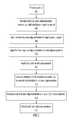
- FIG. 1is a flow chart describing one general aspect of the invention.
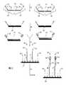
- FIG. 2illustrates an embodiment of a method of the invention that utilizes hybridization detection of loci.
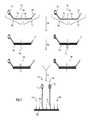
- FIG. 3illustrates an alternative embodiment of a method of the invention that utilizes hybridization detection of loci.
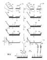
- FIG. 4illustrates another alternative embodiment of a method of the invention that utilizes hybridization detection of loci.
- FIG. 5illustrates yet another alternative embodiment of a method of the invention that utilizes hybridization detection of loci.
- FIG. 6illustrates another alternative embodiment of a method of the invention that utilizes bridging oligonucleotides in combination with fixed sequence oligonucleotides and hybridization detection of loci.
- FIG. 7illustrates another alternative embodiment of a method of the invention that utilizes hybridization detection of loci to detect polymorphisms.
- FIG. 8illustrates another alternative embodiment of a method of the invention that utilizes hybridization detection of loci to detect polymorphisms.
- FIG. 9illustrates another alternative embodiment of a method of the invention that utilizes hybridization detection of nucleic acid regions to detect polymorphisms.
- FIG. 10illustrates a method of the invention that utilizes hybridization detection of nucleic acid regions with a bridging oligonucleotide and dual cleavage.
- FIG. 11 of a method of the inventionthat utilizes hybridization detection of nucleic acid regions with a bridging oligonucleotide and dual cleavage to detect polymorphisms.
- FIG. 12illustrates a method of the invention that utilizes hybridization detection of nucleic acid regions resulting from a single cleavage event and employing differentially labeled universal primers.
- FIG. 13illustrates an alternative method to that illustrated in FIG. 12 also utilizing hybridization detection of nucleic acid regions resulting from a single cleavage event and employing differentially labeled universal primers.
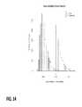
- FIG. 14shows the distribution of assay variability across samples for arrays and next generation sequencing.
- allele indexrefers generally to a series of nucleotides that corresponds to a specific SNP.
- the allele indexmay contain additional nucleotides that allow for the detection of deletion, substitution, or insertion of one or more bases.
- the allele indexmay be combined with any other index to create one index that provides information for two properties (e.g., sample-identification index, allele-locus index).
- Arrayrefers to a solid phase support having a surface, preferably but not exclusively a planar or substantially planar surface, which carries an array of sites containing nucleic acids such that each site of the array comprises substantially identical or identical copies of oligonucleotides or polynucleotides and is spatially defined and not overlapping with other member sites of the array; that is, the sites are spatially discrete.
- the array or microarraycan also comprise a non-planar interrogatable structure with a surface such as a bead or a well.
- the oligonucleotides or polynucleotides of the arraymay be covalently bound to the solid support, or may be non-covalently bound.
- array analysisrefers to analysis, such as, e.g., sequence analysis, of one or more biological molecules using an array.
- arrayrefers to any format of arrayed solid substrates, including a microarray, arrayed beads, an array of molecules within wells, or “liquid” arrays.
- binding pairmeans any two molecules that specifically bind to one another using covalent and/or non-covalent binding, and which can be used, e.g., for attachment of genetic material to a substrate.
- examplesinclude, but are not limited to, ligands and their protein binding partners, e.g., biotin and avidin, biotin and streptavidin, an antibody and its particular epitope, and the like.
- chromosomal abnormalityrefers to any genetic variant for all or part of a chromosome.
- the genetic variantsmay include but are not limited to any copy number variant such as duplications or deletions, translocations, inversions, and mutations.
- Complementaryor “complementarity” are used in reference to nucleic acid molecules (i.e., a sequence of nucleotides) that are related by base-pairing rules.
- Complementary nucleotidesare, generally, A and T (or A and U), or C and G.
- Two single stranded RNA or DNA moleculesare said to be substantially complementary when the nucleotides of one strand, optimally aligned and with appropriate nucleotide insertions or deletions, pair with at least about 90% to about 95% complementarity, and more preferably from about 98% to about 100% complementarity, and even more preferably with 100% complementarity.
- Selective hybridization conditionsinclude, but are not limited to, stringent hybridization conditions.
- Stringent hybridization conditionswill typically include salt concentrations of less than about 1 M, more usually less than about 500 mM and preferably less than about 200 mM.
- Hybridization temperaturesare generally at least about 2° C. to about 6° C. lower than melting temperatures (T m ).
- diagnostic toolrefers to any composition or system of the invention used in combination as, for example, in a system in order to carry out a diagnostic test or detection system on a patient sample.
- hybridizationgenerally means the reaction by which the pairing of complementary strands of nucleic acid occurs.
- DNAis usually double-stranded, and when the strands are separated they will re-hybridize under the appropriate conditions.
- Hybridscan form between DNA-DNA, DNA-RNA or RNA-RNA. They can form between a short strand and a long strand containing a region complementary to the short one. Imperfect hybrids can also form, but the more imperfect they are, the less stable they will be (and the less likely to form).
- ligaserefers generally to a class of enzymes, DNA ligases (typically T4 DNA ligase), which can link pieces of DNA together. The pieces must have compatible ends—either with both of them blunt or with mutually-compatible sticky ends—and the reaction requires ATP. “Ligation” is the process of joining two pieces of DNA together.
- locusand “loci” as used herein refer to a nucleic acid region of known location in a genome.
- maternal samplerefers to any sample taken from a pregnant mammal which comprises both fetal and maternal cell-free DNA.
- maternal samples for use in the inventionare obtained through relatively non-invasive means, e.g., phlebotomy or other standard techniques for extracting peripheral samples from a subject.
- oligonucleotidesor “oligos” as used herein refers to linear oligomers of natural or modified nucleic acid monomers, including deoxyribonucleotides, ribonucleotides, anomeric forms thereof, peptide nucleic acid monomers (PNAs), locked nucleotide acid monomers (LNA), and the like, or a combination thereof, capable of specifically binding to a single-stranded polynucleotide by way of a regular pattern of monomer-to-monomer interactions, such as Watson-Crick type of base pairing, base stacking, Hoogsteen or reverse Hoogsteen types of base pairing, or the like.
- PNAspeptide nucleic acid monomers
- LNAlocked nucleotide acid monomers
- oligonucleotidesranging in size from a few monomeric units, e.g., 8-12, to several tens of monomeric units, e.g., 100-200 or more.
- Suitable nucleic acid moleculesmay be prepared by the phosphoramidite method described by Beaucage and Carruthers (Tetrahedron Lett., 22:1859-1862 (1981)), or by the triester method according to Matteucci, et al. (J. Am. Chem. Soc., 103:3185 (1981)), both of which are incorporated herein by reference, or by other chemical methods such as using a commercial automated oligonucleotide synthesizer.
- nucleotiderefers to a base-sugar-phosphate combination. Nucleotides are monomeric units of a nucleic acid sequence (DNA and RNA).
- the term nucleotideincludes ribonucleoside triphosphates ATP, UTP, CTG, GTP and deoxyribonucleoside triphosphates such as dATP, dCTP, dITP, dUTP, dGTP, dTTP, or derivatives thereof.
- Such derivativesinclude, for example, [ ⁇ S]dATP, 7-deaza-dGTP and 7-deaza-dATP, and nucleotide derivatives that confer nuclease resistance on the nucleic acid molecule containing them.
- nucleotide as used hereinalso refers to dideoxyribonucleoside triphosphates (ddNTPs) and their derivatives.
- ddNTPsdideoxyribonucleoside triphosphates
- Illustrated examples of dideoxyribonucleoside triphosphatesinclude, but are not limited to, ddATP, ddCTP, ddGTP, ddITP, and ddTTP.
- polymeraserefers to an enzyme that links individual nucleotides together into a long strand, using another strand as a template.
- DNA polymeraseswhich synthesize DNA
- RNA polymeraseswhich synthesize RNA.
- sub-types of polymerasesdepending on what type of nucleic acid can function as template and what type of nucleic acid is formed.
- polymerase chain reactionrefers to a technique for replicating a specific piece of target DNA in vitro, even in the presence of excess non-specific DNA.
- Primersare added to the target DNA, where the primers initiate the copying of the target DNA using nucleotides and, typically, Taq polymerase or the like. By cycling the temperature, the target DNA is repetitively denatured and copied. A single copy of the target DNA, even if mixed in with other, random DNA, can be amplified to obtain billions of replicates.
- the polymerase chain reactioncan be used to detect and measure very small amounts of DNA and to create customized pieces of DNA. In some instances, linear amplification methods may be used as an alternative to PCR.
- polymorphismrefers to any genetic changes or variants in a loci that may be indicative of that particular loci, including but not limited to single nucleotide polymorphisms (SNPs), methylation differences, short tandem repeats (STRs), and the like.
- SNPssingle nucleotide polymorphisms
- STRsshort tandem repeats
- a “primer”is an oligonucleotide used to, e.g., prime DNA extension, ligation and/or synthesis, such as in the synthesis step of the polymerase chain reaction or in the primer extension techniques.
- a primermay also be used in hybridization techniques as a means to provide complementarity of a nucleic acid region to a capture oligonucleotide for detection of a specific nucleic acid region.
- search toolrefers to any composition or system of the invention used for scientific enquiry, academic or commercial in nature, including the development of pharmaceutical and/or biological therapeutics.
- the research tools of the inventionare not intended to be therapeutic or to be subject to regulatory approval; rather, the research tools of the invention are intended to facilitate research and aid in such development activities, including any activities performed with the intention to produce information to support a regulatory submission.
- samplerefers to any sample comprising all or a portion of the genetic information of an organism, including but not limited to virus, bacteria, fungus, plants and animals, and in particular mammals.
- the genetic information that can be interrogated within a genetic sampleincludes genomic DNA (both coding and non-coding regions), mitochondrial DNA, RNA, and nucleic acid products derived from each of these.
- nucleic acid productsinclude cDNA created from mRNA or products of pre-amplification to increase the material for analysis.
- target genomic regionrefers to all or a portion of a chromosome or chromosomes, including complete chromosomes, sub-chromosomal regions, groups of loci and single loci.
- the inventionprovides assay methods to identify copy number variants of nucleic acid regions (including loci, sets of loci and larger target genomic regions, e.g., chromosomes), including insertions, deletions, translocations, mutations and polymorphisms in a genetic sample.
- the assay methodsinterrogate loci from two or more target genomic regions in a sample using a directed ligation assay followed by detection of labelled oligonucleotides attached to an array.
- Quantification of the labelled oligonucleotidesallows determination of an atypical copy number of a particular target genomic region based on a comparison between the quantities of detected loci from the target genomic regions (e.g., comparison between two or more portions of a single chromosome or comparison between two or more different chromosomes) in the sample or by comparison to a reference chromosome from the same or a different sample.
- the methodemploys directed analysis of target genomic regions in a sample using sets of fixed sequence oligonucleotides that selectively hybridize to loci within two or more target genomic regions.
- the fixed sequence oligonucleotidesare directly or indirectly ligated to create ligation products.
- the ligation products corresponding to loci associated with a first target genomic regionare associated with a first detectable label and ligation products corresponding to loci associated with a second target genomic region are associated with a second detectable label. If the first and second detectable labels are quantified, the relative frequency of each of the first and second target genomic regions can be determined.
- the methodemploys two different labels that are used to identify two different target genomic regions.
- the methodemploys three different labels corresponding to three different target genomic regions, and so on.
- the ligation productsare detected by hybridization, and in particular by hybridization to an array of capture probes complementary to capture regions present in the ligation products.
- the ligation productsare detected using “universal arrays” that comprise features having the same or substantially similar capture probes.
- the arrayscomprises two or more sets of multiple features with a common sequence, with each set having a different sequence, e.g., an array where up to hundreds of the features on the array have substantially the same sequence.
- the capture probes on the arrayare complementary to the capture regions of the ligation products rather than to the sequence of the loci or their complements. These arrays can be used to interrogate any loci for any target genomic region(s) regardless of the sequence of the loci.
- the capture regionsare preferably introduced to the ligation products in the fixed sequence oligonucleotides that are used to interrogate the loci in the sample.
- the capture regionsare the same amongst all fixed sequence oligonucleotides used, so that ligation products or amplicons or cleavage products thereof from all loci hybridize competitively to capture probes of the same sequence on the array.
- the arrayis comprised of many different capture probes, and the sets of fixed sequence oligonucleotides from different loci comprise different capture regions.
- FIG. 1is a flow chart 100 illustrating an exemplary method of the invention.
- a sampleis provided.
- sets of fixed sequence oligonucleotides comprising a label binding regionare introduced to the sample under conditions that allow the fixed sequence oligonucleotides to hybridize to loci in target genomic regions, and in step 106 the oligonucleotides are hybridized to the target genomic regions.
- the hybridized fixed sequence oligonucleotides from each setare ligated to create ligation products which are then amplified in step 110 to produce amplicons complementary to the ligation products.
- step 112the amplicons are introduced to a hybridization array and allowed to hybridize competitively to capture probes on the array.
- step 114a set of labelled oligonucleotides are introduced to the amplicons and allowed to hybridize to complementary sequences on the amplicons.
- the labelled oligonucleotidesare ligated to the capture probe on the array (not shown).
- step 116the labels are detected.
- Each fixed sequence oligonucleotide of each setcomprises a region complementary to a selected locus (as described in more detail in FIG. 2 ). At least one fixed sequence oligonucleotide of each set further comprises a capture region, which may be the same for all sets of fixed sequence oligonucleotides used to interrogate two or more target genomic regions, may be the same for pairs of sets of fixed sequence oligonucleotides used to interrogate two or more target genomic regions, or may be different between sets of fixed sequence oligonucleotides for individual target genomic regions.
- one fixed sequence oligonucleotide of a setcomprises either a detectable label or a label binding region for association of the fixed sequence oligonucleotide with the detectable label.
- the label binding regioncan be a region complementary to a labeled oligonucleotide associated with a detectable label.
- the fixed sequence oligonucleotide of each set that comprises the capture regionwill not comprise the label or label binding region; that is, the other fixed sequence oligonucleotide of the set comprises the label or label binding region (see, e.g., exemplary embodiments illustrated in FIGS. 2, 3 and 5-8 ); in other embodiments, the fixed sequence oligonucleotide of each set that comprises the capture region also will comprise the label or label binding region (see, e.g., exemplary embodiment illustrated in FIG. 4 ).
- a first set of fixed sequence oligonucleotideshybridizes to loci in a first target genomic region while a second set of fixed sequence oligonucleotides hybridizes to loci in a second target genomic region.
- the ligation productsare optionally amplified using universal primers, and then are hybridized to an array.
- the amplification productis cleaved (e.g., using a restriction endonuclease) and a portion of the amplification product comprising the capture region is introduced to the array for hybridization and detection.
- each set of fixed sequence oligonucleotides used to interrogate a target genomic regioncontains the same label or label binding region. That is, all of the fixed sequence oligonucleotides of the first set are associated with a first label, all of the fixed sequence oligonucleotides of the second set are associated with a second label, and all of the fixed sequence oligonucleotides in a third set are associated with a third label.
- the ligation products, amplicons or cleavage products thereofcan be hybridized to the capture probes on the array and detected by readout from the labels.
- the fixed sequence oligonucleotidesinstead contain a label binding region that is complementary to a labeled oligonucleotide (a “label binding sequence”), a labeled oligonucleotide must be added to the ligation product or amplicons before detection. In either scenario, the labels are then detected and quantified and the relative frequency of each label determined. Quantifying each label allows for quantification of each target genomic region.
- all sets of first and second fixed sequence oligonucleotidescontain substantially the same capture region.
- the ligation products from each target genomic regioncompete to hybridize to the capture probes on a universal array.
- the capture probes on an arraycomprise multiple sequences complementary to different capture regions, and the array comprises features that contain these different capture probes.
- each capture probemay hybridize to an unique capture region.
- more than one capture probe, representing loci from different genomic regionsmay hybridize to a single capture region.
- different loci from the same or different target genomic regionsmay be configured to competitively hybridize against one another and thus would comprise the same capture region, while other loci from the same or different target genomic regions may be configured to competitively hybridize against one another, depending on the assay.
- the target genomic regionsmay be large genomic regions, such as whole chromosomes, or may be smaller genomic regions such as sub-regions of a single chromosome or sub-regions on different chromosomes, even down to a single locus.
- the inventionmay be used to detect genomic variations such as aneuploidies and partial aneuploidies, as well as mutations, SNPs, rearrangements, insertions and deletions.
- the first target genomic regionmay be, e.g., chromosome 21, and all loci to be interrogated with the first set of fixed sequence oligonucleotides will be from chromosome 21.
- the fixed oligonucleotidesmay be ligated to create ligation products which are associated with target genomic region-specific labels.
- the gapcan be closed using primer extension, and/or one or more bridging oligonucleotides.
- oligonucleotidesOnce the oligonucleotides are hybridized contiguously, either directly or following an extension operation or introduction of a bridging oligonucleotide, they may then be ligated to create ligation products which are associated with target genomic region-specific labels.
- a first set of fixed sequence oligonucleotidesare selective for a first chromosome or first target genomic region and a second set of fixed sequence oligonucleotides are selective for a different chromosome or second target genomic region.
- FIG. 2illustrates one embodiment in which each set of labeled fixed sequence oligonucleotides hybridize to loci on different chromosomes and ligation products are evaluated competitively on an array comprising capture probes.
- Two sets of labeled fixed sequence oligonucleotides 202 , 204are provided, each set having a first fixed sequence oligonucleotide 206 , 208 comprising a sequence that is complementary to a selected locus 210 , 212 and a label 214 , 216 and a second fixed sequence oligonucleotide 218 , 220 comprising a sequence complementary to the selected locus 222 , 224 and a capture region 226 , 228 .
- the labels 214 , 216are different for each set of fixed sequence oligonucleotides to allow differentiation between the first target genomic region (in this case, a first chromosome) and the second target genomic region (in this case, a second chromosome) during detection.
- the sets of fixed sequence oligonucleotides 202 , 204are introduced to a sample and allowed to hybridize to loci 232 , 234 on two different chromosomes. Following hybridization, the unhybridized fixed sequence oligonucleotides preferably are separated from the remainder of the sample (not shown).
- the fixed sequence oligonucleotidesare ligated to create ligation products 238 , 240 comprising capture regions 226 , 228 and labels 214 , 216 .
- the fixed sequence oligonucleotidesare illustrated in FIG. 2 as being hybridized adjacently in the loci, there may also be a gap that can be filled, e.g. using an extension reaction or using a bridging oligonucleotide that hybridizes adjacently between the fixed sequence oligonucleotides.
- the ligation products 238 , 240are introduced to a hybridization array 244 comprising a plurality of capture probes 246 wherein the capture regions 226 , 228 of the ligation products 238 , 240 competitively hybridize to the capture probes 246 .
- 226 and 228have substantially the same sequence.
- unhybridized ligation productspreferably are removed from the array (not shown).
- the labels 214 , 216can then be detected using an appropriate detection mechanism depending on the type of label used and the loci corresponding to each of the first and second chromosomes can be quantified to determine the presence and amount of each chromosome in the genetic sample.
- a “nucleotide”may be unlabeled or detectably labeled by well-known techniques. Fluorescent labels and their attachment to oligonucleotides are described in many reviews, including Haugland, Handbook of Fluorescent Probes and Research Chemicals, 9th Ed., Molecular Probes, Inc., Eugene Oreg. (2002); Keller and Manak, DNA Probes, 2nd Ed., Stockton Press, New York (1993); Eckstein, Ed., Oligonucleotides and Analogues: A Practical Approach , IRL Press, Oxford (1991); Wetmur, Critical Reviews in Biochemistry and Molecular Biology, 26:227-259 (1991); and the like.
- Labelingcan also be carried out with quantum dots, as disclosed in the following patents and patent publications: U.S. Pat. Nos. 6,322,901; 6,576,291; 6,423,551; 6,251,303; 6,319,426; 6,426,513; 6,444,143; 5,990,479; 6,207,392; 2002/0045045; and 2003/0017264.
- Detectable labelsinclude, for example, radioactive isotopes, fluorescent labels, chemiluminescent labels, bioluminescent labels and enzyme labels.
- Fluorescent labels of nucleotidesmay include but are not limited fluorescein, 5-carboxyfluorescein (FAM), 2′7′-dimethoxy-4′5-dichloro-6-carboxyfluorescein (JOE), rhodamine, 6-carboxyrhodamine (R6G), N,N,N′,N′-tetramethyl-6-carboxyrhodamine (TAMRA), 6-carboxy-X-rhodamine (ROX), 4-(4′dimethylaminophenylazo) benzoic acid (DABCYL), CASCADE BLUE® (pyrenyloxytrisulfonic acid), OREGON GREENTM (2′,7′-difluorofluorescein), TEXAS REDTM (sulforhodamine 101 acid chloride), Cyanine and 5-(2′-aminoethyl)aminonaphthalene-1-sulfonic acid (EDANS).
- fluroescently labeled nucleotidesinclude [R6G]dUTP, [TAMRA]dUTP, [R110]dCTP, [R6G]dCTP, [TAMRA]dCTP, [JOE]ddATP, [R6G]ddATP, [FAM]ddCTP, [R110]ddCTP, [TAMRA]ddGTP, [ROX]ddTTP, [dR6G]ddATP, [dR110]ddCTP, [dTAMRA]ddGTP, and [dROX]ddTTP available from Perkin Elmer, Foster City, Calif.
- nucleic acids from the sampleare associated with a substrate—e.g., using binding pairs such as, e.g., biotin and streptavidin, to attach the genetic material to a substrate surface or direct covalent attachment—before adding the sets of fixed sequence oligonucleotides to the sample.
- a first member of a binding paire.g., biotin
- a second member of the binding paire.g., avidin or streptavidin
- Attachment of the nucleic acids from the samplecan be particularly useful in removing unhybridized oligonucleotides following hybridization of the fixed sequence oligonucleotides and/or the bridging oligonucleotides to the loci.
- the nucleic acids from the samplecan be hybridized to the fixed sequence oligonucleotides, and then the hybridization complexes are subsequently bound to a substrate.
- the nucleic acids from the samplecan be attached to a solid support prior to hybridization of the fixed sequence oligonucleotides or at the same time.
- the surface of the supportcan be treated to remove any unhybridized or unligated oligonucleotides, e.g., by washing or other removal methods such as degradation of oligonucleotides as discussed in Willis et al., U.S. Pat. Nos. 7,700,323 and 6,858,412.
- Degradation of the oligonucleotidesis a preferred aspect when the two fixed sequence oligonucleotides are on the same probe such that ligation results in a circularized probe.
- Exonucleasesmay then be used to degrade non-circularized nucleic acids, including excess probes and sample DNA.
- nucleic acids with binding pairsthere are a number of methods that may be used to associate nucleic acids with binding pairs. For example, numerous methods may be used for labeling nucleic acids with biotin, including random photobiotinylation, end-labeling with biotin, replicating with biotinylated nucleotides, and replicating with a biotin-labeled primer.
- the number of loci analyzed for each chromosome in the methods of the inventionmay vary from two to 20,000 or more per target genomic region analyzed. In a preferred aspect, the number of loci per target genomic region is between 48 and 1000. In another aspect, the number of loci per target genomic region is at least 100. In another aspect, the number of loci per target genomic region is at least 400. In another aspect, the number of loci per target genomic region is no more than 1000. In another aspect, the number of loci per target genomic region is at least 500 but no more than 2000.
- a labelmay instead be provided by a separate, labeled oligonucleotide that is hybridized to the ligation products of the fixed sequence oligonucleotides or amplicons or cleaved amplicon (as described below) thereof, to allow detection.
- the labeled oligonucleotideis ligated to the capture probe following hybridization to the fixed sequence oligonucleotides or amplicons or cleaved amplicon.
- FIG. 3is an illustration of one embodiment of the invention in which the fixed sequence oligonucleotides are hybridized to loci of interest, ligated, amplified and introduced to an array prior to hybridization of a labeled oligonucleotide and detection of the label.
- two sets of fixed sequence oligonucleotides 302 , 304are provided, wherein the sets comprise a first fixed sequence oligonucleotide 306 , 308 each comprising sequences complementary to a selected locus 310 , 312 , label binding region 314 , 316 and universal primer regions 318 , 320 ; and a second fixed sequence oligonucleotide 322 , 324 comprising sequences complementary to the selected locus 326 , 328 , a capture region 330 , 332 and a universal primer region 334 , 336 .
- the capture region 330 , 332comprise substantially the same sequence and will both hybridize to the same capture probe on an array.
- the label binding regions 314 , 316comprise sequences that are different for each set of fixed sequence oligonucleotides 302 , 304 allowing differential labeling of the fixed sequence oligonucleotides associated with the loci for each target genomic region, while the capture regions 330 , 332 are the same for both sets of fixed sequence oligonucleotides 302 , 304 to allow for competitive hybridization of the ligation products to capture features on an array.
- the sets of fixed sequence oligonucleotides 302 , 304are introduced to a sample and allowed to hybridize to loci 340 , 342 of different target genomic regions. Following hybridization, unhybridized fixed sequence oligonucleotides preferably are separated from the remainder of the genetic sample (not shown).
- step 344the sets of fixed sequence oligonucleotides 302 , 304 are ligated to create ligation products 346 , 348 .
- step 358universal primers 350 , 352 , 354 and 356 are introduced to the ligation products 346 , 348 which bind to the universal primer regions 318 , 334 , 320 , and 336 , respectively, and create amplicons 360 , 362 each comprising capture regions 364 , 366 and label binding regions 368 , 370 .
- 350 and 354have substantially the same sequence, which is complementary to both 318 and 320
- 352 and 356have substantially the same sequence, which is complementary to both 334 and 336 .
- the ampliconsare introduced to a hybridization array 372 comprising a plurality of capture probes 374 wherein the capture regions 364 , 366 of the amplicons 360 , 362 hybridize to the capture probes 374 .
- labeled oligonucleotides 380 , 382are introduced to the array 372 where the label binding regions 368 , 370 of the amplicons 360 , 362 hybridize to target recognition regions 384 , 386 of the labeled oligonucleotides 380 , 382 .
- the labelled oligonucleotidesare ligated to the capture probe on the array (not shown).
- unhybridized labeled oligonucleotidespreferably are separated from the array (not shown).
- the labelscan then be detected and the loci corresponding to each target genomic region quantified to provide information on the presence and quantity of each target genomic region in the sample.
- labeled oligonucleotidesare hybridized to the fixed sequence oligonucleotides, or amplicons or cleaved amplicons thereof, after the ligation products or amplicons or cleaved amplicons are hybridized to an array.
- the labeled oligonucleotidesare hybridized to the fixed sequence oligonucleotides, or amplicons or cleaved amplicons thereof, prior to hybridization to an array.
- the size of the ligation products or ampliconsmay be reduced prior to hybridization to an array.
- the ligation productsare cleaved (e.g., using restriction endonucleases or other enzymatic cleaving mechanisms) to reduce the size of the ligation product to be detected, leaving, e.g., the capture region and the label binding region available for detection. Detection of a cleaved labeled ligation product or a cleaved amplicon serves as a surrogate in lieu of detecting the entire ligation product.
- Reducing the size of the ligation products, amplicons and/or labeled ligation productscan facilitate binding on the array, e.g., by improving hybridization kinetics and by decreasing steric hindrance.
- reduction in the size of the ligation productsis accomplished by cleaving the ligation products or amplicons using a restriction enzyme.
- one of the fixed sequence oligonucleotides in each set of fixed sequence oligonucleotidescomprises a restriction enzyme recognition site proximal to the capture region or label binding region (or the corresponding complementary sequences thereof depending on the embodiment).
- a restriction enzymecan be used to cleave the ligation product at the restriction enzyme recognition site leaving the label or label binding region and the capture region available for hybridization and detection.
- a restriction enzyme recognition sitemay be located in any position that leaves the capture region and the label binding region available for detection after cleavage.
- the restriction enzyme recognition sitemay be located directly next to the capture region or the label binding region or within a few bases from either of the capture region or label binding region.
- cleavageis carried out prior to hybridization of the labeled oligonucleotide to the ligation products or amplicons. In certain other aspects, cleavage occurs after hybridization to the array for detection.
- FIG. 4is an illustration of a specific embodiment of the invention in which the ligation product is cleaved prior to hybridization to an array.
- two sets of fixed sequence oligonucleotides 402 , 404are provided.
- Each setcomprises a first fixed sequence oligonucleotide 406 , 408 comprising sequences complementary to loci 410 , 412 , label binding regions 414 , 416 , capture regions 418 , 420 , universal primer regions 422 , 424 and restriction enzyme recognition site regions 426 , 428 .
- the label binding regions 414 , 416comprise sequences that are different for the sets of fixed sequence oligonucleotides 402 , 404 to allow differential labeling of fixed sequence oligonucleotides associated with each different target genomic region, while the capture regions 418 , 420 in this embodiment are the same for both sets of fixed sequence oligonucleotides 402 , 404 to allow for competitive hybridization to the capture features of a hybridization array.
- the restriction enzyme recognition sites 426 , 428can be the same for both sets of fixed sequence oligonucleotides 402 , 404 or different for each set depending on the embodiment.
- step 442the sets of fixed sequence oligonucleotides 402 , 404 are introduced to a sample and allowed to hybridize to elected loci 444 , 446 . Following hybridization and/or ligation, unhybridized fixed sequence oligonucleotides preferably are separated from the remainder of the sample (not shown).
- step 448the sets of fixed sequence oligonucleotides 402 , 404 are ligated to create ligation products 450 , 452 .
- step 454universal primers 456 , 458 , 460 and 462 are introduced to the ligation products 450 , 452 which bind to universal primer regions 422 , 438 , 424 , and 440 , respectively, to create amplicons 464 , 466 comprising label binding regions 472 , 474 , capture regions 468 , 470 and restriction enzyme recognition sites 476 , 478 .
- a restriction enzymeis introduced to the amplicons 464 , 466 which binds to the restriction enzyme recognition site 476 , 478 and cleaves the amplicons leaving a cleaved amplicon 482 , 484 comprising the label binding regions 472 , 474 and the capture regions 468 , 470 .
- the cleaved amplicons 482 , 484are introduced to a hybridization array 486 comprising a plurality of capture probes 488 where the capture regions 468 , 470 of the cleaved amplicons 482 , 484 competitively hybridize to the capture probes 488 .
- labeled oligonucleotides 492 , 494are introduced to the array 486 where the label binding regions 472 , 474 of each cleaved amplicon 482 , 484 hybridize to target recognition regions 496 , 497 of the labeled oligonucleotides 492 , 494 .
- unhybridized labeled oligonucleotidespreferably are separated from the array (not shown).
- the labels 498 , 499 of the labeled oligonucleotides 492 , 494can then be detected and the cleaved amplicons corresponding to each target genomic region can be quantified to provide information on the presence and amount of the target genomic regions in the sample.
- labeled oligonucleotides 492 , 494abut capture probes 488 , and thus labeled oligonucleotides 492 , 494 can be ligated to capture probes 488 to, e.g., increase binding stability.
- Cleaved amplicons 482 , 484can then be eliminated from the array by washing.
- FIG. 5is an illustration of another specific embodiment of the invention in which the ligation product is cleaved prior to hybridization to an array.
- two sets of fixed sequence oligonucleotides 502 , 504are provided. Each set comprises a first fixed sequence oligonucleotide 506 , 508 comprising sequences complementary to loci 510 , 512 , label binding regions 514 , 516 , capture regions 518 , 520 , universal primer regions 522 , 524 and restriction enzyme recognition site regions 526 , 528 .
- the label binding regions 514 , 516comprise sequences that are different for the sets of fixed sequence oligonucleotides 502 , 504 to allow differential labeling of fixed sequence oligonucleotides associated with each different target genomic region, while the capture regions 518 , 520 in this embodiment are the same for both sets of fixed sequence oligonucleotides 502 , 504 to allow for competitive hybridization to the capture features of a hybridization array.
- the restriction enzyme recognition sites 526 , 528can be the same for both sets of fixed sequence oligonucleotides 502 , 504 or different for each set depending on the embodiment.
- step 542the sets of fixed sequence oligonucleotides 502 , 504 are introduced to a sample and allowed to hybridize loci 544 , 546 . Following hybridization, unhybridized fixed sequence oligonucleotides preferably are separated from the remainder of the sample (not shown).
- step 548the sets of fixed sequence oligonucleotides 502 , 504 are ligated to create ligation products 550 , 552 .
- step 554universal primers 556 , 558 , 560 and 562 are introduced to the ligation products 550 , 552 which bind to universal primer regions 522 , 538 , 524 , and 540 , respectively, to create amplicons 564 , 566 comprising label binding regions 572 , 574 , capture regions 568 , 570 and restriction enzyme recognition sites 576 , 578 .
- a restriction enzymeis introduced to the amplicons 564 , 566 which binds to the restriction enzyme recognition site 576 , 578 and cleaves the amplicons leaving a cleaved amplicon 582 , 584 comprising the label binding regions 572 , 574 and the capture regions 568 , 570 .
- the cleaved productsare bound 580 to their respective labels 582 , 584 and introduced to a hybridization array 586 comprising a plurality of capture probes 588 where the capture regions 568 , 570 of the cleaved amplicons competitively hybridize 588 to capture probes 590 .
- the labeled oligonucleotidesmay be ligated to the capture probes of the hybridization array to increase the binding stability.
- This embodimentrequires juxtaposition of the labelled oligonucleotide and the capture probe.
- the capture probes, cleaved ligation products and labeled oligonucleotidesmay be configured so that the labeled oligonucleotides and capture probes are hybridized adjacently to one another, or the labeled oligonucleotides and the capture probes can be extended or a gap-filling oligonucleotide may be employed as described elsewhere herein.
- the inventionprovides methods to identify copy number variants of target genomic regions (relatively short genomic regions, larger genomic regions, sub-chromosomal regions and chromosomes), mutations, and polymorphisms in a sample.
- target genomic regionsrelatively short genomic regions, larger genomic regions, sub-chromosomal regions and chromosomes
- mutationsand polymorphisms in a sample.
- the present inventionprovides particularly powerful methods for identifying fetal chromosomal aneuploidies in maternal samples comprising both maternal and fetal DNA.
- the methods of the inventioncan also be used to analyze multiple loci on multiple chromosomes and average the frequency of the loci from a particular chromosome together. Normalization or standardization of the frequencies can be performed for one or more target sequences.
- the methods of the inventioncan be used to sum the frequencies of the loci on each chromosome for both sources in a mixed sample such as a maternal serum sample, e.g., by detecting an overall signal of a label, and then comparing the sum of the labels from the loci on one chromosome to the sum of the labels from the loci on another chromosome to determine whether a chromosomal abnormality exists.
- a mixed samplesuch as a maternal serum sample
- the data used to determine the frequency of the locimay exclude outlier data that appear to be due to experimental error, or that have elevated or depressed levels based on an idiopathic genetic bias within a particular sample.
- the data used for summationmay exclude DNA regions with a particularly elevated frequency in one or more samples.
- the data used for summationmay exclude loci that are found in a particularly low abundance in one or more samples.
- Subsets of locican be chosen randomly but with sufficient numbers to yield a statistically significant result in determining whether a chromosomal abnormality exists.
- Multiple analyses of different subsets of locican be performed within a mixed sample and on different arrays to yield more statistical power. For example, if there are 100 loci for chromosome 21 and 100 loci for chromosome 18, a series of analyses could be performed that evaluate fewer than 100 loci for each of the chromosomes on different arrays. In this example, target sequences are not being selectively excluded.
- the quantity of different loci detectable on certain chromosomesmay vary depending upon a number of factors, including general representation of fetal loci in maternal samples, degradation rates of the different loci representing fetal loci in maternal samples, sample preparation methods, and the like.
- the methods of the inventionemploy tandem ligation methods comprising the use of first and second fixed sequence oligonucleotides that are complimentary to loci in a target genomic region, e.g., a chromosome of interest or a reference chromosome, and one or more short, bridging oligonucleotides (also called “splint” oligonucleotides or “gap” or “gap-filling” oligonucleotides) complementary to the regions of the loci between and immediately adjacent to the first and second fixed sequence oligonucleotides.
- first and second fixed sequence oligonucleotidesthat are complimentary to loci in a target genomic region, e.g., a chromosome of interest or a reference chromosome
- bridging oligonucleotidesalso called “splint” oligonucleotides or “gap” or “gap-filling” oligonucleotides
- a tandem ligation assaytends to be more discriminating than use of a single oligonucleotide probe or use of only two fixed oligonucleotide probes, as perfect complementarity between the fixed sequence and bridging oligonucleotides and the selected locus must exist at both ligation sites for ligation to occur.
- a single bridging oligonucleotideis used which hybridizes adjacently between the fixed sequence oligonucleotides in the tandem ligation methods.
- the tandem ligation methodsuse sets of two fixed sequence oligonucleotides with a set of two or more bridging oligonucleotides that hybridize adjacently in the region of the nucleic acid between the region complementary to the first and second fixed sequence oligonucleotides.
- These bridging oligonucleotideshybridize adjacent to one another and to the fixed sequence oligonucleotides.
- the two fixed sequence oligonucleotides and two bridging oligonucleotidesare ligated during the ligation reaction, resulting in a single ligation product which serves as a template for amplification and, ultimately for detection.
- the methodemploys sets of fixed sequence oligonucleotides that bind to non-adjacent regions within a selected locus, and primer extension is utilized to create a contiguous set of hybridized oligonucleotides prior to the ligation step.
- the methodsmay employ sets of fixed sequence oligonucleotides, one or more bridging oligonucleotides and extension of one or more of the hybridized oligonucleotides. The combination of extension and ligation provides a ligation product which in turn serves as a template for amplification, followed by detection and quantification.
- the methods of the inventionemploy a multiplexed reaction with a set of three or more oligonucleotides for each selected locus.
- This general aspectis illustrated in FIG. 6 in which two sets of fixed sequence oligonucleotides 602 , 604 are provided.
- Each set of fixed sequence oligonucleotides 602 , 604comprises first fixed sequence oligonucleotides 606 , 608 each comprising a sequence complementary to a nucleic acid region of interest 610 , 612 , a label binding region 614 , 616 and a universal primer region 618 , 620 , and second fixed sequence oligonucleotides 622 , 624 each comprising a sequence complementary to the nucleic acid region of interest 626 , 628 , a capture region 630 , 632 and a universal primer region 634 , 636 .
- the sets of fixed sequence oligonucleotides 602 , 604are introduced to a sample and allowed to specifically hybridize to complementary portions of loci 640 , 642 in the target genomic regions.
- unhybridized fixed sequence oligonucleotidespreferably are separated from the remainder of the genetic sample (not shown). The removal of the unhybridized fixed sequence oligonucleotides may alternatively be separated from the remainder of the genetic sample after the ligation step below.
- step 644sets of bridging oligonucleotides 646 , 648 are introduced and allowed to specifically hybridize to the region of the loci between the first fixed sequence oligonucleotide 606 , 608 and second fixed sequence oligonucleotide 622 , 624 .
- the bridging oligonucleotides 646 , 648can be introduced simultaneously with the fixed sequence oligonucleotides.
- step 650the sets of fixed sequence oligonucleotides 602 , 604 and bridging oligonucleotides 630 , 632 are ligated to create ligation products 652 , 654 complementary to the loci 640 , 642 .
- universal primers 658 , 660 , 662 and 664are introduced to the ligation products 652 , 654 where the universal primers bind to the universal primer regions 618 , 634 , 620 , and 636 , respectively, and amplify the ligation products to create amplicons 678 , 680 comprising label binding regions 666 , 668 and capture regions 662 , 664 .
- universal primers 658 and 662have substantially the same sequence, which is complementary to both 618 and 620
- universal primers 660 and 664have substantially the same sequence, which is complementary to both 634 and 636 .
- the amplicons 678 , 680are introduced to a hybridization array 670 comprising a plurality of capture probes 672 wherein the capture regions 662 , 664 of the amplicons 658 , 660 competitively hybridize to target capture regions 674 on the capture probes 672 .
- target recognition regions 682 , 684 of the labeled oligonucleotides 688 , 690specifically hybridize to the label binding regions 666 , 668 of the amplicons 678 , 680 .
- unhybridized labeled oligonucleotidespreferably are removed from the array (not shown).
- the labels 686 , 688 of the labeled oligonucleotides 688 , 690can then be detected and the loci optionally quantified to provide information on the presence and amount of each genomic region in the genetic sample.
- the bridging oligonucleotidescan be composed of a mixture of oligonucleotides with degeneracy in each of the positions, so that the mixture of bridging oligonucleotides will be compatible with all reactions in the multiplexed assay requiring a bridging of a given length.
- the bridging oligonucleotideis a randomer, where all combinations of the bridging oligonucleotide are synthesized.
- the bridging oligonucleotidescan be of various lengths so that the mixture of oligonucleotides is compatible with ligation reactions requiring bridging oligonucleotides of different lengths.
- the bridging oligonucleotidecan have degeneracy at specific positions—i.e., known polymorphic sites—and the tandem ligation reactions are restricted to those that require a specific sequence provided at the polymorphic site.
- the bridging oligonucleotideis specific, synthesized to match the sequences in the gap.
- the number of unique oligonucleotides provided in the assaywould be equal to or less than the number of loci.
- a number of bridging oligonucleotides less than the number of locicould be achieved if the gap sequence was shared between two or more loci.
- sequences of the bridging oligonucleotidesare designed and the loci are selected so that all loci share the same base(s) at each end of the bridging oligonucleotide. For instance, one might choose loci and their gap location such that all of the gaps shared an “A” base at the first position and a “G” base at the last position of the gap. Any combination of a first and last base could be utilized, based upon factors such as the genome investigated, the likelihood of sequence variation in that area, and the like.
- the bridging oligonucleotidescan be synthesized by random degeneracy of bases at the internal positions of the bridging oligonucleotide, with nucleotides specific at the first and last position of the bridging oligonucleotide.
- the second, third and fourth positionswould be degenerate, and the two specific nucleotides at the end of the bridging oligonucleotide would be fixed.
- the frequency of the dinucleotide CGis much lower than expected by the respective mononucleotide frequencies. This presents an opportunity to enhance the specificity of an assay with a particular mixture of bridging oligonucleotides.
- the bridging oligonucleotidesmay be selected to have a 5′ G and a 3′ C. This base selection allows each oligonucleotide to have a high frequency in the human genome but makes it a rare event for two bridging oligonucleotides to hybridize adjacent to each other. The probability is then reduced that multiple oligonucleotides are ligated in locations of the genome that are not targeted in the assay.
- the bridging oligonucleotideis added to the reaction after the sets of fixed sequence oligonucleotides have been hybridized, and following the optional removal of all unhybridized fixed sequence oligonucleotides.
- the conditions of the hybridization reactionpreferably are optimized near the T m of the bridging oligonucleotide to prevent erroneous hybridization of oligonucleotides that are not fully complementary to the nucleic acid region. If the bridging oligonucleotides have a T m significantly lower than the fixed sequence oligonucleotides, the bridging oligonucleotide is preferably added as a part of the ligase reaction.
- the advantage of using short bridging oligonucleotidesis that ligation on either end would likely occur only when all bases of the bridging oligonucleotide match the gap sequence.
- a further advantage of using short bridging oligonucleotidesis that the number of different bridging oligonucleotides necessary could be less than the number of loci, raising the oligonucleotides' effective concentration to allow perfect matches to happen faster. Fewer numbers of bridging oligonucleotides provides the advantages of cost savings and quality control.
- the advantages of using fixed first and last bases and random bases in betweeninclude the ability to utilize longer bridging oligonucleotides for greater specificity while reducing the number of total bridging oligonucleotides in the reaction.
- the methods of the inventiondetect one or more target genomic regions that comprise a polymorphism.
- this methodologyis not necessarily designed to identify a particular allele, e.g., as maternal versus fetal, but rather to ensure that different alleles corresponding to loci in a target genomic region are included in the quantification methods of the invention.
- the methods of the inventionare intended to encompass both mechanisms for detection of SNP-containing loci for direct determination of copy number variation through quantification as well as detection of SNPs for ensuring overall efficiency of the assay.
- allelic discriminationis provided through the fixed sequence oligonucleotides or through the bridging oligonucleotides used for detection of the loci.
- the label binding regionmay serve as an allele index that is embedded in either the first fixed sequence oligonucleotide or the second fixed sequence oligonucleotide in a set.
- an allele indexe.g., label binding region
- the number of fixed sequence oligonucleotides used in such aspectscan correspond to the number of possible alleles being assessed for a selected locus, and detection of a label associated with the allele index can detect presence, amount or absence of a specific allele in a genetic sample.
- FIG. 7illustrates one aspect of the invention in which polymorphisms are detected using competitive hybridization to an array.
- two sets of fixed sequence oligonucleotides 702 , 704are provided in which the first fixed sequence oligonucleotide 706 , 708 of each set comprises a sequence that is complementary to a locus of interest 710 , 712 comprising for example an A/T or G/C SNP, respectively and a label 714 , 716 .
- the second fixed sequence oligonucleotide 722 , 724comprises a sequence that is complementary to the selected locus 726 , 728 and a capture region 730 , 732 .
- fixed sequence oligonucleotide 706is the same as fixed sequence oligonucleotide 708 except for SNPs 718 and 720 and labels 714 and 716 , respectively; and fixed sequence oligonucleotide 722 is the same as fixed sequence oligonucleotide 724 . That is, the difference between the sets of fixed sequence oligonucleotides is the SNP interrogated and the label that corresponds to the SNP.
- the sets of fixed sequence oligonucleotides 702 , 704are introduced to the sample and allowed to specifically hybridize to locus of interest 736 , 738 comprising SNPs 740 , 742 .
- unhybridized fixed sequence oligonucleotidespreferably are separated from the remainder of the genetic sample (not shown).
- the sets fixed sequence oligonucleotides 702 , 704are ligated to create ligation products 746 , 748 .
- the ligationis allele-specific as long as the allele-specific nucleotide is close to the ligation junction.
- the allele-specific nucleotidemust be within 5 nucleotides of the ligated end; however, in preferred aspects, the allele-specific nucleotide is the terminal base.
- the ligation products 746 , 748are introduced to a hybridization array 752 comprising a plurality of capture probes 754 where the capture regions 730 , 732 of the ligation products 746 , 748 hybridize to the capture probes 754 .
- unhybridized ligation productspreferably are separated from the remainder of the genetic sample (not shown).
- the labels 714 , 716can then be detected and alleles specific to the loci corresponding to each target genomic region are quantified to provide information on the presence and amount each allele and target genomic region in the genetic sample.
- the allele-specific nucleotideis disposed in the first and/or second fixed sequence oligonucleotides and bridging oligonucleotides are used to create ligation products.
- FIG. 8illustrates this aspect of the invention.
- two sets of fixed sequence oligonucleotides 802 , 804are provided, where both sets of fixed sequence oligonucleotides are configured to interrogate different SNPs at the same selected locus.
- the first fixed sequence oligonucleotides 806 , 808 of each setcomprise a sequence that is complementary to a selected locus 810 , 812 comprising for example an A/T or G/C SNP, respectively, a label binding region 814 , 816 , and a universal primer region 818 , 820 ; and second fixed sequence oligonucleotides 826 , 828 each comprising sequences that are complementary to a selected locus 830 , 832 , capture region 834 , 836 and universal primer regions 838 , 840 .
- second fixed sequence oligonucleotides 826 and 828have substantially the same sequence and first fixed sequence oligonucleotides 806 , 808 have substantially the same sequence except for the SNP site and the label binding regions 814 , 816 .
- the sets of fixed sequence oligonucleotides 802 , 804are introduced to the sample and are allowed to hybridize to the loci 844 , 846 comprising SNPs 848 , 850 .
- unhybridized fixed sequence oligonucleotidespreferably are separated from the remainder of the genetic sample (not shown).
- a set of bridging oligonucleotides 854 , 856are introduced to the sample and allowed to hybridize to the locus 844 , 846 between the first and second fixed oligonucleotides of each set.
- 854 and 856have substantially the same sequence.
- the hybridized fixed sequence oligonucleotides and hybridized bridging oligonucleotidesare ligated to create ligation products 860 , 862 .
- step 864universal primers 866 , 868 , 870 and 872 are introduced to the ligation products 860 , 862 which bind to universal primer regions 818 , 838 , 820 , and 840 , respectively and amplify the ligation products 860 , 862 to produce amplicons 874 , 876 comprising label binding regions 882 , 884 and capture regions 878 , 880 .
- the ampliconsare introduced to a hybridization array 886 comprising capture probes 888 where the capture regions 878 , 880 of the amplicons 874 , 876 hybridize to the capture probes 888 .
- labeled oligonucleotides 894 , 895are introduced to the hybridization array 886 under conditions that allow the target recognition regions 896 , 897 of the labeled oligonucleotides 894 , 895 to selectively hybridize to the label binding regions 882 , 884 of the amplicons 874 , 876 .
- unhybridized labeled oligonucleotidespreferably are removed from the array (not shown).
- the labels 898 , 899can then be detected and alleles corresponding to the loci corresponding to the target genomic regions quantified to provide information on the presence and amount of the alleles at loci and the target genomic regions in the genetic sample.
- allelic discriminationis provided through the bridging oligonucleotide.
- the bridging oligonucleotideis located over a SNP, preferably located close enough to one end of a ligation junction so as to provide allelic-specificity.
- both allele bridging oligonucleotide variantsare present in the same reaction mixture and allele detection results from subsequent hybridization of associated labels to hybridization arrays.
- FIG. 9illustrates this aspect.
- two sets of fixed sequence oligonucleotides 902 , 904are provided where both sets of fixed sequence oligonucleotides are configured to interrogate the same selected locus and are the same except for the label binding region.
- the bridging oligonucleotideinterrogates the SNP, and the assay must be performed in two separate vessels at least until after the ligation step has taken place.
- the first fixed sequence oligonucleotides 906 , 908 in each setcomprise sequences that are complementary to a locus 910 , 912 , a capture region 914 , 916 and universal primer region 918 , 920 .
- the second fixed sequence oligonucleotides 922 , 924 of each setcomprise sequences that are complementary to the locus 926 , 928 , a label binding region 930 , 932 region and a universal primer region 934 , 936 .
- the sets of fixed sequence oligonucleotides 902 , 904are introduced to the sample under conditions that allow each set 902 , 904 to specifically hybridize to the locus 940 , 942 wherein the selected locus comprises a SNP 944 , 946 .
- unhybridized fixed sequence oligonucleotidespreferably are separated from the remainder of the genetic sample (not shown).
- bridging oligonucleotides 950 , 952 corresponding to an A/T SNP or a G/C SNPare introduced and allowed to bind to the region of the locus 940 , 942 between the first and second fixed sequence oligonucleotides.
- the bridging oligonucleotidescan be introduced to the sample simultaneously with the fixed sequence oligonucleotides.
- step 954the hybridized fixed sequence oligonucleotides and bridging oligonucleotides are ligated to create ligation products 956 , 958 .
- the two separate assaysoptionally can be combined into a single vessel but need not be.
- step 960universal amplification primers 962 , 964 , 966 and 968 are introduced to the ligation products 956 , 958 where the universal primers bind to universal primer regions 918 , 934 , 920 , and 936 , respectively, and amplify the ligation products 956 , 958 to produce amplicons 970 , 972 , each comprising a label binding region 980 , 982 and a capture region 974 , 976 .
- the amplicons 970 , 972are introduced to a hybridization array 984 comprising a plurality of capture probes 986 wherein the capture regions 974 , 976 of the amplicons 970 , 972 competitively hybridize to the capture probes 986 .
- labeled oligonucleotides 992 , 994are introduced to the array 984 where target recognition regions 996 , 997 of the labeled oligonucleotides 992 , 994 hybridize to label binding regions 980 , 982 of the amplicons 970 , 972 .
- unhybridized labeled oligonucleotidespreferably are removed from the array (not shown).
- the labels 998 , 999can then be detected and alleles corresponding to the loci corresponding in turn to the target genomic regions are quantified to provide information on the presence and amount of each allele and target genomic region in the sample.
- FIG. 10is an illustration of another specific embodiment of the invention in which bridging oligonucleotides are used and the ligation products are dually cleaved prior to hybridization to an array.
- two sets of fixed sequence oligonucleotides 1002 , 1004are provided. Each set comprises a first fixed sequence oligonucleotide 1006 , 1008 comprising sequences complementary to loci 1010 , 1012 , label binding regions 1018 , 1020 , capture regions 1014 , 1016 , universal primer regions 1022 , 1024 and restriction enzyme recognition site regions 1026 , 1028 .
- the label binding regions 1018 , 1020comprise sequences that are different for the sets of fixed sequence oligonucleotides 1002 , 1004 to allow differential labeling of fixed sequence oligonucleotides associated with each different target genomic region, while the capture regions 1014 , 1016 in this embodiment are the same for both sets of fixed sequence oligonucleotides 1002 , 1004 to allow for competitive hybridization to the capture features of a hybridization array.
- the restriction enzyme recognition sites 1026 , 1028can be the same for both cleavage sites on each fixed sequence oligonucleotide, and/or the same for the different sets of fixed sequence oligonucleotides 1002 , 1004 depending on the embodiment.
- each second fixed sequence oligonucleotide 1030 , 1032comprises a sequence complementary to the loci 1034 , 1036 and a universal primer region 1038 , 1040 .
- step 1042the sets of fixed sequence oligonucleotides 1002 , 1004 are introduced to a sample and allowed to hybridize loci 1044 , 1046 . Following hybridization (or alternatively ligation), unhybridized fixed sequence oligonucleotides preferably are separated from the remainder of the sample (not shown).
- step 1048a bridging oligonucleotide 1026 , 1028 is added to each set and hybridized adjacently between the hybridized fixed sequence oligonucleotides.
- the sets of oligonucleotidesare ligated to create ligation products 1050 , 1052 .
- step 1054universal primers 1056 , 1058 , 1060 and 1062 are introduced to the ligation products 1050 , 1052 which bind to universal primer regions 1022 , 1038 , 1024 , and 1040 , respectively, to create amplicons 1064 , 1066 comprising label binding regions 1018 , 1020 , capture regions 1014 , 1016 , and restriction enzyme recognition sites 1072 , 1074 , 1076 , and 1078 .
- step 1070one or more restriction enzymes are introduced to the amplicons 1064 , 1066 and the amplicons are dually cleaved to create a cleaved amplicon comprising the label binding regions 1018 , 1020 and the capture regions 1014 , 1016 .
- the cleaved productsare introduced to a hybridization array 1086 comprising a plurality of capture probes 1088 where the capture regions 1014 , 1016 of the cleaved amplicons competitively hybridize to capture probes 1090 .
- the hybridized capture regionsare then introduced 1088 to the labeled oligonucleotides 1082 , 1084 , which bind to complementary sequences on the cleaved products and are detected.
- FIG. 11is an illustration of another specific embodiment of the invention in which bridging oligonucleotides are used to identify different alleles in the same locus and the ligation products are dually cleaved prior to hybridization to an array.
- two sets of fixed sequence oligonucleotides 1102 , 1104are provided for a single locus having a possible polymorphism.
- Each setcomprises a first fixed sequence oligonucleotide 1106 , 1108 comprising sequences complementary to one allele of the selected locus 1110 , 1112 , label binding regions 1118 , 1120 , capture regions 1114 , 1116 , universal primer regions 1122 , 1124 and restriction enzyme recognition site regions 1126 , 1128 .
- the label binding regions 1118 , 1120comprise sequences that are different for the sets of fixed sequence oligonucleotides 1102 , 1104 to allow differential labeling of fixed sequence oligonucleotides associated with each different allele, while the capture regions 1114 , 1116 in this embodiment are the same for both sets of fixed sequence oligonucleotides 1102 , 1104 to allow for competitive hybridization to the capture features of a hybridization array.
- the restriction enzyme recognition sites 1126 , 1128can be the same for both cleavage sites on each fixed sequence oligonucleotide, and/or the same for the different sets of fixed sequence oligonucleotides 1102 , 1104 depending on the embodiment.
- each second fixed sequence oligonucleotide 1130 , 1132comprises a sequence complementary to the selected locus 1134 , 1136 and a universal primer region 1138 , 1140 .
- step 1142the sets of fixed sequence oligonucleotides 1102 , 1104 are introduced to a sample and allowed to hybridize to the selected locus 1144 , 1146 . Following hybridization (or alternatively ligation), unhybridized fixed sequence oligonucleotides preferably are separated from the remainder of the sample (not shown).
- step 1148a bridging oligonucleotide 1126 , 1128 is added to each set and hybridized adjacently between the hybridized fixed sequence oligonucleotides.
- the sets of oligonucleotidesare ligated to create ligation products 1150 , 1152 .
- step 1154universal primers 1156 , 1158 , 1160 and 1162 are introduced to the ligation products 1150 , 1152 which bind to universal primer regions 1122 , 1138 , 1124 , and 1140 , respectively, to create amplicons 1164 , 1166 comprising label binding regions 1118 , 1120 , capture regions 1114 , 1116 , and restriction enzyme recognition sites 1172 , 1174 , 1176 , and 1178 .
- step 1170one or more restriction enzymes are introduced to the amplicons 1164 , 1166 and the amplicons dually cleaved to create a cleaved amplicon comprising the label binding regions 1118 , 1120 and the capture regions 1114 , 1116 .
- the cleaved productsare introduced to a hybridization array 1186 comprising a plurality of capture probes 1188 where the capture regions 1118 , 1120 of the cleaved amplicons competitively hybridize to capture probes 1190 .
- the hybridized capture regionsare then introduced 1188 to the labeled oligonucleotides 1182 , 1184 , which bind to complementary sequences on the cleaved products and are detected to differentiate between the different alleles of the locus.
- FIG. 12is an illustration of another specific embodiment of the invention in which bridging oligonucleotides are used and the ligation products are cleaved prior to hybridization to an array.
- the primers used to amplify the ligation productsare differentially labeled.
- two sets of fixed sequence oligonucleotides 1202 , 1204are provided. Each set comprises a first fixed sequence oligonucleotide 1206 , 1208 comprising sequences complementary to loci 1210 , 1212 , primer binding regions 1218 , 1224 (which are different), capture regions 1214 , 1220 , and restriction enzyme recognition site regions 1226 , 1228 .
- the primer binding regions 1218 , 1224comprise sequences that are different for the sets of fixed sequence oligonucleotides 1202 , 1204 to allow differential labeling of fixed sequence oligonucleotides associated with each different target genomic region, while the capture regions 1214 , 1220 in this embodiment are the same for both sets of fixed sequence oligonucleotides 1202 , 1204 to allow for competitive hybridization to the capture features of a hybridization array.
- the restriction enzyme recognition sites 1226 , 1228can be the same for both cleavage sites on each fixed sequence oligonucleotide, and/or the same for the different sets of fixed sequence oligonucleotides 1302 , 1304 depending on the embodiment.
- each second fixed sequence oligonucleotide 1230 , 1232comprises a sequence complementary to the loci 1234 , 1236 and a primer region 1238 , 1240 .
- step 1242the sets of fixed sequence oligonucleotides 1202 , 1204 are introduced to a sample and allowed to hybridize loci 1244 , 1246 . Following hybridization (or alternatively ligation), unhybridized fixed sequence oligonucleotides preferably are separated from the remainder of the sample (not shown).
- step 1248a bridging oligonucleotide 1226 , 1228 is added to each set and hybridized adjacently between the hybridized fixed sequence oligonucleotides.
- the sets of oligonucleotidesare ligated to create ligation products 1250 , 1252 .
- primers 1256 , 1258 , 1260 and 1262are introduced to the ligation products 1250 , 1252 which bind to primer regions 1218 , 1238 , 1224 , and 1240 , respectively, to create amplicons 1264 , 1266 comprising primer binding regions 1278 , 1284 , capture regions 1276 , 1280 , and restriction enzyme recognition sites 1272 and 1274 .
- Primers 1256 and 1260are differentially labeled thus allowing for differentiation between amplicons from the different loci once the amplicons are hybridized to an array.
- step 1270one or more restriction enzymes are introduced to the amplicons 1264 , 1266 and the amplicons are cleaved to create a cleaved amplicon comprising the primer binding regions 1278 , 1284 and the capture regions 1276 , 1280 .
- the cleaved productsare introduced to a hybridization array 1286 at step 1290 comprising a plurality of capture probes 1288 where the capture regions 1276 , 1280 of the cleaved amplicons competitively hybridize to capture probes 1288 .
- FIG. 13is an illustration of another specific embodiment of the invention in which bridging oligonucleotides are used and the ligation products are cleaved prior to hybridization to an array.
- FIG. 13illustrates an embodiment very similar to that in FIG. 12 .
- the primers used to amplify the ligation productsare differentially labeled.
- two sets of fixed sequence oligonucleotides 1302 , 1304are provided.
- Each setcomprises a first fixed sequence oligonucleotide 1306 , 1308 comprising sequences complementary to loci 1310 , 1312 , primer binding regions 1318 , 1324 (which are different), capture regions 1314 , 1320 (which are the same), and restriction enzyme recognition site regions 1326 , 1328 .
- the primer binding regions 1318 , 1324comprise sequences that are different for the sets of fixed sequence oligonucleotides 1302 , 1304 to allow differential labeling of fixed sequence oligonucleotides associated with each different target genomic region, while the capture regions 1314 , 1320 in this embodiment are the same for both sets of fixed sequence oligonucleotides 1302 , 1304 to allow for competitive hybridization to the capture features of a hybridization array.
- the restriction enzyme recognition sites 1326 , 1328can be the same for both cleavage sites on each fixed sequence oligonucleotide, and/or the same for the different sets of fixed sequence oligonucleotides 1302 , 1304 depending on the embodiment.
- each second fixed sequence oligonucleotide 1330 , 1332comprises a sequence complementary to the loci 1334 , 1326 and a primer region 1338 , 1340 .
- step 1342the sets of fixed sequence oligonucleotides 1302 , 1304 are introduced to a sample and allowed to hybridize loci 1344 , 1346 . Following hybridization (or alternatively ligation), unhybridized fixed sequence oligonucleotides preferably are separated from the remainder of the sample (not shown).
- step 1348a bridging oligonucleotide 1326 , 1328 is added to each set and hybridized adjacently between the hybridized fixed sequence oligonucleotides.
- the sets of oligonucleotidesare ligated to create ligation products 1350 , 1352 .
- primers 1356 , 1358 , 1360 and 1362are introduced to the ligation products 1350 , 1352 which bind to primer regions 1318 , 1338 , 1324 , and 1340 , respectively, to create amplicons 1364 , 1366 comprising primer binding regions 1378 , 1384 , capture regions 1376 , 1380 , and restriction enzyme recognition sites 1372 and 1374 .
- Primers 1356 and 1360are differentially labeled thus allowing for differentiation between amplicons from the different loci once the amplicons are hybridized to an array.
- step 1370one or more restriction enzymes are introduced to the amplicons 1364 , 1366 and the amplicons are cleaved to create a cleaved amplicon comprising the primer binding regions 1378 , 1384 and the capture regions 1376 , 1380 .
- the cleaved productsare introduced to a hybridization array 1386 at step 1390 comprising a plurality of capture probes 1388 where the capture regions 1376 , 1380 of the cleaved amplicons competitively hybridize to capture probes 1388 .
- the cleavage amplicons introduced to the arraydo not include any portion of the target genomic region or a sequence complementary thereof.
- the capture regionsare used in association with different target genomic regions, and the labeled oligonucleotide is used to differentiate between regions.
- the cleaved ampliconscompetitively hybridize to common capture probes, and the quantitation of the target genomic regions is determined by the detected labels corresponding to cleaved products that bind to the array.
- FIGS. 2-13depict two separate fixed sequence oligonucleotides used to interrogate each locus or allele.
- a single probecan be used which comprises two or more distinct non-adjacent fixed sequence oligonucleotides that are complementary to the loci including precircle probes.
- precircle probesare described, e.g., by Lizardi in U.S. Pat. Nos. 5,854,033 and 6,316,229, and can be linearized prior to hybridization to the array by, e.g., including a site for a restriction endonuclease in the probe.
- universal amplificationis used to amplify the ligation products following hybridization and ligation of the fixed sequence oligonucleotides, either directly or following extension or the introduction of a bridging oligonucleotide.
- amplificationpreferably is done through universal amplification using universal primers that hybridize to universal primer regions on the first and second fixed sequence oligonucleotide of each set.
- the universal primer regions from the fixed sequence oligonucleotidesbecome a part of the ligation products, where the ligation products can then be amplified in a single universal amplification reaction.
- these universal primer sequencespreferably are introduced via the fixed sequence oligonucleotides, they may also be added to the proximal ends of the ligation products via ligation.
- the introduction of universal primer regions to the fixed sequence oligonucleotidesallows a subsequent controlled universal amplification of all or a portion of the ligation products prior to array hybridization.
- the amplicon produced from this amplification processcan be used directly or further processed prior to introduction to the array for detection.
- the ampliconis cleaved, and the portion comprising the capture region is introduced to the array for hybridization and detection. Bias and variability can be introduced during DNA amplification, such as that seen during polymerase chain reaction (PCR).
- the universal primer regions used in the methodsare designed to be compatible with conventional multiplexed assay methods that utilize general priming mechanisms to analyze large numbers of nucleic acids simultaneously. Such “universal” priming methods allow for efficient, high volume analysis of the quantity of nucleic acid regions present in a genetic sample, and allow for comprehensive quantification of the presence of nucleic acid regions within such a sample.
- the entirety of a ligation reaction or an aliquot of the ligation reactionmay be used for universal amplification.
- Using an aliquotallows parallel amplification reactions to be undertaken using the same or different conditions (e.g., polymerase, buffers, and the like), e.g., to ensure that bias is not inadvertently introduced due to experimental conditions.
- variations in primer concentrationsmay be used to effectively limit the number of sequence specific amplification cycles. Examples of multiplexing assay methods include, but are not limited to, those described in Oliphant et al., U.S. Pat. No. 7,582,420.
- many methods of the inventionutilize coupled reactions for multiplex detection where oligonucleotides from an early phase of the multi-step process contain sequences that may be used in a later phase of the process.
- Processes known in the art for amplifying and/or detecting nucleic acids in samplescan be used, alone or in combination, including but not limited to the methods described below.
- the methods of the inventionutilize one of the following combined selective and universal amplification techniques: (1) LDR coupled to PCR; (2) primary PCR coupled to secondary PCR coupled to LDR; and (3) primary PCR coupled to secondary PCR.
- Each of these combinations of techniquescan utilize probe regions from an early phase in the process that may be used as a primer sequence in a later phase of the process.
- padlock probesalso called “precircle probes” or “multi-inversion probes”
- LDRcoupled ligase detection reaction
- PCRpolymerase chain reaction
- Ronaghi et al.U.S. Pat. No. 7,622,281 describes amplification techniques for labeling and amplifying a nucleic acid using an adapter comprising a unique primer and a barcode.
- the nucleic acid regions of interestare identified using hybridization techniques and arrays. Methods for conducting polynucleotide hybridization assays for detection are well-developed and well-known in the art. Hybridization assay procedures and conditions will vary depending on the application and are selected in accordance with the general binding methods known including those referred to in: Maniatis et al. Molecular Cloning: A Laboratory Manual (2 nd Ed. Cold Spring Harbor, N.Y., 1989); Berger and Kimmel Methods in Enzymology, Vol. 152, Guide to Molecular Cloning Techniques (Academic Press, Inc., San Diego, Calif., 1987); Young and Davis, P.N.A.S, 80: 1194 (1983).
- the arrayscomprise multiple substrates in solution, such as those taught, e.g., in U.S. Appln No. 20140057269 and U.S. Appln No. 20140042366 and U.S. Appln No. 20140024550.
- the present inventionalso contemplates signal detection of hybridization between ligands in certain preferred aspects. See U.S. Pat. Nos. 5,143,854, 5,578,832; 5,631,734; 5,834,758; 5,936,324; 5,981,956; 6,025,601; 6,141,096; 6,185,030; 6,201,639; 6,218,803; and 6,225,625, in U.S. Patent application 60/364,731 and in PCT Application PCT/US99/06097 (published as WO99/47964).
- the capture region of the ligation products from a single sample or the amplicons thereofcontain index sequences that identify the ligation products as being from a particular sample.
- the features of the arraywill comprise capture probes that include sequences complementary to the index sequences of the different samples to allow identification of the loci from a particular sample from which the loci originated.
- the proportion of fetal DNA in a maternal samplemay be used as a part of the risk calculation of the present invention, as fetal proportion provides important information on the expected statistical presence of chromosomal dosage. Variation from the expected statistical presence may be indicative of fetal aneuploidy, an in particular a fetal trisomy or monosomy of a particular chromosome.
- fetal DNA proportionmay be used as one component of the risk calculation, including quantifying Y sequences if the fetus is male or looking at epigenetic markers (see, e.g., Chim, et al., PNAS USA, 102:14753-58 (2005)).
- Using fetal proportion as one component of the risk calculationis particularly helpful in circumstances where the level of fetal DNA in a maternal sample is low.
- knowledge of the fetal DNA percentagemay be used to determine what if any additional analyses can be performed on the sample, as it may be the case at a certain lower bound of fetal DNA percentage a system is not able to reliably perform analysis.
- determining the fetal DNA proportion in a maternal samplemay additionally affect the level of certainty or power in detecting a fetal aneuploidy.
- the proportioncan also be determined on a chromosome by chromosome basis. For instance frequency information for fetal chromosome 21 can be determined as compared to fetal chromosome 18. In another example, two or more chromosomes can be used in detecting a fetal proportion, e.g., frequency of loci on chromosomes 1 and 2 can be used.
- the chromosome used for determining fetal proportionis the chromosome interrogated for possible aneuploidy. In another aspect, the chromosome(s) used for determining fetal proportion are specifically not the chromosome interrogated for possible aneuploidy.
- the DNA from a fetuswill have approximately 50% of its loci inherited from the mother and approximately 50% its loci inherited from the father. Determining which genetic loci are contributed to the fetus from non-maternal sources (informative loci) allows the estimation of fetal DNA proportion in a maternal sample, and thus provides information used to calculate statistically significant differences in chromosomal dosages for chromosomes of interest.
- determination of non-maternal polymorphismsis achieved through targeted SNP and/or mutation analysis to estimate the percentage of fetal DNA in a maternal sample—a process which is particularly adaptable to the array analysis of the present invention.
- the percent fetal cell free DNA in a maternal samplecan be quantified using multiplexed SNP detection without prior knowledge of the maternal or paternal genotype.
- two or more selected polymorphic nucleic acid regions with a known SNP in each regionare used.
- the selected polymorphic nucleic acid regionsare located on an autosomal chromosome that is unlikely to be aneuploid, e.g., not chromosomes 21, 18, or 13.
- the selected polymorphic nucleic acid regions from the maternal sampleare amplified.
- the amplificationis universal; and in a preferred embodiment, the selected polymorphic nucleic acid regions are amplified in one reaction in one vessel, and in a preferred embodiment, the selected polymorphic nucleic acid regions are amplified in one reaction in one vessel with the ligation products of the fixed sequence oligonucleotides used to determine a chromosomal aneuploidy.
- Each allele of the selected polymorphic nucleic acid regions in the maternal sampleis determined and quantified. In a preferred aspect, high throughput sequencing is used for such determination and quantification.
- Lociare thus identified where the maternal and non-maternal genotypes are different; e.g., the maternal genotype is homozygous and the non-maternal genotype is heterozygous.
- This identification of informative lociis accomplished by observing a high frequency of one allele (>80%) and a low frequency ( ⁇ 20% and >0.15%) of the other allele for a particular selected nucleic acid region.
- the use of multiple lociis particularly advantageous as it reduces the amount of variation in the measurement of the abundance of the alleles between loci. All or a subset of the loci that meet this requirement are used to determine fetal contribution through statistical analysis.
- fetal contributionis determined by summing the low frequency alleles from two or more loci together, dividing by the sum of the low and high frequency alleles and multiplying by two.
- maternal and non-maternal sequencesmay be homozygous and identical, and as this information therefore does not distinguish between maternal and non-maternal DNA it is not useful in the determination of percent fetal DNA in a maternal sample.
- the present inventionutilizes allelic information where there is a distinguishable difference between the non-maternal and maternal DNA (e.g., a fetal allele containing at least one allele that differs from the maternal allele) in calculations of percent fetal DNA.
- Data pertaining to allelic regions that are the same for maternal and non-maternal DNAare thus not selected for analysis, or are removed from the pertinent data prior to estimation of the fetal DNA proportion so as not to mask the useful data. Additional exemplary processes for quantifying fetal DNA in maternal plasma can be found, e.g., in Chu, et al., Prenat. Diagn., 30:1226-29 (2010), which is incorporated herein by reference.
- this datamay be combined with methods for aneuploidy detection to determine the likelihood that a fetus may contain an aneuploidy.
- an aneuploidy detection methodthat utilizes analysis of random DNA segments is used, such as that described in, e.g., Quake, U.S. Ser. No. 11/701,686; and Shoemaker et al., U.S. Ser. No. 12/230,628.
- aneuploidy detection methodsthat utilize analysis of selected nucleic acid regions are used.
- the percent fetal cell free DNA for a sampleis calculated.
- the chromosomal ratio for that sample, a chromosomal ratio for the normal population and a variation for the chromosomal ratio for the normal populationis determined, as described herein.
- the calculated chromosomal rationuses an expectation for a chromosomally normal sample and an expectation for an aneuploid sample. The calculated chromosomal ratio for a sample is then compared to those expectations.
- the chromosomal ratio and its variation for the normal populationare determined from normal samples that have a similar percentage of fetal DNA.
- An expected aneuploid chromosomal ratio for a DNA sample with that percent fetal cell free DNAis calculated by adding the percent contribution from the aneuploid chromosome.
- the chromosomal ratio for the samplemay then be compared to the chromosomal ratio for the normal population and to the expected aneuploid chromosomal ratio to determine statistically, using the variation of the chromosomal ratio, if the sample is more likely normal or aneuploid, and the statistical probability that it is one or the other.
- the selected regions of a maternal sampleinclude both regions for estimation of fetal DNA content as well as non-polymorphic regions from two or more chromosomes to detect a fetal aneuploidy in a single reaction.
- the single reactionhelps to minimize the risk of contamination or bias that may be introduced during various steps in the assay system which may otherwise skew results when utilizing fetal DNA content to help determine the presence or absence of a chromosomal abnormality.
- a selected nucleic acid region or regionsmay be utilized both for estimation of fetal DNA content as well as detection of fetal chromosomal abnormalities.
- the alleles for selected nucleic acid regionscan be used to estimate fetal DNA content and these same selected nucleic acid regions can then be used to detect fetal chromosomal abnormalities ignoring the allelic information. Utilizing the same selected nucleic acid regions for both fetal DNA content and detection of chromosomal abnormalities may further help minimize any bias due to experimental error or contamination.
- fetal source contribution in a maternal sample regardless of fetal genderis measured using autosomal SNPs (see, Sparks, et al., Am. J. Obstet & Gyn., 206:319.e1-9 (2012)).
- the processes utilizeddo not require prior knowledge of paternal genotype, as the non-maternal alleles are identified during the methods without regard to knowledge of paternal inheritance.
- a maximum likelihood estimate using the binomial distributionmay be used to calculate the estimated fetal nucleic acid contribution across several informative loci in each maternal sample.
- the processes for calculation of fetal acid contribution usedare described, for example, in US Pub. No. 2013/0024127.
- the polymorphic regions used for determination of fetal contributionmay be from chromosomes 1-12, and preferably do not target the blood group antigens.
- the estimate of fetal contribution from the polymorphic assaysis used to define expected response magnitudes when a test chromosome is trisomic, which informs the statistical testing.
- the test statisticmay consist of two components: a measure of deviation from the expected proportion when the sample is disomic; and a measure of deviation from the expected proportion when the sample is trisomic. Each component is in the form of a Wald statistic (e.g., Harrell, Regression modeling strategies, (2001, Springer-Verlag), Sections 9.2.2 and 10.5) which compares an observed proportion to an expected proportion and divides by the variation of the observation.
- Wald statistice.g., Harrell, Regression modeling strategies, (2001, Springer-Verlag), Sections 9.2.2 and 10.5
- the statistic Wjmay be used to measure the deviation from expectation when the sample j is disomic, and is defined as
- W jp j - p 0 ⁇ pj , where pj and p 0 are defined as described supra with the Z statistic, and ⁇ pj is the standard deviation of the observed proportion of representation for a given chromosome of interest.
- the standard deviationmay be estimated using parametric bootstrap sampling to create a distribution of pj proportions based on the mean counts and standard errors for our chromosomes of interest.
- the second statisticis ⁇ j , which replaces p 0 with the fetal fraction adjusted reference proportion ⁇ circumflex over (p) ⁇ j is defined as
- f jis the fetal fraction for sample j and p 0 is the reference proportion as before. This adjustment accounts for the increased representation of a test chromosome when the fetus was trisomic.
- deviations from disomic expectation and trisomic expectationare simultaneously evaluated and summarized into this single statistic.
- the particular advantage of combining these two indicatorsis that while deviation from disomy might be high, it may not reach the deviation expected for trisomy at a particular fetal contribution level.
- the ⁇ j componentwill be negative in this case, in effect penalizing the deviation from disomy.
- An S j0 indicated an equal chance of being disomic vs. trisomic.
- a computerexecutes a software component that calculates fetal proportion and applies this information to the values of the dosage of genomic regions and/or chromosomes.
- the computermay comprise a personal computer, but the computer may comprise any type of machine that includes at least one processor and memory.
- the output of the software componentcomprises a report with a value of probability that a genomic region and/or a chromosome has a dosage abnormality.
- this reportis an odds ratio of a value of the likelihood that a region or chromosome has two copies (e.g., is disomic) and a value of the likelihood that a region or chromosome has more copies (e.g., is trisomic) or less copies (e.g., is monosomic) copies.
- the reportmay be paper that is printed out, or electronic, which may be displayed on a monitor and/or communicated electronically to users via e-mail, FTP, text messaging, posted on a server, and the like.
- normalization process of the inventionis shown as being implemented as software, it can also be implemented as a combination of hardware and software.
- the software for normalizationmay be implemented as multiple components operating on the same or different computers.
- Both the server and the computermay include hardware components of typical computing devices, including a processor, input devices (e.g., keyboard, pointing device, microphone for voice commands, buttons, touchscreen, etc.), and output devices (e.g., a display device, speakers, and the like).
- the server and computermay include computer-readable media, e.g., memory and storage devices (e.g., flash memory, hard drive, optical disk drive, magnetic disk drive, and the like) containing computer instructions that implement the functionality disclosed when executed by the processor.
- the server and the computermay further include wired or wireless network communication interfaces for communication.
- a total of 878 maternal venous blood sampleswere analyzed with the following classification of trisomy status: 691 were disomic, 18 were trisomy 13, 37 were trisomy 18, and 132 were trisomy 21.
- the blood sampleswere obtained from pregnant women at least 18 years old with singleton pregnancies at 10-34 weeks' gestation. The trisomy classification had previously been determined for all samples tested; 486 samples were originally tested using the Harmony Prenatal Test from Ariosa Diagnostics, Inc. (San Jose, Calif.), and 396 samples were sourced from patients who underwent either amniotic diagnostic karyotyping or postnatal newborn examination followed by karyotyping when trisomy was suspected.
- ligation assaysusing sets of fixed sequence oligonucleotides and bridging oligonucleotides corresponding to 576 polymorphic sites on chromosomes 1 through 12 were developed to evaluate the fraction of fetal cfDNA in each sample.
- a portion of the ligation assay product produced from each samplewas amplified using universal primers and sequenced and the remaining portion of the ligation assay product was hybridized to a custom manufactured DNA array. Prior to hybridization, the amplicons were cleaved. These portions contained identical products.
- Custom DNA arrayswere manufactured by Affymetrix, Inc. (Santa Clara, Calif.) to specifically quantify products of the DANSR assay. DNA arrays were imaged on an Affymetrix GeneTitan® Multi-Channel (MC) Instrument. Each patient sample was assayed on a single custom DNA array. DNA arrays were manufactured and processed in interconnected sets of 384. Next generation sequencing data was produced on an Illumina HiSeq® 2500 (San Diego, Calif.). Clusters were generated on an Illumina Cluster Station using TruSeqTM Cluster Generation reagents (San Diego, Calif.). On average 1104 sequencing counts per assay were obtained.
- FORTETMFetal-Fraction Optimized Risk of Trisomy Evaluation
- Non-polymorphic ligation assays on chromosomes 13, 18, and 21were used to determine chromosome proportion for each of these chromosomes.
- Polymorphic ligation assayswere used to ascertain fetal fraction.
- Assay variabilitywas defined as the coefficient of variation (CV) of sequence counts (sequencing) or intensities (DNA arrays) of an assay across samples; lower assay variability is preferred.
- Fetal fraction variabilityis defined as the relative standard-error of the measured fetal fraction.
- array datadecreases the assay variability by approximately two-fold. Accuracy, as measured by decreasing assay variability, was enhanced for array-based hybridization analysis compared to sequencing-based analysis.
- the median assay variability for array sequence detectionshowed a nearly two-fold improvement over next-generation-sequencing-based detection (0.051 versus 0.099; p-value ⁇ 0.0001) (See FIG. 14 ).
- the bars of the histogramshow the number of ligation assays (y-axis) that share a specific range of assay variability (x-axis).
- Array datais plotted in dark gray, sequencing data is plotted in white. Where the two populations of data overlap, the bars are light gray.
- the array-based quantified ligation productshave significantly lower assay variability, where lower assay variability is better.
- Arrayscan be utilized to lower fetal fraction variability:
- the fraction of fetal DNA in plasmaimpacts the accuracy of the testing.
- the methods of the present inventionmeasures, reports, and leverages fetal fraction using the FORTE algorithm to provide highly sensitive results with a low false positive rate (Sparks, et al., Am J Obstet Gynecol., 206(4):319.e1-9 (2012)).
- FORTE calculated fetal fractionsare extremely reproducible between array-based and sequencing-based analysis data (R ⁇ circumflex over ( ) ⁇ 2>0.99). Because the array is able to simultaneously measure a larger number of ligation assays than multiplexed sequencing, fetal fraction estimates using array-based analysis are more precise with a median relative standard error of 0.013 compared to 0.021.
- the sequence based analysisleverages sample multiplexing in order to achieve economically efficient use of available sequence capacity.
- a single samplecan consume the majority of sequence reads in a flow-cell, reducing the reads available for determining trisomy risk in the remaining samples.
- laborious and expensive processes to normalize quantities of input DNAare required.
- Array-based NIPT approachesrequire no sample multiplexing.
- each sampleis hybridized individually to a single array.
- Processing throughputis enhanced by physically connecting, e.g., 384 arrays onto a single multi-array plate for convenient high throughput handling. Because each sample is handled individually, time is saved and cost is reduced.
Landscapes
- Chemical & Material Sciences (AREA)
- Life Sciences & Earth Sciences (AREA)
- Health & Medical Sciences (AREA)
- Organic Chemistry (AREA)
- Engineering & Computer Science (AREA)
- Proteomics, Peptides & Aminoacids (AREA)
- Zoology (AREA)
- Wood Science & Technology (AREA)
- Physics & Mathematics (AREA)
- Bioinformatics & Cheminformatics (AREA)
- Genetics & Genomics (AREA)
- Biophysics (AREA)
- Biotechnology (AREA)
- General Health & Medical Sciences (AREA)
- Analytical Chemistry (AREA)
- Molecular Biology (AREA)
- Immunology (AREA)
- Microbiology (AREA)
- Biochemistry (AREA)
- General Engineering & Computer Science (AREA)
- Medical Informatics (AREA)
- Theoretical Computer Science (AREA)
- Bioinformatics & Computational Biology (AREA)
- Evolutionary Biology (AREA)
- Spectroscopy & Molecular Physics (AREA)
- Chemical Kinetics & Catalysis (AREA)
- Pathology (AREA)
- Artificial Intelligence (AREA)
- Bioethics (AREA)
- Computer Vision & Pattern Recognition (AREA)
- Data Mining & Analysis (AREA)
- Databases & Information Systems (AREA)
- Epidemiology (AREA)
- Evolutionary Computation (AREA)
- Public Health (AREA)
- Software Systems (AREA)
- Measuring Or Testing Involving Enzymes Or Micro-Organisms (AREA)
Abstract
Description
where pj and p0 are defined as described supra with the Z statistic, and σpjis the standard deviation of the observed proportion of representation for a given chromosome of interest. The standard deviation may be estimated using parametric bootstrap sampling to create a distribution of pj proportions based on the mean counts and standard errors for our chromosomes of interest. The second statistic is Ŵj, which replaces p0 with the fetal fraction adjusted reference proportion {circumflex over (p)}jis defined as
where fjis the fetal fraction for sample j and p0is the reference proportion as before. This adjustment accounts for the increased representation of a test chromosome when the fetus was trisomic. Because this variance of counts across many loci is measured as a natural result of using multiple non-polymorphic assays for the test chromosomes, all estimates are taken within a nascent data set and do not require external reference samples or historical information with normalizing adjustments to control for process drift as is typically required for variance around the expected proportion.
Claims (23)
Priority Applications (4)
| Application Number | Priority Date | Filing Date | Title |
|---|---|---|---|
| US15/385,785US10533223B2 (en) | 2010-08-06 | 2016-12-20 | Detection of target nucleic acids using hybridization |
| US16/741,665US11203786B2 (en) | 2010-08-06 | 2020-01-13 | Detection of target nucleic acids using hybridization |
| US17/535,496US20220081718A1 (en) | 2010-08-06 | 2021-11-24 | Determination of fetal dna fraction in a sample |
| US17/561,562US12180548B2 (en) | 2010-08-06 | 2021-12-23 | Detection of target nucleic acids using hybridization |
Applications Claiming Priority (10)
| Application Number | Priority Date | Filing Date | Title |
|---|---|---|---|
| US37160510P | 2010-08-06 | 2010-08-06 | |
| US201161436135P | 2011-01-25 | 2011-01-25 | |
| US13/013,732US20120034603A1 (en) | 2010-08-06 | 2011-01-25 | Ligation-based detection of genetic variants |
| US13/205,490US20130040375A1 (en) | 2011-08-08 | 2011-08-08 | Assay systems for genetic analysis |
| US13/205,603US10308981B2 (en) | 2010-08-06 | 2011-08-08 | Assay systems for determination of source contribution in a sample |
| US13/316,154US20120190557A1 (en) | 2011-01-25 | 2011-12-09 | Risk calculation for evaluation of fetal aneuploidy |
| US13/338,963US8700338B2 (en) | 2011-01-25 | 2011-12-28 | Risk calculation for evaluation of fetal aneuploidy |
| US14/450,144US20140342940A1 (en) | 2011-01-25 | 2014-08-01 | Detection of Target Nucleic Acids using Hybridization |
| US14/453,396US9567639B2 (en) | 2010-08-06 | 2014-08-06 | Detection of target nucleic acids using hybridization |
| US15/385,785US10533223B2 (en) | 2010-08-06 | 2016-12-20 | Detection of target nucleic acids using hybridization |
Related Parent Applications (1)
| Application Number | Title | Priority Date | Filing Date |
|---|---|---|---|
| US14/453,396ContinuationUS9567639B2 (en) | 2010-08-06 | 2014-08-06 | Detection of target nucleic acids using hybridization |
Related Child Applications (1)
| Application Number | Title | Priority Date | Filing Date |
|---|---|---|---|
| US16/741,665ContinuationUS11203786B2 (en) | 2010-08-06 | 2020-01-13 | Detection of target nucleic acids using hybridization |
Publications (2)
| Publication Number | Publication Date |
|---|---|
| US20170145510A1 US20170145510A1 (en) | 2017-05-25 |
| US10533223B2true US10533223B2 (en) | 2020-01-14 |
Family
ID=72336162
Family Applications (1)
| Application Number | Title | Priority Date | Filing Date |
|---|---|---|---|
| US15/385,785Active2031-02-26US10533223B2 (en) | 2010-08-06 | 2016-12-20 | Detection of target nucleic acids using hybridization |
Country Status (1)
| Country | Link |
|---|---|
| US (1) | US10533223B2 (en) |
Families Citing this family (4)
| Publication number | Priority date | Publication date | Assignee | Title |
|---|---|---|---|---|
| US20120034603A1 (en) | 2010-08-06 | 2012-02-09 | Tandem Diagnostics, Inc. | Ligation-based detection of genetic variants |
| US20130261003A1 (en) | 2010-08-06 | 2013-10-03 | Ariosa Diagnostics, In. | Ligation-based detection of genetic variants |
| US8700338B2 (en) | 2011-01-25 | 2014-04-15 | Ariosa Diagnosis, Inc. | Risk calculation for evaluation of fetal aneuploidy |
| US20130040375A1 (en) | 2011-08-08 | 2013-02-14 | Tandem Diagnotics, Inc. | Assay systems for genetic analysis |
Citations (180)
| Publication number | Priority date | Publication date | Assignee | Title |
|---|---|---|---|---|
| GB970444A (en) | 1961-11-23 | 1964-09-23 | Ciba Ltd | Process for curing epoxy resins |
| US4683195A (en) | 1986-01-30 | 1987-07-28 | Cetus Corporation | Process for amplifying, detecting, and/or-cloning nucleic acid sequences |
| WO1987006270A1 (en) | 1986-04-16 | 1987-10-22 | The Salk Institute For Biological Studies | Replicative rna reporter systems |
| WO1990006995A1 (en) | 1988-12-16 | 1990-06-28 | Siska Diagnostics, Inc. | Self-sustained sequence replication system |
| US4988617A (en) | 1988-03-25 | 1991-01-29 | California Institute Of Technology | Method of detecting a nucleotide change in nucleic acids |
| US5143854A (en) | 1989-06-07 | 1992-09-01 | Affymax Technologies N.V. | Large scale photolithographic solid phase synthesis of polypeptides and receptor binding screening thereof |
| US5242794A (en) | 1984-12-13 | 1993-09-07 | Applied Biosystems, Inc. | Detection of specific sequences in nucleic acids |
| US5270184A (en) | 1991-11-19 | 1993-12-14 | Becton, Dickinson And Company | Nucleic acid target generation |
| US5399491A (en) | 1989-07-11 | 1995-03-21 | Gen-Probe Incorporated | Nucleic acid sequence amplification methods |
| US5409818A (en) | 1988-02-24 | 1995-04-25 | Cangene Corporation | Nucleic acid amplification process |
| US5413909A (en) | 1990-08-24 | 1995-05-09 | The University Of Tennessee Research Corp. | Method for profiling nucleic acids of unknown sequence using arbitrary oligonucleotide primers |
| US5422252A (en) | 1993-06-04 | 1995-06-06 | Becton, Dickinson And Company | Simultaneous amplification of multiple targets |
| US5437975A (en) | 1991-02-25 | 1995-08-01 | California Institute Of Biological Research | Consensus sequence primed polymerase chain reaction method for fingerprinting genomes |
| US5494810A (en) | 1990-05-03 | 1996-02-27 | Cornell Research Foundation, Inc. | Thermostable ligase-mediated DNA amplifications system for the detection of genetic disease |
| US5547839A (en) | 1989-06-07 | 1996-08-20 | Affymax Technologies N.V. | Sequencing of surface immobilized polymers utilizing microflourescence detection |
| US5554517A (en) | 1988-02-24 | 1996-09-10 | Akzo Nobel N.V. | Nucleic acid amplification process |
| GB2299166A (en) | 1993-12-16 | 1996-09-25 | Maurice Stroun | Method for diagnosing cancer |
| US5578832A (en) | 1994-09-02 | 1996-11-26 | Affymetrix, Inc. | Method and apparatus for imaging a sample on a device |
| US5631734A (en) | 1994-02-10 | 1997-05-20 | Affymetrix, Inc. | Method and apparatus for detection of fluorescently labeled materials |
| US5648245A (en) | 1995-05-09 | 1997-07-15 | Carnegie Institution Of Washington | Method for constructing an oligonucleotide concatamer library by rolling circle replication |
| US5800992A (en) | 1989-06-07 | 1998-09-01 | Fodor; Stephen P.A. | Method of detecting nucleic acids |
| WO1998039474A1 (en) | 1997-03-04 | 1998-09-11 | Isis Innovation Limited | Non-invasive prenatal diagnosis |
| US5808041A (en) | 1993-08-30 | 1998-09-15 | Promega Corporation | Nucleic acid purification using silica gel and glass particles |
| US5856092A (en) | 1989-02-13 | 1999-01-05 | Geneco Pty Ltd | Detection of a nucleic acid sequence or a change therein |
| US5861245A (en) | 1990-10-15 | 1999-01-19 | Stratagene & California Institute Of Biological Research | Arbitrarily primed polymerase chain reaction method for fingerprinting genomes |
| US5871928A (en) | 1989-06-07 | 1999-02-16 | Fodor; Stephen P. A. | Methods for nucleic acid analysis |
| US5874219A (en) | 1995-06-07 | 1999-02-23 | Affymetrix, Inc. | Methods for concurrently processing multiple biological chip assays |
| US5876924A (en) | 1994-06-22 | 1999-03-02 | Mount Sinai School Of Medicine | Nucleic acid amplification method hybridization signal amplification method (HSAM) |
| US5888740A (en) | 1997-09-19 | 1999-03-30 | Genaco Biomedical Products, Inc. | Detection of aneuploidy and gene deletion by PCR-based gene- dose co-amplification of chromosome specific sequences with synthetic sequences with synthetic internal controls |
| US5898071A (en) | 1994-09-20 | 1999-04-27 | Whitehead Institute For Biomedical Research | DNA purification and isolation using magnetic particles |
| US5936324A (en) | 1998-03-30 | 1999-08-10 | Genetic Microsystems Inc. | Moving magnet scanner |
| WO1999047964A1 (en) | 1998-03-20 | 1999-09-23 | Genetic Microsystems, Inc. | Wide field of view and high speed scanning microscopy |
| US5981956A (en) | 1996-05-16 | 1999-11-09 | Affymetrix, Inc. | Systems and methods for detection of labeled materials |
| WO2000004193A1 (en) | 1998-07-20 | 2000-01-27 | Yale University | Method for detecting nucleic acids using target-mediated ligation of bipartite primers |
| US6027889A (en) | 1996-05-29 | 2000-02-22 | Cornell Research Foundation, Inc. | Detection of nucleic acid sequence differences using coupled ligase detection and polymerase chain reactions |
| US6045996A (en) | 1993-10-26 | 2000-04-04 | Affymetrix, Inc. | Hybridization assays on oligonucleotide arrays |
| US6090555A (en) | 1997-12-11 | 2000-07-18 | Affymetrix, Inc. | Scanned image alignment systems and methods |
| WO2000056927A2 (en) | 1999-03-19 | 2000-09-28 | Cornell Research Foundation, Inc. | Detection of nucleic acid sequence differences using the ligase detection reaction with addressable arrays |
| US6143495A (en) | 1995-11-21 | 2000-11-07 | Yale University | Unimolecular segment amplification and sequencing |
| US6156504A (en) | 1996-03-15 | 2000-12-05 | The Penn State Research Foundation | Detection of extracellular tumor-associated nucleic acid in blood plasma or serum using nucleic acid amplification assays |
| US6201639B1 (en) | 1998-03-20 | 2001-03-13 | James W. Overbeck | Wide field of view and high speed scanning microscopy |
| US6210891B1 (en) | 1996-09-27 | 2001-04-03 | Pyrosequencing Ab | Method of sequencing DNA |
| US6218803B1 (en) | 1999-06-04 | 2001-04-17 | Genetic Microsystems, Inc. | Position sensing with variable capacitance transducers |
| US6309824B1 (en) | 1997-01-16 | 2001-10-30 | Hyseq, Inc. | Methods for analyzing a target nucleic acid using immobilized heterogeneous mixtures of oligonucleotide probes |
| US6310199B1 (en) | 1999-05-14 | 2001-10-30 | Promega Corporation | pH dependent ion exchange matrix and method of use in the isolation of nucleic acids |
| US6312892B1 (en) | 1996-07-19 | 2001-11-06 | Cornell Research Foundation, Inc. | High fidelity detection of nucleic acid differences by ligase detection reaction |
| US6329179B1 (en) | 1996-03-26 | 2001-12-11 | Oncomedx, Inc. | Method enabling use of extracellular RNA extracted from plasma or serum to detect, monitor or evaluate cancer |
| US20010051341A1 (en) | 1997-03-04 | 2001-12-13 | Isis Innovation Limited | Non-invasive prenatal diagnosis |
| US6342387B1 (en) | 1997-09-22 | 2002-01-29 | Riken | Method for isolating DNA |
| US20020045176A1 (en) | 2000-10-17 | 2002-04-18 | Lo Yuk Ming Dennis | Non-invasive prenatal monitoring |
| US6386749B1 (en) | 2000-06-26 | 2002-05-14 | Affymetrix, Inc. | Systems and methods for heating and mixing fluids |
| US6391623B1 (en) | 1996-03-26 | 2002-05-21 | Affymetrix, Inc. | Fluidics station injection needles with distal end and side ports and method of using |
| US6401267B1 (en) | 1993-09-27 | 2002-06-11 | Radoje Drmanac | Methods and compositions for efficient nucleic acid sequencing |
| US6410276B1 (en) | 1987-07-31 | 2002-06-25 | The Board Of Trustees Of The Leland Stanford Junior University | Selective amplification of target polynucleotide sequences |
| US20020132241A1 (en) | 2000-02-07 | 2002-09-19 | Jian-Bing Fan | Multiplexed detection of analytes |
| US20020160361A1 (en) | 2000-01-28 | 2002-10-31 | Christine Loehrlein | Methods for analysis of gene expression |
| US20030003459A1 (en) | 2001-07-02 | 2003-01-02 | Stahl Douglas C. | Genotype pattern recognition and classification |
| US20030044388A1 (en) | 2001-08-31 | 2003-03-06 | The Chinese University Of Hong Kong | Methods for detecting DNA originating from different individuals |
| US6534293B1 (en) | 1999-01-06 | 2003-03-18 | Cornell Research Foundation, Inc. | Accelerating identification of single nucleotide polymorphisms and alignment of clones in genomic sequencing |
| US6534262B1 (en) | 1998-05-14 | 2003-03-18 | Whitehead Institute For Biomedical Research | Solid phase technique for selectively isolating nucleic acids |
| US20030054386A1 (en) | 2001-06-22 | 2003-03-20 | Stylianos Antonarakis | Method for detecting diseases caused by chromosomal imbalances |
| US20030064366A1 (en) | 2000-07-07 | 2003-04-03 | Susan Hardin | Real-time sequence determination |
| WO2003038120A2 (en) | 2001-10-26 | 2003-05-08 | Epigenomics Ag | Method for analysis of genomic methylation models |
| US6562573B2 (en) | 1999-01-27 | 2003-05-13 | Folim G. Halaka | Materials and methods for the purification of polyelectrolytes, particularly nucleic acids |
| US6573103B1 (en) | 1998-04-29 | 2003-06-03 | Nicholas J Wald | Antenatal screening for Down's syndrome |
| US20030108913A1 (en) | 2000-02-15 | 2003-06-12 | Schouten Johannes Petrus | Multiplex ligatable probe amplification |
| US20030143599A1 (en) | 2001-11-13 | 2003-07-31 | Rubicon Genomics Inc. | DNA amplification and sequencing using DNA molecules generated by random fragmentation |
| US20040009518A1 (en) | 2002-05-14 | 2004-01-15 | The Chinese University Of Hong Kong | Methods for evaluating a disease condition by nucleic acid detection and fractionation |
| US20040086864A1 (en) | 2002-10-22 | 2004-05-06 | The Chinese University Of Hong Kong | Novel classification methods for pleural effusions |
| US20040101835A1 (en) | 2000-10-24 | 2004-05-27 | Willis Thomas D. | Direct multiplex characterization of genomic dna |
| US20040137470A1 (en) | 2002-03-01 | 2004-07-15 | Dhallan Ravinder S. | Methods for detection of genetic disorders |
| US20040203037A1 (en) | 2003-01-17 | 2004-10-14 | The Chinese University Of Hong Kong | Circulating mRNA as diagnostic markers |
| US6828100B1 (en) | 1999-01-22 | 2004-12-07 | Biotage Ab | Method of DNA sequencing |
| US6833246B2 (en) | 1999-09-29 | 2004-12-21 | Solexa, Ltd. | Polynucleotide sequencing |
| US6852487B1 (en) | 1996-02-09 | 2005-02-08 | Cornell Research Foundation, Inc. | Detection of nucleic acid sequence differences using the ligase detection reaction with addressable arrays |
| US6864052B1 (en) | 1999-01-06 | 2005-03-08 | Callida Genomics, Inc. | Enhanced sequencing by hybridization using pools of probes |
| US20050095618A1 (en) | 2003-07-29 | 2005-05-05 | The Chinese University Of Hong Kong | Compositions and methods for diagnosing and treating severe acute respiratory syndrome (SARS) |
| US20050100939A1 (en) | 2003-09-18 | 2005-05-12 | Eugeni Namsaraev | System and methods for enhancing signal-to-noise ratios of microarray-based measurements |
| US6911345B2 (en) | 1999-06-28 | 2005-06-28 | California Institute Of Technology | Methods and apparatus for analyzing polynucleotide sequences |
| US6914137B2 (en) | 1997-12-06 | 2005-07-05 | Dna Research Innovations Limited | Isolation of nucleic acids |
| US6949370B1 (en) | 1998-10-30 | 2005-09-27 | Cornell Research Foundation, Inc. | High fidelity thermostable ligase and uses thereof |
| US7014994B1 (en) | 1999-03-19 | 2006-03-21 | Cornell Research Foundation,Inc. | Coupled polymerase chain reaction-restriction-endonuclease digestion-ligase detection reaction process |
| US20060121452A1 (en) | 2002-05-08 | 2006-06-08 | Ravgen, Inc. | Methods for detection of genetic disorders |
| US20060204495A1 (en) | 2002-12-31 | 2006-09-14 | Zang Jingwu Z | Isolation and identification of cross-reactive t cells |
| US20060252068A1 (en) | 2005-03-18 | 2006-11-09 | The Chinese University Of Hong Kong | Markers for prenatal diagnosis and monitoring |
| US20060252071A1 (en) | 2005-03-18 | 2006-11-09 | The Chinese University Of Hong Kong | Method for the detection of chromosomal aneuploidies |
| US7198894B2 (en) | 2000-12-01 | 2007-04-03 | Cornell Research Foundation, Inc. | Detection of nucleic acid differences using combined endonuclease cleavage and ligation reactions |
| US20070087345A1 (en) | 2003-07-10 | 2007-04-19 | Third Wave Technologies, Inc. | Assays for the direct measurement of gene dosage |
| US7208274B2 (en) | 2002-03-01 | 2007-04-24 | Ravgen, Inc. | Rapid analysis of variations in a genome |
| US7232656B2 (en) | 1998-07-30 | 2007-06-19 | Solexa Ltd. | Arrayed biomolecules and their use in sequencing |
| US7244233B2 (en) | 2003-07-29 | 2007-07-17 | Ntd Laboratories, Inc. | System and method for utilizing shape analysis to assess fetal abnormality |
| US20070172873A1 (en) | 2006-01-23 | 2007-07-26 | Sydney Brenner | Molecular counting |
| US20070178478A1 (en) | 2002-05-08 | 2007-08-02 | Dhallan Ravinder S | Methods for detection of genetic disorders |
| US20070207482A1 (en) | 2004-08-04 | 2007-09-06 | President And Fellows Of Harvard College | Wobble sequencing |
| WO2007100243A1 (en) | 2006-03-01 | 2007-09-07 | Keygene N.V. | High throughput sequence-based detection of snps using ligation assays |
| WO2007126377A1 (en) | 2006-04-27 | 2007-11-08 | Vytal Diagnostics Ab | Method and kit for molecular chromosomal quantification |
| US20070275402A1 (en) | 2006-05-03 | 2007-11-29 | The Chinese University Of Hong Kong | Novel markers for prenatal diagnosis and monitoring |
| US7315787B2 (en) | 2003-10-07 | 2008-01-01 | Ntd Laboratories, Inc. | Multi-marker screening protocol for fetal abnormalities |
| US20080020390A1 (en) | 2006-02-28 | 2008-01-24 | Mitchell Aoy T | Detecting fetal chromosomal abnormalities using tandem single nucleotide polymorphisms |
| US7343190B2 (en) | 2003-07-29 | 2008-03-11 | Ntd Laboratories, Inc. | System and method for assessing fetal abnormality based on landmarks |
| US20080081338A1 (en) | 2006-09-27 | 2008-04-03 | The Chinese University Of Hong Kong | Diagnostic Method |
| US7358048B2 (en) | 2004-02-10 | 2008-04-15 | Cornell Research Foundation, Inc. | Method for detection of promoter methylation status |
| US20080090239A1 (en) | 2006-06-14 | 2008-04-17 | Daniel Shoemaker | Rare cell analysis using sample splitting and dna tags |
| US20080096766A1 (en) | 2006-06-16 | 2008-04-24 | Sequenom, Inc. | Methods and compositions for the amplification, detection and quantification of nucleic acid from a sample |
| US20080138809A1 (en) | 2006-06-14 | 2008-06-12 | Ravi Kapur | Methods for the Diagnosis of Fetal Abnormalities |
| US20080206749A1 (en) | 2007-02-26 | 2008-08-28 | The Chinese University Of Hong Kong | Methods and kits for diagnosis, prognosis or monitoring of Epstein-Barr virus (EBV)-associated cancer |
| US20080243398A1 (en) | 2005-12-06 | 2008-10-02 | Matthew Rabinowitz | System and method for cleaning noisy genetic data and determining chromosome copy number |
| WO2008118998A2 (en) | 2007-03-27 | 2008-10-02 | Primera Biosystems Inc. | Method for multiplex detection and quantitation of nucleic acids |
| US7442506B2 (en) | 2002-05-08 | 2008-10-28 | Ravgen, Inc. | Methods for detection of genetic disorders |
| US7455965B2 (en) | 2000-04-14 | 2008-11-25 | Cornell Research Foundation, Inc. | Method of designing addressable array for detection of nucleic acid sequence differences using ligase detection reaction |
| US7459311B2 (en) | 2000-09-07 | 2008-12-02 | Biotage Ab | Method of sequencing DNA |
| US20090018024A1 (en) | 2005-11-14 | 2009-01-15 | President And Fellows Of Harvard College | Nanogrid rolling circle dna sequencing |
| US20090029377A1 (en) | 2007-07-23 | 2009-01-29 | The Chinese University Of Hong Kong | Diagnosing fetal chromosomal aneuploidy using massively parallel genomic sequencing |
| US20090048124A1 (en) | 2003-01-29 | 2009-02-19 | Leamon John H | Methods of amplifying and sequencing nucleic acids |
| US20090061425A1 (en) | 2007-08-30 | 2009-03-05 | The Chinese University Of Hong Kong | Methods and kits for selectively amplifying, detecting or quantifying target DNA with specific end sequences |
| WO2009036525A2 (en) | 2007-09-21 | 2009-03-26 | Katholieke Universiteit Leuven | Tools and methods for genetic tests using next generation sequencing |
| US20090099041A1 (en) | 2006-02-07 | 2009-04-16 | President And Fellows Of Harvard College | Methods for making nucleotide probes for sequencing and synthesis |
| US7527929B2 (en) | 2004-07-30 | 2009-05-05 | Agencourt Bioscience Corporation | Methods of isolating nucleic acids using multifunctional group-coated solid phase carriers |
| US20090155776A1 (en) | 2006-05-03 | 2009-06-18 | The Chinese University Of Hong Kong | Fetal methylation markers |
| WO2009102632A2 (en) | 2008-02-12 | 2009-08-20 | Biocept, Inc. | Method for isolating cell free apoptotic or fetal nucleic acids |
| US7582420B2 (en) | 2001-07-12 | 2009-09-01 | Illumina, Inc. | Multiplex nucleic acid reactions |
| US7601491B2 (en) | 2003-02-06 | 2009-10-13 | Becton, Dickinson And Company | Pretreatment method for extraction of nucleic acid from biological samples and kits therefor |
| US7622281B2 (en) | 2004-05-20 | 2009-11-24 | The Board Of Trustees Of The Leland Stanford Junior University | Methods and compositions for clonal amplification of nucleic acid |
| US20100105049A1 (en) | 2008-09-16 | 2010-04-29 | Sequenom, Inc. | Processes and compositions for methylation-based enrichment of fetal nucleic acid from a maternal sample useful for non invasive prenatal diagnoses |
| US20100105052A1 (en) | 2007-10-29 | 2010-04-29 | Complete Genomics, Inc. | Nucleic acid sequencing and process |
| US7709201B2 (en) | 2004-08-24 | 2010-05-04 | Cornell Research Foundation, Inc. | Detection of nucleic acid differences using endonuclease cleavage/ligase resealing reactions and capillary electrophoresis or microarrays |
| US7709194B2 (en) | 2004-06-04 | 2010-05-04 | The Chinese University Of Hong Kong | Marker for prenatal diagnosis and monitoring |
| US20100112575A1 (en) | 2008-09-20 | 2010-05-06 | The Board Of Trustees Of The Leland Stanford Junior University | Noninvasive Diagnosis of Fetal Aneuploidy by Sequencing |
| US20100112590A1 (en) | 2007-07-23 | 2010-05-06 | The Chinese University Of Hong Kong | Diagnosing Fetal Chromosomal Aneuploidy Using Genomic Sequencing With Enrichment |
| US20100120076A1 (en) | 2006-12-14 | 2010-05-13 | Gur Braun | Method for antenatal estimation of risk of aneuploidy |
| US20100136529A1 (en) | 2006-06-14 | 2010-06-03 | Daniel Shoemaker | Rare cell analysis using sample splitting and DNA tags |
| US20100184044A1 (en) | 2006-02-28 | 2010-07-22 | University Of Louisville Research Foundation | Detecting Genetic Abnormalities |
| US20100184210A1 (en) | 2006-08-10 | 2010-07-22 | Peter Rossmanith | Method for isolating cells |
| US20100184043A1 (en) | 2006-02-28 | 2010-07-22 | University Of Louisville Research Foundation | Detecting Genetic Abnormalities |
| US20100291571A1 (en) | 2006-06-14 | 2010-11-18 | Artemis Health, Inc. | Diagnosis of fetal aneuploidy by quantification of genomic dna from mixed samples |
| US20100291572A1 (en) | 2006-06-14 | 2010-11-18 | Artemis Health, Inc. | Fetal aneuploidy detection by sequencing |
| US20110027771A1 (en) | 2009-07-31 | 2011-02-03 | Artemis Health, Inc. | Methods and compositions for cell stabilization |
| US7888017B2 (en) | 2006-02-02 | 2011-02-15 | The Board Of Trustees Of The Leland Stanford Junior University | Non-invasive fetal genetic screening by digital analysis |
| US20110039724A1 (en) | 2009-08-11 | 2011-02-17 | The Chinese University Of Hong Kong | Method for detecting chromosomal aneuploidy |
| US20110105353A1 (en) | 2009-11-05 | 2011-05-05 | The Chinese University of Hong Kong c/o Technology Licensing Office | Fetal Genomic Analysis From A Maternal Biological Sample |
| US20110117548A1 (en) | 2006-02-28 | 2011-05-19 | University Of Louisville Research Foundation, Inc. | Detecting Fetal Chromosomal Abnormalities Using Tandem Single Nucleotide Polymorphisms |
| US20110124518A1 (en) | 2008-01-17 | 2011-05-26 | Sequenom, Inc. | Methods and compositions for the analysis of biological molecules |
| WO2011066476A1 (en) | 2009-11-25 | 2011-06-03 | Quantalife, Inc. | Methods and compositions for detecting genetic material |
| US20110151442A1 (en) | 2009-12-22 | 2011-06-23 | The Board Of Trustees Of The Leland Stanford Junior University | Direct Molecular Diagnosis of Fetal Aneuploidy |
| US20110172111A1 (en) | 1995-04-11 | 2011-07-14 | Sequenom, Inc. | Solid phase sequencing of biopolymers |
| US20110178719A1 (en) | 2008-08-04 | 2011-07-21 | Gene Security Network, Inc. | Methods for Allele Calling and Ploidy Calling |
| US20110177517A1 (en) | 2010-01-19 | 2011-07-21 | Artemis Health, Inc. | Partition defined detection methods |
| WO2011090556A1 (en) | 2010-01-19 | 2011-07-28 | Verinata Health, Inc. | Methods for determining fraction of fetal nucleic acid in maternal samples |
| WO2011090558A1 (en) | 2010-01-19 | 2011-07-28 | Verinata Health, Inc. | Simultaneous determination of aneuploidy and fetal fraction |
| US20110183330A1 (en) | 2007-08-03 | 2011-07-28 | The Chinese University Of Hong Kong | Analysis for Nucleic Acids by Digital PCR |
| US7989614B2 (en) | 1994-12-12 | 2011-08-02 | Invitrogen Dynal As | Isolation of nucleic acid |
| US20110230358A1 (en) | 2010-01-19 | 2011-09-22 | Artemis Health, Inc. | Identification of polymorphic sequences in mixtures of genomic dna by whole genome sequencing |
| US20110276277A1 (en) | 2009-11-06 | 2011-11-10 | The Chinese University Of Hong Kong | Size-based genomic analysis |
| US20110288780A1 (en) | 2010-05-18 | 2011-11-24 | Gene Security Network Inc. | Methods for Non-Invasive Prenatal Ploidy Calling |
| US20110312503A1 (en) | 2010-01-23 | 2011-12-22 | Artemis Health, Inc. | Methods of fetal abnormality detection |
| US20120034685A1 (en) | 2010-08-06 | 2012-02-09 | Tandem Diagnostics, Inc. | Assay systems for determination of source contribution in a sample |
| US20120100548A1 (en) | 2010-10-26 | 2012-04-26 | Verinata Health, Inc. | Method for determining copy number variations |
| US20120108460A1 (en) | 2010-10-26 | 2012-05-03 | Stanford University | Non-invasive fetal genetic screening by sequencing analysis |
| WO2012088456A2 (en) | 2010-12-22 | 2012-06-28 | Natera, Inc. | Methods for non-invasive prenatal paternity testing |
| US20120184449A1 (en) | 2010-12-23 | 2012-07-19 | Sequenom, Inc. | Fetal genetic variation detection |
| US20120190557A1 (en) | 2011-01-25 | 2012-07-26 | Aria Diagnostics, Inc. | Risk calculation for evaluation of fetal aneuploidy |
| US20120191367A1 (en) | 2010-08-06 | 2012-07-26 | John Stuelpnagel | Assay systems for determination of fetal copy number variation |
| US20120191358A1 (en) | 2011-01-25 | 2012-07-26 | Aria Diagnostics, Inc. | Risk calculation for evaluation of fetal aneuploidy |
| WO2012103031A2 (en) | 2011-01-25 | 2012-08-02 | Ariosa Diagnostics, Inc. | Detection of genetic abnormalities |
| WO2012102945A1 (en) | 2011-01-25 | 2012-08-02 | Ariosa Diagnostics, Inc. | Risk calculation for evaluation of fetal aneuploidy |
| US20120219950A1 (en) | 2011-02-28 | 2012-08-30 | Arnold Oliphant | Assay systems for detection of aneuploidy and sex determination |
| US20120225798A1 (en) | 2003-09-05 | 2012-09-06 | The Trustees Of Boston University | Method for non-invasive prenatal diagnosis |
| US20120237928A1 (en) | 2010-10-26 | 2012-09-20 | Verinata Health, Inc. | Method for determining copy number variations |
| US20120264115A1 (en) | 2011-04-14 | 2012-10-18 | Artemis Health, Inc. | Normalizing chromosomes for the determination and verification of common and rare chromosomal aneuploidies |
| US20120264121A1 (en) | 2011-04-12 | 2012-10-18 | Verinata Health, Inc. | Resolving genome fractions using polymorphism counts |
| US20120270739A1 (en) | 2010-01-19 | 2012-10-25 | Verinata Health, Inc. | Method for sample analysis of aneuploidies in maternal samples |
| WO2013009175A1 (en) | 2011-07-08 | 2013-01-17 | Keygene N.V. | Sequence based genotyping based on oligonucleotide ligation assays |
| US20130029852A1 (en) | 2010-01-19 | 2013-01-31 | Verinata Health, Inc. | Detecting and classifying copy number variation |
| US20130040375A1 (en) | 2011-08-08 | 2013-02-14 | Tandem Diagnotics, Inc. | Assay systems for genetic analysis |
| US20130288244A1 (en) | 2011-10-06 | 2013-10-31 | Sequenom, Inc. | Methods and processes for non-invasive assessment of genetic variations |
| WO2013192292A1 (en) | 2012-06-21 | 2013-12-27 | Justin Lamb | Massively-parallel multiplex locus-specific nucleic acid sequence analysis |
| US20140024538A1 (en) | 2012-07-19 | 2014-01-23 | Ariosa Diagnostics, Inc. | Multiplexed sequential ligation-based detection of genetic variants |
| US8756020B2 (en) | 2011-01-25 | 2014-06-17 | Ariosa Diagnostics, Inc. | Enhanced risk probabilities using biomolecule estimations |
| US20140342940A1 (en) | 2011-01-25 | 2014-11-20 | Ariosa Diagnostics, Inc. | Detection of Target Nucleic Acids using Hybridization |
| WO2016018986A1 (en) | 2014-08-01 | 2016-02-04 | Ariosa Diagnostics, Inc. | Detection of target nucleic acids using hybridization |
- 2016
- 2016-12-20USUS15/385,785patent/US10533223B2/enactiveActive
Patent Citations (273)
| Publication number | Priority date | Publication date | Assignee | Title |
|---|---|---|---|---|
| GB970444A (en) | 1961-11-23 | 1964-09-23 | Ciba Ltd | Process for curing epoxy resins |
| US5242794A (en) | 1984-12-13 | 1993-09-07 | Applied Biosystems, Inc. | Detection of specific sequences in nucleic acids |
| US4683195A (en) | 1986-01-30 | 1987-07-28 | Cetus Corporation | Process for amplifying, detecting, and/or-cloning nucleic acid sequences |
| US4683195B1 (en) | 1986-01-30 | 1990-11-27 | Cetus Corp | |
| WO1987006270A1 (en) | 1986-04-16 | 1987-10-22 | The Salk Institute For Biological Studies | Replicative rna reporter systems |
| US6410276B1 (en) | 1987-07-31 | 2002-06-25 | The Board Of Trustees Of The Leland Stanford Junior University | Selective amplification of target polynucleotide sequences |
| US5554517A (en) | 1988-02-24 | 1996-09-10 | Akzo Nobel N.V. | Nucleic acid amplification process |
| US5409818A (en) | 1988-02-24 | 1995-04-25 | Cangene Corporation | Nucleic acid amplification process |
| US6063603A (en) | 1988-02-24 | 2000-05-16 | Akzo Nobel N.V. | Nucleic acid amplification process |
| US4988617A (en) | 1988-03-25 | 1991-01-29 | California Institute Of Technology | Method of detecting a nucleotide change in nucleic acids |
| WO1990006995A1 (en) | 1988-12-16 | 1990-06-28 | Siska Diagnostics, Inc. | Self-sustained sequence replication system |
| US5856092A (en) | 1989-02-13 | 1999-01-05 | Geneco Pty Ltd | Detection of a nucleic acid sequence or a change therein |
| US5143854A (en) | 1989-06-07 | 1992-09-01 | Affymax Technologies N.V. | Large scale photolithographic solid phase synthesis of polypeptides and receptor binding screening thereof |
| US5547839A (en) | 1989-06-07 | 1996-08-20 | Affymax Technologies N.V. | Sequencing of surface immobilized polymers utilizing microflourescence detection |
| US5902723A (en) | 1989-06-07 | 1999-05-11 | Dower; William J. | Analysis of surface immobilized polymers utilizing microfluorescence detection |
| US5871928A (en) | 1989-06-07 | 1999-02-16 | Fodor; Stephen P. A. | Methods for nucleic acid analysis |
| US5800992A (en) | 1989-06-07 | 1998-09-01 | Fodor; Stephen P.A. | Method of detecting nucleic acids |
| US6225625B1 (en) | 1989-06-07 | 2001-05-01 | Affymetrix, Inc. | Signal detection methods and apparatus |
| US5399491A (en) | 1989-07-11 | 1995-03-21 | Gen-Probe Incorporated | Nucleic acid sequence amplification methods |
| US5494810A (en) | 1990-05-03 | 1996-02-27 | Cornell Research Foundation, Inc. | Thermostable ligase-mediated DNA amplifications system for the detection of genetic disease |
| US6054564A (en) | 1990-05-03 | 2000-04-25 | Cornell Research Foundation, Inc. | Thermostable ligase mediated DNA amplification system for the detection of genetic diseases |
| US5830711A (en) | 1990-05-03 | 1998-11-03 | Cornell Research Foundation, Inc. | Thermostable ligase mediated DNA amplification system for the detection of genetic diseases |
| US5413909A (en) | 1990-08-24 | 1995-05-09 | The University Of Tennessee Research Corp. | Method for profiling nucleic acids of unknown sequence using arbitrary oligonucleotide primers |
| US5861245A (en) | 1990-10-15 | 1999-01-19 | Stratagene & California Institute Of Biological Research | Arbitrarily primed polymerase chain reaction method for fingerprinting genomes |
| US5437975A (en) | 1991-02-25 | 1995-08-01 | California Institute Of Biological Research | Consensus sequence primed polymerase chain reaction method for fingerprinting genomes |
| US5270184A (en) | 1991-11-19 | 1993-12-14 | Becton, Dickinson And Company | Nucleic acid target generation |
| US5422252A (en) | 1993-06-04 | 1995-06-06 | Becton, Dickinson And Company | Simultaneous amplification of multiple targets |
| US5808041A (en) | 1993-08-30 | 1998-09-15 | Promega Corporation | Nucleic acid purification using silica gel and glass particles |
| US6401267B1 (en) | 1993-09-27 | 2002-06-11 | Radoje Drmanac | Methods and compositions for efficient nucleic acid sequencing |
| US6045996A (en) | 1993-10-26 | 2000-04-04 | Affymetrix, Inc. | Hybridization assays on oligonucleotide arrays |
| GB2299166B (en) | 1993-12-16 | 1998-04-15 | Maurice Stroun | Cancer diagnosis by detection of gene mutations in the DNA fro blood plasma |
| GB2299166A (en) | 1993-12-16 | 1996-09-25 | Maurice Stroun | Method for diagnosing cancer |
| US5952170A (en) | 1993-12-16 | 1999-09-14 | Stroun; Maurice | Method for diagnosing cancers |
| US6141096A (en) | 1994-02-10 | 2000-10-31 | Affymetrix, Inc. | Method and apparatus for detection of fluorescently labeled materials |
| US5631734A (en) | 1994-02-10 | 1997-05-20 | Affymetrix, Inc. | Method and apparatus for detection of fluorescently labeled materials |
| US5876924A (en) | 1994-06-22 | 1999-03-02 | Mount Sinai School Of Medicine | Nucleic acid amplification method hybridization signal amplification method (HSAM) |
| US5578832A (en) | 1994-09-02 | 1996-11-26 | Affymetrix, Inc. | Method and apparatus for imaging a sample on a device |
| US5834758A (en) | 1994-09-02 | 1998-11-10 | Affymetrix, Inc. | Method and apparatus for imaging a sample on a device |
| US6025601A (en) | 1994-09-02 | 2000-02-15 | Affymetrix, Inc. | Method and apparatus for imaging a sample on a device |
| US5898071A (en) | 1994-09-20 | 1999-04-27 | Whitehead Institute For Biomedical Research | DNA purification and isolation using magnetic particles |
| US7989614B2 (en) | 1994-12-12 | 2011-08-02 | Invitrogen Dynal As | Isolation of nucleic acid |
| US20110172111A1 (en) | 1995-04-11 | 2011-07-14 | Sequenom, Inc. | Solid phase sequencing of biopolymers |
| US5648245A (en) | 1995-05-09 | 1997-07-15 | Carnegie Institution Of Washington | Method for constructing an oligonucleotide concatamer library by rolling circle replication |
| US5874219A (en) | 1995-06-07 | 1999-02-23 | Affymetrix, Inc. | Methods for concurrently processing multiple biological chip assays |
| US6143495A (en) | 1995-11-21 | 2000-11-07 | Yale University | Unimolecular segment amplification and sequencing |
| US7083917B2 (en) | 1996-02-09 | 2006-08-01 | Cornell Research Foundation, Inc. | Detection of nucleic acid sequence differences using the ligase detection reaction with addressable arrays |
| US6852487B1 (en) | 1996-02-09 | 2005-02-08 | Cornell Research Foundation, Inc. | Detection of nucleic acid sequence differences using the ligase detection reaction with addressable arrays |
| US6156504A (en) | 1996-03-15 | 2000-12-05 | The Penn State Research Foundation | Detection of extracellular tumor-associated nucleic acid in blood plasma or serum using nucleic acid amplification assays |
| US6391623B1 (en) | 1996-03-26 | 2002-05-21 | Affymetrix, Inc. | Fluidics station injection needles with distal end and side ports and method of using |
| US6329179B1 (en) | 1996-03-26 | 2001-12-11 | Oncomedx, Inc. | Method enabling use of extracellular RNA extracted from plasma or serum to detect, monitor or evaluate cancer |
| US5981956A (en) | 1996-05-16 | 1999-11-09 | Affymetrix, Inc. | Systems and methods for detection of labeled materials |
| US7332285B2 (en) | 1996-05-29 | 2008-02-19 | Cornell Research Foundation, Inc. | Detection of nucleic acid sequence differences using coupled ligase detection and polymerase chain reactions |
| US7097980B2 (en) | 1996-05-29 | 2006-08-29 | Cornell Research Foundation, Inc. | Detection of nucleic acid sequence differences using coupled ligase detection and polymerase chain reactions |
| US7556924B2 (en) | 1996-05-29 | 2009-07-07 | Cornell Research Foundation, Inc. | Detection of nucleic acid sequence differences using coupled ligase detection with padlock probes and polymerase chain reactions |
| US7320865B2 (en) | 1996-05-29 | 2008-01-22 | Cornell Research Foundation, Inc. | Detection of nucleic acid sequence differences using coupled ligase detection and polymerase chain reactions |
| US7429453B2 (en) | 1996-05-29 | 2008-09-30 | Cornell Research Foundation, Inc. | Detection of nucleic acid sequence differences using coupled ligase detection and polymerase chain reactions |
| US6268148B1 (en) | 1996-05-29 | 2001-07-31 | Francis Barany | Detection of nucleic acid sequence differences using coupled ligase detection and polymerase chain reactions |
| US6797470B2 (en) | 1996-05-29 | 2004-09-28 | Cornell Research Foundation, Inc. | Detection of nucleic acid sequence differences using coupled ligase detection and polymerase chain reactions |
| US6027889A (en) | 1996-05-29 | 2000-02-22 | Cornell Research Foundation, Inc. | Detection of nucleic acid sequence differences using coupled ligase detection and polymerase chain reactions |
| US7312039B2 (en) | 1996-05-29 | 2007-12-25 | Cornell Research Foundation, Inc. | Detection of nucleic acid sequence differences using coupled ligase detection and polymerase chain reactions |
| US7364858B2 (en) | 1996-05-29 | 2008-04-29 | Cornell Research Foundation, Inc. | Detection of nucleic acid sequence differences using coupled ligase detection and polymerase chain reactions |
| US7166434B2 (en) | 1996-05-29 | 2007-01-23 | Cornell Research Foundation, Inc. | Detection of nucleic acid sequence differences using coupled ligase detection and polymerase chain reactions |
| US6312892B1 (en) | 1996-07-19 | 2001-11-06 | Cornell Research Foundation, Inc. | High fidelity detection of nucleic acid differences by ligase detection reaction |
| US6576453B2 (en) | 1996-07-19 | 2003-06-10 | Cornell Research Foundation, Inc. | Thermostable DNA ligase mutants |
| US7244831B2 (en) | 1996-07-19 | 2007-07-17 | Cornell Research Foundation, Inc. | High fidelity detection of nucleic acid differences by ligase detection reaction |
| US6210891B1 (en) | 1996-09-27 | 2001-04-03 | Pyrosequencing Ab | Method of sequencing DNA |
| US6309824B1 (en) | 1997-01-16 | 2001-10-30 | Hyseq, Inc. | Methods for analyzing a target nucleic acid using immobilized heterogeneous mixtures of oligonucleotide probes |
| US6258540B1 (en) | 1997-03-04 | 2001-07-10 | Isis Innovation Limited | Non-invasive prenatal diagnosis |
| US20010051341A1 (en) | 1997-03-04 | 2001-12-13 | Isis Innovation Limited | Non-invasive prenatal diagnosis |
| WO1998039474A1 (en) | 1997-03-04 | 1998-09-11 | Isis Innovation Limited | Non-invasive prenatal diagnosis |
| US5888740A (en) | 1997-09-19 | 1999-03-30 | Genaco Biomedical Products, Inc. | Detection of aneuploidy and gene deletion by PCR-based gene- dose co-amplification of chromosome specific sequences with synthetic sequences with synthetic internal controls |
| US6342387B1 (en) | 1997-09-22 | 2002-01-29 | Riken | Method for isolating DNA |
| US6914137B2 (en) | 1997-12-06 | 2005-07-05 | Dna Research Innovations Limited | Isolation of nucleic acids |
| US6090555A (en) | 1997-12-11 | 2000-07-18 | Affymetrix, Inc. | Scanned image alignment systems and methods |
| WO1999047964A1 (en) | 1998-03-20 | 1999-09-23 | Genetic Microsystems, Inc. | Wide field of view and high speed scanning microscopy |
| US6185030B1 (en) | 1998-03-20 | 2001-02-06 | James W. Overbeck | Wide field of view and high speed scanning microscopy |
| US6201639B1 (en) | 1998-03-20 | 2001-03-13 | James W. Overbeck | Wide field of view and high speed scanning microscopy |
| US5936324A (en) | 1998-03-30 | 1999-08-10 | Genetic Microsystems Inc. | Moving magnet scanner |
| US6573103B1 (en) | 1998-04-29 | 2003-06-03 | Nicholas J Wald | Antenatal screening for Down's syndrome |
| US6534262B1 (en) | 1998-05-14 | 2003-03-18 | Whitehead Institute For Biomedical Research | Solid phase technique for selectively isolating nucleic acids |
| US20040214175A9 (en) | 1998-05-14 | 2004-10-28 | Whitehead Institute For Biomedical Research | Solid phase technique for selectively isolating nucleic acids |
| WO2000004193A1 (en) | 1998-07-20 | 2000-01-27 | Yale University | Method for detecting nucleic acids using target-mediated ligation of bipartite primers |
| US6316229B1 (en) | 1998-07-20 | 2001-11-13 | Yale University | Single molecule analysis target-mediated ligation of bipartite primers |
| US7232656B2 (en) | 1998-07-30 | 2007-06-19 | Solexa Ltd. | Arrayed biomolecules and their use in sequencing |
| US6949370B1 (en) | 1998-10-30 | 2005-09-27 | Cornell Research Foundation, Inc. | High fidelity thermostable ligase and uses thereof |
| US6534293B1 (en) | 1999-01-06 | 2003-03-18 | Cornell Research Foundation, Inc. | Accelerating identification of single nucleotide polymorphisms and alignment of clones in genomic sequencing |
| US20050191656A1 (en) | 1999-01-06 | 2005-09-01 | Callida Genomics, Inc. | Enhanced sequencing by hybridization using pools of probes |
| US6864052B1 (en) | 1999-01-06 | 2005-03-08 | Callida Genomics, Inc. | Enhanced sequencing by hybridization using pools of probes |
| US6828100B1 (en) | 1999-01-22 | 2004-12-07 | Biotage Ab | Method of DNA sequencing |
| US6562573B2 (en) | 1999-01-27 | 2003-05-13 | Folim G. Halaka | Materials and methods for the purification of polyelectrolytes, particularly nucleic acids |
| US7014994B1 (en) | 1999-03-19 | 2006-03-21 | Cornell Research Foundation,Inc. | Coupled polymerase chain reaction-restriction-endonuclease digestion-ligase detection reaction process |
| WO2000056927A3 (en) | 1999-03-19 | 2002-03-14 | Cornell Res Foundation Inc | Detection of nucleic acid sequence differences using the ligase detection reaction with addressable arrays |
| US6506594B1 (en) | 1999-03-19 | 2003-01-14 | Cornell Res Foundation Inc | Detection of nucleic acid sequence differences using the ligase detection reaction with addressable arrays |
| WO2000056927A2 (en) | 1999-03-19 | 2000-09-28 | Cornell Research Foundation, Inc. | Detection of nucleic acid sequence differences using the ligase detection reaction with addressable arrays |
| US6310199B1 (en) | 1999-05-14 | 2001-10-30 | Promega Corporation | pH dependent ion exchange matrix and method of use in the isolation of nucleic acids |
| US6218803B1 (en) | 1999-06-04 | 2001-04-17 | Genetic Microsystems, Inc. | Position sensing with variable capacitance transducers |
| US6911345B2 (en) | 1999-06-28 | 2005-06-28 | California Institute Of Technology | Methods and apparatus for analyzing polynucleotide sequences |
| US6833246B2 (en) | 1999-09-29 | 2004-12-21 | Solexa, Ltd. | Polynucleotide sequencing |
| US20020160361A1 (en) | 2000-01-28 | 2002-10-31 | Christine Loehrlein | Methods for analysis of gene expression |
| US20020132241A1 (en) | 2000-02-07 | 2002-09-19 | Jian-Bing Fan | Multiplexed detection of analytes |
| US20030108913A1 (en) | 2000-02-15 | 2003-06-12 | Schouten Johannes Petrus | Multiplex ligatable probe amplification |
| US7455965B2 (en) | 2000-04-14 | 2008-11-25 | Cornell Research Foundation, Inc. | Method of designing addressable array for detection of nucleic acid sequence differences using ligase detection reaction |
| US6386749B1 (en) | 2000-06-26 | 2002-05-14 | Affymetrix, Inc. | Systems and methods for heating and mixing fluids |
| US20030064366A1 (en) | 2000-07-07 | 2003-04-03 | Susan Hardin | Real-time sequence determination |
| US7648824B2 (en) | 2000-09-07 | 2010-01-19 | Qiagen Gmbh | Method of sequencing DNA |
| US7459311B2 (en) | 2000-09-07 | 2008-12-02 | Biotage Ab | Method of sequencing DNA |
| US20020045176A1 (en) | 2000-10-17 | 2002-04-18 | Lo Yuk Ming Dennis | Non-invasive prenatal monitoring |
| US6858412B2 (en) | 2000-10-24 | 2005-02-22 | The Board Of Trustees Of The Leland Stanford Junior University | Direct multiplex characterization of genomic DNA |
| US20040101835A1 (en) | 2000-10-24 | 2004-05-27 | Willis Thomas D. | Direct multiplex characterization of genomic dna |
| US20060275789A1 (en) | 2000-10-24 | 2006-12-07 | Willis Thomas D | Direct multiplex characterization of genomic DNA |
| US7700323B2 (en) | 2000-10-24 | 2010-04-20 | The Board Of Trustees Of The Leland Stanford Junior University | Method for detecting and amplifying target DNA |
| US7198894B2 (en) | 2000-12-01 | 2007-04-03 | Cornell Research Foundation, Inc. | Detection of nucleic acid differences using combined endonuclease cleavage and ligation reactions |
| US7807431B2 (en) | 2000-12-01 | 2010-10-05 | Cornell Research Foundation, Inc. | Detection of nucleic acid differences using combined endonuclease cleavage and ligation reactions |
| US20030054386A1 (en) | 2001-06-22 | 2003-03-20 | Stylianos Antonarakis | Method for detecting diseases caused by chromosomal imbalances |
| US20030003459A1 (en) | 2001-07-02 | 2003-01-02 | Stahl Douglas C. | Genotype pattern recognition and classification |
| US7582420B2 (en) | 2001-07-12 | 2009-09-01 | Illumina, Inc. | Multiplex nucleic acid reactions |
| US20030044388A1 (en) | 2001-08-31 | 2003-03-06 | The Chinese University Of Hong Kong | Methods for detecting DNA originating from different individuals |
| US6927028B2 (en) | 2001-08-31 | 2005-08-09 | Chinese University Of Hong Kong | Non-invasive methods for detecting non-host DNA in a host using epigenetic differences between the host and non-host DNA |
| WO2003038120A2 (en) | 2001-10-26 | 2003-05-08 | Epigenomics Ag | Method for analysis of genomic methylation models |
| WO2003038120A3 (en) | 2001-10-26 | 2003-12-11 | Epigenomics Ag | Method for analysis of genomic methylation models |
| US20030143599A1 (en) | 2001-11-13 | 2003-07-31 | Rubicon Genomics Inc. | DNA amplification and sequencing using DNA molecules generated by random fragmentation |
| US20040137470A1 (en) | 2002-03-01 | 2004-07-15 | Dhallan Ravinder S. | Methods for detection of genetic disorders |
| US6977162B2 (en) | 2002-03-01 | 2005-12-20 | Ravgen, Inc. | Rapid analysis of variations in a genome |
| US7718370B2 (en) | 2002-03-01 | 2010-05-18 | Ravgen, Inc. | Methods for detection of genetic disorders |
| US7598060B2 (en) | 2002-03-01 | 2009-10-06 | Ravgen, Inc. | Rapid analysis of variations in a genome |
| US7332277B2 (en) | 2002-03-01 | 2008-02-19 | Ravgen, Inc. | Methods for detection of genetic disorders |
| US7208274B2 (en) | 2002-03-01 | 2007-04-24 | Ravgen, Inc. | Rapid analysis of variations in a genome |
| US7727720B2 (en) | 2002-05-08 | 2010-06-01 | Ravgen, Inc. | Methods for detection of genetic disorders |
| US20060121452A1 (en) | 2002-05-08 | 2006-06-08 | Ravgen, Inc. | Methods for detection of genetic disorders |
| US7442506B2 (en) | 2002-05-08 | 2008-10-28 | Ravgen, Inc. | Methods for detection of genetic disorders |
| US20070178478A1 (en) | 2002-05-08 | 2007-08-02 | Dhallan Ravinder S | Methods for detection of genetic disorders |
| US20040009518A1 (en) | 2002-05-14 | 2004-01-15 | The Chinese University Of Hong Kong | Methods for evaluating a disease condition by nucleic acid detection and fractionation |
| US20110086357A1 (en) | 2002-05-14 | 2011-04-14 | The Chinese University Of Hong Kong | Methods for Evaluating a Disease Condition by Nucleic Acid Detection and Fractionation |
| US20040086864A1 (en) | 2002-10-22 | 2004-05-06 | The Chinese University Of Hong Kong | Novel classification methods for pleural effusions |
| US20060204495A1 (en) | 2002-12-31 | 2006-09-14 | Zang Jingwu Z | Isolation and identification of cross-reactive t cells |
| US20040203037A1 (en) | 2003-01-17 | 2004-10-14 | The Chinese University Of Hong Kong | Circulating mRNA as diagnostic markers |
| US20090048124A1 (en) | 2003-01-29 | 2009-02-19 | Leamon John H | Methods of amplifying and sequencing nucleic acids |
| US7727727B2 (en) | 2003-02-06 | 2010-06-01 | Becton Dickinson And Company | Pretreatment method for extraction of nucleic acid from biological samples and kits therefor |
| US7601491B2 (en) | 2003-02-06 | 2009-10-13 | Becton, Dickinson And Company | Pretreatment method for extraction of nucleic acid from biological samples and kits therefor |
| US20070087345A1 (en) | 2003-07-10 | 2007-04-19 | Third Wave Technologies, Inc. | Assays for the direct measurement of gene dosage |
| US7343190B2 (en) | 2003-07-29 | 2008-03-11 | Ntd Laboratories, Inc. | System and method for assessing fetal abnormality based on landmarks |
| US7244233B2 (en) | 2003-07-29 | 2007-07-17 | Ntd Laboratories, Inc. | System and method for utilizing shape analysis to assess fetal abnormality |
| US20050095618A1 (en) | 2003-07-29 | 2005-05-05 | The Chinese University Of Hong Kong | Compositions and methods for diagnosing and treating severe acute respiratory syndrome (SARS) |
| US7780600B2 (en) | 2003-07-29 | 2010-08-24 | Ntd Laboratories, Inc. | System and method for utilizing shape analysis to assess fetal abnormality |
| US20120225798A1 (en) | 2003-09-05 | 2012-09-06 | The Trustees Of Boston University | Method for non-invasive prenatal diagnosis |
| US20050100939A1 (en) | 2003-09-18 | 2005-05-12 | Eugeni Namsaraev | System and methods for enhancing signal-to-noise ratios of microarray-based measurements |
| US7315787B2 (en) | 2003-10-07 | 2008-01-01 | Ntd Laboratories, Inc. | Multi-marker screening protocol for fetal abnormalities |
| US7358048B2 (en) | 2004-02-10 | 2008-04-15 | Cornell Research Foundation, Inc. | Method for detection of promoter methylation status |
| US7622281B2 (en) | 2004-05-20 | 2009-11-24 | The Board Of Trustees Of The Leland Stanford Junior University | Methods and compositions for clonal amplification of nucleic acid |
| US20120003650A1 (en) | 2004-06-04 | 2012-01-05 | The Chinese University Of Hong Kong | Marker for prenatal diagnosis and monitoring |
| US7709194B2 (en) | 2004-06-04 | 2010-05-04 | The Chinese University Of Hong Kong | Marker for prenatal diagnosis and monitoring |
| US7527929B2 (en) | 2004-07-30 | 2009-05-05 | Agencourt Bioscience Corporation | Methods of isolating nucleic acids using multifunctional group-coated solid phase carriers |
| US20070207482A1 (en) | 2004-08-04 | 2007-09-06 | President And Fellows Of Harvard College | Wobble sequencing |
| US7709201B2 (en) | 2004-08-24 | 2010-05-04 | Cornell Research Foundation, Inc. | Detection of nucleic acid differences using endonuclease cleavage/ligase resealing reactions and capillary electrophoresis or microarrays |
| US7718367B2 (en) | 2005-03-18 | 2010-05-18 | The Chinese University Of Hong Kong | Markers for prenatal diagnosis and monitoring |
| US20060252068A1 (en) | 2005-03-18 | 2006-11-09 | The Chinese University Of Hong Kong | Markers for prenatal diagnosis and monitoring |
| US20100267034A1 (en) | 2005-03-18 | 2010-10-21 | The Chinese University Of Hong Kong | Markers for prenatal diagnosis and monitoring |
| US7645576B2 (en) | 2005-03-18 | 2010-01-12 | The Chinese University Of Hong Kong | Method for the detection of chromosomal aneuploidies |
| US20060252071A1 (en) | 2005-03-18 | 2006-11-09 | The Chinese University Of Hong Kong | Method for the detection of chromosomal aneuploidies |
| US20090018024A1 (en) | 2005-11-14 | 2009-01-15 | President And Fellows Of Harvard College | Nanogrid rolling circle dna sequencing |
| US20080243398A1 (en) | 2005-12-06 | 2008-10-02 | Matthew Rabinowitz | System and method for cleaning noisy genetic data and determining chromosome copy number |
| US20070172873A1 (en) | 2006-01-23 | 2007-07-26 | Sydney Brenner | Molecular counting |
| US7888017B2 (en) | 2006-02-02 | 2011-02-15 | The Board Of Trustees Of The Leland Stanford Junior University | Non-invasive fetal genetic screening by digital analysis |
| US8008018B2 (en) | 2006-02-02 | 2011-08-30 | The Board Of Trustees Of The Leland Stanford Junior University | Determination of fetal aneuploidies by massively parallel DNA sequencing |
| US20090099041A1 (en) | 2006-02-07 | 2009-04-16 | President And Fellows Of Harvard College | Methods for making nucleotide probes for sequencing and synthesis |
| US20100184043A1 (en) | 2006-02-28 | 2010-07-22 | University Of Louisville Research Foundation | Detecting Genetic Abnormalities |
| US20110059451A1 (en) | 2006-02-28 | 2011-03-10 | University Of Louisville Research Foundation | Detecting fetal chromosomal abnormalities using tandem single nucleotide polymorphisms |
| US7799531B2 (en) | 2006-02-28 | 2010-09-21 | University Of Louisville Research Foundation | Detecting fetal chromosomal abnormalities using tandem single nucleotide polymorphisms |
| US20110117548A1 (en) | 2006-02-28 | 2011-05-19 | University Of Louisville Research Foundation, Inc. | Detecting Fetal Chromosomal Abnormalities Using Tandem Single Nucleotide Polymorphisms |
| US20100184044A1 (en) | 2006-02-28 | 2010-07-22 | University Of Louisville Research Foundation | Detecting Genetic Abnormalities |
| US20080020390A1 (en) | 2006-02-28 | 2008-01-24 | Mitchell Aoy T | Detecting fetal chromosomal abnormalities using tandem single nucleotide polymorphisms |
| WO2007100243A1 (en) | 2006-03-01 | 2007-09-07 | Keygene N.V. | High throughput sequence-based detection of snps using ligation assays |
| WO2007126377A1 (en) | 2006-04-27 | 2007-11-08 | Vytal Diagnostics Ab | Method and kit for molecular chromosomal quantification |
| US20070275402A1 (en) | 2006-05-03 | 2007-11-29 | The Chinese University Of Hong Kong | Novel markers for prenatal diagnosis and monitoring |
| US20090155776A1 (en) | 2006-05-03 | 2009-06-18 | The Chinese University Of Hong Kong | Fetal methylation markers |
| US20110143342A1 (en) | 2006-05-03 | 2011-06-16 | The Chinese University Of Hong Kong | New fetal methylation markers |
| US7754428B2 (en) | 2006-05-03 | 2010-07-13 | The Chinese University Of Hong Kong | Fetal methylation markers |
| US7901884B2 (en) | 2006-05-03 | 2011-03-08 | The Chinese University Of Hong Kong | Markers for prenatal diagnosis and monitoring |
| US20080090239A1 (en) | 2006-06-14 | 2008-04-17 | Daniel Shoemaker | Rare cell analysis using sample splitting and dna tags |
| US20100291571A1 (en) | 2006-06-14 | 2010-11-18 | Artemis Health, Inc. | Diagnosis of fetal aneuploidy by quantification of genomic dna from mixed samples |
| US20080138809A1 (en) | 2006-06-14 | 2008-06-12 | Ravi Kapur | Methods for the Diagnosis of Fetal Abnormalities |
| US20110171638A1 (en) | 2006-06-14 | 2011-07-14 | Artemis Health, Inc. | Diagnosis of fetal abnormalities using polymorphisms including short tandem repeats |
| US20100136529A1 (en) | 2006-06-14 | 2010-06-03 | Daniel Shoemaker | Rare cell analysis using sample splitting and DNA tags |
| US20110003293A1 (en) | 2006-06-14 | 2011-01-06 | Artemis Health, Inc. | Fetal aneuploidy detection by sequencing |
| US20100291572A1 (en) | 2006-06-14 | 2010-11-18 | Artemis Health, Inc. | Fetal aneuploidy detection by sequencing |
| US20080096766A1 (en) | 2006-06-16 | 2008-04-24 | Sequenom, Inc. | Methods and compositions for the amplification, detection and quantification of nucleic acid from a sample |
| US20100184210A1 (en) | 2006-08-10 | 2010-07-22 | Peter Rossmanith | Method for isolating cells |
| US20080081338A1 (en) | 2006-09-27 | 2008-04-03 | The Chinese University Of Hong Kong | Diagnostic Method |
| US20100120076A1 (en) | 2006-12-14 | 2010-05-13 | Gur Braun | Method for antenatal estimation of risk of aneuploidy |
| US20080206749A1 (en) | 2007-02-26 | 2008-08-28 | The Chinese University Of Hong Kong | Methods and kits for diagnosis, prognosis or monitoring of Epstein-Barr virus (EBV)-associated cancer |
| WO2008118998A3 (en) | 2007-03-27 | 2008-11-27 | Primera Biosystems Inc | Method for multiplex detection and quantitation of nucleic acids |
| WO2008118998A2 (en) | 2007-03-27 | 2008-10-02 | Primera Biosystems Inc. | Method for multiplex detection and quantitation of nucleic acids |
| US20100112590A1 (en) | 2007-07-23 | 2010-05-06 | The Chinese University Of Hong Kong | Diagnosing Fetal Chromosomal Aneuploidy Using Genomic Sequencing With Enrichment |
| US20090087847A1 (en) | 2007-07-23 | 2009-04-02 | The Chinese University Of Hong Kong | Determining a nucleic acid sequence imbalance |
| US20090029377A1 (en) | 2007-07-23 | 2009-01-29 | The Chinese University Of Hong Kong | Diagnosing fetal chromosomal aneuploidy using massively parallel genomic sequencing |
| US20110183330A1 (en) | 2007-08-03 | 2011-07-28 | The Chinese University Of Hong Kong | Analysis for Nucleic Acids by Digital PCR |
| US20090061425A1 (en) | 2007-08-30 | 2009-03-05 | The Chinese University Of Hong Kong | Methods and kits for selectively amplifying, detecting or quantifying target DNA with specific end sequences |
| WO2009036525A2 (en) | 2007-09-21 | 2009-03-26 | Katholieke Universiteit Leuven | Tools and methods for genetic tests using next generation sequencing |
| WO2009036525A3 (en) | 2007-09-21 | 2009-08-27 | Katholieke Universiteit Leuven | Tools and methods for genetic tests using next generation sequencing |
| US20100105052A1 (en) | 2007-10-29 | 2010-04-29 | Complete Genomics, Inc. | Nucleic acid sequencing and process |
| US20110124518A1 (en) | 2008-01-17 | 2011-05-26 | Sequenom, Inc. | Methods and compositions for the analysis of biological molecules |
| WO2009102632A2 (en) | 2008-02-12 | 2009-08-20 | Biocept, Inc. | Method for isolating cell free apoptotic or fetal nucleic acids |
| WO2009102632A3 (en) | 2008-02-12 | 2009-11-26 | Biocept, Inc. | Method for isolating cell free apoptotic or fetal nucleic acids |
| US20110178719A1 (en) | 2008-08-04 | 2011-07-21 | Gene Security Network, Inc. | Methods for Allele Calling and Ploidy Calling |
| US20100105049A1 (en) | 2008-09-16 | 2010-04-29 | Sequenom, Inc. | Processes and compositions for methylation-based enrichment of fetal nucleic acid from a maternal sample useful for non invasive prenatal diagnoses |
| US20100112575A1 (en) | 2008-09-20 | 2010-05-06 | The Board Of Trustees Of The Leland Stanford Junior University | Noninvasive Diagnosis of Fetal Aneuploidy by Sequencing |
| US8296076B2 (en) | 2008-09-20 | 2012-10-23 | The Board Of Trustees Of The Leland Stanford Junior University | Noninvasive diagnosis of fetal aneuoploidy by sequencing |
| US8195415B2 (en) | 2008-09-20 | 2012-06-05 | The Board Of Trustees Of The Leland Stanford Junior University | Noninvasive diagnosis of fetal aneuploidy by sequencing |
| US20110027771A1 (en) | 2009-07-31 | 2011-02-03 | Artemis Health, Inc. | Methods and compositions for cell stabilization |
| US20110039724A1 (en) | 2009-08-11 | 2011-02-17 | The Chinese University Of Hong Kong | Method for detecting chromosomal aneuploidy |
| US20110105353A1 (en) | 2009-11-05 | 2011-05-05 | The Chinese University of Hong Kong c/o Technology Licensing Office | Fetal Genomic Analysis From A Maternal Biological Sample |
| US20110276277A1 (en) | 2009-11-06 | 2011-11-10 | The Chinese University Of Hong Kong | Size-based genomic analysis |
| WO2011066476A8 (en) | 2009-11-25 | 2012-08-09 | Quantalife, Inc. | Methods and compositions for detecting genetic material |
| WO2011066476A1 (en) | 2009-11-25 | 2011-06-03 | Quantalife, Inc. | Methods and compositions for detecting genetic material |
| US20110151442A1 (en) | 2009-12-22 | 2011-06-23 | The Board Of Trustees Of The Leland Stanford Junior University | Direct Molecular Diagnosis of Fetal Aneuploidy |
| US20110177517A1 (en) | 2010-01-19 | 2011-07-21 | Artemis Health, Inc. | Partition defined detection methods |
| US20110245085A1 (en) | 2010-01-19 | 2011-10-06 | Rava Richard P | Methods for determining copy number variations |
| US20110230358A1 (en) | 2010-01-19 | 2011-09-22 | Artemis Health, Inc. | Identification of polymorphic sequences in mixtures of genomic dna by whole genome sequencing |
| US20130029852A1 (en) | 2010-01-19 | 2013-01-31 | Verinata Health, Inc. | Detecting and classifying copy number variation |
| US20110224087A1 (en) | 2010-01-19 | 2011-09-15 | Stephen Quake | Simultaneous determination of aneuploidy and fetal fraction |
| WO2011090557A1 (en) | 2010-01-19 | 2011-07-28 | Verinata Health, Inc. | Method for determining copy number variations |
| US20120010085A1 (en) | 2010-01-19 | 2012-01-12 | Rava Richard P | Methods for determining fraction of fetal nucleic acids in maternal samples |
| US20120270739A1 (en) | 2010-01-19 | 2012-10-25 | Verinata Health, Inc. | Method for sample analysis of aneuploidies in maternal samples |
| US20110201507A1 (en) | 2010-01-19 | 2011-08-18 | Rava Richard P | Sequencing methods and compositions for prenatal diagnoses |
| WO2011090556A1 (en) | 2010-01-19 | 2011-07-28 | Verinata Health, Inc. | Methods for determining fraction of fetal nucleic acid in maternal samples |
| WO2011090558A1 (en) | 2010-01-19 | 2011-07-28 | Verinata Health, Inc. | Simultaneous determination of aneuploidy and fetal fraction |
| US20120165203A1 (en) | 2010-01-19 | 2012-06-28 | Verinata Health, Inc. | Simultaneous determination of aneuploidy and fetal fraction |
| US8318430B2 (en) | 2010-01-23 | 2012-11-27 | Verinata Health, Inc. | Methods of fetal abnormality detection |
| US20110312503A1 (en) | 2010-01-23 | 2011-12-22 | Artemis Health, Inc. | Methods of fetal abnormality detection |
| US20110288780A1 (en) | 2010-05-18 | 2011-11-24 | Gene Security Network Inc. | Methods for Non-Invasive Prenatal Ploidy Calling |
| US20120034603A1 (en) | 2010-08-06 | 2012-02-09 | Tandem Diagnostics, Inc. | Ligation-based detection of genetic variants |
| WO2012019187A2 (en) | 2010-08-06 | 2012-02-09 | Tandem Diagnostics, Inc. | Ligation-based detection of genetic variants |
| WO2012019193A2 (en) | 2010-08-06 | 2012-02-09 | Tandem Diagnostics, Inc. | Assay systems for genetic analysis |
| US20130122500A1 (en) | 2010-08-06 | 2013-05-16 | Ariosa Diagnostics, Inc. | Assay systems for genetic analysis |
| US20130172213A1 (en) | 2010-08-06 | 2013-07-04 | Ariosa Diagnostics, Inc. | Ligation-based detection of genetic variants |
| US20170121769A1 (en) | 2010-08-06 | 2017-05-04 | Ariosa Diagnostics, Inc. | Assay systems for genetic analysis |
| US20130172211A1 (en) | 2010-08-06 | 2013-07-04 | Ariosa Diagnostics, Inc. | Ligation-based detection of genetic variants |
| US9567639B2 (en) | 2010-08-06 | 2017-02-14 | Ariosa Diagnostics, Inc. | Detection of target nucleic acids using hybridization |
| US20130288252A1 (en) | 2010-08-06 | 2013-10-31 | Ariosa Diagnostics, Inc. | Assay systems for genetic analysis |
| WO2012019200A3 (en) | 2010-08-06 | 2012-11-29 | Ariosa Diagnostics, Inc. | Assay systems for determination of source contribution in a sample |
| WO2012019187A3 (en) | 2010-08-06 | 2012-06-21 | Aroisa Diagnostics, Inc. | Ligation-based detection of genetic variants |
| US20120191367A1 (en) | 2010-08-06 | 2012-07-26 | John Stuelpnagel | Assay systems for determination of fetal copy number variation |
| WO2012019193A3 (en) | 2010-08-06 | 2013-02-28 | Ariosa Diagnostics, Inc. | Assay systems for genetic analysis |
| US20120040859A1 (en) | 2010-08-06 | 2012-02-16 | Tandem Diagnostics, Inc. | Assay systems for genetic analysis |
| US20130210640A1 (en) | 2010-08-06 | 2013-08-15 | Ariosa Diagnostics, Inc. | Assay systems for genetic analysis |
| US20130172212A1 (en) | 2010-08-06 | 2013-07-04 | Ariosa Diagnostics, Inc. | Ligation-based detection of genetic variants |
| WO2012019200A2 (en) | 2010-08-06 | 2012-02-09 | Tandem Diagnostics, Inc. | Assay systems for determination of source contribution in a sample |
| US20120034685A1 (en) | 2010-08-06 | 2012-02-09 | Tandem Diagnostics, Inc. | Assay systems for determination of source contribution in a sample |
| US20120237928A1 (en) | 2010-10-26 | 2012-09-20 | Verinata Health, Inc. | Method for determining copy number variations |
| US20120100548A1 (en) | 2010-10-26 | 2012-04-26 | Verinata Health, Inc. | Method for determining copy number variations |
| US20120108460A1 (en) | 2010-10-26 | 2012-05-03 | Stanford University | Non-invasive fetal genetic screening by sequencing analysis |
| WO2012088456A3 (en) | 2010-12-22 | 2013-10-24 | Natera, Inc. | Methods for non-invasive prenatal paternity testing |
| WO2012088456A2 (en) | 2010-12-22 | 2012-06-28 | Natera, Inc. | Methods for non-invasive prenatal paternity testing |
| US20120184449A1 (en) | 2010-12-23 | 2012-07-19 | Sequenom, Inc. | Fetal genetic variation detection |
| WO2012102945A1 (en) | 2011-01-25 | 2012-08-02 | Ariosa Diagnostics, Inc. | Risk calculation for evaluation of fetal aneuploidy |
| US8756020B2 (en) | 2011-01-25 | 2014-06-17 | Ariosa Diagnostics, Inc. | Enhanced risk probabilities using biomolecule estimations |
| US20120190557A1 (en) | 2011-01-25 | 2012-07-26 | Aria Diagnostics, Inc. | Risk calculation for evaluation of fetal aneuploidy |
| WO2012103031A3 (en) | 2011-01-25 | 2013-01-24 | Ariosa Diagnostics, Inc. | Detection of genetic abnormalities |
| US20120191358A1 (en) | 2011-01-25 | 2012-07-26 | Aria Diagnostics, Inc. | Risk calculation for evaluation of fetal aneuploidy |
| US20160002729A1 (en) | 2011-01-25 | 2016-01-07 | Ariosa Diagnostics, Inc. | Risk calculation for evaluation of fetal aneuploidy |
| US20140342940A1 (en) | 2011-01-25 | 2014-11-20 | Ariosa Diagnostics, Inc. | Detection of Target Nucleic Acids using Hybridization |
| US20130089863A1 (en) | 2011-01-25 | 2013-04-11 | Ariosa Diagnostics, Inc. | Risk calculation for evaluation of fetal aneuploidy |
| US8700338B2 (en) | 2011-01-25 | 2014-04-15 | Ariosa Diagnosis, Inc. | Risk calculation for evaluation of fetal aneuploidy |
| WO2012103031A2 (en) | 2011-01-25 | 2012-08-02 | Ariosa Diagnostics, Inc. | Detection of genetic abnormalities |
| US20120219950A1 (en) | 2011-02-28 | 2012-08-30 | Arnold Oliphant | Assay systems for detection of aneuploidy and sex determination |
| US20120264121A1 (en) | 2011-04-12 | 2012-10-18 | Verinata Health, Inc. | Resolving genome fractions using polymorphism counts |
| US20120264115A1 (en) | 2011-04-14 | 2012-10-18 | Artemis Health, Inc. | Normalizing chromosomes for the determination and verification of common and rare chromosomal aneuploidies |
| WO2013009175A1 (en) | 2011-07-08 | 2013-01-17 | Keygene N.V. | Sequence based genotyping based on oligonucleotide ligation assays |
| US20130040375A1 (en) | 2011-08-08 | 2013-02-14 | Tandem Diagnotics, Inc. | Assay systems for genetic analysis |
| US20130288244A1 (en) | 2011-10-06 | 2013-10-31 | Sequenom, Inc. | Methods and processes for non-invasive assessment of genetic variations |
| WO2013192292A1 (en) | 2012-06-21 | 2013-12-27 | Justin Lamb | Massively-parallel multiplex locus-specific nucleic acid sequence analysis |
| US20140024538A1 (en) | 2012-07-19 | 2014-01-23 | Ariosa Diagnostics, Inc. | Multiplexed sequential ligation-based detection of genetic variants |
| WO2016018986A1 (en) | 2014-08-01 | 2016-02-04 | Ariosa Diagnostics, Inc. | Detection of target nucleic acids using hybridization |
Non-Patent Citations (271)
| Title |
|---|
| Abadia-Molina, et al., "Immune phenotype and cytotoxic activity of lymploycytes from human term decidua against trophoblast", J. of Reproductive Immunology, n31:109-23 (1996). |
| Advisory Action Received dated Jan. 29, 2013 for U.S. Appl. No. 13/293,419 (ARIA011US, inventor A. Sparks, filed Nov. 10, 2011), entire document. |
| Agostini, et al., "Circulating cell-free DNA: a promising marker of pathologic tumor response in rectal cancer patients receiving pre-operative chemotherapy", Ann. Surg. Oncol., 18(9):2461-68 (2011). |
| Alexandrov, et al., "Definition of a new alpha satellite suprachromosomal family characterized by monomeric organization", Nucleic Acids Research, 21(9):2209-15 (2003). |
| Anker, et al., "K-ras Mutations are found in DNA extreacted from the plasma of patients with colorectal cancer," Gastroenterology, 112:1114-20 (1997). |
| Anker, et al., "Spontaneous Release of DNA by Human Blood Lymphocytes as Shown in an in Vitro System", Cancer Research, 35:2375-82 (1975). |
| Anker, et al., Information carried by the DNA release by antigen-stimulated lymphocytes:, Immunology, 37:753-63 (1979). |
| Arnheim, et al., "Molecular evidence for genetic exchanges among ribosomal genes on nonhomologous chromosomes in man and apes", PNAS USA, 77(12):7323-27 (1980). |
| Ashoor, et al., "Chromosome-selective sequencing of maternal plasma cell-free DNA for first-trimester detection of trisomy 21 and trisomy 18", Am. J. of Obstet. Gynecol., (2012), doi: 10.1016/j.ajog.2012.01.029. |
| Ashoor, et al., Fetal Fraction in Maternal Plasma Cell-Free DNA at 11-13 Weeks' Gestation: Effect of Maternal and Fetal Factors, Fetal Dian Ther D01:10.1159/000337373 (Pub'd online May 4, 2012). |
| Australian Patent Examination Report No. 1 dated Feb. 20, 2014 for 2011285512 (ARIA004AU), entire document. |
| Australian Patent Examination Report No. 1 dated Feb. 7, 2014 for 2011285477 (ARIA012AU), entire document. |
| Australian Patent Examination Report No. 1 dated Mar. 4, 2014 for 2011285518 (ARIA009AU), entire document. |
| Avent, N.D. (2008). "RHD genotyping from maternal plasma: guidelines and technical challenges," Methods in Molecular Biology 444:185-201. |
| Bandyopadhyay, et al, "Identification and characterization of satellite III subfamilies to the acrocentric chromosomes", Chromosome Research, 9:223-33 (2001). |
| Batzer and Deininger, "ALU Repeats and Human Genomic Diversity", Nature, 3:370-79 (2002). |
| Beard, "Embryological Aspects and Etiology of Carcinoma", The Lancet, Jun. 21, 1902, pp. 1758-1761. |
| Belokhvostov, et al., "Changes in the Fractional Composition of the Nucleic Acids in Blood Serum upon Rediation Damage Early Stage Abnormalities Following Gamma-Irradiation of Rats", Tsitologiia (Cytology) 1986. |
| Bianchi, "Isolation of fetal DNA from nucleated erythrocytes in maternal blood", PNAS USA, 87:3279-83 (1990). |
| Bianchi, "PCR Quantitation of Fetal Cells in Maternal Blood in Normal and Aneuploid Pregnancies", Am J. Hum. Genet., 61:822-29 (1997). |
| Bianchi, "Prenatal diagnosis by analysis of fetal cells in maternal blood", J. of Pediatrics, 127(6):847-56 (1995). |
| Bianchi, et al., "Large Amounts of Cell-free DNAS Are Present in Amniotic Fluid", Clin. Chem., 47(10) 1867-69 (2001). |
| Biran, "On the Oncodevelopmental Rold of Human Imprinted Genes", 43:119-23 (1994). |
| Blaschke and Rappold, "The Pseudoautosomal regions, SHOX and disease", Curr. Opin. Gene. Dev., 16(3):23-29 (2006). |
| Bodurtha and Strauss, "Genomics and Prenatal Care", New Eng. J. of Medicine, 366:64-73 (2012). |
| Bombard, et al., "Fetal RHD genotype detection from circulating cell-free DNA in maternal plasma in non-sensitized RhD negative women", Prenat Diagn, 31:802-08 (2011). |
| Boyle, E.A. et al (Sep. 15, 2014, e-published May 26, 2014). "MIPgen: optimized modeling and design of molecular inversion probes for targeted resequencing," Bioinformatics 30(18):2670-2672. |
| Bradstock, et al., "Functional and phenotypic assessment of neonatal human leucocytes expressing natural killer cell-associated antigen", Immunology and Cell Biology (71:535-42 (1993). |
| Bustamante-Aragones et al. (Mar. 2010). "Noninvasive prenatal diagnosis using ccffDNA in maternal blood:state of the art," Expert Reviews in Molecular Diagnostics 10(2):197-205. |
| Camaschella, et al., "Prenatal Diagnosis of Fetal Hemoglobin Lepore-Boston Disease on Maternal Peripheral Blood", Blood, 75(11):2102-06 (1990). |
| Campbell, et al., "Subclonal phylogenetic structions in cancer revealed by ultra-deep sequencing", PNAS, 105(35):13081-86 (2008). |
| Cappuzzo, et al., "Epidermal growth factor receptor gene and protein and gefitinib sensitivity in non-small-cell lung cancer", J. Nati Cancer Inst., 97(9):643-55 (2005). |
| Centre for Genomics Education, "Changes to Chromosome Structure—Translocations", The Australasian Genetics Resource Book, www. aenetics corn, pp. 1-5 (2007). |
| Chen, et al., "Microsatellite alterations in plasma DNA of small cell lung cancer patients", Nature Medicine, 2(9):1033-35 (1996). |
| Chen, et al., "Noninvasive prenatal diagnosis of fetal trisomy 18 and trisomy 13 by maternal plasma DNA sequencing", PLos One, 6:e21791 (2011). |
| Chim, et al., "Detection of the placental epigenetic signature of the maspin gene in maternal plasma", PNAS USA, 102(41):14753-58 (2005). |
| Chinen, P.A. et al. (Nov. 2010), e-published Apr. 20, 2010). "Noninvasive determination of fetal rh blood group, D antigen status by cell-free DNA analysis in maternal plasma: experience in a Brazilian population," Am J Perinatol 27(1):759-762. |
| Chiu and Lo, "Non-invasive prenatal diagnosis by fetal nucleic acid analysis in maternal plasma: the coming of age", Semin. Fetal Neonatal Med., 16(2):88-93 (2011). |
| Chiu, et al, "Effects of Blood-Processing Protocols on Fetal and Total DNA Quantification in Maternal Plasma", Clin. Chem., 47(9):1607-1613 (2001). |
| Chiu, et al, "Noninvasive prenatal diagnosis of fetal chromosomal aneuploidy by massively parallel genomic sequencing of DNA in maternal plasma", PNAS USA 105:20458-63 (2008). |
| Chiu, et al., "Maternal plasma DNA analysis with massively parallel sequencing by ligation for noninvasive prenatal diagnosis of trisomy 21", 56:459-63 (2010). |
| Chiu, et al., "Non-invasive prenatal assessment of trisomy 21 by multiplexed maternal plasma DNA sequencing: large scale validity study", Br Med J. 342:c7401 (2011). |
| Chiu, et al., "Non-invasive prenatal diagnosis by single molecule counting technologies", Trends in Genomics, 25(7):324-31 (2009). |
| Choo, et al., "A Chromosome 14-specific Human Satellite III DNA Subfamily That Shows Variable Presence on Different Chromosomes 14", Am J. Hum. Genet., 50:706-16 (1992). |
| Choo, et al., "A homologous subfamily of satellite III DNA on human chromosomes 14 and 22", Nucleic Acids Research, 18(19):5641-47 (1990). |
| Chromosome 14 map, Nature 409:947-48 (2001). |
| Chu, et al., "A novel approach toward the challenge of accurately quantifying fetal DNA in maternal plasma", Prenat. Diag., 30:1226-29 (2010). |
| Ciccodicola, et al., "Differentially regulated and evolved genes in the fully sequences Xq/Yq pseudoautosomal region", Hum. Mol. Genet., 9(3):395-401 (2000). |
| Cicuttini and Boyd, "Hemopoietic and Lymphoid Progenitro Cells in Human Umbilical Cord Blood", Developmental Immunology, 4:1-11 (1994). |
| Cirigiliano, et al., "Clinical application of multiplex quantitative fluorescent polymerase chain reaction QF-PCR for the repaid prenatal detection of common chromosome aneuploidies", Molecular Human Reproduction, 7(10):1001-06 (2001). |
| Cirigiliano, et al., "Rapid prenatal diagnosis of common chromosome aneuploidies by QF-PCR, results of 9 years of clinical experience", Prenatal diagnosis, 29:40-49 (2009). |
| Cockwell, et al., "Distribution of the D15A1 copy number polymorphism", European J. of Hum. Genet., 15:441-45 (2007). |
| Conover, Practical Nonparametric Statistics, pp. 295-301 (John Wiley & Sons, NY)(1971). |
| Costa, et al., "New strategy for prenatal diagnosis of X-linked disorders", N. Engl J. Med., 346:1502 (2002). |
| Datta, et al., "Sensitive Detection of Occult Breast Cancer by the Reverse-Transcriptase Polymerase Chain Reaction", J. of Clinical Oncology, 12(3): 475-82 (1994). |
| Dear, et al., "A High-Resolution Metric HAPPY Map of Human Chromosome 14" Genmoics, 48 232-41 (1998). |
| Dennin, "DNA of Free and Complexed Origin in Human Plasma: Concentration and Length Distribution", Klin. Wochenschr., 57:451-56 (1979). |
| Dhallan, et al., "A non-invasive test for prenatal diagnosis based on fetal DNA present in maternal blood: a preliminary study", Lancet, 369(9560):474-81 (2007). |
| Dobrzycka, et al., "Circulating free DNA and p53 antibodies in plasma of patients with ovarian epithelial cancers", Annals of Oncology, 22:1133-40 (2011). |
| Dobrzycka, et al., "Prognostic significance of VEGF and its receptors in endometrioid endometrial cancer", Ginekol Pol. 81(6):422-25 (2010). |
| Duan, et al., "PstSNP-HapMap3: a database of SNPs with high population differentiation for HapMap3", Bioinformation, 3(3):139-41 (2008). |
| Earle, et al., "Identification of DNA Sequences Flanking the Breakpoin of Human t(14q21q) Robertsonian Translocations", Am J. Hum Genet., 50:717-24 (1992). |
| Enders, et al., "Fetal morbidity and mortality after acute human parvovirus B19 infection in pregnancy: prospective evaluation of 1018 cases", Prenatal Diagnosis, 24:513-18 (2004). |
| Enrich, et al., "Noninvasive detection of fetal trisomy 21 by sequencing of fetal DNA in maternal blood: a study in a clinical setting", Am J. Obstet Gynecol, 2011:204:205 el-11 (2011). |
| EPO Examination Report dated Nov. 21, 2013 for App. No. 11745880.2 (ARIA004EP), entire document. |
| EPO Examination Report dated Nov. 21, 2013 for App. No. 11745881.1 (ARIA009EP), entire document. |
| EPO Examination Report dated Nov. 28, 2013 for App. No. 11745883.6 (ARIA012EP), entire document. |
| Fan, et al., "Analysis of the Size Distributions of Fetal and Maternal Cell-Free DNA by Paired-End Sequencing", Clin. Chem., 56(8):1279-80 (2010). |
| Fan, et al., "Noninvasive diagnosis of fetal aneuploidy by shotgun sequencing DNA from maternal blood", PNAS USA, 105(42):16266-71 (2008). |
| Fan, et al., "Sensitivity of noninvasive prenatal detection of fetal aneuploidy from maternal plasma using shotgun sequencing is limited only by counting statistics", PLoS One, 5:e10439 (2010). |
| Fejgin, et al., "Fetal cells in the uterine cervix: a source for early non-invasive prenatal diagnosis", Prenat. Diag., 21:619-21 (2001). |
| Final Office Action dated Jul. 8, 2013 for U.S. Appl. No. 13/689,206 (ARIA003CIPC, inventor A. Oliphant, filed Nov. 39, 2012), entire document. |
| Final Office Action dated Oct. 12, 2012 for U.S. Appl. No. 13/293,419 (ARIA011US, inventor A. Sparks, filed Nov. 10, 2011), entire document. |
| Finning, et al., "Effect of high throughput RHD typing of fetal DNA in maternal plasma on use of anti-RhD immunoglobulin in RhD negative pregnant women: prospective feasibility study", Br Med J., 336:816-18 (2008). |
| Fisher, et al., "Genetic evidence that placental site trophoblastic tumours can originate from a hydatidiform mole or a normal conceptus", Br. J. Cancer, 65:355-58 (1992). |
| Fournie, et al., "Plasma DNA as Cell Death Marker in Elderly Patients", Gerontology, 39:215-221 (1993). |
| Fowke, Genetic analysis of human DNA recovered from minute amounts of serum and plasma, J. of Immunol. Meth., 180:45-51 (1995). |
| Geifman-Holzman, et al., "Fetal RhD genotyping in fetal cells flow sorted from maternal blood", Am. J.Obstet. Gynecol., 174(3):818-22 (1996). |
| Ghossein, et al.. "Detection of Circulating Tumor Cells in Patients With Localized and Metastatic Prostatic Carcinoma Clinical Implications", J. of Clin. Oncology, 13(5):1995-200 (1995). |
| Gold, "Cancer and Pregnancy: Parallels in Growth, Invasion, and Immune Modulation and Implicationsa for Cancer Therapeutic Agents", Mayo Clin. Proc., 84(11):985-1000 (2009). |
| Gosden, et al., "Satellite DNA Sequences in the Human Acrocentric Chromosomes: Information from Translocations and Heteromorphisms", Am. J. Hum. Genet., 33:243-51 (1981). |
| Greeley, et al., "Get ready for the flood of fetal gene screening", Nature, 469:289-91 (2011). |
| Green, et al., "Gestational Trophoblastic Disease: A Spectrum of Radiologic Diagnosis", Radiographics, 16(6):1371-84 (1996). |
| Gribben, et al., "Detection of Residual Lymphoma Cells by Polymerase Chain Reaction in Peripheral Blood is Significantly Less Predictive for Relapse Than Detection in Bone Marrow", Blood, 83(12):3800-07 (1994). |
| Guatelli, et al., "Isothermal, in vitro amplification of nucleic acids by a multienzyme reaction modeled after retroviral replication", PNAS USA, 87(5):1874- (1990). |
| Guo, Q. et al. (Sep. 2010, e-published Jul. 9, 2010). "Simultaneous detection of trisomies 13, 18, and 21 with multiplex ligation-dependent probe amplification-based real-time PCR," Clin Chem 56(9):1451-1459. |
| Han, et al, "Molecular Chytogenetic Characterization of 17 rob(13q14q) Robertsonian Translocations by FISH, Narrowing the Region Containing the Breakpoints", Am J. Hum. Genet., 55:960-67 (1994). |
| Hardenbol et al., Multiplexed genotyping with sequence-tagged molecular inversion probes. Nat Biotechnol. Jun. 2003;21(6):673-8. |
| Hardingham, et al., "Detection of Circulating Tumor Cells in Colorectal Cancer by Immunogead-PCR is a Sensitive Prognostic marker for Relapse of Disease", Molecular Medicine, 1(7):789-94 (1995). |
| Harrell, Regression modeling strategies, §§9.2.2 and 1.10.5 (Springer Vertag)(2001). |
| Hayden, et al., "Multiplex-Ready PCR: A new method for multiplexed SSH and SNP genotyping", BMC Genomics, 9:80:1-12 (2007). |
| Heid, et al., "Real Time Quantitative PCR", Genome Res., 6:986-94 (1996). |
| Heilig, et al., "The DNA sequence and analysis of human chromosome 14", Nature, 421:601-09 (2003). |
| Ho, et al., "Activation Status of T and NK Cells in the Endometrium Throughout Menstrual Cycle and Normal and Abnormal Early Pregnancy", Human Immunology, 49:130-36 (1996). |
| Hoon, et al., "Detection of Occult Melanoma Cells in Blood With a Multiple-Marker Polymerase Chain Reaction Assay", J. of Clinical Oncology, 13(8):2109-116 (1995). |
| Hosny, et al., "TP53 mutations in circulating fee DNA from Egyptian patients with non-Hodgkin's lymphoma", Cancer Lett., 275(2):234-39 (2009). |
| Hsuih, et al., "Novel, ligation-depdent PCR assay for detection of hepatitis C in serum", J. of Clin. Microbiology, 34(3):501-07 (1996). |
| Huang, et al., "Identification of a family of alternatively splied mRNA species of angiopoietin-1", Blood, 95:1993-99 (2000). |
| Indolfi, et al., "Perinatal Transmission of Hepatitis C Virus Infection", J. Med. Virol., 81:836-43 (2009). |
| International Human Genome Sequencing Consortium, "Initial sequencing and analysis of the human genome", Nature, 409:860-921 (2001). |
| International Search Report dated Aug. 12, 2014 for PCT/US2013/075683, filed on Dec. 17, 2013, 5 pages. |
| Irizarry, et al., "Summaries of Affymetrix GeneChip probe level data", Nuc. Acid Res., 31(4):e5 (2003). |
| Jiang, et al., Noninvasive Fetal Trisomy (NIFTY) test: an advanced noninvasive prenatal diagnosis methodology for fetal autosomal and sex chromosomal aneuploidies. BMC Med Genomics. Dec. 1, 2012;5:57. |
| Kamnasaran and Cox, "Current status of chromosome 14", J. Med. Genet., 39:81-90 (2002). |
| Kazakov, et al., "Extracellular DNA in the Blood of Pregnant Women", Tsitologiia (Cytology), 37(3):232-37 (1995). |
| Kogan, et al., "An improved method for prenatal diagnosis ofgenetic diseases by analysis of amplified DNA sequences", New England J. of Medicine, 317(6):985-90 (1987). |
| Koumbaris, G. et al. (Jun. 2016, e-published Apr. 26, 2016). "Cell-Free DNA Analysis of Targeted Genomic Regions in Maternal Plasma for Non-Invasive Prenatal Testing of Trisomy 21, Trisomy 18, Trisomy 13, and Fetal Sex," Clin Chem 62(6):848-855. |
| Krebs, et al., "The Unitarian or Trophoblastic Thesis of Cancer" Medical Record, 163:149-74 (Jul. 1950). |
| Landegren, et al., "A ligase-mediated gene detection technique", Science, 241:1077 (1988). |
| Lapair, et al., "Cell-Free DNA in Amniotic Fluid: Unique Fragmentation Signatures in Euploid and Aneuploid Fetuses", Clinical Chem., 53(3):405-11 (2007). |
| Leon, "Free DNA in the Serum of Cancer Patients and the Effect of Therapy", Cancer Res., 37:646-50 (1977). |
| Li, et al., "A photocleavable fluorescent nucleotide for DNA sequencing and analysis", PNAS USA, 100(2):414-19 (2003). |
| Liao, et al., "Targeted massively parallel sequencing of maternal plasma DNA permits efficient and unbiased detection of fetal alleles", Clin Che, 57:92-101 (2011). |
| Lo, "Fetal nucleic acids in maternal blood: the promises", Clin. Chem. Lab Med., 50(5):xxx-xxx (DOI 10.1515/CCLM.2011.765) (2011). |
| Lo, et al., "Detection of single-copy fetal DNA sequence from maternal blood", The Lancet, 335:1463-64 (1990). |
| Lo, et al., "Digital PCR for the molecular detection of fetal chromosomal aneuploidy", PNAS USA, 104:13116-21 (2007). |
| Lo, et al., "Plasma placental RNA allelic ratio permits noninvasive prenatal chromosomal aneuploidy detection", Nat. Med., 13:218-23 (2007). |
| Lo, et al., "Prenatal diagnosis of fetal RhD status by molecular analysis of maternal plasma", N Engl J Med, 339:1734-38 (1998). |
| Lo, et al., "Presence of fetal DNA in maternal plasma and serum", The Lancet, 350:485-86 (1997). |
| Lo, et al., "Quantitative analysis of fetal DNA in maternal plasma and serum: implications for noninvasive prenatal diagnosis", Am J. Hum. Genetics, 62:768-75 (1998). |
| Lo, et al., "Rapid clearance of fetal DNA from maternal plasma", Am J. Hum. Genetics, 64:218-24 (1999). |
| Lo, et al., "Two-way cell traffic between mother and fetus: biological and clinical implications", Blood, 88:4390-95 (1996). |
| Lo, et al., Maternal plasma DNA sequencing reveals the genome-wide genetic and mutational profile of the fetus. Sci Transl Med, 2:61ra91 (2010). |
| Lun, et al., "Microfluidics Digital PCR Reveals a Higher than Expected Fraction of Fetal DNA in Maternal Plasma", Clin. Chem., 54(10):1664-72 (2008). |
| Lun, et al., "Noninvasive prenatal diagnosis of monogenic diseases by digital size selection and relative mutation dosage on DNA in maternal plasma", PNAS USA, 105(50):19920-25 (2008). |
| Makrigiorgos, et al., "A PCR-based amplification method retaining the quantitative difference between two complex genomes", Nat. Biotech., 20:936-39 (2002). |
| Mangs, Curr. Genomics, "The Human Pseudoautosomal Region (PAR): Origin, Function and Future", 8(2):129-36 (2007). |
| Mansfield, "Diagnosis of Down syndrome and other aneuploidies using quantitative polymerase chain reaction and small tandem repeat polymorphisms", Human Molecular Genetics, 2(1):43-50 (1993). |
| Mantzaris, et al., "Preliminary report: correct diagnosis of sex in fetal cells isolated from cervical mucus during early pregnancy", ANZJOG, 45(6):529-32 (2005). |
| Mardis, et al., "The impact of next-generation sequencing technology on genetics", Trends in Genetics, 24(3):133-41 (2007). |
| Margulies, et al., "Genome sequencing in microfabricated high-density picolitre reactors", Nature, 437(15):376-80 and errata (2005). |
| Mikhaylov, et al., "Changes in the quantity and synthesis of DNA in the nuclei of large decidual cells of rats in the course of their differentiation", Tsitologia (Cytology), 41(6):677-83 (1992). |
| Mikhaylov, et al., "Synthesis and content of DNA in human decidual cells at various stages of differentiation according to flow cytometry analysis", Tsitologiia (Cytology), 34(6):67-72 (1992). |
| Moffet-King, et al., "Natural Killer Cells and Pregnancy", Nature Reviews Immunology, 2002(2):656-63. |
| Moreno and Gomella, "Circulating Prostate Cancer Cells Detected by Reverse Transcription-Polymerase Chain Reaction (RT-PCR: What do they mean?", Cancer control Journal, 5(6) (1998). |
| Mueller, et al., "Isolation of fetal trophoblast cells from peripheral blood of pregnant women", The Lancet, 336:197-200 (1990). |
| Mujezinovic and Alfirevic, Obstet. Gynecol., "Procedure-Related Complications of Amniocentesis and Chorionic Villous Sampling: A Systematic Review", 110(3):687-94 (2007). |
| Mulcahy, H.E. et al., (Apr. 2000). "Plasma DNA K-rase Mutations in Patients with Gastrointestinal Malignancies," Annals New York Academy of Sciences, 906:25-28. |
| Myllykangas, S. et al. (2010). "Targeted deep resequencing of the human cancer genome using next-generation technologies," Biotechnol Genet Eng Rev 27:135-158. |
| Nawroz, et al., "Microsatellite alterations in serum DNA of head and neck cancer patients", Nature Medicine, 2(9):1035-37 (1996). |
| Nelson, et al., "Alu polymerase chain reaction: A method for rapid isolation of human-specific sequence from complex DNA sources," PNAS USA, 86:6686-90 (1989). |
| Ng, et al., "rnRNA of placental origin is readily detectable in maternal plasma", PNAS USA, 100:4748-53 (2003). |
| Nicolaides, et al., "Validation of targeted sequencing of single-nucleotide polymorphisms for non-invasive prenatal detection of aneuploidy of chromosomes 13, 18, 21, X and Y", Prenatal Diagnosis, 33:1-5 (2013). |
| Oei, et al., "Clusters of regulatory signals for RNA polymerase II transcription associated with Alu family repeats and CpG islands in human promoters", Genomics, 83:873-82 (2004). |
| Office Action dated Apr. 11, 2013 for U.S. Appl. No. 13/013,732 (ARIA004US, inventor A. Oliphant, filed Jun. 25, 2011), entire document. |
| Office Action dated Apr. 14, 2014 for U.S. Appl. No. 13/013,732, filed Jun. 25, 2011, inventor Oliphant (ARIA004US), entire document. |
| Office Action dated Apr. 15, 2013 for U.S. Appl. No. 13/356,133 (ARIA003US, inventor A. Oliphant, filed Jan. 23, 2012), entire document. |
| Office Action dated Apr. 5, 2013 for U.S. Appl. No. 13/689,206 (ARIA003CIPC, inventor A. Oliphant, filed Nov. 29, 2012), entire document. |
| Office Action dated Aug. 30, 2013 for U.S. Appl. No. 13/205,570, filed Aug. 8, 2011, inventor Sparks (ARIA010US), entire document. |
| Office Action dated Aug. 30, 2013 for U.S. Appl. No. 13/245,133, filed Sep. 26, 2011, inventor Oliphant (ARIA014CIP), entire document. |
| Office Action dated Aug. 30, 2013 for U.S. Appl. No. 13/687,169, filed Nov. 28, 2012, inventor Sparks (ARIA009CON), entire document. |
| Office Action dated Aug. 8, 2013 for U.S. Appl. No. 13/605,505, filed Sep. 6, 2012, inventor Struble (ARIA008US), entire document. |
| Office Action dated Dec. 10, 2013 for U.S. Appl. No. 13/205,490, filed Aug. 8, 2011, inventor Sparks (ARIA009US), entire document. |
| Office Action dated Dec. 11, 2013 for U.S. Appl. No. 13/274,309, filed Oct. 15, 2011, inventor Struble (ARIA013US), entire document. |
| Office Action dated Dec. 30, 2013 for U.S. Appl. No. 13/205,603, filed Aug. 8, 2011, inventor Sparks (ARIA012US), entire document. |
| Office Action dated Dec. 7, 2012 for U.S. Appl. No. 13/013,732 (ARIA004US, inventor A. Oliphant, filed Jun. 25, 2011), entire document. |
| Office Action dated Feb. 11, 2014 for U.S. Appl. No. 13/405,839, filed Feb. 27, 2012, inventor Oliphant (ARIA005US), entire document. |
| Office Action dated Feb. 11, 2014 for U.S. Appl. No. 13/689,206, filed Nov. 29, 2012, inventor Oliphant (ARIA003CIPC), entire document. |
| Office Action dated Feb. 15, 2013 for U.S. Appl. No. 13/689,417 (ARIA016CIPC, inventor A. Oliphant, filed Nov. 29, 2012), entire document. |
| Office Action dated Feb. 28, 2013 for U.S. Appl. No. 13/205,570 (ARIA010US, inventor A. Sparks, filed Aug. 8, 2011), entire document. |
| Office Action dated Feb. 28, 2013 for U.S. Appl. No. 13/245,133, filed Sep. 26, 2011, inventor Oliphant (ARIA014CIP), entire document. |
| Office Action dated Jan. 31, 2014 for U.S. Appl. No. 13/605,505, filed Sep. 6, 2012, inventor Struble (ARIA008US), entire document. |
| Office Action dated Jul. 14, 2014 for U.S. Appl. No. 13/293,419, filed Nov. 10, 2011, Sparks (ARIA011US), entire document. |
| Office Action dated Jul. 16, 2014 for U.S. Appl. No. 13/687,025, filed Nov. 28, 2011, inventor Sparks (ARIA01000N), entire document. |
| Office Action dated Jul. 30, 2014 for U.S. Appl. No. 13/407,978, filed Feb. 29, 2012, inventor Song (ARIA006US), entire document. |
| Office Action dated Jul. 31, 2013 for U.S. Appl. No. 13/013,732, filed Jun. 25, 2011, inventor Oliphant (ARIA004US), entire document. |
| Office Action dated Jul. 5, 2012 for U.S. Appl. No. 13/013,732 (ARIA004US, inventor A. Oliphant, filed Jun. 25, 2011). |
| Office Action dated Jul. 8, 2013 for U.S. Appl. No. 13/205,490, filed Aug. 8, 2011, inventor Sparks (ARIA009US, entire document. |
| Office Action dated Jun. 13, 2013 for U.S. Appl. No. 13/316,154 (ARIA016US, inventor A. Oliphant, filed Dec. 9, 2011), entire document. |
| Office Action dated Jun. 13, 2013 for U.S. Appl. No. 13/338,963 (ARIA016CIP, inventor A. Oliphant, filed Dec. 28, 2011), entire document. |
| Office Action dated Jun. 25, 2014 for U.S. Appl. No. 13/689,206, filed Nov. 29, 2012, inventor Oliphant (ARIA003CIPC), entire document. |
| Office Action dated Jun. 26, 2014 for U.S. Appl. No. 13/205,603, filed Aug. 8, 2011, inventor Sparks (ARIA012US), entire document. |
| Office Action dated Jun. 26, 2014 for U.S. Appl. No. 13/356,133, filed Jan. 23, 2012, inventor Oliphant (ARIA003US), entire document. |
| Office Action dated Mar. 14, 2013 for U.S. Appl. No. 13/687,025 (ARIA010C, inventor A. Sparks, filed Nov. 28, 2012), entire document. |
| Office Action dated Mar. 28, 2013 for U.S. Appl. No. 13/687,169 (ARIA009C, inventor A. Sparks, filed Nov. 28, 2012), entire document. |
| Office Action dated May 10, 2012 for U.S. Appl. No. 13/293,419 (ARIA011US, inventor A. Sparks, filed Nov. 10, 2011), entire document. |
| Office Action dated May 12, 2014 for U.S. Appl. No. 13/689,417, filed Nov. 29, 2012, inventor Oliphant (ARIA016CIPC), entire document. |
| Office Action dated May 13, 2013 for U.S. Appl. No. 13/407,978 (ARIA006US, inventor K. Song, filed Feb. 29, 2012), entire document. |
| Office Action dated May 17, 2013 for U.S. Appl. No. 13/356,575 (ARIA003CIP, inventor A. Oliphant, filed Jan. 23, 2012), entire document. |
| Office Action dated May 8, 2014 for U.S. Appl. No. 13/205,570, filed Aug. 8, 2011, inventor Sparks (ARIA010US), entire document. |
| Office Action dated May 8, 2014 for U.S. Appl. No. 13/245,133, filed Sep. 26, 2011, inventor Oliphant (ARIA014CIP), entire document. |
| Office Action dated May 8, 2014 for U.S. Appl. No. 13/687,169, filed Nov. 28, 2012, inventor Sparks (ARIA009CON), entire document. |
| Office Action dated Oct. 18, 2013 for U.S. Appl. No. 13/356,133, filed Jan. 23, 2012, inventor Oliphant (ARIA003US), entire document. |
| Office Action dated Oct. 2, 2013 for U.S. Appl. No. 13/687,025, filed Nov. 28, 2012, inventor Sparks (ARIA01000N), entire document. |
| Office Action dated Oct. 25, 2013 for U.S. Appl. No. 13/407,978, filed Feb. 29, 2012, inventor Song (ARIA006US), entire document. |
| Office Action dated Oct. 31, 2013 for U.S. Appl. No. 13/356,575, filed Jan. 23, 2012, inventor Oliphant (ARIA003CIP), entire document. |
| Office Action dated Oct. 31, 2013, filed Dec. 9, 2011, inventor Oliphant for U.S. Appl. No. 13/316,154 (ARIA016US), entire document. |
| Page, et al., "Breakpoint diversity illustrates distinct mechanisms for Robertsonian translocation formation", Hum. Molec. Genet., 5(9):1279-88 (1996). |
| Page, et al., Br. J. Cancer, "Detection of HER2 amplification in circulating free DNA in patients with breast cancer", 104(8):1342-48 (2011). |
| Palomaki, et al., DNA sequencing of maternal plasma reliably identifies trisomy 18 and trisomy 13 as well as Down syndrome: an international collaborative study. Genet Med. Mar. 2012;14(3):296-305. |
| Paolella, et al., "The Alu family repeat promoter has a tRNA-like bipartite structure", EMBO J., 2(5):691-96 (1983). |
| Papageorgiou, et al., "DNA methylation ratio permits noninvasive prenatal diagnosis of trisomy 21", Nat. Med., 17:510-13 (2011). |
| Petersen, et al., "Down Syndrome Due to De Novo Robertsonian Translocation t(14q21q): DNA Polymorphism Analysis Suggests that the Origin of the Extra q21 is Maternal", Am. JU. Hum. Genet. 49:529-36 (1991). |
| Poon, et al., "Differential DNA methylation between fetus and mother as a strategy for detecting fetal DNA in maternal plasma", Clin Chem, 48:35-41 (2002). |
| Porreca, et al., "Multiplex amplification of large sets of human exons", Nat. Methods, 4(11):931-36 (2007). |
| Rijinders, et al., "Fetal sex determination from maternal plasma in pregnancies at risk for congenital adrenal hyperplasia", Obstet Gynecol, 98:374-78 (2001). |
| Ro, et al., "Association of Polymorphisms of Interleukin-8, CXCR1, CXCR2, and Selectin With Allograft Outcomes in Kidney Transplantation", Transplantation, 91(1):57-64 (2011). |
| Robbins, et al., Pathologic Basis of Disease 5th Ed., Chapter 23, pp. 1071-1088 (1994). |
| Ronaghi, et al., "A Sequencing Method Based on Real_Time Pyrophosphate", Science, 281:363-65 (1998). |
| Ross, et al., "The DNA sequence of the human X Chromosome", Nature 434:325-37 (2005). |
| Roth, et al., Molec. Oncol., "Screening for circulating nucleic acids and caspase activity in the peripheral blood as potential diagnostic tools in lung cancer", 5(3):281-91 (2011). |
| Royston, "An extension of Shapiro and Wilk's W test for normality to large samples", Applied Statistics, 31:115-24 (1982). |
| Royston, "Model-based screening by risk with application to Down's syndrome", Statistics in Medicine, 11(2)257-68 (1992). |
| Saiki, et al., "Primer-directed Enzymatic Amplification of DNA with a Thermostable DNA Polymerase", Science, 239:487-91 (1987). |
| Savas, "Useful genetic variation databases for oncologists investigating the genetic basis of variable treatment response and survival in cancer", Acta Oncol., 49(8):1217-26 (2010). |
| Schallhammer, et al., "Phenotypic comparison of natural killer cells from peripheral blood and from early pregnancy decidua", Early Pregnancy: Biology and Medicine, 3:15-22 (1997). |
| Schouten, et al., "Relative quantification of 40 nucleic acid sequences by multiplex ligation-dependent probe amplification", Nuc. Ac. Res., 30(12):e57 (2002). |
| Schroder, et al., "Transplacental passage of blood cells", J. of Medical Genetics, 12:230-42 (1974). |
| Schuster, et al, "Next-generation sequencing transforms today's biology", Nat. Methods, 5:16-18 (2008). |
| Scriven, et al., "Robertsonian translocations—reproductive reisks and indications for preimplantation genetic diagnosis", Human Reproduction, 16(11):2267-73 (2001). |
| Search Report dated Apr. 19, 2013 for ARIA015PCT (PCT/US2012/70177), entire document. |
| Search Report dated Aug. 13, 2012 for ARIA010PCT (PCT/US2011/046976), entire document. |
| Search Report dated Feb. 21, 2013 for ARIA009PCT (PCT/US2011/046963), entire document. |
| Search Report dated Jan. 20, 2012 for ARIA016CIPPCT (PCT/US2012/21955). |
| Search Report dated May 10, 2012 for ARIA005PCT (PCT/US2012/026754). |
| Search Report dated May 11, 2012 for ARIA016CIP2PCT (PCT/US2012/022261). |
| Search Report dated May 14, 2013 for PCT/US 2014/17092 (ARIA023PCT), entire document. |
| Search Report dated May 2, 2012 for ARIA004PCT PCT/US2011/046935). |
| Search Report dated Nov. 15, 2013 for PCT/US 2013/51310 (ARIA021 PCT), entire document. |
| Search Report dated Oct. 15, 2012 for ARIA012PCT (PCT/US2011/046981). |
| Search Report dated Sep. 12, 2013 for PCT/US 2012/026754 (ARIA005PCT), entire document. |
| Sebat, et al., "Strong Association of De Novo Copy Number Mutations with Autism", Science, 316(5823):445-49 (2007). |
| Sehnert, et al., "Optimal detection of fetal chromosomal abnormalities by massively parallel DNA sequencing of cell-free fetal DNA from maternal blood", Clin Chem, 57: 1042-49 (2011). |
| Shamash, et al., "Preimplantation genetic haplotyping a new application for diagnosis of translocation carrier's embryo—preliminary observations of two robertsonian translocation carrier families", J. Assist. Reprod. Genet., 28:77-83 (2011). |
| Shapiro, et al., "Determination of Circulating DNA Levels in Patients with Benign or Malignant Gastrointestinal Disease", Cancer, 51:2116-20 (1983). |
| Shea, J.L. et al. (Aug. 2013, e-published Feb. 20, 2013). "A new era in prenatal diagnosis: the use of cell-free fetal DNA in maternal circulation for detection of chromosomal aneuploidies," Clin Chem 59(8):1151-1159. |
| Shen et al. "High-throughput SNP genotyping on universal bead arrays," Mutation Research 2005. 573:70-82. |
| Shen et al. BMC Genetics. 2008. 9:27, 1-18. |
| Shendure, et al., "Accurate Multiplex Polony Sequencing of an Evolved Bacterial Genome", Science, 309:1728-32 (2005). |
| Simpson and Elias, "Isolating Fetal Cells from Maternal Blood", JAMA, 270(19):2357-61 (1993). |
| Simpson, "Is Cell-Free Fetal DNA from Maternal Blood Finally Ready for Prime Time?", Obst & Gynecol., 119(5):1-3 (2012). |
| Simpson, et al., "Isolating Fetal Cells in Maternal Circulation for Prenatal Diagnosis", Prenatal Diagnosis, 14:1229-42 (1994). |
| Smith, et al., "Quantitative phenotyping via deep barcode sequencing", Genome Res., 19:1836-42 (2009). |
| Smith, et al.,"Detection of melanoma cells in peripheral blood by means of reverse transcriptase and polymerase chain reaction", The Lancet, 338:1227-29 (1991). |
| Smith, et al.. "Placental apoptosis in normal human pregnancy", Am. J. Obstet. Gynecol, Jul. 1997, pp. 57-65. |
| Snyder, et al., "Universal noninvasive detection of solid organ transplant rejection", PNAS USA, 108(5):6229-34 (2011). |
| Sorenson, "Cancer Epidemiology, Biomarkers and Prevention", Cancer Epidem. Biomarkers Prey., 3_67-71 (1994). |
| Sorenson, et al., "Soluble normal and mutated DNA sequences from single-copy genes in human blood", Cancer Epidemmiol. Biomarkers, 3:64-71 (1994). |
| Sparks, et al., "Non-invasive prenatal detection and selective analysis of cell-free DNA obtained from maternal blood: evaluation for trisomy 21 and trisomy 18", Am. J. Obstet. Gynecol., (2012), 206:319.e1-9. |
| Sparks, et al., "Selective analysis of cell-free DNA in maternal blood for evaluation of fetal trisomy", Prenatal Diagnosis, 32:3-9 (2012). |
| St. Clair, "Copy Number Variation and Schizophrenia", Schizophr. Bull., 35(1):9-12 (2009). |
| Stroun, et al., "Circulating Nulceic Acids in Higher Organisms", Rev. Cytol. 51:1-48 (1977). |
| Stroun, et al., "Isolation and Characterization of DNA from the Plasma of Cancer Patients", Eur. J. Cancer Clin. Oncol., 23(6):707-12 (1987). |
| Stroun, et al., "Neoplastic Characteristics of the DNA Found in the Plasma of Cancer Patients", Oncology, 46:318-322 (1989). |
| Stroun, et al., The Origin and Mechanism of Circulating DNA, Annals New York Academy of Sciences, 906:161-68 (2000). |
| Sullivan, et al., "Evidence for Structural Heterogeneity from Molecular Cytogenetic Analysis of Dicentric Robertsonian Translocations", Am. J. Hum. Genet., 59:167-75 (1996). |
| Tagle, et al., "An optimized Alu-PCR primer pair for human-specific amplification of YACs and somatic cells hybrids", Human Molecular Genetics, 1(2):121-22 (1992). |
| Tewhey, et al., "Microdroplet-based PCR enrichment for large-scale targeted sequencing", Nat. Biotech., 27(11):1025-31 (2009). |
| Tomilin, et al., "Mechanisms of Chromosome Destabilization in Human Cells", Sov. Sci. Rev. D. Physiochem. Biol., 10:39-89 (1992). |
| Tong, et al., "Noninvasive prenatal detection of fetal trisomy 18 by epigenetic allelic ratio analysis in maternal plasma: theoretical and empirical considerations", Clin Chem, 52:2194-202 (2006). |
| Tsui, et al., "Noninvasive prenatal diagnosis of hemophilia by microfluidics digital PCR analysis of maternal plasma DNA", Blood, 117:3684-91 (2011). |
| Tsui, et al., "Systematic microarray-based identification of placental mRNA in maternal plasma: towards noninvasive prenatal gene expression profiling", J. Med Genet, 41:461-67 (2004). |
| Turner, E.H. et al. (May 2009, e-published Apr. 6, 2009). "Massively parallel exon capture and library-free resequencing across 16 genomes," Nat Methods 6(5):315-316. |
| U.S. National Library of Medicine. (Jan. 2013). Genetics Home Reference—ATM serine/threonine kinase. Located at <https://ghr.nlm.nih.gov/gene/ATM>, last visited Jun. 13, 2013, 4 pages. |
| Ulbright, "Germ cell tumors of the gonads: a selective review emphasizing problems in differential diagnosis, newly appreciated, and controversial issues," Modern Pathology, 18:S61-S79 (2005). |
| Van Opstal, et al., "Rapdi aneuploidy detection with multiplex ligation-dependent probe amplification: a prospective study of 4000 amniotice fluid samples", Eur. J. of Hum. Genetics, 17:112-21 (2009). |
| Vasioukhin, et al., "Point mutations in the N-ras gene in the blood plasma DNA of patients with myelodysplastic cyndrome or acute myelogenous leukaemia", British J. of Haematology, 86:774-79 (1994). |
| Vogelstein, et al., "Digital PCR", PNAS USA, 96:9236-41 (1999). |
| Wachtel, et al., "Fetal cells in the maternal circulation: Isolation by multiparameter flow cytometry and confirmation by polymerase chain reaction", Human Reprod., 6(10):1466-69 (1991). |
| Wald, et al., "Antenatal maternal serum screening for Down's syndrome: results of a demonstration project", BMJ, 305(6850):391-94 (1992). |
| Wald, et al., "Maternal serum screening for Down's syndrome in early pregnancy", BMJ, 297:883-87 (1988). |
| Walker, et al., "Human DNA quantitation using Alu element-based polymerase chain reaction", Analytical Biochem., 315:122-28 (2003). |
| Wang, et al., "PennCNV: An integrated hidden Markov model designed for high-resolution copy number variation detection in whole-genome SNP genotyping data", Genome Res., 17:1665-74 (2007). |
| Ward, et al., "Reactivities of serotyping monoclonal antibodies with culture-adapted human rotaviruses", J. Clin. Microbiol. 29(3):422-25 (1991). |
| Winsor, et al., "Maternal Cell Contamination in Uncultured Amniotic Fluid", Prenatal Diagnosis, 16:49-54 (1996). |
| Witt, et al., "An improved, non-isotopic method of screening cells from patients with abnormalities of sexual differentiation for Y chromosomal DNA content", J. Med. Genet., 30:304-07 (1993). |
| Wright, C.F. et al. (Jan.-Feb. 2009, e-published Oct. 22, 2008). "The use of cell-free fetal nucleic acids in maternal blood for non-invasive prenatal diagnosis," Human Reprod Update 15(10):139-151. |
| Wu and Wallace, "The ligation amplification reaction (LAR)—Amplification of specific DNA sequences using sequential rounds of template-dependent ligation", Genomics, 4:560-69 (1989). |
| Xie and Tammi, "CNV-seq, a new method to detect copy number variation using high throughput sequencing", BMC Bioinformatics, 10:80 (2008), doi 10.1186/1471-2105-10-80, p. 1-9. |
| Young and Davis, "Efficient isolation of genes by using antibody probes", PNAS 80:1194-98 (1983). |
| Zolotukhina, et al., "Analysis of Cell-free DNA in Plasma and Serum of Pregnant Women", J. of Hist. And Cytochem., 53:297-99 (2005). |
Also Published As
| Publication number | Publication date |
|---|---|
| US20170145510A1 (en) | 2017-05-25 |
Similar Documents
| Publication | Publication Date | Title |
|---|---|---|
| US12180548B2 (en) | Detection of target nucleic acids using hybridization | |
| US9567639B2 (en) | Detection of target nucleic acids using hybridization | |
| EP3175236B1 (en) | Detection of target nucleic acids using hybridization | |
| US20220157400A1 (en) | Statistical analysis for non-invasive sex chromosome aneuploidy determination | |
| JP6662983B2 (en) | A method for non-invasively calculating the risk of fetal sex chromosome aneuploidy | |
| CN109243536B (en) | Noninvasive detection of fetal aneuploidy in pregnancy with egg donor | |
| US20120219950A1 (en) | Assay systems for detection of aneuploidy and sex determination | |
| US20120190020A1 (en) | Detection of genetic abnormalities | |
| US20140242582A1 (en) | Detection of genetic abnormalities using ligation-based detection and digital pcr | |
| US11795508B2 (en) | Non-invasive fetal sex determination | |
| US10533223B2 (en) | Detection of target nucleic acids using hybridization |
Legal Events
| Date | Code | Title | Description |
|---|---|---|---|
| STPP | Information on status: patent application and granting procedure in general | Free format text:FINAL REJECTION MAILED | |
| STPP | Information on status: patent application and granting procedure in general | Free format text:RESPONSE AFTER FINAL ACTION FORWARDED TO EXAMINER | |
| STPP | Information on status: patent application and granting procedure in general | Free format text:ADVISORY ACTION MAILED | |
| STPP | Information on status: patent application and granting procedure in general | Free format text:DOCKETED NEW CASE - READY FOR EXAMINATION | |
| STPP | Information on status: patent application and granting procedure in general | Free format text:NOTICE OF ALLOWANCE MAILED -- APPLICATION RECEIVED IN OFFICE OF PUBLICATIONS | |
| STPP | Information on status: patent application and granting procedure in general | Free format text:AWAITING TC RESP., ISSUE FEE NOT PAID | |
| STPP | Information on status: patent application and granting procedure in general | Free format text:NOTICE OF ALLOWANCE MAILED -- APPLICATION RECEIVED IN OFFICE OF PUBLICATIONS | |
| STPP | Information on status: patent application and granting procedure in general | Free format text:PUBLICATIONS -- ISSUE FEE PAYMENT RECEIVED | |
| STCF | Information on status: patent grant | Free format text:PATENTED CASE | |
| AS | Assignment | Owner name:ROCHE MOLECULAR SYSTEMS, INC., CALIFORNIA Free format text:ASSIGNMENT OF ASSIGNORS INTEREST;ASSIGNOR:ARIOSA DIAGNOSTICS, INC.;REEL/FRAME:056969/0905 Effective date:20210721 | |
| AS | Assignment | Owner name:ROCHE MOLECULAR SYSTEMS, INC., CALIFORNIA Free format text:CORRECTIVE ASSIGNMENT TO CORRECT THE CORRECT ASSIGNMENT RECORDAL BY REMOVING PATENT NUMBER 8399195 PREVIOUSLY RECORDED ON REEL 056969 FRAME 0905. ASSIGNOR(S) HEREBY CONFIRMS THE ASSIGNMENT;ASSIGNOR:ARIOSA DIAGNOSTICS, INC.;REEL/FRAME:059847/0803 Effective date:20210721 | |
| MAFP | Maintenance fee payment | Free format text:PAYMENT OF MAINTENANCE FEE, 4TH YEAR, LARGE ENTITY (ORIGINAL EVENT CODE: M1551); ENTITY STATUS OF PATENT OWNER: LARGE ENTITY Year of fee payment:4 |