US10531858B2 - Methods and systems for guiding the acquisition of ultrasound images - Google Patents
Methods and systems for guiding the acquisition of ultrasound imagesDownload PDFInfo
- Publication number
- US10531858B2 US10531858B2US12/176,774US17677408AUS10531858B2US 10531858 B2US10531858 B2US 10531858B2US 17677408 AUS17677408 AUS 17677408AUS 10531858 B2US10531858 B2US 10531858B2
- Authority
- US
- United States
- Prior art keywords
- ultrasound
- ultrasound probe
- patient
- contour
- image
- Prior art date
- Legal status (The legal status is an assumption and is not a legal conclusion. Google has not performed a legal analysis and makes no representation as to the accuracy of the status listed.)
- Active, expires
Links
- 238000002604ultrasonographyMethods0.000titleclaimsabstractdescription96
- 238000000034methodMethods0.000titleclaimsabstractdescription25
- 239000000523sampleSubstances0.000claimsabstractdescription50
- 230000003287optical effectEffects0.000claimsdescription5
- 238000003384imaging methodMethods0.000claims1
- 238000011282treatmentMethods0.000abstractdescription17
- 210000003484anatomyAnatomy0.000abstractdescription14
- 238000002591computed tomographyMethods0.000description24
- 238000001959radiotherapyMethods0.000description9
- 210000000481breastAnatomy0.000description7
- 210000002307prostateAnatomy0.000description6
- 230000005855radiationEffects0.000description6
- 210000000056organAnatomy0.000description4
- 238000012549trainingMethods0.000description4
- 206010028980NeoplasmDiseases0.000description3
- 230000003902lesionEffects0.000description3
- 238000012285ultrasound imagingMethods0.000description3
- 238000013500data storageMethods0.000description2
- 230000004807localizationEffects0.000description2
- 238000010408sweepingMethods0.000description2
- 238000012800visualizationMethods0.000description2
- 206010006187Breast cancerDiseases0.000description1
- 208000026310Breast neoplasmDiseases0.000description1
- 238000013459approachMethods0.000description1
- 230000003190augmentative effectEffects0.000description1
- 230000037237body shapeEffects0.000description1
- 238000006243chemical reactionMethods0.000description1
- 238000004519manufacturing processMethods0.000description1
- 238000005192partitionMethods0.000description1
- 230000002123temporal effectEffects0.000description1
- 230000001225therapeutic effectEffects0.000description1
- 210000001519tissueAnatomy0.000description1
- 238000012795verificationMethods0.000description1
- 230000000007visual effectEffects0.000description1
- 230000003442weekly effectEffects0.000description1
Images


Classifications
- A—HUMAN NECESSITIES
- A61—MEDICAL OR VETERINARY SCIENCE; HYGIENE
- A61B—DIAGNOSIS; SURGERY; IDENTIFICATION
- A61B8/00—Diagnosis using ultrasonic, sonic or infrasonic waves
- A61B8/08—Clinical applications
- A61B8/0825—Clinical applications for diagnosis of the breast, e.g. mammography
- A—HUMAN NECESSITIES
- A61—MEDICAL OR VETERINARY SCIENCE; HYGIENE
- A61B—DIAGNOSIS; SURGERY; IDENTIFICATION
- A61B8/00—Diagnosis using ultrasonic, sonic or infrasonic waves
- A61B8/13—Tomography
- A61B8/14—Echo-tomography
- A—HUMAN NECESSITIES
- A61—MEDICAL OR VETERINARY SCIENCE; HYGIENE
- A61B—DIAGNOSIS; SURGERY; IDENTIFICATION
- A61B8/00—Diagnosis using ultrasonic, sonic or infrasonic waves
- A61B8/42—Details of probe positioning or probe attachment to the patient
- A61B8/4245—Details of probe positioning or probe attachment to the patient involving determining the position of the probe, e.g. with respect to an external reference frame or to the patient
- A—HUMAN NECESSITIES
- A61—MEDICAL OR VETERINARY SCIENCE; HYGIENE
- A61B—DIAGNOSIS; SURGERY; IDENTIFICATION
- A61B8/00—Diagnosis using ultrasonic, sonic or infrasonic waves
- A61B8/44—Constructional features of the ultrasonic, sonic or infrasonic diagnostic device
- A61B8/4416—Constructional features of the ultrasonic, sonic or infrasonic diagnostic device related to combined acquisition of different diagnostic modalities, e.g. combination of ultrasound and X-ray acquisitions
- A—HUMAN NECESSITIES
- A61—MEDICAL OR VETERINARY SCIENCE; HYGIENE
- A61B—DIAGNOSIS; SURGERY; IDENTIFICATION
- A61B8/00—Diagnosis using ultrasonic, sonic or infrasonic waves
- A61B8/42—Details of probe positioning or probe attachment to the patient
- A61B8/4209—Details of probe positioning or probe attachment to the patient by using holders, e.g. positioning frames
- A61B8/4218—Details of probe positioning or probe attachment to the patient by using holders, e.g. positioning frames characterised by articulated arms
Definitions
- This inventionrelates to methods for guiding the acquisition of images of anatomical features, and more specifically, guiding the rapid acquisition of ultrasound images that encompass particular features of interest.
- ultrasound imaging systemsrequire advanced training and is most often performed by sonographers or physicians who have an intimate knowledge of the anatomy under study, as well as the expected appearance of anatomical features in an ultrasound image.
- an operatorin order to pinpoint the correct probe positioning to acquire an image of a desired anatomical structure (e.g., an organ, lesion or tumor), an operator must coordinate moving the ultrasound probe to the correct location on the anatomy while simultaneously interpreting the resulting images on a display.
- a desired anatomical structuree.g., an organ, lesion or tumor
- Ultrasoundhas recently been introduced into the field of image-guided radiotherapy (IGRT) in which anatomical structures of interest are imaged immediately prior to a radiotherapy treatment session in order to correctly align the structures of interest to therapeutic radiation beams.
- IGRTimage-guided radiotherapy
- ultrasoundhas most commonly been used for prostate IGRT in which a three-dimensional ultrasound scan (or, in some cases, multiple two-dimensional scans having known three-dimensional positions in space) of the prostate are acquired. These images are used to align the prostate to reproduce a previously-defined treatment plan accounting for daily prostate motion, growth, etc.
- radiation therapistswho typically have no ultrasound training, are expected to acquire the ultrasound images during the radiation delivery phase of treatment. Because the prostate is always in approximately the same location relative to the bladder, finding it using ultrasound can be relatively easy given some training for the therapists.
- ultrasound-based IGRTthere are other potential applications for ultrasound-based IGRT in which an operator cannot rely on a consistent location of the anatomical feature of interest, for example, guiding localized breast-cancer radiotherapy treatments in which the main structure of interest is typically a lumpectomy cavity (i.e. the original site of the surgically-removed tumor). While some cavities may be easily found using ultrasound, others can be more difficult to identify. Further, unlike the prostate, which is always in the same general area within the patient, the therapist does not know where to look for the cavity—it can be anywhere within the breast, and therefore requires significant time and experience to find.
- a computed tomography (CT) scan of the area of interestis typically taken for planning purposes prior to the first treatment session.
- CTcomputed tomography
- a three-dimensional ultrasound imagemay also be acquired immediately before or after acquiring the CT scan. This image is typically acquired by either a CT technologist, radiation therapist or radiation oncologist, who also may not have ultrasound experience, giving rise to the same issues as ultrasound-based IGRT.
- an anatomical structuresuch as a lesion cavity using an ultrasound imaging system can be a limiting task in ultrasound-based IGRT of the breast.
- Other anatomical sitesmay share the same problem, such as tumors or nodes in the head and neck region. Therefore, approaches are needed to assist the radiation therapist in finding anatomical structures of interest using ultrasound.
- the present inventionprovides methods and systems for assisting users in locating anatomical structures of interest using ultrasound imaging devices.
- One exemplary application of the inventionis ultrasound-based IGRT.
- a planning CT image and planning ultrasound imageare acquired.
- the planning ultrasound scanmay have been taken in the CT room, where typically more time is available for scanning, and it is desirable to utilize these images in the treatment room (possibly on a daily or weekly basis), where the time and resources available to acquire ultrasound scans is limited.
- the user contourse.g., draws, either programmatically or manually
- the external body shape on the CT image, and in some cases the anatomical structure of interestmay be displayed with the CT image, typically using the planning ultrasound or CT image, (the structure being referred to herein as the “scanning site contour” or SSC).
- the SSCmay correspond to a contour of the lumpectomy cavity within the breast.
- the path used to create the planning ultrasound imagemay be projected onto the contoured CT scan, resulting in an image of the desired ultrasound path on the CT scan.
- the SSCmay be included on the display, whereas in other instances it may not.
- the imagecan then be printed, or preferably, appear on a display in the treatment room, which the user may then consult while scanning the patient for subsequent ultrasound images. Because the path used to create the original ultrasound image is provided, the user does not need extensive training with regard to human anatomy or ultrasound scanning techniques, and the time required to obtain the image is greatly reduced.
- the planning CTis acquired, displayed and interrogated by the user prior to the planning ultrasound scan in order to aid in the localization of the organ of interest during the acquisition of the planning ultrasound.
- the usercan interrogate the CT image by scrolling through the planning CT data until the organ of interest comes into view, at which point the user has information about the organ's location relative to the landmarks on the surface of the planning CT, and can use these landmarks to guide acquisition of the planning ultrasound.
- the inventionmay facilitate the identification of the desired ultrasound scanning path using the two contours.
- the desired pathis found by tracing a line on the external contour which minimizes the distance between the line and the SSC.
- the path linemay then be projected onto the external surface for visualization, as previously described.
- Such techniquesmay improve ultrasound scanning in both the treatment and planning rooms, and is especially useful in the planning room as there is no previous ultrasound image to use as a guide.
- the inventionprovides real-time feedback to the user as to the current probe position relative to the intended path.
- the three-dimensional location of the probeis known via a tracking system (such as a guided mechanical arm or optical-based tracking system), and this information can be overlaid with the current location of the probe relative to the external surface contour and intended path, indicating both where the probe is and where it should be.
- a tracking systemsuch as a guided mechanical arm or optical-based tracking system
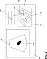
- FIG. 1illustrates one embodiment of a screen display indicating a proposed path and external contour surface for obtaining ultrasound images.
- FIG. 2illustrates the screen display of FIG. 1 with a current location of an ultrasound probe superimposed on the suggested scan path and external contour surface.
- FIG. 3illustrates one embodiment of a display indicating suggested probe angles along the suggested scan path of FIG. 1 .
- FIG. 4illustrates one embodiment of the invention in which a suggested scan path is constructed without the benefit of a previously-obtained ultrasound scan.
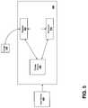
- FIG. 5is a schematic representation of a system in accordance with an embodiment of the invention.
- FIG. 1illustrates one embodiment of the invention in which a path suggestion line 100 is shown projected onto an external contour 105 of a patient P.
- a scanning site contour (SSC) 110is also shown, and may be derived from a three-dimensional surface contour of the anatomical structure of interest, in this case a breast lumpectomy site.
- the contoursmay be displayed in semi-transparent fashion to allow full visibility of the SSC and other anatomical features.
- the three-dimensional informationmay be manipulated to improve viewing (using, for example, conventional visualization devices and/or software), including being be rotated and/or zoomed.
- the imagesmay also be printed, but preferably are available directly on an ultrasound scanning screen 115 in a treatment planning and/or delivery room.
- an operatorscans the patient using an ultrasound device while reviewing a real-time image 120 to find the area of interest.
- the operatorcan find the correct treatment delivery location, indicated in FIG. 1 at 130 .
- the usercan then adjust ultrasound settings 135 , and obtain a three-dimensional scan which includes the intended treatment region by visually reproducing the intended scanning path 100 on the patient's skin.
- the ultrasound probemay be tracked (typically in real-time or near real-time) such that its three-dimensional position and orientation in space is known with respect to landmarks or other system components in the treatment room.
- the ultrasound slicescan be positioned in space relative to the corresponding coordinates of the treatment room and/or a linear accelerator used to deliver radiotherapy.
- Techniques for tracking ultrasound devicestypically rely on either a mechanical arm affixed to the probe or an optical camera attached to a fixed location in the room (often the ceiling) which tracks passive or active markers affixed to the probe.
- a representation of the probe 200(based on its location as determined above) is shown in the location-guidance image 125 .
- the probe representation 200may be updated in real-time as the operator moves the probe according to tracking information obtained from the tracking system, helping the operator see how close the current probe position is relative to the suggested path 100 (and thereby enabling the operator to include the appropriate anatomy in the scan with minimal searching). It also allows the operator to reproduce the same (or approximately the same) scan from one radiotherapy treatment session to the next.
- the probe representationdenotes both an angle and a position of the probe in space and relative to the patient P, while in some cases only its position is indicated.
- the intended scanning path 100is shown as a line, while in others it also denotes a suggested direction of travel for the probe along the line.
- the intended scanning path 300includes a series of lines 305 perpendicular to the scanning path 100 indicating the intended scan direction along the path, thus indicating whether, for example, the path was scanned in a sweeping fashion or a translational motion.
- a movie loopmay be compiled showing an actual representation of the suggested probe motion along the intended scan path, indicating a more detailed version of the motion.
- a high-quality scanmay have already been obtained in a first scanning session (typically during a treatment planning phase), and this scan may then be reproduced in future scanning sessions and used as a guide for augmenting the CT image with a preferred ultrasound path.
- planning sessionsthere is generally more time to obtain a good scan than in a treatment session, and the operators are typically more experienced and can obtain higher quality ultrasound scans than a radiotherapy technician who is not familiar with the representations of the anatomical structures in ultrasound images.
- the external contour 105may be obtained from the CT and the SSC obtained from either the CT or the initial ultrasound image.
- the external contour 105can be drawn slice-by-slice on the CT scan, and converted into a three-dimensional surface (which can be represented as a mesh, for example) before being provided to the scanning system.
- the three-dimensional surfaceis extracted automatically using techniques, such as thresholding, which uses differences in pixel characteristics find the interface between air and tissue.
- the SSCcan be found by either manually segmenting a region of interest in the CT or ultrasound image, or can be centered about a point in the general area of interest and defining the SSC as a sphere centered at that point.
- the suggested scanning pathcan be found by analyzing the temporal tracked probe positions of the first approved scan using, for example, the tracking data supplied by the tracking system and determining the position of a given pixel in the ultrasound image throughout the progression of the three-dimensional scan.
- the top-central pixelmay be used as the reference pixel because it can be related to the center of the probe.
- the three-dimensional path of the reference pixelgenerates a series of three-dimensional points in space, which when connected in order define the scanning path. If the direction of the probe along the path is also of importance, this can be found by finding a vector connecting the top-center reference pixel and any other pixel in the center line of the ultrasound image.
- the pathwill fall directly on the external contour.
- the pathmay not fall exactly on the skin. In some cases it may be preferable to project the path directly onto the external contour.
- the inventionfacilitates the definition of a scanning path without the benefit of an initial ultrasound image.
- the suggested scan pathmay be defined automatically using a previously acquired CT dataset with an associated external contour as input.
- the suggested scan path 400can be extracted by finding a path which is constrained to lie on the external contour 405 , and for which perpendicular lines 410 pass through the SSC 415 .
- additional constraintsmay be imposed to determine the preferred path. For example, one such constraint is to minimize the distance 420 along each perpendicular line between the SSC and the surface.
- the scan pathmay be calculated at the depth below the patient's skin of the SSC by finding an arbitrary line which passes through the SSC, and then projecting this line to the external contour surface such that the distance between the original line and the projected line is minimized. Continuity between the perpendicular lines should also be enforced since the ultrasound scan should be smooth. Further, the algorithm determines whether the preferred path is a sweeping, “fan” motion or a translational scan, in which all the perpendicular lines are substantially parallel to each other. Even with these constraints, there remains a number of possible paths, but in general choosing one such path is sufficient to guide the user.
- FIG. 5schematically depicts a hardware embodiment of the invention realized as a system 500 for presenting a preferred path for obtaining an ultrasound image.
- the system 500comprises a register 505 and a processor 515 .
- the register 505which may be any suitably organized data storage facility (e.g., partitions n RAM, etc.), receives images from an imager 525 such as an MRI, CT/PET scanner, ultrasound device, or x-ray device. In some embodiments, the images are stored on a data storage device separate from the imager (e.g., a database, microfiche, etc.) and sent to the system 500 .
- the register 505may receive the images through conventional data ports and may also include circuitry for receiving analog image data and analog-to-digital conversion circuitry for digitizing the image data.
- the register 505provides the image to the processor 515 which implements the functionality of the present invention in hardware or software, or a combination of both on a general-purpose computer.
- a programmay set aside portions of a computer's random access memory to provide control logic that affects one or more of the image capture, user manipulation (using, for example, an input device 530 ) and presentation on a display 520 .
- the programmay be written in any one of a number of high-level languages, such as FORTRAN, PASCAL, C, C++, C#, Java, Tel, or BASIC. Further, the program can be written in a script, macro, or functionality embedded in commercially available software, such as EXCEL or VISUAL BASIC.
- the softwarecan be implemented in an assembly language directed to a microprocessor resident on a computer.
- the softwarecan be implemented in Intel 80 ⁇ 86 assembly language if it is configured to run on an IBM PC or PC clone.
- the softwaremay be embedded on an article of manufacture including, but not limited to, “computer-readable program means” such as a floppy disk, a hard disk, an optical disk, a magnetic tape, a PROM, an EPROM, or CD-ROM.
Landscapes
- Life Sciences & Earth Sciences (AREA)
- Health & Medical Sciences (AREA)
- Biomedical Technology (AREA)
- Biophysics (AREA)
- Nuclear Medicine, Radiotherapy & Molecular Imaging (AREA)
- Pathology (AREA)
- Radiology & Medical Imaging (AREA)
- Engineering & Computer Science (AREA)
- Physics & Mathematics (AREA)
- Heart & Thoracic Surgery (AREA)
- Medical Informatics (AREA)
- Molecular Biology (AREA)
- Surgery (AREA)
- Animal Behavior & Ethology (AREA)
- General Health & Medical Sciences (AREA)
- Public Health (AREA)
- Veterinary Medicine (AREA)
- Ultra Sonic Daignosis Equipment (AREA)
Abstract
Description
Claims (16)
Priority Applications (1)
| Application Number | Priority Date | Filing Date | Title |
|---|---|---|---|
| US12/176,774US10531858B2 (en) | 2007-07-20 | 2008-07-21 | Methods and systems for guiding the acquisition of ultrasound images |
Applications Claiming Priority (2)
| Application Number | Priority Date | Filing Date | Title |
|---|---|---|---|
| US95100107P | 2007-07-20 | 2007-07-20 | |
| US12/176,774US10531858B2 (en) | 2007-07-20 | 2008-07-21 | Methods and systems for guiding the acquisition of ultrasound images |
Publications (2)
| Publication Number | Publication Date |
|---|---|
| US20090024030A1 US20090024030A1 (en) | 2009-01-22 |
| US10531858B2true US10531858B2 (en) | 2020-01-14 |
Family
ID=40265410
Family Applications (1)
| Application Number | Title | Priority Date | Filing Date |
|---|---|---|---|
| US12/176,774Active2031-01-15US10531858B2 (en) | 2007-07-20 | 2008-07-21 | Methods and systems for guiding the acquisition of ultrasound images |
Country Status (2)
| Country | Link |
|---|---|
| US (1) | US10531858B2 (en) |
| WO (1) | WO2009012576A1 (en) |
Cited By (2)
| Publication number | Priority date | Publication date | Assignee | Title |
|---|---|---|---|---|
| US20220000563A1 (en)* | 2018-02-14 | 2022-01-06 | Epica International, Inc. | Method for determination of surgical procedure access |
| US11826197B2 (en) | 2021-05-13 | 2023-11-28 | General Electric Company | Patient-specific neuromodulation alignment structures |
Families Citing this family (52)
| Publication number | Priority date | Publication date | Assignee | Title |
|---|---|---|---|---|
| US20090163809A1 (en)* | 2004-06-03 | 2009-06-25 | Kane Scott D | Medical method and associated apparatus utilizable in accessing internal organs through skin surface |
| US7728868B2 (en) | 2006-08-02 | 2010-06-01 | Inneroptic Technology, Inc. | System and method of providing real-time dynamic imagery of a medical procedure site using multiple modalities |
| JP5127371B2 (en)* | 2007-08-31 | 2013-01-23 | キヤノン株式会社 | Ultrasound image diagnostic system and control method thereof |
| WO2009094646A2 (en) | 2008-01-24 | 2009-07-30 | The University Of North Carolina At Chapel Hill | Methods, systems, and computer readable media for image guided ablation |
| US8340379B2 (en) | 2008-03-07 | 2012-12-25 | Inneroptic Technology, Inc. | Systems and methods for displaying guidance data based on updated deformable imaging data |
| EP2285287B1 (en)* | 2008-04-22 | 2015-04-01 | eZono AG | Ultrasound imaging system and method for providing assistance in an ultrasound imaging system |
| US8690776B2 (en) | 2009-02-17 | 2014-04-08 | Inneroptic Technology, Inc. | Systems, methods, apparatuses, and computer-readable media for image guided surgery |
| US11464578B2 (en) | 2009-02-17 | 2022-10-11 | Inneroptic Technology, Inc. | Systems, methods, apparatuses, and computer-readable media for image management in image-guided medical procedures |
| US8641621B2 (en) | 2009-02-17 | 2014-02-04 | Inneroptic Technology, Inc. | Systems, methods, apparatuses, and computer-readable media for image management in image-guided medical procedures |
| US8554307B2 (en) | 2010-04-12 | 2013-10-08 | Inneroptic Technology, Inc. | Image annotation in image-guided medical procedures |
| US10542962B2 (en)* | 2009-07-10 | 2020-01-28 | Elekta, LTD | Adaptive radiotherapy treatment using ultrasound |
| US9019262B2 (en)* | 2009-11-27 | 2015-04-28 | Hologic, Inc. | Systems and methods for tracking positions between imaging modalities and transforming a displayed three-dimensional image corresponding to a position and orientation of a probe |
| FR2957514B1 (en)* | 2010-03-17 | 2013-03-22 | Gen Electric | MEDICAL IMAGING DEVICE COMPRISING RADIOGRAPHIC ACQUISITION MEANS AND GUIDING MEANS FOR AN ULTRASONIC PROBE |
| WO2012012549A2 (en)* | 2010-07-21 | 2012-01-26 | The Regents Of The University Of California | Method to reduce radiation dose in multidetector ct while maintaining image quality |
| WO2012073164A1 (en)* | 2010-12-03 | 2012-06-07 | Koninklijke Philips Electronics N.V. | Device and method for ultrasound imaging |
| US9901320B2 (en) | 2010-12-14 | 2018-02-27 | Hologic, Inc. | System and method for fusing three dimensional image data from a plurality of different imaging systems for use in diagnostic imaging |
| JP6071282B2 (en)* | 2011-08-31 | 2017-02-01 | キヤノン株式会社 | Information processing apparatus, ultrasonic imaging apparatus, and information processing method |
| US8670816B2 (en) | 2012-01-30 | 2014-03-11 | Inneroptic Technology, Inc. | Multiple medical device guidance |
| RU2634295C2 (en)* | 2012-02-13 | 2017-10-24 | Конинклейке Филипс Н.В. | Simultaneous ultrasonic observation of three-dimensional volume from multiple directions |
| US20140171799A1 (en)* | 2012-12-13 | 2014-06-19 | General Electric Company | Systems and methods for providing ultrasound probe location and image information |
| US10314559B2 (en) | 2013-03-14 | 2019-06-11 | Inneroptic Technology, Inc. | Medical device guidance |
| AU2014237346B2 (en) | 2013-03-15 | 2020-02-27 | Hologic, Inc. | System and method for reviewing and analyzing cytological specimens |
| JP5785214B2 (en)* | 2013-05-08 | 2015-09-24 | 富士フイルム株式会社 | Mold, surgical support set, surgical support device, surgical support method, and surgical support program |
| US9901406B2 (en) | 2014-10-02 | 2018-02-27 | Inneroptic Technology, Inc. | Affected region display associated with a medical device |
| US10188467B2 (en) | 2014-12-12 | 2019-01-29 | Inneroptic Technology, Inc. | Surgical guidance intersection display |
| WO2016201637A1 (en)* | 2015-06-17 | 2016-12-22 | Covidien Lp | Guided ultrasound breast cancer screening system |
| US10709416B2 (en)* | 2015-06-30 | 2020-07-14 | Wisconsin Alumni Research Foundation | Obstetrical imaging at the point of care for untrained or minimally trained operators |
| US9949700B2 (en) | 2015-07-22 | 2018-04-24 | Inneroptic Technology, Inc. | Medical device approaches |
| US9675319B1 (en) | 2016-02-17 | 2017-06-13 | Inneroptic Technology, Inc. | Loupe display |
| WO2017186610A1 (en) | 2016-04-28 | 2017-11-02 | Koninklijke Philips N.V. | Image guided treatment delivery |
| US11266377B2 (en)* | 2016-05-17 | 2022-03-08 | Canon Medical Systems Corporation | Support apparatus and support method |
| CN109313698B (en) | 2016-05-27 | 2022-08-30 | 霍罗吉克公司 | Simultaneous surface and internal tumor detection |
| US10729396B2 (en)* | 2016-08-31 | 2020-08-04 | International Business Machines Corporation | Tracking anatomical findings within medical images |
| US10276265B2 (en) | 2016-08-31 | 2019-04-30 | International Business Machines Corporation | Automated anatomically-based reporting of medical images via image annotation |
| US10278778B2 (en) | 2016-10-27 | 2019-05-07 | Inneroptic Technology, Inc. | Medical device navigation using a virtual 3D space |
| CN106890006B (en)* | 2017-03-22 | 2019-08-30 | 苏州佳世达电通有限公司 | A kind of ultrasonic system and scanning bootstrap technique |
| USD855651S1 (en) | 2017-05-12 | 2019-08-06 | International Business Machines Corporation | Display screen with a graphical user interface for image-annotation classification |
| CN107158581B (en)* | 2017-05-15 | 2019-11-22 | 中国医学科学院肿瘤医院 | Intraoperative radiotherapy scanning path planning method and intraoperative radiotherapy system |
| US11259879B2 (en) | 2017-08-01 | 2022-03-01 | Inneroptic Technology, Inc. | Selective transparency to assist medical device navigation |
| US11043144B2 (en)* | 2017-08-04 | 2021-06-22 | Clarius Mobile Health Corp. | Systems and methods for providing an interactive demonstration of an ultrasound user interface |
| EP3485816A1 (en)* | 2017-11-21 | 2019-05-22 | Koninklijke Philips N.V. | Method and apparatus for guiding an ultrasound probe |
| US11484365B2 (en) | 2018-01-23 | 2022-11-01 | Inneroptic Technology, Inc. | Medical image guidance |
| EP3843636A4 (en)* | 2018-08-29 | 2022-05-25 | Butterfly Network, Inc. | Methods and apparatuses for collection of ultrasound data |
| CN109646047B (en)* | 2019-01-23 | 2023-12-26 | 上海浅葱网络技术有限公司 | Display method for operation guidance of ultrasonic equipment |
| WO2020246151A1 (en)* | 2019-06-06 | 2020-12-10 | 富士フイルム株式会社 | Three-dimensional ultrasonic image generation device, method, and program |
| US11883206B2 (en) | 2019-07-29 | 2024-01-30 | Hologic, Inc. | Personalized breast imaging system |
| CN110432931A (en)* | 2019-08-19 | 2019-11-12 | 深圳瀚维智能医疗科技有限公司 | Mammary gland scanning guiding device and mammary gland scanning guidance method |
| CN110664438B (en)* | 2019-10-22 | 2021-09-10 | 深圳瀚维智能医疗科技有限公司 | Ultrasonic scanning track planning method and device, storage medium and computer equipment |
| CN112741616A (en)* | 2019-10-31 | 2021-05-04 | 通用电气精准医疗有限责任公司 | Scanning position navigation device and method capable of being used in scanning imaging detection system |
| US12186132B2 (en) | 2019-11-21 | 2025-01-07 | Koninklijke Philips N.V. | Point-of-care ultrasound (POCUS) scan assistance and associated devices, systems, and methods |
| KR20220163445A (en)* | 2020-04-07 | 2022-12-09 | 베라톤 인코포레이티드 | Automated Prostate Analysis System |
| EP4480424A1 (en)* | 2023-06-22 | 2024-12-25 | Koninklijke Philips N.V. | Providing ultrasound imaging guidance |
Citations (185)
| Publication number | Priority date | Publication date | Assignee | Title |
|---|---|---|---|---|
| US3082322A (en) | 1958-11-28 | 1963-03-19 | Westinghouse Electric Corp | Therapy unit |
| US3777124A (en) | 1970-11-27 | 1973-12-04 | Varian Associates | Computer assisted radiation therapy machine |
| US3987281A (en) | 1974-07-29 | 1976-10-19 | The United States Of America As Represented By The Department Of Health, Education And Welfare | Method of radiation therapy treatment planning |
| US3991310A (en) | 1970-08-03 | 1976-11-09 | Morrison Richard A | Biplane radiographic localization of target center for radiotherapy |
| US4118631A (en) | 1976-03-30 | 1978-10-03 | Emi Limited | Radiographic apparatus |
| US4618978A (en) | 1983-10-21 | 1986-10-21 | Cosman Eric R | Means for localizing target coordinates in a body relative to a guidance system reference frame in any arbitrary plane as viewed by a tomographic image through the body |
| US4882741A (en) | 1987-10-28 | 1989-11-21 | U.S. Philips Corporation | Multileaf collimator and related apparatus |
| US4923459A (en) | 1987-09-14 | 1990-05-08 | Kabushiki Kaisha Toshiba | Stereotactics apparatus |
| US4943990A (en) | 1987-12-11 | 1990-07-24 | Bbc Brown Boveri Ag | Therapy simulator |
| US5039867A (en) | 1987-08-24 | 1991-08-13 | Mitsubishi Denki Kabushiki Kaisha | Therapeutic apparatus |
| US5080100A (en) | 1988-10-04 | 1992-01-14 | Cgr Mev | System and method for measuring and/or checking the position of a patient in a radio-therapy machine |
| US5086401A (en) | 1990-05-11 | 1992-02-04 | International Business Machines Corporation | Image-directed robotic system for precise robotic surgery including redundant consistency checking |
| US5099846A (en) | 1988-12-23 | 1992-03-31 | Hardy Tyrone L | Method and apparatus for video presentation from a variety of scanner imaging sources |
| US5107839A (en) | 1990-05-04 | 1992-04-28 | Pavel V. Houdek | Computer controlled stereotaxic radiotherapy system and method |
| US5117829A (en) | 1989-03-31 | 1992-06-02 | Loma Linda University Medical Center | Patient alignment system and procedure for radiation treatment |
| US5138647A (en) | 1990-08-03 | 1992-08-11 | Siemens Medical Laboratories, Inc. | Portal imaging device |
| US5207223A (en) | 1990-10-19 | 1993-05-04 | Accuray, Inc. | Apparatus for and method of performing stereotaxic surgery |
| US5222499A (en) | 1989-11-15 | 1993-06-29 | Allen George S | Method and apparatus for imaging the anatomy |
| US5233990A (en) | 1992-01-13 | 1993-08-10 | Gideon Barnea | Method and apparatus for diagnostic imaging in radiation therapy |
| US5291889A (en) | 1991-05-23 | 1994-03-08 | Vanguard Imaging Ltd. | Apparatus and method for spatially positioning images |
| US5295483A (en) | 1990-05-11 | 1994-03-22 | Christopher Nowacki | Locating target in human body |
| US5301674A (en) | 1992-03-27 | 1994-04-12 | Diasonics, Inc. | Method and apparatus for focusing transmission and reception of ultrasonic beams |
| US5379642A (en) | 1993-07-19 | 1995-01-10 | Diasonics Ultrasound, Inc. | Method and apparatus for performing imaging |
| US5389101A (en) | 1992-04-21 | 1995-02-14 | University Of Utah | Apparatus and method for photogrammetric surgical localization |
| US5391139A (en) | 1992-09-03 | 1995-02-21 | William Beaumont Hospital | Real time radiation treatment planning system |
| US5397329A (en) | 1987-11-10 | 1995-03-14 | Allen; George S. | Fiducial implant and system of such implants |
| EP0647457A1 (en) | 1993-10-08 | 1995-04-12 | Nomos Corporation | Method and apparatus for lesion position verification |
| US5408101A (en) | 1992-07-06 | 1995-04-18 | Telaire Systems, Inc. | Laser assisted quasi-blackbody radiation source |
| US5438991A (en) | 1993-10-18 | 1995-08-08 | William Beaumont Hospital | Method and apparatus for controlling a radiation treatment field |
| US5442675A (en) | 1992-03-19 | 1995-08-15 | Wisconsin Alumni Research Foundation | Dynamic collimator for radiation therapy |
| US5446548A (en) | 1993-10-08 | 1995-08-29 | Siemens Medical Systems, Inc. | Patient positioning and monitoring system |
| US5447154A (en) | 1992-07-31 | 1995-09-05 | Universite Joseph Fourier | Method for determining the position of an organ |
| US5483961A (en) | 1993-03-19 | 1996-01-16 | Kelly; Patrick J. | Magnetic field digitizer for stereotactic surgery |
| US5511549A (en) | 1995-02-13 | 1996-04-30 | Loma Linda Medical Center | Normalizing and calibrating therapeutic radiation delivery systems |
| US5524627A (en)* | 1994-08-23 | 1996-06-11 | Sonotron Ltd. | Ultrasonic imaging system |
| US5531227A (en) | 1994-01-28 | 1996-07-02 | Schneider Medical Technologies, Inc. | Imaging device and method |
| US5531520A (en) | 1994-09-01 | 1996-07-02 | Massachusetts Institute Of Technology | System and method of registration of three-dimensional data sets including anatomical body data |
| US5553618A (en) | 1993-03-12 | 1996-09-10 | Kabushiki Kaisha Toshiba | Method and apparatus for ultrasound medical treatment |
| US5591983A (en) | 1995-06-30 | 1997-01-07 | Siemens Medical Systems, Inc. | Multiple layer multileaf collimator |
| US5603318A (en) | 1992-04-21 | 1997-02-18 | University Of Utah Research Foundation | Apparatus and method for photogrammetric surgical localization |
| US5609485A (en)* | 1994-10-03 | 1997-03-11 | Medsim, Ltd. | Medical reproduction system |
| US5645066A (en)* | 1996-04-26 | 1997-07-08 | Advanced Technology Laboratories, Inc. | Medical ultrasonic diagnostic imaging system with scanning guide for three dimensional imaging |
| US5673300A (en) | 1996-06-11 | 1997-09-30 | Wisconsin Alumni Research Foundation | Method of registering a radiation treatment plan to a patient |
| US5690108A (en) | 1994-11-28 | 1997-11-25 | Chakeres; Donald W. | Interventional medicine apparatus |
| US5715166A (en) | 1992-03-02 | 1998-02-03 | General Motors Corporation | Apparatus for the registration of three-dimensional shapes |
| US5734384A (en) | 1991-11-29 | 1998-03-31 | Picker International, Inc. | Cross-referenced sectioning and reprojection of diagnostic image volumes |
| US5740225A (en) | 1995-12-07 | 1998-04-14 | Kabushiki Kaisha Toshiba | Radiation therapy planning method and its system and apparatus |
| US5754623A (en)* | 1994-03-25 | 1998-05-19 | Kabushiki Kaisha Toshiba | Radiotherapy system |
| US5757881A (en) | 1997-01-06 | 1998-05-26 | Siemens Business Communication Systems, Inc. | Redundant field-defining arrays for a radiation system |
| US5778043A (en) | 1996-09-20 | 1998-07-07 | Cosman; Eric R. | Radiation beam control system |
| US5810007A (en)* | 1995-07-26 | 1998-09-22 | Associates Of The Joint Center For Radiation Therapy, Inc. | Ultrasound localization and image fusion for the treatment of prostate cancer |
| US5851183A (en) | 1990-10-19 | 1998-12-22 | St. Louis University | System for indicating the position of a surgical probe within a head on an image of the head |
| US5859891A (en) | 1997-03-07 | 1999-01-12 | Hibbard; Lyn | Autosegmentation/autocontouring system and method for use with three-dimensional radiation therapy treatment planning |
| WO1999002074A1 (en) | 1997-07-10 | 1999-01-21 | Von Der Heyde Christian P | Apparatus and method for preventing sudden infant death syndrome |
| WO1999006644A1 (en) | 1997-07-28 | 1999-02-11 | Josu Corporation Pty. Ltd. | Conduit fitting for termites |
| US5871445A (en) | 1993-04-26 | 1999-02-16 | St. Louis University | System for indicating the position of a surgical probe within a head on an image of the head |
| WO1999026534A1 (en) | 1995-07-26 | 1999-06-03 | Burdette Medical Systems, Inc. | Virtual reality 3d visualization for surgical procedures |
| WO1999027839A2 (en) | 1997-12-01 | 1999-06-10 | Cosman Eric R | Surgical positioning system |
| US5952577A (en)* | 1997-07-21 | 1999-09-14 | Sonotron Ltd. | Ultrasonic imaging system |
| EP0951697A1 (en) | 1997-01-13 | 1999-10-27 | Qualisys AB | Motion analysis system |
| FR2778574A1 (en) | 1998-05-13 | 1999-11-19 | Technomed Medical Systems | Method to determine effect of ultrasonic treatment therapy on tissue |
| US5991703A (en) | 1997-08-15 | 1999-11-23 | The Institute Of Physical And Chemical Research | Method of synthesizing measurement data of free-form surface |
| US6019724A (en) | 1995-02-22 | 2000-02-01 | Gronningsaeter; Aage | Method for ultrasound guidance during clinical procedures |
| US6038283A (en) | 1996-10-24 | 2000-03-14 | Nomos Corporation | Planning method and apparatus for radiation dosimetry |
| US6094508A (en) | 1997-12-08 | 2000-07-25 | Intel Corporation | Perceptual thresholding for gradient-based local edge detection |
| US6106470A (en) | 1998-03-20 | 2000-08-22 | General Electric Company | Method and appartus for calculating distance between ultrasound images using sum of absolute differences |
| US6112341A (en) | 1994-09-08 | 2000-09-05 | Itt Manufacturing Enterprises, Inc. | Rotary pulsing valve |
| US6119033A (en) | 1997-03-04 | 2000-09-12 | Biotrack, Inc. | Method of monitoring a location of an area of interest within a patient during a medical procedure |
| US6118848A (en) | 1998-01-14 | 2000-09-12 | Reiffel; Leonard | System to stabilize an irradiated internal target |
| US6117081A (en) | 1998-10-01 | 2000-09-12 | Atl Ultrasound, Inc. | Method for correcting blurring of spatially compounded ultrasonic diagnostic images |
| US6122341A (en) | 1992-06-12 | 2000-09-19 | Butler; William E. | System for determining target positions in the body observed in CT image data |
| US6129670A (en) | 1997-11-24 | 2000-10-10 | Burdette Medical Systems | Real time brachytherapy spatial registration and visualization system |
| US6138495A (en) | 1997-12-31 | 2000-10-31 | Ultraguide Ltd. | Calibration method and apparatus for calibrating position sensors on scanning transducers |
| US6144875A (en) | 1999-03-16 | 2000-11-07 | Accuray Incorporated | Apparatus and method for compensating for respiratory and patient motion during treatment |
| WO2001005316A1 (en) | 1999-07-19 | 2001-01-25 | Light Sciences Corporation | Real-time monitoring of photodynamic therapy over an extended time |
| US6198957B1 (en) | 1997-12-19 | 2001-03-06 | Varian, Inc. | Radiotherapy machine including magnetic resonance imaging system |
| US6259943B1 (en) | 1995-02-16 | 2001-07-10 | Sherwood Services Ag | Frameless to frame-based registration system |
| US6269143B1 (en) | 1998-08-31 | 2001-07-31 | Shimadzu Corporation | Radiotherapy planning system |
| US6285805B1 (en) | 1999-01-25 | 2001-09-04 | International Business Machines Corp. | System and method for finding the distance from a moving query point to the closest point on one or more convex or non-convex shapes |
| US6292578B1 (en) | 1998-02-18 | 2001-09-18 | International Business Machines Corporation | System and method for restoring, describing and graphically displaying noise-corrupted boundaries in tomography images |
| US6307914B1 (en) | 1998-03-12 | 2001-10-23 | Mitsubishi Denki Kabushiki Kaisha | Moving body pursuit irradiating device and positioning method using this device |
| US20010035871A1 (en) | 2000-03-30 | 2001-11-01 | Johannes Bieger | System and method for generating an image |
| US6314310B1 (en) | 1997-02-14 | 2001-11-06 | Biosense, Inc. | X-ray guided surgical location system with extended mapping volume |
| US6325758B1 (en) | 1997-10-27 | 2001-12-04 | Nomos Corporation | Method and apparatus for target position verification |
| US20010049475A1 (en) | 2000-01-31 | 2001-12-06 | Bucholz Richard D. | System combining proton beam irradiation and magnetic resonance imaging |
| US6345114B1 (en) | 1995-06-14 | 2002-02-05 | Wisconsin Alumni Research Foundation | Method and apparatus for calibration of radiation therapy equipment and verification of radiation treatment |
| WO2002009588A1 (en) | 2000-08-01 | 2002-02-07 | Tony Falco | Method and apparatus for lesion localization, definition and verification |
| US20020018588A1 (en) | 2000-03-30 | 2002-02-14 | Jochen Kusch | System and method for generating an image dataset |
| US6385288B1 (en) | 2001-01-19 | 2002-05-07 | Mitsubishi Denki Kabushiki Kaisha | Radiotherapy apparatus with independent rotation mechanisms |
| US6385286B1 (en) | 1998-08-06 | 2002-05-07 | Wisconsin Alumni Research Foundation | Delivery modification system for radiation therapy |
| US6390982B1 (en) | 1999-07-23 | 2002-05-21 | Univ Florida | Ultrasonic guidance of target structures for medical procedures |
| US20020082494A1 (en) | 2000-12-27 | 2002-06-27 | Ge Medical Systems Global Technology Company, Llc | Multi-plane graphic prescription interface and method |
| US20020087101A1 (en) | 2000-01-04 | 2002-07-04 | Barrick Earl Frederick | System and method for automatic shape registration and instrument tracking |
| US6423009B1 (en) | 1996-11-29 | 2002-07-23 | Life Imaging Systems, Inc. | System, employing three-dimensional ultrasonographic imaging, for assisting in guiding and placing medical instruments |
| US6438202B1 (en) | 1998-08-06 | 2002-08-20 | Wisconsin Alumni Research Foundation | Method using post-patient radiation monitor to verify entrance radiation and dose in a radiation therapy machine |
| US20020122530A1 (en) | 2001-03-05 | 2002-09-05 | Stephan Erbel | Method for producing or updating radiotherapy plan |
| US6459769B1 (en) | 1999-05-03 | 2002-10-01 | Sherwood Services Ag | Movable miniature multi-leaf collimator |
| US6470207B1 (en)* | 1999-03-23 | 2002-10-22 | Surgical Navigation Technologies, Inc. | Navigational guidance via computer-assisted fluoroscopic imaging |
| US20020156375A1 (en) | 1999-10-28 | 2002-10-24 | Paul Kessman | Navigation information overlay onto ultrasound imagery |
| US20020176541A1 (en) | 2001-05-22 | 2002-11-28 | Mario Schubert | Registering image information |
| US20020183610A1 (en) | 1994-10-07 | 2002-12-05 | Saint Louis University And Surgical Navigation Technologies, Inc. | Bone navigation system |
| US20030018232A1 (en) | 2000-06-05 | 2003-01-23 | Mentor Corporation | Automated implantation system for radioisotope seeds |
| US6511430B1 (en) | 1998-08-19 | 2003-01-28 | University Health Network | Use of high frequency ultrasound imaging to detect and monitor the process of apoptosis in living tissues, ex-vivo tissues and cell-culture |
| US6516046B1 (en) | 1999-11-04 | 2003-02-04 | Brainlab Ag | Exact patient positioning by compairing reconstructed x-ray images and linac x-ray images |
| US20030028401A1 (en) | 2001-07-17 | 2003-02-06 | Leon Kaufman | Customizable lung report generator |
| US6535574B1 (en) | 2001-11-01 | 2003-03-18 | Siemens Medical Solutions Usa, Inc. | Patient positioning system employing surface photogrammetry and portal imaging |
| US6546073B1 (en) | 1999-11-05 | 2003-04-08 | Georgia Tech Research Corporation | Systems and methods for global optimization of treatment planning for external beam radiation therapy |
| US6553152B1 (en) | 1996-07-10 | 2003-04-22 | Surgical Navigation Technologies, Inc. | Method and apparatus for image registration |
| US6560311B1 (en) | 1998-08-06 | 2003-05-06 | Wisconsin Alumni Research Foundation | Method for preparing a radiation therapy plan |
| WO2003039370A1 (en) | 2001-11-05 | 2003-05-15 | Computerized Medical Systems, Inc. | Apparatus and method for registration, guidance, and targeting of external beam radiation therapy |
| US6567684B1 (en) | 2000-11-08 | 2003-05-20 | Regents Of The University Of Michigan | Imaging system, computer, program product and method for detecting changes in rates of water diffusion in a tissue using magnetic resonance imaging (MRI) |
| US6585651B2 (en) | 1999-04-20 | 2003-07-01 | Synthes Ag Chur | Method and device for percutaneous determination of points associated with the surface of an organ |
| US6591127B1 (en) | 1999-03-15 | 2003-07-08 | General Electric Company | Integrated multi-modality imaging system and method |
| US6600810B1 (en) | 1998-08-10 | 2003-07-29 | Siemens Medical Solutions Usa, Inc. | Multiple layer multileaf collimator design to improve resolution and reduce leakage |
| US20030144813A1 (en) | 2002-01-31 | 2003-07-31 | Canon Kabushiki Kaisha | Position and orientation determination method and apparatus and storage medium |
| US20030153825A1 (en) | 2002-02-12 | 2003-08-14 | Science & Engineering Associates, Inc. | Cancer detection and adaptive dose optimization treatment system |
| US6621889B1 (en) | 1998-10-23 | 2003-09-16 | Varian Medical Systems, Inc. | Method and system for predictive physiological gating of radiation therapy |
| WO2003076003A2 (en) | 2002-03-06 | 2003-09-18 | Tomotherapy Incorporated | Method for modification of radiotherapy treatment delivery |
| US20030182072A1 (en) | 2002-03-19 | 2003-09-25 | Canon Kabushiki Kaisha | Sensor calibration apparatus, sensor calibration method, program, storage medium, information processing method, and information processing apparatus |
| US6628983B1 (en) | 2000-10-25 | 2003-09-30 | Koninklijke Philips Electronics N.V. | Nuclear imaging systems and methods with feature-enhanced transmission imaging |
| US6631284B2 (en) | 1999-10-14 | 2003-10-07 | Cti Pet Systems, Inc. | Combined PET and X-ray CT tomograph |
| US6636622B2 (en) | 1997-10-15 | 2003-10-21 | Wisconsin Alumni Research Foundation | Method and apparatus for calibration of radiation therapy equipment and verification of radiation treatment |
| US6641539B2 (en) | 1999-12-08 | 2003-11-04 | Olympus Optical Co., Ltd. | Ultrasonic probe for operation under microscope |
| US20030220557A1 (en)* | 2002-03-01 | 2003-11-27 | Kevin Cleary | Image guided liver interventions based on magnetic tracking of internal organ motion |
| US6661870B2 (en) | 2001-03-09 | 2003-12-09 | Tomotherapy Incorporated | Fluence adjustment for improving delivery to voxels without reoptimization |
| US20030231790A1 (en) | 2002-05-02 | 2003-12-18 | Bottema Murk Jan | Method and system for computer aided detection of cancer |
| US20040015075A1 (en) | 2000-08-21 | 2004-01-22 | Yoav Kimchy | Radioactive emission detector equipped with a position tracking system and utilization thereof with medical systems and in medical procedures |
| US20040015176A1 (en) | 1994-06-20 | 2004-01-22 | Cosman Eric R. | Stereotactic localizer system with dental impression |
| US6683985B1 (en) | 1997-04-25 | 2004-01-27 | Riken | Method of discriminating shape of free-form curved surface |
| US6690965B1 (en) | 1998-10-23 | 2004-02-10 | Varian Medical Systems, Inc. | Method and system for physiological gating of radiation therapy |
| US6714627B1 (en) | 1998-08-28 | 2004-03-30 | Elekta Ab | Collimator for radiotherapy apparatus |
| US6725079B2 (en) | 2001-06-20 | 2004-04-20 | Odin Medical Technologies, Ltd. | Dual pointer device and method for surgical navigation |
| US6728424B1 (en) | 2000-09-15 | 2004-04-27 | Koninklijke Philips Electronics, N.V. | Imaging registration system and method using likelihood maximization |
| US6731970B2 (en) | 2000-07-07 | 2004-05-04 | Brainlab Ag | Method for breath compensation in radiation therapy |
| US20040092815A1 (en) | 2002-11-12 | 2004-05-13 | Achim Schweikard | Method and apparatus for tracking an internal target region without an implanted fiducial |
| EP1426806A2 (en) | 1997-09-26 | 2004-06-09 | Z-Kat, Inc. | Microscope calibration |
| US6750873B1 (en) | 2000-06-27 | 2004-06-15 | International Business Machines Corporation | High quality texture reconstruction from multiple scans |
| US6754374B1 (en) | 1998-12-16 | 2004-06-22 | Surgical Navigation Technologies, Inc. | Method and apparatus for processing images with regions representing target objects |
| US20040146137A1 (en) | 2001-05-16 | 2004-07-29 | Herbert Bruder | Method for computer tomography and computer tomography device for carrying out the method |
| US6785409B1 (en) | 2000-10-24 | 2004-08-31 | Koninklijke Philips Electronics, N.V. | Segmentation method and apparatus for medical images using diffusion propagation, pixel classification, and mathematical morphology |
| US20040176925A1 (en) | 2003-01-10 | 2004-09-09 | Canon Kabushiki Kaisha | Position/orientation measurement method, and position/orientation measurement apparatus |
| US20040184646A1 (en) | 2003-03-19 | 2004-09-23 | Fuji Photo Film Co., Ltd. | Method, apparatus, and program for judging images |
| US6804548B2 (en) | 2000-12-14 | 2004-10-12 | Mitsubishi Denki Kabushiki Kaisha | Irradiation system and its irradiation target movement monitoring method, and irradiation target position recognizing method |
| US20040252870A1 (en) | 2000-04-11 | 2004-12-16 | Reeves Anthony P. | System and method for three-dimensional image rendering and analysis |
| US20040260142A1 (en) | 2003-06-18 | 2004-12-23 | Lovoi Paul A. | Method for intraoperative radiation treatment of breast cancer |
| US6842502B2 (en) | 2000-02-18 | 2005-01-11 | Dilliam Beaumont Hospital | Cone beam computed tomography with a flat panel imager |
| US20050020917A1 (en) | 2002-10-07 | 2005-01-27 | Scherch John David | Method and apparatus for target position verification |
| US6915008B2 (en) | 2001-03-08 | 2005-07-05 | Point Grey Research Inc. | Method and apparatus for multi-nodal, three-dimensional imaging |
| US6914959B2 (en) | 2001-08-09 | 2005-07-05 | Analogic Corporation | Combined radiation therapy and imaging system and method |
| US20050180544A1 (en) | 2004-02-17 | 2005-08-18 | Frank Sauer | System and method for patient positioning for radiotherapy in the presence of respiratory motion |
| US20050251029A1 (en) | 2004-04-21 | 2005-11-10 | Ali Khamene | Radiation therapy treatment plan |
| US6980679B2 (en) | 1998-10-23 | 2005-12-27 | Varian Medical System Technologies, Inc. | Method and system for monitoring breathing activity of a subject |
| JP2006000220A (en) | 2004-06-15 | 2006-01-05 | Ishikawajima Harima Heavy Ind Co Ltd | Multi-leaf collimator |
| US20060020195A1 (en) | 2004-07-20 | 2006-01-26 | Tony Falco | Verifying lesion characteristics using beam shapes |
| US20060074292A1 (en) | 2004-09-30 | 2006-04-06 | Accuray, Inc. | Dynamic tracking of moving targets |
| US20060093205A1 (en) | 2004-10-29 | 2006-05-04 | Bryll Robert K | System and method for automatically recovering video tools in a vision system |
| WO2006051523A2 (en) | 2004-11-10 | 2006-05-18 | Mediguide Ltd. | Method and apparatus for invasive device tracking using organ timing signal generated from mps sensors |
| US20060120608A1 (en) | 2004-11-22 | 2006-06-08 | Jiebo Luo | Detecting and classifying lesions in ultrasound images |
| US7095823B2 (en) | 2003-07-08 | 2006-08-22 | Elekta Ab (Publ) | Multi-leaf collimator |
| US20060241443A1 (en) | 2004-11-22 | 2006-10-26 | Whitmore Willet F Iii | Real time ultrasound monitoring of the motion of internal structures during respiration for control of therapy delivery |
| US20060285641A1 (en) | 2005-06-16 | 2006-12-21 | Nomos Corporation | System, tracker, and program product to facilitate and verify proper target alignment for radiation delivery, and related methods |
| US20060293583A1 (en) | 2005-06-27 | 2006-12-28 | Saracen Michael J | Method for automatic anatomy-specific treatment planning protocols based on historical integration of previously accepted plans |
| US20070010743A1 (en)* | 2003-05-08 | 2007-01-11 | Osamu Arai | Reference image display method for ultrasonography and ultrasonograph |
| US20070015991A1 (en) | 2005-06-29 | 2007-01-18 | Dongshan Fu | Dynamic tracking of soft tissue targets with ultrasound images, without using fiducial markers |
| US20070038058A1 (en) | 2005-08-11 | 2007-02-15 | West Jay B | Patient tracking using a virtual image |
| US20070055090A1 (en) | 2004-08-12 | 2007-03-08 | Navotek Medical Ltd. | Medical Treatment System and Method |
| CA2621741A1 (en) | 2005-09-06 | 2007-03-15 | Resonant Medical Inc. | System and method for patient setup for radiotherapy treatment |
| US20080021317A1 (en)* | 2006-07-24 | 2008-01-24 | Siemens Medical Solutions Usa, Inc. | Ultrasound medical imaging with robotic assistance for volume imaging |
| US7333644B2 (en) | 2003-03-11 | 2008-02-19 | Siemens Medical Solutions Usa, Inc. | Systems and methods for providing automatic 3D lesion segmentation and measurements |
| US7343030B2 (en) | 2003-08-05 | 2008-03-11 | Imquant, Inc. | Dynamic tumor treatment system |
| US20080064953A1 (en) | 2006-09-13 | 2008-03-13 | Tony Falco | Incorporating Internal Anatomy In Clinical Radiotherapy Setups |
| US20080200794A1 (en)* | 2007-02-19 | 2008-08-21 | Robert Teichman | Multi-configuration tracknig array and related method |
| US7430321B2 (en) | 2004-09-09 | 2008-09-30 | Siemens Medical Solutions Usa, Inc. | System and method for volumetric tumor segmentation using joint space-intensity likelihood ratio test |
| US20080292194A1 (en) | 2005-04-27 | 2008-11-27 | Mark Schmidt | Method and System for Automatic Detection and Segmentation of Tumors and Associated Edema (Swelling) in Magnetic Resonance (Mri) Images |
| US20090003523A1 (en) | 2007-06-29 | 2009-01-01 | Accuray Incorporated | Non-collocated imaging and treatment in image-guided radiation treatment systems |
| US20090093716A1 (en) | 2007-10-04 | 2009-04-09 | General Electric Company | Method and apparatus for evaluation of labor with ultrasound |
| US20090110145A1 (en) | 2007-10-25 | 2009-04-30 | Tomotherapy Incorporated | Method for adapting fractionation of a radiation therapy dose |
| US7535411B2 (en) | 2005-08-01 | 2009-05-19 | Resonant Medical, Inc. | System and method for detecting drifts in calibrated tracking systems |
| US7662097B2 (en) | 2004-09-20 | 2010-02-16 | Resonant Medical, Inc. | Radiotherapy treatment monitoring using ultrasound |
| US7672705B2 (en) | 2004-07-19 | 2010-03-02 | Resonant Medical, Inc. | Weighted surface-to-surface mapping |
| US7801349B2 (en) | 2005-06-20 | 2010-09-21 | Accuray Incorporated | Automatic generation of an envelope of constraint points for inverse planning |
| US20110069815A1 (en) | 2009-09-22 | 2011-03-24 | Varian Medical Systems International Ag | Method and Apparatus to Facilitate Supplementing a Dose-Volume Histogram Constraint Using an Adaptive Dose-Volume Histogram Constraint |
| US8042209B2 (en) | 2005-04-13 | 2011-10-25 | University Of Maryland | Techniques for compensating movement of a treatment target in a patient |
| US8160676B2 (en)* | 2006-09-08 | 2012-04-17 | Medtronic, Inc. | Method for planning a surgical procedure |
| US8232535B2 (en) | 2005-05-10 | 2012-07-31 | Tomotherapy Incorporated | System and method of treating a patient with radiation therapy |
Family Cites Families (2)
| Publication number | Priority date | Publication date | Assignee | Title |
|---|---|---|---|---|
| GB9516304D0 (en)* | 1995-08-09 | 1995-10-11 | Flight Refueling Ltd | Detection and location of current leakage paths and detection of oscillations |
| US6543073B2 (en)* | 2001-08-27 | 2003-04-08 | Hsin-Tsai Wu | Inflation seat assembly for an inflatable article |
- 2008
- 2008-07-21USUS12/176,774patent/US10531858B2/enactiveActive
- 2008-07-21WOPCT/CA2008/001338patent/WO2009012576A1/enactiveApplication Filing
Patent Citations (212)
| Publication number | Priority date | Publication date | Assignee | Title |
|---|---|---|---|---|
| US3082322A (en) | 1958-11-28 | 1963-03-19 | Westinghouse Electric Corp | Therapy unit |
| US3991310A (en) | 1970-08-03 | 1976-11-09 | Morrison Richard A | Biplane radiographic localization of target center for radiotherapy |
| US3777124A (en) | 1970-11-27 | 1973-12-04 | Varian Associates | Computer assisted radiation therapy machine |
| US3987281A (en) | 1974-07-29 | 1976-10-19 | The United States Of America As Represented By The Department Of Health, Education And Welfare | Method of radiation therapy treatment planning |
| US4118631A (en) | 1976-03-30 | 1978-10-03 | Emi Limited | Radiographic apparatus |
| US4618978A (en) | 1983-10-21 | 1986-10-21 | Cosman Eric R | Means for localizing target coordinates in a body relative to a guidance system reference frame in any arbitrary plane as viewed by a tomographic image through the body |
| US5039867A (en) | 1987-08-24 | 1991-08-13 | Mitsubishi Denki Kabushiki Kaisha | Therapeutic apparatus |
| US4923459A (en) | 1987-09-14 | 1990-05-08 | Kabushiki Kaisha Toshiba | Stereotactics apparatus |
| US4882741A (en) | 1987-10-28 | 1989-11-21 | U.S. Philips Corporation | Multileaf collimator and related apparatus |
| US5397329A (en) | 1987-11-10 | 1995-03-14 | Allen; George S. | Fiducial implant and system of such implants |
| US4943990A (en) | 1987-12-11 | 1990-07-24 | Bbc Brown Boveri Ag | Therapy simulator |
| US5080100A (en) | 1988-10-04 | 1992-01-14 | Cgr Mev | System and method for measuring and/or checking the position of a patient in a radio-therapy machine |
| US5099846A (en) | 1988-12-23 | 1992-03-31 | Hardy Tyrone L | Method and apparatus for video presentation from a variety of scanner imaging sources |
| US5117829A (en) | 1989-03-31 | 1992-06-02 | Loma Linda University Medical Center | Patient alignment system and procedure for radiation treatment |
| US5222499A (en) | 1989-11-15 | 1993-06-29 | Allen George S | Method and apparatus for imaging the anatomy |
| US5107839A (en) | 1990-05-04 | 1992-04-28 | Pavel V. Houdek | Computer controlled stereotaxic radiotherapy system and method |
| US5295483A (en) | 1990-05-11 | 1994-03-22 | Christopher Nowacki | Locating target in human body |
| US5086401A (en) | 1990-05-11 | 1992-02-04 | International Business Machines Corporation | Image-directed robotic system for precise robotic surgery including redundant consistency checking |
| US5299288A (en) | 1990-05-11 | 1994-03-29 | International Business Machines Corporation | Image-directed robotic system for precise robotic surgery including redundant consistency checking |
| US5138647A (en) | 1990-08-03 | 1992-08-11 | Siemens Medical Laboratories, Inc. | Portal imaging device |
| US5851183A (en) | 1990-10-19 | 1998-12-22 | St. Louis University | System for indicating the position of a surgical probe within a head on an image of the head |
| US5207223A (en) | 1990-10-19 | 1993-05-04 | Accuray, Inc. | Apparatus for and method of performing stereotaxic surgery |
| US20020188194A1 (en) | 1991-01-28 | 2002-12-12 | Sherwood Services Ag | Surgical positioning system |
| US6662036B2 (en) | 1991-01-28 | 2003-12-09 | Sherwood Services Ag | Surgical positioning system |
| US6405072B1 (en) | 1991-01-28 | 2002-06-11 | Sherwood Services Ag | Apparatus and method for determining a location of an anatomical target with reference to a medical apparatus |
| US20020065461A1 (en) | 1991-01-28 | 2002-05-30 | Cosman Eric R. | Surgical positioning system |
| US5291889A (en) | 1991-05-23 | 1994-03-08 | Vanguard Imaging Ltd. | Apparatus and method for spatially positioning images |
| US5734384A (en) | 1991-11-29 | 1998-03-31 | Picker International, Inc. | Cross-referenced sectioning and reprojection of diagnostic image volumes |
| US5233990A (en) | 1992-01-13 | 1993-08-10 | Gideon Barnea | Method and apparatus for diagnostic imaging in radiation therapy |
| US5715166A (en) | 1992-03-02 | 1998-02-03 | General Motors Corporation | Apparatus for the registration of three-dimensional shapes |
| US5442675A (en) | 1992-03-19 | 1995-08-15 | Wisconsin Alumni Research Foundation | Dynamic collimator for radiation therapy |
| US5301674A (en) | 1992-03-27 | 1994-04-12 | Diasonics, Inc. | Method and apparatus for focusing transmission and reception of ultrasonic beams |
| US5836954A (en) | 1992-04-21 | 1998-11-17 | University Of Utah Research Foundation | Apparatus and method for photogrammetric surgical localization |
| US6146390A (en) | 1992-04-21 | 2000-11-14 | Sofamor Danek Holdings, Inc. | Apparatus and method for photogrammetric surgical localization |
| US5389101A (en) | 1992-04-21 | 1995-02-14 | University Of Utah | Apparatus and method for photogrammetric surgical localization |
| US6491702B2 (en) | 1992-04-21 | 2002-12-10 | Sofamor Danek Holdings, Inc. | Apparatus and method for photogrammetric surgical localization |
| US5603318A (en) | 1992-04-21 | 1997-02-18 | University Of Utah Research Foundation | Apparatus and method for photogrammetric surgical localization |
| US6359959B1 (en) | 1992-06-12 | 2002-03-19 | Sherwood Services Ag | System for determining target positions in the body observed in CT image data |
| US6122341A (en) | 1992-06-12 | 2000-09-19 | Butler; William E. | System for determining target positions in the body observed in CT image data |
| US5408101A (en) | 1992-07-06 | 1995-04-18 | Telaire Systems, Inc. | Laser assisted quasi-blackbody radiation source |
| US5447154A (en) | 1992-07-31 | 1995-09-05 | Universite Joseph Fourier | Method for determining the position of an organ |
| US5391139A (en) | 1992-09-03 | 1995-02-21 | William Beaumont Hospital | Real time radiation treatment planning system |
| US5553618A (en) | 1993-03-12 | 1996-09-10 | Kabushiki Kaisha Toshiba | Method and apparatus for ultrasound medical treatment |
| US5483961A (en) | 1993-03-19 | 1996-01-16 | Kelly; Patrick J. | Magnetic field digitizer for stereotactic surgery |
| US5871445A (en) | 1993-04-26 | 1999-02-16 | St. Louis University | System for indicating the position of a surgical probe within a head on an image of the head |
| US5379642A (en) | 1993-07-19 | 1995-01-10 | Diasonics Ultrasound, Inc. | Method and apparatus for performing imaging |
| EP0647457A1 (en) | 1993-10-08 | 1995-04-12 | Nomos Corporation | Method and apparatus for lesion position verification |
| US5411026A (en) | 1993-10-08 | 1995-05-02 | Nomos Corporation | Method and apparatus for lesion position verification |
| US5446548A (en) | 1993-10-08 | 1995-08-29 | Siemens Medical Systems, Inc. | Patient positioning and monitoring system |
| US5438991A (en) | 1993-10-18 | 1995-08-08 | William Beaumont Hospital | Method and apparatus for controlling a radiation treatment field |
| US5531227A (en) | 1994-01-28 | 1996-07-02 | Schneider Medical Technologies, Inc. | Imaging device and method |
| US5754623A (en)* | 1994-03-25 | 1998-05-19 | Kabushiki Kaisha Toshiba | Radiotherapy system |
| US20040015176A1 (en) | 1994-06-20 | 2004-01-22 | Cosman Eric R. | Stereotactic localizer system with dental impression |
| US5524627A (en)* | 1994-08-23 | 1996-06-11 | Sonotron Ltd. | Ultrasonic imaging system |
| US5531520A (en) | 1994-09-01 | 1996-07-02 | Massachusetts Institute Of Technology | System and method of registration of three-dimensional data sets including anatomical body data |
| US6112341A (en) | 1994-09-08 | 2000-09-05 | Itt Manufacturing Enterprises, Inc. | Rotary pulsing valve |
| US5609485A (en)* | 1994-10-03 | 1997-03-11 | Medsim, Ltd. | Medical reproduction system |
| US20020183610A1 (en) | 1994-10-07 | 2002-12-05 | Saint Louis University And Surgical Navigation Technologies, Inc. | Bone navigation system |
| US5690108A (en) | 1994-11-28 | 1997-11-25 | Chakeres; Donald W. | Interventional medicine apparatus |
| US5511549A (en) | 1995-02-13 | 1996-04-30 | Loma Linda Medical Center | Normalizing and calibrating therapeutic radiation delivery systems |
| US6259943B1 (en) | 1995-02-16 | 2001-07-10 | Sherwood Services Ag | Frameless to frame-based registration system |
| US6019724A (en) | 1995-02-22 | 2000-02-01 | Gronningsaeter; Aage | Method for ultrasound guidance during clinical procedures |
| US6345114B1 (en) | 1995-06-14 | 2002-02-05 | Wisconsin Alumni Research Foundation | Method and apparatus for calibration of radiation therapy equipment and verification of radiation treatment |
| US5591983A (en) | 1995-06-30 | 1997-01-07 | Siemens Medical Systems, Inc. | Multiple layer multileaf collimator |
| WO1999026534A1 (en) | 1995-07-26 | 1999-06-03 | Burdette Medical Systems, Inc. | Virtual reality 3d visualization for surgical procedures |
| US5810007A (en)* | 1995-07-26 | 1998-09-22 | Associates Of The Joint Center For Radiation Therapy, Inc. | Ultrasound localization and image fusion for the treatment of prostate cancer |
| US6208883B1 (en) | 1995-07-26 | 2001-03-27 | Associates Of The Joint Center For Radiation Therapy, Inc. | Ultrasound localization and image fusion for the treatment of prostate cancer |
| US5740225A (en) | 1995-12-07 | 1998-04-14 | Kabushiki Kaisha Toshiba | Radiation therapy planning method and its system and apparatus |
| US5645066A (en)* | 1996-04-26 | 1997-07-08 | Advanced Technology Laboratories, Inc. | Medical ultrasonic diagnostic imaging system with scanning guide for three dimensional imaging |
| US5673300A (en) | 1996-06-11 | 1997-09-30 | Wisconsin Alumni Research Foundation | Method of registering a radiation treatment plan to a patient |
| US6553152B1 (en) | 1996-07-10 | 2003-04-22 | Surgical Navigation Technologies, Inc. | Method and apparatus for image registration |
| US5778043A (en) | 1996-09-20 | 1998-07-07 | Cosman; Eric R. | Radiation beam control system |
| US6038283A (en) | 1996-10-24 | 2000-03-14 | Nomos Corporation | Planning method and apparatus for radiation dosimetry |
| US6423009B1 (en) | 1996-11-29 | 2002-07-23 | Life Imaging Systems, Inc. | System, employing three-dimensional ultrasonographic imaging, for assisting in guiding and placing medical instruments |
| US5757881A (en) | 1997-01-06 | 1998-05-26 | Siemens Business Communication Systems, Inc. | Redundant field-defining arrays for a radiation system |
| EP0951697A1 (en) | 1997-01-13 | 1999-10-27 | Qualisys AB | Motion analysis system |
| US6314310B1 (en) | 1997-02-14 | 2001-11-06 | Biosense, Inc. | X-ray guided surgical location system with extended mapping volume |
| US6119033A (en) | 1997-03-04 | 2000-09-12 | Biotrack, Inc. | Method of monitoring a location of an area of interest within a patient during a medical procedure |
| US5859891A (en) | 1997-03-07 | 1999-01-12 | Hibbard; Lyn | Autosegmentation/autocontouring system and method for use with three-dimensional radiation therapy treatment planning |
| US6683985B1 (en) | 1997-04-25 | 2004-01-27 | Riken | Method of discriminating shape of free-form curved surface |
| WO1999002074A1 (en) | 1997-07-10 | 1999-01-21 | Von Der Heyde Christian P | Apparatus and method for preventing sudden infant death syndrome |
| US5952577A (en)* | 1997-07-21 | 1999-09-14 | Sonotron Ltd. | Ultrasonic imaging system |
| WO1999006644A1 (en) | 1997-07-28 | 1999-02-11 | Josu Corporation Pty. Ltd. | Conduit fitting for termites |
| US5991703A (en) | 1997-08-15 | 1999-11-23 | The Institute Of Physical And Chemical Research | Method of synthesizing measurement data of free-form surface |
| EP1426806A2 (en) | 1997-09-26 | 2004-06-09 | Z-Kat, Inc. | Microscope calibration |
| US6636622B2 (en) | 1997-10-15 | 2003-10-21 | Wisconsin Alumni Research Foundation | Method and apparatus for calibration of radiation therapy equipment and verification of radiation treatment |
| US6325758B1 (en) | 1997-10-27 | 2001-12-04 | Nomos Corporation | Method and apparatus for target position verification |
| US6129670A (en) | 1997-11-24 | 2000-10-10 | Burdette Medical Systems | Real time brachytherapy spatial registration and visualization system |
| WO1999027839A2 (en) | 1997-12-01 | 1999-06-10 | Cosman Eric R | Surgical positioning system |
| US6094508A (en) | 1997-12-08 | 2000-07-25 | Intel Corporation | Perceptual thresholding for gradient-based local edge detection |
| US6198957B1 (en) | 1997-12-19 | 2001-03-06 | Varian, Inc. | Radiotherapy machine including magnetic resonance imaging system |
| US6366798B2 (en) | 1997-12-19 | 2002-04-02 | Varian, Inc. | Radiotherapy machine including magnetic resonance imaging system |
| US6138495A (en) | 1997-12-31 | 2000-10-31 | Ultraguide Ltd. | Calibration method and apparatus for calibrating position sensors on scanning transducers |
| US6118848A (en) | 1998-01-14 | 2000-09-12 | Reiffel; Leonard | System to stabilize an irradiated internal target |
| US6292578B1 (en) | 1998-02-18 | 2001-09-18 | International Business Machines Corporation | System and method for restoring, describing and graphically displaying noise-corrupted boundaries in tomography images |
| US6307914B1 (en) | 1998-03-12 | 2001-10-23 | Mitsubishi Denki Kabushiki Kaisha | Moving body pursuit irradiating device and positioning method using this device |
| US6106470A (en) | 1998-03-20 | 2000-08-22 | General Electric Company | Method and appartus for calculating distance between ultrasound images using sum of absolute differences |
| FR2778574A1 (en) | 1998-05-13 | 1999-11-19 | Technomed Medical Systems | Method to determine effect of ultrasonic treatment therapy on tissue |
| US6385286B1 (en) | 1998-08-06 | 2002-05-07 | Wisconsin Alumni Research Foundation | Delivery modification system for radiation therapy |
| US6560311B1 (en) | 1998-08-06 | 2003-05-06 | Wisconsin Alumni Research Foundation | Method for preparing a radiation therapy plan |
| US6438202B1 (en) | 1998-08-06 | 2002-08-20 | Wisconsin Alumni Research Foundation | Method using post-patient radiation monitor to verify entrance radiation and dose in a radiation therapy machine |
| US6600810B1 (en) | 1998-08-10 | 2003-07-29 | Siemens Medical Solutions Usa, Inc. | Multiple layer multileaf collimator design to improve resolution and reduce leakage |
| US6511430B1 (en) | 1998-08-19 | 2003-01-28 | University Health Network | Use of high frequency ultrasound imaging to detect and monitor the process of apoptosis in living tissues, ex-vivo tissues and cell-culture |
| US6714627B1 (en) | 1998-08-28 | 2004-03-30 | Elekta Ab | Collimator for radiotherapy apparatus |
| US6269143B1 (en) | 1998-08-31 | 2001-07-31 | Shimadzu Corporation | Radiotherapy planning system |
| US6117081A (en) | 1998-10-01 | 2000-09-12 | Atl Ultrasound, Inc. | Method for correcting blurring of spatially compounded ultrasonic diagnostic images |
| US6621889B1 (en) | 1998-10-23 | 2003-09-16 | Varian Medical Systems, Inc. | Method and system for predictive physiological gating of radiation therapy |
| US6980679B2 (en) | 1998-10-23 | 2005-12-27 | Varian Medical System Technologies, Inc. | Method and system for monitoring breathing activity of a subject |
| US6690965B1 (en) | 1998-10-23 | 2004-02-10 | Varian Medical Systems, Inc. | Method and system for physiological gating of radiation therapy |
| US6754374B1 (en) | 1998-12-16 | 2004-06-22 | Surgical Navigation Technologies, Inc. | Method and apparatus for processing images with regions representing target objects |
| US6285805B1 (en) | 1999-01-25 | 2001-09-04 | International Business Machines Corp. | System and method for finding the distance from a moving query point to the closest point on one or more convex or non-convex shapes |
| US6591127B1 (en) | 1999-03-15 | 2003-07-08 | General Electric Company | Integrated multi-modality imaging system and method |
| US6144875A (en) | 1999-03-16 | 2000-11-07 | Accuray Incorporated | Apparatus and method for compensating for respiratory and patient motion during treatment |
| US6470207B1 (en)* | 1999-03-23 | 2002-10-22 | Surgical Navigation Technologies, Inc. | Navigational guidance via computer-assisted fluoroscopic imaging |
| EP1757228A1 (en) | 1999-03-23 | 2007-02-28 | Medtronic Surgical Navigation Technologies | Navigational guidance via computer-assisted fluoroscopic imaging |
| US6585651B2 (en) | 1999-04-20 | 2003-07-01 | Synthes Ag Chur | Method and device for percutaneous determination of points associated with the surface of an organ |
| US6459769B1 (en) | 1999-05-03 | 2002-10-01 | Sherwood Services Ag | Movable miniature multi-leaf collimator |
| WO2001005316A1 (en) | 1999-07-19 | 2001-01-25 | Light Sciences Corporation | Real-time monitoring of photodynamic therapy over an extended time |
| US6390982B1 (en) | 1999-07-23 | 2002-05-21 | Univ Florida | Ultrasonic guidance of target structures for medical procedures |
| US6631284B2 (en) | 1999-10-14 | 2003-10-07 | Cti Pet Systems, Inc. | Combined PET and X-ray CT tomograph |
| US6669635B2 (en) | 1999-10-28 | 2003-12-30 | Surgical Navigation Technologies, Inc. | Navigation information overlay onto ultrasound imagery |
| US6968224B2 (en) | 1999-10-28 | 2005-11-22 | Surgical Navigation Technologies, Inc. | Method of detecting organ matter shift in a patient |
| US20020156375A1 (en) | 1999-10-28 | 2002-10-24 | Paul Kessman | Navigation information overlay onto ultrasound imagery |
| US6516046B1 (en) | 1999-11-04 | 2003-02-04 | Brainlab Ag | Exact patient positioning by compairing reconstructed x-ray images and linac x-ray images |
| US6546073B1 (en) | 1999-11-05 | 2003-04-08 | Georgia Tech Research Corporation | Systems and methods for global optimization of treatment planning for external beam radiation therapy |
| US6641539B2 (en) | 1999-12-08 | 2003-11-04 | Olympus Optical Co., Ltd. | Ultrasonic probe for operation under microscope |
| US20020087101A1 (en) | 2000-01-04 | 2002-07-04 | Barrick Earl Frederick | System and method for automatic shape registration and instrument tracking |
| US20010049475A1 (en) | 2000-01-31 | 2001-12-06 | Bucholz Richard D. | System combining proton beam irradiation and magnetic resonance imaging |
| US6842502B2 (en) | 2000-02-18 | 2005-01-11 | Dilliam Beaumont Hospital | Cone beam computed tomography with a flat panel imager |
| US20020018588A1 (en) | 2000-03-30 | 2002-02-14 | Jochen Kusch | System and method for generating an image dataset |
| US20010035871A1 (en) | 2000-03-30 | 2001-11-01 | Johannes Bieger | System and method for generating an image |
| US20040252870A1 (en) | 2000-04-11 | 2004-12-16 | Reeves Anthony P. | System and method for three-dimensional image rendering and analysis |
| US20030018232A1 (en) | 2000-06-05 | 2003-01-23 | Mentor Corporation | Automated implantation system for radioisotope seeds |
| US6750873B1 (en) | 2000-06-27 | 2004-06-15 | International Business Machines Corporation | High quality texture reconstruction from multiple scans |
| US6731970B2 (en) | 2000-07-07 | 2004-05-04 | Brainlab Ag | Method for breath compensation in radiation therapy |
| CA2416887A1 (en) | 2000-08-01 | 2002-02-07 | Tony Falco | Method and apparatus for lesion localization, definition and verification |
| US20040034301A1 (en) | 2000-08-01 | 2004-02-19 | Tony Falco | Method and apparatus for lesion localization, definition and verification |
| US7634304B2 (en) | 2000-08-01 | 2009-12-15 | Mcgill University | Method and apparatus for lesion localization, definition and verification |
| EP1304960A1 (en) | 2000-08-01 | 2003-05-02 | Tony Falco | Method and apparatus for lesion localization, definition and verification |
| WO2002009588A1 (en) | 2000-08-01 | 2002-02-07 | Tony Falco | Method and apparatus for lesion localization, definition and verification |
| US20040015075A1 (en) | 2000-08-21 | 2004-01-22 | Yoav Kimchy | Radioactive emission detector equipped with a position tracking system and utilization thereof with medical systems and in medical procedures |
| US6728424B1 (en) | 2000-09-15 | 2004-04-27 | Koninklijke Philips Electronics, N.V. | Imaging registration system and method using likelihood maximization |
| US6785409B1 (en) | 2000-10-24 | 2004-08-31 | Koninklijke Philips Electronics, N.V. | Segmentation method and apparatus for medical images using diffusion propagation, pixel classification, and mathematical morphology |
| US6628983B1 (en) | 2000-10-25 | 2003-09-30 | Koninklijke Philips Electronics N.V. | Nuclear imaging systems and methods with feature-enhanced transmission imaging |
| US6567684B1 (en) | 2000-11-08 | 2003-05-20 | Regents Of The University Of Michigan | Imaging system, computer, program product and method for detecting changes in rates of water diffusion in a tissue using magnetic resonance imaging (MRI) |
| US6804548B2 (en) | 2000-12-14 | 2004-10-12 | Mitsubishi Denki Kabushiki Kaisha | Irradiation system and its irradiation target movement monitoring method, and irradiation target position recognizing method |
| US20020082494A1 (en) | 2000-12-27 | 2002-06-27 | Ge Medical Systems Global Technology Company, Llc | Multi-plane graphic prescription interface and method |
| US6385288B1 (en) | 2001-01-19 | 2002-05-07 | Mitsubishi Denki Kabushiki Kaisha | Radiotherapy apparatus with independent rotation mechanisms |
| US20020122530A1 (en) | 2001-03-05 | 2002-09-05 | Stephan Erbel | Method for producing or updating radiotherapy plan |
| US6915008B2 (en) | 2001-03-08 | 2005-07-05 | Point Grey Research Inc. | Method and apparatus for multi-nodal, three-dimensional imaging |
| US6661870B2 (en) | 2001-03-09 | 2003-12-09 | Tomotherapy Incorporated | Fluence adjustment for improving delivery to voxels without reoptimization |
| US20040146137A1 (en) | 2001-05-16 | 2004-07-29 | Herbert Bruder | Method for computer tomography and computer tomography device for carrying out the method |
| US20020176541A1 (en) | 2001-05-22 | 2002-11-28 | Mario Schubert | Registering image information |
| US6725079B2 (en) | 2001-06-20 | 2004-04-20 | Odin Medical Technologies, Ltd. | Dual pointer device and method for surgical navigation |
| US20030028401A1 (en) | 2001-07-17 | 2003-02-06 | Leon Kaufman | Customizable lung report generator |
| US6914959B2 (en) | 2001-08-09 | 2005-07-05 | Analogic Corporation | Combined radiation therapy and imaging system and method |
| US6535574B1 (en) | 2001-11-01 | 2003-03-18 | Siemens Medical Solutions Usa, Inc. | Patient positioning system employing surface photogrammetry and portal imaging |
| US7438685B2 (en) | 2001-11-05 | 2008-10-21 | Computerized Medical Systems, Inc. | Apparatus and method for registration, guidance and targeting of external beam radiation therapy |
| WO2003039370A1 (en) | 2001-11-05 | 2003-05-15 | Computerized Medical Systems, Inc. | Apparatus and method for registration, guidance, and targeting of external beam radiation therapy |
| US20030112922A1 (en) | 2001-11-05 | 2003-06-19 | Computerized Medical Systems, Inc. | Apparatus and method for registration, guidance and targeting of external beam radiation therapy |
| US20030144813A1 (en) | 2002-01-31 | 2003-07-31 | Canon Kabushiki Kaisha | Position and orientation determination method and apparatus and storage medium |
| US20030153825A1 (en) | 2002-02-12 | 2003-08-14 | Science & Engineering Associates, Inc. | Cancer detection and adaptive dose optimization treatment system |
| US20030220557A1 (en)* | 2002-03-01 | 2003-11-27 | Kevin Cleary | Image guided liver interventions based on magnetic tracking of internal organ motion |
| WO2003076003A2 (en) | 2002-03-06 | 2003-09-18 | Tomotherapy Incorporated | Method for modification of radiotherapy treatment delivery |
| US20030182072A1 (en) | 2002-03-19 | 2003-09-25 | Canon Kabushiki Kaisha | Sensor calibration apparatus, sensor calibration method, program, storage medium, information processing method, and information processing apparatus |
| US20030231790A1 (en) | 2002-05-02 | 2003-12-18 | Bottema Murk Jan | Method and system for computer aided detection of cancer |
| US20050020917A1 (en) | 2002-10-07 | 2005-01-27 | Scherch John David | Method and apparatus for target position verification |
| US7260426B2 (en) | 2002-11-12 | 2007-08-21 | Accuray Incorporated | Method and apparatus for tracking an internal target region without an implanted fiducial |
| US20040092815A1 (en) | 2002-11-12 | 2004-05-13 | Achim Schweikard | Method and apparatus for tracking an internal target region without an implanted fiducial |
| US7092109B2 (en) | 2003-01-10 | 2006-08-15 | Canon Kabushiki Kaisha | Position/orientation measurement method, and position/orientation measurement apparatus |
| US20040176925A1 (en) | 2003-01-10 | 2004-09-09 | Canon Kabushiki Kaisha | Position/orientation measurement method, and position/orientation measurement apparatus |
| US7333644B2 (en) | 2003-03-11 | 2008-02-19 | Siemens Medical Solutions Usa, Inc. | Systems and methods for providing automatic 3D lesion segmentation and measurements |
| US20040184646A1 (en) | 2003-03-19 | 2004-09-23 | Fuji Photo Film Co., Ltd. | Method, apparatus, and program for judging images |
| US20070010743A1 (en)* | 2003-05-08 | 2007-01-11 | Osamu Arai | Reference image display method for ultrasonography and ultrasonograph |
| US20040260142A1 (en) | 2003-06-18 | 2004-12-23 | Lovoi Paul A. | Method for intraoperative radiation treatment of breast cancer |
| US7095823B2 (en) | 2003-07-08 | 2006-08-22 | Elekta Ab (Publ) | Multi-leaf collimator |
| US7343030B2 (en) | 2003-08-05 | 2008-03-11 | Imquant, Inc. | Dynamic tumor treatment system |
| US20050180544A1 (en) | 2004-02-17 | 2005-08-18 | Frank Sauer | System and method for patient positioning for radiotherapy in the presence of respiratory motion |
| US20050251029A1 (en) | 2004-04-21 | 2005-11-10 | Ali Khamene | Radiation therapy treatment plan |
| JP2006000220A (en) | 2004-06-15 | 2006-01-05 | Ishikawajima Harima Heavy Ind Co Ltd | Multi-leaf collimator |
| US7672705B2 (en) | 2004-07-19 | 2010-03-02 | Resonant Medical, Inc. | Weighted surface-to-surface mapping |
| US7729744B2 (en) | 2004-07-20 | 2010-06-01 | Resonant Medical, Inc. | Verifying lesion characteristics using beam shapes |
| US20060020195A1 (en) | 2004-07-20 | 2006-01-26 | Tony Falco | Verifying lesion characteristics using beam shapes |
| US20070055090A1 (en) | 2004-08-12 | 2007-03-08 | Navotek Medical Ltd. | Medical Treatment System and Method |
| US7430321B2 (en) | 2004-09-09 | 2008-09-30 | Siemens Medical Solutions Usa, Inc. | System and method for volumetric tumor segmentation using joint space-intensity likelihood ratio test |
| US7662097B2 (en) | 2004-09-20 | 2010-02-16 | Resonant Medical, Inc. | Radiotherapy treatment monitoring using ultrasound |
| US20060074292A1 (en) | 2004-09-30 | 2006-04-06 | Accuray, Inc. | Dynamic tracking of moving targets |
| US20080039713A1 (en) | 2004-09-30 | 2008-02-14 | Euan Thomson | Dynamic tracking of moving targets |
| US20060093205A1 (en) | 2004-10-29 | 2006-05-04 | Bryll Robert K | System and method for automatically recovering video tools in a vision system |
| WO2006051523A2 (en) | 2004-11-10 | 2006-05-18 | Mediguide Ltd. | Method and apparatus for invasive device tracking using organ timing signal generated from mps sensors |
| US20060241443A1 (en) | 2004-11-22 | 2006-10-26 | Whitmore Willet F Iii | Real time ultrasound monitoring of the motion of internal structures during respiration for control of therapy delivery |
| US20060120608A1 (en) | 2004-11-22 | 2006-06-08 | Jiebo Luo | Detecting and classifying lesions in ultrasound images |
| US8042209B2 (en) | 2005-04-13 | 2011-10-25 | University Of Maryland | Techniques for compensating movement of a treatment target in a patient |
| US20080292194A1 (en) | 2005-04-27 | 2008-11-27 | Mark Schmidt | Method and System for Automatic Detection and Segmentation of Tumors and Associated Edema (Swelling) in Magnetic Resonance (Mri) Images |
| US8232535B2 (en) | 2005-05-10 | 2012-07-31 | Tomotherapy Incorporated | System and method of treating a patient with radiation therapy |
| US7613501B2 (en) | 2005-06-16 | 2009-11-03 | Best Medical International, Inc. | System, tracker, and program product to facilitate and verify proper target alignment for radiation delivery, and related methods |
| US20060285641A1 (en) | 2005-06-16 | 2006-12-21 | Nomos Corporation | System, tracker, and program product to facilitate and verify proper target alignment for radiation delivery, and related methods |
| US7801349B2 (en) | 2005-06-20 | 2010-09-21 | Accuray Incorporated | Automatic generation of an envelope of constraint points for inverse planning |
| US20060293583A1 (en) | 2005-06-27 | 2006-12-28 | Saracen Michael J | Method for automatic anatomy-specific treatment planning protocols based on historical integration of previously accepted plans |
| US20070015991A1 (en) | 2005-06-29 | 2007-01-18 | Dongshan Fu | Dynamic tracking of soft tissue targets with ultrasound images, without using fiducial markers |
| US7535411B2 (en) | 2005-08-01 | 2009-05-19 | Resonant Medical, Inc. | System and method for detecting drifts in calibrated tracking systems |
| US20070038058A1 (en) | 2005-08-11 | 2007-02-15 | West Jay B | Patient tracking using a virtual image |
| US20080219405A1 (en) | 2005-09-06 | 2008-09-11 | Tony Falco | System and method for patient setup for radiotherapy treatment |
| CA2621741A1 (en) | 2005-09-06 | 2007-03-15 | Resonant Medical Inc. | System and method for patient setup for radiotherapy treatment |
| US20080021317A1 (en)* | 2006-07-24 | 2008-01-24 | Siemens Medical Solutions Usa, Inc. | Ultrasound medical imaging with robotic assistance for volume imaging |
| US8160676B2 (en)* | 2006-09-08 | 2012-04-17 | Medtronic, Inc. | Method for planning a surgical procedure |
| US20080064953A1 (en) | 2006-09-13 | 2008-03-13 | Tony Falco | Incorporating Internal Anatomy In Clinical Radiotherapy Setups |
| US20080200794A1 (en)* | 2007-02-19 | 2008-08-21 | Robert Teichman | Multi-configuration tracknig array and related method |
| US20090003523A1 (en) | 2007-06-29 | 2009-01-01 | Accuray Incorporated | Non-collocated imaging and treatment in image-guided radiation treatment systems |
| US20090093716A1 (en) | 2007-10-04 | 2009-04-09 | General Electric Company | Method and apparatus for evaluation of labor with ultrasound |
| US20090110145A1 (en) | 2007-10-25 | 2009-04-30 | Tomotherapy Incorporated | Method for adapting fractionation of a radiation therapy dose |
| US20110069815A1 (en) | 2009-09-22 | 2011-03-24 | Varian Medical Systems International Ag | Method and Apparatus to Facilitate Supplementing a Dose-Volume Histogram Constraint Using an Adaptive Dose-Volume Histogram Constraint |
Non-Patent Citations (104)
| Title |
|---|
| <http://www.acmp.org/meetings/hershey_2001/highlights/benedict.pdf>. |
| <http://www.emoryradiationoncology.org/high-technology.htm>. |
| <http://www.gemedicalsystems.com/patient/see_treat/positioning.html>. |
| <http://www.ucsf.edu/jpouliot/Course/chapter5.htm>. |
| <http://www.ucsf.edu/jpouliot/Course/conformal_radiation_therapy.htm>. |
| <http://www.ucsf.edu/jpouliot/Course/Lesson22.htm>. |
| <http://www.varian.com/pinf/imr000c.html>. |
| Aoki, Y. et al. An Integrated Radiotherapy Treatment System and its Clinical Application, Radiation Medicine, vol. 5, No. 4, pp. 131-141, 1987. |
| Barratt, Dean C., "Accuracy of an Electromagnetic Three-Dimensional Ultrasound System for Carotid Artery Imaging" from Ultrasound in Medicine and Biology, vol. 27, No. 10, 2001, pp. 1421-1425. |
| Besl et al., A Method for Registration of 3d Shapes, IEEE Transactions on Pattern Analysis and Machine Intelligence 14(2):239-256 (1992). |
| Bijhold, J. et al. Fast evaluation of patient set-up during radiotherapy by aligning features in portal and simulator images, Phys. Med. Biol., 1999, vol. 36, No. 12, pp. 1665-1679. |
| Bijhold, J. Three-dimensional verification of patient placement during radiotherapy using portal images, Med. Phys. 20 (2), Pt. 1, Mar./Apr. 1993. pp. 347-356. |
| Boctor, et al., A Rapid Calibration Method for Registration and 3D Tracking of Ultrasound Images Using Spatial Localizer, Proceedings of the SPIE (2003). |
| Booth, Modelling the impact of treatment uncertainties in radiotherapy, University of Adelaide, Mar. 2002), Section 2.4 (<http://thesis.library.adelaide.edu.au/uploads/approved/adt-SUA20020816.175301/public/03chapter2.pdf>. |
| Boyer, A. A review of electronic portal imaging devices (EPIDs), Med. Phys. 19 (1), Jan./Feb. 1992 pp. 1-. |
| Brigger, et al., "B-Spline Snakes: A Flexible Tool for Parametric Contour Detection," IEEE Transactions on Image Processing, vol. 9, No. 9, Sep. 2000, pp. 1484-1496. |
| Brujic et al., Analysis of Free-Form Surface Registration, International Conference on Image Processing, pp. 393-396 (1996). |
| Brunie L. et al. Pre-and intra-irradiation multimodal image registration: principles and first experiments, Radiotherapy and Oncology 29 (1993) pp. 244-252. |
| Christensen G. E., Inverse consistent registration with object boundary constraints, Biomedical Imaging: Macro to Nano, 2004, IEEE International Symposium on Arlington, VA, USA Apr. 15-18, 2004, Piscataway, NJ, USA, IEEE (4 pages). |
| Claim Chart for Claim 10 of U.S. Pat. No. 5,447,154. |
| Cuadra, M.B. et al., Atlas-based Segmentation of pathological MR brain images using a model of lesion growth; Medical Imaging IEEE Transactions on, vol. 23, No. 10, pp. 1301-1314, Oct. 2004. |
| Cuisenaire, O., http://www.tele.ucl.ac.be/PEOPLE/OC/these/node12.html, Posted Oct. 5, 1999, Downloaded from the Internet on Aug. 10, 2004. |
| Cuisenaire, O., http://www.tele.ucl.ac.be/PEOPLE/OC/these/node74.html, Posted Oct. 5, 1999, Downloaded from the Internet on Aug. 10, 2004. |
| Cuisenaire, O., http://www.tele.ucl.ac.be/PEOPLE/OC/these/node75.html, Posted Oct. 5, 1999, Downloaded from the Internet on Aug. 10, 2004. |
| Czamota G.J. et al. Ultrasound imaging of apoptosis: high-resolution non-invasive monitoring of programmed cell death in vitro, in situ and in vivo, British Journal of Cancer (1999) 81(3), pp. 520-527. |
| Dubois et al. Intraobserver and Interobserver Variability of MR Imaging- and CT-derived Prostate Volumes after Transperineal Interstitial Permanent Prostate Brachytherapy, Radiology. 207(3):785-9 (1998). |
| Eggert et al., Simultaneous Registration of Multiple Range Views for Reverse Engineering, International Conference of Pattern Recognition, pp. 243-247 (1996). |
| European Search Report for PCT/CA2007/001626 dated Nov. 5, 2010 (6 pages). |
| Hanks et al., Clinical and Biochemical Evidence of Control of Prostate Cancer at 5 Years After External Beam Radiation, The Journal of Urology, vol. 154, 456-459 (1995). |
| Hanks, et al. , Three Dimensional Conformal External Beam Treatment of Prostate Cancer <http://prostate-help.org/download/pilgrim/10rad.pdf>. |
| Haralick et al., Pose Estimation From Corresponding Data Point, IEEE Transactions on Systems, Man, and Cybernetics, 19(6):1426-1446 (1989). |
| Holupka, et al., (1996), "Ultrasound Image Fusion for External Beam Radiotherapy for Prostate Cancer," J. Radiation Oncology Biol. Phys., vol. 35, No. 5, pp. 975-984. |
| Hua et al., Development of a Semi-Automatic Alignment Tool for Accelerated Localization of the Prostate, Int. J. Radiation Oncology Biol. Phys., 55(3):811-823 (2003). |
| International Preliminary Report on Patentability for International Application No. PCT/CA2005/001106 dated Jan. 23, 2007. |
| International Preliminary Report on Patentability for PCT/CA2005/001428 dated Oct. 3, 2007 (1 page). |
| International Search Report and Written Opinion for International Application No. PCT/CA2010/002008 dated May 2, 2012, 7 pages. |
| International Search Report and Written Opinion for PCT/CA2009/000750, dated Sep. 18, 2009 (8 pages). |
| International Search Report for PCT/CA2005/001106 dated Nov. 15, 2005. |
| International Search Report for PCT/CA2005/001428 dated Nov. 16, 2005. |
| International Search Report for PCT/CA2005/01105 dated Oct. 27, 2005. |
| International Search Report for PCT/CA2006/001289 dated Oct. 30, 2006 (3 pages). |
| International Search Report for PCT/CA2006/001461 dated Nov. 30, 2006 (5 pages). |
| International Search Report for PCT/CA2007/000898 dated Jul. 12, 2007 (3 pages). |
| International Search Report for PCT/CA2007/001626 dated Jan. 3, 2008 (4 pages). |
| International Search Report for PCT/CA2008/001338 dated Oct. 16, 2008, (2 pages). |
| Jiang et al., A New Approach to 3-d Registration of Multimodality Medical Images by Surface Matching, SPIE vol. 1808 Visualization in Biomedical Computing,, pp. 196-213 (1992). |
| Krempien et al., Daily patient set-up control in radiation therapy by coded light projection, 3 pages. |
| Le Verre, C. et al. Intensity-Based Registration of Portal Images for Patient Positioning in Radiotherapy. |
| Leszczynski K W et al., "An Image Registration scheme applied to verification of radiation therapy" British Journal of Radiology British Inst. Radiol UK [Online] vol. 71, No. 844, Apr. 1998 (Apr. 1998), ISSN: 0007-1285, retrieved from the Internet: url:http://bjr.birjournals.org/cgi/reprint/71/844/413.pdf. [retrieved on Nov. 10, 2009]. |
| Lizzi, Frederic, et al., "Ultrasonic Spectrum Analysis of Tissue Assays and Therapy Evaluation," International Journal of Imaging Systems and Technology, Wiley and Sons, New York, vol. 8, No. 1, (Jan. 1, 1997), pp. 3-10. |
| Maurer C R et al., Registration of 3-D Images Using Weighted Geometrical Features, IEEE Transactions on Medical Imaging, IEEE Service Center, Piscataway, NJ, US vol. 15, No. 6, Dec. 1, 1996 (14 pages). |
| Meertens, H. et al. A method for the measurement of field placement errors in digital portal images, Phys. Med. Biol., 1990, vol. 35, No. 3, pp. 299-323. |
| Mencarelli, et al., "A Dosimetric Method to derive optimal couch corrections in the presence of anatomical deformations for H & N cancer," abstract, 2011, 2 pages. |
| Michalski et al., Three-Dimensional Conformal Radiation Therapy (3DCRT) for Prostate Cancer, Radiation Oncology Center, Mallinckrodt Institute of Radiology, Washington University Medical Center, St. Louis, Missouri (1996) <http://www.phoenix5.org/Infolink/Michalski/#3>. |
| Nagel, et al., "Online dose-guided setup correction protocol for hypo fractionated lung radiotherapy," abstract, 2009, 1 page. |
| Paskalev et al., Daily Target Localization for Prostate Patients based on 3-D Image Correlation, Phys. Med. Biol., vol. 49, pp. 931-939 (2004). |
| Pennec et al,. A Framework for Uncertainty and Validation of 3-D Registration Methods Based on Points and Frames, International Journal of Computer Vision 25(3), 203-229 (1997). |
| Pito, A Registration Aid, International Conference on Recent Advanced in 3D Digital Imaging and Modelling, pp. 85-92 (1997). |
| Pollack et al., Conventional vs. Conformal Radiotherapy for Prostate Cancer: Preliminary Results of Dosimetry and Acute Toxicity, Int. J. Radiation Oncology Biol. Phys., 34(3):555-564. |
| Reinstein, L. et al. Radiotherapy Portal Imaging Quality, Report of AAPM Task Group No. 28, American Association of Physicists in Medicine by the American Institute of Physics, New York, 1988. |
| Robb, Three-Dimensional Visualization in Medicine and Biology. Book Chapter in: Handbook of Medical Imaging: Processing and Analysis, ed. Isaac N. Bankman, Academic Press, San Diego, CA, Chapter 42, pp. 685-71 (2000). |
| Robinson, Advances in Multi-Modal Data Analysis: The ANALYZE Software Environment, <http://www.ii.metu.edu.tr/˜med-ii/makaleler/analyze_sw_enve.pdf>, 5 pages. Downloaded on Aug. 10, 2004. |
| Search Report for European Application No. 08783253.1 dated Dec. 30, 2011 (8 pages). |
| Search Report for European Patent Application No. 06790638.8, dated Apr. 23, 2010 (7 pages). |
| Simpson, R.G. et al. A 4-MV CT scanner for radiation therapy: The prototype system. Med. Phys. 9(4), Jul./Aug. 1982, pp. 574-579. |
| Soffen E.M. et al. Conformal static field radiation therapy treatment of early prostate cancer versus non-conformal techniques: A reduction in acute morbidity. Int J Radiat Oncol Biol Phys, 24: 485-488 (1992). |
| Supplementary European Search Report dated Oct. 25, 2010 (5 pages). |
| Supplementary European Search Report dated Oct. 30, 2008 for European Patent Application No. 05788508.9/PCT/CA2005001428. |
| Supplementary European Search Report for PCT/CA2005001106_RNM-003PC_dated Nov. 10, 2009, 6 pages. |
| Supplementary European Search Report, for PCT Application No. PCT/CA2005001135, dated Feb. 27, 2009 (12 pages). |
| Supplementary Partial European Search Report for EP Application No. 5763463, dated Nov. 30, 2009, 7 pages. |
| Swindell, W. et al. Computed tomography with a linear accelerator with radiotheraphy applications, Med. Phys. 10(4), Jul./Aug. 1983, pp. 416-420. |
| Thayananthan, A. et al., <http://mi.eng.cam.ac.uk/˜bdrs2/papers/thayananthan_cvpr03.pdf>, pp. 1-8. Downloaded from the Internet on Aug. 10, 2004. |
| Tome et al., Commissioning and Quality Assurance of an Optically Guided Three-dimensional Ultrasound Target Localization System for Radiotherapy, Med. Phys., 29(8):1781-1788 (2002). |
| Troccaz, J. et al. Conformal external radiotherapy of prostatic carcinoma: requirements and experimental results, Radiotherapy and Oncology 29 (1993) pp. 176-183. |
| Troccaz., J et al. Patient Setup Optimization for External Conformal Radiotherapy, Journal of Image Guided Surgery, 1, pp. 113-120 (1995). |
| U.S. Appl. No. 11/313,236, filed Dec. 20, 2005 by Lathuiliere et al. |
| U.S. Appl. No. 11/516,722, filed Sep. 6, 2006 by Falco et al. |
| U.S. Appl. No. 11/804,594, filed May 18, 2007 by Koptenko et al. |
| U.S. Appl. No. 11/852,492, filed Sep. 10, 2007 by Falco et al. |
| U.S. Appl. No. 12/176,785, filed Jul. 21, 2008 by Falco et al. |
| U.S. Appl. No. 12/186,801, filed Aug. 6, 2008 by Lachaine et al. |
| U.S. Appl. No. 12/473,506, filed May 28, 2009 by Dussault et al. |
| U.S. Appl. No. 12/479,126, filed Jun. 5, 2009 by Falco, et al. |
| U.S. Appl. No. 12/647,852, filed Dec. 28, 2009 by Falco et al. |
| U.S. Appl. No. 12/769,272, filed Apr. 28, 2010 by Falco et al. |
| U.S. Appl. No. 12/831,546, filed Jul. 7, 2010 by Lachaine et al. |
| U.S. Appl. No. 12/956,991, filed Nov. 30, 2010 by Lachaine et al. |
| U.S. Appl. No. 13/083,680, filed Apr. 11, 2011 by Luzzara. |
| U.S. Appl. No. 13/166,957, filed Jun. 23, 2011 by Remeijer et al. |
| U.S. Appl. No. 13/179,818, filed Jul. 11, 2011 by Jarliden et al. |
| U.S. Appl. No. 13/239,795, filed Sep. 22, 2011 by Lachaine et al. |
| Van de Geijn, J. et al. A Graticule for Evaluation of Megavolt X Ray Port Films, Radiation Oncology Biology Physics, Nov. 1982, vol. 8, No. 11 pp. 1999-2000. |
| Written Opinion for PCT/CA2005/001106 dated Nov. 15, 2005. |
| Written Opinion of the International Search report for PCT/CA2005/001105 dated Oct. 27, 2005. |
| Written Opinion of the International Searching Authority for International Application No. PCT/CA2005/001106 dated Oct. 25, 2005. |
| Written Opinion of the International Searching Authority for PCT/CA2005/001428 dated Nov. 8, 2005 (6 pages). |
| Written Opinion of the International Searching Authority for PCT/CA2006/001289 dated Oct. 30, 2006 (6 pages). |
| Written Opinion of the International Searching Authority for PCT/CA2006/001461 dated Dec. 8, 2006 (5 pages). |
| Written Opinion of the International Searching Authority for PCT/CA2007/000898 dated Jul. 23, 2007 (6 pages). |
| Written Opinion of the International Searching Authority for PCT/CA2007/001626 dated Dec. 21, 2007 (7 pages). |
| Written Opinion of the International Searching Authority for PCT/CA2008/001338 dated Oct. 16, 2008 (6 pages). |
| Zhang, Iterative Point Matching for Registration of Free-Form Curves and Surfaces, International Journal of Computer Vision, 13(2):119-152 (1994). |
| Zitova, B. et al., Image Registration Methods: A survey, Image and Vision Computing, Elsevier, Guildford, GB, vol. 21, No. 11, Oct. 1, 2003 (24 pages). |
Cited By (4)
| Publication number | Priority date | Publication date | Assignee | Title |
|---|---|---|---|---|
| US20220000563A1 (en)* | 2018-02-14 | 2022-01-06 | Epica International, Inc. | Method for determination of surgical procedure access |
| US11648061B2 (en)* | 2018-02-14 | 2023-05-16 | Epica International, Inc. | Method for determination of surgical procedure access |
| US12324629B2 (en) | 2018-02-14 | 2025-06-10 | Epica International, Inc. | Method for determination of surgical procedure access |
| US11826197B2 (en) | 2021-05-13 | 2023-11-28 | General Electric Company | Patient-specific neuromodulation alignment structures |
Also Published As
| Publication number | Publication date |
|---|---|
| WO2009012576A1 (en) | 2009-01-29 |
| US20090024030A1 (en) | 2009-01-22 |
Similar Documents
| Publication | Publication Date | Title |
|---|---|---|
| US10531858B2 (en) | Methods and systems for guiding the acquisition of ultrasound images | |
| US9451928B2 (en) | Incorporating internal anatomy in clinical radiotherapy setups | |
| US11443441B2 (en) | Deep inspiration breath-hold setup using x-ray imaging | |
| US6546279B1 (en) | Computer controlled guidance of a biopsy needle | |
| US6961405B2 (en) | Method and apparatus for target position verification | |
| US9232928B2 (en) | Method and system for predictive physiological gating | |
| RU2541887C2 (en) | Automated anatomy delineation for image guided therapy planning | |
| US8090175B2 (en) | Target tracking using direct target registration | |
| US8249317B2 (en) | Methods and systems for compensating for changes in anatomy of radiotherapy patients | |
| EP1664996B1 (en) | Systems for gating medical procedures | |
| US20080037843A1 (en) | Image segmentation for DRR generation and image registration | |
| JP2008526270A (en) | Improved data representation for RTP | |
| US11278742B2 (en) | Image guided treatment delivery | |
| US20160242710A1 (en) | Patient position control for computed tomography during minimally invasive intervention | |
| CN113436236B (en) | A kind of image processing method and system | |
| US20250114641A1 (en) | Multi-system synchronization for optical surface monitoring | |
| KR20240067034A (en) | Technique for determining a surface registration based on mechanically acquired tissue surface data | |
| CN114225236A (en) | Radiation therapy guidance device, method, electronic device and storage medium | |
| US20240298979A1 (en) | Collision avoidance when positioning a medical imaging device and a patient positioning apparatus | |
| US20250114640A1 (en) | Multi-system synchronization for surface-guided radiation therapy | |
| JP2001238895A (en) | Patient position identification method and apparatus for surgical navigation system | |
| WO2024194367A1 (en) | Image reconstruction method for a medical system |
Legal Events
| Date | Code | Title | Description |
|---|---|---|---|
| AS | Assignment | Owner name:RESONANT MEDICAL, INC., CANADA Free format text:ASSIGNMENT OF ASSIGNORS INTEREST;ASSIGNORS:LACHAINE, MARTIN;FALCO, TONY;REEL/FRAME:021508/0776 Effective date:20070914 | |
| AS | Assignment | Owner name:ELEKTA LTD., CANADA Free format text:CHANGE OF NAME;ASSIGNOR:RESONANT MEDICAL, INC.;REEL/FRAME:028103/0568 Effective date:20110501 | |
| STPP | Information on status: patent application and granting procedure in general | Free format text:NON FINAL ACTION MAILED | |
| STPP | Information on status: patent application and granting procedure in general | Free format text:RESPONSE TO NON-FINAL OFFICE ACTION ENTERED AND FORWARDED TO EXAMINER | |
| STPP | Information on status: patent application and granting procedure in general | Free format text:NOTICE OF ALLOWANCE MAILED -- APPLICATION RECEIVED IN OFFICE OF PUBLICATIONS | |
| STPP | Information on status: patent application and granting procedure in general | Free format text:NOTICE OF ALLOWANCE MAILED -- APPLICATION RECEIVED IN OFFICE OF PUBLICATIONS | |
| STPP | Information on status: patent application and granting procedure in general | Free format text:PUBLICATIONS -- ISSUE FEE PAYMENT VERIFIED | |
| STCF | Information on status: patent grant | Free format text:PATENTED CASE | |
| CC | Certificate of correction | ||
| MAFP | Maintenance fee payment | Free format text:PAYMENT OF MAINTENANCE FEE, 4TH YEAR, LARGE ENTITY (ORIGINAL EVENT CODE: M1551); ENTITY STATUS OF PATENT OWNER: LARGE ENTITY Year of fee payment:4 |