US10478096B2 - Neural event detection - Google Patents
Neural event detectionDownload PDFInfo
- Publication number
- US10478096B2 US10478096B2US13/965,457US201313965457AUS10478096B2US 10478096 B2US10478096 B2US 10478096B2US 201313965457 AUS201313965457 AUS 201313965457AUS 10478096 B2US10478096 B2US 10478096B2
- Authority
- US
- United States
- Prior art keywords
- frequency
- monitoring system
- muscle
- mechanical
- output signal
- Prior art date
- Legal status (The legal status is an assumption and is not a legal conclusion. Google has not performed a legal analysis and makes no representation as to the accuracy of the status listed.)
- Active, expires
Links
Images


Classifications
- A—HUMAN NECESSITIES
- A61—MEDICAL OR VETERINARY SCIENCE; HYGIENE
- A61B—DIAGNOSIS; SURGERY; IDENTIFICATION
- A61B5/00—Measuring for diagnostic purposes; Identification of persons
- A61B5/103—Measuring devices for testing the shape, pattern, colour, size or movement of the body or parts thereof, for diagnostic purposes
- A61B5/11—Measuring movement of the entire body or parts thereof, e.g. head or hand tremor or mobility of a limb
- A61B5/1104—Measuring movement of the entire body or parts thereof, e.g. head or hand tremor or mobility of a limb induced by stimuli or drugs
- A61B5/1106—Measuring movement of the entire body or parts thereof, e.g. head or hand tremor or mobility of a limb induced by stimuli or drugs to assess neuromuscular blockade, e.g. to estimate depth of anaesthesia
- A—HUMAN NECESSITIES
- A61—MEDICAL OR VETERINARY SCIENCE; HYGIENE
- A61B—DIAGNOSIS; SURGERY; IDENTIFICATION
- A61B5/00—Measuring for diagnostic purposes; Identification of persons
- A61B5/103—Measuring devices for testing the shape, pattern, colour, size or movement of the body or parts thereof, for diagnostic purposes
- A61B5/11—Measuring movement of the entire body or parts thereof, e.g. head or hand tremor or mobility of a limb
- A61B5/1107—Measuring contraction of parts of the body, e.g. organ or muscle
- A61B5/1109—Measuring contraction of parts of the body, e.g. organ or muscle of wounds, e.g. at the operation site
- A—HUMAN NECESSITIES
- A61—MEDICAL OR VETERINARY SCIENCE; HYGIENE
- A61B—DIAGNOSIS; SURGERY; IDENTIFICATION
- A61B2505/00—Evaluating, monitoring or diagnosing in the context of a particular type of medical care
- A61B2505/05—Surgical care
- A—HUMAN NECESSITIES
- A61—MEDICAL OR VETERINARY SCIENCE; HYGIENE
- A61B—DIAGNOSIS; SURGERY; IDENTIFICATION
- A61B2562/00—Details of sensors; Constructional details of sensor housings or probes; Accessories for sensors
- A61B2562/02—Details of sensors specially adapted for in-vivo measurements
- A61B2562/0204—Acoustic sensors
- A—HUMAN NECESSITIES
- A61—MEDICAL OR VETERINARY SCIENCE; HYGIENE
- A61B—DIAGNOSIS; SURGERY; IDENTIFICATION
- A61B2562/00—Details of sensors; Constructional details of sensor housings or probes; Accessories for sensors
- A61B2562/02—Details of sensors specially adapted for in-vivo measurements
- A61B2562/0219—Inertial sensors, e.g. accelerometers, gyroscopes, tilt switches
- A—HUMAN NECESSITIES
- A61—MEDICAL OR VETERINARY SCIENCE; HYGIENE
- A61B—DIAGNOSIS; SURGERY; IDENTIFICATION
- A61B2562/00—Details of sensors; Constructional details of sensor housings or probes; Accessories for sensors
- A61B2562/02—Details of sensors specially adapted for in-vivo measurements
- A61B2562/0261—Strain gauges
Definitions
- the present inventionrelates generally to a surgical diagnostic system for detecting the presence of one or more nerves.
- a neural monitoring systemis provided that is capable of detecting an artificially-induced mechanical response of a muscle to an electrical stimulus that is provided within an intracorporeal treatment area of a human subject.
- the electrical stimulusmay be a periodic stimulus provided at a stimulation frequency, and having a predetermined pulse width.
- the intracorporeal treatment areagenerally includes a nerve that innervates the monitored muscle.
- the neural monitoring systemincludes a non-invasive mechanical sensor and a processor.
- the non-invasive mechanical sensoris configured to be placed in mechanical communication with the muscle and to generate a mechanomyography output signal corresponding to a sensed mechanical movement of the muscle.
- the mechanical sensormay generally include an accelerometer, a microphone, a strain gauge, or a piezoelectric device.
- the processoris in communication with the mechanical sensor and is configured to receive the mechanomyography output signal from the mechanical sensor.
- the processormay determine a frequency component of the mechanomyography output signal that has a peak magnitude relative to one or more adjacent frequencies. The processor may provide an indication to a user if the determined frequency component is either equal to, or an integer multiple of the stimulation frequency.
- the processormay determine a fundamental frequency of the mechanomyography output signal, and provide an indication to a user if the determined fundamental frequency is either equal to, or an integer multiple of the stimulation frequency.
- FIG. 1is a schematic diagram of a neural monitoring system for detecting an artificially-induced mechanical muscle response.
- FIG. 2is a schematic front view of the placement of a plurality of mechanical sensors on the legs of a subject.
- FIG. 3is a schematic side view of an intracorporeal treatment area including a portion of the lumbar spine.
- FIG. 4is a schematic time-domain graph of a mechnomyography output signal in response to a periodic electrical stimulus.
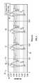
- FIG. 5is a schematic frequency-domain graph of a mechnomyography output signal in response to a periodic electrical stimulus.
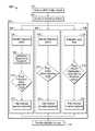
- FIG. 6is a schematic flow diagram of a method for detecting an artificially-induced mechanical muscle response to an electrical stimulus.
- FIG. 7is a schematic flow diagram of a method of inferring an artificially-induced mechanical muscle response from a free-running mechanomyography output signal.
- FIG. 1schematically illustrates a neural monitoring system 10 that may be used to identify the presence of one or more nerves within an intracorporeal treatment area 12 of a subject 14 .
- the system 10may monitor one or more muscles of the subject 14 for a mechanical motion, and may be capable of discriminating an artificially-induced mechanical response of a muscle (also referred to as an “artificially-induced mechanical muscle response”) from a subject-intended muscle contraction/relaxation and/or an environmentally caused movement. If an artificially-induced mechanical muscle response is detected during the procedure, the system 10 may provide an indication to a user.
- an artificially-induced mechanical muscle responserefers to a contraction or relaxation of a muscle in response to a stimulus that is not received through natural sensory means (e.g., sight, sound, taste, smell, and touch). Instead, it is a contraction/relaxation of a muscle that is induced by the application of a stimulus directly to a nerve that innervates the muscle.
- Examples of stimuli that may cause an “artificially-induced” muscle responsemay include an electrical current applied directly to the nerve or to intracorporeal tissue or fluid immediately surrounding the nerve. In this example, if the applied electrical current is sufficiently strong and/or sufficiently close to the nerve, it may artificially cause the nerve to depolarize (resulting in a corresponding contraction of the muscle innervated by that nerve).
- artificial stimulimay involve mechanically-induced depolarization (e.g., physically stretching or compressing a nerve, such as with a tissue retractor), thermally-induced depolarization (e.g., through ultrasonic cautery), or chemically-induced depolarization (e.g., through the application of a chemical agent to the tissue surrounding the nerve).
- mechanically-induced depolarizatione.g., physically stretching or compressing a nerve, such as with a tissue retractor
- thermally-induced depolarizatione.g., through ultrasonic cautery
- chemically-induced depolarizatione.g., through the application of a chemical agent to the tissue surrounding the nerve.
- a muscle innervated by the artificially depolarized nervemay physically contract or relax (i.e., a mechanical response).
- a mechanical reactionmay primarily occur along a longitudinal direction of the muscle (i.e., a direction aligned with the constituent fibers of the muscle), though may further result in a respective swelling/relaxing of the muscle in a lateral direction (which may be substantially normal to the skin for most skeletal muscles).
- This local movement of the muscle during an artificially-induced mechanical muscle responsemay be measured relative to the position of the muscle when in a non-stimulated state, and is distinguished from other global translations of the muscle
- the neural monitoring system 10may include a processor 20 that is in communication with at least one mechanical sensor 22 .
- the mechanical sensor 22may include, for example, a strain gauge, a force transducer, a position encoder, an accelerometer, a piezoelectric material, or any other transducer or combination of transducers that may convert a physical motion into a variable electrical signal.
- Each mechanical sensor 22may specially be configured to monitor a local mechanical movement of a muscle of the subject 14 .
- each sensor 22may include a fastening means, such as an adhesive material/patch, that allows the sensor 22 to be adhered, bandaged, or otherwise affixed to the skin of the subject 14 (i.e. affixed on an external skin surface).
- suitable fastening meansmay include bandages, sleeves, or other elastic fastening devices that may hold the sensor 22 in physical contact with the subject 14 .
- the mechanical sensor 22 (and/or coupled device)may be configured to monitor a local mechanical movement of a muscle by virtue of its physical design.
- the sensors/coupled devicesmay include catheters, balloons, bite guards, orifice plugs or endotracheal tubes that may be positioned within a lumen or natural opening of the subject to monitor a response of the lumen or orifice, or of a muscle that is directly adjacent to and/or connected with the lumen or orifice.
- the mechanical sensormay be a non-invasive device, whereby the term “non-invasive” is intended to mean that the sensor is not surgically placed within the body of the subject (i.e., via cutting of tissue to effectuate the placement).
- non-invasive sensorsmay include sensors that are placed within naturally occurring body lumens that are accessible without the need for an incision.
- the senor 22may include a contact detection device, that may provide an indication if the sensor 22 is in physical contact with the skin of the subject 14 .
- the contact detection devicemay, for example, include a pair of electrodes that are configured to contact the skin of the subject 14 when the sensor 22 is properly positioned. The sensor 22 /contact detection device may then monitor an impedance between the electrodes to determine whether the electrodes are in contact with the skin.
- Other examples of suitable contact detection devicesmay include capacitive touch sensors or buttons that protrude slightly beyond the surface of the sensor.
- the system 10may further include one or more elongate medical instruments 30 that are capable of selectively providing a stimulus within the intracorporeal treatment area 12 of the subject 14 (i.e., also referred to as a stimulator 30 ).
- the elongate medical instrument 30may include a probe 32 (e.g., a ball-tip probe, k-wire, or needle) that has an electrode 34 disposed on a distal end portion 36 .
- the electrode 34may be selectively electrified, at either the request of a user/physician, or at the command of the processor 20 , to provide an electrical stimulus 38 to intracorporeal tissue of the subject.
- the elongate medical instrument 30may include a dialator, retractor, clip, cautery probe, pedicle screw, or any other medical instrument that may be used in an invasive medical procedure. Regardless of the instrument, if the intended artificial stimulus is an electrical current, the instrument 30 may include a selectively electrifiable electrode 34 disposed at a portion of the instrument that is intended to contact tissue within the intracorporeal treatment area 12 during a procedure.
- the user/surgeonmay selectively administer the stimulus to intracorporeal tissue within the treatment area 12 to identify the presence of one or more nerve bundles or fibers.
- the user/surgeonmay administer the stimulus, for example, upon depressing a button or foot pedal that is in communication with the system 10 , and more specifically in communication with the stimulator 30 .
- the electrical stimulus 38may, for example, be a discrete pulse (e.g., a step pulse) having a pulse width within the range of about 30 ⁇ s to about 500 ⁇ s.
- the discrete pulsemay have a pulse width within the range of about 50 ⁇ s to about 200 ⁇ s, or within the range of about 75 ⁇ s to about 125 ⁇ s.
- the discrete pulsemay be periodically applied at a frequency of, for example, between about 1 Hz and about 10 Hz.
- the electrical stimulus 38may cause the nerve to depolarize, resulting in a mechanical twitch of a muscle that is innervated by the nerve (i.e., an artificially-induced mechanical muscle response).
- a mechanical twitch of a muscle that is innervated by the nervei.e., an artificially-induced mechanical muscle response.
- the magnitude of the response/twitchmay be directly correlated to the distance between the electrode and the nerve, and the magnitude of the stimulus current.
- a lookup tablemay be employed by the processor 20 to provide an approximate distance between the electrode and the nerve, given a known stimulus magnitude and a measured mechanical muscle response.
- the one or more mechanical sensors 22may be placed in mechanical communication with one or more muscles of the subject 14 .
- a sensor 22may be in mechanical communication with the muscle if it can physically detect a movement, velocity, acceleration, strain or other physical response of the muscle, either via direct contact with the muscle, or via a mechanical relationship through one or more intermediate materials and/or tissues (e.g., skin and/or subcutaneous tissue).
- FIG. 2illustrates an example of the placement of a plurality of mechanical sensors 22 for a surgical procedure that may occur proximate the L2, L3, and/or L4 vertebrae of the lumbar spine (shown schematically in FIG. 3 ).
- the nerves 50 , 52 and 54 exiting the L2, L3 and L4 foramen 56 , 58 , 60may therefore either lie within the treatment area 12 (i.e., the area surrounding the L2, L3, and/or L4 vertebrae), or may be immediately proximate to this area.
- the surgeonmay understand that damage to these nerves 50 , 52 , 54 may affect the functioning of the vastus medialis muscles and the tibialis anterior muscles.
- the surgeonmay place mechanical sensors 22 a - 22 d on or near the vastus medialis muscles and the tibialis anterior muscles to guard against inadvertent manipulation of the nerves during the procedure.
- mechanical sensors 22 a and 22 bare placed on the vastus medialis muscles, which are innervated by the nerves 50 , 52 exiting the L2 and L3 foramen 56 , 58
- sensors 22 c and 22 dare placed on the tibialis anterior muscles, which are innervated by the nerves 54 exiting the L4 foramen 60 .
- each mechanical sensor 22may generate a mechanomyography (MMG) output signal (schematically shown at 62 ) that corresponds to a sensed mechanical movement/response of the adjacent muscle.
- the MMG output signal 62may be either a digital or analog signal, and may typically be provided to the processor 20 through either wired or wireless communication means (e.g., through a physical wire, or using radio frequency communication protocols, such as according to IEEE 802.11 or another protocol such as Bluetooth).
- the MMG output signal 62is intended to be separate and distinct from any electrical potentials of the muscle or skin (often referred to as electromyography (EMG) signals). While electrical (EMG) and mechanical (MMG) muscle responses may be related, their relationship is complex, and not easily described (e.g., electrical potentials are very location specific, with a potentially variable electrical potential across the volume of the muscle of interest).
- the processor 20may be in communication with the stimulator 30 and the mechanical sensor 22 , and may be configured to receive the MMG output signal 62 from the mechanical sensor 22 .
- the processor 20may be embodied as one or multiple digital computers, data processing devices, and/or digital signal processors (DSPs), which may have one or more microcontrollers or central processing units (CPUs), read only memory (ROM), random access memory (RAM), electrically-erasable programmable read only memory (EEPROM), a high-speed clock, analog-to-digital (A/D) circuitry, digital-to-analog (D/A) circuitry, input/output (I/O) circuitry, and/or signal conditioning and buffering electronics.
- DSPsdigital signal processors
- the processor 20may be configured to automatically perform one or more signal processing algorithms 70 or methods to determine whether a sensed mechanical movement (i.e., via the MMG output signal 62 ) is representative of an artificially-induced mechanical muscle response or if it is merely a subject-intended muscle movement and/or an environmentally caused movement.
- These processing algorithms 70may be embodied as software or firmware, and may either be stored locally on the processor 20 , or may be readily assessable by the processor 20 .
- FIG. 4generally illustrates a graph 80 of an MMG output signal 82 in response to a periodic electrical stimulus 38 provided proximate to a nerve.
- This MMG output signal 82may be in response to an electrical stimulus 38 that is provided at about a 3 Hz frequency.
- the MMG output signal 82has an amplitude 84 that varies as a function of time 86 and includes a plurality of generally discrete contraction events 88 .
- Each contraction event 88may include, for example, an initial response 90 , and a plurality of subsequent peaks/valleys 92 that are smaller than the initial response 90 .
- the muscle contractionsmay be easily represented in the time domain (as generally illustrated by the graph 80 in FIG. 4 ), they may also be represented in the frequency domain (as generally illustrated by the graph 94 in FIG. 5 ).
- the frequency domainrepresents a signal as a plurality of discrete frequency components 96 , each having a respective magnitude 98 . It has been found that an induced muscle response has identifiable frequency content in the range of about 0 Hz to about 50 Hz, with a majority of the signal energy being in the range of about 0 Hz to about 20 Hz.
- FIGS. 6 and 7generally illustrate two methods 100 , 200 for detecting an artificially-induced mechanical muscle response, both occurring in the frequency domain.
- the method 100 provided in FIG. 6may operate by correlating the MMG output signal 62 in the frequency domain to an applied electrical stimulus 38 .
- the method 200 provided in FIG. 7may operate by solely monitoring the MMG output signal 62 in the frequency domain (i.e., a “free-run” MMG monitoring technique).
- the two provided methods 100 , 200may be used individually, or may be combined together to provide a more robust detection scheme.
- the flow diagram of FIG. 6generally illustrates a method 100 for detecting an artificially-induced mechanical muscle response to an electrical stimulus 38 that is provided proximate to a nerve.
- the method 100begins when the processor 20 receives the MMG output signal 62 from the mechanical sensor 22 (at step 102 ).
- the MMG output signal 62may be received as a time domain signal, and may potentially be filtered (either by circuitry associated with the mechanical sensor 22 or processor 20 , or through digital filtering techniques performed by the processor 20 ) to remove high frequency noise (such filtering is shown by example in FIG. 4 via the two data traces).
- the MMG output signal 62may be filtered using a low pass filter having a cutoff frequency greater than about 200 Hz.
- a low pass filtermay be used that has a cut off frequency greater than about 50 Hz, or greater than about 20 Hz, or in the range of about 20 Hz to about 50 Hz.
- the MMG output signal 62may be further filtered by a high pass filter.
- the high pass filtermay have a cut off frequency that is greater than about 0 Hz though less than about 10 Hz.
- the processor 20may convert the received signal 62 from the time domain to the frequency domain (at step 104 ). Such a conversion may occur using, for example, Fourier Methods (e.g., a Fourier Transform, a Fast Fourier Transform, or a Discrete Fourier Transform), or through other similar methodologies.
- the processor 20may then determine one or more frequency components that have a peak magnitude (at step 106 ). While numerous methods may be used to detect magnitude peaks, the most basic method includes identifying one or more frequencies that have magnitudes greater than the magnitudes of adjacent frequencies.
- FIG. 5generally illustrates at least 6 detectable peaks/peak magnitudes 150 . As shown, these peaks 150 correspond to frequencies of about 3 Hz, about 6 Hz, about 9 Hz, about 12 Hz, about 15 Hz, and about 18 Hz.
- the processor 20may determine whether an artificially-induced mechanical muscle response has been sensed by the mechanical sensor 22 (at step 108 ). If the processor 20 detects such an induced muscle response, then it may provide an indication to a user corresponding to the detected event (at step 110 ).
- this indicationmay include one or more of an illuminated light/LED, a colored light/LED, a textual or symbolic alert on a display device associated with the processor 20 , a vibration in the handle of the stimulator, and an audible alert such as a single frequency alert, a multiple frequency alert, and/or an audible natural language alert.
- the indication/alertmay include an estimation of the proximity between the electrode and the nerve, such as may be derived using a lookup table as described above, or as explained in U.S. Pat. No. 8,343,065 to Bartol, et al., entitled “NEURAL EVENT DETECTION,” which is hereby incorporated by reference in its entirety and for all of the disclosure setforth therein.
- the processor 20may employ two strategies ( 112 , 114 ) that the processor 20 may employ to determine whether a sensed movement of the mechanical sensor 22 is indicative of an artificially-induced mechanical muscle response.
- These strategies 112 , 114may both operate by attempting to correlate the detected frequency peaks of the MMG output signal with an attribute of the provided electrical stimulus 38 .
- the processor 20may use either of these strategies alone, or it may combine them both together into a single strategy (i.e., either performed concurrently, sequentially, or in combination).
- the processor 20may receive an indication of the frequency at which the electrical stimulus 38 is administered (at 118 ). This frequency indication may either be received directly from the stimulator, or from a register or memory location within the processor itself.
- the processor 20may examine the frequencies corresponding to magnitude peaks from step 106 , and determine whether any of the identified frequencies is an integer multiple of the stimulation frequency.
- the processor 20may identify that the sensed response is indicative of an artificially-induced mechanical muscle response if it is determined that one or more of the identified frequencies is an integer multiple of the stimulation frequency. In other configurations, this determination may require that two or more, or even three or more frequencies are integer multiples of the stimulation frequency before an artificially-induced mechanical muscle response is identified.
- the processor 20may similarly receive an indication of the frequency at which the electrical stimulus 38 is administered (at 118 ).
- the processor 20may examine the frequencies corresponding to magnitude peaks from step 106 , and determine a fundamental frequency of the MMG output signal 62 . This may be accomplished, for example, by determining a greatest common factor of a plurality of the frequencies where magnitude peaks are detected. This technique applied to the peaks 150 shown in FIG. 6 would result in a fundamental frequency of 3 Hz (i.e., a common factor of 3, 6, 9, 12, 15, and 18). In most cases, the determined fundamental frequency may lie within the range of about 1 Hz to about 10 Hz (i.e., the typical range of simulation frequencies).
- the processor 20may determine whether the fundamental frequency is either equal to, or an integer multiple of the stimulation frequency, and if so, in step 122 , the processor 20 may identify that the sensed response is indicative of an artificially-induced mechanical muscle response.
- the method 100 illustrated in FIG. 6may generally provide a high-confidence indication that a nerve is proximate to the stimulator by correlating an applied stimulus with a monitored response.
- the use of a stimulatormay either be impractical, or may not adequately assess the risks posed to nerves that are further away.
- stimulated detectionmay be extremely useful when making a lateral transpsoas approach to the spine
- the subsequent use of a mechanical retractor to provide an operating corridormay affect nerves that are outside of the electrically-stimulated detection radius.
- One embodiment of a free-running detection algorithmis shown by the method 200 provided in FIG. 7 . While this method 200 may only provide an “inference” of an artificially-induced mechanical muscle response (i.e., due to the absence of a direct correlation), it may be equally valuable in some circumstances.
- the free-running detection method 200may be similar to the method 100 of FIG. 6 , though may make detection inferences without the knowledge of a stimulation frequency.
- the method 200begins when the processor 20 receives the MMG output signal 62 from the mechanical sensor 22 (at step 102 ), and subsequently converts the MMG output signal 62 into the frequency domain (step 104 ).
- the processor 20may determine (i.e., infer) whether sensed motion detected by the mechanical sensor 22 is indicative of an artificially-induced mechanical muscle response. If the processor 20 does determine that such an induced muscle response has occurred, it may then provide an indication to a user of such an event (at step 110 ).
- step 202there may be three free-run detection strategies ( 204 , 206 , 208 ) that the processor 20 may employ to determine/infer whether a sensed movement of the mechanical sensor 22 is indicative of an artificially-induced mechanical muscle response.
- These free-run strategies 204 , 206 , 208may operate by monitoring the MMG output signal, and attempting to detect signal attributes that may be indicative of an artificially-induced mechanical muscle response.
- the processor 20may use any of these strategies alone, or it may combine two or more of them together into a single strategy (i.e., either performed concurrently, sequentially, or in combination, and/or may be combined with the stimulated techniques of FIG. 6 ).
- the processor 20may first determine one or more frequency components that have a peak magnitude (at step 106 ). In step 124 , the processor 20 may examine the frequencies corresponding to magnitude peaks from step 106 , and determine a fundamental frequency of the MMG output signal 62 . Finally, the processor 20 may compare the determined fundamental frequency to a range of expected fundamental frequencies for an artificially-induced mechanical muscle response in step 210 . Such a range may be, for example, between about 1 Hz and about 10 Hz. If the determined fundamental frequency falls within this range, the processor 20 may infer that the sensed mechanical sensor movement is indicative of an artificially-induced mechanical muscle response (in step 212 ).
- the processor 20may begin by determining one or more frequency components that have a peak magnitude (at step 106 ). Once the peaks are identified, the processor 20 may compare the peaks to a range of frequencies where frequency content is expected to exist for an artificially-induced mechanical muscle response (in step 214 ). Such a range may be, for example between about 1 Hz and about 20 Hz. In one configuration, if one or more of the identified peaks are within this range, the processor 20 may infer that the sensed response is indicative of an artificially-induced mechanical muscle response in step 212 . In other configurations, the processor 20 may require that two or more, or even three or more of the identified peaks lie within the range before it infers that the sensed response is indicative of an artificially-induced mechanical muscle response.
- the processor 20does not necessarily need to compute the frequencies corresponding to magnitude peaks, instead, it may first establish a noise floor at step 216 , and then it may determine if any of the respective frequency magnitudes exceed the noise floor by a threshold amount (in step 218 ). If so, the processor 20 may infer that the sensed response is indicative of an artificially-induced mechanical muscle response in step 212 .
- the noise floormay generally represent the normal background mechanical noise/movement that may be reported by the sensor. It may be a function of the precision of the transducer within the sensor, it may include received electromagnetic radiation, and/or it may include mechanical movement that may be attributable to continuous rhythmic events such as breathing or heart beat.
- the noise floormay either have a varying magnitude for each frequency, or may generally be a constant value across all frequencies.
- the thresholdmay be either a fixed amount above the noise floor, or may be a multiple of the noise floor (e.g., a level twice the noise floor, or a level that is set about one or more standard deviations above an average noise level across a period of time).
- an MMG output signal that is characteristic of an induced muscle responsemay include peaks at 3 hz, 5 hz, 6 hz, 9 hz, 10 hz, 12 hz, 15 hz within the frequency domain. Also, peaks may be apparent at multiples of the difference in the frequencies (i.e., 2 Hz, 4 Hz, 8 Hz, etc)
- the present nerve monitoring system 10 and described artificially-induced mechanical muscle response detection algorithmsmay be used by a robotic surgical system, such as described in U.S. patent application Ser. No. 13/428,693, filed 23 Mar. 2012, entitled “ROBOTIC SURGICAL SYSTEM WITH MECHANOMYOGRAPHY FEEDBACK,” which is incorporated by reference in its entirety and for all of the disclosure setforth therein.
- the above-described neural monitoring system 10may be used to provide one or more control signals to a robotic surgical system if an artificially-induced mechanical muscle response is detected.
- the one or more elongate medical instruments 30 described abovemay be robotically controlled in up to 6 or more degrees of freedom/motion by a robotic controller.
- This instrumentmay be configured to perform a surgical procedure within an intracorporeal treatment area at the direction of the robotic controller, and may provide an electrical stimulus 38 in the manner described above. If an artificially-induced mechanical muscle response is detected, the neural monitoring system 10 may instruct the robotic controller (via the provided control signal) to limit the range of available motion of the elongate medical instrument 30 and/or to prevent an actuation of an end effector that may be disposed on the instrument 30 and controllable by the robotic controller.
Landscapes
- Health & Medical Sciences (AREA)
- Life Sciences & Earth Sciences (AREA)
- Engineering & Computer Science (AREA)
- Surgery (AREA)
- Biomedical Technology (AREA)
- Veterinary Medicine (AREA)
- Public Health (AREA)
- Physiology (AREA)
- Dentistry (AREA)
- Oral & Maxillofacial Surgery (AREA)
- Physics & Mathematics (AREA)
- General Health & Medical Sciences (AREA)
- Biophysics (AREA)
- Pathology (AREA)
- Animal Behavior & Ethology (AREA)
- Heart & Thoracic Surgery (AREA)
- Medical Informatics (AREA)
- Molecular Biology (AREA)
- Anesthesiology (AREA)
- Chemical & Material Sciences (AREA)
- Neurology (AREA)
- Medicinal Chemistry (AREA)
- Bioinformatics & Cheminformatics (AREA)
- Measurement And Recording Of Electrical Phenomena And Electrical Characteristics Of The Living Body (AREA)
Abstract
Description
The present invention relates generally to a surgical diagnostic system for detecting the presence of one or more nerves.
Traditional surgical practices emphasize the importance of recognizing or verifying the location of nerves to avoid injuring them. Advances in surgical techniques include development of techniques including ever smaller exposures, such as minimally invasive surgical procedures, and the insertion of ever more complex medical devices. With these advances in surgical techniques, there is a corresponding need for improvements in methods of detecting and/or avoiding nerves.
A neural monitoring system is provided that is capable of detecting an artificially-induced mechanical response of a muscle to an electrical stimulus that is provided within an intracorporeal treatment area of a human subject. The electrical stimulus may be a periodic stimulus provided at a stimulation frequency, and having a predetermined pulse width. Additionally, the intracorporeal treatment area generally includes a nerve that innervates the monitored muscle.
The neural monitoring system includes a non-invasive mechanical sensor and a processor. The non-invasive mechanical sensor is configured to be placed in mechanical communication with the muscle and to generate a mechanomyography output signal corresponding to a sensed mechanical movement of the muscle. By non-invasive, it is intended that the mechanical sensor does not require an incision or related surgical procedure to be properly positioned. Instead, it may be held in contact with an external surface of the skin, or may be positioned within a naturally occurring lumen/orifice. The mechanical sensor may generally include an accelerometer, a microphone, a strain gauge, or a piezoelectric device.
The processor is in communication with the mechanical sensor and is configured to receive the mechanomyography output signal from the mechanical sensor. In one configuration, the processor may determine a frequency component of the mechanomyography output signal that has a peak magnitude relative to one or more adjacent frequencies. The processor may provide an indication to a user if the determined frequency component is either equal to, or an integer multiple of the stimulation frequency. In another configuration, the processor may determine a fundamental frequency of the mechanomyography output signal, and provide an indication to a user if the determined fundamental frequency is either equal to, or an integer multiple of the stimulation frequency.
The above features and advantages and other features and advantages of the present invention are readily apparent from the following detailed description of the best modes for carrying out the invention when taken in connection with the accompanying drawings.
“A,” “an,” “the,” “at least one,” and “one or more” are used interchangeably to indicate that at least one of the item is present; a plurality of such items may be present unless the context clearly indicates otherwise. All numerical values of parameters (e.g., of quantities or conditions) in this specification, including the appended claims, are to be understood as being modified in all instances by the term “about” whether or not “about” actually appears before the numerical value. “About” indicates that the stated numerical value allows some slight imprecision (with some approach to exactness in the value; about or reasonably close to the value; nearly). If the imprecision provided by “about” is not otherwise understood in the art with this ordinary meaning, then “about” as used herein indicates at least variations that may arise from ordinary methods of measuring and using such parameters. In addition, disclosure of ranges includes disclosure of all values and further divided ranges within the entire range. Each value within a range and the endpoints of a range are hereby all disclosed as separate embodiment.
Referring to the drawings, wherein like reference numerals are used to identify like or identical components in the various views,FIG. 1 schematically illustrates aneural monitoring system 10 that may be used to identify the presence of one or more nerves within anintracorporeal treatment area 12 of asubject 14. As will be described in greater detail below, thesystem 10 may monitor one or more muscles of thesubject 14 for a mechanical motion, and may be capable of discriminating an artificially-induced mechanical response of a muscle (also referred to as an “artificially-induced mechanical muscle response”) from a subject-intended muscle contraction/relaxation and/or an environmentally caused movement. If an artificially-induced mechanical muscle response is detected during the procedure, thesystem 10 may provide an indication to a user.
As used herein, an artificially-induced mechanical muscle response refers to a contraction or relaxation of a muscle in response to a stimulus that is not received through natural sensory means (e.g., sight, sound, taste, smell, and touch). Instead, it is a contraction/relaxation of a muscle that is induced by the application of a stimulus directly to a nerve that innervates the muscle. Examples of stimuli that may cause an “artificially-induced” muscle response may include an electrical current applied directly to the nerve or to intracorporeal tissue or fluid immediately surrounding the nerve. In this example, if the applied electrical current is sufficiently strong and/or sufficiently close to the nerve, it may artificially cause the nerve to depolarize (resulting in a corresponding contraction of the muscle innervated by that nerve). Other examples of such “artificial stimuli” may involve mechanically-induced depolarization (e.g., physically stretching or compressing a nerve, such as with a tissue retractor), thermally-induced depolarization (e.g., through ultrasonic cautery), or chemically-induced depolarization (e.g., through the application of a chemical agent to the tissue surrounding the nerve).
During an artificially-induced mechanical muscle response, a muscle innervated by the artificially depolarized nerve may physically contract or relax (i.e., a mechanical response). Such a mechanical reaction may primarily occur along a longitudinal direction of the muscle (i.e., a direction aligned with the constituent fibers of the muscle), though may further result in a respective swelling/relaxing of the muscle in a lateral direction (which may be substantially normal to the skin for most skeletal muscles). This local movement of the muscle during an artificially-induced mechanical muscle response may be measured relative to the position of the muscle when in a non-stimulated state, and is distinguished from other global translations of the muscle
Theneural monitoring system 10 may include aprocessor 20 that is in communication with at least onemechanical sensor 22. Themechanical sensor 22 may include, for example, a strain gauge, a force transducer, a position encoder, an accelerometer, a piezoelectric material, or any other transducer or combination of transducers that may convert a physical motion into a variable electrical signal.
Eachmechanical sensor 22 may specially be configured to monitor a local mechanical movement of a muscle of thesubject 14. For example, eachsensor 22 may include a fastening means, such as an adhesive material/patch, that allows thesensor 22 to be adhered, bandaged, or otherwise affixed to the skin of the subject14 (i.e. affixed on an external skin surface). Other examples of suitable fastening means may include bandages, sleeves, or other elastic fastening devices that may hold thesensor 22 in physical contact with thesubject 14. Alternatively, the mechanical sensor22 (and/or coupled device) may be configured to monitor a local mechanical movement of a muscle by virtue of its physical design. For example, the sensors/coupled devices may include catheters, balloons, bite guards, orifice plugs or endotracheal tubes that may be positioned within a lumen or natural opening of the subject to monitor a response of the lumen or orifice, or of a muscle that is directly adjacent to and/or connected with the lumen or orifice. In one configuration, the mechanical sensor may be a non-invasive device, whereby the term “non-invasive” is intended to mean that the sensor is not surgically placed within the body of the subject (i.e., via cutting of tissue to effectuate the placement). For the purposes of this disclosure, non-invasive sensors may include sensors that are placed within naturally occurring body lumens that are accessible without the need for an incision.
In one configuration, thesensor 22 may include a contact detection device, that may provide an indication if thesensor 22 is in physical contact with the skin of thesubject 14. The contact detection device may, for example, include a pair of electrodes that are configured to contact the skin of thesubject 14 when thesensor 22 is properly positioned. Thesensor 22/contact detection device may then monitor an impedance between the electrodes to determine whether the electrodes are in contact with the skin. Other examples of suitable contact detection devices may include capacitive touch sensors or buttons that protrude slightly beyond the surface of the sensor.
Thesystem 10 may further include one or more elongatemedical instruments 30 that are capable of selectively providing a stimulus within theintracorporeal treatment area 12 of the subject14 (i.e., also referred to as a stimulator30). For example, in one configuration, the elongatemedical instrument 30 may include a probe32 (e.g., a ball-tip probe, k-wire, or needle) that has anelectrode 34 disposed on adistal end portion 36. Theelectrode 34 may be selectively electrified, at either the request of a user/physician, or at the command of theprocessor 20, to provide anelectrical stimulus 38 to intracorporeal tissue of the subject. In other configurations, the elongatemedical instrument 30 may include a dialator, retractor, clip, cautery probe, pedicle screw, or any other medical instrument that may be used in an invasive medical procedure. Regardless of the instrument, if the intended artificial stimulus is an electrical current, theinstrument 30 may include a selectivelyelectrifiable electrode 34 disposed at a portion of the instrument that is intended to contact tissue within theintracorporeal treatment area 12 during a procedure.
During a surgical procedure, the user/surgeon may selectively administer the stimulus to intracorporeal tissue within thetreatment area 12 to identify the presence of one or more nerve bundles or fibers. For anelectrical stimulus 38, the user/surgeon may administer the stimulus, for example, upon depressing a button or foot pedal that is in communication with thesystem 10, and more specifically in communication with thestimulator 30. Theelectrical stimulus 38 may, for example, be a discrete pulse (e.g., a step pulse) having a pulse width within the range of about 30 μs to about 500 μs. In other examples, the discrete pulse may have a pulse width within the range of about 50 μs to about 200 μs, or within the range of about 75 μs to about 125 μs. The discrete pulse may be periodically applied at a frequency of, for example, between about 1 Hz and about 10 Hz.
If a nerve extends within a predetermined distance of theelectrode 34, theelectrical stimulus 38 may cause the nerve to depolarize, resulting in a mechanical twitch of a muscle that is innervated by the nerve (i.e., an artificially-induced mechanical muscle response). In general, the magnitude of the response/twitch may be directly correlated to the distance between the electrode and the nerve, and the magnitude of the stimulus current. In one configuration, a lookup table may be employed by theprocessor 20 to provide an approximate distance between the electrode and the nerve, given a known stimulus magnitude and a measured mechanical muscle response.
Prior to beginning a surgical procedure, the one or moremechanical sensors 22 may be placed in mechanical communication with one or more muscles of the subject14. In the present context, asensor 22 may be in mechanical communication with the muscle if it can physically detect a movement, velocity, acceleration, strain or other physical response of the muscle, either via direct contact with the muscle, or via a mechanical relationship through one or more intermediate materials and/or tissues (e.g., skin and/or subcutaneous tissue).
In general, eachmechanical sensor 22 may generate a mechanomyography (MMG) output signal (schematically shown at62) that corresponds to a sensed mechanical movement/response of the adjacent muscle. TheMMG output signal 62 may be either a digital or analog signal, and may typically be provided to theprocessor 20 through either wired or wireless communication means (e.g., through a physical wire, or using radio frequency communication protocols, such as according to IEEE 802.11 or another protocol such as Bluetooth). As a specific signal, theMMG output signal 62 is intended to be separate and distinct from any electrical potentials of the muscle or skin (often referred to as electromyography (EMG) signals). While electrical (EMG) and mechanical (MMG) muscle responses may be related, their relationship is complex, and not easily described (e.g., electrical potentials are very location specific, with a potentially variable electrical potential across the volume of the muscle of interest).
Referring again toFIG. 1 , theprocessor 20 may be in communication with thestimulator 30 and themechanical sensor 22, and may be configured to receive theMMG output signal 62 from themechanical sensor 22. Theprocessor 20 may be embodied as one or multiple digital computers, data processing devices, and/or digital signal processors (DSPs), which may have one or more microcontrollers or central processing units (CPUs), read only memory (ROM), random access memory (RAM), electrically-erasable programmable read only memory (EEPROM), a high-speed clock, analog-to-digital (A/D) circuitry, digital-to-analog (D/A) circuitry, input/output (I/O) circuitry, and/or signal conditioning and buffering electronics.
Theprocessor 20 may be configured to automatically perform one or moresignal processing algorithms 70 or methods to determine whether a sensed mechanical movement (i.e., via the MMG output signal62) is representative of an artificially-induced mechanical muscle response or if it is merely a subject-intended muscle movement and/or an environmentally caused movement. Theseprocessing algorithms 70 may be embodied as software or firmware, and may either be stored locally on theprocessor 20, or may be readily assessable by theprocessor 20.
While the muscle contractions may be easily represented in the time domain (as generally illustrated by thegraph 80 inFIG. 4 ), they may also be represented in the frequency domain (as generally illustrated by thegraph 94 inFIG. 5 ). In general, the frequency domain represents a signal as a plurality ofdiscrete frequency components 96, each having a respective magnitude98. It has been found that an induced muscle response has identifiable frequency content in the range of about 0 Hz to about 50 Hz, with a majority of the signal energy being in the range of about 0 Hz to about 20 Hz.
The flow diagram ofFIG. 6 generally illustrates amethod 100 for detecting an artificially-induced mechanical muscle response to anelectrical stimulus 38 that is provided proximate to a nerve. As shown, themethod 100 begins when theprocessor 20 receives theMMG output signal 62 from the mechanical sensor22 (at step102). TheMMG output signal 62 may be received as a time domain signal, and may potentially be filtered (either by circuitry associated with themechanical sensor 22 orprocessor 20, or through digital filtering techniques performed by the processor20) to remove high frequency noise (such filtering is shown by example inFIG. 4 via the two data traces). For example, in one configuration, theMMG output signal 62 may be filtered using a low pass filter having a cutoff frequency greater than about 200 Hz. In other embodiments, a low pass filter may be used that has a cut off frequency greater than about 50 Hz, or greater than about 20 Hz, or in the range of about 20 Hz to about 50 Hz. In addition to filtering through a low pass filter, theMMG output signal 62 may be further filtered by a high pass filter. In one configuration, the high pass filter may have a cut off frequency that is greater than about 0 Hz though less than about 10 Hz.
After theprocessor 20 receives theMMG output signal 62 it may convert the receivedsignal 62 from the time domain to the frequency domain (at step104). Such a conversion may occur using, for example, Fourier Methods (e.g., a Fourier Transform, a Fast Fourier Transform, or a Discrete Fourier Transform), or through other similar methodologies. Once in the frequency domain, theprocessor 20 may then determine one or more frequency components that have a peak magnitude (at step106). While numerous methods may be used to detect magnitude peaks, the most basic method includes identifying one or more frequencies that have magnitudes greater than the magnitudes of adjacent frequencies.FIG. 5 generally illustrates at least 6 detectable peaks/peak magnitudes 150. As shown, thesepeaks 150 correspond to frequencies of about 3 Hz, about 6 Hz, about 9 Hz, about 12 Hz, about 15 Hz, and about 18 Hz.
Referring again toFIG. 6 , once theMMG output signal 62 has been converted into the frequency domain (step104) and a subset of frequencies that correspond to magnitude peaks have been identified (step106), theprocessor 20 may determine whether an artificially-induced mechanical muscle response has been sensed by the mechanical sensor22 (at step108). If theprocessor 20 detects such an induced muscle response, then it may provide an indication to a user corresponding to the detected event (at step110). In one configuration, this indication may include one or more of an illuminated light/LED, a colored light/LED, a textual or symbolic alert on a display device associated with theprocessor 20, a vibration in the handle of the stimulator, and an audible alert such as a single frequency alert, a multiple frequency alert, and/or an audible natural language alert. Moreover, the indication/alert may include an estimation of the proximity between the electrode and the nerve, such as may be derived using a lookup table as described above, or as explained in U.S. Pat. No. 8,343,065 to Bartol, et al., entitled “NEURAL EVENT DETECTION,” which is hereby incorporated by reference in its entirety and for all of the disclosure setforth therein.
Referring back to step108, in general, there may be two strategies (112,114) that theprocessor 20 may employ to determine whether a sensed movement of themechanical sensor 22 is indicative of an artificially-induced mechanical muscle response. Thesestrategies electrical stimulus 38. In practice, theprocessor 20 may use either of these strategies alone, or it may combine them both together into a single strategy (i.e., either performed concurrently, sequentially, or in combination).
In thefirst detection strategy 112, theprocessor 20 may receive an indication of the frequency at which theelectrical stimulus 38 is administered (at118). This frequency indication may either be received directly from the stimulator, or from a register or memory location within the processor itself. Instep 120, theprocessor 20 may examine the frequencies corresponding to magnitude peaks fromstep 106, and determine whether any of the identified frequencies is an integer multiple of the stimulation frequency. Finally, instep 122 theprocessor 20 may identify that the sensed response is indicative of an artificially-induced mechanical muscle response if it is determined that one or more of the identified frequencies is an integer multiple of the stimulation frequency. In other configurations, this determination may require that two or more, or even three or more frequencies are integer multiples of the stimulation frequency before an artificially-induced mechanical muscle response is identified.
In thesecond detection strategy 114, theprocessor 20 may similarly receive an indication of the frequency at which theelectrical stimulus 38 is administered (at118). Instep 124, theprocessor 20 may examine the frequencies corresponding to magnitude peaks fromstep 106, and determine a fundamental frequency of theMMG output signal 62. This may be accomplished, for example, by determining a greatest common factor of a plurality of the frequencies where magnitude peaks are detected. This technique applied to thepeaks 150 shown inFIG. 6 would result in a fundamental frequency of 3 Hz (i.e., a common factor of 3, 6, 9, 12, 15, and 18). In most cases, the determined fundamental frequency may lie within the range of about 1 Hz to about 10 Hz (i.e., the typical range of simulation frequencies). Instep 126, theprocessor 20 may determine whether the fundamental frequency is either equal to, or an integer multiple of the stimulation frequency, and if so, instep 122, theprocessor 20 may identify that the sensed response is indicative of an artificially-induced mechanical muscle response.
Themethod 100 illustrated inFIG. 6 may generally provide a high-confidence indication that a nerve is proximate to the stimulator by correlating an applied stimulus with a monitored response. In some procedures, however, the use of a stimulator may either be impractical, or may not adequately assess the risks posed to nerves that are further away. For example, while stimulated detection may be extremely useful when making a lateral transpsoas approach to the spine, the subsequent use of a mechanical retractor to provide an operating corridor may affect nerves that are outside of the electrically-stimulated detection radius. In this manner it may also be useful to have a free-running detection algorithm that may monitor for artificially-induced mechanical muscle responses that are attributable to, for example, mechanical stimuli. One embodiment of a free-running detection algorithm is shown by themethod 200 provided inFIG. 7 . While thismethod 200 may only provide an “inference” of an artificially-induced mechanical muscle response (i.e., due to the absence of a direct correlation), it may be equally valuable in some circumstances.
As shown inFIG. 7 , the free-runningdetection method 200 may be similar to themethod 100 ofFIG. 6 , though may make detection inferences without the knowledge of a stimulation frequency. As shown, themethod 200 begins when theprocessor 20 receives theMMG output signal 62 from the mechanical sensor22 (at step102), and subsequently converts theMMG output signal 62 into the frequency domain (step104). Instep 202, theprocessor 20 may determine (i.e., infer) whether sensed motion detected by themechanical sensor 22 is indicative of an artificially-induced mechanical muscle response. If theprocessor 20 does determine that such an induced muscle response has occurred, it may then provide an indication to a user of such an event (at step110).
Referring back to step202, in general, there may be three free-run detection strategies (204,206,208) that theprocessor 20 may employ to determine/infer whether a sensed movement of themechanical sensor 22 is indicative of an artificially-induced mechanical muscle response. These free-runstrategies processor 20 may use any of these strategies alone, or it may combine two or more of them together into a single strategy (i.e., either performed concurrently, sequentially, or in combination, and/or may be combined with the stimulated techniques ofFIG. 6 ).
In the first free-run detection strategy 204, theprocessor 20 may first determine one or more frequency components that have a peak magnitude (at step106). Instep 124, theprocessor 20 may examine the frequencies corresponding to magnitude peaks fromstep 106, and determine a fundamental frequency of theMMG output signal 62. Finally, theprocessor 20 may compare the determined fundamental frequency to a range of expected fundamental frequencies for an artificially-induced mechanical muscle response instep 210. Such a range may be, for example, between about 1 Hz and about 10 Hz. If the determined fundamental frequency falls within this range, theprocessor 20 may infer that the sensed mechanical sensor movement is indicative of an artificially-induced mechanical muscle response (in step212).
In the second free-run detection strategy 206, theprocessor 20 may begin by determining one or more frequency components that have a peak magnitude (at step106). Once the peaks are identified, theprocessor 20 may compare the peaks to a range of frequencies where frequency content is expected to exist for an artificially-induced mechanical muscle response (in step214). Such a range may be, for example between about 1 Hz and about 20 Hz. In one configuration, if one or more of the identified peaks are within this range, theprocessor 20 may infer that the sensed response is indicative of an artificially-induced mechanical muscle response instep 212. In other configurations, theprocessor 20 may require that two or more, or even three or more of the identified peaks lie within the range before it infers that the sensed response is indicative of an artificially-induced mechanical muscle response.
Finally, in the third free-run detection strategy,208 theprocessor 20 does not necessarily need to compute the frequencies corresponding to magnitude peaks, instead, it may first establish a noise floor atstep 216, and then it may determine if any of the respective frequency magnitudes exceed the noise floor by a threshold amount (in step218). If so, theprocessor 20 may infer that the sensed response is indicative of an artificially-induced mechanical muscle response instep 212.
The noise floor may generally represent the normal background mechanical noise/movement that may be reported by the sensor. It may be a function of the precision of the transducer within the sensor, it may include received electromagnetic radiation, and/or it may include mechanical movement that may be attributable to continuous rhythmic events such as breathing or heart beat. The noise floor may either have a varying magnitude for each frequency, or may generally be a constant value across all frequencies. In thisstrategy 208, the threshold may be either a fixed amount above the noise floor, or may be a multiple of the noise floor (e.g., a level twice the noise floor, or a level that is set about one or more standard deviations above an average noise level across a period of time).
While the stimulation-based methods/strategies ofFIG. 6 , as well as the graphs inFIGS. 4 and 5 generally illustrate the present detection schemes using only a single simulation frequency, these methods/strategies may be expanded to the use of multiple stimulation frequencies as well. For example, if a stimulator were to provide an electrical stimulus having both a 3 Hz component and a 5 Hz component, an MMG output signal that is characteristic of an induced muscle response may include peaks at 3 hz, 5 hz, 6 hz, 9 hz, 10 hz, 12 hz, 15 hz within the frequency domain. Also, peaks may be apparent at multiples of the difference in the frequencies (i.e., 2 Hz, 4 Hz, 8 Hz, etc)
In addition to use as a stand alone, or hand-held nerve monitoring apparatus, the presentnerve monitoring system 10 and described artificially-induced mechanical muscle response detection algorithms (as described within method100) may be used by a robotic surgical system, such as described in U.S. patent application Ser. No. 13/428,693, filed 23 Mar. 2012, entitled “ROBOTIC SURGICAL SYSTEM WITH MECHANOMYOGRAPHY FEEDBACK,” which is incorporated by reference in its entirety and for all of the disclosure setforth therein. In such a system, the above-describedneural monitoring system 10 may be used to provide one or more control signals to a robotic surgical system if an artificially-induced mechanical muscle response is detected. In such an embodiment, the one or more elongatemedical instruments 30 described above may be robotically controlled in up to 6 or more degrees of freedom/motion by a robotic controller. This instrument may be configured to perform a surgical procedure within an intracorporeal treatment area at the direction of the robotic controller, and may provide anelectrical stimulus 38 in the manner described above. If an artificially-induced mechanical muscle response is detected, theneural monitoring system 10 may instruct the robotic controller (via the provided control signal) to limit the range of available motion of the elongatemedical instrument 30 and/or to prevent an actuation of an end effector that may be disposed on theinstrument 30 and controllable by the robotic controller.
While the best modes for carrying out the invention have been described in detail, those familiar with the art to which this invention relates will recognize various alternative designs and embodiments for practicing the invention within the scope of the appended claims. It is intended that all matter contained in the above description or shown in the accompanying drawings shall be interpreted as illustrative only and not as limiting.
Claims (19)
1. A neural monitoring system for detecting an artificially-induced mechanical response of a muscle to an electrical stimulus provided within an intracorporeal treatment area of a human subject at a stimulation frequency, the intracorporeal treatment area including a nerve that innervates the muscle, the neural monitoring system comprising:
a non-invasive mechanical sensor configured to be placed in mechanical communication with the muscle and to generate a mechanomyography output signal corresponding to a sensed mechanical movement of the muscle; and
a processor in communication with the mechanical sensor and configured to:
receive the mechanomyography output signal from the mechanical sensor;
determine a frequency component of the mechanomyography output signal, wherein the frequency component has a peak magnitude relative to adjacent frequencies; and
provide an indication of an artificially-induced mechanical muscle response to a user if the determined frequency component is either equal to, or an integer multiple of the stimulation frequency.
2. The neural monitoring system ofclaim 1 , wherein the processor is further configured to attenuate frequency content of the mechanomyography output signal that is above a cutoff frequency.
3. The neural monitoring system ofclaim 2 , wherein the cutoff frequency is in the range of about 20 Hz to about 50 Hz.
4. The neural monitoring system ofclaim 1 , wherein the processor is configured to determine a frequency component of the mechanomyography output signal that has a peak magnitude by:
converting the received mechanomyography output signal from a time domain to a frequency domain, wherein the frequency domain is characterized by a plurality of discrete frequencies, each having a corresponding magnitude; and
identifying the frequency component from the plurality of discrete frequencies that corresponds to a magnitude peak.
5. The neural monitoring system ofclaim 1 , wherein the frequency component is within the range of greater than 0 Hz to about 20 Hz.
6. The neural monitoring system ofclaim 1 , wherein the mechanical sensor includes an accelerometer, a microphone, a strain gauge, or a piezoelectric device.
7. The neural monitoring system ofclaim 1 , wherein the frequency component is a fundamental frequency of the mechanomyography output signal.
8. The neural monitoring system ofclaim 1 , further comprising an elongate medical instrument that is configured to selectively provide the electrical stimulus within the intracorporeal treatment area of the subject at the stimulation frequency.
9. The neural monitoring system ofclaim 8 , wherein the electrical stimulus includes a periodic pulse having a pulse width between about 30 μs and about 500 μs.
10. A neural monitoring system for detecting an artificially-induced mechanical response of a muscle to an electrical stimulus provided within an intracorporeal treatment area of a human subject at a stimulation frequency, the intracorporeal treatment area including a nerve that innervates the muscle, the neural monitoring system comprising:
a non-invasive mechanical sensor configured to be placed in mechanical communication with the muscle and to generate a mechanomyography output signal corresponding to a sensed mechanical movement of the muscle; and
a processor in communication with the mechanical sensor and configured to:
receive the mechanomyography output signal from the mechanical sensor;
determine a fundamental frequency of the mechanomyography output signal;
provide an indication of an artificially-induced mechanical muscle response to a user if the determined fundamental frequency is either equal to, or an integer multiple of the stimulation frequency.
11. The neural monitoring system ofclaim 10 , wherein the processor is further configured to attenuate frequency content of the mechanomyography output signal that is above a cutoff frequency.
12. The neural monitoring system ofclaim 11 , wherein the cutoff frequency is in the range of about 20 Hz to about 50 Hz.
13. The neural monitoring system ofclaim 10 , wherein the processor is configured to determine a fundamental frequency of the mechanomyography output signal by:
converting the received mechanomyography output signal from a time domain to a frequency domain, wherein the frequency domain is characterized by a plurality of discrete frequencies, each having a corresponding magnitude;
identifying a subset of the plurality of discrete frequencies wherein each discrete frequency belonging to the subset corresponds to a respective magnitude peak; and
determining a greatest common factor of the frequencies belonging to the subset.
14. The neural monitoring system ofclaim 10 , wherein the fundamental frequency is within the range of greater than 0 Hz to about 10 Hz.
15. The neural monitoring system ofclaim 10 , wherein the mechanical sensor includes an accelerometer, a microphone, a strain gauge, or a piezoelectric device.
16. The neural monitoring system ofclaim 10 , further comprising an elongate medical instrument that is configured to selectively provide the electrical stimulus within the intracorporeal treatment area of the subject at the stimulation frequency.
17. The neural monitoring system ofclaim 16 , wherein the electrical stimulus includes a periodic pulse having a pulse width between about 30 μs and about 500 μs.
18. A neural monitoring system for detecting an artificially-induced mechanical response of a muscle to an electrical stimulus provided within an intracorporeal treatment area of a human subject at a stimulation frequency, the intracorporeal treatment area including a nerve that innervates the muscle, the neural monitoring system comprising:
a non-invasive mechanical sensor configured to be placed in mechanical communication with the muscle and to generate a mechanomyography output signal corresponding to a sensed mechanical movement of the muscle; and
a processor in communication with the mechanical sensor and configured to:
receive the mechanomyography output signal from the mechanical sensor;
determine a frequency component of the mechanomyography output signal, wherein the frequency component has a peak magnitude relative to adjacent frequencies; and
provide an indication of an artificially-induced mechanical muscle response to a user if the determined frequency component is either equal to, or an integer multiple of the stimulation frequency, and either:
the determined frequency component is within the range of greater than 0 Hz to about 20 Hz; or
the peak magnitude is above an established noise floor by a threshold amount.
19. The neural monitoring system ofclaim 18 , wherein the mechanical sensor includes an accelerometer, a microphone, a strain gauge, or a piezoelectric device.
Priority Applications (1)
| Application Number | Priority Date | Filing Date | Title |
|---|---|---|---|
| US13/965,457US10478096B2 (en) | 2013-08-13 | 2013-08-13 | Neural event detection |
Applications Claiming Priority (1)
| Application Number | Priority Date | Filing Date | Title |
|---|---|---|---|
| US13/965,457US10478096B2 (en) | 2013-08-13 | 2013-08-13 | Neural event detection |
Publications (2)
| Publication Number | Publication Date |
|---|---|
| US20150051506A1 US20150051506A1 (en) | 2015-02-19 |
| US10478096B2true US10478096B2 (en) | 2019-11-19 |
Family
ID=52467305
Family Applications (1)
| Application Number | Title | Priority Date | Filing Date |
|---|---|---|---|
| US13/965,457Active2037-08-16US10478096B2 (en) | 2013-08-13 | 2013-08-13 | Neural event detection |
Country Status (1)
| Country | Link |
|---|---|
| US (1) | US10478096B2 (en) |
Cited By (3)
| Publication number | Priority date | Publication date | Assignee | Title |
|---|---|---|---|---|
| US12279879B1 (en) | 2024-08-09 | 2025-04-22 | Neuralytix, Llc | System and method for empirical assessment of nerve health during a spinal decompression procedure |
| US12279880B1 (en) | 2024-08-09 | 2025-04-22 | Neuralytix, Llc | System and method for assessing nerve health during a spinal decompression procedure |
| US12279881B1 (en) | 2024-08-09 | 2025-04-22 | Neuralytix, Llc | System and method for assessing nerve health during a spinal decompression procedure using a calculated nerve function index |
Families Citing this family (19)
| Publication number | Priority date | Publication date | Assignee | Title |
|---|---|---|---|---|
| US8992558B2 (en) | 2008-12-18 | 2015-03-31 | Osteomed, Llc | Lateral access system for the lumbar spine |
| US10449002B2 (en) | 2013-09-20 | 2019-10-22 | Innovative Surgical Solutions, Llc | Method of mapping a nerve |
| US10376209B2 (en) | 2013-09-20 | 2019-08-13 | Innovative Surgical Solutions, Llc | Neural locating method |
| US10376208B2 (en) | 2013-09-20 | 2019-08-13 | Innovative Surgical Solutions, Llc | Nerve mapping system |
| CN105030334A (en)* | 2015-06-10 | 2015-11-11 | 中国人民解放军第二军医大学 | Opening navigation detection system for spinal surgeries |
| CN104939823A (en)* | 2015-07-03 | 2015-09-30 | 太原科技大学 | Human body muscle activity signal collection device |
| WO2018039228A1 (en) | 2016-08-23 | 2018-03-01 | Stryker European Holdings I, Llc | Instrumentation for the implantation of spinal implants |
| US10321833B2 (en) | 2016-10-05 | 2019-06-18 | Innovative Surgical Solutions. | Neural locating method |
| AU2017339255B2 (en) | 2016-10-05 | 2022-04-14 | Innovative Surgical Solutions, Llc | Neural locating and mapping |
| CN110072451B (en) | 2016-10-14 | 2022-05-27 | 布林克设备有限责任公司 | Quantitative neuromuscular blockade sensing systems and methods |
| EP3668442A2 (en) | 2017-08-17 | 2020-06-24 | Stryker European Holdings I, LLC | Lateral access alignment guide and rigid arm |
| EP3545857B1 (en) | 2018-03-30 | 2024-01-03 | Stryker European Operations Holdings LLC | Lateral access retractor and core insertion |
| US10869616B2 (en) | 2018-06-01 | 2020-12-22 | DePuy Synthes Products, Inc. | Neural event detection |
| US20200113485A1 (en) | 2018-10-12 | 2020-04-16 | DePuy Synthes Products, Inc. | Wireless neuromuscular sensing device |
| US10870002B2 (en) | 2018-10-12 | 2020-12-22 | DePuy Synthes Products, Inc. | Neuromuscular sensing device with multi-sensor array |
| CN109730717A (en)* | 2019-03-15 | 2019-05-10 | 彭德科 | Muscle signals acquisition device suitable for neuromuscular monitoring |
| US11399777B2 (en) | 2019-09-27 | 2022-08-02 | DePuy Synthes Products, Inc. | Intraoperative neural monitoring system and method |
| US11564674B2 (en) | 2019-11-27 | 2023-01-31 | K2M, Inc. | Lateral access system and method of use |
| US11850057B1 (en) | 2023-02-16 | 2023-12-26 | Neuralytix, Llc | Intraoperative neural monitoring method utilizing wavelet-based event detection |
Citations (252)
| Publication number | Priority date | Publication date | Assignee | Title |
|---|---|---|---|---|
| US3200814A (en) | 1963-03-12 | 1965-08-17 | Ellis R Taylor | Apparatus for measuring reflexes, tremors and the like |
| US3565080A (en) | 1964-12-21 | 1971-02-23 | Burroughs Wellcome Co | Neuromuscular block monitoring apparatus |
| US3797010A (en) | 1972-07-31 | 1974-03-12 | R Adler | Jogging computer |
| US4155353A (en) | 1976-11-18 | 1979-05-22 | Davis William E | Electrode and method for laryngeal electromyography |
| US4493327A (en) | 1982-07-20 | 1985-01-15 | Neurometrics, Inc. | Automatic evoked potential detection |
| US4807642A (en) | 1985-08-16 | 1989-02-28 | Brown David A | Electromyographic repetitive strain injury monitor |
| US4817628A (en) | 1985-10-18 | 1989-04-04 | David L. Zealear | System and method for evaluating neurological function controlling muscular movements |
| US4940453A (en) | 1987-01-28 | 1990-07-10 | Cadwell Industries, Inc. | Method and apparatus for magnetically stimulating neurons |
| US4994015A (en) | 1987-09-14 | 1991-02-19 | Cadwell Industries, Inc. | Magnetic stimulator coils |
| US5047005A (en) | 1987-01-28 | 1991-09-10 | Cadwell Industries, Inc. | Method and apparatus for magnetically stimulating neurons |
| US5078674A (en) | 1989-02-10 | 1992-01-07 | Cadwll Industries, Inc. | Magnetic stimulator coils |
| US5116304A (en) | 1987-01-28 | 1992-05-26 | Cadwell Industries, Inc. | Magnetic stimulator with skullcap-shaped coil |
| US5178145A (en) | 1991-07-24 | 1993-01-12 | Rea James L | Self retaining laryngeal surface electrode and method for independent identification of human recurrent laryngeal nerve |
| US5284153A (en) | 1992-04-14 | 1994-02-08 | Brigham And Women's Hospital | Method for locating a nerve and for protecting nerves from injury during surgery |
| US5482038A (en) | 1994-06-28 | 1996-01-09 | Cadwell Industries, Inc. | Needle electrode assembly |
| US5566678A (en) | 1993-09-10 | 1996-10-22 | Cadwell Industries, Inc. | Digital EEG noise synthesizer |
| US5593429A (en) | 1994-06-28 | 1997-01-14 | Cadwell Industries, Inc. | Needle electrode with depth of penetration limiter |
| US5631667A (en) | 1993-12-08 | 1997-05-20 | Cadwell Industries, Inc. | Frequency and amplitude measurement tool for electronic displays |
| US5775331A (en) | 1995-06-07 | 1998-07-07 | Uromed Corporation | Apparatus and method for locating a nerve |
| US5860939A (en) | 1996-03-21 | 1999-01-19 | Jasao Corporation | Method for verifying efficacy of manipulative therapy |
| US5888370A (en) | 1996-02-23 | 1999-03-30 | Board Of Regents, The University Of Texas System | Method and apparatus for fractionation using generalized dielectrophoresis and field flow fractionation |
| US5993630A (en) | 1996-01-31 | 1999-11-30 | Board Of Regents The University Of Texas System | Method and apparatus for fractionation using conventional dielectrophoresis and field flow fractionation |
| US6030401A (en) | 1998-10-07 | 2000-02-29 | Nuvasive, Inc. | Vertebral enplate decorticator and osteophyte resector |
| US6093205A (en) | 1997-06-25 | 2000-07-25 | Bridport-Gundry Plc C/O Pearsalls Implants | Surgical implant |
| WO2000078209A2 (en) | 1999-06-18 | 2000-12-28 | Masimo Corporation | Pulse oximeter probe-off detection system |
| US6181961B1 (en) | 1997-12-16 | 2001-01-30 | Richard L. Prass | Method and apparatus for an automatic setup of a multi-channel nerve integrity monitoring system |
| US6183518B1 (en) | 1999-02-22 | 2001-02-06 | Anthony C. Ross | Method of replacing nucleus pulposus and repairing the intervertebral disk |
| US6206921B1 (en) | 1999-02-22 | 2001-03-27 | Peter A. Guagliano | Method of replacing nucleus pulposus and repairing the intervertebral disk |
| US6221082B1 (en) | 1998-06-09 | 2001-04-24 | Nuvasive, Inc. | Spinal surgery guidance platform |
| US6224603B1 (en) | 1998-06-09 | 2001-05-01 | Nuvasive, Inc. | Transiliac approach to entering a patient's intervertebral space |
| US6251140B1 (en) | 1998-05-27 | 2001-06-26 | Nuvasive, Inc. | Interlocking spinal inserts |
| US6264659B1 (en) | 1999-02-22 | 2001-07-24 | Anthony C. Ross | Method of treating an intervertebral disk |
| US6266394B1 (en) | 1998-06-09 | 2001-07-24 | Nuvasive, Inc. | Image intensifier reticle system |
| US6266558B1 (en) | 1998-12-01 | 2001-07-24 | Neurometrix, Inc. | Apparatus and method for nerve conduction measurements with automatic setting of stimulus intensity |
| US6280447B1 (en) | 1998-12-23 | 2001-08-28 | Nuvasive, Inc. | Bony tissue resector |
| US6290724B1 (en) | 1998-05-27 | 2001-09-18 | Nuvasive, Inc. | Methods for separating and stabilizing adjacent vertebrae |
| US20010031916A1 (en) | 1995-06-06 | 2001-10-18 | Bennett Henry L. | Electrode assembly and method for signaling a monitor |
| US6324432B1 (en)* | 1999-11-01 | 2001-11-27 | Compex Sa | Electrical neuromuscular stimulator for measuring muscle responses to electrical stimulation pulses |
| US6361508B1 (en) | 2000-04-20 | 2002-03-26 | The United States Of America As Represented By The Secretary Of The Army | Personal event monitor with linear omnidirectional response |
| US20020038092A1 (en) | 2000-08-10 | 2002-03-28 | Stanaland Thomas G. | Capacitively coupled electrode system for sensing voltage potentials at the surface of tissue |
| US6368325B1 (en) | 1998-05-27 | 2002-04-09 | Nuvasive, Inc. | Bone blocks and methods for inserting bone blocks into intervertebral spaces |
| US6387130B1 (en) | 1999-04-16 | 2002-05-14 | Nuvasive, Inc. | Segmented linked intervertebral implant systems |
| US6387070B1 (en) | 1999-09-17 | 2002-05-14 | Nuvasive, Inc. | Bone slurry recovery |
| US6436143B1 (en) | 1999-02-22 | 2002-08-20 | Anthony C. Ross | Method and apparatus for treating intervertebral disks |
| US6466817B1 (en) | 1999-11-24 | 2002-10-15 | Nuvasive, Inc. | Nerve proximity and status detection system and method |
| US20020165590A1 (en) | 2001-01-16 | 2002-11-07 | Crowe Louis Michael | Apparatus for stimulating a muscle of a subject |
| US6478805B1 (en) | 1999-04-16 | 2002-11-12 | Nuvasive, Inc. | System for removing cut tissue from the inner bore of a surgical instrument |
| US6485518B1 (en) | 1999-12-10 | 2002-11-26 | Nuvasive | Facet screw and bone allograft intervertebral support and fusion system |
| US6491626B1 (en) | 1999-04-16 | 2002-12-10 | Nuvasive | Articulation systems for positioning minimally invasive surgical tools |
| US6500128B2 (en) | 2000-06-08 | 2002-12-31 | Nuvasive, Inc. | Nerve movement and status detection system and method |
| US6519319B1 (en) | 1999-02-19 | 2003-02-11 | Nuvasive, Inc. | Image intensifier reticle system |
| US6533797B1 (en) | 1999-11-24 | 2003-03-18 | Nuvasive | Control grip assembly |
| US20030074037A1 (en) | 2001-10-17 | 2003-04-17 | Rehabilicare, Inc. | Electrical nerve stimulation device |
| US6564078B1 (en) | 1998-12-23 | 2003-05-13 | Nuvasive, Inc. | Nerve surveillance cannula systems |
| US6638281B2 (en) | 2002-03-21 | 2003-10-28 | Spinecore, Inc. | Gravity dependent pedicle screw tap hole guide |
| US6641708B1 (en) | 1996-01-31 | 2003-11-04 | Board Of Regents, The University Of Texas System | Method and apparatus for fractionation using conventional dielectrophoresis and field flow fractionation |
| US6654634B1 (en) | 1997-12-16 | 2003-11-25 | Richard L. Prass | Method and apparatus for connection of stimulus and recording electrodes of a multi-channel nerve integrity monitoring system |
| US20040077969A1 (en) | 2001-02-22 | 2004-04-22 | Tomohiro Onda | Apparatus for measuring body fat |
| EP1417000A2 (en) | 2001-07-11 | 2004-05-12 | Nuvasive, Inc. | System and methods for determining nerve proximity, direction, and pathology during surgery |
| US6739112B1 (en) | 2000-08-21 | 2004-05-25 | Nu Vasive, Inc. | Bone allograft packaging system |
| US6760616B2 (en) | 2000-05-18 | 2004-07-06 | Nu Vasive, Inc. | Tissue discrimination and applications in medical procedures |
| US6764452B1 (en) | 1999-11-23 | 2004-07-20 | Nuvasive, Inc. | Bone graft harvester |
| US6764489B2 (en) | 2001-03-27 | 2004-07-20 | Novasive, Inc | Hinged anterior thoracic/lumbar plate |
| US20040186535A1 (en) | 1999-06-30 | 2004-09-23 | Knowlton Edward W. | Fluid delivery apparatus |
| US6802844B2 (en) | 2001-03-26 | 2004-10-12 | Nuvasive, Inc | Spinal alignment apparatus and methods |
| US6807438B1 (en) | 1999-08-26 | 2004-10-19 | Riccardo Brun Del Re | Electric field sensor |
| US6805668B1 (en) | 2001-06-26 | 2004-10-19 | Cadwell Industries, Inc. | System and method for processing patient polysomnograph data utilizing multiple neural network processing |
| US20040230138A1 (en) | 2003-04-10 | 2004-11-18 | Shigeyuki Inoue | Physical movement analyzer and physical movement analyzing method |
| US20040243018A1 (en) | 2002-11-27 | 2004-12-02 | Z-Tech (Canada) Inc. | Apparatus and method for determining adequacy of electrode-to-skin contact and electrode quality for bioelectrical measurements |
| US6852126B2 (en) | 2000-07-17 | 2005-02-08 | Nuvasive, Inc. | Stackable interlocking intervertebral support system |
| US6870109B1 (en) | 2001-06-29 | 2005-03-22 | Cadwell Industries, Inc. | System and device for reducing signal interference in patient monitoring systems |
| US20050075578A1 (en) | 2001-09-25 | 2005-04-07 | James Gharib | System and methods for performing surgical procedures and assessments |
| US20050085741A1 (en) | 2003-10-17 | 2005-04-21 | Terho Hoskonen | Sensor arrangement |
| US20050102007A1 (en) | 2001-04-26 | 2005-05-12 | Biocontrol Medical Ltd. | Nerve stimulation for treating spasticity, tremor, muscle weakness, and other motor disorders |
| US6923814B1 (en) | 2001-10-30 | 2005-08-02 | Nuvasive, Inc. | System and methods for cervical spinal fusion |
| EP1575010A1 (en) | 2004-03-12 | 2005-09-14 | Eugenio Ruiz-Morales Fadrique | Detector and people-monitoring device for the provision of tele-assistance |
| US6945973B2 (en) | 2003-05-01 | 2005-09-20 | Nuvasive, Inc. | Slidable bone plate system |
| US20050240086A1 (en) | 2004-03-12 | 2005-10-27 | Metin Akay | Intelligent wearable monitor systems and methods |
| US20050245839A1 (en) | 2002-08-22 | 2005-11-03 | John Stivoric | Non-invasive temperature monitoring device |
| US6964674B1 (en) | 1999-09-20 | 2005-11-15 | Nuvasive, Inc. | Annulotomy closure device |
| US6972199B2 (en) | 2000-07-25 | 2005-12-06 | Verimetra, Inc. | Method of making a cutting instrument having integrated sensors |
| US20050283204A1 (en)* | 2004-01-30 | 2005-12-22 | Felix Buhlmann | Automated adaptive muscle stimulation method and apparatus |
| US20050280531A1 (en) | 2004-06-18 | 2005-12-22 | Fadem Kalford C | Device and method for transmitting physiologic data |
| US6981990B2 (en) | 2003-08-04 | 2006-01-03 | Cervitech, Inc. | Cervical prosthesis with insertion instrument |
| US20060020177A1 (en) | 2004-07-24 | 2006-01-26 | Samsung Electronics Co., Ltd. | Apparatus and method for measuring quantity of physical exercise using acceleration sensor |
| US7001432B2 (en) | 2002-03-12 | 2006-02-21 | Cervitech, Inc. | Intervertebral prosthesis |
| US20060052726A1 (en) | 2004-09-08 | 2006-03-09 | Weisz Donald J | Key device to measure pronation and supination of the forearm |
| US7025769B1 (en) | 2002-06-04 | 2006-04-11 | Nuvasive, Inc. | Surgical fixation system and related methods |
| US20060135888A1 (en) | 2003-02-07 | 2006-06-22 | Koninklijke Philips Electronics N.V. | Device for determining a value that is representative of accelerations as well as an ergometer |
| US7072521B1 (en) | 2000-06-19 | 2006-07-04 | Cadwell Industries, Inc. | System and method for the compression and quantitative measurement of movement from synchronous video |
| US7079883B2 (en) | 1998-12-23 | 2006-07-18 | Nuvaslve, Inc. | Nerve surveillance cannulae systems |
| US20060270949A1 (en) | 2003-08-15 | 2006-11-30 | Mathie Merryn J | Monitoring apparatus for ambulatory subject and a method for monitoring the same |
| US7160303B2 (en) | 2003-01-23 | 2007-01-09 | Cervitech, Inc. | Medical implant with a secured bone screw |
| US7166113B2 (en) | 2000-06-22 | 2007-01-23 | Nuvasive, Inc. | Polar coordinate surgical guideframe |
| US7175662B2 (en) | 2004-04-01 | 2007-02-13 | Cervitech, Inc. | Cervical intervertebral prosthesis |
| US20070038155A1 (en) | 2001-01-05 | 2007-02-15 | Kelly Paul B Jr | Attitude Indicator And Activity Monitoring Device |
| WO2007024147A1 (en) | 2005-08-26 | 2007-03-01 | Fisher & Paykel Healthcare Limited | Adjustment mechanism for electrical medical appliances, and methods of use |
| US7207949B2 (en) | 2003-09-25 | 2007-04-24 | Nuvasive, Inc. | Surgical access system and related methods |
| US7216001B2 (en) | 2003-01-22 | 2007-05-08 | Medtronic Xomed, Inc. | Apparatus for intraoperative neural monitoring |
| US7214197B2 (en) | 2001-11-06 | 2007-05-08 | Prass Richard L | Intraoperative neurophysiological monitoring system |
| US7214225B2 (en) | 2000-10-11 | 2007-05-08 | Julian Ellis | Connector |
| US7230688B1 (en) | 2003-02-14 | 2007-06-12 | Cadwell Industries, Inc. | System and method for processing information in a pulse oximeter |
| US7236832B2 (en) | 2002-12-09 | 2007-06-26 | Val-Chum, Societe En Commandite | Neuromuscular monitoring using phonomyography |
| US7267691B2 (en) | 2002-03-12 | 2007-09-11 | Cervitech, Inc. | Cervical intervertebral prosthesis |
| US20070232958A1 (en) | 2006-02-17 | 2007-10-04 | Sdgi Holdings, Inc. | Sensor and method for spinal monitoring |
| US20070265675A1 (en) | 2006-05-09 | 2007-11-15 | Ams Research Corporation | Testing Efficacy of Therapeutic Mechanical or Electrical Nerve or Muscle Stimulation |
| US7296500B1 (en) | 2005-01-15 | 2007-11-20 | Nu Vasive, Inc. | System and method for applying torque to a fastener |
| US20070276270A1 (en) | 2006-05-24 | 2007-11-29 | Bao Tran | Mesh network stroke monitoring appliance |
| US7320689B2 (en) | 2003-07-15 | 2008-01-22 | Cervitech, Inc. | Multi-part cervical endoprosthesis with insertion instrument |
| US20080051643A1 (en) | 2006-08-22 | 2008-02-28 | Samsung Electronics Co., Ltd. | Apparatus for measuring skin moisture content and it's operation method |
| US7338531B2 (en) | 2000-10-11 | 2008-03-04 | Julian Ellis | Textile prosthesis |
| US20080058656A1 (en) | 2004-10-08 | 2008-03-06 | Costello Benedict J | Electric tomography |
| US7367958B2 (en) | 2002-11-21 | 2008-05-06 | Massachusetts Institute Of Technology | Method of using powered orthotic device |
| US7374448B2 (en) | 2006-11-03 | 2008-05-20 | Cadwell Lab Inc | Electrical connector locking system |
| US7379767B2 (en) | 2006-01-03 | 2008-05-27 | James Lee Rea | Attachable and size adjustable surface electrode for laryngeal electromyography |
| US20080167695A1 (en) | 2003-10-15 | 2008-07-10 | Tehrani Amir J | Therapeutic diaphragm stimulation device and method |
| US20080287761A1 (en) | 2007-05-14 | 2008-11-20 | Abbott Diabetes Care, Inc. | Method and apparatus for providing data processing and control in a medical communication system |
| US20080306397A1 (en) | 2005-05-06 | 2008-12-11 | Giorgio Bonmassar | Apparatuses and Methods For Electrophysiological Signal Delivery and Recording During Mri |
| US20080306363A1 (en) | 2005-01-06 | 2008-12-11 | Lightouch Medical, Inc. | Specialized Human Servo Device And Process For Tissue Modulation Of Human Fingerprints |
| US20080312709A1 (en) | 2007-06-13 | 2008-12-18 | Volpe Shane S | Wearable medical treatment device with motion/position detection |
| US20080312560A1 (en) | 2004-06-04 | 2008-12-18 | Newtest Oy | Transducer Unit, Device Arrangement and a Method Utilizing the Device Arrangement for Creating and Presenting an Estimate of Bone Mass Development |
| US7470236B1 (en) | 1999-11-24 | 2008-12-30 | Nuvasive, Inc. | Electromyography system |
| US7485146B1 (en) | 2004-03-08 | 2009-02-03 | Nuvasive, Inc. | Total disc replacement system and related methods |
| US20090036747A1 (en) | 2007-07-31 | 2009-02-05 | Abbott Diabetes Care, Inc. | Method and apparatus for providing data processing and control in a medical communication system |
| FR2920087A1 (en) | 2007-08-24 | 2009-02-27 | Univ Grenoble 1 | DEVICE FOR PREVENTING URINARY LEAKS |
| US20090062696A1 (en) | 2007-05-18 | 2009-03-05 | Vaidhi Nathan | Abnormal motion detector and monitor |
| US20090069722A1 (en) | 2006-03-17 | 2009-03-12 | Flaction Patrick | Method and device for assessing muscular capacities of athletes using short tests |
| US20090069709A1 (en) | 2007-09-06 | 2009-03-12 | Baxano, Inc. | Method, system, and apparatus for neural localization |
| US20090076336A1 (en) | 2007-09-14 | 2009-03-19 | Corventis, Inc. | Medical Device Automatic Start-up Upon Contact to Patient Tissue |
| US7527649B1 (en) | 2002-02-15 | 2009-05-05 | Nuvasive, Inc. | Intervertebral implant and related methods |
| US7527629B2 (en) | 2002-03-12 | 2009-05-05 | Cervitech, Inc. | Instrument set for fitting an intervertebral joint prosthesis |
| US7555343B2 (en) | 2004-10-15 | 2009-06-30 | Baxano, Inc. | Devices and methods for selective surgical removal of tissue |
| US20090171381A1 (en) | 2007-12-28 | 2009-07-02 | Schmitz Gregory P | Devices, methods and systems for neural localization |
| US20090192416A1 (en) | 2006-04-10 | 2009-07-30 | Arneborg Ernst | Mobile balancing prosthesis |
| US7569067B2 (en) | 2003-07-15 | 2009-08-04 | Cervitech, Inc. | Insertion instrument for cervical prostheses |
| US7578819B2 (en) | 2005-05-16 | 2009-08-25 | Baxano, Inc. | Spinal access and neural localization |
| US7582058B1 (en) | 2002-06-26 | 2009-09-01 | Nuvasive, Inc. | Surgical access system and related methods |
| US20090247910A1 (en) | 2004-01-07 | 2009-10-01 | David Klapper | Method and apparatus for classification of movement states in Parkinson's disease |
| US7611522B2 (en) | 2003-06-02 | 2009-11-03 | Nuvasive, Inc. | Gravity dependent pedicle screw tap hole guide and data processing device |
| US7618423B1 (en) | 2002-06-15 | 2009-11-17 | Nuvasive, Inc. | System and method for performing spinal fusion |
| US7628813B2 (en) | 2003-10-20 | 2009-12-08 | Cervitech, Inc. | Cervical intervertebral prosthesis system |
| US20090306741A1 (en) | 2006-10-26 | 2009-12-10 | Wicab, Inc. | Systems and methods for altering brain and body functions and for treating conditions and diseases of the same |
| US7634315B2 (en) | 2007-05-31 | 2009-12-15 | Pacesetter, Inc. | Techniques to monitor and trend nerve damage and recovery |
| US7657308B2 (en) | 2003-08-05 | 2010-02-02 | Nuvasive, Inc. | System and methods for performing dynamic pedicle integrity assessments |
| US7664544B2 (en) | 2002-10-30 | 2010-02-16 | Nuvasive, Inc. | System and methods for performing percutaneous pedicle integrity assessments |
| US7666195B2 (en) | 2002-09-09 | 2010-02-23 | Brian Kelleher | Device and method for endoluminal therapy |
| US7668588B2 (en) | 2006-03-03 | 2010-02-23 | PhysioWave, Inc. | Dual-mode physiologic monitoring systems and methods |
| US7691057B2 (en) | 2003-01-16 | 2010-04-06 | Nuvasive, Inc. | Surgical access system and related methods |
| US7708776B1 (en) | 2002-01-16 | 2010-05-04 | Nuvasive, Inc. | Intervertebral disk replacement system and methods |
| US7713463B1 (en) | 2007-11-13 | 2010-05-11 | Nuvasive, Inc. | Method of manufacturing embroidered surgical implants |
| US7722673B2 (en) | 2003-07-22 | 2010-05-25 | Cervitech, Inc. | Intervertebral disc prosthesis |
| US7722613B2 (en) | 2002-09-11 | 2010-05-25 | Nuvasive, Inc. | Systems and methods for removing body tissue |
| US20100137748A1 (en) | 2006-05-29 | 2010-06-03 | Motoki Sone | Fatigue estimation device and electronic apparatus having the fatigue estimation device mounted thereon |
| US7738969B2 (en) | 2004-10-15 | 2010-06-15 | Baxano, Inc. | Devices and methods for selective surgical removal of tissue |
| US20100152623A1 (en) | 2005-05-02 | 2010-06-17 | University Of Virginia Patent Foundation | Systems, Devices and Methods for Interpreting Movement |
| US20100152622A1 (en) | 2007-05-16 | 2010-06-17 | Johannes Leonardus Hermanus Maria Teulings | Motor phenomenon measurement |
| US20100152619A1 (en) | 2008-12-16 | 2010-06-17 | 24/8 Llc | System, method, and computer-program product for measuring pressure points |
| US20100168559A1 (en) | 2008-12-31 | 2010-07-01 | Tegg Troy T | Accelerometer-based contact sensing assembly and system |
| US7766816B2 (en) | 2005-06-09 | 2010-08-03 | Chf Technologies, Inc. | Method and apparatus for closing off a portion of a heart ventricle |
| US7776049B1 (en) | 2002-10-02 | 2010-08-17 | Nuvasive, Inc. | Spinal implant inserter, implant, and method |
| US7785248B2 (en) | 2005-08-19 | 2010-08-31 | BioVentrix (a CHF Technologies Company) | Steerable lesion excluding heart implants for congestive heart failure |
| US7785253B1 (en) | 2005-01-31 | 2010-08-31 | Nuvasive, Inc. | Surgical access system and related methods |
| EP2231003A1 (en) | 2008-12-22 | 2010-09-29 | Pneumoflex Systems L.L.C. | Techniques for evaluating stress urinary incontinence (sui) using involuntary reflex cough test |
| US7815682B1 (en) | 2005-09-24 | 2010-10-19 | Nuvasive, Inc. | Spinal fusion implant and related methods |
| US7819801B2 (en) | 2003-02-27 | 2010-10-26 | Nuvasive, Inc. | Surgical access system and related methods |
| US7833251B1 (en) | 2004-01-06 | 2010-11-16 | Nuvasive, Inc. | System and method for performing spinal fixation |
| US20100292617A1 (en) | 2009-05-15 | 2010-11-18 | Kin Fong Lei | method and system for quantifying an intention of movement of a user |
| US7857813B2 (en) | 2006-08-29 | 2010-12-28 | Baxano, Inc. | Tissue access guidewire system and method |
| US7862614B2 (en) | 2002-03-12 | 2011-01-04 | Cervitech, Inc. | Intervertebral prosthesis system, in particular for the cervical spine |
| US7862592B2 (en) | 2005-12-06 | 2011-01-04 | Nuvasive, Inc. | Methods and apparatus for treating spinal stenosis |
| US20110004207A1 (en) | 2004-10-15 | 2011-01-06 | Baxano, Inc. | Flexible Neural Localization Devices and Methods |
| US7867277B1 (en) | 2005-07-15 | 2011-01-11 | Nuvasive Inc. | Spinal fusion implant and related methods |
| US7887595B1 (en) | 2005-12-05 | 2011-02-15 | Nuvasive, Inc. | Methods and apparatus for spinal fusion |
| US7887538B2 (en) | 2005-10-15 | 2011-02-15 | Baxano, Inc. | Methods and apparatus for tissue modification |
| US7905886B1 (en) | 2003-07-07 | 2011-03-15 | Nuvasive Inc. | System and methods for performing transforaminal lumbar interbody fusion |
| US7905840B2 (en) | 2003-10-17 | 2011-03-15 | Nuvasive, Inc. | Surgical access system and related methods |
| US7914350B1 (en) | 2010-04-13 | 2011-03-29 | Cadwell Labs | Apparatus, system, and method for creating an electrical connection to a tool |
| US7918891B1 (en) | 2004-03-29 | 2011-04-05 | Nuvasive Inc. | Systems and methods for spinal fusion |
| US7918849B2 (en) | 2004-10-15 | 2011-04-05 | Baxano, Inc. | Devices and methods for tissue access |
| US7927337B2 (en) | 2004-04-19 | 2011-04-19 | Nuvasive, Inc. | Bone separator |
| US7938830B2 (en) | 2004-10-15 | 2011-05-10 | Baxano, Inc. | Powered tissue modification devices and methods |
| US7942104B2 (en) | 2007-01-22 | 2011-05-17 | Nuvasive, Inc. | 3-dimensional embroidery structures via tension shaping |
| US7942826B1 (en) | 2005-06-06 | 2011-05-17 | Nuvasive, Inc. | Insulated pedicle access system and related methods |
| US7946236B2 (en) | 2007-01-31 | 2011-05-24 | Nuvasive, Inc. | Using zigzags to create three-dimensional embroidered structures |
| US7981144B2 (en) | 2006-09-21 | 2011-07-19 | Integrity Intellect, Inc. | Implant equipped for nerve location and method of use |
| US8012212B2 (en) | 2003-04-07 | 2011-09-06 | Nuvasive, Inc. | Cervical intervertebral disk prosthesis |
| US8016776B2 (en) | 2005-12-02 | 2011-09-13 | Medtronic, Inc. | Wearable ambulatory data recorder |
| US20110237974A1 (en)* | 2007-10-18 | 2011-09-29 | Innovative Surgical Solutions, Llc | Neural monitoring system |
| US8048080B2 (en) | 2004-10-15 | 2011-11-01 | Baxano, Inc. | Flexible tissue rasp |
| US20110270121A1 (en) | 2010-03-10 | 2011-11-03 | Tyco Healthcare Group Lp | System and Method for Determining Proximity Relative to a Nerve |
| US8062300B2 (en) | 2006-05-04 | 2011-11-22 | Baxano, Inc. | Tissue removal with at least partially flexible devices |
| US8063770B2 (en) | 2007-08-01 | 2011-11-22 | Peter Costantino | System and method for facial nerve monitoring |
| US8062298B2 (en) | 2005-10-15 | 2011-11-22 | Baxano, Inc. | Flexible tissue removal devices and methods |
| US8070812B2 (en) | 2004-09-23 | 2011-12-06 | Cervitech, Inc. | Prosthesis for partial replacement of a vertebral body |
| US8075601B2 (en) | 2007-04-30 | 2011-12-13 | Warsaw Orthopedic, Inc. | Deformity correction using neural integrity monitoring |
| US8074591B2 (en) | 2006-09-25 | 2011-12-13 | Nuvasive, Inc. | Embroidery using soluble thread |
| US8083685B2 (en) | 2007-05-08 | 2011-12-27 | Propep, Llc | System and method for laparoscopic nerve detection |
| US8083796B1 (en) | 2008-02-29 | 2011-12-27 | Nuvasive, Inc. | Implants and methods for spinal fusion |
| US8088163B1 (en) | 2008-02-06 | 2012-01-03 | Kleiner Jeffrey B | Tools and methods for spinal fusion |
| US8088164B2 (en) | 2005-01-26 | 2012-01-03 | Cervitech, Inc. | Cervical intervertebral prosthesis |
| US8092456B2 (en) | 2005-10-15 | 2012-01-10 | Baxano, Inc. | Multiple pathways for spinal nerve root decompression from a single access point |
| US8092455B2 (en) | 2005-02-07 | 2012-01-10 | Warsaw Orthopedic, Inc. | Device and method for operating a tool relative to bone tissue and detecting neural elements |
| US8103339B2 (en) | 2008-04-21 | 2012-01-24 | Neurovision Medical Products, Inc. | Nerve stimulator with suction capability |
| US8114162B1 (en) | 2006-08-09 | 2012-02-14 | Nuvasive, Inc. | Spinal fusion implant and related methods |
| US8123668B2 (en) | 2006-09-28 | 2012-02-28 | Bioventrix (A Chf Technologies' Company) | Signal transmitting and lesion excluding heart implants for pacing defibrillating and/or sensing of heart beat |
| US8137284B2 (en) | 2002-10-08 | 2012-03-20 | Nuvasive, Inc. | Surgical access system and related methods |
| US8147421B2 (en) | 2003-01-15 | 2012-04-03 | Nuvasive, Inc. | System and methods for determining nerve direction to a surgical instrument |
| US8167915B2 (en) | 2005-09-28 | 2012-05-01 | Nuvasive, Inc. | Methods and apparatus for treating spinal stenosis |
| US8206312B2 (en) | 2005-09-22 | 2012-06-26 | Nuvasive, Inc. | Multi-channel stimulation threshold detection algorithm for use in neurophysiology monitoring |
| US8255045B2 (en) | 2007-04-03 | 2012-08-28 | Nuvasive, Inc. | Neurophysiologic monitoring system |
| EP2535082A1 (en) | 2000-09-27 | 2012-12-19 | CVRX, Inc. | Devices for cardiovascular reflex control |
| US8343079B2 (en) | 2007-10-18 | 2013-01-01 | Innovative Surgical Solutions, Llc | Neural monitoring sensor |
| US8343065B2 (en) | 2007-10-18 | 2013-01-01 | Innovative Surgical Solutions, Llc | Neural event detection |
| US8394129B2 (en) | 2011-03-10 | 2013-03-12 | Interventional Spine, Inc. | Method and apparatus for minimally invasive insertion of intervertebral implants |
| US20130197321A1 (en) | 2012-01-26 | 2013-08-01 | Neurostream Technologies G.P. | Neural monitoring methods and systems for treating upper airway disorders |
| US8500738B2 (en) | 2006-10-06 | 2013-08-06 | II Erich Wolf | Electromagnetic apparatus and method for nerve localization during spinal surgery |
| US20130204097A1 (en) | 2010-03-12 | 2013-08-08 | Inspire Medical Systems | Method and system for identifying a location for nerve stimulation |
| US20130213659A1 (en) | 2010-06-30 | 2013-08-22 | M-I L.L.C. | Breaker and displacement fluid |
| US8538539B2 (en) | 2004-10-07 | 2013-09-17 | Nu Vasive, Inc. | System and methods for assessing the neuromuscular pathway prior to nerve testing |
| US8535224B2 (en) | 2010-02-18 | 2013-09-17 | MaryRose Cusimano Reaston | Electro diagnostic functional assessment unit (EFA-2) |
| US20130253533A1 (en) | 2012-03-23 | 2013-09-26 | Innovative Surgical Solutions, Llc | Robotic surgical system with mechanomyography feedback |
| US8562660B2 (en) | 2005-08-05 | 2013-10-22 | Gholam A. Peyman | Methods to regulate polarization and enhance function of excitable cells |
| US8568317B1 (en) | 2005-09-27 | 2013-10-29 | Nuvasive, Inc. | System and methods for nerve monitoring |
| US8591431B2 (en) | 2005-09-22 | 2013-11-26 | Nuvasive, Inc. | System and methods for performing pedicle integrity assessments of the thoracic spine |
| US20140020178A1 (en) | 2012-05-02 | 2014-01-23 | Safeop | System, method, and computer algorithm and characterization and classification of electrophysiological evoked potentials |
| US8731654B2 (en) | 2009-06-09 | 2014-05-20 | Safeop Surgical, Inc. | System, method, apparatus, device and computer program product for automatically detecting positioning effect |
| US20140163411A1 (en) | 2012-12-07 | 2014-06-12 | Neurovision Medical Products, Inc. | Method of detecting reversible nerve injury |
| US8792977B2 (en) | 2007-08-31 | 2014-07-29 | Tokyo Metropolitan Institute Of Medical Science | Quantitative motor function evaluation system |
| US20140276195A1 (en) | 2013-03-12 | 2014-09-18 | Autonomic Technologies, Inc. | Methods of using nerve evoked potentials to monitor a surgical procedure |
| US20140275926A1 (en) | 2013-03-15 | 2014-09-18 | Cadwell Laboratories, Inc. | Neuromonitoring systems and methods |
| WO2014160832A2 (en) | 2013-03-27 | 2014-10-02 | Tricord Holdings, L.L.C. | Systems and methods for neurological traffic and/or receptor functional evaluation and/or modification |
| US8864654B2 (en) | 2010-04-20 | 2014-10-21 | Jeffrey B. Kleiner | Method and apparatus for performing retro peritoneal dissection |
| US20140358026A1 (en) | 2012-07-26 | 2014-12-04 | Adi Mashiach | Implant Unit Delivery Tool |
| US8936626B1 (en) | 2012-02-17 | 2015-01-20 | Nuvasive, Inc. | Bi-cortical screw fixation |
| US20150045783A1 (en) | 2013-08-08 | 2015-02-12 | Relievant Medsystems, Inc. | Modulating nerves within bone using bone fasteners |
| US8989855B2 (en) | 2009-01-30 | 2015-03-24 | Medtronic Xomed, Inc. | Nerve monitoring during electrosurgery |
| US9014797B2 (en) | 2005-09-21 | 2015-04-21 | Beth Israel Deaconess Medical Center, Inc. | Electrical impedance myography |
| US20150112325A1 (en) | 2013-10-18 | 2015-04-23 | Atlantic Health System, Inc., a NJ non-profit corporation | Nerve protecting dissection device |
| US9066701B1 (en) | 2012-02-06 | 2015-06-30 | Nuvasive, Inc. | Systems and methods for performing neurophysiologic monitoring during spine surgery |
| US9084551B2 (en) | 2008-12-08 | 2015-07-21 | Medtronic Xomed, Inc. | Method and system for monitoring a nerve |
| US20150230749A1 (en) | 2012-11-06 | 2015-08-20 | Nuvasive, Inc. | Systems and methods for performing neurophysiologic monitoring during spine surgery |
| US9131947B2 (en) | 2003-05-08 | 2015-09-15 | Nuvasive, Inc. | Neurophysiological apparatus and procedures |
| WO2015171619A1 (en) | 2014-05-05 | 2015-11-12 | Wake Forest University Health Sciences | MONITORING SOMATOSENSORY EVOKED POTENTIALS (SSEPs) FOR COMPARTMENT SYNDROME |
| US9192415B1 (en) | 2008-02-06 | 2015-11-24 | Nuvasive, Inc. | Systems and methods for holding and implanting bone anchors |
| US20150342621A1 (en) | 2014-05-29 | 2015-12-03 | Avery M. Jackson, III | Illuminated endoscopic pedicle probe with dynamic real time monitoring for proximity to nerves |
| US20150342521A1 (en) | 2014-05-29 | 2015-12-03 | Association Of Nantan General Hospital For National Health Insurance | Surgical apparatus and system |
| US20160051812A1 (en) | 2014-08-20 | 2016-02-25 | Greenville Neuromodulation Center | Method and System For Physiological Target Localization From Macroelectrode Recordings and Monitoring Spinal Cord Function |
| US9295401B2 (en) | 2012-11-27 | 2016-03-29 | Cadwell Laboratories, Inc. | Neuromonitoring systems and methods |
| US9301711B2 (en) | 2011-11-10 | 2016-04-05 | Innovative Surgical Solutions, Llc | System and method for assessing neural health |
| WO2016100340A1 (en) | 2014-12-15 | 2016-06-23 | Memorial Sloan Kettering Cancer Center | Cyclic peptides with enhanced nerve-binding selectivity, nanoparticles bound with said cyclic peptides, and use of same for real-time in vivo nerve tissue imaging |
| US9392953B1 (en) | 2010-09-17 | 2016-07-19 | Nuvasive, Inc. | Neurophysiologic monitoring |
| US9446259B2 (en) | 2009-11-12 | 2016-09-20 | Neosync, Inc. | Systems and methods for neuro-EEG synchronization therapy |
- 2013
- 2013-08-13USUS13/965,457patent/US10478096B2/enactiveActive
Patent Citations (320)
| Publication number | Priority date | Publication date | Assignee | Title |
|---|---|---|---|---|
| US3200814A (en) | 1963-03-12 | 1965-08-17 | Ellis R Taylor | Apparatus for measuring reflexes, tremors and the like |
| US3565080A (en) | 1964-12-21 | 1971-02-23 | Burroughs Wellcome Co | Neuromuscular block monitoring apparatus |
| US3797010A (en) | 1972-07-31 | 1974-03-12 | R Adler | Jogging computer |
| US4155353A (en) | 1976-11-18 | 1979-05-22 | Davis William E | Electrode and method for laryngeal electromyography |
| US4493327A (en) | 1982-07-20 | 1985-01-15 | Neurometrics, Inc. | Automatic evoked potential detection |
| US4807642A (en) | 1985-08-16 | 1989-02-28 | Brown David A | Electromyographic repetitive strain injury monitor |
| US4817628A (en) | 1985-10-18 | 1989-04-04 | David L. Zealear | System and method for evaluating neurological function controlling muscular movements |
| US4940453A (en) | 1987-01-28 | 1990-07-10 | Cadwell Industries, Inc. | Method and apparatus for magnetically stimulating neurons |
| US5047005A (en) | 1987-01-28 | 1991-09-10 | Cadwell Industries, Inc. | Method and apparatus for magnetically stimulating neurons |
| US5116304A (en) | 1987-01-28 | 1992-05-26 | Cadwell Industries, Inc. | Magnetic stimulator with skullcap-shaped coil |
| US4994015A (en) | 1987-09-14 | 1991-02-19 | Cadwell Industries, Inc. | Magnetic stimulator coils |
| US5078674A (en) | 1989-02-10 | 1992-01-07 | Cadwll Industries, Inc. | Magnetic stimulator coils |
| US5178145A (en) | 1991-07-24 | 1993-01-12 | Rea James L | Self retaining laryngeal surface electrode and method for independent identification of human recurrent laryngeal nerve |
| US5284153A (en) | 1992-04-14 | 1994-02-08 | Brigham And Women's Hospital | Method for locating a nerve and for protecting nerves from injury during surgery |
| US5284154A (en) | 1992-04-14 | 1994-02-08 | Brigham And Women's Hospital | Apparatus for locating a nerve and for protecting nerves from injury during surgery |
| US5566678A (en) | 1993-09-10 | 1996-10-22 | Cadwell Industries, Inc. | Digital EEG noise synthesizer |
| US5566678B1 (en) | 1993-09-10 | 1999-11-30 | Cadwell Ind Inc | Digital eeg noise synthesizer |
| US5631667A (en) | 1993-12-08 | 1997-05-20 | Cadwell Industries, Inc. | Frequency and amplitude measurement tool for electronic displays |
| US5482038A (en) | 1994-06-28 | 1996-01-09 | Cadwell Industries, Inc. | Needle electrode assembly |
| US5593429A (en) | 1994-06-28 | 1997-01-14 | Cadwell Industries, Inc. | Needle electrode with depth of penetration limiter |
| US20010031916A1 (en) | 1995-06-06 | 2001-10-18 | Bennett Henry L. | Electrode assembly and method for signaling a monitor |
| US5775331A (en) | 1995-06-07 | 1998-07-07 | Uromed Corporation | Apparatus and method for locating a nerve |
| US5993630A (en) | 1996-01-31 | 1999-11-30 | Board Of Regents The University Of Texas System | Method and apparatus for fractionation using conventional dielectrophoresis and field flow fractionation |
| US6641708B1 (en) | 1996-01-31 | 2003-11-04 | Board Of Regents, The University Of Texas System | Method and apparatus for fractionation using conventional dielectrophoresis and field flow fractionation |
| US6287832B1 (en) | 1996-02-23 | 2001-09-11 | Board Of Regents, The University Of Texas System | Method and apparatus for fractionation using generalized dielectrophoresis and field flow fractionation |
| US5888370A (en) | 1996-02-23 | 1999-03-30 | Board Of Regents, The University Of Texas System | Method and apparatus for fractionation using generalized dielectrophoresis and field flow fractionation |
| US5993632A (en) | 1996-02-23 | 1999-11-30 | Board Of Regents The University Of Texas System | Method and apparatus for fractionation using generalized dielectrophoresis and field flow fractionation |
| US5860939A (en) | 1996-03-21 | 1999-01-19 | Jasao Corporation | Method for verifying efficacy of manipulative therapy |
| US6093205A (en) | 1997-06-25 | 2000-07-25 | Bridport-Gundry Plc C/O Pearsalls Implants | Surgical implant |
| US6181961B1 (en) | 1997-12-16 | 2001-01-30 | Richard L. Prass | Method and apparatus for an automatic setup of a multi-channel nerve integrity monitoring system |
| US6654634B1 (en) | 1997-12-16 | 2003-11-25 | Richard L. Prass | Method and apparatus for connection of stimulus and recording electrodes of a multi-channel nerve integrity monitoring system |
| US6290724B1 (en) | 1998-05-27 | 2001-09-18 | Nuvasive, Inc. | Methods for separating and stabilizing adjacent vertebrae |
| US7776094B2 (en) | 1998-05-27 | 2010-08-17 | Nuvasive, Inc. | Spinal implants and methods for inserting spinal implants into intervertebral spaces |
| US6368325B1 (en) | 1998-05-27 | 2002-04-09 | Nuvasive, Inc. | Bone blocks and methods for inserting bone blocks into intervertebral spaces |
| US6251140B1 (en) | 1998-05-27 | 2001-06-26 | Nuvasive, Inc. | Interlocking spinal inserts |
| US6887248B2 (en) | 1998-05-27 | 2005-05-03 | Nuvasive, Inc. | Bone blocks and methods for inserting bone blocks into intervertebral spaces |
| US6224603B1 (en) | 1998-06-09 | 2001-05-01 | Nuvasive, Inc. | Transiliac approach to entering a patient's intervertebral space |
| US6530930B1 (en) | 1998-06-09 | 2003-03-11 | Nu Vasive, Inc. | Spinal surgery guidance platform |
| US6266394B1 (en) | 1998-06-09 | 2001-07-24 | Nuvasive, Inc. | Image intensifier reticle system |
| US6221082B1 (en) | 1998-06-09 | 2001-04-24 | Nuvasive, Inc. | Spinal surgery guidance platform |
| US6030401A (en) | 1998-10-07 | 2000-02-29 | Nuvasive, Inc. | Vertebral enplate decorticator and osteophyte resector |
| US6266558B1 (en) | 1998-12-01 | 2001-07-24 | Neurometrix, Inc. | Apparatus and method for nerve conduction measurements with automatic setting of stimulus intensity |
| US6312443B1 (en) | 1998-12-23 | 2001-11-06 | Nuvasive, Inc. | Expandable cannula |
| US8165653B2 (en) | 1998-12-23 | 2012-04-24 | Nuvasive, Inc. | Surgical access and nerve surveillance |
| US7962191B2 (en) | 1998-12-23 | 2011-06-14 | Nuvasive, Inc. | Nerve surveillance cannulae systems |
| US9014776B2 (en) | 1998-12-23 | 2015-04-21 | Nuvasive, Inc. | Surgical access and nerve surveillance |
| US6280447B1 (en) | 1998-12-23 | 2001-08-28 | Nuvasive, Inc. | Bony tissue resector |
| US7693562B2 (en) | 1998-12-23 | 2010-04-06 | Nuvasive, Inc. | Nerve surveillance cannulae systems |
| US6564078B1 (en) | 1998-12-23 | 2003-05-13 | Nuvasive, Inc. | Nerve surveillance cannula systems |
| US7079883B2 (en) | 1998-12-23 | 2006-07-18 | Nuvaslve, Inc. | Nerve surveillance cannulae systems |
| US6519319B1 (en) | 1999-02-19 | 2003-02-11 | Nuvasive, Inc. | Image intensifier reticle system |
| US6264659B1 (en) | 1999-02-22 | 2001-07-24 | Anthony C. Ross | Method of treating an intervertebral disk |
| US6436143B1 (en) | 1999-02-22 | 2002-08-20 | Anthony C. Ross | Method and apparatus for treating intervertebral disks |
| US6206921B1 (en) | 1999-02-22 | 2001-03-27 | Peter A. Guagliano | Method of replacing nucleus pulposus and repairing the intervertebral disk |
| US6183518B1 (en) | 1999-02-22 | 2001-02-06 | Anthony C. Ross | Method of replacing nucleus pulposus and repairing the intervertebral disk |
| US6491626B1 (en) | 1999-04-16 | 2002-12-10 | Nuvasive | Articulation systems for positioning minimally invasive surgical tools |
| US6540747B1 (en) | 1999-04-16 | 2003-04-01 | Nuvasive, Inc. | System for securing joints together |
| US6478805B1 (en) | 1999-04-16 | 2002-11-12 | Nuvasive, Inc. | System for removing cut tissue from the inner bore of a surgical instrument |
| US6387130B1 (en) | 1999-04-16 | 2002-05-14 | Nuvasive, Inc. | Segmented linked intervertebral implant systems |
| WO2000078209A2 (en) | 1999-06-18 | 2000-12-28 | Masimo Corporation | Pulse oximeter probe-off detection system |
| US20040186535A1 (en) | 1999-06-30 | 2004-09-23 | Knowlton Edward W. | Fluid delivery apparatus |
| US6807438B1 (en) | 1999-08-26 | 2004-10-19 | Riccardo Brun Del Re | Electric field sensor |
| US6387070B1 (en) | 1999-09-17 | 2002-05-14 | Nuvasive, Inc. | Bone slurry recovery |
| US6964674B1 (en) | 1999-09-20 | 2005-11-15 | Nuvasive, Inc. | Annulotomy closure device |
| US7901430B2 (en) | 1999-09-20 | 2011-03-08 | Nuvasive, Inc. | Annulotomy closure device and related methods |
| US7883527B2 (en) | 1999-09-20 | 2011-02-08 | Nuvasive, Inc. | Annulotomy closure device and related methods |
| US6324432B1 (en)* | 1999-11-01 | 2001-11-27 | Compex Sa | Electrical neuromuscular stimulator for measuring muscle responses to electrical stimulation pulses |
| US6764452B1 (en) | 1999-11-23 | 2004-07-20 | Nuvasive, Inc. | Bone graft harvester |
| US7177677B2 (en) | 1999-11-24 | 2007-02-13 | Nuvasive, Inc. | Nerve proximity and status detection system and method |
| US8337410B2 (en) | 1999-11-24 | 2012-12-25 | Nu Vasive, Inc. | Electromyography system |
| US6466817B1 (en) | 1999-11-24 | 2002-10-15 | Nuvasive, Inc. | Nerve proximity and status detection system and method |
| US6533797B1 (en) | 1999-11-24 | 2003-03-18 | Nuvasive | Control grip assembly |
| US8562539B2 (en) | 1999-11-24 | 2013-10-22 | Nuvasive, Inc. | Electromyography system |
| US8641638B2 (en) | 1999-11-24 | 2014-02-04 | Nuvasive, Inc. | Electromyography system |
| US7470236B1 (en) | 1999-11-24 | 2008-12-30 | Nuvasive, Inc. | Electromyography system |
| US7963927B2 (en) | 1999-11-24 | 2011-06-21 | Nuvasive, Inc. | Electromyography system |
| US8958869B2 (en) | 1999-11-24 | 2015-02-17 | Nuvasive, Inc. | Electromyography system |
| US7991463B2 (en) | 1999-11-24 | 2011-08-02 | Nuvasive, Inc. | Electromyography system |
| US6485518B1 (en) | 1999-12-10 | 2002-11-26 | Nuvasive | Facet screw and bone allograft intervertebral support and fusion system |
| US6361508B1 (en) | 2000-04-20 | 2002-03-26 | The United States Of America As Represented By The Secretary Of The Army | Personal event monitor with linear omnidirectional response |
| US7050848B2 (en) | 2000-05-18 | 2006-05-23 | Nuvasive, Inc. | Tissue discrimination and applications in medical procedures |
| US6760616B2 (en) | 2000-05-18 | 2004-07-06 | Nu Vasive, Inc. | Tissue discrimination and applications in medical procedures |
| US8090436B2 (en) | 2000-05-18 | 2012-01-03 | Nuvasive, Inc. | Tissue discrimination and applications in medical procedures |
| US6500128B2 (en) | 2000-06-08 | 2002-12-31 | Nuvasive, Inc. | Nerve movement and status detection system and method |
| US7072521B1 (en) | 2000-06-19 | 2006-07-04 | Cadwell Industries, Inc. | System and method for the compression and quantitative measurement of movement from synchronous video |
| US7166113B2 (en) | 2000-06-22 | 2007-01-23 | Nuvasive, Inc. | Polar coordinate surgical guideframe |
| US6852126B2 (en) | 2000-07-17 | 2005-02-08 | Nuvasive, Inc. | Stackable interlocking intervertebral support system |
| US7887568B2 (en) | 2000-07-17 | 2011-02-15 | Nuvasive, Inc. | Stackable spinal support system and related methods |
| US6972199B2 (en) | 2000-07-25 | 2005-12-06 | Verimetra, Inc. | Method of making a cutting instrument having integrated sensors |
| US20020038092A1 (en) | 2000-08-10 | 2002-03-28 | Stanaland Thomas G. | Capacitively coupled electrode system for sensing voltage potentials at the surface of tissue |
| US6739112B1 (en) | 2000-08-21 | 2004-05-25 | Nu Vasive, Inc. | Bone allograft packaging system |
| US7162850B2 (en) | 2000-08-21 | 2007-01-16 | Nuvasive, Inc. | Method of packaging a bone allograft intended for a spinal fusion procedure |
| EP2535082A1 (en) | 2000-09-27 | 2012-12-19 | CVRX, Inc. | Devices for cardiovascular reflex control |
| US7338531B2 (en) | 2000-10-11 | 2008-03-04 | Julian Ellis | Textile prosthesis |
| US7828855B2 (en) | 2000-10-11 | 2010-11-09 | Nuvasive, Inc. | Textile prosthesis |
| US7214225B2 (en) | 2000-10-11 | 2007-05-08 | Julian Ellis | Connector |
| US20070038155A1 (en) | 2001-01-05 | 2007-02-15 | Kelly Paul B Jr | Attitude Indicator And Activity Monitoring Device |
| US20020165590A1 (en) | 2001-01-16 | 2002-11-07 | Crowe Louis Michael | Apparatus for stimulating a muscle of a subject |
| US20040077969A1 (en) | 2001-02-22 | 2004-04-22 | Tomohiro Onda | Apparatus for measuring body fat |
| US6802844B2 (en) | 2001-03-26 | 2004-10-12 | Nuvasive, Inc | Spinal alignment apparatus and methods |
| US7341590B2 (en) | 2001-03-27 | 2008-03-11 | Nuvasive, Inc. | Hinged anterior thoracic/lumbar plate |
| US6843790B2 (en) | 2001-03-27 | 2005-01-18 | Bret A. Ferree | Anatomic posterior lumbar plate |
| US6764489B2 (en) | 2001-03-27 | 2004-07-20 | Novasive, Inc | Hinged anterior thoracic/lumbar plate |
| US20050102007A1 (en) | 2001-04-26 | 2005-05-12 | Biocontrol Medical Ltd. | Nerve stimulation for treating spasticity, tremor, muscle weakness, and other motor disorders |
| US6805668B1 (en) | 2001-06-26 | 2004-10-19 | Cadwell Industries, Inc. | System and method for processing patient polysomnograph data utilizing multiple neural network processing |
| US6870109B1 (en) | 2001-06-29 | 2005-03-22 | Cadwell Industries, Inc. | System and device for reducing signal interference in patient monitoring systems |
| US8068912B2 (en) | 2001-07-11 | 2011-11-29 | Nuvasive, Inc. | System and methods for determining nerve proximity, direction, and pathology during surgery |
| EP1417000A2 (en) | 2001-07-11 | 2004-05-12 | Nuvasive, Inc. | System and methods for determining nerve proximity, direction, and pathology during surgery |
| US8055349B2 (en)* | 2001-07-11 | 2011-11-08 | Nuvasive, Inc. | System and methods for determining nerve proximity, direction, and pathology during surgery |
| US8050769B2 (en) | 2001-07-11 | 2011-11-01 | Nuvasive, Inc. | System and methods for determining nerve proximity, direction, and pathology during surgery |
| US9037250B2 (en) | 2001-07-11 | 2015-05-19 | Nuvasive, Inc. | System and methods for determining nerve proximity, direction and pathology during surgery |
| US7920922B2 (en) | 2001-07-11 | 2011-04-05 | Nuvasive, Inc. | System and methods for determining nerve proximity, direction, and pathology during surgery |
| US8000782B2 (en) | 2001-09-25 | 2011-08-16 | Nuvasive, Inc. | System and methods for performing surgical procedures and assessments |
| US8027716B2 (en) | 2001-09-25 | 2011-09-27 | Nuvasive, Inc. | System and methods for performing surgical procedures and assessments |
| US8005535B2 (en) | 2001-09-25 | 2011-08-23 | Nuvasive, Inc. | System and methods for performing surgical procedures and assessments |
| US7522953B2 (en) | 2001-09-25 | 2009-04-21 | Nuvasive, Inc. | System and methods for performing surgical procedures and assessments |
| US20050075578A1 (en) | 2001-09-25 | 2005-04-07 | James Gharib | System and methods for performing surgical procedures and assessments |
| US20030074037A1 (en) | 2001-10-17 | 2003-04-17 | Rehabilicare, Inc. | Electrical nerve stimulation device |
| US6923814B1 (en) | 2001-10-30 | 2005-08-02 | Nuvasive, Inc. | System and methods for cervical spinal fusion |
| US7214197B2 (en) | 2001-11-06 | 2007-05-08 | Prass Richard L | Intraoperative neurophysiological monitoring system |
| US7708776B1 (en) | 2002-01-16 | 2010-05-04 | Nuvasive, Inc. | Intervertebral disk replacement system and methods |
| US7527649B1 (en) | 2002-02-15 | 2009-05-05 | Nuvasive, Inc. | Intervertebral implant and related methods |
| US7001432B2 (en) | 2002-03-12 | 2006-02-21 | Cervitech, Inc. | Intervertebral prosthesis |
| US8062370B2 (en) | 2002-03-12 | 2011-11-22 | Cervitech, Inc. | Intervertebral prosthesis with a limit stop plate |
| US7267691B2 (en) | 2002-03-12 | 2007-09-11 | Cervitech, Inc. | Cervical intervertebral prosthesis |
| US7527629B2 (en) | 2002-03-12 | 2009-05-05 | Cervitech, Inc. | Instrument set for fitting an intervertebral joint prosthesis |
| US7862614B2 (en) | 2002-03-12 | 2011-01-04 | Cervitech, Inc. | Intervertebral prosthesis system, in particular for the cervical spine |
| US6638281B2 (en) | 2002-03-21 | 2003-10-28 | Spinecore, Inc. | Gravity dependent pedicle screw tap hole guide |
| US7025769B1 (en) | 2002-06-04 | 2006-04-11 | Nuvasive, Inc. | Surgical fixation system and related methods |
| US7618423B1 (en) | 2002-06-15 | 2009-11-17 | Nuvasive, Inc. | System and method for performing spinal fusion |
| US7582058B1 (en) | 2002-06-26 | 2009-09-01 | Nuvasive, Inc. | Surgical access system and related methods |
| US7935051B2 (en) | 2002-06-26 | 2011-05-03 | Nuvasive, Inc. | Surgical access system and related methods |
| US20050245839A1 (en) | 2002-08-22 | 2005-11-03 | John Stivoric | Non-invasive temperature monitoring device |
| US7666195B2 (en) | 2002-09-09 | 2010-02-23 | Brian Kelleher | Device and method for endoluminal therapy |
| US7722613B2 (en) | 2002-09-11 | 2010-05-25 | Nuvasive, Inc. | Systems and methods for removing body tissue |
| US7776049B1 (en) | 2002-10-02 | 2010-08-17 | Nuvasive, Inc. | Spinal implant inserter, implant, and method |
| US8137284B2 (en) | 2002-10-08 | 2012-03-20 | Nuvasive, Inc. | Surgical access system and related methods |
| US7664544B2 (en) | 2002-10-30 | 2010-02-16 | Nuvasive, Inc. | System and methods for performing percutaneous pedicle integrity assessments |
| US7367958B2 (en) | 2002-11-21 | 2008-05-06 | Massachusetts Institute Of Technology | Method of using powered orthotic device |
| US20040243018A1 (en) | 2002-11-27 | 2004-12-02 | Z-Tech (Canada) Inc. | Apparatus and method for determining adequacy of electrode-to-skin contact and electrode quality for bioelectrical measurements |
| US7236832B2 (en) | 2002-12-09 | 2007-06-26 | Val-Chum, Societe En Commandite | Neuromuscular monitoring using phonomyography |
| US8147421B2 (en) | 2003-01-15 | 2012-04-03 | Nuvasive, Inc. | System and methods for determining nerve direction to a surgical instrument |
| US7691057B2 (en) | 2003-01-16 | 2010-04-06 | Nuvasive, Inc. | Surgical access system and related methods |
| US8133173B2 (en) | 2003-01-16 | 2012-03-13 | Nuvasive, Inc. | Surgical access system and related methods |
| US8114019B2 (en) | 2003-01-16 | 2012-02-14 | Nuvasive, Inc. | Surgical access system and related methods |
| US8172750B2 (en) | 2003-01-16 | 2012-05-08 | Nuvasive, Inc. | Surgical access system and related methods |
| US7216001B2 (en) | 2003-01-22 | 2007-05-08 | Medtronic Xomed, Inc. | Apparatus for intraoperative neural monitoring |
| US7160303B2 (en) | 2003-01-23 | 2007-01-09 | Cervitech, Inc. | Medical implant with a secured bone screw |
| US20060135888A1 (en) | 2003-02-07 | 2006-06-22 | Koninklijke Philips Electronics N.V. | Device for determining a value that is representative of accelerations as well as an ergometer |
| US7230688B1 (en) | 2003-02-14 | 2007-06-12 | Cadwell Industries, Inc. | System and method for processing information in a pulse oximeter |
| US7892173B2 (en) | 2003-02-27 | 2011-02-22 | Nuvasive, Inc. | Surgical access system and related methods |
| US7819801B2 (en) | 2003-02-27 | 2010-10-26 | Nuvasive, Inc. | Surgical access system and related methods |
| US8147551B2 (en) | 2003-04-07 | 2012-04-03 | Cervitech, Inc. | Method for implanting an intervertebral disk prosthesis |
| US8012212B2 (en) | 2003-04-07 | 2011-09-06 | Nuvasive, Inc. | Cervical intervertebral disk prosthesis |
| US20040230138A1 (en) | 2003-04-10 | 2004-11-18 | Shigeyuki Inoue | Physical movement analyzer and physical movement analyzing method |
| US6945973B2 (en) | 2003-05-01 | 2005-09-20 | Nuvasive, Inc. | Slidable bone plate system |
| US9131947B2 (en) | 2003-05-08 | 2015-09-15 | Nuvasive, Inc. | Neurophysiological apparatus and procedures |
| US7611522B2 (en) | 2003-06-02 | 2009-11-03 | Nuvasive, Inc. | Gravity dependent pedicle screw tap hole guide and data processing device |
| US7905886B1 (en) | 2003-07-07 | 2011-03-15 | Nuvasive Inc. | System and methods for performing transforaminal lumbar interbody fusion |
| US7569067B2 (en) | 2003-07-15 | 2009-08-04 | Cervitech, Inc. | Insertion instrument for cervical prostheses |
| US7320689B2 (en) | 2003-07-15 | 2008-01-22 | Cervitech, Inc. | Multi-part cervical endoprosthesis with insertion instrument |
| US7722673B2 (en) | 2003-07-22 | 2010-05-25 | Cervitech, Inc. | Intervertebral disc prosthesis |
| US6981990B2 (en) | 2003-08-04 | 2006-01-03 | Cervitech, Inc. | Cervical prosthesis with insertion instrument |
| US8255044B2 (en) | 2003-08-05 | 2012-08-28 | Nuvasive, Inc. | System and methods for performing dynamic pedicle integrity assessments |
| US7657308B2 (en) | 2003-08-05 | 2010-02-02 | Nuvasive, Inc. | System and methods for performing dynamic pedicle integrity assessments |
| US20060270949A1 (en) | 2003-08-15 | 2006-11-30 | Mathie Merryn J | Monitoring apparatus for ambulatory subject and a method for monitoring the same |
| US8016767B2 (en) | 2003-09-25 | 2011-09-13 | Nuvasive, Inc. | Surgical access system and related methods |
| US8303515B2 (en) | 2003-09-25 | 2012-11-06 | Nuvasive, Inc. | Surgical access system and related methods |
| US8556808B2 (en) | 2003-09-25 | 2013-10-15 | Nuvasive, Inc. | Surgical access system and related methods |
| US7207949B2 (en) | 2003-09-25 | 2007-04-24 | Nuvasive, Inc. | Surgical access system and related methods |
| US8945004B2 (en) | 2003-09-25 | 2015-02-03 | Nuvasive, Inc. | Surgical access system and related methods |
| US20080167695A1 (en) | 2003-10-15 | 2008-07-10 | Tehrani Amir J | Therapeutic diaphragm stimulation device and method |
| US7905840B2 (en) | 2003-10-17 | 2011-03-15 | Nuvasive, Inc. | Surgical access system and related methods |
| US20050085741A1 (en) | 2003-10-17 | 2005-04-21 | Terho Hoskonen | Sensor arrangement |
| US8062369B2 (en) | 2003-10-20 | 2011-11-22 | Cervitech, Inc. | Cervical intervertebral prosthesis system |
| US7628813B2 (en) | 2003-10-20 | 2009-12-08 | Cervitech, Inc. | Cervical intervertebral prosthesis system |
| US7833251B1 (en) | 2004-01-06 | 2010-11-16 | Nuvasive, Inc. | System and method for performing spinal fixation |
| US20090247910A1 (en) | 2004-01-07 | 2009-10-01 | David Klapper | Method and apparatus for classification of movement states in Parkinson's disease |
| US20090228068A1 (en) | 2004-01-30 | 2009-09-10 | Felix Buhlmann | Automated adaptive muscle stimulation method and apparatus |
| US20050283204A1 (en)* | 2004-01-30 | 2005-12-22 | Felix Buhlmann | Automated adaptive muscle stimulation method and apparatus |
| US7485146B1 (en) | 2004-03-08 | 2009-02-03 | Nuvasive, Inc. | Total disc replacement system and related methods |
| US7981058B2 (en) | 2004-03-12 | 2011-07-19 | The Trustees Of Dartmouth College | Intelligent wearable monitor systems and methods |
| EP1575010A1 (en) | 2004-03-12 | 2005-09-14 | Eugenio Ruiz-Morales Fadrique | Detector and people-monitoring device for the provision of tele-assistance |
| US20050240086A1 (en) | 2004-03-12 | 2005-10-27 | Metin Akay | Intelligent wearable monitor systems and methods |
| US7918891B1 (en) | 2004-03-29 | 2011-04-05 | Nuvasive Inc. | Systems and methods for spinal fusion |
| US7175662B2 (en) | 2004-04-01 | 2007-02-13 | Cervitech, Inc. | Cervical intervertebral prosthesis |
| US7927337B2 (en) | 2004-04-19 | 2011-04-19 | Nuvasive, Inc. | Bone separator |
| US20080312560A1 (en) | 2004-06-04 | 2008-12-18 | Newtest Oy | Transducer Unit, Device Arrangement and a Method Utilizing the Device Arrangement for Creating and Presenting an Estimate of Bone Mass Development |
| US20050280531A1 (en) | 2004-06-18 | 2005-12-22 | Fadem Kalford C | Device and method for transmitting physiologic data |
| US20060020177A1 (en) | 2004-07-24 | 2006-01-26 | Samsung Electronics Co., Ltd. | Apparatus and method for measuring quantity of physical exercise using acceleration sensor |
| US20060052726A1 (en) | 2004-09-08 | 2006-03-09 | Weisz Donald J | Key device to measure pronation and supination of the forearm |
| US8070812B2 (en) | 2004-09-23 | 2011-12-06 | Cervitech, Inc. | Prosthesis for partial replacement of a vertebral body |
| US8538539B2 (en) | 2004-10-07 | 2013-09-17 | Nu Vasive, Inc. | System and methods for assessing the neuromuscular pathway prior to nerve testing |
| US8989866B2 (en) | 2004-10-07 | 2015-03-24 | Nuvasive, Inc. | System and methods for assessing the neuromuscular pathway prior to nerve testing |
| US20080058656A1 (en) | 2004-10-08 | 2008-03-06 | Costello Benedict J | Electric tomography |
| US7938830B2 (en) | 2004-10-15 | 2011-05-10 | Baxano, Inc. | Powered tissue modification devices and methods |
| US7553307B2 (en) | 2004-10-15 | 2009-06-30 | Baxano, Inc. | Devices and methods for tissue modification |
| US8048080B2 (en) | 2004-10-15 | 2011-11-01 | Baxano, Inc. | Flexible tissue rasp |
| US7963915B2 (en) | 2004-10-15 | 2011-06-21 | Baxano, Inc. | Devices and methods for tissue access |
| US20110004207A1 (en) | 2004-10-15 | 2011-01-06 | Baxano, Inc. | Flexible Neural Localization Devices and Methods |
| US7738969B2 (en) | 2004-10-15 | 2010-06-15 | Baxano, Inc. | Devices and methods for selective surgical removal of tissue |
| US7740631B2 (en) | 2004-10-15 | 2010-06-22 | Baxano, Inc. | Devices and methods for tissue modification |
| US7555343B2 (en) | 2004-10-15 | 2009-06-30 | Baxano, Inc. | Devices and methods for selective surgical removal of tissue |
| US7738968B2 (en) | 2004-10-15 | 2010-06-15 | Baxano, Inc. | Devices and methods for selective surgical removal of tissue |
| US7918849B2 (en) | 2004-10-15 | 2011-04-05 | Baxano, Inc. | Devices and methods for tissue access |
| US20080306363A1 (en) | 2005-01-06 | 2008-12-11 | Lightouch Medical, Inc. | Specialized Human Servo Device And Process For Tissue Modulation Of Human Fingerprints |
| US7296500B1 (en) | 2005-01-15 | 2007-11-20 | Nu Vasive, Inc. | System and method for applying torque to a fastener |
| US8088164B2 (en) | 2005-01-26 | 2012-01-03 | Cervitech, Inc. | Cervical intervertebral prosthesis |
| US7785253B1 (en) | 2005-01-31 | 2010-08-31 | Nuvasive, Inc. | Surgical access system and related methods |
| US8092455B2 (en) | 2005-02-07 | 2012-01-10 | Warsaw Orthopedic, Inc. | Device and method for operating a tool relative to bone tissue and detecting neural elements |
| US20100152623A1 (en) | 2005-05-02 | 2010-06-17 | University Of Virginia Patent Foundation | Systems, Devices and Methods for Interpreting Movement |
| US20080306397A1 (en) | 2005-05-06 | 2008-12-11 | Giorgio Bonmassar | Apparatuses and Methods For Electrophysiological Signal Delivery and Recording During Mri |
| US7578819B2 (en) | 2005-05-16 | 2009-08-25 | Baxano, Inc. | Spinal access and neural localization |
| US7942826B1 (en) | 2005-06-06 | 2011-05-17 | Nuvasive, Inc. | Insulated pedicle access system and related methods |
| US8784330B1 (en) | 2005-06-06 | 2014-07-22 | Nu Vasive, Inc. | Insulated pedicle access system and related methods |
| US7766816B2 (en) | 2005-06-09 | 2010-08-03 | Chf Technologies, Inc. | Method and apparatus for closing off a portion of a heart ventricle |
| US7867277B1 (en) | 2005-07-15 | 2011-01-11 | Nuvasive Inc. | Spinal fusion implant and related methods |
| US8562660B2 (en) | 2005-08-05 | 2013-10-22 | Gholam A. Peyman | Methods to regulate polarization and enhance function of excitable cells |
| US7785248B2 (en) | 2005-08-19 | 2010-08-31 | BioVentrix (a CHF Technologies Company) | Steerable lesion excluding heart implants for congestive heart failure |
| WO2007024147A1 (en) | 2005-08-26 | 2007-03-01 | Fisher & Paykel Healthcare Limited | Adjustment mechanism for electrical medical appliances, and methods of use |
| US20080234767A1 (en) | 2005-08-26 | 2008-09-25 | Andrew Salmon | Adjustment Mechanism for Electrical Medical Appliances, and Methods of Use |
| US9014797B2 (en) | 2005-09-21 | 2015-04-21 | Beth Israel Deaconess Medical Center, Inc. | Electrical impedance myography |
| US8591431B2 (en) | 2005-09-22 | 2013-11-26 | Nuvasive, Inc. | System and methods for performing pedicle integrity assessments of the thoracic spine |
| US8500653B2 (en) | 2005-09-22 | 2013-08-06 | Nuvasive, Inc. | Neurophysiology monitoring system configured for rapid stimulation threshold acquisition |
| US8206312B2 (en) | 2005-09-22 | 2012-06-26 | Nuvasive, Inc. | Multi-channel stimulation threshold detection algorithm for use in neurophysiology monitoring |
| US7815682B1 (en) | 2005-09-24 | 2010-10-19 | Nuvasive, Inc. | Spinal fusion implant and related methods |
| US8568317B1 (en) | 2005-09-27 | 2013-10-29 | Nuvasive, Inc. | System and methods for nerve monitoring |
| US8167915B2 (en) | 2005-09-28 | 2012-05-01 | Nuvasive, Inc. | Methods and apparatus for treating spinal stenosis |
| US7887538B2 (en) | 2005-10-15 | 2011-02-15 | Baxano, Inc. | Methods and apparatus for tissue modification |
| US8062298B2 (en) | 2005-10-15 | 2011-11-22 | Baxano, Inc. | Flexible tissue removal devices and methods |
| US8092456B2 (en) | 2005-10-15 | 2012-01-10 | Baxano, Inc. | Multiple pathways for spinal nerve root decompression from a single access point |
| US8016776B2 (en) | 2005-12-02 | 2011-09-13 | Medtronic, Inc. | Wearable ambulatory data recorder |
| US7887595B1 (en) | 2005-12-05 | 2011-02-15 | Nuvasive, Inc. | Methods and apparatus for spinal fusion |
| US7862592B2 (en) | 2005-12-06 | 2011-01-04 | Nuvasive, Inc. | Methods and apparatus for treating spinal stenosis |
| US7379767B2 (en) | 2006-01-03 | 2008-05-27 | James Lee Rea | Attachable and size adjustable surface electrode for laryngeal electromyography |
| US7583991B2 (en) | 2006-01-03 | 2009-09-01 | James Lee Rea | Attachable surface electrode for laryngeal electromyography |
| US20070232958A1 (en) | 2006-02-17 | 2007-10-04 | Sdgi Holdings, Inc. | Sensor and method for spinal monitoring |
| US7668588B2 (en) | 2006-03-03 | 2010-02-23 | PhysioWave, Inc. | Dual-mode physiologic monitoring systems and methods |
| US20090069722A1 (en) | 2006-03-17 | 2009-03-12 | Flaction Patrick | Method and device for assessing muscular capacities of athletes using short tests |
| US20090192416A1 (en) | 2006-04-10 | 2009-07-30 | Arneborg Ernst | Mobile balancing prosthesis |
| US8062300B2 (en) | 2006-05-04 | 2011-11-22 | Baxano, Inc. | Tissue removal with at least partially flexible devices |
| US20070265675A1 (en) | 2006-05-09 | 2007-11-15 | Ams Research Corporation | Testing Efficacy of Therapeutic Mechanical or Electrical Nerve or Muscle Stimulation |
| US20090318779A1 (en)* | 2006-05-24 | 2009-12-24 | Bao Tran | Mesh network stroke monitoring appliance |
| US20070276270A1 (en) | 2006-05-24 | 2007-11-29 | Bao Tran | Mesh network stroke monitoring appliance |
| US20100137748A1 (en) | 2006-05-29 | 2010-06-03 | Motoki Sone | Fatigue estimation device and electronic apparatus having the fatigue estimation device mounted thereon |
| US8114162B1 (en) | 2006-08-09 | 2012-02-14 | Nuvasive, Inc. | Spinal fusion implant and related methods |
| US20080051643A1 (en) | 2006-08-22 | 2008-02-28 | Samsung Electronics Co., Ltd. | Apparatus for measuring skin moisture content and it's operation method |
| US7857813B2 (en) | 2006-08-29 | 2010-12-28 | Baxano, Inc. | Tissue access guidewire system and method |
| US7981144B2 (en) | 2006-09-21 | 2011-07-19 | Integrity Intellect, Inc. | Implant equipped for nerve location and method of use |
| US8074591B2 (en) | 2006-09-25 | 2011-12-13 | Nuvasive, Inc. | Embroidery using soluble thread |
| US8123668B2 (en) | 2006-09-28 | 2012-02-28 | Bioventrix (A Chf Technologies' Company) | Signal transmitting and lesion excluding heart implants for pacing defibrillating and/or sensing of heart beat |
| US8500738B2 (en) | 2006-10-06 | 2013-08-06 | II Erich Wolf | Electromagnetic apparatus and method for nerve localization during spinal surgery |
| US20090306741A1 (en) | 2006-10-26 | 2009-12-10 | Wicab, Inc. | Systems and methods for altering brain and body functions and for treating conditions and diseases of the same |
| US7374448B2 (en) | 2006-11-03 | 2008-05-20 | Cadwell Lab Inc | Electrical connector locking system |
| US7942104B2 (en) | 2007-01-22 | 2011-05-17 | Nuvasive, Inc. | 3-dimensional embroidery structures via tension shaping |
| US7946236B2 (en) | 2007-01-31 | 2011-05-24 | Nuvasive, Inc. | Using zigzags to create three-dimensional embroidered structures |
| US8255045B2 (en) | 2007-04-03 | 2012-08-28 | Nuvasive, Inc. | Neurophysiologic monitoring system |
| US9295396B2 (en) | 2007-04-03 | 2016-03-29 | Nuvasive, Inc. | Neurophysiologic monitoring system |
| US8075601B2 (en) | 2007-04-30 | 2011-12-13 | Warsaw Orthopedic, Inc. | Deformity correction using neural integrity monitoring |
| US8083685B2 (en) | 2007-05-08 | 2011-12-27 | Propep, Llc | System and method for laparoscopic nerve detection |
| US20080287761A1 (en) | 2007-05-14 | 2008-11-20 | Abbott Diabetes Care, Inc. | Method and apparatus for providing data processing and control in a medical communication system |
| US20100152622A1 (en) | 2007-05-16 | 2010-06-17 | Johannes Leonardus Hermanus Maria Teulings | Motor phenomenon measurement |
| US8075499B2 (en) | 2007-05-18 | 2011-12-13 | Vaidhi Nathan | Abnormal motion detector and monitor |
| US20090062696A1 (en) | 2007-05-18 | 2009-03-05 | Vaidhi Nathan | Abnormal motion detector and monitor |
| US20120053491A1 (en) | 2007-05-18 | 2012-03-01 | Vaidhi Nathan | Abnormal motion detector and monitor |
| US7634315B2 (en) | 2007-05-31 | 2009-12-15 | Pacesetter, Inc. | Techniques to monitor and trend nerve damage and recovery |
| US20080312709A1 (en) | 2007-06-13 | 2008-12-18 | Volpe Shane S | Wearable medical treatment device with motion/position detection |
| US20090036747A1 (en) | 2007-07-31 | 2009-02-05 | Abbott Diabetes Care, Inc. | Method and apparatus for providing data processing and control in a medical communication system |
| US8063770B2 (en) | 2007-08-01 | 2011-11-22 | Peter Costantino | System and method for facial nerve monitoring |
| FR2920087A1 (en) | 2007-08-24 | 2009-02-27 | Univ Grenoble 1 | DEVICE FOR PREVENTING URINARY LEAKS |
| US8792977B2 (en) | 2007-08-31 | 2014-07-29 | Tokyo Metropolitan Institute Of Medical Science | Quantitative motor function evaluation system |
| US7959577B2 (en) | 2007-09-06 | 2011-06-14 | Baxano, Inc. | Method, system, and apparatus for neural localization |
| US20090069709A1 (en) | 2007-09-06 | 2009-03-12 | Baxano, Inc. | Method, system, and apparatus for neural localization |
| US20090076336A1 (en) | 2007-09-14 | 2009-03-19 | Corventis, Inc. | Medical Device Automatic Start-up Upon Contact to Patient Tissue |
| US20110237974A1 (en)* | 2007-10-18 | 2011-09-29 | Innovative Surgical Solutions, Llc | Neural monitoring system |
| US8517954B2 (en) | 2007-10-18 | 2013-08-27 | Innovative Surgical Solutions, Llc | Neural monitoring system |
| US8343079B2 (en) | 2007-10-18 | 2013-01-01 | Innovative Surgical Solutions, Llc | Neural monitoring sensor |
| US8343065B2 (en) | 2007-10-18 | 2013-01-01 | Innovative Surgical Solutions, Llc | Neural event detection |
| US7713463B1 (en) | 2007-11-13 | 2010-05-11 | Nuvasive, Inc. | Method of manufacturing embroidered surgical implants |
| US20090171381A1 (en) | 2007-12-28 | 2009-07-02 | Schmitz Gregory P | Devices, methods and systems for neural localization |
| US9192415B1 (en) | 2008-02-06 | 2015-11-24 | Nuvasive, Inc. | Systems and methods for holding and implanting bone anchors |
| US8088163B1 (en) | 2008-02-06 | 2012-01-03 | Kleiner Jeffrey B | Tools and methods for spinal fusion |
| US8083796B1 (en) | 2008-02-29 | 2011-12-27 | Nuvasive, Inc. | Implants and methods for spinal fusion |
| US8103339B2 (en) | 2008-04-21 | 2012-01-24 | Neurovision Medical Products, Inc. | Nerve stimulator with suction capability |
| US9084551B2 (en) | 2008-12-08 | 2015-07-21 | Medtronic Xomed, Inc. | Method and system for monitoring a nerve |
| US20100152619A1 (en) | 2008-12-16 | 2010-06-17 | 24/8 Llc | System, method, and computer-program product for measuring pressure points |
| EP2231003A1 (en) | 2008-12-22 | 2010-09-29 | Pneumoflex Systems L.L.C. | Techniques for evaluating stress urinary incontinence (sui) using involuntary reflex cough test |
| US20100168559A1 (en) | 2008-12-31 | 2010-07-01 | Tegg Troy T | Accelerometer-based contact sensing assembly and system |
| US8989855B2 (en) | 2009-01-30 | 2015-03-24 | Medtronic Xomed, Inc. | Nerve monitoring during electrosurgery |
| US20100292617A1 (en) | 2009-05-15 | 2010-11-18 | Kin Fong Lei | method and system for quantifying an intention of movement of a user |
| US8731654B2 (en) | 2009-06-09 | 2014-05-20 | Safeop Surgical, Inc. | System, method, apparatus, device and computer program product for automatically detecting positioning effect |
| US9446259B2 (en) | 2009-11-12 | 2016-09-20 | Neosync, Inc. | Systems and methods for neuro-EEG synchronization therapy |
| US8535224B2 (en) | 2010-02-18 | 2013-09-17 | MaryRose Cusimano Reaston | Electro diagnostic functional assessment unit (EFA-2) |
| US20110270121A1 (en) | 2010-03-10 | 2011-11-03 | Tyco Healthcare Group Lp | System and Method for Determining Proximity Relative to a Nerve |
| US20130204097A1 (en) | 2010-03-12 | 2013-08-08 | Inspire Medical Systems | Method and system for identifying a location for nerve stimulation |
| US7914350B1 (en) | 2010-04-13 | 2011-03-29 | Cadwell Labs | Apparatus, system, and method for creating an electrical connection to a tool |
| US8864654B2 (en) | 2010-04-20 | 2014-10-21 | Jeffrey B. Kleiner | Method and apparatus for performing retro peritoneal dissection |
| US20130213659A1 (en) | 2010-06-30 | 2013-08-22 | M-I L.L.C. | Breaker and displacement fluid |
| US9392953B1 (en) | 2010-09-17 | 2016-07-19 | Nuvasive, Inc. | Neurophysiologic monitoring |
| US8394129B2 (en) | 2011-03-10 | 2013-03-12 | Interventional Spine, Inc. | Method and apparatus for minimally invasive insertion of intervertebral implants |
| US9301711B2 (en) | 2011-11-10 | 2016-04-05 | Innovative Surgical Solutions, Llc | System and method for assessing neural health |
| US20130197321A1 (en) | 2012-01-26 | 2013-08-01 | Neurostream Technologies G.P. | Neural monitoring methods and systems for treating upper airway disorders |
| US9066701B1 (en) | 2012-02-06 | 2015-06-30 | Nuvasive, Inc. | Systems and methods for performing neurophysiologic monitoring during spine surgery |
| US8936626B1 (en) | 2012-02-17 | 2015-01-20 | Nuvasive, Inc. | Bi-cortical screw fixation |
| US20130253533A1 (en) | 2012-03-23 | 2013-09-26 | Innovative Surgical Solutions, Llc | Robotic surgical system with mechanomyography feedback |
| US20140020178A1 (en) | 2012-05-02 | 2014-01-23 | Safeop | System, method, and computer algorithm and characterization and classification of electrophysiological evoked potentials |
| US20140358026A1 (en) | 2012-07-26 | 2014-12-04 | Adi Mashiach | Implant Unit Delivery Tool |
| US20150230749A1 (en) | 2012-11-06 | 2015-08-20 | Nuvasive, Inc. | Systems and methods for performing neurophysiologic monitoring during spine surgery |
| US9295401B2 (en) | 2012-11-27 | 2016-03-29 | Cadwell Laboratories, Inc. | Neuromonitoring systems and methods |
| US20140163411A1 (en) | 2012-12-07 | 2014-06-12 | Neurovision Medical Products, Inc. | Method of detecting reversible nerve injury |
| US20140276195A1 (en) | 2013-03-12 | 2014-09-18 | Autonomic Technologies, Inc. | Methods of using nerve evoked potentials to monitor a surgical procedure |
| US20140275926A1 (en) | 2013-03-15 | 2014-09-18 | Cadwell Laboratories, Inc. | Neuromonitoring systems and methods |
| WO2014160832A2 (en) | 2013-03-27 | 2014-10-02 | Tricord Holdings, L.L.C. | Systems and methods for neurological traffic and/or receptor functional evaluation and/or modification |
| US20150045783A1 (en) | 2013-08-08 | 2015-02-12 | Relievant Medsystems, Inc. | Modulating nerves within bone using bone fasteners |
| US20150112325A1 (en) | 2013-10-18 | 2015-04-23 | Atlantic Health System, Inc., a NJ non-profit corporation | Nerve protecting dissection device |
| WO2015171619A1 (en) | 2014-05-05 | 2015-11-12 | Wake Forest University Health Sciences | MONITORING SOMATOSENSORY EVOKED POTENTIALS (SSEPs) FOR COMPARTMENT SYNDROME |
| US20150342621A1 (en) | 2014-05-29 | 2015-12-03 | Avery M. Jackson, III | Illuminated endoscopic pedicle probe with dynamic real time monitoring for proximity to nerves |
| US20150342521A1 (en) | 2014-05-29 | 2015-12-03 | Association Of Nantan General Hospital For National Health Insurance | Surgical apparatus and system |
| US20160051812A1 (en) | 2014-08-20 | 2016-02-25 | Greenville Neuromodulation Center | Method and System For Physiological Target Localization From Macroelectrode Recordings and Monitoring Spinal Cord Function |
| WO2016100340A1 (en) | 2014-12-15 | 2016-06-23 | Memorial Sloan Kettering Cancer Center | Cyclic peptides with enhanced nerve-binding selectivity, nanoparticles bound with said cyclic peptides, and use of same for real-time in vivo nerve tissue imaging |
Non-Patent Citations (29)
| Title |
|---|
| A. Moller, Neuromonitoring in Operations in the Skull Base, The Keio Journal of Medicine, Oct. 1991, pp. 151-159. |
| Bartol, Stephen MD, and Laschuk, Maria MD, "Arthroscopic Microscopic Discectomy in Awake Patients: The Effectiveness of Local/Neurolept Anaesthetic", Canadian Spine Society Meeting, Vernon, BC, Canada, Mar. 2002. |
| Bartol, Stephen MD, and Laschuk, Maria MD, "Use of Nerve Stimulator to Localize the Spinal Nerve Root During Arthroscopic Discectomy Procedures", Canadian Spine Society Meeting, Vernon, BC, Canada, Mar. 2002. |
| Beck et al., Mechanomyographic amplitude and frequency responses during dynamic muscle actions: a comprehensive review, Dec. 2005, BioMedical Engineering OnLine 2005, 4:67 doi:10.1186/1475-925X-4-67.* |
| Begg et al. "Computational Intelligence for Movement Sciences: Neural Networks and Other Emerging Techniques" 2006. |
| Bourke et al. "A Threshold-Based Fall-Detection Algorithm Using a Bi-Axial Gyroscope Sensor" Medical Engineering and Physics 30 (2008) 84-90. |
| C. Harper, J. Daube, Facial Nerve Electromyography and Other Cranial Nerve Monitoring, Journal of Clinical Neurophysiology, May 1998, pp. 206-216, vol. 15, Issue 3. |
| C. Yingling PhD, J. Gardi PhD, Intraoperative Monitoring of Facial and Cochlear Nerves During Acoustic Neuroma Surgery, Acoustic Neuroma I, Apr. 1992, pp. 413-448, vol. 25, No. 2, Otolaryngologic Clinics of North America. |
| D. Beck, J. Ben Ecke Jr, Intraoperative Facial Nerve Monitoring Technical Aspects, Official Journal of the American Academy of Otolaryngology-Head and Neck Surgery Foundation, Apr. 27, 1989. |
| Fee JR., James W.; Miller, Freeman; Lennon, Nancy; "EMG Reaction in Muscles About the Knee to Passive Velocity, Acceleration, and Jerk Manipulations"; Alfred I. duPont Hospital for Children, Gait Laboratory, 1600 Rockland Road, Wilmington, DE 19899, United States Journal of Electromyography and Kinesiology 19 (2009) 467-475. |
| J. Boston, L. Deneault, Sensory evoked potentials: a system for clinical testing and patient monitoring, International Journal of Clinical Monitoring and Computing, 1984, pp. 13-19, Martinus Nijhoff Publishers, Netherlands. |
| J. Herdmann MD; V. Deletis MD PhD; H.Edmonds PhD; N. Morota MD, Spinal Cord and Nerve Root Monitoring in Spine Surgery and Related Procedures, Spine Journal, Apr. 1, 1996, pp. 879-885, vol. 21. |
| J. Maurer, H. Pelster, W. Mann, Intraoperatives Monitoring motorischer Hurnnerven bei Operationen an Hals und Schadelbasis, Laryngo-Rhino-Otol, pp. 561-567, vol. 73. |
| K. Sugita MD, S. Kobayashi MD, Technical and instrumental improvements in the surgical treatment of acoustic neurinomas, Journal of Neurosurgery, Dec. 1982, pp. 747-752, vol. 57. |
| Koceja, D.M., Bernacki, R.H. and Kamen, G., "Methodology for the Quantitative Assessment of Human Crossed-Spinal Reflex Pathways," Medical & Biological Engineering & Computing, Nov. 1991, pp. 603-606, No. 6, US. |
| M. Imai MS, Y. Harada MD, Y. Atsuta MD, Y. Takemitsu MD, T. Iwahara MD, Automated Spinal Cord Monitoring for Spinal Surgery, Paraplegia, 1989, pp. 204-211. |
| Moed MD, B. Ahmad MD, J. Craig MD, G. Jacobson PhD, M. Anders MD, Intraoperative Monitoring with Stimulus-Evoked Electromyography during Placement Iliosacral Screws, The Journal of Bone and Joint Surgery, Apr. 1998, pp. 537-546, vol. 80-A, No. 4, The Journal of Bone and Joint Surgery, Inc. |
| Murphy, Chris; Campbell, Niall; Caulfield, Brian; Ward, Tomas and Deegan, Catherine; "Micro Electro Mechanical Systems Based Sensor for Mechanomyography", 19th international conference BIOSIGNAL 2008, Brno, Czech Republic. |
| N. Holland MB, BS, J. Kostuik MD, Continuous Electromyographic Monitoring to Detect Nerve Root Injury During Thoracolumbar Scoliosis Surgery, Spine, 1997, pp. 2547-2550, vol. 22, No. 21, Lippincott-Raven Publishers. |
| N. Hollands MB, BS; J. Kostuik. Continuous Electromyographic Moniotring to Detect Nerve Root Injury During Thoracolumbar Scoliosis Surgery, Spine Journal, Nov. 1, 1997, pp. 2547-2550, vol. 22, Issue 21. |
| Nijsen, Tamara M.E.; Aarts, Ronald M.; Arends, Johan B.A.M.; Cluitmans, Pierre J.M.; "Model for Arm Movements During Myoclonic Seizures"; Proceedings of the 29th Annual International Conference of the IEEE EMBS Cite Internationale, Lyon, France Aug. 23-26, 2007. |
| Ohta, Yoichi; Shima, Norihiro; Yabe, Kyonosuke; "Superimposed Mechanomyographic Response at Different Contraction Intensity in Medial Gastrocnemius and Soleus Muscles"; International Journal of Sport and Health Science, vol. 5, 63-70, 2007. |
| P. Dulguerov, F. Marchal, W. Lehmann, Postparotidectomy Facial Nerve Paralysis: Possible Etiologic Factors and Results with Routine Facial Nerve Monitoring, The Laryngoscope, May 1999, pp. 754-762 vol. 109, Lippincott Williams & Wilkins, Inc. Philadelphia, Pennsylvania. |
| R. Gopalan, P. Parker, R. Scott, Microprocessor-Based System for Monitoring Spinal Evoked Potentials During Surgery, IEEE Transactions on Biomedical Engineering, Oct. 1986, pp. 982-985, vol. BME-22, No. 10. |
| R. Witt, Facial nerve monitoring in parotid surgery: The standard of care?, Otolaryngology-Head and Neck Surgery, Nov. 1998, pp. 468-470, vol. 119, No. 5. |
| Tarata, M.; Spaepen, A.; Puers, R.; "The Accelerometer MMG Measurement Approach, in Monitoring the Muscular Fatigue"; Measurement Science Review; 2001; vol. 1, No. 1. |
| W. Friedman MD, M. Curran R. EPT, Somatosensory Evoked Potentials after Sequential Extremity Stimulation: A New Method for Improved Monitoring Accuracy, Neurosurgery, 1987, pp. 755-758, vol. 21, No. 5. |
| W. Welch MD, R. Rose PhD, J. Balzer PhD, G. Jacobs, MD, Evaluation with evoked and spontaneous electromyography during lumbar instrumentation: a prospective study, Journal of Neurosurgery, Sep. 1997, pp. 397-402, vol. 87, No. 3. |
| W. Young MD, D. Morledge PhD, W. Martin PhD, K. Park MD, Intraoperative Stimulation of Pedicle Screws: A New Method for Verification of Screw Placement, Journal of Neurosurgery, 1995, pp. 544-547 vol. 44. |
Cited By (3)
| Publication number | Priority date | Publication date | Assignee | Title |
|---|---|---|---|---|
| US12279879B1 (en) | 2024-08-09 | 2025-04-22 | Neuralytix, Llc | System and method for empirical assessment of nerve health during a spinal decompression procedure |
| US12279880B1 (en) | 2024-08-09 | 2025-04-22 | Neuralytix, Llc | System and method for assessing nerve health during a spinal decompression procedure |
| US12279881B1 (en) | 2024-08-09 | 2025-04-22 | Neuralytix, Llc | System and method for assessing nerve health during a spinal decompression procedure using a calculated nerve function index |
Also Published As
| Publication number | Publication date |
|---|---|
| US20150051506A1 (en) | 2015-02-19 |
Similar Documents
| Publication | Publication Date | Title |
|---|---|---|
| US10478096B2 (en) | Neural event detection | |
| US10478097B2 (en) | Neural event detection | |
| JP7404021B2 (en) | Neuromuscular sensing device with multi-sensor array | |
| US9301711B2 (en) | System and method for assessing neural health | |
| US9622684B2 (en) | Neural locating system | |
| US8942797B2 (en) | Neural monitoring system | |
| US12303301B2 (en) | Intraoperative neural monitoring system and method | |
| US9084550B1 (en) | Minimally invasive nerve monitoring device and method | |
| JP7322074B2 (en) | Detection of neural events | |
| EP3636143B1 (en) | Wireless neuromuscular sensing device | |
| US20090105788A1 (en) | Minimally invasive nerve monitoring device and method | |
| JP2015506246A (en) | Method and system for assessing muscle electrical activity in response to motor nerve stimulation | |
| US11850057B1 (en) | Intraoperative neural monitoring method utilizing wavelet-based event detection | |
| US20240099632A1 (en) | Nerve stimulation patterns and device for detectability of nerve function and health | |
| KR101658680B1 (en) | Apparatus and method for monitoring facial nerve | |
| US11980476B1 (en) | Intraoperative neural monitoring method |
Legal Events
| Date | Code | Title | Description |
|---|---|---|---|
| AS | Assignment | Owner name:INNOVATIVE SURGICAL SOLUTIONS, LLC, MICHIGAN Free format text:ASSIGNMENT OF ASSIGNORS INTEREST;ASSIGNORS:WYBO, CHRISTOPHER;SCARFE, DARREN;SHAH, AASHISH;REEL/FRAME:031004/0796 Effective date:20130812 | |
| STCV | Information on status: appeal procedure | Free format text:ON APPEAL -- AWAITING DECISION BY THE BOARD OF APPEALS | |
| STCV | Information on status: appeal procedure | Free format text:BOARD OF APPEALS DECISION RENDERED | |
| STPP | Information on status: patent application and granting procedure in general | Free format text:NOTICE OF ALLOWANCE MAILED -- APPLICATION RECEIVED IN OFFICE OF PUBLICATIONS | |
| STCF | Information on status: patent grant | Free format text:PATENTED CASE | |
| MAFP | Maintenance fee payment | Free format text:PAYMENT OF MAINTENANCE FEE, 4TH YEAR, LARGE ENTITY (ORIGINAL EVENT CODE: M1551); ENTITY STATUS OF PATENT OWNER: LARGE ENTITY Year of fee payment:4 |