US10471249B2 - Enhanced analyte access through epithelial tissue - Google Patents
Enhanced analyte access through epithelial tissueDownload PDFInfo
- Publication number
- US10471249B2 US10471249B2US15/617,649US201715617649AUS10471249B2US 10471249 B2US10471249 B2US 10471249B2US 201715617649 AUS201715617649 AUS 201715617649AUS 10471249 B2US10471249 B2US 10471249B2
- Authority
- US
- United States
- Prior art keywords
- agent
- iontophoresis
- sweat
- analyte
- epithelial tissue
- Prior art date
- Legal status (The legal status is an assumption and is not a legal conclusion. Google has not performed a legal analysis and makes no representation as to the accuracy of the status listed.)
- Active, expires
Links
- 210000000981epitheliumAnatomy0.000titleclaimsabstractdescription36
- 239000012491analyteSubstances0.000titleclaimsabstractdescription31
- 210000004243sweatAnatomy0.000claimsabstractdescription56
- 230000035699permeabilityEffects0.000claimsabstractdescription36
- 230000002441reversible effectEffects0.000claimsabstractdescription21
- 210000003296salivaAnatomy0.000claimsabstractdescription18
- 230000002708enhancing effectEffects0.000claimsabstractdescription8
- 239000003795chemical substances by applicationSubstances0.000claimsdescription85
- KRKNYBCHXYNGOX-UHFFFAOYSA-Ncitric acidChemical compoundOC(=O)CC(O)(C(O)=O)CC(O)=OKRKNYBCHXYNGOX-UHFFFAOYSA-N0.000claimsdescription42
- 210000000106sweat glandAnatomy0.000claimsdescription24
- 239000002738chelating agentSubstances0.000claimsdescription13
- 102000004169proteins and genesHuman genes0.000claimsdescription13
- 108090000623proteins and genesProteins0.000claimsdescription13
- 210000004379membraneAnatomy0.000claimsdescription11
- 239000012528membraneSubstances0.000claimsdescription11
- 210000002615epidermisAnatomy0.000claimsdescription9
- 150000002632lipidsChemical class0.000claimsdescription8
- WWZKQHOCKIZLMA-UHFFFAOYSA-Noctanoic acidChemical compoundCCCCCCCC(O)=OWWZKQHOCKIZLMA-UHFFFAOYSA-N0.000claimsdescription8
- KCXVZYZYPLLWCC-UHFFFAOYSA-NEDTAChemical compoundOC(=O)CN(CC(O)=O)CCN(CC(O)=O)CC(O)=OKCXVZYZYPLLWCC-UHFFFAOYSA-N0.000claimsdescription7
- 238000009792diffusion processMethods0.000claimsdescription7
- 210000001578tight junctionAnatomy0.000claimsdescription7
- ZQPPMHVWECSIRJ-KTKRTIGZSA-Noleic acidChemical compoundCCCCCCCC\C=C/CCCCCCCC(O)=OZQPPMHVWECSIRJ-KTKRTIGZSA-N0.000claimsdescription6
- WRIDQFICGBMAFQ-UHFFFAOYSA-N(E)-8-Octadecenoic acidNatural productsCCCCCCCCCC=CCCCCCCC(O)=OWRIDQFICGBMAFQ-UHFFFAOYSA-N0.000claimsdescription5
- WRGQSWVCFNIUNZ-GDCKJWNLSA-N1-oleoyl-sn-glycerol 3-phosphateChemical compoundCCCCCCCC\C=C/CCCCCCCC(=O)OC[C@@H](O)COP(O)(O)=OWRGQSWVCFNIUNZ-GDCKJWNLSA-N0.000claimsdescription5
- LQJBNNIYVWPHFW-UHFFFAOYSA-N20:1omega9c fatty acidNatural productsCCCCCCCCCCC=CCCCCCCCC(O)=OLQJBNNIYVWPHFW-UHFFFAOYSA-N0.000claimsdescription5
- QSBYPNXLFMSGKH-UHFFFAOYSA-N9-HeptadecensaeureNatural productsCCCCCCCC=CCCCCCCCC(O)=OQSBYPNXLFMSGKH-UHFFFAOYSA-N0.000claimsdescription5
- 239000005642Oleic acidSubstances0.000claimsdescription5
- ZQPPMHVWECSIRJ-UHFFFAOYSA-NOleic acidNatural productsCCCCCCCCC=CCCCCCCCC(O)=OZQPPMHVWECSIRJ-UHFFFAOYSA-N0.000claimsdescription5
- AWUCVROLDVIAJX-UHFFFAOYSA-Nalpha-glycerophosphateNatural productsOCC(O)COP(O)(O)=OAWUCVROLDVIAJX-UHFFFAOYSA-N0.000claimsdescription5
- QXJSBBXBKPUZAA-UHFFFAOYSA-Nisooleic acidNatural productsCCCCCCCC=CCCCCCCCCC(O)=OQXJSBBXBKPUZAA-UHFFFAOYSA-N0.000claimsdescription5
- 210000002200mouth mucosaAnatomy0.000claimsdescription5
- 239000005635Caprylic acid (CAS 124-07-2)Substances0.000claimsdescription4
- 239000002253acidSubstances0.000claimsdescription4
- 229960002446octanoic acidDrugs0.000claimsdescription4
- 210000003079salivary glandAnatomy0.000claimsdescription4
- 210000004907glandAnatomy0.000claimsdescription2
- 229920005646polycarboxylatePolymers0.000claimsdescription2
- 239000003053toxinSubstances0.000claimsdescription2
- 231100000765toxinToxicity0.000claimsdescription2
- 238000005370electroosmosisMethods0.000abstractdescription28
- 230000001965increasing effectEffects0.000abstractdescription12
- 238000000034methodMethods0.000abstractdescription10
- 230000001939inductive effectEffects0.000abstractdescription3
- 210000003722extracellular fluidAnatomy0.000description35
- 210000003491skinAnatomy0.000description29
- OYPRJOBELJOOCE-UHFFFAOYSA-NCalciumChemical compound[Ca]OYPRJOBELJOOCE-UHFFFAOYSA-N0.000description24
- 239000011575calciumSubstances0.000description24
- 229910052791calciumInorganic materials0.000description24
- 210000004369bloodAnatomy0.000description20
- 239000008280bloodSubstances0.000description20
- 239000012530fluidSubstances0.000description19
- 210000004027cellAnatomy0.000description13
- 239000000463materialSubstances0.000description11
- 230000005684electric fieldEffects0.000description9
- 150000002500ionsChemical class0.000description9
- XLYOFNOQVPJJNP-UHFFFAOYSA-NwaterSubstancesOXLYOFNOQVPJJNP-UHFFFAOYSA-N0.000description9
- 230000006870functionEffects0.000description8
- 230000033001locomotionEffects0.000description8
- 239000000243solutionSubstances0.000description8
- 238000012384transportation and deliveryMethods0.000description8
- 230000000694effectsEffects0.000description7
- 239000000758substrateSubstances0.000description7
- 210000001519tissueAnatomy0.000description7
- 230000032258transportEffects0.000description7
- 150000001768cationsChemical class0.000description6
- 239000000126substanceSubstances0.000description6
- 239000000499gelSubstances0.000description5
- 210000001138tearAnatomy0.000description5
- 108090000045G-Protein-Coupled ReceptorsProteins0.000description4
- 102000003688G-Protein-Coupled ReceptorsHuman genes0.000description4
- 230000027455bindingEffects0.000description4
- 239000008364bulk solutionSubstances0.000description4
- -1capillary tubeSubstances0.000description4
- 230000006378damageEffects0.000description4
- 230000007246mechanismEffects0.000description4
- 230000035900sweatingEffects0.000description4
- ZSZXYWFCIKKZBT-IVYVYLGESA-N1,2-dihexadecanoyl-sn-glycero-3-phospho-(1D-myo-inositol-3,4,5-trisphosphate)Chemical compoundCCCCCCCCCCCCCCCC(=O)OC[C@@H](OC(=O)CCCCCCCCCCCCCCC)COP(O)(=O)O[C@@H]1[C@H](O)[C@H](OP(O)(O)=O)[C@@H](OP(O)(O)=O)[C@H](OP(O)(O)=O)[C@H]1OZSZXYWFCIKKZBT-IVYVYLGESA-N0.000description3
- WQZGKKKJIJFFOK-GASJEMHNSA-NGlucoseNatural productsOC[C@H]1OC(O)[C@H](O)[C@@H](O)[C@@H]1OWQZGKKKJIJFFOK-GASJEMHNSA-N0.000description3
- 230000002745absorbentEffects0.000description3
- 239000002250absorbentSubstances0.000description3
- 238000010521absorption reactionMethods0.000description3
- 238000004458analytical methodMethods0.000description3
- 210000004082barrier epithelial cellAnatomy0.000description3
- 230000004888barrier functionEffects0.000description3
- 239000000090biomarkerSubstances0.000description3
- 238000004364calculation methodMethods0.000description3
- 210000004207dermisAnatomy0.000description3
- 239000003814drugSubstances0.000description3
- 230000004890epithelial barrier functionEffects0.000description3
- 230000004907fluxEffects0.000description3
- 239000008103glucoseSubstances0.000description3
- 230000036541healthEffects0.000description3
- 238000005259measurementMethods0.000description3
- 229910021645metal ionInorganic materials0.000description3
- 230000001717pathogenic effectEffects0.000description3
- 230000037361pathwayEffects0.000description3
- 230000003248secreting effectEffects0.000description3
- 230000000638stimulationEffects0.000description3
- BHPQYMZQTOCNFJ-UHFFFAOYSA-NCalcium cationChemical compound[Ca+2]BHPQYMZQTOCNFJ-UHFFFAOYSA-N0.000description2
- 102000008186CollagenHuman genes0.000description2
- 108010035532CollagenProteins0.000description2
- 102000010834Extracellular Matrix ProteinsHuman genes0.000description2
- 108010037362Extracellular Matrix ProteinsProteins0.000description2
- 108070000009Free fatty acid receptorsProteins0.000description2
- 229920002683GlycosaminoglycanPolymers0.000description2
- DGAQECJNVWCQMB-PUAWFVPOSA-MIlexoside XXIXChemical compoundC[C@@H]1CC[C@@]2(CC[C@@]3(C(=CC[C@H]4[C@]3(CC[C@@H]5[C@@]4(CC[C@@H](C5(C)C)OS(=O)(=O)[O-])C)C)[C@@H]2[C@]1(C)O)C)C(=O)O[C@H]6[C@@H]([C@H]([C@@H]([C@H](O6)CO)O)O)O.[Na+]DGAQECJNVWCQMB-PUAWFVPOSA-M0.000description2
- 239000000232Lipid BilayerSubstances0.000description2
- VYPSYNLAJGMNEJ-UHFFFAOYSA-NSilicium dioxideChemical compoundO=[Si]=OVYPSYNLAJGMNEJ-UHFFFAOYSA-N0.000description2
- 102000000591Tight Junction ProteinsHuman genes0.000description2
- 108010002321Tight Junction ProteinsProteins0.000description2
- 102000014384Type C PhospholipasesHuman genes0.000description2
- 108010079194Type C PhospholipasesProteins0.000description2
- 241000607626Vibrio choleraeSpecies0.000description2
- 239000000853adhesiveSubstances0.000description2
- 230000001070adhesive effectEffects0.000description2
- 230000008901benefitEffects0.000description2
- 229910001424calcium ionInorganic materials0.000description2
- 150000001732carboxylic acid derivativesChemical group0.000description2
- 210000000170cell membraneAnatomy0.000description2
- 229920002678cellulosePolymers0.000description2
- 229920001436collagenPolymers0.000description2
- 230000001419dependent effectEffects0.000description2
- 238000001514detection methodMethods0.000description2
- 229940079593drugDrugs0.000description2
- 239000002158endotoxinSubstances0.000description2
- 210000005081epithelial layerAnatomy0.000description2
- 210000002744extracellular matrixAnatomy0.000description2
- 235000021472generally recognized as safeNutrition0.000description2
- XLYOFNOQVPJJNP-ZSJDYOACSA-Nheavy waterSubstances[2H]O[2H]XLYOFNOQVPJJNP-ZSJDYOACSA-N0.000description2
- JYGXADMDTFJGBT-VWUMJDOOSA-NhydrocortisoneChemical compoundO=C1CC[C@]2(C)[C@H]3[C@@H](O)C[C@](C)([C@@](CC4)(O)C(=O)CO)[C@@H]4[C@@H]3CCC2=C1JYGXADMDTFJGBT-VWUMJDOOSA-N0.000description2
- 230000002209hydrophobic effectEffects0.000description2
- 230000002706hydrostatic effectEffects0.000description2
- 238000007726management methodMethods0.000description2
- 238000012806monitoring deviceMethods0.000description2
- 230000009871nonspecific bindingEffects0.000description2
- 230000003287optical effectEffects0.000description2
- 230000000144pharmacologic effectEffects0.000description2
- 229920002647polyamidePolymers0.000description2
- 229920000139polyethylene terephthalatePolymers0.000description2
- 239000005020polyethylene terephthalateSubstances0.000description2
- 230000001105regulatory effectEffects0.000description2
- 238000007634remodelingMethods0.000description2
- 230000004044responseEffects0.000description2
- 229920006395saturated elastomerPolymers0.000description2
- 229910052708sodiumInorganic materials0.000description2
- 239000011734sodiumSubstances0.000description2
- NIXOWILDQLNWCW-UHFFFAOYSA-MAcrylateChemical compound[O-]C(=O)C=CNIXOWILDQLNWCW-UHFFFAOYSA-M0.000description1
- 102000007469ActinsHuman genes0.000description1
- 108010085238ActinsProteins0.000description1
- 108010088751AlbuminsProteins0.000description1
- 102000009027AlbuminsHuman genes0.000description1
- 108091023037AptamerProteins0.000description1
- 102000000584CalmodulinHuman genes0.000description1
- 108010041952CalmodulinProteins0.000description1
- KRKNYBCHXYNGOX-UHFFFAOYSA-KCitrateChemical class[O-]C(=O)CC(O)(CC([O-])=O)C([O-])=OKRKNYBCHXYNGOX-UHFFFAOYSA-K0.000description1
- MYMOFIZGZYHOMD-UHFFFAOYSA-NDioxygenChemical compoundO=OMYMOFIZGZYHOMD-UHFFFAOYSA-N0.000description1
- 101001051777Homo sapiens Protein kinase C alpha typeProteins0.000description1
- UFHFLCQGNIYNRP-UHFFFAOYSA-NHydrogenChemical compound[H][H]UFHFLCQGNIYNRP-UHFFFAOYSA-N0.000description1
- 239000004952PolyamideSubstances0.000description1
- ZLMJMSJWJFRBEC-UHFFFAOYSA-NPotassiumChemical compound[K]ZLMJMSJWJFRBEC-UHFFFAOYSA-N0.000description1
- 108010050276Protein Kinase C-alphaProteins0.000description1
- 102000015537Protein Kinase C-alphaHuman genes0.000description1
- 102100024924Protein kinase C alpha typeHuman genes0.000description1
- 229920000297RayonPolymers0.000description1
- KEAYESYHFKHZAL-UHFFFAOYSA-NSodiumChemical compound[Na]KEAYESYHFKHZAL-UHFFFAOYSA-N0.000description1
- XSQUKJJJFZCRTK-UHFFFAOYSA-NUreaChemical compoundNC(N)=OXSQUKJJJFZCRTK-UHFFFAOYSA-N0.000description1
- 208000027418Wounds and injuryDiseases0.000description1
- 238000009825accumulationMethods0.000description1
- 150000007513acidsChemical class0.000description1
- 230000003466anti-cipated effectEffects0.000description1
- 238000013459approachMethods0.000description1
- 244000052616bacterial pathogenSpecies0.000description1
- 239000011324beadSubstances0.000description1
- WQZGKKKJIJFFOK-VFUOTHLCSA-Nbeta-D-glucoseChemical compoundOC[C@H]1O[C@@H](O)[C@H](O)[C@@H](O)[C@@H]1OWQZGKKKJIJFFOK-VFUOTHLCSA-N0.000description1
- 230000031018biological processes and functionsEffects0.000description1
- 229920001222biopolymerPolymers0.000description1
- 239000004202carbamideSubstances0.000description1
- 230000001413cellular effectEffects0.000description1
- 239000001913celluloseSubstances0.000description1
- 229920002301cellulose acetatePolymers0.000description1
- 230000009920chelationEffects0.000description1
- 230000006395clathrin-mediated endocytosisEffects0.000description1
- 239000000470constituentSubstances0.000description1
- 238000011109contaminationMethods0.000description1
- 230000008602contractionEffects0.000description1
- 239000008406cosmetic ingredientSubstances0.000description1
- 238000010586diagramMethods0.000description1
- 235000014113dietary fatty acidsNutrition0.000description1
- 238000007865dilutingMethods0.000description1
- 229910001882dioxygenInorganic materials0.000description1
- 235000019262disodium citrateNutrition0.000description1
- 239000002526disodium citrateSubstances0.000description1
- CEYULKASIQJZGP-UHFFFAOYSA-Ldisodium;2-(carboxymethyl)-2-hydroxybutanedioateChemical compound[Na+].[Na+].[O-]C(=O)CC(O)(C(=O)O)CC([O-])=OCEYULKASIQJZGP-UHFFFAOYSA-L0.000description1
- 238000005868electrolysis reactionMethods0.000description1
- 210000002472endoplasmic reticulumAnatomy0.000description1
- 238000005516engineering processMethods0.000description1
- 230000002255enzymatic effectEffects0.000description1
- 210000002919epithelial cellAnatomy0.000description1
- 238000001704evaporationMethods0.000description1
- 230000008020evaporationEffects0.000description1
- 210000001723extracellular spaceAnatomy0.000description1
- 238000000605extractionMethods0.000description1
- 239000000194fatty acidSubstances0.000description1
- 229930195729fatty acidNatural products0.000description1
- 150000004665fatty acidsChemical class0.000description1
- 235000021588free fatty acidsNutrition0.000description1
- 244000053095fungal pathogenSpecies0.000description1
- 230000007274generation of a signal involved in cell-cell signalingEffects0.000description1
- PCHJSUWPFVWCPO-UHFFFAOYSA-NgoldChemical compound[Au]PCHJSUWPFVWCPO-UHFFFAOYSA-N0.000description1
- 238000010438heat treatmentMethods0.000description1
- 229940088597hormoneDrugs0.000description1
- 239000005556hormoneSubstances0.000description1
- 229960000890hydrocortisoneDrugs0.000description1
- 239000000017hydrogelSubstances0.000description1
- 239000001257hydrogenSubstances0.000description1
- 229910052739hydrogenInorganic materials0.000description1
- 230000000774hypoallergenic effectEffects0.000description1
- 208000014674injuryDiseases0.000description1
- 230000000968intestinal effectEffects0.000description1
- 230000003834intracellular effectEffects0.000description1
- YWXYYJSYQOXTPL-SLPGGIOYSA-Nisosorbide mononitrateChemical compound[O-][N+](=O)O[C@@H]1CO[C@@H]2[C@@H](O)CO[C@@H]21YWXYYJSYQOXTPL-SLPGGIOYSA-N0.000description1
- 239000007788liquidSubstances0.000description1
- 239000007937lozengeSubstances0.000description1
- 210000003632microfilamentAnatomy0.000description1
- 235000021243milk fatNutrition0.000description1
- 239000000203mixtureSubstances0.000description1
- 238000012986modificationMethods0.000description1
- 230000004048modificationEffects0.000description1
- 238000012544monitoring processMethods0.000description1
- 239000002324mouth washSubstances0.000description1
- 229940051866mouthwashDrugs0.000description1
- 210000005036nerveAnatomy0.000description1
- 235000015097nutrientsNutrition0.000description1
- 239000004006olive oilSubstances0.000description1
- 235000008390olive oilNutrition0.000description1
- 230000003647oxidationEffects0.000description1
- 238000007254oxidation reactionMethods0.000description1
- 230000003725paracellular diffusionEffects0.000description1
- 150000003905phosphatidylinositolsChemical class0.000description1
- 239000004417polycarbonateSubstances0.000description1
- 229920000515polycarbonatePolymers0.000description1
- 229920001721polyimidePolymers0.000description1
- 229920000642polymerPolymers0.000description1
- 239000011148porous materialSubstances0.000description1
- 229910052700potassiumInorganic materials0.000description1
- 239000011591potassiumSubstances0.000description1
- 239000000843powderSubstances0.000description1
- 230000008569processEffects0.000description1
- 239000002964rayonSubstances0.000description1
- 238000011084recoveryMethods0.000description1
- 230000009467reductionEffects0.000description1
- 238000012552reviewMethods0.000description1
- 210000001581salivary ductAnatomy0.000description1
- 238000005070samplingMethods0.000description1
- 230000009919sequestrationEffects0.000description1
- 230000019491signal transductionEffects0.000description1
- 230000011664signalingEffects0.000description1
- 239000000377silicon dioxideSubstances0.000description1
- 239000002904solventSubstances0.000description1
- 241000894007speciesSpecies0.000description1
- 239000004753textileSubstances0.000description1
- 230000001225therapeutic effectEffects0.000description1
- 230000000699topical effectEffects0.000description1
- 238000010361transductionMethods0.000description1
- 230000007723transport mechanismEffects0.000description1
- 229940118696vibrio choleraeDrugs0.000description1
- 239000002699waste materialSubstances0.000description1
- 108010053256zonula occludens toxin receptorProteins0.000description1
Images


Classifications
- A—HUMAN NECESSITIES
- A61—MEDICAL OR VETERINARY SCIENCE; HYGIENE
- A61N—ELECTROTHERAPY; MAGNETOTHERAPY; RADIATION THERAPY; ULTRASOUND THERAPY
- A61N1/00—Electrotherapy; Circuits therefor
- A61N1/02—Details
- A61N1/04—Electrodes
- A61N1/0404—Electrodes for external use
- A61N1/0408—Use-related aspects
- A61N1/0428—Specially adapted for iontophoresis, e.g. AC, DC or including drug reservoirs
- A61N1/0444—Membrane
- A—HUMAN NECESSITIES
- A61—MEDICAL OR VETERINARY SCIENCE; HYGIENE
- A61B—DIAGNOSIS; SURGERY; IDENTIFICATION
- A61B10/00—Instruments for taking body samples for diagnostic purposes; Other methods or instruments for diagnosis, e.g. for vaccination diagnosis, sex determination or ovulation-period determination; Throat striking implements
- A61B10/0045—Devices for taking samples of body liquids
- A61B10/0064—Devices for taking samples of body liquids for taking sweat or sebum samples
- A—HUMAN NECESSITIES
- A61—MEDICAL OR VETERINARY SCIENCE; HYGIENE
- A61B—DIAGNOSIS; SURGERY; IDENTIFICATION
- A61B5/00—Measuring for diagnostic purposes; Identification of persons
- A61B5/145—Measuring characteristics of blood in vivo, e.g. gas concentration or pH-value ; Measuring characteristics of body fluids or tissues, e.g. interstitial fluid or cerebral tissue
- A61B5/14507—Measuring characteristics of blood in vivo, e.g. gas concentration or pH-value ; Measuring characteristics of body fluids or tissues, e.g. interstitial fluid or cerebral tissue specially adapted for measuring characteristics of body fluids other than blood
- A61B5/1451—Measuring characteristics of blood in vivo, e.g. gas concentration or pH-value ; Measuring characteristics of body fluids or tissues, e.g. interstitial fluid or cerebral tissue specially adapted for measuring characteristics of body fluids other than blood for interstitial fluid
- A—HUMAN NECESSITIES
- A61—MEDICAL OR VETERINARY SCIENCE; HYGIENE
- A61B—DIAGNOSIS; SURGERY; IDENTIFICATION
- A61B5/00—Measuring for diagnostic purposes; Identification of persons
- A61B5/145—Measuring characteristics of blood in vivo, e.g. gas concentration or pH-value ; Measuring characteristics of body fluids or tissues, e.g. interstitial fluid or cerebral tissue
- A61B5/14507—Measuring characteristics of blood in vivo, e.g. gas concentration or pH-value ; Measuring characteristics of body fluids or tissues, e.g. interstitial fluid or cerebral tissue specially adapted for measuring characteristics of body fluids other than blood
- A61B5/14517—Measuring characteristics of blood in vivo, e.g. gas concentration or pH-value ; Measuring characteristics of body fluids or tissues, e.g. interstitial fluid or cerebral tissue specially adapted for measuring characteristics of body fluids other than blood for sweat
- A—HUMAN NECESSITIES
- A61—MEDICAL OR VETERINARY SCIENCE; HYGIENE
- A61B—DIAGNOSIS; SURGERY; IDENTIFICATION
- A61B5/00—Measuring for diagnostic purposes; Identification of persons
- A61B5/48—Other medical applications
- A61B5/4836—Diagnosis combined with treatment in closed-loop systems or methods
- A61B5/4839—Diagnosis combined with treatment in closed-loop systems or methods combined with drug delivery
- A—HUMAN NECESSITIES
- A61—MEDICAL OR VETERINARY SCIENCE; HYGIENE
- A61N—ELECTROTHERAPY; MAGNETOTHERAPY; RADIATION THERAPY; ULTRASOUND THERAPY
- A61N1/00—Electrotherapy; Circuits therefor
- A61N1/02—Details
- A61N1/04—Electrodes
- A61N1/0404—Electrodes for external use
- A61N1/0408—Use-related aspects
- A61N1/0428—Specially adapted for iontophoresis, e.g. AC, DC or including drug reservoirs
- A61N1/0432—Anode and cathode
- A—HUMAN NECESSITIES
- A61—MEDICAL OR VETERINARY SCIENCE; HYGIENE
- A61N—ELECTROTHERAPY; MAGNETOTHERAPY; RADIATION THERAPY; ULTRASOUND THERAPY
- A61N1/00—Electrotherapy; Circuits therefor
- A61N1/02—Details
- A61N1/04—Electrodes
- A61N1/0404—Electrodes for external use
- A61N1/0408—Use-related aspects
- A61N1/0428—Specially adapted for iontophoresis, e.g. AC, DC or including drug reservoirs
- A61N1/0448—Drug reservoir
- A—HUMAN NECESSITIES
- A61—MEDICAL OR VETERINARY SCIENCE; HYGIENE
- A61N—ELECTROTHERAPY; MAGNETOTHERAPY; RADIATION THERAPY; ULTRASOUND THERAPY
- A61N1/00—Electrotherapy; Circuits therefor
- A61N1/02—Details
- A61N1/04—Electrodes
- A61N1/05—Electrodes for implantation or insertion into the body, e.g. heart electrode
- A61N1/0526—Head electrodes
- A61N1/0548—Oral electrodes
- A—HUMAN NECESSITIES
- A61—MEDICAL OR VETERINARY SCIENCE; HYGIENE
- A61N—ELECTROTHERAPY; MAGNETOTHERAPY; RADIATION THERAPY; ULTRASOUND THERAPY
- A61N1/00—Electrotherapy; Circuits therefor
- A61N1/18—Applying electric currents by contact electrodes
- A61N1/32—Applying electric currents by contact electrodes alternating or intermittent currents
- A61N1/325—Applying electric currents by contact electrodes alternating or intermittent currents for iontophoresis, i.e. transfer of media in ionic state by an electromotoric force into the body
- A—HUMAN NECESSITIES
- A61—MEDICAL OR VETERINARY SCIENCE; HYGIENE
- A61M—DEVICES FOR INTRODUCING MEDIA INTO, OR ONTO, THE BODY; DEVICES FOR TRANSDUCING BODY MEDIA OR FOR TAKING MEDIA FROM THE BODY; DEVICES FOR PRODUCING OR ENDING SLEEP OR STUPOR
- A61M37/00—Other apparatus for introducing media into the body; Percutany, i.e. introducing medicines into the body by diffusion through the skin
- A61M2037/0007—Other apparatus for introducing media into the body; Percutany, i.e. introducing medicines into the body by diffusion through the skin having means for enhancing the permeation of substances through the epidermis, e.g. using suction or depression, electric or magnetic fields, sound waves or chemical agents
- A—HUMAN NECESSITIES
- A61—MEDICAL OR VETERINARY SCIENCE; HYGIENE
- A61M—DEVICES FOR INTRODUCING MEDIA INTO, OR ONTO, THE BODY; DEVICES FOR TRANSDUCING BODY MEDIA OR FOR TAKING MEDIA FROM THE BODY; DEVICES FOR PRODUCING OR ENDING SLEEP OR STUPOR
- A61M2202/00—Special media to be introduced, removed or treated
- A61M2202/07—Proteins
- A—HUMAN NECESSITIES
- A61—MEDICAL OR VETERINARY SCIENCE; HYGIENE
- A61M—DEVICES FOR INTRODUCING MEDIA INTO, OR ONTO, THE BODY; DEVICES FOR TRANSDUCING BODY MEDIA OR FOR TAKING MEDIA FROM THE BODY; DEVICES FOR PRODUCING OR ENDING SLEEP OR STUPOR
- A61M2202/00—Special media to be introduced, removed or treated
- A61M2202/08—Lipoids
- A—HUMAN NECESSITIES
- A61—MEDICAL OR VETERINARY SCIENCE; HYGIENE
- A61M—DEVICES FOR INTRODUCING MEDIA INTO, OR ONTO, THE BODY; DEVICES FOR TRANSDUCING BODY MEDIA OR FOR TAKING MEDIA FROM THE BODY; DEVICES FOR PRODUCING OR ENDING SLEEP OR STUPOR
- A61M2230/00—Measuring parameters of the user
- A61M2230/20—Blood composition characteristics
Definitions
- Biofluidssuch as blood, which requires invasive, needle-based draws at discrete time points.
- biofluidssuch as sweat, saliva, and tears contain analytes that can be continuously measured non-invasively. These non-invasive sensing mechanisms are limited because certain analytes are present in blood at more physiologically relevant concentrations compared to non-invasively accessible biofluids (e.g., sweat, saliva, and tears).
- Embodiments of the disclosed inventionfunction to increase the concentration of analytes in biofluids by increasing the paracellular permeability of the epithelial barrier using at least one paracellular-permeability-enhancing agent.
- Thiscan be applied to epithelia such as, but not limited to, the epidermis, the sweat gland epithelium, and the oral mucosa.
- paracellular-permeability-enhancing agentsinclude but are not limited to chelators, lipids, and proteins.
- the paracellular-permeability-enhancing agentcan be delivered topically. Since these agents are usually charged, in another embodiment, the agent can be delivered using iontophoresis. This is advantageous in cases where simple diffusion is incapable of delivering enough agent to produce an effect.
- analyte-rich interstitial fluidcan be actively flowed into the secreted biofluid via electro-osmotic flow induced by reverse iontophoresis.
- Modifying the paracellular permeability of the epithelial barrierhas been extensively studied for pharmacological purposes such as to increase drug absorption in gut epithelia or transdermal drug absorption through the epidermis.
- modifying the paracellular permeability of epithelial tissuescan also be useful in non-invasive biosensing applications to enhance extraction of analytes.
- blood and interstitial fluid (ISF)contain analytes at more physiologically relevant concentrations compared to non-invasively accessible biofluids (e.g., sweat, saliva, and tears)
- enhancing paracellular permeabilitycan improve the measurement of the target analyte(s).
- ISFis under negative hydrostatic pressure, so an active method of flowing ISF from the basolateral side to the apical side of the epithelium may be necessary.
- This active fluid flowcan be established using electro-osmotic flow induced by reverse iontophoresis.
- the electrical double layer needed for electro-osmosisis formed by the negatively-charged epithelial cell surface (due to the carboxylic acid moieties within glycans [including glycosaminoglycans] and proteins [including collagen]) interacting with the positively-charged ions in the extracellular matrix (including sodium and potassium).
- Electro-osmosisdescribes the phenomenon in which the movement of the cations within the double layer causes the bulk solution to move toward the electrochemical cathode. Therefore, reverse-iontophoresis-induced electro-osmotic flow can be used to drive the flow of analytes from blood and ISF into biofluids that can be non-invasively measured outside of the body.
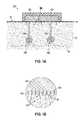
- FIGS. 1A and 1Care cross-sectional views of a device according to an embodiment of the disclosed invention capable of both delivering a paracellular-permeability-enhancing agent topically and collecting the resulting analyte-rich fluid extracted from the apical surface of the epithelium.
- FIGS. 1B and 1Dare enlarged cross-sectional views of the epidermis before and after enhancement of the paracellular permeability by the device of FIGS. 1A and 1C , respectively.
- FIG. 2is a cross-sectional view of a device according to an embodiment of the disclosed invention capable of delivering a paracellular-permeability-enhancing agent using iontophoresis.
- FIG. 3Ais a cross-sectional view of a mechanism capable of causing reverse-iontophoresis-induced electro-osmotic flow according to an embodiment of the disclosed invention.
- FIG. 3Bis a cross-sectional view of the ductal lumen shown during reverse-iontophoresis-induced electro-osmotic flow caused by the device of FIG. 3A .
- FIG. 4is a cross-sectional view of a device according to an embodiment of the disclosed invention capable of delivering a paracellular-permeability-enhancing agent via iontophoresis and simultaneously performing reverse iontophoresis to actively flow analytes from blood and ISF into the device to be sensed by sensors in real time.
- interstitial fluidor “tissue fluid” is a solution that bathes and surrounds tissue cells.
- the interstitial fluidis found in the interstices—the spaces between cells.
- Embodiments of the disclosed inventionfocus on interstitial fluid found in the skin and, particularly, interstitial fluid found in the dermis.
- the interstitial fluidcontains some sweat or saliva as well, or alternately, sweat or saliva may contain some interstitial fluid.
- biofluidis a fluid that is comprised mainly (e.g., 50% by volume or greater) of interstitial fluid, sweat, or saliva as it emerges from the sweat ducts or salivary glands.
- a fluid that is 45% interstitial fluid, 45% sweat, and 10% bloodis a biofluid as used herein.
- a fluid that is 20% interstitial fluid, 20% sweat, and 60% bloodis not a biofluid as used herein.
- a fluid that is 100% sweat or 100% interstitial fluidis a biofluid.
- a biofluidmay be diluted with water or other solvents inside a device because the term biofluid refers to the state of the fluid as it emerges from the skin.
- sweatis dilute of large sized analytes (e.g., greater than 1000 ⁇ for proteins, etc.) and to a lesser extent, as compared to blood, interstitial fluid is dilute for some larger sized analytes (e.g., 10-100 ⁇ or more or less depending on the specific analyte, current density, etc.).
- large sized analytese.g., greater than 1000 ⁇ for proteins, etc.
- interstitial fluidis dilute for some larger sized analytes (e.g., 10-100 ⁇ or more or less depending on the specific analyte, current density, etc.).
- “advective transport”is a transport mechanism of a substance or conserved property by a fluid due to the fluid's bulk motion.
- reverse iontophoresisis a subset of “iontophoresis” and is a technique by which electrical current and electrical field cause molecules to be removed from within the body by electro-osmosis and/or iontophoresis.
- electro-osmosisthe term “reverse iontophoresis” as used herein may also apply to flux of analytes brought to or into a device of the disclosed invention, where the flux is in whole or at least in part due to iontophoresis (e.g., some negatively charged analytes may be transported against the direction of electro-osmotic flow and eventually onto a device according to an embodiment of the disclosed invention).
- Electro-osmotic flowis the motion of liquid induced by an applied potential across a porous material, capillary tube, membrane, microchannel, or any other fluid conduit. Because electro-osmotic velocities are independent of conduit size, as long as the electrical double layer is much smaller than the characteristic length scale of the channel, electro-osmotic flow is most significant when in small channels. In biological tissues, the negative surface charge of plasma membranes causes accumulation of positively charged ions such as sodium. Accordingly, fluid flow due to reverse iontophoresis in the skin or oral mucosa is typically in the direction of where a negative voltage is applied (i.e., the advective flow of fluid is in the direction of the applied electric field).
- iontophoresismay be substituted for “reverse iontophoresis” in any embodiment where there is a net advective transport of biofluid to the surface of the skin. For example, if a flow of sweat exists, then negatively charged analytes may be brought into the advectively flowing sweat by iontophoresis. The net advective flow of sweat would typically be needed, because in this case, a net electro-osmotic fluid flow would be in the direction of sweat into interstitial fluid (and without a net advective flow of sweat, the sweat would be lost, and there would be no pathway for transporting the analyte to at least one sensor).
- iontophoresisis a subset or more specific form of “iontophoresis”
- the term “iontophoresis”may refer to both “reverse iontophoresis” and “iontophoresis”.
- the terms “reverse iontophoresis” and “iontophoresis”are interchangeable in the disclosed invention.
- analyte-specific sensoris a sensor specific to an analyte and performs specific chemical recognition of the presence or concentration of the analyte (e.g., ion-selective electrodes, enzymatic sensors, electrochemical aptamer-based sensors, etc.).
- sensors that sense impedance or conductance of a fluid, such as biofluidare excluded from the definition of “analyte-specific sensor” because sensing impedance or conductance merges measurements of all ions in biofluid (i.e., the sensor is not chemically selective; it provides an indirect measurement).
- Sensorscould also be optical, mechanical, or use other physical/chemical methods which are specific to a single analyte. Further, multiple sensors can each be specific to one of multiple analytes.
- a paracellular-permeability-enhancing agentis an agent that increases the paracellular permeability of an epithelial barrier such that extracted analyte concentration is increased by at least 2 ⁇ compared to the case of using no paracellular-permeability-enhancing agent.
- a sweat sensing device for glucose or for albumincould collect a sweat sample with have a concentration of 25 ⁇ M or 120 ng/mL, respectively, and with application of the paracellular-permeability-enhancing agent as taught herein, these concentrations in a sweat sample could be increased to greater than 1000 ⁇ M or greater than 280 ng/mL, respectively, which is greater than a 2 ⁇ increase.
- ISFinterstitial fluid
- the circulatory systemserves as the fastest way to circulate biomarkers throughout the body, allowing blood to be the gold-standard for biomarker detection.
- Epithelial tissueis usually heavily vascularized with capillaries, potentially providing an indirect path to sample blood.
- the function of the capillariesis to exchange nutrients and signaling molecules and to remove waste. These functions require a degree of natural permeability, which allows analytes to pass from blood into the fluid surrounding the extracellular space, ISF.
- ISFhas similar concentrations of analytes compared to blood after some amount of time. For example, ISF glucose concentrations match that of blood after about 15 minutes.
- biofluidssuch as sweat, saliva, and tears are separated from blood and ISF by an epithelial layer, which acts as a barrier for analyte entry, diluting the concentration of analytes of interest and making them difficult to detect by current sensing and analysis methodologies.
- the lipid bilayerforms a continuous barrier preventing transcellular transport except for small and/or hydrophobic molecules.
- Lipid bilayersprevent diffusion of charged molecules and large (e.g., greater than 500 Da) polar molecules, including biopolymers, and retard diffusion of polar molecules such as water, urea, and glucose.
- Small hydrophobic molecules, such as cortisol and other hormones,can more readily diffuse across the lipid membrane to enter biofluids, but will still diffuse more slowly than in the absence of the bilayer.
- Paracellular pathwaysin comparison, are discontinuous and can allow larger molecules to diffuse through, although the network of proteins and glycans significantly slows paracellular diffusion.
- cells in the epithelium layerare joined near their apical surface by tight junctions that function as a barrier, limiting diffusion of analytes from the interstitial fluid surrounding the basolateral side of the cells.
- the paracellular space between these cellsis 10's of nanometers wide, leaving plenty of room for large analytes to pass.
- tight junctionslink neighboring cell membranes to form a seal that allows relatively free passage of water and monoatomic ions but tends to filter other substances, reducing effective concentrations observed in the secretory biofluid by at least 10 ⁇ , and more often 100 to 1000 to greater than 10,000 ⁇ .
- paracellular-permeability-enhancing agentsact as extracellular signals that regulate the remodeling of the tight junctions between the cells.
- the addition of these paracellular-permeability-enhancing agentsresults in increased permeability along the paracellular pathway for large, uncharged, polar molecules as well as for charged molecules.
- the extracellular signal to be modulatedmay vary based on the intended application.
- Exemplary extracellular signals that may be regulated to increase paracellular permeabilityinclude, but are not limited to, the presence and amount of extracellular calcium, the binding of fatty acids to free fatty acid receptors including G-protein-coupled receptors (GPCRs), and the presence and amount of pathogenic proteins or lipids (e.g., oleic acid, caprylic acid, and lysophosphatidic acid (LPA)).
- GPCRsG-protein-coupled receptors
- pathogenic proteins or lipidse.g., oleic acid, caprylic acid, and lysophosphatidic acid (LPA)
- Exemplary pathogenic proteinsinclude endotoxins, such as the Zonula occludens toxin (Zot) produced by Vibrio cholera .
- ZotZonula occludens toxin
- An exemplary paracellular-permeability-enhancing agentis a chelator.
- Chelatorsbind metal ions and can be used to sequester extracellular metal ions causing a local drop in the concentration of the metal ions. Chelators having different binding affinities for various cations based on their molecular structure and/or pH may be used.
- Exemplary chelatorsinclude polycarboxylates and polycarboxylic acids, such as ethylenediaminetetraacetic acid (EDTA) and citric acid, as well as their conjugate bases. Both EDTA and citric acid bind to calcium in a roughly 1:1 molar ratio depending on the pH of the solution.
- the extracellular calcium concentration within the sweat lumenis roughly 1-2 mM, and around 1-2 mM of either EDTA or citric acid may be used to sufficiently sequester the extracellular calcium ions due to the tight affinity of chelators for divalent cations.
- This drop in calcium concentrationhas been demonstrated to trigger an increase in clathrin-mediated endocytosis of tight junction proteins connecting epithelial tissues.
- Both EDTA and citric acid (and its citrate derivatives)have been reviewed by the Cosmetic Ingredient Review (CIR) and are used at concentrations up to 70 mM and 520 mM, respectively, which is well below the concentration used to fully sequester the extracellular calcium in biofluids.
- Another exemplary paracellular-permeability-enhancing agentis a protein, such as an endotoxin produced by pathogenic bacteria or fungi. Zot, produced by Vibrio cholerae , binds to intestinal epithelial Zot receptors to activate protein kinase C alpha (PKC ⁇ ), which causes an increase in paracellular permeability.
- An effective dose of a protein paracellular-permeability-enhancing agentmay be in a picomolar or micromolar range. For example, an effective dose may be in a range of 1 pM to 100 pM.
- Another exemplary paracellular-permeability-enhancing agentis a lipid.
- Lipidssuch as oleic acid (C18:1) or caprylic acid (C10), bind to free fatty acid receptors on the cell surface. These binding events trigger signal generation and transduction via secondary messengers inside the cell to trigger a host of cellular events. For example, binding of oleic acid to G-protein coupled receptors (GPCR) (G q ) activates phospholipase C (PLC) which phosphorylates phosphatidylinositol (4,5)-bisphosphate (PIP 2 ) into phosphatidylinositol (3,4,5)-trisphosphate (PIP 3 ).
- GPCRG-protein coupled receptors
- PLCphospholipase C
- PIP 3triggers the release of calcium stores in the endoplasmic reticulum.
- the rise in intracellular calcium levelscauses the contraction of calmodulin-dependent actin microfilaments resulting in an enhanced tight junction permeability.
- Oleic acid and caprylic acidare major constituents of olive oil and milk fat, respectively. Both are classified as Generally Recognized as Safe (GRAS) by the FDA.
- Other lipidshave been identified (e.g., lysophosphatidic acid (LPA)) that increase paracellular permeability but are not currently FDA approved.
- An effective dose of a lipid or protein paracellular-permeability-enhancing agentmay be in a picomolar, micromolar, or millimolar range. For example, an effective dose may be in a range of 20 pM to 30 mM.
- a device 100delivers a paracellular-permeability-enhancing agent through diffusion.
- the device 100is positioned on the skin 12 composed of the epidermis 13 , the dermis 15 , and layers of skin below the dermis 16 .
- the skin 12contains multiple sweat glands 14 each containing a ductal lumen 14 a and a secretory coil 14 b .
- the device 100delivers a paracellular-permeability-enhancing agent topically by securing a component 102 including an agent-containing gel or solution 104 against the skin 12 (i.e., the apical surface of the epidermis 13 ).
- the epithelium cells 13 aPrior to the agent enhancing the paracellular permeability, the epithelium cells 13 a are packed tightly together, which prevents certain analytes 18 (e.g., large, polar analytes) from passing through the epidermis 13 .
- the agentis allowed time to diffuse into the skin 12 and trigger the remodeling of the tight junctions resulting in increased paracellular permeability.
- a component 106 with absorbent disks 108may be used to collect the analytes 18 that are now able to pass through the epidermis 13 , as shown in FIG. 1D .
- the absorbent disks 108are made of a material with low non-specific absorption that may have near 100% sample recovery and may have a shape other than a disk.
- a suitable materialincludes SEFAR NITEX, which is a polyamide-based non-woven, hydrophilic mesh with low surface area and low non-specific binding of analytes such as proteins.
- Other suitable materialsinclude filter paper, tech wipes, or rayon.
- the material for the absorbent disks 108may be surface treated to prevent non-specific binding to analytes.
- This example paracellular-permeability-enhancing featurehas a discrete permeability enhancing step, which is separate from the collection step. It should be recognized that other methods of applying a paracellular-permeability-enhancing agent are possible, and an increase in permeability can occur simultaneously with collection and/or sensing as described below.
- a device 200actively delivers a paracellular-permeability-enhancing agent using iontophoresis.
- Iontophoresisis the movement of a charged molecule in response to an electric field (lines 202 ). The movement of molecules is directly related to the current. Since iontophoresis can only move charged molecules, only charged paracellular-permeability-enhancing agents can be delivered in this manner. Most paracellular-permeability-enhancing agents are charged: chelators have a negative charge, free-fatty acids have a negative charge associated with the acid moiety, and proteins usually have some charge. Iontophoresis is especially useful when the epithelium to be targeted is not directly accessible as in the case of the sweat gland ductal epithelium.
- a reservoir 250includes an agent-containing gel or solution and is placed on the surface of the skin 12 .
- the device 200includes an electrode 290 , which is in electrical contact with the agent-containing gel or solution in the reservoir 250 , and a counter electrode 292 .
- the counter electrode 292is situated on the surface of the skin 12 adjacent to and spaced apart from reservoir 250 and electrode 290 .
- the device 200further includes a substrate 210 around the reservoir 250 and the electrode 290 .
- the material for the substrate 210is impermeable to biofluids and may be, for example, a polyimide film or polyethylene terephthalate (PET).
- the substrate 210may include an adhesive backing made of, for example, a hypoallergenic acrylate or other similar medical-grade material.
- a constant currentis applied between the electrodes 290 , 292 .
- the current to be appliedtakes into consideration the area over which it will be applied. As a result, the current density is usually considered.
- the minimum current density used to drive the paracellular-permeability-enhancing agentdepends at least in part on the amount of calcium re-entering the system as well as the concentration of competing negatively-charged ions and molecules in solution. The maximum current that would be applied depends on safety and user comfort.
- the current densityis less than 0.25 mA/cm 2 or in a range between 0.01 mA/cm 2 and 0.25 mA/cm 2 .
- the voltage needed to produce these current densitiesis a function of the resistance of the skin. The resistance of the skin when sweating is lower than the resistance when the skin is dry. Because the device 200 is used when the skin is sweating, the voltages needed to produce these current densities are less than 30 V. The voltage may be within the range of 5-15 V.
- the electrode 290 in electrical contact with the agent-containing gel or solutionis configured to have the same charge compared to the agent. For example, to deliver citric acid, a negatively-charged chelating agent, the electrode 290 in electrical contact with citric acid will be negative.
- this devicemay also include a biofluid collection and/or sensing component.
- the effectiveness of a chelatoris proportional to the amount of calcium ions sequestered.
- chelatorse.g., citric acid and EDTA
- the amount of calcium present in the sweatcan be determined from known quantities—volume of the sweat gland and concentration of calcium in sweat, for example. This yields the amount of calcium within a single, saturated sweat gland.
- Iontophoresisis usually performed on an area that can contain hundreds of sweat glands. Knowing the size of the area where iontophoresis is performed and the density of sweat glands within that area yields the number of sweat glands within the treatment area.
- the product of the number of sweat glands and the amount of calcium per sweat glandis the total amount of calcium that can be sequestered by chelators. Note that, as time goes on, the sweat gland will continue to produce sweat containing additional calcium.
- the effectiveness of the chelation treatmentrelies on the total percent sequestration of calcium within the sweat glands. The effectiveness of the treatment will be reversible as the chelating agent is depleted and calcium from the interstitial fluid and from the body is pulled into the sweat gland.
- citric acidcan be iontophoretically delivered using a current of 2 mA for 1 hour over an area of 1.89 cm 2 . Within this area are roughly 280 sweat glands, assuming a sweat gland density of 150 glands/cm 2 . Using Equation 1, approximately 74.6 ⁇ mol of citric acid would be delivered during this treatment. Iontophoresis as prescribed above would deliver an electric charge of 7.2 C (i.e., a current of 2 mA over the duration of 1 hour provides a charge of 7.2 C). The dominant species of citric acid has one negative charge at sweat pH. Citric acid binds calcium in approximately 1:1 molar ratio, meaning that about 75 ⁇ mol of calcium would be sequestered.
- a reverse iontophoresis mechanism 300includes two electrodes 302 , 304 spaced apart on the skin 12 .
- the paracellular permeabilitymay first be enhanced using an agent as described herein.
- the voltage applied by the electrodes 302 , 304 on the surface of the skin 12will be transferred by the conductive sweat down to the ductal lumen 14 a .
- the voltagemay be relatively low such as, for example, about 5 V, less than or equal to 15 V, or less than or equal to 30 V. In an embodiment, the voltage may be in a range of about 5-30 V or a range of about 5-15 V.
- the electrical double layer needed for electro-osmosisis formed by the negatively-charged cell surface (i.e., due to the carboxylic acid moieties within glycans, such as glycosaminoglycans, and proteins, such as collagen) interacting with the positively-charged ions in the extracellular matrix.
- an external electric fieldlines 306
- cations in both the double layer and bulk solutionwill move toward the electrochemical cathode.
- the movement of the cations within the double layercauses the bulk solution to move toward the electrochemical cathode.
- an epitheliumis treated with an agent to increase paracellular permeability followed by electro-osmosis to drive blood and ISF to the apical surface of the epithelium.
- the increased paracellular permeability and the reverse-iontophoresis-induced electro-osmotic flowmay increase a concentration the analyte of interest in the advective flow of the biofluid by 2 ⁇ or greater, 10 ⁇ or greater, 50 ⁇ or greater, and up to 1000 ⁇ .
- the increasemay be in a range of 2-1000 ⁇ .
- the amount of the paracellular-permeability-enhancing agent that is delivered to the sweat glandis proportional to the total current during iontophoresis.
- the total doseis at least partially dependent on the electric field strength, the concentration of analyte, and the total duration of the applied iontophoresis.
- a paracellular-permeability-enhancing agent of interestis repulsed from the electrochemical cathode due to the agent's negative charge and migrates at a velocity based on the local electric field it is experiencing, its molecular size and shape, and its mass-to-charge ratio.
- the applied forceincreases proportionally, increasing the velocity and thus the flux of the agent toward the epithelium.
- Both iontophoresis and reverse-iontophoresis-induced electro-osmosisrequire electrical stimulation of the epithelium. If electrical contact is poor, then the surface area stimulated will be smaller than expected, and the resulting current density may be higher than anticipated.
- the two major concerns with electrical stimulationare the possibilities of 1) electrical and/or 2) pH-induced acid/base damage. Electrical damage is caused by Joule heating effects as a result of an electric current flowing through the epithelium. These types of damage are largely avoidable by staying within safe current density limits set out by FDA-approved products designed to provide on-body electrical stimulation, such as the Nanoduct (0.26 mA/cm 2 ).
- the electrolysis of waterresults in pH-induced burns, which occurs when voltages applied during iontophoresis or reverse iontophoresis exceed that of the standard electrode potential of water ( ⁇ 1.23 V).
- reduction of waterwill produce hydrogen gas and hydroxide ions, increasing the basicity of the water (2H 2 O(l)+2e ⁇ ⁇ H 2 (g)+2 OH ⁇ (aq)).
- oxidation of waterwill produce oxygen gas and hydrogen ions, increasing the acidity of the water (2H 2 O(l) ⁇ O 2 (g)+4H ⁇ (aq)+4e ⁇ ). It is important to consider how the applied current will affect the chemical composition including pH to avoid any injury. Also, the safe current density assumes good electrode contact with the entire area.
- the duration of the effect produced by a single delivery of a paracellular-permeability-enhancing agentmay not last long enough to increase the concentration of analytes during the entire collection or sensing period, warranting additional dosing events or continuous delivery.
- ISFhas a negative hydrostatic pressure, meaning that an increase in paracellular permeability alone may not be sufficient to bring in analytes from ISF.
- simultaneous iontophoretic delivery of agent and reverse-iontophoresis-induced electro-osmotic flowmay be needed depending on the particular application and the biofluid to be sensed.
- a device 400is capable of delivering a paracellular-permeability-enhancing agent, generating reverse-iontophoresis-induced electro-osmotic flow, and sensing the resulting fluid in real-time, continuously.
- the device 400includes a reservoir 450 including an agent-containing gel or solution, an electrode 490 in electrical contact with the agent in the reservoir 450 , and a counter electrode 492 .
- the electrodes 490 , 492are adapted to deliver the agent iontophoretically and adapted to increase a concentration of the analyte of interest in an advective flow of the biofluid by at least 2 ⁇ .
- the reservoir 450is bordered vertically by sidewalls 440 and on the top by the electrode 490 .
- the material for the sidewalls 440is impermeable to biofluid and to the agent even when iontophoresis is applied; the material may be similar to that described for the substrate 210 .
- a membrane 442is positioned between the reservoir 450 and the skin 12 and prevents the agent from diffusing out and onto the skin without iontophoresis.
- the membrane 442is only slightly permeable to the agent so that there is limited or no diffusion without the application of iontophoresis. When an iontophoretic voltage is applied, the agent has sufficient energy to pass through the membrane 442 .
- Suitable materials for the membrane 442include cellulose acetate, cellulose ester, polyamide, and track-etched polycarbonate membranes.
- the device 400includes analyte-specific sensors 420 , 422 and sweat sample management components 430 , 432 , 434 .
- the component 430acts as a sweat sample collector and transports sweat from the surface of the skin 12 .
- a portion of the sample collector 430may be positioned between the electrode 490 and the skin 12 because the analyte-rich advective flow of biofluid is drawn toward the electrode 490 .
- the component 432acts as sample coupler and transports sweat from the sample collector 430 across the sensors 420 , 422 .
- the component 434acts as a sample pump and moves excess or old sweat from the sample collector 430 and the sample coupler 432 .
- the sample pump 434may be designed to store 10's to 100's of ⁇ L of old sweat.
- Suitable materials for the sweat sample management components 430 , 432 , 434include, for example, a textile; stacks of hydrophilic membrane filters; hydrophilic beads (e.g., commercial monodisperse Reade Silica powder); a longer-chain length hydrogel; a porous polymer; nano-cellulose; and microfluidic channels, among other suitable materials.
- the device 400further includes substrates 410 , 412 , 414 , which may include adhesive backings as described above. The substrate 410 secures the counter electrode 492 and separates the sensors 420 , 422 from the skin 12 .
- the substrates 412prevent evaporation of the sweat from the top of the sample collector 430 and sample coupler 432 .
- a devicemay include sweat sensing along with a different technique used to apply a paracellular-permeability-enhancing agent (e.g., topically).
- the paracellular permeabilityis enhanced and then the sweat is sensed using the device 400 .
- the paracellular-permeability-enhancing agent in the reservoir 450is iontophoretically delivered to the skin 12 .
- An applied electric field between the electrodes 490 , 492drives the agent through the membrane 442 and into the epidermis 13 .
- fluidis drawn to the negative electrode due to reverse-iontophoresis-induced electro-osmosis.
- paracellular-permeability-enhancing agents that are negatively chargedwill be delivered while the electro-osmosis is causing an active flow of the analyte-rich fluid into the sweat gland ductal epithelium.
- the current densitymay be in the ⁇ A/cm 2 range, far below the 0.25 mA/cm 2 precedent set by the FDA-approved Nanoduct device, which is designed to work on dry, non-sweating skin.
- the sweat glands 14are saturated with highly conductive sweat and allow the applied current to be conducted much deeper into the tissue, closer to nerves.
- the sample collector 430transports the sweat towards the sensors 420 , 422 .
- the sample coupler 432transports the sweat from the sample collector 430 to the sensors 420 , 422 to be sensed.
- the sample pump 434draws the old sweat away from the sensors 420 , 422 to prevent contamination of new sweat samples.
- a net advective flow of biofluid from the skin 12 to the sensor(s) in the devicemay be required for the sensor(s) to sense the desired analytes in the biofluid.
- Enhancing the paracellular permeability and inducing the electro-osmotic flowmay be accomplished in series or in parallel (e.g., simultaneously).
- the paracellular-permeability-enhancing agentmay be first applied to the epithelial tissue and then the electro-osmotic flow may be induced (e.g., as described in relation FIG. 3 ).
- the application of a current between the electrodes 490 , 492iontophoretically delivers the agent and induces electro-osmotic flow simultaneously.
- enhancing the paracellular permeability and inducing the electro-osmotic flowmay each be accomplished during one or more discrete time periods or continuously.
- the device 400could be placed in the mouth with the skin 12 representing the tissue lining the mouth.
- the skinmay include the oral mucosa or other tissue in the mouth where salivary glands exist (e.g., under the tongue). Saliva generation rates are generally much higher than sweat generation rates.
- substantial portions of the epithelial tissue in oral mucosalacks a keratinized layer, fewer cells exist separating the superficial layer (mucosal layer) from the ISF and/or capillaries.
- paracellular permeability enhancementis not limited solely to the salivary gland, but can be effective for the entire non-keratinized mucosal layer.
- topical delivery of the agentmay provide the desired effect without the use of iontophoretic delivery.
- the paracellular-permeability-enhancing agentmay be applied topically, such as via a lozenge or a mouthwash, and/or iontophoretically. After application of the agent, fresh saliva could be provided to sensors quickly and be displaced as new saliva appears without the function of a wicking component for real-time sensing applications.
- Saliva monitoring devicescould be mechanically less comfortable or ergonomic for longer term use than sweat monitoring devices.
- a device that enhances paracellular permeability and collects the biofluid for later analysismay be useful.
- Certain embodiments of the disclosed inventionshow sensors as simple individual elements. It is understood that many sensors require two or more electrodes, reference electrodes, or additional supporting technology or features which are not explicitly described in the description herein. Sensors are preferably electrical in nature, but may also include optical, chemical, mechanical, or other known biosensing mechanisms. Sensors can be in duplicate, triplicate, or more, to provide improved data and readings.
- the above description of various embodiments of the disclosed inventionmay not include a description of each and every component that may be used for the functioning of the devices depending on the application (e.g., a battery or a controller), although it should be recognized that such components are included in the scope of the disclosed invention. For the purpose of brevity and to provide a focus on the inventive aspects described above, such components are not explicitly shown in the diagrams or included in the relevant description.
Landscapes
- Health & Medical Sciences (AREA)
- Life Sciences & Earth Sciences (AREA)
- Engineering & Computer Science (AREA)
- Animal Behavior & Ethology (AREA)
- Veterinary Medicine (AREA)
- Public Health (AREA)
- General Health & Medical Sciences (AREA)
- Biomedical Technology (AREA)
- Radiology & Medical Imaging (AREA)
- Bioinformatics & Cheminformatics (AREA)
- Nuclear Medicine, Radiotherapy & Molecular Imaging (AREA)
- Heart & Thoracic Surgery (AREA)
- Physics & Mathematics (AREA)
- Medical Informatics (AREA)
- Molecular Biology (AREA)
- Surgery (AREA)
- Pathology (AREA)
- Biophysics (AREA)
- Pharmacology & Pharmacy (AREA)
- Optics & Photonics (AREA)
- Hematology (AREA)
- Medicinal Chemistry (AREA)
- Chemical & Material Sciences (AREA)
- Cardiology (AREA)
- Investigating Or Analysing Biological Materials (AREA)
- Dermatology (AREA)
- Anesthesiology (AREA)
Abstract
Description
Claims (15)
Priority Applications (1)
| Application Number | Priority Date | Filing Date | Title |
|---|---|---|---|
| US15/617,649US10471249B2 (en) | 2016-06-08 | 2017-06-08 | Enhanced analyte access through epithelial tissue |
Applications Claiming Priority (2)
| Application Number | Priority Date | Filing Date | Title |
|---|---|---|---|
| US201662347281P | 2016-06-08 | 2016-06-08 | |
| US15/617,649US10471249B2 (en) | 2016-06-08 | 2017-06-08 | Enhanced analyte access through epithelial tissue |
Publications (2)
| Publication Number | Publication Date |
|---|---|
| US20170354808A1 US20170354808A1 (en) | 2017-12-14 |
| US10471249B2true US10471249B2 (en) | 2019-11-12 |
Family
ID=60573515
Family Applications (1)
| Application Number | Title | Priority Date | Filing Date |
|---|---|---|---|
| US15/617,649Active2037-10-11US10471249B2 (en) | 2016-06-08 | 2017-06-08 | Enhanced analyte access through epithelial tissue |
Country Status (1)
| Country | Link |
|---|---|
| US (1) | US10471249B2 (en) |
Families Citing this family (7)
| Publication number | Priority date | Publication date | Assignee | Title |
|---|---|---|---|---|
| US20210353211A1 (en)* | 2018-09-21 | 2021-11-18 | University Of Cincinnati | Devices for integrated, repeated, prolonged, and/or reliable sweat stimulation and biosensing and for removing excess water during sweat stimulation |
| JP7167824B2 (en)* | 2019-04-08 | 2022-11-09 | 日本電信電話株式会社 | perspiration analyzer |
| WO2020207793A1 (en)* | 2019-04-08 | 2020-10-15 | Universite De Lille | Cutaneous device for storing and releasing molecules, and corresponding method |
| CN111888641B (en)* | 2019-05-06 | 2023-09-22 | 上海肤泰科技有限公司 | Iontophoresis drug delivery device |
| EP4132638A4 (en)* | 2020-04-09 | 2024-04-10 | University of Cincinnati | UNIFORMED PROCEDURE FOR WELD SAMPLE COLLECTORS |
| BR112022021049A2 (en)* | 2020-04-17 | 2022-12-27 | Biolectrics Llc | SYSTEMS AND METHODS FOR ORAL IONTOPHORESIS |
| WO2022026579A2 (en) | 2020-07-28 | 2022-02-03 | Biolectrics Llc | Systems and methods related to intraoral electrical stimulation |
Citations (123)
| Publication number | Priority date | Publication date | Assignee | Title |
|---|---|---|---|---|
| US4190060A (en) | 1978-04-19 | 1980-02-26 | The United States Of America As Represented By The Administrator Of The National Aeronautics And Space Administration | Sweat collection capsule |
| US4542751A (en) | 1982-03-15 | 1985-09-24 | Wescor, Inc. | Sweat-collecting device and method |
| US4756314A (en) | 1985-10-28 | 1988-07-12 | Alza Corporation | Sweat collection patch |
| EP0282349A2 (en) | 1987-03-12 | 1988-09-14 | MediSense, Inc. | Ion selective sensors |
| US4820263A (en) | 1981-03-06 | 1989-04-11 | Medtronic, Inc. | Apparatus and method for iontophoretic drug delivery |
| WO1990011519A1 (en) | 1989-03-23 | 1990-10-04 | Bunce Roger A | Liquid transfer devices |
| US5036861A (en) | 1990-01-11 | 1991-08-06 | Sembrowich Walter L | Method and apparatus for non-invasively monitoring plasma glucose levels |
| US5050604A (en) | 1989-10-16 | 1991-09-24 | Israel Reshef | Apparatus and method for monitoring the health condition of a subject |
| EP0453283A1 (en) | 1990-04-19 | 1991-10-23 | Teknekron Sensor Development Corporation | An integral interstitial fluid sensor |
| US5140985A (en) | 1989-12-11 | 1992-08-25 | Schroeder Jon M | Noninvasive blood glucose measuring device |
| US5246003A (en) | 1991-08-28 | 1993-09-21 | Nellcor Incorporated | Disposable pulse oximeter sensor |
| WO1994014062A1 (en) | 1992-12-11 | 1994-06-23 | Sudor Partners | Method and apparatus for determination of chemical species in perspiration |
| EP0634215A1 (en) | 1993-07-15 | 1995-01-18 | Roche Diagnostics GmbH | Device for simultaneous determination of analytes |
| US5438984A (en) | 1988-09-08 | 1995-08-08 | Sudor Partners | Apparatus and method for the collection of analytes on a dermal patch |
| US5814599A (en) | 1995-08-04 | 1998-09-29 | Massachusetts Insitiute Of Technology | Transdermal delivery of encapsulated drugs |
| US5944662A (en) | 1988-09-08 | 1999-08-31 | Sudormed, Inc. | Method and apparatus of determination of chemical species in perspiration |
| WO2000014535A1 (en) | 1998-09-09 | 2000-03-16 | Amira Medical | Interstitial fluid methods and devices for determination of an analyte in the body |
| US6198953B1 (en) | 1999-03-11 | 2001-03-06 | Henry L. Webster | Method and system for continuous sweat collection and analysis |
| US6256533B1 (en) | 1999-06-09 | 2001-07-03 | The Procter & Gamble Company | Apparatus and method for using an intracutaneous microneedle array |
| US6269265B1 (en) | 1999-02-22 | 2001-07-31 | Wr Medical Electronics Co. | Apparatus and method for evoking and capturing a sweat sample |
| US6299578B1 (en) | 1995-12-28 | 2001-10-09 | Cygnus, Inc. | Methods for monitoring a physiological analyte |
| WO2001088525A1 (en) | 2000-05-12 | 2001-11-22 | University Of Cincinnati | Structurally programmable microfluidic systems |
| US20020091312A1 (en) | 1998-05-13 | 2002-07-11 | Cygnus, Inc. | Monitoring of physiological analytes |
| US6592529B2 (en) | 2001-07-31 | 2003-07-15 | Pheromone Sciences Corp. | Method and device for predicting the fertile phase of women |
| US20030135100A1 (en) | 1998-03-13 | 2003-07-17 | Cygnus, Inc. | Biosensor, iontophoretic sampling system, and methods of use thereof |
| US20030191376A1 (en) | 1998-07-21 | 2003-10-09 | Samuels Mark A. | System and method for continuous analyte monitoring |
| US20030201194A1 (en) | 1997-02-06 | 2003-10-30 | Therasense, Inc. | Small volume in vitro analyte sensor |
| US6666821B2 (en) | 2001-01-08 | 2003-12-23 | Medtronic, Inc. | Sensor system |
| US20040249310A1 (en) | 2001-06-12 | 2004-12-09 | Robert Shartle | Biological fluid constituent sampling and measurement devices and methods |
| US20040267189A1 (en) | 2001-10-24 | 2004-12-30 | Daniela Mavor | Device and method for controlled delivery of active substance into the skin |
| EP1500937A1 (en) | 2002-04-30 | 2005-01-26 | Arkray, Inc. | Analysis instrument, sample analysis method and analysis device using the instrument, and method of forming opening in the instrument |
| US20050069925A1 (en) | 2003-08-15 | 2005-03-31 | Russell Ford | Microprocessors, devices, and methods for use in monitoring of physiological analytes |
| US20050106713A1 (en) | 2003-09-03 | 2005-05-19 | Phan Brigitte C. | Personal diagnostic devices and related methods |
| US20050177035A1 (en) | 2003-12-18 | 2005-08-11 | Elliot Botvinick | Implantable biosensor and methods of use thereof |
| US20050192528A1 (en) | 2004-01-08 | 2005-09-01 | Robert Tapper | Methods, apparatus and charged chemicals for control of ions, molecules or electrons |
| US20050197554A1 (en) | 2004-02-26 | 2005-09-08 | Michael Polcha | Composite thin-film glucose sensor |
| US20060004271A1 (en) | 2004-07-01 | 2006-01-05 | Peyser Thomas A | Devices, methods, and kits for non-invasive glucose measurement |
| EP1637889A1 (en) | 2003-06-19 | 2006-03-22 | Arkray, Inc. | Analyzer instrument with liquid storage portion |
| US20060062852A1 (en) | 2003-09-11 | 2006-03-23 | Holmes Elizabeth A | Medical device for analyte monitoring and drug delivery |
| US20060127964A1 (en) | 2004-07-30 | 2006-06-15 | Russell Ford | Microprocessors, devices, and methods for use in monitoring of physiological analytes |
| US20060253011A1 (en) | 2005-05-09 | 2006-11-09 | Edmonson Peter J | Sweat sensor system and method of characterizing the compositional analysis of sweat fluid |
| US20060254341A1 (en) | 2005-03-04 | 2006-11-16 | Campbell Michael J E | Method and apparatus for determining transpiration characteristics of a permeable membrane |
| WO2006133101A2 (en) | 2005-06-03 | 2006-12-14 | Trans-Dermal Patents Company, Llc | Agent delivery system |
| US20070027383A1 (en) | 2004-07-01 | 2007-02-01 | Peyser Thomas A | Patches, systems, and methods for non-invasive glucose measurement |
| US20070032731A1 (en) | 2005-08-05 | 2007-02-08 | Lovejoy Jeffrey L | Non-invasive pulse rate detection via headphone mounted electrodes / monitoring system |
| US7190986B1 (en) | 2002-10-18 | 2007-03-13 | Nellcor Puritan Bennett Inc. | Non-adhesive oximeter sensor for sensitive skin |
| WO2007097754A1 (en) | 2006-02-22 | 2007-08-30 | Dexcom, Inc. | Analyte sensor |
| JP2007532260A (en) | 2004-04-16 | 2007-11-15 | エイ カルマリ ラシダ | Assembly for collecting, processing and analyzing samples |
| US20080015494A1 (en) | 2006-07-11 | 2008-01-17 | Microchips, Inc. | Multi-reservoir pump device for dialysis, biosensing, or delivery of substances |
| US20080045816A1 (en) | 2006-08-18 | 2008-02-21 | Samsung Electronics Co., Ltd. | Apparatus, method and medium measuring skin moisture content |
| JP2008505330A (en) | 2004-07-09 | 2008-02-21 | エフ ホフマン−ラ ロッシュ アクチェン ゲゼルシャフト | Analytical test element |
| US20080114282A1 (en)* | 2006-09-05 | 2008-05-15 | Transcu Ltd. | Transdermal drug delivery systems, devices, and methods using inductive power supplies |
| WO2008083687A1 (en) | 2007-01-10 | 2008-07-17 | Scandinavian Micro Biodevices Aps | A microfluidic device and a microfluidic system and a method of performing a test |
| WO2008095940A1 (en) | 2007-02-05 | 2008-08-14 | Dublin City University | Flow analysis apparatus and method |
| US20080286349A1 (en) | 2007-05-18 | 2008-11-20 | Youhei Nomoto | Systems, devices, and methods for passive transdermal delivery of active agents to a biological interface |
| US20080306362A1 (en) | 2007-06-05 | 2008-12-11 | Owen Davis | Device and system for monitoring contents of perspiration |
| WO2009004001A1 (en) | 2007-07-02 | 2009-01-08 | Biogauge - Nordic Bioimpedance Research As | Method and kit for sweat activity measurement |
| US20090076345A1 (en) | 2007-09-14 | 2009-03-19 | Corventis, Inc. | Adherent Device with Multiple Physiological Sensors |
| WO2009052321A2 (en) | 2007-10-18 | 2009-04-23 | Advanced Liquid Logic, Inc. | Droplet actuators, systems and methods |
| JP2009118420A (en) | 2007-11-09 | 2009-05-28 | Sony Corp | Information processing device and method, program, recording medium, and information processing system |
| US20090204008A1 (en) | 2008-02-08 | 2009-08-13 | Daniel Beilin | Whole body infrared thermography systems and methods |
| WO2010017578A1 (en) | 2008-08-14 | 2010-02-18 | Monash University | Switches for microfluidic systems |
| US20100044224A1 (en) | 2006-09-13 | 2010-02-25 | Ritu Kataky | Biological fluid analysis system |
| US20100063372A1 (en) | 2008-09-09 | 2010-03-11 | Potts Russell O | Sweat collection devices for glucose measurement |
| US20100130843A1 (en) | 2008-11-24 | 2010-05-27 | Tecnicas Cientificas Para Laboratorio, S.A | Wireless device for confirmatory diagnosis of cystic fibrosis through analysis of sweat chloride |
| US20100132485A1 (en) | 2007-08-06 | 2010-06-03 | Mordechai Erez | Sweat collectors and methods of collecting sweat |
| US7749445B2 (en) | 2005-05-02 | 2010-07-06 | Bioscale, Inc. | Method and apparatus for analyzing bioprocess fluids |
| US20100198521A1 (en) | 2007-07-24 | 2010-08-05 | Technion Research And Development Foundation Ltd. | Chemically sensitive field effect transistors and uses thereof in electronic nose devices |
| US7813780B2 (en) | 2005-12-13 | 2010-10-12 | Medtronic Minimed, Inc. | Biosensors and methods for making and using them |
| US7842234B2 (en) | 2002-12-02 | 2010-11-30 | Epocal Inc. | Diagnostic devices incorporating fluidics and methods of manufacture |
| US20110079521A1 (en) | 2009-10-02 | 2011-04-07 | Commissariat A L'energie Atomique Et Aux Energies Alternatives | Method and cell for measuring the global ion concentration of a body fluid |
| US20110118656A1 (en) | 2009-11-13 | 2011-05-19 | Searete Llc, A Limited Liability Corporation Of The State Of Delaware | Device, system, and method for targeted delivery of anti-inflammatory medicaments to a mammalian subject |
| US7959791B2 (en) | 2005-05-17 | 2011-06-14 | Radiometer Medical Aps | Enzyme sensor with a cover membrane layer covered by a hydrophilic polymer |
| US20110178380A1 (en) | 2008-06-30 | 2011-07-21 | Nemaura Pharma Limited | Patch for reverse iontophoresis |
| US20110196283A1 (en) | 2010-02-10 | 2011-08-11 | Mir Imran | Methods and architecture for power optimization of iontophoretic transdermal drug delivery |
| US20110208458A1 (en) | 2008-10-16 | 2011-08-25 | Koninklijke Philips Electronics N.V. | Impedance measurement circuit and method |
| WO2011117952A1 (en) | 2010-03-24 | 2011-09-29 | 株式会社島津製作所 | Measurement system |
| US20110275918A1 (en) | 2009-01-23 | 2011-11-10 | Omron Healthcare Co., Ltd. | Body fluid collecting device for efficiently collecting body fluid and body fluid analyzer for accurate analysis |
| US20120004570A1 (en) | 2009-02-20 | 2012-01-05 | Omron Healthcare Co., Ltd. | Biological information measurement device, biological information measurement method, and body composition measurement device |
| US20120028283A1 (en) | 2010-07-28 | 2012-02-02 | Abott Diabetes Care Inc. | Analyte Sensors Having Temperature Independent Membranes |
| US20120123220A1 (en) | 2009-04-27 | 2012-05-17 | Avery Dennison Corporation | Systems, Methods and Materials for Delivery and Debonding on Demand |
| US20120165626A1 (en) | 2009-07-13 | 2012-06-28 | Irina Finkelshtein V | Devices, methods, and kits for determining analyte concentrations |
| US20120209226A1 (en) | 2011-02-14 | 2012-08-16 | Kci Licensing, Inc. | Reduced-pressure dressings, systems, and methods for use with linear wounds |
| US20120229661A1 (en) | 2009-12-28 | 2012-09-13 | Nikon Corporation | Photography lens, photographing apparatus, photographing system, image capturing apparatus, and personal apparatus |
| US20120277697A1 (en) | 2011-04-29 | 2012-11-01 | Seventh Sense Biosystems, Inc. | Systems and methods for collecting fluid from a subject |
| US20120285829A1 (en) | 2009-12-09 | 2012-11-15 | Iti Scotland Limited | Detecting analytes |
| US20120317430A1 (en) | 2011-06-11 | 2012-12-13 | Aliphcom | Power management in a data-capable strapband |
| US20130006079A1 (en) | 2004-02-09 | 2013-01-03 | Abbott Diabetes Care Inc. | Analyte Sensor, and Associated System and Method Employing a Catalytic Agent |
| US20130013028A1 (en) | 2007-10-30 | 2013-01-10 | Mcneil-Ppc,Inc. | Microcurrent Device With A Sensory Cue |
| EP2551784A1 (en) | 2011-07-28 | 2013-01-30 | Roche Diagnostics GmbH | Method of controlling the display of a dataset |
| US20130053668A1 (en) | 2011-08-26 | 2013-02-28 | Compose Element Limited | Kit and method for detecting blood sugar |
| US8391946B2 (en) | 2007-11-09 | 2013-03-05 | Omron Healthcare Co., Ltd. | Device and method for accurately measuring concentration of blood component |
| US20130079605A1 (en) | 2011-09-23 | 2013-03-28 | Bhaskara Rao BANDARU | Transdermal analyte extraction & detection system and the method thereof |
| US20130099937A1 (en) | 2011-10-25 | 2013-04-25 | Vital Connect, Inc. | System and method for reliable and scalable health monitoring |
| US20130108667A1 (en) | 2011-10-27 | 2013-05-02 | Soiwisa Soikum | Method, apparatus and system for electroporation |
| US20130123595A1 (en) | 2010-12-10 | 2013-05-16 | Flexible Medical Systems Llc | Transdermal Sampling and Analysis Device |
| US20130183399A1 (en) | 2011-10-11 | 2013-07-18 | Precision Hydration Ltd. | Method for Improving Physical Performance During Physical Exertion |
| CA2869469A1 (en) | 2012-04-04 | 2013-10-10 | University Of Cincinnati | Sweat simulation, collection and sensing systems |
| US20130306491A1 (en) | 2003-05-16 | 2013-11-21 | Nanomix, Inc. | Electrochemical nanosensors for biomolecule detection |
| US8593287B2 (en) | 2007-05-08 | 2013-11-26 | Abbott Diabetes Care Inc. | Analyte monitoring system and methods |
| US20130317333A1 (en) | 2012-05-24 | 2013-11-28 | Vigilo Networks, Inc. | Modular wearable sensor device |
| WO2013181436A1 (en) | 2012-05-30 | 2013-12-05 | Medisens Wireless, Inc. | System and method for fluid sensing |
| US8617067B2 (en) | 2011-05-13 | 2013-12-31 | Fujitsu Limited | Continuous monitoring of stress using environmental data |
| WO2014001577A1 (en) | 2012-06-29 | 2014-01-03 | Smart Solutions Technologies, S.L. | Electronic textile assembly |
| US20140012114A1 (en) | 2012-07-06 | 2014-01-09 | Nederlandse Organisatie Voor Toegepast-Natuurwetenschappelijk Onderzoek - Tno | Monitoring of Fluid Content |
| US20140025000A1 (en) | 2000-06-01 | 2014-01-23 | Science Applications International Corporation | Systems and Methods For Monitoring Health and Delivering Drugs Transdermally |
| WO2014025430A2 (en) | 2012-05-10 | 2014-02-13 | The Regents Of The University Of California | Wearable electrochemical sensors |
| US20140206977A1 (en) | 2013-01-24 | 2014-07-24 | Irhythm Technologies, Inc. | Physiological monitoring device |
| US20140275862A1 (en) | 2013-03-15 | 2014-09-18 | William R. Kennedy | Systems and methods for assessing sweat gland output |
| US20140276220A1 (en) | 2013-03-15 | 2014-09-18 | Dennis R. Briscoe | Sweat collecting device |
| US20140343371A1 (en) | 2013-05-14 | 2014-11-20 | Ii Thomas Skerik Sowers | Wearable sensor device for health monitoring and methods of use |
| US20150112165A1 (en) | 2013-10-18 | 2015-04-23 | University Of Cincinnati | Sweat sensing with chronological assurance |
| US20150112164A1 (en) | 2013-10-18 | 2015-04-23 | University Of Cincinnati | Devices for integrated, repeated, prolonged, and/or reliable sweat stimulation and biosensing |
| WO2015184097A2 (en) | 2014-05-28 | 2015-12-03 | University Of Cincinnati | Devices with reduced sweat volumes between sensors and sweat glands |
| WO2015184072A1 (en) | 2014-05-28 | 2015-12-03 | University Of Cincinnati | Advanced sweat sensor adhesion, sealing, and fluidic strategies |
| WO2016049019A1 (en) | 2014-09-22 | 2016-03-31 | University Of Cincinnati | Sweat sensing with analytical assurance |
| WO2016061362A2 (en) | 2014-10-15 | 2016-04-21 | Eccrine Systems, Inc. | Sweat sensing device communication security and compliance |
| WO2016090189A1 (en) | 2014-12-03 | 2016-06-09 | The Regents Of The University Of California | Non-invasive and wearable chemical sensors and biosensors |
| US20160157768A1 (en) | 2005-02-14 | 2016-06-09 | Optiscan Biomedical Corporation | Analyte detection systems and methods using multiple measurements |
| WO2016130905A1 (en) | 2015-02-13 | 2016-08-18 | University Of Cincinnati | Devices for integrated indirect sweat stimulation and sensing |
| WO2016138087A1 (en) | 2015-02-24 | 2016-09-01 | Eccrine Systems, Inc. | Dynamic sweat sensor management |
| WO2017019602A1 (en) | 2015-07-24 | 2017-02-02 | University Of Cincinnati | Reduced sample volume for sensing of analytes generated by reverse iontophoresis |
| WO2017070640A1 (en) | 2015-10-23 | 2017-04-27 | Eccrine Systems, Inc. | Devices capable of sample concentration for extended sensing of sweat analytes |
Family Cites Families (1)
| Publication number | Priority date | Publication date | Assignee | Title |
|---|---|---|---|---|
| US9408127B2 (en)* | 2012-01-29 | 2016-08-02 | Lg Electronics Inc. | Method for re-selecting cell in wireless communication system, and apparatus therefor |
- 2017
- 2017-06-08USUS15/617,649patent/US10471249B2/enactiveActive
Patent Citations (148)
| Publication number | Priority date | Publication date | Assignee | Title |
|---|---|---|---|---|
| US4190060A (en) | 1978-04-19 | 1980-02-26 | The United States Of America As Represented By The Administrator Of The National Aeronautics And Space Administration | Sweat collection capsule |
| US4820263A (en) | 1981-03-06 | 1989-04-11 | Medtronic, Inc. | Apparatus and method for iontophoretic drug delivery |
| US4542751A (en) | 1982-03-15 | 1985-09-24 | Wescor, Inc. | Sweat-collecting device and method |
| US4756314A (en) | 1985-10-28 | 1988-07-12 | Alza Corporation | Sweat collection patch |
| EP0282349A2 (en) | 1987-03-12 | 1988-09-14 | MediSense, Inc. | Ion selective sensors |
| US5438984A (en) | 1988-09-08 | 1995-08-08 | Sudor Partners | Apparatus and method for the collection of analytes on a dermal patch |
| US5944662A (en) | 1988-09-08 | 1999-08-31 | Sudormed, Inc. | Method and apparatus of determination of chemical species in perspiration |
| WO1990011519A1 (en) | 1989-03-23 | 1990-10-04 | Bunce Roger A | Liquid transfer devices |
| US5050604A (en) | 1989-10-16 | 1991-09-24 | Israel Reshef | Apparatus and method for monitoring the health condition of a subject |
| US5140985A (en) | 1989-12-11 | 1992-08-25 | Schroeder Jon M | Noninvasive blood glucose measuring device |
| US5036861A (en) | 1990-01-11 | 1991-08-06 | Sembrowich Walter L | Method and apparatus for non-invasively monitoring plasma glucose levels |
| EP0453283A1 (en) | 1990-04-19 | 1991-10-23 | Teknekron Sensor Development Corporation | An integral interstitial fluid sensor |
| US5246003A (en) | 1991-08-28 | 1993-09-21 | Nellcor Incorporated | Disposable pulse oximeter sensor |
| WO1994014062A1 (en) | 1992-12-11 | 1994-06-23 | Sudor Partners | Method and apparatus for determination of chemical species in perspiration |
| JPH08504513A (en) | 1992-12-11 | 1996-05-14 | スダー パートナーズ | Method and instrument for measuring chemical species in sweat |
| EP0634215A1 (en) | 1993-07-15 | 1995-01-18 | Roche Diagnostics GmbH | Device for simultaneous determination of analytes |
| JPH0777525A (en) | 1993-07-15 | 1995-03-20 | Boehringer Mannheim Gmbh | Method and device for simultaneous measurement of plurality of materials to be analyzed |
| US5556789A (en) | 1993-07-15 | 1996-09-17 | Boehringer Mannheim Gmbh | Device for the simultaneous determination of several analytes |
| US5814599A (en) | 1995-08-04 | 1998-09-29 | Massachusetts Insitiute Of Technology | Transdermal delivery of encapsulated drugs |
| US6299578B1 (en) | 1995-12-28 | 2001-10-09 | Cygnus, Inc. | Methods for monitoring a physiological analyte |
| US20030201194A1 (en) | 1997-02-06 | 2003-10-30 | Therasense, Inc. | Small volume in vitro analyte sensor |
| US20030135100A1 (en) | 1998-03-13 | 2003-07-17 | Cygnus, Inc. | Biosensor, iontophoretic sampling system, and methods of use thereof |
| US20020091312A1 (en) | 1998-05-13 | 2002-07-11 | Cygnus, Inc. | Monitoring of physiological analytes |
| US7384396B2 (en) | 1998-07-21 | 2008-06-10 | Spectrx Inc. | System and method for continuous analyte monitoring |
| US20030191376A1 (en) | 1998-07-21 | 2003-10-09 | Samuels Mark A. | System and method for continuous analyte monitoring |
| WO2000014535A1 (en) | 1998-09-09 | 2000-03-16 | Amira Medical | Interstitial fluid methods and devices for determination of an analyte in the body |
| US6269265B1 (en) | 1999-02-22 | 2001-07-31 | Wr Medical Electronics Co. | Apparatus and method for evoking and capturing a sweat sample |
| US6198953B1 (en) | 1999-03-11 | 2001-03-06 | Henry L. Webster | Method and system for continuous sweat collection and analysis |
| US6256533B1 (en) | 1999-06-09 | 2001-07-03 | The Procter & Gamble Company | Apparatus and method for using an intracutaneous microneedle array |
| WO2001088525A1 (en) | 2000-05-12 | 2001-11-22 | University Of Cincinnati | Structurally programmable microfluidic systems |
| US20140025000A1 (en) | 2000-06-01 | 2014-01-23 | Science Applications International Corporation | Systems and Methods For Monitoring Health and Delivering Drugs Transdermally |
| US6666821B2 (en) | 2001-01-08 | 2003-12-23 | Medtronic, Inc. | Sensor system |
| US20040249310A1 (en) | 2001-06-12 | 2004-12-09 | Robert Shartle | Biological fluid constituent sampling and measurement devices and methods |
| US6592529B2 (en) | 2001-07-31 | 2003-07-15 | Pheromone Sciences Corp. | Method and device for predicting the fertile phase of women |
| US20040267189A1 (en) | 2001-10-24 | 2004-12-30 | Daniela Mavor | Device and method for controlled delivery of active substance into the skin |
| JP2009063597A (en) | 2002-04-30 | 2009-03-26 | Arkray Inc | Aperture formation method for assay tool |
| US8128889B2 (en) | 2002-04-30 | 2012-03-06 | Arkray, Inc. | Analyzing article, analyzer and method of analyzing a sample using the analyzing article, and a method of forming an opening in the analyzing article |
| EP1500937A1 (en) | 2002-04-30 | 2005-01-26 | Arkray, Inc. | Analysis instrument, sample analysis method and analysis device using the instrument, and method of forming opening in the instrument |
| US7190986B1 (en) | 2002-10-18 | 2007-03-13 | Nellcor Puritan Bennett Inc. | Non-adhesive oximeter sensor for sensitive skin |
| US7842234B2 (en) | 2002-12-02 | 2010-11-30 | Epocal Inc. | Diagnostic devices incorporating fluidics and methods of manufacture |
| US20130306491A1 (en) | 2003-05-16 | 2013-11-21 | Nanomix, Inc. | Electrochemical nanosensors for biomolecule detection |
| EP1637889A1 (en) | 2003-06-19 | 2006-03-22 | Arkray, Inc. | Analyzer instrument with liquid storage portion |
| US20050069925A1 (en) | 2003-08-15 | 2005-03-31 | Russell Ford | Microprocessors, devices, and methods for use in monitoring of physiological analytes |
| US9133024B2 (en) | 2003-09-03 | 2015-09-15 | Brigitte Chau Phan | Personal diagnostic devices including related methods and systems |
| US20050106713A1 (en) | 2003-09-03 | 2005-05-19 | Phan Brigitte C. | Personal diagnostic devices and related methods |
| US20160066828A1 (en) | 2003-09-03 | 2016-03-10 | Life Patch International, Inc. | Personal diagnostic device having an air sample collection unit and a fluidic circuit |
| US20160058354A1 (en) | 2003-09-03 | 2016-03-03 | Life Patch International, Inc. | Personal diagnostic device having a fluidic circuit with a plurality of analysis chambers |
| JP2007503958A (en) | 2003-09-03 | 2007-03-01 | ライフパッチ インターナショナル,インコーポレイテッド | Personal diagnostic equipment and related methods |
| US20060062852A1 (en) | 2003-09-11 | 2006-03-23 | Holmes Elizabeth A | Medical device for analyte monitoring and drug delivery |
| US20050177035A1 (en) | 2003-12-18 | 2005-08-11 | Elliot Botvinick | Implantable biosensor and methods of use thereof |
| US20050192528A1 (en) | 2004-01-08 | 2005-09-01 | Robert Tapper | Methods, apparatus and charged chemicals for control of ions, molecules or electrons |
| US20130006079A1 (en) | 2004-02-09 | 2013-01-03 | Abbott Diabetes Care Inc. | Analyte Sensor, and Associated System and Method Employing a Catalytic Agent |
| US20050197554A1 (en) | 2004-02-26 | 2005-09-08 | Michael Polcha | Composite thin-film glucose sensor |
| JP2007532260A (en) | 2004-04-16 | 2007-11-15 | エイ カルマリ ラシダ | Assembly for collecting, processing and analyzing samples |
| US7378054B2 (en) | 2004-04-16 | 2008-05-27 | Savvipharm Inc | Specimen collecting, processing and analytical assembly |
| US20090270704A1 (en) | 2004-07-01 | 2009-10-29 | Peyser Thomas A | Patches, systems, and methods for non-invasive glucose measurement |
| US20060004271A1 (en) | 2004-07-01 | 2006-01-05 | Peyser Thomas A | Devices, methods, and kits for non-invasive glucose measurement |
| US20070179371A1 (en) | 2004-07-01 | 2007-08-02 | Peyser Thomas A | Patches, systems, and methods for non-invasive glucose measurement |
| US20070027383A1 (en) | 2004-07-01 | 2007-02-01 | Peyser Thomas A | Patches, systems, and methods for non-invasive glucose measurement |
| JP2008505330A (en) | 2004-07-09 | 2008-02-21 | エフ ホフマン−ラ ロッシュ アクチェン ゲゼルシャフト | Analytical test element |
| US8252248B2 (en) | 2004-07-09 | 2012-08-28 | Roche Diagnostics Operations, Inc. | Analytical test element |
| US20060127964A1 (en) | 2004-07-30 | 2006-06-15 | Russell Ford | Microprocessors, devices, and methods for use in monitoring of physiological analytes |
| US20160157768A1 (en) | 2005-02-14 | 2016-06-09 | Optiscan Biomedical Corporation | Analyte detection systems and methods using multiple measurements |
| US20060254341A1 (en) | 2005-03-04 | 2006-11-16 | Campbell Michael J E | Method and apparatus for determining transpiration characteristics of a permeable membrane |
| US7219534B2 (en) | 2005-03-04 | 2007-05-22 | Nova Technology Corporation | Method and apparatus for determining transpiration characteristics of a permeable membrane |
| US7749445B2 (en) | 2005-05-02 | 2010-07-06 | Bioscale, Inc. | Method and apparatus for analyzing bioprocess fluids |
| US20060253011A1 (en) | 2005-05-09 | 2006-11-09 | Edmonson Peter J | Sweat sensor system and method of characterizing the compositional analysis of sweat fluid |
| US7383072B2 (en) | 2005-05-09 | 2008-06-03 | P. J. Edmonson Ltd | Sweat sensor system and method of characterizing the compositional analysis of sweat fluid |
| US7959791B2 (en) | 2005-05-17 | 2011-06-14 | Radiometer Medical Aps | Enzyme sensor with a cover membrane layer covered by a hydrophilic polymer |
| WO2006133101A2 (en) | 2005-06-03 | 2006-12-14 | Trans-Dermal Patents Company, Llc | Agent delivery system |
| US20080154179A1 (en) | 2005-06-03 | 2008-06-26 | Transdermal Patents Company, Llc | Agent delivery system |
| US20070032731A1 (en) | 2005-08-05 | 2007-02-08 | Lovejoy Jeffrey L | Non-invasive pulse rate detection via headphone mounted electrodes / monitoring system |
| US7813780B2 (en) | 2005-12-13 | 2010-10-12 | Medtronic Minimed, Inc. | Biosensors and methods for making and using them |
| WO2007097754A1 (en) | 2006-02-22 | 2007-08-30 | Dexcom, Inc. | Analyte sensor |
| WO2007146047A1 (en) | 2006-06-12 | 2007-12-21 | Vivomedical, Inc. | Patches, systems, and methods for non-invasive glucose measurement |
| CN101489470A (en) | 2006-06-12 | 2009-07-22 | 维沃医学公司 | Patches, systems, and methods for non-invasive glucose measurement |
| US20080015494A1 (en) | 2006-07-11 | 2008-01-17 | Microchips, Inc. | Multi-reservoir pump device for dialysis, biosensing, or delivery of substances |
| US20080045816A1 (en) | 2006-08-18 | 2008-02-21 | Samsung Electronics Co., Ltd. | Apparatus, method and medium measuring skin moisture content |
| US20080114282A1 (en)* | 2006-09-05 | 2008-05-15 | Transcu Ltd. | Transdermal drug delivery systems, devices, and methods using inductive power supplies |
| US20100044224A1 (en) | 2006-09-13 | 2010-02-25 | Ritu Kataky | Biological fluid analysis system |
| WO2008083687A1 (en) | 2007-01-10 | 2008-07-17 | Scandinavian Micro Biodevices Aps | A microfluidic device and a microfluidic system and a method of performing a test |
| WO2008095940A1 (en) | 2007-02-05 | 2008-08-14 | Dublin City University | Flow analysis apparatus and method |
| US8593287B2 (en) | 2007-05-08 | 2013-11-26 | Abbott Diabetes Care Inc. | Analyte monitoring system and methods |
| US20080286349A1 (en) | 2007-05-18 | 2008-11-20 | Youhei Nomoto | Systems, devices, and methods for passive transdermal delivery of active agents to a biological interface |
| US20080306362A1 (en) | 2007-06-05 | 2008-12-11 | Owen Davis | Device and system for monitoring contents of perspiration |
| US8565850B2 (en) | 2007-07-02 | 2013-10-22 | Universitetet I Oslo | Method and kit for sweat activity measurement |
| WO2009004001A1 (en) | 2007-07-02 | 2009-01-08 | Biogauge - Nordic Bioimpedance Research As | Method and kit for sweat activity measurement |
| US20100198521A1 (en) | 2007-07-24 | 2010-08-05 | Technion Research And Development Foundation Ltd. | Chemically sensitive field effect transistors and uses thereof in electronic nose devices |
| US20100132485A1 (en) | 2007-08-06 | 2010-06-03 | Mordechai Erez | Sweat collectors and methods of collecting sweat |
| US20090076345A1 (en) | 2007-09-14 | 2009-03-19 | Corventis, Inc. | Adherent Device with Multiple Physiological Sensors |
| WO2009052321A2 (en) | 2007-10-18 | 2009-04-23 | Advanced Liquid Logic, Inc. | Droplet actuators, systems and methods |
| US20130013028A1 (en) | 2007-10-30 | 2013-01-10 | Mcneil-Ppc,Inc. | Microcurrent Device With A Sensory Cue |
| US8391946B2 (en) | 2007-11-09 | 2013-03-05 | Omron Healthcare Co., Ltd. | Device and method for accurately measuring concentration of blood component |
| JP2009118420A (en) | 2007-11-09 | 2009-05-28 | Sony Corp | Information processing device and method, program, recording medium, and information processing system |
| US8125539B2 (en) | 2007-11-09 | 2012-02-28 | Sony Corporation | Information-processing apparatus and method detecting living-body information |
| US20090204008A1 (en) | 2008-02-08 | 2009-08-13 | Daniel Beilin | Whole body infrared thermography systems and methods |
| US20110178380A1 (en) | 2008-06-30 | 2011-07-21 | Nemaura Pharma Limited | Patch for reverse iontophoresis |
| US20120323097A9 (en) | 2008-06-30 | 2012-12-20 | Nemaura Pharma Limited | Patch for reverse iontophoresis |
| WO2010017578A1 (en) | 2008-08-14 | 2010-02-18 | Monash University | Switches for microfluidic systems |
| US20100063372A1 (en) | 2008-09-09 | 2010-03-11 | Potts Russell O | Sweat collection devices for glucose measurement |
| US20110208458A1 (en) | 2008-10-16 | 2011-08-25 | Koninklijke Philips Electronics N.V. | Impedance measurement circuit and method |
| US20100130843A1 (en) | 2008-11-24 | 2010-05-27 | Tecnicas Cientificas Para Laboratorio, S.A | Wireless device for confirmatory diagnosis of cystic fibrosis through analysis of sweat chloride |
| US20110275918A1 (en) | 2009-01-23 | 2011-11-10 | Omron Healthcare Co., Ltd. | Body fluid collecting device for efficiently collecting body fluid and body fluid analyzer for accurate analysis |
| US20120004570A1 (en) | 2009-02-20 | 2012-01-05 | Omron Healthcare Co., Ltd. | Biological information measurement device, biological information measurement method, and body composition measurement device |
| US20120123220A1 (en) | 2009-04-27 | 2012-05-17 | Avery Dennison Corporation | Systems, Methods and Materials for Delivery and Debonding on Demand |
| US20120165626A1 (en) | 2009-07-13 | 2012-06-28 | Irina Finkelshtein V | Devices, methods, and kits for determining analyte concentrations |
| US20110079521A1 (en) | 2009-10-02 | 2011-04-07 | Commissariat A L'energie Atomique Et Aux Energies Alternatives | Method and cell for measuring the global ion concentration of a body fluid |
| US20110118656A1 (en) | 2009-11-13 | 2011-05-19 | Searete Llc, A Limited Liability Corporation Of The State Of Delaware | Device, system, and method for targeted delivery of anti-inflammatory medicaments to a mammalian subject |
| US20120285829A1 (en) | 2009-12-09 | 2012-11-15 | Iti Scotland Limited | Detecting analytes |
| US20120229661A1 (en) | 2009-12-28 | 2012-09-13 | Nikon Corporation | Photography lens, photographing apparatus, photographing system, image capturing apparatus, and personal apparatus |
| US20110196283A1 (en) | 2010-02-10 | 2011-08-11 | Mir Imran | Methods and architecture for power optimization of iontophoretic transdermal drug delivery |
| US20130010108A1 (en) | 2010-03-24 | 2013-01-10 | National Institute Of Radiological Sciences | Measuring system |
| WO2011117952A1 (en) | 2010-03-24 | 2011-09-29 | 株式会社島津製作所 | Measurement system |
| US20120028283A1 (en) | 2010-07-28 | 2012-02-02 | Abott Diabetes Care Inc. | Analyte Sensors Having Temperature Independent Membranes |
| US20130123595A1 (en) | 2010-12-10 | 2013-05-16 | Flexible Medical Systems Llc | Transdermal Sampling and Analysis Device |
| US20120209226A1 (en) | 2011-02-14 | 2012-08-16 | Kci Licensing, Inc. | Reduced-pressure dressings, systems, and methods for use with linear wounds |
| US20120277697A1 (en) | 2011-04-29 | 2012-11-01 | Seventh Sense Biosystems, Inc. | Systems and methods for collecting fluid from a subject |
| US8617067B2 (en) | 2011-05-13 | 2013-12-31 | Fujitsu Limited | Continuous monitoring of stress using environmental data |
| US20120317430A1 (en) | 2011-06-11 | 2012-12-13 | Aliphcom | Power management in a data-capable strapband |
| EP2551784A1 (en) | 2011-07-28 | 2013-01-30 | Roche Diagnostics GmbH | Method of controlling the display of a dataset |
| US20130053668A1 (en) | 2011-08-26 | 2013-02-28 | Compose Element Limited | Kit and method for detecting blood sugar |
| US20130079605A1 (en) | 2011-09-23 | 2013-03-28 | Bhaskara Rao BANDARU | Transdermal analyte extraction & detection system and the method thereof |
| US20130183399A1 (en) | 2011-10-11 | 2013-07-18 | Precision Hydration Ltd. | Method for Improving Physical Performance During Physical Exertion |
| US20130099937A1 (en) | 2011-10-25 | 2013-04-25 | Vital Connect, Inc. | System and method for reliable and scalable health monitoring |
| US20130108667A1 (en) | 2011-10-27 | 2013-05-02 | Soiwisa Soikum | Method, apparatus and system for electroporation |
| US20150057515A1 (en) | 2012-04-04 | 2015-02-26 | University Of Cincinnati | Sweat simulation, collecting and sensing systems |
| WO2013152087A2 (en) | 2012-04-04 | 2013-10-10 | University Of Cincinnati | Sweat simulation, collection and sensing systems |
| CA2869469A1 (en) | 2012-04-04 | 2013-10-10 | University Of Cincinnati | Sweat simulation, collection and sensing systems |
| WO2014025430A2 (en) | 2012-05-10 | 2014-02-13 | The Regents Of The University Of California | Wearable electrochemical sensors |
| US20130317333A1 (en) | 2012-05-24 | 2013-11-28 | Vigilo Networks, Inc. | Modular wearable sensor device |
| WO2013181436A1 (en) | 2012-05-30 | 2013-12-05 | Medisens Wireless, Inc. | System and method for fluid sensing |
| WO2014001577A1 (en) | 2012-06-29 | 2014-01-03 | Smart Solutions Technologies, S.L. | Electronic textile assembly |
| US20140012114A1 (en) | 2012-07-06 | 2014-01-09 | Nederlandse Organisatie Voor Toegepast-Natuurwetenschappelijk Onderzoek - Tno | Monitoring of Fluid Content |
| US20140206977A1 (en) | 2013-01-24 | 2014-07-24 | Irhythm Technologies, Inc. | Physiological monitoring device |
| US20140275862A1 (en) | 2013-03-15 | 2014-09-18 | William R. Kennedy | Systems and methods for assessing sweat gland output |
| US20140276220A1 (en) | 2013-03-15 | 2014-09-18 | Dennis R. Briscoe | Sweat collecting device |
| US20140343371A1 (en) | 2013-05-14 | 2014-11-20 | Ii Thomas Skerik Sowers | Wearable sensor device for health monitoring and methods of use |
| US20150112164A1 (en) | 2013-10-18 | 2015-04-23 | University Of Cincinnati | Devices for integrated, repeated, prolonged, and/or reliable sweat stimulation and biosensing |
| US20150112165A1 (en) | 2013-10-18 | 2015-04-23 | University Of Cincinnati | Sweat sensing with chronological assurance |
| WO2015184072A1 (en) | 2014-05-28 | 2015-12-03 | University Of Cincinnati | Advanced sweat sensor adhesion, sealing, and fluidic strategies |
| WO2015184097A2 (en) | 2014-05-28 | 2015-12-03 | University Of Cincinnati | Devices with reduced sweat volumes between sensors and sweat glands |
| WO2016049019A1 (en) | 2014-09-22 | 2016-03-31 | University Of Cincinnati | Sweat sensing with analytical assurance |
| WO2016061362A2 (en) | 2014-10-15 | 2016-04-21 | Eccrine Systems, Inc. | Sweat sensing device communication security and compliance |
| WO2016090189A1 (en) | 2014-12-03 | 2016-06-09 | The Regents Of The University Of California | Non-invasive and wearable chemical sensors and biosensors |
| WO2016130905A1 (en) | 2015-02-13 | 2016-08-18 | University Of Cincinnati | Devices for integrated indirect sweat stimulation and sensing |
| WO2016138087A1 (en) | 2015-02-24 | 2016-09-01 | Eccrine Systems, Inc. | Dynamic sweat sensor management |
| WO2017019602A1 (en) | 2015-07-24 | 2017-02-02 | University Of Cincinnati | Reduced sample volume for sensing of analytes generated by reverse iontophoresis |
| WO2017070640A1 (en) | 2015-10-23 | 2017-04-27 | Eccrine Systems, Inc. | Devices capable of sample concentration for extended sensing of sweat analytes |
Non-Patent Citations (49)
| Title |
|---|
| Australian Patent Office, Notice of Acceptance for Patent Application issued in Australian Application No. 2013243541 dated Mar. 23, 2017 (3 pages). |
| Australian Patent Office, Patent Examination Report No. 1 issued in Australian Application No. 2013243541 dated Nov. 25, 2016, 4 pages. |
| Chinese Patent Office, First Office Action issued in Chinese Application No. 201380028053.8 dated Dec. 21, 2015, 4 pages. |
| Chinese Patent Office, Second Office Action issued in Chinese Application No. 201380028053.8 dated Sep. 20, 2016, 8 pages (including English language translation). |
| Chinese Patent Office, Third Office Action issued in Chinese Application No. 201380028053.8 dated Mar. 20, 2017, 17 pages (including English language translation). |
| De Jong, J. et al., "Membranes and microfluidics: a review," Lab Chip, 2006, 6, 1125-1139 (15 pages). |
| European Patent Office, Extended European Search Report issued in European Application No. 15819306.0-1115 dated Feb. 9, 2018 (9 pages). |
| European Patent Office, Extended Search Report issued for European Application No. 15800043.0-1115 dated Apr. 16, 2018, 11 pages. |
| European Patent Office, Extended Search Report issued in European Application No. 15844313.5 dated Mar. 15, 2018, 15 pages. |
| European Patent Office, Official Communication for EP Application No. 13 718 933.8-1101 dated Feb. 14, 2018 (5 pages). |
| European Patent Office, Partial European Search Report issued in European Application No. 15800043.0-115 dated Jan. 8, 2018, 13 pages. |
| European Patent Office, Partial European Search Report issued in European Application No. 16203346.8-1657 dated Mar. 24, 2017, 7 pages. |
| European Patent Office, Supplemental European Search Report issued in European Application No. 15799317.1-1657 dated Dec. 21, 2017, 9 pages. |
| European Patent Office, Supplemental European Search Report issued in European Application No. 15799514.3-1657 dated Dec. 7, 2017, 8 pages. |
| European Patent Office, Written Opinion of the International Searching Authority / International Preliminary Report on Patentability dated Oct. 16, 2014 (14 pages). |
| Fu et al., "Controlled Reagent Transport in Disposable 2D Paper Networks", The Royal Society of Chemistry 2010, Lab Chip, 2010, 10, 918-920. |
| International Bureau, Notification Concerning Transmittal of Copy of International Preliminary Report on Patentability issued in International Application No. PCT/US13/35092 dated Oct. 7, 2014, 14 pages. |
| International Searching Authority, Invitation to Pay Additional Fees and, Where Applicable, Protest Fee, and Communication Relating to the Results of the Partial International Search, issued in International Application No. PCT/US13/35092 dated Aug. 26, 2013, 9 pages. |
| International Searching Authority, Invitation to Pay Additional Search Fees and, Where Applicable, Protest Fee, and Communication Relating to the Results of the Partial International Search, issued in International Application No. PCT/US2014/061083 dated Dec. 15, 2014, 6 pages. |
| International Searching Authority, Invitation to Pay Additional Search Fees and, Where Applicable, Protest Fee, and Communication Relating to the Results of the Partial International Search, issued in International Application No. PCT/US2015/032843 dated Aug. 18, 2015, 2 pages. |
| International Searching Authority, Invitation to Pay Additional Search Fees and, Where Applicable, Protest Fee, and Communication Relating to the Results of the Partial International Search, issued in International Application No. PCT/US2015/032866 dated Aug. 31, 2015, 2 pages. |
| International Searching Authority, Invitation to Pay Additional Search Fees and, Where Applicable, Protest Fee, and Communication Relating to the Results of the Partial International Search, issued in International Application No. PCT/US2015/032893 dated Aug. 31, 2015, 2 pages. |
| International Searching Authority, Invitation to Pay Additional Search Fees and, Where Applicable, Protest Fee, and Communication Relating to the Results of the Partial International Search, issued in International Application No. PCT/US2015/040113 dated Dec. 1, 2015, 2 pages. |
| International Searching Authority, Search Report and Written Opinion issued in International Application No. PCT/US16/17726 dated May 12, 2016, 9 pages. |
| International Searching Authority, Search Report and Written Opinion issued in International Application No. PCT/US16/18635 dated May 6, 2016, 12 pages. |
| International Searching Authority, Search Report and Written Opinion issued in International Application No. PCT/US16/43862 dated Oct. 19, 2016, 14 pages. |
| International Searching Authority, Search Report and Written Opinion issued in International Application No. PCT/US16/50928 dated Sep. 9, 2016, 8 pages. |
| International Searching Authority, Search Report and Written Opinion issued in International Application No. PCT/US16/59392 dated Oct. 28, 2016, 13 pages. |
| International Searching Authority, Search Report and Written Opinion issued in International Application No. PCT/US2014/061083 dated Mar. 31, 2015, 18 pages. |
| International Searching Authority, Search Report and Written Opinion issued in International Application No. PCT/US2014/061098 dated Dec. 19, 2014, 13 pages. |
| International Searching Authority, Search Report and Written Opinion issued in International Application No. PCT/US2015/032830 dated Aug. 14, 2015, 9 pages. |
| International Searching Authority, Search Report and Written Opinion issued in International Application No. PCT/US2015/032843 dated Oct. 26, 2015, 11 pages. |
| International Searching Authority, Search Report and Written Opinion issued in International Application No. PCT/US2015/032866 dated Nov. 19, 2015, 12 pages. |
| International Searching Authority, Search Report and Written Opinion issued in International Application No. PCT/US2015/032893 dated Nov. 13, 2015, 14 pages. |
| International Searching Authority, Search Report and Written Opinion issued in International Application No. PCT/US2015/040113 dated Feb. 4, 2016, 13 pages. |
| International Searching Authority, Search Report and Written Opinion issued in International Application No. PCT/US2015/051439 dated Dec. 28, 2015, 7 pages. |
| International Searching Authority, Search Report and Written Opinion issued in International Application No. PCT/US2017/047574 dated Nov. 16, 2017, 14 pages. |
| International Searching Authority, Search Report and Written Opinion issued in International Application No. PCT/US2017/052651 dated Dec. 12, 2017, 14 pages. |
| International Searching Authority, Search Report and Written Opinion issued in International Application No. PCT/US2017/067495 dated Mar. 1, 2018, 10 pages. |
| International Searching Authority, Search Report and Written Opinion issued in related International Application No. PCT/US2017/013453 dated May 18, 2017, 14 pages. |
| International Searching Authority, Search Report and Written Opinion issued in related International Application No. PCT/US2017/039421 dated Sep. 6, 2017, 10 pages. |
| International Searching Authority, Search Report and Written Opinion issued in related International Application No. PCT/US2017/040588 dated Sep. 25, 2017, 11 pages. |
| International Searching Authority/US, International Search Report and Written Opinion issued in corresponding PCT Application No. PCT/US2016/059392, dated Feb. 15, 2017 (12 pages). |
| Japanese Patent Office, Office Action issued in Japanese Application No. 2015-504702 dated Jan. 20, 2017, 7 pages (including English language translation). |
| Pike, Douglas J., et al., "Flow Cell Design for Effective Biosensing," Sensors, ISSN 1424-8220, Dec. 2012, vol. 13, pp. 58-70, www.mdpi.com/journal/sensors, 13 pages. |
| Saini, Vipin, et al., "Non Invasive Therapeutic Drug Monitoring of Propranolol Hydrochloride by Reverse Iontophoresis," International Research Journal of Pharmacy, 2012, vol. 3(4), pp. 395-399 (6 pages). |
| Sonner, Z., et al., "The microfluidics of the eccrine sweat gland, including biomarker partitioning, transport, and biosensing implications," Biomicrofluidics, vol. 9, pp. 031301-1-031301-19, CrossMark, 19 pages. |
| Stoppa, Matteo, et al., "Wearable Electronics and Smart Tectiles: A Critical Review," Sensors, 2014, pp. 11957-11992, vol. 14 (36 pages). |
| Yamazaki, T. et al., "Smart Integrated Sensor for Multiple Detections of Glucose and L-Lactate Using On-Chip Electrochemical System," Journal of Sensors, vol. 2011, Article ID 190284, doi:10.1155/2011/190284, Accepted Apr. 26, 2011, 7 pages. |
Also Published As
| Publication number | Publication date |
|---|---|
| US20170354808A1 (en) | 2017-12-14 |
Similar Documents
| Publication | Publication Date | Title |
|---|---|---|
| US10471249B2 (en) | Enhanced analyte access through epithelial tissue | |
| US11266381B2 (en) | Devices for integrated, repeated, prolonged, and/or reliable sweat stimulation and biosensing | |
| US20180353748A1 (en) | Reverse iontophoresis biosensing with reduced sample volumes | |
| US20180008165A1 (en) | Electrode sensor kit, electrode assembly, and topical preparation for establishing electrical contact with skin, use thereof, and method of electro-impedance tomography (eit) imaging using these | |
| US20190029654A1 (en) | Advanced electroporation devices and methods for analyte access in biofluids | |
| Pérez et al. | Wearable electrochemical biosensors to measure biomarkers with complex blood-to-sweat partition such as proteins and hormones | |
| Peng et al. | A new oil/membrane approach for integrated sweat sampling and sensing: sample volumes reduced from μL's to nL's and reduction of analyte contamination from skin | |
| JP2007523695A5 (en) | ||
| US8046043B2 (en) | Extraction device, analyzer, extraction method, and analysis method | |
| CN101489470A (en) | Patches, systems, and methods for non-invasive glucose measurement | |
| Sweilam et al. | Textile-based non-invasive lithium drug monitoring: A proof-of-concept study for wearable sensing | |
| US20210353211A1 (en) | Devices for integrated, repeated, prolonged, and/or reliable sweat stimulation and biosensing and for removing excess water during sweat stimulation | |
| Ventura et al. | Cortisol extraction through human skin by reverse iontophoresis | |
| Leboulanger et al. | Reverse iontophoresis as a noninvasive tool for lithium monitoring and pharmacokinetic profiling | |
| Kushwaha et al. | Transdermal Drug Delivery Systems: Different Generations and Dermatokinetic Assessment of Drug Concentration in Skin | |
| US6383138B1 (en) | Method for transdermal sampling of analytes | |
| Djabri et al. | Iontophoretic transdermal sampling of iohexol as a non-invasive tool to assess glomerular filtration rate | |
| Nair et al. | Therapeutic drug monitoring by reverse iontophoresis | |
| Hirvonen | Topical iontophoretic delivery: progress to date and therapeutic potential | |
| Simmers | Prolonged Biofluid Access by Iontophoretic Delivery of Slowly Metabolized Cholinergic Agents and Integration into a Wearable Device | |
| Lboutounne et al. | In vivo Skin Absorption and Skin Pharmacology | |
| Kasha | Topical And Transdermal Iontophoretic Delivery Of Therapeutic Agents Using Self-contained Patches. | |
| Chittal | Investigation of the effect of the receiving fluid composition on permeability of Lidocaine gel | |
| Lboutounne et al. | Skin Absorption in Man In Vivo | |
| Holovics | Investigation of sampling techniques for drug delivery |
Legal Events
| Date | Code | Title | Description |
|---|---|---|---|
| STPP | Information on status: patent application and granting procedure in general | Free format text:NON FINAL ACTION MAILED | |
| STPP | Information on status: patent application and granting procedure in general | Free format text:RESPONSE TO NON-FINAL OFFICE ACTION ENTERED AND FORWARDED TO EXAMINER | |
| STPP | Information on status: patent application and granting procedure in general | Free format text:NOTICE OF ALLOWANCE MAILED -- APPLICATION RECEIVED IN OFFICE OF PUBLICATIONS | |
| AS | Assignment | Owner name:UNIVERSITY OF CINCINNATI, OHIO Free format text:ASSIGNMENT OF ASSIGNORS INTEREST;ASSIGNORS:HEIKENFELD, JASON CHARLES;JAJACK, ANDREW;REEL/FRAME:050403/0696 Effective date:20160609 | |
| STPP | Information on status: patent application and granting procedure in general | Free format text:PUBLICATIONS -- ISSUE FEE PAYMENT VERIFIED | |
| STCF | Information on status: patent grant | Free format text:PATENTED CASE | |
| AS | Assignment | Owner name:EPICORE BIOSYSTEMS, INC., MASSACHUSETTS Free format text:ASSIGNMENT OF ASSIGNORS INTEREST;ASSIGNOR:ECCRINE SYSTEMS, INC.;REEL/FRAME:059760/0684 Effective date:20220331 | |
| MAFP | Maintenance fee payment | Free format text:PAYMENT OF MAINTENANCE FEE, 4TH YR, SMALL ENTITY (ORIGINAL EVENT CODE: M2551); ENTITY STATUS OF PATENT OWNER: SMALL ENTITY Year of fee payment:4 |