US10448951B2 - Abdominal wall treatment devices - Google Patents
Abdominal wall treatment devicesDownload PDFInfo
- Publication number
- US10448951B2 US10448951B2US13/029,487US201113029487AUS10448951B2US 10448951 B2US10448951 B2US 10448951B2US 201113029487 AUS201113029487 AUS 201113029487AUS 10448951 B2US10448951 B2US 10448951B2
- Authority
- US
- United States
- Prior art keywords
- polymeric material
- sheet
- tissue matrix
- synthetic polymeric
- acellular tissue
- Prior art date
- Legal status (The legal status is an assumption and is not a legal conclusion. Google has not performed a legal analysis and makes no representation as to the accuracy of the status listed.)
- Expired - Fee Related, expires
Links
- 238000011282treatmentMethods0.000titleclaimsdescription15
- 210000003815abdominal wallAnatomy0.000titledescription7
- 230000003187abdominal effectEffects0.000claimsabstractdescription45
- 238000000034methodMethods0.000claimsabstractdescription22
- 239000000463materialSubstances0.000claimsdescription96
- 239000011159matrix materialSubstances0.000claimsdescription85
- 230000007547defectEffects0.000claimsdescription32
- 210000003195fasciaAnatomy0.000claimsdescription25
- 230000002093peripheral effectEffects0.000claimsdescription15
- 239000000853adhesiveSubstances0.000claimsdescription9
- 230000001070adhesive effectEffects0.000claimsdescription9
- 241000282414Homo sapiensSpecies0.000claimsdescription6
- 230000002500effect on skinEffects0.000claimsdescription6
- 102000009123FibrinHuman genes0.000claims2
- 108010073385FibrinProteins0.000claims2
- BWGVNKXGVNDBDI-UHFFFAOYSA-NFibrin monomerChemical compoundCNC(=O)CNC(=O)CNBWGVNKXGVNDBDI-UHFFFAOYSA-N0.000claims2
- 239000004830Super GlueSubstances0.000claims2
- FGBJXOREULPLGL-UHFFFAOYSA-Nethyl cyanoacrylateChemical compoundCCOC(=O)C(=C)C#NFGBJXOREULPLGL-UHFFFAOYSA-N0.000claims2
- 229950003499fibrinDrugs0.000claims2
- 210000001519tissueAnatomy0.000description113
- 210000004027cellAnatomy0.000description15
- 239000000243solutionSubstances0.000description11
- 125000003169alpha-Gal epitope groupChemical group[C@H]1([C@H](O)[C@@H](O)[C@@H](O)[C@H](O1)CO)O[C@@H]1[C@H]([C@@H](O[C@@H]([C@@H]1O)CO)O[C@H]1[C@@H]([C@H](C(O[C@@H]1CO)*)NC(C)=O)O)O0.000description10
- 206010052428WoundDiseases0.000description9
- 208000027418Wounds and injuryDiseases0.000description9
- 108010035532CollagenProteins0.000description8
- 102000008186CollagenHuman genes0.000description8
- 229920001436collagenPolymers0.000description8
- 230000008961swellingEffects0.000description8
- 102000016911DeoxyribonucleasesHuman genes0.000description7
- 108010053770DeoxyribonucleasesProteins0.000description7
- 241000288906PrimatesSpecies0.000description7
- 238000001356surgical procedureMethods0.000description7
- 239000000872bufferSubstances0.000description6
- 241001465754MetazoaSpecies0.000description5
- 210000003491skinAnatomy0.000description5
- 229920002994synthetic fiberPolymers0.000description5
- FHIVAFMUCKRCQO-UHFFFAOYSA-NdiazinonChemical compoundCCOP(=S)(OCC)OC1=CC(C)=NC(C(C)C)=N1FHIVAFMUCKRCQO-UHFFFAOYSA-N0.000description4
- 238000009826distributionMethods0.000description4
- 241000282693CercopithecidaeSpecies0.000description3
- 102000004190EnzymesHuman genes0.000description3
- 108090000790EnzymesProteins0.000description3
- 241000282412HomoSpecies0.000description3
- 241000282887SuidaeSpecies0.000description3
- 210000001015abdomenAnatomy0.000description3
- 108010030291alpha-GalactosidaseProteins0.000description3
- 102000005840alpha-GalactosidaseHuman genes0.000description3
- 230000000890antigenic effectEffects0.000description3
- 210000004204blood vesselAnatomy0.000description3
- 230000028993immune responseEffects0.000description3
- 238000002513implantationMethods0.000description3
- 208000014674injuryDiseases0.000description3
- 230000014759maintenance of locationEffects0.000description3
- 238000007634remodelingMethods0.000description3
- 230000008733traumaEffects0.000description3
- ODDPRQJTYDIWJU-UHFFFAOYSA-N3'-beta-D-galactopyranosyl-lactoseNatural productsOC1C(O)C(O)C(CO)OC1OC1C(O)C(OC2C(OC(O)C(O)C2O)CO)OC(CO)C1OODDPRQJTYDIWJU-UHFFFAOYSA-N0.000description2
- 229920001651CyanoacrylatePolymers0.000description2
- KCXVZYZYPLLWCC-UHFFFAOYSA-NEDTAChemical compoundOC(=O)CN(CC(O)=O)CCN(CC(O)=O)CC(O)=OKCXVZYZYPLLWCC-UHFFFAOYSA-N0.000description2
- CEAZRRDELHUEMR-URQXQFDESA-NGentamicinChemical compoundO1[C@H](C(C)NC)CC[C@@H](N)[C@H]1O[C@H]1[C@H](O)[C@@H](O[C@@H]2[C@@H]([C@@H](NC)[C@@](C)(O)CO2)O)[C@H](N)C[C@@H]1NCEAZRRDELHUEMR-URQXQFDESA-N0.000description2
- 229930182566GentamicinNatural products0.000description2
- 206010061218InflammationDiseases0.000description2
- TWRXJAOTZQYOKJ-UHFFFAOYSA-LMagnesium chlorideChemical compound[Mg+2].[Cl-].[Cl-]TWRXJAOTZQYOKJ-UHFFFAOYSA-L0.000description2
- 241000124008MammaliaSpecies0.000description2
- 239000004792ProleneSubstances0.000description2
- FAPWRFPIFSIZLT-UHFFFAOYSA-MSodium chlorideChemical compound[Na+].[Cl-]FAPWRFPIFSIZLT-UHFFFAOYSA-M0.000description2
- 239000003242anti bacterial agentSubstances0.000description2
- 239000000427antigenSubstances0.000description2
- 102000036639antigensHuman genes0.000description2
- 108091007433antigensProteins0.000description2
- 239000012620biological materialSubstances0.000description2
- 239000003795chemical substances by applicationSubstances0.000description2
- 210000002808connective tissueAnatomy0.000description2
- 238000004132cross linkingMethods0.000description2
- 230000006378damageEffects0.000description2
- 229960003964deoxycholic acidDrugs0.000description2
- 210000004207dermisAnatomy0.000description2
- 239000003599detergentSubstances0.000description2
- 230000003292diminished effectEffects0.000description2
- 229940042399direct acting antivirals protease inhibitorsDrugs0.000description2
- 201000010099diseaseDiseases0.000description2
- 208000037265diseases, disorders, signs and symptomsDiseases0.000description2
- 230000008030eliminationEffects0.000description2
- 238000003379elimination reactionMethods0.000description2
- 229960002518gentamicinDrugs0.000description2
- 210000003709heart valveAnatomy0.000description2
- 238000001727in vivoMethods0.000description2
- 230000004054inflammatory processEffects0.000description2
- 238000004519manufacturing processMethods0.000description2
- 239000005445natural materialSubstances0.000description2
- 210000005036nerveAnatomy0.000description2
- 239000000137peptide hydrolase inhibitorSubstances0.000description2
- 230000008929regenerationEffects0.000description2
- 238000011069regeneration methodMethods0.000description2
- 239000011780sodium chlorideSubstances0.000description2
- FHHPUSMSKHSNKW-SMOYURAASA-Msodium deoxycholateChemical compound[Na+].C([C@H]1CC2)[C@H](O)CC[C@]1(C)[C@@H]1[C@@H]2[C@@H]2CC[C@H]([C@@H](CCC([O-])=O)C)[C@@]2(C)[C@@H](O)C1FHHPUSMSKHSNKW-SMOYURAASA-M0.000description2
- 241000894007speciesSpecies0.000description2
- 230000000087stabilizing effectEffects0.000description2
- 239000000126substanceSubstances0.000description2
- 239000003106tissue adhesiveSubstances0.000description2
- 229940075469tissue adhesivesDrugs0.000description2
- 238000002054transplantationMethods0.000description2
- GPRLSGONYQIRFK-MNYXATJNSA-NtritonChemical compound[3H+]GPRLSGONYQIRFK-MNYXATJNSA-N0.000description2
- 210000001835visceraAnatomy0.000description2
- 238000002689xenotransplantationMethods0.000description2
- JKMHFZQWWAIEOD-UHFFFAOYSA-N2-[4-(2-hydroxyethyl)piperazin-1-yl]ethanesulfonic acidChemical compoundOCC[NH+]1CCN(CCS([O-])(=O)=O)CC1JKMHFZQWWAIEOD-UHFFFAOYSA-N0.000description1
- CQVWXNBVRLKXPE-UHFFFAOYSA-N2-octyl cyanoacrylateChemical compoundCCCCCCC(C)OC(=O)C(=C)C#NCQVWXNBVRLKXPE-UHFFFAOYSA-N0.000description1
- 108010023728AllodermProteins0.000description1
- 241000894006BacteriaSpecies0.000description1
- 241000283690Bos taurusSpecies0.000description1
- UXVMQQNJUSDDNG-UHFFFAOYSA-LCalcium chlorideChemical compound[Cl-].[Cl-].[Ca+2]UXVMQQNJUSDDNG-UHFFFAOYSA-L0.000description1
- 241000282472Canis lupus familiarisSpecies0.000description1
- 241000283707CapraSpecies0.000description1
- 241000700198CaviaSpecies0.000description1
- 229920001661ChitosanPolymers0.000description1
- 241000699800CricetinaeSpecies0.000description1
- 241000283086EquidaeSpecies0.000description1
- 241000282326Felis catusSpecies0.000description1
- 108010080379Fibrin Tissue AdhesiveProteins0.000description1
- 206010016654FibrosisDiseases0.000description1
- 241000699694GerbillinaeSpecies0.000description1
- 229920000544Gore-TexPolymers0.000description1
- 241001272567HominoideaSpecies0.000description1
- 206010021143HypoxiaDiseases0.000description1
- 229920000339MarlexPolymers0.000description1
- MWCLLHOVUTZFKS-UHFFFAOYSA-NMethyl cyanoacrylateChemical compoundCOC(=O)C(=C)C#NMWCLLHOVUTZFKS-UHFFFAOYSA-N0.000description1
- 241000699670Mus sp.Species0.000description1
- 241000283973Oryctolagus cuniculusSpecies0.000description1
- 241000282579PanSpecies0.000description1
- 241000282520PapioSpecies0.000description1
- 241001494479PecoraSpecies0.000description1
- 241000288935PlatyrrhiniSpecies0.000description1
- 239000004952PolyamideSubstances0.000description1
- 239000004698PolyethyleneSubstances0.000description1
- 102000016611ProteoglycansHuman genes0.000description1
- 108010067787ProteoglycansProteins0.000description1
- 241000700159RattusSpecies0.000description1
- 239000006146Roswell Park Memorial Institute mediumSubstances0.000description1
- 208000002847Surgical WoundDiseases0.000description1
- 210000000683abdominal cavityAnatomy0.000description1
- 230000004913activationEffects0.000description1
- 210000004504adult stem cellAnatomy0.000description1
- WQZGKKKJIJFFOK-PHYPRBDBSA-Nalpha-D-galactoseChemical groupOC[C@H]1O[C@H](O)[C@H](O)[C@@H](O)[C@H]1OWQZGKKKJIJFFOK-PHYPRBDBSA-N0.000description1
- QIGJYVCQYDKYDW-SDOYDPJRSA-Nalpha-D-galactosyl-(1->3)-D-galactoseChemical groupO[C@@H]1[C@@H](O)[C@@H](O)[C@@H](CO)O[C@@H]1O[C@H]1[C@@H](O)[C@@H](CO)OC(O)[C@@H]1OQIGJYVCQYDKYDW-SDOYDPJRSA-N0.000description1
- 230000000181anti-adherent effectEffects0.000description1
- 229940088710antibiotic agentDrugs0.000description1
- 239000012984antibiotic solutionSubstances0.000description1
- 230000010056antibody-dependent cellular cytotoxicityEffects0.000description1
- 239000004599antimicrobialSubstances0.000description1
- 239000003963antioxidant agentSubstances0.000description1
- 230000002358autolytic effectEffects0.000description1
- 230000003115biocidal effectEffects0.000description1
- 239000001110calcium chlorideSubstances0.000description1
- 229910001628calcium chlorideInorganic materials0.000description1
- 235000011148calcium chlorideNutrition0.000description1
- 230000015556catabolic processEffects0.000description1
- 230000001413cellular effectEffects0.000description1
- 238000006243chemical reactionMethods0.000description1
- 238000000576coating methodMethods0.000description1
- 230000000295complement effectEffects0.000description1
- 238000011109contaminationMethods0.000description1
- 238000012258culturingMethods0.000description1
- 238000005520cutting processMethods0.000description1
- 230000007423decreaseEffects0.000description1
- 238000006731degradation reactionMethods0.000description1
- 230000023753dehiscenceEffects0.000description1
- 239000003814drugSubstances0.000description1
- 230000000694effectsEffects0.000description1
- 210000001671embryonic stem cellAnatomy0.000description1
- 210000002889endothelial cellAnatomy0.000description1
- 230000002255enzymatic effectEffects0.000description1
- 210000002919epithelial cellAnatomy0.000description1
- 210000002950fibroblastAnatomy0.000description1
- 230000004761fibrosisEffects0.000description1
- 230000002496gastric effectEffects0.000description1
- 238000003306harvestingMethods0.000description1
- 230000035876healingEffects0.000description1
- 229920001903high density polyethylenePolymers0.000description1
- 239000004700high-density polyethyleneSubstances0.000description1
- 230000001146hypoxic effectEffects0.000description1
- 210000000987immune systemAnatomy0.000description1
- 230000002163immunogenEffects0.000description1
- 238000000338in vitroMethods0.000description1
- 238000011065in-situ storageMethods0.000description1
- 208000015181infectious diseaseDiseases0.000description1
- 229920002521macromoleculePolymers0.000description1
- 229910001629magnesium chlorideInorganic materials0.000description1
- 230000007246mechanismEffects0.000description1
- 210000002901mesenchymal stem cellAnatomy0.000description1
- 230000000813microbial effectEffects0.000description1
- 210000002569neuronAnatomy0.000description1
- 210000000056organAnatomy0.000description1
- 230000003204osmotic effectEffects0.000description1
- 239000008363phosphate bufferSubstances0.000description1
- 229920002647polyamidePolymers0.000description1
- 229920000573polyethylenePolymers0.000description1
- -1polyethylenesPolymers0.000description1
- 229920000642polymerPolymers0.000description1
- 235000010482polyoxyethylene sorbitan monooleateNutrition0.000description1
- 229920001343polytetrafluoroethylenePolymers0.000description1
- 230000002265preventionEffects0.000description1
- 230000017854proteolysisEffects0.000description1
- 230000005855radiationEffects0.000description1
- 238000002271resectionMethods0.000description1
- 150000003839saltsChemical class0.000description1
- 230000036573scar formationEffects0.000description1
- 210000002460smooth muscleAnatomy0.000description1
- 210000000329smooth muscle myocyteAnatomy0.000description1
- 239000000050smooth muscle relaxantSubstances0.000description1
- 210000004872soft tissueAnatomy0.000description1
- 210000004003subcutaneous fatAnatomy0.000description1
- 229940124597therapeutic agentDrugs0.000description1
- 230000017423tissue regenerationEffects0.000description1
- 238000005406washingMethods0.000description1
- 238000009941weavingMethods0.000description1
Images


Classifications
- A—HUMAN NECESSITIES
- A61—MEDICAL OR VETERINARY SCIENCE; HYGIENE
- A61B—DIAGNOSIS; SURGERY; IDENTIFICATION
- A61B17/00—Surgical instruments, devices or methods
- A61B17/08—Wound clamps or clips, i.e. not or only partly penetrating the tissue ; Devices for bringing together the edges of a wound
- A—HUMAN NECESSITIES
- A61—MEDICAL OR VETERINARY SCIENCE; HYGIENE
- A61B—DIAGNOSIS; SURGERY; IDENTIFICATION
- A61B17/00—Surgical instruments, devices or methods
- A61B17/08—Wound clamps or clips, i.e. not or only partly penetrating the tissue ; Devices for bringing together the edges of a wound
- A61B17/085—Wound clamps or clips, i.e. not or only partly penetrating the tissue ; Devices for bringing together the edges of a wound with adhesive layer
- A—HUMAN NECESSITIES
- A61—MEDICAL OR VETERINARY SCIENCE; HYGIENE
- A61B—DIAGNOSIS; SURGERY; IDENTIFICATION
- A61B17/00—Surgical instruments, devices or methods
- A61B2017/00004—(bio)absorbable, (bio)resorbable or resorptive
- A—HUMAN NECESSITIES
- A61—MEDICAL OR VETERINARY SCIENCE; HYGIENE
- A61F—FILTERS IMPLANTABLE INTO BLOOD VESSELS; PROSTHESES; DEVICES PROVIDING PATENCY TO, OR PREVENTING COLLAPSING OF, TUBULAR STRUCTURES OF THE BODY, e.g. STENTS; ORTHOPAEDIC, NURSING OR CONTRACEPTIVE DEVICES; FOMENTATION; TREATMENT OR PROTECTION OF EYES OR EARS; BANDAGES, DRESSINGS OR ABSORBENT PADS; FIRST-AID KITS
- A61F13/00—Bandages or dressings; Absorbent pads
- A61F2013/00361—Plasters
- A61F2013/00365—Plasters use
- A61F2013/00451—Plasters use for surgical sutures, e.g. butterfly type
Definitions
- the present disclosurerelates to devices and methods for treating or repairing openings in body cavities, including abdominal openings.
- abdominal visceramay swell, making it very difficult to return the abdominal contents to the abdomen after creating a relatively large incision.
- very large (e.g., obese) patientsor for patients who have lost a portion of their abdominal wall due, for example, to prior surgical resection or trauma, it can be difficult or impossible to close the abdominal wall completely.
- various devices and methods for closing abdominal incisionshave had certain disadvantages.
- the devicemay comprise a first synthetic polymeric material and an acellular tissue matrix attached to a peripheral border of the synthetic polymeric material such that the acellular tissue matrix can be secured to tissues surrounding an opening in a body cavity to close the body cavity without attaching the first synthetic polymeric material to tissue.
- a method of treating an abdominal or fascia openingmay comprise positioning a synthetic polymeric material in the opening, wherein the synthetic polymeric material is attached to an acellular tissue matrix along a peripheral border of the synthetic polymeric material.
- the methodfurther comprises securing the acellular tissue matrix to tissues surrounding a peripheral border of the abdominal opening to close the opening.
- the devicemay comprise a sheet of acellular tissue matrix, wherein the sheet includes an elongated opening, and on opposite sides of the opening, multiple reinforced holes for receiving sutures.
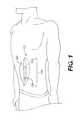
- FIG. 1illustrates a device and method for treating abdominal openings, according to certain embodiments.
- FIG. 2illustrates a device for treating abdominal openings, according to certain embodiments.
- FIG. 3illustrates a device for treating abdominal openings, according to certain embodiments.
- FIG. 4illustrates the device of FIG. 3 , as it may be used for treating abdominal openings, according to certain embodiments.
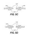
- FIGS. 5A-5Dare cross sectional views of the device of FIG. 2 , according to various exemplary embodiments.
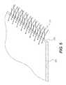
- FIG. 6illustrates a perspective view of the device of FIG. 2 , according to certain embodiments.
- acellular tissue matrixrefers generally to any tissue matrix that is substantially free of cells and other antigenic material. Skin, parts of skin (e.g., dermis), and other tissues such as blood vessels, heart valves, fascia and nerve connective tissue may be used to create acellular matrices within the scope of the present disclosure.
- abdominal defectrefers generally to a disruption in the abdominal wall.
- the disruptioncan include a hole that passes through the entire abdominal wall, such as an incision through the wall, or can include an incision or defect in one or more layers of the abdominal wall, such as the skin and subcutaneous fat.
- FIG. 1illustrates a device and method for treating abdominal openings, according to certain embodiments.
- the device 100can be used to close an abdominal defect 140 , including, for example, an incision created by surgery.
- the device 100can assist in closure of a midline incision, or can be used to assist in closure of other incisions (e.g., more laterally positioned incisions, transverse incisions, or oblique incisions).
- the device 100can include one or more sheets of material 110 , 120 that can be used to connect opposing edges of a wound, surgical incision, or other abdominal defect 140 .
- the device 100can provide additional material to allow tissues (e.g., fascia) surrounding a defect 140 to be connected and to cover the entire defect 140 .
- the device 100can be used to cover the defect 140 temporarily until a final closure is desired or possible. For example, if final closure is not possible due to swelling of abdominal contents, the device 100 can be used to close the abdomen until swelling abates.
- the device 100can provide an access site to allow multiple surgeries.
- the device 100can be adjusted during two or more surgeries to allow more normal surgical closure, as described further below.
- the sheets 110 , 120 of the device 100include a biologic material, including an acellular tissue matrix, such as a dermal acellular tissue matrix.
- the sheets 110 , 120further include a synthetic polymeric material that is attached to the acellular tissue matrix.
- FIGS. 2-5DVarious embodiments of the device 100 , are described with reference to FIGS. 2-5D below (labeled 200 , 300 ).
- FIG. 2illustrates a device 200 for treating abdominal defects, according to certain embodiments.
- the device 200includes a first synthetic polymeric material 210 and an acellular tissue matrix 220 attached to an entire peripheral border 230 of the synthetic polymeric material 210 .
- the acellular tissue matrix 220can be secured to tissues surrounding a defect 140 in a body cavity to close the body cavity (e.g., the abdomen) without attaching the first synthetic polymeric material to tissue.
- tissue surrounding a defect 140 in a body cavitye.g., the abdomen
- the device 200can assist in closure of an incision or other defect and can be used to re-access the surgical site and/or to close the defect after problems that prevented normal closure abate (e.g., swelling diminishes or subsequent surgical steps are complete).
- synthetic polymeric materialincludes any polymeric material sheet produced by man, either from a chemical reaction, or by assembling a natural material to produce a sheet.
- polymers produced by mancan include, polyethylenes or polyamides.
- Materials produced by assembling a natural materialcan include, for example, sheets produced from silk.
- the synthetic polymeric material 210 with an acellular tissue matrix 220 attached to its peripheral border 230 to form a joint 235is positioned in the defect in the abdominal wall.
- the acellular tissue matrixis attached to tissues surrounding a peripheral border of the abdominal defect to close the defect.
- the acellular tissue matrix 220will be secured to abdominal fascia (e.g., the rectus sheath), thereby acting as an extension of the rectus sheath, which is normally used to close midline abdominal incisions.
- the acellular tissue matrixcan be attached to the tissues using typical sutures, surgical staples, or clips, or other suitable connecting mechanisms, as are known in the art.
- the acellular tissue matrix 220can be connected by passing sutures through the acellular tissue matrix 220 .
- the suturescan be passed through preformed openings 295 , which may be reinforced (or openings 360 , as shown in FIG. 3 ).
- materialscan be used to produce the synthetic polymeric material 210 and acellular tissue matrix 220 (collectively “materials”). Generally, both materials should be sterile or asceptic and should possess suitable biomechanical properties to prevent rupture or tearing during use. In addition, in some embodiments, the mechanical properties of the materials are compatible to provide even stress distributions relative to the different materials to prevent failure, as described in more detail below. In addition, the synthetic material should be generally inert or biologically compatible to prevent undue inflammation. Suitable synthetic materials can include, for example, GORE-TEX® (or other polytetrafluroethylene materials), MARLEX® (high density polyethylene), or prolene. In certain embodiments, the synthetic materials can include synthetic, resorbable materials over part or all of their dimensions. In addition, the materials may be coated with therapeutic agents, (e.g., anti-adhesive coatings, antimicrobials, etc.).
- therapeutic agentse.g., anti-adhesive coatings, antimicrobials, etc.
- the acellular tissue matrixcan be selected to provide a variety of different biological and mechanical properties.
- the acellular tissue matrixcan be selected to allow tissue ingrowth and remodeling to allow regeneration of tissue normally found at the site where the matrix is implanted.
- the acellular tissue matrixwhen implanted on or into fascia, may be selected to allow regeneration of the fascia without excessive fibrosis or scar formation.
- the acellular tissue matrixshould not elicit an excessive inflammatory reaction and should ultimately be remodeled to produce tissue similar to the original host tissue.
- the acellular tissue matrixcan include ALLODERM® or StratticeTM, which are human and porcine acellular dermal matrices respectively.
- ALLODERM® or StratticeTMwhich are human and porcine acellular dermal matrices respectively.
- other suitable acellular tissue matricescan be used, as described further below.
- both the synthetic polymeric material 210 and acellular tissue matrix 220should possess mechanical properties such that the materials will not fail (i.e., rupture or tear) during use.
- the materialsshould have sufficient flexibility and elasticity to be handled by a surgeon when implanted, to be shaped to allow coverage of underlying structures, and to allow stretching during patient movement to provide even stress distribution to adjacent tissues without tearing. It will be understood that these properties can be varied by altering the general material properties (e.g., tensile strength and elastic properties), as well as the structural characteristics of the materials (e.g., thickness). In certain embodiments, the materials will have been selected such that the materials can withstand a tensile force of at least 20N without failure.
- the materialscan withstand a minimum force per unit width, such as at least 20N/cm, at least 24N/cm, or higher, depending on the patient.
- the materialsare selected to allow retention of sutures.
- the materialshave a suture retention strength of at least 20N.
- the materials 210 , 220may be selected and sized such that, during use, the stress distribution across the materials remains relatively even.
- the synthetic polymeric material 210 and the acellular tissue matrix 220can be selected such that the ultimate tensile strength and/or elastic properties over typical operating ranges are relatively equal, or within a certain range of one another.
- the mechanical properties of the joint 235 between the synthetic polymeric material 210 and acellular tissue matrix 220can be similarly matched with those of the synthetic polymeric material 210 and/or acellular tissue matrix 220 .
- the ultimate strength of the synthetic polymeric material 210differs from the ultimate strength of the acellular tissue matrix 220 by less than 20%, less than 15%, less than 10%, less than 5%, or any value between those percentages.
- the elastic modulus of the synthetic polymeric material 210differs from the elastic modulus of the acellular tissue matrix 220 by less than 20%, less than 15%, less than 10%, less than 5%, or any value between those percentages.
- the synthetic polymeric material 210 and acellular tissue matrix 220can be attached to one another using a number of devices or techniques.
- the materials 210 , 220may be connected using various sutures, staples, tacks, or adhesives including permanent sutures, such as prolene sutures.
- the materials 210 , 220can be connected to one another in a number of configurations.
- FIGS. 5A-5Dare cross sectional views of the device of FIG. 2 , according to various exemplary embodiments. As illustrated, the materials can be attached at an end-to-end joint 235 ( FIG. 5A ), by an overlapping joint 235 ′ ( FIG. 5B ), with the synthetic material 210 forming a bifurcated pocket joint 235 ′′ ( FIG. 5C ), or with the acellular tissue matrix forming a bifurcated pocket joint 235 ′′′ ( FIG. 5D ).
- the materialscan be attached by weaving one or both of the materials to the other.
- FIG. 6illustrates an acellular tissue matrix 220 that is attach to a woven synthetic material 211 at a joint 250 .
- the biologic material 220can be woven, or both materials 220 , 211 are woven to produce a joint 250 with sufficient mechanical properties to prevent failure during use, while allowing relatively even stress distribution.
- the acellular tissue matrix 220can be secured to tissues surrounding a defect 140 in a body cavity to close the defect without attaching the first synthetic polymeric material to tissue.
- the acellular tissue matrix 220which is selected to allow tissue ingrowth and remodeling, is the only material (other than sutures or other connecting devices) that is connected, attached, and/or anchored to the tissue. Further, after attachment, the fascia or other tissue can begin ingrowth and remodeling.
- the synthetic polymeric materialcan include an opening 240 or can be cut, without cutting adjacent tissue, to allow repeated access.
- the opening 240can then be resealed with sutures 260 or other devices.
- part of the synthetic polymeric material(delimited by oval 250 ) can be removed, and the synthetic polymeric material 210 can be shortened to provide additional tension on the incision margins or to remove excess or contaminated materials.
- the synthetic polymeric material 210may be removed at a later time, e.g., after swelling has diminished or subsequent surgeries have been completed, and the acellular tissue matrix 220 can be left attached to the tissues surrounding the peripheral border of the abdominal defect.
- the abdominal defectcan then be closed after removing the synthetic polymeric material 210 by attaching remaining portions of the acellular tissue matrix 220 to one another using sutures, staples, or other surgical means.
- the acellular tissue matrix 220will bolster the fascia or other tissue around the defect to prevent reopening or dehiscence.
- the acellular tissue matrixcan provide additional tissue in cases where there is insufficient tissue present for normal fascia closure.
- the acellular tissue matrix 220can include openings 295 , and the openings can be used to receive sutures for closing the abdominal opening.
- the openings 295can be reinforced, as described further below.
- FIG. 3illustrates a device 300 for treating abdominal defects, according to certain embodiments.
- the device 300comprises a sheet 310 of acellular tissue matrix, wherein the sheet 310 includes an elongated opening 340 , and on opposite sides of the opening 340 multiple holes 360 for receiving sutures, and wherein the multiple holes 360 are reinforced.
- the device 300can be secured to wound margins (e.g., via fascia using sutures), and the reinforced holes 360 can receive sutures that provide tension to the device 300 and wound margins to close the wound or incision.
- the opening 340can be reopened, for example, to perform a subsequent operation, clean a wound/abdominal site, or for any other purpose.
- the device 300can have multiple sets of reinforced holes 360 , to allow the device to be sutured with at varying distances, for example, to provide increasing tension to wound margins, or to remove excess material.
- the preformed holes 360include two or more rows 365 , 367 of holes positioned on each side of the elongated opening 340 , and sutures can be placed through holes at selected distances apart. For example, as shown in FIG. 4 , sutures may initially be attached through a first row of holes 365 nearest the opening 340 , to close an incision.
- a surgeonmay add additional sutures or replace the sutures, passing the sutures through openings 367 . In this way, the wound or incision margins can be pulled closer together as the sutures are tightened or shortened.
- the device 300can include a single sheet of material. However, in some embodiments, two or more pieces of acellular tissue matrix 310 may be used. For example, the device of FIG. 3 can be divided into two pieces along a line extending from line 370 to produce two pieces of material. The two pieces can be implanted on opposite sides of a wound or incision and sutured in place to close the wound or incision, as described above.
- the openings 360can be reinforced in a number of ways.
- the openings 360can be reinforced using a biocompatible adhesive placed around the rim or edge of the openings 360 .
- Suitable adhesivesinclude, for example, fibrin glue, cyanoacrylate-based tissue adhesives (e.g., DERMABOND®), and chitosan tissue adhesives.
- the rim or edges of the openings 360can be crosslinked to increase their strength and prevent tearing (e.g., using chemical or radiation induced cross-linking).
- acellular tissue matrixrefers generally to any tissue matrix that is substantially free of cells and other antigenic material. Skin, parts of skin (e.g., dermis), and other tissues such as blood vessels, heart valves, fascia and nerve connective tissue may be used to create acellular matrices within the scope of the present disclosure.
- the steps involved in the production of an acellular tissue matrixinclude harvesting the tissue from a donor (e.g., a human cadaver or animal source) and cell removal under conditions that preserve biological and structural function.
- the processincludes chemical treatment to stabilize the tissue and avoid biochemical and structural degradation together with or before cell removal.
- the stabilizing solutionarrests and prevents osmotic, hypoxic, autolytic, and proteolytic degradation, protects against microbial contamination, and reduces mechanical damage that can occur with tissues that contain, for example, smooth muscle components (e.g., blood vessels).
- the stabilizing solutionmay contain an appropriate buffer, one or more antioxidants, one or more oncotic agents, one or more antibiotics, one or more protease inhibitors, and/or one or more smooth muscle relaxants.
- the tissueis then placed in a decellularization solution to remove viable cells (e.g., epithelial cells, endothelial cells, smooth muscle cells, and fibroblasts) from the structural matrix without damaging the biological and structural integrity of the collagen matrix.
- the decellularization solutionmay contain an appropriate buffer, salt, an antibiotic, one or more detergents (e.g., TRITON X-100TM, sodium deoxycholate, polyoxyethylene (20) sorbitan mono-oleate), one or more agents to prevent cross-linking, one or more protease inhibitors, and/or one or more enzymes.
- the decellularization solutioncomprises 1% TRITON X-100TM in RPMI media with Gentamicin and 25 mM EDTA (ethylenediaminetetraacetic acid).
- the tissueis incubated in the decellularization solution overnight at 37° C. with gentle shaking at 90 rpm.
- additional detergentsmay be used to remove fat from the tissue sample. For example, in some embodiments, 2% sodium deoxycholate is added to the decellularization solution.
- the tissue sampleis washed thoroughly with saline.
- the decellularized tissueis then treated overnight at room temperature with a deoxyribonuclease (DNase) solution.
- DNasedeoxyribonuclease
- the tissue sampleis treated with a DNase solution prepared in DNase buffer (20 mM HEPES (4-(2-hydroxyethyl)-1-piperazineethanesulfonic acid), 20 mM CaCl2 and 20 mM MgCl2).
- an antibiotic solutione.g., Gentamicin
- Any suitable buffercan be used as long as the buffer provides suitable DNase activity.
- an acellular tissue matrixmay be made from one or more individuals of the same species as the recipient of the acellular tissue matrix graft, this is not necessarily the case.
- an acellular tissue matrixmay be made from porcine tissue and implanted in a human patient.
- Species that can serve as recipients of acellular tissue matrix and donors of tissues or organs for the production of the acellular tissue matrixinclude, without limitation, mammals, such as humans, nonhuman primates (e.g., monkeys, baboons, or chimpanzees), pigs, cows, horses, goats, sheep, dogs, cats, rabbits, guinea pigs, gerbils, hamsters, rats, or mice.
- Elimination of the ⁇ -gal epitopes from the collagen-containing materialmay diminish the immune response against the collagen-containing material.
- the ⁇ -gal epitopeis expressed in non-primate mammals and in New World monkeys (monkeys of South America) as well as on macromolecules such as proteoglycans of the extracellular components.
- Anti-gal antibodiesare produced in humans and primates as a result of an immune response to ⁇ -gal epitope carbohydrate structures on gastrointestinal bacteria.
- non-primate mammalse.g., pigs
- xenotransplantation of collagen-containing material from these mammals into primatesoften results in rejection because of primate anti-Gal binding to these epitopes on the collagen-containing material.
- the bindingresults in the destruction of the collagen-containing material by complement fixation and by antibody dependent cell cytotoxicity.
- U. Galili et al.Immunology Today 14: 480 (1993); M. Sandrin et al., Proc. Natl. Acad. Sci. USA 90: 11391 (1993); H. Good et al., Transplant. Proc. 24: 559 (1992); B. H. Collins et al., J. Immunol.
- xenotransplantationresults in major activation of the immune system to produce increased amounts of high affinity anti-gal antibodies.
- tissue sourcewhen animals that produce ⁇ -gal epitopes are used as the tissue source, the substantial elimination of ⁇ -gal epitopes from cells and from extracellular components of the collagen-containing material, and the prevention of re-expression of cellular ⁇ -gal epitopes can diminish the immune response against the collagen-containing material associated with anti-gal antibody binding to ⁇ -gal epitopes.
- the tissue samplemay be subjected to one or more enzymatic treatments to remove certain immunogenic antigens, if present in the sample.
- the tissue samplemay be treated with an ⁇ -galactosidase enzyme to eliminate ⁇ -gal epitopes if present in the tissue.
- the tissue sampleis treated with ⁇ -galactosidase at a concentration of 300 U/L prepared in 100 mM phosphate buffer at pH 6.0 In other embodiments, the concentration of ⁇ -galactosidase is increased to 400 U/L for adequate removal of the ⁇ -gal epitopes from the harvested tissue. Any suitable enzyme concentration and buffer can be used as long as sufficient removal of antigens is achieved.
- animals that have been genetically modified to lack one or more antigenic epitopesmay be selected as the tissue source.
- animalse.g., pigs
- animalsthat have been genetically engineered to lack the terminal ⁇ -galactose moiety can be selected as the tissue source.
- histocompatible, viable cellsmay optionally be seeded in the acellular tissue matrix to produce a graft that may be further remodeled by the host.
- histocompatible viable cellsmay be added to the matrices by standard in vitro cell co-culturing techniques prior to transplantation, or by in vivo repopulation following transplantation. In vivo repopulation can be by the recipient's own cells migrating into the acellular tissue matrix or by infusing or injecting cells obtained from the recipient or histocompatible cells from another donor into the acellular tissue matrix in situ.
- Various cell typescan be used, including embryonic stem cells, adult stem cells (e.g.
- the cellscan be directly applied to the inner portion of the acellular tissue matrix just before or after implantation. In certain embodiments, the cells can be placed within the acellular tissue matrix to be implanted, and cultured prior to implantation.
Landscapes
- Health & Medical Sciences (AREA)
- Life Sciences & Earth Sciences (AREA)
- Surgery (AREA)
- Molecular Biology (AREA)
- Engineering & Computer Science (AREA)
- Biomedical Technology (AREA)
- Heart & Thoracic Surgery (AREA)
- Medical Informatics (AREA)
- Nuclear Medicine, Radiotherapy & Molecular Imaging (AREA)
- Animal Behavior & Ethology (AREA)
- General Health & Medical Sciences (AREA)
- Public Health (AREA)
- Veterinary Medicine (AREA)
- Materials For Medical Uses (AREA)
- Prostheses (AREA)
- Surgical Instruments (AREA)
Abstract
Description
Claims (20)
Priority Applications (2)
| Application Number | Priority Date | Filing Date | Title |
|---|---|---|---|
| US13/029,487US10448951B2 (en) | 2010-02-19 | 2011-02-17 | Abdominal wall treatment devices |
| US16/566,220US20200000472A1 (en) | 2010-02-19 | 2019-09-10 | Abdominal wall treatment devices |
Applications Claiming Priority (2)
| Application Number | Priority Date | Filing Date | Title |
|---|---|---|---|
| US30600610P | 2010-02-19 | 2010-02-19 | |
| US13/029,487US10448951B2 (en) | 2010-02-19 | 2011-02-17 | Abdominal wall treatment devices |
Related Child Applications (1)
| Application Number | Title | Priority Date | Filing Date |
|---|---|---|---|
| US16/566,220ContinuationUS20200000472A1 (en) | 2010-02-19 | 2019-09-10 | Abdominal wall treatment devices |
Publications (2)
| Publication Number | Publication Date |
|---|---|
| US20110208320A1 US20110208320A1 (en) | 2011-08-25 |
| US10448951B2true US10448951B2 (en) | 2019-10-22 |
Family
ID=43838024
Family Applications (2)
| Application Number | Title | Priority Date | Filing Date |
|---|---|---|---|
| US13/029,487Expired - Fee RelatedUS10448951B2 (en) | 2010-02-19 | 2011-02-17 | Abdominal wall treatment devices |
| US16/566,220AbandonedUS20200000472A1 (en) | 2010-02-19 | 2019-09-10 | Abdominal wall treatment devices |
Family Applications After (1)
| Application Number | Title | Priority Date | Filing Date |
|---|---|---|---|
| US16/566,220AbandonedUS20200000472A1 (en) | 2010-02-19 | 2019-09-10 | Abdominal wall treatment devices |
Country Status (9)
| Country | Link |
|---|---|
| US (2) | US10448951B2 (en) |
| EP (3) | EP2536340B1 (en) |
| JP (1) | JP5701908B2 (en) |
| CN (1) | CN102753105B (en) |
| AU (1) | AU2011218066B2 (en) |
| CA (1) | CA2786228C (en) |
| DK (1) | DK2536340T3 (en) |
| ES (2) | ES2811027T3 (en) |
| WO (1) | WO2011103276A2 (en) |
Cited By (1)
| Publication number | Priority date | Publication date | Assignee | Title |
|---|---|---|---|---|
| US20180064522A1 (en)* | 2015-03-31 | 2018-03-08 | Prevent Patch, LLC | Devices and methods for preventing incisional hernias |
Families Citing this family (7)
| Publication number | Priority date | Publication date | Assignee | Title |
|---|---|---|---|---|
| AU2011232374B2 (en)* | 2010-03-25 | 2015-06-25 | Lifecell Corporation | Preparation of regenerative tissue scaffolds |
| WO2013009993A1 (en)* | 2011-07-12 | 2013-01-17 | Bengtson Bradley P | Surgical fixation devices, systems, and methods |
| WO2013165942A1 (en) | 2012-05-01 | 2013-11-07 | Brigham And Women's Hospital, Inc. | Suturing device for laparoscopic procedures |
| WO2015050999A1 (en)* | 2013-10-02 | 2015-04-09 | Kia Michael Amirfarzad | A suture device |
| CA2998136C (en) | 2015-09-11 | 2023-10-10 | Lifecell Corporation | Perforated tissue matrix |
| WO2018067644A1 (en)* | 2016-10-06 | 2018-04-12 | Lifecell Corporation | Tissue matrix with preformed openings or pilot openings |
| CA3096883A1 (en)* | 2018-04-12 | 2019-10-17 | Axogen Corporation | Tissue grafts with pre-made attachment points |
Citations (68)
| Publication number | Priority date | Publication date | Assignee | Title |
|---|---|---|---|---|
| DE2303444B1 (en) | 1973-01-25 | 1974-05-09 | Temca Chemische Union Gmbh, 8500 Nuernberg | Adhesive plaster |
| US4034750A (en) | 1975-04-22 | 1977-07-12 | Maurice Seiderman | Electrochemically linking collagen fibrils to animal tissue |
| US4796603A (en) | 1986-06-07 | 1989-01-10 | Ethicon, Inc. | Pad-like implant |
| US4841962A (en) | 1984-03-27 | 1989-06-27 | Berg Richard A | Collagen matrix/polymer film composite dressing |
| US4854316A (en) | 1986-10-03 | 1989-08-08 | Davis Emsley A | Apparatus and method for repairing and preventing para-stomal hernias |
| US5336616A (en) | 1990-09-12 | 1994-08-09 | Lifecell Corporation | Method for processing and preserving collagen-based tissues for transplantation |
| US5364756A (en) | 1990-09-12 | 1994-11-15 | Lifecell | Method of cryopreserving a suspension of biological material |
| US5425740A (en)* | 1994-05-17 | 1995-06-20 | Hutchinson, Jr.; William B. | Endoscopic hernia repair clip and method |
| US5634931A (en) | 1994-09-29 | 1997-06-03 | Surgical Sense, Inc. | Hernia mesh patches and methods of their use |
| US5972007A (en) | 1997-10-31 | 1999-10-26 | Ethicon Endo-Surgery, Inc. | Energy-base method applied to prosthetics for repairing tissue defects |
| WO2000016822A1 (en) | 1998-09-21 | 2000-03-30 | The Brigham And Women's Hospital, Inc. | Compositions and methods for tissue repair |
| US6113623A (en) | 1994-04-20 | 2000-09-05 | Cabinet Beau De Lomenie | Prosthetic device and method for eventration repair |
| US6174320B1 (en) | 1994-09-29 | 2001-01-16 | Bard Asdi Inc. | Hernia mesh patch with slit |
| US6179872B1 (en) | 1998-03-17 | 2001-01-30 | Tissue Engineering | Biopolymer matt for use in tissue repair and reconstruction |
| US6197036B1 (en) | 1997-10-01 | 2001-03-06 | Scimed Life Systems, Inc. | Pelvic floor reconstruction |
| US6381026B1 (en) | 1999-03-15 | 2002-04-30 | Lifecell Corp. | Method of measuring the contour of a biological surface |
| JP2002186581A (en) | 2000-12-21 | 2002-07-02 | Asahi Optical Co Ltd | Endoscope forceps stopper |
| US20020099344A1 (en) | 1999-07-15 | 2002-07-25 | Hessel Lassel L. | Implant |
| US20020103542A1 (en) | 2000-09-18 | 2002-08-01 | Bilbo Patrick R. | Methods for treating a patient using a bioengineered flat sheet graft prostheses |
| US20030035843A1 (en) | 1990-09-12 | 2003-02-20 | Lifecell Corporation, A Delaware Corporation | Method for processing and preserving collagen-based tissues for transplantation |
| US20030119985A1 (en) | 1995-12-18 | 2003-06-26 | Sehl Louis C. | Methods for tissue repair using adhesive materials |
| US6599318B1 (en) | 1999-11-30 | 2003-07-29 | Shlomo Gabbay | Implantable support apparatus and method of using same |
| US20030143207A1 (en) | 2001-10-18 | 2003-07-31 | Livesey Stephen A. | Remodeling of tissues and organ |
| US6610006B1 (en) | 2000-07-25 | 2003-08-26 | C. R. Bard, Inc. | Implantable prosthesis |
| US6638284B1 (en) | 1999-06-08 | 2003-10-28 | Ethicon, Inc. | Knitted surgical mesh |
| US6652559B1 (en) | 1999-02-23 | 2003-11-25 | Advanced Terapeutic Technologies AT2 Inc. | Wound closure system |
| US20030225355A1 (en) | 1998-10-01 | 2003-12-04 | Butler Charles E. | Composite material for wound repair |
| US20040078089A1 (en) | 2000-10-11 | 2004-04-22 | Julian Ellis | Textile prosthesis |
| US20040078077A1 (en) | 2002-10-18 | 2004-04-22 | Francois Binette | Biocompatible scaffold for ligament or tendon repair |
| US6790213B2 (en) | 2002-01-07 | 2004-09-14 | C.R. Bard, Inc. | Implantable prosthesis |
| US20040220591A1 (en) | 2003-04-30 | 2004-11-04 | Bonutti Peter M. | Tissue fastener and methods for using same |
| US20050009178A1 (en) | 2003-06-04 | 2005-01-13 | University Of South Carolina | Tissue scaffold having aligned fibrils, apparatus and method for producing the same, and artificial tissue and methods of use thereof |
| US6843767B2 (en)* | 2001-10-18 | 2005-01-18 | Spiration, Inc. | Constriction device including tear resistant structures |
| US20050015088A1 (en)* | 2003-07-15 | 2005-01-20 | Ringeisen Timothy A. | Compliant osteosynthesis fixation plate |
| US20050028228A1 (en) | 2003-07-21 | 2005-02-03 | Lifecell Corporation | Acellular tissue matrices made from alpa-1,3-galactose-deficient tissue |
| US20050043716A1 (en) | 2003-02-11 | 2005-02-24 | C.R. Bard, Inc. | Implantable prosthesis and method of use |
| US20050049638A1 (en) | 2003-05-21 | 2005-03-03 | Mandelbaum Jon A. | Surgical method and composition utilizing submucosal tissue to prevent incisional hernias |
| US20050058629A1 (en) | 2003-06-27 | 2005-03-17 | Harmon Alexander M. | Soft tissue repair and regeneration using postpartum-derived cells |
| US20050085757A1 (en) | 2003-10-15 | 2005-04-21 | Steven Santanello | Expandable temporary abdominal closure |
| US6933326B1 (en) | 1998-06-19 | 2005-08-23 | Lifecell Coporation | Particulate acellular tissue matrix |
| US20050288691A1 (en) | 2004-06-28 | 2005-12-29 | Leiboff Arnold R | Hernia patch |
| US20060073592A1 (en) | 2004-10-06 | 2006-04-06 | Wendell Sun | Methods of storing tissue matrices |
| US20060105026A1 (en) | 2003-04-04 | 2006-05-18 | Fortune David H | Tissue-adhesive formulations |
| US20060106419A1 (en) | 2002-08-23 | 2006-05-18 | Peter Gingras | Three dimensional implant |
| US20060235511A1 (en) | 2004-02-09 | 2006-10-19 | Cook Incorporated | Woven implantable device |
| US20060247206A1 (en) | 2005-03-23 | 2006-11-02 | Feins Neil R | Minimally invasive clinical treatment method for closure of umbilical hernias |
| US20060276908A1 (en) | 2002-07-25 | 2006-12-07 | Torben Sogaard-Andersen | Method for prevention of a parastomal hernia in a patient |
| US7163563B2 (en)* | 2001-07-16 | 2007-01-16 | Depuy Products, Inc. | Unitary surgical device and method |
| US20070111937A1 (en) | 2005-03-24 | 2007-05-17 | Pickar James H | Use of fibrous tissue inducing proteins for hernia repair |
| US7235295B2 (en) | 2003-09-10 | 2007-06-26 | Laurencin Cato T | Polymeric nanofibers for tissue engineering and drug delivery |
| US20070202173A1 (en) | 2006-01-26 | 2007-08-30 | Jorge Cueto-Garcia | Biodegradable, non-toxic biological adhesive for use in abdominal surgery |
| US20070248575A1 (en) | 2006-04-19 | 2007-10-25 | Jerome Connor | Bone graft composition |
| US20070293878A1 (en) | 2006-06-16 | 2007-12-20 | Butsch John L | Magnetic mesh support for tissue walls |
| US20080027542A1 (en) | 2006-05-09 | 2008-01-31 | Lifecell Corporation | Reinforced Biological Tissue |
| US20080033461A1 (en) | 2004-04-26 | 2008-02-07 | Ferdinand Koeckerling | Tow-Dimensional Mesh Implant For Hernia Care |
| US20080071300A1 (en) | 2003-11-26 | 2008-03-20 | Nicholas Popadiuk | Prosthetic repair device |
| US20080091277A1 (en) | 2004-08-13 | 2008-04-17 | Kai Deusch | Surgical prosthesis having biodegradable and nonbiodegradable regions |
| US20080095819A1 (en) | 2004-12-21 | 2008-04-24 | Robert Gourdie | Composition and Methods for Promoting Wound Healing and tissue Regeneration |
| US20080113035A1 (en) | 1996-12-02 | 2008-05-15 | Angiotech International Ag | Compositions and methods for treating or preventing inflammatory diseases |
| WO2008060973A1 (en) | 2006-11-10 | 2008-05-22 | Vance Products Incorporated D/B/A Cook Urological Incorporated | Graft for hysterotomy closure |
| US20080131509A1 (en) | 2006-12-04 | 2008-06-05 | Syed Hossainy | Methods and Compositions for Treating Tissue Using Silk Proteins |
| US20080167729A1 (en) | 2007-01-10 | 2008-07-10 | Nelson Christopher M | Implantable devices useful for reinforcing a surgically created stoma |
| US20090035289A1 (en) | 2005-09-26 | 2009-02-05 | Lifecell Corporation | Dry platelet composition |
| WO2009075786A1 (en)* | 2007-12-07 | 2009-06-18 | C.R. Bard, Inc. | Implantable prosthesis |
| DE102009020763A1 (en) | 2008-05-15 | 2009-11-19 | Aesculap Aktiengesellschaft | wound closure |
| US20090306790A1 (en) | 2008-06-06 | 2009-12-10 | Wendell Sun | Elastase Treatment of Tissue Matrices |
| US20110004306A1 (en)* | 2009-07-02 | 2011-01-06 | Lifecell Corporation | Device and method for treatment of incision or hernia |
| US20110295283A1 (en)* | 2008-11-21 | 2011-12-01 | C.R. Bard, Inc. | Soft tissue repair prosthesis, expandable device, and method of soft tissue repair |
Family Cites Families (6)
| Publication number | Priority date | Publication date | Assignee | Title |
|---|---|---|---|---|
| ZA96225B (en)* | 1995-01-24 | 1997-07-11 | Bristol Myers Squibb Co | Method and closure tape for improved wound or incision healing |
| US6166288A (en) | 1995-09-27 | 2000-12-26 | Nextran Inc. | Method of producing transgenic animals for xenotransplantation expressing both an enzyme masking or reducing the level of the gal epitope and a complement inhibitor |
| CN1986007B (en)* | 2005-12-20 | 2011-09-14 | 广东冠昊生物科技股份有限公司 | Biological surgical patch |
| BRPI0717388A2 (en)* | 2006-10-23 | 2013-10-15 | Cook Biotech Inc | ECM MATERIALS PROCESSED WITH BEST COMPONENT PROFILES |
| DK3473260T3 (en)* | 2007-07-10 | 2020-12-21 | Lifecell Corp | ACELLULAR TISSUE MATRIX COMPOSITIONS FOR TISSUE REPAIR |
| JP5313142B2 (en)* | 2007-08-09 | 2013-10-09 | グンゼ株式会社 | Prosthetic material for living organs |
- 2011
- 2011-02-17EPEP11708128.1Apatent/EP2536340B1/ennot_activeNot-in-force
- 2011-02-17AUAU2011218066Apatent/AU2011218066B2/ennot_activeCeased
- 2011-02-17DKDK11708128.1Tpatent/DK2536340T3/enactive
- 2011-02-17ESES18195811Tpatent/ES2811027T3/enactiveActive
- 2011-02-17CACA2786228Apatent/CA2786228C/ennot_activeExpired - Fee Related
- 2011-02-17JPJP2012554021Apatent/JP5701908B2/ennot_activeExpired - Fee Related
- 2011-02-17ESES11708128Tpatent/ES2705684T3/enactiveActive
- 2011-02-17EPEP18195811.7Apatent/EP3434201B1/ennot_activeNot-in-force
- 2011-02-17USUS13/029,487patent/US10448951B2/ennot_activeExpired - Fee Related
- 2011-02-17WOPCT/US2011/025224patent/WO2011103276A2/enactiveApplication Filing
- 2011-02-17CNCN201180008668.5Apatent/CN102753105B/ennot_activeExpired - Fee Related
- 2011-02-17EPEP20173310.2Apatent/EP3769695A1/ennot_activeWithdrawn
- 2019
- 2019-09-10USUS16/566,220patent/US20200000472A1/ennot_activeAbandoned
Patent Citations (77)
| Publication number | Priority date | Publication date | Assignee | Title |
|---|---|---|---|---|
| DE2303444B1 (en) | 1973-01-25 | 1974-05-09 | Temca Chemische Union Gmbh, 8500 Nuernberg | Adhesive plaster |
| US4034750A (en) | 1975-04-22 | 1977-07-12 | Maurice Seiderman | Electrochemically linking collagen fibrils to animal tissue |
| US4841962A (en) | 1984-03-27 | 1989-06-27 | Berg Richard A | Collagen matrix/polymer film composite dressing |
| US4796603A (en) | 1986-06-07 | 1989-01-10 | Ethicon, Inc. | Pad-like implant |
| US4854316A (en) | 1986-10-03 | 1989-08-08 | Davis Emsley A | Apparatus and method for repairing and preventing para-stomal hernias |
| US5336616A (en) | 1990-09-12 | 1994-08-09 | Lifecell Corporation | Method for processing and preserving collagen-based tissues for transplantation |
| US5364756A (en) | 1990-09-12 | 1994-11-15 | Lifecell | Method of cryopreserving a suspension of biological material |
| US20030035843A1 (en) | 1990-09-12 | 2003-02-20 | Lifecell Corporation, A Delaware Corporation | Method for processing and preserving collagen-based tissues for transplantation |
| US5780295A (en) | 1990-09-12 | 1998-07-14 | Life Cell Corporation | Apparatus for cryopreparation, dry stabilization and rehydration of biological suspensions |
| US6194136B1 (en) | 1990-09-12 | 2001-02-27 | Lifecell Corporation | Cryoprotective solutions comprising DMSO, PG, 2,3-butanediol,raffinose and PVP |
| US6113623A (en) | 1994-04-20 | 2000-09-05 | Cabinet Beau De Lomenie | Prosthetic device and method for eventration repair |
| US5425740A (en)* | 1994-05-17 | 1995-06-20 | Hutchinson, Jr.; William B. | Endoscopic hernia repair clip and method |
| US5634931A (en) | 1994-09-29 | 1997-06-03 | Surgical Sense, Inc. | Hernia mesh patches and methods of their use |
| US6174320B1 (en) | 1994-09-29 | 2001-01-16 | Bard Asdi Inc. | Hernia mesh patch with slit |
| US6833408B2 (en) | 1995-12-18 | 2004-12-21 | Cohesion Technologies, Inc. | Methods for tissue repair using adhesive materials |
| US20050054771A1 (en) | 1995-12-18 | 2005-03-10 | Sehl Louis C. | Adhesive tissue repair patch |
| US20030119985A1 (en) | 1995-12-18 | 2003-06-26 | Sehl Louis C. | Methods for tissue repair using adhesive materials |
| US20080113035A1 (en) | 1996-12-02 | 2008-05-15 | Angiotech International Ag | Compositions and methods for treating or preventing inflammatory diseases |
| US6197036B1 (en) | 1997-10-01 | 2001-03-06 | Scimed Life Systems, Inc. | Pelvic floor reconstruction |
| US5972007A (en) | 1997-10-31 | 1999-10-26 | Ethicon Endo-Surgery, Inc. | Energy-base method applied to prosthetics for repairing tissue defects |
| US6179872B1 (en) | 1998-03-17 | 2001-01-30 | Tissue Engineering | Biopolymer matt for use in tissue repair and reconstruction |
| US6933326B1 (en) | 1998-06-19 | 2005-08-23 | Lifecell Coporation | Particulate acellular tissue matrix |
| US7358284B2 (en) | 1998-06-19 | 2008-04-15 | Lifecell Corporation | Particulate acellular tissue matrix |
| WO2000016822A1 (en) | 1998-09-21 | 2000-03-30 | The Brigham And Women's Hospital, Inc. | Compositions and methods for tissue repair |
| US20030225355A1 (en) | 1998-10-01 | 2003-12-04 | Butler Charles E. | Composite material for wound repair |
| US6652559B1 (en) | 1999-02-23 | 2003-11-25 | Advanced Terapeutic Technologies AT2 Inc. | Wound closure system |
| US6381026B1 (en) | 1999-03-15 | 2002-04-30 | Lifecell Corp. | Method of measuring the contour of a biological surface |
| US6638284B1 (en) | 1999-06-08 | 2003-10-28 | Ethicon, Inc. | Knitted surgical mesh |
| US20020099344A1 (en) | 1999-07-15 | 2002-07-25 | Hessel Lassel L. | Implant |
| US6726660B2 (en) | 1999-07-15 | 2004-04-27 | Biotap A/S | Implant |
| US6599318B1 (en) | 1999-11-30 | 2003-07-29 | Shlomo Gabbay | Implantable support apparatus and method of using same |
| US6610006B1 (en) | 2000-07-25 | 2003-08-26 | C. R. Bard, Inc. | Implantable prosthesis |
| US20020103542A1 (en) | 2000-09-18 | 2002-08-01 | Bilbo Patrick R. | Methods for treating a patient using a bioengineered flat sheet graft prostheses |
| US20040078089A1 (en) | 2000-10-11 | 2004-04-22 | Julian Ellis | Textile prosthesis |
| JP2002186581A (en) | 2000-12-21 | 2002-07-02 | Asahi Optical Co Ltd | Endoscope forceps stopper |
| US7163563B2 (en)* | 2001-07-16 | 2007-01-16 | Depuy Products, Inc. | Unitary surgical device and method |
| US6843767B2 (en)* | 2001-10-18 | 2005-01-18 | Spiration, Inc. | Constriction device including tear resistant structures |
| US20030143207A1 (en) | 2001-10-18 | 2003-07-31 | Livesey Stephen A. | Remodeling of tissues and organ |
| US6790213B2 (en) | 2002-01-07 | 2004-09-14 | C.R. Bard, Inc. | Implantable prosthesis |
| US20060276908A1 (en) | 2002-07-25 | 2006-12-07 | Torben Sogaard-Andersen | Method for prevention of a parastomal hernia in a patient |
| US20060106419A1 (en) | 2002-08-23 | 2006-05-18 | Peter Gingras | Three dimensional implant |
| US20040078077A1 (en) | 2002-10-18 | 2004-04-22 | Francois Binette | Biocompatible scaffold for ligament or tendon repair |
| US20050043716A1 (en) | 2003-02-11 | 2005-02-24 | C.R. Bard, Inc. | Implantable prosthesis and method of use |
| US20060105026A1 (en) | 2003-04-04 | 2006-05-18 | Fortune David H | Tissue-adhesive formulations |
| US20040220591A1 (en) | 2003-04-30 | 2004-11-04 | Bonutti Peter M. | Tissue fastener and methods for using same |
| US20050049638A1 (en) | 2003-05-21 | 2005-03-03 | Mandelbaum Jon A. | Surgical method and composition utilizing submucosal tissue to prevent incisional hernias |
| US7105001B2 (en) | 2003-05-21 | 2006-09-12 | Mandelbaum Jon A | Surgical method and composition utilizing submucosal tissue to prevent incisional hernias |
| US20050009178A1 (en) | 2003-06-04 | 2005-01-13 | University Of South Carolina | Tissue scaffold having aligned fibrils, apparatus and method for producing the same, and artificial tissue and methods of use thereof |
| US20080147199A1 (en) | 2003-06-04 | 2008-06-19 | University Of South Carolina | Tissue scaffold having aligned fibrils, apparatus and method for producing the same, and artificial tissue and methods of use thereof |
| US20050058629A1 (en) | 2003-06-27 | 2005-03-17 | Harmon Alexander M. | Soft tissue repair and regeneration using postpartum-derived cells |
| US20050015088A1 (en)* | 2003-07-15 | 2005-01-20 | Ringeisen Timothy A. | Compliant osteosynthesis fixation plate |
| US20050028228A1 (en) | 2003-07-21 | 2005-02-03 | Lifecell Corporation | Acellular tissue matrices made from alpa-1,3-galactose-deficient tissue |
| US7235295B2 (en) | 2003-09-10 | 2007-06-26 | Laurencin Cato T | Polymeric nanofibers for tissue engineering and drug delivery |
| US20050085757A1 (en) | 2003-10-15 | 2005-04-21 | Steven Santanello | Expandable temporary abdominal closure |
| US20080071300A1 (en) | 2003-11-26 | 2008-03-20 | Nicholas Popadiuk | Prosthetic repair device |
| US20060235511A1 (en) | 2004-02-09 | 2006-10-19 | Cook Incorporated | Woven implantable device |
| US20080033461A1 (en) | 2004-04-26 | 2008-02-07 | Ferdinand Koeckerling | Tow-Dimensional Mesh Implant For Hernia Care |
| US20050288691A1 (en) | 2004-06-28 | 2005-12-29 | Leiboff Arnold R | Hernia patch |
| US20080091277A1 (en) | 2004-08-13 | 2008-04-17 | Kai Deusch | Surgical prosthesis having biodegradable and nonbiodegradable regions |
| US20060073592A1 (en) | 2004-10-06 | 2006-04-06 | Wendell Sun | Methods of storing tissue matrices |
| US20080095819A1 (en) | 2004-12-21 | 2008-04-24 | Robert Gourdie | Composition and Methods for Promoting Wound Healing and tissue Regeneration |
| US20060247206A1 (en) | 2005-03-23 | 2006-11-02 | Feins Neil R | Minimally invasive clinical treatment method for closure of umbilical hernias |
| US20070111937A1 (en) | 2005-03-24 | 2007-05-17 | Pickar James H | Use of fibrous tissue inducing proteins for hernia repair |
| US20090035289A1 (en) | 2005-09-26 | 2009-02-05 | Lifecell Corporation | Dry platelet composition |
| US20070202173A1 (en) | 2006-01-26 | 2007-08-30 | Jorge Cueto-Garcia | Biodegradable, non-toxic biological adhesive for use in abdominal surgery |
| US20070248575A1 (en) | 2006-04-19 | 2007-10-25 | Jerome Connor | Bone graft composition |
| US20080027542A1 (en) | 2006-05-09 | 2008-01-31 | Lifecell Corporation | Reinforced Biological Tissue |
| US20070293878A1 (en) | 2006-06-16 | 2007-12-20 | Butsch John L | Magnetic mesh support for tissue walls |
| WO2008060973A1 (en) | 2006-11-10 | 2008-05-22 | Vance Products Incorporated D/B/A Cook Urological Incorporated | Graft for hysterotomy closure |
| US20080131509A1 (en) | 2006-12-04 | 2008-06-05 | Syed Hossainy | Methods and Compositions for Treating Tissue Using Silk Proteins |
| US20080167729A1 (en) | 2007-01-10 | 2008-07-10 | Nelson Christopher M | Implantable devices useful for reinforcing a surgically created stoma |
| WO2009075786A1 (en)* | 2007-12-07 | 2009-06-18 | C.R. Bard, Inc. | Implantable prosthesis |
| US20110015760A1 (en)* | 2007-12-07 | 2011-01-20 | Kullas Karen E | Implantable prosthesis |
| DE102009020763A1 (en) | 2008-05-15 | 2009-11-19 | Aesculap Aktiengesellschaft | wound closure |
| US20090306790A1 (en) | 2008-06-06 | 2009-12-10 | Wendell Sun | Elastase Treatment of Tissue Matrices |
| US20110295283A1 (en)* | 2008-11-21 | 2011-12-01 | C.R. Bard, Inc. | Soft tissue repair prosthesis, expandable device, and method of soft tissue repair |
| US20110004306A1 (en)* | 2009-07-02 | 2011-01-06 | Lifecell Corporation | Device and method for treatment of incision or hernia |
Non-Patent Citations (17)
| Title |
|---|
| Aycock et al., "Parastomal Hernia Repair With Acellular Dermal Matrix," J. Wound Ostomy Continence Nurs. (2007), p. 521-523, 34(5). |
| Buinewicz et al., "Acellular Cadaveric Dermis (AlloDerm): A New Alternative for Abdominal Hernia Repair", Annals of Plastic Surgery, 52(2), pp. 188-194, Feb. 2004.* |
| Burns et al., "Non-Cross-Linked Porcine Acellular Dermal Matrices for Abdominal Wall Reconstruction", Plastic and Reconstructive Surgery Journal, 125(1), pp. 167-176, Jan. 2010.* |
| Candage et al., "Use of Human Acellular Dermal Matrix for Hernia Repair: Friend or Foe?" Journal of Surgery, 144(4), pp. 703-711, Oct. 2008.* |
| Eberli, Daniel et al. "In vivo evalulation of acellular human dermis for abdominal wall repair" Journal of Biomedical Materials Research Part A. vol. 93 (4) pp. 1527-1538 Published online Dec. 14, 2009.* |
| Greenstein et al., "Parastomal Hernia Repair Using Cross-Linked Porcine Dermis: Report of a Case," Surg. Today (2008), p. 1048-1051, 38. |
| Gupta et al., "Ventral Herniorrhaphy: Experience with Two Different Biosynthetic Mesh Materials, Surgisis and Alloderm", Hernia, 2006(10), pp. 419-425, Aug. 2006.* |
| Hammond et al., "Human in vivo Cellular Response to a Cross-Linked Acellular Collagen Implant," British Journal of Surgery (2008), p. 438-446, 95. |
| Hammond et al., "Parastomal Hernia Prevention Using a Novel Collagen Implant: A Randomised Controlled Phase 1 Study," Hernia (2008), p. 475-481, 12. |
| Inan et al., "Laparoscopic Repair of Parastomal Hernia Using a Porcine Dermal Collagen (Permacol™) Implant," Dis. Colon Rectum (2007), p. 1465, 50. |
| International Search Report and Written Opinion for PCT/US2011/025224 dated Aug. 29, 2011, from the International Searching Authority of the European Patent Office. |
| Israelsson, "Preventing and Treating Parastomal Hernia," World J. Surg. (2005), p. 1086-1089, 29. |
| Jänes et al., "Randomized Clinical Trial of the Use of a Prosthetic Mesh to Prevent Parastomal Hernia," British Journal of Surgery (2004), p. 280-282, 91. |
| Kasperk et al., "The Repair of Large Parastomal Hernias Using a Midline Approach and a Prosthetic Mesh in the Sublay Position," The American Journal of Surgery (2000), p. 186-188, 179. |
| Kish et al., "Acellular Dermal Matrix (AlloDerm): New Material in the Repair of Stoma Site Hernias," The American Surgeon (2005), p. 1047-1050, 71. |
| Lochan et al., "Letter 1: Parastomal Hernia," Br. J. Surg.(2003), p. 784-793, 90, abstract. |
| Petersen et al., "Ventral Rectus Fascia Closure on Top of Mesh Hernia Repair in the Sublay Technique," Plastic and Reconstructive Surgery (2004), p. 1754-1760, 114(7). |
Cited By (2)
| Publication number | Priority date | Publication date | Assignee | Title |
|---|---|---|---|---|
| US20180064522A1 (en)* | 2015-03-31 | 2018-03-08 | Prevent Patch, LLC | Devices and methods for preventing incisional hernias |
| US10603154B2 (en)* | 2015-03-31 | 2020-03-31 | Prevent Patch, LLC | Devices and methods for preventing incisional hernias |
Also Published As
| Publication number | Publication date |
|---|---|
| EP2536340B1 (en) | 2018-10-17 |
| CA2786228C (en) | 2018-02-06 |
| AU2011218066A1 (en) | 2012-07-12 |
| AU2011218066B2 (en) | 2015-04-23 |
| DK2536340T3 (en) | 2019-02-11 |
| WO2011103276A3 (en) | 2011-10-27 |
| CN102753105A (en) | 2012-10-24 |
| US20200000472A1 (en) | 2020-01-02 |
| CN102753105B (en) | 2015-09-30 |
| CA2786228A1 (en) | 2011-08-25 |
| US20110208320A1 (en) | 2011-08-25 |
| JP5701908B2 (en) | 2015-04-15 |
| EP2536340A2 (en) | 2012-12-26 |
| ES2705684T3 (en) | 2019-03-26 |
| JP2013520234A (en) | 2013-06-06 |
| WO2011103276A2 (en) | 2011-08-25 |
| ES2811027T3 (en) | 2021-03-10 |
| EP3434201A1 (en) | 2019-01-30 |
| EP3434201B1 (en) | 2020-06-24 |
| EP3769695A1 (en) | 2021-01-27 |
Similar Documents
| Publication | Publication Date | Title |
|---|---|---|
| US20200000472A1 (en) | Abdominal wall treatment devices | |
| US20210204945A1 (en) | Tissue matrices and methods of treatment | |
| US20160151062A1 (en) | Tissue matrices and methods of treatment | |
| AU2017232076B2 (en) | Methods for localized modification of tissue products | |
| US10478168B2 (en) | Device and method for treatment of incision or hernia | |
| JP2012531956A (en) | Incision or hernia treatment device and method | |
| AU2019210635B2 (en) | Abdominal wall treatment devices | |
| HK40031860A (en) | Methods for localized modification of tissue products |
Legal Events
| Date | Code | Title | Description |
|---|---|---|---|
| AS | Assignment | Owner name:LIFECELL CORPORATION, NEW JERSEY Free format text:ASSIGNMENT OF ASSIGNORS INTEREST;ASSIGNORS:STEVENSON, ERIC;SUN, WENDELL;BARERE, AARON;REEL/FRAME:036783/0664 Effective date:20100421 | |
| AS | Assignment | Owner name:WILMINGTON TRUST, NATIONAL ASSOCIATION, MINNESOTA Free format text:SECURITY INTEREST;ASSIGNORS:KINETIC CONCEPTS, INC.;KCI USA, INC.;ACELITY L.P. INC.;AND OTHERS;REEL/FRAME:037845/0497 Effective date:20160209 | |
| AS | Assignment | Owner name:WILMINGTON TRUST, NATIONAL ASSOCIATION, AS COLLATERAL AGENT, MINNESOTA Free format text:SECOND LIEN SECURITY AGREEMENT;ASSIGNORS:KCI USA, INC.;LIFECELL CORPORATION;KCI LICENSING, INC.;REEL/FRAME:040098/0268 Effective date:20160920 Owner name:WILMINGTON TRUST, NATIONAL ASSOCIATION, AS COLLATE Free format text:SECOND LIEN SECURITY AGREEMENT;ASSIGNORS:KCI USA, INC.;LIFECELL CORPORATION;KCI LICENSING, INC.;REEL/FRAME:040098/0268 Effective date:20160920 | |
| AS | Assignment | Owner name:WILMINGTON TRUST, NATIONAL ASSOCIATION, AS COLLATERAL AGENT, MINNESOTA Free format text:LIMITED THIRD LIEN INTELLECTUAL PROPERTY SECURITY AGREEMENT;ASSIGNORS:KCI USA, INC.;LIFECELL CORPORATION;KCI LICENSING, INC.;REEL/FRAME:040291/0237 Effective date:20161006 Owner name:WILMINGTON TRUST, NATIONAL ASSOCIATION, AS COLLATE Free format text:LIMITED THIRD LIEN INTELLECTUAL PROPERTY SECURITY AGREEMENT;ASSIGNORS:KCI USA, INC.;LIFECELL CORPORATION;KCI LICENSING, INC.;REEL/FRAME:040291/0237 Effective date:20161006 | |
| AS | Assignment | Owner name:LIFECELL CORPORATION, NEW JERSEY Free format text:RELEASE OF SECURITY INTEREST 040098/0268;ASSIGNOR:WILMINGTON TRUST;REEL/FRAME:041608/0554 Effective date:20170131 Owner name:LIFECELL CORPORATION, NEW JERSEY Free format text:RELEASE OF SECURITY INTEREST 040291/0237;ASSIGNOR:WILMINGTON TRUST;REEL/FRAME:041608/0702 Effective date:20170131 Owner name:LIFECELL CORPORATION, NEW JERSEY Free format text:RELEASE OF SECURITY INTEREST 037845/0497;ASSIGNOR:WILMINGTON TRUST;REEL/FRAME:041608/0603 Effective date:20170131 | |
| STCV | Information on status: appeal procedure | Free format text:BOARD OF APPEALS DECISION RENDERED | |
| STPP | Information on status: patent application and granting procedure in general | Free format text:NOTICE OF ALLOWANCE MAILED -- APPLICATION RECEIVED IN OFFICE OF PUBLICATIONS | |
| STPP | Information on status: patent application and granting procedure in general | Free format text:PUBLICATIONS -- ISSUE FEE PAYMENT VERIFIED | |
| STCF | Information on status: patent grant | Free format text:PATENTED CASE | |
| FEPP | Fee payment procedure | Free format text:MAINTENANCE FEE REMINDER MAILED (ORIGINAL EVENT CODE: REM.); ENTITY STATUS OF PATENT OWNER: LARGE ENTITY | |
| LAPS | Lapse for failure to pay maintenance fees | Free format text:PATENT EXPIRED FOR FAILURE TO PAY MAINTENANCE FEES (ORIGINAL EVENT CODE: EXP.); ENTITY STATUS OF PATENT OWNER: LARGE ENTITY | |
| STCH | Information on status: patent discontinuation | Free format text:PATENT EXPIRED DUE TO NONPAYMENT OF MAINTENANCE FEES UNDER 37 CFR 1.362 | |
| FP | Lapsed due to failure to pay maintenance fee | Effective date:20231022 |