US10430949B1 - Automatic method and system for vessel refine segmentation in biomedical images using tree structure based deep learning model - Google Patents
Automatic method and system for vessel refine segmentation in biomedical images using tree structure based deep learning modelDownload PDFInfo
- Publication number
- US10430949B1 US10430949B1US16/392,516US201916392516AUS10430949B1US 10430949 B1US10430949 B1US 10430949B1US 201916392516 AUS201916392516 AUS 201916392516AUS 10430949 B1US10430949 B1US 10430949B1
- Authority
- US
- United States
- Prior art keywords
- image
- tree structure
- biomedical
- patches
- learning model
- Prior art date
- Legal status (The legal status is an assumption and is not a legal conclusion. Google has not performed a legal analysis and makes no representation as to the accuracy of the status listed.)
- Active - Reinstated
Links
Images


Classifications
- G—PHYSICS
- G06—COMPUTING OR CALCULATING; COUNTING
- G06T—IMAGE DATA PROCESSING OR GENERATION, IN GENERAL
- G06T7/00—Image analysis
- G06T7/10—Segmentation; Edge detection
- G06T7/11—Region-based segmentation
- G—PHYSICS
- G06—COMPUTING OR CALCULATING; COUNTING
- G06N—COMPUTING ARRANGEMENTS BASED ON SPECIFIC COMPUTATIONAL MODELS
- G06N20/00—Machine learning
- G—PHYSICS
- G06—COMPUTING OR CALCULATING; COUNTING
- G06N—COMPUTING ARRANGEMENTS BASED ON SPECIFIC COMPUTATIONAL MODELS
- G06N3/00—Computing arrangements based on biological models
- G06N3/02—Neural networks
- G06N3/04—Architecture, e.g. interconnection topology
- G06N3/044—Recurrent networks, e.g. Hopfield networks
- G06N3/0442—Recurrent networks, e.g. Hopfield networks characterised by memory or gating, e.g. long short-term memory [LSTM] or gated recurrent units [GRU]
- G—PHYSICS
- G06—COMPUTING OR CALCULATING; COUNTING
- G06N—COMPUTING ARRANGEMENTS BASED ON SPECIFIC COMPUTATIONAL MODELS
- G06N3/00—Computing arrangements based on biological models
- G06N3/02—Neural networks
- G06N3/04—Architecture, e.g. interconnection topology
- G06N3/045—Combinations of networks
- G—PHYSICS
- G06—COMPUTING OR CALCULATING; COUNTING
- G06N—COMPUTING ARRANGEMENTS BASED ON SPECIFIC COMPUTATIONAL MODELS
- G06N3/00—Computing arrangements based on biological models
- G06N3/02—Neural networks
- G06N3/04—Architecture, e.g. interconnection topology
- G06N3/0464—Convolutional networks [CNN, ConvNet]
- G—PHYSICS
- G06—COMPUTING OR CALCULATING; COUNTING
- G06N—COMPUTING ARRANGEMENTS BASED ON SPECIFIC COMPUTATIONAL MODELS
- G06N3/00—Computing arrangements based on biological models
- G06N3/02—Neural networks
- G06N3/04—Architecture, e.g. interconnection topology
- G06N3/049—Temporal neural networks, e.g. delay elements, oscillating neurons or pulsed inputs
- G—PHYSICS
- G06—COMPUTING OR CALCULATING; COUNTING
- G06N—COMPUTING ARRANGEMENTS BASED ON SPECIFIC COMPUTATIONAL MODELS
- G06N3/00—Computing arrangements based on biological models
- G06N3/02—Neural networks
- G06N3/08—Learning methods
- G—PHYSICS
- G06—COMPUTING OR CALCULATING; COUNTING
- G06N—COMPUTING ARRANGEMENTS BASED ON SPECIFIC COMPUTATIONAL MODELS
- G06N3/00—Computing arrangements based on biological models
- G06N3/02—Neural networks
- G06N3/08—Learning methods
- G06N3/084—Backpropagation, e.g. using gradient descent
- G—PHYSICS
- G06—COMPUTING OR CALCULATING; COUNTING
- G06N—COMPUTING ARRANGEMENTS BASED ON SPECIFIC COMPUTATIONAL MODELS
- G06N3/00—Computing arrangements based on biological models
- G06N3/02—Neural networks
- G06N3/08—Learning methods
- G06N3/09—Supervised learning
- G—PHYSICS
- G06—COMPUTING OR CALCULATING; COUNTING
- G06T—IMAGE DATA PROCESSING OR GENERATION, IN GENERAL
- G06T7/00—Image analysis
- G06T7/0002—Inspection of images, e.g. flaw detection
- G06T7/0012—Biomedical image inspection
- G—PHYSICS
- G06—COMPUTING OR CALCULATING; COUNTING
- G06T—IMAGE DATA PROCESSING OR GENERATION, IN GENERAL
- G06T7/00—Image analysis
- G06T7/10—Segmentation; Edge detection
- G06T7/174—Segmentation; Edge detection involving the use of two or more images
- G—PHYSICS
- G06—COMPUTING OR CALCULATING; COUNTING
- G06N—COMPUTING ARRANGEMENTS BASED ON SPECIFIC COMPUTATIONAL MODELS
- G06N5/00—Computing arrangements using knowledge-based models
- G06N5/01—Dynamic search techniques; Heuristics; Dynamic trees; Branch-and-bound
- G—PHYSICS
- G06—COMPUTING OR CALCULATING; COUNTING
- G06T—IMAGE DATA PROCESSING OR GENERATION, IN GENERAL
- G06T2207/00—Indexing scheme for image analysis or image enhancement
- G06T2207/10—Image acquisition modality
- G06T2207/10004—Still image; Photographic image
- G06T2207/10012—Stereo images
- G—PHYSICS
- G06—COMPUTING OR CALCULATING; COUNTING
- G06T—IMAGE DATA PROCESSING OR GENERATION, IN GENERAL
- G06T2207/00—Indexing scheme for image analysis or image enhancement
- G06T2207/10—Image acquisition modality
- G06T2207/10072—Tomographic images
- G—PHYSICS
- G06—COMPUTING OR CALCULATING; COUNTING
- G06T—IMAGE DATA PROCESSING OR GENERATION, IN GENERAL
- G06T2207/00—Indexing scheme for image analysis or image enhancement
- G06T2207/10—Image acquisition modality
- G06T2207/10116—X-ray image
- G—PHYSICS
- G06—COMPUTING OR CALCULATING; COUNTING
- G06T—IMAGE DATA PROCESSING OR GENERATION, IN GENERAL
- G06T2207/00—Indexing scheme for image analysis or image enhancement
- G06T2207/10—Image acquisition modality
- G06T2207/10132—Ultrasound image
- G—PHYSICS
- G06—COMPUTING OR CALCULATING; COUNTING
- G06T—IMAGE DATA PROCESSING OR GENERATION, IN GENERAL
- G06T2207/00—Indexing scheme for image analysis or image enhancement
- G06T2207/20—Special algorithmic details
- G06T2207/20081—Training; Learning
- G—PHYSICS
- G06—COMPUTING OR CALCULATING; COUNTING
- G06T—IMAGE DATA PROCESSING OR GENERATION, IN GENERAL
- G06T2207/00—Indexing scheme for image analysis or image enhancement
- G06T2207/20—Special algorithmic details
- G06T2207/20084—Artificial neural networks [ANN]
- G—PHYSICS
- G06—COMPUTING OR CALCULATING; COUNTING
- G06T—IMAGE DATA PROCESSING OR GENERATION, IN GENERAL
- G06T2207/00—Indexing scheme for image analysis or image enhancement
- G06T2207/30—Subject of image; Context of image processing
- G06T2207/30004—Biomedical image processing
- G06T2207/30101—Blood vessel; Artery; Vein; Vascular
Definitions
- the present disclosurerelates to systems and methods for biomedical image segmentation, and more particularly to, systems and methods for vessel refine segmentation in biomedical images using tree structure based deep learning model.
- a fundamental problemis the segmentation of vessel in two-dimensional (2D)/three-dimensional (3D) images, to identify target 2D/3D objects such as coronary vessel tree in CT and MRI images, and blood vessel segmentation in retinal images.
- 2Dtwo-dimensional
- 3Dthree-dimensional
- Embodiments of the disclosureaddress the above problems by methods and systems for vessel refine segmentation using tree structure based deep learning model.
- a novel deep learning-based architectureis disclosed to handle the challenging automatic segmentation task based on the tree structure nature of vessels.
- embodiments of the disclosureprovide a system for segmenting a biomedical image including at least one tree structure object.
- the systemincludes a communication interface configured to receive the biomedical image and a learning model.
- the biomedical imageis acquired by an image acquisition device.
- the systemfurther includes at least one processor configured to extract a plurality of image patches from the biomedical image and apply the learning model to the plurality of image patches to segment the biomedical image.
- the learning modelincludes a convolutional network configured to process the plurality of image patches to construct respective feature maps and a tree structure network configured to process the feature maps collectively to obtain a segmentation mask for the tree structure object.
- the tree structure networkmodels a spatial constraint of the plurality of image patches.
- embodiments of the disclosurealso provide a method for segmenting a biomedical image including at least one tree structure object.
- the methodincludes receiving the biomedical image and a learning model.
- the biomedical imageis acquired by an image acquisition device.
- the methodfurther includes extracting, by at least one processor, a plurality of image patches from the biomedical image.
- the methodalso includes applying, by the at least one processor, the learning model to the plurality of image patches to segment the biomedical image.
- the learning modelincludes a convolutional network configured to process the plurality of image patches to construct respective feature maps and a tree structure network configured to process the feature maps collectively to obtain a segmentation mask for the tree structure object.
- the tree structure networkmodels a spatial constraint of the plurality of image patches.
- embodiments of the disclosurefurther provide a non-transitory computer-readable medium having instructions stored thereon that, when executed by at least one processor, causes the at least one processor to perform a method for segmenting a biomedical image including at least one tree structure object.
- the methodincludes receiving the biomedical image and a learning model.
- the biomedical imageis acquired by an image acquisition device.
- the methodfurther includes extracting a plurality of image patches from the biomedical image.
- the methodalso includes applying the learning model to the plurality of image patches to segment the biomedical image.
- the learning modelincludes a convolutional network configured to process the plurality of image patches to construct respective feature maps and a tree structure network configured to process the feature maps collectively to obtain a segmentation mask for the tree structure object.
- the tree structure networkmodels a spatial constraint of the plurality of image patches.
- the tree structure objectis a blood vessel that has a tree structure, such as a coronary vessel, or a retinal vessel.
- the convolutional networkis a fully convolutional network (e.g., a Unet) and the tree structure network is a tree structure convolution Recurrent Neural Network (RNN), e.g., a Long-Short Term Memory (LSTM), a Gated Recurrent Unit (GRU), etc.
- RNNtree structure convolution Recurrent Neural Network
- LSTMLong-Short Term Memory
- GRUGated Recurrent Unit
- FIG. 1illustrates a schematic diagram of an exemplary image segmentation system, according to embodiments of the disclosure.
- FIG. 2illustrates an exemplary biomedical image of a coronary vessel, according to embodiments of the disclosure.
- FIG. 3illustrates a block diagram of an exemplary image processing device, according to embodiments of the disclosure.
- FIG. 4illustrates a schematic diagram of an exemplary tree structure based deep learning model, according to embodiments of the disclosure.
- FIG. 5illustrates a schematic diagram of an exemplary bidirectional Convolution RNN architecture, according to embodiments of the disclosure.
- FIG. 6is a flowchart of an exemplary method for training a tree structure based learning model, according to embodiments of the disclosure.
- FIG. 7is a flowchart of an exemplary method for biomedical image segmentation using a tree structure based learning model, according to embodiments of the disclosure.
- this end-to-end learning modelmay include two stages: a convolutional network configured to process the image patches to construct respective feature maps and a tree structure network configured to process the feature maps collectively to obtain a segmentation mask for the tree structure object.
- the tree structure networkmodels a spatial constraint of the image patches.
- the convolutional networkproduces a feature map as the output, which is used by the tree structure network as the input.
- the tree structure networkthen provides a segmentation mask or a probability map as the output of the end-to-end learning model.
- the disclosed systems and methodsnot only consider the appearances of each image patch independently, but also embed tree structured spatial relationships between neighboring image patches in the deep architecture. For example, the successive slices along vessel have dependencies on each other and thus, vessel segmentation at adjacent slices need to have consistent shape.
- the disclosed learning modelexplicitly models this spatial constraint, which performs segmentation on an image by integrating segmentation predictions of the neighboring image patches in the image. With the information propagation of the nodes in the tree structure deep network, the disclosed systems and methods can seamlessly integrate the information from the successive image patches to make a better prediction.
- Such an end-to-end approachis straightforward and flexible to learn. It allows modeling for data of varying structures. For example, sequence structure is only a special case of tree structure.
- the disclosed tree-structure trained modelpredicts segmentation for all image patches on the coronary tree simultaneously. It avoids potential errors caused by post processing.
- the disclosed systemuses convolution RNN to avoid the spatial information being encoded in the output of fully convolutional network during learning process.
- FIG. 1illustrates an exemplary image segmentation system 100 , according to some embodiments of the present disclosure.
- image segmentation system 100is configured to segment a biomedical image acquired by an image acquisition device 105 .
- image acquisition device 105may be using one or more imaging modalities, including, e.g., Magnetic Resonance Imaging (MRI), Computed Tomography (CT), functional MRI (e.g., fMRI, DCE-MRI and diffusion MRI), Cone Beam CT (CBCT), Positron Emission Tomography (PET), Single-Photon Emission Computed Tomography (SPECT), X-ray, Optical Coherence Tomography (OCT), fluorescence imaging, ultrasound imaging, and radiotherapy portal imaging, etc.
- MRIMagnetic Resonance Imaging
- CTComputed Tomography
- functional MRIe.g., fMRI, DCE-MRI and diffusion MRI
- CBCTCone Beam CT
- PETPositron Emission Tomography
- SPECTSingle-Photon E
- image acquisition device 105may capture images containing at least one tree structure object, such as blood vessels.
- image acquisition device 105may be an MRI scanner or a CT scanner that captures coronary vessel images, or an OCT device that captures retinal vessel images.
- the biomedical image capturedmay be two dimensional (2D) or three dimensional (3D) images.
- a 3D imagemay contain multiple 2D image slices.
- image segmentation system 100may include components for performing two phases, a training phase and a prediction phase.
- image segmentation system 100may include a training database 101 and a model training device 102 .
- image segmentation system 100may include an image processing device 103 and a biomedical image database 104 .
- image segmentation system 100may include more or less of the components shown in FIG. 1 .
- image segmentation system 100may include only image processing device 103 and biomedical image database 104 .
- Image segmentation system 100may optionally include a network 106 to facilitate the communication among the various components of image segmentation system 100 , such as databases 101 and 104 , devices 102 , 103 , and 105 .
- network 106may be a local area network (LAN), a wireless network, a cloud computing environment (e.g., software as a service, platform as a service, infrastructure as a service), a client-server, a wide area network (WAN), etc.
- LANlocal area network
- cloud computing environmente.g., software as a service, platform as a service, infrastructure as a service
- client-servere.g., a client-server
- WANwide area network
- the various components of image segmentation system 100may be remote from each other or in different locations, and be connected through network 106 as shown in FIG. 1 .
- certain components of image segmentation system 100may be located on the same site or inside one device.
- training database 101may be located on-site with or be part of model training device 102 .
- model training device 102 and image processing device 103may be inside the same computer or processing device.
- Model training device 102may use the training data received from training database 101 to train a segmentation model for segmenting a biomedical image received from, e.g., biomedical image database 104 . As shown in FIG. 1 , model training device 102 may communicate with training database 101 to receive one or more sets of training data. Each set of training data may include image patches extracted from a biomedical image and its corresponding ground truth segmentation mask that provides the segmentation result to each image patch.
- Training images stored in training database 101may be obtained from a biomedical image database containing previously acquired images of tree structure objects.
- the training imagescan be 2D or 3D images.
- the biomedical imagemay be segmented by model training device 102 to identify a centerline of the tree structure object, and extract image patches along the centerline.
- an initial artery segmentationmay be performed and verified by experts.
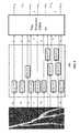
- FIG. 2illustrates an exemplary biomedical image 200 of a coronary vessel, according to embodiments of the disclosure. Biomedical image 200 may be obtained through the initial segmentation. Based on biomedical image 200 , a centerline 210 can be extracted.
- a “centerline”may be a skeleton line of the vessel that generally tracks the tree structure, including the “trunk” and the “branches.”
- Image patches 220are then extracted along centerline 210 .
- the image patchesmay be pre-extracted and saved in training database 101 as training data.
- a set of image patches extracted from the same biomedical imagemay be associated in training database 101 .
- the ground truthcan be a series of segmentation masks along the vessel path.
- the training imagesare previously segmented or annotated by expert operators with each pixel/voxel classified and labeled, e.g., with value 1 if the pixel/voxel belongs to a vessel or value 0 if otherwise.
- the ground truthmay be probability maps where each pixel/voxel is associated with a probability value indicating how likely the pixel/voxel belong to the vessel.
- the aim of the training phaseis to learn a mapping between the image patches and the ground truth segmentation mask by finding the best fit between predictions and ground truth values over the sets of training data.
- the training phasemay be performed “online” or “offline.”
- An “online” trainingrefers to performing the training phase contemporarily with the prediction phase, e.g., learning the model in real-time just prior to segmenting a biomedical image.
- An “online” trainingmay have the benefit to obtain a most updated learning model based on the training data that is then available.
- an “online” trainingmay be computational costive to perform and may not always be possible if the training data is large and/or the model is complicate.
- an “offline” trainingis used where the training phase is performed separately from the prediction phase. The learned model trained offline is saved and reused for segmenting images.
- Model training device 102may be implemented with hardware specially programmed by software that performs the training process.
- model training device 102may include a processor and a non-transitory computer-readable medium (discussed in detail in connection with FIG. 3 ).
- the processormay conduct the training by performing instructions of a training process stored in the computer-readable medium.
- Model training device 102may additionally include input and output interfaces to communicate with training database 101 , network 106 , and/or a user interface (not shown).
- the user interfacemay be used for selecting sets of training data, adjusting one or more parameters of the training process, selecting or modifying a framework of the learning model, and/or manually or semi-automatically providing prediction results associated with an image for training.
- the segmentation modelmay be a machine learning model that include at least two stages: a convolutional network configured to process the image patches to construct feature maps and a tree structure network configured to process the feature maps collectively to obtain a segmentation mask for the tree structure object.
- a convolutional networkconfigured to process the image patches to construct feature maps
- a tree structure networkconfigured to process the feature maps collectively to obtain a segmentation mask for the tree structure object.
- the structure of the learning modelis not limited to what is disclosed as long as the learning model encodes a spatial relationship in a tree structure among the image patches.
- the convolutional networkmay be a Fully Convolutional Network (FCN), e.g., a Unet.
- FCNFully Convolutional Network
- the tree structure networkmay be a Tree Structure Convolution Recurrent Neutral Network (CRNN).
- CRNNTree Structure Convolution Recurrent Neutral Network
- tree structure networkmay also be a Long Short Term Memory (LSTM) or a Gated Recurrent Unit (GRU).
- LSTMLong Short Term Memory
- GRUGated Recurrent Unit
- FIG. 4illustrates a schematic diagram of an exemplary tree structure based deep learning model 400 , according to embodiments of the disclosure.
- tree structure based deep learning model 400consists of two components to form an end-to-end trainable network: an FCN 410 and a Tree Structure CRNN 420 .
- Fully convolutional network 410aims to construct a feature map for each 2D image patch, from which object-relevant information will be extracted.
- Tree Structure CRNN 420aims to handle the spatial constraint in the tree structure.
- Tree structure based deep learning model 400generally performs a mapping (or transformation) S: ⁇ x 1 , x 2 , x 2 , . . . , x T ⁇ y 1 , y 2 , . . . , y T ⁇ , where training image patches ⁇ x 1 , x 2 , . . . , x T ⁇ are the input to model 400 and respective ground truths ⁇ y 1 , y 2 , . . . , y T ⁇ are the output.
- Tree structure based deep learning model 400works by passing each input x t , through a transformation M(x t ) performed by FCN 410 , to produce feature maps z t . The outputs z t are then passed into Tree Structure CRNN 420 .
- the architecture of FCN 410may include a stack of distinct layers (not shown) that transform the input into the output. Examples of the different layers may include one or more convolution layers or fully-convolutional layers, non-linear operator layers, pooling or subsampling layers, fully connected layers, and/or final loss layers. Each layer may connect one upstream layer and one downstream layer.
- FCN 410receives the training image patches ⁇ X 1 , x 2 , . . . , x T ⁇ and produces respective feature maps ⁇ z 1 , z 2 , . . . , z T ⁇ , from which object-relevant information will be extracted.
- FCN 410is applied to the image patches simultaneously so that the transformation M is performed on the image patches in parallel. It may avoid potential errors caused by post processing. Parallel processing may also save computation time of FCN 410 .
- Tree Structure CRNN 420aims to handle the spatial constraint in the tree structure object.
- the spatial constraintis defined by the spatial relationship of the image patches within a 2D image slice, e.g., whether an image patch belongs to the vessel has an implication on whether the image patch spatially adjacent to it may also belong to the vessel.
- the spatial constraintmay also be defined by the spatial relationship across multiple 2D image slice, e.g., whether an image patch in one image slice belongs to the vessel has an implication on whether the corresponding image patch in its spatially adjacent image slice may also belong to the vessel.
- the spatial constraintalso limits the image patches that belong to the vessel to follow a tree structure. Accordingly, the tree-like nature of vessels is encoded in the Tree Structure CRNN 420 , and thus tree structure based deep learning model 400 .
- Tree Structure CRNN 420can be implemented with single direction either from the root to the terminals or from the terminals to the root. It can also be implemented by considering both directions.
- FIG. 5illustrates a schematic diagram of an exemplary bidirectional Convolution RNN 500 , according to embodiments of the disclosure. It can be trained using all available input information from both upstream and the downstream of the tree structure. Additional depth can be added by stacking them on top of each other.
- model training device 102jointly trains the convolutional network and the tree structure network, using the training data from training database 101 .
- the end-to-end networkis trained as one piece rather than the different stages separately.
- the jointly trained networkcan integrate information of the neighboring image patches in the image and provide a better prediction. Therefore, when later used to segment a new biomedical image, the segmentation models can leverage the tree structure information to aid the segmentation.
- training a learning modelrefers to determining one or more parameters of at least one layer in the learning model.
- a convolutional layer of an FCN modelmay include at least one filter or kernel.
- One or more parameters, such as kernel weights, size, shape, and structure, of the at least one filtermay be determined by e.g., a backpropagation-based training process.
- the segmentation modelmay be trained using supervised learning, semi-supervised learning, or unsupervised learning.
- the parameters (V, W) of the fully convolutional network 410 and Tree Structure CRNN 420can be jointly optimized by minimizing a cost function of the ground truth outputs y t and the predicted values ⁇ for each image patch t.
- parameters (V, W)may be optimized to minimize a cost function of y and ⁇ .
- the cost functionmay be the mean square error between y and
- tree structure based deep learning model 400can be trained using stochastic gradient descent related methods, with backpropagation used to compute the gradient ⁇ V,W L(V, W) of the cost function J with respect to all parameters (V, W) over mini batches sampled from the training dataset.
- the trained segmentation modelmay be used by image processing device to segment new biomedical images.
- Image processing device 103may receive the segmentation model, e.g., tree structure based deep learning model 400 , from model training device 102 .
- Image processing device 103may include a processor and a non-transitory computer-readable medium (discussed in detail in connection with FIG. 3 ).
- the processormay perform instructions of an image segmentation process stored in the medium.
- Image processing device 103may additionally include input and output interfaces (discussed in detail in connection with FIG. 3 ) to communicate with biomedical image database 104 , network 106 , and/or a user interface (not shown).
- the user interfacemay be used for selecting a biomedical image for segmentation, initiating the segmentation process, displaying the biomedical image and/or the segmentation results.
- Image processing device 103may communicate with biomedical image database 104 to receive one or more biomedical images.
- the biomedical images stored in biomedical image database 104may include 2D image slices from a 3D scan.
- the biomedical imagesmay be acquired by image acquisition devices 105 .
- Image processing device 103may perform an initial artery segmentation to locate a rough profile of the vessel structure. Based on the initial segmentation, image processing device 103 then extracts a centerline of the vessel and extracts multiple image patches along the centerline. Image processing device 103 then uses the trained segmentation model received from model training device 102 to predict whether the image patches belong to the vessel, and outputs a segmentation mask of the biomedical image.
- FIG. 3illustrates an exemplary image processing device 103 , according to some embodiments of the present disclosure.
- image processing device 103may be a special-purpose computer, or a general-purpose computer.
- image processing device 103may be a computer custom-built for hospitals to perform image acquisition and image processing tasks.
- image processing device 103may include a communication interface 302 , a storage 304 , a memory 306 , a processor 308 , and a bus 310 .
- Communication interface 302 , storage 304 , memory 306 , and processor 308are connected with bus 310 and communicate with each other through bus 310 .
- Communication interface 302may include a network adaptor, a cable connector, a serial connector, a USB connector, a parallel connector, a high-speed data transmission adaptor, such as fiber, USB 3.0, thunderbolt, and the like, a wireless network adaptor, such as a WiFi adaptor, a telecommunication (3G, 4G/LTE and the like) adaptor, etc.
- Image processing device 103may be connected to other components of image segmentation system 100 and network 106 through communication interface 302 .
- communication interface 302receives biomedical image from image acquisition device 105 .
- the biomedical imagecaptures a tree structure object, such as a vessel.
- the biomedical imagemay be a coronary vessel image or a retinal vessel image.
- communication interface 302also receives the segmentation model, e.g., tree structure based deep learning model 400 , from modeling training device 102 .
- Storage 304 /memory 306may be a non-transitory computer-readable medium, such as a read-only memory (ROM), a random access memory (RAM), a phase-change random access memory (PRAM), a static random access memory (SRAM), a dynamic random access memory (DRAM), an electrically erasable programmable read-only memory (EEPROM), other types of random access memories (RAMs), a flash disk or other forms of flash memory, a cache, a register, a static memory, a compact disc read-only memory (CD-ROM), a digital versatile disc (DVD) or other optical storage, a cassette tape or other magnetic storage devices, or any other non-transitory medium that may be used to store information or instructions capable of being accessed by a computer device, etc.
- ROMread-only memory
- RAMrandom access memory
- PRAMphase-change random access memory
- SRAMstatic random access memory
- DRAMdynamic random access memory
- EEPROMelectrically erasable programmable read-only memory
- storage 304may store the trained learning model, e.g., tree structure based deep learning model 400 , and data, such as extracted image patches and feature maps generated while executing the computer programs, etc.
- memory 306may store computer-executable instructions, such as one or more image processing programs.
- multiple image patchesmay be extracted along the centerline of the tree structure object in a biomedical image stored in storage 304 . The image patches may be read from storage 304 one by one or simultaneously and stored in memory 306 .
- Processor 308may be a processing device that includes one or more general processing devices, such as a microprocessor, a central processing unit (CPU), a graphics processing unit (GPU), and the like. More specifically, the processor may be a complex instruction set computing (CISC) microprocessor, a reduced instruction set computing (RISC) microprocessor, a very long instruction word (VLIW) microprocessor, a processor running other instruction sets, or a processor that runs a combination of instruction sets. The processor may also be one or more dedicated processing devices such as application specific integrated circuits (ASICs), field programmable gate arrays (FPGAs), digital signal processors (DSPs), system-on-chip (SoCs), and the like. Processor 308 may be communicatively coupled to memory 306 and configured to execute the computer-executable instructions stored thereon.
- ASICsapplication specific integrated circuits
- FPGAsfield programmable gate arrays
- DSPsdigital signal processors
- SoCssystem-on-chip
- processor 308is configured to segment the biomedical image. For example, processor 308 first perform an initial artery segmentation to find the vessel structure. Based on the initial segmentation, processor 308 then extracts a centerline of the vessel and extracts multiple image patches along the centerline. Processor 308 then uses the trained segmentation model, such as tree structure based deep learning model 400 , to predict whether the image patches belong to the vessel, and outputs a segmentation mask of the biomedical image.
- the trained segmentation modelmay include an FCN (e.g., FCN 410 ) and a Tree Structure CRNN (e.g., Tree Structure CRNN 420 ) encoded with spatial constraint reflecting the tree structure of the object in the biomedical image.
- Applying the segmentation model to the image patchesmay include applying the FCN and the Tree Structure CRNN sequentially.
- FCN 410is applied to the image patches in parallel to produce respective feature maps
- Tree Structure CRNN 420is applied to the feature maps to produce the segmentation mask.
- Tree Structure CRNN 420may produce a probability map indicating the probability each pixel in the image patch belongs to the tree structure object.
- Processor 308may then perform a thresholding to obtain a segmentation mask. For example, processor 308 may set pixels with probabilities above 0.8 as 1 (i.e., belong to the tree structure object) and the remaining pixels as 0 (i.e., not belong to the tree structure object). The threshold may be set by an operator or automatically selected by processor 308 .
- An exemplary image segmentation processwill be described in connection with FIG. 7 .
- model training device 102can have same or similar structures as image processing device 103 .
- model training device 102includes a processor, among other components, configured to jointly train the FCN and the Tree Structure CRNN using training images. An exemplary network training process will be described in connection with FIG. 6 .
- FIG. 6is a flowchart of an exemplary method 600 for training a tree structure based learning model, according to embodiments of the disclosure.
- method 600may be implemented by model training device 102 in FIG. 1 .
- method 600is not limited to that exemplary embodiment.
- Method 600may include steps S 602 -S 612 as described below. It is to be appreciated that some of the steps may be optional to perform the disclosure provided herein. Further, some of the steps may be performed simultaneously, or in a different order than shown in FIG. 6 .
- model training device 102may communicate with training database 101 to receive one or more sets of training data.
- Each set of training datamay include training image patches extracted from a biomedical image and its corresponding ground truth segmentation mask that provides the segmentation result to each image patch.
- model training device 102may initialize the parameters of a tree structure based learning model. Training the learning model is a process of determining one or more parameters of the learning model. Consistent with the present disclosure, model training device 102 jointly trains the convolutional network and the tree structure network, using the training data from training database 101 . That is, the set of parameters of the two networks are trained together. For example, set of parameters V of the fully convolutional network 410 and set of parameters W of Tree Structure CRNN 420 can be jointly trained. The parameters may be initially set to certain values. The initial values may be predetermined, selected by an operator, or decided by model training device 102 based on prior experience of similar images. For example, parameters of a learning model previously trained for coronary vessel image of patient A may be used as initial values for the parameters of the learning model being trained for coronary vessel image of patient B.
- model training device 102may calculate the value of a cost function.
- the cost functionmay be the mean square error between y and
- the calculated valuemay be compared with a predetermined threshold.
- the predetermined thresholdis also known as the stopping criteria for interactive methods. The smaller it is, the more optimal the parameters, but the longer it takes (i.e., more iterations) for the computation to converge. Therefore, the threshold may be selected to balance the accuracy of the prediction and the computational cost.
- step S 608If the value is below the predetermined threshold (step S 608 : Yes), the method is considered as have converged, and the cost function is minimized.
- step S 610model training device 102 outputs the learning model with the optimized sets of parameters and method 600 concludes. Otherwise (step S 608 : No), model training device 102 may further adjust the two sets of parameters jointly in step S 612 .
- a stochastic gradient descent related method with backpropagationmay be used.
- the parameters L(V, W)may be adjust with a gradient ⁇ V,W L(V, W) of the cost function J with respect to all parameters (V, W) over mini batches sampled from the training dataset.
- Method 600may return to step S 606 to calculate value of the cost function based on outputs obtained from the learning model with the adjusted sets of parameters. Each pass of steps S 606 -S 612 is considered as one iteration. Method 600 iterates until the value of the cost function is reduced to below the predetermined threshold (step S 608 ).
- FIG. 7is a flowchart of an exemplary method 700 for biomedical image segmentation using a tree structure based learning model, according to embodiments of the disclosure.
- method 700may be implemented by image processing device 103 in FIG. 1 .
- method 700is not limited to that exemplary embodiment.
- Method 700may include steps S 702 -S 712 as described below. It is to be appreciated that some of the steps may be optional to perform the disclosure provided herein. Further, some of the steps may be performed simultaneously, or in a different order than shown in FIG. 7 .
- image processing device 103receives a biomedical image, e.g., from biomedical image database 104 .
- the biomedical imagecaptures a tree structure object, such as a blood vessel.
- Image processing device 103may additionally receive a segmentation model, e.g., tree structure based deep learning model 400 .
- the segmentation modelmay be trained using method 600 .
- step S 704image processing device 103 segments the biomedical image.
- an initial artery segmentationmay be performed to locate a profile of the tree structure object.
- image processing device 103extracts a centerline from the segmented image. For example, the centerline tracks the passageways of the vessel structure.
- image processing device 103extracts image patches along the centerline.
- An image patchmay be square and includes multiple pixels, which may also be referred as a superpixel.
- the image patchesmay be 4 ⁇ 4, 8 ⁇ 8, 16 ⁇ 16, or 32 ⁇ 32 in size.
- image processing device 103segments the image patches using a tree structure based learning model, e.g., model 400 .
- the learning modeltakes the image patches as inputs and produce a segmentation mask for the tree structure object (step S 712 ).
- image processing device 103may segment the image patches using an FCN and a Tree Structure CRNN sequentially.
- FCN 410is first applied to the image patches in parallel to produce respective feature maps, and then Tree Structure CRNN 420 is applied to the feature maps to produce the segmentation mask.
- Tree Structure CRNN 420may produce a probability map indicating the probability each pixel in the image patch belongs to the tree structure object.
- Image processing devicemay then perform a thresholding to obtain a segmentation mask.
- the computer-readable mediummay include volatile or non-volatile, magnetic, semiconductor, tape, optical, removable, non-removable, or other types of computer-readable medium or computer-readable storage devices.
- the computer-readable mediummay be the storage device or the memory module having the computer instructions stored thereon, as disclosed.
- the computer-readable mediummay be a disc or a flash drive having the computer instructions stored thereon.
Landscapes
- Engineering & Computer Science (AREA)
- Theoretical Computer Science (AREA)
- Physics & Mathematics (AREA)
- General Physics & Mathematics (AREA)
- Software Systems (AREA)
- Health & Medical Sciences (AREA)
- Artificial Intelligence (AREA)
- General Engineering & Computer Science (AREA)
- Mathematical Physics (AREA)
- Data Mining & Analysis (AREA)
- Evolutionary Computation (AREA)
- General Health & Medical Sciences (AREA)
- Computing Systems (AREA)
- Biophysics (AREA)
- Molecular Biology (AREA)
- Computational Linguistics (AREA)
- Biomedical Technology (AREA)
- Life Sciences & Earth Sciences (AREA)
- Computer Vision & Pattern Recognition (AREA)
- Medical Informatics (AREA)
- Nuclear Medicine, Radiotherapy & Molecular Imaging (AREA)
- Radiology & Medical Imaging (AREA)
- Quality & Reliability (AREA)
- Image Analysis (AREA)
Abstract
Description
where ytis the ground truth outputs contained in the training data and ŷ is the predicted values for each image patch t.
Claims (20)
Priority Applications (2)
| Application Number | Priority Date | Filing Date | Title |
|---|---|---|---|
| US16/392,516US10430949B1 (en) | 2018-04-24 | 2019-04-23 | Automatic method and system for vessel refine segmentation in biomedical images using tree structure based deep learning model |
| US16/529,769US10573005B2 (en) | 2018-04-24 | 2019-08-01 | Automatic method and system for vessel refine segmentation in biomedical images using tree structure based deep learning model |
Applications Claiming Priority (2)
| Application Number | Priority Date | Filing Date | Title |
|---|---|---|---|
| US201862662212P | 2018-04-24 | 2018-04-24 | |
| US16/392,516US10430949B1 (en) | 2018-04-24 | 2019-04-23 | Automatic method and system for vessel refine segmentation in biomedical images using tree structure based deep learning model |
Related Child Applications (1)
| Application Number | Title | Priority Date | Filing Date |
|---|---|---|---|
| US16/529,769ContinuationUS10573005B2 (en) | 2018-04-24 | 2019-08-01 | Automatic method and system for vessel refine segmentation in biomedical images using tree structure based deep learning model |
Publications (2)
| Publication Number | Publication Date |
|---|---|
| US10430949B1true US10430949B1 (en) | 2019-10-01 |
| US20190325579A1 US20190325579A1 (en) | 2019-10-24 |
Family
ID=67320378
Family Applications (2)
| Application Number | Title | Priority Date | Filing Date |
|---|---|---|---|
| US16/392,516Active - ReinstatedUS10430949B1 (en) | 2018-04-24 | 2019-04-23 | Automatic method and system for vessel refine segmentation in biomedical images using tree structure based deep learning model |
| US16/529,769ActiveUS10573005B2 (en) | 2018-04-24 | 2019-08-01 | Automatic method and system for vessel refine segmentation in biomedical images using tree structure based deep learning model |
Family Applications After (1)
| Application Number | Title | Priority Date | Filing Date |
|---|---|---|---|
| US16/529,769ActiveUS10573005B2 (en) | 2018-04-24 | 2019-08-01 | Automatic method and system for vessel refine segmentation in biomedical images using tree structure based deep learning model |
Country Status (2)
| Country | Link |
|---|---|
| US (2) | US10430949B1 (en) |
| CN (2) | CN112070776B (en) |
Cited By (18)
| Publication number | Priority date | Publication date | Assignee | Title |
|---|---|---|---|---|
| CN111369576A (en)* | 2020-05-28 | 2020-07-03 | 腾讯科技(深圳)有限公司 | Training method of image segmentation model, image segmentation method, device and equipment |
| CN112288739A (en)* | 2020-11-20 | 2021-01-29 | 哈尔滨工业大学 | Vein segmentation method based on deep learning |
| CN112508457A (en)* | 2020-12-25 | 2021-03-16 | 树根互联技术有限公司 | Data processing method and device, industrial equipment and storage medium |
| CN113034425A (en)* | 2019-12-25 | 2021-06-25 | 阿里巴巴集团控股有限公司 | Data processing method, device and storage medium |
| US20210209747A1 (en)* | 2020-01-06 | 2021-07-08 | The Texas A&M University System | Unmanned aerial system genotype analysis using machine learning routines |
| CN113192069A (en)* | 2021-06-03 | 2021-07-30 | 清华大学 | Semantic segmentation method and device for tree structure in three-dimensional tomography image |
| US20220084209A1 (en)* | 2020-09-17 | 2022-03-17 | International Business Machines Corporation | Reducing structural redundancy in automatic image segmentation |
| US11288812B2 (en) | 2018-01-10 | 2022-03-29 | Institut De Recherche Sur Les Cancers De I | Automatic segmentation process of a 3D medical image by one or several neural networks through structured convolution according to the anatomic geometry of the 3D medical image |
| CN114758329A (en)* | 2022-04-15 | 2022-07-15 | 东北大学 | System and method for predicting temperature of target area in thermal image based on deep learning |
| US11449716B2 (en) | 2020-03-31 | 2022-09-20 | International Business Machines Corporation | Model training using partially-annotated images |
| US20220344033A1 (en)* | 2021-04-23 | 2022-10-27 | Shenzhen Keya Medical Technology Corporation | Method and System for Anatomical Labels Generation |
| US11526694B2 (en) | 2020-03-31 | 2022-12-13 | International Business Machines Corporation | Model training using fully and partially-annotated images |
| US11551362B2 (en) | 2018-01-10 | 2023-01-10 | Institut De Recherche Sur Les Cancers De I | Automatic segmentation process of a 3D medical image by several neural networks through structured convolution according to the geometry of the 3D medical image |
| US20230034782A1 (en)* | 2021-07-29 | 2023-02-02 | GE Precision Healthcare LLC | Learning-based clean data selection |
| CN115810018A (en)* | 2022-11-30 | 2023-03-17 | 明峰医疗系统股份有限公司 | Method and system for optimizing segmentation results of blood vessel tree and coronary artery tree of CT image |
| US20240048152A1 (en)* | 2022-08-03 | 2024-02-08 | Arm Limited | Weight processing for a neural network |
| US11896316B2 (en)* | 2018-08-23 | 2024-02-13 | Intuitive Surgical Operations, Inc. | Systems and methods for generating anatomic tree structures using backward pathway growth |
| CN117726585A (en)* | 2023-12-05 | 2024-03-19 | 武汉大学 | Dual image filtering-based semi-supervised medical image segmentation method and system |
Families Citing this family (20)
| Publication number | Priority date | Publication date | Assignee | Title |
|---|---|---|---|---|
| US11308362B2 (en)* | 2019-03-26 | 2022-04-19 | Shenzhen Keya Medical Technology Corporation | Method and system for generating a centerline for an object, and computer readable medium |
| US11200976B2 (en)* | 2019-08-23 | 2021-12-14 | Canon Medical Systems Corporation | Tracking method and apparatus |
| CN110796652B (en)* | 2019-10-30 | 2023-03-21 | 上海联影智能医疗科技有限公司 | Image processing method, computer device, and storage medium |
| CN111325759B (en)* | 2020-03-13 | 2024-04-16 | 上海联影智能医疗科技有限公司 | Vessel segmentation method, apparatus, computer device, and readable storage medium |
| EP4128040A4 (en)* | 2020-04-24 | 2023-09-13 | Shanghai United Imaging Healthcare Co., Ltd. | OBJECT RECOGNITION SYSTEMS AND METHODS |
| US20210383533A1 (en)* | 2020-06-03 | 2021-12-09 | Nvidia Corporation | Machine-learning-based object detection system |
| CN111681224A (en)* | 2020-06-09 | 2020-09-18 | 上海联影医疗科技有限公司 | Method and device for acquiring blood vessel parameters |
| CN112037305B (en)* | 2020-11-09 | 2021-03-19 | 腾讯科技(深圳)有限公司 | Method, device and storage medium for reconstructing tree-like organization in image |
| CN112561860B (en)* | 2020-11-23 | 2022-05-03 | 重庆邮电大学 | A BCA-UNet Liver Segmentation Method Based on Prior Shape Constraints |
| CN112308857B (en)* | 2020-12-25 | 2021-05-11 | 数坤(北京)网络科技有限公司 | Method and device for determining center line of blood vessel and readable storage medium |
| US12086981B2 (en)* | 2021-01-04 | 2024-09-10 | Shenzhen Keya Medical Technology Corporation | Methods and systems for computer-assisted medical image analysis using sequential model |
| CN112785591B (en)* | 2021-03-05 | 2023-06-13 | 杭州健培科技有限公司 | Method and device for detecting and segmenting rib fracture in CT image |
| CN113470107B (en)* | 2021-06-04 | 2023-07-14 | 广州医科大学附属第一医院 | Bronchial Centerline Extraction Method, System and Storage Medium |
| CN113902741B (en)* | 2021-12-08 | 2022-03-11 | 深圳科亚医疗科技有限公司 | Method, device and medium for performing blood vessel segmentation on medical image |
| CN114119602B (en)* | 2021-12-20 | 2022-04-15 | 深圳科亚医疗科技有限公司 | Method, device and storage medium for object analysis of medical images |
| CN114581418B (en)* | 2021-12-31 | 2025-05-23 | 深圳科亚医疗科技有限公司 | Method, device and storage medium for object analysis of medical images |
| FR3132145B1 (en)* | 2022-01-24 | 2025-08-01 | Quantmetry | METHOD FOR AUTOMATED DETECTION OF A BIOLOGICAL ELEMENT IN A TISSUE SAMPLE |
| CN114998582A (en)* | 2022-05-10 | 2022-09-02 | 深圳市第二人民医院(深圳市转化医学研究院) | Coronary blood vessel segmentation method, device and storage medium |
| CN114862878B (en)* | 2022-05-30 | 2023-06-06 | 北京百度网讯科技有限公司 | Image segmentation model generation method and device, image segmentation method and device |
| CN114862879B (en)* | 2022-07-05 | 2022-09-27 | 深圳科亚医疗科技有限公司 | Method, system and medium for processing images containing physiological tubular structures |
Citations (5)
| Publication number | Priority date | Publication date | Assignee | Title |
|---|---|---|---|---|
| US20100172567A1 (en)* | 2007-04-17 | 2010-07-08 | Prokoski Francine J | System and method for using three dimensional infrared imaging to provide detailed anatomical structure maps |
| US20130034283A1 (en)* | 2010-08-17 | 2013-02-07 | Toshiba Medical Systems Corporation | Medical image diagnosis apparatus |
| US20150003706A1 (en)* | 2011-12-12 | 2015-01-01 | University Of Stavanger | Probability mapping for visualisation and analysis of biomedical images |
| US20150227809A1 (en)* | 2014-02-12 | 2015-08-13 | International Business Machines Corporation | Anomaly detection in medical imagery |
| US20160328855A1 (en)* | 2015-05-04 | 2016-11-10 | Siemens Aktiengesellschaft | Method and System for Whole Body Bone Removal and Vascular Visualization in Medical Image Data |
Family Cites Families (16)
| Publication number | Priority date | Publication date | Assignee | Title |
|---|---|---|---|---|
| US9208582B2 (en)* | 2005-11-02 | 2015-12-08 | Hitachi Medical Corporation | Image analyzing system and method |
| CN101425186B (en)* | 2008-11-17 | 2012-03-28 | 华中科技大学 | A Liver Segmentation Method and System Based on CT Image |
| BR112013005879A2 (en)* | 2010-09-15 | 2016-05-10 | Konink Philps Electronics Nv | '' Robot guidance system, control units for an endoscope and robot guidance method '' |
| CN103458772B (en)* | 2011-04-07 | 2017-10-31 | 香港中文大学 | Retinal image analysis method and device |
| US9349178B1 (en)* | 2014-11-24 | 2016-05-24 | Siemens Aktiengesellschaft | Synthetic data-driven hemodynamic determination in medical imaging |
| US9959615B2 (en)* | 2015-07-01 | 2018-05-01 | Arizona Board Of Regents On Behalf Of Arizona State University | System and method for automatic pulmonary embolism detection |
| US10405739B2 (en)* | 2015-10-23 | 2019-09-10 | International Business Machines Corporation | Automatically detecting eye type in retinal fundus images |
| US10115039B2 (en)* | 2016-03-10 | 2018-10-30 | Siemens Healthcare Gmbh | Method and system for machine learning based classification of vascular branches |
| CN106127120B (en)* | 2016-06-16 | 2018-03-13 | 北京市商汤科技开发有限公司 | Posture estimation method and device, computer system |
| CN106408562B (en)* | 2016-09-22 | 2019-04-09 | 华南理工大学 | A method and system for retinal blood vessel segmentation in fundus images based on deep learning |
| CN107977709B (en)* | 2017-04-01 | 2021-03-16 | 北京科亚方舟医疗科技股份有限公司 | Deep Learning Model and System for Predicting Blood Flow Characteristics on Vessel Paths in Vascular Trees |
| CN107256550A (en)* | 2017-06-06 | 2017-10-17 | 电子科技大学 | A kind of retinal image segmentation method based on efficient CNN CRF networks |
| CN107563983B (en)* | 2017-09-28 | 2020-09-01 | 上海联影医疗科技有限公司 | Image processing method and medical imaging device |
| CN107609541B (en)* | 2017-10-17 | 2020-11-10 | 哈尔滨理工大学 | Human body posture estimation method based on deformable convolution neural network |
| CN108305246B (en)* | 2017-11-15 | 2020-10-09 | 深圳科亚医疗科技有限公司 | Device and system for predicting blood flow characteristics based on medical images |
| CN107887010A (en)* | 2017-11-29 | 2018-04-06 | 天津中科爱乐芙医疗科技有限公司 | A kind of angiocardiopathy data acquisition examines platform with dividing |
- 2019
- 2019-04-23USUS16/392,516patent/US10430949B1/enactiveActive - Reinstated
- 2019-04-24CNCN202011055196.6Apatent/CN112070776B/enactiveActive
- 2019-04-24CNCN201910331967.0Apatent/CN110060263B/enactiveActive
- 2019-08-01USUS16/529,769patent/US10573005B2/enactiveActive
Patent Citations (6)
| Publication number | Priority date | Publication date | Assignee | Title |
|---|---|---|---|---|
| US20100172567A1 (en)* | 2007-04-17 | 2010-07-08 | Prokoski Francine J | System and method for using three dimensional infrared imaging to provide detailed anatomical structure maps |
| US20130034283A1 (en)* | 2010-08-17 | 2013-02-07 | Toshiba Medical Systems Corporation | Medical image diagnosis apparatus |
| US20150003706A1 (en)* | 2011-12-12 | 2015-01-01 | University Of Stavanger | Probability mapping for visualisation and analysis of biomedical images |
| US20150227809A1 (en)* | 2014-02-12 | 2015-08-13 | International Business Machines Corporation | Anomaly detection in medical imagery |
| US9704059B2 (en)* | 2014-02-12 | 2017-07-11 | International Business Machines Corporation | Anomaly detection in medical imagery |
| US20160328855A1 (en)* | 2015-05-04 | 2016-11-10 | Siemens Aktiengesellschaft | Method and System for Whole Body Bone Removal and Vascular Visualization in Medical Image Data |
Cited By (28)
| Publication number | Priority date | Publication date | Assignee | Title |
|---|---|---|---|---|
| US11288812B2 (en) | 2018-01-10 | 2022-03-29 | Institut De Recherche Sur Les Cancers De I | Automatic segmentation process of a 3D medical image by one or several neural networks through structured convolution according to the anatomic geometry of the 3D medical image |
| US11551362B2 (en) | 2018-01-10 | 2023-01-10 | Institut De Recherche Sur Les Cancers De I | Automatic segmentation process of a 3D medical image by several neural networks through structured convolution according to the geometry of the 3D medical image |
| US11896316B2 (en)* | 2018-08-23 | 2024-02-13 | Intuitive Surgical Operations, Inc. | Systems and methods for generating anatomic tree structures using backward pathway growth |
| CN113034425A (en)* | 2019-12-25 | 2021-06-25 | 阿里巴巴集团控股有限公司 | Data processing method, device and storage medium |
| US20210201481A1 (en)* | 2019-12-25 | 2021-07-01 | Alibaba Group Holding Limited | Data processing method, equipment and storage medium |
| CN113034425B (en)* | 2019-12-25 | 2024-05-28 | 阿里巴巴集团控股有限公司 | Data processing method, device and storage medium |
| US11721015B2 (en)* | 2019-12-25 | 2023-08-08 | Alibaba Group Holding Limited | Data processing method, equipment and storage medium |
| US20210209747A1 (en)* | 2020-01-06 | 2021-07-08 | The Texas A&M University System | Unmanned aerial system genotype analysis using machine learning routines |
| US11816834B2 (en)* | 2020-01-06 | 2023-11-14 | The Texas A&M University System | Unmanned aerial system genotype analysis using machine learning routines |
| US11449716B2 (en) | 2020-03-31 | 2022-09-20 | International Business Machines Corporation | Model training using partially-annotated images |
| US11526694B2 (en) | 2020-03-31 | 2022-12-13 | International Business Machines Corporation | Model training using fully and partially-annotated images |
| CN111369576A (en)* | 2020-05-28 | 2020-07-03 | 腾讯科技(深圳)有限公司 | Training method of image segmentation model, image segmentation method, device and equipment |
| US20220084209A1 (en)* | 2020-09-17 | 2022-03-17 | International Business Machines Corporation | Reducing structural redundancy in automatic image segmentation |
| US11651499B2 (en)* | 2020-09-17 | 2023-05-16 | International Business Machines Corporation | Reducing structural redundancy in automatic image segmentation |
| CN112288739B (en)* | 2020-11-20 | 2022-04-22 | 哈尔滨工业大学 | A deep learning-based method for venous vessel segmentation |
| CN112288739A (en)* | 2020-11-20 | 2021-01-29 | 哈尔滨工业大学 | Vein segmentation method based on deep learning |
| CN112508457A (en)* | 2020-12-25 | 2021-03-16 | 树根互联技术有限公司 | Data processing method and device, industrial equipment and storage medium |
| CN112508457B (en)* | 2020-12-25 | 2024-05-31 | 树根互联股份有限公司 | Data processing method and device, industrial equipment and storage medium |
| US12094596B2 (en)* | 2021-04-23 | 2024-09-17 | Shenzhen Keya Medical Technology Corporation | Method and system for anatomical labels generation |
| US20220344033A1 (en)* | 2021-04-23 | 2022-10-27 | Shenzhen Keya Medical Technology Corporation | Method and System for Anatomical Labels Generation |
| CN113192069A (en)* | 2021-06-03 | 2021-07-30 | 清华大学 | Semantic segmentation method and device for tree structure in three-dimensional tomography image |
| US20230034782A1 (en)* | 2021-07-29 | 2023-02-02 | GE Precision Healthcare LLC | Learning-based clean data selection |
| US12430563B2 (en)* | 2021-07-29 | 2025-09-30 | GE Precision Healthcare LLC | Learning-based clean data selection |
| CN114758329A (en)* | 2022-04-15 | 2022-07-15 | 东北大学 | System and method for predicting temperature of target area in thermal image based on deep learning |
| US12040821B2 (en)* | 2022-08-03 | 2024-07-16 | Arm Limited | Weight processing for a neural network |
| US20240048152A1 (en)* | 2022-08-03 | 2024-02-08 | Arm Limited | Weight processing for a neural network |
| CN115810018A (en)* | 2022-11-30 | 2023-03-17 | 明峰医疗系统股份有限公司 | Method and system for optimizing segmentation results of blood vessel tree and coronary artery tree of CT image |
| CN117726585A (en)* | 2023-12-05 | 2024-03-19 | 武汉大学 | Dual image filtering-based semi-supervised medical image segmentation method and system |
Also Published As
| Publication number | Publication date |
|---|---|
| CN110060263B (en) | 2020-11-13 |
| US20190325579A1 (en) | 2019-10-24 |
| US20190355120A1 (en) | 2019-11-21 |
| CN112070776B (en) | 2024-08-02 |
| CN110060263A (en) | 2019-07-26 |
| CN112070776A (en) | 2020-12-11 |
| US10573005B2 (en) | 2020-02-25 |
Similar Documents
| Publication | Publication Date | Title |
|---|---|---|
| US10430949B1 (en) | Automatic method and system for vessel refine segmentation in biomedical images using tree structure based deep learning model | |
| US11748879B2 (en) | Method and system for intracerebral hemorrhage detection and segmentation based on a multi-task fully convolutional network | |
| US11574406B2 (en) | Systems and methods for image segmentation using a scalable and compact convolutional neural network | |
| JP6884853B2 (en) | Image segmentation using neural network method | |
| US11847547B2 (en) | Method and system for generating a centerline for an object, and computer readable medium | |
| US12119117B2 (en) | Method and system for disease quantification of anatomical structures | |
| US10346986B2 (en) | System and methods for image segmentation using convolutional neural network | |
| CN109493347B (en) | Method and system for segmenting sparsely distributed objects in an image | |
| US20220415510A1 (en) | Method and system for disease quantification modeling of anatomical tree structure | |
| US10258304B1 (en) | Method and system for accurate boundary delineation of tubular structures in medical images using infinitely recurrent neural networks | |
| US10846854B2 (en) | Systems and methods for detecting cancer metastasis using a neural network | |
| US11763468B2 (en) | Structured landmark detection via topology-adapting deep graph learning | |
| US20220344033A1 (en) | Method and System for Anatomical Labels Generation | |
| US12086981B2 (en) | Methods and systems for computer-assisted medical image analysis using sequential model | |
| CN117474879A (en) | Aortic dissection true and false cavity segmentation method and device, electronic equipment and storage medium | |
| US20220392059A1 (en) | Method and system for representation learning with sparse convolution | |
| Choudhury | Contributions to Autonomous Systems in Trading, Transportation, and Diagnostic Imaging | |
| CN117474877A (en) | Aortic dissection true and false cavity segmentation method and device, electronic equipment and storage medium | |
| Jeong | Curvilinear structure modeling and its applications in computer vision |
Legal Events
| Date | Code | Title | Description |
|---|---|---|---|
| AS | Assignment | Owner name:SHENZHEN KEYA MEDICAL TECHNOLOGY CORPORATION, CHIN Free format text:ASSIGNMENT OF ASSIGNORS INTEREST;ASSIGNORS:WANG, XIN;YIN, YOUBING;BAI, JUNJIE;AND OTHERS;REEL/FRAME:048975/0705 Effective date:20190423 | |
| FEPP | Fee payment procedure | Free format text:ENTITY STATUS SET TO UNDISCOUNTED (ORIGINAL EVENT CODE: BIG.); ENTITY STATUS OF PATENT OWNER: SMALL ENTITY | |
| FEPP | Fee payment procedure | Free format text:ENTITY STATUS SET TO SMALL (ORIGINAL EVENT CODE: SMAL); ENTITY STATUS OF PATENT OWNER: SMALL ENTITY | |
| STCF | Information on status: patent grant | Free format text:PATENTED CASE | |
| FEPP | Fee payment procedure | Free format text:MAINTENANCE FEE REMINDER MAILED (ORIGINAL EVENT CODE: REM.); ENTITY STATUS OF PATENT OWNER: SMALL ENTITY | |
| LAPS | Lapse for failure to pay maintenance fees | Free format text:PATENT EXPIRED FOR FAILURE TO PAY MAINTENANCE FEES (ORIGINAL EVENT CODE: EXP.); ENTITY STATUS OF PATENT OWNER: SMALL ENTITY | |
| STCH | Information on status: patent discontinuation | Free format text:PATENT EXPIRED DUE TO NONPAYMENT OF MAINTENANCE FEES UNDER 37 CFR 1.362 | |
| PRDP | Patent reinstated due to the acceptance of a late maintenance fee | Effective date:20231201 | |
| FP | Lapsed due to failure to pay maintenance fee | Effective date:20231001 | |
| FEPP | Fee payment procedure | Free format text:PETITION RELATED TO MAINTENANCE FEES FILED (ORIGINAL EVENT CODE: PMFP); ENTITY STATUS OF PATENT OWNER: SMALL ENTITY Free format text:PETITION RELATED TO MAINTENANCE FEES GRANTED (ORIGINAL EVENT CODE: PMFG); ENTITY STATUS OF PATENT OWNER: SMALL ENTITY Free format text:SURCHARGE, PETITION TO ACCEPT PYMT AFTER EXP, UNINTENTIONAL. (ORIGINAL EVENT CODE: M2558); ENTITY STATUS OF PATENT OWNER: SMALL ENTITY | |
| MAFP | Maintenance fee payment | Free format text:PAYMENT OF MAINTENANCE FEE, 4TH YR, SMALL ENTITY (ORIGINAL EVENT CODE: M2551); ENTITY STATUS OF PATENT OWNER: SMALL ENTITY Year of fee payment:4 | |
| STCF | Information on status: patent grant | Free format text:PATENTED CASE |