US10426548B2 - Methods and systems for providing electromagnetic radiation to at least one portion of a sample using conformal laser therapy procedures - Google Patents
Methods and systems for providing electromagnetic radiation to at least one portion of a sample using conformal laser therapy proceduresDownload PDFInfo
- Publication number
- US10426548B2 US10426548B2US11/670,043US67004307AUS10426548B2US 10426548 B2US10426548 B2US 10426548B2US 67004307 AUS67004307 AUS 67004307AUS 10426548 B2US10426548 B2US 10426548B2
- Authority
- US
- United States
- Prior art keywords
- laser radiation
- laser
- biological structure
- wavelength
- depth
- Prior art date
- Legal status (The legal status is an assumption and is not a legal conclusion. Google has not performed a legal analysis and makes no representation as to the accuracy of the status listed.)
- Active, expires
Links
Images


Classifications
- A—HUMAN NECESSITIES
- A61—MEDICAL OR VETERINARY SCIENCE; HYGIENE
- A61B—DIAGNOSIS; SURGERY; IDENTIFICATION
- A61B18/00—Surgical instruments, devices or methods for transferring non-mechanical forms of energy to or from the body
- A61B18/18—Surgical instruments, devices or methods for transferring non-mechanical forms of energy to or from the body by applying electromagnetic radiation, e.g. microwaves
- A61B18/20—Surgical instruments, devices or methods for transferring non-mechanical forms of energy to or from the body by applying electromagnetic radiation, e.g. microwaves using laser
- A61B18/22—Surgical instruments, devices or methods for transferring non-mechanical forms of energy to or from the body by applying electromagnetic radiation, e.g. microwaves using laser the beam being directed along or through a flexible conduit, e.g. an optical fibre; Couplings or hand-pieces therefor
- A61B18/24—Surgical instruments, devices or methods for transferring non-mechanical forms of energy to or from the body by applying electromagnetic radiation, e.g. microwaves using laser the beam being directed along or through a flexible conduit, e.g. an optical fibre; Couplings or hand-pieces therefor with a catheter
- A—HUMAN NECESSITIES
- A61—MEDICAL OR VETERINARY SCIENCE; HYGIENE
- A61B—DIAGNOSIS; SURGERY; IDENTIFICATION
- A61B17/00—Surgical instruments, devices or methods
- A61B2017/00017—Electrical control of surgical instruments
- A61B2017/00022—Sensing or detecting at the treatment site
- A61B2017/00057—Light
- A61B2017/00066—Light intensity
- A—HUMAN NECESSITIES
- A61—MEDICAL OR VETERINARY SCIENCE; HYGIENE
- A61B—DIAGNOSIS; SURGERY; IDENTIFICATION
- A61B17/00—Surgical instruments, devices or methods
- A61B17/22—Implements for squeezing-off ulcers or the like on inner organs of the body; Implements for scraping-out cavities of body organs, e.g. bones; for invasive removal or destruction of calculus using mechanical vibrations; for removing obstructions in blood vessels, not otherwise provided for
- A61B2017/22051—Implements for squeezing-off ulcers or the like on inner organs of the body; Implements for scraping-out cavities of body organs, e.g. bones; for invasive removal or destruction of calculus using mechanical vibrations; for removing obstructions in blood vessels, not otherwise provided for with an inflatable part, e.g. balloon, for positioning, blocking, or immobilisation
- A—HUMAN NECESSITIES
- A61—MEDICAL OR VETERINARY SCIENCE; HYGIENE
- A61B—DIAGNOSIS; SURGERY; IDENTIFICATION
- A61B18/00—Surgical instruments, devices or methods for transferring non-mechanical forms of energy to or from the body
- A61B18/18—Surgical instruments, devices or methods for transferring non-mechanical forms of energy to or from the body by applying electromagnetic radiation, e.g. microwaves
- A61B18/20—Surgical instruments, devices or methods for transferring non-mechanical forms of energy to or from the body by applying electromagnetic radiation, e.g. microwaves using laser
- A61B18/22—Surgical instruments, devices or methods for transferring non-mechanical forms of energy to or from the body by applying electromagnetic radiation, e.g. microwaves using laser the beam being directed along or through a flexible conduit, e.g. an optical fibre; Couplings or hand-pieces therefor
- A61B2018/2255—Optical elements at the distal end of probe tips
- A61B2018/2272—Optical elements at the distal end of probe tips with reflective or refractive surfaces for deflecting the beam
- A—HUMAN NECESSITIES
- A61—MEDICAL OR VETERINARY SCIENCE; HYGIENE
- A61B—DIAGNOSIS; SURGERY; IDENTIFICATION
- A61B5/00—Measuring for diagnostic purposes; Identification of persons
- A61B5/0059—Measuring for diagnostic purposes; Identification of persons using light, e.g. diagnosis by transillumination, diascopy, fluorescence
- A61B5/0062—Arrangements for scanning
- A61B5/0066—Optical coherence imaging
Definitions
- the present inventionrelates to systems and methods for controlling an application of at least one electromagnetic radiation in at least one portion of a sample using conformal laser therapy procedures.
- a use of lasers for ablating or thermally destroying diseased tissueis known and at time preferred, primarily due to the potential for precise local effect with minimal collateral damage.
- laser therapyhas been less than perfect for use in certain clinical applications, such as the treatment of early epithelial cancers and their precursors.
- One of the problems with laser therapy for these applicationshas been the inability to accurately control and guide the treatment depth, resulting in either disease recurrence due to incomplete therapy or complications associated with overly aggressive treatment.
- Epithelial canceralso presents challenges for therapy. Since they are superficial, access to epithelial lesions can frequently be obtained through the use of minimally invasive catheters or endoscope. The therapeutic challenge, however, is in comprehensively killing, resecting or ablating the entire lesion without damage to underlying or adjacent tissues. This is particularly challenging since the depth of disease and even the thickness of normal epithelial layers can vary substantially. Additionally, epithelial tissues are highly compliant and therapeutic instrumentation can result in significant compression. As a result, therapies designed to affect tissue to a fixed depth risk either under-treatment resulting in recurrence, or over-treatment that can lead to significant complications.
- BEBarrett's esophagus
- the current consensus(as described in publications 2 and 3 identified below) holds that comprehensive destruction of BE in a controlled fashion, along with anti-reflux treatment, results in squamous regrowth and that continued reflux control prevents the return of BE.
- the challengeis in achieving comprehensive removal of the pathologic mucosa, while preserving the underlying tissues of the esophageal wall. Treatment that is incomplete can result in a squamous overgrowth that masks underlying pathology. Overly aggressive therapy can result in stricture or perforation of the esophageal wall.
- Information relating to screening and therapy of BEis provided below.
- Brush cytologyas described in publications 4 and 5 identified below
- biological markerssuch as the deletion and/or mutation of the 17 p (p 53) gene
- High magnification video endoscopyas described in publication 8 identified below
- fluorescence spectroscopyas described in publications 9 identified below
- light-scattering spectroscopyas described in publications 10 identified below
- High-resolution endoscopic ultrasound and chromoendoscopycan both be applied to a wide field, but have suffered from low sensitivity and specificity.
- OCToptical coherence tomography
- Certain accurate OCT diagnostic criteriahave been developed for specialized intestinal metaplasia, dysplasia and adenocarcinoma, as described in International Patent Application PCT/US2004/029148, filed Sep. 8, 2004, U.S. patent application Ser. No. 10/501,276, filed Jul. 9, 2004, and publications 15-17 identified below.
- advances in OCT technologyhave occurred demonstrating that the acquisition of an OCT signal in the wavelength domain (as opposed to the time domain) can provide orders of magnitude improvement in imaging speed while maintaining excellent image quality, as described in publications 18-20 identified below.
- OFDIoptical frequency domain imaging
- U.S. patent application Ser. No. 11/266,779filed Nov. 2, 2005 and publication 21 identified below.
- OFDI methods, techniques and systemshigh-resolution ranging can be conducted in a tissue by detecting spectrally-resolved interference between the tissue sample and a reference while the source wavelength is tuned.
- OFDI methods, techniques and systemsmay be capable of capturing (e.g., 10 ⁇ m) 3 voxels at rates of approximately 40 million per second and the imaging speeds may likely be more than double in the near future, as provided in publication 23 identified below.
- phase-sensitive OFDI methods, techniques and systemshas been used for imaging flow, as provided in publication 24 identified below.
- Certain endoluminal approacheshave been evaluated for the treatment of SIM (with and without dysplasia), including photodynamic therapy (PDT) (as provided in reference 25 identified below), laser (532 nm and 1064 nm) (as provided in reference 26 identified below), multipolar electrocoagulation (as provided in reference 27 identified below), argon plasma coagulation (as provided in reference 28 identified below), endoscopic mucosal resection (as provided in reference 29 identified below), radiofrequency ablation (as provided in reference 30 identified below) and cryoablation (as provided in reference 31 identified below) using liquid nitrogen.
- PDTphotodynamic therapy
- laser532 nm and 1064 nm
- multipolar electrocoagulationas provided in reference 27 identified below
- argon plasma coagulationas provided in reference 28 identified below
- endoscopic mucosal resectionas provided in reference 29 identified below
- radiofrequency ablationas provided in reference 30 identified below
- cryoablationas provided in reference 31 identified below
- PDTmay result in a stricture rate of 30% for single treatments and 50% for more than one treatment (as provided in reference 32 identified below).
- An exemplary reason for failureis not entirely clear but possible contributing causes include the operator-dependent nature of many of these hand-held, hand-aimed devices, the large surface area that requires treatment and the inherent preference for a physician-determined visual end point for the treatment (as provided in references 3 and 30 identified below).
- a high variabilitymay exist in the thickness of mucosal layers within and between patients and have directly observed significant compression of the soft tissues of the esophagus.
- the prior therapeutic approachesdo not account for the variability of layer thickness or compressibility of the esophageal wall.
- exemplary embodiments of methods and systemscan be provided for controlling an application of at least one electromagnetic radiation in at least one portion of a sample using conformal laser therapy procedures.
- method and systemcan be re provided for applying a laser radiation to at least one portion of a biological structure.
- a beam of the laser radiationcan be provided to the portion, whereas a cross-sectional area of the beam is at most about 1/10th of an entire area of the at least one portion.
- the beamcan be applied to the portion (a) based on a predetermined pattern, (b) while modulating a wavelength of the laser radiation, and/or (c) while monitoring a depth of the application of the laser radiation.
- the beamcan be applied to the portion while modulating a scanning speed and/or a power of the laser radiation.
- the beamcan have a first cross-sectional width in which at least 50% of power of the laser radiation is contained.
- the amount of energy of the laser radiation applied to a second area of the biological structure that has a second width and includes the predetermined pathmay be approximately constant.
- the first and second widthscan be approximately the same and are measured transverse to a translation direction of the beam.
- the beamcan have a shape such that an integration of a power of the laser radiation in a direction that is approximately parallel to a scanning direction of the laser radiation is approximately constant.
- the beamcan be shaped using at least one non-circular aperture.
- a shape of the beamcan be modified to be non-circular.
- the predetermined pathcan be approximately be (i) approximately helical, (ii) approximately circular, and/or (iii) a set of approximately parallel lines.
- the biological structurecan have a tubular shape.
- FIG. 1Ais a schematic diagram of an OFDI balloon catheter in accordance with an exemplary embodiment of the present invention
- FIG. 1Bis a photograph of the OFDI balloon catheter shown in FIG. 1A ;
- FIG. 2Ais an exemplary image of a perspective view of a swine esophagus obtained using the OFDI balloon catheter in accordance with an exemplary embodiment of the present invention
- FIG. 2Bis an exemplary image of a top view of the swine esophagus of FIG. 2A ;
- FIG. 2Cis an exemplary image of a side view of a wall of the swine esophagus of FIG. 2A ;
- FIG. 3is an exemplary OFDI image acquired in a human subject using a BE technique in accordance with an exemplary embodiment of the present invention
- FIG. 4is a schematic diagram of exemplary arrangement and usage thereof for treating and monitoring tissue in accordance with an exemplary embodiment of the present invention
- FIG. 5is a set of multiple exemplary m-mode OFDI phase images obtained using the exemplary arrangement of FIG. 4 together with a corresponding histology;
- FIGS. 6A-6Dare exemplary images associated with the OFDI data acquired for a translating sample in accordance with the exemplary embodiment of the present invention.
- FIG. 7Ais an exemplary pre-laser treatment OFDI image obtained using the exemplary embodiment of the present invention.
- FIG. 7Bis an exemplary pre-laser treatment birefringence image obtained using the exemplary embodiment of the present invention.
- FIG. 7Cis an exemplary post-laser treatment OFDI image obtained using the exemplary embodiment of the present invention.
- FIG. 7Dis an exemplary post-laser treatment birefringence image obtained using the exemplary embodiment of the present invention.
- FIG. 8is an image of an exemplary vascular map extrated from a comprehensive dataset obtained from porcine esophagus in-vivo which can be obtained using the exemplary embodiment of the present invention
- FIG. 9is an exemplary in-vivo Doppler flow image of a porcine esophagus obtained using the exemplary embodiment of the present invention.
- FIG. 10is an plot of water absorption coefficient and corresponding penetration depth as a function of wavelength obtained using the exemplary embodiment of the present invention.
- FIG. 11is a schematic diagram of a two beam catheter probe in accordance with another exemplary embodiment of the present invention.
- FIG. 12is schematic side and front illustrations of a three beam catheter probe in accordance with yet another exemplary embodiment of the present invention.
- FIG. 13is a perspective view of a watch-spring multichannel optical rotary junction in accordance with an exemplary embodiment of the present invention.
- FIG. 14is a conceptual rendering of an image which can provide feedback to a user obtained using an exemplary embodiment of the present invention.
- FIG. 15is a block diagram of a sample arm of an OFDI system incorporating an optical switch in accordance with a further exemplary embodiment of the present invention.
- FIG. 16is a block diagram of the sample arm of the OFDI system incorporating an optical splitter in accordance with a still further exemplary embodiment of the present invention.
- FIG. 17is a block diagram of the sample arm of the OFDI system incorporating a single wavelength-division multiplexer in accordance with yet further exemplary embodiment of the present invention.
- FIG. 18is a block diagram of the sample arm of the OFDI system incorporating a cladding mode coupler and a dual-clad fiber in accordance with still another exemplary embodiment of the present invention.
- FIG. 19is a block diagram of a three-port rotary coupler and catheter in accordance with an exemplary embodiment of the present invention.
- FIG. 20is a block diagram of a single-fiber rotary coupler with subsequent demultiplexing of the therapy light and capable of splitting of imaging light in accordance with another exemplary embodiment of the present invention
- FIG. 21is a schematic diagram and usage of a two-beam in-line catheter probe in accordance with an exemplary embodiment of the present invention.
- FIG. 22are front and side illustrations of a three-beam catheter probe and balloon catheter in accordance with an exemplary embodiment of the present invention.
- FIG. 23is a side view of a micro-motor-based arrangement capable of generating a slowly rotatable therapy beam and fast scanning imaging beam in accordance with an exemplary embodiment of the present invention
- FIG. 24is a block diagram of a therapy source incorporating a low power tunable source followed by a broadband booster amplifier in accordance with an exemplary embodiment of the present invention
- FIG. 25is a block diagram of a therapy source incorporating multiple laser diodes (LDs) at difference wavelengths and polarizations in accordance with another exemplary embodiment of the present invention.
- LDslaser diodes
- FIG. 26is an illustration of a wavelength-tunable therapy source incorporating a laser diode bar and results generated thereby in accordance with an exemplary embodiment of the present invention
- FIG. 27is a side view of another exemplary embodiment of a system which includes a galvanometric scanner which can allow the OFDI beam to be repetitively scanned across the surface of the tissue, and usage thereof;
- FIG. 28is a schematic diagram of a further exemplary embodiment of the OFDI system according to the present invention which can be used to detect both the imaging and monitoring signals via acousto-optic frequency shifters;
- FIG. 29Ais a flow diagram of an exemplary embodiment of a method for obtaining information associated with at least one portion of a sample according to the present invention.
- FIG. 29Bis a flow diagram of another exemplary embodiment of the method for controlling a temperature distribution in the sample according to the present invention.
- FIG. 29Cis a flow diagram of yet another exemplary embodiment of the method for applying a laser radiation to at least one portion of a biological structure according to the present invention.
- An exemplary embodiment of the system and method according to the present invention for controlling and localizing therapycan be based on a thermal excitation delivered by a conventional, spatially scanned laser beam.
- the laser energy absorbed by tissuecan be substantially or entirely converted to a temperature rise, as described in publication 33 identified below.
- temperatures in excess of 60-70° C.generally can lead to irreversible protein denaturation and cell death irrespective of duration, as described in publication 34 identified below.
- the energyis absorbed, it can be subject to a spatial redistribution by a thermal diffusion.
- Eq. 1indicates that laser parameters P d , t, and wavelength can be used to control the depth of thermal injury and to minimize collateral damage to underlying tissues.
- P d , t, and wavelengthcan be used to control the depth of thermal injury and to minimize collateral damage to underlying tissues.
- the absorption spectrum of biological tissues near 1.45 ⁇ mmay be dominated by water, and can therefore be roughly constant across a range of tissues.
- absorption lengthscan be selected that range from over 2 mm to 300 ⁇ m. This exemplary range is well matched to the depths characteristic of epithelial lesions.
- the monitoring signalarose only after the zone of thermal injury has transitioned across a boundary of the specific tissue types. None could determine the depth of thermal injury or the spatial relationship of the damaged tissue to adjacent viable tissue. Certain degree of spatial resolution has been achieved by monitoring the portion of laser light that is not absorbed by the tissue. By inserting an optical fiber through a needle, this light can be collected from different perspectives surrounding the heated volume and temperature-dependent scattering changes can be measured (as described in publication 43 identified below). A more direct approach, high-resolution in situ imaging, has also been demonstrated for visualizing scattering changes and the physical removal of tissue resulting from ablative laser irradiation (as described in publication 44 identified below).
- Exemplary embodiments of monitoring systems, methods and techniques according to the present inventionmay utilize information regarding well-known tissue responses to a thermal injury.
- These exemplary responsescan include, but not limited to, microscopic deformation (as described in publication 33 identified below) and changes in scattering (as described in publications 36, 38 and 45 identified below), birefringence (as described in publication 46 identified below), and blood flow (as described in publication 47 identified below) that can result from laser heating and that can be observed over a range of temperatures beginning as low as 45° C.
- microscopic deformationas described in publication 33 identified below
- changes in scatteringas described in publications 36, 38 and 45 identified below
- birefringenceas described in publication 46 identified below
- blood flowas described in publication 47 identified below
- One exemplary aspect of an exemplary embodiment of the method and technique according to the present inventionis that these thermal responses can be detected with high spatial resolution and presented in a cross-sectional image format along with the microscopic tissue structure.
- a system, arrangement and methodcan be provided that are capable of screening and delivering precisely guided laser therapy. Since the characteristic length-scales preferably usable for comprehensive screening and comprehensive therapy are likely distinct, it is possible to separately perform these objectives.
- the screeninge.g., possibly performed as a first step
- This exemplary procedurecan be used to identify regions for subsequent therapy. After the performance of the screening procedure, the endoscopic probe can be directed back to the specified regions, and therapy may be performed under real-time guidance so that all disease is treated and collateral damage is minimized.
- This exemplary resultcan improve the management of patients with Barrett's esophagus by, e.g., increasing the effectiveness of therapy while decreasing the risk of complications.
- the exemplary embodiments of the system, techniques and methods according to the present inventioncan be applicable to any application of laser treatment including but not limited to, for example, applications in dermatology.
- Some relevant epithelial cancers and precancerous lesions addressed by the exemplary embodiments of the present inventioncan include, but not limited to, the larynx, cervix and ovaries, bladder, oral cavity and lung.
- the exemplary embodiments of the present inventioncan be applicable to the areas of monitoring photodynamic therapy, radiofrequency ablation, and cryotherapy to provide control over depth and spatial extent of therapy.
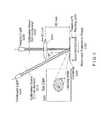
- an exemplary embodiment of an OFDI cathetermay be provided in accordance with the present invention that can be centered within the esophageal lumen using a balloon sheath shown in FIGS. 1A and 1B .
- the exemplary cathetermay include of a probe scanner 2000 which can rotate, and may pull back an inner optical core 2010 .
- the inner core 2010can be enclosed within a transparent sheath 2020 .
- the balloon 2040which, when inflated, may center the imaging optics.
- the imaging beam 2030can be focused onto the esophageal surface 2050 . This imaging beam 2030 may be scanned to achieve comprehensive imaging.
- the balloon 2040can have an inflated diameter of 1.8 cm, and may allow for a longitudinal imaging over a length of 4.5 cm without repositioning.
- the optical core 2010 of the cathetercan include an optical fiber, a spacer for expansion of the optical beam, a gradient index lens for focusing, and a right-angle prism for directing the beam perpendicularly to the longitudinal axis of the catheter.
- a miniature cylindrical lenswas fabricated in-house and placed on the second surface of the prism. This lens compensated for astigmatism induced by the plastic sheaths and resulted in a diffraction-limited beam (30 ⁇ m diameter) on the tissue surface.
- the exemplary cathetermay be rotated at rate of about 4 revolutions per second, allowing the acquisition of 2500 axial scans per circular cross-section.
- This exemplary OFDI systemcan record an encoder signal to precisely track the rotation and pull-back of the catheter. This information is used in reconstructing the 3-dimensional data set.
- FIGS. 2A-2CThe esophageal imaging techniques can be performed in two ⁇ 50 kg swine. Although the complete 20 GB data set may likely not be represented in discrete figures, the information content is shown by FIGS. 2A-2C .
- an image 2100provides a 3D rendering of the entire imaged esophagus.
- an image 2110illustrates a single transverse cross section of the imaged esophagus.
- image 2120shows a zoomed cross sectional image of at least one portion of the esophagus.
- a sampling with a resolution of 10 ⁇ m ⁇ 20 ⁇ m ⁇ 30 ⁇ m (r, ⁇ ,z)can yield a comprehensive microscopic data set that can be displayed volumetrically a the image 2100 of FIG. 2A for mapping and orientation, or in high-resolution cross-sectional images in which the entire esophageal wall can be visualized as the image 2110 in FIG. 2B .
- An expanded view of the image 2120 of FIG. 2Cdepicts the architectural structure of the mucosal layers.
- FIG. 3An exemplary single rotational image 2150 is shown in FIG. 3 . Hallmark features of SIM (disorganized epithelial architecture with irregular surface; presence of large epithelial glands) of a patient are shown therein. This patient had a prior diagnosis of BE, and imaging was performed prior to PDT.
- SIMstructured epithelial architecture with irregular surface; presence of large epithelial glands
- tissue proteins and collagencan denature, giving rise to microscopic deformation (described in publications 33 identified below), increased in scattering (described in publications 36, 38 and 45 identified below), reduced birefringence (described in publication 46 identified below), and reduced blood flow (described in publication 47 identified below).
- the description belowprovides the methods for monitoring these changes using exemplary OFDI in accordance with the exemplary embodiments of the present invention.
- porcine esophagus samples and duodenum samples(as a proxy for SIM) were mounted with a microscope cover glass on the epithelial surface so that the approximate pressure and thermal conductivity of the balloon catheter could be simulated.
- FIG. 4An exemplary embodiment of an apparatus for collecting OFDI signals during a laser irradiation and use thereof according to the present invention is shown in FIG. 4 .
- the treatment lightis delivered through a collimator 2200 .
- the imaging lightis delivered through a second collimator 2220 .
- the treatment beam 2210 and imaging beam 2230overlap when reaching the tissue 2270 which is covered with a thin glass cover slip 2260 and resting on a backing 2280 .
- the tissueis translated by a motorized translation stage 2290 .
- the imaging beamis focused by a lens 2250 .
- the top-down image depicting beam overlap 2250is provided.
- the OFDI sampling beamcan be focused at the tissue surface to, e.g., a 1/e2 intensity diameter of 23 ⁇ m and aligned so that it overlapped with the laser spot as shown in FIG. 4 .
- the samplesmay be held at a fixed location and/or translated using a motorized stage.
- the sampleswere held at a fixed location.
- OFDI depth-scanswere acquired continuously at a rate of about 10 kHz while the 1450 nm laser was switched on, held at a constant power of 400 mW for a predetermined duration, and switched off.
- a red horizontal line 2330 a , 2330 b , 2330 c , at the top of each phase-shift imagedenotes the interval over which the laser was on.
- a superficial region of positive phase-shift overlying a lower region of negative shifthas been observed.
- the depth at which the phase transitioned from positive to negativebecame progressively deeper and the magnitude of the overlying phase-shift decreased. No measurable phase-shift was detected after the laser was switched off.
- a protein denaturationgives rise to local microscopic structural changes and a nidus of local deformation that is detected as a phase-shift in the interferometric signal.
- the zone of active denaturationpropagates in depth with overlying tissues becoming completely denatured. The depth at which the direction of the phase-shift reverses identifies the focal center of active denaturation.
- NBTCnitro-blue tetrazolium chloride
- the depth-derivative of the phase-shifthas been determined for each A-line and defined the depth of injury as the point of maximum negative value of the derivative.
- the depths determined in this wayare provided in FIG. 5 as vertical lines adjacent to each M-mode image and show a good correspondence with histomorphometry.
- Laser treatment of large epithelial surface areascan be facilitated by adding a therapeutic laser beam to the existing OFDI catheter so that the laser and OFDI beams are simultaneously scanned.
- the preliminary imaging studiesdemonstrated comprehensive esophageal imaging with an OFDI beam size of 30 ⁇ m. Obtaining a precise alignment of >1 mm diameter laser beam on successive rotational scans should therefore be obtainable.
- the computer-controlled translation stage 2290(see FIG. 4 ) can be controlled to repetitively toggle the sample velocity from 1.8 mm/s to 0.9 mm/s.
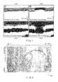
- FIG. 6AAn OFDI intensity image 2400 acquired with no laser irradiation is shown in FIG. 6A .
- the 1450 nm laser powerwas about 400 mW.
- the translation of the samples during the exposureresulted in a line of laser damage across the surface of the sample. Since the thermal energy deposition can be proportional to the exposure time (see Eq. 1), the depth of laser damage can vary along the line according to the inverse of the translation velocity. Histology sections, obtained from regions of fast and slow translation and with an orientation perpendicular to the line, indicated laser injury depths of 0.41 mm and 0.69 mm respectively.
- phase-shift data corresponding to the image 2410 of FIG. 6Bis illustrated as the image 2420 in FIG. 6C .
- the depth of the damage determined by the phase-shift datacan be 0.40 mm and 0.67 mm in the regions of fast and slow velocity, respectively.
- Speckleis a phenomenon that is commonly observed when imaging with coherent illumination and manifests as a grainy pattern of high- and low-intensity that does not appear to correlate with the macroscopic structure.
- speckleIn tissue, speckle generally arises from the interference between photons that have traversed different paths during propagation within the sample. If the scatterers within the tissue are moving, even on a microscopic scale, the speckle pattern is likely seen to rapidly fluctuate. The measurements of the time-evolution of the speckle pattern can therefore provide insight into microscopic motion within the sample.
- This exemplary techniquehas been provided for investigating biomechanical properties (as described in publication 50 identified below), and thermal excitation (as described in publication 51 identified below), in biological tissues. The extension of these concepts to the depth-resolved monitoring of laser tissue interactions with OFDI has been reviewed.
- the image 2430 of FIG. 6Dis the speckle decorrelation image corresponding to the images 2410 and 2420 of FIGS. 6B and 6C , respectively.
- the depth of the peak decorrelation 2431 rateblack band, denoted by arrows in FIG. 6D ) can be observed to vary in correspondence with the translational rate of the sample and the depth of laser damage indicated in histology. The consistency of this finding across samples of esophagus and duodenum confirm that the depth of peak decorrelation rate is a quantifiable metric for determining the depth of laser injury.
- PS-OCTPolarization-sensitive OCT
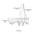
- the apparatus of FIG. 4can be modified to include a galvanometric scanner so that the OFDI beam may be repetitively scanned across the surface of the tissue while the sample was held stationary and the 1450 nm laser spot remained fixed on center, as shown in FIG. 27 .
- the treatment lightcan be delivered through a first collimator 2500 providing a treatment beam 2510 incident on the tissue 2550 that is covered by a cover slip 2540 and against a backing 2560 .
- the imaging lightmay be provided by a second collimator 2570 producing an imaging beam 2580 that is directed by a galvo-mirror 2520 through a lens 2530 .
- This arrangement/systemcan be an exemplary embodiment of a therapeutic monitoring system applicable to applications in dermatology. OFDI images or video of esophageal and duodenal tissues were acquired during laser irradiation.
- FIGS. 7A-7Dshow images of the representative data.
- the layered esophageal structurecan be observed in the intensity image 2450 (see FIG. 7A ) and characteristic birefringence banding can be observed in the corresponding polarization image 2460 (see FIG. 7B ).
- the epithelial scattering intensitymay be increased dramatically within the 1.1 mm laser spot 2470 (see FIG. 7C ), and the birefringence banding in the corresponding polarization image 2480 (see FIG. 7D ) can be lost.
- a zone of decreased birefringencecan be observed that may begin near the surface and propagated downward. These observations are generally consistent with a downward propagating zone of denatured tissue. Measurements of the percent-loss of birefringence is a quantitative metric for monitoring laser thermal damage.
- Thermally induced changes to the microscopic structure of tissuecan alter optical scattering. Since the signal in OFDI arises from scattering and small changes can be detected over a large dynamic range, we investigated the use of scattering measurements for monitoring thermally induced changes in tissue. Scattering changes observed in image 2460 of FIG. 7B may be representative of the preliminary observations in both duodenum and esophagus samples. In certain cases, it was determined that the significant scattering changes within the epithelium and relatively smaller changes in the underlying tissues of the muscularis mucosa and muscularis basement. For example, two potential quantitative metrics for laser damage that could be obtained from scattering measurements: changes in the depth-resolved scattering intensity and changes in the depth-integrated scatting intensity.
- FIG. 8An image 2490 of FIG. 8 , acquired during our recent swine studies, graphically illustrates the porcine esophageal vascularity. This exemplary image 2490 was generated by unwrapping the tubular image data to display the epithelial surface as if the esophagus was longitudinally opened and pinned flat. The intensity data has been integrated over depth into the tissue. Although this type of large scale visualization is a convenient way to map the vessels, it is possible to use a more sensitive and quantitative method/technique/system for measuring blood flow.
- Doppler OCT(as described in publications 55 and 56 identified below) has been demonstrated for visualizing and quantifying blood flow in tissue and has been investigated as an arrangement for assessing flow following laser therapy (as described in publication 57 identified below).
- the Doppler measurements with OFDI(as described in publication 24 identified below) have been described, and the possibility of simultaneously measuring structure and flow in vivo has been reviewed.
- a cross-sectional view of an exemplary image 2590 of FIG. 9has been acquired in the esophagus of a living swine and displays intensity as a grayscale and Doppler as a superimposed color.
- the coordinates (r, ⁇ ) of this datahave been mapped to Cartesian coordinates (vertical, horizontal) for simplicity of display. This result was representative of our observations at multiple locations in two swine. Additionally, in time-sequences of Doppler images, we clearly observed pulsatile flow.
- phase-shift and speckle decorrelationwhich are only applicable during laser irradiation, may be more sensitive and provide greater spatial resolution.
- the changes in birefringence, scattering and floware persistent and could be applied for follow-up imaging after laser treatment.
- thermal injurycan be controlled through the use of a raster-scanned, spatially-collimated beam.
- a flat-top beam with a diameter of 1-3 mm with well-defined edgesmay allow spatial control while also permitting therapy of large epithelial areas through raster scanning.
- Exemplary laser control parametersare described herein below in the context of Eq. 1.
- the temperature distribution of Eq. 1generally applies only in the limit of weak scattering.
- ⁇ awould likely be an optimal parameter for control over depth of laser injury.
- ⁇ ais characteristic of the sample rather than an externally controllable parameter
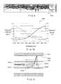
- absorption lengthsIn the vicinity of the water absorption band near 1.45 ⁇ m, absorption lengths (see graph 2595 of FIG. 10 ) range from 0.3 mm to over 2 mm within a narrow spectral range (1375 nm to 1430 nm). These lengths correspond well to the characteristic length scales appropriate for the treatment of epithelial disease.
- a tunable laser, operable in the vicinity of the 1450 nm water absorption bandcan be used to control therapy through wavelength tuning.
- the absorption coefficientdoes more than control the exponential depth decay of the temperature distribution; e.g., it also can control the amplitude. Since the amplitude term is also dependent upon power density and exposure duration, these variables can be used to normalize the amplitude while allowing the absorption coefficient to change.
- the procedure performance timemay be estimated by 2 At/( ⁇ rv), where At is the treatment area, r is the laser spot radius, and v is the laser spot scan velocity.
- Atis the treatment area
- ris the laser spot radius
- vis the laser spot scan velocity.
- a combined systemcan be provided which may allow for a controlled laser excitation.
- the exemplary systemcan be used endoscopically for conformal laser therapy capable of comprehensively treating epithelial lesions while minimizing collateral damage to adjacent tissues.
- a systemcan be provided for performing conformal laser therapy of epithelial disease through a combination of monitoring and control. Since laser beams can easily be shaped and spatially scanned and since margins in the transverse plane (along the surface of the esophagus) are less critical, the primary challenge for achieving accurate control of laser therapy is in limiting and adjusting the depth of laser damage. Based on the modeling and analysis described above, it is possible to utilize laser wavelength and power and scanning speed to vary the depth of laser damage over a clinically significant range while not significantly altering the transverse extent of injury.
- the laser wavelengths between approximately 1375 nm and 1430 nmcan provide absorption lengths ranging from over 2 mm to less than 0.3 mm.
- Semiconductor laserscan operate in this spectral range. Since such lasers can be compact and environmentally stable, these laser can be effectively used in clinical applications. Materials suitable for this specific wavelength range, however, may not be standard.
- a less expensive alternative for the early testing phase of exemplary embodiments of the methods according to the presentcan be provided by a solid-state laser material, tetravalent chromium-doped YAG (Cr4+:YAG). For example, a tunability with this material over the spectral range of 1340 nm-1570 nm can be implemented (as described in publications 58 and 59 identified below).
- the OFDI sampling beammay be focused at the sample to a diameter of ⁇ 25 ⁇ m.
- the axial location of the focuscan be determined using a standard z-scan technique, and may be registered within the OFDI cross-sectional image.
- the subsequent axial positioning of the samples within the OFDI image windowmay ensure a constant focus location for all samples.
- Datamay be collected with the two beams fixed with respect to each other and while the sample is translated perpendicular to the laser beam axis.
- the offset between the OFDI beam and the center of the laser spotis not critical for monitoring.
- OFDI datamay be collected for various offsets (as depicted in FIG. 4 ) to determine the offset that yields the greatest indicated depth of thermal injury.
- This offsetcan then be used in all subsequent studies and may be registered as follows.
- a small, low-power, short-duration epithelial burnmay be induced on the surface of the sample while the sample is held fixed (non-translating).
- the increase in epithelial scatteringcan be readily observed in OFDI and is spatially localized as defined by the laser beam profile.
- the OFDI beamcan be relayed to the focusing lens by a pair of galvanometers that provide two-dimensional scanning.
- the galvanometersmay be used to generate an en face OFDI image of the sample and the epithelial burn may appear as a circle of increased scattering.
- the galvanometerscan then be positioned and fixed so that the OFDI beam is positioned with the desired offset (as schematically shown in FIG. 4 ).
- Laser wavelengthmay be varied from about 1375 nm to 1405 nm in 2 nm steps with laser spot size and scanning speed held constant.
- the laser powermay be adjusted so that the product P d ⁇ a in Eq. 1 can be maintained as constant. This should yield lines of constant width and with damage depth ranging from approximately 0.25 to 1.5 mm.
- One exemplary embodiment according to the present invention for affecting therapeutic depthmay include scaling the scan velocity.
- the therapy beam scan speedcan be varied from 1 mm/s to 5 mm/s. Slower scan speeds allow time for heat to conduct to deeper areas of the tissue, producing deeper therapy.
- the spatial relationship between the OFDI sampling beam and the laser spotcan be controlled.
- One exemplary embodiment of the present inventioncan include an endoscopic probe for comprehensive, volumetric imaging and simultaneous laser therapy, as shown in FIG. 11 .
- two beam relay optics 2640 a and 2640 bmay be used, one of which conveys imaging light 2640 b and the other therapy light 2640 a .
- These relay opticsare placed within a housing 2630 that is enclosed within a first transparent sheath 2600 .
- a balloon centration mechanism (as described above) 2620may be used to maintain a constant distance between the optical probe 2630 and the tissue surface 2610 .
- Laser light and the OFDI beammay be delivered through separate optical fibers 2641 a and 2641 b .
- Each fibermay have its own relay optics to produce independently controllable spot sizes.
- a further exemplary embodiment of the present inventioncan include these relay optics designed to produce overlapping spots.
- the optical fibers and distal opticsmay be housed in a wound-wire drive shaft and placed inside a balloon-centering probe identical to the balloon shea
- Longitudinal scanscan be activated using a computer controlled translation stage attached to the proximal end of the drive shaft.
- This exemplary arrangementmay be the same as the arrangement which can be used for the pull-back esophageal imaging of our preliminary studies.
- a manual rotation of the drive shaftmay be possible, as is automated rotation using an exemplary rotary coupler 2900 shown in FIG. 13 .
- an endoscopic systemmay screen for disease over large fields-of-view, accurate monitoring of laser-tissue interaction, and precisely control laser therapy.
- One of the applications of such exemplary embodimentmay be the identification and treatment of epithelial cancers and their precursors.
- the systemcan incorporate procedures and software modules than can directly link screening, monitoring, and control.
- the systemmay be used to generate a high-resolution, 3-dimensional map of the entire distal esophagus to facilitate therapeutic planning. Thereafter, the use may be presented with a ‘live’ cross-sectional image comprising three sections, as illustrated in FIG. 14 .
- a right section 2700 of the imagemay be the tissue immediately ahead of the therapeutic lase
- the center 2730 of the imagemay be the location of the laser with a marker 2740 designating the zone of therapy
- the left section 2710 of the imagemay be the tissue that has already been treated. Since the three beams may be continuously scanning, the tissue may appear to move from right to left as the image updates.
- the usermay operate a control servo to start/stop the treatment and increase or decrease the depth of therapy.
- the usermay be able to steer and conform the region of laser therapy to the desired target.
- FIG. 12An exemplary embodiment of the endoscopic probe for imaging, monitoring and laser therapy through a centering balloon according to the present invention is shown in FIG. 12 .
- This exemplary probecan rotate to scan the esophagus circumferentially and may be longitudinally translated at a slower rate to define segments for therapy.
- This probemay include, e.g., three or more optical channels: a first channel 2800 c for imaging the tissue prior to laser irradiation, a second channel 2800 b for treatment, and a third channel 2800 a for monitoring.
- Each optical fibermay be separately imaged transversely onto the esophageal wall through the balloon.
- the alignment of the resulting output beamsmay be such that, upon rotation in the clockwise direction, the imaging beam precedes the treatment beam sufficiently so that non-treated tissue may be sampled.
- the monitoring beammay be aligned to fall within the laser spot.
- the opticsmay be bonded together with epoxy, and the alignment may be fixed.
- FIG. 13An exemplary rotational coupler according to the present invention which can connect the three-channel catheter to the OFDI system is shown in FIG. 13 , and can be referred to “watch-spring” rotary junction (since it can rely on two concentric spools).
- watch-springrotary junction
- optical fiberis wound from the outer spool 2910 onto the inner spool 2900 .
- the fibercan unwind from the inner spool.
- Ribbon optical fibermay be used and two parallel plates 2920 with a gap matched to the ribbon width can ensure that the windings remain flat and do not bind.
- the platesmay be sufficiently large so that up to, e.g., 100 rotations may be possible prior to requiring counter rotation.
- a further exemplary embodiment of the system and arrangement according to the present inventioncan utilize, e.g., a high-speed imaging system.
- the exemplary embodiment of the digital acquisition and processing systemcan be based on VME-bus hardware for acquiring, processing and storing the OFDI signals in real-time.
- the exemplary components of such exemplary system and arrangementmay comprise a VME chassis containing high-speed digitizers residing on a single-board computer and fiber-optic links to a RAID storage array.
- This exemplary system and arrangementcan be controlled via a host processor (e.g., a personal computer).
- the analog OFDI signalsmay be digitized using wideband receivers (e.g., 12 bit, 210 MS/s) with integrated field-programmable-gate-array (FPGA) processors. Processing power, resident on the acquisition board, may be importance since the raw data rate may be 800 MB/s for the two polarization channels of the OFDI system.
- the FPGA processorcan be configured or programmed to transform each polarization channel from the frequency-domain to a 1024-element array representing reflectivity versus depth (one A-line). This data can be passed to the single-board computer for subsequent processing and for combining the two channels prior to transferring the final data to a RAID array of hard drives.
- the final data storage ratemay be, e.g., 400 MB/s. By striping the data across multiple hard drives, this data rate can be continuously sustained.
- Software on a processing arrangement in accordance with an exemplary embodiment of the present inventioncan permit a user control over the exemplary system, and may enable a display of the images at a down-sampled rate in real-time.
- the exemplary systemcan be used in two exemplary modes: a burst mode at full data rate, and continuous mode at half data rate.
- the exemplary endoscopic system and arrangementcan include the components and software described above, and additional procedures (e.g., software) can be provided to program both the FPGA processor and single-board computer to facilitate the computations of phase-shift, birefringence, speckle, and Doppler signals in real-time.
- the combined computational capacity of the Vertex 4 Pro FPGA and quad G4 single-board computersmay be ample for displaying the monitoring signal in real-time.
- the spot size while maintaining a constant scan velocitycan be doubled by using a 4-fold increase in the laser power in order to maintain a constant temperature distribution in the tissue. Doubling the scan velocity at a constant spot size should use a doubled laser power.
- One exemplary embodiment of a laser arrangement in accordance with the present inventioncan utilize a single-emitter semiconductor laser diode. Previous devices have provided more than 3 W of laser power over this spectral range using a simple external cavity design including a diffraction grating for wavelength control. The laser power and wavelength may be controlled via the host processing arrangement of the OFDI system based on an analog signal from a potentiometer.
- the potentiometermay be a hand-held dial that the user (e.g., an endoscopist) may use to increase or decrease the depth of laser damage.
- the exemplary embodiment of the system and method according to the present inventioncan provide a user interface to the operator that includes a cross-sectional image of the tissue.
- the imagemay be continuously updated and may include views of treated and upcoming, untreated tissue as well as a designation for the zone of laser treatment as determined by the monitoring procedures.
- the user interfacemay be programmed on the host processing arrangement, and can use computational results from the FPGA processor and single-board computer. Images and laser parameters may be archived to the RAID array.
- the imaging system/arrangement 100can be connected to a three-fiber probe using an optical switch 115 as shown in a block diagram of FIG. 15 .
- the exemplary probesuch as that described above with reference to FIG. 12 , can include two imaging fibers and one therapy fiber.
- the switch 115can alternately couple imaging light to one of the two imaging fibers 120 a , 120 b which can be used to acquire re-therapy images and, e.g., during-therapy imaging.
- a therapeutic light source 105may connect directly to the therapy fiber 125 c .
- the fiberscan be connected to the catheter 130 , which can be, e.g., the exemplary catheter shown FIG. 12 .
- a signal from the imaging system 100can control the state of the optical switch 115 .
- the exemplary imaging system/arrangement 200can be coupled to an exemplary three-port catheter such as one shown in FIG. 12 via an optical splitter 215 that can couple light to both of two imaging fibers 220 a , 220 b .
- This exemplary imaging systemcan separate the image signal from each using path-length encoding techniques.
- an optical delay 235may be placed in one fiber 220 b or multiple fibers.
- the therapeutic light source 205can be coupled directly or indirectly to the therapy fiber 225 c of the catheter.
- lightmay be combined with the therapy source 805 using a single wavelength-division multiplexer 810 .
- the combined lightmay be coupled to a single fiber rotary coupler, and then to an exemplary single fiber catheter such as the catheter shown in FIG. 21 .
- lightmay be combined with the therapy light 905 using a cladding mode coupler that couples the imaging system 900 light from the single mode fiber 901 to the single mode core of a dual-clad fiber 911 and the therapy light from a multimode fiber 906 to the cladding mode of a dual-clad fiber 911 .
- FIG. 19shows exemplary connections between a system 400 with three output fibers 405 a , 405 b , 405 c , such as one shown schematically in, e.g., FIGS. 15 and 16 , and a three-port catheter 415 , such as one shown in FIG. 12 via a multi-channel rotary coupler 410 , such as one shown in FIG. 13 .
- FIG. 20shows a schematic diagram of an exemplary system 300 according to the present invention in which a single fiber 305 containing both the imaging light and therapy light may be coupled to a single-channel rotary coupler 310 .
- the lightcan be divided by a wavelength-division multiplexer (WDM) 330 that separates the imaging light onto a first fiber 332 and the therapy light onto a second fiber 331 .
- the imaging lightmay further be separated using an optical splitter 335 that greats two imaging ports 336 a and 336 b .
- the fibers 31 , 336 a , 336 bcan be connected to a three-port catheter 325 design such as that shown in FIG. 12 .
- the catheter section 320may be flexible allowing endoscopic insertion and the section containing a WDM 330 and a splitter 335 can be enclosed within a rigid tube 315 to protect these components.
- FIG. 21shows a side view of an exemplary embodiment of a distal optics arrangement according to the present invention that may create a single imaging beam 1125 and a separate therapy beam 1120 from a single-mode fiber 1101 .
- the light from the fiber containing both imaging and therapy lightcan first be focused by a first GRIN lens 1100 .
- the lightis then passed into a wavelength-division multiplexing prism 1105 that can direct the therapy light wavelengths upward to create the therapy beam 1120 , and transmits the imaging light wavelengths to a second GRIN lens 1110 , which can alternately focus the imaging light and directs it to a final prism 1115 that directs the imaging beam 1125 upward.
- the angle of the prisms 1105 and 1115may be such that the beams are made to overlap at the appropriate distance from the device.
- FIG. 22shows side and front views of an exemplary embodiment of a three-port catheter in accordance with the present invention.
- the exemplary cathetercan include three fibers 1005 that connect to three sets of focusing optics 1035 contained in a V-groove 1020 inside a housing 1040 .
- the focusing opticscan provide beam focusing.
- Micro-prisms 1025can redirect the optical beam upward through a cylindrical lens 1030 that corrects for astigmatism induced by the transparent sheath 1000 .
- a balloon 1010 centration mechanismmay be used to maintain centering of the optics 1035 within the luminal tissue 1015 .
- the monitoring beam 1050 c , therapy beam 1050 b , and pre-imaging beam 1050 acan be seen.
- the housing 1040can be adapted to rotate by a multichannel rotary coupler such as one shown in FIG. 13 .
- FIG. 23shows a side view of an exemplary embodiment of a catheter in accordance with the present invention which can utilize a miniature motor 1260 to achieve rotation of the imaging beam.
- the motor 1260can be enclosed within a transparent sheath 1235 .
- the rotation of the motor shaftcan rotate a prism 1220 .
- the imaging lightcan be coupled to the distal optics via a fiber 1210 , where the light can be focused by focusing optics 1215 , and reflected onto the prism 1220 by a reflector 1225 .
- the rotation of the prism 1220sweeps the imaging beam circumferentially.
- the motor electrical connections 1205can be achieved through the same lumen as the fiber 1210 .
- the therapy lightis coupled to the distal optics on fiber 1200 .
- This therapy lightmay be focused using focusing optics 1250 , and directed sideways by prism 1245 at a fixed rotational angle relative to the inner sheath 1235 .
- the imaging beamthus sweeps through the fixed therapy spot.
- the translation of the therapy spotis achieved by rotation of the entire inner sheath 1235 within the outer sheath 1240 .
- This exemplary rotationcan be achieved through the use of a multi-channel rotary coupler such as a coupler shown in FIG. 13 .
- the cathetercan use a balloon 1255 for centration of an optical core 1230 .
- FIG. 24shows a block diagram of an exemplary embodiment of a laser therapy source according to the present invention with wavelength tunability utilizing a low power wavelength tunable source 600 , followed by a broadband booster amplifier 605 to increase the optical power.
- FIG. 25shows a block and functional diagram of an exemplary embodiment of a laser therapy source incorporating multiple laser diodes 500 a , 500 b , 500 c , 500 d at difference wavelengths and polarizations, and the exemplary procedure to implement such arrangement.
- the lightcan be combined by the polarization multiplexers 505 a , 505 b and wavelength division multiplexers 510 to a single mode fiber 515 .
- the lightcan be coupled to a multimode fiber 520 .
- a fast mode scrambler 525can be used to scramble the transverse mode pattern output from the multimode fiber at a fast rate.
- Other source arrangements which can output light on a single mode fibercan use a similar design to couple light to a multimode fiber.
- FIG. 26shows an exemplary embodiment of a therapy light source and use thereof according to the present invention.
- a laser diode bar 700can be used with multiple wavelengths 701 a - g .
- Each waveguidecan be coupled to a free-space laser cavity through a lens apparatus 705 and a grating 710 and a partially reflecting end mirror 715 . Because of the wavelength dispersion of the grating, the laser formed by each waveguide lases at a different wavelength. Thus, by adjusting the drive current to each of the waveguides 701 a - g , the laser output 720 power and spectral shape can be adjusted.
- a single OFDI systemcan be modified to facilitate a detection of both the imaging and monitoring signals through the use of acousto-optic frequency shifters as shown in FIG. 28 .
- a wavelength swept laser source 3000can be separated by a first splitter 3020 to produce a sample arm path and reference arm path.
- the sample arm pathis further separated by a second splitter 3030 , with a first output of this splitter being directed to a first frequency shifter 3061 and a second output being directed to a second frequency shifter 3060 .
- Each of the frequency shifterscan be driven with a separate frequency.
- the light from the first frequency shifter 3061can be coupled through an optical circulator 3071 to the imaging fiber 3072 of a three-fiber rotary coupler 3110 like that shown in FIG. 13 .
- the light from the second frequency shifter 3060may be coupled through a circulator 3070 to a monitoring fiber 3073 of the same rotary coupler.
- a separate therapy laser 3010can be coupled to the third therapy fiber.
- the returned light on the imaging fiber 3072 and monitoring fiber 3073may be recombined at an optical combiner 2080 , and mixed with the reference arm light at a second combiner 3090 with the output directed to a set of detectors 3100 .
- the interference signal due to the imaging light and the interference signal due to the monitoring lightare encoded at separate carrier frequencies, e.g., using a computer 3095 which is part of an exemplary imaging system shown in FIGS. 15-20 , and can be separated through conventional frequency domain techniques.
- FIG. 29Ashows a flow diagram of an exemplary embodiment of a method for obtaining information associated with at least one portion of a sample according to the present invention.
- a temperature changecan be caused in the portion of the sample in step 3105 .
- At least one first electro-magnetic radiationcan be forwarded to a section near or in the portion of the sample in step 3110 .
- a deformation of the sectioncan be identified at a plurality of depths as a function of (i) a phase of at least one second electro-magnetic radiation provided from the section, and/or (ii) a rate of change of the phase and/or an amplitude of the second electro-magnetic radiation in step 3120 .
- FIG. 29Bshows a flow diagram of another exemplary embodiment of the method for controlling a temperature distribution in the sample according to the present invention.
- an electromagnetic radiationcan be provided to the section in the sample at a particular wavelength in step 3130 .
- the temperature distributioncan be controlled by modifying the particular wavelength of the electromagnetic radiation when the electromagnetic radiation can be provided to the section in step 3140 .
- FIG. 29Cillustrates a flow diagram of yet another exemplary embodiment of the method for applying a laser radiation to at least one portion of a biological structure according to the present invention.
- a beam of the laser radiationcan be provided to the portion in step 3150 , whereas a cross-sectional area of the beam is at most about 1/10 th of an entire area of the at least one portion.
- the beamcan be applied to the portion (I) based on a predetermined pattern, (II) while modulating a wavelength of the laser radiation, and/or (III) while monitoring a depth of the application of the laser radiation.
Landscapes
- Health & Medical Sciences (AREA)
- Surgery (AREA)
- Physics & Mathematics (AREA)
- Life Sciences & Earth Sciences (AREA)
- Engineering & Computer Science (AREA)
- Medical Informatics (AREA)
- Nuclear Medicine, Radiotherapy & Molecular Imaging (AREA)
- Electromagnetism (AREA)
- Optics & Photonics (AREA)
- Biomedical Technology (AREA)
- Heart & Thoracic Surgery (AREA)
- Otolaryngology (AREA)
- Molecular Biology (AREA)
- Animal Behavior & Ethology (AREA)
- General Health & Medical Sciences (AREA)
- Public Health (AREA)
- Veterinary Medicine (AREA)
- Laser Surgery Devices (AREA)
- Radiation-Therapy Devices (AREA)
Abstract
Description
where ρ is the tissue density, c the heat capacity, and r the radial distance from the center of a Gaussian laser beam of 1/e2 radius, W. Although this approximation neglects scattering of the laser light as it propagates into the tissue, models that explicitly include scattering (as described in publication 36 identified below) indicate less than 10% deviation from Eq. 1 under the stated conditions.
| Summary of Monitoring |
| Cause | Effect | Measurement |
| Thermal denaturing of | Focal deformation | Phase & Speckle |
| cellular proteins and collagen | Loss of birefringence | Polarization |
| ΔScattering | Intensity | |
| Thermal coagulation of | Loss of blood flow | Doppler flow & |
| vessels | Vascular Map | |
- 1. Devesa S S, Blot W J and Fraumeni J F, Jr. Changing patterns in the incidence of esophageal and gastric carcinoma in the United States. Cancer 1998; 83:2049-2053.
- 2. Barr H, Stone N and Rembacken B. Endoscopic therapy for Barrett's oesophagus. Gut 2005; 54:875-884.
- 3. Johnston M H. Technology insight: ablative techniques for Barrett's esophagus—current and emerging trends. Nature Clinical Practice Gastroenterology & Hepatology 2005; 2:323-330.
- 4. Falk G W, Chittajallu R, Goldblum J R, Biscotti C V, Geisinger K R, Petras R E, Birgisson S, Rice T W and Richter J E. Surveillance of patients with Barrett's esophagus for dysplasia and cancer with balloon cytology. Gastroenterology 1997; 112:1787-1797.
- 5. Spechler S J. Barrett's esophagus: should we brush off this ballooning problem? Gastroenterology 1997; 112:2138-2142.
- 6. Kubba A K, Poole N A and Watson A. Role of p 53 assessment in management of Barrett's esophagus. Dig Dis Sci 1999; 44:659-667.
- 7. Reid B J. p 53 and neoplastic progression in Barrett's esophagus. Am J Gastroenterol 2001; 96:1321-1323.
- 8. Sharma P, Weston A P, Topalovski M, Cheman R, Bhattacharyya A, Sampliner R E. Magnification chromoendoscopy for the detection of intestinal metaplasia and dysplasia in Barrett's oesophagus, GUT 2003; 52: 24-27.
- 9. Kuipers E J and Haringsma J. Diagnostic and therapeutic endoscopy. Journal of Surgical Oncology 2005; 92:203-209.
- 10. Georgakoudi I, Jacobson B C, Van Dam J, Backman V, Wallace M B, Muller M G, Zhang Q, Badizadegan K, Sun D, Thomas G A, Perelman L T and Feld M S. Fluorescence, reflectance, and light-scattering spectroscopy for evaluating dysplasia in patients with Barrett's esophagus. Gastroenterology 2001; 120:1620-1629.
- 11. Adrain A L, Ter H C, Cassidy M J, Schiano T D, Liu J B and Miller L S. High-resolution endoluminal sonography is a sensitive modality for the identification of Barrett's metaplasia. Gastrointest Endosc 1997; 46:147-151.
- 12. Canto M I. Vital staining and Barrett's esophagus. Gastrointest Endosc 1999; 49:S12-16.
- 13. Huang D, Swanson E A, Lin C P, Schuman J S, Stinson W G, Chang W, Hee M R, Flotte T, Gregory K, Puliafito C A and Fujimoto J G. Optical coherence tomography. Science 1991; 254:1178-1181.
- 14. Teamey G J, Brezinski M E, Bouma B E, Boppart S A, Pitvis C, Southern J F and Fujimoto J G. In vivo endoscopic optical biopsy with optical coherence tomography. Science 1997; 276:2037-2039.
- 15. Evans J A, Poneros J M, Bouma B E, Bressner J, Halpern E F, Shishkov M, Lauwers G Y, Mino-Kenudson M, Nishioka N S and Teamey G J. Optical Coherence Tomography to Identify Intramucosal Carcinoma and High Grade Dysplasia in Barrett's Esophagus. Clinical Gastroenterology and Hepatology 2005; 4:38-43.
- 16. Poneros J M, Brand S, Bouma B E, Teamey G J, Compton C C and Nishioka N S. Diagnosis of Specialized Intestinal Metaplasia by Optical Coherence Tomography. Gastroenterology 2001; 120:7-12.
- 17. Brand S, Poneros J M, Bouma B E, Teamey G J, Compton C C and Nishioka N S. Optical Coherent Tomography in the Gastrointestinal Tract.
Endoscopy 2000; 32:796-803. - 18. de Boer J F, Cense B, Park B H, Pierce M C, Teamey G J and Bouma B E. Improved signal-to-noise ratio in spectral-domain compared with time-domain optical coherence tomography. Optics Letters 2003; 28:2067-2069.
- 19. Choma M A, Sarunic M V, Changhuei Y and Izatt J A. Sensitivity advantage of swept source and Fourier domain optical coherence tomography. Optics Express 2003; 11:2183-2189.
- 20. Leitgeb R, Hitzenberger C K and Fercher A F. Performance of Fourier domain vs. time domain optical coherence tomography. Optics Express 2003; 11:889-894.
- 21. Yun S H, Teamey G J, de Boer J F, Iftimia N and Bouma B E. High-speed optical frequency-domain imaging. Optics Express 2003; 11:2953-2963.
- 22. Yun S H, Boudoux C, Teamey G J and Bouma B E. High-speed wavelength-swept semiconductor laser with a polygon-scanner-based wavelength filter. Optics Letters 2003; 28:1981-1983.
- 23. Oh W Y, Yun S H, Teamey G J and Bouma B E. 115 kHz tuning repetition rate ultrahigh-speed wavelength-swept semiconductor laser. Optics Letters 2005; 30:3159-3161.
- 24. Vakoc B J, Yun S H, de Boer J F, Tearney G J and Bouma B E. Phase-resolved optical frequency domain imaging. Optics Express 2005; 13:5483-5493.
- 25. Brown S B, Brown E A and Walker I. The present and future role of photodynamic therapy in cancer treatment. Lancet Oncol 2004; 5:497-508.
- 26. van den Boogert J, van Hillegersberg R, Siersema P D, de Bruin R W and Tilanus H W. Endoscopic ablation therapy for Barrett's esophagus with high-grade dysplasia: a review. Am J Gastroenterol 1999; 94:1153-1160.
- 27. Sampliner R E, Fennerty B and Garewal H S. Reversal of Barrett's esophagus with acid suppression and multipolar electrocoagulation: preliminary results. Gastrointest Endosc 1996; 44:532-535.
- 28. Sampliner R E. Endoscopic ablative therapy for Barrett's esophagus: current status. Gastrointest Endosc 2004; 59:66-69.
- 29. Soetikno R M, Gotoda T, Nakanishi Y and Soehendra N. Endoscopic mucosal resection. Gastrointest Endosc 2003; 57:567-579.
- 30. Ganz R A, Utley D S, Stem R A, Jackson J, Batts K P and Termin P. Complete ablation of esophageal epithelium with a balloon-based bipolar electrode: a phased evaluation in the porcine and in the human esophagus. Gastrointest Endosc 2004; 60:1002-1010.
- 31. Mark H. Johnston, Brooks D. Cash, Cathy A. Dykes, Halisha S. Mays and Lavonne R. Johnston Cryoablation of Dysplasia in Barrett's Esophagus (BE) and Early Stage Esophageal Cancer. Gastrointest Endosc 2006; 63: AB223.
- 32. Overholt B F, Panjehpour M and Haydek J M. Photodynamic therapy for Barrett's esophagus: follow-up in 100 patients. Gastrointest Endosc 1999; 49:1-7.
- 33. Vogel A and Venugopalan V. Mechanisms of pulsed laser ablation of biological tissues. Chemical Reviews 2003; 103:2079-2079.
- 34. McKenzie A L. Physics of thermal processes in laser-tissue interactions. Physics in Medicine & Biology 1990; 35:1175-1209.
- 35. Anderson R R and Parrish J A. Selective photothermolysis: precise microsurgery by selective absorption of pulsed radiation. Science 1983; 220:524-527.
- 36. Jacques S L. Role of tissue optics and pulse duration on tissue effects during high-power laser irradiation. Applied Optics 1993; 32:2447-2454.
- 37. Nahen K and Vogel A. Investigations on acoustic on-line monitoring of IR laser ablation of burned skin. Lasers in Surgery & Medicine 1999; 25:69-78.
- 38. Jerath M R, Kaisig D, Rylander H G, 3rd and Welch A J. Calibrated real-time control of lesion size based on reflectance images. Applied Optics 1993; 32:1200-1209.
- 39. Jerath M R, Gardner C M, Rylander H G, 3rd and Welch A J. Dynamic optical property changes: implications for reflectance feedback control of photocoagulation. Journal of Photochemistry & Photobiology B—Biology 1992; 16:113-126.
- 40. Deckelbaum L I. Coronary laser angioplasty. Lasers in Surgery & Medicine 1994; 14:101-110.
- 41. Kim B M, Feit M D, Rubenchik A M, Mammini B M and Da Silva L B. Optical feedback signal for ultra short laser pulse ablation of tissue. Applied Surface Science 1998; 127-129:857-862.
- 42. Brinkmann R, Hansen C, Mohrenstecher D, Scheu M and Birngruber R. Analysis of cavitation dynamics during pulsed laser tissue ablation by optical on-line monitoring. Selected Topics in Quantum Electronics, IEEE Journal of 1996; 2:826.
- 43. Whelan W M, Davidson S R H, Chin L C L and Vitkin I A. A Novel Strategy For Monitoring Laser Thermal Therapy Based on Changes in Optothermal Properties of Heated Tissues. International Journal of Thermophysics 2005; 26:233-241.
- 44. Boppart S A, Herrmann J, Pitris C, Stamper D L, Brezinski M E and Fujimoto J G. High-resolution optical coherence tomography-guided laser ablation of surgical tissue. Journal of Surgical Research 1999; 82:275-284.
- 45. Thomsen S L, Jacques S L and Flock S T. Microscopic correlates of macroscopic optical property changes during thermal coagulation of myocardium. Proceedings of the SPIE 1990; 1202:2-11.
- 46. Maitland D J and Walsh J T, Jr. Quantitative measurements of linear birefringence during heating of native collagen. Lasers Surg Med 1997; 20:310-318.
- 47. Kimel S, Svaasand L O, Hammer-Wilson M, Schell M J, Milner T E, Nelson J S and Berns M W. Differential vascular response to laser photothermolysis. Journal of Investigative Dermatology 1994; 103:693-700.
- 48. Khan M H, Sink R K, Manstein D, Eimerl D and Anderson R R. Intradermally focused infrared laser pulses: Thermal effects at defined tissue depths. Lasers in Surgery and Medicine 2005; 36:270-280.
- 49. Neumann R A, Knobler R M, Pieczkowski F and Gebhart W. Enzyme histochemical analysis of cell viability after argon laser-induced coagulation necrosis of the skin. Journal of the American Academy of Dermatology 1991; 25:991-998.
- 50. Nadkarni S, Helg T, Bouma B E, Chan R C, Minsky M S, Chau A H, Motz J, Houser S L and Tearney G J. Characterization of atherosclerotic plaques by laser speckle analysis. Circulation 2005; 112:885-892.
- 51. Zimnyakov D A, Agafonov D N, Sviridov A P, Omel'chenko A I, Kuznetsova L V and Bagratashvili V N. Speckle-contrast monitoring of tissue thermal modification. Appl Opt 2002; 41:5989-5996.
- 52. Pierce M C, Sheridan R L, Park B H, Cense B and de Boer J F. Collagen denaturation can be quantified in burned human skin using polarization-sensitive optical coherence tomography. Burns 2004; 30:511-517.
- 53. de Boer J F, Milner T E, van Gemert M J C and Nelson J S. Two-dimensional birefringence imaging in biological tissue using polarization sensitive optical coherence tomography. Optics Letters 1997; 22:934-936.
- 54. Morelli J G, Tan O T, Garden J, Margolis R, Seki Y, Boll J, Carney J M, Anderson R R, Furumoto H and Parrish J A. Tunable dye laser (577 nm) treatment of port wine stains. Lasers Surg Med 1986; 6:94-99.
- 55. Chen Z P, Milner T E, Dave D and Nelson J S. Optical Doppler tomographic imaging of fluid flow velocity in highly scattering media. Optics Letters 1997; 22:64-66.
- 56. Izatt J A, Kulkarni M D, Yazdanfar S, Barton J K and Welch A J. In vivo bidirectional color Doppler flow imaging of picoliter blood volumes using optical coherence tomography. Optics Letters 1997; 22:1439.
- 57. Barton J K, Welch A J and Izatt J A. Investigating pulsed dye laser-blood vessel interaction with color Doppler optical coherence tomography. Optics Express 1998; 3:251-256.
- 58. French P M W, Rizvi N H, Taylor J R and Shestakov A V. Continuous-wave mode-locked Cr4+:YAG laser. Optics Letters 1993; 18:39-41.
- 59. Sennaroglu A, Pollock C R and Nathel H. Efficient continuous-wave chromium-doped YAG laser. Journal of the Optical Society of America B 1995; 12:930-937.
- 60. Bouma B, Gouveia-Neto A, Izatt J A, Russell J, Sierra R, Keller U and Fujimoto J G. Hybrid mode locking of a flash-lamp-pumped Ti:Al2O3laser. Optics Letters 1994; 19:1858-1860.
- 61. Bouma B, Tearney G J, Boppart S A, Hee M R, Brezinski M E and Fujimoto J G. High-resolution optical coherence tomographic imaging using a mode-locked Ti:Al2O3laser source. Optics Letters 1995; 20:1486-1488.
- 62. Bouma B E and Fujimoto J G. Compact Kerr-lens mode-locked resonators. Optics Letters 1996; 21:134-136.
- 63. Bouma B E, Nelson L E, Teamey G J, Jones D J, Brezinski M E and Fujimoto J G. Optical coherence tomographic imaging of human tissue at 1.55 μm and 1.81 μm using Er and Tm-doped fiber sources. Journal of Biomedical Optics 1998; 3:76-79.
- 64. Bouma B E, Ramaswamy-Paye M and Fujimoto J G. Compact resonator designs for mode-locked solid-state lasers. Applied Physics B (Lasers and Optics) 1997; 65:213-220.
- 65. Bouma B E, Tearney G J, Bilinsky I P, Golubovic B and Fujimoto J G. Self-phase-modulated Kerr-lens mode-locked Cr:forsterite laser source for optical coherence tomography. Optics Letters 1996; 21:1839-1841.
Claims (21)
Priority Applications (1)
| Application Number | Priority Date | Filing Date | Title |
|---|---|---|---|
| US11/670,043US10426548B2 (en) | 2006-02-01 | 2007-02-01 | Methods and systems for providing electromagnetic radiation to at least one portion of a sample using conformal laser therapy procedures |
Applications Claiming Priority (3)
| Application Number | Priority Date | Filing Date | Title |
|---|---|---|---|
| US76462206P | 2006-02-01 | 2006-02-01 | |
| US81086906P | 2006-06-01 | 2006-06-01 | |
| US11/670,043US10426548B2 (en) | 2006-02-01 | 2007-02-01 | Methods and systems for providing electromagnetic radiation to at least one portion of a sample using conformal laser therapy procedures |
Publications (2)
| Publication Number | Publication Date |
|---|---|
| US20070282403A1 US20070282403A1 (en) | 2007-12-06 |
| US10426548B2true US10426548B2 (en) | 2019-10-01 |
Family
ID=38834172
Family Applications (1)
| Application Number | Title | Priority Date | Filing Date |
|---|---|---|---|
| US11/670,043Active2036-01-07US10426548B2 (en) | 2006-02-01 | 2007-02-01 | Methods and systems for providing electromagnetic radiation to at least one portion of a sample using conformal laser therapy procedures |
Country Status (4)
| Country | Link |
|---|---|
| US (1) | US10426548B2 (en) |
| EP (1) | EP1983921B1 (en) |
| JP (1) | JP5524487B2 (en) |
| WO (1) | WO2007149602A2 (en) |
Cited By (11)
| Publication number | Priority date | Publication date | Assignee | Title |
|---|---|---|---|---|
| US11110006B2 (en) | 2018-09-07 | 2021-09-07 | Vialase, Inc. | Non-invasive and minimally invasive laser surgery for the reduction of intraocular pressure in the eye |
| US11173067B2 (en) | 2018-09-07 | 2021-11-16 | Vialase, Inc. | Surgical system and procedure for precise intraocular pressure reduction |
| US11246754B2 (en) | 2018-07-16 | 2022-02-15 | Vialase, Inc. | Surgical system and procedure for treatment of the trabecular meshwork and Schlemm's canal using a femtosecond laser |
| US11564567B2 (en) | 2020-02-04 | 2023-01-31 | Vialase, Inc. | System and method for locating a surface of ocular tissue for glaucoma surgery based on dual aiming beams |
| US11612315B2 (en) | 2020-04-09 | 2023-03-28 | Vialase, Inc. | Alignment and diagnostic device and methods for imaging and surgery at the irido-corneal angle of the eye |
| US11666483B2 (en) | 2018-07-16 | 2023-06-06 | Vialase, Inc. | System and method for angled optical access to the irido-corneal angle of the eye |
| US11986424B2 (en) | 2018-07-16 | 2024-05-21 | Vialase, Inc. | Method, system, and apparatus for imaging and surgical scanning of the irido-corneal angle for laser surgery of glaucoma |
| US12002567B2 (en) | 2021-11-29 | 2024-06-04 | Vialase, Inc. | System and method for laser treatment of ocular tissue based on patient biometric data and apparatus and method for determining laser energy based on an anatomical model |
| US12016799B2 (en) | 2018-07-16 | 2024-06-25 | Vialase, Inc. | Integrated surgical system and method for treatment in the irido-corneal angle of the eye |
| US12226345B2 (en) | 2019-12-14 | 2025-02-18 | Vialase, Inc. | Near eye reflective devices for diagnostic and therapeutic ophthalmic procedures |
| US12245974B2 (en) | 2018-07-16 | 2025-03-11 | Vialase, Inc. | System and method for clearing an obstruction from the path of a surgical laser |
Families Citing this family (25)
| Publication number | Priority date | Publication date | Assignee | Title |
|---|---|---|---|---|
| JP2010042182A (en)* | 2008-08-18 | 2010-02-25 | Fujifilm Corp | Laser treatment device |
| JP5819823B2 (en)* | 2009-07-14 | 2015-11-24 | ザ ジェネラル ホスピタル コーポレイション | Device for measuring the flow and pressure inside a blood vessel and method of operating the device |
| US8478384B2 (en) | 2010-01-19 | 2013-07-02 | Lightlab Imaging, Inc. | Intravascular optical coherence tomography system with pressure monitoring interface and accessories |
| US8774903B2 (en)* | 2010-03-26 | 2014-07-08 | Headwater Partners Ii Llc | Medical imaging apparatus and method |
| US8936592B2 (en)* | 2010-06-03 | 2015-01-20 | Ams Research Corporation | Laser tissue ablation system |
| US9550063B2 (en) | 2010-10-14 | 2017-01-24 | II Erich W. Wolf | Apparatus and method using near infrared reflectometry to reduce the effect of positional changes during spinal cord stimulation |
| US9656097B2 (en)* | 2010-10-14 | 2017-05-23 | II Erich W. Wolf | Apparatus and method using near infrared reflectometry to reduce the effect of positional changes during spinal cord stimulation |
| US8876804B2 (en) | 2010-12-17 | 2014-11-04 | Ams Research Corporation | Ablation device |
| EP2817061B1 (en) | 2012-02-24 | 2018-02-07 | Isolase, Ltd. | Improvements in ablation techniques for the treatment of atrial fibrillation |
| RU2623299C2 (en)* | 2012-02-28 | 2017-06-23 | Конинклейке Филипс Н.В. | Device for skin treatment based on energy |
| JP2015517387A (en)* | 2012-05-21 | 2015-06-22 | ザ ジェネラル ホスピタル コーポレイション | Apparatus, device and method for capsule microscopy |
| US20140066756A1 (en)* | 2012-09-04 | 2014-03-06 | Ninepoint Medical, Inc. | Low cost molded optical probe with astigmatic correction, fiber port, low back reflection, and highly reproducible in manufacturing quantities |
| CN105073186B (en) | 2013-01-31 | 2018-07-10 | 迪格玛医疗有限公司 | Methods and systems for reducing neural activity in an organ of a subject |
| CN105188559B (en) | 2013-02-28 | 2017-09-22 | 爱飞纽医疗机械贸易有限公司 | Detect the method and ultrasonic medical equipment of cavitation |
| CN105050475B (en)* | 2013-03-29 | 2017-10-13 | 索尼公司 | Device and Laser Scanning are observed in laser scanning |
| EP2983579B1 (en)* | 2013-04-12 | 2025-07-09 | NinePoint Medical, Inc. | Multiple aperture, multiple modal optical systems and methods |
| US10835313B2 (en)* | 2014-01-30 | 2020-11-17 | Medlumics S.L. | Radiofrequency ablation catheter with optical tissue evaluation |
| US10194981B2 (en) | 2015-07-29 | 2019-02-05 | Medlumics S.L. | Radiofrequency ablation catheter with optical tissue evaluation |
| US10375330B2 (en) | 2016-05-27 | 2019-08-06 | Verily Life Sciences Llc | Systems and methods for surface topography acquisition using laser speckle |
| US10969571B2 (en)* | 2016-05-30 | 2021-04-06 | Eric Swanson | Few-mode fiber endoscope |
| JP7048606B2 (en)* | 2016-08-14 | 2022-04-05 | ジグマ メディカル リミテッド | Devices and methods for nerve ablation in the walls of the gastrointestinal tract |
| US10575904B1 (en) | 2016-08-14 | 2020-03-03 | Digma Medical Ltd. | Apparatus and method for selective submucosal ablation |
| JP2018139532A (en)* | 2017-02-28 | 2018-09-13 | 株式会社島津製作所 | Cell observation device |
| JP2025501258A (en)* | 2021-12-30 | 2025-01-17 | オプセラス アーエス | Fiber optic medical treatment device for treatment of the urinary tract of a subject - Patents.com |
| CN118785863A (en)* | 2021-12-30 | 2024-10-15 | 奥普思瑞斯股份有限公司 | Disposable optical fiber apparatus |
Citations (669)
| Publication number | Priority date | Publication date | Assignee | Title |
|---|---|---|---|---|
| US2339754A (en) | 1941-03-04 | 1944-01-25 | Westinghouse Electric & Mfg Co | Supervisory apparatus |
| US3090753A (en) | 1960-08-02 | 1963-05-21 | Exxon Research Engineering Co | Ester oil compositions containing acid anhydride |
| US3601480A (en) | 1968-07-10 | 1971-08-24 | Physics Int Co | Optical tunnel high-speed camera system |
| GB1257778A (en) | 1967-12-07 | 1971-12-22 | ||
| US3856000A (en) | 1972-06-19 | 1974-12-24 | Machido Seisakusho Kk | Endoscope |
| US3872407A (en) | 1972-09-01 | 1975-03-18 | Us Navy | Rapidly tunable laser |
| US3941121A (en) | 1974-12-20 | 1976-03-02 | The University Of Cincinnati | Focusing fiber-optic needle endoscope |
| US3973219A (en) | 1975-04-24 | 1976-08-03 | Cornell Research Foundation, Inc. | Very rapidly tuned cw dye laser |
| US3983507A (en) | 1975-01-06 | 1976-09-28 | Research Corporation | Tunable laser systems and method |
| US4030831A (en) | 1976-03-22 | 1977-06-21 | The United States Of America As Represented By The Secretary Of The Navy | Phase detector for optical figure sensing |
| US4030827A (en) | 1973-12-03 | 1977-06-21 | Institut National De La Sante Et De La Recherche Medicale (Inserm) | Apparatus for the non-destructive examination of heterogeneous samples |
| US4140364A (en) | 1973-06-23 | 1979-02-20 | Olympus Optical Co., Ltd. | Variable field optical system for endoscopes |
| US4141362A (en) | 1977-05-23 | 1979-02-27 | Richard Wolf Gmbh | Laser endoscope |
| WO1979000841A1 (en) | 1978-03-09 | 1979-10-18 | Nat Res Dev | Speckle interferometric measurement of small oscillatory movements |
| JPS559417A (en) | 1978-07-05 | 1980-01-23 | Seiko Epson Corp | Semiconductor integrated circuit |
| GB2030313A (en) | 1978-06-29 | 1980-04-02 | Wolf Gmbh Richard | Endoscopes |
| US4224929A (en) | 1977-11-08 | 1980-09-30 | Olympus Optical Co., Ltd. | Endoscope with expansible cuff member and operation section |
| US4295738A (en) | 1979-08-30 | 1981-10-20 | United Technologies Corporation | Fiber optic strain sensor |
| US4300816A (en) | 1979-08-30 | 1981-11-17 | United Technologies Corporation | Wide band multicore optical fiber |
| US4303300A (en) | 1979-02-07 | 1981-12-01 | Thomson-Csf | Rotary-joint device providing for an optical waveguide transmission |
| US4428643A (en) | 1981-04-08 | 1984-01-31 | Xerox Corporation | Optical scanning system with wavelength shift correction |
| EP0110201A2 (en) | 1982-11-25 | 1984-06-13 | Központi Elelmiszeripari Kutato Intezet | Apparatus for providing radiation of controlled spectral composition |
| US4479499A (en) | 1982-01-29 | 1984-10-30 | Alfano Robert R | Method and apparatus for detecting the presence of caries in teeth using visible light |
| JPS6073405A (en) | 1983-09-12 | 1985-04-25 | バテル メモリアル インステイチユート | Method and device for measuring position of surface element |
| US4533247A (en) | 1981-09-03 | 1985-08-06 | International Standard Electric Corporation | Optical transmission system |
| JPS6140633A (en) | 1984-08-02 | 1986-02-26 | Nec Corp | Tablet device |
| US4601036A (en) | 1982-09-30 | 1986-07-15 | Honeywell Inc. | Rapidly tunable laser |
| US4607622A (en) | 1985-04-11 | 1986-08-26 | Charles D. Fritch | Fiber optic ocular endoscope |
| US4631498A (en) | 1985-04-26 | 1986-12-23 | Hewlett-Packard Company | CW Laser wavemeter/frequency locking technique |
| US4639999A (en) | 1984-11-02 | 1987-02-03 | Xerox Corporation | High resolution, high efficiency I.R. LED printing array fabrication method |
| US4650327A (en) | 1985-10-28 | 1987-03-17 | Oximetrix, Inc. | Optical catheter calibrating assembly |
| JPS62188001A (en) | 1986-02-13 | 1987-08-17 | Matsushita Electric Ind Co Ltd | Rotating magnetic head recording device |
| EP0251062A2 (en) | 1986-06-20 | 1988-01-07 | Fujitsu Limited | Dual balanced optical signal receiver |
| US4734578A (en) | 1985-03-27 | 1988-03-29 | Olympus Optical Co., Ltd. | Two-dimensional scanning photo-electric microscope |
| US4744656A (en) | 1986-12-08 | 1988-05-17 | Spectramed, Inc. | Disposable calibration boot for optical-type cardiovascular catheter |
| US4751706A (en) | 1986-12-31 | 1988-06-14 | The United States Of America As Represented By The Secretary Of The Army | Laser for providing rapid sequence of different wavelengths |
| US4763977A (en) | 1985-01-09 | 1988-08-16 | Canadian Patents And Development Limited-Societe | Optical fiber coupler with tunable coupling ratio and method of making |
| US4770492A (en) | 1986-10-28 | 1988-09-13 | Spectran Corporation | Pressure or strain sensitive optical fiber |
| GB2209221A (en) | 1987-09-01 | 1989-05-04 | Litton Systems Inc | A demodulator circuit for an interferometer type of hydrophone |
| US4827907A (en) | 1986-11-28 | 1989-05-09 | Teac Optical Co., Ltd. | Intra-observation apparatus |
| US4834111A (en) | 1987-01-12 | 1989-05-30 | The Trustees Of Columbia University In The City Of New York | Heterodyne interferometer |
| US4868834A (en) | 1988-09-14 | 1989-09-19 | The United States Of America As Represented By The Secretary Of The Army | System for rapidly tuning a low pressure pulsed laser |
| US4890901A (en) | 1987-12-22 | 1990-01-02 | Hughes Aircraft Company | Color corrector for embedded prisms |
| US4892406A (en) | 1988-01-11 | 1990-01-09 | United Technologies Corporation | Method of and arrangement for measuring vibrations |
| US4905169A (en) | 1988-06-02 | 1990-02-27 | The United States Of America As Represented By The United States Department Of Energy | Method and apparatus for simultaneously measuring a plurality of spectral wavelengths present in electromagnetic radiation |
| US4909631A (en) | 1987-12-18 | 1990-03-20 | Tan Raul Y | Method for film thickness and refractive index determination |
| US4925302A (en) | 1988-04-13 | 1990-05-15 | Hewlett-Packard Company | Frequency locking device |
| US4928005A (en) | 1988-01-25 | 1990-05-22 | Thomson-Csf | Multiple-point temperature sensor using optic fibers |
| US4940328A (en) | 1988-11-04 | 1990-07-10 | Georgia Tech Research Corporation | Optical sensing apparatus and method |
| JPH02259617A (en) | 1989-03-30 | 1990-10-22 | Sony Corp | Laser beam deflector |
| US4965599A (en) | 1989-11-13 | 1990-10-23 | Eastman Kodak Company | Scanning apparatus for halftone image screen writing |
| US4965441A (en) | 1988-01-27 | 1990-10-23 | Commissariat A L'energie Atomique | Method for the scanning confocal light-optical microscopic and indepth examination of an extended field and devices for implementing said method |
| US4966589A (en) | 1988-11-14 | 1990-10-30 | Hemedix International, Inc. | Intravenous catheter placement device |
| US4984888A (en) | 1989-12-13 | 1991-01-15 | Imo Industries, Inc. | Two-dimensional spectrometer |
| US4993834A (en) | 1988-10-03 | 1991-02-19 | Fried. Krupp Gmbh | Spectrometer for the simultaneous measurement of intensity in various spectral regions |
| US4998972A (en) | 1988-04-28 | 1991-03-12 | Thomas J. Fogarty | Real time angioscopy imaging system |
| JPH03126449A (en) | 1989-10-09 | 1991-05-29 | Olympus Optical Co Ltd | Laser equipment for medical treatment |
| US5039193A (en) | 1990-04-03 | 1991-08-13 | Focal Technologies Incorporated | Fibre optic single mode rotary joint |
| US5040889A (en) | 1986-05-30 | 1991-08-20 | Pacific Scientific Company | Spectrometer with combined visible and ultraviolet sample illumination |
| US5045936A (en) | 1988-07-25 | 1991-09-03 | Keymed (Medical And Industrial Equipment) Limited | Laser scanning imaging apparatus and method of ranging |
| US5046501A (en) | 1989-01-18 | 1991-09-10 | Wayne State University | Atherosclerotic identification |
| DE4105221A1 (en) | 1990-03-05 | 1991-09-12 | Jenoptik Jena Gmbh | LIGHT GUIDE FOR A MEDICAL OBSERVATION DEVICE |
| US5065331A (en) | 1981-05-18 | 1991-11-12 | Vachon Reginald I | Apparatus and method for determining the stress and strain in pipes, pressure vessels, structural members and other deformable bodies |
| US5085496A (en) | 1989-03-31 | 1992-02-04 | Sharp Kabushiki Kaisha | Optical element and optical pickup device comprising it |
| WO1992001966A1 (en) | 1990-07-18 | 1992-02-06 | Medical Research Council | Confocal scanning optical microscope |
| JPH0456907A (en) | 1990-06-26 | 1992-02-24 | Fujikura Ltd | Optical fiber coupler |
| WO1992003977A2 (en) | 1990-09-06 | 1992-03-19 | Massachusetts Institute Of Technology | A pulsed laser system for the surgical removal of tissue |
| US5120953A (en) | 1988-07-13 | 1992-06-09 | Harris Martin R | Scanning confocal microscope including a single fibre for transmitting light to and receiving light from an object |
| US5121983A (en) | 1989-12-14 | 1992-06-16 | Goldstar Co., Ltd. | Stereoscopic projector |
| US5127730A (en) | 1990-08-10 | 1992-07-07 | Regents Of The University Of Minnesota | Multi-color laser scanning confocal imaging system |
| WO1992016865A1 (en) | 1991-03-22 | 1992-10-01 | Hicks John W | Multifiber endoscope with multiple scanning modes to produce an image free of fixed pattern noise |
| WO1992019930A1 (en) | 1991-04-29 | 1992-11-12 | Massachusetts Institute Of Technology | Method and apparatus for optical imaging and measurement |
| US5177488A (en) | 1991-10-08 | 1993-01-05 | Hughes Aircraft Company | Programmable fiber optic delay line, and radar target simulation system incorporating the same |
| WO1993003672A1 (en) | 1991-08-20 | 1993-03-04 | Redd Douglas C B | Optical histochemical analysis, in vivo detection and real-time guidance for ablation of abnormal tissues using a raman spectroscopic detection system |
| US5197470A (en) | 1990-07-16 | 1993-03-30 | Eastman Kodak Company | Near infrared diagnostic method and instrument |
| US5202931A (en) | 1987-10-06 | 1993-04-13 | Cell Analysis Systems, Inc. | Methods and apparatus for the quantitation of nuclear protein |
| US5202745A (en) | 1990-11-07 | 1993-04-13 | Hewlett-Packard Company | Polarization independent optical coherence-domain reflectometry |
| US5208651A (en) | 1991-07-16 | 1993-05-04 | The Regents Of The University Of California | Apparatus and method for measuring fluorescence intensities at a plurality of wavelengths and lifetimes |
| US5212667A (en) | 1992-02-03 | 1993-05-18 | General Electric Company | Light imaging in a scattering medium, using ultrasonic probing and speckle image differencing |
| US5214538A (en) | 1988-07-25 | 1993-05-25 | Keymed (Medical And Industrial Equipment) Limited | Optical apparatus |
| US5217456A (en) | 1992-02-24 | 1993-06-08 | Pdt Cardiovascular, Inc. | Device and method for intra-vascular optical radial imaging |
| US5228001A (en) | 1991-01-23 | 1993-07-13 | Syracuse University | Optical random access memory |
| US5241364A (en) | 1990-10-19 | 1993-08-31 | Fuji Photo Film Co., Ltd. | Confocal scanning type of phase contrast microscope and scanning microscope |
| US5248876A (en) | 1992-04-21 | 1993-09-28 | International Business Machines Corporation | Tandem linear scanning confocal imaging system with focal volumes at different heights |
| US5251009A (en) | 1990-01-22 | 1993-10-05 | Ciba-Geigy Corporation | Interferometric measuring arrangement for refractive index measurements in capillary tubes |
| US5250186A (en) | 1990-10-23 | 1993-10-05 | Cetus Corporation | HPLC light scattering detector for biopolymers |
| US5262644A (en) | 1990-06-29 | 1993-11-16 | Southwest Research Institute | Remote spectroscopy for raman and brillouin scattering |
| US5275594A (en) | 1990-11-09 | 1994-01-04 | C. R. Bard, Inc. | Angioplasty system having means for identification of atherosclerotic plaque |
| US5281811A (en) | 1991-06-17 | 1994-01-25 | Litton Systems, Inc. | Digital wavelength division multiplex optical transducer having an improved decoder |
| US5283795A (en) | 1992-04-21 | 1994-02-01 | Hughes Aircraft Company | Diffraction grating driven linear frequency chirped laser |
| US5290277A (en)* | 1992-04-03 | 1994-03-01 | Angeion Corporation | Multi-fiber linear array laser catheter connector |
| US5291885A (en) | 1990-11-27 | 1994-03-08 | Kowa Company Ltd. | Apparatus for measuring blood flow |
| US5293873A (en) | 1991-08-29 | 1994-03-15 | Siemens Aktiengesellschaft | Measuring arrangement for tissue-optical examination of a subject with visible, NIR or IR light |
| US5293872A (en) | 1991-04-03 | 1994-03-15 | Alfano Robert R | Method for distinguishing between calcified atherosclerotic tissue and fibrous atherosclerotic tissue or normal cardiovascular tissue using Raman spectroscopy |
| EP0590268A1 (en) | 1985-03-22 | 1994-04-06 | Massachusetts Institute Of Technology | Catheter for Laser angiosurgery |
| US5302025A (en) | 1982-08-06 | 1994-04-12 | Kleinerman Marcos Y | Optical systems for sensing temperature and other physical parameters |
| US5304173A (en) | 1985-03-22 | 1994-04-19 | Massachusetts Institute Of Technology | Spectral diagonostic and treatment system |
| US5305759A (en) | 1990-09-26 | 1994-04-26 | Olympus Optical Co., Ltd. | Examined body interior information observing apparatus by using photo-pulses controlling gains for depths |
| US5317389A (en) | 1989-06-12 | 1994-05-31 | California Institute Of Technology | Method and apparatus for white-light dispersed-fringe interferometric measurement of corneal topography |
| US5333144A (en) | 1991-12-30 | 1994-07-26 | U.S. Philips Corporation | Diode laser device having a reflecting feedback element, and apparatus using the device |
| US5348003A (en) | 1992-09-03 | 1994-09-20 | Sirraya, Inc. | Method and apparatus for chemical analysis |
| DE4309056A1 (en) | 1993-03-20 | 1994-09-22 | Haeusler Gerd | Method for determining the distance and intensity of scattering of the scattering points |
| EP0617286A2 (en) | 1993-03-18 | 1994-09-28 | Wallac Oy | Biospecific solid phase carrier |
| US5353790A (en) | 1992-01-17 | 1994-10-11 | Board Of Regents, The University Of Texas System | Method and apparatus for optical measurement of bilirubin in tissue |
| US5383467A (en) | 1992-11-18 | 1995-01-24 | Spectrascience, Inc. | Guidewire catheter and apparatus for diagnostic imaging |
| US5394235A (en) | 1993-03-17 | 1995-02-28 | Ando Electric Co., Ltd. | Apparatus for measuring distortion position of optical fiber |
| US5400771A (en) | 1993-01-21 | 1995-03-28 | Pirak; Leon | Endotracheal intubation assembly and related method |
| US5404415A (en) | 1993-01-27 | 1995-04-04 | Shin-Etsu Chemical Co., Ltd. | Optical fiber coupler and method for preparing same |
| US5411016A (en) | 1994-02-22 | 1995-05-02 | Scimed Life Systems, Inc. | Intravascular balloon catheter for use in combination with an angioscope |
| US5414509A (en) | 1993-03-08 | 1995-05-09 | Associated Universities, Inc. | Optical pressure/density measuring means |
| US5419323A (en) | 1988-12-21 | 1995-05-30 | Massachusetts Institute Of Technology | Method for laser induced fluorescence of tissue |
| US5424827A (en) | 1993-04-30 | 1995-06-13 | Litton Systems, Inc. | Optical system and method for eliminating overlap of diffraction spectra |
| US5439000A (en) | 1992-11-18 | 1995-08-08 | Spectrascience, Inc. | Method of diagnosing tissue with guidewire |
| US5441053A (en) | 1991-05-03 | 1995-08-15 | University Of Kentucky Research Foundation | Apparatus and method for multiple wavelength of tissue |
| US5450203A (en) | 1993-12-22 | 1995-09-12 | Electroglas, Inc. | Method and apparatus for determining an objects position, topography and for imaging |
| US5454807A (en) | 1993-05-14 | 1995-10-03 | Boston Scientific Corporation | Medical treatment of deeply seated tissue using optical radiation |
| US5459325A (en) | 1994-07-19 | 1995-10-17 | Molecular Dynamics, Inc. | High-speed fluorescence scanner |
| US5465147A (en) | 1991-04-29 | 1995-11-07 | Massachusetts Institute Of Technology | Method and apparatus for acquiring images using a ccd detector array and no transverse scanner |
| US5479928A (en) | 1992-09-21 | 1996-01-02 | Institut National De La Sante Et De La Recherche Medicale | Ultrasonic method and apparatus for flow measurement |
| US5486701A (en) | 1992-06-16 | 1996-01-23 | Prometrix Corporation | Method and apparatus for measuring reflectance in two wavelength bands to enable determination of thin film thickness |
| WO1996002184A1 (en) | 1994-07-14 | 1996-02-01 | Washington Research Foundation | Method and apparatus for detecting barrett's metaplasia of the esophagus |
| US5491552A (en) | 1993-03-29 | 1996-02-13 | Bruker Medizintechnik | Optical interferometer employing mutually coherent light source and an array detector for imaging in strongly scattered media |
| US5491524A (en) | 1994-10-05 | 1996-02-13 | Carl Zeiss, Inc. | Optical coherence tomography corneal mapping apparatus |
| EP0697611A2 (en) | 1994-08-18 | 1996-02-21 | Carl Zeiss | Optical coherence tomography assisted surgical apparatus |
| WO1996004839A1 (en) | 1994-08-08 | 1996-02-22 | Computed Anatomy, Incorporated | Processing of keratoscopic images using local spatial phase |
| US5522004A (en) | 1993-04-30 | 1996-05-28 | Telefonaktiebolaget Lm Ericsson | Device and method for dispersion compensation in a fiber optic transmission system |
| JPH08136345A (en) | 1994-11-10 | 1996-05-31 | Anritsu Corp | Double monochromator |
| US5526338A (en) | 1995-03-10 | 1996-06-11 | Yeda Research & Development Co. Ltd. | Method and apparatus for storage and retrieval with multilayer optical disks |
| JPH08160129A (en) | 1994-12-05 | 1996-06-21 | Uniden Corp | Speed detector |
| GB2298054A (en) | 1995-02-01 | 1996-08-21 | Nikon Precision Inc | Orthogonally polarised light beam(s) for interferometer |
| EP0728440A2 (en) | 1995-02-24 | 1996-08-28 | Ott, Lutz | Method and device for deep, selective, non-invasive detection of muscle activity |
| US5555087A (en) | 1993-06-15 | 1996-09-10 | Fuji Photo Film Co., Ltd. | Method and apparatus for employing a light source and heterodyne interferometer for obtaining information representing the microstructure of a medium at various depths therein |
| WO1996028212A1 (en) | 1995-03-09 | 1996-09-19 | Innotech Usa, Inc. | Laser surgical device and method of its use |
| US5565983A (en) | 1995-05-26 | 1996-10-15 | The Perkin-Elmer Corporation | Optical spectrometer for detecting spectra in separate ranges |
| US5566267A (en) | 1994-12-15 | 1996-10-15 | Ceram Optec Industries Inc. | Flat surfaced optical fibers and diode laser medical delivery devices |
| US5565986A (en) | 1994-03-30 | 1996-10-15 | Kn+E,Uml U+Ee Ttel; Alexander | Stationary optical spectroscopic imaging in turbid objects by special light focusing and signal detection of light with various optical wavelengths |
| US5583342A (en) | 1993-06-03 | 1996-12-10 | Hamamatsu Photonics K.K. | Laser scanning optical system and laser scanning optical apparatus |
| US5590660A (en) | 1994-03-28 | 1997-01-07 | Xillix Technologies Corp. | Apparatus and method for imaging diseased tissue using integrated autofluorescence |
| JPH0910213A (en) | 1995-06-28 | 1997-01-14 | Hitachi Medical Corp | Needle-like ultrasonic probe |
| US5600486A (en) | 1995-01-30 | 1997-02-04 | Lockheed Missiles And Space Company, Inc. | Color separation microlens |
| FR2738343A1 (en) | 1995-08-30 | 1997-03-07 | Cohen Sabban Joseph | OPTICAL MICROSTRATIGRAPHY DEVICE |
| US5621830A (en) | 1995-06-07 | 1997-04-15 | Smith & Nephew Dyonics Inc. | Rotatable fiber optic joint |
| US5623336A (en) | 1993-04-30 | 1997-04-22 | Raab; Michael | Method and apparatus for analyzing optical fibers by inducing Brillouin spectroscopy |
| US5628313A (en) | 1992-06-30 | 1997-05-13 | Cordis Webster, Inc. | Cardiovascular catheter with laterally stable basket-shaped electrode array |
| DE19542955A1 (en) | 1995-11-17 | 1997-05-22 | Schwind Gmbh & Co Kg Herbert | Endoscope with cannula for medical use |
| US5635830A (en) | 1993-03-29 | 1997-06-03 | Matsushita Electric Industrial Co., Ltd. | Optical magnetic field sensor employing differently sized transmission lines |
| US5649924A (en) | 1988-06-10 | 1997-07-22 | Trimedyne, Inc. | Medical device for irradiation of tissue |
| WO1997032182A1 (en) | 1996-02-27 | 1997-09-04 | Massachusetts Institute Of Technology | Method and apparatus for performing optical measurements using a fiber optic imaging guidewire, catheter or endoscope |
| JPH09230248A (en) | 1995-11-07 | 1997-09-05 | Univ Leland Stanford Jr | Small scanning confocal microscope |
| US5697373A (en) | 1995-03-14 | 1997-12-16 | Board Of Regents, The University Of Texas System | Optical method and apparatus for the diagnosis of cervical precancers using raman and fluorescence spectroscopies |
| US5698397A (en) | 1995-06-07 | 1997-12-16 | Sri International | Up-converting reporters for biological and other assays using laser excitation techniques |
| US5701155A (en) | 1992-09-11 | 1997-12-23 | Welch Allyn, Inc. | Processor module for video inspection probe |
| WO1998000057A1 (en) | 1996-06-28 | 1998-01-08 | Board Of Regents, The University Of Texas System | Spectroscopic probe for in vivo measurement of raman signals |
| WO1998001074A1 (en) | 1996-07-08 | 1998-01-15 | Boston Scientific Corporation | Diagnosing and performing interventional procedures on tissue in vivo |
| US5710630A (en) | 1994-05-05 | 1998-01-20 | Boehringer Mannheim Gmbh | Method and apparatus for determining glucose concentration in a biological sample |
| US5716324A (en) | 1992-08-25 | 1998-02-10 | Fuji Photo Film Co., Ltd. | Endoscope with surface and deep portion imaging systems |
| US5719399A (en) | 1995-12-18 | 1998-02-17 | The Research Foundation Of City College Of New York | Imaging and characterization of tissue based upon the preservation of polarized light transmitted therethrough |
| US5730731A (en) | 1988-04-28 | 1998-03-24 | Thomas J. Fogarty | Pressure-based irrigation accumulator |
| US5735276A (en) | 1995-03-21 | 1998-04-07 | Lemelson; Jerome | Method and apparatus for scanning and evaluating matter |
| WO1998014132A1 (en) | 1996-10-01 | 1998-04-09 | Leica Lasertechnik Gmbh | Confocal surface-measuring device |
| RU2108122C1 (en) | 1996-09-24 | 1998-04-10 | Владимир Павлович Жаров | Method and device for physiotherapeutic irradiation with light |
| US5740808A (en) | 1996-10-28 | 1998-04-21 | Ep Technologies, Inc | Systems and methods for guilding diagnostic or therapeutic devices in interior tissue regions |
| US5748598A (en) | 1995-12-22 | 1998-05-05 | Massachusetts Institute Of Technology | Apparatus and methods for reading multilayer storage media using short coherence length sources |
| US5748318A (en)* | 1996-01-23 | 1998-05-05 | Brown University Research Foundation | Optical stress generator and detector |
| US5752518A (en) | 1996-10-28 | 1998-05-19 | Ep Technologies, Inc. | Systems and methods for visualizing interior regions of the body |
| US5784352A (en) | 1995-07-21 | 1998-07-21 | Massachusetts Institute Of Technology | Apparatus and method for accessing data on multilayered optical media |
| US5785651A (en) | 1995-06-07 | 1998-07-28 | Keravision, Inc. | Distance measuring confocal microscope |
| JPH10213485A (en) | 1997-01-29 | 1998-08-11 | Seitai Hikarijoho Kenkyusho:Kk | Light measuring apparatus |
| WO1998035203A2 (en) | 1997-02-07 | 1998-08-13 | Massachusetts Institute Of Technology | Method and apparatus for performing optical measurements using a rapidly frequency-tuned laser |
| US5795295A (en) | 1996-06-25 | 1998-08-18 | Carl Zeiss, Inc. | OCT-assisted surgical microscope with multi-coordinate manipulator |
| US5801831A (en) | 1996-09-20 | 1998-09-01 | Institute For Space And Terrestrial Science | Fabry-Perot spectrometer for detecting a spatially varying spectral signature of an extended source |
| US5801826A (en) | 1997-02-18 | 1998-09-01 | Williams Family Trust B | Spectrometric device and method for recognizing atomic and molecular signatures |
| US5803082A (en) | 1993-11-09 | 1998-09-08 | Staplevision Inc. | Omnispectramammography |
| WO1998038907A1 (en) | 1997-03-06 | 1998-09-11 | Massachusetts Institute Of Technology | Instrument for optically scanning of living tissue |
| US5807261A (en) | 1992-09-14 | 1998-09-15 | Sextant Medical Corporation | Noninvasive system for characterizing tissue in vivo |
| US5817144A (en) | 1994-10-25 | 1998-10-06 | Latis, Inc. | Method for contemporaneous application OF laser energy and localized pharmacologic therapy |
| JPH10267830A (en) | 1997-03-26 | 1998-10-09 | Kowa Co | Optical measuring device |
| JPH10267631A (en) | 1997-03-26 | 1998-10-09 | Kowa Co | Optical measuring device |
| WO1998046123A1 (en) | 1997-04-11 | 1998-10-22 | Imperial College Of Science, Technology And Medicine | Anatomical probe |
| US5829439A (en) | 1995-06-28 | 1998-11-03 | Hitachi Medical Corporation | Needle-like ultrasonic probe for ultrasonic diagnosis apparatus, method of producing same, and ultrasonic diagnosis apparatus using same |
| WO1998048846A1 (en) | 1997-04-29 | 1998-11-05 | Nycomed Imaging As | Light imaging contrast agents |
| WO1998048838A1 (en) | 1997-04-29 | 1998-11-05 | Nycomed Imaging As | Compounds |
| US5836877A (en) | 1997-02-24 | 1998-11-17 | Lucid Inc | System for facilitating pathological examination of a lesion in tissue |
| US5840023A (en) | 1996-01-31 | 1998-11-24 | Oraevsky; Alexander A. | Optoacoustic imaging for medical diagnosis |
| US5840075A (en) | 1996-08-23 | 1998-11-24 | Eclipse Surgical Technologies, Inc. | Dual laser device for transmyocardial revascularization procedures |
| US5840031A (en) | 1993-07-01 | 1998-11-24 | Boston Scientific Corporation | Catheters for imaging, sensing electrical potentials and ablating tissue |
| US5843000A (en) | 1996-05-07 | 1998-12-01 | The General Hospital Corporation | Optical biopsy forceps and method of diagnosing tissue |
| US5843052A (en) | 1996-10-04 | 1998-12-01 | Benja-Athon; Anuthep | Irrigation kit for application of fluids and chemicals for cleansing and sterilizing wounds |
| US5847827A (en) | 1995-06-23 | 1998-12-08 | Carl Zeiss Jena Gmbh | Coherence biometry and coherence tomography with dynamic coherent |
| US5862273A (en) | 1996-02-23 | 1999-01-19 | Kaiser Optical Systems, Inc. | Fiber optic probe with integral optical filtering |
| US5867268A (en) | 1995-03-01 | 1999-02-02 | Optical Coherence Technologies, Inc. | Optical fiber interferometer with PZT scanning of interferometer arm optical length |
| US5865754A (en) | 1995-08-24 | 1999-02-02 | Purdue Research Foundation Office Of Technology Transfer | Fluorescence imaging system and method |
| WO1999005487A1 (en) | 1997-07-25 | 1999-02-04 | Physical Optics Corporation | Accurate tissue injury assessment using hybrid neural network analysis |
| US5868731A (en) | 1996-03-04 | 1999-02-09 | Innotech Usa, Inc. | Laser surgical device and method of its use |
| US5872879A (en) | 1996-11-25 | 1999-02-16 | Boston Scientific Corporation | Rotatable connecting optical fibers |
| US5871449A (en) | 1996-12-27 | 1999-02-16 | Brown; David Lloyd | Device and method for locating inflamed plaque in an artery |
| US5877856A (en) | 1996-05-14 | 1999-03-02 | Carl Zeiss Jena Gmbh | Methods and arrangement for increasing contrast in optical coherence tomography by means of scanning an object with a dual beam |
| US5887009A (en) | 1997-05-22 | 1999-03-23 | Optical Biopsy Technologies, Inc. | Confocal optical scanning system employing a fiber laser |
| US5892583A (en) | 1997-08-21 | 1999-04-06 | Li; Ming-Chiang | High speed inspection of a sample using superbroad radiation coherent interferometer |
| US5910839A (en) | 1996-02-05 | 1999-06-08 | The Regents Of The University Of California | White light velocity interferometer |
| WO1999028856A1 (en) | 1997-12-04 | 1999-06-10 | Applied Spectral Imaging Ltd. | Method of cancer cell detection |
| US5912764A (en) | 1996-09-18 | 1999-06-15 | Olympus Optical Co., Ltd. | Endoscope optical system and image pickup apparatus |
| US5920390A (en) | 1997-06-26 | 1999-07-06 | University Of North Carolina | Fiberoptic interferometer and associated method for analyzing tissue |
| US5920373A (en) | 1997-09-24 | 1999-07-06 | Heidelberg Engineering Optische Messysteme Gmbh | Method and apparatus for determining optical characteristics of a cornea |
| US5921926A (en) | 1997-07-28 | 1999-07-13 | University Of Central Florida | Three dimensional optical imaging colposcopy |
| US5926592A (en) | 1995-03-24 | 1999-07-20 | Optiscan Pty Ltd | Optical fibre confocal imager with variable near-confocal control |
| EP0933096A2 (en) | 1998-01-29 | 1999-08-04 | International Business Machines Corporation | Laser for dermal ablation |
| WO1999044089A1 (en) | 1998-02-26 | 1999-09-02 | The General Hospital Corporation | Confocal microscopy with multi-spectral encoding |
| WO1999045338A1 (en) | 1998-03-06 | 1999-09-10 | Optical Coherence Technologies, Inc. | Optical coherent tomography apparatus, fiberoptic lateral scanner and method for studying biological tissues in vivo |
| US5951482A (en) | 1997-10-03 | 1999-09-14 | Intraluminal Therapeutics, Inc. | Assemblies and methods for advancing a guide wire through body tissue |
| WO1999045838A1 (en) | 1998-03-09 | 1999-09-16 | Spectrascience, Inc. | Optical biopsy system and methods for tissue diagnosis |
| US5955737A (en) | 1997-10-27 | 1999-09-21 | Systems & Processes Engineering Corporation | Chemometric analysis for extraction of individual fluorescence spectrum and lifetimes from a target mixture |
| US5964749A (en)* | 1995-09-15 | 1999-10-12 | Esc Medical Systems Ltd. | Method and apparatus for skin rejuvenation and wrinkle smoothing |
| US5968064A (en) | 1997-02-28 | 1999-10-19 | Lumend, Inc. | Catheter system for treating a vascular occlusion |
| US5975697A (en) | 1998-11-25 | 1999-11-02 | Oti Ophthalmic Technologies, Inc. | Optical mapping apparatus with adjustable depth resolution |
| US5983125A (en) | 1993-12-13 | 1999-11-09 | The Research Foundation Of City College Of New York | Method and apparatus for in vivo examination of subcutaneous tissues inside an organ of a body using optical spectroscopy |
| WO1999057507A1 (en) | 1998-05-01 | 1999-11-11 | Board Of Regents, The University Of Texas System | Method and apparatus for subsurface imaging |
| US5987346A (en) | 1993-02-26 | 1999-11-16 | Benaron; David A. | Device and method for classification of tissue |
| US5984916A (en)* | 1993-04-20 | 1999-11-16 | Lai; Shui T. | Ophthalmic surgical laser and method |
| US5991697A (en) | 1996-12-31 | 1999-11-23 | The Regents Of The University Of California | Method and apparatus for optical Doppler tomographic imaging of fluid flow velocity in highly scattering media |
| US5995223A (en) | 1998-06-01 | 1999-11-30 | Power; Joan Fleurette | Apparatus for rapid phase imaging interferometry and method therefor |
| US5994690A (en) | 1997-03-17 | 1999-11-30 | Kulkarni; Manish D. | Image enhancement in optical coherence tomography using deconvolution |
| US6002480A (en) | 1997-06-02 | 1999-12-14 | Izatt; Joseph A. | Depth-resolved spectroscopic optical coherence tomography |
| US6006128A (en) | 1997-06-02 | 1999-12-21 | Izatt; Joseph A. | Doppler flow imaging using optical coherence tomography |
| US6007996A (en) | 1995-12-12 | 1999-12-28 | Applied Spectral Imaging Ltd. | In situ method of analyzing cells |
| US6010449A (en) | 1997-02-28 | 2000-01-04 | Lumend, Inc. | Intravascular catheter system for treating a vascular occlusion |
| US6014214A (en) | 1997-08-21 | 2000-01-11 | Li; Ming-Chiang | High speed inspection of a sample using coherence processing of scattered superbroad radiation |
| US6016197A (en) | 1995-08-25 | 2000-01-18 | Ceramoptec Industries Inc. | Compact, all-optical spectrum analyzer for chemical and biological fiber optic sensors |
| JP2000023978A (en) | 1998-05-19 | 2000-01-25 | Koninkl Philips Electron Nv | Elastic deformation detecting method and ultrasonograph |
| US6020963A (en) | 1996-06-04 | 2000-02-01 | Northeastern University | Optical quadrature Interferometer |
| US6025956A (en) | 1995-12-26 | 2000-02-15 | Olympus Optical Co., Ltd. | Incident-light fluorescence microscope |
| JP2000046729A (en) | 1998-07-31 | 2000-02-18 | Takahisa Mitsui | Apparatus and method for high-speed measurement of optical topographic image by using wavelength dispersion |
| US6033721A (en) | 1994-10-26 | 2000-03-07 | Revise, Inc. | Image-based three-axis positioner for laser direct write microchemical reaction |
| US6037579A (en) | 1997-11-13 | 2000-03-14 | Biophotonics Information Laboratories, Ltd. | Optical interferometer employing multiple detectors to detect spatially distorted wavefront in imaging of scattering media |
| US6044288A (en) | 1996-11-08 | 2000-03-28 | Imaging Diagnostics Systems, Inc. | Apparatus and method for determining the perimeter of the surface of an object being scanned |
| US6048742A (en) | 1998-02-26 | 2000-04-11 | The United States Of America As Represented By The Secretary Of The Air Force | Process for measuring the thickness and composition of thin semiconductor films deposited on semiconductor wafers |
| US6052186A (en) | 1997-11-05 | 2000-04-18 | Excel Precision, Inc. | Dual laser system for extended heterodyne interferometry |
| US6053613A (en) | 1998-05-15 | 2000-04-25 | Carl Zeiss, Inc. | Optical coherence tomography with new interferometer |
| JP2000121961A (en) | 1998-10-13 | 2000-04-28 | Olympus Optical Co Ltd | Confocal optical scanning probe system |
| JP2000126116A (en) | 1998-10-28 | 2000-05-09 | Olympus Optical Co Ltd | Photo-diagnosis system |
| JP2000131222A (en) | 1998-10-22 | 2000-05-12 | Olympus Optical Co Ltd | Optical tomographic imaging system |
| RU2149464C1 (en) | 1999-01-19 | 2000-05-20 | Таганрогский государственный радиотехнический университет | Dynamic memory unit for storage of radio signals |
| US6069698A (en) | 1997-08-28 | 2000-05-30 | Olympus Optical Co., Ltd. | Optical imaging apparatus which radiates a low coherence light beam onto a test object, receives optical information from light scattered by the object, and constructs therefrom a cross-sectional image of the object |
| US6078047A (en) | 1997-03-14 | 2000-06-20 | Lucent Technologies Inc. | Method and apparatus for terahertz tomographic imaging |
| US6091984A (en) | 1997-10-10 | 2000-07-18 | Massachusetts Institute Of Technology | Measuring tissue morphology |
| US6091496A (en) | 1997-01-28 | 2000-07-18 | Zetetic Institute | Multiple layer, multiple track optical disk access by confocal interference microscopy using wavenumber domain reflectometry and background amplitude reduction and compensation |
| US6094274A (en) | 1998-06-05 | 2000-07-25 | Olympus Optical Co., Ltd. | Fluorescence detecting device |
| WO2000043730A1 (en) | 1999-01-20 | 2000-07-27 | Lightlab Imaging | Methods and apparatus for high-speed longitudinal scanning in imaging systems |
| WO2000042906A2 (en) | 1999-01-22 | 2000-07-27 | Massachusetts Institute Of Technology | Fiber optic needle probes for optical coherence tomography imaging |
| US6107048A (en) | 1997-11-20 | 2000-08-22 | Medical College Of Georgia Research Institute, Inc. | Method of detecting and grading dysplasia in epithelial tissue |
| US6111645A (en) | 1991-04-29 | 2000-08-29 | Massachusetts Institute Of Technology | Grating based phase control optical delay line |
| US6117128A (en) | 1997-04-30 | 2000-09-12 | Kenton W. Gregory | Energy delivery catheter and method for the use thereof |
| US6120516A (en) | 1997-02-28 | 2000-09-19 | Lumend, Inc. | Method for treating vascular occlusion |
| WO2000058766A1 (en) | 1999-03-29 | 2000-10-05 | Scimed Life Systems, Inc. | Single mode optical fiber coupling systems |
| US6134010A (en) | 1997-11-07 | 2000-10-17 | Lucid, Inc. | Imaging system using polarization effects to enhance image quality |
| US6134033A (en) | 1998-02-26 | 2000-10-17 | Tyco Submarine Systems Ltd. | Method and apparatus for improving spectral efficiency in wavelength division multiplexed transmission systems |
| US6151522A (en) | 1998-03-16 | 2000-11-21 | The Research Foundation Of Cuny | Method and system for examining biological materials using low power CW excitation raman spectroscopy |
| US6161031A (en) | 1990-08-10 | 2000-12-12 | Board Of Regents Of The University Of Washington | Optical imaging methods |
| US6159445A (en) | 1994-07-20 | 2000-12-12 | Nycomed Imaging As | Light imaging contrast agents |
| US6162210A (en)* | 1998-08-06 | 2000-12-19 | Shadduck; John H. | Laser mediated treatments for presbyopia and hyperopia |
| US6166373A (en) | 1998-07-21 | 2000-12-26 | The Institute For Technology Development | Focal plane scanner with reciprocating spatial window |
| WO2001001111A1 (en) | 1999-06-28 | 2001-01-04 | University College London | Optical fibre probe for photoacoustic material analysis |
| JP2001500026A (en) | 1996-02-27 | 2001-01-09 | アータン ラボラトリーズ | Prostate imaging method and device |
| JP2001004447A (en) | 1999-06-23 | 2001-01-12 | Yokogawa Electric Corp | Spectroscope |
| US6175669B1 (en) | 1998-03-30 | 2001-01-16 | The Regents Of The Universtiy Of California | Optical coherence domain reflectometry guidewire |
| WO2001004828A1 (en) | 1999-07-13 | 2001-01-18 | Chromavision Medical Systems, Inc. | Automated detection of objects in a biological sample |
| US6185271B1 (en) | 1999-02-16 | 2001-02-06 | Richard Estyn Kinsinger | Helical computed tomography with feedback scan control |
| WO2001008579A1 (en) | 1999-07-30 | 2001-02-08 | Ceramoptec Industries, Inc. | Dual wavelength medical diode laser system |
| US6193676B1 (en) | 1997-10-03 | 2001-02-27 | Intraluminal Therapeutics, Inc. | Guide wire assembly |
| US6198956B1 (en) | 1999-09-30 | 2001-03-06 | Oti Ophthalmic Technologies Inc. | High speed sector scanning apparatus having digital electronic control |
| US6201989B1 (en) | 1997-03-13 | 2001-03-13 | Biomax Technologies Inc. | Methods and apparatus for detecting the rejection of transplanted tissue |
| US6208887B1 (en) | 1999-06-24 | 2001-03-27 | Richard H. Clarke | Catheter-delivered low resolution Raman scattering analyzing system for detecting lesions |
| US6208415B1 (en) | 1997-06-12 | 2001-03-27 | The Regents Of The University Of California | Birefringence imaging in biological tissue using polarization sensitive optical coherent tomography |
| JP2001104315A (en) | 1999-10-08 | 2001-04-17 | Olympus Optical Co Ltd | Ultrasonic-guided paracentesis system device |
| WO2001027679A1 (en) | 1999-10-15 | 2001-04-19 | Cellavision Ab | Microscope and method for manufacturing a composite image with a high resolution |
| WO2001033215A1 (en) | 1999-11-01 | 2001-05-10 | Keren Mechkarim Ichilov, Pnimit D' | System and method for generating a profile of particulate components of a body fluid sample |
| WO2001038820A1 (en) | 1999-11-24 | 2001-05-31 | Haag-Streit Ag | Method and device for measuring the optical properties of at least two regions located at a distance from one another in a transparent and/or diffuse object |
| US6245026B1 (en) | 1996-07-29 | 2001-06-12 | Farallon Medsystems, Inc. | Thermography catheter |
| WO2001042786A2 (en) | 1999-12-09 | 2001-06-14 | Cellomics, Inc. | System for cell based screening : cell spreading |
| WO2001042735A1 (en) | 1999-12-09 | 2001-06-14 | Oti Ophthalmic Technologies Inc. | Optical mapping apparatus with adjustable depth resolution |
| US6249349B1 (en) | 1996-09-27 | 2001-06-19 | Vincent Lauer | Microscope generating a three-dimensional representation of an object |
| US6249381B1 (en) | 1998-05-13 | 2001-06-19 | Sony Corporation | Illuminating method and illuminating device |
| US6249630B1 (en) | 1996-12-13 | 2001-06-19 | Imra America, Inc. | Apparatus and method for delivery of dispersion-compensated ultrashort optical pulses with high peak power |
| JP2001508340A (en) | 1997-01-13 | 2001-06-26 | メディスペクトラ インコーポレーテッド | Spatially resolved optical measurements |
| JP2001174404A (en) | 1999-12-15 | 2001-06-29 | Takahisa Mitsui | Apparatus and method for measuring optical tomographic image |
| JP2001174744A (en) | 1999-10-06 | 2001-06-29 | Olympus Optical Co Ltd | Optical scanning probe device |
| US6264610B1 (en) | 1999-05-05 | 2001-07-24 | The University Of Connecticut | Combined ultrasound and near infrared diffused light imaging system |
| JP2001212086A (en) | 2000-02-07 | 2001-08-07 | Japan Science & Technology Corp | Tomographic image forming method and apparatus therefor |
| US6272268B1 (en) | 1996-12-31 | 2001-08-07 | Corning Incorporated | Optical couplers with multilayer fibers |
| US6272376B1 (en) | 1999-01-22 | 2001-08-07 | Cedars-Sinai Medical Center | Time-resolved, laser-induced fluorescence for the characterization of organic material |
| US6274871B1 (en) | 1998-10-22 | 2001-08-14 | Vysis, Inc. | Method and system for performing infrared study on a biological sample |
| US20010020126A1 (en) | 1996-10-28 | 2001-09-06 | David K. Swanson | Systems and methods for visualizing tissue during diagnostic or therapeutic procedures |
| JP2001264246A (en) | 2000-03-21 | 2001-09-26 | Olympus Optical Co Ltd | Optical imaging device |
| US6297018B1 (en) | 1998-04-17 | 2001-10-02 | Ljl Biosystems, Inc. | Methods and apparatus for detecting nucleic acid polymorphisms |
| US6301048B1 (en) | 2000-05-19 | 2001-10-09 | Avanex Corporation | Tunable chromatic dispersion and dispersion slope compensator utilizing a virtually imaged phased array |
| US6308092B1 (en) | 1999-10-13 | 2001-10-23 | C. R. Bard Inc. | Optical fiber tissue localization device |
| WO2001078830A2 (en) | 2000-04-17 | 2001-10-25 | Medelaser, Llc | Photostimulaton treatment apparatus and methods for use |
| US6324419B1 (en) | 1998-10-27 | 2001-11-27 | Nejat Guzelsu | Apparatus and method for non-invasive measurement of stretch |
| US20010047137A1 (en) | 1998-10-08 | 2001-11-29 | University Of Kentucky Research Foundation, Kentucky Corporation | Methods and apparatus for in vivo identification and characterization of vulnerable atherosclerotic plaques |
| US20010055462A1 (en) | 2000-06-19 | 2001-12-27 | Seibel Eric J. | Medical imaging, diagnosis, and therapy using a scanning single optical fiber system |
| JP2002035005A (en) | 2000-07-21 | 2002-02-05 | Olympus Optical Co Ltd | Therapeutic device |
| US20020016533A1 (en) | 2000-05-03 | 2002-02-07 | Marchitto Kevin S. | Optical imaging of subsurface anatomical structures and biomolecules |
| US20020024015A1 (en) | 2000-08-30 | 2002-02-28 | Juergen Hoffmann | Device and method for the excitation of fluorescent labels and scanning microscope |
| US6353693B1 (en) | 1999-05-31 | 2002-03-05 | Sanyo Electric Co., Ltd. | Optical communication device and slip ring unit for an electronic component-mounting apparatus |
| US6359692B1 (en) | 1999-07-09 | 2002-03-19 | Zygo Corporation | Method and system for profiling objects having multiple reflective surfaces using wavelength-tuning phase-shifting interferometry |
| US20020037252A1 (en) | 2000-09-26 | 2002-03-28 | Fuji Photo Film Co., Ltd. | Method and apparatus for obtaining an optical tomographic image of a sentinel lymph node |
| US6374128B1 (en) | 1998-11-20 | 2002-04-16 | Fuji Photo Film Co., Ltd. | Blood vessel imaging system |
| JP2002113017A (en) | 2000-10-05 | 2002-04-16 | Fuji Photo Film Co Ltd | Laser treatment device |
| US6377349B1 (en) | 1998-03-30 | 2002-04-23 | Carl Zeiss Jena Gmbh | Arrangement for spectral interferometric optical tomography and surface profile measurement |
| US20020048025A1 (en) | 2000-08-12 | 2002-04-25 | Hideyuki Takaoka | Optical system and optical apparatus |
| US20020048026A1 (en) | 2000-03-28 | 2002-04-25 | Fumio Isshiki | Laser interferometer displacement measuring system, exposure apparatus, and elecron beam lithography apparatus |
| US20020052547A1 (en) | 2000-10-31 | 2002-05-02 | Fuji Photo Film Co., Ltd. | Endoscope apparatus |
| US6384915B1 (en) | 1998-03-30 | 2002-05-07 | The Regents Of The University Of California | Catheter guided by optical coherence domain reflectometry |
| WO2002037075A2 (en) | 2000-10-31 | 2002-05-10 | Forskningscenter Risø | Optical amplification in coherent optical frequency modulated continuous wave reflectometry |
| WO2002036015A1 (en) | 2000-10-30 | 2002-05-10 | The General Hospital Corporation | Optical methods and systems for tissue analysis |
| US20020057431A1 (en) | 1999-04-09 | 2002-05-16 | Fateley William G. | System and method for encoded spatio-spectral information processing |
| WO2002038040A2 (en) | 2000-11-10 | 2002-05-16 | The General Hospital Corporation | Spectrally encoded miniature endoscopic imaging probe |
| US6391021B1 (en)* | 1996-10-15 | 2002-05-21 | Eclipse Surgical Technologies, Inc. | Method for non-synchronous laser-assisted myocardial revascularization with stationary firing |
| US6393312B1 (en) | 1999-10-13 | 2002-05-21 | C. R. Bard, Inc. | Connector for coupling an optical fiber tissue localization device to a light source |
| JP2002148185A (en) | 2000-11-08 | 2002-05-22 | Fuji Photo Film Co Ltd | Oct apparatus |
| US6394964B1 (en) | 1998-03-09 | 2002-05-28 | Spectrascience, Inc. | Optical forceps system and method of diagnosing and treating tissue |
| US6396941B1 (en) | 1996-08-23 | 2002-05-28 | Bacus Research Laboratories, Inc. | Method and apparatus for internet, intranet, and local viewing of virtual microscope slides |
| US20020064341A1 (en) | 2000-11-27 | 2002-05-30 | Fauver Mark E. | Micro-fabricated optical waveguide for use in scanning fiber displays and scanned fiber image acquisition |
| JP2002516586A (en) | 1997-06-23 | 2002-06-04 | ティーエイチエス インターナショナル,インコーポレイテッド | Method and apparatus for providing acoustic hemostasis |
| WO2002045572A2 (en) | 2000-12-06 | 2002-06-13 | Spentech, Inc. | Doppler ultrasound method for detecting emboli |
| US20020076152A1 (en) | 2000-12-14 | 2002-06-20 | Hughes Richard P. | Optical fiber termination |
| US6413253B1 (en)* | 1997-08-16 | 2002-07-02 | Cooltouch Corporation | Subsurface heating of material |
| US6413268B1 (en)* | 2000-08-11 | 2002-07-02 | Raymond A. Hartman | Apparatus and method for targeted UV phototherapy of skin disorders |
| US20020086347A1 (en) | 1999-06-23 | 2002-07-04 | Johnson Peter C. | Method for quantitative analysis of blood vessel structure |
| US20020085209A1 (en) | 2000-11-14 | 2002-07-04 | Mittleman Daniel M. | Interferometric imaging system and method |
| WO2002053050A1 (en) | 2000-12-28 | 2002-07-11 | Palomar Medical Technologies, Inc. | Method and apparatus for therapeutic emr treatment of the skin |
| WO2002054027A1 (en) | 2000-12-28 | 2002-07-11 | Coretek, Inc. | Wavelength monitoring system |
| US20020091322A1 (en) | 1998-12-03 | 2002-07-11 | Joseph Chaiken | Method and apparatus for noninvasive assessment of skin condition and diagnosis of skin abnormalities |
| US20020093662A1 (en) | 2000-09-05 | 2002-07-18 | Chen Gang Paul | System and method for fabricating components of precise optical path length |
| JP2002205434A (en) | 2001-01-10 | 2002-07-23 | Seiko Epson Corp | Image output device and printing system |
| JP2002214128A (en) | 2001-01-16 | 2002-07-31 | Japan Science & Technology Corp | Heterodyne beat image synchronous measurement method and apparatus |
| US6432101B1 (en)* | 1998-05-28 | 2002-08-13 | Pearl Technology Holdings, Llc | Surgical device for performing face-lifting using electromagnetic radiation |
| US20020109851A1 (en) | 2000-08-08 | 2002-08-15 | Deck Leslie L. | Frequency transform phase shifting interferometry |
| US6437867B2 (en) | 1996-12-04 | 2002-08-20 | The Research Foundation Of The City University Of New York | Performing selected optical measurements with optical coherence domain reflectometry |
| US20020113965A1 (en) | 2000-02-18 | 2002-08-22 | Roche John W. | High numerical aperture flow cytometer and method of using same |
| US6441959B1 (en) | 2000-05-19 | 2002-08-27 | Avanex Corporation | Method and system for testing a tunable chromatic dispersion, dispersion slope, and polarization mode dispersion compensator utilizing a virtually imaged phased array |
| US6441892B2 (en) | 1999-11-19 | 2002-08-27 | Jobin Yvon, Inc. | Compact spectrofluorometer |
| US6445485B1 (en) | 2000-01-21 | 2002-09-03 | At&T Corp. | Micro-machine polarization-state controller |
| US6445939B1 (en) | 1999-08-09 | 2002-09-03 | Lightlab Imaging, Llc | Ultra-small optical probes, imaging optics, and methods for using same |
| US6445944B1 (en) | 1999-02-01 | 2002-09-03 | Scimed Life Systems | Medical scanning system and related method of scanning |
| US20020122182A1 (en) | 2001-03-01 | 2002-09-05 | Carl Zeiss, Inc. | Optical delay line |
| US20020122246A1 (en) | 1998-02-26 | 2002-09-05 | Tearney Guillermo J. | Confocal microscopy with multi-spectral encoding and system and apparatus for spectroscopically encoded confocal microscopy |
| WO2002068853A1 (en) | 2001-02-22 | 2002-09-06 | Expro North Sea Limited | Improved tubing coupling |
| US20020140942A1 (en) | 2001-02-17 | 2002-10-03 | Fee Michale Sean | Acousto-optic monitoring and imaging in a depth sensitive manner |
| US6463313B1 (en) | 1997-07-09 | 2002-10-08 | Thomas R. Winston | Systems for guiding a medical instrument through a body |
| US6469846B2 (en) | 2000-06-29 | 2002-10-22 | Riken | Grism |
| WO2002083003A1 (en) | 2001-04-11 | 2002-10-24 | Clarke Dana S | Tissue structure identification in advance of instrument |
| WO2002084263A1 (en) | 2001-04-17 | 2002-10-24 | Medizinisches Laserzentrum Lübeck GmbH | Interferometric arrangement for determining the transit time of light in a sample |
| US20020158211A1 (en) | 2001-04-16 | 2002-10-31 | Dakota Technologies, Inc. | Multi-dimensional fluorescence apparatus and method for rapid and highly sensitive quantitative analysis of mixtures |
| US6475159B1 (en) | 1995-09-20 | 2002-11-05 | S. Ward Casscells | Method of detecting vulnerable atherosclerotic plaque |
| US6477403B1 (en) | 1999-08-09 | 2002-11-05 | Asahi Kogaku Kogyo Kabushiki Kaisha | Endoscope system |
| US6475210B1 (en) | 2000-02-11 | 2002-11-05 | Medventure Technology Corp | Light treatment of vulnerable atherosclerosis plaque |
| US20020163622A1 (en) | 2001-04-06 | 2002-11-07 | Paul Magnin | Apparatus and method for selective data collection and signal to noise ratio enhancement using optical coherence tomography |
| US20020166946A1 (en) | 2001-03-12 | 2002-11-14 | Olympus Optical Co., Ltd. | Optical scanning probe device using low coherence light |
| US20020168158A1 (en) | 1999-09-02 | 2002-11-14 | Asahi Kogaku Kogyo Kabushiki Kaisha | Fiber bundle and endoscope apparatus |
| US20020172485A1 (en) | 2001-04-02 | 2002-11-21 | Keaton Gregory L. | Optical wavelength filtering apparatus with depressed-index claddings |
| US6485413B1 (en) | 1991-04-29 | 2002-11-26 | The General Hospital Corporation | Methods and apparatus for forward-directed optical scanning instruments |
| US6485482B1 (en) | 1999-07-30 | 2002-11-26 | Scimed Life Systems, Inc. | Rotational and translational drive coupling for catheter assembly |
| US20020183623A1 (en) | 2001-05-31 | 2002-12-05 | Jing Tang | Multi-path optical catheter |
| US20020188204A1 (en) | 2001-06-07 | 2002-12-12 | Mcnamara Edward I. | Fiber optic endoscopic gastrointestinal probe |
| US20020198457A1 (en) | 2001-04-30 | 2002-12-26 | Tearney Guillermo J. | Method and apparatus for improving image clarity and sensitivity in optical coherence tomography using dynamic feedback to control focal properties and coherence gating |
| US20020196446A1 (en) | 2001-01-22 | 2002-12-26 | Roth Jonathan E. | Method and apparatus for polarization-sensitive optical coherence tomography |
| US6501551B1 (en)* | 1991-04-29 | 2002-12-31 | Massachusetts Institute Of Technology | Fiber optic imaging endoscope interferometer with at least one faraday rotator |
| US20030001071A1 (en) | 2000-07-28 | 2003-01-02 | Mandella Michael J. | Fiber-coupled, high-speed, angled-dual-axis optical coherence scanning microscopes |
| JP2003014585A (en) | 2001-06-07 | 2003-01-15 | Agilent Technol Inc | Determination of optical device characteristics |
| US20030013973A1 (en) | 2001-01-19 | 2003-01-16 | Massachusetts Institute Of Technology | System and methods of fluorescence, reflectance and light scattering spectroscopy for measuring tissue characteristics |
| WO2003003903A2 (en) | 2001-07-02 | 2003-01-16 | Palomar Medical Technologies, Inc. | Laser device for medical/cosmetic procedures |
| US6516014B1 (en) | 1998-11-13 | 2003-02-04 | The Research And Development Institute, Inc. | Programmable frequency reference for laser frequency stabilization, and arbitrary optical clock generator, using persistent spectral hole burning |
| US20030025917A1 (en) | 2001-07-18 | 2003-02-06 | Avraham Suhami | Method and apparatus for dispersion compensated reflected time-of-flight tomography |
| US20030028114A1 (en) | 1995-09-20 | 2003-02-06 | Texas Heart Institute | Method and apparatus for detecting vulnerable atherosclerotic plaque |
| US20030026735A1 (en) | 2001-06-22 | 2003-02-06 | Nolte David D. | Bio-optical compact disk system |
| JP2003035659A (en) | 2001-06-15 | 2003-02-07 | Carl Zeiss Jena Gmbh | Lateral numerical compensation by the stereo variable correlation kernel of dispersion in PCI measurement signal and OCT A scan signal |
| US6517532B1 (en) | 1997-05-15 | 2003-02-11 | Palomar Medical Technologies, Inc. | Light energy delivery head |
| WO2003012405A2 (en) | 2001-08-03 | 2003-02-13 | Rollins Andrew M | Aspects of basic oct engine technologies for high speed optical coherence tomography and light source and other improvements in oct |
| US20030030816A1 (en) | 2001-08-11 | 2003-02-13 | Eom Tae Bong | Nonlinearity error correcting method and phase angle measuring method for displacement measurement in two-freqency laser interferometer and displacement measurement system using the same |
| WO2003013624A2 (en) | 2001-08-10 | 2003-02-20 | C2Cure Inc. | Intra vascular imaging method and apparatus |
| WO2003020119A2 (en) | 2001-09-04 | 2003-03-13 | Bioluminate, Inc. | Multisensor probe for tissue identification |
| US20030053673A1 (en) | 2001-09-18 | 2003-03-20 | Piet Dewaele | Radiographic scoring method |
| US6538817B1 (en) | 1999-10-25 | 2003-03-25 | Aculight Corporation | Method and apparatus for optical coherence tomography with a multispectral laser source |
| US6540391B2 (en) | 2000-04-27 | 2003-04-01 | Iridex Corporation | Method and apparatus for real-time detection, control and recording of sub-clinical therapeutic laser lesions during ocular laser photocoagulation |
| JP2003512085A (en) | 1998-09-11 | 2003-04-02 | スペクトルックス・インコーポレイテッド | Multi-mode optical tissue diagnosis system |
| JP2003102672A (en) | 2001-10-01 | 2003-04-08 | Japan Science & Technology Corp | Method and apparatus for automatically detecting and treating or collecting a target site such as a lesion |
| US20030067607A1 (en) | 2001-10-09 | 2003-04-10 | Ralf Wolleschensky | Method and arrangement for the depth-resolved detection of specimens |
| US6549801B1 (en) | 1998-06-11 | 2003-04-15 | The Regents Of The University Of California | Phase-resolved optical coherence tomography and optical doppler tomography for imaging fluid flow in tissue with fast scanning speed and high velocity sensitivity |
| US6556305B1 (en) | 2000-02-17 | 2003-04-29 | Veeco Instruments, Inc. | Pulsed source scanning interferometer |
| US6556853B1 (en) | 1995-12-12 | 2003-04-29 | Applied Spectral Imaging Ltd. | Spectral bio-imaging of the eye |
| US20030082105A1 (en) | 2001-06-04 | 2003-05-01 | Alan Fischman | Methods and devices for detection and therapy of atheromatous plaque |
| US6560259B1 (en) | 2000-05-31 | 2003-05-06 | Applied Optoelectronics, Inc. | Spatially coherent surface-emitting, grating coupled quantum cascade laser with unstable resonance cavity |
| US6558324B1 (en) | 2000-11-22 | 2003-05-06 | Siemens Medical Solutions, Inc., Usa | System and method for strain image display |
| US6564089B2 (en) | 1999-02-04 | 2003-05-13 | University Hospital Of Cleveland | Optical imaging device |
| US6567585B2 (en) | 2000-04-04 | 2003-05-20 | Optiscan Pty Ltd | Z sharpening for fibre confocal microscopes |
| US20030097048A1 (en) | 2001-05-31 | 2003-05-22 | Ryan S. Eric | Referencing optical catheters |
| US20030103212A1 (en) | 2001-08-03 | 2003-06-05 | Volker Westphal | Real-time imaging system and method |
| WO2003046495A1 (en) | 2001-11-21 | 2003-06-05 | Delta E | Refractive-diffractive spectrometer |
| US20030108911A1 (en) | 2001-08-01 | 2003-06-12 | Chromeon Gmbh | Arrangement and method for multiple-fluorescence measurement |
| WO2003052478A1 (en) | 2001-12-18 | 2003-06-26 | Advanced Cardiovascular Systems Inc. | Rotatable ferrules and interfaces for use with an optical guidewire |
| US20030120137A1 (en) | 2001-12-21 | 2003-06-26 | Romuald Pawluczyk | Raman spectroscopic system with integrating cavity |
| EP1324051A1 (en) | 2001-12-26 | 2003-07-02 | Kevin R. Forrester | Motion measuring device |
| WO2003053226A2 (en) | 2001-12-11 | 2003-07-03 | C2Cure Inc. | Apparatus, method and system for intravascular photographic imaging |
| US6593101B2 (en) | 2000-03-28 | 2003-07-15 | Board Of Regents, The University Of Texas System | Enhancing contrast in biological imaging |
| US20030135101A1 (en) | 2001-12-21 | 2003-07-17 | Advanced Cardiovascular Systems, Inc. | System and methods for imaging within a body lumen |
| RU2209094C2 (en) | 1996-12-26 | 2003-07-27 | Мединол Лтд | Method and equipment for manufacturing stents |
| WO2003062802A2 (en) | 2002-01-24 | 2003-07-31 | The General Hospital Corporation | Apparatus and method for rangings and noise reduction of low coherence interferometry lci and optical coherence tomography (oct) signals by parallel detection of spectral bands |
| US6611833B1 (en) | 1999-06-23 | 2003-08-26 | Tissueinformatics, Inc. | Methods for profiling and classifying tissue using a database that includes indices representative of a tissue population |
| US6615071B1 (en) | 1995-09-20 | 2003-09-02 | Board Of Regents, The University Of Texas System | Method and apparatus for detecting vulnerable atherosclerotic plaque |
| US20030164952A1 (en) | 2000-08-25 | 2003-09-04 | Nikolaj Deichmann | Method and apparatus for three-dimensional optical scanning of interior surfaces |
| US20030165263A1 (en) | 2002-02-19 | 2003-09-04 | Hamer Michael J. | Histological assessment |
| US20030171691A1 (en) | 1999-06-25 | 2003-09-11 | Casscells S. Ward | Method and apparatus for detecting vulnerable atherosclerotic plaque |
| US20030174339A1 (en) | 2002-02-14 | 2003-09-18 | Feldchtein Felix I. | Method for studying a sample and optical interferometer for doing the same |
| US6622732B2 (en) | 1998-07-15 | 2003-09-23 | Corazon Technologies, Inc. | Methods and devices for reducing the mineral content of vascular calcified lesions |
| RU2213421C1 (en) | 2002-06-21 | 2003-09-27 | Южно-Российский государственный университет экономики и сервиса | Dynamic radio-signal memory device |
| US20030191392A1 (en) | 2002-04-05 | 2003-10-09 | Haldeman Paul Craig | Doppler guiding catheter using sensed blood turbulence levels |
| US20030199769A1 (en) | 2002-04-08 | 2003-10-23 | Adrian Podoleanu | Apparatus for high resolution imaging of moving organs |
| WO2003088826A1 (en) | 2001-10-24 | 2003-10-30 | Scimed Life Systems, Inc. | Optical catheter connector |
| US20030216719A1 (en) | 2001-12-12 | 2003-11-20 | Len Debenedictis | Method and apparatus for treating skin using patterns of optical energy |
| US20030218756A1 (en) | 2002-01-16 | 2003-11-27 | Zhongping Chen | High resolution optical coherence tomography with an improved depth range using an axicon lens |
| US20030220749A1 (en) | 2002-04-09 | 2003-11-27 | Zhongping Chen | Phase-resolved functional optical coherence tomography: simultaneous imaging of the stokes vectors, structure, blood flow velocity, standard deviation and birefringence in biological samples |
| US6657730B2 (en) | 2001-01-04 | 2003-12-02 | Tilman Pfau | Interferometer with angled beam entry |
| US6658278B2 (en) | 2001-10-17 | 2003-12-02 | Terumo Cardiovascular Systems Corporation | Steerable infrared imaging catheter having steering fins |
| WO2003105678A2 (en) | 2002-06-12 | 2003-12-24 | Advanced Research And Technology Institute, Inc. | Method and apparatus for improving both lateral and axial resolution in ophthalmoscopy |
| US20030236443A1 (en) | 2002-04-19 | 2003-12-25 | Cespedes Eduardo Ignacio | Methods and apparatus for the identification and stabilization of vulnerable plaque |
| US20040002650A1 (en) | 2001-06-20 | 2004-01-01 | Evgenia Mandrusov | Agents that stimulate therapeutic angiogenesis and techniques and devices that enable their delivery |
| US6680780B1 (en) | 1999-12-23 | 2004-01-20 | Agere Systems, Inc. | Interferometric probe stabilization relative to subject movement |
| JP2004028970A (en) | 2002-06-21 | 2004-01-29 | Institute Of Tsukuba Liaison Co Ltd | Polarization-sensitive optical spectrum interference coherence tomography apparatus and method for measuring polarization information inside sample using the apparatus |
| US6687010B1 (en) | 1999-09-09 | 2004-02-03 | Olympus Corporation | Rapid depth scanning optical imaging device |
| US6687007B1 (en) | 2000-12-14 | 2004-02-03 | Kestrel Corporation | Common path interferometer for spectral image generation |
| US6687036B2 (en) | 2000-11-03 | 2004-02-03 | Nuonics, Inc. | Multiplexed optical scanner technology |
| JP2004037165A (en) | 2002-07-01 | 2004-02-05 | Fuji Photo Optical Co Ltd | Interferometer device |
| JP2004056907A (en) | 2002-07-19 | 2004-02-19 | Nippon Steel Corp | Synchronous machine |
| US20040039252A1 (en) | 2002-06-27 | 2004-02-26 | Koch Kenneth Elmon | Self-navigating endotracheal tube |
| JP2004057652A (en) | 2002-07-31 | 2004-02-26 | Takeshi Shiina | Ultrasonographic system, distortion distribution display method, and elastic modulus distribution display method |
| US20040039298A1 (en) | 1996-09-04 | 2004-02-26 | Abreu Marcio Marc | Noninvasive measurement of chemical substances |
| US20040054268A1 (en) | 2000-03-01 | 2004-03-18 | Rinat Esenaliev | Continuous optoacoustic monitoring of hemoglobin concentration and hematocrit |
| JP2004089552A (en) | 2002-09-03 | 2004-03-25 | Pentax Corp | Diagnostic light irradiation device |
| US6721094B1 (en) | 2001-03-05 | 2004-04-13 | Sandia Corporation | Long working distance interference microscope |
| US20040072200A1 (en) | 2000-11-13 | 2004-04-15 | Rudolf Rigler | Detection of nucleic acid polymorphisms |
| JP2004113780A (en) | 2002-09-06 | 2004-04-15 | Pentax Corp | Endoscope and optical tomography endoscope device |
| US6725073B1 (en) | 1999-08-17 | 2004-04-20 | Board Of Regents, The University Of Texas System | Methods for noninvasive analyte sensing |
| US20040076940A1 (en) | 1998-01-28 | 2004-04-22 | Immersion Medical, Inc. | Interface device and method for interfacing instruments to medical procedure simulation systems |
| US20040075841A1 (en) | 2002-10-16 | 2004-04-22 | Fiso Technologies, Inc. | System and method for measuring an optical path difference in a sensing interferometer |
| US20040077949A1 (en) | 2001-01-11 | 2004-04-22 | Blofgett David W. | Assessment of tooth structure using laser based ultrasonics |
| WO2004034869A2 (en) | 2002-10-18 | 2004-04-29 | Arieh Sher | Atherectomy system with imaging guidewire |
| US20040086245A1 (en) | 2002-03-19 | 2004-05-06 | Farroni Julia A. | Optical fiber |
| US20040085540A1 (en) | 2000-12-28 | 2004-05-06 | Lapotko Dmitri Olegovich | Method and device for photothermal examination of microinhomogeneities |
| US6738144B1 (en) | 1999-12-17 | 2004-05-18 | University Of Central Florida | Non-invasive method and low-coherence apparatus system analysis and process control |
| US20040095464A1 (en) | 2002-11-18 | 2004-05-20 | Kunihiko Miyagi | Vibrating object observing system and vocal cord observing processing apparatus |
| JP2004514920A (en) | 2000-05-03 | 2004-05-20 | ダーク・ソーンクセン | Fully automatic slide scanner for microscope |
| US6741355B2 (en) | 2000-11-20 | 2004-05-25 | Robert Bosch Gmbh | Short coherence fiber probe interferometric measuring device |
| US6741884B1 (en) | 1998-09-03 | 2004-05-25 | Hypermed, Inc. | Infrared endoscopic balloon probes |
| US20040100681A1 (en) | 2000-08-11 | 2004-05-27 | Anders Bjarklev | Optical wavelength converter |
| WO2004043251A1 (en) | 2002-11-07 | 2004-05-27 | Infraredx, Inc. | Spectroscope with modified field-of-view |
| US20040100631A1 (en) | 2002-11-27 | 2004-05-27 | Mark Bashkansky | Method and apparatus for reducing speckle in optical coherence tomography images |
| EP1426799A2 (en) | 2002-11-29 | 2004-06-09 | Matsushita Electric Industrial Co., Ltd. | Optical demultiplexer, optical multi-/demultiplexer, and optical device |
| US20040110206A1 (en) | 2002-09-26 | 2004-06-10 | Bio Techplex Corporation | Waveform modulated light emitting diode (LED) light source for use in a method of and apparatus for screening to identify drug candidates |
| US6757467B1 (en) | 2000-07-25 | 2004-06-29 | Optical Air Data Systems, Lp | Optical fiber system |
| US20040126048A1 (en) | 2001-01-12 | 2004-07-01 | Dave Digant P. | Fiber-based optical low coherence tomography |
| US20040126120A1 (en) | 2001-03-08 | 2004-07-01 | Xtellus, Inc. | Dynamic gain equalizer |
| WO2004057266A2 (en) | 2002-12-20 | 2004-07-08 | Carl Zeiss | Interferometer system and measuring device |
| US20040133191A1 (en) | 2002-12-27 | 2004-07-08 | Masayuki Momiuchi | Laser device for medical treatment system |
| US20040152989A1 (en) | 2003-01-03 | 2004-08-05 | Jayanth Puttappa | Speckle pattern analysis method and system |
| US20040150830A1 (en) | 2003-01-29 | 2004-08-05 | Chan Winston Kong | Interferometer having a scanning mirror |
| WO2004064623A2 (en)* | 2003-01-23 | 2004-08-05 | Ramot At Tel Aviv University Ltd. | Minimally invasive control surgical system with feedback |
| WO2004066824A2 (en) | 2003-01-24 | 2004-08-12 | The General Hospital Corporation | System and method for identifying tissue using low-coherence interferometry |
| US20040165184A1 (en) | 2003-02-24 | 2004-08-26 | Pentax Corporation | Confocal probe |
| US20040166593A1 (en) | 2001-06-22 | 2004-08-26 | Nolte David D. | Adaptive interferometric multi-analyte high-speed biosensor |
| WO2004073501A2 (en) | 2003-02-20 | 2004-09-02 | Gutin Mikhail | Optical coherence tomography with 3d coherence scanning |
| US6790175B1 (en) | 1999-10-28 | 2004-09-14 | Pentax Corporation | Endoscope system |
| US20040188148A1 (en) | 1999-08-31 | 2004-09-30 | Halliburton Energy Service, Inc. | Roller-cone bits, systems, drilling methods, and design methods with optimization of tooth orientation |
| US20040189999A1 (en) | 2003-03-06 | 2004-09-30 | De Groot Peter J. | Profiling complex surface structures using scanning interferometry |
| WO2004088361A2 (en) | 2003-03-31 | 2004-10-14 | The General Hospital Corporation | Speckle reduction in optical coherence tomography by path length encoded angular compounding |
| US20040204651A1 (en) | 1998-09-03 | 2004-10-14 | Freeman Jenny E. | Infrared endoscopic balloon probes |
| US20040212808A1 (en) | 2002-09-25 | 2004-10-28 | Olympus Optical Co., Ltd. | Optical probe system |
| JP2004317437A (en) | 2003-04-18 | 2004-11-11 | Olympus Corp | Optical imaging apparatus |
| WO2004100789A1 (en) | 2003-05-14 | 2004-11-25 | Spectracure Ab | System and method for therapy and diagnosis comprising optical components for distribution of radiation |
| US20040231682A1 (en) | 2003-05-20 | 2004-11-25 | Richard Stoltz | Scanned small spot ablation with a high-rep-rate |
| CN1550203A (en) | 2003-05-12 | 2004-12-01 | ��ʿд������ʽ���� | Air bag type endoscope |
| US20040239938A1 (en) | 2003-05-28 | 2004-12-02 | Duke University | System for fourier domain optical coherence tomography |
| US20040246583A1 (en) | 2001-12-14 | 2004-12-09 | Emmerich Mueller | Retro-reflecting device in particular for tunable lasers |
| WO2004105598A1 (en) | 2003-05-22 | 2004-12-09 | Boston Scientific Limited | Systems and methods for dynamic optical imaging |
| US20040246490A1 (en) | 2003-06-04 | 2004-12-09 | Feiling Wang | Measurements of substances using two different propagation modes of light through a common optical path |
| US20040254474A1 (en)* | 2001-05-07 | 2004-12-16 | Eric Seibel | Optical fiber scanner for performing multimodal optical imaging |
| RU2242710C2 (en) | 2002-06-07 | 2004-12-20 | Геликонов Григорий Валентинович | Method and device for building object image and device for delivering low coherence optical radiation |
| US20040258106A1 (en) | 2003-06-19 | 2004-12-23 | Araujo Francisco M. | Laser source with configurable output beam characteristics |
| US20040263843A1 (en) | 2003-04-18 | 2004-12-30 | Knopp Kevin J. | Raman spectroscopy system and method and specimen holder therefor |
| WO2005000115A1 (en) | 2003-06-23 | 2005-01-06 | Infraredx, Inc. | Intraluminal spectroscope with wall-contacting probe |
| JP2005500108A (en) | 2001-08-15 | 2005-01-06 | リライアント テクノロジーズ,インコーポレイティド | Apparatus and method for thermal excision of biological tissue |
| US20050018133A1 (en) | 2003-05-01 | 2005-01-27 | The Cleveland Clinic Foundation | Method and apparatus for measuring a retinal sublayer characteristic |
| US20050018201A1 (en) | 2002-01-24 | 2005-01-27 | De Boer Johannes F | Apparatus and method for ranging and noise reduction of low coherence interferometry lci and optical coherence tomography oct signals by parallel detection of spectral bands |
| US20050018200A1 (en) | 2002-01-11 | 2005-01-27 | Guillermo Tearney J. | Apparatus for low coherence ranging |
| US20050035295A1 (en) | 2003-06-06 | 2005-02-17 | Brett Bouma | Process and apparatus for a wavelength tuning source |
| US20050036150A1 (en) | 2003-01-24 | 2005-02-17 | Duke University | Method for optical coherence tomography imaging with molecular contrast |
| US20050046837A1 (en) | 2003-09-03 | 2005-03-03 | Fujitsu Limited | Spectroscopic apparatus |
| US20050049488A1 (en) | 2003-08-29 | 2005-03-03 | Olympus Corporation | Medical system |
| JP2005062850A (en) | 2003-07-29 | 2005-03-10 | Olympus Corp | Confocal microscope |
| US20050057756A1 (en) | 2001-12-18 | 2005-03-17 | Massachusetts Institute Of Technology | Systems and methods for phase measurements |
| US20050059894A1 (en) | 2003-09-16 | 2005-03-17 | Haishan Zeng | Automated endoscopy device, diagnostic method, and uses |
| US20050057680A1 (en) | 2003-09-16 | 2005-03-17 | Agan Martin J. | Method and apparatus for controlling integration time in imagers |
| WO2005025400A2 (en)* | 2003-09-18 | 2005-03-24 | Emil Naumovich Sobol | Method for performing an opto-thermo-mechanical action on a biological tissue and device for carrying out said action |
| US20050065421A1 (en) | 2003-09-19 | 2005-03-24 | Siemens Medical Solutions Usa, Inc. | System and method of measuring disease severity of a patient before, during and after treatment |
| US20050075547A1 (en) | 2003-06-04 | 2005-04-07 | Feiling Wang | Coherence-gated optical glucose monitor |
| US20050083534A1 (en) | 2003-08-28 | 2005-04-21 | Riza Nabeel A. | Agile high sensitivity optical sensor |
| JP2005110208A (en) | 2003-09-30 | 2005-04-21 | Lucent Technol Inc | High-speed modulation for optical subcarrier |
| WO2005045362A1 (en) | 2003-10-23 | 2005-05-19 | Carl Zeiss Meditec Ag | Apparatus for interferometric eye length measurement with increased sensitivity |
| WO2005047813A1 (en) | 2003-10-27 | 2005-05-26 | The General Hospital Corporation | Method and apparatus for performing optical imaging using frequency-domain interferometry |
| US6900899B2 (en) | 2001-08-20 | 2005-05-31 | Agilent Technologies, Inc. | Interferometers with coated polarizing beam splitters that are rotated to optimize extinction ratios |
| US20050119567A1 (en) | 2002-04-25 | 2005-06-02 | Cardiac Pacemakers, Inc. | Methods using a dual balloon telescoping guiding catheter |
| DE10351319A1 (en) | 2003-10-31 | 2005-06-16 | Medizinisches Laserzentrum Lübeck GmbH | Interferometer for optical coherence tomography, e.g. for use with a medical scanning OCT-enabled endoscope, has a deflection mirror for beam deflection in the sample arm of the interferometer |
| WO2005054780A1 (en) | 2003-11-28 | 2005-06-16 | The General Hospital Corporation | Method and apparatus for three-dimensional spectrally encoded imaging |
| JP2005156540A (en) | 2003-09-26 | 2005-06-16 | Nippon Telegr & Teleph Corp <Ntt> | Variable wavelength light generator and optical interference tomography device for optical interference tomography |
| US6909105B1 (en) | 1999-03-02 | 2005-06-21 | Max-Planck-Gesellschaft Zur Forderung Der Wissenschaften E.V. | Method and device for representing an object |
| RU2255426C1 (en) | 2004-02-19 | 2005-06-27 | Южно-Российский государственный университет экономики и сервиса | Radio-signal dynamic memory device having series binary fiber- optic system |
| JP2005195485A (en) | 2004-01-08 | 2005-07-21 | Olympus Corp | Confocal microscopic spectroscope |
| US20050165303A1 (en) | 2003-12-15 | 2005-07-28 | Martin Kleen | Catheter device |
| US20050171438A1 (en) | 2003-12-09 | 2005-08-04 | Zhongping Chen | High speed spectral domain functional optical coherence tomography and optical doppler tomography for in vivo blood flow dynamics and tissue structure |
| US20050190372A1 (en) | 2003-08-14 | 2005-09-01 | Aristide Dogariu | Interferometric sensor for characterizing materials |
| JP2005241872A (en) | 2004-02-25 | 2005-09-08 | Fujitsu Ltd | Microscope image photographing system and method |
| US20050197530A1 (en) | 2003-09-25 | 2005-09-08 | Wallace Daniel T. | Balloon visualization for traversing a tissue wall |
| WO2005082225A1 (en) | 2004-02-27 | 2005-09-09 | Optiscan Pty Ltd | Optical element |
| US6949072B2 (en) | 2003-09-22 | 2005-09-27 | Infraredx, Inc. | Devices for vulnerable plaque detection |
| US20050221270A1 (en) | 2001-03-07 | 2005-10-06 | Connelly Patrick R | Process for identifying and treating cells types within a living organism |
| US6961123B1 (en) | 2001-09-28 | 2005-11-01 | The Texas A&M University System | Method and apparatus for obtaining information from polarization-sensitive optical coherence tomography |
| US20050254059A1 (en) | 2004-05-14 | 2005-11-17 | Alphonse Gerard A | Low coherence interferometric system for optical metrology |
| US20050254061A1 (en) | 2004-05-14 | 2005-11-17 | Alphonse Gerard A | Low coherence interferometry for detecting and characterizing plaques |
| US6980299B1 (en) | 2001-10-16 | 2005-12-27 | General Hospital Corporation | Systems and methods for imaging a sample |
| US20060020172A1 (en) | 2004-07-21 | 2006-01-26 | Rowiak Gmbh. | OCT laryngoscope |
| US6996549B2 (en) | 1998-05-01 | 2006-02-07 | Health Discovery Corporation | Computer-aided image analysis |
| WO2006014392A1 (en) | 2004-07-02 | 2006-02-09 | The General Hospital Corporation | Endoscopic imaging probe comprising dual clad fibre |
| US20060033923A1 (en) | 2002-05-17 | 2006-02-16 | Japan Science And Technology Agency | Autonomous ultra-short optical pulse compression, phase compensating and waveform shaping device |
| WO2006020605A2 (en) | 2004-08-10 | 2006-02-23 | The Regents Of The University Of California | Device and method for the delivery and/or elimination of compounds in tissue |
| US20060039004A1 (en) | 2004-08-06 | 2006-02-23 | The General Hospital Corporation | Process, system and software arrangement for determining at least one location in a sample using an optical coherence tomography |
| US7006231B2 (en) | 2001-10-18 | 2006-02-28 | Scimed Life Systems, Inc. | Diffraction grating based interferometric systems and methods |
| US7006232B2 (en) | 2002-04-05 | 2006-02-28 | Case Western Reserve University | Phase-referenced doppler optical coherence tomography |
| US7019838B2 (en) | 2003-05-30 | 2006-03-28 | Duke University | System and method for low coherence broadband quadrature interferometry |
| US7027633B2 (en) | 2000-11-30 | 2006-04-11 | Foran David J | Collaborative diagnostic systems |
| WO2006039091A2 (en) | 2004-09-10 | 2006-04-13 | The General Hospital Corporation | System and method for optical coherence imaging |
| WO2006038876A1 (en) | 2004-10-08 | 2006-04-13 | Trajan Badju | A method and a system for generating three- or two-dimensional images |
| US20060093276A1 (en) | 2004-11-02 | 2006-05-04 | The General Hospital Corporation | Fiber-optic rotational device, optical system and method for imaging a sample |
| WO2006050320A2 (en) | 2004-10-29 | 2006-05-11 | The General Hospital Corporation | Polarization-sensitive optical coherence tomography |
| US20060103850A1 (en) | 2004-11-12 | 2006-05-18 | Alphonse Gerard A | Single trace multi-channel low coherence interferometric sensor |
| US20060106375A1 (en) | 2004-11-15 | 2006-05-18 | Werneth Randell L | Ablation system with feedback |
| WO2006058187A2 (en) | 2004-11-23 | 2006-06-01 | Robert Eric Betzig | Optical lattice microscopy |
| WO2006059109A1 (en) | 2004-12-03 | 2006-06-08 | Ic Innovations Limited | Method for the analysis of cells |
| US7072047B2 (en) | 2002-07-12 | 2006-07-04 | Case Western Reserve University | Method and system for quantitative image correction for optical coherence tomography |
| US20060146339A1 (en) | 2004-12-06 | 2006-07-06 | Fujinon Corporation | Optical tomographic apparatus |
| US20060155193A1 (en) | 2000-01-27 | 2006-07-13 | National Research Center Of Canada | Visible-near infrared spectroscopy in burn injury assessment |
| US20060167363A1 (en) | 2001-10-11 | 2006-07-27 | Osypka Medical Gmbh | System and apparatus for determining the left-ventricular ejection time TLVE of a heart of a subject |
| US20060164639A1 (en) | 2005-01-21 | 2006-07-27 | Horn Jochen M M | Cross-dispersed spectrometer in a spectral domain optical coherence tomography system |
| US20060171503A1 (en) | 2005-01-21 | 2006-08-03 | O'hara Keith E | Method to suppress artifacts in frequency-domain optical coherence tomography |
| US20060184048A1 (en) | 2005-02-02 | 2006-08-17 | Vahid Saadat | Tissue visualization and manipulation system |
| US20060189928A1 (en) | 2005-02-18 | 2006-08-24 | Siemens Aktiengesellschaft | Catheter device |
| US7099358B1 (en) | 2005-08-05 | 2006-08-29 | Santec Corporation | Tunable laser light source |
| US20060193352A1 (en) | 2005-02-25 | 2006-08-31 | Changho Chong | Tunable fiber laser light source |
| US7113625B2 (en) | 2004-10-01 | 2006-09-26 | U.S. Pathology Labs, Inc. | System and method for image analysis of slides |
| US20060224053A1 (en) | 2005-03-30 | 2006-10-05 | Skyline Biomedical, Inc. | Apparatus and method for non-invasive and minimally-invasive sensing of venous oxygen saturation and pH levels |
| US7130320B2 (en) | 2003-11-13 | 2006-10-31 | Mitutoyo Corporation | External cavity laser with rotary tuning element |
| US7139598B2 (en) | 2002-04-04 | 2006-11-21 | Veralight, Inc. | Determination of a measure of a glycation end-product or disease state using tissue fluorescence |
| WO2006124860A1 (en) | 2005-05-13 | 2006-11-23 | The General Hospital Corporation | Arrangements, systems and methods capable of providing spectral-domain optical coherence reflectometry for a sensitive detection of chemical and biological sample |
| US7142835B2 (en) | 2003-09-29 | 2006-11-28 | Silicon Laboratories, Inc. | Apparatus and method for digital image correction in a receiver |
| WO2006130797A2 (en) | 2005-05-31 | 2006-12-07 | The General Hospital Corporation | Spectral encoding heterodyne interferometry techniques for imaging |
| US20060279742A1 (en) | 2005-06-01 | 2006-12-14 | The General Hospital Corporation | Apparatus, method and system for performing phase-resolved optical frequency domain imaging |
| WO2006131859A2 (en) | 2005-06-07 | 2006-12-14 | Philips Intellectual Property & Standards Gmbh | Laser optical feedback tomography sensor and method |
| US20070002435A1 (en) | 2003-05-29 | 2007-01-04 | The Regents Of The University Of Michigan | Double-clad fiber scanning microscope |
| US20070019208A1 (en) | 2004-12-10 | 2007-01-25 | Fuji Photo Film Co., Ltd. | Optical tomography apparatus |
| US20070024860A1 (en) | 2005-08-01 | 2007-02-01 | Mitutoyo Corporation | Dual laser high precision interferometer |
| US20070035743A1 (en) | 2005-08-09 | 2007-02-15 | The General Hospital Corporation | Apparatus, methods and storage medium for performing polarization-based quadrature demodulation in optical coherence tomography |
| US20070038040A1 (en) | 2005-04-22 | 2007-02-15 | The General Hospital Corporation | Arrangements, systems and methods capable of providing spectral-domain polarization-sensitive optical coherence tomography |
| DE102005034443A1 (en) | 2005-07-22 | 2007-02-22 | Carl Zeiss Jena Gmbh | Sample e.g. cell particle, luminescence microscopy method, involves prevailing one of sample regions for image of sample, so that image has local resolution which is enhanced in relation to excitation radiation distribution |
| US20070048818A1 (en) | 1999-03-12 | 2007-03-01 | Human Genome Sciences, Inc. | Human secreted proteins |
| WO2007028531A1 (en) | 2005-09-09 | 2007-03-15 | Carl Zeiss Meditec Ag | Method of bioimage data processing for revealing more meaningful anatomic features of diseased tissues |
| JP2007075403A (en) | 2005-09-15 | 2007-03-29 | Pentax Corp | OCT observation instrument, fixing instrument, and OCT system |
| US20070070496A1 (en) | 2005-09-23 | 2007-03-29 | Gweon Dae G | Confocal self-interference microscopy from which side lobe has been removed |
| US20070076217A1 (en) | 2005-10-05 | 2007-04-05 | Chris Baker | Optical coherence tomography for eye-length measurement |
| JP2007083053A (en) | 2005-09-21 | 2007-04-05 | Siemens Ag | CATHETER DEVICE AND IMAGE MONITORING METHOD DURING TREATMENT OF VESSEL |
| WO2007038787A1 (en) | 2005-09-29 | 2007-04-05 | General Hospital Corporation | Method and apparatus for optical imaging via spectral encoding |
| US20070086013A1 (en) | 2005-10-11 | 2007-04-19 | Zygo Corporation | Interferometry method and system including spectral decomposition |
| US20070086017A1 (en) | 2005-10-07 | 2007-04-19 | Bioptigen, Inc. | Imaging Systems Using Unpolarized Light And Related Methods And Controllers |
| US20070091317A1 (en) | 2005-10-26 | 2007-04-26 | Klaus Freischlad | Method and apparatus for optically analyzing a surface |
| US20070133002A1 (en) | 2005-10-11 | 2007-06-14 | Duke University | Systems and methods for endoscopic angle-resolved low coherence interferometry |
| US7236637B2 (en) | 1999-11-24 | 2007-06-26 | Ge Medical Systems Information Technologies, Inc. | Method and apparatus for transmission and display of a compressed digitized image |
| US7242480B2 (en) | 2004-05-14 | 2007-07-10 | Medeikon Corporation | Low coherence interferometry for detecting and characterizing plaques |
| WO2007084995A2 (en) | 2006-01-19 | 2007-07-26 | The General Hospital Corporation | Methods and systems for optical imaging of epithelial luminal organs by beam scanning thereof |
| WO2007083138A1 (en) | 2006-01-20 | 2007-07-26 | Perkinelmer Singapore Pte Ltd. | Improvements in and relating to imaging of biological samples |
| US20070188855A1 (en) | 2006-01-19 | 2007-08-16 | The General Hospital Corporation | Apparatus for obtaining information for a structure using spectrally-encoded endoscopy teachniques and methods for producing one or more optical arrangements |
| US20070203404A1 (en) | 2006-01-31 | 2007-08-30 | Zysk Adam M | Method and apparatus for measurement of optical properties in tissue |
| US20070208225A1 (en) | 2006-02-23 | 2007-09-06 | Atmos Medizintechnik Gmbh & Co. Kg | Process and arrangement for the production of a signal that corresponds to the opening status of the vocal cords of the larynx |
| US7267494B2 (en) | 2005-02-01 | 2007-09-11 | Finisar Corporation | Fiber stub for cladding mode coupling reduction |
| US7272252B2 (en) | 2002-06-12 | 2007-09-18 | Clarient, Inc. | Automated system for combining bright field and fluorescent microscopy |
| US20070223006A1 (en) | 2006-01-19 | 2007-09-27 | The General Hospital Corporation | Systems and methods for performing rapid fluorescence lifetime, excitation and emission spectral measurements |
| US20070233056A1 (en) | 2006-02-08 | 2007-10-04 | The General Hospital Corporation | Methods, arrangements and systems for obtaining information associated with an anatomical sample using optical microscopy |
| US20070236700A1 (en) | 2006-04-05 | 2007-10-11 | The General Hospital Corporation | Methods, arrangements and systems for polarization-sensitive optical frequency domain imaging of a sample |
| JP2007271761A (en) | 2006-03-30 | 2007-10-18 | Fujitsu Ltd | Spectroscopic device and wavelength dispersion control device |
| US20070253901A1 (en) | 2006-04-27 | 2007-11-01 | David Deng | Atherosclerosis genes and related reagents and methods of use thereof |
| US20070258094A1 (en) | 2006-04-28 | 2007-11-08 | Bioptigen, Inc. | Methods, systems and computer program products for optical coherence tomography (OCT) using automatic dispersion compensation |
| US20070263226A1 (en) | 2006-05-15 | 2007-11-15 | Eastman Kodak Company | Tissue imaging system |
| US20070276185A1 (en)* | 2006-05-24 | 2007-11-29 | Olympus Medical Systems Corp. | Endoscope, endoscopic apparatus, and examination method using endoscope |
| US20070291277A1 (en) | 2006-06-20 | 2007-12-20 | Everett Matthew J | Spectral domain optical coherence tomography system |
| US20080002197A1 (en) | 2006-06-19 | 2008-01-03 | Ke-Xun Sun | Grating angle magnification enhanced angular sensor and scanner |
| US7336366B2 (en) | 2005-01-20 | 2008-02-26 | Duke University | Methods and systems for reducing complex conjugate ambiguity in interferometric data |
| US20080049220A1 (en) | 2006-08-28 | 2008-02-28 | Federico Izzia | Spectroscopic microscopy with image-driven analysis |
| US20080070323A1 (en) | 2005-05-23 | 2008-03-20 | Robert Betzig | Optical microscopy with phototransformable optical labels |
| US7355721B2 (en) | 2003-05-05 | 2008-04-08 | D4D Technologies, Llc | Optical coherence tomography imaging |
| US20080094637A1 (en) | 2003-01-24 | 2008-04-24 | The General Hospital Corporation | Apparatus and method for ranging and noise reduction of low coherence interferometry lci and optical coherence tomography oct signals by parallel detection of spectral bands |
| US20080097225A1 (en) | 2006-10-19 | 2008-04-24 | The General Hospital Corporation | Apparatus and method for obtaining and providing imaging information associated with at least one portion of a sample, and effecting such portion(s) |
| US7366376B2 (en) | 2004-09-29 | 2008-04-29 | The General Hospital Corporation | System and method for optical coherence imaging |
| US20080139906A1 (en) | 2006-11-20 | 2008-06-12 | Karlheinz Bussek | Device and method for non-invasive optical detection of chemical and physical blood values and body content substances |
| US7391520B2 (en) | 2005-07-01 | 2008-06-24 | Carl Zeiss Meditec, Inc. | Fourier domain optical coherence tomography employing a swept multi-wavelength laser and a multi-channel receiver |
| US20080154090A1 (en) | 2005-01-04 | 2008-06-26 | Dune Medical Devices Ltd. | Endoscopic System for In-Vivo Procedures |
| US20080192236A1 (en) | 2004-11-18 | 2008-08-14 | Michelson Diagnostics Limited | Interference Apparatus And Method And Probe |
| JP4135550B2 (en) | 2003-04-18 | 2008-08-20 | 日立電線株式会社 | Semiconductor light emitting device |
| JP4135551B2 (en) | 2002-05-07 | 2008-08-20 | 松下電工株式会社 | Position sensor |
| JP2008533712A (en) | 2005-03-09 | 2008-08-21 | ビーエーエスエフ ソシエタス・ヨーロピア | Photocell containing a photoactive semiconductor material |
| US20080201081A1 (en) | 2004-10-22 | 2008-08-21 | Fermiscan Australia Pty Ltd | Analytical Method and Apparatus |
| US20080204762A1 (en) | 2007-01-17 | 2008-08-28 | Duke University | Methods, systems, and computer program products for removing undesired artifacts in fourier domain optical coherence tomography (FDOCT) systems using integrating buckets |
| US20080218696A1 (en) | 2005-07-01 | 2008-09-11 | Jose Mir | Non-Invasive Monitoring System |
| US20080228086A1 (en) | 2006-02-15 | 2008-09-18 | Olusegun Johnson Ilegbusi | Systems and methods for evaluating vessels |
| US20080226029A1 (en) | 2007-03-12 | 2008-09-18 | Weir Michael P | Medical device including scanned beam unit for imaging and therapy |
| US20080234560A1 (en) | 2007-03-23 | 2008-09-25 | Hitachi, Ltd. | Optical measurement instrument for living body semiconductor laser installation for living body light measuring device |
| US20080252901A1 (en) | 2003-09-26 | 2008-10-16 | School Jiridical Person Kitasato Gakuen | Wavelength-Tunable Light Source And Optical Coherence Tomography |
| US20080265130A1 (en) | 2005-02-23 | 2008-10-30 | Tristan Colomb | Wave Front Sensing Method and Apparatus |
| US7458683B2 (en) | 2003-06-16 | 2008-12-02 | Amo Manufacturing Usa, Llc | Methods and devices for registering optical measurement datasets of an optical system |
| US20080297806A1 (en) | 2007-01-19 | 2008-12-04 | The General Hospital Corporation | Apparatus and method for controlling ranging depth in optical frequency domain imaging |
| US20080308730A1 (en) | 2005-01-27 | 2008-12-18 | Vizi Szilveszter E | Real-Time, 3D, Non-Linear Microscope Measuring System and Method for Application of the Same |
| US20090004453A1 (en) | 2006-02-24 | 2009-01-01 | Shoji Murai | Fiber-Reinforced Thermoplastic Resin Molded Article, Molding Material, and Method for Production of the Molded Article |
| US20090005691A1 (en) | 2007-04-10 | 2009-01-01 | University Of Southern California | Methods and systems for blood flow measurement using doppler optical coherence tomography |
| DE102007030835A1 (en) | 2007-07-03 | 2009-01-08 | Josef Waida | Sport equipment for firing i.e. foot ball to train player to handle differently played balls, has element with surface, which is adjustable in two tapping positions by holding device and stands vertical or horizontal in one position |
| US20090011948A1 (en) | 2005-04-25 | 2009-01-08 | Unlu M Selim | Structured Substrates for Optical Surface Profiling |
| US20090012368A1 (en) | 2003-08-11 | 2009-01-08 | Scimed Life Systems, Inc. | Imaging endoscope |
| US20090044799A1 (en) | 2007-08-15 | 2009-02-19 | Chunyuan Qiu | Systems and methods for intubation |
| US20090051923A1 (en) | 2005-09-30 | 2009-02-26 | Infraredx, Inc. | Arterial probe for oct |
| WO2009033064A2 (en) | 2007-09-05 | 2009-03-12 | The General Hospital Corporation | Systems, methods and computer-accessible medium for providing spectral-domain optical coherence phase microscopy for cell and deep tissue imaging |
| US7530948B2 (en) | 2005-02-28 | 2009-05-12 | University Of Washington | Tethered capsule endoscope for Barrett's Esophagus screening |
| US20090131801A1 (en) | 2007-10-12 | 2009-05-21 | The General Hospital Corporation | Systems and processes for optical imaging of luminal anatomic structures |
| US7538859B2 (en)* | 2006-02-01 | 2009-05-26 | The General Hospital Corporation | Methods and systems for monitoring and obtaining information of at least one portion of a sample using conformal laser therapy procedures, and providing electromagnetic radiation thereto |
| US7539530B2 (en) | 2003-08-22 | 2009-05-26 | Infraredx, Inc. | Method and system for spectral examination of vascular walls through blood during cardiac motion |
| US20090192358A1 (en) | 2008-01-28 | 2009-07-30 | The General Hospital Corporation | Systems, processes and computer-accessible medium for providing hybrid flourescence and optical coherence tomography imaging |
| US20090196477A1 (en) | 2004-05-29 | 2009-08-06 | The General Hospital Corporation | Process, System And Software Arrangement For A Chromatic Dispersion Compensation Using Reflective Layers In Optical Coherence Tomography (OCT) Imaging |
| US20090209834A1 (en) | 2006-10-30 | 2009-08-20 | Elfi-Tech Ltd. | System and Method for In Vivo Measurement of Biological Parameters |
| US20090273777A1 (en) | 2008-04-30 | 2009-11-05 | The General Hospital Corporation | Apparatus and method for cross axis parallel spectroscopy |
| US20090281390A1 (en) | 2006-05-26 | 2009-11-12 | Stichting Voor De Technische Wetenschappen | Optical Triggering System For Stroboscopy, And A Stroboscopic System |
| US20090290156A1 (en) | 2008-05-21 | 2009-11-26 | The Board Of Trustee Of The University Of Illinois | Spatial light interference microscopy and fourier transform light scattering for cell and tissue characterization |
| US20090305309A1 (en) | 2005-09-15 | 2009-12-10 | The Regents Of The University Of California | Methods and Compositions for Detecting Neoplastic Cells |
| US20090306520A1 (en) | 2008-06-02 | 2009-12-10 | Lightlab Imaging, Inc. | Quantitative methods for obtaining tissue characteristics from optical coherence tomography images |
| WO2009153929A1 (en) | 2008-06-19 | 2009-12-23 | 株式会社トプコン | Optical image measuring device |
| US20090323056A1 (en) | 2007-05-04 | 2009-12-31 | The General Hospital Corporation | Methods, arrangements and systems for obtaining information associated with a sample using optical microscopy |
| US20100002241A1 (en) | 2008-07-07 | 2010-01-07 | Canon Kabushiki Kaisha | Optical coherence tomographic imaging apparatus and optical coherence tomographic imaging method |
| US7646905B2 (en) | 2002-12-23 | 2010-01-12 | Qinetiq Limited | Scoring estrogen and progesterone receptors expression based on image analysis |
| EP2149776A1 (en) | 2008-07-30 | 2010-02-03 | Canon Kabushiki Kaisha | Optical coherence tomographic imaging method and optical coherence tomographic imaging apparatus |
| US7664300B2 (en) | 2005-02-03 | 2010-02-16 | Sti Medical Systems, Llc | Uterine cervical cancer computer-aided-diagnosis (CAD) |
| US20100086251A1 (en) | 2006-10-26 | 2010-04-08 | Chris Xu | Production of optical pulses at a desired wavelength using solition self-frequency shift in higher-order-mode fiber |
| US20100145145A1 (en) | 2008-12-05 | 2010-06-10 | Johnson Electric S.A. | Capsule endoscope |
| US20100150467A1 (en) | 2008-07-21 | 2010-06-17 | Mingtao Zhao | Methods, systems, and computer readable media for synthetic wavelength-based phase unwrapping in optical coherence tomography and spectral domain phase microscopy |
| US7782464B2 (en) | 2006-05-12 | 2010-08-24 | The General Hospital Corporation | Processes, arrangements and systems for providing a fiber layer thickness map based on optical coherence tomography images |
| US7799558B1 (en) | 2007-05-22 | 2010-09-21 | Dultz Shane C | Ligand binding assays on microarrays in closed multiwell plates |
| US7805034B2 (en) | 2008-01-29 | 2010-09-28 | Fujifilm Corporation | OCT probe for eliminating ghost images |
| US20100261995A1 (en) | 2009-04-08 | 2010-10-14 | Nellcor Puritan Bennett Llc | Medical device and technique for using the same |
| US20110028967A1 (en) | 2009-07-31 | 2011-02-03 | Case Western Reserve University | Characterizing ablation lesions using optical coherence tomography (oct) |
| WO2011055376A1 (en) | 2009-11-09 | 2011-05-12 | Tata Institute Of Fundamental Research | Biological laser plasma x-ray point source |
| US20110160681A1 (en) | 2008-12-04 | 2011-06-30 | Searete Llc, A Limited Liability Corporation Of The State Of Delaware | Systems, devices, and methods including catheters having light removable coatings based on a sensed condition |
| US7973936B2 (en) | 2001-01-30 | 2011-07-05 | Board Of Trustees Of Michigan State University | Control system and apparatus for use with ultra-fast laser |
| WO2011080713A1 (en) | 2009-12-29 | 2011-07-07 | Perseus-Biomed Inc. | Method and system for tissue imaging and analysis |
| US20110218403A1 (en) | 2010-03-05 | 2011-09-08 | The General Hospital Corporation | Systems, methods and computer-accessible medium which provide microscopic images of at least one anatomical structure at a particular resolution |
| US8315282B2 (en) | 2005-01-20 | 2012-11-20 | Massachusetts Institute Of Technology | Fourier domain mode locking: method and apparatus for control and improved performance |
| JP5509417B2 (en) | 2007-03-26 | 2014-06-04 | 国立大学法人東京海洋大学 | Germ cell marker using fish Vasa gene |
| JP6073405B2 (en) | 2010-01-14 | 2017-02-01 | サムスン エレクトロニクス カンパニー リミテッド | Video decoding method and apparatus |
Family Cites Families (3)
| Publication number | Priority date | Publication date | Assignee | Title |
|---|---|---|---|---|
| JPH02110839A (en)* | 1987-11-18 | 1990-04-24 | Matsushita Electric Ind Co Ltd | optical pickup |
| WO1995018984A1 (en)* | 1994-01-07 | 1995-07-13 | Coherent, Inc. | Apparatus for creating a square or rectangular laser beam with a uniform intensity profile |
| US6214034B1 (en)* | 1996-09-04 | 2001-04-10 | Radiancy, Inc. | Method of selective photothermolysis |
- 2007
- 2007-02-01WOPCT/US2007/061479patent/WO2007149602A2/enactiveSearch and Examination
- 2007-02-01EPEP07845206.7Apatent/EP1983921B1/enactiveActive
- 2007-02-01USUS11/670,043patent/US10426548B2/enactiveActive
- 2007-02-01JPJP2008553509Apatent/JP5524487B2/enactiveActive
Patent Citations (772)
| Publication number | Priority date | Publication date | Assignee | Title |
|---|---|---|---|---|
| US2339754A (en) | 1941-03-04 | 1944-01-25 | Westinghouse Electric & Mfg Co | Supervisory apparatus |
| US3090753A (en) | 1960-08-02 | 1963-05-21 | Exxon Research Engineering Co | Ester oil compositions containing acid anhydride |
| GB1257778A (en) | 1967-12-07 | 1971-12-22 | ||
| US3601480A (en) | 1968-07-10 | 1971-08-24 | Physics Int Co | Optical tunnel high-speed camera system |
| US3856000A (en) | 1972-06-19 | 1974-12-24 | Machido Seisakusho Kk | Endoscope |
| US3872407A (en) | 1972-09-01 | 1975-03-18 | Us Navy | Rapidly tunable laser |
| US4140364A (en) | 1973-06-23 | 1979-02-20 | Olympus Optical Co., Ltd. | Variable field optical system for endoscopes |
| US4030827A (en) | 1973-12-03 | 1977-06-21 | Institut National De La Sante Et De La Recherche Medicale (Inserm) | Apparatus for the non-destructive examination of heterogeneous samples |
| US3941121A (en) | 1974-12-20 | 1976-03-02 | The University Of Cincinnati | Focusing fiber-optic needle endoscope |
| US3983507A (en) | 1975-01-06 | 1976-09-28 | Research Corporation | Tunable laser systems and method |
| US3973219A (en) | 1975-04-24 | 1976-08-03 | Cornell Research Foundation, Inc. | Very rapidly tuned cw dye laser |
| US4030831A (en) | 1976-03-22 | 1977-06-21 | The United States Of America As Represented By The Secretary Of The Navy | Phase detector for optical figure sensing |
| US4141362A (en) | 1977-05-23 | 1979-02-27 | Richard Wolf Gmbh | Laser endoscope |
| US4224929A (en) | 1977-11-08 | 1980-09-30 | Olympus Optical Co., Ltd. | Endoscope with expansible cuff member and operation section |
| WO1979000841A1 (en) | 1978-03-09 | 1979-10-18 | Nat Res Dev | Speckle interferometric measurement of small oscillatory movements |
| GB2030313A (en) | 1978-06-29 | 1980-04-02 | Wolf Gmbh Richard | Endoscopes |
| JPS559417A (en) | 1978-07-05 | 1980-01-23 | Seiko Epson Corp | Semiconductor integrated circuit |
| US4303300A (en) | 1979-02-07 | 1981-12-01 | Thomson-Csf | Rotary-joint device providing for an optical waveguide transmission |
| US4300816A (en) | 1979-08-30 | 1981-11-17 | United Technologies Corporation | Wide band multicore optical fiber |
| US4295738A (en) | 1979-08-30 | 1981-10-20 | United Technologies Corporation | Fiber optic strain sensor |
| US4428643A (en) | 1981-04-08 | 1984-01-31 | Xerox Corporation | Optical scanning system with wavelength shift correction |
| US5065331A (en) | 1981-05-18 | 1991-11-12 | Vachon Reginald I | Apparatus and method for determining the stress and strain in pipes, pressure vessels, structural members and other deformable bodies |
| US4533247A (en) | 1981-09-03 | 1985-08-06 | International Standard Electric Corporation | Optical transmission system |
| US4479499A (en) | 1982-01-29 | 1984-10-30 | Alfano Robert R | Method and apparatus for detecting the presence of caries in teeth using visible light |
| US5302025A (en) | 1982-08-06 | 1994-04-12 | Kleinerman Marcos Y | Optical systems for sensing temperature and other physical parameters |
| US4601036A (en) | 1982-09-30 | 1986-07-15 | Honeywell Inc. | Rapidly tunable laser |
| EP0110201A2 (en) | 1982-11-25 | 1984-06-13 | Központi Elelmiszeripari Kutato Intezet | Apparatus for providing radiation of controlled spectral composition |
| JPS6073405A (en) | 1983-09-12 | 1985-04-25 | バテル メモリアル インステイチユート | Method and device for measuring position of surface element |
| US4585349A (en) | 1983-09-12 | 1986-04-29 | Battelle Memorial Institute | Method of and apparatus for determining the position of a device relative to a reference |
| JPS6140633A (en) | 1984-08-02 | 1986-02-26 | Nec Corp | Tablet device |
| US4639999A (en) | 1984-11-02 | 1987-02-03 | Xerox Corporation | High resolution, high efficiency I.R. LED printing array fabrication method |
| US4763977A (en) | 1985-01-09 | 1988-08-16 | Canadian Patents And Development Limited-Societe | Optical fiber coupler with tunable coupling ratio and method of making |
| EP0590268A1 (en) | 1985-03-22 | 1994-04-06 | Massachusetts Institute Of Technology | Catheter for Laser angiosurgery |
| US5304173A (en) | 1985-03-22 | 1994-04-19 | Massachusetts Institute Of Technology | Spectral diagonostic and treatment system |
| US5318024A (en) | 1985-03-22 | 1994-06-07 | Massachusetts Institute Of Technology | Laser endoscope for spectroscopic imaging |
| US4734578A (en) | 1985-03-27 | 1988-03-29 | Olympus Optical Co., Ltd. | Two-dimensional scanning photo-electric microscope |
| US4607622A (en) | 1985-04-11 | 1986-08-26 | Charles D. Fritch | Fiber optic ocular endoscope |
| US4631498A (en) | 1985-04-26 | 1986-12-23 | Hewlett-Packard Company | CW Laser wavemeter/frequency locking technique |
| US4650327A (en) | 1985-10-28 | 1987-03-17 | Oximetrix, Inc. | Optical catheter calibrating assembly |
| JPS62188001A (en) | 1986-02-13 | 1987-08-17 | Matsushita Electric Ind Co Ltd | Rotating magnetic head recording device |
| US5040889A (en) | 1986-05-30 | 1991-08-20 | Pacific Scientific Company | Spectrometer with combined visible and ultraviolet sample illumination |
| EP0251062A2 (en) | 1986-06-20 | 1988-01-07 | Fujitsu Limited | Dual balanced optical signal receiver |
| US4770492A (en) | 1986-10-28 | 1988-09-13 | Spectran Corporation | Pressure or strain sensitive optical fiber |
| US4827907A (en) | 1986-11-28 | 1989-05-09 | Teac Optical Co., Ltd. | Intra-observation apparatus |
| US4744656A (en) | 1986-12-08 | 1988-05-17 | Spectramed, Inc. | Disposable calibration boot for optical-type cardiovascular catheter |
| US4751706A (en) | 1986-12-31 | 1988-06-14 | The United States Of America As Represented By The Secretary Of The Army | Laser for providing rapid sequence of different wavelengths |
| US4834111A (en) | 1987-01-12 | 1989-05-30 | The Trustees Of Columbia University In The City Of New York | Heterodyne interferometer |
| GB2209221A (en) | 1987-09-01 | 1989-05-04 | Litton Systems Inc | A demodulator circuit for an interferometer type of hydrophone |
| US5202931A (en) | 1987-10-06 | 1993-04-13 | Cell Analysis Systems, Inc. | Methods and apparatus for the quantitation of nuclear protein |
| US4909631A (en) | 1987-12-18 | 1990-03-20 | Tan Raul Y | Method for film thickness and refractive index determination |
| US4890901A (en) | 1987-12-22 | 1990-01-02 | Hughes Aircraft Company | Color corrector for embedded prisms |
| US4892406A (en) | 1988-01-11 | 1990-01-09 | United Technologies Corporation | Method of and arrangement for measuring vibrations |
| US4928005A (en) | 1988-01-25 | 1990-05-22 | Thomson-Csf | Multiple-point temperature sensor using optic fibers |
| US4965441A (en) | 1988-01-27 | 1990-10-23 | Commissariat A L'energie Atomique | Method for the scanning confocal light-optical microscopic and indepth examination of an extended field and devices for implementing said method |
| US4925302A (en) | 1988-04-13 | 1990-05-15 | Hewlett-Packard Company | Frequency locking device |
| US5730731A (en) | 1988-04-28 | 1998-03-24 | Thomas J. Fogarty | Pressure-based irrigation accumulator |
| US4998972A (en) | 1988-04-28 | 1991-03-12 | Thomas J. Fogarty | Real time angioscopy imaging system |
| US4905169A (en) | 1988-06-02 | 1990-02-27 | The United States Of America As Represented By The United States Department Of Energy | Method and apparatus for simultaneously measuring a plurality of spectral wavelengths present in electromagnetic radiation |
| US5649924A (en) | 1988-06-10 | 1997-07-22 | Trimedyne, Inc. | Medical device for irradiation of tissue |
| US5120953A (en) | 1988-07-13 | 1992-06-09 | Harris Martin R | Scanning confocal microscope including a single fibre for transmitting light to and receiving light from an object |
| US5045936A (en) | 1988-07-25 | 1991-09-03 | Keymed (Medical And Industrial Equipment) Limited | Laser scanning imaging apparatus and method of ranging |
| US5214538A (en) | 1988-07-25 | 1993-05-25 | Keymed (Medical And Industrial Equipment) Limited | Optical apparatus |
| US4868834A (en) | 1988-09-14 | 1989-09-19 | The United States Of America As Represented By The Secretary Of The Army | System for rapidly tuning a low pressure pulsed laser |
| US4993834A (en) | 1988-10-03 | 1991-02-19 | Fried. Krupp Gmbh | Spectrometer for the simultaneous measurement of intensity in various spectral regions |
| US4940328A (en) | 1988-11-04 | 1990-07-10 | Georgia Tech Research Corporation | Optical sensing apparatus and method |
| US4966589A (en) | 1988-11-14 | 1990-10-30 | Hemedix International, Inc. | Intravenous catheter placement device |
| US5419323A (en) | 1988-12-21 | 1995-05-30 | Massachusetts Institute Of Technology | Method for laser induced fluorescence of tissue |
| US5562100A (en) | 1988-12-21 | 1996-10-08 | Massachusetts Institute Of Technology | Method for laser induced fluorescence of tissue |
| US5046501A (en) | 1989-01-18 | 1991-09-10 | Wayne State University | Atherosclerotic identification |
| JPH02259617A (en) | 1989-03-30 | 1990-10-22 | Sony Corp | Laser beam deflector |
| US5085496A (en) | 1989-03-31 | 1992-02-04 | Sharp Kabushiki Kaisha | Optical element and optical pickup device comprising it |
| US5317389A (en) | 1989-06-12 | 1994-05-31 | California Institute Of Technology | Method and apparatus for white-light dispersed-fringe interferometric measurement of corneal topography |
| JPH03126449A (en) | 1989-10-09 | 1991-05-29 | Olympus Optical Co Ltd | Laser equipment for medical treatment |
| US4965599A (en) | 1989-11-13 | 1990-10-23 | Eastman Kodak Company | Scanning apparatus for halftone image screen writing |
| US4984888A (en) | 1989-12-13 | 1991-01-15 | Imo Industries, Inc. | Two-dimensional spectrometer |
| US5121983A (en) | 1989-12-14 | 1992-06-16 | Goldstar Co., Ltd. | Stereoscopic projector |
| US5251009A (en) | 1990-01-22 | 1993-10-05 | Ciba-Geigy Corporation | Interferometric measuring arrangement for refractive index measurements in capillary tubes |
| DE4105221A1 (en) | 1990-03-05 | 1991-09-12 | Jenoptik Jena Gmbh | LIGHT GUIDE FOR A MEDICAL OBSERVATION DEVICE |
| US5039193A (en) | 1990-04-03 | 1991-08-13 | Focal Technologies Incorporated | Fibre optic single mode rotary joint |
| JPH0456907A (en) | 1990-06-26 | 1992-02-24 | Fujikura Ltd | Optical fiber coupler |
| US5262644A (en) | 1990-06-29 | 1993-11-16 | Southwest Research Institute | Remote spectroscopy for raman and brillouin scattering |
| US5197470A (en) | 1990-07-16 | 1993-03-30 | Eastman Kodak Company | Near infrared diagnostic method and instrument |
| WO1992001966A1 (en) | 1990-07-18 | 1992-02-06 | Medical Research Council | Confocal scanning optical microscope |
| US5304810A (en) | 1990-07-18 | 1994-04-19 | Medical Research Council | Confocal scanning optical microscope |
| US6161031A (en) | 1990-08-10 | 2000-12-12 | Board Of Regents Of The University Of Washington | Optical imaging methods |
| US5127730A (en) | 1990-08-10 | 1992-07-07 | Regents Of The University Of Minnesota | Multi-color laser scanning confocal imaging system |
| WO1992003977A2 (en) | 1990-09-06 | 1992-03-19 | Massachusetts Institute Of Technology | A pulsed laser system for the surgical removal of tissue |
| US5305759A (en) | 1990-09-26 | 1994-04-26 | Olympus Optical Co., Ltd. | Examined body interior information observing apparatus by using photo-pulses controlling gains for depths |
| US5241364A (en) | 1990-10-19 | 1993-08-31 | Fuji Photo Film Co., Ltd. | Confocal scanning type of phase contrast microscope and scanning microscope |
| US5250186A (en) | 1990-10-23 | 1993-10-05 | Cetus Corporation | HPLC light scattering detector for biopolymers |
| US5202745A (en) | 1990-11-07 | 1993-04-13 | Hewlett-Packard Company | Polarization independent optical coherence-domain reflectometry |
| US5275594A (en) | 1990-11-09 | 1994-01-04 | C. R. Bard, Inc. | Angioplasty system having means for identification of atherosclerotic plaque |
| US5291885A (en) | 1990-11-27 | 1994-03-08 | Kowa Company Ltd. | Apparatus for measuring blood flow |
| US5228001A (en) | 1991-01-23 | 1993-07-13 | Syracuse University | Optical random access memory |
| WO1992016865A1 (en) | 1991-03-22 | 1992-10-01 | Hicks John W | Multifiber endoscope with multiple scanning modes to produce an image free of fixed pattern noise |
| US5293872A (en) | 1991-04-03 | 1994-03-15 | Alfano Robert R | Method for distinguishing between calcified atherosclerotic tissue and fibrous atherosclerotic tissue or normal cardiovascular tissue using Raman spectroscopy |
| US6564087B1 (en) | 1991-04-29 | 2003-05-13 | Massachusetts Institute Of Technology | Fiber optic needle probes for optical coherence tomography imaging |
| US5465147A (en) | 1991-04-29 | 1995-11-07 | Massachusetts Institute Of Technology | Method and apparatus for acquiring images using a ccd detector array and no transverse scanner |
| US6134003A (en) | 1991-04-29 | 2000-10-17 | Massachusetts Institute Of Technology | Method and apparatus for performing optical measurements using a fiber optic imaging guidewire, catheter or endoscope |
| US6160826A (en) | 1991-04-29 | 2000-12-12 | Massachusetts Institute Of Technology | Method and apparatus for performing optical frequency domain reflectometry |
| US6282011B1 (en) | 1991-04-29 | 2001-08-28 | Massachusetts Institute Of Technology | Grating based phase control optical delay line |
| US20010036002A1 (en) | 1991-04-29 | 2001-11-01 | Guillermo Tearney | Grating based phase control optical delay line |
| US5321501A (en) | 1991-04-29 | 1994-06-14 | Massachusetts Institute Of Technology | Method and apparatus for optical imaging with means for controlling the longitudinal range of the sample |
| US6501551B1 (en)* | 1991-04-29 | 2002-12-31 | Massachusetts Institute Of Technology | Fiber optic imaging endoscope interferometer with at least one faraday rotator |
| US5459570A (en) | 1991-04-29 | 1995-10-17 | Massachusetts Institute Of Technology | Method and apparatus for performing optical measurements |
| US6421164B2 (en) | 1991-04-29 | 2002-07-16 | Massachusetts Institute Of Technology | Interferometeric imaging with a grating based phase control optical delay line |
| US6485413B1 (en) | 1991-04-29 | 2002-11-26 | The General Hospital Corporation | Methods and apparatus for forward-directed optical scanning instruments |
| US5956355A (en) | 1991-04-29 | 1999-09-21 | Massachusetts Institute Of Technology | Method and apparatus for performing optical measurements using a rapidly frequency-tuned laser |
| WO1992019930A1 (en) | 1991-04-29 | 1992-11-12 | Massachusetts Institute Of Technology | Method and apparatus for optical imaging and measurement |
| US6111645A (en) | 1991-04-29 | 2000-08-29 | Massachusetts Institute Of Technology | Grating based phase control optical delay line |
| US5441053A (en) | 1991-05-03 | 1995-08-15 | University Of Kentucky Research Foundation | Apparatus and method for multiple wavelength of tissue |
| US5281811A (en) | 1991-06-17 | 1994-01-25 | Litton Systems, Inc. | Digital wavelength division multiplex optical transducer having an improved decoder |
| US5208651A (en) | 1991-07-16 | 1993-05-04 | The Regents Of The University Of California | Apparatus and method for measuring fluorescence intensities at a plurality of wavelengths and lifetimes |
| WO1993003672A1 (en) | 1991-08-20 | 1993-03-04 | Redd Douglas C B | Optical histochemical analysis, in vivo detection and real-time guidance for ablation of abnormal tissues using a raman spectroscopic detection system |
| US5293873A (en) | 1991-08-29 | 1994-03-15 | Siemens Aktiengesellschaft | Measuring arrangement for tissue-optical examination of a subject with visible, NIR or IR light |
| US5177488A (en) | 1991-10-08 | 1993-01-05 | Hughes Aircraft Company | Programmable fiber optic delay line, and radar target simulation system incorporating the same |
| US5333144A (en) | 1991-12-30 | 1994-07-26 | U.S. Philips Corporation | Diode laser device having a reflecting feedback element, and apparatus using the device |
| US5353790A (en) | 1992-01-17 | 1994-10-11 | Board Of Regents, The University Of Texas System | Method and apparatus for optical measurement of bilirubin in tissue |
| US5212667A (en) | 1992-02-03 | 1993-05-18 | General Electric Company | Light imaging in a scattering medium, using ultrasonic probing and speckle image differencing |
| US5217456A (en) | 1992-02-24 | 1993-06-08 | Pdt Cardiovascular, Inc. | Device and method for intra-vascular optical radial imaging |
| US5290277A (en)* | 1992-04-03 | 1994-03-01 | Angeion Corporation | Multi-fiber linear array laser catheter connector |
| US5248876A (en) | 1992-04-21 | 1993-09-28 | International Business Machines Corporation | Tandem linear scanning confocal imaging system with focal volumes at different heights |
| US5283795A (en) | 1992-04-21 | 1994-02-01 | Hughes Aircraft Company | Diffraction grating driven linear frequency chirped laser |
| US5486701A (en) | 1992-06-16 | 1996-01-23 | Prometrix Corporation | Method and apparatus for measuring reflectance in two wavelength bands to enable determination of thin film thickness |
| US5628313A (en) | 1992-06-30 | 1997-05-13 | Cordis Webster, Inc. | Cardiovascular catheter with laterally stable basket-shaped electrode array |
| US5810719A (en) | 1992-08-25 | 1998-09-22 | Fuji Photo Film Co., Ltd. | Endoscope |
| US5716324A (en) | 1992-08-25 | 1998-02-10 | Fuji Photo Film Co., Ltd. | Endoscope with surface and deep portion imaging systems |
| US5348003A (en) | 1992-09-03 | 1994-09-20 | Sirraya, Inc. | Method and apparatus for chemical analysis |
| US5701155A (en) | 1992-09-11 | 1997-12-23 | Welch Allyn, Inc. | Processor module for video inspection probe |
| US5807261A (en) | 1992-09-14 | 1998-09-15 | Sextant Medical Corporation | Noninvasive system for characterizing tissue in vivo |
| US5479928A (en) | 1992-09-21 | 1996-01-02 | Institut National De La Sante Et De La Recherche Medicale | Ultrasonic method and apparatus for flow measurement |
| US5601087A (en) | 1992-11-18 | 1997-02-11 | Spectrascience, Inc. | System for diagnosing tissue with guidewire |
| US5383467A (en) | 1992-11-18 | 1995-01-24 | Spectrascience, Inc. | Guidewire catheter and apparatus for diagnostic imaging |
| US5439000A (en) | 1992-11-18 | 1995-08-08 | Spectrascience, Inc. | Method of diagnosing tissue with guidewire |
| US5400771A (en) | 1993-01-21 | 1995-03-28 | Pirak; Leon | Endotracheal intubation assembly and related method |
| US5404415A (en) | 1993-01-27 | 1995-04-04 | Shin-Etsu Chemical Co., Ltd. | Optical fiber coupler and method for preparing same |
| US5987346A (en) | 1993-02-26 | 1999-11-16 | Benaron; David A. | Device and method for classification of tissue |
| US5414509A (en) | 1993-03-08 | 1995-05-09 | Associated Universities, Inc. | Optical pressure/density measuring means |
| US5394235A (en) | 1993-03-17 | 1995-02-28 | Ando Electric Co., Ltd. | Apparatus for measuring distortion position of optical fiber |
| EP0617286A2 (en) | 1993-03-18 | 1994-09-28 | Wallac Oy | Biospecific solid phase carrier |
| DE4309056A1 (en) | 1993-03-20 | 1994-09-22 | Haeusler Gerd | Method for determining the distance and intensity of scattering of the scattering points |
| US5491552A (en) | 1993-03-29 | 1996-02-13 | Bruker Medizintechnik | Optical interferometer employing mutually coherent light source and an array detector for imaging in strongly scattered media |
| US5635830A (en) | 1993-03-29 | 1997-06-03 | Matsushita Electric Industrial Co., Ltd. | Optical magnetic field sensor employing differently sized transmission lines |
| US5984916A (en)* | 1993-04-20 | 1999-11-16 | Lai; Shui T. | Ophthalmic surgical laser and method |
| US5623336A (en) | 1993-04-30 | 1997-04-22 | Raab; Michael | Method and apparatus for analyzing optical fibers by inducing Brillouin spectroscopy |
| US5424827A (en) | 1993-04-30 | 1995-06-13 | Litton Systems, Inc. | Optical system and method for eliminating overlap of diffraction spectra |
| US5522004A (en) | 1993-04-30 | 1996-05-28 | Telefonaktiebolaget Lm Ericsson | Device and method for dispersion compensation in a fiber optic transmission system |
| US5454807A (en) | 1993-05-14 | 1995-10-03 | Boston Scientific Corporation | Medical treatment of deeply seated tissue using optical radiation |
| US5583342A (en) | 1993-06-03 | 1996-12-10 | Hamamatsu Photonics K.K. | Laser scanning optical system and laser scanning optical apparatus |
| US5555087A (en) | 1993-06-15 | 1996-09-10 | Fuji Photo Film Co., Ltd. | Method and apparatus for employing a light source and heterodyne interferometer for obtaining information representing the microstructure of a medium at various depths therein |
| US5840031A (en) | 1993-07-01 | 1998-11-24 | Boston Scientific Corporation | Catheters for imaging, sensing electrical potentials and ablating tissue |
| US5803082A (en) | 1993-11-09 | 1998-09-08 | Staplevision Inc. | Omnispectramammography |
| US5983125A (en) | 1993-12-13 | 1999-11-09 | The Research Foundation Of City College Of New York | Method and apparatus for in vivo examination of subcutaneous tissues inside an organ of a body using optical spectroscopy |
| US5450203A (en) | 1993-12-22 | 1995-09-12 | Electroglas, Inc. | Method and apparatus for determining an objects position, topography and for imaging |
| US5411016A (en) | 1994-02-22 | 1995-05-02 | Scimed Life Systems, Inc. | Intravascular balloon catheter for use in combination with an angioscope |
| US5590660A (en) | 1994-03-28 | 1997-01-07 | Xillix Technologies Corp. | Apparatus and method for imaging diseased tissue using integrated autofluorescence |
| US5565986A (en) | 1994-03-30 | 1996-10-15 | Kn+E,Uml U+Ee Ttel; Alexander | Stationary optical spectroscopic imaging in turbid objects by special light focusing and signal detection of light with various optical wavelengths |
| US5710630A (en) | 1994-05-05 | 1998-01-20 | Boehringer Mannheim Gmbh | Method and apparatus for determining glucose concentration in a biological sample |
| WO1995033971A1 (en) | 1994-06-02 | 1995-12-14 | Massachusetts Institute Of Technology | Method and apparatus for acquiring images |
| WO1996002184A1 (en) | 1994-07-14 | 1996-02-01 | Washington Research Foundation | Method and apparatus for detecting barrett's metaplasia of the esophagus |
| US5459325A (en) | 1994-07-19 | 1995-10-17 | Molecular Dynamics, Inc. | High-speed fluorescence scanner |
| US6159445A (en) | 1994-07-20 | 2000-12-12 | Nycomed Imaging As | Light imaging contrast agents |
| WO1996004839A1 (en) | 1994-08-08 | 1996-02-22 | Computed Anatomy, Incorporated | Processing of keratoscopic images using local spatial phase |
| US6004314A (en) | 1994-08-18 | 1999-12-21 | Carl Zeiss, Inc. | Optical coherence tomography assisted surgical apparatus |
| EP0697611A2 (en) | 1994-08-18 | 1996-02-21 | Carl Zeiss | Optical coherence tomography assisted surgical apparatus |
| US5491524A (en) | 1994-10-05 | 1996-02-13 | Carl Zeiss, Inc. | Optical coherence tomography corneal mapping apparatus |
| US5817144A (en) | 1994-10-25 | 1998-10-06 | Latis, Inc. | Method for contemporaneous application OF laser energy and localized pharmacologic therapy |
| US6033721A (en) | 1994-10-26 | 2000-03-07 | Revise, Inc. | Image-based three-axis positioner for laser direct write microchemical reaction |
| JPH08136345A (en) | 1994-11-10 | 1996-05-31 | Anritsu Corp | Double monochromator |
| JPH08160129A (en) | 1994-12-05 | 1996-06-21 | Uniden Corp | Speed detector |
| US5566267A (en) | 1994-12-15 | 1996-10-15 | Ceram Optec Industries Inc. | Flat surfaced optical fibers and diode laser medical delivery devices |
| US5600486A (en) | 1995-01-30 | 1997-02-04 | Lockheed Missiles And Space Company, Inc. | Color separation microlens |
| GB2298054A (en) | 1995-02-01 | 1996-08-21 | Nikon Precision Inc | Orthogonally polarised light beam(s) for interferometer |
| EP0728440A2 (en) | 1995-02-24 | 1996-08-28 | Ott, Lutz | Method and device for deep, selective, non-invasive detection of muscle activity |
| US6045511A (en) | 1995-02-24 | 2000-04-04 | Dipl-Ing. Lutz Ott | Device and evaluation procedure for the depth-selective, noninvasive detection of the blood flow and/or intra and/or extra-corporeally flowing liquids in biological tissue |
| US5867268A (en) | 1995-03-01 | 1999-02-02 | Optical Coherence Technologies, Inc. | Optical fiber interferometer with PZT scanning of interferometer arm optical length |
| WO1996028212A1 (en) | 1995-03-09 | 1996-09-19 | Innotech Usa, Inc. | Laser surgical device and method of its use |
| US5526338A (en) | 1995-03-10 | 1996-06-11 | Yeda Research & Development Co. Ltd. | Method and apparatus for storage and retrieval with multilayer optical disks |
| US5697373A (en) | 1995-03-14 | 1997-12-16 | Board Of Regents, The University Of Texas System | Optical method and apparatus for the diagnosis of cervical precancers using raman and fluorescence spectroscopies |
| US5735276A (en) | 1995-03-21 | 1998-04-07 | Lemelson; Jerome | Method and apparatus for scanning and evaluating matter |
| US5926592A (en) | 1995-03-24 | 1999-07-20 | Optiscan Pty Ltd | Optical fibre confocal imager with variable near-confocal control |
| US5565983A (en) | 1995-05-26 | 1996-10-15 | The Perkin-Elmer Corporation | Optical spectrometer for detecting spectra in separate ranges |
| US5621830A (en) | 1995-06-07 | 1997-04-15 | Smith & Nephew Dyonics Inc. | Rotatable fiber optic joint |
| US5785651A (en) | 1995-06-07 | 1998-07-28 | Keravision, Inc. | Distance measuring confocal microscope |
| US5698397A (en) | 1995-06-07 | 1997-12-16 | Sri International | Up-converting reporters for biological and other assays using laser excitation techniques |
| US5847827A (en) | 1995-06-23 | 1998-12-08 | Carl Zeiss Jena Gmbh | Coherence biometry and coherence tomography with dynamic coherent |
| US5829439A (en) | 1995-06-28 | 1998-11-03 | Hitachi Medical Corporation | Needle-like ultrasonic probe for ultrasonic diagnosis apparatus, method of producing same, and ultrasonic diagnosis apparatus using same |
| JPH0910213A (en) | 1995-06-28 | 1997-01-14 | Hitachi Medical Corp | Needle-like ultrasonic probe |
| US5784352A (en) | 1995-07-21 | 1998-07-21 | Massachusetts Institute Of Technology | Apparatus and method for accessing data on multilayered optical media |
| US5865754A (en) | 1995-08-24 | 1999-02-02 | Purdue Research Foundation Office Of Technology Transfer | Fluorescence imaging system and method |
| US6016197A (en) | 1995-08-25 | 2000-01-18 | Ceramoptec Industries Inc. | Compact, all-optical spectrum analyzer for chemical and biological fiber optic sensors |
| FR2738343A1 (en) | 1995-08-30 | 1997-03-07 | Cohen Sabban Joseph | OPTICAL MICROSTRATIGRAPHY DEVICE |
| US5964749A (en)* | 1995-09-15 | 1999-10-12 | Esc Medical Systems Ltd. | Method and apparatus for skin rejuvenation and wrinkle smoothing |
| US20030028114A1 (en) | 1995-09-20 | 2003-02-06 | Texas Heart Institute | Method and apparatus for detecting vulnerable atherosclerotic plaque |
| US6475159B1 (en) | 1995-09-20 | 2002-11-05 | S. Ward Casscells | Method of detecting vulnerable atherosclerotic plaque |
| US6615071B1 (en) | 1995-09-20 | 2003-09-02 | Board Of Regents, The University Of Texas System | Method and apparatus for detecting vulnerable atherosclerotic plaque |
| JPH09230248A (en) | 1995-11-07 | 1997-09-05 | Univ Leland Stanford Jr | Small scanning confocal microscope |
| DE19542955A1 (en) | 1995-11-17 | 1997-05-22 | Schwind Gmbh & Co Kg Herbert | Endoscope with cannula for medical use |
| US6556853B1 (en) | 1995-12-12 | 2003-04-29 | Applied Spectral Imaging Ltd. | Spectral bio-imaging of the eye |
| US6007996A (en) | 1995-12-12 | 1999-12-28 | Applied Spectral Imaging Ltd. | In situ method of analyzing cells |
| US5719399A (en) | 1995-12-18 | 1998-02-17 | The Research Foundation Of City College Of New York | Imaging and characterization of tissue based upon the preservation of polarized light transmitted therethrough |
| US5748598A (en) | 1995-12-22 | 1998-05-05 | Massachusetts Institute Of Technology | Apparatus and methods for reading multilayer storage media using short coherence length sources |
| US6025956A (en) | 1995-12-26 | 2000-02-15 | Olympus Optical Co., Ltd. | Incident-light fluorescence microscope |
| US5748318A (en)* | 1996-01-23 | 1998-05-05 | Brown University Research Foundation | Optical stress generator and detector |
| US5840023A (en) | 1996-01-31 | 1998-11-24 | Oraevsky; Alexander A. | Optoacoustic imaging for medical diagnosis |
| US5910839A (en) | 1996-02-05 | 1999-06-08 | The Regents Of The University Of California | White light velocity interferometer |
| US5862273A (en) | 1996-02-23 | 1999-01-19 | Kaiser Optical Systems, Inc. | Fiber optic probe with integral optical filtering |
| JP2002214127A (en) | 1996-02-27 | 2002-07-31 | Massachusetts Inst Of Technol <Mit> | Method and apparatus for making optical measurements using fiber optic imaging guidewires, catheters or endoscopes |
| WO1997032182A1 (en) | 1996-02-27 | 1997-09-04 | Massachusetts Institute Of Technology | Method and apparatus for performing optical measurements using a fiber optic imaging guidewire, catheter or endoscope |
| JP2001500026A (en) | 1996-02-27 | 2001-01-09 | アータン ラボラトリーズ | Prostate imaging method and device |
| JP2000504234A (en) | 1996-03-04 | 2000-04-11 | イノテック ユーエスエイ インコーポレイテッド | Laser surgical device and method of using the same |
| US5868731A (en) | 1996-03-04 | 1999-02-09 | Innotech Usa, Inc. | Laser surgical device and method of its use |
| US5843000A (en) | 1996-05-07 | 1998-12-01 | The General Hospital Corporation | Optical biopsy forceps and method of diagnosing tissue |
| US5877856A (en) | 1996-05-14 | 1999-03-02 | Carl Zeiss Jena Gmbh | Methods and arrangement for increasing contrast in optical coherence tomography by means of scanning an object with a dual beam |
| US6020963A (en) | 1996-06-04 | 2000-02-01 | Northeastern University | Optical quadrature Interferometer |
| US5795295A (en) | 1996-06-25 | 1998-08-18 | Carl Zeiss, Inc. | OCT-assisted surgical microscope with multi-coordinate manipulator |
| WO1998000057A1 (en) | 1996-06-28 | 1998-01-08 | Board Of Regents, The University Of Texas System | Spectroscopic probe for in vivo measurement of raman signals |
| US5842995A (en) | 1996-06-28 | 1998-12-01 | Board Of Regents, The Univerisity Of Texas System | Spectroscopic probe for in vivo measurement of raman signals |
| WO1998001074A1 (en) | 1996-07-08 | 1998-01-15 | Boston Scientific Corporation | Diagnosing and performing interventional procedures on tissue in vivo |
| US6245026B1 (en) | 1996-07-29 | 2001-06-12 | Farallon Medsystems, Inc. | Thermography catheter |
| US6396941B1 (en) | 1996-08-23 | 2002-05-28 | Bacus Research Laboratories, Inc. | Method and apparatus for internet, intranet, and local viewing of virtual microscope slides |
| US5840075A (en) | 1996-08-23 | 1998-11-24 | Eclipse Surgical Technologies, Inc. | Dual laser device for transmyocardial revascularization procedures |
| US20040039298A1 (en) | 1996-09-04 | 2004-02-26 | Abreu Marcio Marc | Noninvasive measurement of chemical substances |
| US5912764A (en) | 1996-09-18 | 1999-06-15 | Olympus Optical Co., Ltd. | Endoscope optical system and image pickup apparatus |
| US5801831A (en) | 1996-09-20 | 1998-09-01 | Institute For Space And Terrestrial Science | Fabry-Perot spectrometer for detecting a spatially varying spectral signature of an extended source |
| RU2108122C1 (en) | 1996-09-24 | 1998-04-10 | Владимир Павлович Жаров | Method and device for physiotherapeutic irradiation with light |
| US6249349B1 (en) | 1996-09-27 | 2001-06-19 | Vincent Lauer | Microscope generating a three-dimensional representation of an object |
| US6263234B1 (en) | 1996-10-01 | 2001-07-17 | Leica Microsystems Heidelberg Gmbh | Confocal surface-measuring device |
| WO1998014132A1 (en) | 1996-10-01 | 1998-04-09 | Leica Lasertechnik Gmbh | Confocal surface-measuring device |
| US5843052A (en) | 1996-10-04 | 1998-12-01 | Benja-Athon; Anuthep | Irrigation kit for application of fluids and chemicals for cleansing and sterilizing wounds |
| US6391021B1 (en)* | 1996-10-15 | 2002-05-21 | Eclipse Surgical Technologies, Inc. | Method for non-synchronous laser-assisted myocardial revascularization with stationary firing |
| US5752518A (en) | 1996-10-28 | 1998-05-19 | Ep Technologies, Inc. | Systems and methods for visualizing interior regions of the body |
| US5740808A (en) | 1996-10-28 | 1998-04-21 | Ep Technologies, Inc | Systems and methods for guilding diagnostic or therapeutic devices in interior tissue regions |
| US20010020126A1 (en) | 1996-10-28 | 2001-09-06 | David K. Swanson | Systems and methods for visualizing tissue during diagnostic or therapeutic procedures |
| US6044288A (en) | 1996-11-08 | 2000-03-28 | Imaging Diagnostics Systems, Inc. | Apparatus and method for determining the perimeter of the surface of an object being scanned |
| US5872879A (en) | 1996-11-25 | 1999-02-16 | Boston Scientific Corporation | Rotatable connecting optical fibers |
| US5949929A (en) | 1996-11-25 | 1999-09-07 | Boston Scientific Corporation | Rotatably connecting optical fibers |
| JP2001507251A (en) | 1996-11-25 | 2001-06-05 | ボストン サイエンティフィック コーポレイション | Device for rotatably connecting optical fiber |
| US6437867B2 (en) | 1996-12-04 | 2002-08-20 | The Research Foundation Of The City University Of New York | Performing selected optical measurements with optical coherence domain reflectometry |
| US6249630B1 (en) | 1996-12-13 | 2001-06-19 | Imra America, Inc. | Apparatus and method for delivery of dispersion-compensated ultrashort optical pulses with high peak power |
| RU2209094C2 (en) | 1996-12-26 | 2003-07-27 | Мединол Лтд | Method and equipment for manufacturing stents |
| US5871449A (en) | 1996-12-27 | 1999-02-16 | Brown; David Lloyd | Device and method for locating inflamed plaque in an artery |
| US6272268B1 (en) | 1996-12-31 | 2001-08-07 | Corning Incorporated | Optical couplers with multilayer fibers |
| US5991697A (en) | 1996-12-31 | 1999-11-23 | The Regents Of The University Of California | Method and apparatus for optical Doppler tomographic imaging of fluid flow velocity in highly scattering media |
| JP2001508340A (en) | 1997-01-13 | 2001-06-26 | メディスペクトラ インコーポレーテッド | Spatially resolved optical measurements |
| US6091496A (en) | 1997-01-28 | 2000-07-18 | Zetetic Institute | Multiple layer, multiple track optical disk access by confocal interference microscopy using wavenumber domain reflectometry and background amplitude reduction and compensation |
| JPH10213485A (en) | 1997-01-29 | 1998-08-11 | Seitai Hikarijoho Kenkyusho:Kk | Light measuring apparatus |
| WO1998035203A2 (en) | 1997-02-07 | 1998-08-13 | Massachusetts Institute Of Technology | Method and apparatus for performing optical measurements using a rapidly frequency-tuned laser |
| US5801826A (en) | 1997-02-18 | 1998-09-01 | Williams Family Trust B | Spectrometric device and method for recognizing atomic and molecular signatures |
| US5836877A (en) | 1997-02-24 | 1998-11-17 | Lucid Inc | System for facilitating pathological examination of a lesion in tissue |
| US5968064A (en) | 1997-02-28 | 1999-10-19 | Lumend, Inc. | Catheter system for treating a vascular occlusion |
| US6120516A (en) | 1997-02-28 | 2000-09-19 | Lumend, Inc. | Method for treating vascular occlusion |
| US6010449A (en) | 1997-02-28 | 2000-01-04 | Lumend, Inc. | Intravascular catheter system for treating a vascular occlusion |
| WO1998038907A1 (en) | 1997-03-06 | 1998-09-11 | Massachusetts Institute Of Technology | Instrument for optically scanning of living tissue |
| JP2001515382A (en) | 1997-03-06 | 2001-09-18 | マサチューセッツ インスティチュート オブ テクノロジー | Equipment for optical scanning of living tissue |
| US6201989B1 (en) | 1997-03-13 | 2001-03-13 | Biomax Technologies Inc. | Methods and apparatus for detecting the rejection of transplanted tissue |
| US6078047A (en) | 1997-03-14 | 2000-06-20 | Lucent Technologies Inc. | Method and apparatus for terahertz tomographic imaging |
| US5994690A (en) | 1997-03-17 | 1999-11-30 | Kulkarni; Manish D. | Image enhancement in optical coherence tomography using deconvolution |
| JPH10267830A (en) | 1997-03-26 | 1998-10-09 | Kowa Co | Optical measuring device |
| JPH10267631A (en) | 1997-03-26 | 1998-10-09 | Kowa Co | Optical measuring device |
| WO1998046123A1 (en) | 1997-04-11 | 1998-10-22 | Imperial College Of Science, Technology And Medicine | Anatomical probe |
| WO1998048838A1 (en) | 1997-04-29 | 1998-11-05 | Nycomed Imaging As | Compounds |
| WO1998048846A1 (en) | 1997-04-29 | 1998-11-05 | Nycomed Imaging As | Light imaging contrast agents |
| US6117128A (en) | 1997-04-30 | 2000-09-12 | Kenton W. Gregory | Energy delivery catheter and method for the use thereof |
| US6517532B1 (en) | 1997-05-15 | 2003-02-11 | Palomar Medical Technologies, Inc. | Light energy delivery head |
| US5887009A (en) | 1997-05-22 | 1999-03-23 | Optical Biopsy Technologies, Inc. | Confocal optical scanning system employing a fiber laser |
| US6002480A (en) | 1997-06-02 | 1999-12-14 | Izatt; Joseph A. | Depth-resolved spectroscopic optical coherence tomography |
| JP2002503134A (en) | 1997-06-02 | 2002-01-29 | イザット,ジョーゼフ,エイ. | Imaging of Doppler flow using optical coherence tomography |
| US6006128A (en) | 1997-06-02 | 1999-12-21 | Izatt; Joseph A. | Doppler flow imaging using optical coherence tomography |
| US20030023153A1 (en) | 1997-06-02 | 2003-01-30 | Joseph A. Izatt | Doppler flow imaging using optical coherence tomography |
| US6208415B1 (en) | 1997-06-12 | 2001-03-27 | The Regents Of The University Of California | Birefringence imaging in biological tissue using polarization sensitive optical coherent tomography |
| JP2002516586A (en) | 1997-06-23 | 2002-06-04 | ティーエイチエス インターナショナル,インコーポレイテッド | Method and apparatus for providing acoustic hemostasis |
| US5920390A (en) | 1997-06-26 | 1999-07-06 | University Of North Carolina | Fiberoptic interferometer and associated method for analyzing tissue |
| US6463313B1 (en) | 1997-07-09 | 2002-10-08 | Thomas R. Winston | Systems for guiding a medical instrument through a body |
| WO1999005487A1 (en) | 1997-07-25 | 1999-02-04 | Physical Optics Corporation | Accurate tissue injury assessment using hybrid neural network analysis |
| US6141577A (en) | 1997-07-28 | 2000-10-31 | University Of Central Florida | Three dimensional optical imaging colposcopy |
| US5921926A (en) | 1997-07-28 | 1999-07-13 | University Of Central Florida | Three dimensional optical imaging colposcopy |
| US6413253B1 (en)* | 1997-08-16 | 2002-07-02 | Cooltouch Corporation | Subsurface heating of material |
| US6014214A (en) | 1997-08-21 | 2000-01-11 | Li; Ming-Chiang | High speed inspection of a sample using coherence processing of scattered superbroad radiation |
| US5892583A (en) | 1997-08-21 | 1999-04-06 | Li; Ming-Chiang | High speed inspection of a sample using superbroad radiation coherent interferometer |
| US6069698A (en) | 1997-08-28 | 2000-05-30 | Olympus Optical Co., Ltd. | Optical imaging apparatus which radiates a low coherence light beam onto a test object, receives optical information from light scattered by the object, and constructs therefrom a cross-sectional image of the object |
| US5920373A (en) | 1997-09-24 | 1999-07-06 | Heidelberg Engineering Optische Messysteme Gmbh | Method and apparatus for determining optical characteristics of a cornea |
| US6193676B1 (en) | 1997-10-03 | 2001-02-27 | Intraluminal Therapeutics, Inc. | Guide wire assembly |
| US5951482A (en) | 1997-10-03 | 1999-09-14 | Intraluminal Therapeutics, Inc. | Assemblies and methods for advancing a guide wire through body tissue |
| US6091984A (en) | 1997-10-10 | 2000-07-18 | Massachusetts Institute Of Technology | Measuring tissue morphology |
| US5955737A (en) | 1997-10-27 | 1999-09-21 | Systems & Processes Engineering Corporation | Chemometric analysis for extraction of individual fluorescence spectrum and lifetimes from a target mixture |
| US6052186A (en) | 1997-11-05 | 2000-04-18 | Excel Precision, Inc. | Dual laser system for extended heterodyne interferometry |
| US6134010A (en) | 1997-11-07 | 2000-10-17 | Lucid, Inc. | Imaging system using polarization effects to enhance image quality |
| US6037579A (en) | 1997-11-13 | 2000-03-14 | Biophotonics Information Laboratories, Ltd. | Optical interferometer employing multiple detectors to detect spatially distorted wavefront in imaging of scattering media |
| US6107048A (en) | 1997-11-20 | 2000-08-22 | Medical College Of Georgia Research Institute, Inc. | Method of detecting and grading dysplasia in epithelial tissue |
| JP2001525580A (en) | 1997-12-04 | 2001-12-11 | アプライド スペクトラル イメージング リミテッド | Method for detecting cancer cells |
| WO1999028856A1 (en) | 1997-12-04 | 1999-06-10 | Applied Spectral Imaging Ltd. | Method of cancer cell detection |
| US20040076940A1 (en) | 1998-01-28 | 2004-04-22 | Immersion Medical, Inc. | Interface device and method for interfacing instruments to medical procedure simulation systems |
| EP0933096A2 (en) | 1998-01-29 | 1999-08-04 | International Business Machines Corporation | Laser for dermal ablation |
| US6447503B1 (en) | 1998-01-29 | 2002-09-10 | International Business Machines Corporation | Laser dermablator and dermablation |
| JPH11267131A (en) | 1998-01-29 | 1999-10-05 | Internatl Business Mach Corp <Ibm> | Laser Der Mab Creator and Der Mabulation |
| US20020122246A1 (en) | 1998-02-26 | 2002-09-05 | Tearney Guillermo J. | Confocal microscopy with multi-spectral encoding and system and apparatus for spectroscopically encoded confocal microscopy |
| WO1999044089A1 (en) | 1998-02-26 | 1999-09-02 | The General Hospital Corporation | Confocal microscopy with multi-spectral encoding |
| US6341036B1 (en) | 1998-02-26 | 2002-01-22 | The General Hospital Corporation | Confocal microscopy with multi-spectral encoding |
| US6831781B2 (en) | 1998-02-26 | 2004-12-14 | The General Hospital Corporation | Confocal microscopy with multi-spectral encoding and system and apparatus for spectroscopically encoded confocal microscopy |
| US6134033A (en) | 1998-02-26 | 2000-10-17 | Tyco Submarine Systems Ltd. | Method and apparatus for improving spectral efficiency in wavelength division multiplexed transmission systems |
| US6048742A (en) | 1998-02-26 | 2000-04-11 | The United States Of America As Represented By The Secretary Of The Air Force | Process for measuring the thickness and composition of thin semiconductor films deposited on semiconductor wafers |
| WO1999045338A1 (en) | 1998-03-06 | 1999-09-10 | Optical Coherence Technologies, Inc. | Optical coherent tomography apparatus, fiberoptic lateral scanner and method for studying biological tissues in vivo |
| WO1999045838A1 (en) | 1998-03-09 | 1999-09-16 | Spectrascience, Inc. | Optical biopsy system and methods for tissue diagnosis |
| US6394964B1 (en) | 1998-03-09 | 2002-05-28 | Spectrascience, Inc. | Optical forceps system and method of diagnosing and treating tissue |
| US6174291B1 (en) | 1998-03-09 | 2001-01-16 | Spectrascience, Inc. | Optical biopsy system and methods for tissue diagnosis |
| US6151522A (en) | 1998-03-16 | 2000-11-21 | The Research Foundation Of Cuny | Method and system for examining biological materials using low power CW excitation raman spectroscopy |
| US6384915B1 (en) | 1998-03-30 | 2002-05-07 | The Regents Of The University Of California | Catheter guided by optical coherence domain reflectometry |
| US6377349B1 (en) | 1998-03-30 | 2002-04-23 | Carl Zeiss Jena Gmbh | Arrangement for spectral interferometric optical tomography and surface profile measurement |
| US6175669B1 (en) | 1998-03-30 | 2001-01-16 | The Regents Of The Universtiy Of California | Optical coherence domain reflectometry guidewire |
| US6297018B1 (en) | 1998-04-17 | 2001-10-02 | Ljl Biosystems, Inc. | Methods and apparatus for detecting nucleic acid polymorphisms |
| WO1999057507A1 (en) | 1998-05-01 | 1999-11-11 | Board Of Regents, The University Of Texas System | Method and apparatus for subsurface imaging |
| US6996549B2 (en) | 1998-05-01 | 2006-02-07 | Health Discovery Corporation | Computer-aided image analysis |
| US6249381B1 (en) | 1998-05-13 | 2001-06-19 | Sony Corporation | Illuminating method and illuminating device |
| US6053613A (en) | 1998-05-15 | 2000-04-25 | Carl Zeiss, Inc. | Optical coherence tomography with new interferometer |
| JP2000023978A (en) | 1998-05-19 | 2000-01-25 | Koninkl Philips Electron Nv | Elastic deformation detecting method and ultrasonograph |
| US6432101B1 (en)* | 1998-05-28 | 2002-08-13 | Pearl Technology Holdings, Llc | Surgical device for performing face-lifting using electromagnetic radiation |
| US5995223A (en) | 1998-06-01 | 1999-11-30 | Power; Joan Fleurette | Apparatus for rapid phase imaging interferometry and method therefor |
| US6094274A (en) | 1998-06-05 | 2000-07-25 | Olympus Optical Co., Ltd. | Fluorescence detecting device |
| US6549801B1 (en) | 1998-06-11 | 2003-04-15 | The Regents Of The University Of California | Phase-resolved optical coherence tomography and optical doppler tomography for imaging fluid flow in tissue with fast scanning speed and high velocity sensitivity |
| US6622732B2 (en) | 1998-07-15 | 2003-09-23 | Corazon Technologies, Inc. | Methods and devices for reducing the mineral content of vascular calcified lesions |
| US6166373A (en) | 1998-07-21 | 2000-12-26 | The Institute For Technology Development | Focal plane scanner with reciprocating spatial window |
| JP2000046729A (en) | 1998-07-31 | 2000-02-18 | Takahisa Mitsui | Apparatus and method for high-speed measurement of optical topographic image by using wavelength dispersion |
| US6162210A (en)* | 1998-08-06 | 2000-12-19 | Shadduck; John H. | Laser mediated treatments for presbyopia and hyperopia |
| US6741884B1 (en) | 1998-09-03 | 2004-05-25 | Hypermed, Inc. | Infrared endoscopic balloon probes |
| US20040204651A1 (en) | 1998-09-03 | 2004-10-14 | Freeman Jenny E. | Infrared endoscopic balloon probes |
| JP2003512085A (en) | 1998-09-11 | 2003-04-02 | スペクトルックス・インコーポレイテッド | Multi-mode optical tissue diagnosis system |
| US20010047137A1 (en) | 1998-10-08 | 2001-11-29 | University Of Kentucky Research Foundation, Kentucky Corporation | Methods and apparatus for in vivo identification and characterization of vulnerable atherosclerotic plaques |
| US6816743B2 (en) | 1998-10-08 | 2004-11-09 | University Of Kentucky Research Foundation | Methods and apparatus for in vivo identification and characterization of vulnerable atherosclerotic plaques |
| JP2000121961A (en) | 1998-10-13 | 2000-04-28 | Olympus Optical Co Ltd | Confocal optical scanning probe system |
| JP2000131222A (en) | 1998-10-22 | 2000-05-12 | Olympus Optical Co Ltd | Optical tomographic imaging system |
| US6274871B1 (en) | 1998-10-22 | 2001-08-14 | Vysis, Inc. | Method and system for performing infrared study on a biological sample |
| US6324419B1 (en) | 1998-10-27 | 2001-11-27 | Nejat Guzelsu | Apparatus and method for non-invasive measurement of stretch |
| JP2000126116A (en) | 1998-10-28 | 2000-05-09 | Olympus Optical Co Ltd | Photo-diagnosis system |
| US6516014B1 (en) | 1998-11-13 | 2003-02-04 | The Research And Development Institute, Inc. | Programmable frequency reference for laser frequency stabilization, and arbitrary optical clock generator, using persistent spectral hole burning |
| US6374128B1 (en) | 1998-11-20 | 2002-04-16 | Fuji Photo Film Co., Ltd. | Blood vessel imaging system |
| US5975697A (en) | 1998-11-25 | 1999-11-02 | Oti Ophthalmic Technologies, Inc. | Optical mapping apparatus with adjustable depth resolution |
| US20020091322A1 (en) | 1998-12-03 | 2002-07-11 | Joseph Chaiken | Method and apparatus for noninvasive assessment of skin condition and diagnosis of skin abnormalities |
| RU2149464C1 (en) | 1999-01-19 | 2000-05-20 | Таганрогский государственный радиотехнический университет | Dynamic memory unit for storage of radio signals |
| WO2000043730A1 (en) | 1999-01-20 | 2000-07-27 | Lightlab Imaging | Methods and apparatus for high-speed longitudinal scanning in imaging systems |
| US6191862B1 (en) | 1999-01-20 | 2001-02-20 | Lightlab Imaging, Llc | Methods and apparatus for high speed longitudinal scanning in imaging systems |
| US6272376B1 (en) | 1999-01-22 | 2001-08-07 | Cedars-Sinai Medical Center | Time-resolved, laser-induced fluorescence for the characterization of organic material |
| WO2000042906A2 (en) | 1999-01-22 | 2000-07-27 | Massachusetts Institute Of Technology | Fiber optic needle probes for optical coherence tomography imaging |
| US6445944B1 (en) | 1999-02-01 | 2002-09-03 | Scimed Life Systems | Medical scanning system and related method of scanning |
| US6564089B2 (en) | 1999-02-04 | 2003-05-13 | University Hospital Of Cleveland | Optical imaging device |
| US6185271B1 (en) | 1999-02-16 | 2001-02-06 | Richard Estyn Kinsinger | Helical computed tomography with feedback scan control |
| US6909105B1 (en) | 1999-03-02 | 2005-06-21 | Max-Planck-Gesellschaft Zur Forderung Der Wissenschaften E.V. | Method and device for representing an object |
| US20070048818A1 (en) | 1999-03-12 | 2007-03-01 | Human Genome Sciences, Inc. | Human secreted proteins |
| WO2000058766A1 (en) | 1999-03-29 | 2000-10-05 | Scimed Life Systems, Inc. | Single mode optical fiber coupling systems |
| US20020057431A1 (en) | 1999-04-09 | 2002-05-16 | Fateley William G. | System and method for encoded spatio-spectral information processing |
| US6264610B1 (en) | 1999-05-05 | 2001-07-24 | The University Of Connecticut | Combined ultrasound and near infrared diffused light imaging system |
| US6353693B1 (en) | 1999-05-31 | 2002-03-05 | Sanyo Electric Co., Ltd. | Optical communication device and slip ring unit for an electronic component-mounting apparatus |
| JP2001004447A (en) | 1999-06-23 | 2001-01-12 | Yokogawa Electric Corp | Spectroscope |
| US6611833B1 (en) | 1999-06-23 | 2003-08-26 | Tissueinformatics, Inc. | Methods for profiling and classifying tissue using a database that includes indices representative of a tissue population |
| US20020086347A1 (en) | 1999-06-23 | 2002-07-04 | Johnson Peter C. | Method for quantitative analysis of blood vessel structure |
| US6208887B1 (en) | 1999-06-24 | 2001-03-27 | Richard H. Clarke | Catheter-delivered low resolution Raman scattering analyzing system for detecting lesions |
| US20030171691A1 (en) | 1999-06-25 | 2003-09-11 | Casscells S. Ward | Method and apparatus for detecting vulnerable atherosclerotic plaque |
| US6839496B1 (en) | 1999-06-28 | 2005-01-04 | University College Of London | Optical fibre probe for photoacoustic material analysis |
| WO2001001111A1 (en) | 1999-06-28 | 2001-01-04 | University College London | Optical fibre probe for photoacoustic material analysis |
| US6359692B1 (en) | 1999-07-09 | 2002-03-19 | Zygo Corporation | Method and system for profiling objects having multiple reflective surfaces using wavelength-tuning phase-shifting interferometry |
| JP2003504627A (en) | 1999-07-13 | 2003-02-04 | クロマビジョン メディカル システムズ インコーポレイテッド | Automatic detection of objects in biological samples |
| WO2001004828A1 (en) | 1999-07-13 | 2001-01-18 | Chromavision Medical Systems, Inc. | Automated detection of objects in a biological sample |
| US6485482B1 (en) | 1999-07-30 | 2002-11-26 | Scimed Life Systems, Inc. | Rotational and translational drive coupling for catheter assembly |
| WO2001008579A1 (en) | 1999-07-30 | 2001-02-08 | Ceramoptec Industries, Inc. | Dual wavelength medical diode laser system |
| US6445939B1 (en) | 1999-08-09 | 2002-09-03 | Lightlab Imaging, Llc | Ultra-small optical probes, imaging optics, and methods for using same |
| US6477403B1 (en) | 1999-08-09 | 2002-11-05 | Asahi Kogaku Kogyo Kabushiki Kaisha | Endoscope system |
| US6725073B1 (en) | 1999-08-17 | 2004-04-20 | Board Of Regents, The University Of Texas System | Methods for noninvasive analyte sensing |
| US20040188148A1 (en) | 1999-08-31 | 2004-09-30 | Halliburton Energy Service, Inc. | Roller-cone bits, systems, drilling methods, and design methods with optimization of tooth orientation |
| US20020168158A1 (en) | 1999-09-02 | 2002-11-14 | Asahi Kogaku Kogyo Kabushiki Kaisha | Fiber bundle and endoscope apparatus |
| US6687010B1 (en) | 1999-09-09 | 2004-02-03 | Olympus Corporation | Rapid depth scanning optical imaging device |
| US6198956B1 (en) | 1999-09-30 | 2001-03-06 | Oti Ophthalmic Technologies Inc. | High speed sector scanning apparatus having digital electronic control |
| JP2001174744A (en) | 1999-10-06 | 2001-06-29 | Olympus Optical Co Ltd | Optical scanning probe device |
| JP2001104315A (en) | 1999-10-08 | 2001-04-17 | Olympus Optical Co Ltd | Ultrasonic-guided paracentesis system device |
| US6393312B1 (en) | 1999-10-13 | 2002-05-21 | C. R. Bard, Inc. | Connector for coupling an optical fiber tissue localization device to a light source |
| US6308092B1 (en) | 1999-10-13 | 2001-10-23 | C. R. Bard Inc. | Optical fiber tissue localization device |
| WO2001027679A1 (en) | 1999-10-15 | 2001-04-19 | Cellavision Ab | Microscope and method for manufacturing a composite image with a high resolution |
| US6538817B1 (en) | 1999-10-25 | 2003-03-25 | Aculight Corporation | Method and apparatus for optical coherence tomography with a multispectral laser source |
| US6790175B1 (en) | 1999-10-28 | 2004-09-14 | Pentax Corporation | Endoscope system |
| WO2001033215A1 (en) | 1999-11-01 | 2001-05-10 | Keren Mechkarim Ichilov, Pnimit D' | System and method for generating a profile of particulate components of a body fluid sample |
| JP2003513278A (en) | 1999-11-01 | 2003-04-08 | ケレン メッカリム イチロブ, プニミット デー | System and method for generating a particulate component profile of a body fluid sample |
| US6441892B2 (en) | 1999-11-19 | 2002-08-27 | Jobin Yvon, Inc. | Compact spectrofluorometer |
| US6806963B1 (en) | 1999-11-24 | 2004-10-19 | Haag-Streit Ag | Method and device for measuring the optical properties of at least two regions located at a distance from one another in a transparent and/or diffuse object |
| WO2001038820A1 (en) | 1999-11-24 | 2001-05-31 | Haag-Streit Ag | Method and device for measuring the optical properties of at least two regions located at a distance from one another in a transparent and/or diffuse object |
| US7236637B2 (en) | 1999-11-24 | 2007-06-26 | Ge Medical Systems Information Technologies, Inc. | Method and apparatus for transmission and display of a compressed digitized image |
| JP2003516531A (en) | 1999-12-09 | 2003-05-13 | オーティーアイ オフサルミック テクノロジーズ インク | Optical mapping device with variable depth resolution |
| WO2001042786A2 (en) | 1999-12-09 | 2001-06-14 | Cellomics, Inc. | System for cell based screening : cell spreading |
| WO2001042735A1 (en) | 1999-12-09 | 2001-06-14 | Oti Ophthalmic Technologies Inc. | Optical mapping apparatus with adjustable depth resolution |
| JP2001174404A (en) | 1999-12-15 | 2001-06-29 | Takahisa Mitsui | Apparatus and method for measuring optical tomographic image |
| US6738144B1 (en) | 1999-12-17 | 2004-05-18 | University Of Central Florida | Non-invasive method and low-coherence apparatus system analysis and process control |
| US6680780B1 (en) | 1999-12-23 | 2004-01-20 | Agere Systems, Inc. | Interferometric probe stabilization relative to subject movement |
| US6445485B1 (en) | 2000-01-21 | 2002-09-03 | At&T Corp. | Micro-machine polarization-state controller |
| US20060155193A1 (en) | 2000-01-27 | 2006-07-13 | National Research Center Of Canada | Visible-near infrared spectroscopy in burn injury assessment |
| JP2001212086A (en) | 2000-02-07 | 2001-08-07 | Japan Science & Technology Corp | Tomographic image forming method and apparatus therefor |
| US6475210B1 (en) | 2000-02-11 | 2002-11-05 | Medventure Technology Corp | Light treatment of vulnerable atherosclerosis plaque |
| US6556305B1 (en) | 2000-02-17 | 2003-04-29 | Veeco Instruments, Inc. | Pulsed source scanning interferometer |
| US20020113965A1 (en) | 2000-02-18 | 2002-08-22 | Roche John W. | High numerical aperture flow cytometer and method of using same |
| US20040054268A1 (en) | 2000-03-01 | 2004-03-18 | Rinat Esenaliev | Continuous optoacoustic monitoring of hemoglobin concentration and hematocrit |
| JP2001264246A (en) | 2000-03-21 | 2001-09-26 | Olympus Optical Co Ltd | Optical imaging device |
| US6593101B2 (en) | 2000-03-28 | 2003-07-15 | Board Of Regents, The University Of Texas System | Enhancing contrast in biological imaging |
| US20020048026A1 (en) | 2000-03-28 | 2002-04-25 | Fumio Isshiki | Laser interferometer displacement measuring system, exposure apparatus, and elecron beam lithography apparatus |
| US6567585B2 (en) | 2000-04-04 | 2003-05-20 | Optiscan Pty Ltd | Z sharpening for fibre confocal microscopes |
| US6692430B2 (en) | 2000-04-10 | 2004-02-17 | C2Cure Inc. | Intra vascular imaging apparatus |
| WO2001078830A2 (en) | 2000-04-17 | 2001-10-25 | Medelaser, Llc | Photostimulaton treatment apparatus and methods for use |
| US6540391B2 (en) | 2000-04-27 | 2003-04-01 | Iridex Corporation | Method and apparatus for real-time detection, control and recording of sub-clinical therapeutic laser lesions during ocular laser photocoagulation |
| US20020016533A1 (en) | 2000-05-03 | 2002-02-07 | Marchitto Kevin S. | Optical imaging of subsurface anatomical structures and biomolecules |
| JP2004514920A (en) | 2000-05-03 | 2004-05-20 | ダーク・ソーンクセン | Fully automatic slide scanner for microscope |
| US6301048B1 (en) | 2000-05-19 | 2001-10-09 | Avanex Corporation | Tunable chromatic dispersion and dispersion slope compensator utilizing a virtually imaged phased array |
| US6441959B1 (en) | 2000-05-19 | 2002-08-27 | Avanex Corporation | Method and system for testing a tunable chromatic dispersion, dispersion slope, and polarization mode dispersion compensator utilizing a virtually imaged phased array |
| US6560259B1 (en) | 2000-05-31 | 2003-05-06 | Applied Optoelectronics, Inc. | Spatially coherent surface-emitting, grating coupled quantum cascade laser with unstable resonance cavity |
| US20010055462A1 (en) | 2000-06-19 | 2001-12-27 | Seibel Eric J. | Medical imaging, diagnosis, and therapy using a scanning single optical fiber system |
| US6469846B2 (en) | 2000-06-29 | 2002-10-22 | Riken | Grism |
| JP2002035005A (en) | 2000-07-21 | 2002-02-05 | Olympus Optical Co Ltd | Therapeutic device |
| US6757467B1 (en) | 2000-07-25 | 2004-06-29 | Optical Air Data Systems, Lp | Optical fiber system |
| US20030001071A1 (en) | 2000-07-28 | 2003-01-02 | Mandella Michael J. | Fiber-coupled, high-speed, angled-dual-axis optical coherence scanning microscopes |
| US6882432B2 (en) | 2000-08-08 | 2005-04-19 | Zygo Corporation | Frequency transform phase shifting interferometry |
| US20020109851A1 (en) | 2000-08-08 | 2002-08-15 | Deck Leslie L. | Frequency transform phase shifting interferometry |
| US6413268B1 (en)* | 2000-08-11 | 2002-07-02 | Raymond A. Hartman | Apparatus and method for targeted UV phototherapy of skin disorders |
| US20040100681A1 (en) | 2000-08-11 | 2004-05-27 | Anders Bjarklev | Optical wavelength converter |
| US20020048025A1 (en) | 2000-08-12 | 2002-04-25 | Hideyuki Takaoka | Optical system and optical apparatus |
| US20030164952A1 (en) | 2000-08-25 | 2003-09-04 | Nikolaj Deichmann | Method and apparatus for three-dimensional optical scanning of interior surfaces |
| US20020024015A1 (en) | 2000-08-30 | 2002-02-28 | Juergen Hoffmann | Device and method for the excitation of fluorescent labels and scanning microscope |
| US20020093662A1 (en) | 2000-09-05 | 2002-07-18 | Chen Gang Paul | System and method for fabricating components of precise optical path length |
| US6459487B1 (en) | 2000-09-05 | 2002-10-01 | Gang Paul Chen | System and method for fabricating components of precise optical path length |
| US20020037252A1 (en) | 2000-09-26 | 2002-03-28 | Fuji Photo Film Co., Ltd. | Method and apparatus for obtaining an optical tomographic image of a sentinel lymph node |
| JP2002095663A (en) | 2000-09-26 | 2002-04-02 | Fuji Photo Film Co Ltd | Method of acquiring optical tomographic image of sentinel lymph node and its device |
| JP2002113017A (en) | 2000-10-05 | 2002-04-16 | Fuji Photo Film Co Ltd | Laser treatment device |
| WO2002036015A1 (en) | 2000-10-30 | 2002-05-10 | The General Hospital Corporation | Optical methods and systems for tissue analysis |
| US7231243B2 (en) | 2000-10-30 | 2007-06-12 | The General Hospital Corporation | Optical methods for tissue analysis |
| WO2002037075A2 (en) | 2000-10-31 | 2002-05-10 | Forskningscenter Risø | Optical amplification in coherent optical frequency modulated continuous wave reflectometry |
| US20020052547A1 (en) | 2000-10-31 | 2002-05-02 | Fuji Photo Film Co., Ltd. | Endoscope apparatus |
| US6687036B2 (en) | 2000-11-03 | 2004-02-03 | Nuonics, Inc. | Multiplexed optical scanner technology |
| JP2002148185A (en) | 2000-11-08 | 2002-05-22 | Fuji Photo Film Co Ltd | Oct apparatus |
| US20080013960A1 (en) | 2000-11-10 | 2008-01-17 | The General Hospital Corporation | Apparatus and method for providing information for at least one structure |
| WO2002038040A2 (en) | 2000-11-10 | 2002-05-16 | The General Hospital Corporation | Spectrally encoded miniature endoscopic imaging probe |
| US20040072200A1 (en) | 2000-11-13 | 2004-04-15 | Rudolf Rigler | Detection of nucleic acid polymorphisms |
| US20020085209A1 (en) | 2000-11-14 | 2002-07-04 | Mittleman Daniel M. | Interferometric imaging system and method |
| US6741355B2 (en) | 2000-11-20 | 2004-05-25 | Robert Bosch Gmbh | Short coherence fiber probe interferometric measuring device |
| US6558324B1 (en) | 2000-11-22 | 2003-05-06 | Siemens Medical Solutions, Inc., Usa | System and method for strain image display |
| US20020064341A1 (en) | 2000-11-27 | 2002-05-30 | Fauver Mark E. | Micro-fabricated optical waveguide for use in scanning fiber displays and scanned fiber image acquisition |
| US7027633B2 (en) | 2000-11-30 | 2006-04-11 | Foran David J | Collaborative diagnostic systems |
| WO2002045572A2 (en) | 2000-12-06 | 2002-06-13 | Spentech, Inc. | Doppler ultrasound method for detecting emboli |
| US6501878B2 (en) | 2000-12-14 | 2002-12-31 | Nortel Networks Limited | Optical fiber termination |
| US20020076152A1 (en) | 2000-12-14 | 2002-06-20 | Hughes Richard P. | Optical fiber termination |
| US6687007B1 (en) | 2000-12-14 | 2004-02-03 | Kestrel Corporation | Common path interferometer for spectral image generation |
| WO2002053050A1 (en) | 2000-12-28 | 2002-07-11 | Palomar Medical Technologies, Inc. | Method and apparatus for therapeutic emr treatment of the skin |
| US20020161357A1 (en) | 2000-12-28 | 2002-10-31 | Anderson R. Rox | Method and apparatus for EMR treatment |
| US7230708B2 (en) | 2000-12-28 | 2007-06-12 | Dmitri Olegovich Lapotko | Method and device for photothermal examination of microinhomogeneities |
| US20040085540A1 (en) | 2000-12-28 | 2004-05-06 | Lapotko Dmitri Olegovich | Method and device for photothermal examination of microinhomogeneities |
| WO2002054027A1 (en) | 2000-12-28 | 2002-07-11 | Coretek, Inc. | Wavelength monitoring system |
| US6657730B2 (en) | 2001-01-04 | 2003-12-02 | Tilman Pfau | Interferometer with angled beam entry |
| JP2002205434A (en) | 2001-01-10 | 2002-07-23 | Seiko Epson Corp | Image output device and printing system |
| US20040077949A1 (en) | 2001-01-11 | 2004-04-22 | Blofgett David W. | Assessment of tooth structure using laser based ultrasonics |
| US20040126048A1 (en) | 2001-01-12 | 2004-07-01 | Dave Digant P. | Fiber-based optical low coherence tomography |
| JP2002214128A (en) | 2001-01-16 | 2002-07-31 | Japan Science & Technology Corp | Heterodyne beat image synchronous measurement method and apparatus |
| US20030013973A1 (en) | 2001-01-19 | 2003-01-16 | Massachusetts Institute Of Technology | System and methods of fluorescence, reflectance and light scattering spectroscopy for measuring tissue characteristics |
| US20020196446A1 (en) | 2001-01-22 | 2002-12-26 | Roth Jonathan E. | Method and apparatus for polarization-sensitive optical coherence tomography |
| US7973936B2 (en) | 2001-01-30 | 2011-07-05 | Board Of Trustees Of Michigan State University | Control system and apparatus for use with ultra-fast laser |
| US20020140942A1 (en) | 2001-02-17 | 2002-10-03 | Fee Michale Sean | Acousto-optic monitoring and imaging in a depth sensitive manner |
| WO2002068853A1 (en) | 2001-02-22 | 2002-09-06 | Expro North Sea Limited | Improved tubing coupling |
| US6654127B2 (en) | 2001-03-01 | 2003-11-25 | Carl Zeiss Ophthalmic Systems, Inc. | Optical delay line |
| US20020122182A1 (en) | 2001-03-01 | 2002-09-05 | Carl Zeiss, Inc. | Optical delay line |
| US6721094B1 (en) | 2001-03-05 | 2004-04-13 | Sandia Corporation | Long working distance interference microscope |
| US20050221270A1 (en) | 2001-03-07 | 2005-10-06 | Connelly Patrick R | Process for identifying and treating cells types within a living organism |
| US20040126120A1 (en) | 2001-03-08 | 2004-07-01 | Xtellus, Inc. | Dynamic gain equalizer |
| US20020166946A1 (en) | 2001-03-12 | 2002-11-14 | Olympus Optical Co., Ltd. | Optical scanning probe device using low coherence light |
| US20020172485A1 (en) | 2001-04-02 | 2002-11-21 | Keaton Gregory L. | Optical wavelength filtering apparatus with depressed-index claddings |
| US20020163622A1 (en) | 2001-04-06 | 2002-11-07 | Paul Magnin | Apparatus and method for selective data collection and signal to noise ratio enhancement using optical coherence tomography |
| US6552796B2 (en) | 2001-04-06 | 2003-04-22 | Lightlab Imaging, Llc | Apparatus and method for selective data collection and signal to noise ratio enhancement using optical coherence tomography |
| WO2002083003A1 (en) | 2001-04-11 | 2002-10-24 | Clarke Dana S | Tissue structure identification in advance of instrument |
| US20020158211A1 (en) | 2001-04-16 | 2002-10-31 | Dakota Technologies, Inc. | Multi-dimensional fluorescence apparatus and method for rapid and highly sensitive quantitative analysis of mixtures |
| WO2002084263A1 (en) | 2001-04-17 | 2002-10-24 | Medizinisches Laserzentrum Lübeck GmbH | Interferometric arrangement for determining the transit time of light in a sample |
| US20040150829A1 (en) | 2001-04-17 | 2004-08-05 | Peter Koch | Interferometric arrangement for determining the transit time of light in a sample |
| US20020198457A1 (en) | 2001-04-30 | 2002-12-26 | Tearney Guillermo J. | Method and apparatus for improving image clarity and sensitivity in optical coherence tomography using dynamic feedback to control focal properties and coherence gating |
| US20040254474A1 (en)* | 2001-05-07 | 2004-12-16 | Eric Seibel | Optical fiber scanner for performing multimodal optical imaging |
| US6701181B2 (en) | 2001-05-31 | 2004-03-02 | Infraredx, Inc. | Multi-path optical catheter |
| US20020183623A1 (en) | 2001-05-31 | 2002-12-05 | Jing Tang | Multi-path optical catheter |
| US20030097048A1 (en) | 2001-05-31 | 2003-05-22 | Ryan S. Eric | Referencing optical catheters |
| US20030082105A1 (en) | 2001-06-04 | 2003-05-01 | Alan Fischman | Methods and devices for detection and therapy of atheromatous plaque |
| JP2003014585A (en) | 2001-06-07 | 2003-01-15 | Agilent Technol Inc | Determination of optical device characteristics |
| US20020188204A1 (en) | 2001-06-07 | 2002-12-12 | Mcnamara Edward I. | Fiber optic endoscopic gastrointestinal probe |
| US7113288B2 (en) | 2001-06-15 | 2006-09-26 | Carl Zeiss Jena Gmbh | Numerical a posteriori dispersion compensation in PCI measurement signals and OCT A-scan signals with spatially variant correlation core |
| US20030043381A1 (en) | 2001-06-15 | 2003-03-06 | Carl Zeiss Jena Gmbh | Numerical a posteriori dispersion compensation in PCI measurement signals and OCT A-scan signals with spatially variant correlation core |
| JP2003035659A (en) | 2001-06-15 | 2003-02-07 | Carl Zeiss Jena Gmbh | Lateral numerical compensation by the stereo variable correlation kernel of dispersion in PCI measurement signal and OCT A scan signal |
| US20040002650A1 (en) | 2001-06-20 | 2004-01-01 | Evgenia Mandrusov | Agents that stimulate therapeutic angiogenesis and techniques and devices that enable their delivery |
| US6685885B2 (en) | 2001-06-22 | 2004-02-03 | Purdue Research Foundation | Bio-optical compact dist system |
| US20030026735A1 (en) | 2001-06-22 | 2003-02-06 | Nolte David D. | Bio-optical compact disk system |
| US20040166593A1 (en) | 2001-06-22 | 2004-08-26 | Nolte David D. | Adaptive interferometric multi-analyte high-speed biosensor |
| WO2003003903A2 (en) | 2001-07-02 | 2003-01-16 | Palomar Medical Technologies, Inc. | Laser device for medical/cosmetic procedures |
| US20030025917A1 (en) | 2001-07-18 | 2003-02-06 | Avraham Suhami | Method and apparatus for dispersion compensated reflected time-of-flight tomography |
| US20030108911A1 (en) | 2001-08-01 | 2003-06-12 | Chromeon Gmbh | Arrangement and method for multiple-fluorescence measurement |
| US20030103212A1 (en) | 2001-08-03 | 2003-06-05 | Volker Westphal | Real-time imaging system and method |
| US7061622B2 (en) | 2001-08-03 | 2006-06-13 | Case Western Reserve University | Aspects of basic OCT engine technologies for high speed optical coherence tomography and light source and other improvements in optical coherence tomography |
| US20030137669A1 (en) | 2001-08-03 | 2003-07-24 | Rollins Andrew M. | Aspects of basic OCT engine technologies for high speed optical coherence tomography and light source and other improvements in optical coherence tomography |
| WO2003012405A2 (en) | 2001-08-03 | 2003-02-13 | Rollins Andrew M | Aspects of basic oct engine technologies for high speed optical coherence tomography and light source and other improvements in oct |
| WO2003013624A2 (en) | 2001-08-10 | 2003-02-20 | C2Cure Inc. | Intra vascular imaging method and apparatus |
| US20030030816A1 (en) | 2001-08-11 | 2003-02-13 | Eom Tae Bong | Nonlinearity error correcting method and phase angle measuring method for displacement measurement in two-freqency laser interferometer and displacement measurement system using the same |
| JP2005500108A (en) | 2001-08-15 | 2005-01-06 | リライアント テクノロジーズ,インコーポレイティド | Apparatus and method for thermal excision of biological tissue |
| US6900899B2 (en) | 2001-08-20 | 2005-05-31 | Agilent Technologies, Inc. | Interferometers with coated polarizing beam splitters that are rotated to optimize extinction ratios |
| WO2003020119A2 (en) | 2001-09-04 | 2003-03-13 | Bioluminate, Inc. | Multisensor probe for tissue identification |
| US20030053673A1 (en) | 2001-09-18 | 2003-03-20 | Piet Dewaele | Radiographic scoring method |
| US6961123B1 (en) | 2001-09-28 | 2005-11-01 | The Texas A&M University System | Method and apparatus for obtaining information from polarization-sensitive optical coherence tomography |
| JP2003102672A (en) | 2001-10-01 | 2003-04-08 | Japan Science & Technology Corp | Method and apparatus for automatically detecting and treating or collecting a target site such as a lesion |
| US20030067607A1 (en) | 2001-10-09 | 2003-04-10 | Ralf Wolleschensky | Method and arrangement for the depth-resolved detection of specimens |
| US20060167363A1 (en) | 2001-10-11 | 2006-07-27 | Osypka Medical Gmbh | System and apparatus for determining the left-ventricular ejection time TLVE of a heart of a subject |
| US6980299B1 (en) | 2001-10-16 | 2005-12-27 | General Hospital Corporation | Systems and methods for imaging a sample |
| US7148970B2 (en) | 2001-10-16 | 2006-12-12 | The General Hospital Corporation | Systems and methods for imaging a sample |
| US6658278B2 (en) | 2001-10-17 | 2003-12-02 | Terumo Cardiovascular Systems Corporation | Steerable infrared imaging catheter having steering fins |
| US7006231B2 (en) | 2001-10-18 | 2006-02-28 | Scimed Life Systems, Inc. | Diffraction grating based interferometric systems and methods |
| WO2003088826A1 (en) | 2001-10-24 | 2003-10-30 | Scimed Life Systems, Inc. | Optical catheter connector |
| WO2003046495A1 (en) | 2001-11-21 | 2003-06-05 | Delta E | Refractive-diffractive spectrometer |
| WO2003046636A1 (en) | 2001-11-29 | 2003-06-05 | The General Hospital Corporation | Confocal microscopy with multi-spectral encoding and system and apparatus for spectroscopically encoded confocal microscopy |
| JP2005510323A (en) | 2001-11-29 | 2005-04-21 | ザ・ジェネラル・ホスピタル・コーポレイション | Confocal microscopy using multispectral encoding and systems and apparatus for spectroscopy-encoded confocal microscopy |
| WO2003053226A2 (en) | 2001-12-11 | 2003-07-03 | C2Cure Inc. | Apparatus, method and system for intravascular photographic imaging |
| US20030216719A1 (en) | 2001-12-12 | 2003-11-20 | Len Debenedictis | Method and apparatus for treating skin using patterns of optical energy |
| US20040246583A1 (en) | 2001-12-14 | 2004-12-09 | Emmerich Mueller | Retro-reflecting device in particular for tunable lasers |
| US7365858B2 (en) | 2001-12-18 | 2008-04-29 | Massachusetts Institute Of Technology | Systems and methods for phase measurements |
| US20050057756A1 (en) | 2001-12-18 | 2005-03-17 | Massachusetts Institute Of Technology | Systems and methods for phase measurements |
| WO2003052478A1 (en) | 2001-12-18 | 2003-06-26 | Advanced Cardiovascular Systems Inc. | Rotatable ferrules and interfaces for use with an optical guidewire |
| US20030120137A1 (en) | 2001-12-21 | 2003-06-26 | Romuald Pawluczyk | Raman spectroscopic system with integrating cavity |
| US20030135101A1 (en) | 2001-12-21 | 2003-07-17 | Advanced Cardiovascular Systems, Inc. | System and methods for imaging within a body lumen |
| EP1324051A1 (en) | 2001-12-26 | 2003-07-02 | Kevin R. Forrester | Motion measuring device |
| US7310150B2 (en) | 2002-01-11 | 2007-12-18 | The General Hospital Corporation | Apparatus and method for low coherence ranging |
| US20050018200A1 (en) | 2002-01-11 | 2005-01-27 | Guillermo Tearney J. | Apparatus for low coherence ranging |
| US20030218756A1 (en) | 2002-01-16 | 2003-11-27 | Zhongping Chen | High resolution optical coherence tomography with an improved depth range using an axicon lens |
| US7643152B2 (en) | 2002-01-24 | 2010-01-05 | The General Hospital Corporation | Apparatus and method for ranging and noise reduction of low coherence interferometry LCI and optical coherence tomography OCT signals by parallel detection of spectral bands |
| US7355716B2 (en) | 2002-01-24 | 2008-04-08 | The General Hospital Corporation | Apparatus and method for ranging and noise reduction of low coherence interferometry LCI and optical coherence tomography OCT signals by parallel detection of spectral bands |
| US20080152353A1 (en) | 2002-01-24 | 2008-06-26 | The General Hospital Corporation | Apparatus and method for ranging and noise reduction of low coherence interferometry lci and optical coherence tomography oct signals by parallel detection of spectral bands |
| US7630083B2 (en) | 2002-01-24 | 2009-12-08 | The General Hospital Corporation | Apparatus and method for ranging and noise reduction of low coherence interferometry LCI and optical coherence tomography OCT signals by parallel detection of spectral bands |
| JP2005516187A (en) | 2002-01-24 | 2005-06-02 | ザ ジェネラル ホスピタル コーポレーション | Apparatus and method for ranging with parallel detection of spectral bands and noise reduction of low coherence interferometry (LCI) and optical coherence tomography (OCT) signals |
| US20050018201A1 (en) | 2002-01-24 | 2005-01-27 | De Boer Johannes F | Apparatus and method for ranging and noise reduction of low coherence interferometry lci and optical coherence tomography oct signals by parallel detection of spectral bands |
| US20100094576A1 (en) | 2002-01-24 | 2010-04-15 | The General Hospital Corporation | Apparatus and method for ranging and noise reduction of low coherence interferometry lci and optical coherence tomography oct signals by parallel detection of spectral bands |
| US20080100837A1 (en) | 2002-01-24 | 2008-05-01 | The General Hospital Corporation | Apparatus and method for ranging and noise reduction of low coherence interferometry lci and optical coherence tomography oct signals by parallel detection of spectral bands |
| WO2003062802A2 (en) | 2002-01-24 | 2003-07-31 | The General Hospital Corporation | Apparatus and method for rangings and noise reduction of low coherence interferometry lci and optical coherence tomography (oct) signals by parallel detection of spectral bands |
| US20080097709A1 (en) | 2002-01-24 | 2008-04-24 | The General Hospital Corporation | Apparatus and method for rangings and noise reduction of low coherence interferometry lci and optical coherence tomography oct signals by parallel detection of spectral bands |
| US20030174339A1 (en) | 2002-02-14 | 2003-09-18 | Feldchtein Felix I. | Method for studying a sample and optical interferometer for doing the same |
| US20030165263A1 (en) | 2002-02-19 | 2003-09-04 | Hamer Michael J. | Histological assessment |
| US20040086245A1 (en) | 2002-03-19 | 2004-05-06 | Farroni Julia A. | Optical fiber |
| US7139598B2 (en) | 2002-04-04 | 2006-11-21 | Veralight, Inc. | Determination of a measure of a glycation end-product or disease state using tissue fluorescence |
| US7006232B2 (en) | 2002-04-05 | 2006-02-28 | Case Western Reserve University | Phase-referenced doppler optical coherence tomography |
| US20030191392A1 (en) | 2002-04-05 | 2003-10-09 | Haldeman Paul Craig | Doppler guiding catheter using sensed blood turbulence levels |
| US20030199769A1 (en) | 2002-04-08 | 2003-10-23 | Adrian Podoleanu | Apparatus for high resolution imaging of moving organs |
| US20030220749A1 (en) | 2002-04-09 | 2003-11-27 | Zhongping Chen | Phase-resolved functional optical coherence tomography: simultaneous imaging of the stokes vectors, structure, blood flow velocity, standard deviation and birefringence in biological samples |
| US20030236443A1 (en) | 2002-04-19 | 2003-12-25 | Cespedes Eduardo Ignacio | Methods and apparatus for the identification and stabilization of vulnerable plaque |
| US20050119567A1 (en) | 2002-04-25 | 2005-06-02 | Cardiac Pacemakers, Inc. | Methods using a dual balloon telescoping guiding catheter |
| JP4135551B2 (en) | 2002-05-07 | 2008-08-20 | 松下電工株式会社 | Position sensor |
| US7177027B2 (en) | 2002-05-17 | 2007-02-13 | Japan Science And Technology Agency | Autonomous ultra-short optical pulse compression, phase compensating and waveform shaping device |
| US20060033923A1 (en) | 2002-05-17 | 2006-02-16 | Japan Science And Technology Agency | Autonomous ultra-short optical pulse compression, phase compensating and waveform shaping device |
| RU2242710C2 (en) | 2002-06-07 | 2004-12-20 | Геликонов Григорий Валентинович | Method and device for building object image and device for delivering low coherence optical radiation |
| US7272252B2 (en) | 2002-06-12 | 2007-09-18 | Clarient, Inc. | Automated system for combining bright field and fluorescent microscopy |
| WO2003105678A2 (en) | 2002-06-12 | 2003-12-24 | Advanced Research And Technology Institute, Inc. | Method and apparatus for improving both lateral and axial resolution in ophthalmoscopy |
| JP2004028970A (en) | 2002-06-21 | 2004-01-29 | Institute Of Tsukuba Liaison Co Ltd | Polarization-sensitive optical spectrum interference coherence tomography apparatus and method for measuring polarization information inside sample using the apparatus |
| RU2213421C1 (en) | 2002-06-21 | 2003-09-27 | Южно-Российский государственный университет экономики и сервиса | Dynamic radio-signal memory device |
| US20040039252A1 (en) | 2002-06-27 | 2004-02-26 | Koch Kenneth Elmon | Self-navigating endotracheal tube |
| JP2004037165A (en) | 2002-07-01 | 2004-02-05 | Fuji Photo Optical Co Ltd | Interferometer device |
| US7072047B2 (en) | 2002-07-12 | 2006-07-04 | Case Western Reserve University | Method and system for quantitative image correction for optical coherence tomography |
| JP2004056907A (en) | 2002-07-19 | 2004-02-19 | Nippon Steel Corp | Synchronous machine |
| JP2004057652A (en) | 2002-07-31 | 2004-02-26 | Takeshi Shiina | Ultrasonographic system, distortion distribution display method, and elastic modulus distribution display method |
| JP2004089552A (en) | 2002-09-03 | 2004-03-25 | Pentax Corp | Diagnostic light irradiation device |
| JP2004113780A (en) | 2002-09-06 | 2004-04-15 | Pentax Corp | Endoscope and optical tomography endoscope device |
| US20040212808A1 (en) | 2002-09-25 | 2004-10-28 | Olympus Optical Co., Ltd. | Optical probe system |
| US20040110206A1 (en) | 2002-09-26 | 2004-06-10 | Bio Techplex Corporation | Waveform modulated light emitting diode (LED) light source for use in a method of and apparatus for screening to identify drug candidates |
| US20040075841A1 (en) | 2002-10-16 | 2004-04-22 | Fiso Technologies, Inc. | System and method for measuring an optical path difference in a sensing interferometer |
| WO2004034869A2 (en) | 2002-10-18 | 2004-04-29 | Arieh Sher | Atherectomy system with imaging guidewire |
| WO2004037068A2 (en) | 2002-10-22 | 2004-05-06 | Reliant Technologies, Inc. | Method and apparatus for treating skin using patterns of optical energy |
| WO2004043251A1 (en) | 2002-11-07 | 2004-05-27 | Infraredx, Inc. | Spectroscope with modified field-of-view |
| US20040095464A1 (en) | 2002-11-18 | 2004-05-20 | Kunihiko Miyagi | Vibrating object observing system and vocal cord observing processing apparatus |
| US20040100631A1 (en) | 2002-11-27 | 2004-05-27 | Mark Bashkansky | Method and apparatus for reducing speckle in optical coherence tomography images |
| EP1426799A2 (en) | 2002-11-29 | 2004-06-09 | Matsushita Electric Industrial Co., Ltd. | Optical demultiplexer, optical multi-/demultiplexer, and optical device |
| WO2004057266A2 (en) | 2002-12-20 | 2004-07-08 | Carl Zeiss | Interferometer system and measuring device |
| US7646905B2 (en) | 2002-12-23 | 2010-01-12 | Qinetiq Limited | Scoring estrogen and progesterone receptors expression based on image analysis |
| US20040133191A1 (en) | 2002-12-27 | 2004-07-08 | Masayuki Momiuchi | Laser device for medical treatment system |
| US20040152989A1 (en) | 2003-01-03 | 2004-08-05 | Jayanth Puttappa | Speckle pattern analysis method and system |
| WO2004064623A2 (en)* | 2003-01-23 | 2004-08-05 | Ramot At Tel Aviv University Ltd. | Minimally invasive control surgical system with feedback |
| US20080094613A1 (en) | 2003-01-24 | 2008-04-24 | The General Hospital Corporation | Apparatus and method for ranging and noise reduction of low coherence interferometry lci and optical coherence tomography oct signals by parallel detection of spectral bands |
| US7075658B2 (en) | 2003-01-24 | 2006-07-11 | Duke University | Method for optical coherence tomography imaging with molecular contrast |
| WO2004066824A2 (en) | 2003-01-24 | 2004-08-12 | The General Hospital Corporation | System and method for identifying tissue using low-coherence interferometry |
| US7643153B2 (en) | 2003-01-24 | 2010-01-05 | The General Hospital Corporation | Apparatus and method for ranging and noise reduction of low coherence interferometry LCI and optical coherence tomography OCT signals by parallel detection of spectral bands |
| US20050036150A1 (en) | 2003-01-24 | 2005-02-17 | Duke University | Method for optical coherence tomography imaging with molecular contrast |
| US20050004453A1 (en) | 2003-01-24 | 2005-01-06 | Tearney Guillermo J. | System and method for identifying tissue using low-coherence interferometry |
| US20080094637A1 (en) | 2003-01-24 | 2008-04-24 | The General Hospital Corporation | Apparatus and method for ranging and noise reduction of low coherence interferometry lci and optical coherence tomography oct signals by parallel detection of spectral bands |
| US20040150830A1 (en) | 2003-01-29 | 2004-08-05 | Chan Winston Kong | Interferometer having a scanning mirror |
| WO2004073501A2 (en) | 2003-02-20 | 2004-09-02 | Gutin Mikhail | Optical coherence tomography with 3d coherence scanning |
| US20040165184A1 (en) | 2003-02-24 | 2004-08-26 | Pentax Corporation | Confocal probe |
| JP2004258144A (en) | 2003-02-24 | 2004-09-16 | Pentax Corp | Confocal probe and confocal microscope |
| US20040189999A1 (en) | 2003-03-06 | 2004-09-30 | De Groot Peter J. | Profiling complex surface structures using scanning interferometry |
| WO2004088361A2 (en) | 2003-03-31 | 2004-10-14 | The General Hospital Corporation | Speckle reduction in optical coherence tomography by path length encoded angular compounding |
| JP4135550B2 (en) | 2003-04-18 | 2008-08-20 | 日立電線株式会社 | Semiconductor light emitting device |
| JP2004317437A (en) | 2003-04-18 | 2004-11-11 | Olympus Corp | Optical imaging apparatus |
| US20040263843A1 (en) | 2003-04-18 | 2004-12-30 | Knopp Kevin J. | Raman spectroscopy system and method and specimen holder therefor |
| US20040247268A1 (en) | 2003-04-18 | 2004-12-09 | Olympus Corporation | Optical imaging system |
| US20050018133A1 (en) | 2003-05-01 | 2005-01-27 | The Cleveland Clinic Foundation | Method and apparatus for measuring a retinal sublayer characteristic |
| US7355721B2 (en) | 2003-05-05 | 2008-04-08 | D4D Technologies, Llc | Optical coherence tomography imaging |
| CN1550203A (en) | 2003-05-12 | 2004-12-01 | ��ʿд������ʽ���� | Air bag type endoscope |
| WO2004100789A1 (en) | 2003-05-14 | 2004-11-25 | Spectracure Ab | System and method for therapy and diagnosis comprising optical components for distribution of radiation |
| US20040231682A1 (en) | 2003-05-20 | 2004-11-25 | Richard Stoltz | Scanned small spot ablation with a high-rep-rate |
| WO2004105598A1 (en) | 2003-05-22 | 2004-12-09 | Boston Scientific Limited | Systems and methods for dynamic optical imaging |
| JP2007500059A (en) | 2003-05-22 | 2007-01-11 | ボストン サイエンティフィック リミテッド | Dynamic optical image forming apparatus and method |
| US20040239938A1 (en) | 2003-05-28 | 2004-12-02 | Duke University | System for fourier domain optical coherence tomography |
| US20070002435A1 (en) | 2003-05-29 | 2007-01-04 | The Regents Of The University Of Michigan | Double-clad fiber scanning microscope |
| US7019838B2 (en) | 2003-05-30 | 2006-03-28 | Duke University | System and method for low coherence broadband quadrature interferometry |
| US20040246490A1 (en) | 2003-06-04 | 2004-12-09 | Feiling Wang | Measurements of substances using two different propagation modes of light through a common optical path |
| US20050075547A1 (en) | 2003-06-04 | 2005-04-07 | Feiling Wang | Coherence-gated optical glucose monitor |
| US6903820B2 (en) | 2003-06-04 | 2005-06-07 | Tomophase Corporation | Measurements of substances using two different propagation modes of light through a common optical path |
| US20050035295A1 (en) | 2003-06-06 | 2005-02-17 | Brett Bouma | Process and apparatus for a wavelength tuning source |
| US7458683B2 (en) | 2003-06-16 | 2008-12-02 | Amo Manufacturing Usa, Llc | Methods and devices for registering optical measurement datasets of an optical system |
| US20040258106A1 (en) | 2003-06-19 | 2004-12-23 | Araujo Francisco M. | Laser source with configurable output beam characteristics |
| JP2007524455A (en) | 2003-06-23 | 2007-08-30 | インフレアデックス, インク. | Intraluminal spectrometer with wall contact probe |
| WO2005000115A1 (en) | 2003-06-23 | 2005-01-06 | Infraredx, Inc. | Intraluminal spectroscope with wall-contacting probe |
| JP2005062850A (en) | 2003-07-29 | 2005-03-10 | Olympus Corp | Confocal microscope |
| US20090012368A1 (en) | 2003-08-11 | 2009-01-08 | Scimed Life Systems, Inc. | Imaging endoscope |
| US20050190372A1 (en) | 2003-08-14 | 2005-09-01 | Aristide Dogariu | Interferometric sensor for characterizing materials |
| US7539530B2 (en) | 2003-08-22 | 2009-05-26 | Infraredx, Inc. | Method and system for spectral examination of vascular walls through blood during cardiac motion |
| US20050083534A1 (en) | 2003-08-28 | 2005-04-21 | Riza Nabeel A. | Agile high sensitivity optical sensor |
| US20050049488A1 (en) | 2003-08-29 | 2005-03-03 | Olympus Corporation | Medical system |
| US20050046837A1 (en) | 2003-09-03 | 2005-03-03 | Fujitsu Limited | Spectroscopic apparatus |
| US7304798B2 (en) | 2003-09-03 | 2007-12-04 | Fujitsu Limited | Spectroscopic apparatus |
| US20050059894A1 (en) | 2003-09-16 | 2005-03-17 | Haishan Zeng | Automated endoscopy device, diagnostic method, and uses |
| US20050057680A1 (en) | 2003-09-16 | 2005-03-17 | Agan Martin J. | Method and apparatus for controlling integration time in imagers |
| WO2005025400A2 (en)* | 2003-09-18 | 2005-03-24 | Emil Naumovich Sobol | Method for performing an opto-thermo-mechanical action on a biological tissue and device for carrying out said action |
| US20050119643A1 (en)* | 2003-09-18 | 2005-06-02 | Sobol Emil N. | Method and apparatus for opto-thermo-mechanical treatment of biological tissue |
| US20050065421A1 (en) | 2003-09-19 | 2005-03-24 | Siemens Medical Solutions Usa, Inc. | System and method of measuring disease severity of a patient before, during and after treatment |
| US6949072B2 (en) | 2003-09-22 | 2005-09-27 | Infraredx, Inc. | Devices for vulnerable plaque detection |
| US20050197530A1 (en) | 2003-09-25 | 2005-09-08 | Wallace Daniel T. | Balloon visualization for traversing a tissue wall |
| JP2005156540A (en) | 2003-09-26 | 2005-06-16 | Nippon Telegr & Teleph Corp <Ntt> | Variable wavelength light generator and optical interference tomography device for optical interference tomography |
| US20080252901A1 (en) | 2003-09-26 | 2008-10-16 | School Jiridical Person Kitasato Gakuen | Wavelength-Tunable Light Source And Optical Coherence Tomography |
| US7142835B2 (en) | 2003-09-29 | 2006-11-28 | Silicon Laboratories, Inc. | Apparatus and method for digital image correction in a receiver |
| JP2005110208A (en) | 2003-09-30 | 2005-04-21 | Lucent Technol Inc | High-speed modulation for optical subcarrier |
| WO2005045362A1 (en) | 2003-10-23 | 2005-05-19 | Carl Zeiss Meditec Ag | Apparatus for interferometric eye length measurement with increased sensitivity |
| WO2005047813A1 (en) | 2003-10-27 | 2005-05-26 | The General Hospital Corporation | Method and apparatus for performing optical imaging using frequency-domain interferometry |
| US7969578B2 (en) | 2003-10-27 | 2011-06-28 | The General Hospital Corporation | Method and apparatus for performing optical imaging using frequency-domain interferometry |
| US7733497B2 (en) | 2003-10-27 | 2010-06-08 | The General Hospital Corporation | Method and apparatus for performing optical imaging using frequency-domain interferometry |
| US20060244973A1 (en) | 2003-10-27 | 2006-11-02 | Seok-Hyun Yun | Method and apparatus for performing optical imaging using frequency-domain interferometry |
| DE10351319A1 (en) | 2003-10-31 | 2005-06-16 | Medizinisches Laserzentrum Lübeck GmbH | Interferometer for optical coherence tomography, e.g. for use with a medical scanning OCT-enabled endoscope, has a deflection mirror for beam deflection in the sample arm of the interferometer |
| US7130320B2 (en) | 2003-11-13 | 2006-10-31 | Mitutoyo Corporation | External cavity laser with rotary tuning element |
| US20050128488A1 (en) | 2003-11-28 | 2005-06-16 | Dvir Yelin | Method and apparatus for three-dimensional spectrally encoded imaging |
| WO2005054780A1 (en) | 2003-11-28 | 2005-06-16 | The General Hospital Corporation | Method and apparatus for three-dimensional spectrally encoded imaging |
| US20050171438A1 (en) | 2003-12-09 | 2005-08-04 | Zhongping Chen | High speed spectral domain functional optical coherence tomography and optical doppler tomography for in vivo blood flow dynamics and tissue structure |
| US7359062B2 (en) | 2003-12-09 | 2008-04-15 | The Regents Of The University Of California | High speed spectral domain functional optical coherence tomography and optical doppler tomography for in vivo blood flow dynamics and tissue structure |
| US20050165303A1 (en) | 2003-12-15 | 2005-07-28 | Martin Kleen | Catheter device |
| JP2005195485A (en) | 2004-01-08 | 2005-07-21 | Olympus Corp | Confocal microscopic spectroscope |
| RU2255426C1 (en) | 2004-02-19 | 2005-06-27 | Южно-Российский государственный университет экономики и сервиса | Radio-signal dynamic memory device having series binary fiber- optic system |
| JP2005241872A (en) | 2004-02-25 | 2005-09-08 | Fujitsu Ltd | Microscope image photographing system and method |
| WO2005082225A1 (en) | 2004-02-27 | 2005-09-09 | Optiscan Pty Ltd | Optical element |
| US7190464B2 (en) | 2004-05-14 | 2007-03-13 | Medeikon Corporation | Low coherence interferometry for detecting and characterizing plaques |
| US20050254059A1 (en) | 2004-05-14 | 2005-11-17 | Alphonse Gerard A | Low coherence interferometric system for optical metrology |
| US20050254061A1 (en) | 2004-05-14 | 2005-11-17 | Alphonse Gerard A | Low coherence interferometry for detecting and characterizing plaques |
| US7242480B2 (en) | 2004-05-14 | 2007-07-10 | Medeikon Corporation | Low coherence interferometry for detecting and characterizing plaques |
| US20090196477A1 (en) | 2004-05-29 | 2009-08-06 | The General Hospital Corporation | Process, System And Software Arrangement For A Chromatic Dispersion Compensation Using Reflective Layers In Optical Coherence Tomography (OCT) Imaging |
| WO2006004743A2 (en) | 2004-06-28 | 2006-01-12 | University Of Washington | Optical fiber scanner for performing multimodal optical imaging |
| WO2006014392A1 (en) | 2004-07-02 | 2006-02-09 | The General Hospital Corporation | Endoscopic imaging probe comprising dual clad fibre |
| US20060020172A1 (en) | 2004-07-21 | 2006-01-26 | Rowiak Gmbh. | OCT laryngoscope |
| US20060039004A1 (en) | 2004-08-06 | 2006-02-23 | The General Hospital Corporation | Process, system and software arrangement for determining at least one location in a sample using an optical coherence tomography |
| WO2006020605A2 (en) | 2004-08-10 | 2006-02-23 | The Regents Of The University Of California | Device and method for the delivery and/or elimination of compounds in tissue |
| WO2006039091A2 (en) | 2004-09-10 | 2006-04-13 | The General Hospital Corporation | System and method for optical coherence imaging |
| US7366376B2 (en) | 2004-09-29 | 2008-04-29 | The General Hospital Corporation | System and method for optical coherence imaging |
| US7113625B2 (en) | 2004-10-01 | 2006-09-26 | U.S. Pathology Labs, Inc. | System and method for image analysis of slides |
| WO2006038876A1 (en) | 2004-10-08 | 2006-04-13 | Trajan Badju | A method and a system for generating three- or two-dimensional images |
| US20080201081A1 (en) | 2004-10-22 | 2008-08-21 | Fermiscan Australia Pty Ltd | Analytical Method and Apparatus |
| WO2006050320A2 (en) | 2004-10-29 | 2006-05-11 | The General Hospital Corporation | Polarization-sensitive optical coherence tomography |
| US20080007734A1 (en) | 2004-10-29 | 2008-01-10 | The General Hospital Corporation | System and method for providing Jones matrix-based analysis to determine non-depolarizing polarization parameters using polarization-sensitive optical coherence tomography |
| US20060093276A1 (en) | 2004-11-02 | 2006-05-04 | The General Hospital Corporation | Fiber-optic rotational device, optical system and method for imaging a sample |
| US7382949B2 (en) | 2004-11-02 | 2008-06-03 | The General Hospital Corporation | Fiber-optic rotational device, optical system and method for imaging a sample |
| US20060103850A1 (en) | 2004-11-12 | 2006-05-18 | Alphonse Gerard A | Single trace multi-channel low coherence interferometric sensor |
| US20060106375A1 (en) | 2004-11-15 | 2006-05-18 | Werneth Randell L | Ablation system with feedback |
| US20080192236A1 (en) | 2004-11-18 | 2008-08-14 | Michelson Diagnostics Limited | Interference Apparatus And Method And Probe |
| WO2006058187A2 (en) | 2004-11-23 | 2006-06-01 | Robert Eric Betzig | Optical lattice microscopy |
| US7609391B2 (en) | 2004-11-23 | 2009-10-27 | Robert Eric Betzig | Optical lattice microscopy |
| WO2006059109A1 (en) | 2004-12-03 | 2006-06-08 | Ic Innovations Limited | Method for the analysis of cells |
| US20060146339A1 (en) | 2004-12-06 | 2006-07-06 | Fujinon Corporation | Optical tomographic apparatus |
| US20070019208A1 (en) | 2004-12-10 | 2007-01-25 | Fuji Photo Film Co., Ltd. | Optical tomography apparatus |
| US20080154090A1 (en) | 2005-01-04 | 2008-06-26 | Dune Medical Devices Ltd. | Endoscopic System for In-Vivo Procedures |
| US7336366B2 (en) | 2005-01-20 | 2008-02-26 | Duke University | Methods and systems for reducing complex conjugate ambiguity in interferometric data |
| US8315282B2 (en) | 2005-01-20 | 2012-11-20 | Massachusetts Institute Of Technology | Fourier domain mode locking: method and apparatus for control and improved performance |
| US7342659B2 (en) | 2005-01-21 | 2008-03-11 | Carl Zeiss Meditec, Inc. | Cross-dispersed spectrometer in a spectral domain optical coherence tomography system |
| US20060171503A1 (en) | 2005-01-21 | 2006-08-03 | O'hara Keith E | Method to suppress artifacts in frequency-domain optical coherence tomography |
| US20060164639A1 (en) | 2005-01-21 | 2006-07-27 | Horn Jochen M M | Cross-dispersed spectrometer in a spectral domain optical coherence tomography system |
| US7330270B2 (en) | 2005-01-21 | 2008-02-12 | Carl Zeiss Meditec, Inc. | Method to suppress artifacts in frequency-domain optical coherence tomography |
| US20080308730A1 (en) | 2005-01-27 | 2008-12-18 | Vizi Szilveszter E | Real-Time, 3D, Non-Linear Microscope Measuring System and Method for Application of the Same |
| US7267494B2 (en) | 2005-02-01 | 2007-09-11 | Finisar Corporation | Fiber stub for cladding mode coupling reduction |
| US20060184048A1 (en) | 2005-02-02 | 2006-08-17 | Vahid Saadat | Tissue visualization and manipulation system |
| US7664300B2 (en) | 2005-02-03 | 2010-02-16 | Sti Medical Systems, Llc | Uterine cervical cancer computer-aided-diagnosis (CAD) |
| US20060189928A1 (en) | 2005-02-18 | 2006-08-24 | Siemens Aktiengesellschaft | Catheter device |
| US7649160B2 (en) | 2005-02-23 | 2010-01-19 | Lyncee Tec S.A. | Wave front sensing method and apparatus |
| US20080265130A1 (en) | 2005-02-23 | 2008-10-30 | Tristan Colomb | Wave Front Sensing Method and Apparatus |
| JP2006237359A (en) | 2005-02-25 | 2006-09-07 | Sun Tec Kk | Wavelength scanning fiber laser light source |
| US20060193352A1 (en) | 2005-02-25 | 2006-08-31 | Changho Chong | Tunable fiber laser light source |
| US7382809B2 (en) | 2005-02-25 | 2008-06-03 | Santec Corporation | Tunable fiber laser light source |
| US7530948B2 (en) | 2005-02-28 | 2009-05-12 | University Of Washington | Tethered capsule endoscope for Barrett's Esophagus screening |
| JP2008533712A (en) | 2005-03-09 | 2008-08-21 | ビーエーエスエフ ソシエタス・ヨーロピア | Photocell containing a photoactive semiconductor material |
| US20060224053A1 (en) | 2005-03-30 | 2006-10-05 | Skyline Biomedical, Inc. | Apparatus and method for non-invasive and minimally-invasive sensing of venous oxygen saturation and pH levels |
| US20070038040A1 (en) | 2005-04-22 | 2007-02-15 | The General Hospital Corporation | Arrangements, systems and methods capable of providing spectral-domain polarization-sensitive optical coherence tomography |
| US20090011948A1 (en) | 2005-04-25 | 2009-01-08 | Unlu M Selim | Structured Substrates for Optical Surface Profiling |
| WO2006124860A1 (en) | 2005-05-13 | 2006-11-23 | The General Hospital Corporation | Arrangements, systems and methods capable of providing spectral-domain optical coherence reflectometry for a sensitive detection of chemical and biological sample |
| US20080070323A1 (en) | 2005-05-23 | 2008-03-20 | Robert Betzig | Optical microscopy with phototransformable optical labels |
| WO2006130797A2 (en) | 2005-05-31 | 2006-12-07 | The General Hospital Corporation | Spectral encoding heterodyne interferometry techniques for imaging |
| US20060279742A1 (en) | 2005-06-01 | 2006-12-14 | The General Hospital Corporation | Apparatus, method and system for performing phase-resolved optical frequency domain imaging |
| WO2006131859A2 (en) | 2005-06-07 | 2006-12-14 | Philips Intellectual Property & Standards Gmbh | Laser optical feedback tomography sensor and method |
| US20080218696A1 (en) | 2005-07-01 | 2008-09-11 | Jose Mir | Non-Invasive Monitoring System |
| US7391520B2 (en) | 2005-07-01 | 2008-06-24 | Carl Zeiss Meditec, Inc. | Fourier domain optical coherence tomography employing a swept multi-wavelength laser and a multi-channel receiver |
| DE102005034443A1 (en) | 2005-07-22 | 2007-02-22 | Carl Zeiss Jena Gmbh | Sample e.g. cell particle, luminescence microscopy method, involves prevailing one of sample regions for image of sample, so that image has local resolution which is enhanced in relation to excitation radiation distribution |
| US20070024860A1 (en) | 2005-08-01 | 2007-02-01 | Mitutoyo Corporation | Dual laser high precision interferometer |
| US7099358B1 (en) | 2005-08-05 | 2006-08-29 | Santec Corporation | Tunable laser light source |
| US20070035743A1 (en) | 2005-08-09 | 2007-02-15 | The General Hospital Corporation | Apparatus, methods and storage medium for performing polarization-based quadrature demodulation in optical coherence tomography |
| WO2007028531A1 (en) | 2005-09-09 | 2007-03-15 | Carl Zeiss Meditec Ag | Method of bioimage data processing for revealing more meaningful anatomic features of diseased tissues |
| JP2007075403A (en) | 2005-09-15 | 2007-03-29 | Pentax Corp | OCT observation instrument, fixing instrument, and OCT system |
| US20090305309A1 (en) | 2005-09-15 | 2009-12-10 | The Regents Of The University Of California | Methods and Compositions for Detecting Neoplastic Cells |
| JP2007083053A (en) | 2005-09-21 | 2007-04-05 | Siemens Ag | CATHETER DEVICE AND IMAGE MONITORING METHOD DURING TREATMENT OF VESSEL |
| US20070070496A1 (en) | 2005-09-23 | 2007-03-29 | Gweon Dae G | Confocal self-interference microscopy from which side lobe has been removed |
| US20070233396A1 (en) | 2005-09-29 | 2007-10-04 | The General Hospital Corporation | Method and apparatus for optical imaging via spectral encoding |
| WO2007038787A1 (en) | 2005-09-29 | 2007-04-05 | General Hospital Corporation | Method and apparatus for optical imaging via spectral encoding |
| US20090051923A1 (en) | 2005-09-30 | 2009-02-26 | Infraredx, Inc. | Arterial probe for oct |
| US20070076217A1 (en) | 2005-10-05 | 2007-04-05 | Chris Baker | Optical coherence tomography for eye-length measurement |
| US20070086017A1 (en) | 2005-10-07 | 2007-04-19 | Bioptigen, Inc. | Imaging Systems Using Unpolarized Light And Related Methods And Controllers |
| US20070086013A1 (en) | 2005-10-11 | 2007-04-19 | Zygo Corporation | Interferometry method and system including spectral decomposition |
| US20070133002A1 (en) | 2005-10-11 | 2007-06-14 | Duke University | Systems and methods for endoscopic angle-resolved low coherence interferometry |
| US20070091317A1 (en) | 2005-10-26 | 2007-04-26 | Klaus Freischlad | Method and apparatus for optically analyzing a surface |
| US20070223006A1 (en) | 2006-01-19 | 2007-09-27 | The General Hospital Corporation | Systems and methods for performing rapid fluorescence lifetime, excitation and emission spectral measurements |
| WO2007084995A2 (en) | 2006-01-19 | 2007-07-26 | The General Hospital Corporation | Methods and systems for optical imaging of epithelial luminal organs by beam scanning thereof |
| US20080021275A1 (en) | 2006-01-19 | 2008-01-24 | The General Hospital Corporation | Methods and systems for optical imaging or epithelial luminal organs by beam scanning thereof |
| US20070188855A1 (en) | 2006-01-19 | 2007-08-16 | The General Hospital Corporation | Apparatus for obtaining information for a structure using spectrally-encoded endoscopy teachniques and methods for producing one or more optical arrangements |
| WO2007083138A1 (en) | 2006-01-20 | 2007-07-26 | Perkinelmer Singapore Pte Ltd. | Improvements in and relating to imaging of biological samples |
| US20070203404A1 (en) | 2006-01-31 | 2007-08-30 | Zysk Adam M | Method and apparatus for measurement of optical properties in tissue |
| US7538859B2 (en)* | 2006-02-01 | 2009-05-26 | The General Hospital Corporation | Methods and systems for monitoring and obtaining information of at least one portion of a sample using conformal laser therapy procedures, and providing electromagnetic radiation thereto |
| US20070233056A1 (en) | 2006-02-08 | 2007-10-04 | The General Hospital Corporation | Methods, arrangements and systems for obtaining information associated with an anatomical sample using optical microscopy |
| US20080228086A1 (en) | 2006-02-15 | 2008-09-18 | Olusegun Johnson Ilegbusi | Systems and methods for evaluating vessels |
| US20070208225A1 (en) | 2006-02-23 | 2007-09-06 | Atmos Medizintechnik Gmbh & Co. Kg | Process and arrangement for the production of a signal that corresponds to the opening status of the vocal cords of the larynx |
| US20090004453A1 (en) | 2006-02-24 | 2009-01-01 | Shoji Murai | Fiber-Reinforced Thermoplastic Resin Molded Article, Molding Material, and Method for Production of the Molded Article |
| JP2007271761A (en) | 2006-03-30 | 2007-10-18 | Fujitsu Ltd | Spectroscopic device and wavelength dispersion control device |
| US20070236700A1 (en) | 2006-04-05 | 2007-10-11 | The General Hospital Corporation | Methods, arrangements and systems for polarization-sensitive optical frequency domain imaging of a sample |
| US20070253901A1 (en) | 2006-04-27 | 2007-11-01 | David Deng | Atherosclerosis genes and related reagents and methods of use thereof |
| US20070258094A1 (en) | 2006-04-28 | 2007-11-08 | Bioptigen, Inc. | Methods, systems and computer program products for optical coherence tomography (OCT) using automatic dispersion compensation |
| US7782464B2 (en) | 2006-05-12 | 2010-08-24 | The General Hospital Corporation | Processes, arrangements and systems for providing a fiber layer thickness map based on optical coherence tomography images |
| US20070263226A1 (en) | 2006-05-15 | 2007-11-15 | Eastman Kodak Company | Tissue imaging system |
| US20070276185A1 (en)* | 2006-05-24 | 2007-11-29 | Olympus Medical Systems Corp. | Endoscope, endoscopic apparatus, and examination method using endoscope |
| US20090281390A1 (en) | 2006-05-26 | 2009-11-12 | Stichting Voor De Technische Wetenschappen | Optical Triggering System For Stroboscopy, And A Stroboscopic System |
| US20080002197A1 (en) | 2006-06-19 | 2008-01-03 | Ke-Xun Sun | Grating angle magnification enhanced angular sensor and scanner |
| US20070291277A1 (en) | 2006-06-20 | 2007-12-20 | Everett Matthew J | Spectral domain optical coherence tomography system |
| US20080049220A1 (en) | 2006-08-28 | 2008-02-28 | Federico Izzia | Spectroscopic microscopy with image-driven analysis |
| US20080097225A1 (en) | 2006-10-19 | 2008-04-24 | The General Hospital Corporation | Apparatus and method for obtaining and providing imaging information associated with at least one portion of a sample, and effecting such portion(s) |
| US20100086251A1 (en) | 2006-10-26 | 2010-04-08 | Chris Xu | Production of optical pulses at a desired wavelength using solition self-frequency shift in higher-order-mode fiber |
| US20090209834A1 (en) | 2006-10-30 | 2009-08-20 | Elfi-Tech Ltd. | System and Method for In Vivo Measurement of Biological Parameters |
| US20080139906A1 (en) | 2006-11-20 | 2008-06-12 | Karlheinz Bussek | Device and method for non-invasive optical detection of chemical and physical blood values and body content substances |
| US20080204762A1 (en) | 2007-01-17 | 2008-08-28 | Duke University | Methods, systems, and computer program products for removing undesired artifacts in fourier domain optical coherence tomography (FDOCT) systems using integrating buckets |
| US7911621B2 (en) | 2007-01-19 | 2011-03-22 | The General Hospital Corporation | Apparatus and method for controlling ranging depth in optical frequency domain imaging |
| US20080297806A1 (en) | 2007-01-19 | 2008-12-04 | The General Hospital Corporation | Apparatus and method for controlling ranging depth in optical frequency domain imaging |
| US20080226029A1 (en) | 2007-03-12 | 2008-09-18 | Weir Michael P | Medical device including scanned beam unit for imaging and therapy |
| US20080234560A1 (en) | 2007-03-23 | 2008-09-25 | Hitachi, Ltd. | Optical measurement instrument for living body semiconductor laser installation for living body light measuring device |
| JP5509417B2 (en) | 2007-03-26 | 2014-06-04 | 国立大学法人東京海洋大学 | Germ cell marker using fish Vasa gene |
| US20090005691A1 (en) | 2007-04-10 | 2009-01-01 | University Of Southern California | Methods and systems for blood flow measurement using doppler optical coherence tomography |
| US20090323056A1 (en) | 2007-05-04 | 2009-12-31 | The General Hospital Corporation | Methods, arrangements and systems for obtaining information associated with a sample using optical microscopy |
| US7799558B1 (en) | 2007-05-22 | 2010-09-21 | Dultz Shane C | Ligand binding assays on microarrays in closed multiwell plates |
| DE102007030835A1 (en) | 2007-07-03 | 2009-01-08 | Josef Waida | Sport equipment for firing i.e. foot ball to train player to handle differently played balls, has element with surface, which is adjustable in two tapping positions by holding device and stands vertical or horizontal in one position |
| US20090044799A1 (en) | 2007-08-15 | 2009-02-19 | Chunyuan Qiu | Systems and methods for intubation |
| WO2009033064A2 (en) | 2007-09-05 | 2009-03-12 | The General Hospital Corporation | Systems, methods and computer-accessible medium for providing spectral-domain optical coherence phase microscopy for cell and deep tissue imaging |
| US20090131801A1 (en) | 2007-10-12 | 2009-05-21 | The General Hospital Corporation | Systems and processes for optical imaging of luminal anatomic structures |
| US20090192358A1 (en) | 2008-01-28 | 2009-07-30 | The General Hospital Corporation | Systems, processes and computer-accessible medium for providing hybrid flourescence and optical coherence tomography imaging |
| US7805034B2 (en) | 2008-01-29 | 2010-09-28 | Fujifilm Corporation | OCT probe for eliminating ghost images |
| US20090273777A1 (en) | 2008-04-30 | 2009-11-05 | The General Hospital Corporation | Apparatus and method for cross axis parallel spectroscopy |
| US20090290156A1 (en) | 2008-05-21 | 2009-11-26 | The Board Of Trustee Of The University Of Illinois | Spatial light interference microscopy and fourier transform light scattering for cell and tissue characterization |
| US20090306520A1 (en) | 2008-06-02 | 2009-12-10 | Lightlab Imaging, Inc. | Quantitative methods for obtaining tissue characteristics from optical coherence tomography images |
| WO2009153929A1 (en) | 2008-06-19 | 2009-12-23 | 株式会社トプコン | Optical image measuring device |
| US20100002241A1 (en) | 2008-07-07 | 2010-01-07 | Canon Kabushiki Kaisha | Optical coherence tomographic imaging apparatus and optical coherence tomographic imaging method |
| US20100150467A1 (en) | 2008-07-21 | 2010-06-17 | Mingtao Zhao | Methods, systems, and computer readable media for synthetic wavelength-based phase unwrapping in optical coherence tomography and spectral domain phase microscopy |
| EP2149776A1 (en) | 2008-07-30 | 2010-02-03 | Canon Kabushiki Kaisha | Optical coherence tomographic imaging method and optical coherence tomographic imaging apparatus |
| US20110160681A1 (en) | 2008-12-04 | 2011-06-30 | Searete Llc, A Limited Liability Corporation Of The State Of Delaware | Systems, devices, and methods including catheters having light removable coatings based on a sensed condition |
| US20100145145A1 (en) | 2008-12-05 | 2010-06-10 | Johnson Electric S.A. | Capsule endoscope |
| US20100261995A1 (en) | 2009-04-08 | 2010-10-14 | Nellcor Puritan Bennett Llc | Medical device and technique for using the same |
| US20110028967A1 (en) | 2009-07-31 | 2011-02-03 | Case Western Reserve University | Characterizing ablation lesions using optical coherence tomography (oct) |
| WO2011055376A1 (en) | 2009-11-09 | 2011-05-12 | Tata Institute Of Fundamental Research | Biological laser plasma x-ray point source |
| WO2011080713A1 (en) | 2009-12-29 | 2011-07-07 | Perseus-Biomed Inc. | Method and system for tissue imaging and analysis |
| JP6073405B2 (en) | 2010-01-14 | 2017-02-01 | サムスン エレクトロニクス カンパニー リミテッド | Video decoding method and apparatus |
| US20110218403A1 (en) | 2010-03-05 | 2011-09-08 | The General Hospital Corporation | Systems, methods and computer-accessible medium which provide microscopic images of at least one anatomical structure at a particular resolution |
Non-Patent Citations (458)
| Title |
|---|
| A.A. Bednov et al., "Investigation of Statistical Properties of Lymph Flow Dynamics Using Speckle-Microscopy," SPIE, 2981: 181-90 (1997). |
| Abbas, G.L., V.W.S. Chan et al., "Local-Oscillator Excess-Noise Suppression for Homodyne and Heterodyne-Dectection," Optics Letters, vol. 8, pp. 419-421, Aug. 1983 issue. |
| Agrawal, G.P., "Population Pulsations and Nondegenerate 4-Wave Mixing in Semiconductor-Lasers and Amplifiers," Journal of the Optical Society of America B-Optical Physics, vol. 5, pp. 147-159, Jan. 1998. |
| Agrawal, G.P., "Population Pulsations and Nondegenerate 4-Wave Mixing in Semiconductor-Lasers and Amplifiers," Journal of the Optical Society of America B—Optical Physics, vol. 5, pp. 147-159, Jan. 1998. |
| Aizu, Y et al. (1991) "Bio-Speckle Phenomena and Their Application to the Evaluation of Blood Flow" Optics and Laser Technology, vol. 23, No. 4, Aug. 1, 1991. |
| Akiba, Masahiro et al. "En-face optical coherence imaging for three-dimensional microscopy", SPIE, 2002, pp. 8-15. |
| Athey, B.D. el al., "Development and Demonstration of a Networked Telepathology 3-D Imaging, Databasing, and Communication System", 1998 ("C2") , pp. 5-17. |
| Australian Examiner's Report dated May 27, 2008 for Australian patent application No. 2003210669. |
| B. Bailey et al. "Enhancement of Axial Resolution in Fluorescence Microscopy by Standing-Wave Excitation," Nature 366:44 (1993). |
| Bachmann A.H. et al: "Heterodyne Fourier Domain Optical Coherence Tomography for Full Range Probing with High Axial Resolution", Optics Express, OSA, vol. 14, No. 4, Feb. 20, 2006. |
| Ballif, J. et al., "Rapid and Scalable Scans at 21 m/s in optical Low-Coherence Reflectometry," Optics Letters, vol. 22, pp. 757-759, Jun. 1997. |
| Barakat, Richard, "Statistics of the Stokes Parameters," J. Opt. Soc. Am. B., vol. 4, No. 7, Jul. 1987, pp. 1256-1263. |
| Barfuss H. et al., "Modified Optical Frequency-Domain Reflectometry with High Spatial-Resolution for Components of Integrated Optic Systems," Journal of Lightwave Technology, vol. 7, pp. 3-10, Jan. 1989. |
| Bashkansky, et al., "Signal Processing for Improving Field Cross-Correclation Function in Optical Coherence Tomography," Optics & Photonics News, vol. 9, pp. 8137-3138, May 1998. |
| Baxter: "Image Zooming", Jan. 25, 2005, Retrieved from the Internet. |
| Beaud, P. et al., "Optical Reflectometry with Micrometer Resolution for the Investigation of Integrated Optical-Devices," Leee Journal of Quantum Electronics, vol. 25, pp. 755-759, Apr. 1989. |
| Beddow et al, (May 2002) "Improved Performance Interferomater Designs for Optical Coherence Tomography", IEEE Optical Fiber Sensors Conference, pp. 527-530. |
| Bernet, S et al: "Quantitative Imaging of Complex Samples by Spiral Phase Contrast Microscopy", Optics Express, May 9, 2006. |
| Berovic, N. "Observation of Brillion scattering from single muscle fibers", European Biophysics Journal, 1989, vol. 17, pp. 69-74. |
| Bickel, S. William et al., "Stokes Vectors, Mueller Matrices, and Polarized Scattered Light," Am. J. Phys., vol. 53, No. 5, May 1985 pp. 468-478. |
| Bilenca A et al: "The Role of Amplitude and phase in Fluorescence Coherence Imaging: From Wide Filed to Nanometer Depth Profiling", Optics IEEE, May 5, 2007. |
| Bingid U. et al., "Fibre-Optic Laser-Assisted Infrared Tumour Diagnostics (FLAIR); Infrared Tomour Diagnostics" Journal of Physics D. Applied Physics, vol. 38, No. 15, Aug. 7, 2005. |
| Boppart et al., "Interventional optical coherence tomography for surgical guidance",Lasers and Electro-Optics, 1998. CLEO 98. Technical Digest. pp. 123-124 Summaries of papers presented at the Conference on May 3-8, 1998, Conference Location : San Francisco, CA, USA.* |
| Boppart et al., High-Resolution Optical Coherence Tomography-Guided Laser Ablation of Surgical Tissue, Sep. 17, 1998, Journal of Surgical Research vol. 82, pp. 275-284.* |
| Bouma B E et al: Diagnosis of Specialized Intestinal Metaplasia of the Esophagus with Optical Coherence Tomography, Conference on Lasers and Electro-Optics. Technical Digest, OSA, US, vol. 56, May 6, 2001. |
| Bouma, Brett et al., "Power-Efficient Nonreciprocal Interferometer and Linear-Scanning Fiber-Optic Optic Catheter for Optical Coherence Tomography,", Optics Letters, vol. 24, pp. 531-533, Apr. 1999. |
| Brinkmeyer, E. et al., "Efficient Algorithm Non-Equidistant Interpolation of Sampled Data," Electronics Letters, vol. 28, p. 693, Mar. 1992. |
| Brinkmeyer, E. et al., "High-Resolution OCDR in Dispersive Wave-Guides," Electronics Letters, vol. 26, pp. 413-414, Mar. 1990. |
| C. Joo, et al. "Spectral Domain optical coherence phase and multiphoton microscopy," Optics Letters 32:623 (2007). |
| Cameron, Brent D. et al., "Measurement and Calculation of the Two-Dimensional Backscattering Mueller Matrix of a Turbid Medium," Optics Letters, vol. 23, No. 7, Apr. 1, 1998, pp. 485-487. |
| Canadian Office Action dated Oct. 10, 2012 for 2,514,189. |
| Cense, Barry et al., "In Vivo Birefringence and Thickness Measurements of the Human Retinal Nerve Fiber Layer Using Polarization-Sensitive Optical Coherence Tomography," Journal of Biomedical Optics, vol. 9, No. 1, Jan./Feb. 2004, pp. 121-125. |
| Cense, Barry et al., "In Vivo Depth-Resolved Birefringence Measurements of the Human Retinal Nerve Fiber Layer by Polarization-Sensitive Optical Coherence Tomography," Optics Letters, vol. 27, No. 18, Sep. 15, 2002, pp. 1610-1612. |
| Chinese office action dated Aug. 4, 2010 for CN 200780005949.9. |
| Chinese office action dated Aug. 4, 2010 for CN 200780016266.3. |
| Chinn, S.R. et al., "Optical Coherence Tomography Using a Frequency-Tunable Optical Source," Optics Letters, vol. 22, pp. 340-342, Mar. 1997. |
| Communication pursuant to Article 94(3) EPC dated Aug. 30, 2012 for European Patent Application No. 07845206.7. |
| Communication pursuant to Article 94(3) EPC dated Sep. 13, 2010 for European Patent Application No. 07845206.7. |
| Communication pursuant to Article 94(3) EPC dated Septmeber 18, 2013 for European Patent Application No. 07845206.7. |
| Constance R. Chu et al: Arthroscopic Microscopy of Articular Cartilage Using Optical Coherence Tomography, American Journal of Sports Medicine, American Orthopedic Society for Sports Medicine, Waltham, MA, Vo. 32, No. 9, Apr. 1, 2004. |
| Copy of European Search Report dated Jun. 25, 2012 for EP 10733985.5. |
| D. Huang et al., "Optical Coherence Tomography," Science, vol. 254, pp. 1178-1181, Nov. 1991. |
| D. Yelin et al., "Three-dimensional imaging using spectral encoding heterodyne interferometry", Optics Letters, Jul. 15, 2005, vol. 30, No. 14, pp. 1794-1796. |
| D.J. Bowery et al., (1999) "Patterns of Gastritis in Patients with Gastro-Oesophageal Reflux Disease,", Gut, vol. 45, pp. 798-803. |
| D'Amico, A.V., et al., "Optical Coherence Tomography as a Method for Identifying Benign and Maliganat Microscopic Structures in the Prostate Gland", Urology, vol. 55, Isue 5, May 2000 ("C3"), pp. 783-787. |
| Danielson, B.L. et al., "Absolute Optical Ranging Using Low Coherence Interferometry," Applied Optics, vol. 30, p. 2975, Jul. 1991. |
| Database Biosis Biosciences Information Service, Philadelphia, PA, US; Oct. 2006, Yelin D. et al: "Three-Dimensional Miniature Endoscopy". |
| Database Compendex Engineering Information, Inc., New York, NY, US; Mar. 5, 2007, Yelin, Dvir et al: "Spectral-Domain Spectrally-Encoded Endoscopy". |
| Davé, Digant P. et al., "Polarization-Maintaining Fiber-Based Optical Low-Coherence Reflectometer for Characterization and Ranging of Birefringence," Optics Letters, vol. 28, No. 19, Oct. 1, 2003, pp. 1775-1777. |
| De Boer et al., "Two-dimensional birefrigence imaging in biological tissue by polarization-sensitive optical coherence tomography", Gune 15, 1997, vol. 22, No. 12, Optical Letters, pp. 934-936.* |
| De Boer, Johannes F. et al., "Determination of the Depth-Resolved Stokes Parameters of Light Backscattered from Turbid Media by use of Polarization-Sensitive Optical Coherence Tomography," Optics Letters, vol. 24, No. 5, Mar. 1, 1999, pp. 300-302. |
| De Boer, Johannes F. et al., "Imaging Thermally Damaged Tissue by Polarization Sensitive Optical Coherence Tomography," Optics Express, vol. 3, No. 6, Sep. 14, 1998, pp. 212-218. |
| De Boer, Johannes F. et al., "Improved Signal-to-Noise Ratio in Spectral-Domain Compared with Time-Domain Optical Coherence Tomography," Optics Letters, vol. 28, No. 21, Nov. 1, 2003, pp. 2067-2069. |
| De Boer, Johannes F. et al., "Polarization Effects in Optical Coherence Tomography of Various Viological Tissues," IEEE Journal of Selected Topics in Quantum Electronics, vol. 5, No. 4, Jul./Aug. 1999, pp. 1200-1204. |
| De Boer, Johannes F. et al., "Review of Polarization Sensitive Optical Coherence Tomography and Stokes Vector Determination," Journal of Biomedical Optics, Jul. 2002, vol. 7, No. 3, pp. 359-371. |
| De Boer, Johannes F. et al., "Review of Polarization Sensitive Optical Coherence Tomography and Stokes Vector Determination," Journal of Biomedical Optics, vol. 7, No. 3, Jul. 2002, pp. 359-371. |
| De Boer, Johannes F. et al., "Two-Dimensional Birefringence Imaging in Biological Tissue by Polarization-Sensitive Optical Coherence Tomography," Optics Letters, vol. 22, No. 12, Jun. 15, 1997, pp. 934-936. |
| Desjardins A.E., et al., "Speckle reduction in OCT using massively-parallel detection and frequency-domain ranging", Optics Express, May 15, 2006, vol. 14, No. 11, pp. 4736-4745. |
| Dorrer, C. et al., "Spectral Resolution and Sampling Issues in Fourier-Transform Spectral Interferometry," Journal of the Optical Society of America B-Optical Physics, vol. 17, pp. 1795-1802, Oct. 2000. |
| Dorrer, C. et al., "Spectral Resolution and Sampling Issues in Fourier-Transform Spectral Interferometry," Journal of the Optical Society of America B—Optical Physics, vol. 17, pp. 1795-1802, Oct. 2000. |
| Ducros, Mathieu G. et al., "Polarization Sensitive Optical Coherence Tomography of the Rabbit Eye," IEEE Journal of Selected Topics in Quantum Electronics, vol. 5, No. 4, Jul./Aug. 1999, pp. 1159-1167. |
| Dudley, J.M. et al., "Cross-Correlation Frequency Resolved Optical Gating Analysis of Broadband Continuum Generation in Photonic Crystal Fiber: Simulations and Experiments," Optics Express, vol. 10, p. 1215, Oct. 2002. |
| E. Betzig et al. "Imaging intracellular fluorescent proteins at nanometer resolution," Science 313:1642 (2006), stochastic optical reconstruction microscopy (STORM). |
| E. Montgomery et al., "Reproducibility of the Diagnosis of Dysplasia in Barrett Esophagus: A Reaffirmation," Apr. 2001, Human Pathology, vol. 32, No. 4, pp. 368-378. |
| Eickhoff, W. et al., "Optical Frequency-Domain Reflectometry in Single-Mode Fiber," Applied Physics Letters, vol. 39, pp. 693-695, 1981. |
| Elena Zagaynova et al: "Optical Coherence Tomography: Potentialities in Clinical Practice", Proceedings of SPIE, Aug. 20, 2004. |
| Erdelyi et al. "Generation of diffraction-free beams for applications in optical microlithography", J. Vac. Sci. Technol. B 15 (12), Mar./Apr. 1997, pp. 287-292. |
| European communication dated May 15, 2008 for European patent application No. 05819917.5. |
| European Communication Pursuant to EPC Article 94(3) for EP 07843206.7 dated Aug. 30, 2012. |
| European Extended Search Report dated Feb. 1, 2013 for EP 12171521.3. |
| European Extended Search Report dated Mar. 26, 2013 for EP 09825421.1. |
| European Official Action dated Dec. 2, 2008 for EP 07718117.0. |
| European Official Communication dated Aug. 1, 2012 for EP 10193526.0. |
| European Official Communication dated Feb. 6, 2013 for 04822169.1. |
| European Patent Office Search report for Application No. 01991092.6-2305 dated Jan. 12, 2006. |
| European Search Report dated Mar. 11, 2013 doe EP 10739129.4. |
| European Search Report dated May 5, 2009 for European Application No. 01991471.2. |
| European Search Report for 12194876.4 dated Feb. 1, 2013. |
| Extended European Search Report dated Dec. 14, 2010 for EP 10182301.1. |
| Feng et al., "Mesocopic Conductors and Correlations in Laser Speckle Patters" Science, New Series, vol. 251, No. 4994, pp. 633-639 (Feb. 8, 1991). |
| Fercher, Adolf "Optical Coherence Tomography," Journal of Biomedical Optics, vol. 1, pp. 157-173, Apr. 1996. |
| Fernandez-Suarez, M. et al., "Fluorescent Probes for Super-Resolution Imaging in Living Cells" Nature Reviews Molecular Cell Biology vol. 9, Dec. 2008. |
| Ferreira, L.A. et al., "Polarization-Insensitive Fiberoptic White-Light Interferometry," Optics Communications, vol. 114, pp. 386-392, Feb. 1995. |
| Fox, J.A. et al; "A New Galvanometric Scanner for Rapid tuning of C02 Lasers" New York, IEEE, US vol. Apr. 7, 1991. |
| Fried, Daniel et al., "Imaging Caries Lesions and Lesion Progression with Polarization Sensitive Optical Coherence Tomography," Journal of Biomedical Optics, vol. 7, No. 4, Oct. 2002, pp. 618-627. |
| Fu L e tal: Double-Clad Photonic Crystal Fiber Coupler for compact Nonlinear Optical Microscopy Imaging, Optics Letters, OSA, Optical Society of America, vol. 31, No. 10, May 15, 2006, pp. 1471-1473. |
| Fujii, Yohji, "High-Isolation Polarization-Independent Optical Circulator", Journal of Lightwave Technology, vol. 9, pp. 1239-1243, Oct. 1991. |
| Gang Yao et al. "Monte Carlo Simulation of an Optical Coherence Tomography Signal in Homogenous Turbid Media," Physics in Medicine and Biology, 1999. |
| Ge Z et al: "Identification of Colonic Dysplasia and Neoplasia by Diffuse Reflectance Spectroscopy and Pattern Recognition Techniques", Applied Spectroscopy, The Society for Applied Spectroscopy, vol. 52, No. 6, Jun. 1, 1998. |
| Gelikono, V. M. et al. Oct. 1, 2004 "Two-Wavelength Optical Coherence Tomography" Radio physics and Quantum Electronics, Kluwer Academic Publishers-Consultants. vol. 47, No. 10-1. |
| Gelikono, V. M. et al. Oct. 1, 2004 "Two-Wavelength Optical Coherence Tomography" Radio physics and Quantum Electronics, Kluwer Academic Publishers—Consultants. vol. 47, No. 10-1. |
| Giuliano, Scarcelli et al., "Confocal Brillouin Microscopy for Three-Dimensional Mechanical Imaging." Nat Photonis, Dec. 9, 2007. |
| Giuliano, Scarcelli et al., "Three-Dimensional Brillouin Confocal Microscopy". Optical Society of American, 2007, CtuV5. |
| Glance, B., "Polarization Independent Coherent Optical Receiver," Journal of Lightwave Technology, vol. LT-5, p. 274, Feb. 1987. |
| Glombitza, U., "Coherent Frequency-Domain Reflectometry for Characterization of Single-Mode Integrated-Optical Wave-Guides," Journal of Lightwave Technology, vol. 11, pp. 1377-1384, Aug. 1993. |
| Goldberg et al., "Real time monitoring of laser ablation for treatment of Barrett's Esophagus by Optical Coherence Tomography" Biomedical Topical Meeting held on Apr. 14, 2004 in Miami Beach, Florida, USA.* |
| Golubovic, B. et al., "Optical Frequency-Domain Reflectometry Using Rapid Wavelength Tuning of a Cr4+:Forsterite Laser," Optics Letters, vol. 11, pp. 1704-1706, Nov. 1997. |
| Gonick, Maria M., et al (2002) "Visualization of Blood Microcirculation Parameters in Human Tissues by Time Integrated Dynamic Speckles Analysis" vol. 972, No. 1, Oct. 1, 2002. |
| Gonzalez, R.C. and Wintz, P., "Digital Image Processing" Addison-Wesley Publishing Company, Reading MA, 1987. |
| Götzinger, Erich et al., "Measurement and Imaging of Birefringent Properties of the Human Cornea with Phase-Resolved, Polarization-Sensitive Optical Coherence Tomography," Journal of Biomedical Optics, vol. 9, No. 1, Jan./Feb. 2004, pp. 94-102. |
| Groner, Warren et al., "Orthogonal Polarization Spectral Imaging: A New Method for Study of the Microcirculation," Nature Medicine Inc., vol. 5 No. 10, Oct. 1999, pp. 1209-1213. |
| Groot De P et al: "Three Dimensional Imaging by Sub-Nyquist Sampling of White-Light Interferograms", Optics Letters, vol. 18, No. 17, Sep. 1, 1993. |
| Guo, Bujin et al., "Laser-based mid-infrared reflectance imaging of biological tissues", Optics Express, Jan. 12, 2004, vol. 12, No. 1, pp. 208-219. |
| Guo, Shuguang et al., "Depth-Resolved Birefringence and Differential Optical Axis Orientaion Measurements with Finer-based Polarization-Sensitive Optical Coherence Tomography," Optics Letters, vol. 29, No. 17, Sep. 1, 2004, pp. 2025-2027. |
| H. Geddert et al., "Expression Cyclin B1 in the Metaplasia-Dysphasia-Carcinoma Sequence of Barrett Esophagus," Jan. 2002, Cancer, vol. 94, No. 1, pp. 212-218. |
| Haberland, U. H. P. et al., "Chirp Optical Coherence Tomography of Layered Scattering Media," Journal of Biomedical Optics, vol. 3, pp. 259-266, Jul. 1998. |
| Haggitt et al: "Barrett's Esophaagus, Dysplasia, and Adenocarcinoma", Human Pathology, Saunders, Philadelphia, PA, US; vol. 25, No. 10, Oct. 1, 1994. |
| Hammer, Daniel X. et al., "Spectrally Resolved White-Light Interferometry for Measurement of Ocular Dispersion," Journal of the Optical Society of America A-Optics Image Science and Vision, vol. 16, pp. 2092-2102, Sep. 1999. |
| Hammer, Daniel X. et al., "Spectrally Resolved White-Light Interferometry for Measurement of Ocular Dispersion," Journal of the Optical Society of America A—Optics Image Science and Vision, vol. 16, pp. 2092-2102, Sep. 1999. |
| Hariri, Lida P. et al. "Endoscopic Optical Coherence Tomography and Laser-Induced Fluorescence Spectroscopy in a Murine Colon Cancer Model", Laser in Surgery and Medicine, vol. 38, 2006, pp. 305-313. |
| Harvey, K. C. et al., "External-Cavity Grating Diode-Laser Using a Grazing-Incidence Diffraction Grating," Optics Letters, vol. 16, pp. 910-912, Jun. 1991. |
| Hausler, Gerd et al., "'Coherence Radar' and 'Spectral Radar' New Tools for Dermatological Diagnosis," Journal of Biomedical Optics, vol. 3, pp. 21-31, Jan. 1998. |
| Hausler, Gerd et al., "‘Coherence Radar’ and ‘Spectral Radar’ New Tools for Dermatological Diagnosis," Journal of Biomedical Optics, vol. 3, pp. 21-31, Jan. 1998. |
| Hee, Michael R. et al., "Polarization-Sensitive Low-Coherence Reflectometer for Birefringence Characterization and Ranging," J. Opt. Soc. Am. B., vol. 9, No. 6, Jun. 1992, pp. 903-908. |
| Hee, Michael R. et al., "Polarization-Sensitive Low-Coherence Reflectometer for Birefringence Characterization and Ranging," Journal of the Optical Society of America B (Optical Physics), vol. 9, p. 903-908, Jun. 1992. |
| Hendrik Verschueren, "Interference Reflection Microscopy in Cell Biology", J. Cell Sci. 75, 1985, pp. 289-301. |
| Hess, S.T. et al. "Ultra-high Resolution Imaging by Fluorescence Photoactivation Localization Microscopy" Biophysical Journal vol. 91, Dec. 2006, 4258-4272. |
| Hitzenberger, Christopher K. et al., "Measurement and Imaging of Birefringence and Optic Axis Orientation by Phase Resolved Polarization Sensitive Optical Coherence Tomography," Optics Express, vol. 9, No. 13, Dec. 17, 2001, pp. 780-790. |
| Hotate Kazuo et al., "Optical Coherence Domain Reflectometry by Synthesis of Coherence Function," Journal of Lightwave Technology, vol. 11, pp. 1701-1710, Oct. 1993. |
| Huang, Xiang-Run et al.,"Variation of Peripapillary Retinal Nerve Fiber Layer Birefringence in Normal Human Subjects," Investigative Ophthalmology & Visual Science, vol. 45, No. 9, Sep. 2004, pp. 3073-3080. |
| Huber, R et al: "Fourier Domain Mode Locked Lasers for OCT Imaging at up to 290 kHz Sweep Rates", Proceedings of SPIE, SPIE-International Society for Optical Engineering, US, vol. 5861, No. 1, Jan. 1, 2005. |
| Huber, R et al: "Fourier Domain Mode Locked Lasers for OCT Imaging at up to 290 kHz Sweep Rates", Proceedings of SPIE, SPIE—International Society for Optical Engineering, US, vol. 5861, No. 1, Jan. 1, 2005. |
| Igor Gurov et al: "High-Speed Signal Evaluation in Optical Coherence Tomography Based on Sub-Nyquist Sampling and Kalman Filtering Method" AIP Coherence Proceedings, vol. 860, Jan. 1, 2006. |
| Igor Gurov et al: (2007) "Full-field High-Speed Optical Coherence Tomography System for Evaluting Multilayer and Random Tissues", Proc. of SPIE, vol. 6618. |
| Inoue, Kyo et al., "Nearly Degenerate 4-Wave-Mixing in a Traveling-Wave Semiconductor-Laser Amplifier," Applied Physics Letters, vol. 51, pp. 1051-1053, 1987. |
| Inoue, Yusuke et al: "Varible Phase-Contrast Fluorescence Spectrometry for Fluorescently Strained Cells", Applied Physics Letters, Sep. 18, 2006. |
| International Preliminary Report on Patentability dated Jun. 7, 2007 for PCT/US2005/042408. |
| International Search Report and Written Opinion dated Aug. 11, 2008 for International Application No. PCT/US2008/058703. |
| International Search Report and Written Opinion dated Aug. 30, 2012 for PCT/US2012/035234. |
| International Search Report and Written Opinion dated Dec. 20, 2004 for PCT/US04/10152. |
| International Search Report and Written Opinion dated Feb. 2, 2009 for International Application No. PCT/US2008/071786. |
| International Search Report and Written Opinion dated Feb. 24, 2009 for PCT/US2008/076447. |
| International Search Report and Written Opinion dated Feb. 28, 2007 for International Application No. PCT/US2006/038277. |
| International Search Report and Written Opinion dated Feb. 9, 2012 based on PCT/US2011/034810. |
| International Search Report and Written Opinion dated Jan. 15, 2009 for International Application No. PCT/US2008/074863. |
| International Search Report and Written Opinion dated Jan. 30, 2009 for International Application No. PCT/US2008/081834. |
| International Search Report and Written Opinion dated Jan. 31, 2013 for PCT/US2012/060843. |
| International Search Report and Written Opinion dated Jul. 17, 2008 for International Application No. PCT/US2008/057450. |
| International Search Report and Written Opinion dated Jul. 18, 2008 for PCT/US2008/057533. |
| International Search Report and Written Opinion dated Jul. 4, 2008 for PCT/US2008/051432. |
| International Search Report and Written Opinion dated Jun. 10, 2008 for PCT/US2008/051335. |
| International Search Report and Written Opinion dated Jun. 10, 2009 for PCT/US08/075456. |
| International Search Report and Written Opinion dated Mar. 14, 2005 for PCT/US2004/018045. |
| International Search Report and Written Opinion dated Mar. 23, 2006 for PCT/US2005/042408. |
| International Search Report and Written Opinion dated Mar. 7, 2006 for PCT/US2005/035711. |
| International Search Report and Written Opinion dated May 26, 2008 for International Application No. PCT/US2008/051404. |
| International Search Report and Written Opinion dated Oct. 25, 2012 for PCT/US2012/047415. |
| International Search Report and Written Opinion for PCT/US2007/081982 dated Oct. 19, 2007. |
| International Search Report and Written Opinion for PCT/US2013/022136. |
| International Search report dated Apr. 29, 2011 for PCT/US2010/051715. |
| International Search Report dated Feb. 23, 2010 for U.S. App. No. 11/445,131. |
| International Search Report dated Jan. 27, 2010 for PCT/US2009/047988. |
| International Search Report dated Jan. 27, 2010 for PCT/US2009/050553. |
| International Search Report dated Jan. 31, 2013 for PCT/US2012/061135. |
| International Search Report dated Jul. 28, 2011 for PCT/US2010/059534. |
| International Search Report dated May 27, 2010 for PCT/US2009/063420. |
| International Search report dated Nov. 18, 2011 for PCT/US2011/027437. |
| International Search report dated Nov. 18, 2011 for PCT/US2011/027450. |
| International Search report dated Nov. 22, 2011 for PCT/US2011/027421. |
| International Search report dated Sep. 13, 2010 for PCT/US2010/023215. |
| International Written Opinion for International Patent application No. PCT/US2006/016677 filed Apr. 28, 2006. |
| International Written Opinion for International Patent application No. PCT/US2006/018865 filed May 5, 2006. |
| Invitation of Pay Additional Fees dated Aug. 7, 2008 for International Application No. PCT/US2008/062354. |
| Invitation of Pay Additional Fees dated Jul. 20, 2008 for International Application No. PCT/US2007/081982. |
| Ivanov, A. P. et al., "Interferometric Study of the Spatial Structure of a Light-Scattering Medium," Journal of Applied Spectroscopy, vol. 28, pp. 518-525, 1978. |
| Ivanov, A. P. et al., "New Method for High-Range Resolution Measurements of Light Scattering in Optically Dense Inhomogeneous Media," Optics Letters, vol. 1, pp. 226-228, Dec. 1977. |
| J.R. Turner et al., MN Antigen Expression in Normal Preneoplastic, and Neoplastic Esophagus: A Clinicopathological Study of a New Cancer-Associated Biomarker,: Jun. 1997, Human Pathology, vol. 28, No. 6, pp. 740-744. |
| Japanese language Appeal Decision dated Jan. 10, 2012 for JP 2006-503161. |
| Japanese Notice of Grounds for Rejection dated Dec. 28, 2011 for JP2008-535793. |
| Japanese Notice of Grounds for Rejection dated Oct. 28, 2011 for JP2009-294737. |
| Japanese Notice of Reasons for Rejection dated Dec. 12, 2011 for JP 2008-533712. |
| Japanese Notice of Reasons for Rejection dated Jul. 14, 2009 for Japanese Patent application No. 2006-503161. |
| Japanese Notice of Reasons for Rejection dated Jun. 20, 2012 for JP 2009-546534. |
| Japanese Notice of Reasons for Rejection dated Mar. 27, 2012 for JP 2003-102672. |
| Japanese Notice of Reasons for Rejection dated May 21, 2012 for JP 2008-551523. |
| Japanese Notice of Reasons for Rejection dated May 8, 2012 for JP 2008-533727. |
| Japanese Notice of Reasons for Rejections dated Nov. 27, 2012 for JP 2009-554772. |
| Japanese Notice of Reasons for Rejections dated Nov. 9, 2012 for JP 2007-530134. |
| Japanese Notice of Reasons for Rejections dated Oct. 11, 2012 for JP 2008-533712. |
| Japanese Notice of Reasons for Rejections dated Oct. 2, 2012 for 2007-543626. |
| Japanese Notice of Reasons or Rejections dated Oct. 10, 2012 for 2008-553511. |
| Japanese Office Action dated Apr. 13, 2010 for Japanese Patent application No. 2007-515029. |
| Jiao, Shuliang et al., "Contrast Mechanisms in Polarization-Sensitive Mueller-Matrix Optical Coherence Tomography and Application in Burn Imaging," Applied Optics, vol. 42, No. 25, Sep. 1, 2003, pp. 5191-5197. |
| Jiao, Shuliang et al., "Depth-Resolved Two-Dimensional Stokes Vectors of Backscattered Light and Mueller Matrices of Biological Tissue Measured with Optical Coherence Tomography," Applied Optics, vol. 39, No. 34, Dec. 1, 2000, pp. 6318-6324. |
| Jiao, Shuliang et al., "Jones-Matrix Imaging of Biological Tissues with Quadruple-Channel Optical Coherence Tomography," Journal of Biomedical Optics, vol. 7, No. 3, Jul. 2002, pp. 350-358. |
| Jiao, Shuliang et al., "Optical-Fiber-Based Mueller Optical Coherence Tomography," Optics Letters, vol. 28, No. 14, Jul. 15, 2003, pp. 1206-1208. |
| Jiao, Shuliang et al., "Two-Dimensional Depth-Resolved Mueller Matrix of Biological Tissue Measured with Double-Beam Polarization-Sensitive Optical Coherence Tomography," Optics Letters, vol. 27, No. 2, Jan. 15, 2002, pp. 101-103. |
| Joo, Chulmin et al., Spectral-domain optical coherence phase microscopy for quantitative phase-contrast imaging, Optics Letters, Aug. 15, 2005, vol. 30, No. 16, pp. 2131-2133. |
| Joshua, Fox et al: "Measuring Primate RNFL Thickness with OCT", IEEE Journal of Selected Topics in Quantum Electronics, IEEE Service Center, Piscataway, NJ, US, vol. 7,No. 6, Nov. 1, 2001. |
| Jun Zhang et al. "Full Range Polarization-Sensitive Fourier Domain Optical Coherence Tomography" Optics Express, vol. 12, No. 24. Nov. 29, 2004. |
| K. Drabe et al. "Localization of Spontaneous Emission in front of a mirror," Optics Communications 73:91 (1989). |
| Kazovsky, L. G. et al., "Heterodyne Detection Through Rain, Snow, and Turbid Media: Effective Receiver Size at Optical Through Millimeter Wavelenghths," Applied Optics, vol. 22, pp. 706-710, Mar. 1983. |
| Kersey, A. D. et al., "Adaptive Polarization Diversity Receiver Configuration for Coherent Optical Fiber Communication," Electronics Letters, vol. 25, pp. 275-277, Feb. 1989. |
| Kinoshita, Masaya et al., "Optical Frequency-Domain Imaging Microprofilmetry with a Frequency-Tunable Liquid-Crystal Fbry-Perot Etalon Device" Applied Optics, vol. 38, No. 34, Dec. 1, 1999. |
| Kohlhaas, Andreas et al., "High-Resolution OCDR for Testing Integrated-Optical Waveguides: Dispersion-Corrupted Experimental Data by a Numerical Algorithm," Journal of Lightwave Technology, vol. 9, pp. 1493-1502, Nov. 1991. |
| Korean Office Action dated May 25, 2012 for KR 10-2007-7008116. |
| Kulkarni, et al., "Image Enhancement in Optical Coherence Tomography Using Deconvolution," Electronics Letters, vol. 33, pp. 1365-1367, Jul. 1997. |
| Kuranov, R.V. et al., "Complementary Use of Cross-Polarization and Standard OCT for Differential Diagnosis of Pathological Tissues," Optics Express, vol. 10, No. 15, Jul. 29, 2002, pp. 707-713. |
| Kuznetsova et al., "Comparative analysis of two modalities for speckle contrast monitoring of tissue structure modification", Saratov Fall Meeting 2003: Coherent Optics of Ordered and Random Media IV, Proc. of SPIE vol. 5475, pp. 126-129.* |
| Larkin, Kieran G., "Efficient Nonlinear Algorithm for Envelope Detection in White Light Interferometry," Journal of the Optical Society of America A-Optics Image Science and Vision, vol. 13, pp. 832-843, Apr. 1996. |
| Larkin, Kieran G., "Efficient Nonlinear Algorithm for Envelope Detection in White Light Interferometry," Journal of the Optical Society of America A—Optics Image Science and Vision, vol. 13, pp. 832-843, Apr. 1996. |
| Lee et al., "The Unstable Atheroma," Arteriosclerosis, Thrombosis & Vascular Biology, 17:1859-67 (1997). |
| Lee, Seok-Jeong et al., "Ultrahigh Scanning Speed Optical Coherence Tomography Using Optical Frequency Comb Generators", The Japan Soceity of Applied Physics, vol. 40 (2001). |
| Lees, S. et al., "Studies of Compact Hard Tissues and Collagen by Means of Brillouin Light Scattering", Connective Tissue Research, 1990, vol. 24, pp. 187-205. |
| Leitgeb, R. et al., "Spectral measurement of Absorption by Spectroscopic Frequency-Domain Optical Coherence Tomography," Optics Letters, vol. 25, pp. 820-822, Jun. 2000. |
| Lewis, Neil E. et al., "Applications of Fourier Transform Infrared Imaging Microscopy in Neurotoxicity", Annals New York Academy of Sciences, pp. 234-246. |
| Lexer, F., et al., "Wavelength-Tuning Interforometry of Intraocular Distances," Applied Optics,vol. 36, pp. 6548-6553, Sep. 1997. |
| Lisauskas B. Jennifer et al., "Investigation of Plaque Biomechanics from Intravascular Ultrasound Images using Finite Element Modeling", Proceedings of the 19th International Conference-IEEE Oct. 30-Nov. 2, 1997, pp. 887-888. |
| Lisauskas B. Jennifer et al., "Investigation of Plaque Biomechanics from Intravascular Ultrasound Images using Finite Element Modeling", Proceedings of the 19th International Conference—IEEE Oct. 30-Nov. 2, 1997, pp. 887-888. |
| Lu, Shih-Yau et al., "Homogeneous and Inhomogeneous Jones Matrices," J. Opt. Soc. Am. A., vol. 11, No. 2, Feb. 1994, pp. 766-773. |
| M. Gualini Muddassir et al., "Recent Advancements of Optical Interferometry Applied to Medicine", IEEE Transactions on Medical Imaging, vol. 23, No. 2, Feb. 2004, pp. 205-212. |
| M. Gustafsson "Nonlinear structured illumination microscopy: Wide-field fluorescence imaging with theoretically unlimited resolution," PNAS 102:13081 (2005)). |
| M. Gustafsson "Surpassing the lateral resolution limit by a factor of two using structured illumination microscopy," Journal of Microscopy 198:82 (2000). |
| M. Kourogi et al: "Programmable High Speed (1MHz) Vernier-mode-locked Frequency-Swept Laser for OCT Imaging", Proceedings of SPIE, vol. 6847, Feb. 7, 2008. |
| M. Rust et al. "Sub-diffraction-limited imaging by stochastic optical reconstruction microscopy (STORM)," Nature Methods 3:783 (2006), and structured illumination microscopy (SIM). |
| M.I. Canto et al., (1999) "Vital Staining and Barrett's Esophagus," Gastrointestinal Endoscopy, vol. 49, No. 3, Part 2, pp. SI2-S16. |
| Masahiro, Yamanari et al: "polarization-Sensitive Swept-Source Optical Coherence Tomography with Continuous Source Polarization Modulation", Optics Express, vol. 16, No. 8, Apr. 14, 2008. |
| Matcher, Stephen J. et al., "The Collagen Structure of Bovine Intervertebral Disc Studied Using Polarization-Sensitive Optical Coherence Tomography," Physics in Medicine and Biology, 2004, pp. 1295-1306. |
| Maurice L. Roch et al. "Noninvasive Vascular Elastography: Theoretical Framework", IEEE Transactions on Medical Imaging, vol. 23, No. 2, Feb. 2004, pp. 164-180. |
| Moiseev et al., "Spectral Self-Interfence Fluorescence Microscopy", J. Appl. Phys. 96 (9) 2004, pp. 5311-5315. |
| Montag Ethan D., "Parts of the Eye" online textbook for JIMG 774: Vision & Psycophysics, download on Jun. 23, 2010 from http://www.cis.rit.edu/people/faculty/montag/vandplite/pages/chap_8/ch8p3.html. |
| Moreau, Julien et al., "Full-Field Birefringence Imaging by Thermal-Light Polarization-Sensitive Optical Coherence Tomography. I. Theory," Applied Optics, vol. 42, No. 19, Jul. 1, 2003, pp. 3800-3810. |
| Moreau, Julien et al., "Full-Field Birefringence Imaging by Thermal-Light Polarization-Sensitive Optical Coherence Tomography. II. Instrument and Results," Applied Optics, vol. 42, No. 19, Jul. 1, 2003, pp. 3811-3818. |
| Morgan, Stephen P. et al., "Surface-Reflection Elimination in Polarization Imaging of Superficial Tissue," Optics Letters, vol. 28, No. 2, Jan. 15, 2003, pp. 114-116. |
| Motaghian Nezam, S.M. et al: "High-speed Wavelength-Swept Semiconductor laser using a Diffrection Grating and a Polygon Scanner in Littro Configuration" Optical Fiber Communication and the National Fiber Optic Engineers Conference Mar. 29, 2007. |
| Motz, J.T. et al: "Spectral-and Frequency-Encoded Fluorescence Imaging" Optics Letters, OSA, Optical Society of America, Washington, DC, US, vol. 30, No. 20, Oct. 15, 2005, pp. 2760-2762. |
| Murakami, K. "A Miniature Confocal Optical Scanning Microscopy for Endscopes", Proceedings of SPIE, vol. 5721, Feb. 28, 2005, pp. 119-131. |
| Nadkarni, Seemantini K., et al., "Measurement of fibrous cap thickness in atherosclerotic plaques by spatiotemporal analysis of laser speckle images", Journal of Biomedical Optics, vol. 11 Mar./Apr. 2006, pp. 021006-1-021006-8. |
| Naganuma, Kazunori et al., "Group-Delay Measurement Using the Fourier-Transform of an Interferometric Cross-Correlation Generated by White Light," Optics Letters, vol. 15, pp. 393-395, Apr. 1990. |
| Nakamura, Koichiro et al., "A New Technique of Optical Ranging by a Frequency-Shifted Feedback Laser", IEEE Phontonics Technology Letters, vol. 10, No. 12, pp. 1041-1135, Dec. 1998. |
| Nassif, N.A. et al., "In Vivo High-Resolution Video-Rate Spectral-Domain Optical Coherence Tomography of the Human Retina and Optic Nerve," Optics Express, vol. 12, No. 3, Feb. 9, 2004, pp. 367-376. |
| Nassif, Nader et al., "In Vivo Human Retinal Imaging by Ultrahigh-Speed Spectral Domain Optical Coherence Tomography," Optics Letters, vol. 29, No. 5, Mar. 1, 2004, pp. 480-482. |
| Notice of Allowance dated Jun. 4, 2008 for U.S. Appl. No. 11/174,425. |
| Notice of Allowance dated Oct. 3, 2007 for U.S. Appl. No. 11/225,840. |
| Notice of Reasons for Rejection dated Apr. 16, 2013 for JP 2009-510092. |
| Notice of Reasons for Rejection dated Dec. 2, 2008 for Japanese patent application No. 2000-533782. |
| Notice of Reasons for Rejection dated Dec. 4, 2012 for Japanese Patent Application No. 2008-553509. |
| Notice of Reasons for Rejection dated Feb. 19, 2013 for JP 2008-507983. |
| Notice of Reasons for Rejection dated Feb. 21, 2012 for Japanese Patent Application No. 2008-553509. |
| Notice of Reasons for Rejection dated Feb. 5, 2013 for JP 2008-509233. |
| Notice of Reasons for Rejection dated Oct. 22, 2013 for Japanese Patent Application No. 2008-553509. |
| Notification Concerning Transmittal of International Preliminary Report on Patentability dated Oct. 13, 2005 for PCT/US04/10152. |
| Notification of the Fifth Office Action dated Jul. 3, 2013 for Chinese Patent Application No. 200780010692.6. |
| Notification of the First Office Action dated May 12, 2010 for Chinese Patent Application No. 200780010692.6. |
| Notification of the Fourth Office Action dated Dec. 3, 2012 for Chinese Patent Application No. 200780010692.6. |
| Notification of the international Preliminary Report on Patentability dated Oct. 21, 2005. |
| Notification of the Second Office Action dated Jun. 29, 2011 for Chinese Patent Application No. 200780010692.6. |
| Notification of the Third Office Action dated May 3, 2012 for Chinese Patent Application No. 200780010692.6. |
| Office Action dated Apr. 15, 2009 for U.S. Appl. No. 12/205,775. |
| Office Action dated Apr. 17, 2009 for U.S. Appl. No. 11/537,343. |
| Office Action dated Apr. 8, 2010 of U.S. Appl. No. 11/414,564. |
| Office Action dated Aug. 10, 2007 for U.S. Appl. No. 10/997,789. |
| Office Action dated Aug. 13, 2009 for U.S. Appl. No. 10/136,813. |
| Office Action dated Aug. 18, 2009 for U.S. Appl. No. 12/277,178. |
| Office Action dated Aug. 21, 2008 for U.S. Appl. No. 11/505,700. |
| Office Action dated Aug. 21, 2008 for U.S. Appl. No. 11/956,079. |
| Office Action dated Aug. 24, 2006 for U.S. Appl. No. 10/137,749. |
| Office Action dated Aug. 25, 2008 for U.S. Appl. No. 11/264,655. |
| Office Action dated Aug. 27, 2010 for U.S. Appl. No. 11/569,790. |
| Office Action dated Aug. 31, 2010 for U.S. Appl. No. 11/677,278. |
| Office Action dated Aug. 5, 2010 for U.S. Appl. No. 11/623,852. |
| Office Action dated Aug. 6, 2009 for U.S. Appl. No. 11/624,455. |
| Office Action dated Dec. 15, 2009 for U.S. Appl. No. 11/549,397. |
| Office Action dated Dec. 18, 2006 for U.S. Appl. No. 10/501,276. |
| Office Action dated Dec. 18, 2007 for U.S. Appl. No. 11/288,994. |
| Office Action dated Dec. 21, 2007 for U.S. Appl. No. 11/264,655. |
| Office Action dated Dec. 23, 2008 for U.S. Appl. No. 11/780,261. |
| Office Action dated Dec. 6, 2006 for U.S. Appl. No. 10/997,789. |
| Office Action dated Dec. 9, 2008 for U.S. Appl. No. 09/709,162. |
| Office Action dated Feb. 17, 2009 for U.S. Appl. No. 11/211,483. |
| Office Action dated Feb. 18, 2009 for U.S. Appl. No. 11/285,301. |
| Office Action dated Feb. 18, 2009 for U.S. Appl. No. 11/697,012. |
| Office Action dated Feb. 2, 2007 for U.S. Appl. No. 11/174,425. |
| Office Action dated Feb. 23, 2009 for U.S. Appl. No. 11/956,129. |
| Office Action dated Feb. 4, 2008 for U.S. Appl. No. 10/861,179. |
| Office Action dated Jan. 10, 2008 for U.S. Appl. No. 11/410,937. |
| Office Action dated Jan. 10, 2008 for U.S. Appl. No. 11/435,228. |
| Office Action dated Jan. 11, 2008 for U.S. Appl. No. 11/445,990. |
| Office Action dated Jan. 25, 2010 for U.S. Appl. No. 11/537,048. |
| Office Action dated Jan. 3, 2008 for U.S. Appl. No. 10/997,789. |
| Office Action dated Jan. 9, 2010 for U.S. Appl. No. 11/624,455. |
| Office Action dated Jul. 16, 2010 for U.S. Appl. No. 11/445,990. |
| Office Action dated Jul. 20, 2010 for U.S. Appl. No. 11/625,135. |
| Office Action dated Jul. 7, 2008 for U.S. Appl. No. 10/551,735. |
| Office Action dated Jul. 7, 2010 for U.S. Appl. No. 11/624,277. |
| Office Action dated Jun. 2, 2010 for U.S. Appl. No. 12/112,205. |
| Office Action dated Jun. 30, 2008 for U.S. Appl. No. 11/670,058. |
| Office Action dated Mar. 16, 2009 for U.S. Appl. No. 11/621,694. |
| Office Action dated Mar. 18, 2010 of U.S. Appl. No. 11/844,454. |
| Office Action dated Mar. 28, 2007 for U.S. Appl. No. 11/241,907. |
| Office Action dated May 15, 2009 for U.S. Appl. No. 11/537,123. |
| Office Action dated May 23, 2007 for U.S. Appl. No. 10/406,751. |
| Office Action dated May 23, 2007 for U.S. Appl. No. 10/551,735. |
| Office Action dated May 28, 2010 for U.S. Appl. No. 12/015,642. |
| Office Action dated Nov. 13, 2006 for U.S. Appl. No. 10/501,268. |
| Office Action dated Nov. 20, 2006 for U.S. Appl. No. 09/709,162. |
| Office Action dated Oct. 1, 2008 for U.S. Appl. No. 11/955,986. |
| Office Action dated Oct. 1, 2009 for U.S. Appl. No. 11/677,278. |
| Office Action dated Oct. 11, 2007 for U.S. Appl. No. 11/534,095. |
| Office Action dated Oct. 30, 2007 for U.S. Appl. No. 11/670,069. |
| Office Action dated Oct. 6, 2009 for U.S. Appl. No. 12/015,642. |
| Office Action dated Oct. 9, 2007 for U.S. Appl. No. 09/709,162. |
| Office Action dated Sep. 11, 2008 for U.S. Appl. No. 11/624,334. |
| Office Action dated Sep. 3, 2010 for U.S. Appl. No. 12/139,314. |
| Oh, Jung-Taek et al., "Polarization-Sensitive Optical Coherence Tomography for Photoelasticity Testing of Glass/Epoxy Composites," Optics Express, vol. 11, No. 14, Jul. 14, 2003, pp. 1669-1676. |
| Okoshi,Takanori, "Polarization-State Control Schemes Heterodyne or Homodyne Optical Fiber Communications," Journal of Lightwave Technology, vol. LT-3, pp. 1232-1237, Dec. 1995. |
| O'Reich et al., (2000) "Expression of Oestrogen and Progesterone Receptors in Low-Grade Endometrial Stromal Sarcomas,", British Journal of Cancer, vol. 82, No. 5, pp. 1030-1034. |
| Oscar Eduardo Martinex, "3000 Times Grating Compress or with Positive Group Velocity Dispersion," IEEE, vol. QE-23, pp. 59-64, Jan. 1987. |
| P. Pfau et al., (2003) "Criteria for the Diagnosis of Dysphasia by Endoscopic Optical Coherence Tomography," Gastrointestinal Endoscopy, vol. 58, No. 2, pp. 196-2002. |
| Park et al., "Diffraction Phase and Fluorescence Microscopy", Opt. Expr. 14 (18) 2006, pp. 8263-8268. |
| Park, B. Hyle et al., "Comment on Optical-Fiber-Based Mueller Optical Coherence Tomography," Optics Letters, vol. 29, No. 24, Dec. 15, 2004, pp. 2873-2874. |
| Park, B. Hyle et al., "In Vivo Burn Depth Determination by High-Speed Fiber-Based Polarization Sensitive Optical Coherence Tomography," Journal of Biomedical Optics, vol. 6 No. 4, Oct. 2001, pp. 474-479 |
| Park, B. Hyle et al., "Jones Matrix Analysis for a Poliarization-Sensitive Optical Coherence Tomography System Using Fiber-Optic Components," Optics Letters, vol. 29, No. 21, Nov. 1, 2004, pp. 2512-2514. |
| Park, B. Hyle et al., "Real-Time Multi-Functional Optical Coherence Tomography," Optics Express, vol. 11, No. 7, Apr. 7, 2003, pp. 782-793. |
| Parker K. J. et al., "Techniques for Elastic Imaging: A Review", IEEE Engineering in Medicine and Biology, Nov./Dec. 1996, pp. 52-59. |
| Passy, R. et al., "Experimental and Theoretical Investigations of Coherent OFDR with Semiconductor-Laser Sources," Journal of Lightwave Technology, vol. 12, pp. 1622-1630, Sep. 1994. |
| PCT International Preliminary Report on Patentability for Application No. PCT/US2005/043951 dated Jun. 7, 2007. |
| PCT International Preliminary Report on Patentability for International Application No. PCT/US2004/038404 dated Jun. 2, 2006. |
| PCT International Search Report and Written Opinion for Application No. PCT/US2004/023585 filed Jul. 23, 2004. |
| PCT International Search Report and Written Opinion for Application No. PCT/US2006/031905 dated May 3, 2007. |
| PCT International Search Report and Written Opinion for Application No. PCT/US2007/060319 dated Jun. 6, 2007. |
| PCT International Search Report and Written Opinion for Application No. PCT/US2007/060481 dated May 23, 2007. |
| PCT International Search Report and Written Opinion for Application No. PCT/US2007/060657 dated Aug. 13, 2007. |
| PCT International Search Report and Written Opinion for Application No. PCT/US2007/060670 dated Sep. 21, 2007. |
| PCT International Search Report and Written Opinion for Application No. PCT/US2007/060717 dated May 24, 2007. |
| PCT International Search Report and Written Opinion for Application No. PCT/US2007/061463 dated Jan. 23, 2008. |
| PCT International Search Report and Written Opinion for Application No. PCT/US2007/061481 dated Mar. 17, 2008. |
| PCT International Search Report and Written Opinion for Application No. PCT/US2007/061815 dated Aug. 2, 2007. |
| PCT International Search Report and Written Opinion for Application No. PCT/US2007/062465 dated Aug. 8, 2007. |
| PCT International Search Report and Written Opinion for Application No. PCT/US2007/066017 dated Aug. 30, 2007. |
| PCT International Search Report and Written Opinion for Application No. PCT/US2007/078254 dated Mar. 28, 2008. |
| PCT International Search Report for Application No. PCT/US2006/016677 filed Apr. 28, 2006. |
| PCT International Search Report for Application No. PCT/US2006/018865 filed May 5, 2006. |
| PCT International Search Report for Application No. PCT/US2007/060787 dated Mar. 18, 2008. |
| PCT International Search Report for Application No. PCT/US2007/068233 dated Feb. 21, 2008. |
| Pierce, Mark C. et al., "Birefringence Measurements in Human Skin Using Polarization-Sensitive Optical Coherence Tomography," Journal of Biomedical Optics, vol. 9, No. 2, Mar./Apr. 2004, pp. 287-291. |
| Pierce, Mark C. et al., "Simultaneous Intensity, Birefringence, and Flow Measurements with High-Speed Fiber-Based Optical Coherence Tomography," Optics Letters, vol. 27, No. 17, Sep. 1, 2002, pp. 1534-1536 |
| Pircher, Michael et al., "Imaging of Polarization Properties of Human Retina in Vivo with Phase Resolved Transversal PS-OCT," Optics Express, vol. 12, No. 24, Nov. 29, 2004 pp. 5940-5951. |
| Pircher, Michael et al., "Transversal Phase Resolved Polarization Sensitive Optical Coherence Tomography," Physics in Medicine & Biology, 2004, pp. 1257-1263. |
| Podoleanu, Adrian G., "Unbalanced Versus Balanced Operation in an Optical Coherence Tomography System," Applied Optics, vol. 39, pp. 173-182, Jan. 2000. |
| Poneros er al: "Optical Coherence Tomography of the Biliary Tree During ERCP", Gastrointestinal Endoscopy, Elsevier, NL, vol. 55, No. 1, Jan. 1, 2002, pp. 84-88. |
| Price, J. H. V. et al., "Tunable, Femtosecond Pulse Source Operating in the Range 1.06-1.33 mu m Based on an Yb3+-doped Holey Fiber Amplifier," Journal of the Optical Society of America B-Optical Physics, vol. 19, pp. 1286-1294, Jun. 2002. |
| Price, J. H. V. et al., "Tunable, Femtosecond Pulse Source Operating in the Range 1.06-1.33 mu m Based on an Yb3+-doped Holey Fiber Amplifier," Journal of the Optical Society of America B—Optical Physics, vol. 19, pp. 1286-1294, Jun. 2002. |
| Pyhtila John W. et al., "Determining nuclear morphology using an improved angle-resolved low coherence interferometry system", Optics Express, Dec. 15, 2003, vol. 11, No. 25, pp. 3473-3484. |
| Pyhtila John W. et al., "Rapid, depth-resolved light scattering measurements using Fourier domain, angle-resolved low coherence interferometry", Optics Society of America, 2004. |
| Qiang Zhou et al: "A Novel Machine Vision Application for Analysis and Visualization of Confocal Microscopic Images" Machine Vision and Applications, vol. 16, No. 2, Feb. 1, 2005. |
| R. Haggitt et al., "Barrett's Esophagus Correlation Between Mucin Histochemistry, Flow Cytometry, and Histological Diagnosis for Predicting Increased Cancer Risk," Apr. 1988, American Journal of Pathology, vol. 131, No. 1, pp. 53-61. |
| R. Kiesslich et al., (2004) "Confocal Laser Endoscopy for Diagnosing Intraepithelial Neoplasias and Colorectal Cancer in Vivo," Gastroenterology, vol. 127, No. 3, pp. 706-713. |
| R. Thompson et al. "Precise nanometer localization analysis for individual fluorescent probes," Biophysical Journal 82:2775 (2002). |
| R.H. Hardwick et al., (1995) "c-erbB-2 Overexpression in the Dysplasia/Carcinoma Sequence of Barrett's Oesophagus," Journal of Clinical Pathology, vol. 48, No. 2, pp. 129-132. |
| Ren, Hongwu et al., "Phase-Resolved Functional Optical Coherence Tomography: Simultaneous Imaging of In Situ Tissue Structure, Blood Flow Velocity, Standard Deviation, Birefringence, and Stokes Vectors in Human Skin," Optics Letters, vol. 27, No. 19, Oct. 1, 2002, pp. 1702-1704. |
| Richards G.J. et al. (1997) "Laser Speckle Contrast Analysis (LASCA): A Technique for Measuring Capillary Blood Flow Using the First Order Statistics of Laser Speckle Patterns" Apr. 2, 1997. |
| Rollins, et al., "In Vivo Video Rate Optical Coherence Tomography," Optics Express, vol. 3, pp. 219-229, Sep. 1998. |
| Roth, Jonathan E. et al., "Simplified Method for Polarization-Sensitive Optical Coherence Tomography," Optics Letters, vol. 26, No. 14, Jul. 15, 2001, pp. 1069-1071. |
| S. Hell et al. "Ground-State-Depletion fluorescence microscopy-a concept for breaking the diffraction resolution limit," Applied Physics B. 60:780 (1994)) fluorescence microscopy, photo-activated localization microscopy (PALM). |
| S. Hell et al. "Ground-State-Depletion fluorescence microscopy—a concept for breaking the diffraction resolution limit," Applied Physics B. 60:780 (1994)) fluorescence microscopy, photo-activated localization microscopy (PALM). |
| S. Hell et al., "Breaking the diffraction resolution limit by stimulated-emission-stimulated-emission-depletion fluorescence microscopy," Optics Letters. 19:495 (1995) and Ground State Depletion (GSD). |
| S. Hell et al., "Breaking the diffraction resolution limit by stimulated-emission—stimulated-emission-depletion fluorescence microscopy," Optics Letters. 19:495 (1995) and Ground State Depletion (GSD). |
| S. Jackle et al., (2000) "In Vivo Endoscopic Optical Coherence Tomography of the Human Gastrointestinal Tract-Toward Optical Biopsy," Encoscopy, vol. 32, No. 10, pp. 743-749. |
| Sanchez et al., "Near-Field Fluorscence Microscopy Based on Two-Photon Excvitation withMetal Tips", Phys. Rev. Lett. 82 (20) 1999, pp. 4014-4017. |
| Saxer, et al., High Speed Fiber-Based Polarization-Sensitive Optical Coherence Tomography of in Vivo Human Skin, Optical Society of America, vol. 25, pp. 1355-1357, Sep. 2000. |
| Schmitt M. Joseph et al. "OCT elastography: imaging microscopic deformation and strain of tissue", Optics Express, vol. 3, No. 6, Sep. 14, 1998, pp. 199-211. |
| Schmitt, J. M. et al, "Measurement of Optical-Properties of Biological Tissues By Low-Coherence Reflectometry," Applied Optics, vol. 32, pp. 6032-6042, Oct. 1993. |
| Schmitt, J.M. et al., "Cross-Polarized Backscatter in Optical Coherence Tomography of Biological Tissue," Optics Letters, vol. 23, No. 13, Jul. 1, 1998, pp. 1060-1062. |
| Schoenenberger, Klaus et al., "Mapping of Birefringence and Thermal Damage in Tissue by use of Polarization-Sensitive Optical Coherence Tomography," Applied Optics, vol. 37, No. 25, Sep. 1, 1998, pp. 6026-6036. |
| Seltzer et al., (1991) "160 nm Continuous Tuning of a MQW Laser in an External Cavity Across the Entire 1.3 μm Communications Window," Electronics Letters, vol. 27, pp. 95-96. |
| Seok, H. Yun et al: "Comprehensive Volumetric Optical Microscopy in Vivo", Nature Medicine, vol. 12, No. 12, Jan. 1, 2007. |
| Shen et al: "Ex Vivo Histology-Correlated Optical Coherence Tomography in the Detection of Transmural Inflammation in Crohn's Disease", Clinical Gastroenterology and Heptalogy, vol. 2, No. 9, Sep. 1, 2004. |
| Shen et al: "In Vivo Colonscopic Optical Coherence Tomography for Transmural Inflammation in Inflammatory Bowel Disease", Clinical Gastroenterology and Hepatology, American Gastroenterological Association, US, vol. 2, No. 12, Dec. 1, 2004. |
| Shim M.G. et al., "Study of Fiber-Optic Probes For In vivo Medical Raman Spectroscopy" Applied Spectroscopy. vol. 53, No. 6, Jun. 1999. |
| Shribak, Michael et al., "Techniques for Fast and Sensitive Measurements of Two-Dimensional Birefringence Distributions," Applied Optics, vol. 42, No. 16, Jun. 1, 2003, pp. 3009-3017. |
| Siavash et al., "Self-Referenced Doppler Optical Coherence Tomography" Optics Letters, vol. 27, No. 23, Dec. 1, 2002. |
| Silberberg, Y. et al., "Passive-Mode Locking of a Semiconductor Diode-Laser," Optics Letters, vol. 9, pp. 507-509, Nov. 1984 |
| Silva et al: "Extended Range, Rapid Scanning Optical Delay Line for Biomedial Interferometric Imaging", Electronics Letter, IEE Stevenage, GB vol. 35, No. 17, Aug. 19, 1999. |
| Sir Randall, John et al., "Brillouin scattering in systems of biological significance", Phil. Trans. R. Soc. Lond. A 293, 1979, pp. 341-348. |
| Smith, L. Montgomery et al., "Absolute Displacement Measurements Using Modulation of the Spectrum of White-Light in a Michelson Interferometer," Applied Optics, vol. 28, pp. 3339-3342, Aug. 1989 |
| Somervell, A.R.D. et al., "Direct Measurement of Fringe Amplitude and Phase Using a Heterodyne Interferometer Operating in Broadband Light," Elsevier, Optics Communications, Oct. 2003. |
| Sonnenschein, C. M. et al., "Signal-To-Noise Relationships for Coaxial Systems that Heterodyne Backscatter from Atmosphere," Applied Optics, vol. 10, pp. 1600-1604, Jul. 1971. |
| Sorin, W. V. et al., "A Simple Intensity Noise-Reduction Technique for Optical Low-Coherence Reflectometry," IEEE Photonics Technology Letters, vol. 4, pp. 1404-1406, Dec. 1992. |
| Sorin, W. V. et al., "Measurement of Rayleigh Backscattering at 1.55 mu m with 32 mu m Spatial Resolution," IEEE Photonics Technology Letters, vol. 4, pp. 374-376, Apr. 1992. |
| Srinivas, Shyam M. et al., "Determination of Burn Depth by Polarization-Sensitive Optical Coherence Tomography," Journal of Biomedical Optics, vol. 9, No. 1, Jan./Feb. 2004, pp. 207-212. |
| Statement under Article 19 and Reply to PCT Written Opinion for PCT International Application No. PCT/US2005/043951 dated Jun. 6, 2006. |
| Sticker, Markus (2002) En Face Imaging of Single Cell layers by Differential Phase-Contrast Optical Coherence Microscopy) Optics Letters, col. 27, No. 13, Jul. 1, 2002. |
| Stifter, D. et al., "Polarisation-Sensitive Optical Coherence Tomography for Material Characterisation and Strain-Field Mapping," Applied Physics A 76, Materials Science & Processing, Jan. 2003, pp. 947-951. |
| Strasswimmer, John et al., "Polarization-Sensitive Optical Coherence Tomography of Invasive Basal Cell Carcinoma," Journal of Biomedical Optics, vol. 9, No. 2, Mar./Apr. 2004, pp. 292-298. |
| Summons to attend Oral Proceedings dated Oct. 9, 2009 for European patent application No. 06813365.1. |
| Summons to attend oral proceedings pursuant to Rule 115(1) EPC dated Mar. 25, 2015 for European Patent Application No. 07845206.7. |
| Swan et al. "Toward nanometer-scale resolution in fluorescence microscopy using spectral self-interference," IEEE Quantum Electronics 9:294 (2003). |
| Swan et al., "High Resolution Spectral Self-Interference Fluorescence Microscopy", Proc. SPIE 4621, 2002, pp. 77-85. |
| Swan et al., "Toward Nanometer-Scale Resolution in Fluorescence Microscopy using Spectral Self-Inteference" IEEE Journal. Selected Topics in Quantum Electronics 9 (2) 2003, pp. 294-300. |
| Swanson, E. A. et al., "High-Speed Optical Coherence Domain Reflectometry," Optics Letters, vol. 17, pp. 151-153, Jan. 1992 |
| Takada, K. et al., "High-Resolution OFDR with Incorporated Fiberoptic Frequency Encoder," IEEE Photonics Technology Letters, vol. 4, pp. 1069-1072, Sep. 1992. |
| Takada, Kazumasa et al., "Narrow-Band light Source with Acoustooptic Tunable Filter for Optical Low-Coherence Reflectometry," IEEE Photonics Technology Letters, vol. 8, pp. 658-660, May 1996 |
| Takada, Kazumasa et al., "New Measurement System for Fault Location in Optical Wave-Guide Devices Based on an Interometric-Technique," Applied Optics, vol. 26, pp. 1603-1606, May 1987. |
| Takagi, Yasunari, "Application of a microscope to Brillouin scattering spectroscopy", Review of Scientific Instruments, No. 12, Dec. 1992, pp. 5552-5555. |
| Tang C. L. et al., "Transient effects in wavelength-modulated dye lasers", Applied Physics Letters, vol. 26, No. 9, May 1, 1975, pp. 534-537. |
| Tang C. L. et al., "Wide-band electro-optical tuning of semiconductor lasers", Applied Physics Letters, vol. 30, No. 2, Jan. 15, 1977, pp. 113-116. |
| Tateda, Mitsuhiro et al., "Interferometric Method for Chromatic Dispersion Measurement in a Single-Mode Optical Fiber," IEEE Journal of Quantum Electronics, vol. 17, pp. 404-407, Mar. 1981. |
| Tearney et al., "High-Speed Phase-and Group Delay Scanning with a Grating Based Phase Control Delay Line," Optics Letters, vol. 22, pp. 1811-1813, Dec. 1997. |
| Tearney et al., "Spectrally encoded miniature endoscopy" Optical Society of America; Optical Letters vol. 27, No. 6, Mar. 15, 2002; pp. 412-414. |
| Tearney et al., "High-Speed Phase—and Group Delay Scanning with a Grating Based Phase Control Delay Line," Optics Letters, vol. 22, pp. 1811-1813, Dec. 1997. |
| Tearney, et al., "In Vivo Endoscopic Optical Biopsy with Optical Coherence Tomography," Science, vol. 276, Jun. 1997. |
| Tearney, G.J. et al., "In Vivo Endoscopic Optical Biopsy with Optical Coherence Tomography", Science, vol. 276, No. 5321, Jun. 27, 1997 ("C6"), pp. 2307-2309. |
| Telle M. John, et al., "New method for electro-optical tuning of tunable lasers", Applied Physics Letters, vol. 24, No. 2, Jan. 15, 1974, pp. 85-87. |
| Telle M. John, et al., "Very rapid tuning of cw dye laser", Applied Physics Letters, vol. 26, No. 10, May 15, 1975, pp. 572-574. |
| The Fourth Office Action for Chinese Patent Application 200780009476.X dated Nov. 4, 2014. |
| The Third Office Action for European Patent Application 07845211.7 dated Oct. 22, 2014. |
| The Third Office Action for Japanese Patent Application 2011-276472 dated Jan. 13, 2015. |
| Thomas J. Flotte: "Pathology Correlations with Optical Biopsy Techniques", Annals of the New York Academy of Sciences, Wiley-Blackwell Publishing, Inc. SU, vol. 838, No. 1, Feb. 1, 1998, pp. 143-149. |
| Todorovi{hacek over (c)}, Milo{hacek over (s)} et al., "Determination of Local Polarization Properties of Biological Samples in the Presence of Diattenuation by use of Mueller Optical Coherence Tomography," Optics Letters, vol. 29, No. 20, Oct. 15, 2004, pp. 2402-2404. |
| Toide, M. et al., "Two-Dimensional Coherent Detection Imaging in Multiple Scattering Media Based the Directional Resolution Capability of the Optical Heterodyne Method," Applied Physics B (Photophysics and Laser Chemistry), vol. B52, pp. 391-394, 1991. |
| Tripathi, Renu et al., "Spectral Shaping for Non-Gaussian Source Spectra in Optical Coherence Tomography," Optics Letters, vol. 27, No. 6, Mar. 15, 2002, pp. 406-408. |
| Trutna, W. R. et al., "Continuously Tuned External-Cavity Semiconductor-Laser," Journal of Lightwave Technology, vol. 11, pp. 1279-1286, Aug. 1993. |
| US National Library of Medicine (NLM), Bethesda, MD, US; Oct. 2007 (Oct. 2007), "Abstracts of the 19th Annual Symposium of Transcatheter Cardiovascular Therapeutics, Oct. 20-25, 2007, Washington, DC, USA." |
| Uttam, Deepak et al., "Precision Time Domain Reflectometry in Optical Fiber Systems Using a Frequency Modulated Continuous Wave Ranging Technology," Journal of Lightwave Technology, vol. 3, pp. 971-977, Oct. 1985. |
| V. Tuchin et al., "Speckle interferometry in the measurements ofbiotissues vibrations," SPIE, 1647: 125 (1992). |
| Vaughan, J.M. et al., "Brillouin Scattering, Density and Elastic Properties of the Lens and Cornea of the Eye", Nature, vol. 284, Apr. 3, 1980, pp. 489-491. |
| Victor S. Y. Lin et al., "A Porous Silicon-Based Optical Interferometric Biosensor," Science Magazine, vol. 278, pp. 840-843, Oct. 31, 1997. |
| Viliyam K. Pratt, Lazernye SistemySvyazi, Moskva, Izdatelstvo "Svyaz", 1972. p. 68-70. |
| Virmani et al., "Lesions from sudden coronary death: A comprehensive morphological classification scheme for atherosclerotic lesions," Arterioscler. Thromb. Vase. Bio., 20:1262-75 (2000). |
| Von Der Weid, J. P. et al., "On the Characterization of Optical Fiber Network Components with Optical Frequency Domain Reflectometry," Journal of Lightwave Technology, vol. 15, pp. 1131-1141, Jul. 1997. |
| W. Drexler et al., "In Vivo Ultrahigh-Resolution Optical Coherence Tomography," Optics Letters vol. 24, pp. 1221-1223, Sep. 1999. |
| W. Polkowski et al, (1998) Clinical Decision making in Barrett's Oesophagus can be supported by Computerized Immunoquantitation and Morphometry of Features Associated with Proliferation and Differentiation, Journal of pathology, vol. 184, pp. 161-168. |
| W.Y. Oh et al: "High-Speek Polarization Sensitive Optical Frequency Domain Imaging with Frequency Multiplexing", Optics Express, vol. 16, No. 2, Jan. 1, 2008. |
| Wang, Xiao-Jun et al., "Characterization of Dentin and Enamel by Use of Optical Coherence Tomography," Applied Optics, vol. 38, No. 10, Apr. 1, 1999, pp. 2092-2096. |
| Westphal et al: "Correlation of Endoscopic Optical Coherence Tomography with Histology in the Lower-GI Tract", Gastrointestinal Endoscopy, Elsevier, NL, vol. 61, No. 4, Apr. 1, 2005. |
| White, Brian R. et al., "In Vivo Dynamic Human Retinal Blood Flow Imaging Using Ultra-High-Speed Spectral Domain Optical Doppler Tomography," Optics Express, vol. 11, No. 25, Dec. 15, 2003, pp. 3490-3497. |
| Wieser, Wolfgang et al., "Multi-Megahertz OCT: High Quality 3D Imaging at 20 million A-Scans and 4.5 Gvoxels Per Second" Jul. 5, 2010, vol. 18, No. 14, Optics Express. |
| Wojtkowski, Maciej, Ph.D. "Three-Dimensional Retinal Imaging with High-Speed Ultrahigh-Resolution Optical Coherence Tomography" Ophthalmology, Oct. 2005, 112(10): 1734-1746. |
| Wong, Brian J.F. et al., "Optical Coherence Tomography of the Rat Cochlea," Journal of Biomedical Optics, vol. 5, No. 4, Oct. 2000, pp. 367-370. |
| Wysocki, P.F. et al., "Broad-Spectrum, Wavelength-Swept, Erbium-Doped Fiber Laser at 1.55-Mu-M," Optics Letters, vol. 15, pp. 879-881, Aug. 1990. |
| X. Qi et al., (2004) "Computer Aided Diagnosis of Dysphasia in Barrett's Esophagus Using Endoscopic Optical Conherence Tomography," SPIE, Coherence Domain Optical Methods and Optical Coherence Tomography in Biomedicine VIII. Proc. of Conference on., vol. 5316, pp. 33-40. |
| Yamanari M. et al., "Polarization sensitive Fourier domain optical coherence tomography with continuous polarization modulation", Proc. of SPIE, vol. 6079, 2006. |
| Yang, Ying et al., "Observations of Birefringence in Tissues from Optic-Fibre-Based Optical Coherence Tomography," Measurement Science and Technology, Nov. 2002, pp. 41-46. |
| Yao, Gang et al., "Propagation of Polarized Light in Turbid Media: Simulated Animation Sequences," Optics Express, vol. 7, No. 5, Aug. 28, 2000, pp. 198-203. |
| Yao, Gang et al., "Two-Dimensional Depth-Resolved Mueller Matrix Characterization of Biological Tissue by Optical Coherence Tomography," Optics Letters, Apr. 15, 1999, vol. 24, No. 8, pp. 537-539. |
| Yaqoob et al., (Jun. 2002) "High-Speed Wavelength-Multiplexed Fiber-Optic Sensors for Biomedicine," Sensors Proceedings of the IEEE, pp. 325-330. |
| Yasuno, Y. et al., "Birefringence Imaging of Human Skin by Polarization-Sensitive Spectral Interferometric Optical Coherence Tomography," Optics Letters, vol. 27, No. 20, Oct. 15, 2002 pp. 1803-1805. |
| Yasuno, Yoshiaki et al., "Polarization-Sensitive Complex Fourier Domain Optical Coherence Tomography for Jones Matrix Imaging of Biological Samples," Applied Physics Letters, vol. 85, No. 15, Oct. 11, 2004, pp. 3023-3025. |
| Yelin et al., "Double-clad Fiber for Endoscopy" Optical Society of America; Optical Letters vol. 29, No. 20, Oct. 16, 2005; pp. 2408-2410. |
| Yoden, K. et al. "An Approach to Optical Reflection Tomography Along the Geometrial Thickness," Optical Review, vol. 7, No. 5, Oct. 1, 2000. |
| Yong Zhao et al: "Virtual Data Grid Middleware Services for Data-Intensive Science", Concurrency and Computation: Practice and Experience, Wiley, London, GB, Jan. 1, 2000, pp. 1-7, pp. 1532-0626. |
| Yonghua et al., "Real-Time Phase-Resolved Functional Optical Hilbert Transformation" Optics Letters, vol. 27, No. 2, Jan. 15, 2002. |
| Youngquist, Robert C. et al., "Optical Coherence-Domain Reflectometry-A New Optical Evaluation Technique," Optics Letters , vol. 12, pp. 158-160, Mar. 1987. |
| Youngquist, Robert C. et al., "Optical Coherence-Domain Reflectometry—A New Optical Evaluation Technique," Optics Letters , vol. 12, pp. 158-160, Mar. 1987. |
| Yun et al., "High-speed optical frequency-domain imaging" , Nov. 3, 2003, vol. 11, No. 22, Optics Express, pp. 2953-2963.* |
| Yun, S. H. et al., "Interrogation of Fiber Grating Sensor Arrays with a Wavelength-Swept Fiber Laser," Optics Letters, vol. 23, pp. 843-845, Jun. 1998. |
| Yun, S. H. et al., "Wavelength-Swept Fiber Laser with Frequency Shifted Feedback and Resonantly Swept Intra-Cavity Acoustooptic Tunable Filter," IEEE Journal of Selected Topics in Quantum Electronics, vol. 3, pp. 1087-1096, Aug. 1997. |
| Yun, S.H. et al., "High-Speed Optical Frequency-Domain Imaging," Optics Express, vol. 11, No. 22, Nov. 3, 2003, pp. 2953-2963. |
| Yun, S.H. et al., "High-Speed Spectral-Domain Optical Coherence Tomography at 1.3 μm Wavelength," Optics Express, vol. 11, No. 26, Dec. 29, 2003, pp. 3598-3604. |
| Yung et al., "Phase-Domain Processing of Optical Coherence Tomography Images," Journal of Biomedical Optics, vol. 4, pp. 125-136, Jan. 1999. |
| Yung, K. M., "Phase-Domain Processing of Optical Coherence Tomography Images," Journal of Biomedical Optics, vol. 4, pp. 125-136, Jan. 1999. |
| Zhang et al, (Sep. 2004), "Fourier Domain Functional Optical Coherence Tomography", Saratov Fall Meeting 2004, pp. 8-14. |
| Zhang et al., "Full Range Polarization-Sensitive Fourier Domain Optical Coherence Tomography" Optics Express, Nov. 29, 2004, vol. 12, No. 24. |
| Zhang Jun et al., "Full range polarization-sensitive Fourier domain optical coherence tomography", Optics Express, Nov. 29, 2004, vol. 12, No. 24, pp. 6033-6039. |
| Zhang, Jun et al., "Determination of Birefringence and Absolute Optic Axis Orientation Using Polarization-Sensitive Optical Coherence Tomography with PM Fibers," Optics Express, vol. 11, No. 24, Dec. 1, 2003, pp. 3262-3270. |
| Zhou, Xiao-Qun et al., "Extended-Range FMCW Reflectometry Using an optical Loop with a Frequency Shifter," IEEE Photonics Technology Letters, vol. 8, pp. 248-250, Feb. 1996. |
| Zorabedian, Paul et al., "Tuning Fidelity of Acoustooptically Controlled External Cavity Semiconductor-Lasers," Journal of Lightwave Technology, vol. 13, pp. 62-66, Jan. 1995. |
Cited By (16)
| Publication number | Priority date | Publication date | Assignee | Title |
|---|---|---|---|---|
| US11666483B2 (en) | 2018-07-16 | 2023-06-06 | Vialase, Inc. | System and method for angled optical access to the irido-corneal angle of the eye |
| US12245974B2 (en) | 2018-07-16 | 2025-03-11 | Vialase, Inc. | System and method for clearing an obstruction from the path of a surgical laser |
| US11246754B2 (en) | 2018-07-16 | 2022-02-15 | Vialase, Inc. | Surgical system and procedure for treatment of the trabecular meshwork and Schlemm's canal using a femtosecond laser |
| US12016799B2 (en) | 2018-07-16 | 2024-06-25 | Vialase, Inc. | Integrated surgical system and method for treatment in the irido-corneal angle of the eye |
| US11986424B2 (en) | 2018-07-16 | 2024-05-21 | Vialase, Inc. | Method, system, and apparatus for imaging and surgical scanning of the irido-corneal angle for laser surgery of glaucoma |
| US12115105B2 (en) | 2018-09-07 | 2024-10-15 | Vialase, Inc. | Non-invasive and minimally invasive laser surgery for the reduction of intraocular pressure in the eye |
| US11654053B2 (en) | 2018-09-07 | 2023-05-23 | Vialase, Inc. | Surgical system and procedure for precise intraocular pressure reduction |
| US11583445B2 (en) | 2018-09-07 | 2023-02-21 | Vialase, Inc. | Non-invasive and minimally invasive laser surgery for the reduction of intraocular pressure in the eye |
| US11110006B2 (en) | 2018-09-07 | 2021-09-07 | Vialase, Inc. | Non-invasive and minimally invasive laser surgery for the reduction of intraocular pressure in the eye |
| US11173067B2 (en) | 2018-09-07 | 2021-11-16 | Vialase, Inc. | Surgical system and procedure for precise intraocular pressure reduction |
| US12226345B2 (en) | 2019-12-14 | 2025-02-18 | Vialase, Inc. | Near eye reflective devices for diagnostic and therapeutic ophthalmic procedures |
| US11564567B2 (en) | 2020-02-04 | 2023-01-31 | Vialase, Inc. | System and method for locating a surface of ocular tissue for glaucoma surgery based on dual aiming beams |
| US12245813B2 (en) | 2020-02-04 | 2025-03-11 | Vialase, Inc. | System and method for locating a surface of ocular tissue for glaucoma surgery based on dual aiming beams |
| US11612315B2 (en) | 2020-04-09 | 2023-03-28 | Vialase, Inc. | Alignment and diagnostic device and methods for imaging and surgery at the irido-corneal angle of the eye |
| US11969211B2 (en) | 2020-04-09 | 2024-04-30 | Vialase, Inc. | Alignment and diagnostic device and methods for imaging and surgery at the irido-corneal angle of the eye |
| US12002567B2 (en) | 2021-11-29 | 2024-06-04 | Vialase, Inc. | System and method for laser treatment of ocular tissue based on patient biometric data and apparatus and method for determining laser energy based on an anatomical model |
Also Published As
| Publication number | Publication date |
|---|---|
| EP1983921B1 (en) | 2016-05-25 |
| WO2007149602A2 (en) | 2007-12-27 |
| JP5524487B2 (en) | 2014-06-18 |
| US20070282403A1 (en) | 2007-12-06 |
| WO2007149602A3 (en) | 2008-04-03 |
| JP2009536531A (en) | 2009-10-15 |
| EP1983921A2 (en) | 2008-10-29 |
Similar Documents
| Publication | Publication Date | Title |
|---|---|---|
| US10426548B2 (en) | Methods and systems for providing electromagnetic radiation to at least one portion of a sample using conformal laser therapy procedures | |
| US9186067B2 (en) | Apparatus for applying a plurality of electro-magnetic radiations to a sample | |
| US7418169B2 (en) | Apparatus for controlling at least one of at least two sections of at least one fiber | |
| CN101410069B (en) | Methods and systems for delivering electromagnetic radiation to at least a portion of a sample using a conformal laser treatment process | |
| US10568687B2 (en) | Integrated intraoperative diagnosis and thermal therapy system | |
| US7952718B2 (en) | High resolution optical coherence tomography based imaging for intraluminal and interstitial use implemented with a reduced form factor | |
| US8838213B2 (en) | Apparatus and method for obtaining and providing imaging information associated with at least one portion of a sample, and effecting such portion(s) | |
| EP1983920B1 (en) | Methods and systems for monitoring and obtaining information of at least one portion of a sample using conformal laser therapy procedures, and providing electromagnetic radiation thereto |
Legal Events
| Date | Code | Title | Description |
|---|---|---|---|
| AS | Assignment | Owner name:GENERAL HOSPITAL CORPORATION, THE, MASSACHUSETTS Free format text:ASSIGNMENT OF 1/2 OF ASSIGNORS INTEREST;ASSIGNORS:TEARNEY, GUILLERMO J.;SHISHKOV, MILEN;BOUMA, BRETT EUGENE;AND OTHERS;REEL/FRAME:018849/0007 Effective date:20070126 | |
| AS | Assignment | Owner name:US GOVERNMENT - SECRETARY FOR THE ARMY, MARYLAND Free format text:CONFIRMATORY LICENSE;ASSIGNOR:THE GENERAL HOSPITAL CORPORATION;REEL/FRAME:023048/0478 Effective date:20081106 | |
| STPP | Information on status: patent application and granting procedure in general | Free format text:RESPONSE TO NON-FINAL OFFICE ACTION ENTERED AND FORWARDED TO EXAMINER | |
| STPP | Information on status: patent application and granting procedure in general | Free format text:NOTICE OF ALLOWANCE MAILED -- APPLICATION RECEIVED IN OFFICE OF PUBLICATIONS | |
| STPP | Information on status: patent application and granting procedure in general | Free format text:PUBLICATIONS -- ISSUE FEE PAYMENT VERIFIED | |
| STCF | Information on status: patent grant | Free format text:PATENTED CASE | |
| CC | Certificate of correction | ||
| MAFP | Maintenance fee payment | Free format text:PAYMENT OF MAINTENANCE FEE, 4TH YR, SMALL ENTITY (ORIGINAL EVENT CODE: M2551); ENTITY STATUS OF PATENT OWNER: SMALL ENTITY Year of fee payment:4 |