US10416344B2 - Inspection devices for quarantine - Google Patents
Inspection devices for quarantineDownload PDFInfo
- Publication number
- US10416344B2 US10416344B2US15/279,239US201615279239AUS10416344B2US 10416344 B2US10416344 B2US 10416344B2US 201615279239 AUS201615279239 AUS 201615279239AUS 10416344 B2US10416344 B2US 10416344B2
- Authority
- US
- United States
- Prior art keywords
- ray source
- bearing mechanism
- scanned
- computer
- detection
- Prior art date
- Legal status (The legal status is an assumption and is not a legal conclusion. Google has not performed a legal analysis and makes no representation as to the accuracy of the status listed.)
- Active, expires
Links
- 238000007689inspectionMethods0.000titleclaimsabstractdescription58
- 239000000126substanceSubstances0.000claimsabstractdescription31
- 241001465754MetazoaSpecies0.000claimsabstractdescription9
- 238000002372labellingMethods0.000claimsabstractdescription9
- 235000013372meatNutrition0.000claimsabstractdescription8
- 230000002708enhancing effectEffects0.000claimsabstractdescription3
- 238000002591computed tomographyMethods0.000claimsdescription50
- 238000001514detection methodMethods0.000claimsdescription45
- 230000007246mechanismEffects0.000claimsdescription44
- 238000013480data collectionMethods0.000claimsdescription34
- 230000005540biological transmissionEffects0.000claimsdescription12
- 238000000034methodMethods0.000claimsdescription9
- 238000001228spectrumMethods0.000claimsdescription8
- 235000013622meat productNutrition0.000claimsdescription3
- 238000010586diagramMethods0.000description10
- 230000006870functionEffects0.000description5
- 238000005516engineering processMethods0.000description3
- 230000007274generation of a signal involved in cell-cell signalingEffects0.000description3
- 238000003384imaging methodMethods0.000description3
- 238000013170computed tomography imagingMethods0.000description2
- 238000004590computer programMethods0.000description2
- 239000000203mixtureSubstances0.000description2
- 230000008569processEffects0.000description2
- 241000251468ActinopterygiiSpecies0.000description1
- 241000271566AvesSpecies0.000description1
- 241000124008MammaliaSpecies0.000description1
- 239000003086colorantSubstances0.000description1
- 239000003814drugSubstances0.000description1
- 229940079593drugDrugs0.000description1
- 239000002360explosiveSubstances0.000description1
- 239000000284extractSubstances0.000description1
- 235000021022fresh fruitsNutrition0.000description1
- 239000000463materialSubstances0.000description1
- 239000002184metalSubstances0.000description1
- 238000012986modificationMethods0.000description1
- 230000004048modificationEffects0.000description1
- 239000011368organic materialSubstances0.000description1
- 230000005855radiationEffects0.000description1
- 238000002601radiographyMethods0.000description1
- 235000013311vegetablesNutrition0.000description1
Images


Classifications
- G—PHYSICS
- G01—MEASURING; TESTING
- G01V—GEOPHYSICS; GRAVITATIONAL MEASUREMENTS; DETECTING MASSES OR OBJECTS; TAGS
- G01V5/00—Prospecting or detecting by the use of ionising radiation, e.g. of natural or induced radioactivity
- G01V5/20—Detecting prohibited goods, e.g. weapons, explosives, hazardous substances, contraband or smuggled objects
- G01V5/22—Active interrogation, i.e. by irradiating objects or goods using external radiation sources, e.g. using gamma rays or cosmic rays
- G01V5/226—Active interrogation, i.e. by irradiating objects or goods using external radiation sources, e.g. using gamma rays or cosmic rays using tomography
- G—PHYSICS
- G01—MEASURING; TESTING
- G01V—GEOPHYSICS; GRAVITATIONAL MEASUREMENTS; DETECTING MASSES OR OBJECTS; TAGS
- G01V5/00—Prospecting or detecting by the use of ionising radiation, e.g. of natural or induced radioactivity
- G01V5/20—Detecting prohibited goods, e.g. weapons, explosives, hazardous substances, contraband or smuggled objects
- G01V5/22—Active interrogation, i.e. by irradiating objects or goods using external radiation sources, e.g. using gamma rays or cosmic rays
- G01V5/224—Multiple energy techniques using one type of radiation, e.g. X-rays of different energies
- G01V5/005—
- G—PHYSICS
- G01—MEASURING; TESTING
- G01N—INVESTIGATING OR ANALYSING MATERIALS BY DETERMINING THEIR CHEMICAL OR PHYSICAL PROPERTIES
- G01N23/00—Investigating or analysing materials by the use of wave or particle radiation, e.g. X-rays or neutrons, not covered by groups G01N3/00 – G01N17/00, G01N21/00 or G01N22/00
- G01N23/02—Investigating or analysing materials by the use of wave or particle radiation, e.g. X-rays or neutrons, not covered by groups G01N3/00 – G01N17/00, G01N21/00 or G01N22/00 by transmitting the radiation through the material
- G01N23/04—Investigating or analysing materials by the use of wave or particle radiation, e.g. X-rays or neutrons, not covered by groups G01N3/00 – G01N17/00, G01N21/00 or G01N22/00 by transmitting the radiation through the material and forming images of the material
- G01N23/046—Investigating or analysing materials by the use of wave or particle radiation, e.g. X-rays or neutrons, not covered by groups G01N3/00 – G01N17/00, G01N21/00 or G01N22/00 by transmitting the radiation through the material and forming images of the material using tomography, e.g. computed tomography [CT]
- G01V5/0041—
Definitions
- the present disclosurerelates to the field of radiation imaging detection, and in particular, to inspection devices for quarantine.
- a DECTDevice-Energy Computed Tomography
- DRDigital-Radiography
- CTComputer-Computed Tomography
- a traditional dual-energy CT inspection devicegenerally distinguishes substances to be scanned into three categories of inorganics (or metal), organics, and mixture, mainly for identifying objects such as explosives and/or drugs.
- objects on which inspection in ports focusesmost objects are plants, animals and their products and so on, such as mammals, birds, fish and other pets, meat and meat products, aquatic products, fresh fruits, vegetables, seedlings, flowers and other plant material capable of reproduction, etc., which may have a significant impact on ecological security.
- the present disclosureprovides inspection devices for quarantine.
- an inspection device for quarantineincludes a bearing mechanism configured to bear an object being scanned; a first X-ray source arranged at a side of the bearing mechanism and configured to emit X-rays substantially perpendicular to a movement direction of the bearing mechanism; a first detection and data collection apparatus arranged at another side of the bearing mechanism opposite to the first X-ray source, wherein an inspection area is formed between the first X-ray source and the bearing mechanism; a controller connected to the bearing mechanism, the first X-ray source and the first detection and data collection apparatus, and configured to control the bearing mechanism and the first X-ray source to perform a CT scan on the object being scanned; and a computer connected to the controller and the first detection and data collection apparatus, and configured to store data obtained by the CT scan, perform image reconstruction, identify a concerning substance for quarantine, and output an identification result of the concerning substance for quarantine.
- the inspection devicefurther includes a second X-ray source arranged in parallel with the first X-ray source, which is arranged at the side of the bearing mechanism and configured to emit X-rays substantially perpendicular to a movement direction of the bearing mechanism; a second detection and data collection apparatus, arranged at the other side of the bearing mechanism opposite to the second X-ray source, wherein an inspection area is formed between the second X-ray source and the bearing mechanism; wherein the controller is connected to the second X-ray source and the second detection and data collection apparatus, and controls the second X-ray source and the second detection and data collection apparatus to perform a transmission scan on the object being scanned; and the computer is connected to the second detection and data collection apparatus, and stores data obtained by the transmission scan.
- a second X-ray sourcearranged in parallel with the first X-ray source, which is arranged at the side of the bearing mechanism and configured to emit X-rays substantially perpendicular to a movement direction of the bearing mechanism
- a second detection and data collection apparatus
- the controlleris connected to the bearing mechanism, the first X-ray source, the second X-ray source, the first detection and data collection apparatus and the second detection and data collection apparatus by a control line, and controls the CT scan and the transmission scan to be performed synchronously.
- the controlleris connected to the first X-ray source and the first detection and data collection apparatus, so that the first X-ray source emits continuous energy spectrum X-rays for performing a dual-energy CT scan or a multi-energy CT scan in combination with a dual-energy detector or an energy spectrum detector; or so that the first X-ray source emits high and low energy X-rays for performing a dual-energy CT scan.
- the X-ray sourceis a single X-ray source point or distributed X-ray sources.
- the first X-ray source and the first detection and data collection apparatusare arranged opposite to each other, implementing a CT structure with a gantry or a CT structure without a gantry.
- the computerhas functions of enhancing display of an object focused in quarantine, automatic labeling, highlighting a suspect object focused in quarantine, and automatic alarming.
- the computercan automatically hide one or more non-organic components in the object being scanned when a reconstructed image is displayed.
- the computeridentifies and distinguishes organics, and automatically labels the object focused in quarantine.
- the computercan automatically segment mixed objects being scanned into separated articles.
- the computerhighlights the suspect object focused in quarantine according to a 3D shape of the article in connection with a substance identification result, and alarms automatically.
- the computerautomatically records shape features of contrabands which are frequently labeled by a user.
- the computeris connected to a cloud server, and uploads inspection data to the cloud server or updates a database from the cloud server.
- the inspection devicesmay be used for providing a 3D image of the object being scanned, performing substance identification on the object being scanned, and automatically labeling a substance focused in quarantine. Additionally, the inspection devices can highlight the suspect object focused in quarantine according to the 3D image, so that the image may be judged more intuitively. By using the inspection device, accuracy and efficiency of inspection for quarantine at a port can be improved.
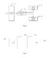
- FIG. 1shows a schematic diagram of an inspection device according to an embodiment of the present disclosure
- FIG. 2shows a schematic structure diagram of a computer in the inspection device as shown in FIG. 1 ;
- FIG. 3shows a schematic structure diagram of a controller in an inspection device according to an embodiment of the present disclosure
- FIG. 4shows a schematic diagram of an inspection device according to another embodiment of the present disclosure.
- FIG. 5shows a schematic structure diagram of the inspection device as shown in FIG. 4 .
- FIG. 1shows a schematic diagram of an inspection device according to an embodiment of the present disclosure.
- the inspection device according to the present embodimentis, in particular, a CT device for the field of quarantine.
- the CT devicemay include a gantry 20 , a bearing mechanism 40 , a controller 50 , a computer 60 , etc.
- the gantry 20may include a ray source 10 for emitting X-rays, such as an X-ray machine, and a detection and collection apparatus 30 .
- the X-ray sourcemay be enabled to emit continuous energy spectrum X-rays, and to perform a dual-energy CT scan or a multi-energy CT scan in combination with a dual-energy detector or an energy spectrum detector.
- the X-ray sourcemay be a dual-energy X-ray source
- the detectormay also receive dual-energy or multi-energy X-rays, so as to perform a dual-energy X-ray inspection on an object 70 being scanned.
- the bearing mechanism 40bears the object 70 being scanned to go through a scan area between the ray source 10 and the detection and collection apparatus 30 of the gantry 20 , during which the gantry 20 rotates around a heading direction of the object 70 being scanned so that the rays emitted from the ray source 10 can pass through the object 70 being scanned for performing the CT scan on the object 70 being scanned.
- the CT scan performed heremay be a spiral scan, or a circular scan, or the like.
- the detection and collection apparatus 30may be a detector and a data collector in an integrated module structure, e.g., an array detector, which may be used for detecting the rays passing through an article being scanned so as to obtain an analog signal, and for converting the analog signal to a digital signal, thereby outputting projection data of the object 70 being scanned for the X-rays.
- an integrated module structuree.g., an array detector
- the controller 50controls various components of the whole system to work synchronously.
- the computer 60processes and reconstructs data collected by the data collector, and outputs the result. For example, after the object 70 being scanned is scanned successively by the CT device, the obtained dual-energy 3D image data are input to the computer 60 ; a substance identification system installed in the computer 60 performs substance identification on the object 70 being scanned according to the image data so as to obtain feature information of the substance, such as an equivalent atomic number, density etc., and tints the 3D image, automatically labeling the article which is judged as an object focused in quarantine (plant, animal, meat, etc.).
- the ray source 10is arranged at one side of the object 70 being scanned; and the detection and collection apparatus 30 , including the detector and the data collector, is arranged at another side of the object 70 being scanned and is used for obtaining transmission data and/or multi-angle projection data of the object 70 being scanned.
- a data amplifying circuitis included in the data collector, which may work in a (current) integrated mode or a pulse (counting) mode.
- a data output cable of the detection and collection apparatus 30is connected to the controller 50 and the computer 60 , and the collected data are stored in the computer 60 according to a trigger command.
- a CT structure with a gantryis used in the illustrated embodiment, it will be understood by the skilled in the art that the ray source and the detector may utilize a CT structure without a gantry in other embodiments.
- FIG. 2shows a schematic structure diagram of a computer 60 as shown in FIG. 1 .
- the data collected by the data collectorare stored in a memory 61 via an interface unit 68 and a bus 64 .
- Configuration data and program of a computer data processorare stored in a ROM (Read-Only Memory) 62 .
- a RAM (Random-Access Memory) 63is used for temporarily storing various data during operations of a processor 66 .
- computer programs for data processingare also stored in the memory 61 , e.g., a substance identification program, an image processing program, etc.
- the internal bus 64connects the memory 61 , the ROM 62 , the RAM 63 , an input apparatus 65 , the processor 66 , a display apparatus 67 and the interface unit 68 .
- instruction codes of the computer programsinstruct the processor 66 to execute a predetermined data processing algorithm; and to display a data processing result on the display apparatus 67 such as a LCD display or to output the processing result directly in a form of a hard copy, such as printing, after the data processing result is obtained.
- the projection data obtained by the detection and collection apparatus 30are stored in the computer 60 for reconstruction of the CT image, so as to obtain slice image data of the object 70 being scanned. Then, the computer 60 extracts a 3D shape parameter of at least one article of the object 70 being scanned from the slice image data, for providing a judgment basis for judging whether the object 70 to be scanned is contraband.
- the CT imaging system as illustrated abovealso may be a dual-energy CT system, i.e., the X-ray source 10 on the gantry 20 may emit high-energy rays and low-energy rays.
- dual-energy CT reconstructionmay be performed by the computer data processor 60 to obtain the equivalent atomic numbers and/or density data of various slices of the object 70 being scanned.
- the computer 60may obtain a substance attribute of the object 70 being scanned, e.g. a plant or meat, for providing a judgment basis for judging whether the object 70 to be scanned is contraband.
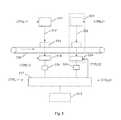
- FIG. 3shows a schematic structure diagram of a controller according to an embodiment of the present disclosure.
- the controller 50includes: a control unit 51 , configured to control the ray source 10 , the bearing mechanism 40 and the detection and collection apparatus 30 according to instructions from the computer 60 ; a trigger signal generation unit 52 , configured to generate a trigger command for triggering actions of the ray source 10 , the detection and collection apparatus 40 and the bearing mechanism 40 under control of the control unit; a first driving device 53 , configured to drive the bearing mechanism 40 to convey the object 70 being scanned according to the trigger command generated by the trigger signal generation unit 52 under the control of the control unit 51 ; and a second driving device 54 , configured to drive the gantry 20 to rotate according to the trigger command generated by the trigger signal generation unit 52 under the control of the control unit 51 .
- the projection data obtained by the detection and collection apparatus 30are stored in the computer 60 for reconstruction of the CT data, so as to obtain the slice image data of the object 70 being scanned. Then, the computer 60 identifies the atomic number of the substance by executing software, facilitating the work of the image judging officer.
- the CT imaging system as illustrated abovemay also be a dual-energy CT system, i.e., the X-ray source 10 on the gantry 20 may emit high-energy rays and low-energy rays. After the detection and collection apparatus 30 detects the projection data at different energy levels, the dual-energy CT reconstruction may be performed by the computer 60 to obtain the equivalent atomic numbers and/or density data of various slices of the object 70 being scanned.
- FIG. 4shows a schematic diagram of an inspection device according to another embodiment of the present disclosure.
- an object 410 being scannedis placed on a bearing structure 400 for inspection, which sequentially passes through a DR system 420 and a dual-energy CT system 430 .
- the dual-energy CT system 430 and the DR system 420may be operated synchronously.
- FIG. 5shows a schematic structure diagram of the inspection device as shown in FIG. 4 in detail.
- the inspection device as shown in FIG. 5includes a DR system on the left and a dual-energy CT system on the right, both of which share a bearing mechanism 530 , such as a belt, bearing an object 513 being scanned to move ahead.
- a bearing mechanism 530such as a belt
- An X-ray source 511 for DRemits X-rays 512 , which transmit through the object 513 being scanned on the bearing mechanism 530 ; a transmission signal is received by a detector module 514 ; an analog signal is converted by a collection circuit 515 into a digital signal, which is transmitted to a controller 517 and a computer 518 , etc.
- a transmission image of the object 513 being scannedis obtained in the computer 518 , which is stored in a memory or is displayed.
- the ray source 511may include a single X-ray source point, or include a plurality of X-ray generators, e.g., distributed X-ray sources including a plurality of X-ray source points.
- the bearing mechanism 530bears the object 513 being scanned to go through a scan area between the ray source 511 and the detector 514 .
- the detector 514 and the collection circuit 515are a detector and a data collector in an integrated module structure, e.g., a plurality of detectors, for detecting the rays passing through an article being scanned so as to obtain the analog signal, and for converting the analog signal to the digital signal, thereby outputting projection data of the object 513 being scanned for the X-rays.
- the controller 517controls various components of the whole system to work synchronously.
- the computer 518processes and reconstructs data collected by the data collector, and outputs the result.
- the detector 514 and the collection circuit 515are used for obtaining transmission data of the object 513 being scanned.
- a data amplifying circuitis included in the collection circuit 515 , which may work in a (current) integrated manner or a pulse (counting) manner.
- the collection circuit 515is connected to the controller 517 and the computer 518 , and the collected data are stored in the computer 60 according to a trigger command.
- the detector module 514may include a plurality of detection units for receiving the X-rays which pass through the object being scanned.
- the data collection circuit 515is coupled to the detector module 514 for converting a signal generated by the detector module 514 to detection data.
- the controller 517is connected via a control line CTRL 11 to the ray source 511 and is connected via a control line CTRL 12 to the detector module 514 which is in turn connected to the data collection circuit 515 , so as to control at least one X-ray generator of the ray source to generate the X-rays which are emitted for passing through the object being scanned as the object being scanned moves.
- the controller 517controls the detector module 514 and the data collection circuit 515 to obtain the detection data.
- the computer 518reconstructs the image of the object being scanned based on the detection data.
- the dual-energy CT systemperforms the CT scan on the object being scanned.
- a X-ray source 521 for CTemits X-rays 522 , which pass through the object 513 being scanned on the bearing mechanism 530 .
- the ray source 521 and a detector 524are rotated for the CT scan, a projection signal is received by the detector module 524 , an analog signal is converted by a collection circuit 525 into a digital signal, which is sent to the controller 517 and the computer 518 , etc.
- Slice images of the object 513 being scannedare obtained in the computer 518 , which are stored in the memory or are displayed.
- the detector module 524includes a plurality of detection units for receiving the X-rays which pass through the object being scanned.
- the data collection circuit 525is coupled to the detector module 524 for converting a signal generated by the detector module 524 to detection data.
- the controller 517is connected via a control line CTRL 21 to the ray source 521 and is connected via a control line CTRL 22 to the detector module 524 which is in turn connected to the data collection circuit 525 , so as to control two high and low energy X-ray generators of the ray source to alternately generate high and low energy X-rays which are emitted for passing through the object being scanned as the object being scanned moves, achieving a dual-energy CT scan.
- the controller 517controls the detector module 524 and the data collection circuit 525 to obtain the projection data.
- the computer 518reconstructs the image of the object being scanned based on the projection data, and performs the substance identification.
- the object 410 being scannedgoes through the DR system 420 and the dual-energy CT system 430 sequentially, and the obtained 2D and dual-energy 3D image data are input to the computer for substance identification.
- the substance identification system installed in the computerperforms substance identification on the object being scanned according to the image data so as to obtain information of different substances, such as the equivalent atomic numbers, densities, etc., and tints the 2D image and the 3D image, automatically labeling the article which is judged as the object focused in quarantine (plant, animal, meat, etc.).
- the substance identification systemmay hide a part of the object being scanned which is identified to be inorganic, so as to highlight one or more organic components of the object being scanned.
- the substance identification systemmay further identify and distinguish organics more accurately, respectively labeling plants, animals, meat and their products, etc. with different colors.
- the image which has been labeled by substance identificationis input to the image processing system in the computer 518 , which in turn highlights the suspect of quarantine, and alarms automatically.
- the image processing system in the computer 518may automatically segment the image of the object being scanned which comprises a mixture of various kinds of articles into separate articles according to their profile edges, facilitating the inspection officer to judge the image.
- the image processing system in the computer 518may compare an appearance of the 3D image of the object being scanned with a suspect image database in connection with the substance identification result, and further highlight the suspect of quarantine, so as to improve accuracy of an automatic alarm.
- the image processing system in the computer 518may have a self-learning function, and may automatically identify shape features of contrabands which are frequently labeled by a user and record them in the database, so as to improve accuracy of the automatic alarm.
- the image processing system in the computer 518may be configured with a cloud data collection function, e.g., may be connected to a cloud server for uploading the inspection data to the cloud server.
- the usermay authorize different image read rights of the cloud server to different persons, or enable the image read rights of the cloud server to have access to another management system.
- the image processing system in the computer 518may be configured with a database cloud update function, e.g., may be connected to the cloud server for updating an alarm database from the cloud.
Landscapes
- Physics & Mathematics (AREA)
- Life Sciences & Earth Sciences (AREA)
- General Physics & Mathematics (AREA)
- Geophysics (AREA)
- General Life Sciences & Earth Sciences (AREA)
- High Energy & Nuclear Physics (AREA)
- Health & Medical Sciences (AREA)
- Nuclear Medicine, Radiotherapy & Molecular Imaging (AREA)
- Radiology & Medical Imaging (AREA)
- General Health & Medical Sciences (AREA)
- Biochemistry (AREA)
- Analytical Chemistry (AREA)
- Chemical & Material Sciences (AREA)
- Immunology (AREA)
- Pathology (AREA)
- Theoretical Computer Science (AREA)
- Pulmonology (AREA)
- Engineering & Computer Science (AREA)
- Analysing Materials By The Use Of Radiation (AREA)
- Spectroscopy & Molecular Physics (AREA)
- Apparatus For Radiation Diagnosis (AREA)
Abstract
Description
Claims (20)
Applications Claiming Priority (3)
| Application Number | Priority Date | Filing Date | Title |
|---|---|---|---|
| CN201511009740.2ACN105527654B (en) | 2015-12-29 | 2015-12-29 | An inspection device for inspection and quarantine |
| CN201511009740.2 | 2015-12-29 | ||
| CN201511009740 | 2015-12-29 |
Publications (2)
| Publication Number | Publication Date |
|---|---|
| US20170184757A1 US20170184757A1 (en) | 2017-06-29 |
| US10416344B2true US10416344B2 (en) | 2019-09-17 |
Family
ID=55769989
Family Applications (1)
| Application Number | Title | Priority Date | Filing Date |
|---|---|---|---|
| US15/279,239Active2037-02-21US10416344B2 (en) | 2015-12-29 | 2016-09-28 | Inspection devices for quarantine |
Country Status (3)
| Country | Link |
|---|---|
| US (1) | US10416344B2 (en) |
| EP (1) | EP3187904B1 (en) |
| CN (1) | CN105527654B (en) |
Cited By (1)
| Publication number | Priority date | Publication date | Assignee | Title |
|---|---|---|---|---|
| US12306359B2 (en)* | 2021-11-26 | 2025-05-20 | Nuctech Company Limited | Area array detector, detection method, and corresponding container/vehicle inspection system |
Families Citing this family (8)
| Publication number | Priority date | Publication date | Assignee | Title |
|---|---|---|---|---|
| CN108254397B (en)* | 2017-12-12 | 2021-03-05 | 北京航星机器制造有限公司 | Luggage article safety inspection device and inspection method thereof |
| CN108780488B (en)* | 2018-03-22 | 2023-05-05 | 北京云端光科技术有限公司 | Method, system and device for determining uploading permission of substance detection information |
| US11039808B2 (en) | 2019-02-13 | 2021-06-22 | Analogic Corporation | Scanning systems configured to inspect conveyed objects and related systems and methods |
| CN109932376B (en)* | 2019-04-30 | 2023-11-28 | 王振 | Liquid detection method and device |
| US20210358242A1 (en)* | 2020-05-13 | 2021-11-18 | Weon Kook KIM | Quarantine Gate Apparatus For Supporting Quarantine Measures For A Facility To Be Accessed By Multiple Persons In An Non-Contact Manner |
| CN113514482B (en)* | 2021-05-07 | 2025-04-11 | 清华大学 | Static CT testing equipment |
| GB2608187B (en)* | 2021-06-25 | 2024-06-19 | Smiths Detection France S A S | X-ray inspection system and control architecture for an X-ray inspection system |
| JP7662923B2 (en) | 2023-06-15 | 2025-04-16 | 財務省大臣官房会計課長 | X-ray CT inspection system, X-ray CT inspection method and program |
Citations (25)
| Publication number | Priority date | Publication date | Assignee | Title |
|---|---|---|---|---|
| US4539648A (en)* | 1982-09-29 | 1985-09-03 | The United States Of America As Represented By The Secretary Of Agriculture | Detection of agricultural contraband in baggage |
| EP0223545A2 (en) | 1985-11-15 | 1987-05-27 | Picker International, Inc. | Energy dependent gain correction |
| US5367552A (en) | 1991-10-03 | 1994-11-22 | In Vision Technologies, Inc. | Automatic concealed object detection system having a pre-scan stage |
| US20050276376A1 (en) | 2004-05-27 | 2005-12-15 | L-3 Communications Security And Detection Systems, Inc. | Contraband detection systems using a large-angle cone beam CT system |
| CN201130157Y (en) | 2007-12-27 | 2008-10-08 | 同方威视技术股份有限公司 | Item detection device |
| US20090010386A1 (en)* | 2003-09-15 | 2009-01-08 | Peschmann Kristian R | Methods and Systems for Rapid Detection of Concealed Objects Using Fluorescence |
| CN101403710A (en) | 2007-10-05 | 2009-04-08 | 清华大学 | Liquid article inspection method and apparatus |
| US20090129544A1 (en)* | 2007-11-15 | 2009-05-21 | Zhiqiang Chen | Method and apparatus for substance identification |
| CN101470082A (en) | 2007-12-27 | 2009-07-01 | 同方威视技术股份有限公司 | Article detection apparatus and detection method thereof |
| CN101641589A (en) | 2008-01-15 | 2010-02-03 | 西门子公司 | Method and device for producing a tomosynthetic 3d x-ray image |
| EP2309257A1 (en) | 2008-03-27 | 2011-04-13 | Analogic Corporation | Method of and system for three-dimensional workstation for security and medical applications |
| CN102162798A (en) | 2007-10-05 | 2011-08-24 | 清华大学 | Method and equipment for inspecting liquid article |
| CN102435620A (en) | 2007-10-05 | 2012-05-02 | 清华大学 | Method and device for checking liquid articles |
| CN102565107A (en) | 2007-10-05 | 2012-07-11 | 清华大学 | Liquid article inspection method and equipment |
| CN102590234A (en) | 2009-05-27 | 2012-07-18 | 清华大学 | Dual-energy under-sampling substance identification system and method based on linear track scanning |
| WO2013078344A1 (en) | 2011-11-22 | 2013-05-30 | Xinray Systems Inc | High speed, small footprint x-ray tomography inspection systems, devices, and methods |
| CN103903297A (en) | 2012-12-27 | 2014-07-02 | 同方威视技术股份有限公司 | Three-dimensional data processing and identification method |
| CN103900503A (en) | 2012-12-27 | 2014-07-02 | 清华大学 | Shape characteristic extraction method, safety inspection method and device |
| CN103913472A (en) | 2012-12-31 | 2014-07-09 | 同方威视技术股份有限公司 | CT imaging system and method |
| CN103926628A (en) | 2014-04-22 | 2014-07-16 | 史崇政 | Security inspection device and method for identifying forbidden objects using same |
| CN204008508U (en) | 2014-06-25 | 2014-12-10 | 清华大学 | A kind of caliberating device of CT image and a kind of CT system |
| CN104435783A (en) | 2014-12-12 | 2015-03-25 | 济南木齐健康科技有限公司 | Energy health-preserving moxibustion compound and using method thereof |
| KR20150117417A (en) | 2014-04-10 | 2015-10-20 | 한국생산기술연구원 | Computed tomography image processing apparatus and method of generating three-dimensional reconstructed image |
| CN105004741A (en) | 2015-07-07 | 2015-10-28 | 中国检验检疫科学研究院 | Rapid and automatic quarantine contraband screening method based on X-ray article machine-based and device thereof |
| CN205353380U (en) | 2015-12-29 | 2016-06-29 | 中检科威(北京)科技有限公司 | Inspection device for inspection and quarantine |
Family Cites Families (2)
| Publication number | Priority date | Publication date | Assignee | Title |
|---|---|---|---|---|
| US20060269135A1 (en)* | 2005-03-15 | 2006-11-30 | Ramsay Thomas E | System and method for identifying objects of interest in image data |
| US8254656B2 (en)* | 2009-10-13 | 2012-08-28 | Morpho Detection, Inc. | Methods and system for selective resolution improvement in computed tomography |
- 2015
- 2015-12-29CNCN201511009740.2Apatent/CN105527654B/enactiveActive
- 2016
- 2016-09-28USUS15/279,239patent/US10416344B2/enactiveActive
- 2016-09-28EPEP16191227.4Apatent/EP3187904B1/enactiveActive
Patent Citations (32)
| Publication number | Priority date | Publication date | Assignee | Title |
|---|---|---|---|---|
| US4539648A (en)* | 1982-09-29 | 1985-09-03 | The United States Of America As Represented By The Secretary Of Agriculture | Detection of agricultural contraband in baggage |
| EP0223545A2 (en) | 1985-11-15 | 1987-05-27 | Picker International, Inc. | Energy dependent gain correction |
| US5367552A (en) | 1991-10-03 | 1994-11-22 | In Vision Technologies, Inc. | Automatic concealed object detection system having a pre-scan stage |
| US20090010386A1 (en)* | 2003-09-15 | 2009-01-08 | Peschmann Kristian R | Methods and Systems for Rapid Detection of Concealed Objects Using Fluorescence |
| US20050276376A1 (en) | 2004-05-27 | 2005-12-15 | L-3 Communications Security And Detection Systems, Inc. | Contraband detection systems using a large-angle cone beam CT system |
| CN101403710A (en) | 2007-10-05 | 2009-04-08 | 清华大学 | Liquid article inspection method and apparatus |
| US8036337B2 (en) | 2007-10-05 | 2011-10-11 | Tsinghua University | Method and device for inspection of liquid articles |
| US8320523B2 (en) | 2007-10-05 | 2012-11-27 | Tshinghua University | Method and device for inspection of liquid articles |
| CN102565107A (en) | 2007-10-05 | 2012-07-11 | 清华大学 | Liquid article inspection method and equipment |
| US20100284514A1 (en)* | 2007-10-05 | 2010-11-11 | Li Zhang | Method and device for inspection of liquid articles |
| CN102435620A (en) | 2007-10-05 | 2012-05-02 | 清华大学 | Method and device for checking liquid articles |
| CN102162798A (en) | 2007-10-05 | 2011-08-24 | 清华大学 | Method and equipment for inspecting liquid article |
| US20090129544A1 (en)* | 2007-11-15 | 2009-05-21 | Zhiqiang Chen | Method and apparatus for substance identification |
| CN201130157Y (en) | 2007-12-27 | 2008-10-08 | 同方威视技术股份有限公司 | Item detection device |
| CN101470082A (en) | 2007-12-27 | 2009-07-01 | 同方威视技术股份有限公司 | Article detection apparatus and detection method thereof |
| CN101641589A (en) | 2008-01-15 | 2010-02-03 | 西门子公司 | Method and device for producing a tomosynthetic 3d x-ray image |
| EP2309257A1 (en) | 2008-03-27 | 2011-04-13 | Analogic Corporation | Method of and system for three-dimensional workstation for security and medical applications |
| CN102590234A (en) | 2009-05-27 | 2012-07-18 | 清华大学 | Dual-energy under-sampling substance identification system and method based on linear track scanning |
| WO2013078344A1 (en) | 2011-11-22 | 2013-05-30 | Xinray Systems Inc | High speed, small footprint x-ray tomography inspection systems, devices, and methods |
| US9465975B2 (en) | 2012-12-27 | 2016-10-11 | Nuctech Company Limited | Three-dimensional data processing and recognizing method involving inspecting an object and extracting data matching to features |
| CN103903297A (en) | 2012-12-27 | 2014-07-02 | 同方威视技术股份有限公司 | Three-dimensional data processing and identification method |
| CN103900503A (en) | 2012-12-27 | 2014-07-02 | 清华大学 | Shape characteristic extraction method, safety inspection method and device |
| US10102641B2 (en) | 2012-12-27 | 2018-10-16 | Tsinghua University | Methods for extracting shape feature, inspection methods and apparatuses |
| US9412019B2 (en) | 2012-12-27 | 2016-08-09 | Tsinghua University | Methods for extracting shape feature, inspection methods and apparatuses |
| CN103913472A (en) | 2012-12-31 | 2014-07-09 | 同方威视技术股份有限公司 | CT imaging system and method |
| US9495772B2 (en) | 2012-12-31 | 2016-11-15 | Nuctech Company Limited | CT imaging systems and methods thereof |
| KR20150117417A (en) | 2014-04-10 | 2015-10-20 | 한국생산기술연구원 | Computed tomography image processing apparatus and method of generating three-dimensional reconstructed image |
| CN103926628A (en) | 2014-04-22 | 2014-07-16 | 史崇政 | Security inspection device and method for identifying forbidden objects using same |
| CN204008508U (en) | 2014-06-25 | 2014-12-10 | 清华大学 | A kind of caliberating device of CT image and a kind of CT system |
| CN104435783A (en) | 2014-12-12 | 2015-03-25 | 济南木齐健康科技有限公司 | Energy health-preserving moxibustion compound and using method thereof |
| CN105004741A (en) | 2015-07-07 | 2015-10-28 | 中国检验检疫科学研究院 | Rapid and automatic quarantine contraband screening method based on X-ray article machine-based and device thereof |
| CN205353380U (en) | 2015-12-29 | 2016-06-29 | 中检科威(北京)科技有限公司 | Inspection device for inspection and quarantine |
Non-Patent Citations (11)
| Title |
|---|
| Examination Report as issued in Australian Patent Application No. 2016235025 dated Aug. 28, 2017. |
| Extended European Search Report as issued in European Patent Application No. 16191227.4, dated May 17, 2017. |
| Final Office Action as issued in U.S. Appl. No. 15/279,207, dated Jan. 18, 2019. |
| International Search Report and the Written Opinion of the International Searching Authority as issued in International Patent Application No. PCT/CN2016/097577, dated Nov. 24, 2016. |
| Notice of Allowance as issued in U.S. Appl. No. 15/279,207, dated May 24, 2019. |
| Office Action as issued in Canadian Patent Application No. 2,943,764, dated Jul. 26, 2018. |
| Office Action as issued in Chinese Patent Application No. 201511009740.2, dated Feb. 24, 2018. |
| Office Action as issued in Chinese Patent Application No. 201511009740.2, dated May 4, 2017. |
| Office Action as issued in Chinese Patent Application No. 201511010081.4, dated Dec. 29, 2018. |
| Office Action as issued in U.S. Appl. No. 15/279,207, dated Jul. 27, 2018. |
| Taina, I. A., et al., "Application of X-ray computed tomography to soil science: A literature review," Canadian Journal of Soil Science, Department of Land Resource Science, University of Guelph, <http://www.nrcresearchpress.com/doi/10.4141/CJSS06027#.W1XTGdVKhEZ>, Oct. 2, 2007, 25 pages. |
Cited By (1)
| Publication number | Priority date | Publication date | Assignee | Title |
|---|---|---|---|---|
| US12306359B2 (en)* | 2021-11-26 | 2025-05-20 | Nuctech Company Limited | Area array detector, detection method, and corresponding container/vehicle inspection system |
Also Published As
| Publication number | Publication date |
|---|---|
| US20170184757A1 (en) | 2017-06-29 |
| CN105527654B (en) | 2019-05-03 |
| EP3187904B1 (en) | 2021-03-24 |
| EP3187904A1 (en) | 2017-07-05 |
| CN105527654A (en) | 2016-04-27 |
Similar Documents
| Publication | Publication Date | Title |
|---|---|---|
| US10416344B2 (en) | Inspection devices for quarantine | |
| US10436932B2 (en) | Inspection systems for quarantine and methods thereof | |
| US10520452B2 (en) | Automated quality control and selection | |
| US7492862B2 (en) | Computed tomography cargo inspection system and method | |
| CN105784731B (en) | A method and security inspection system for locating a target in a three-dimensional CT image | |
| US9865066B2 (en) | Computed tomography system for cargo and transported containers | |
| RU2400735C2 (en) | Method of inspecting cargo using translucence at different angles | |
| US7529341B2 (en) | Automatic material discrimination by using computer tomography | |
| CN105806856B (en) | Dual Energy Ray Imaging Method and System | |
| US9552521B2 (en) | Human body security inspection method and system | |
| US20100277312A1 (en) | In-line high-throughput contraband detection system | |
| US9546968B2 (en) | Meat assessment device | |
| US8180139B2 (en) | Method and system for inspection of containers | |
| US20080181357A1 (en) | Combined computed tomography and nuclear resonance fluorescence cargo inspection system and method | |
| CN205353380U (en) | Inspection device for inspection and quarantine | |
| AU2017376794A1 (en) | Dual-energy microfocus radiographic imaging system and method for poultry and meat inspection | |
| EP3112852B1 (en) | Method for positioning target in three-dimensional ct image and security check system | |
| EP3112909B1 (en) | Method for positioning target in three-dimensional ct image and security check ct system | |
| US20110186739A1 (en) | Mobile tomographic cargo inspection system | |
| DK181309B1 (en) | A system and a method for determination of the tissue composition in meat products | |
| WO2023209332A1 (en) | A method of analysing a geological sample | |
| HK1217796A1 (en) | Image display methods | |
| Jansen et al. | Validation of X-ray for borehole detection in intact trees |
Legal Events
| Date | Code | Title | Description |
|---|---|---|---|
| AS | Assignment | Owner name:NUCTECH COMPANY LIMITED, CHINA Free format text:ASSIGNMENT OF ASSIGNORS INTEREST;ASSIGNORS:MIAO, QITIAN;CHEN, ZHIQIANG;ZHANG, LI;AND OTHERS;REEL/FRAME:048626/0418 Effective date:20190315 Owner name:CINTS CO. LTD., CHINA Free format text:ASSIGNMENT OF ASSIGNORS INTEREST;ASSIGNORS:MIAO, QITIAN;CHEN, ZHIQIANG;ZHANG, LI;AND OTHERS;REEL/FRAME:048626/0418 Effective date:20190315 | |
| STPP | Information on status: patent application and granting procedure in general | Free format text:PUBLICATIONS -- ISSUE FEE PAYMENT VERIFIED | |
| STPP | Information on status: patent application and granting procedure in general | Free format text:DOCKETED NEW CASE - READY FOR EXAMINATION | |
| STPP | Information on status: patent application and granting procedure in general | Free format text:NOTICE OF ALLOWANCE MAILED -- APPLICATION RECEIVED IN OFFICE OF PUBLICATIONS | |
| STPP | Information on status: patent application and granting procedure in general | Free format text:AWAITING TC RESP., ISSUE FEE NOT PAID | |
| STPP | Information on status: patent application and granting procedure in general | Free format text:PUBLICATIONS -- ISSUE FEE PAYMENT VERIFIED | |
| STCF | Information on status: patent grant | Free format text:PATENTED CASE | |
| CC | Certificate of correction | ||
| MAFP | Maintenance fee payment | Free format text:PAYMENT OF MAINTENANCE FEE, 4TH YEAR, LARGE ENTITY (ORIGINAL EVENT CODE: M1551); ENTITY STATUS OF PATENT OWNER: LARGE ENTITY Year of fee payment:4 |