US10413636B2 - Tissue scaffolds - Google Patents
Tissue scaffoldsDownload PDFInfo
- Publication number
- US10413636B2 US10413636B2US12/540,489US54048909AUS10413636B2US 10413636 B2US10413636 B2US 10413636B2US 54048909 AUS54048909 AUS 54048909AUS 10413636 B2US10413636 B2US 10413636B2
- Authority
- US
- United States
- Prior art keywords
- tissue matrix
- acellular tissue
- slurry
- tissue
- sodium acetate
- Prior art date
- Legal status (The legal status is an assumption and is not a legal conclusion. Google has not performed a legal analysis and makes no representation as to the accuracy of the status listed.)
- Expired - Fee Related, expires
Links
- 239000002407tissue scaffoldSubstances0.000titleclaimsabstractdescription75
- 238000000034methodMethods0.000claimsabstractdescription40
- 239000011159matrix materialSubstances0.000claimsdescription57
- 239000002002slurrySubstances0.000claimsdescription37
- VMHLLURERBWHNL-UHFFFAOYSA-MSodium acetateChemical compound[Na+].CC([O-])=OVMHLLURERBWHNL-UHFFFAOYSA-M0.000claimsdescription35
- 235000017281sodium acetateNutrition0.000claimsdescription33
- 239000001632sodium acetateSubstances0.000claimsdescription33
- 239000000243solutionSubstances0.000claimsdescription21
- 238000005266castingMethods0.000claimsdescription17
- 239000012530fluidSubstances0.000claimsdescription14
- 238000001816coolingMethods0.000claimsdescription13
- 239000000203mixtureSubstances0.000claimsdescription13
- UIIMBOGNXHQVGW-UHFFFAOYSA-MSodium bicarbonateChemical compound[Na+].OC([O-])=OUIIMBOGNXHQVGW-UHFFFAOYSA-M0.000claimsdescription12
- 239000007864aqueous solutionSubstances0.000claimsdescription12
- 230000002500effect on skinEffects0.000claimsdescription10
- 238000007710freezingMethods0.000claimsdescription10
- 230000008014freezingEffects0.000claimsdescription10
- 238000000265homogenisationMethods0.000claimsdescription9
- 108010035532CollagenProteins0.000claimsdescription8
- 102000008186CollagenHuman genes0.000claimsdescription8
- 229920001436collagenPolymers0.000claimsdescription8
- 229910000030sodium bicarbonateInorganic materials0.000claimsdescription6
- 235000017557sodium bicarbonateNutrition0.000claimsdescription6
- 230000002792vascularEffects0.000claimsdescription6
- 108010014258ElastinProteins0.000claimsdescription4
- 102000016942ElastinHuman genes0.000claimsdescription4
- 229920002549elastinPolymers0.000claimsdescription4
- 230000036571hydrationEffects0.000claimsdescription3
- 238000006703hydration reactionMethods0.000claimsdescription3
- 206010052428WoundDiseases0.000abstractdescription24
- 208000027418Wounds and injuryDiseases0.000abstractdescription24
- 210000001519tissueAnatomy0.000description74
- 239000000523sampleSubstances0.000description31
- 239000000463materialSubstances0.000description26
- 239000011148porous materialSubstances0.000description19
- 238000011282treatmentMethods0.000description15
- 239000000853adhesiveSubstances0.000description14
- 230000001070adhesive effectEffects0.000description14
- 210000001612chondrocyteAnatomy0.000description12
- 230000035699permeabilityEffects0.000description12
- 108010023728AllodermProteins0.000description11
- 210000000845cartilageAnatomy0.000description11
- 238000002560therapeutic procedureMethods0.000description11
- 238000012545processingMethods0.000description9
- 238000011069regeneration methodMethods0.000description8
- 239000000725suspensionSubstances0.000description8
- 230000017423tissue regenerationEffects0.000description8
- 230000008929regenerationEffects0.000description7
- 210000003491skinAnatomy0.000description7
- 210000000988bone and boneAnatomy0.000description6
- 230000007547defectEffects0.000description6
- 210000004027cellAnatomy0.000description5
- IJGRMHOSHXDMSA-UHFFFAOYSA-NAtomic nitrogenChemical compoundN#NIJGRMHOSHXDMSA-UHFFFAOYSA-N0.000description4
- 230000008859changeEffects0.000description4
- 230000000694effectsEffects0.000description4
- 239000006260foamSubstances0.000description4
- 230000035876healingEffects0.000description4
- 230000008467tissue growthEffects0.000description4
- XLYOFNOQVPJJNP-UHFFFAOYSA-NwaterSubstancesOXLYOFNOQVPJJNP-UHFFFAOYSA-N0.000description4
- 206010017076FractureDiseases0.000description3
- 208000013201Stress fractureDiseases0.000description3
- 210000002808connective tissueAnatomy0.000description3
- 210000004207dermisAnatomy0.000description3
- 238000009826distributionMethods0.000description3
- 239000000499gelSubstances0.000description3
- 238000011534incubationMethods0.000description3
- 239000007788liquidSubstances0.000description3
- 210000005036nerveAnatomy0.000description3
- 239000000758substrateSubstances0.000description3
- RYGMFSIKBFXOCR-UHFFFAOYSA-NCopperChemical compound[Cu]RYGMFSIKBFXOCR-UHFFFAOYSA-N0.000description2
- 239000000427antigenSubstances0.000description2
- 102000036639antigensHuman genes0.000description2
- 108091007433antigensProteins0.000description2
- 210000004204blood vesselAnatomy0.000description2
- 238000004891communicationMethods0.000description2
- 229910052802copperInorganic materials0.000description2
- 239000010949copperSubstances0.000description2
- 230000003247decreasing effectEffects0.000description2
- 210000003195fasciaAnatomy0.000description2
- 210000002950fibroblastAnatomy0.000description2
- 238000005187foamingMethods0.000description2
- 239000007789gasSubstances0.000description2
- 230000012010growthEffects0.000description2
- 210000003709heart valveAnatomy0.000description2
- 230000002706hydrostatic effectEffects0.000description2
- 239000007943implantSubstances0.000description2
- 208000015181infectious diseaseDiseases0.000description2
- 210000003041ligamentAnatomy0.000description2
- 229910052757nitrogenInorganic materials0.000description2
- 230000008439repair processEffects0.000description2
- 238000002791soakingMethods0.000description2
- 210000004872soft tissueAnatomy0.000description2
- 210000002435tendonAnatomy0.000description2
- 108091003079Bovine Serum AlbuminProteins0.000description1
- 102000029816CollagenaseHuman genes0.000description1
- 108060005980CollagenaseProteins0.000description1
- 239000006144Dulbecco’s modified Eagle's mediumSubstances0.000description1
- 206010063560Excessive granulation tissueDiseases0.000description1
- 102000009123FibrinHuman genes0.000description1
- 108010073385FibrinProteins0.000description1
- BWGVNKXGVNDBDI-UHFFFAOYSA-NFibrin monomerChemical compoundCNC(=O)CNC(=O)CNBWGVNKXGVNDBDI-UHFFFAOYSA-N0.000description1
- 241001465754MetazoaSpecies0.000description1
- 206010028980NeoplasmDiseases0.000description1
- 241001494479PecoraSpecies0.000description1
- 208000025865UlcerDiseases0.000description1
- 239000003522acrylic cementSubstances0.000description1
- 210000000577adipose tissueAnatomy0.000description1
- QIGJYVCQYDKYDW-SDOYDPJRSA-Nalpha-D-galactosyl-(1->3)-D-galactoseChemical groupO[C@@H]1[C@@H](O)[C@@H](O)[C@@H](CO)O[C@@H]1O[C@H]1[C@@H](O)[C@@H](CO)OC(O)[C@@H]1OQIGJYVCQYDKYDW-SDOYDPJRSA-N0.000description1
- 230000000890antigenic effectEffects0.000description1
- 238000013459approachMethods0.000description1
- 230000009286beneficial effectEffects0.000description1
- 239000012620biological materialSubstances0.000description1
- 210000001185bone marrowAnatomy0.000description1
- 230000010478bone regenerationEffects0.000description1
- 210000000481breastAnatomy0.000description1
- 230000003848cartilage regenerationEffects0.000description1
- 239000004568cementSubstances0.000description1
- 239000011248coating agentSubstances0.000description1
- 238000000576coating methodMethods0.000description1
- 229960002424collagenaseDrugs0.000description1
- 239000000084colloidal systemSubstances0.000description1
- 239000002537cosmeticSubstances0.000description1
- 238000002316cosmetic surgeryMethods0.000description1
- 230000002950deficientEffects0.000description1
- 230000004069differentiationEffects0.000description1
- 230000029087digestionEffects0.000description1
- 239000012091fetal bovine serumSubstances0.000description1
- 229950003499fibrinDrugs0.000description1
- 230000009969flowable effectEffects0.000description1
- 238000004108freeze dryingMethods0.000description1
- 239000003292glueSubstances0.000description1
- 210000001126granulation tissueAnatomy0.000description1
- 239000003102growth factorSubstances0.000description1
- 238000010438heat treatmentMethods0.000description1
- 210000002758humerusAnatomy0.000description1
- 230000028993immune responseEffects0.000description1
- 238000002513implantationMethods0.000description1
- 230000008595infiltrationEffects0.000description1
- 238000001764infiltrationMethods0.000description1
- 238000011221initial treatmentMethods0.000description1
- 208000014674injuryDiseases0.000description1
- 230000010354integrationEffects0.000description1
- 230000007794irritationEffects0.000description1
- 230000007774longtermEffects0.000description1
- 238000012792lyophilization processMethods0.000description1
- 230000014759maintenance of locationEffects0.000description1
- 238000004519manufacturing processMethods0.000description1
- 239000002609mediumSubstances0.000description1
- 238000002844meltingMethods0.000description1
- 230000008018meltingEffects0.000description1
- 239000012528membraneSubstances0.000description1
- 229910052751metalInorganic materials0.000description1
- 239000002184metalSubstances0.000description1
- 238000012986modificationMethods0.000description1
- 230000004048modificationEffects0.000description1
- 210000003205muscleAnatomy0.000description1
- 230000001537neural effectEffects0.000description1
- 210000000056organAnatomy0.000description1
- 230000003239periodontal effectEffects0.000description1
- 230000000704physical effectEffects0.000description1
- 229920000642polymerPolymers0.000description1
- 238000002360preparation methodMethods0.000description1
- 230000009467reductionEffects0.000description1
- 238000007634remodelingMethods0.000description1
- 239000011347resinSubstances0.000description1
- 229920005989resinPolymers0.000description1
- 239000000126substanceSubstances0.000description1
- 230000003685thermal hair damageEffects0.000description1
- 230000008733traumaEffects0.000description1
- 231100000397ulcerToxicity0.000description1
- 210000000689upper legAnatomy0.000description1
- 230000029663wound healingEffects0.000description1
Images


Classifications
- A—HUMAN NECESSITIES
- A61—MEDICAL OR VETERINARY SCIENCE; HYGIENE
- A61L—METHODS OR APPARATUS FOR STERILISING MATERIALS OR OBJECTS IN GENERAL; DISINFECTION, STERILISATION OR DEODORISATION OF AIR; CHEMICAL ASPECTS OF BANDAGES, DRESSINGS, ABSORBENT PADS OR SURGICAL ARTICLES; MATERIALS FOR BANDAGES, DRESSINGS, ABSORBENT PADS OR SURGICAL ARTICLES
- A61L27/00—Materials for grafts or prostheses or for coating grafts or prostheses
- A61L27/36—Materials for grafts or prostheses or for coating grafts or prostheses containing ingredients of undetermined constitution or reaction products thereof, e.g. transplant tissue, natural bone, extracellular matrix
- A61L27/3683—Materials for grafts or prostheses or for coating grafts or prostheses containing ingredients of undetermined constitution or reaction products thereof, e.g. transplant tissue, natural bone, extracellular matrix subjected to a specific treatment prior to implantation, e.g. decellularising, demineralising, grinding, cellular disruption/non-collagenous protein removal, anti-calcification, crosslinking, supercritical fluid extraction, enzyme treatment
- A—HUMAN NECESSITIES
- A61—MEDICAL OR VETERINARY SCIENCE; HYGIENE
- A61L—METHODS OR APPARATUS FOR STERILISING MATERIALS OR OBJECTS IN GENERAL; DISINFECTION, STERILISATION OR DEODORISATION OF AIR; CHEMICAL ASPECTS OF BANDAGES, DRESSINGS, ABSORBENT PADS OR SURGICAL ARTICLES; MATERIALS FOR BANDAGES, DRESSINGS, ABSORBENT PADS OR SURGICAL ARTICLES
- A61L27/00—Materials for grafts or prostheses or for coating grafts or prostheses
- A61L27/36—Materials for grafts or prostheses or for coating grafts or prostheses containing ingredients of undetermined constitution or reaction products thereof, e.g. transplant tissue, natural bone, extracellular matrix
- A61L27/3604—Materials for grafts or prostheses or for coating grafts or prostheses containing ingredients of undetermined constitution or reaction products thereof, e.g. transplant tissue, natural bone, extracellular matrix characterised by the human or animal origin of the biological material, e.g. hair, fascia, fish scales, silk, shellac, pericardium, pleura, renal tissue, amniotic membrane, parenchymal tissue, fetal tissue, muscle tissue, fat tissue, enamel
- A61L27/362—Skin, e.g. dermal papillae
- A—HUMAN NECESSITIES
- A61—MEDICAL OR VETERINARY SCIENCE; HYGIENE
- A61L—METHODS OR APPARATUS FOR STERILISING MATERIALS OR OBJECTS IN GENERAL; DISINFECTION, STERILISATION OR DEODORISATION OF AIR; CHEMICAL ASPECTS OF BANDAGES, DRESSINGS, ABSORBENT PADS OR SURGICAL ARTICLES; MATERIALS FOR BANDAGES, DRESSINGS, ABSORBENT PADS OR SURGICAL ARTICLES
- A61L27/00—Materials for grafts or prostheses or for coating grafts or prostheses
- A61L27/36—Materials for grafts or prostheses or for coating grafts or prostheses containing ingredients of undetermined constitution or reaction products thereof, e.g. transplant tissue, natural bone, extracellular matrix
- A61L27/3604—Materials for grafts or prostheses or for coating grafts or prostheses containing ingredients of undetermined constitution or reaction products thereof, e.g. transplant tissue, natural bone, extracellular matrix characterised by the human or animal origin of the biological material, e.g. hair, fascia, fish scales, silk, shellac, pericardium, pleura, renal tissue, amniotic membrane, parenchymal tissue, fetal tissue, muscle tissue, fat tissue, enamel
- A61L27/3633—Extracellular matrix [ECM]
- A—HUMAN NECESSITIES
- A61—MEDICAL OR VETERINARY SCIENCE; HYGIENE
- A61L—METHODS OR APPARATUS FOR STERILISING MATERIALS OR OBJECTS IN GENERAL; DISINFECTION, STERILISATION OR DEODORISATION OF AIR; CHEMICAL ASPECTS OF BANDAGES, DRESSINGS, ABSORBENT PADS OR SURGICAL ARTICLES; MATERIALS FOR BANDAGES, DRESSINGS, ABSORBENT PADS OR SURGICAL ARTICLES
- A61L2430/00—Materials or treatment for tissue regeneration
- A61L2430/40—Preparation and treatment of biological tissue for implantation, e.g. decellularisation, cross-linking
Definitions
- Reduced pressure, or vacuum-assisted, therapiescan be effective for improving wound healing due to a variety of different causes and at a number of different anatomical locations.
- reduced pressure therapiesinclude a porous material that is placed at a wound site, which aids in the distribution of the reduced pressure.
- Typical porous materialsare sized to fit the wound, and may be periodically replaced with smaller pieces of the porous material as the wound begins to heal and becomes smaller.
- a membrane or drapeis placed over the porous material to provide an airtight seal at the wound area, and a negative pressure is applied to the porous material to provide a reduced pressure at the wound site.
- a method of processing an acellular tissue matrix for preparing a tissue scaffoldcomprising adding an acellular tissue matrix to a first aqueous solution of sodium acetate; incubating the first aqueous sodium acetate solution containing the acellular tissue matrix; removing the incubated acellular tissue matrix from the first aqueous sodium acetate solution; treating the incubated acellular tissue matrix with a second aqueous solution of sodium acetate to form a suspension; homogenizing the suspension to form a slurry; cooling the slurry; casting the slurry in a casting container; and lyophilizing the slurry.
- a tissue scaffoldcomprising an acellular tissue matrix and sodium acetate is provided.
- a wound treatment devicecomprising a reduced pressure source and a tissue scaffold is provided.
- a tissue scaffoldcomprising an acellular tissue matrix that has been processed to have a porosity of between 75% and 90% is provided.
- FIG. 1illustrates a wound treatment device, which provides reduced pressure therapy, according to certain exemplary embodiments.
- FIG. 2illustrates a method of treating a cartilage defect using a tissue scaffold, according to certain embodiments.
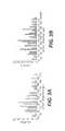
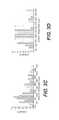
- FIGS. 3A-3Dare graphs showing the strut spacing for tissue scaffolds, as described in Example 1.
- FIG. 4is a graph showing the permeability versus composition for tissue scaffolds, as described in Example 2.
- FIG. 5is a graph showing the strut spacing for a tissue scaffold produced according to certain exemplary embodiments, as described in Example 3.
- FIGS. 6A-6Bare photomicrographs of chondrocytes cultured on tissue scaffolds, as described in Example 4.
- FIGS. 7A-7Bare photomicrographs of chondrocytes cultured on tissue scaffolds, as described in Example 4.
- the present disclosurepertains to a method of processing an acelluar tissue matrix for preparing a tissue scaffold.
- the tissue scaffold of the present disclosuremay be used as part of a wound treatment device that provides reduced pressure therapy.
- the physical properties of certain tissue scaffoldssuch as porosity, strut density, and permeability, may be controlled or altered by adjusting the concentrations, components, and temperatures at which the scaffolds are produced.
- acellular tissue matrixrefers generally to any tissue matrix that is substantially free of cells and other antigenic material.
- acellular tissue matrices derived from human or xenogenic sourcesmay be used to produce the scaffolds.
- Skin, parts of skin (e.g., dermis), and other tissuessuch as blood vessels, heart valves, fascia, nerve, or other collagen containing-organ or tissue may be used to create an acellular matrices to produce tissues scaffolds within the scope of the present disclosure.
- the term “permeability”refers generally to the movement of fluid through a porous medium.
- the specific permeability values of particular tissue scaffoldsare calculated by Darcy's Law:
- Qequals the total discharge (units of volume per time, e.g., m 2 /s)
- Ais the cross-sectional area to flow
- ⁇ Pis the pressure drop across the system
- ⁇is the dynamic viscosity (in SI units e.g. kg/(m ⁇ s) or Pa's)
- (l)is the length over which the pressure drop is taking place over.
- reduced pressuregenerally refers to a pressure less than the ambient pressure at a tissue site that is being subjected to treatment. In most cases, this reduced pressure will be less than the atmospheric pressure at which the patient is located. Alternatively, the reduced pressure may be less than a hydrostatic pressure of tissue at the tissue site. Reduced pressure may initially generate fluid flow in the area of the tissue site and/or a fluid conduit in communication with the tissue site, for example, as shown in FIG. 1 . As the hydrostatic pressure around the tissue site approaches the desired reduced pressure, the flow may subside, and the reduced pressure is then maintained. In some embodiments, small amounts of gas can be introduced at intervals to facilitate fluid movement if required. Unless otherwise indicated, values of pressure stated herein are gage pressures.
- fluidgenerally refers to a gas or liquid, but may also include any other flowable material, including but not limited to gels, colloids, and foams.
- tissue scaffoldscan be used in a wide array of applications.
- Certain exemplary applicationsinclude, but are not limited to, absorptive dressing, dermal regeneration (for example, for treatments of all types of ulcers and burns), nerve regeneration, cartilage regeneration, connective tissue regeneration or repair (for example, tendon/ligament sleeve), bone regeneration, periodontal applications, wound/foam lining, integrated bandage dressing, substrate/base for skin grafts, vascular regeneration, cosmetic surgery, cosmetic injectable gel, metal and/or polymer implant coating (for example, to increase implant integration and biocompatibility), and replacement of lost tissue (e.g., after trauma, breast reduction, mastectomy, lumpectomy, parotidectomy, or excision of tumors).
- FIG. 1illustrates a wound treatment device 100 , including a reduced pressure source 120 , according to certain exemplary embodiments.
- suitable reduced pressure therapy devicesinclude V.A.C.® therapy devices produced by Kinetic Concepts, Inc. (San Antonio, Tex.).
- Such reduced pressure therapy devicescan include a vacuum pump that can be fluidly connected to the wound site 150 , via a fluid conduit 125 or other fluid connection.
- Such devicesmay also include a flexible sheet 160 to cover the wound site 150 and at least partially seal the wound to allow reduced pressure therapy to be provided at the wound site.
- tissue scaffold 180may be placed at the wound site and facilitates wound closure, healing, tissue regeneration or repair, prevents or treats infection, and/or has other beneficial effects.
- the tissue scaffold 180assists in distributing fluid flow or negative pressure across a site to be treated.
- the flexible sheet 160will include a flexible polymeric material.
- any suitable polymeric materialcan be selected.
- the materialdoes not cause significant irritation, immune response, or heightened risk of infection.
- the specific materialgenerally should be of sufficient thickness and impermeability to allow reduced pressure therapy at a wound site under the sheet 160 .
- the device 100will include an adhesive.
- adhesivewill be understood to refer to any substance that causes the surfaces of two objects to be attached to one another.
- suitable adhesivescan include a variety of different cements, glues, resins, or other materials that can facilitate attachment of the flexible sheet 160 to tissue.
- the adhesivecan include a pressure-sensitive acrylic adhesive.
- the adhesivescan be applied directly to the structures to be joined, or the adhesives may be applied on tape, or with other supporting substrate materials.
- the adhesivecan be applied to a surface of the flexible sheet 160 to attach the sheet to skin or other tissue. In some embodiments, the adhesive will be applied to the surface of the sheet and packaged and/or distributed with the sheet 160 . In some embodiments, the adhesive is applied to a surface of the sheet 160 and covered by a non-adhesive material that can be removed to expose the adhesive for use. In certain embodiments, the adhesive can be supplied as a separate component (e.g., in a container or on a tape) that is applied to the sheet 160 to attach the sheet 160 to tissue. In some embodiments, the adhesive can be applied to a patient's skin or other tissue, and the sheet can be applied to the adhesive.
- tissue scaffold 180can include a variety of suitable materials. For example, a number of different tissue scaffolds will be compatible for use with the above-noted V.A.C.® treatment systems.
- the tissue scaffoldmay comprise a processed acellular tissue matrix.
- the acellular tissue matrixmay be derived from human skin or from a xenogenic source.
- other tissuessuch as blood vessels, heart valves, fascia, nerve, connective tissue, or other collagen-containing organs or tissues may be used to create a specific acellular matrix within the scope of the present disclosure.
- the acellular tissue matrixis an acellular dermal matrix.
- the acellular dermal matrixis produced from human dermis or pig dermis.
- the methods disclosed hereinutilize a dehydrated acellular tissue matrix tissue, such as ALLODERM®, which is commercially available from LifeCell Corporation, Branchburg, N.J.
- the methods disclosed hereinutilize an acellular tissue matrix tissue, such as STRATTICETM, which is commercially available from LifeCell Corporation, Branchburg, N.J.
- acellular tissue matricescan be produced using a variety of different tissue processing techniques. For example, certain exemplary tissue processing techniques for producing acellular tissue matrices are described in U.S. Pat. Nos. 5,336,616 and 5,364,756, both to Livesey et al., in U.S. Pat. No. 6,933,326 to Schiff et al., each of which is incorporated by reference in its entirety.
- acellular tissue matrices made from non-human animalscan be treated to remove various antigens, or produced from animals genetically modified to lack certain antigens.
- FIG. 2illustrates use of a tissue scaffold to treat a cartilage defect, according to certain embodiments.
- a scaffold 180is used to treat a cartilage defect in a long bone (e.g., femur or humerus).
- a scaffold 180can be used to treat an articular surface or cartilage 510 of any joint.
- the tissue scaffold 180is placed in a defect or excised area of an articular surface or cartilage 510 , and a negative pressure is applied to the tissue scaffold 180 through a fluid conduit 125 , as described above.
- a second material 190is applied over the tissue scaffold 180 , and the second material 190 acts as a manifold to distribute pressure to tissue scaffold 180 .
- the fluid conduitis in fluid communication with the tissue scaffold 180 without a second material 180 .
- the tissue scaffoldcan be used as a primary treatment method or in connection with another procedure or treatment.
- cartilage repair or regenerationcan be performed using a technique known in the art as microfracture. As shown in FIG. 2 , during a microfracture procedure, a surgeon creates small fractures or openings 515 in bone adjacent to an articular defect. In various instances, the fractures or openings 515 can allow chondrocytes or other cells that can differentiate into chondrocytes to migrate to the articular defect from adjacent bone, bone marrow space, or cartilage. In various instances, these cells can, in turn, help repair or regenerate cartilage.
- the tissue scaffold 180is placed over the microfracture site, and negative pressure is applied to the scaffold.
- the tissue scaffoldacts as a manifold to distribute negative pressure over the site to be treated.
- the tissue scaffoldprovides a substrate to support tissue growth, repair, and/or regeneration.
- negative pressureis applied to draw cells, growth factors, and/or other biologic elements into the tissue scaffold 180 from the bone 500 .
- a method of processing an acellular tissue matrix to produce a tissue scaffoldis provided.
- the acellular tissue matrixcomprises collagen, elastin, and vascular channels.
- the acellular tissue matrixis ALLODERM®.
- the acellular tissue matrixis STRATTICETM.
- a method for producing a tissue scaffoldcomprises adding an acellular tissue matrix to a first aqueous solution of sodium acetate; incubating the first aqueous sodium acetate solution containing the acellular tissue matrix; removing the incubated acellular tissue matrix from the first aqueous sodium acetate solution; treating the incubated acellular tissue matrix with a second aqueous solution of sodium acetate to form a suspension; homogenizing the suspension to form a slurry; cooling the slurry; casting the slurry in a casting container; and lyophilizing the frozen slurry.
- the acellular tissue matrix added to the first aqueous sodium acetate solutionis in dehydrated form.
- the acellular tissue matrixis cut into small pieces, e.g., cubes, after removal from the first aqueous sodium acetate solution prior to homogenization.
- the incubating steptakes place at about 4° C. for more than 12 hours.
- the homogenizing step and the cooling stepare repeated at least three times.
- the homogenizing stepis accomplished by a homogenizing Dremmel probe.
- the cooling stepis accomplished at about 0° C. for about 1.5 minutes or longer.
- the casted slurryis frozen at about ⁇ 70° C. or less for about 2 hours or longer. In some embodiments, the casted slurry is frozen at about ⁇ 200° C. or less.
- the desired shape and height of the resulting tissue scaffoldis determined by the shape and height of the casting container.
- the first aqueous sodium acetate solutionhas a pH of about 3.4, or between about 3.4 and 7.0, or between about 3.4 and 5.0.
- the first aqueous solutionachieves a final concentration of about 0.1% w/v to about 15% w/v of acellular tissue matrix.
- the porosity of the resulting tissue scaffoldis from about 75% to about 90%.
- the strut density of the resulting tissue scaffoldis from about 0.13 g/cm 3 to about 0.24 g/cm 3 .
- the acellular tissue matrixcomprises collagen, elastin, and vascular channels. In some embodiments, the acellular tissue matrix is ALLODERM®. In some embodiments, the acellular tissue matrix comprises collagen, elastin, and vascular channels. In some embodiments, the acellular tissue matrix is STRATTICETM.
- the tissue scaffoldhas a desired permeability.
- the permeabilitymay be selected to allow adequate manifolding or distribution of pressure or flow applied to a wound or therapy site across the site.
- the permeabilityis controlled by controlling the porosity of the tissue scaffold.
- the permeabilityis at least 1 ⁇ 10 ⁇ 11 m 2 .
- sodium bicarbonateis further added to either one or both of the first or second aqueous sodium acetate solutions, and/or to the slurry.
- sodium bicarbonatecan be added to the solution just before or during casting.
- the amount of sodium acetatecan be selected to cause foaming of the solution and/or slurry.
- the amount of foamingis selected to control the porosity of all or a portion of a tissue scaffold.
- the slurryis frozen soon after adding sodium acetate to create a desired porosity in the tissue scaffold.
- the tissue scaffoldcan have a porosity that varies across its thickness.
- the tissue scaffoldcomprises an acellular tissue matrix and sodium acetate. In some embodiments, the tissue scaffold further comprises sodium bicarbonate.
- tissue scaffoldsit may be desirable for tissue scaffolds to be resorbed by the body rather than persist for extended periods.
- tissue scaffoldspersist for extended periods, e.g., several months or longer.
- extended periodsprovide continued tissue regrowth, remodeling, and regeneration.
- a material placed over a wound bedis generally replaced periodically (e.g., every few days).
- replacement of the materialscan be painful or damaging to the wound site, especially if granulation tissue has grown into the material.
- the tissue scaffoldsare bioresorbable.
- the tissue scaffoldscan be placed in a wound site or implanted, and will be resorbed by the body such that the devices are not removed or replaced.
- the suspensionis cooled sufficiently to prevent heating of the suspension to near the melting point of collagen within the suspension during subsequent homogenization, thereby preventing thermal damage to the collagen.
- the homogenization and cooling stepsare repeated three (3) times.
- the homogenizer tipis then rinsed, and the slurry is then poured into the desired casting containers for the right shape and height.
- the containersare then covered and placed in a freezer at ⁇ 70° C. for more than 2 hours to ensure complete freezing.
- samplesmay be frozen at other temperatures to achieve faster freezing, e.g., on liquid nitrogen at about ⁇ 200° C.
- the samplesare removed from the freezer and placed in a freeze drier.
- the scaffoldsare then removed from the freeze drier upon completion (e.g., about 24 hours or when the temperature of the vessel reaches ambient temperature) of the lyophilization process, and are stored in a dessicator or under vacuum.
- sodium bicarbonatecan be added to the sample before or after any of the homogenization steps, or just before or during casting.
- freezing rate, freezing temperature, the addition of sodium bicarbonate and the material compositionsmay all be modified.
- the modificationscontrol the final composition, mechanical strength, hydration, and/or fluid conductance of the resulting tissue scaffold.
- ALLODERM®Aseptically prepared ALLODERM® was cut into strips (approximately 2-3 mm wide) and weighed dry. The strips were immersed in the appropriate volume of 20 mM sodium acetate to achieve a final concentration of 5.0% w/v (weight of dried material to volume of solution) and pH of about 3.4. The samples were incubated overnight at 4° C. After incubation overnight, the sample pH rose to about 7.0, and the pH was adjusted back to about 3.4 before further processing. Samples were homogenized three times using a Dremmel type probe to produce a slurry. Samples were cooled on ice for 1 minute between each homogenization step. The samples were casted in six-well culture plates.
- FIGS. 3A-3Dare graphs showing the strut spacing for the tissue scaffolds.
- FIG. 3Arepresents data for a 1 ml sample
- FIG. 3Bis date for a 2 ml sample
- FIG. 3Cis data for a 4 ml sample
- FIG. 3Dis data for a 6 ml sample.
- Table 1provides data including the average pore size, standard deviation, median pore size, and minimum and maximum pore sizes for samples having varying thicknesses. As shown in Table 1, the standard deviation of pore size, range of pore sizes (difference between minimum and maximum pore sizes) and average pore size all increase with increasing material thickness.
- the ability of tissue scaffolds to bind watercan be important for scaffolds remaining hydrated and being effective in supporting tissue repair or regeneration.
- Sample permeability and water-binding capacitywere studied as a function of sample composition (i.e., variation in w/v %). Scaffolds were produced by casting and lyophilizing slurries having varying compositions.
- Aseptically prepared ALLODERM®was cut into strips (approximately 2-3 mm wide) and weighed dry. The desired weight of the strips was immersed in the appropriate volume of 20 mM sodium acetate to achieve a final concentrations of 0.1% w/v, 0.5% w/v, 1.0% w/v, 3.0% w/v, 5% w/v, and 8% w/v (each being material weight to sodium acetate solution volume). Each sample pH was about 3.4. Samples were incubated overnight at 4° C. After incubation, the sample pHs rose to about 7.0, and the pHs were adjusted back to about 3.4 before further processing. Samples were homogenized three times using a Dremmel type probe to produce a slurry. Samples were cooled on ice for 1 minute between each homogenization step. Samples were cooled at ⁇ 70° C. for four hours and were then freeze-dried to produce tissue scaffolds.
- Table 2provides data for sample water-binding capacity as a function of sample composition
- FIG. 4is a graph showing the permeability versus composition for tissue scaffolds. As shown in Table 2 and FIG. 4 , as the sample w/v % increased, the sample water-binding capacity increased and sample permeability decreased.
- Table 3provides data on sample strut density and porosity as a function of sample composition.
- the sample porosityfollowed a relatively normal distribution, with sample strut spacing and porosity increasing as sample composition varies from 0.1% to about 5.0%, and decreasing with further increase in sample composition.
- these featurescan be controlled by controlling the temperature at which the sample is cooled and/or the cooling rate.
- Aseptically prepared ALLODERM®was cut into strips (approximately 2-3 mm wide) and weighed dry. The strips were immersed in the appropriate volume of 20 mM sodium acetate to achieve a final concentration of 5.0% w/v (weight of dried material to volume of solution) and pH of 3.4. Samples were incubated for about 48 hours at 4° C. After incubation, the sample pH rose to about 7.0, and the pH was adjusted back to about 3.4 before further processing. Samples were homogenized three times using a Dremmel type probe to produce a slurry. Samples were cooled on ice for 1 minute between each homogenization step. The slurry was poured into copper wells. The wells in which samples were casted had a cylindrical structure with a 35 mm diameter. The samples were flash frozen at ⁇ 200° C. by placing copper wells filled with slurry in liquid nitrogen and then immediately freeze-drying to produce tissue scaffolds.
- Foam Strut Spacingfor Samples Cooled at ⁇ 200° C.
- Foam Strut Spacing(microns) Average 9.5 Standard Deviation 2.2 Median 9.7 Maximum 14.2 Minimum 4.2
- tissue scaffolds used for regeneration of cartilagesupport a continued chondrocyte phenotype when implanted at a treatment site.
- Certain tissue scaffoldswere tested to determine if they support growth of chondrocytes without causing differentiation to fibroblasts.
- One set of scaffoldswas produced using human acellular dermal matrices (ALLODERM®), and a second set was produced using porcine acellular dermal matrices (STRATTICETM).
- the materialwas cut into strips (approximately 2-3 mm wide) and weighed dry. The strips were immersed in the appropriate volume of 20 mM sodium acetate to achieve a final concentration of 5.0-8.0% w/v (weight of dried material to volume of solution) and were stored for about 48 hours at 4° C. Samples were homogenized three times using a Dremmel type probe to produce a slurry. Samples were cooled on ice for 1 minute between each homogenization step. The samples were casted in six-well culture plates. The wells in which samples were casted had a cylindrical structure with a 35 mm diameter. Samples were cooled at ⁇ 70° C. for four hours and were then freeze-dried to produce tissue scaffolds.
- the scaffoldswere seeded with primary sheep articular chondrocytes isolated via an overnight digestion in collagenase, according to standard protocols, The cells were cultured for 14 or 21 days in 10% Fetal Bovine Serum in DMEM at 37° C. and 5% Atmospheric CO 2 with 100% humidity
- FIGS. 6A-6Bare photomicrographs of chondrocytes cultured on tissue scaffolds produced with ALLODERM®
- FIGS. 7A-7Bare photomicrographs of chondrocytes cultured on tissue scaffolds produced with STRATTICETM. Both human and porcine tissue scaffolds supported chondrocyte growth and infiltration, and grossly appeared to maintain chondrocyte phenotypes.
- tissue growth and healingWhile systems and methods have been described with reference to tissue growth and healing in human patients, it should be recognized that these systems and methods for applying reduced pressure tissue treatment can be used in any living organism in which it is desired to promote tissue growth or healing. Similarly, the systems and methods may be applied to any tissue, including without limitation bone tissue, adipose tissue, muscle tissue, neural tissue, dermal tissue, vascular tissue, connective tissue, cartilage, tendons, or ligaments. While the healing of soft tissue may be an exemplary focus of applying reduced pressure tissue treatment as described herein, the application of reduced pressure tissue treatment, especially to tissues located beneath a patient's skin, may also be used to generate tissue growth in tissues that are not diseased, defective, or damaged.
- the percutaneous implantation techniquesmay be used to apply reduced pressure tissue treatment to grow additional tissue at a tissue site that can then be harvested.
- the harvested tissuemay be transplanted to another tissue site to replace diseased or damaged tissue, or alternatively the harvested tissue may be transplanted to another patient.
Landscapes
- Health & Medical Sciences (AREA)
- Life Sciences & Earth Sciences (AREA)
- Chemical & Material Sciences (AREA)
- Engineering & Computer Science (AREA)
- Biomedical Technology (AREA)
- Chemical Kinetics & Catalysis (AREA)
- Medicinal Chemistry (AREA)
- Veterinary Medicine (AREA)
- Molecular Biology (AREA)
- Botany (AREA)
- Public Health (AREA)
- Dermatology (AREA)
- General Health & Medical Sciences (AREA)
- Oral & Maxillofacial Surgery (AREA)
- Transplantation (AREA)
- Epidemiology (AREA)
- Animal Behavior & Ethology (AREA)
- Urology & Nephrology (AREA)
- Zoology (AREA)
- Biophysics (AREA)
- Materials For Medical Uses (AREA)
Abstract
Description
| TABLE 1 | |||
| Final Volume of NaOAc for Scaffolds | |||
| Produced with 5% w/v Slurry | |||
| (material to casting solution) | |||
| 1 | 2 | 4 | 6 ml | ||
| Average Strut | 89 μm | 108 μm | 133 μm | 212 μm |
| Spacing | ||||
| Standard | 29 μm | 41 μm | 51 μm | 67 μm |
| Deviation of | ||||
| Strut Spacing | ||||
| Median Strut | 83 μm | 101 μm | 97 μm | 202 μm |
| Spacing | ||||
| Maximum Strut | 167 μm | 211 | 260 μm | 356 μm |
| Spacing | ||||
| Minimum Strut | 43 μm | 33 μm | 35 μm | 96 μm |
| Spacing | ||||
| TABLE 2 |
| Soaking Capacity of Exemplary Tissue Scaffolds |
| Dry | Soaking | Thickness | Breadth | Volume | |
| AlloDerm/NaOac | Capcity | (% | Length | (% | (% |
| (w/v) | (g/cm3) | change) | (% change) | change) | change) |
| 0.1% | n/a | −79.2 | 8.5 | 5.6 | −76.1 |
| 0.5% | 0.53 | −42.9 | 1.5 | 1.6 | −41.0 |
| 1.0% | 0.32 | 3.8 | 2.3 | 3.4 | 9.8 |
| 3.0% | 0.56 | 3.6 | 2.4 | 2.6 | 8.8 |
| 5.0% | 0.68 | 7.6 | 6.7 | 4.8 | 20.3 |
| 8.0% | 0.80 | 9.1 | 0.7 | 0.8 | 10.8 |
| TABLE 3 |
| Porosity and Strut Density of Tissue Scaffolds |
| Dry AlloDerm/NaOAc (w/v) | Strut Density (g/cm3) | % Porosity |
| 0.1% | 0.132 | 89.0 |
| 0.5% | 0.075 | 84.6 |
| 1.0% | 0.154 | 89.9 |
| 4.0% | 0.264 | 88.7 |
| 5.0% | 0.423 | 91.0 |
| 8.0% | 0.486 | 89.2 |
| 10.0% | 0.395 | 84.1 |
| 15.0% | 0.234 | 79.7 |
| TABLE 4 |
| Foam Strut Spacing for Samples Cooled at −200° C. |
| Foam Strut Spacing (microns) | ||
| Average | 9.5 | ||
| Standard Deviation | 2.2 | ||
| Median | 9.7 | ||
| Maximum | 14.2 | ||
| Minimum | 4.2 | ||
Claims (18)
Priority Applications (2)
| Application Number | Priority Date | Filing Date | Title |
|---|---|---|---|
| US12/540,489US10413636B2 (en) | 2008-08-14 | 2009-08-13 | Tissue scaffolds |
| US16/544,433US20190365950A1 (en) | 2008-08-14 | 2019-08-19 | Tissue scaffolds |
Applications Claiming Priority (2)
| Application Number | Priority Date | Filing Date | Title |
|---|---|---|---|
| US18909708P | 2008-08-14 | 2008-08-14 | |
| US12/540,489US10413636B2 (en) | 2008-08-14 | 2009-08-13 | Tissue scaffolds |
Related Child Applications (1)
| Application Number | Title | Priority Date | Filing Date |
|---|---|---|---|
| US16/544,433ContinuationUS20190365950A1 (en) | 2008-08-14 | 2019-08-19 | Tissue scaffolds |
Publications (2)
| Publication Number | Publication Date |
|---|---|
| US20100040687A1 US20100040687A1 (en) | 2010-02-18 |
| US10413636B2true US10413636B2 (en) | 2019-09-17 |
Family
ID=41527785
Family Applications (2)
| Application Number | Title | Priority Date | Filing Date |
|---|---|---|---|
| US12/540,489Expired - Fee RelatedUS10413636B2 (en) | 2008-08-14 | 2009-08-13 | Tissue scaffolds |
| US16/544,433AbandonedUS20190365950A1 (en) | 2008-08-14 | 2019-08-19 | Tissue scaffolds |
Family Applications After (1)
| Application Number | Title | Priority Date | Filing Date |
|---|---|---|---|
| US16/544,433AbandonedUS20190365950A1 (en) | 2008-08-14 | 2019-08-19 | Tissue scaffolds |
Country Status (6)
| Country | Link |
|---|---|
| US (2) | US10413636B2 (en) |
| EP (4) | EP2313120B1 (en) |
| AU (1) | AU2009281937B2 (en) |
| CA (1) | CA2731082C (en) |
| TW (1) | TWI432230B (en) |
| WO (1) | WO2010019753A2 (en) |
Families Citing this family (43)
| Publication number | Priority date | Publication date | Assignee | Title |
|---|---|---|---|---|
| US8469779B1 (en) | 2009-01-02 | 2013-06-25 | Lifecell Corporation | Method for debristling animal skin |
| US9649408B1 (en) | 2009-11-05 | 2017-05-16 | Lifecell Corporation | Systems and methods for sterilization of bone or bone components |
| AU2011232374B2 (en)* | 2010-03-25 | 2015-06-25 | Lifecell Corporation | Preparation of regenerative tissue scaffolds |
| WO2012122044A2 (en)* | 2011-03-04 | 2012-09-13 | Orthovita, Inc. | Flowable collagen-based hemostat and methods of use |
| WO2012142419A1 (en) | 2011-04-14 | 2012-10-18 | Lifecell Corporation | Regenerative materials |
| EP3851131A1 (en) | 2011-05-31 | 2021-07-21 | LifeCell Corporation | Adipose tissue matrices |
| US9089523B2 (en) | 2011-07-28 | 2015-07-28 | Lifecell Corporation | Natural tissue scaffolds as tissue fillers |
| EP2776082B1 (en)* | 2011-11-10 | 2020-01-08 | LifeCell Corporation | Method for elimination of space through tissue approximation |
| ES2726105T3 (en) | 2011-12-20 | 2019-10-01 | Lifecell Corp | Fluidizable fabric products |
| AU2012355463C1 (en) | 2011-12-20 | 2016-09-22 | Lifecell Corporation | Sheet tissue products |
| AU2013212592B2 (en) | 2012-01-24 | 2016-06-30 | Lifecell Corporation | Elongated tissue matrices |
| US9592278B2 (en) | 2012-03-08 | 2017-03-14 | Lifecell Corporation | Enzyme-activated collagen and tissue matrices |
| BR112014026088B1 (en) | 2012-04-24 | 2019-11-05 | Lifecell Corp | tissue treatment product |
| EP2841116A1 (en) | 2012-04-24 | 2015-03-04 | Lifecell Corporation | Flowable tissue matrices |
| US10363343B2 (en) | 2012-07-05 | 2019-07-30 | Lifecell Corporation | Tissue-based drain manifolds |
| AU2013289045B2 (en) | 2012-07-13 | 2017-02-16 | Lifecell Corporation | Methods for improved treatment of adipose tissue |
| US9370536B2 (en) | 2012-09-26 | 2016-06-21 | Lifecell Corporation | Processed adipose tissue |
| EP2953658B1 (en) | 2013-02-06 | 2020-01-01 | LifeCell Corporation | Methods for localized modification of tissue products |
| WO2015017500A1 (en) | 2013-07-30 | 2015-02-05 | Musculoskeletal Transplant Foundation | Acellular soft tissue-derived matrices and methods for preparing same |
| US9238090B1 (en) | 2014-12-24 | 2016-01-19 | Fettech, Llc | Tissue-based compositions |
| WO2016130559A1 (en) | 2015-02-10 | 2016-08-18 | Lifenet Health | Biologically functional soft tissue scaffolds and implants |
| CN104740685A (en)* | 2015-04-16 | 2015-07-01 | 烟台隽秀生物科技有限公司 | Nerve repairing film and preparation method thereof |
| US10912864B2 (en) | 2015-07-24 | 2021-02-09 | Musculoskeletal Transplant Foundation | Acellular soft tissue-derived matrices and methods for preparing same |
| US11052175B2 (en) | 2015-08-19 | 2021-07-06 | Musculoskeletal Transplant Foundation | Cartilage-derived implants and methods of making and using same |
| CA2998136C (en)* | 2015-09-11 | 2023-10-10 | Lifecell Corporation | Perforated tissue matrix |
| WO2017100166A1 (en) | 2015-12-11 | 2017-06-15 | Lifecell Corporation | Wound treatment device |
| CN106913907B (en)* | 2015-12-25 | 2022-04-05 | 北京瑞健高科生物科技有限公司 | Preparation method of cell growth scaffold with structural memory characteristic |
| CN106913908B (en)* | 2015-12-25 | 2020-05-26 | 北京瑞健高科生物科技有限公司 | Cell growth support with structure memory characteristic |
| US10945831B2 (en) | 2016-06-03 | 2021-03-16 | Musculoskeletal Transplant Foundation | Asymmetric tissue graft |
| USD856517S1 (en) | 2016-06-03 | 2019-08-13 | Musculoskeletal Transplant Foundation | Asymmetric tissue graft |
| WO2017210109A1 (en) | 2016-06-03 | 2017-12-07 | Lifecell Corporation | Methods for localized modification of tissue products |
| US20170368231A1 (en)* | 2016-06-23 | 2017-12-28 | Dermagenesis, Llc | Bioengineered Regenerative Graft Matrix, and Methods for Making Thereof |
| EP3481446B1 (en) | 2016-07-05 | 2020-09-30 | LifeCell Corporation | Tissue matrices incorporating multiple tissue types |
| TWI584829B (en) | 2016-08-23 | 2017-06-01 | 國立成功大學 | Moulding container for tissue engineering scaffolds |
| US11045583B2 (en) | 2016-12-22 | 2021-06-29 | Lifecell Corporation | Devices and methods for tissue cryomilling |
| US11123375B2 (en) | 2017-10-18 | 2021-09-21 | Lifecell Corporation | Methods of treating tissue voids following removal of implantable infusion ports using adipose tissue products |
| JP7297739B2 (en) | 2017-10-18 | 2023-06-26 | ライフセル コーポレーション | Adipose tissue products and manufacturing methods |
| US11246994B2 (en) | 2017-10-19 | 2022-02-15 | Lifecell Corporation | Methods for introduction of flowable acellular tissue matrix products into a hand |
| AU2018351314A1 (en) | 2017-10-19 | 2020-03-19 | Lifecell Corporation | Flowable acellular tissue matrix products and methods of production |
| US10813743B2 (en) | 2018-09-07 | 2020-10-27 | Musculoskeletal Transplant Foundation | Soft tissue repair grafts and processes for preparing and using same |
| USD895812S1 (en) | 2018-09-07 | 2020-09-08 | Musculoskeletal Transplant Foundation | Soft tissue repair graft |
| EP3976127B1 (en) | 2019-05-30 | 2025-09-24 | LifeCell Corporation | Biologic breast implant |
| EP4395798A4 (en)* | 2022-06-29 | 2025-09-17 | Biostem Tech Inc | STERILE HUMAN PLACENTAL ALLOGRANS WITH A MULTIPLE NUMBER OF SLITTS, OPENINGS AND/OR FENESTRATIONS THEREON |
Citations (39)
| Publication number | Priority date | Publication date | Assignee | Title |
|---|---|---|---|---|
| US4233969A (en) | 1976-11-11 | 1980-11-18 | Lock Peter M | Wound dressing materials |
| US4373519A (en) | 1981-06-26 | 1983-02-15 | Minnesota Mining And Manufacturing Company | Composite wound dressing |
| US4569348A (en) | 1980-02-22 | 1986-02-11 | Velcro Usa Inc. | Catheter tube holder strap |
| US4703108A (en)* | 1984-03-27 | 1987-10-27 | University Of Medicine & Dentistry Of New Jersey | Biodegradable matrix and methods for producing same |
| WO1990000060A1 (en)* | 1988-06-30 | 1990-01-11 | Collagen Corporation | Collagen wound healing matrices and process for their production |
| US5149331A (en) | 1991-05-03 | 1992-09-22 | Ariel Ferdman | Method and device for wound closure |
| US5263971A (en) | 1991-02-13 | 1993-11-23 | Life Medical Sciences, Inc. | Apparatus for the closure of wide skin defects by stretching of skin |
| US5298015A (en) | 1989-07-11 | 1994-03-29 | Nippon Zeon Co., Ltd. | Wound dressing having a porous structure |
| US5336616A (en) | 1990-09-12 | 1994-08-09 | Lifecell Corporation | Method for processing and preserving collagen-based tissues for transplantation |
| US5364756A (en) | 1990-09-12 | 1994-11-15 | Lifecell | Method of cryopreserving a suspension of biological material |
| US5437651A (en) | 1993-09-01 | 1995-08-01 | Research Medical, Inc. | Medical suction apparatus |
| US5549584A (en) | 1994-02-14 | 1996-08-27 | The Kendall Company | Apparatus for removing fluid from a wound |
| US5636643A (en) | 1991-11-14 | 1997-06-10 | Wake Forest University | Wound treatment employing reduced pressure |
| US5645081A (en) | 1991-11-14 | 1997-07-08 | Wake Forest University | Method of treating tissue damage and apparatus for same |
| US5993844A (en)* | 1997-05-08 | 1999-11-30 | Organogenesis, Inc. | Chemical treatment, without detergents or enzymes, of tissue to form an acellular, collagenous matrix |
| US6071267A (en) | 1998-02-06 | 2000-06-06 | Kinetic Concepts, Inc. | Medical patient fluid management interface system and method |
| US6135116A (en) | 1997-07-28 | 2000-10-24 | Kci Licensing, Inc. | Therapeutic method for treating ulcers |
| US6345623B1 (en) | 1997-09-12 | 2002-02-12 | Keith Patrick Heaton | Surgical drape and suction head for wound treatment |
| WO2002040630A2 (en) | 2000-11-15 | 2002-05-23 | Amiel Gilad E | Process of decellularizing biological matrices and acellular biological matrices useful in tissue engineering |
| US20020120185A1 (en) | 2000-05-26 | 2002-08-29 | Kci Licensing, Inc. | System for combined transcutaneous blood gas monitoring and vacuum assisted wound closure |
| US20030036636A1 (en) | 2001-08-17 | 2003-02-20 | National Cheng Kung University | Process for preparing porous collagen matrix from connective tissue |
| US20030035843A1 (en) | 1990-09-12 | 2003-02-20 | Lifecell Corporation, A Delaware Corporation | Method for processing and preserving collagen-based tissues for transplantation |
| US20030143207A1 (en) | 2001-10-18 | 2003-07-31 | Livesey Stephen A. | Remodeling of tissues and organ |
| US20040028738A1 (en) | 2000-10-05 | 2004-02-12 | Lynn L.H. Huang | Process for the preparation of porous collagen matrix |
| US20050028228A1 (en) | 2003-07-21 | 2005-02-03 | Lifecell Corporation | Acellular tissue matrices made from alpa-1,3-galactose-deficient tissue |
| US20050043819A1 (en) | 2002-09-27 | 2005-02-24 | Board Of Regents, The University Of Texas System | Cell-free tissue replacement for tissue engineering |
| US20050159822A1 (en) | 1998-06-19 | 2005-07-21 | Lifecell Corporation, A Delaware Corporation | Particulate acellular tissue matrix |
| US20050260612A1 (en)* | 2003-10-02 | 2005-11-24 | Depuy Spine, Inc. | Chemical treatment for removing cellular and nuclear material from naturally occurring extracellular matrix-based biomaterials |
| US20060073592A1 (en) | 2004-10-06 | 2006-04-06 | Wendell Sun | Methods of storing tissue matrices |
| US20060127375A1 (en) | 1998-05-26 | 2006-06-15 | Lifecell Corporation | Cryopreservation of human red blood cells |
| US20060149040A1 (en)* | 2002-05-21 | 2006-07-06 | John Snowden | Collagen and method for producing same |
| US7198046B1 (en) | 1991-11-14 | 2007-04-03 | Wake Forest University Health Sciences | Wound treatment employing reduced pressure |
| US20070219471A1 (en) | 2006-03-14 | 2007-09-20 | Johnson Royce W | System for percutaneously administering reduced pressure treatment using balloon dissection |
| US20070248575A1 (en) | 2006-04-19 | 2007-10-25 | Jerome Connor | Bone graft composition |
| US20080027542A1 (en) | 2006-05-09 | 2008-01-31 | Lifecell Corporation | Reinforced Biological Tissue |
| US20090035289A1 (en) | 2005-09-26 | 2009-02-05 | Lifecell Corporation | Dry platelet composition |
| US7498040B2 (en) | 2005-10-12 | 2009-03-03 | Lifenet Health | Compositions for repair of defects in osseous tissues, and methods of making the same |
| US7498041B2 (en) | 2005-10-12 | 2009-03-03 | Lifenet Health | Composition for repair of defects in osseous tissues |
| WO2009155600A2 (en) | 2008-06-20 | 2009-12-23 | Cook Biotech Incorporated | Compressible/expandable medical graft products, and methods for applying hemostasis |
- 2009
- 2009-08-13EPEP09791466.7Apatent/EP2313120B1/ennot_activeNot-in-force
- 2009-08-13WOPCT/US2009/053667patent/WO2010019753A2/enactiveApplication Filing
- 2009-08-13EPEP16201265.2Apatent/EP3202429A1/ennot_activeWithdrawn
- 2009-08-13TWTW098127328Apatent/TWI432230B/ennot_activeIP Right Cessation
- 2009-08-13USUS12/540,489patent/US10413636B2/ennot_activeExpired - Fee Related
- 2009-08-13EPEP11153969.8Apatent/EP2319548B1/ennot_activeNot-in-force
- 2009-08-13EPEP19214110.9Apatent/EP3744357A1/ennot_activeWithdrawn
- 2009-08-13CACA2731082Apatent/CA2731082C/enactiveActive
- 2009-08-13AUAU2009281937Apatent/AU2009281937B2/ennot_activeCeased
- 2019
- 2019-08-19USUS16/544,433patent/US20190365950A1/ennot_activeAbandoned
Patent Citations (44)
| Publication number | Priority date | Publication date | Assignee | Title |
|---|---|---|---|---|
| US4233969A (en) | 1976-11-11 | 1980-11-18 | Lock Peter M | Wound dressing materials |
| US4569348A (en) | 1980-02-22 | 1986-02-11 | Velcro Usa Inc. | Catheter tube holder strap |
| US4373519A (en) | 1981-06-26 | 1983-02-15 | Minnesota Mining And Manufacturing Company | Composite wound dressing |
| US4703108A (en)* | 1984-03-27 | 1987-10-27 | University Of Medicine & Dentistry Of New Jersey | Biodegradable matrix and methods for producing same |
| WO1990000060A1 (en)* | 1988-06-30 | 1990-01-11 | Collagen Corporation | Collagen wound healing matrices and process for their production |
| US5298015A (en) | 1989-07-11 | 1994-03-29 | Nippon Zeon Co., Ltd. | Wound dressing having a porous structure |
| US5336616A (en) | 1990-09-12 | 1994-08-09 | Lifecell Corporation | Method for processing and preserving collagen-based tissues for transplantation |
| US20030035843A1 (en) | 1990-09-12 | 2003-02-20 | Lifecell Corporation, A Delaware Corporation | Method for processing and preserving collagen-based tissues for transplantation |
| US5364756A (en) | 1990-09-12 | 1994-11-15 | Lifecell | Method of cryopreserving a suspension of biological material |
| US20060210960A1 (en) | 1990-09-12 | 2006-09-21 | Lifecell Corporation, A Texas Corporation | Method for processing and preserving collagen-based tissues for transplantation |
| US5263971A (en) | 1991-02-13 | 1993-11-23 | Life Medical Sciences, Inc. | Apparatus for the closure of wide skin defects by stretching of skin |
| US5149331A (en) | 1991-05-03 | 1992-09-22 | Ariel Ferdman | Method and device for wound closure |
| US5636643A (en) | 1991-11-14 | 1997-06-10 | Wake Forest University | Wound treatment employing reduced pressure |
| US5645081A (en) | 1991-11-14 | 1997-07-08 | Wake Forest University | Method of treating tissue damage and apparatus for same |
| US7198046B1 (en) | 1991-11-14 | 2007-04-03 | Wake Forest University Health Sciences | Wound treatment employing reduced pressure |
| US5437651A (en) | 1993-09-01 | 1995-08-01 | Research Medical, Inc. | Medical suction apparatus |
| US5549584A (en) | 1994-02-14 | 1996-08-27 | The Kendall Company | Apparatus for removing fluid from a wound |
| US5993844A (en)* | 1997-05-08 | 1999-11-30 | Organogenesis, Inc. | Chemical treatment, without detergents or enzymes, of tissue to form an acellular, collagenous matrix |
| US6135116A (en) | 1997-07-28 | 2000-10-24 | Kci Licensing, Inc. | Therapeutic method for treating ulcers |
| US6553998B2 (en) | 1997-09-12 | 2003-04-29 | Kci Licensing, Inc. | Surgical drape and suction head for wound treatment |
| US6814079B2 (en) | 1997-09-12 | 2004-11-09 | Kci Licensing, Inc. | Surgical drape and suction head for wound treatment |
| US6345623B1 (en) | 1997-09-12 | 2002-02-12 | Keith Patrick Heaton | Surgical drape and suction head for wound treatment |
| US6071267A (en) | 1998-02-06 | 2000-06-06 | Kinetic Concepts, Inc. | Medical patient fluid management interface system and method |
| US20060127375A1 (en) | 1998-05-26 | 2006-06-15 | Lifecell Corporation | Cryopreservation of human red blood cells |
| US6933326B1 (en) | 1998-06-19 | 2005-08-23 | Lifecell Coporation | Particulate acellular tissue matrix |
| US20050159822A1 (en) | 1998-06-19 | 2005-07-21 | Lifecell Corporation, A Delaware Corporation | Particulate acellular tissue matrix |
| US20020120185A1 (en) | 2000-05-26 | 2002-08-29 | Kci Licensing, Inc. | System for combined transcutaneous blood gas monitoring and vacuum assisted wound closure |
| US20040028738A1 (en) | 2000-10-05 | 2004-02-12 | Lynn L.H. Huang | Process for the preparation of porous collagen matrix |
| WO2002040630A2 (en) | 2000-11-15 | 2002-05-23 | Amiel Gilad E | Process of decellularizing biological matrices and acellular biological matrices useful in tissue engineering |
| US20030036636A1 (en) | 2001-08-17 | 2003-02-20 | National Cheng Kung University | Process for preparing porous collagen matrix from connective tissue |
| US7498412B2 (en)* | 2001-08-17 | 2009-03-03 | National Cheng Kung University | Process for preparing porous collagen matrix from connective tissue |
| US20030143207A1 (en) | 2001-10-18 | 2003-07-31 | Livesey Stephen A. | Remodeling of tissues and organ |
| US20060149040A1 (en)* | 2002-05-21 | 2006-07-06 | John Snowden | Collagen and method for producing same |
| US20050043819A1 (en) | 2002-09-27 | 2005-02-24 | Board Of Regents, The University Of Texas System | Cell-free tissue replacement for tissue engineering |
| US20050028228A1 (en) | 2003-07-21 | 2005-02-03 | Lifecell Corporation | Acellular tissue matrices made from alpa-1,3-galactose-deficient tissue |
| US20050260612A1 (en)* | 2003-10-02 | 2005-11-24 | Depuy Spine, Inc. | Chemical treatment for removing cellular and nuclear material from naturally occurring extracellular matrix-based biomaterials |
| US20060073592A1 (en) | 2004-10-06 | 2006-04-06 | Wendell Sun | Methods of storing tissue matrices |
| US20090035289A1 (en) | 2005-09-26 | 2009-02-05 | Lifecell Corporation | Dry platelet composition |
| US7498040B2 (en) | 2005-10-12 | 2009-03-03 | Lifenet Health | Compositions for repair of defects in osseous tissues, and methods of making the same |
| US7498041B2 (en) | 2005-10-12 | 2009-03-03 | Lifenet Health | Composition for repair of defects in osseous tissues |
| US20070219471A1 (en) | 2006-03-14 | 2007-09-20 | Johnson Royce W | System for percutaneously administering reduced pressure treatment using balloon dissection |
| US20070248575A1 (en) | 2006-04-19 | 2007-10-25 | Jerome Connor | Bone graft composition |
| US20080027542A1 (en) | 2006-05-09 | 2008-01-31 | Lifecell Corporation | Reinforced Biological Tissue |
| WO2009155600A2 (en) | 2008-06-20 | 2009-12-23 | Cook Biotech Incorporated | Compressible/expandable medical graft products, and methods for applying hemostasis |
Non-Patent Citations (28)
| Title |
|---|
| Argenta, L.C. et al. "Vacuum-Assisted Closure: A New Method for Wound Control and Treatment: Clinical Experience" Annals of Plastic Surgery, vol. 38, No. 6, Jun. 1997; pp. 563-577. |
| Australian Examination Report issued in AU Patent Application No. 2009281937, dated Aug. 5, 2014, 5 pgs. |
| Blackburn II, J.H. et al. "Negative-Pressure Dressings as a Bolster for Skin Grafts" Annals of Plastic Surgery, vol. 40, No. 5, May 1998, pp. 453-457. |
| Brandi C., et al., "Treatment with vacuum-assisted closure and cryo-preserved homologous de-epidermalised dermis of complex traumas to the lower limbs with loss of substance, and bones and tendons exposure," Journal of Plastic, Reconstructive and Aesthetic Surgery, 61(12): 1507-1511 (2008). |
| Cao et al.,Tissue Engineering, 2003, 9: S103-S111.* |
| Chariker, M.E. et al. "Effective management of incisional and cutaneous fistulae with closed suction wound drainage" Contemporary Surgery, vol. 34, Jun. 1989, pp. 59-63. |
| Chinn, S.D. et al. "Closed Wound Suction Drainage" The Journal of Foot Surgery, vol. 24, No. 1, 1985; pp. 76-81. |
| Dagalakis et al., J Biomed Mater Res, 1980, 14: 511-528.* |
| Dattilo Jr., P.P. et al. "Medical Textiles: Application of an Absorbable Barbed Bi-directional Surgical Suture" Journal of Textile and Apparel, Technology and Management, vol. 2, Issue 2, Spring 2002, pp. 1-5. |
| Defranzo, A.J. et al. "Vacuum-Assisted Closure for the Treatment of Abdominal Wounds" Clinics in Plastic Surgery, vol. 33, No. 2, Apr. 2006, pp. 213-224. |
| Examination Report issued by the European Patent Office for European Application No. 11153969.8 dated Apr. 9, 2013 (3 pages). |
| Flack, S. et al. "An economic evaluation of VAC therapy compared with wound dressings in the treatment of diabetic foot ulcers" J. Wound Care, vol. 17, No. 2, Feb. 2008, pp. 71-78. |
| Griffey, S. et al. "Particulate Dermal Matrix as an Injectable Soft Tissue Replacement Material" J. Biomed. Mater. Res. (Appl. Biomater) vol. 58, 2001, pp. 10-15. |
| International Search and Written Opinion for PCT/US2009/053667 dated Oct. 12, 2010, from the European Patent Office. |
| Ju et al., J. Biomed. Mater. Res. Part B: Applied Biomater., online Oct. 31, 2007, 85B: 252-260.* |
| KCI Licensing, Inc. "V.A.C.® Therapy Safety Information" 2007, pp. 1-4. |
| Leitinger, Annu. Rev. Cell Dev, Biol., 2011, 27: 265-290.* |
| Madan , Organic Reaction, Conversion, Mechanisms and Problems, 2001.* |
| Masters, J. "Reliable, Inexpensive and Simple Suction Dressings" Letters to the Editor, British Journal of Plastic Surgery, vol. 51, No. 3, 1998, p. 267. |
| O'Brien et al., Biomaterials, 2005, 26: 433-441.* |
| O'Connor, J. et al. "Vacuum-Assisted Closure for the Treatment of Complex Chest Wounds" Ann. Thorac. Surg., vol. 79, 2005, pp. 1196-1200. |
| Partial International Search for PCT/US2009/053667 dated Jun. 17, 2010, from the European Patent Office. |
| Randall K.L., et al., "Use of an Acellular Regenerative Tissue Matrix in Combination with Vacuum-assisted Closure Therapy for Treatment of a Diabetic Foot Wound," The Journal of Foot & Ankle Surgery, 47(5): 430-433 (2008). |
| Smyth et al., Statistical Application in Genetics and Molecular Biology, 20010, 9: 1-12.* |
| Vogel, Eur. J. Dermatol., 2001, 11: 506-511; Abstract.* |
| Wei H-J, et al., "Construction of varying porous structures in acellular bovine pericardia as a tissue-engineering extracellular matrix," Biomaterials, 26(14): 1905-1913 (2005). |
| Wu et al., Chinese Medical Journal, 2003, 116: 419-423.* |
| Yang et al., "A cartilage ECM-derived 3-D porous acellular matrix scaffold for in vivo cartilage tissue engineering with PKH26-labeled chondrogenic bone marrow-derived mesenchymal stem cells," Biomaterials, 29(15): 2378-2387 (2008). |
Also Published As
| Publication number | Publication date |
|---|---|
| AU2009281937B2 (en) | 2015-05-07 |
| EP3744357A1 (en) | 2020-12-02 |
| CA2731082C (en) | 2016-08-09 |
| EP2313120B1 (en) | 2016-11-30 |
| WO2010019753A3 (en) | 2010-11-25 |
| EP2319548A3 (en) | 2011-08-03 |
| EP2319548A2 (en) | 2011-05-11 |
| CA2731082A1 (en) | 2010-02-18 |
| TWI432230B (en) | 2014-04-01 |
| EP3202429A1 (en) | 2017-08-09 |
| EP2319548B1 (en) | 2016-06-01 |
| US20100040687A1 (en) | 2010-02-18 |
| AU2009281937A1 (en) | 2010-02-18 |
| US20190365950A1 (en) | 2019-12-05 |
| EP2313120A2 (en) | 2011-04-27 |
| TW201012491A (en) | 2010-04-01 |
| WO2010019753A2 (en) | 2010-02-18 |
Similar Documents
| Publication | Publication Date | Title |
|---|---|---|
| US20190365950A1 (en) | Tissue scaffolds | |
| Ronfard et al. | Use of human keratinocytes cultured on fibrin glue in the treatment of burn wounds | |
| Rennekampff et al. | Current concepts in the development of cultured skin replacements | |
| US6383220B1 (en) | Artificial skin | |
| US4609551A (en) | Process of and material for stimulating growth of cartilage and bony tissue at anatomical sites | |
| AU732726B2 (en) | Biomaterial derived from vertebrate liver tissue | |
| US20080260801A1 (en) | Composite material, especially for medical use, and method for producing the material | |
| EP1259269A1 (en) | Agent for the treatment of wounds | |
| AU2020267589B2 (en) | Tissue derived porous matrices and methods for making and using same | |
| US10149924B1 (en) | Ready to use biodegradable and biocompatible artificial skin substitute and a method of preparation thereof | |
| JPH11503946A (en) | Artificial skin containing a biocompatible substance based on a hyaluronic acid derivative as a support | |
| PT1702979E (en) | Improved keratinocyte culture and uses thereof | |
| JP2002504412A (en) | Live chimera skin replacement | |
| RU2240135C1 (en) | Cell culture comprising precursor cells of osteogenesis, implant based on thereof and its applying for recovery bone integrity | |
| EP1005873B1 (en) | Artificial skin | |
| WO2017066822A1 (en) | Porous matrix with incorporated cells | |
| Martin et al. | Epidermal stem cells and their use in regenerative applications for severe cutaneous injuries | |
| Sreekanth et al. | PERIODONTAL REGENERATION THROUGH CELL SHEET ENGINEERING-A NOVEL APPROACH | |
| Donati et al. | GG LGL G LGGLGLSS GGGGGG GGG GGGLS CLINICAL APPLICATION IN EURIN GGGGGGGL GGL GGLGGGGGGGGGG L CULTURED ONI A HYALURONIC GGG LSL GGG LS GGGGGGLGGGLS | |
| AU6178599A (en) | Artificial skin |
Legal Events
| Date | Code | Title | Description |
|---|---|---|---|
| AS | Assignment | Owner name:KCI LICENSING, INC., TEXAS Free format text:ASSIGNMENT OF ASSIGNORS INTEREST;ASSIGNORS:GRIFFEY, EDWARD S.;PEDROZO, HUGO;KAUFMANN, CHRISTOPHER R.;SIGNING DATES FROM 20090916 TO 20091023;REEL/FRAME:023425/0299 Owner name:KCI LICENSING, INC.,TEXAS Free format text:ASSIGNMENT OF ASSIGNORS INTEREST;ASSIGNORS:GRIFFEY, EDWARD S.;PEDROZO, HUGO;KAUFMANN, CHRISTOPHER R.;SIGNING DATES FROM 20090916 TO 20091023;REEL/FRAME:023425/0299 | |
| AS | Assignment | Owner name:WILMINGTON TRUST, NATIONAL ASSOCIATION, AS COLLATERAL AGENT, MINNESOTA Free format text:SECOND LIEN SECURITY AGREEMENT;ASSIGNORS:KCI USA, INC.;LIFECELL CORPORATION;KCI LICENSING, INC.;REEL/FRAME:040098/0268 Effective date:20160920 Owner name:WILMINGTON TRUST, NATIONAL ASSOCIATION, AS COLLATE Free format text:SECOND LIEN SECURITY AGREEMENT;ASSIGNORS:KCI USA, INC.;LIFECELL CORPORATION;KCI LICENSING, INC.;REEL/FRAME:040098/0268 Effective date:20160920 | |
| AS | Assignment | Owner name:WILMINGTON TRUST, NATIONAL ASSOCIATION, AS COLLATERAL AGENT, MINNESOTA Free format text:LIMITED THIRD LIEN INTELLECTUAL PROPERTY SECURITY AGREEMENT;ASSIGNORS:KCI USA, INC.;LIFECELL CORPORATION;KCI LICENSING, INC.;REEL/FRAME:040291/0237 Effective date:20161006 Owner name:WILMINGTON TRUST, NATIONAL ASSOCIATION, AS COLLATE Free format text:LIMITED THIRD LIEN INTELLECTUAL PROPERTY SECURITY AGREEMENT;ASSIGNORS:KCI USA, INC.;LIFECELL CORPORATION;KCI LICENSING, INC.;REEL/FRAME:040291/0237 Effective date:20161006 | |
| AS | Assignment | Owner name:KCI USA, INC., TEXAS Free format text:RELEASE OF SECURITY INTEREST REEL/FRAME 040098/0268;ASSIGNOR:WILMINGTON TRUST;REEL/FRAME:041666/0320 Effective date:20170203 | |
| STCV | Information on status: appeal procedure | Free format text:BOARD OF APPEALS DECISION RENDERED | |
| STPP | Information on status: patent application and granting procedure in general | Free format text:NOTICE OF ALLOWANCE MAILED -- APPLICATION RECEIVED IN OFFICE OF PUBLICATIONS | |
| ZAAA | Notice of allowance and fees due | Free format text:ORIGINAL CODE: NOA | |
| ZAAB | Notice of allowance mailed | Free format text:ORIGINAL CODE: MN/=. | |
| STPP | Information on status: patent application and granting procedure in general | Free format text:PUBLICATIONS -- ISSUE FEE PAYMENT VERIFIED | |
| STCF | Information on status: patent grant | Free format text:PATENTED CASE | |
| AS | Assignment | Owner name:KCI USA, INC., TEXAS Free format text:RELEASE OF SECURITY INTEREST IN PATENTS;ASSIGNOR:WILMINGTON TRUST, NATIONAL ASSOCIATION;REEL/FRAME:050966/0547 Effective date:20191011 Owner name:KCI LICENSING, INC., TEXAS Free format text:RELEASE OF SECURITY INTEREST IN PATENTS;ASSIGNOR:WILMINGTON TRUST, NATIONAL ASSOCIATION;REEL/FRAME:050966/0547 Effective date:20191011 | |
| FEPP | Fee payment procedure | Free format text:MAINTENANCE FEE REMINDER MAILED (ORIGINAL EVENT CODE: REM.); ENTITY STATUS OF PATENT OWNER: LARGE ENTITY | |
| LAPS | Lapse for failure to pay maintenance fees | Free format text:PATENT EXPIRED FOR FAILURE TO PAY MAINTENANCE FEES (ORIGINAL EVENT CODE: EXP.); ENTITY STATUS OF PATENT OWNER: LARGE ENTITY | |
| STCH | Information on status: patent discontinuation | Free format text:PATENT EXPIRED DUE TO NONPAYMENT OF MAINTENANCE FEES UNDER 37 CFR 1.362 | |
| FP | Lapsed due to failure to pay maintenance fee | Effective date:20230917 |