US10398309B2 - Noninvasive rapid screening of mild traumatic brain injury using combination of subject's objective oculomotor, vestibular and reaction time analytic variables - Google Patents
Noninvasive rapid screening of mild traumatic brain injury using combination of subject's objective oculomotor, vestibular and reaction time analytic variablesDownload PDFInfo
- Publication number
- US10398309B2 US10398309B2US14/687,871US201514687871AUS10398309B2US 10398309 B2US10398309 B2US 10398309B2US 201514687871 AUS201514687871 AUS 201514687871AUS 10398309 B2US10398309 B2US 10398309B2
- Authority
- US
- United States
- Prior art keywords
- subject
- brain injury
- test
- traumatic brain
- diagnosis
- Prior art date
- Legal status (The legal status is an assumption and is not a legal conclusion. Google has not performed a legal analysis and makes no representation as to the accuracy of the status listed.)
- Active, expires
Links
Images
Classifications
- A—HUMAN NECESSITIES
- A61—MEDICAL OR VETERINARY SCIENCE; HYGIENE
- A61B—DIAGNOSIS; SURGERY; IDENTIFICATION
- A61B3/00—Apparatus for testing the eyes; Instruments for examining the eyes
- A61B3/10—Objective types, i.e. instruments for examining the eyes independent of the patients' perceptions or reactions
- A61B3/113—Objective types, i.e. instruments for examining the eyes independent of the patients' perceptions or reactions for determining or recording eye movement
- A—HUMAN NECESSITIES
- A61—MEDICAL OR VETERINARY SCIENCE; HYGIENE
- A61B—DIAGNOSIS; SURGERY; IDENTIFICATION
- A61B3/00—Apparatus for testing the eyes; Instruments for examining the eyes
- A61B3/0016—Operational features thereof
- A61B3/0041—Operational features thereof characterised by display arrangements
- A—HUMAN NECESSITIES
- A61—MEDICAL OR VETERINARY SCIENCE; HYGIENE
- A61B—DIAGNOSIS; SURGERY; IDENTIFICATION
- A61B3/00—Apparatus for testing the eyes; Instruments for examining the eyes
- A61B3/10—Objective types, i.e. instruments for examining the eyes independent of the patients' perceptions or reactions
- A61B3/14—Arrangements specially adapted for eye photography
- A—HUMAN NECESSITIES
- A61—MEDICAL OR VETERINARY SCIENCE; HYGIENE
- A61B—DIAGNOSIS; SURGERY; IDENTIFICATION
- A61B5/00—Measuring for diagnostic purposes; Identification of persons
- A61B5/16—Devices for psychotechnics; Testing reaction times ; Devices for evaluating the psychological state
- A—HUMAN NECESSITIES
- A61—MEDICAL OR VETERINARY SCIENCE; HYGIENE
- A61B—DIAGNOSIS; SURGERY; IDENTIFICATION
- A61B5/00—Measuring for diagnostic purposes; Identification of persons
- A61B5/16—Devices for psychotechnics; Testing reaction times ; Devices for evaluating the psychological state
- A61B5/163—Devices for psychotechnics; Testing reaction times ; Devices for evaluating the psychological state by tracking eye movement, gaze, or pupil change
- A—HUMAN NECESSITIES
- A61—MEDICAL OR VETERINARY SCIENCE; HYGIENE
- A61B—DIAGNOSIS; SURGERY; IDENTIFICATION
- A61B5/00—Measuring for diagnostic purposes; Identification of persons
- A61B5/40—Detecting, measuring or recording for evaluating the nervous system
- A61B5/4058—Detecting, measuring or recording for evaluating the nervous system for evaluating the central nervous system
- A61B5/4064—Evaluating the brain
- A—HUMAN NECESSITIES
- A61—MEDICAL OR VETERINARY SCIENCE; HYGIENE
- A61B—DIAGNOSIS; SURGERY; IDENTIFICATION
- A61B5/00—Measuring for diagnostic purposes; Identification of persons
- A61B5/68—Arrangements of detecting, measuring or recording means, e.g. sensors, in relation to patient
- A61B5/6801—Arrangements of detecting, measuring or recording means, e.g. sensors, in relation to patient specially adapted to be attached to or worn on the body surface
- A61B5/6802—Sensor mounted on worn items
- A61B5/6803—Head-worn items, e.g. helmets, masks, headphones or goggles
- G—PHYSICS
- G02—OPTICS
- G02B—OPTICAL ELEMENTS, SYSTEMS OR APPARATUS
- G02B27/00—Optical systems or apparatus not provided for by any of the groups G02B1/00 - G02B26/00, G02B30/00
- G02B27/01—Head-up displays
- G02B27/017—Head mounted
- A—HUMAN NECESSITIES
- A61—MEDICAL OR VETERINARY SCIENCE; HYGIENE
- A61B—DIAGNOSIS; SURGERY; IDENTIFICATION
- A61B5/00—Measuring for diagnostic purposes; Identification of persons
- A61B5/48—Other medical applications
- A61B5/4863—Measuring or inducing nystagmus
- G—PHYSICS
- G02—OPTICS
- G02B—OPTICAL ELEMENTS, SYSTEMS OR APPARATUS
- G02B27/00—Optical systems or apparatus not provided for by any of the groups G02B1/00 - G02B26/00, G02B30/00
- G02B27/01—Head-up displays
- G02B27/0101—Head-up displays characterised by optical features
- G02B2027/0138—Head-up displays characterised by optical features comprising image capture systems, e.g. camera
- G—PHYSICS
- G02—OPTICS
- G02B—OPTICAL ELEMENTS, SYSTEMS OR APPARATUS
- G02B27/00—Optical systems or apparatus not provided for by any of the groups G02B1/00 - G02B26/00, G02B30/00
- G02B27/01—Head-up displays
- G02B27/0101—Head-up displays characterised by optical features
- G02B2027/014—Head-up displays characterised by optical features comprising information/image processing systems
- G—PHYSICS
- G02—OPTICS
- G02B—OPTICAL ELEMENTS, SYSTEMS OR APPARATUS
- G02B27/00—Optical systems or apparatus not provided for by any of the groups G02B1/00 - G02B26/00, G02B30/00
- G02B27/01—Head-up displays
- G02B27/0179—Display position adjusting means not related to the information to be displayed
- G02B2027/0187—Display position adjusting means not related to the information to be displayed slaved to motion of at least a part of the body of the user, e.g. head, eye
- G—PHYSICS
- G02—OPTICS
- G02B—OPTICAL ELEMENTS, SYSTEMS OR APPARATUS
- G02B27/00—Optical systems or apparatus not provided for by any of the groups G02B1/00 - G02B26/00, G02B30/00
- G02B27/0093—Optical systems or apparatus not provided for by any of the groups G02B1/00 - G02B26/00, G02B30/00 with means for monitoring data relating to the user, e.g. head-tracking, eye-tracking
Definitions
- U.S. patent application Ser. No. 14/336,254is a Continuation of U.S. patent application Ser. No. 14/083,145 filed Nov. 18, 2013 and entitled “Quantitative, Non-Invasive, Clinical Diagnosis of Traumatic Brain Injury Using VOG Device for Neurologic Testing” which published Jul. 10, 2010 as U.S. Patent Publication 2014-0192326 which is incorporated herein by reference.
- U.S. patent application 14/083,145has now issued as U.S. Pat. No. 9,039,631
- U.S. patent application Ser. No. 14/083,145is a Continuation in Part of U.S. patent application Ser. No. 12/577,143 filed Oct. 9, 2009 and entitled “Quantitative, Non-Invasive, Clinical Diagnosis of Traumatic Brain Injury Using Simulated Distance Visual Stimulus Device for Neurologic Testing”.
- U.S. patent application Ser. No. 12/577,143published as U.S. Publication Number 2010-0094161 on Apr. 15, 2010, and issued as U.S. Pat. No. 8,585,609 on Nov. 19, 2013, which publication and patent are incorporated herein by reference.
- the present inventionrelates to Traumatic Brain Injury (TBI including mild Traumatic Brain Injury or mTBI) and psychological health, and more specifically to quantitative, noninvasive, clinical diagnosis of traumatic brain injury, particularly for military applications.
- TBITraumatic Brain Injury
- the present inventionis directed to noninvasive rapid screening of mild traumatic brain injury using combination of subject's objective oculomotor, vestibular and reaction time analytic variables.
- Traumatic Brain Injuryis the result of a blunt blow, jolt or blast overpressure to the head that disrupts brain function.
- the subset of mild TBI, or mTBIhas represented a harder segment of TBI to diagnose.
- mTBIis a subset of TBI.
- mild TBI (mTBI) and concussionare commonly used interchangeably in the art, and have been linked with Post Traumatic Stress Disorder.
- the severity of head injuriesrange from a brief change in mental status or consciousness to extended unconsciousness and amnesia. In severe or multiple concussion cases, personality changes can occur with devastating results.
- TBItraumatic brain injury
- DCoETraumatic Brain Injury
- TBI injuryProper treatment of TBI injury requires an accurate diagnosis of the structures affected. Proper treatment of TBI injury requires an accurate diagnosis of the structures affected.
- the mechanisms of injury in TBIcause a variety of abnormalities in the peripheral vestibular mechanisms, central vestibular structures, ocular-motor tracts, cerebellum, as well as all portions of the brain communicating with these structures.
- the onset of vestibular deficitsgenerally occurs within seven to ten days post injury. While reported symptoms of dizziness resolve after three months, 15% have persistent symptoms one year later.
- the Center for Disease Control(at http://www.cdc.gov/TraumaticBrainlnjury/statistics.html) estimates that “About 75% of TBIs that occur each year are concussions or other forms of mild TBI.”
- Brain Injury Association of America at www.BIAUSA.orgThe Brain injury Association of America (BAN) is the country's oldest and largest nationwide, brain injury advocacy organization.
- the present inventionis drawn to the development of a portable VOG device that will facilitate the effective and efficient screening for TBI in subjects such as military personnel in forward deployed military settings or remote locations using minimally trained staff.
- One aspect of the inventionprovides a method of diagnosis of traumatic brain injury comprising the steps of: providing a stimulus generating eye tracking unit, such as a head mounted goggle based eye tracking unit coupled to the subject; presenting a plurality of virtual reality based visual stimulus to the subject, wherein at least one visual stimulus is at a simulated distance in the eye tracking unit, wherein each visual stimulus provides a target stimulus for a visual based neurologic test; obtaining objective physiologic response of the subject from the eye tracking unit based upon each of neurologic test associated with each visual stimulus presented to the subject; and using the objective physiologic responses to the neurologic tests to diagnose the presence of traumatic brain injury.
- a stimulus generating eye tracking unitsuch as a head mounted goggle based eye tracking unit coupled to the subject
- presenting a plurality of virtual reality based visual stimulusto the subject, wherein at least one visual stimulus is at a simulated distance in the eye tracking unit, wherein each visual stimulus provides a target stimulus for a visual based neurologic test
- the method of diagnosis of traumatic brain injurymay provide that the visual stimulus presented to the subject includes nystagmus tests, such as at least one horizontal nystagmus test, one vertical and one spontaneous nystagmus test.
- the visual stimulus presented to the subjectmay include at least one horizontal smooth pursuit test and at least one vertical smooth pursuit test.
- the visual stimulus presented to the subjectmay include at least one horizontal saccades test and at least one vertical saccades test.
- the step of using the objective physiologic responses to diagnose the presence of traumatic brain injurymay include determining whether at least one post-trauma objective physiologic responses of the subject differs from an associated objective physiologic response of a normative database of similar subjects by greater than a preset threshold for that response.
- the method of diagnosis of traumatic brain injury according to inventionmay further include the step of obtaining pre-trauma objective physiologic responses of the subject from the head mounted goggle unit based upon each of the visual stimulus presented to the subject, wherein the pre-trauma objective physiologic responses form a baseline for the subject.
- One implementation of the present inventionincludes the establishment of a protocol that will provide cost effective pre-screening of military personnel prior to deployment to establish a baseline of brain function prior to possible future injury.
- the efficiency of the devicewill promote subsequent follow-up screening to assess the effectiveness of prescribed TBI treatment.
- Further protocols for diagnosis and rehabilitation applications using the same virtual reality portable devicewill allow more advanced usage for clinicians providing ongoing evaluation and treatment.
- the present inventionprovides a simple, quantitative, noninvasive method to diagnose TBI including mTBI that can be used for deployed troops; efficient clinical diagnostic criteria methodologies for detecting TBI, while distinguishing it from psychological co-morbidities; innovative therapies for TBI; and an impact on rehabilitation strategies on neural plasticity and neurogenesis following TBI.
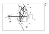
- FIG. 1is schematic representation of a portable virtual reality device that will facilitate the effective and efficient screening for TBI in accordance with the present invention.
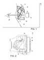
- FIG. 2is schematic representation of an alternative portable virtual reality device that will facilitate the effective and efficient screening for TBI in accordance with the present invention.
- the method of diagnosis of traumatic brain injurycomprising the steps of: providing a stimulus generating eye tracking unit 10 (examples of which are shown in FIGS. 1 and 2 ), such as a head mounted goggle based eye tracking unit that can present virtual reality based visual targets to the subject.
- the unit 10 as shownmay be categorized as a type of Video-oculography (VOG) system, which as accurately defined by Richard E. Gans, PhD, who is the Founder and Executive Director of the American Institute of Balance and he served on the board of the American Academy of Audiology, in the Hearing Journal: May 2001—Volume 54—Issue 5—pp 40, 42 “Video-oculography is a method of recording eye movement through the use of digital video cameras.
- VOGVideo-oculography
- VNGVideonystagmograpy
- a VNG unitis typically a diagnostic system for recording, analyzing and reporting (generally) involuntary eye movements, called nystagmus for involuntary movements, using video imaging technology.
- the eye tracking unit 10as described in greater detail below, may also be defined as a VNG system. VNG systems are considered, for the purpose of this application, to be a subset of the broader VOG terminology
- the VOG/VNG unit 10coupled to the subject; presenting a plurality of visual stimulus to the subject, wherein the visual stimulus may be at a simulated distance in the eye tracking unit, wherein substantially each visual stimulus provides a target stimulus for a visual based neurologic test; obtaining objective physiologic response of the subject from the eye tracking unit based upon each of neurologic test associated with each visual stimulus presented to the subject; and using the objective physiologic responses to the neurologic tests to diagnose the presence of traumatic brain injury.
- VRvirtual reality
- the VR use in the rehabilitation of TBIaccelerates the compensation of an acute loss of peripheral or central vestibular function by improving adaptive modifications of the vestibulo-ocular reflex.
- This devicehas the substantial and tremendous potential of being used bedside and in the home to increase rehabilitation compensation speed and degree.
- Innovations of this portable deviceinclude: Efficient pre-screening of military personnel. Immediate post-accident screening of soldiers for TBI, including mTBI, in forward deployed areas of operation. Following-up screening for assessing prescribed TBI or mTBI treatment. Use as a portable rehabilitation tool for mTBI patients.
- the device 10provides Combined VR and visual stimulus with eye tracking technology in portable package. Remote data access from forward deployed facilities to other medical personnel for triage can be implemented.
- FIG. 1is a schematic design of VOG/VNG unit 10 that includes the head-mounted goggles with the built-in 940 nm infrared micro LED 22 for illumination of the eyes 24 and the beam splitter plastic coated optic 14 that reflects visible light from OLED micro display 12 .
- the setupallows reflected IR light from the eyes to be sent directly to the eye tracking miniature digital cameras 18 behind the mirrors 20 .
- the VR screen 12provides the visual stimulus and the cameras 18 capture eye 24 response for quick analysis and triage.
- the details of the VR display 12are believed to be known to those or ordinary skill in the art and it allows the system 10 to present visual images or targets to the user that have a perceived or simulated distance much greater than the actual distance in the goggles.
- the targetcould be a standard Eye Chart that is typically spaced 20 feet from the subject.
- the goggle VOG/VNG unit 10 of the present inventionallow such a chart to be present to the subject on the goggle and would allow the operator to perform testing on such a chart without setting up an actual full scale system.
- the eye tracking technologyis also known in the art, and the camera based eye tracking may use the IPORTAL® brand goggle based eye tracking cameras and software available from the assignee of this invention.
- FIG. 2illustrates an alternative design having the display 12 in front of the user and the cameras 18 above.
- the assembly in FIG. 2allows for two sets of optics 14 , with the first primarily designed for proper field for the camera 18 while a second optic, behind the mirror 20 , is designed to optimize for the display 12 .
- the combination of the eye tracking and the display of simulated distanced visual targetsallow the VOG/VNG unit 10 to automatically run a number of preprogrammed neurologic tests and to record the physiologic responses thereto. Essentially the unit provides a full room sized visual testing platform in a single goggle mounting VOG/VNG unit 10 .
- the rational/purpose of the proposed systemis to rapidly assess field-deployed personnel for potential TBI or mTBI.
- the technician in the fieldmerely needs to put the unit on the subject and run the pre-identified tests.
- the contemplated system designwill incorporate over 15 standard neurological tests simplified for a pass/refer criterion, that will facilitate rehabilitation and the monitoring of recovery and the compensation process.
- the devicewill provide a cost effective means to pre-screen soldiers prior to deployment to establish baseline brain function for future comparison if a future mTBI occurs.
- the devicewill allow full vestibular diagnostics and VOR rehabilitation for more in depth usage and follow up care.
- This portable VR devicewill consist of: (a) rugged tablet PC, preferably meeting military specifications to provide for rugged use, equipped with software used to control the VR stimuli as well as to collect and analyze eye response data; (b) head mounted goggle with VR display used to present stimuli at the designated simulated distance for the test and integrated binocular eye tracking cameras.
- the present inventionprovides a solution to overcome the limitations of existing screening, diagnostic and rehabilitation methods for mTBI patients.
- the proposed new systememploys portable, head mounted VR eye tracking goggles from field to post-deployment.
- the systemwill incorporate efficient clinical diagnostic and screening methodologies for detecting mTBI related vestibular and neurological abnormalities.
- This technologywill be instrumental in pre-screening, diagnosing and monitoring the progression of mTBI in soldiers who are deployed in remote locations, as well as those seeking post-deployment clinical services. Having the ability to collect objective, functional data will aid the clinicians in the diagnosis between mTBI and other psychological disorders.
- the present inventionuses analytical and 3-D design methods, in the development of anatomically and functionally correct head-mounted goggle that can accommodate existing VR optics and miniature digital cameras.
- the VR stimulus softwareis integrated into existing vestibular/neurological software for protocol setup, test results analysis, and to create VR stimulus.
- the screening protocols of the googles 10is anticipated to include the following standard tests horizontal and vertical calibration of subject eyes, nystagmus tests (horizontal, vertical and spontaneous), horizontal and vertical smooth pursuit, horizontal and vertical saccades, optokinetic tests, subjective visual horizontal and vertical and two rehabilitation protocols (exercises), one VOR and second optokinetic.
- the inventionmay include the step of obtaining pre-trauma objective physiologic responses of the subject from the head mounted goggle unit based upon each of the visual stimulus presented to the subject, wherein the pre-trauma objective physiologic responses form a baseline for the subject.
- the step of using the objective physiologic responses to diagnose the presence of traumatic brain injuryincludes a determining whether at least one post-trauma objective physiologic responses of the subject differs from the associated pre-trauma objective physiologic response by greater than a preset threshold for that response.
- the inventionmay utilize a normative database of similar subjects (e.g.
- the step of using the objective physiologic responses to diagnose the presence of traumatic brain injuryincludes determining whether at least one post-trauma objective physiologic responses of the subject differs from an associated objective physiologic response of a normative database of similar subjects by greater than a preset threshold for that response.
- the baseline approachis preferred, but may not always be available
- the following testing protocolrepresents one effective protocol under the invention for diagnosis of mTBI in patients.
- the protocolis performed with the VOG/VNG unit 10 and will be administered at each visit comprised of the following summary:
- Pre-protocol calibrationrequired each time prior to test administration (elapsed time—about 33 seconds) and followed by:
- the total test battery timeis less than 15 minutes per patient (approximately 13 minutes 30 seconds) and the unit 10 allows for the testing to progress in an almost fully automated method making it particularly easy for technicians.
- the technicianmerely needs to explain the upcoming or current test and review the results for testing abnormalities (e.g., results indicating the goggles have slipped off the patient, or the power has been interrupted, etc).
- the subjectIn the Horizontal random saccade (HS) the subject is directed to follow (or jump to) a target (the stimulus, such as a dot, although other stimulus may be utilized) as it is displayed at a fixed location on the screen.
- the visual stimulusis presented in this test at pseudo-randomly distributed times (between 1 to 2 seconds) and will exhibit displacements from ⁇ 30 to +30 degrees measured along the horizontal axis. At least 30 trials or saccadic movements will be observed.
- the unit 10will obtain values for at least eye peak velocity, latency, accuracy for both main saccade, and combined main and secondary corrective saccade. Corrective Saccadic movement and the measurement thereof is described in U.S.
- Patent Publication 2012-0081666entitled “Method and Apparatus for Corrective Secondary Saccades Analysis with Video Oculography System”, which is incorporated herein by reference
- Each variableis calculated separately for left and right eyes, and may be combined with movement in the left and right directions (Vertical saccade).
- the main eye peak velocity during HS testingis a measure of the highest velocity during the main (or initial) saccadic eye movement and the corrective eye peak velocity is a measure of the highest velocity during the corrective saccadic movement.
- the unit 10is configured to obtain a velocity measurement between every positional measurement of the eye.
- the peak eye velocityis believed to be a better biomarker than the average velocity, which is merely the total amount of eye movement from the beginning of the movement to the end of the main or corrective saccadic movement.
- the main latency during the HS testingis the time duration from the initial display of the subject target or stimulus and the beginning of the main saccadic eye movement
- corrective latencyis a measure of the time duration from the initial display of the subject target or stimulus and the beginning of the corrective saccadic eye movement.
- a more precise corrective latencyis time duration from the initial display of the subject target or stimulus and the beginning of the corrective saccadic eye movement and minus the main latency and minus the duration of the main saccade movement.
- the main accuracyis a ratio of the main saccadic movement over the target amplitude, such that values greater than 1 or 100% are “overshoots” and values under this are “undershoots” (although the ratio can easily be inverted).
- the Vertical random saccade (VS) testingis analogous to HS testing in that the patient or subject is directed to follow (or jump to) a target stimulus on the display 12 (such as a dot) as it is displayed at pseudo-randomly distributed times (between 1 to 2 seconds) and displacements (from ⁇ 20 to +20 degrees) along the vertical axis. Thirty (30) trials or targets for VS testing are presented. As with HS testing, in VS testing the unit 10 will obtain values for at least eye peak velocity, latency, accuracy for both main saccade, and combined main and secondary corrective saccade. Each variable is calculated separately for left and right eyes, and may be combined with movement in the up and down directions (the HS testing).
- the subjectIn the Horizontal predictive saccade (HPS) testing the subject is directed to view a visual stimulus as it is quickly displayed at a fixed location. Subject will be presented with 6 pseudo-random saccade stimuli followed by 20 “mirrored” saccade stimuli meaning these have a repeated displacement of +/ ⁇ 10 degrees in the horizontal direction and the stimuli are presented at a constant time interval of 0.65 seconds.
- the unit 10will measure the first predicted saccade which is the number of mirrored saccade stimuli until the main latency is less than zero for that stimuli trial, although a number slightly higher than zero could be used as such could still be indicative of a predictive aspect if the number was small enough. Most subjects will reach a predictive saccade within several repetitions. In the HPS testing the unit 10 will measure the percentage of predicted saccades which is merely the number of saccadic stimuli having the latency lower than zero (or a slightly higher threshold than zero, if desired) divided by the total number of mirrored stimulus. As noted from this description, in the HPS testing the unit 10 will measure the main saccade latency. Each variable may be calculated separately for left and right eyes in HPS testing.
- the subjectIn the Smooth pursuit horizontal (SPH) testing the subject is directed to follow a visual stimulus (e.g. a dot on screen 12 ) as it moves through a sinusoidal displacement of +/ ⁇ 10 degrees along the horizontal axis.
- the SPH testingis run at three distinct frequencies, namely at frequencies of: (1) 3 cycles at 0.1 Hz; (2) 5 cycles at 0.75 Hz; and (3) 7 cycles at 1.25 Hz.
- the unit 10measures the Velocity gain (also called the pursuit gain) to the right and to the left.
- the velocity gainis ratio of the eye velocity to the target velocity.
- the unit 10measures the velocity gain asymmetry which is the difference between the gain to the right and to the left.
- the unit 10measures velocity phase to the right and to the left which is a measure of the patient eye velocity relative to the target velocity profile.
- the unit 10measures the percent of saccade, which is the percent of saccadic eye movement components that comprise the whole of the smooth pursuit test.
- the unit 10measures the position gain to the right and to the left which is comparison of the eye position to the target position and asymmetry values between right and left.
- the unit 10provides a spectral purity measurement and an initiation latency measurement. The SPH testing may provide values separately for the left and right eyes.
- SPV testingIn Smooth pursuit vertical (SPV) testing the subject is directed to follow a target as it moves through a sinusoidal displacement of +/ ⁇ 10 degrees along the vertical axis, analogous to the SPH testing discussed above.
- the SPV testingis run at frequencies of: (1) 3 cycles at 0.1 Hz; (2) 5 cycles at 0.75 Hz; and (3) 7 cycles at 1.0 Hz.
- the SPV variablesare the same as SPH testing discussed above.
- Gaze Horizontal (GH) testingthe subject is directed to fixate on a target light or stimulus for 3 seconds, which target is located 10° to the right of a origin or center position. The stimulus or Light is then extinguished for 15 seconds. The subject is directed to fixate on a target light for 3 seconds, which is located 10° to the left of center. Light is then extinguished for 15 seconds.
- the unit 10will calculate Peak slow phase velocity for horizontal and vertical eye movement components with and without the target or fixation light on.
- the SPH testingmay provide values separately for the left and right eyes.
- the two Optokinetic (OKN) teststhe patients will see stimulus (e.g., lighted dots moving on the display first to the right, then to the left.
- the two optokinetic stimuluswill be at rotation rates or speeds of 20 and 60 deg/sec, respectively.
- Each testconsists of 15 seconds clockwise (CW) and 15 seconds counterclockwise (CCW) rotation stimulus.
- the unit 10will measure at least the Average slow phase gain, average slow phase asymmetry, fast phase velocity vs. amplitude, and fast phase velocity asymmetry for each test and for left and right eyes.
- the subjects or patientsare required to fixate on a central target for 1.5 to 2.5 seconds and then presented with a peripheral target; patients are instructed to generate an eye movement in the same distance as the target displacement, but in the exact opposite direction.
- Patientsare trained using slow presentations of the task during which a technician provides verbal instructions about task demands and appropriate responses (training test).
- the measured testingconstitutes 20 anti-saccades stimulus with time between saccades randomly selected from 1 to 2 seconds and random displacement for each target of between ⁇ 30 to +30 degrees.
- the unit 10measures Pro-saccade error (also called an anti-suppression error) measurement, anti-saccade latency (measure from the stimulus to the start of the anti-saccadic eye movement), latency of pro-saccade error (generally the main saccadic latency), anti-saccade peak velocity, accuracy of main anti-saccade, and accuracy of main and corrective anti-saccades.
- the AS testing measurementsmay be for each of the left and right eyes.
- the subjectIn the subjective visual vertical (SVV) testing the subject is presented with a red line on the display and directed to use control left and right buttons (or any desired input device such as a joystick etc) to manipulate the displayed line into the vertical (upright).
- One input buttonrotates the line in one direction and the other input device rotates the line in the other.
- Subjectis directed to inform the clinician when they are finished and they believe the line is vertical, known as the the subjective vertical position.
- the unit 10measures the mean and standard deviation from subjective vertical position and the true vertical position.
- the subjectIn the subjective visual Horizontal (SVH) testing the subject is presented with a red line on the display and directed to use control left and right buttons (or any desired input device such as a joystick etc) to manipulate the displayed line into the horizontal (flat).
- One input buttonrotates the line in one direction and the other input device rotates the line in the other.
- Subjectagain is directed to inform the clinician when they are finished and they now believe the line is horizontal, known as the subjective horizontal position.
- the unit 10measures the mean and standard deviation from subjective horizontal position and the true horizontal position.
- VRTVisual reaction time
- SDstandard deviation
- ARTAuditory reaction time
- the unit 10measures the same descriptive variables as regular saccade (HS and VS) along with latency, SD, and percent of error for each direction.
- LRLight Reflex
- the comprehensive I-Portal® system based neuro-otologic test device 10 described aboveis an effective diagnostic tool for mTBI detection and to monitor recovery from head trauma.
- the I-Portal® system based neuro-otologic testsare neuro-physiologic; measuring the eye's response to various motion, oculomotor, and optokinetic stimuli.
- Initial datashows I-Portal® tests described above can detect abnormalities resulting from head trauma even when structural imaging technologies show no damage.
- the test batteryalso suggests the data is significantly more objective than current neuro-psychological tests that rely partially on patient self diagnosis.
- the unit 10is and may continue to be used by military and sports medicine researchers to study new methods to objectively diagnose and monitor blast and blunt force head trauma.
- the above described battery of testshelp examiners make better return to play and return to duty decisions.
- the abnormal results, based upon an individual baseline or a normative group, to the above battery of testspresents a effective screening tool for an increased likelihood of the presence of mTBI.
- the followingdescribes a method for detecting the presence, and assessing the severity of a concussion (or a mild traumatic brain injury, or mTBI) in any patient presenting to a clinical professional, who is suspected of having a concussion/mTBI following a precipitating head injury. Specifically, the following describes the process by which the method is developed and applied.
- a battery of vestibular, oculomotor, and reaction time testsare administered to a cohort of mTBI patients and an age-matched control population.
- This battery associated with this exampleis listed below.
- the batteryis performed on devices capable of providing visual and auditory stimuli to a subject, measuring eye movements at a rate of at least 100 Hz, and capturing button presses at a comparable rate.
- a third logistic regressionis performed on these 8 variables, which generates a set of 8 coefficients.
- a cutoff value(in the range of 0 to 1) is chosen as a threshold value.
- Assessmentis performed by administering the same test battery and recording (at the minimum) the eight measures determined to be critical variables. These variables are used as input parameters to the assessment model, and a result between 0 and 1 is produced.
- Presence of mTBIis determined categorically by relationship to the chosen cutoff, i.e. patients with scores above the cutoff are categorized as having a concussion mTBI, or screening positive for the presence of a concusion/mTBI.
- Severity of mTBIis determined parametrically by the magnitude of the assessment model output, with 1 representing the most severe cases.
- the final set of 8 variables for this particular exampleis as follows:
- variable sets that are generated at each reduction stepwill also vary based on the initial population.
- the number and membership of each variable set at each reduction stepwill also change if any of a multitude of (equally valid) statistical procedures are applied in lieu of factor analysis or logistic regression (e.g. discriminate analysis, non-parametric classifiers such as decision tree, support vector machine, neural network analysis, etc.).
- Example TEST mTBI Study Protocolmeasures Spontaneous 1 cycle - Stimulus light is projected at Amount, nystagmus a central fixation point for 3 seconds, rate, and followed by light off for 15 seconds.
- direction of spontaneous nystagmusOptokinetic 2 cycles - Full field random dot stimulus Integrity of nystagmus continually moves left 10 seconds, and fixation then right 10 seconds, at 20°/second and reflex 60°/second velocity combining pursuits and saccades Smooth Single light stimulus moves smoothly Gain of eye pursuit - left, then right, with sinusoidal relative to horizontal velocity and maximum displacement of stimulus; 10°. 3 cycles at 0.1 Hz, 5 cycles at presence of 1.0 Hz, 7 cycles at 1.25 Hz.
- saccadic intrusionsSmooth Same as horizontal, but with 5 cycles Same pursuit - at 0.75 Hz, 7 cycles at 1.0 Hz.
- vertical Saccade - 30 cyclesSingle light stimulus Saccade random projected at random horizontal onset horizontal displacements and time, with maximum latency, displacement of 30°, and time between accuracy, stimuli 1.2 to 2.0 seconds.
- vertical Saccade - 20 cycles - Single light stimulusis Ability to predictive projected at 10° left or right adapt to horizontal displacement (alternating) with a fixed predictable 0.65 second interval. timing and position (latency and accuracy of saccades) Saccade - Same as horizontal, using 10° up and Same predictive down displacements.
- the present inventionprovides the use of 5-15, preferably 8-10, of the list of 440+ occulomotor, vestibular and reaction time measures to screen for the presence of mTBI. More narrowly the present invention provides the use of 5-15, preferably 8-10, of the list of 82 variables obtained after conducting a conventional dimension reduction by factor analysis on the 440+ occulomotor, vestibular and reaction time measures to screen for the presence of mTBI.
- the inventionmay be described as the use of 5-15, preferably 8-10, of a subset of variables selected using conventional Wald Statistical Analysis with p>0.03 to p>0.09, generally p>0.05, on the list of 82 variable obtained after conducting a conventional dimension reduction by factor analysis on the 440+ occulomotor, vestibular and reaction time measures to screen for the presence of mTBI.
Landscapes
- Health & Medical Sciences (AREA)
- Life Sciences & Earth Sciences (AREA)
- Engineering & Computer Science (AREA)
- Physics & Mathematics (AREA)
- General Health & Medical Sciences (AREA)
- Animal Behavior & Ethology (AREA)
- Biomedical Technology (AREA)
- Heart & Thoracic Surgery (AREA)
- Medical Informatics (AREA)
- Molecular Biology (AREA)
- Surgery (AREA)
- Biophysics (AREA)
- Veterinary Medicine (AREA)
- Public Health (AREA)
- Pathology (AREA)
- Neurology (AREA)
- Psychology (AREA)
- Ophthalmology & Optometry (AREA)
- Psychiatry (AREA)
- Developmental Disabilities (AREA)
- Social Psychology (AREA)
- Educational Technology (AREA)
- Hospice & Palliative Care (AREA)
- Child & Adolescent Psychology (AREA)
- Physiology (AREA)
- Human Computer Interaction (AREA)
- Neurosurgery (AREA)
- General Physics & Mathematics (AREA)
- Optics & Photonics (AREA)
- Eye Examination Apparatus (AREA)
- Artificial Intelligence (AREA)
- Computer Vision & Pattern Recognition (AREA)
- Signal Processing (AREA)
Abstract
Description
- 1. Horizontal random saccade (about 50 seconds)
- 2. Vertical random saccade (about 46 seconds)
- 3. Horizontal predictive saccade (about 20 seconds)
- 4. Smooth pursuit horizontal (3 frequencies) (about 33 seconds)
- 5. Smooth pursuit vertical (3 frequencies) (about 33 seconds)
- 6. Gaze Horizontal (alternatively Spontaneous nystagmus) (about 30 seconds)
- 7. Optokinetic (OKN) (2 tests) (about 31 seconds)
- 8. Antisaccade (about 1 minute 11 seconds)
- 9. Subjective visual vertical (about 24 seconds)
- 10. Subjective visual horizontal (about 37 seconds)
- 11. Visual reaction time (about 32 seconds)
- 12. Auditory reaction time (about 18 seconds)
- 13. Saccade and reaction time (about 1 minutes 2 seconds)
- 14. Light Reflex (about 45 seconds)
- 1. The variable set is reduced by factor analysis. This reduces the original variable set to a first subset of variable, in this particular example from 447 to 82 variables.
- 2. A preliminary logistic regression is performed on the initial subset 82 variables, coding mTBI patients as 1 and controls as 0. As a product of this process, a Wald statistic is generated for each variable. All variables with a p (probability) value >a preset threshold, such as 0.05, for the Wald statistic are removed. This reduces the variable set to a second subset of variables, specifically 33 variables in this example.
- 3. Step 2 is repeated: A second linear regression is performed on the second subset of variables and variables with Wald statistic p-values >a preset threshold, again such as 0.05, are again removed. This reduces the variable set to a final desired set, namely 8 variables in the specific example.
- Optokinetic nystagmus, gain of eye relative to stimulus
- Subjective visual line placement, mean error in degrees
- Horizontal smooth pursuit, presence of saccades
- Horizontal smooth pursuit, phase lag of eye
- Auditory reaction time variability
- Latency to onset of vertical smooth pursuit tracking
- Combined saccade+ reaction time, latency of saccades
- Vertical smooth pursuit, upward phase lag of eye
| Example | ||
| TEST | mTBI Study Protocol | measures |
| Spontaneous | 1 cycle - Stimulus light is projected at | Amount, |
| nystagmus | a central fixation point for 3 seconds, | rate, and |
| followed by light off for 15 seconds. | direction of | |
| spontaneous | ||
| nystagmus | ||
| Optokinetic | 2 cycles - Full field random dot stimulus | Integrity of |
| nystagmus | continually moves left 10 seconds, and | fixation |
| then right 10 seconds, at 20°/second and | reflex | |
| 60°/second velocity | combining | |
| pursuits and | ||
| saccades | ||
| Smooth | Single light stimulus moves smoothly | Gain of eye |
| pursuit - | left, then right, with sinusoidal | relative to |
| horizontal | velocity and maximum displacement of | stimulus; |
| 10°. 3 cycles at 0.1 Hz, 5 cycles at | presence of | |
| 1.0 Hz, 7 cycles at 1.25 Hz. | saccadic | |
| intrusions | ||
| Smooth | Same as horizontal, but with 5 cycles | Same |
| pursuit - | at 0.75 Hz, 7 cycles at 1.0 Hz. | |
| vertical | ||
| Saccade - | 30 cycles - Single light stimulus | Saccade |
| random | projected at random horizontal | onset |
| horizontal | displacements and time, with maximum | latency, |
| displacement of 30°, and time between | accuracy, | |
| stimuli 1.2 to 2.0 seconds. | presence of | |
| corrective | ||
| saccades | ||
| Saccade - | Same as horizontal, but with maximal | Same |
| random | vertical displacement of 20°. | |
| vertical | ||
| Saccade - | 20 cycles - Single light stimulus is | Ability to |
| predictive | projected at 10° left or right | adapt to |
| horizontal | displacement (alternating) with a fixed | predictable |
| 0.65 second interval. | timing and | |
| position | ||
| (latency and | ||
| accuracy of | ||
| saccades) | ||
| Saccade - | Same as horizontal, using 10° up and | Same |
| predictive | down displacements. | |
| vertical | ||
| Saccade - | Same as Saccade - random horizontal, | Number of |
| antisaccade | except that subject is instructed to | incorrect |
| horizontal | look away from the target. | pro- |
| saccades, | ||
| corrective | ||
| anti- | ||
| saccades; | ||
| latency | ||
| Saccade and | Single light stimulus projected at | Saccade |
| reaction time | random horizontal displacements and | onset |
| time, with maximum displacement of 30°, | latency, | |
| and time between stimuli 1.2 to | accuracy, | |
| 2.0 sec, subject asked to click left | presence of | |
| or right buttons depends on direction | corrective | |
| of saccades. | saccades, | |
| latency and | ||
| S.D. for left | ||
| and right | ||
| buttons | ||
| Visual | 20 cycles, random single light | Latency and |
| Reaction | stimulus appears at center of vision | latency |
| time | and subject using his/her dominate | standard |
| hand click on button on | ||
| Auditory | ||
| 20 cycles, random auditory stimulus 85 | Latency and | |
| reaction time | decibel presented and subject using his/ | latency |
| her dominate hand click on button as | standard | |
| stimulus on | deviation | |
| Subjective | 6 cycles - Straight line stimulus | Angular error |
| visual - | appears tilted off vertical axis, up to | from vertical |
| vertical | 30° displacement clockwise or | axis |
| counterclockwise. Subject asked to press | ||
| buttons to tilt line back to vertical | ||
| alignment. | ||
| Subjective | 6 cycles - Same as vertical, except asked | Angular error |
| visual - | to tilt line until it is horizontal. | from |
| horizontal | horizontal | |
| axis | ||
Claims (20)
Priority Applications (2)
| Application Number | Priority Date | Filing Date | Title |
|---|---|---|---|
| US14/687,871US10398309B2 (en) | 2008-10-09 | 2015-04-15 | Noninvasive rapid screening of mild traumatic brain injury using combination of subject's objective oculomotor, vestibular and reaction time analytic variables |
| US15/050,119US20160270711A1 (en) | 2008-10-09 | 2016-02-22 | Method and Apparatus for MTBi Assessment Using Multi Variable Regression Analysis |
Applications Claiming Priority (6)
| Application Number | Priority Date | Filing Date | Title |
|---|---|---|---|
| US10413308P | 2008-10-09 | 2008-10-09 | |
| US12/577,143US8585609B2 (en) | 2008-10-09 | 2009-10-09 | Quantitative, non-invasive, clinical diagnosis of traumatic brain injury using simulated distance visual stimulus device for neurologic testing |
| US14/083,145US9039631B2 (en) | 2008-10-09 | 2013-11-18 | Quantitative, non-invasive, clinical diagnosis of traumatic brain injury using VOG device for neurologic testing |
| US201461979765P | 2014-04-15 | 2014-04-15 | |
| US14/336,254US9039632B2 (en) | 2008-10-09 | 2014-07-21 | Quantitative, non-invasive, clinical diagnosis of traumatic brain injury using VOG device for neurologic optokinetic testing |
| US14/687,871US10398309B2 (en) | 2008-10-09 | 2015-04-15 | Noninvasive rapid screening of mild traumatic brain injury using combination of subject's objective oculomotor, vestibular and reaction time analytic variables |
Related Parent Applications (1)
| Application Number | Title | Priority Date | Filing Date |
|---|---|---|---|
| US14/336,254Continuation-In-PartUS9039632B2 (en) | 2008-10-09 | 2014-07-21 | Quantitative, non-invasive, clinical diagnosis of traumatic brain injury using VOG device for neurologic optokinetic testing |
Related Child Applications (1)
| Application Number | Title | Priority Date | Filing Date |
|---|---|---|---|
| US15/050,119Continuation-In-PartUS20160270711A1 (en) | 2008-10-09 | 2016-02-22 | Method and Apparatus for MTBi Assessment Using Multi Variable Regression Analysis |
Publications (2)
| Publication Number | Publication Date |
|---|---|
| US20150335278A1 US20150335278A1 (en) | 2015-11-26 |
| US10398309B2true US10398309B2 (en) | 2019-09-03 |
Family
ID=54555192
Family Applications (1)
| Application Number | Title | Priority Date | Filing Date |
|---|---|---|---|
| US14/687,871Active2032-10-21US10398309B2 (en) | 2008-10-09 | 2015-04-15 | Noninvasive rapid screening of mild traumatic brain injury using combination of subject's objective oculomotor, vestibular and reaction time analytic variables |
Country Status (1)
| Country | Link |
|---|---|
| US (1) | US10398309B2 (en) |
Cited By (1)
| Publication number | Priority date | Publication date | Assignee | Title |
|---|---|---|---|---|
| US20160270711A1 (en)* | 2008-10-09 | 2016-09-22 | Neuro Kinetics, Inc. | Method and Apparatus for MTBi Assessment Using Multi Variable Regression Analysis |
Families Citing this family (10)
| Publication number | Priority date | Publication date | Assignee | Title |
|---|---|---|---|---|
| US20170042462A1 (en)* | 2015-08-10 | 2017-02-16 | Neuro Kinetics, Inc. | Automated Data Acquisition, Appraisal and Analysis in Noninvasive Rapid Screening of Neuro-Otologic Conditions Using Combination of Subject's Objective Oculomotor Vestibular and Reaction Time Analytic Variables |
| CN106214118A (en)* | 2016-01-28 | 2016-12-14 | 北京爱生科贸有限公司 | A kind of ocular movement based on virtual reality monitoring system |
| WO2017147141A1 (en) | 2016-02-22 | 2017-08-31 | Neuro Kinetics, Inc. | Objective testing of vergence dysfunction for diagnosis and vergence recovery convalescence using dynamic vergence testing platform including 3d head mounted display system with integrated eye tracking technology |
| US11419493B2 (en) | 2017-02-14 | 2022-08-23 | Neuro Kinetics, Inc & University Of Pittsburgh | Method and apparatus for mTBI diagnosis implementing eye movement and pupil movement analysis in objective vergence testing |
| CN109276228B (en)* | 2017-07-21 | 2020-12-25 | 成都集思鸣智科技有限公司 | System and device for detecting brain function |
| CN109839742A (en)* | 2017-11-29 | 2019-06-04 | 深圳市掌网科技股份有限公司 | A kind of augmented reality device based on Eye-controlling focus |
| US10470657B1 (en) | 2019-02-28 | 2019-11-12 | Thomas E. Lister | Systems and methods for administering a gaze nystagmus test |
| US20230103276A9 (en)* | 2019-06-06 | 2023-03-30 | CannSight Technologies Inc. | Impairement screening system and method |
| US11803237B2 (en) | 2020-11-14 | 2023-10-31 | Facense Ltd. | Controlling an eye tracking camera according to eye movement velocity |
| US11503998B1 (en)* | 2021-05-05 | 2022-11-22 | Innodem Neurosciences | Method and a system for detection of eye gaze-pattern abnormalities and related neurological diseases |
Citations (109)
| Publication number | Priority date | Publication date | Assignee | Title |
|---|---|---|---|---|
| US3612642A (en) | 1969-06-27 | 1971-10-12 | Bulova Watch Co Inc | A high-velocity optical scanner including a torsional fork supporting two reflectors |
| US4006974A (en) | 1975-09-26 | 1977-02-08 | Resnick Sam L | Eyeglass structure |
| US4084182A (en) | 1974-07-01 | 1978-04-11 | Laser Video, Inc. | Multi-beam modulator and method for light beam displays |
| US4309608A (en) | 1980-05-16 | 1982-01-05 | The United States Of America As Represented By The Secretary Of The Army | Flightline goggle tester |
| US4320768A (en) | 1979-07-17 | 1982-03-23 | Georgetown University Medical Center | Computerized electro-oculographic (CEOG) system |
| US4474186A (en) | 1979-07-17 | 1984-10-02 | Georgetown University | Computerized electro-oculographic (CEOG) system with feedback control of stimuli |
| US4572199A (en) | 1982-12-27 | 1986-02-25 | University Of New Hampshire | System to determine arterial occlusion and other maladies |
| US4836219A (en) | 1987-07-08 | 1989-06-06 | President & Fellows Of Harvard College | Electronic sleep monitor headgear |
| US4852988A (en) | 1988-09-12 | 1989-08-01 | Applied Science Laboratories | Visor and camera providing a parallax-free field-of-view image for a head-mounted eye movement measurement system |
| US4863259A (en) | 1988-03-09 | 1989-09-05 | Schneider Michael B | Rapid eye movement sleep state detector |
| US5070883A (en) | 1988-12-16 | 1991-12-10 | Konan Camera Research Institute Inc. | Eye movement analyzing device utilizing pupil center-of-gravity data |
| US5098426A (en) | 1989-02-06 | 1992-03-24 | Phoenix Laser Systems, Inc. | Method and apparatus for precision laser surgery |
| US5130838A (en) | 1990-06-18 | 1992-07-14 | Pioneer Electronic Corporation | Laser projection type display unit |
| US5252999A (en) | 1990-10-26 | 1993-10-12 | Nidek Co., Ltd. | Laser apparatus including binocular indirect ophthalmoscope |
| US5304112A (en) | 1991-10-16 | 1994-04-19 | Theresia A. Mrklas | Stress reduction system and method |
| US5305746A (en) | 1992-09-29 | 1994-04-26 | Aspect Medical Systems, Inc. | Disposable, pre-gelled, self-prepping electrode |
| US5320109A (en) | 1991-10-25 | 1994-06-14 | Aspect Medical Systems, Inc. | Cerebral biopotential analysis system and method |
| US5345281A (en) | 1992-12-17 | 1994-09-06 | John Taboada | Eye tracking system and method |
| US5365941A (en) | 1992-11-27 | 1994-11-22 | Atr Auditory And Visual Perception Research Laboratories | Apparatus for detecting small involuntary movement |
| US5368041A (en) | 1992-10-15 | 1994-11-29 | Aspect Medical Systems, Inc. | Monitor and method for acquiring and processing electrical signals relating to bodily functions |
| US5410376A (en) | 1994-02-04 | 1995-04-25 | Pulse Medical Instruments | Eye tracking method and apparatus |
| US5458117A (en) | 1991-10-25 | 1995-10-17 | Aspect Medical Systems, Inc. | Cerebral biopotential analysis system and method |
| US5481622A (en) | 1994-03-01 | 1996-01-02 | Rensselaer Polytechnic Institute | Eye tracking apparatus and method employing grayscale threshold values |
| US5491492A (en) | 1992-02-05 | 1996-02-13 | Biocontrol Systems, Inc. | Method and apparatus for eye tracking for convergence and strabismus measurement |
| US5652756A (en) | 1995-01-20 | 1997-07-29 | Hughes Electronics | Glass fiber laser system using U-doped crystal Q-switch |
| US5687020A (en) | 1995-06-29 | 1997-11-11 | Samsung Electronics Co., Ltd. | Image projector using acousto-optic tunable filter |
| US5704369A (en) | 1994-07-25 | 1998-01-06 | Beth Israel Hospital Association, Inc. | Non-invasive method for diagnosing Alzeheimer's disease in a patient |
| US5714967A (en) | 1994-05-16 | 1998-02-03 | Olympus Optical Co., Ltd. | Head-mounted or face-mounted image display apparatus with an increased exit pupil |
| US5792069A (en) | 1996-12-24 | 1998-08-11 | Aspect Medical Systems, Inc. | Method and system for the extraction of cardiac artifacts from EEG signals |
| US5813404A (en) | 1995-10-20 | 1998-09-29 | Aspect Medical Systems, Inc. | Electrode connector system |
| US5821521A (en) | 1990-05-08 | 1998-10-13 | Symbol Technologies, Inc. | Optical scanning assembly with flexible diaphragm |
| US5838420A (en) | 1994-09-30 | 1998-11-17 | Bid Instruments Limited | Method and apparatus for ocular motility testing |
| US5877732A (en) | 1994-04-13 | 1999-03-02 | Resonance Technology Co. | Three-dimensional high resolution MRI video and audio system and method |
| US5892566A (en) | 1998-01-20 | 1999-04-06 | Bullwinkel; Paul E. | Fiber optic eye-tracking system |
| JPH11184621A (en) | 1997-12-25 | 1999-07-09 | Shimadzu Corp | Eye-gaze input device |
| US5943116A (en) | 1997-04-01 | 1999-08-24 | Johns Hopkins University | System for imaging an ocular fundus semi-automatically at high resolution and wide field |
| US5942954A (en) | 1997-08-22 | 1999-08-24 | Massachusetts Institute Of Technology | Apparatus and method for measuring vestibular ocular reflex function |
| US5963300A (en) | 1998-02-17 | 1999-10-05 | Amt Technologies, Corp. | Ocular biometer |
| US5983128A (en) | 1994-07-15 | 1999-11-09 | Centre National De La Recherche Scientifique | Device for examining a subject and, in particular, determining his or her vestibular evoked potentials |
| US5980513A (en) | 1994-04-25 | 1999-11-09 | Autonomous Technologies Corp. | Laser beam delivery and eye tracking system |
| US6003991A (en) | 1996-02-17 | 1999-12-21 | Erik Scott Viirre | Eye examination apparatus and method for remote examination of a patient by a health professional |
| US6032072A (en) | 1998-01-30 | 2000-02-29 | Aspect Medical Systems, Inc. | Method for enhancing and separating biopotential signals |
| US6032064A (en) | 1996-10-11 | 2000-02-29 | Aspect Medical Systems, Inc. | Electrode array system for measuring electrophysiological signals |
| US6033073A (en) | 1997-08-15 | 2000-03-07 | Potapova; Olga | Visual training system and apparatus for vision correction, especially for various forms of strabismus ("crossed" eyes) |
| US6077237A (en) | 1998-11-06 | 2000-06-20 | Adaboy, Inc. | Headset for vestibular stimulation in virtual environments |
| US6089716A (en) | 1996-07-29 | 2000-07-18 | Lashkari; Kameran | Electro-optic binocular indirect ophthalmoscope for stereoscopic observation of retina |
| US6090051A (en) | 1999-03-03 | 2000-07-18 | Marshall; Sandra P. | Method and apparatus for eye tracking and monitoring pupil dilation to evaluate cognitive activity |
| US6099124A (en) | 1999-12-14 | 2000-08-08 | Hidaji; Faramarz | Ophthalmological system and method |
| US6113237A (en) | 1999-12-06 | 2000-09-05 | Ober; Jan Krzysztof | Adaptable eye movement measurement device |
| US6120461A (en) | 1999-08-09 | 2000-09-19 | The United States Of America As Represented By The Secretary Of The Army | Apparatus for tracking the human eye with a retinal scanning display, and method thereof |
| US6213943B1 (en) | 1996-09-04 | 2001-04-10 | Marcio Marc Abreu | Apparatus for signal transmission and detection using a contact device for physical measurement on the eye |
| US6231187B1 (en) | 1999-02-11 | 2001-05-15 | Queen's University At Kingston | Method and apparatus for detecting eye movement |
| US6247813B1 (en) | 1999-04-09 | 2001-06-19 | Iritech, Inc. | Iris identification system and method of identifying a person through iris recognition |
| US6271915B1 (en) | 1996-11-25 | 2001-08-07 | Autonomous Technologies Corporation | Objective measurement and correction of optical systems using wavefront analysis |
| US6275718B1 (en) | 1999-03-23 | 2001-08-14 | Philip Lempert | Method and apparatus for imaging and analysis of ocular tissue |
| US6299308B1 (en) | 1999-04-02 | 2001-10-09 | Cybernet Systems Corporation | Low-cost non-imaging eye tracker system for computer control |
| US20020027779A1 (en) | 1999-07-02 | 2002-03-07 | Cassarly William J. | Image generator having an improved illumination system |
| US6367932B1 (en) | 1997-10-30 | 2002-04-09 | Bid Instruments Limited | Apparatus and method for visual field testing |
| US6402320B1 (en) | 1999-08-09 | 2002-06-11 | Childrens Hospital Los Angeles | Methods and apparatus for measuring visual acuity in preverbal children |
| US20020085174A1 (en) | 2000-11-22 | 2002-07-04 | Ciaran Bolger | Method and apparatus for monitoring eye tremor |
| US6456261B1 (en) | 1998-11-23 | 2002-09-24 | Evan Y. W. Zhang | Head/helmet mounted passive and active infrared imaging system with/without parallax |
| US6459446B1 (en) | 1997-11-21 | 2002-10-01 | Dynamic Digital Depth Research Pty. Ltd. | Eye tracking apparatus |
| US6467905B1 (en) | 1998-09-25 | 2002-10-22 | John S. Stahl | Acquired pendular nystagmus treatment device |
| US20020171805A1 (en) | 2001-05-18 | 2002-11-21 | Odom James V. | Non-invasive ocular assessment method and associated apparatus |
| US20020175880A1 (en) | 1998-01-20 | 2002-11-28 | Melville Charles D. | Augmented retinal display with view tracking and data positioning |
| US20030028081A1 (en) | 2000-06-20 | 2003-02-06 | Eastman Kodak Company | ADHD detection by eye saccades |
| US6524581B1 (en) | 1998-12-30 | 2003-02-25 | The Children's Medical Center Corporation | Prevention and treatment of retinal ischemia and edema |
| US6542081B2 (en) | 1996-08-19 | 2003-04-01 | William C. Torch | System and method for monitoring eye movement |
| US6551575B1 (en) | 1999-12-02 | 2003-04-22 | Neurosciences Research Foundation, Inc. | Methods for identifying compounds for motion sickness, vertigo and other disorders related to balance and the perception of gravity |
| US6568808B2 (en) | 2000-04-25 | 2003-05-27 | Alcon Universal Ltd. | Eye tracker control system and method |
| US6574352B1 (en) | 1999-05-18 | 2003-06-03 | Evans & Sutherland Computer Corporation | Process for anticipation and tracking of eye movement |
| US6609523B1 (en) | 1999-10-26 | 2003-08-26 | Philip F. Anthony | Computer based business model for a statistical method for the diagnosis and treatment of BPPV |
| US6629935B1 (en) | 1998-06-09 | 2003-10-07 | The University Of Queensland | Method and apparatus for diagnosis of a mood disorder or predisposition therefor |
| US6634749B1 (en) | 1998-11-02 | 2003-10-21 | Leica Microsystems (Schweiz) Ag | Eye tracking system |
| US6637883B1 (en) | 2003-01-23 | 2003-10-28 | Vishwas V. Tengshe | Gaze tracking system and method |
| US6659611B2 (en) | 2001-12-28 | 2003-12-09 | International Business Machines Corporation | System and method for eye gaze tracking using corneal image mapping |
| US6669341B2 (en) | 2001-08-31 | 2003-12-30 | Metrologic Instruments, Inc. | Ophthalmic instrument having wavefront sensor with multiple imaging devices that simultaneously capture multiple images of an array of spots produced by a lenslet array |
| US6697894B1 (en) | 1999-03-29 | 2004-02-24 | Siemens Dematic Postal Automation, L.P. | System, apparatus and method for providing maintenance instructions to a user at a remote location |
| US6748275B2 (en) | 1999-05-05 | 2004-06-08 | Respironics, Inc. | Vestibular stimulation system and method |
| US20040181168A1 (en) | 2003-03-13 | 2004-09-16 | Plant Charles P. | Saccadic motion sensing |
| US6796947B2 (en) | 2000-06-19 | 2004-09-28 | Canadian Space Agency | Method for evaluating vestibular response |
| US6800062B2 (en) | 2002-07-03 | 2004-10-05 | Epley Research, L.L.C. | Comprehensive vertigo management |
| USRE38668E1 (en) | 1997-10-16 | 2004-12-07 | The Board Of Trustees Of The Leland Stanford Junior University | Method for inferring metal states from eye movements |
| US20050004489A1 (en) | 2003-07-02 | 2005-01-06 | Mika Sarkela | Method of positioning electrodes for central nervous system monitoring |
| US20050024586A1 (en) | 2001-02-09 | 2005-02-03 | Sensomotoric Instruments Gmbh | Multidimensional eye tracking and position measurement system for diagnosis and treatment of the eye |
| US20050079636A1 (en) | 2001-09-25 | 2005-04-14 | White Keith D. | Method and apparatus for diagnosing schizophrenia and schizophrenia subtype |
| US20050099601A1 (en) | 2003-11-07 | 2005-05-12 | Neuro Kinetics, Inc. | Portable video oculography system |
| US20050110950A1 (en) | 2003-03-13 | 2005-05-26 | Thorpe William P. | Saccadic motion sensing |
| US6943754B2 (en) | 2002-09-27 | 2005-09-13 | The Boeing Company | Gaze tracking system, eye-tracking assembly and an associated method of calibration |
| US20050216243A1 (en) | 2004-03-02 | 2005-09-29 | Simon Graham | Computer-simulated virtual reality environments for evaluation of neurobehavioral performance |
| US20060098087A1 (en) | 2002-11-08 | 2006-05-11 | Ludwig-Maximilians-Universitat | Housing device for head-worn image recording and method for control of the housing device |
| US20060235331A1 (en) | 2004-07-13 | 2006-10-19 | Neuro Kinetics Incorporated | Compact neuro-otologic, neuro-ophthalmologic testing device and dynamic visual acuity testing and desensitization platform |
| US7398119B2 (en) | 1998-07-13 | 2008-07-08 | Childrens Hospital Los Angeles | Assessing blood brain barrier dynamics or identifying or measuring selected substances, including ethanol or toxins, in a subject by analyzing Raman spectrum signals |
| US20090118593A1 (en) | 2007-11-07 | 2009-05-07 | Searete Llc, A Limited Liability Corporation Of The State Of Delaware | Determining a demographic characteristic based on computational user-health testing of a user interaction with advertiser-specified content |
| US20090119154A1 (en) | 2007-11-07 | 2009-05-07 | Searete Llc, A Limited Liability Corporation Of The State Of Delaware | Determining a demographic characteristic based on computational user-health testing of a user interaction with advertiser-specified content |
| US20090132275A1 (en) | 2007-11-19 | 2009-05-21 | Searete Llc, A Limited Liability Corporation Of The State Of Delaware | Determining a demographic characteristic of a user based on computational user-health testing |
| US20090299645A1 (en) | 2008-03-19 | 2009-12-03 | Brandon Colby | Genetic analysis |
| US7819818B2 (en) | 2004-02-11 | 2010-10-26 | Jamshid Ghajar | Cognition and motor timing diagnosis using smooth eye pursuit analysis |
| US20100280372A1 (en) | 2009-05-03 | 2010-11-04 | Pieter Poolman | Observation device and method |
| US20110086914A1 (en) | 2009-10-13 | 2011-04-14 | Bailes Julian E | Methods for Treating Traumatic Brain Injury |
| US7988287B1 (en) | 2004-11-04 | 2011-08-02 | Kestrel Corporation | Objective traumatic brain injury assessment system and method |
| US20110208060A1 (en) | 2010-02-24 | 2011-08-25 | Haase Wayne C | Non-contact Biometric Monitor |
| US20110229862A1 (en) | 2010-03-18 | 2011-09-22 | Ohm Technologies Llc | Method and Apparatus for Training Brain Development Disorders |
| US8065240B2 (en) | 2007-10-31 | 2011-11-22 | The Invention Science Fund I | Computational user-health testing responsive to a user interaction with advertiser-configured content |
| US20120330178A1 (en) | 2011-06-24 | 2012-12-27 | U.S. Government As Represented By The Secretary Of The Army | Method and apparatus for multimodal mobile screening to quantitatively detect brain function impairment |
| US8406859B2 (en) | 2008-08-10 | 2013-03-26 | Board Of Regents, The University Of Texas System | Digital light processing hyperspectral imaging apparatus |
| US8568311B2 (en) | 2004-02-13 | 2013-10-29 | Emory University | Display enhanced testing for concussions and mild traumatic brain injury |
| US8585609B2 (en) | 2008-10-09 | 2013-11-19 | Neuro Kinetics, Inc. | Quantitative, non-invasive, clinical diagnosis of traumatic brain injury using simulated distance visual stimulus device for neurologic testing |
| US20140192326A1 (en) | 2008-10-09 | 2014-07-10 | Neuro Kinetics, Inc. | Quantitative, non-invasive, clinical diagnosis of traumatic brain injury using vog device for neurologic testing |
- 2015
- 2015-04-15USUS14/687,871patent/US10398309B2/enactiveActive
Patent Citations (129)
| Publication number | Priority date | Publication date | Assignee | Title |
|---|---|---|---|---|
| US3612642A (en) | 1969-06-27 | 1971-10-12 | Bulova Watch Co Inc | A high-velocity optical scanner including a torsional fork supporting two reflectors |
| US4084182A (en) | 1974-07-01 | 1978-04-11 | Laser Video, Inc. | Multi-beam modulator and method for light beam displays |
| US4006974A (en) | 1975-09-26 | 1977-02-08 | Resnick Sam L | Eyeglass structure |
| US4320768A (en) | 1979-07-17 | 1982-03-23 | Georgetown University Medical Center | Computerized electro-oculographic (CEOG) system |
| US4474186A (en) | 1979-07-17 | 1984-10-02 | Georgetown University | Computerized electro-oculographic (CEOG) system with feedback control of stimuli |
| US4309608A (en) | 1980-05-16 | 1982-01-05 | The United States Of America As Represented By The Secretary Of The Army | Flightline goggle tester |
| US4572199A (en) | 1982-12-27 | 1986-02-25 | University Of New Hampshire | System to determine arterial occlusion and other maladies |
| US4836219A (en) | 1987-07-08 | 1989-06-06 | President & Fellows Of Harvard College | Electronic sleep monitor headgear |
| US4863259A (en) | 1988-03-09 | 1989-09-05 | Schneider Michael B | Rapid eye movement sleep state detector |
| US4852988A (en) | 1988-09-12 | 1989-08-01 | Applied Science Laboratories | Visor and camera providing a parallax-free field-of-view image for a head-mounted eye movement measurement system |
| US5070883A (en) | 1988-12-16 | 1991-12-10 | Konan Camera Research Institute Inc. | Eye movement analyzing device utilizing pupil center-of-gravity data |
| US5098426A (en) | 1989-02-06 | 1992-03-24 | Phoenix Laser Systems, Inc. | Method and apparatus for precision laser surgery |
| US5821521A (en) | 1990-05-08 | 1998-10-13 | Symbol Technologies, Inc. | Optical scanning assembly with flexible diaphragm |
| US5130838A (en) | 1990-06-18 | 1992-07-14 | Pioneer Electronic Corporation | Laser projection type display unit |
| US5252999A (en) | 1990-10-26 | 1993-10-12 | Nidek Co., Ltd. | Laser apparatus including binocular indirect ophthalmoscope |
| US5304112A (en) | 1991-10-16 | 1994-04-19 | Theresia A. Mrklas | Stress reduction system and method |
| US5458117A (en) | 1991-10-25 | 1995-10-17 | Aspect Medical Systems, Inc. | Cerebral biopotential analysis system and method |
| US5320109A (en) | 1991-10-25 | 1994-06-14 | Aspect Medical Systems, Inc. | Cerebral biopotential analysis system and method |
| US5491492A (en) | 1992-02-05 | 1996-02-13 | Biocontrol Systems, Inc. | Method and apparatus for eye tracking for convergence and strabismus measurement |
| US5305746A (en) | 1992-09-29 | 1994-04-26 | Aspect Medical Systems, Inc. | Disposable, pre-gelled, self-prepping electrode |
| US5381804A (en) | 1992-10-15 | 1995-01-17 | Aspect Medical Systems, Inc. | Monitor and method for acquiring and processing electrical signals relating to bodily functions |
| US5368041A (en) | 1992-10-15 | 1994-11-29 | Aspect Medical Systems, Inc. | Monitor and method for acquiring and processing electrical signals relating to bodily functions |
| US5365941A (en) | 1992-11-27 | 1994-11-22 | Atr Auditory And Visual Perception Research Laboratories | Apparatus for detecting small involuntary movement |
| US5345281A (en) | 1992-12-17 | 1994-09-06 | John Taboada | Eye tracking system and method |
| US5410376A (en) | 1994-02-04 | 1995-04-25 | Pulse Medical Instruments | Eye tracking method and apparatus |
| US5481622A (en) | 1994-03-01 | 1996-01-02 | Rensselaer Polytechnic Institute | Eye tracking apparatus and method employing grayscale threshold values |
| US5877732A (en) | 1994-04-13 | 1999-03-02 | Resonance Technology Co. | Three-dimensional high resolution MRI video and audio system and method |
| US5980513A (en) | 1994-04-25 | 1999-11-09 | Autonomous Technologies Corp. | Laser beam delivery and eye tracking system |
| US5714967A (en) | 1994-05-16 | 1998-02-03 | Olympus Optical Co., Ltd. | Head-mounted or face-mounted image display apparatus with an increased exit pupil |
| US5983128A (en) | 1994-07-15 | 1999-11-09 | Centre National De La Recherche Scientifique | Device for examining a subject and, in particular, determining his or her vestibular evoked potentials |
| US5704369A (en) | 1994-07-25 | 1998-01-06 | Beth Israel Hospital Association, Inc. | Non-invasive method for diagnosing Alzeheimer's disease in a patient |
| US6162186A (en) | 1994-07-25 | 2000-12-19 | Beth Israel Deaconess Medical Center | Non-invasive method for diagnosing alzheimer's disease in a patient |
| US6024707A (en) | 1994-07-25 | 2000-02-15 | Beth Israel Deaconess Medical Center | Non-invasive method for diagnosing Alzheimer's disease in a patient |
| US5838420A (en) | 1994-09-30 | 1998-11-17 | Bid Instruments Limited | Method and apparatus for ocular motility testing |
| US5652756A (en) | 1995-01-20 | 1997-07-29 | Hughes Electronics | Glass fiber laser system using U-doped crystal Q-switch |
| US5687020A (en) | 1995-06-29 | 1997-11-11 | Samsung Electronics Co., Ltd. | Image projector using acousto-optic tunable filter |
| US5813404A (en) | 1995-10-20 | 1998-09-29 | Aspect Medical Systems, Inc. | Electrode connector system |
| US6003991A (en) | 1996-02-17 | 1999-12-21 | Erik Scott Viirre | Eye examination apparatus and method for remote examination of a patient by a health professional |
| US6089716A (en) | 1996-07-29 | 2000-07-18 | Lashkari; Kameran | Electro-optic binocular indirect ophthalmoscope for stereoscopic observation of retina |
| US6542081B2 (en) | 1996-08-19 | 2003-04-01 | William C. Torch | System and method for monitoring eye movement |
| US6213943B1 (en) | 1996-09-04 | 2001-04-10 | Marcio Marc Abreu | Apparatus for signal transmission and detection using a contact device for physical measurement on the eye |
| US6032064A (en) | 1996-10-11 | 2000-02-29 | Aspect Medical Systems, Inc. | Electrode array system for measuring electrophysiological signals |
| US6271915B1 (en) | 1996-11-25 | 2001-08-07 | Autonomous Technologies Corporation | Objective measurement and correction of optical systems using wavefront analysis |
| US5792069A (en) | 1996-12-24 | 1998-08-11 | Aspect Medical Systems, Inc. | Method and system for the extraction of cardiac artifacts from EEG signals |
| US5943116A (en) | 1997-04-01 | 1999-08-24 | Johns Hopkins University | System for imaging an ocular fundus semi-automatically at high resolution and wide field |
| US6033073A (en) | 1997-08-15 | 2000-03-07 | Potapova; Olga | Visual training system and apparatus for vision correction, especially for various forms of strabismus ("crossed" eyes) |
| US5942954A (en) | 1997-08-22 | 1999-08-24 | Massachusetts Institute Of Technology | Apparatus and method for measuring vestibular ocular reflex function |
| USRE38668E1 (en) | 1997-10-16 | 2004-12-07 | The Board Of Trustees Of The Leland Stanford Junior University | Method for inferring metal states from eye movements |
| US6367932B1 (en) | 1997-10-30 | 2002-04-09 | Bid Instruments Limited | Apparatus and method for visual field testing |
| US6459446B1 (en) | 1997-11-21 | 2002-10-01 | Dynamic Digital Depth Research Pty. Ltd. | Eye tracking apparatus |
| JPH11184621A (en) | 1997-12-25 | 1999-07-09 | Shimadzu Corp | Eye-gaze input device |
| US20020175880A1 (en) | 1998-01-20 | 2002-11-28 | Melville Charles D. | Augmented retinal display with view tracking and data positioning |
| US5892566A (en) | 1998-01-20 | 1999-04-06 | Bullwinkel; Paul E. | Fiber optic eye-tracking system |
| US6032072A (en) | 1998-01-30 | 2000-02-29 | Aspect Medical Systems, Inc. | Method for enhancing and separating biopotential signals |
| US5963300A (en) | 1998-02-17 | 1999-10-05 | Amt Technologies, Corp. | Ocular biometer |
| US6629935B1 (en) | 1998-06-09 | 2003-10-07 | The University Of Queensland | Method and apparatus for diagnosis of a mood disorder or predisposition therefor |
| US20050101877A1 (en) | 1998-06-09 | 2005-05-12 | Miller Steven M. | Method and apparatus for diagnosis of a mood disorder or predisposition therefor |
| US7115099B2 (en) | 1998-06-09 | 2006-10-03 | The University Of Queensland | Method and apparatus for diagnosis of a mood disorder or predisposition therefor |
| US7398119B2 (en) | 1998-07-13 | 2008-07-08 | Childrens Hospital Los Angeles | Assessing blood brain barrier dynamics or identifying or measuring selected substances, including ethanol or toxins, in a subject by analyzing Raman spectrum signals |
| US6467905B1 (en) | 1998-09-25 | 2002-10-22 | John S. Stahl | Acquired pendular nystagmus treatment device |
| US6634749B1 (en) | 1998-11-02 | 2003-10-21 | Leica Microsystems (Schweiz) Ag | Eye tracking system |
| US6077237A (en) | 1998-11-06 | 2000-06-20 | Adaboy, Inc. | Headset for vestibular stimulation in virtual environments |
| US6456261B1 (en) | 1998-11-23 | 2002-09-24 | Evan Y. W. Zhang | Head/helmet mounted passive and active infrared imaging system with/without parallax |
| US6524581B1 (en) | 1998-12-30 | 2003-02-25 | The Children's Medical Center Corporation | Prevention and treatment of retinal ischemia and edema |
| US6231187B1 (en) | 1999-02-11 | 2001-05-15 | Queen's University At Kingston | Method and apparatus for detecting eye movement |
| US6090051A (en) | 1999-03-03 | 2000-07-18 | Marshall; Sandra P. | Method and apparatus for eye tracking and monitoring pupil dilation to evaluate cognitive activity |
| US6275718B1 (en) | 1999-03-23 | 2001-08-14 | Philip Lempert | Method and apparatus for imaging and analysis of ocular tissue |
| US6697894B1 (en) | 1999-03-29 | 2004-02-24 | Siemens Dematic Postal Automation, L.P. | System, apparatus and method for providing maintenance instructions to a user at a remote location |
| US6299308B1 (en) | 1999-04-02 | 2001-10-09 | Cybernet Systems Corporation | Low-cost non-imaging eye tracker system for computer control |
| US6247813B1 (en) | 1999-04-09 | 2001-06-19 | Iritech, Inc. | Iris identification system and method of identifying a person through iris recognition |
| US6748275B2 (en) | 1999-05-05 | 2004-06-08 | Respironics, Inc. | Vestibular stimulation system and method |
| US6574352B1 (en) | 1999-05-18 | 2003-06-03 | Evans & Sutherland Computer Corporation | Process for anticipation and tracking of eye movement |
| US20020027779A1 (en) | 1999-07-02 | 2002-03-07 | Cassarly William J. | Image generator having an improved illumination system |
| US6120461A (en) | 1999-08-09 | 2000-09-19 | The United States Of America As Represented By The Secretary Of The Army | Apparatus for tracking the human eye with a retinal scanning display, and method thereof |
| US6402320B1 (en) | 1999-08-09 | 2002-06-11 | Childrens Hospital Los Angeles | Methods and apparatus for measuring visual acuity in preverbal children |
| US6609523B1 (en) | 1999-10-26 | 2003-08-26 | Philip F. Anthony | Computer based business model for a statistical method for the diagnosis and treatment of BPPV |
| US6551575B1 (en) | 1999-12-02 | 2003-04-22 | Neurosciences Research Foundation, Inc. | Methods for identifying compounds for motion sickness, vertigo and other disorders related to balance and the perception of gravity |
| US6113237A (en) | 1999-12-06 | 2000-09-05 | Ober; Jan Krzysztof | Adaptable eye movement measurement device |
| US6099124A (en) | 1999-12-14 | 2000-08-08 | Hidaji; Faramarz | Ophthalmological system and method |
| US6568808B2 (en) | 2000-04-25 | 2003-05-27 | Alcon Universal Ltd. | Eye tracker control system and method |
| US6796947B2 (en) | 2000-06-19 | 2004-09-28 | Canadian Space Agency | Method for evaluating vestibular response |
| US20030028081A1 (en) | 2000-06-20 | 2003-02-06 | Eastman Kodak Company | ADHD detection by eye saccades |
| US6652458B2 (en) | 2000-06-20 | 2003-11-25 | The Mclean Hospital Corporation | ADHD detection by eye saccades |
| US20020085174A1 (en) | 2000-11-22 | 2002-07-04 | Ciaran Bolger | Method and apparatus for monitoring eye tremor |
| US20050024586A1 (en) | 2001-02-09 | 2005-02-03 | Sensomotoric Instruments Gmbh | Multidimensional eye tracking and position measurement system for diagnosis and treatment of the eye |
| US6631989B2 (en) | 2001-05-18 | 2003-10-14 | West Virginia University | Non-invasive ocular assessment method and associated apparatus |
| US20020171805A1 (en) | 2001-05-18 | 2002-11-21 | Odom James V. | Non-invasive ocular assessment method and associated apparatus |
| US6669341B2 (en) | 2001-08-31 | 2003-12-30 | Metrologic Instruments, Inc. | Ophthalmic instrument having wavefront sensor with multiple imaging devices that simultaneously capture multiple images of an array of spots produced by a lenslet array |
| US20050079636A1 (en) | 2001-09-25 | 2005-04-14 | White Keith D. | Method and apparatus for diagnosing schizophrenia and schizophrenia subtype |
| US6659611B2 (en) | 2001-12-28 | 2003-12-09 | International Business Machines Corporation | System and method for eye gaze tracking using corneal image mapping |
| US6800062B2 (en) | 2002-07-03 | 2004-10-05 | Epley Research, L.L.C. | Comprehensive vertigo management |
| US6943754B2 (en) | 2002-09-27 | 2005-09-13 | The Boeing Company | Gaze tracking system, eye-tracking assembly and an associated method of calibration |
| US20060098087A1 (en) | 2002-11-08 | 2006-05-11 | Ludwig-Maximilians-Universitat | Housing device for head-worn image recording and method for control of the housing device |
| US6637883B1 (en) | 2003-01-23 | 2003-10-28 | Vishwas V. Tengshe | Gaze tracking system and method |
| US20050110950A1 (en) | 2003-03-13 | 2005-05-26 | Thorpe William P. | Saccadic motion sensing |
| US20040181168A1 (en) | 2003-03-13 | 2004-09-16 | Plant Charles P. | Saccadic motion sensing |
| US20050004489A1 (en) | 2003-07-02 | 2005-01-06 | Mika Sarkela | Method of positioning electrodes for central nervous system monitoring |
| US20080278685A1 (en) | 2003-11-07 | 2008-11-13 | Neuro Kinetics, Inc. | Portable modular video oculography system and video occulography system with head position sensor and video occulography system with animated eye display |
| US7520614B2 (en) | 2003-11-07 | 2009-04-21 | Neuro Kinetics, Inc | Portable video oculography with region of interest image processing |
| US20070132841A1 (en) | 2003-11-07 | 2007-06-14 | Neuro Kinetics, Inc. | Portable video oculography system with integral calibration light |
| US20050099601A1 (en) | 2003-11-07 | 2005-05-12 | Neuro Kinetics, Inc. | Portable video oculography system |
| US20080273084A1 (en) | 2003-11-07 | 2008-11-06 | Neuro Kinetics, Inc. | Integrated video and electro-oculography system |
| US7448751B2 (en) | 2003-11-07 | 2008-11-11 | Neuro Kinetics, Inc. | Portable video oculography system with integral light stimulus system |
| US7665845B2 (en) | 2003-11-07 | 2010-02-23 | Neuro Kinetics | Portable high speed head mounted pupil dilation tracking system |
| US7819818B2 (en) | 2004-02-11 | 2010-10-26 | Jamshid Ghajar | Cognition and motor timing diagnosis using smooth eye pursuit analysis |
| US8568311B2 (en) | 2004-02-13 | 2013-10-29 | Emory University | Display enhanced testing for concussions and mild traumatic brain injury |
| US20050216243A1 (en) | 2004-03-02 | 2005-09-29 | Simon Graham | Computer-simulated virtual reality environments for evaluation of neurobehavioral performance |
| US20060235331A1 (en) | 2004-07-13 | 2006-10-19 | Neuro Kinetics Incorporated | Compact neuro-otologic, neuro-ophthalmologic testing device and dynamic visual acuity testing and desensitization platform |
| US7651224B2 (en) | 2004-07-13 | 2010-01-26 | Neuro Kinetics, Inc. | Dynamic visual vestibular acuity testing protocol |
| US8333472B2 (en) | 2004-07-13 | 2012-12-18 | Neuro Kinetics | Compact neuro-otologic, neuro-ophthalmologic testing device and dynamic visual acuity testing and desensitization platform |
| US7988287B1 (en) | 2004-11-04 | 2011-08-02 | Kestrel Corporation | Objective traumatic brain injury assessment system and method |
| US8065240B2 (en) | 2007-10-31 | 2011-11-22 | The Invention Science Fund I | Computational user-health testing responsive to a user interaction with advertiser-configured content |
| US20090119154A1 (en) | 2007-11-07 | 2009-05-07 | Searete Llc, A Limited Liability Corporation Of The State Of Delaware | Determining a demographic characteristic based on computational user-health testing of a user interaction with advertiser-specified content |
| US20090118593A1 (en) | 2007-11-07 | 2009-05-07 | Searete Llc, A Limited Liability Corporation Of The State Of Delaware | Determining a demographic characteristic based on computational user-health testing of a user interaction with advertiser-specified content |
| US20090132275A1 (en) | 2007-11-19 | 2009-05-21 | Searete Llc, A Limited Liability Corporation Of The State Of Delaware | Determining a demographic characteristic of a user based on computational user-health testing |
| US20090307179A1 (en) | 2008-03-19 | 2009-12-10 | Brandon Colby | Genetic analysis |
| US20090307180A1 (en) | 2008-03-19 | 2009-12-10 | Brandon Colby | Genetic analysis |
| US20090307181A1 (en) | 2008-03-19 | 2009-12-10 | Brandon Colby | Genetic analysis |
| US20090299645A1 (en) | 2008-03-19 | 2009-12-03 | Brandon Colby | Genetic analysis |
| US8406859B2 (en) | 2008-08-10 | 2013-03-26 | Board Of Regents, The University Of Texas System | Digital light processing hyperspectral imaging apparatus |
| US9039631B2 (en) | 2008-10-09 | 2015-05-26 | Neuro Kinetics | Quantitative, non-invasive, clinical diagnosis of traumatic brain injury using VOG device for neurologic testing |
| US9039632B2 (en) | 2008-10-09 | 2015-05-26 | Neuro Kinetics, Inc | Quantitative, non-invasive, clinical diagnosis of traumatic brain injury using VOG device for neurologic optokinetic testing |
| US8585609B2 (en) | 2008-10-09 | 2013-11-19 | Neuro Kinetics, Inc. | Quantitative, non-invasive, clinical diagnosis of traumatic brain injury using simulated distance visual stimulus device for neurologic testing |
| US20140192326A1 (en) | 2008-10-09 | 2014-07-10 | Neuro Kinetics, Inc. | Quantitative, non-invasive, clinical diagnosis of traumatic brain injury using vog device for neurologic testing |
| US20100280372A1 (en) | 2009-05-03 | 2010-11-04 | Pieter Poolman | Observation device and method |
| US20110086914A1 (en) | 2009-10-13 | 2011-04-14 | Bailes Julian E | Methods for Treating Traumatic Brain Injury |
| US20110208060A1 (en) | 2010-02-24 | 2011-08-25 | Haase Wayne C | Non-contact Biometric Monitor |
| US20110229862A1 (en) | 2010-03-18 | 2011-09-22 | Ohm Technologies Llc | Method and Apparatus for Training Brain Development Disorders |
| US20120330178A1 (en) | 2011-06-24 | 2012-12-27 | U.S. Government As Represented By The Secretary Of The Army | Method and apparatus for multimodal mobile screening to quantitatively detect brain function impairment |
Non-Patent Citations (6)
| Title |
|---|
| Abstract of K. Yamada, K. Kaga, N. Furuya;Computer Analysis of the Optokinetic Pattern Test in Acoustic Tumors, Brain Stem and Cerebellar Lesions;Acta Otolaryngol (Stockh) 1991; Suppl. 48. |
| Abstract of Kanayama R;Kato I;Nakamura T;Koike Y;The Fast-phase Velocity of Optokinetic Nystagmus in Central Nervous System Disorders;Nov.-Dec. 1987; 104(5-6):39Informa Healthcare, London. |
| Abstract of Y Chiba, M.D. and N. Furuya. M.D.; Aging and Reference Values of the Parameters in Optokinetic Nystagmus; 1989; Oto-Rhino-Laryngological Society of Japan; p. 1416-1423. |
| Cohen B, Matsuo V, Raphan T.; Quantitative analysis of the velocity characteristics of optokinetic nystagmus and optokinetic after-nystagmus.;J. Physiol. (1977), 270, p. 321-324. |
| D. Zhu et al., Computer Methods and Programs in Biomedicine 59 (1999), pp. 146-157. |
| Jason S. Babcock, Jeff B. Pelz, Building a lightweight eyetracker, http://www.cis.rit.edu/people/faculty/pelz/publications/ETRA04_babcock_pelz.pdf, 2004. |
Cited By (1)
| Publication number | Priority date | Publication date | Assignee | Title |
|---|---|---|---|---|
| US20160270711A1 (en)* | 2008-10-09 | 2016-09-22 | Neuro Kinetics, Inc. | Method and Apparatus for MTBi Assessment Using Multi Variable Regression Analysis |
Also Published As
| Publication number | Publication date |
|---|---|
| US20150335278A1 (en) | 2015-11-26 |
Similar Documents
| Publication | Publication Date | Title |
|---|---|---|
| US9039632B2 (en) | Quantitative, non-invasive, clinical diagnosis of traumatic brain injury using VOG device for neurologic optokinetic testing | |
| US10398309B2 (en) | Noninvasive rapid screening of mild traumatic brain injury using combination of subject's objective oculomotor, vestibular and reaction time analytic variables | |
| US20160270711A1 (en) | Method and Apparatus for MTBi Assessment Using Multi Variable Regression Analysis | |
| US8585609B2 (en) | Quantitative, non-invasive, clinical diagnosis of traumatic brain injury using simulated distance visual stimulus device for neurologic testing | |
| US11393564B2 (en) | Systems and methods for a web platform hosting multiple assessments of human visual performance | |
| US11389059B2 (en) | Ocular-performance-based head impact measurement using a faceguard | |
| US8668337B2 (en) | System for the physiological evaluation of brain function | |
| US12059207B2 (en) | Cognitive training system with binocular coordination analysis and cognitive timing training feedback | |
| US12042294B2 (en) | Systems and methods to measure ocular parameters and determine neurologic health status | |
| US9101312B2 (en) | System for the physiological evaluation of brain function | |
| AU2014329339B2 (en) | Eye movement monitoring of brain function | |
| US20230389853A1 (en) | Device, system and method for monitoring neurological functional status | |
| US20240293058A1 (en) | Automated Data Acquisition, Appraisal and Analysis in Noninvasive Rapid Screening of Neuro-Otologic Conditions Using Combination of Subject's Objective Oculomotor Vestibular and Reaction Time Analytic Variables | |
| US20120268715A1 (en) | Pupilometer With Pupil Irregularity Detection, Pupil Tracking, And Pupil Response Detection Capability, Glaucoma Screening Capability, Intracranial Pressure Detection Capability, And Ocular Aberration Measurement Capability | |
| CN106793942A (en) | System, method and apparatus for measuring eye movement and pupillary reaction | |
| US11317861B2 (en) | Vestibular-ocular reflex test and training system | |
| US12133567B2 (en) | Systems and methods for using eye imaging on face protection equipment to assess human health | |
| US20230145600A1 (en) | Non-invasive, Objective, Oculomotor, Vestibular, Reaction Time, and Cognitive Response Assessment Protocol for Post SARS Infection Based Neurological Injuries | |
| US20250152082A1 (en) | Measuring a patient's visual motion detection threshold | |
| US11419493B2 (en) | Method and apparatus for mTBI diagnosis implementing eye movement and pupil movement analysis in objective vergence testing | |
| WO2025051943A1 (en) | Device and method for obtaining dynamic measurements of eye optical surfaces | |
| CN116725536A (en) | Eye movement checking equipment |
Legal Events
| Date | Code | Title | Description |
|---|---|---|---|
| AS | Assignment | Owner name:NEURO KINETICS, INC., PENNSYLVANIA Free format text:ASSIGNMENT OF ASSIGNORS INTEREST;ASSIGNORS:ASHMORE, ROBIN C.;SCHROEDER, HOWISON;SWEENEY, DANIEL;AND OTHERS;SIGNING DATES FROM 20180921 TO 20181017;REEL/FRAME:047218/0217 | |
| STPP | Information on status: patent application and granting procedure in general | Free format text:FINAL REJECTION MAILED | |
| STPP | Information on status: patent application and granting procedure in general | Free format text:RESPONSE AFTER FINAL ACTION FORWARDED TO EXAMINER | |
| STPP | Information on status: patent application and granting procedure in general | Free format text:NOTICE OF ALLOWANCE MAILED -- APPLICATION RECEIVED IN OFFICE OF PUBLICATIONS | |
| STPP | Information on status: patent application and granting procedure in general | Free format text:PUBLICATIONS -- ISSUE FEE PAYMENT VERIFIED | |
| STCF | Information on status: patent grant | Free format text:PATENTED CASE | |
| AS | Assignment | Owner name:NEUROLIGN USA, LLC., PENNSYLVANIA Free format text:ASSIGNMENT OF ASSIGNORS INTEREST;ASSIGNOR:NEURO KINETICS, INC.;REEL/FRAME:061325/0437 Effective date:20220929 | |
| AS | Assignment | Owner name:128 GAMMA LIQUIDATION, INC., PENNSYLVANIA Free format text:CHANGE OF NAME;ASSIGNOR:NEURO KINETICS, INC.;REEL/FRAME:061627/0550 Effective date:20200624 Owner name:128 GAMMA LIQUIDATING TRUST, PENNSYLVANIA Free format text:ASSIGNMENT OF ASSIGNORS INTEREST;ASSIGNOR:128 GAMMA LIQUIDATION, INC.;REEL/FRAME:061349/0606 Effective date:20211231 | |
| MAFP | Maintenance fee payment | Free format text:PAYMENT OF MAINTENANCE FEE, 4TH YR, SMALL ENTITY (ORIGINAL EVENT CODE: M2551); ENTITY STATUS OF PATENT OWNER: SMALL ENTITY Year of fee payment:4 | |
| AS | Assignment | Owner name:NEUROLIGN USA, LLC, PENNSYLVANIA Free format text:ASSIGNMENT OF ASSIGNORS INTEREST;ASSIGNOR:NEURO KINETICS, INC.;REEL/FRAME:066775/0256 Effective date:20240117 | |
| AS | Assignment | Owner name:SPRYSON INC., DELAWARE Free format text:ASSIGNMENT OF ASSIGNORS INTEREST;ASSIGNOR:NEURO KINETICS, INC.;REEL/FRAME:066803/0430 Effective date:20240129 |