US10314992B2 - Methods and devices for passive residual lung volume reduction and functional lung volume expansion - Google Patents
Methods and devices for passive residual lung volume reduction and functional lung volume expansionDownload PDFInfo
- Publication number
- US10314992B2 US10314992B2US14/703,670US201514703670AUS10314992B2US 10314992 B2US10314992 B2US 10314992B2US 201514703670 AUS201514703670 AUS 201514703670AUS 10314992 B2US10314992 B2US 10314992B2
- Authority
- US
- United States
- Prior art keywords
- lung
- catheter
- flow
- lung compartment
- valve
- Prior art date
- Legal status (The legal status is an assumption and is not a legal conclusion. Google has not performed a legal analysis and makes no representation as to the accuracy of the status listed.)
- Active, expires
Links
Images


Classifications
- A—HUMAN NECESSITIES
- A61—MEDICAL OR VETERINARY SCIENCE; HYGIENE
- A61M—DEVICES FOR INTRODUCING MEDIA INTO, OR ONTO, THE BODY; DEVICES FOR TRANSDUCING BODY MEDIA OR FOR TAKING MEDIA FROM THE BODY; DEVICES FOR PRODUCING OR ENDING SLEEP OR STUPOR
- A61M16/00—Devices for influencing the respiratory system of patients by gas treatment, e.g. ventilators; Tracheal tubes
- A61M16/04—Tracheal tubes
- A61M16/0434—Cuffs
- A—HUMAN NECESSITIES
- A61—MEDICAL OR VETERINARY SCIENCE; HYGIENE
- A61B—DIAGNOSIS; SURGERY; IDENTIFICATION
- A61B1/00—Instruments for performing medical examinations of the interior of cavities or tubes of the body by visual or photographical inspection, e.g. endoscopes; Illuminating arrangements therefor
- A61B1/267—Instruments for performing medical examinations of the interior of cavities or tubes of the body by visual or photographical inspection, e.g. endoscopes; Illuminating arrangements therefor for the respiratory tract, e.g. laryngoscopes, bronchoscopes
- A61B1/2676—Bronchoscopes
- A—HUMAN NECESSITIES
- A61—MEDICAL OR VETERINARY SCIENCE; HYGIENE
- A61B—DIAGNOSIS; SURGERY; IDENTIFICATION
- A61B17/00—Surgical instruments, devices or methods
- A61B17/12—Surgical instruments, devices or methods for ligaturing or otherwise compressing tubular parts of the body, e.g. blood vessels or umbilical cord
- A61B17/12022—Occluding by internal devices, e.g. balloons or releasable wires
- A61B17/12099—Occluding by internal devices, e.g. balloons or releasable wires characterised by the location of the occluder
- A61B17/12104—Occluding by internal devices, e.g. balloons or releasable wires characterised by the location of the occluder in an air passage
- A—HUMAN NECESSITIES
- A61—MEDICAL OR VETERINARY SCIENCE; HYGIENE
- A61B—DIAGNOSIS; SURGERY; IDENTIFICATION
- A61B17/00—Surgical instruments, devices or methods
- A61B17/12—Surgical instruments, devices or methods for ligaturing or otherwise compressing tubular parts of the body, e.g. blood vessels or umbilical cord
- A61B17/12022—Occluding by internal devices, e.g. balloons or releasable wires
- A61B17/12131—Occluding by internal devices, e.g. balloons or releasable wires characterised by the type of occluding device
- A61B17/12136—Balloons
- A—HUMAN NECESSITIES
- A61—MEDICAL OR VETERINARY SCIENCE; HYGIENE
- A61B—DIAGNOSIS; SURGERY; IDENTIFICATION
- A61B5/00—Measuring for diagnostic purposes; Identification of persons
- A61B5/0059—Measuring for diagnostic purposes; Identification of persons using light, e.g. diagnosis by transillumination, diascopy, fluorescence
- A61B5/0082—Measuring for diagnostic purposes; Identification of persons using light, e.g. diagnosis by transillumination, diascopy, fluorescence adapted for particular medical purposes
- A61B5/0084—Measuring for diagnostic purposes; Identification of persons using light, e.g. diagnosis by transillumination, diascopy, fluorescence adapted for particular medical purposes for introduction into the body, e.g. by catheters
- A—HUMAN NECESSITIES
- A61—MEDICAL OR VETERINARY SCIENCE; HYGIENE
- A61B—DIAGNOSIS; SURGERY; IDENTIFICATION
- A61B5/00—Measuring for diagnostic purposes; Identification of persons
- A61B5/08—Measuring devices for evaluating the respiratory organs
- A—HUMAN NECESSITIES
- A61—MEDICAL OR VETERINARY SCIENCE; HYGIENE
- A61B—DIAGNOSIS; SURGERY; IDENTIFICATION
- A61B5/00—Measuring for diagnostic purposes; Identification of persons
- A61B5/48—Other medical applications
- A61B5/4836—Diagnosis combined with treatment in closed-loop systems or methods
- A—HUMAN NECESSITIES
- A61—MEDICAL OR VETERINARY SCIENCE; HYGIENE
- A61M—DEVICES FOR INTRODUCING MEDIA INTO, OR ONTO, THE BODY; DEVICES FOR TRANSDUCING BODY MEDIA OR FOR TAKING MEDIA FROM THE BODY; DEVICES FOR PRODUCING OR ENDING SLEEP OR STUPOR
- A61M16/00—Devices for influencing the respiratory system of patients by gas treatment, e.g. ventilators; Tracheal tubes
- A61M16/0003—Accessories therefor, e.g. sensors, vibrators, negative pressure
- A—HUMAN NECESSITIES
- A61—MEDICAL OR VETERINARY SCIENCE; HYGIENE
- A61M—DEVICES FOR INTRODUCING MEDIA INTO, OR ONTO, THE BODY; DEVICES FOR TRANSDUCING BODY MEDIA OR FOR TAKING MEDIA FROM THE BODY; DEVICES FOR PRODUCING OR ENDING SLEEP OR STUPOR
- A61M16/00—Devices for influencing the respiratory system of patients by gas treatment, e.g. ventilators; Tracheal tubes
- A61M16/20—Valves specially adapted to medical respiratory devices
- A61M16/208—Non-controlled one-way valves, e.g. exhalation, check, pop-off non-rebreathing valves
- A—HUMAN NECESSITIES
- A61—MEDICAL OR VETERINARY SCIENCE; HYGIENE
- A61F—FILTERS IMPLANTABLE INTO BLOOD VESSELS; PROSTHESES; DEVICES PROVIDING PATENCY TO, OR PREVENTING COLLAPSING OF, TUBULAR STRUCTURES OF THE BODY, e.g. STENTS; ORTHOPAEDIC, NURSING OR CONTRACEPTIVE DEVICES; FOMENTATION; TREATMENT OR PROTECTION OF EYES OR EARS; BANDAGES, DRESSINGS OR ABSORBENT PADS; FIRST-AID KITS
- A61F2/00—Filters implantable into blood vessels; Prostheses, i.e. artificial substitutes or replacements for parts of the body; Appliances for connecting them with the body; Devices providing patency to, or preventing collapsing of, tubular structures of the body, e.g. stents
- A61F2/02—Prostheses implantable into the body
- A61F2/04—Hollow or tubular parts of organs, e.g. bladders, tracheae, bronchi or bile ducts
- A61F2002/043—Bronchi
- A—HUMAN NECESSITIES
- A61—MEDICAL OR VETERINARY SCIENCE; HYGIENE
- A61M—DEVICES FOR INTRODUCING MEDIA INTO, OR ONTO, THE BODY; DEVICES FOR TRANSDUCING BODY MEDIA OR FOR TAKING MEDIA FROM THE BODY; DEVICES FOR PRODUCING OR ENDING SLEEP OR STUPOR
- A61M16/00—Devices for influencing the respiratory system of patients by gas treatment, e.g. ventilators; Tracheal tubes
- A61M16/04—Tracheal tubes
- A61M16/0434—Cuffs
- A61M16/0436—Special fillings therefor
- A—HUMAN NECESSITIES
- A61—MEDICAL OR VETERINARY SCIENCE; HYGIENE
- A61M—DEVICES FOR INTRODUCING MEDIA INTO, OR ONTO, THE BODY; DEVICES FOR TRANSDUCING BODY MEDIA OR FOR TAKING MEDIA FROM THE BODY; DEVICES FOR PRODUCING OR ENDING SLEEP OR STUPOR
- A61M16/00—Devices for influencing the respiratory system of patients by gas treatment, e.g. ventilators; Tracheal tubes
- A61M16/0003—Accessories therefor, e.g. sensors, vibrators, negative pressure
- A61M2016/0027—Accessories therefor, e.g. sensors, vibrators, negative pressure pressure meter
- A—HUMAN NECESSITIES
- A61—MEDICAL OR VETERINARY SCIENCE; HYGIENE
- A61M—DEVICES FOR INTRODUCING MEDIA INTO, OR ONTO, THE BODY; DEVICES FOR TRANSDUCING BODY MEDIA OR FOR TAKING MEDIA FROM THE BODY; DEVICES FOR PRODUCING OR ENDING SLEEP OR STUPOR
- A61M16/00—Devices for influencing the respiratory system of patients by gas treatment, e.g. ventilators; Tracheal tubes
- A61M16/0003—Accessories therefor, e.g. sensors, vibrators, negative pressure
- A61M2016/003—Accessories therefor, e.g. sensors, vibrators, negative pressure with a flowmeter
- A—HUMAN NECESSITIES
- A61—MEDICAL OR VETERINARY SCIENCE; HYGIENE
- A61M—DEVICES FOR INTRODUCING MEDIA INTO, OR ONTO, THE BODY; DEVICES FOR TRANSDUCING BODY MEDIA OR FOR TAKING MEDIA FROM THE BODY; DEVICES FOR PRODUCING OR ENDING SLEEP OR STUPOR
- A61M25/00—Catheters; Hollow probes
- A61M25/10—Balloon catheters
- A61M2025/1043—Balloon catheters with special features or adapted for special applications
- A61M2025/1052—Balloon catheters with special features or adapted for special applications for temporarily occluding a vessel for isolating a sector
- A—HUMAN NECESSITIES
- A61—MEDICAL OR VETERINARY SCIENCE; HYGIENE
- A61M—DEVICES FOR INTRODUCING MEDIA INTO, OR ONTO, THE BODY; DEVICES FOR TRANSDUCING BODY MEDIA OR FOR TAKING MEDIA FROM THE BODY; DEVICES FOR PRODUCING OR ENDING SLEEP OR STUPOR
- A61M2202/00—Special media to be introduced, removed or treated
- A61M2202/02—Gases
- A61M2202/0208—Oxygen
- A—HUMAN NECESSITIES
- A61—MEDICAL OR VETERINARY SCIENCE; HYGIENE
- A61M—DEVICES FOR INTRODUCING MEDIA INTO, OR ONTO, THE BODY; DEVICES FOR TRANSDUCING BODY MEDIA OR FOR TAKING MEDIA FROM THE BODY; DEVICES FOR PRODUCING OR ENDING SLEEP OR STUPOR
- A61M2205/00—General characteristics of the apparatus
- A61M2205/33—Controlling, regulating or measuring
- A61M2205/3331—Pressure; Flow
- A61M2205/3334—Measuring or controlling the flow rate
- A—HUMAN NECESSITIES
- A61—MEDICAL OR VETERINARY SCIENCE; HYGIENE
- A61M—DEVICES FOR INTRODUCING MEDIA INTO, OR ONTO, THE BODY; DEVICES FOR TRANSDUCING BODY MEDIA OR FOR TAKING MEDIA FROM THE BODY; DEVICES FOR PRODUCING OR ENDING SLEEP OR STUPOR
- A61M2207/00—Methods of manufacture, assembly or production
- A61M2207/10—Device therefor
- A—HUMAN NECESSITIES
- A61—MEDICAL OR VETERINARY SCIENCE; HYGIENE
- A61M—DEVICES FOR INTRODUCING MEDIA INTO, OR ONTO, THE BODY; DEVICES FOR TRANSDUCING BODY MEDIA OR FOR TAKING MEDIA FROM THE BODY; DEVICES FOR PRODUCING OR ENDING SLEEP OR STUPOR
- A61M25/00—Catheters; Hollow probes
- A61M25/10—Balloon catheters
- Y—GENERAL TAGGING OF NEW TECHNOLOGICAL DEVELOPMENTS; GENERAL TAGGING OF CROSS-SECTIONAL TECHNOLOGIES SPANNING OVER SEVERAL SECTIONS OF THE IPC; TECHNICAL SUBJECTS COVERED BY FORMER USPC CROSS-REFERENCE ART COLLECTIONS [XRACs] AND DIGESTS
- Y10—TECHNICAL SUBJECTS COVERED BY FORMER USPC
- Y10T—TECHNICAL SUBJECTS COVERED BY FORMER US CLASSIFICATION
- Y10T29/00—Metal working
- Y10T29/49—Method of mechanical manufacture
- Y10T29/49826—Assembling or joining
Definitions
- the present inventionrelates generally to medical methods and apparatus. More particularly, the present invention relates to methods and apparatus for endobronchial residual lung volume reduction by passive deflation of hyperinflated segments with functional lung volume expansion as a result.
- Chronic obstructive pulmonary diseaseis a significant medical problem affecting 16 million people or about 6% of the U.S. population. Specific diseases in this group include chronic bronchitis, asthmatic bronchitis, and emphysema. While a number of therapeutic interventions are used and have been proposed, none are completely effective, and chronic obstructive pulmonary disease remains the fourth most common cause of death in the United States. Thus, improved and alternative treatments and therapies would be of significant benefit.
- lung function in patients suffering from some forms of chronic obstructive pulmonary diseasecan be improved by reducing the effective lung volume, typically by resecting diseased portions of the lung.
- Resection of diseased portions of the lungsboth promotes expansion of the non-diseased regions of the lung and decreases the portion of inhaled air which goes into the lungs but is unable to transfer oxygen to the blood.
- Lung volume reductionis conventionally performed in open chest or thoracoscopic procedures where the lung is resected, typically using stapling devices having integral cutting blades.
- valvesWhile promising, the use of implantable, one-way valve structures is problematic in at least several respects.
- the valvesmust be implanted prior to assessing whether they are functioning properly. Thus, if the valve fails to either allow expiratory flow from or inhibit inspiratory flow into the diseased region, that failure will only be determined after the valve structure has been implanted, requiring surgical removal. Additionally, even if the valve structure functions properly, many patients have diseased lung segments with collateral flow from adjacent, healthy lung segments. In those patients, the lung volume reduction of the diseased region will be significantly impaired, even after successfully occluding inspiration through the main airway leading to the diseased region, since air will enter collaterally from the adjacent healthy lung region. When implanting one-way valve structures, the existence of such collateral flow will only be evident after the lung region fails to deflate over time, requiring further treatment.
- the methods and apparatuswill preferably allow for passive deflation of an isolated lung region without the need to implant a one-way valve structure in the lung.
- the methods and apparatuswill preferably be compatible with known protocols for occluding diseased lung segments and regions after deflation, such as placement of plugs and occluding members within the airways leading to such diseased segments and regions. Additionally, such methods and devices should be compatible with protocols for identifying and treating patients having diseased lung segments and regions which suffer from collateral flow with adjacent healthy lung regions. At least some of these objectives will be met by the inventions described hereinbelow.
- the present inventionprovides methods and apparatus for passively reducing the residual volume (the volume of air remaining after maximal exhalation) of hyperinflated or otherwise diseased lung compartments or segments.
- passively reducingit is meant that air can be removed from the diseased lung region without the use of a vacuum aspiration to draw the air from the region.
- passive reductionwill rely on a non-implanted one-way flow element, structure, or assembly which permits air to be exhaled or exhausted from the lung region while preventing or inhibiting the inspiration of air back into the lung region.
- non-implantedit is meant that some portion of the element, structure, or assembly will be temporarily placed in an airway or bronchus leading to the lung region in a manner that allows that portion to be removed later, typically within days or hours, without the need for surgical intervention
- the methods of the present inventionwill not require the permanent implantation of valves or other structures prior to actually achieving the desired residual lung volume reduction, as with the one-way implantable valve structures of the prior art.
- the methods and apparatus of the present inventioncan be terminated and all apparatus removed should it appear for any reason that the desired residual lung volume reduction is not being achieved. Commonly, such failure can be the result of collateral flow into the diseased lung region from adjacent healthy lung region(s). In such cases, steps can be taken to limit or stop the collateral flow and allow resumption of the passive lung volume reduction protocols. In other cases, it might be desirable or necessary to employ open surgical, thoracoscopic, or other surgical procedures for lung resection.
- Patients who successfully achieve residual volume reduction of hyperinflated or other diseased lung regions in accordance with the principles of the present inventionwill typically have those regions sealed permanently to prevent reinflation.
- Such sealingcan be achieved by a variety of known techniques, including the application of radiofrequency or other energy for shrinking or sealing the walls of the airways feeding the lung region.
- synthetic or biological gluescould be used for achieving sealing of the airway walls.
- expandable plugswill be implanted in the airways leading to the deflated lung region to achieve the sealing.
- methods for reducing the residual volume of a hyperinflated lung compartmentcomprise sealingly engaging a distal end of a catheter in an airway feeding the lung compartment. Air is allowed to be expelled from the lung compartment through a passage in the catheter while the patient is exhaling, and air is blocked from re-entering the lung compartment through the catheter passage while the patient is inhaling. As the residual volume diminishes, the hyperinflated lung compartment reduces in size freeing up the previously occupied space in the thoracic cavity.
- TLCTotal Lung Capacity
- FVCFunctional Vital Capacity
- VCVital Capacity
- the hyperinflated lung compartmentwill usually be substantially free of collateral flow from adjacent lung compartments, and optionally the patient can be tested for the presence of such collateral flow, for example using techniques taught in copending, commonly assigned application Ser. No. 11/296,951, filed on Dec. 7, 2005; Ser. No. 11/550,660, filed on Oct. 18, 2006; and application Ser. No. 11/428,762, filed on Jul. 5, 2006, the full disclosures of which are incorporated herein by reference.
- the methods of the present invention for reducing residual lung volumecan be performed in patients having collateral flow channels leading into the hyperinflated or other diseased lung compartment.
- the collateral flow channelsmay first be blocked, for example, by introducing glues, occlusive particles, hydrogels or other blocking substances, as taught for example in copending application Ser. No. 11/684,950, filed on Mar. 12, 2008, the full disclosure of which is incorporated herein by reference.
- those channelswill partially or fully collapse as the residual lung volume is reduced.
- the patientmay be treated as if the collateral flow channels did not exist.
- the effectiveness of reduction in hyperinflationhowever will depend on the collateral resistance between the hyperinflated compartment and the neighboring compartments, as illustrated in FIG. 9 , where residual volume reduction is negligible when the resistance to collateral flow Rcoll is very small (significant collateral flow channels) and maximally effective when Rcoll is very high (no collateral flow channels).
- Absorption atelectasispromotes absorption of the remaining or residual gas in the compartment into the blood to further reduce the volume, either before or after permanent sealing of the lung volume compartment or segment.
- the present inventionprovides catheters for isolating and deflating hyperinflated and other diseased lung compartments.
- the cathetercomprises a catheter body, an expandable occluding member on the catheter body, and a one-way flow element associated with the catheter body.
- the catheter bodyusually has a distal end, a proximal end, and at least one lumen extending from a location at or near the distal end to a location at or near the proximal end. At least a distal portion of the catheter body is adapted to be advanced into and through the airways of a lung so that the distal end can reach an airway which feeds a target lung compartment or segment to be treated.
- the expandable occluding memberis disposed at or near the distal end of the catheter body and is adapted to be expanded in the airway which feeds the target lung compartment or segment so that said compartment or segment can be isolated with access provided only through the lumen or catheter body when the occluding member is expanded.
- the catheter of the present inventioncan be used in conjunction with, or independent of, a viewing scope such as a bronchoscope. Since it is generally configured to be narrower than a visualization tube such as a bronchoscope, the catheter may be introduced into narrower passageways and is used to isolate a portion of lung tissue.
- the expandable occluding elementis disposed near the distal end of the catheter body.
- the expandable occluding elementis configured such that both the proximal and distal ends of the expandable occluding element are attached to the outer surface of the catheter body.
- the expandable occluding elementis disposed at the distal end of the catheter body, and is configured to form a cover over the rim of the lumen.
- This embodimentprevents or inhibits entry of mucus into the lumen, and prevents the catheter tip from contacting the airway wall.
- a method of manufacturing this embodiment of the catheteris also disclosed.
- One end of the occluding elementis attached to the internal surface of the central passageway at the tip of the catheter.
- the occluding elementis then inverted over the catheter body and a second end of the occluding element is attached to the outer surface of the catheter body.
- the expandable occluding elementis optionally transparent to enable viewing the body passageway (for example during diagnostic or treatment procedures).
- the one-way flow elementis adapted to be disposed within or in-line with the lumen of the catheter body in order to allow flow in a distal-to-proximal direction so that air will be expelled from the isolated lung compartment or segment as the patient exhales.
- the one-way flow elementinhibits or prevents flow through the lumen in a proximal-to-distal direction so that air cannot enter the isolated lung compartment or segment while the patient is inhaling.
- the catheter bodywill typically have a length in the range from 20 cm to 200 cm, preferably from 80 cm to 120 cm, and a diameter near the distal end in the range from 0.1 mm to 10 mm, preferably from 1 mm to 5 mm.
- the expandable occluding memberwill typically be an inflatable balloon or cuff, where the balloon or cuff has a width in the range from 1 mm to 30 mm, preferably from 5 mm to 20 mm, when inflated.
- the one-way flow elementis typically a conventional one-way flow valve, such as a duck-bill valve, a flap valve, or the like, which is disposed in the lumen of the catheter body, either near the distal end or at any other point within the lumen.
- the one-way flow elementcould be provided as a separate component, for example, in a hub which is detachably mounted at the proximal end of the catheter body.
- a one-way flow control assemblyis provided as part of an external console attached in-line with the catheter lumen.
- the flow-control assemblycomprises a valve that is controlled electrically or through other means, sensors for sensing flow and pressure in the lumen, and a valve controller for controlling the valve based on input from the sensors.
- the sensorsmonitor flow to detect the beginning of an inhalation cycle and pressure to detect the beginning of an exhalation cycle.
- the valve controlleropens the valve at the beginning of the exhalation cycle to deflate the lung region and closes the valve at the beginning of the inhalation cycle to prevent reinflation of the lung region.
- FIG. 1 ais a perspective view of an isolation and deflation catheter constructed in accordance with the principles of the present invention.
- FIG. 1 billustrates an embodiment of the occluding element covering the distal end of the catheter.
- FIGS. 1 c and 1 dshow a method of manufacture of the embodiment of the occluding element shown in FIG. 1 b.
- FIGS. 2-4illustrate alternative placements of one-way flow elements within a central lumen of the catheter of FIG. 1 .
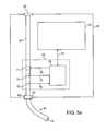
- FIG. 5 ashows an alternative embodiment of a one-way flow element comprising a valve controller coupled to sensors and an electrically-controlled valve.
- FIG. 5 bshows an external console housing the one-way flow element shown in FIG. 5 a.
- FIG. 6 ashows a flowchart and FIG. 6 b show flow and pressure graphs, illustrating the operation of the one-way flow element shown in FIG. 5 a.
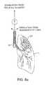
- FIG. 7illustrates the trans-esophageal endobronchial placement of the catheter of FIG. 1 in an airway leading to a diseased lung region in accordance with the principles of the present invention.
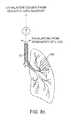
- FIGS. 8 a -8 dillustrate use of the catheter as placed in FIG. 7 for isolating and reduction of the volume of the diseased lung region in accordance with the principles of the present invention.
- FIG. 9is a graph showing the relationship between collateral resistance Rcoll and residual volume reduction in an isolated lung compartment.
- an endobronchial lung volume reduction catheter 10constructed in accordance with the principles of the present invention includes an elongate catheter body 12 having a distal end 14 and a proximal end 16 .
- Catheter body 12includes at least one lumen or central passage 18 extending generally from the distal end 14 to the proximal end 16 .
- Lumen 18will have a distal opening 19 at or near the distal end 14 in order to permit air or other lung gases to enter the lumen and flow in a distal-to-proximal direction out through the proximal end of the lumen.
- catheter body 12will have an expandable occluding element 15 at or near the distal end 14 , to occlude an air passageway during treatment.
- the expandable occluding memberis disposed near the distal end of the catheter body to seal the passageway, while in an alternate embodiment the expandable occluding element forms a cover of the rim of the catheter lumen in order to seal the passageway, prevent or inhibit mucus entry into the lumen, and shield the passageway wall from the tip of the catheter.
- the expandable occluding membermay be transparent to allow viewing of the passageway.
- the expandable occluding element 15is located at or near the distal end 14 .
- the expandable occluding element 15is configured such that the proximal and distal ends of the expandable occluding element 15 are attached to the outer surface of the catheter body 12 .
- An auxiliary lumen 17 Aextends from the inflation port 17 to the occluding element 15 to provide for expansion of the occluding element.
- the expandable occluding element 15is disposed at the distal end of the catheter body 12 , and is configured to form a cover over the rim of the distal opening 19 of the catheter body 12 .
- the proximal end of the occluding element 15is attached to the outer surface of the catheter body 12 , while the inner surface of the occluding element 15 wraps over the rim of the catheter body 12 and is attached to the inner surface of the catheter body 12 .
- Inflation lumen 17 Ais used to inflate the occluding element 15 through inflation port 17 B.
- the occluding element 15When inflated, the occluding element 15 will form a cover (or “lip”), over the rim of the catheter body 12 , thereby preventing or inhibiting entry of mucus into the lumen 18 of the catheter, and preventing or inhibiting the opening 19 from contacting the walls of the passageway.
- the inflated occluding element 15also helps prevent or inhibit accidental placement of the catheter tip into an airway segment that is smaller than the intended airway segment.
- the occluding element 15 and the distal portion of the catheter body 12comprise a transparent material to enable viewing past the occluding element 15 .
- FIGS. 1 c and 1 dManufacture of the second embodiment of the catheter 10 is shown in FIGS. 1 c and 1 d .
- one end 15 A of the occluding element 15is circumferentially attached to the inner wall of the lumen, using any suitable technique such as thermal bonding or adhesive bonding.
- the occluding element 15is inverted over the catheter tip and catheter body 12 , as shown in FIG. 1 d .
- the second end 15 B of the occluding element 15which is now proximal to the tip of the catheter, is attached circumferentially to the outer surface of the catheter body 12 , using any suitable technique such as thermal bonding or adhesive bonding.
- the occluding element 15thus encloses the outer rim of the distal end of the catheter. Further, the occluding element 15 is configured such that it is fed for inflation by an inflation port 17 B leading from an inflation lumen 17 A. Though the figures describe a preformed balloon-like occluding element 15 , any suitable material of any shape may be used to manufacture the occluding element 15 in the described manner, as should be obvious to one of ordinary skill in the art. For example, as described above, some portion of the catheter body 12 and/or of the occluding element 15 may be configured to be transparent. Optionally, a hub 20 will be provided at the proximal end, for example as shown in FIG. 1 a , but the hub is not a necessary component of the catheter.
- catheter 10is configured to be introducible into the passageway via a viewing scope such as a bronchoscope (not shown).
- a viewing scopesuch as a bronchoscope (not shown).
- a catheter 10comprising one or more transparent components as described above, enables enhanced viewing of the body passageway during diagnostic or treatment procedures, by allowing a user to view the body passageway through the transparent occluding element 15 .
- a transparent occluding element 15could serve as a lens to be used in conjunction with the scope. When so used, light from the scope would interact with the occluding element 15 in such a manner as to enable more enhanced viewing than would be obtained without the use of a transparent occluding element 15 .
- Examples of such enhanced viewingcould include: obtaining wide angle or fish-eye views or a greater field of vision, telephoto properties (macro, zoom, etc.) or color filtration. These can be achieved by manipulating the material properties of the occluding element 15 .
- a cathetermay be equipped with a transparent expandable element similar to that shown in FIG. 1 b .
- the transparent expandable elementserves as an image enhancer or diagnostic lens, and need not be fully occlusive. Similar to the above description, when used in conjunction with a viewing scope, it would enable more enhanced diagnostic viewing than would be obtained without the use of a transparent expandable element. Examples of such enhanced viewing could include: obtaining wide angle or fish-eye views or a greater field of vision, telephoto properties (macro, zoom, etc.) or color filtration. These can be achieved by manipulating the material properties of the transparent expandable element.
- the transparent expandable elementmay be configured to allow for therapeutic procedures, such as delivery of a therapeutic electromagnetic energy (e.g., laser, infrared, etc.) to the lung or other tissue.
- a therapeutic electromagnetic energye.g., laser, infrared, etc.
- the surface, shape, material, size or other properties of the lenscan be chosen to allow a user to manipulate the therapeutic laser energy. For example a user could focus or diffuse the energy by moving the source of laser energy back and forth relative to the transparent expandable occluding element.
- the present inventionrelies on placement of a one-way flow element within or in-line with the lumen 18 so that flow from an isolated lung compartment or segment (as described hereinbelow) may occur in a distal-to-proximal direction but flow back into the lung compartment or segment is inhibited or blocked in the proximal-to-distal direction.
- a one-way flow element 22may be provided in the lumen 18 near the distal end 14 of the catheter body 12 , optionally being immediately proximal of the distal opening 19 .
- the one-way flow element 22is a duck-bill valve which opens as shown in broken line as the patient exhales to increase the pressure on the upstream or distal side of the one-way flow element 22 . As the patient inhales, the pressure on the upstream or distal side of the valve is reduced, drawing the valve leaflets closed as shown in full line.
- the one-way flow element 22could be provided anywhere else in the lumen 18 , and two, three, four, or more such valve structures could be included in order to provide redundancy.
- a one-way valve structure 26 in the form of a flap valvecould be provided within the hub 20 .
- the hub 20could be removable or permanently fixed to the catheter body 12 .
- Other structures for providing in-line flow controlcould also be utilized, as will be presently described.
- one-way flow functionalitymay be provided using an actively controlled one-way flow control assembly.
- One-way flowcan be controlled by measuring the flow and pressure through the lumen and using this information to determine the beginning and end of inhalation and exhalation cycles and thereby determining whether the valve should remain open or closed.
- the one-way flow control assemblyis provided as part of an external console attached in-line with the catheter lumen.
- the consolecomprises a channel for air flow to which the proximal end of the catheter connects via a standard connector. When the patient exhales, air is forced through the catheter lumen into the console's air channel, and then exits through an exhaust port of the console.
- the one-way flow control assemblycomprises a valve that is within or in-line with the catheter lumen and can be opened or closed by a valve controller to control the air flow through the air channel.
- the valve controlleropens and closes the valve based on input from flow and pressure sensors within or in-line with the catheter lumen.
- the sensorsmeasure the air flow and air pressure to detect the inhalation and exhalation cycles of the patient.
- the valve controlleropens the valve at the beginning of the exhalation cycle, and closes the valve at the beginning of the inhalation cycle.
- the valve controllermay control the valve electrically, magnetically, mechanically or through other means known in the art.
- FIG. 5 ashows an illustration of such an actively controlled one-way flow control assembly provided as part of an external console.
- the external console 60comprises an air channel 61 , a connector 62 , and an exhaust port 64 .
- Catheter 10is detachably coupled to air channel 61 using a standard connector 62 , such that air channel 61 is in-line with lumen 18 .
- a filter 63is provided between the air channel 61 and lumen 18 to maintain sterility of air channel 61 and promote reusability of console 60 .
- air flowing into air channel 61is expelled through exhaust port 64 .
- Console 60comprises a one-way flow assembly 70 in-line with lumen 18 of catheter 10 .
- One-way flow assembly 70comprises an electrically controlled valve 71 , a flow sensor 73 , a pressure sensor 74 , and a valve controller 75 .
- valve 71 , flow sensor 73 , and pressure sensor 74are disposed within air channel 61 .
- Valve controller 75provides one-way flow functionality by opening and closing valve 71 based on flow and pressure signals received from sensors 73 and 74 , respectively. When valve 71 is closed, it prevents air from flowing into the lumen of catheter 10 (during inhalation); during exhalation, valve 71 remains open and allows air to flow out of the isolated lung compartment.
- valve 71is a solenoid-based valve.
- valve 71may be any other valve that can be opened and closed via an electrical control signal.
- Flow sensor 73 and pressure sensor 74respectively, measure air flow and pressure in lumen 18 .
- Valve controller 75receives a flow indicator signal 76 from the flow sensor 73 and a pressure indicator signal 77 from pressure sensor 74 and produces a valve control signal 78 to open or close valve 71 .
- one or more of flow sensor 73 , pressure sensor 74 , and valve 71may reside within lumen 18 and be in communication with valve controller 75 via connections between the catheter 10 and console 60 .
- FIG. 5 bshows one embodiment of an external console 60 connected to catheter 10 .
- External console 60optionally comprises a visual display 79 that receives and displays flow and pressure data as sensed by sensors 73 and 74 , for example, via a connection 72 to the controller 75 .
- visual display 79is a touch-screen display allowing a user to interact with console 60 .
- FIGS. 6 a and 6 billustrate the operation of one-way flow assembly 70 .
- FIG. 6 ais a flowchart showing the operational steps of valve controller 75 as it produces the electrical valve control signal 78 to open or close valve 71 based on input from flow sensor 73 and pressure sensor 74 .
- FIG. 6 bis a graph showing exemplary signals generated by the flow sensor 73 (top panel) and pressure sensor 74 (bottom panel) during a series of respiration cycles.
- the flow and pressure direction during exhalationis herein referred to as the positive flow and pressure direction and plotted on the positive ordinate of the graphs in FIG. 6B
- the flow and pressure direction during inhalationis referred to as the negative flow direction and plotted on the negative ordinate of FIG. 6B .
- valve controller 75waits for the completion of an inhalation cycle, until flow sensor 73 indicates a flow value that is greater than a specified flow threshold value. This is shown as step 81 in FIG. 6 a and shown as the first flow and pressure cycle in FIG. 6B lasting for a period indicated as 81 p .
- the flow threshold valueis chosen to indicate the beginning of an exhalation cycle.
- FIGS. 6 a and 6 b and the present descriptionassume an exemplary flow threshold value of zero.
- the flow threshold valueis configurable to a value other than zero.
- step 82 in FIG. 6 a(also indicating the positive flow and pressure in FIG. 6 b ), valve controller 75 maintains valve 71 in an open state during exhalation until flow sensor 73 receives a flow value less than or equal to zero.
- step 82lasts for a period indicated as 82 p as long as flow sensor 73 senses an air flow value greater than zero.
- valve controller 75closes valve 71 in step 83 in FIG. 6 a and no air flows through the lumen into the lung compartment.
- Step 83occurs contemporaneously with the flow value reaching zero or lower at the point in time denoted 83 p .
- the flowreduces to zero at the end of exhalation, at which point valve controller 75 closes the valve 71 .
- valve controller 75refers to a pressure threshold value.
- the pressure threshold valueis chosen to indicate the beginning of an exhalation cycle. This value is configurable, and in what follows, an example pressure threshold value of zero is assumed.
- valve controller 75reopen valve 71 when the pressure increases to or above the pressure threshold value.
- the pressure as sensed and reported by pressure sensor 74 at the end of exhalationmay fluctuate around zero, causing chatter of valve 71 .
- valve controller 75maintains valve 71 in a closed state while the pressure remains above a specified minimum pressure value, denoted as min_pressure in FIGS. 6 a and 6 b .
- valve 71remains closed during the period 84 p.
- valve controller 75also monitors pressure to ensure that valve 71 will open if the patient starts exhalation prior to the pressure decreasing to below min_pressure, To this end, during step 84 , valve controller 75 is optionally configured to open valve 71 if pressure increases to a value that is above the pressure threshold value by an amount referred to as a safeguard offset value.
- the safeguard offset valueis configurable.
- step 85 in FIG. 6 aonce the pressure passes below “min_pressure”, valve controller 75 maintains valve 71 in a closed state until the pressure increases to or passes the pressure threshold value.
- step 85lasts the duration between the achievement of min_pressure in step 84 and the attainment of the pressure threshold value, with the period denoted as 85 p in FIG. 6 b.
- the valve controller 75opens the valve 71 at step 86 in FIG. 6 a .
- the opening of the valve in step 86occurs at point 86 p and is contemporaneous with the pressure increasing to or passing a zero value. This allows air to empty from the lung compartment in communication with lumen 18 .
- valve controller 75resumes operation at Step 82 (close valve 71 and prevent airflow into the target lung compartment), for a new respiration cycle, until the lung reduction process is terminated.
- FIG. 7Use of the endobronchial lung volume reduction catheter 10 to reduce the residual volume of a diseased region DR of a lung L is illustrated beginning in FIG. 7 .
- Catheter 10is introduced through the patient's mouth, down past the trachea T and into a lung L.
- the distal end 14 of the catheter 10is advanced to the main airway AW leading into the diseased region DR of the lung.
- Introduction and guidance of the cathetermay be achieved in conventional manners, such as described in commonly-owned U.S. Pat. Nos. 6,287,290; 6,398,775; and 6,527,761, the full disclosures of which are incorporated herein by reference.
- an expandable occluding element 15is expanded to occlude the airway.
- the expandable occluding elementmay be a balloon, cuff, or a braided balloon as described in copending applications 60/823,734, filed on Aug. 28, 2006, and 60/828,496 filed on Oct. 6, 2006, the full disclosures of which are incorporated herein by reference.
- the only path between the atmosphere and the diseased region DR of the lungis through the lumen 18 of the catheter 10 .
- the patient exhalesas shown in FIG.
- air from the diseased region DRflows outwardly through the lumen 18 and the one-way flow element 22 , one-way flow assembly 70 , or any other one-way flow structure, causing a reduction in residual air within the region and a consequent reduction in volume.
- Air from the remainder of the lungalso passes outward in the annular region around the catheter 10 in a normal manner.
- the airway AW feeding the diseased region DRcan be occluded, by applying heat, radiofrequency energy, glues, or preferably by implanting an occluding device 30 , as shown in FIG. 8D .
- Implantation of the occluding device 30may be achieved by any of the techniques described in commonly-owned U.S. Pat. Nos. 6,287,290; and 6,527,761, the full disclosures of which have been previously incorporated herein by reference.
Landscapes
- Health & Medical Sciences (AREA)
- Life Sciences & Earth Sciences (AREA)
- Surgery (AREA)
- Veterinary Medicine (AREA)
- General Health & Medical Sciences (AREA)
- Biomedical Technology (AREA)
- Heart & Thoracic Surgery (AREA)
- Public Health (AREA)
- Engineering & Computer Science (AREA)
- Animal Behavior & Ethology (AREA)
- Molecular Biology (AREA)
- Medical Informatics (AREA)
- Pulmonology (AREA)
- Physics & Mathematics (AREA)
- Biophysics (AREA)
- Pathology (AREA)
- Nuclear Medicine, Radiotherapy & Molecular Imaging (AREA)
- Vascular Medicine (AREA)
- Reproductive Health (AREA)
- Hematology (AREA)
- Anesthesiology (AREA)
- Emergency Medicine (AREA)
- Physiology (AREA)
- Otolaryngology (AREA)
- Optics & Photonics (AREA)
- Radiology & Medical Imaging (AREA)
- Media Introduction/Drainage Providing Device (AREA)
- Surgical Instruments (AREA)
- Measurement Of The Respiration, Hearing Ability, Form, And Blood Characteristics Of Living Organisms (AREA)
- Ventilation (AREA)
Abstract
Description
Claims (17)
Priority Applications (3)
| Application Number | Priority Date | Filing Date | Title |
|---|---|---|---|
| US14/703,670US10314992B2 (en) | 2007-03-12 | 2015-05-04 | Methods and devices for passive residual lung volume reduction and functional lung volume expansion |
| US16/428,544US11298489B2 (en) | 2007-03-12 | 2019-05-31 | Methods and devices for passive residual lung volume reduction and functional lung volume expansion |
| US17/688,498US12350428B2 (en) | 2007-03-12 | 2022-03-07 | Methods and devices for passive residual lung volume reduction and functional lung volume expansion |
Applications Claiming Priority (4)
| Application Number | Priority Date | Filing Date | Title |
|---|---|---|---|
| US11/685,008US20080228137A1 (en) | 2007-03-12 | 2007-03-12 | Methods and devices for passive residual lung volume reduction and functional lung volume expansion |
| PCT/US2008/056706WO2008112797A2 (en) | 2007-03-12 | 2008-03-12 | Methods and devices for passive residual lung volume reduction and functional lung volume expansion |
| US12/407,709US9050094B2 (en) | 2007-03-12 | 2009-03-19 | Methods and devices for passive residual lung volume reduction and functional lung volume expansion |
| US14/703,670US10314992B2 (en) | 2007-03-12 | 2015-05-04 | Methods and devices for passive residual lung volume reduction and functional lung volume expansion |
Related Parent Applications (1)
| Application Number | Title | Priority Date | Filing Date |
|---|---|---|---|
| US12/407,709DivisionUS9050094B2 (en) | 2007-03-12 | 2009-03-19 | Methods and devices for passive residual lung volume reduction and functional lung volume expansion |
Related Child Applications (1)
| Application Number | Title | Priority Date | Filing Date |
|---|---|---|---|
| US16/428,544ContinuationUS11298489B2 (en) | 2007-03-12 | 2019-05-31 | Methods and devices for passive residual lung volume reduction and functional lung volume expansion |
Publications (2)
| Publication Number | Publication Date |
|---|---|
| US20150231353A1 US20150231353A1 (en) | 2015-08-20 |
| US10314992B2true US10314992B2 (en) | 2019-06-11 |
Family
ID=39760381
Family Applications (5)
| Application Number | Title | Priority Date | Filing Date |
|---|---|---|---|
| US11/685,008AbandonedUS20080228137A1 (en) | 2005-01-20 | 2007-03-12 | Methods and devices for passive residual lung volume reduction and functional lung volume expansion |
| US12/407,709Active2030-10-07US9050094B2 (en) | 2007-03-12 | 2009-03-19 | Methods and devices for passive residual lung volume reduction and functional lung volume expansion |
| US14/703,670Active2029-02-19US10314992B2 (en) | 2007-03-12 | 2015-05-04 | Methods and devices for passive residual lung volume reduction and functional lung volume expansion |
| US16/428,544ActiveUS11298489B2 (en) | 2007-03-12 | 2019-05-31 | Methods and devices for passive residual lung volume reduction and functional lung volume expansion |
| US17/688,498Active2029-12-13US12350428B2 (en) | 2007-03-12 | 2022-03-07 | Methods and devices for passive residual lung volume reduction and functional lung volume expansion |
Family Applications Before (2)
| Application Number | Title | Priority Date | Filing Date |
|---|---|---|---|
| US11/685,008AbandonedUS20080228137A1 (en) | 2005-01-20 | 2007-03-12 | Methods and devices for passive residual lung volume reduction and functional lung volume expansion |
| US12/407,709Active2030-10-07US9050094B2 (en) | 2007-03-12 | 2009-03-19 | Methods and devices for passive residual lung volume reduction and functional lung volume expansion |
Family Applications After (2)
| Application Number | Title | Priority Date | Filing Date |
|---|---|---|---|
| US16/428,544ActiveUS11298489B2 (en) | 2007-03-12 | 2019-05-31 | Methods and devices for passive residual lung volume reduction and functional lung volume expansion |
| US17/688,498Active2029-12-13US12350428B2 (en) | 2007-03-12 | 2022-03-07 | Methods and devices for passive residual lung volume reduction and functional lung volume expansion |
Country Status (5)
| Country | Link |
|---|---|
| US (5) | US20080228137A1 (en) |
| EP (1) | EP2121091B1 (en) |
| JP (1) | JP5452238B2 (en) |
| ES (1) | ES2713187T3 (en) |
| WO (1) | WO2008112797A2 (en) |
Cited By (3)
| Publication number | Priority date | Publication date | Assignee | Title |
|---|---|---|---|---|
| US11298489B2 (en)* | 2007-03-12 | 2022-04-12 | Pulmonx Corporation | Methods and devices for passive residual lung volume reduction and functional lung volume expansion |
| US11413045B2 (en) | 2005-01-20 | 2022-08-16 | Pulmonx Corporation | Methods and devices for passive residual lung volume reduction and functional lung volume expansion |
| US11883029B2 (en) | 2005-01-20 | 2024-01-30 | Pulmonx Corporation | Methods and devices for passive residual lung volume reduction and functional lung volume expansion |
Families Citing this family (32)
| Publication number | Priority date | Publication date | Assignee | Title |
|---|---|---|---|---|
| FR2858236B1 (en) | 2003-07-29 | 2006-04-28 | Airox | DEVICE AND METHOD FOR SUPPLYING RESPIRATORY GAS IN PRESSURE OR VOLUME |
| US8302602B2 (en) | 2008-09-30 | 2012-11-06 | Nellcor Puritan Bennett Llc | Breathing assistance system with multiple pressure sensors |
| US8444635B2 (en)* | 2008-11-19 | 2013-05-21 | Samuel Victor Lichtenstein | Methods for selectively heating tissue |
| US8434479B2 (en) | 2009-02-27 | 2013-05-07 | Covidien Lp | Flow rate compensation for transient thermal response of hot-wire anemometers |
| US8439036B2 (en) | 2009-12-01 | 2013-05-14 | Covidien Lp | Exhalation valve assembly with integral flow sensor |
| US8469031B2 (en) | 2009-12-01 | 2013-06-25 | Covidien Lp | Exhalation valve assembly with integrated filter |
| US8439037B2 (en) | 2009-12-01 | 2013-05-14 | Covidien Lp | Exhalation valve assembly with integrated filter and flow sensor |
| US8469030B2 (en)* | 2009-12-01 | 2013-06-25 | Covidien Lp | Exhalation valve assembly with selectable contagious/non-contagious latch |
| US20110301414A1 (en)* | 2010-06-04 | 2011-12-08 | Robert Hotto | Intelligent endoscopy systems and methods |
| US9629971B2 (en) | 2011-04-29 | 2017-04-25 | Covidien Lp | Methods and systems for exhalation control and trajectory optimization |
| US9364624B2 (en) | 2011-12-07 | 2016-06-14 | Covidien Lp | Methods and systems for adaptive base flow |
| US9498589B2 (en) | 2011-12-31 | 2016-11-22 | Covidien Lp | Methods and systems for adaptive base flow and leak compensation |
| US10272226B2 (en)* | 2012-04-13 | 2019-04-30 | Fresca Medical, Inc. | Auto-feedback valve for a sleep apnea device |
| US9144658B2 (en) | 2012-04-30 | 2015-09-29 | Covidien Lp | Minimizing imposed expiratory resistance of mechanical ventilator by optimizing exhalation valve control |
| US20130310771A1 (en)* | 2012-05-15 | 2013-11-21 | Daniel Karlin | Medical conduit protection devices, systems and methods |
| AU2013290002A1 (en)* | 2012-07-13 | 2015-01-29 | The Henry M. Jackson Foundation For The Advancement Of Military Medicine, Inc | Infrared illuminated airway management devices and kits and methods for using the same |
| USD731049S1 (en) | 2013-03-05 | 2015-06-02 | Covidien Lp | EVQ housing of an exhalation module |
| USD744095S1 (en) | 2013-03-08 | 2015-11-24 | Covidien Lp | Exhalation module EVQ internal flow sensor |
| USD731048S1 (en) | 2013-03-08 | 2015-06-02 | Covidien Lp | EVQ diaphragm of an exhalation module |
| USD701601S1 (en) | 2013-03-08 | 2014-03-25 | Covidien Lp | Condensate vial of an exhalation module |
| USD736905S1 (en) | 2013-03-08 | 2015-08-18 | Covidien Lp | Exhalation module EVQ housing |
| USD731065S1 (en) | 2013-03-08 | 2015-06-02 | Covidien Lp | EVQ pressure sensor filter of an exhalation module |
| USD693001S1 (en) | 2013-03-08 | 2013-11-05 | Covidien Lp | Neonate expiratory filter assembly of an exhalation module |
| USD692556S1 (en) | 2013-03-08 | 2013-10-29 | Covidien Lp | Expiratory filter body of an exhalation module |
| US9950135B2 (en) | 2013-03-15 | 2018-04-24 | Covidien Lp | Maintaining an exhalation valve sensor assembly |
| US9681938B2 (en) | 2013-04-26 | 2017-06-20 | Boston Scientific Scimed, Inc. | Devices for obstructing passage of air or other contaminants into a portion of a lung and methods of use |
| GB201401231D0 (en)* | 2014-01-24 | 2014-03-12 | Smiths Medical Int Ltd | Tracheal tubes |
| USD775345S1 (en) | 2015-04-10 | 2016-12-27 | Covidien Lp | Ventilator console |
| US10980737B1 (en) | 2016-03-08 | 2021-04-20 | Samuel Victor Lichtenstein | System for treating unwanted tissue using heat and heat activated drugs |
| WO2021107903A1 (en)* | 2019-11-26 | 2021-06-03 | Istanbul Medipol Universitesi | Hourglass balloon for transcatheter aortic valve implantation |
| US11896767B2 (en) | 2020-03-20 | 2024-02-13 | Covidien Lp | Model-driven system integration in medical ventilators |
| CN111743779B (en)* | 2020-06-11 | 2023-04-11 | 湘南学院附属医院 | Auxiliary tube feeding device and method for medical nasogastric feeding tube for gastrointestinal surgical nursing |
Citations (144)
| Publication number | Priority date | Publication date | Assignee | Title |
|---|---|---|---|---|
| US3322126A (en) | 1963-04-19 | 1967-05-30 | Willy Rusch Fa | Endotracheal catheter |
| US3498286A (en) | 1966-09-21 | 1970-03-03 | American Optical Corp | Catheters |
| US3669098A (en) | 1968-10-05 | 1972-06-13 | Olympus Optical Co | Endotracheal tube |
| US3677262A (en) | 1970-07-23 | 1972-07-18 | Henry J Zukowski | Surgical instrument illuminating endotracheal tube inserter |
| US3768504A (en) | 1972-06-19 | 1973-10-30 | S Rentsch | Check valve for use with a snorkel type breathing tube |
| US3776222A (en) | 1971-12-23 | 1973-12-04 | Lurosso A | Fiber optic entubator and method of entubation of the trachea through the nasopharynx |
| US3794026A (en) | 1970-07-29 | 1974-02-26 | H Jacobs | Ventilating apparatus embodying selective volume or pressure operation and catheter means for use therewith |
| US3866599A (en) | 1972-01-21 | 1975-02-18 | Univ Washington | Fiberoptic catheter |
| US3913568A (en) | 1973-01-22 | 1975-10-21 | American Optical Corp | Nasopharyngoscope |
| US4041936A (en) | 1975-04-23 | 1977-08-16 | Medical Engineering Corporation | Bronchoscopy tube |
| US4134407A (en) | 1977-03-25 | 1979-01-16 | Elam James O | External pressure-volume monitor for endotracheal cuff |
| US4147169A (en) | 1977-05-02 | 1979-04-03 | The Kendall Company | Balloon catheter with balloon retaining sleeves |
| US4327721A (en) | 1978-07-07 | 1982-05-04 | George Hanover | Endotracheal tube with topical agent delivery system and method of using the same |
| US4327720A (en) | 1979-01-22 | 1982-05-04 | Bronson Paul A | Esophageal-endotracheal airway |
| US4382442A (en) | 1978-04-24 | 1983-05-10 | Jones James W | Thoracostomy pump-tube apparatus |
| US4453545A (en) | 1981-05-07 | 1984-06-12 | Hiroshi Inoue | Endotracheal tube with movable endobronchial blocker for one-lung anesthesia |
| US4468216A (en) | 1982-05-20 | 1984-08-28 | Rudolph Muto | Irrigation suction catheter |
| US4470407A (en)* | 1982-03-11 | 1984-09-11 | Laserscope, Inc. | Endoscopic device |
| US4538607A (en) | 1984-02-06 | 1985-09-03 | Ab Fixfabriken | Tracheostomy valve |
| US4567882A (en) | 1982-12-06 | 1986-02-04 | Vanderbilt University | Method for locating the illuminated tip of an endotracheal tube |
| US4681093A (en) | 1982-12-13 | 1987-07-21 | Sumitomo Electric Industries, Ltd. | Endoscope |
| US4716896A (en) | 1986-08-01 | 1988-01-05 | Ackrad Laboratories | Bronchial catheter |
| US4742819A (en) | 1987-03-23 | 1988-05-10 | George Gordon P | Intubating scope with camera and screen |
| US4784133A (en) | 1987-01-28 | 1988-11-15 | Mackin Robert A | Working well balloon angioscope and method |
| US4796639A (en) | 1987-11-05 | 1989-01-10 | Medical Graphics Corporation | Pulmonary diagnostic system |
| US4819664A (en) | 1984-11-15 | 1989-04-11 | Stefano Nazari | Device for selective bronchial intubation and separate lung ventilation, particularly during anesthesia, intensive therapy and reanimation |
| US4846153A (en) | 1988-06-10 | 1989-07-11 | George Berci | Intubating video endoscope |
| US4850371A (en) | 1988-06-13 | 1989-07-25 | Broadhurst John H | Novel endotracheal tube and mass spectrometer |
| US4852568A (en) | 1987-02-17 | 1989-08-01 | Kensey Nash Corporation | Method and apparatus for sealing an opening in tissue of a living being |
| US4862874A (en) | 1987-06-10 | 1989-09-05 | Kellner Hans Joerg | Endoscope for removal of thrombi from pulmonary arterial vessels |
| US4896941A (en) | 1985-04-27 | 1990-01-30 | Doryokuro Kakunenryo Kaihatsu Jigyodan | Image-transmitting fiber |
| US4949716A (en) | 1988-10-31 | 1990-08-21 | Medical Devices, Inc. | Nasal intubation adjunct |
| US4955375A (en) | 1989-01-23 | 1990-09-11 | Ricardo Martinez | Endotracheal tube with channel for delivering drugs |
| US4958932A (en) | 1988-08-18 | 1990-09-25 | Mcdonnell Douglas Corporation | Optical measuring apparatus |
| US4961738A (en) | 1987-01-28 | 1990-10-09 | Mackin Robert A | Angioplasty catheter with illumination and visualization within angioplasty balloon |
| US4976710A (en) | 1987-01-28 | 1990-12-11 | Mackin Robert A | Working well balloon method |
| US5056529A (en) | 1990-04-03 | 1991-10-15 | Groot William J De | Apparatus and method for performing a transbroncheal biopsy |
| WO1992010971A1 (en) | 1990-12-21 | 1992-07-09 | Ballard Medical Products | Bronchoalveolar lavage catheter |
| US5143062A (en) | 1990-10-26 | 1992-09-01 | Mallinckrodt Medical, Inc. | Endotracheal tube having irrigation means |
| US5146916A (en) | 1990-01-05 | 1992-09-15 | Catalani Angelo S | Endotracheal tube incorporating a drug-irrigation device |
| US5165420A (en) | 1990-12-21 | 1992-11-24 | Ballard Medical Products | Bronchoalveolar lavage catheter |
| US5181913A (en) | 1987-03-09 | 1993-01-26 | Prn Services, Inc. | Catheter with check valve and rolled sheath |
| US5285778A (en) | 1991-04-19 | 1994-02-15 | Mackin Robert A | Endotracheal tube wih fibers optic illumination and viewing and auxiliary tube |
| US5308325A (en) | 1991-01-28 | 1994-05-03 | Corpak, Inc. | Retention balloon for percutaneous catheter |
| US5309903A (en) | 1989-12-12 | 1994-05-10 | Burroughs Wellcome Co. | Method for administering surfactant to the lungs while concurrently providing one-lung ventilation |
| US5329940A (en) | 1990-02-14 | 1994-07-19 | Adair Edwin Lloyd | Endotracheal tube intubation assist device |
| US5331947A (en) | 1992-05-01 | 1994-07-26 | Shturman Cardiology Systems, Inc. | Inflatable sheath for introduction of ultrasonic catheter through the lumen of a fiber optic endoscope |
| US5361753A (en) | 1992-07-07 | 1994-11-08 | Deutsche Aerospace Ag | Method of measuring and regulating the pressure in the sealing cuff of a tracheal tube and apparatus for implementing the method |
| US5447165A (en) | 1991-09-27 | 1995-09-05 | Gustafsson; Lars E. | Method for ascertaining prevailing lung condition and a device |
| WO1995033506A1 (en) | 1994-06-04 | 1995-12-14 | Archibald Ian Jeremy Brain | A fibreoptic intubating laryngeal mask airway |
| US5477851A (en) | 1995-01-26 | 1995-12-26 | Callaghan; Eric B. | Laryngeal mask assembly and method for removing same |
| US5499625A (en) | 1994-01-27 | 1996-03-19 | The Kendall Company | Esophageal-tracheal double lumen airway |
| US5546935A (en)* | 1993-03-09 | 1996-08-20 | Medamicus, Inc. | Endotracheal tube mounted pressure transducer |
| US5588424A (en) | 1995-06-28 | 1996-12-31 | The Cleveland Clinic Foundation | Bronchial blocker endotracheal apparatus |
| US5598840A (en) | 1995-03-17 | 1997-02-04 | Sorenson Critical Care, Inc. | Apparatus and method for ventilation and aspiration |
| US5624449A (en) | 1993-11-03 | 1997-04-29 | Target Therapeutics | Electrolytically severable joint for endovascular embolic devices |
| US5642730A (en) | 1994-06-17 | 1997-07-01 | Trudell Medical Limited | Catheter system for delivery of aerosolized medicine for use with pressurized propellant canister |
| US5645519A (en) | 1994-03-18 | 1997-07-08 | Jai S. Lee | Endoscopic instrument for controlled introduction of tubular members in the body and methods therefor |
| US5653231A (en) | 1995-11-28 | 1997-08-05 | Medcare Medical Group, Inc. | Tracheostomy length single use suction catheter |
| US5660175A (en) | 1995-08-21 | 1997-08-26 | Dayal; Bimal | Endotracheal device |
| EP0791340A1 (en) | 1996-02-22 | 1997-08-27 | Cordis Corporation | Temporary filter catheter |
| US5662712A (en)* | 1993-04-28 | 1997-09-02 | Focal, Inc. | Apparatus for intraluminal photothermoforming |
| US5682880A (en) | 1996-07-26 | 1997-11-04 | Brain; Archibald Ian Jeremy | Laryngeal-mask airway with guide element, stiffener, and fiberoptic access |
| EP0815803A1 (en) | 1996-07-03 | 1998-01-07 | Cordis Europa N.V. | Catheter with temporary vena cava filter |
| US5707352A (en) | 1989-08-28 | 1998-01-13 | Alliance Pharmaceutical Corp. | Pulmonary delivery of therapeutic agent |
| US5752921A (en) | 1996-01-11 | 1998-05-19 | Korr Medical Technologies, Inc. | Method and apparatus for determining tracheal pressure |
| US5765557A (en) | 1995-03-17 | 1998-06-16 | Board Of Regents, The University Of Texas System | Method and apparatus for directing air flow within an intubated patient |
| US5795322A (en) | 1995-04-10 | 1998-08-18 | Cordis Corporation | Catheter with filter and thrombus-discharge device |
| US5800455A (en) | 1993-04-19 | 1998-09-01 | Target Therapeutics, Inc. | Detachable embolic coil assembly |
| WO1998044854A1 (en) | 1997-04-07 | 1998-10-15 | Broncus Technologies, Inc. | Bronchial stenter |
| WO1998049191A1 (en) | 1997-04-28 | 1998-11-05 | The Scripps Research Institute | Novel pulmonary surfactants and therapeutic uses, including pulmonary lavage |
| WO1998048706A1 (en) | 1997-04-30 | 1998-11-05 | Bradford Hospitals Nhs Trust | Occlusion device |
| WO1999001076A1 (en) | 1997-07-02 | 1999-01-14 | Broncus Technologies, Inc. | Bleb reducer |
| US5893841A (en) | 1996-08-30 | 1999-04-13 | Delcath Systems, Inc. | Balloon catheter with occluded segment bypass |
| WO1999017827A2 (en) | 1997-10-03 | 1999-04-15 | Scimed Life Systems, Inc. | Braided angiography catheter having full length radiopacity and controlled flexibility |
| US5897528A (en) | 1998-04-30 | 1999-04-27 | Medtronic, Inc. | Filtered intracerebroventricular or intraspinal access port with direct cerebrospinal fluid access |
| WO1999020332A1 (en) | 1997-10-20 | 1999-04-29 | Christopher Kent L | System for monitoring and treating sleep disorders using a transtracheal catheter |
| US5915383A (en) | 1997-04-29 | 1999-06-29 | Smiths Industries Public Limited Company | Cuffed medico-surgical tubes |
| WO1999032040A1 (en) | 1997-12-19 | 1999-07-01 | Broncus Technologies, Inc. | Bronchial stenter |
| WO1999034741A1 (en) | 1998-01-07 | 1999-07-15 | Broncus Technologies, Inc. | Bronchial stenter having diametrically adjustable electrodes |
| WO1999064109A1 (en) | 1998-06-10 | 1999-12-16 | Broncus Technologies, Inc. | Smooth muscle treatment apparatus and method |
| EP0982044A2 (en) | 1998-08-26 | 2000-03-01 | Instrumentarium Corporation | Method and apparatus for detecting an empty gas compartment in a patient ventilator |
| WO2000041612A2 (en) | 1999-01-11 | 2000-07-20 | Libra Medical Systems, Inc. | Apparatus and methods for treating congestive heart disease |
| WO2000051510A1 (en) | 1999-03-01 | 2000-09-08 | Broncus Technologies, Inc. | Bronchial stenter having expandable electrodes |
| WO2000062699A2 (en) | 1999-04-21 | 2000-10-26 | Broncus Technologies, Inc. | Modification of airways by application of energy |
| WO2001002042A1 (en) | 1999-07-02 | 2001-01-11 | Pulmonx | Methods, systems, and kits for lung volume reduction |
| US6174307B1 (en) | 1996-03-29 | 2001-01-16 | Eclipse Surgical Technologies, Inc. | Viewing surgical scope for minimally invasive procedures |
| US6174323B1 (en) | 1998-06-05 | 2001-01-16 | Broncus Technologies, Inc. | Method and assembly for lung volume reduction |
| WO2001003642A1 (en) | 1999-07-08 | 2001-01-18 | Broncus Technologies, Inc. | Increasing gas exchange of a lung |
| WO2001010314A2 (en) | 1999-08-05 | 2001-02-15 | Broncus Technologies, Inc. | Methods and devices for creating collateral channels in the lungs |
| WO2001013908A2 (en) | 1999-08-23 | 2001-03-01 | Ingenito Edward P | Tissue volume reduction |
| WO2001013839A1 (en) | 1999-08-24 | 2001-03-01 | Spiration, Inc. | Lung reduction device, system, and method |
| USRE37117E1 (en) | 1992-09-22 | 2001-03-27 | Target Therapeutics, Inc. | Detachable embolic coil assembly using interlocking clasps and method of use |
| US20010051899A1 (en) | 2000-06-13 | 2001-12-13 | Takahiko Kawashima | Document managing apparatus for managing transaction slip data in electronic commerce |
| US6346074B1 (en)* | 1993-02-22 | 2002-02-12 | Heartport, Inc. | Devices for less invasive intracardiac interventions |
| US6398775B1 (en) | 1999-10-21 | 2002-06-04 | Pulmonx | Apparatus and method for isolated lung access |
| US20020169413A1 (en) | 1999-01-11 | 2002-11-14 | Libra Medical Systems, Inc. | Apparatus and methods for treating congestive heart disease |
| US6527761B1 (en) | 2000-10-27 | 2003-03-04 | Pulmonx, Inc. | Methods and devices for obstructing and aspirating lung tissue segments |
| WO2003022221A2 (en) | 2001-09-10 | 2003-03-20 | Pulmonx | Method and apparatus for endobronchial diagnosis |
| WO2003022124A2 (en) | 2001-09-11 | 2003-03-20 | Spiration, Inc. | Removable lung reduction devices, systems, and methods |
| US6585639B1 (en) | 2000-10-27 | 2003-07-01 | Pulmonx | Sheath and method for reconfiguring lung viewing scope |
| US6609521B1 (en) | 2001-04-09 | 2003-08-26 | Regents Of The University Of Minnesota | Endotracheal tube |
| US20030171332A1 (en) | 2001-05-23 | 2003-09-11 | Abraham William M. | Treatment of respiratory conditions associated with bronchoconstriction with aerosolized hyaluronic acid |
| US20030228344A1 (en) | 2002-03-08 | 2003-12-11 | Fields Antony J. | Methods and devices for inducing collapse in lung regions fed by collateral pathways |
| US6679264B1 (en)* | 2000-03-04 | 2004-01-20 | Emphasys Medical, Inc. | Methods and devices for use in performing pulmonary procedures |
| US6712812B2 (en) | 1999-08-05 | 2004-03-30 | Broncus Technologies, Inc. | Devices for creating collateral channels |
| US6722360B2 (en) | 2000-06-16 | 2004-04-20 | Rajiv Doshi | Methods and devices for improving breathing in patients with pulmonary disease |
| US6749606B2 (en) | 1999-08-05 | 2004-06-15 | Thomas Keast | Devices for creating collateral channels |
| US6792947B1 (en) | 2000-08-25 | 2004-09-21 | O-Two Systems International Inc. | Flow control valve for manual resuscitator devices |
| US20040243016A1 (en) | 2001-08-29 | 2004-12-02 | Sanderson Penelope Margaret | Method and means of physiological monitoring using sonification |
| US20050016530A1 (en) | 2003-07-09 | 2005-01-27 | Mccutcheon John | Treatment planning with implantable bronchial isolation devices |
| US20050022809A1 (en) | 2003-04-25 | 2005-02-03 | Wondka Anthony David | Methods, systems and devices for desufflating a lung area |
| US6886558B2 (en) | 2002-08-28 | 2005-05-03 | Cordis Corporation | Collateral ventilation bypass trap system |
| US20050126572A1 (en) | 2003-12-11 | 2005-06-16 | Safety Tech International Inc. | Pneumatic sealing system for protection masks |
| US20050166924A1 (en) | 2003-05-06 | 2005-08-04 | Ralph Thomas | Multiple cannula systems and methods |
| US20050187561A1 (en) | 2004-02-25 | 2005-08-25 | Femasys, Inc. | Methods and devices for conduit occlusion |
| US6941950B2 (en) | 2001-10-11 | 2005-09-13 | Emphasys Medical, Inc. | Bronchial flow control devices and methods of use |
| US20050288684A1 (en) | 2004-06-16 | 2005-12-29 | Aronson Nathan A | Method of reducing collateral flow in a portion of a lung |
| US6997189B2 (en) | 1998-06-05 | 2006-02-14 | Broncus Technologies, Inc. | Method for lung volume reduction |
| US7011094B2 (en) | 2001-03-02 | 2006-03-14 | Emphasys Medical, Inc. | Bronchial flow control devices and methods of use |
| US7022088B2 (en) | 1999-08-05 | 2006-04-04 | Broncus Technologies, Inc. | Devices for applying energy to tissue |
| US20060102186A1 (en) | 2004-11-18 | 2006-05-18 | Mark Adler | Intra-bronchial apparatus for aspiration and insufflation of lung regions distal to placement or cross communication and deployment and placement system therefor |
| WO2006055692A2 (en) | 2004-11-16 | 2006-05-26 | Pulmonx | Pulmonary occlusal stent delivery catheter, loading system and methods of use |
| US20060122647A1 (en) | 2004-09-24 | 2006-06-08 | Callaghan David J | Occluder device double securement system for delivery/recovery of such occluder device |
| US20060129134A1 (en) | 2004-11-29 | 2006-06-15 | Andrew Kerr | Dialysis catheter |
| WO2006078451A2 (en) | 2005-01-20 | 2006-07-27 | Pulmonx | Minimally invasive determination of collateral ventilation in lungs |
| US7086398B2 (en) | 2002-07-31 | 2006-08-08 | Cordis Corporation | Long term oxygen therapy system |
| WO2006091597A1 (en) | 2005-02-22 | 2006-08-31 | Cardiofocus, Inc. | Deflectable sheath catheters |
| EP1078601B1 (en) | 1999-08-24 | 2006-10-04 | Spiration, Inc. | Kit for lung volume reduction |
| US20060264772A1 (en) | 2001-09-10 | 2006-11-23 | Pulmonx | Minimally invasive determination of collateral ventilation in lungs |
| US20070096048A1 (en) | 2005-10-14 | 2007-05-03 | Claude Clerc | Bronchoscopic lung volume reduction valve |
| US20070142742A1 (en) | 2005-07-13 | 2007-06-21 | Pulmonx | Methods and systems for segmental lung diagnostics |
| US20070225747A1 (en) | 2006-03-08 | 2007-09-27 | Pulmonx | Methods and devices to induce controlled atelectasis and hypoxic pulmonary vasoconstriction |
| US7276077B2 (en) | 1997-09-16 | 2007-10-02 | Emphasys Medical, Inc. | Body fluid flow control device |
| US20080051719A1 (en) | 2006-08-28 | 2008-02-28 | Pulmonx | Functional assessment and treatment catheters and methods for their use in the lung |
| US20080228130A1 (en) | 2007-03-12 | 2008-09-18 | Pulmonx | Methods and systems for occluding collateral flow channels in the lung |
| US20080228137A1 (en) | 2007-03-12 | 2008-09-18 | Pulmonx | Methods and devices for passive residual lung volume reduction and functional lung volume expansion |
| US7449010B1 (en) | 2002-07-15 | 2008-11-11 | Motoya Hayase | Material removal catheter and method |
| US7588033B2 (en) | 2003-06-18 | 2009-09-15 | Breathe Technologies, Inc. | Methods, systems and devices for improving ventilation in a lung area |
| US20100031964A1 (en) | 2008-08-11 | 2010-02-11 | Joseph William Turek | Flow control adapter for performing spirometry and pulmonary function testing |
| US20110011406A1 (en) | 2005-12-27 | 2011-01-20 | Hansa Medical Products, Inc. | Valved Fenestrated Tracheotomy Tube Having Outer and Inner Cannulae |
| US20110152678A1 (en) | 2005-01-20 | 2011-06-23 | Pulmonx Corporation | Methods and devices for passive residual lung volume reduction and functional lung volume expansion |
| US20110203594A1 (en) | 2001-08-23 | 2011-08-25 | Indian Ocean Medical Inc. | Disposable laryngeal mask airway device |
| US20110259339A1 (en) | 2006-09-11 | 2011-10-27 | Ric Investments, Llc | Ventilating apparatus and method enabling a patient to talk with or without a trachostomy tube check valve |
Family Cites Families (15)
| Publication number | Priority date | Publication date | Assignee | Title |
|---|---|---|---|---|
| US37117A (en) | 1862-12-09 | Improved composition for blasting-powder | ||
| US3050066A (en)* | 1958-12-31 | 1962-08-21 | Wilbur R Koehn | Retention catheters |
| JPS62211074A (en) | 1986-03-07 | 1987-09-17 | ボ−ド・オブ・リ−ジエンツ、ザ・ユニバ−シテイ−・オブ・テキサス・システム | Suction catheter with balloon tip |
| US5041089A (en)* | 1987-12-11 | 1991-08-20 | Devices For Vascular Intervention, Inc. | Vascular dilation catheter construction |
| US4946440A (en)* | 1988-10-05 | 1990-08-07 | Hall John E | Evertible membrane catheter and method of use |
| US5161525A (en) | 1990-05-11 | 1992-11-10 | Puritan-Bennett Corporation | System and method for flow triggering of pressure supported ventilation |
| US5439444A (en)* | 1991-01-28 | 1995-08-08 | Corpak, Inc. | Pre-formed member for percutaneous catheter |
| JP3838671B2 (en) | 1993-10-25 | 2006-10-25 | アークレイ株式会社 | Breath collection device |
| DE69839888D1 (en)* | 1997-11-12 | 2008-09-25 | Genesis Technologies Llc | DEVICE FOR REMOVING OCCLUSIONS IN BIOLOGICAL PASSES |
| US5997546A (en)* | 1999-01-07 | 1999-12-07 | Ballard Medical Products | Gastric balloon catheter with improved balloon orientation |
| US7175644B2 (en) | 2001-02-14 | 2007-02-13 | Broncus Technologies, Inc. | Devices and methods for maintaining collateral channels in tissue |
| US20020104544A1 (en) | 2000-11-30 | 2002-08-08 | Kuraray Co., Ltd. | Endotracheal tube |
| TWI235073B (en)* | 2002-08-20 | 2005-07-01 | Toray Industries | Catheter for treating cardiac arrhythmias |
| WO2006133239A2 (en)* | 2005-06-06 | 2006-12-14 | C.R. Bard, Inc. | Feeding device including balloon tip and method of manufacture |
| US8551043B2 (en)* | 2006-04-21 | 2013-10-08 | C. R. Bard, Inc. | Feeding device and bolster apparatus and method for making the same |
- 2007
- 2007-03-12USUS11/685,008patent/US20080228137A1/ennot_activeAbandoned
- 2008
- 2008-03-12EPEP08732032.1Apatent/EP2121091B1/enactiveActive
- 2008-03-12WOPCT/US2008/056706patent/WO2008112797A2/enactiveApplication Filing
- 2008-03-12JPJP2009553749Apatent/JP5452238B2/enactiveActive
- 2008-03-12ESES08732032Tpatent/ES2713187T3/enactiveActive
- 2009
- 2009-03-19USUS12/407,709patent/US9050094B2/enactiveActive
- 2015
- 2015-05-04USUS14/703,670patent/US10314992B2/enactiveActive
- 2019
- 2019-05-31USUS16/428,544patent/US11298489B2/enactiveActive
- 2022
- 2022-03-07USUS17/688,498patent/US12350428B2/enactiveActive
Patent Citations (174)
| Publication number | Priority date | Publication date | Assignee | Title |
|---|---|---|---|---|
| US3322126A (en) | 1963-04-19 | 1967-05-30 | Willy Rusch Fa | Endotracheal catheter |
| US3498286A (en) | 1966-09-21 | 1970-03-03 | American Optical Corp | Catheters |
| US3669098A (en) | 1968-10-05 | 1972-06-13 | Olympus Optical Co | Endotracheal tube |
| US3677262A (en) | 1970-07-23 | 1972-07-18 | Henry J Zukowski | Surgical instrument illuminating endotracheal tube inserter |
| US3794026A (en) | 1970-07-29 | 1974-02-26 | H Jacobs | Ventilating apparatus embodying selective volume or pressure operation and catheter means for use therewith |
| US3776222A (en) | 1971-12-23 | 1973-12-04 | Lurosso A | Fiber optic entubator and method of entubation of the trachea through the nasopharynx |
| US3866599A (en) | 1972-01-21 | 1975-02-18 | Univ Washington | Fiberoptic catheter |
| US3768504A (en) | 1972-06-19 | 1973-10-30 | S Rentsch | Check valve for use with a snorkel type breathing tube |
| US3913568A (en) | 1973-01-22 | 1975-10-21 | American Optical Corp | Nasopharyngoscope |
| US4041936A (en) | 1975-04-23 | 1977-08-16 | Medical Engineering Corporation | Bronchoscopy tube |
| US4134407A (en) | 1977-03-25 | 1979-01-16 | Elam James O | External pressure-volume monitor for endotracheal cuff |
| US4147169A (en) | 1977-05-02 | 1979-04-03 | The Kendall Company | Balloon catheter with balloon retaining sleeves |
| US4382442A (en) | 1978-04-24 | 1983-05-10 | Jones James W | Thoracostomy pump-tube apparatus |
| US4327721A (en) | 1978-07-07 | 1982-05-04 | George Hanover | Endotracheal tube with topical agent delivery system and method of using the same |
| US4327720A (en) | 1979-01-22 | 1982-05-04 | Bronson Paul A | Esophageal-endotracheal airway |
| US4453545A (en) | 1981-05-07 | 1984-06-12 | Hiroshi Inoue | Endotracheal tube with movable endobronchial blocker for one-lung anesthesia |
| US4470407A (en)* | 1982-03-11 | 1984-09-11 | Laserscope, Inc. | Endoscopic device |
| US4468216A (en) | 1982-05-20 | 1984-08-28 | Rudolph Muto | Irrigation suction catheter |
| US4567882A (en) | 1982-12-06 | 1986-02-04 | Vanderbilt University | Method for locating the illuminated tip of an endotracheal tube |
| US4681093A (en) | 1982-12-13 | 1987-07-21 | Sumitomo Electric Industries, Ltd. | Endoscope |
| US4538607A (en) | 1984-02-06 | 1985-09-03 | Ab Fixfabriken | Tracheostomy valve |
| US4819664A (en) | 1984-11-15 | 1989-04-11 | Stefano Nazari | Device for selective bronchial intubation and separate lung ventilation, particularly during anesthesia, intensive therapy and reanimation |
| US4896941A (en) | 1985-04-27 | 1990-01-30 | Doryokuro Kakunenryo Kaihatsu Jigyodan | Image-transmitting fiber |
| US4716896A (en) | 1986-08-01 | 1988-01-05 | Ackrad Laboratories | Bronchial catheter |
| US4961738A (en) | 1987-01-28 | 1990-10-09 | Mackin Robert A | Angioplasty catheter with illumination and visualization within angioplasty balloon |
| US4784133A (en) | 1987-01-28 | 1988-11-15 | Mackin Robert A | Working well balloon angioscope and method |
| US4976710A (en) | 1987-01-28 | 1990-12-11 | Mackin Robert A | Working well balloon method |
| US4852568A (en) | 1987-02-17 | 1989-08-01 | Kensey Nash Corporation | Method and apparatus for sealing an opening in tissue of a living being |
| US5181913A (en) | 1987-03-09 | 1993-01-26 | Prn Services, Inc. | Catheter with check valve and rolled sheath |
| US4742819A (en) | 1987-03-23 | 1988-05-10 | George Gordon P | Intubating scope with camera and screen |
| US4862874A (en) | 1987-06-10 | 1989-09-05 | Kellner Hans Joerg | Endoscope for removal of thrombi from pulmonary arterial vessels |
| US4796639A (en) | 1987-11-05 | 1989-01-10 | Medical Graphics Corporation | Pulmonary diagnostic system |
| US4846153A (en) | 1988-06-10 | 1989-07-11 | George Berci | Intubating video endoscope |
| US4850371A (en) | 1988-06-13 | 1989-07-25 | Broadhurst John H | Novel endotracheal tube and mass spectrometer |
| US4958932A (en) | 1988-08-18 | 1990-09-25 | Mcdonnell Douglas Corporation | Optical measuring apparatus |
| US4949716A (en) | 1988-10-31 | 1990-08-21 | Medical Devices, Inc. | Nasal intubation adjunct |
| US4955375A (en) | 1989-01-23 | 1990-09-11 | Ricardo Martinez | Endotracheal tube with channel for delivering drugs |
| US5707352A (en) | 1989-08-28 | 1998-01-13 | Alliance Pharmaceutical Corp. | Pulmonary delivery of therapeutic agent |
| US5309903A (en) | 1989-12-12 | 1994-05-10 | Burroughs Wellcome Co. | Method for administering surfactant to the lungs while concurrently providing one-lung ventilation |
| US5146916A (en) | 1990-01-05 | 1992-09-15 | Catalani Angelo S | Endotracheal tube incorporating a drug-irrigation device |
| US5329940A (en) | 1990-02-14 | 1994-07-19 | Adair Edwin Lloyd | Endotracheal tube intubation assist device |
| US5056529A (en) | 1990-04-03 | 1991-10-15 | Groot William J De | Apparatus and method for performing a transbroncheal biopsy |
| US5143062A (en) | 1990-10-26 | 1992-09-01 | Mallinckrodt Medical, Inc. | Endotracheal tube having irrigation means |
| US5246012A (en)* | 1990-12-21 | 1993-09-21 | Ballard Medical Products | Bronchoalveolar lavage catheter |
| WO1992010971A1 (en) | 1990-12-21 | 1992-07-09 | Ballard Medical Products | Bronchoalveolar lavage catheter |
| US5165420A (en) | 1990-12-21 | 1992-11-24 | Ballard Medical Products | Bronchoalveolar lavage catheter |
| US5308325A (en) | 1991-01-28 | 1994-05-03 | Corpak, Inc. | Retention balloon for percutaneous catheter |
| US5285778A (en) | 1991-04-19 | 1994-02-15 | Mackin Robert A | Endotracheal tube wih fibers optic illumination and viewing and auxiliary tube |
| US5447165A (en) | 1991-09-27 | 1995-09-05 | Gustafsson; Lars E. | Method for ascertaining prevailing lung condition and a device |
| US5331947A (en) | 1992-05-01 | 1994-07-26 | Shturman Cardiology Systems, Inc. | Inflatable sheath for introduction of ultrasonic catheter through the lumen of a fiber optic endoscope |
| US5361753A (en) | 1992-07-07 | 1994-11-08 | Deutsche Aerospace Ag | Method of measuring and regulating the pressure in the sealing cuff of a tracheal tube and apparatus for implementing the method |
| USRE37117E1 (en) | 1992-09-22 | 2001-03-27 | Target Therapeutics, Inc. | Detachable embolic coil assembly using interlocking clasps and method of use |
| US6651672B2 (en)* | 1993-02-22 | 2003-11-25 | Heartport, Inc. | Devices for less-invasive intracardiac interventions |
| US6346074B1 (en)* | 1993-02-22 | 2002-02-12 | Heartport, Inc. | Devices for less invasive intracardiac interventions |
| US5546935A (en)* | 1993-03-09 | 1996-08-20 | Medamicus, Inc. | Endotracheal tube mounted pressure transducer |
| US6099546A (en) | 1993-04-19 | 2000-08-08 | Target Therapeutics, Inc. | Detachable embolic coil assembly using interlocking hooks and slots |
| US5800455A (en) | 1993-04-19 | 1998-09-01 | Target Therapeutics, Inc. | Detachable embolic coil assembly |
| US5662712A (en)* | 1993-04-28 | 1997-09-02 | Focal, Inc. | Apparatus for intraluminal photothermoforming |
| US5624449A (en) | 1993-11-03 | 1997-04-29 | Target Therapeutics | Electrolytically severable joint for endovascular embolic devices |
| US5499625A (en) | 1994-01-27 | 1996-03-19 | The Kendall Company | Esophageal-tracheal double lumen airway |
| US5645519A (en) | 1994-03-18 | 1997-07-08 | Jai S. Lee | Endoscopic instrument for controlled introduction of tubular members in the body and methods therefor |
| WO1995033506A1 (en) | 1994-06-04 | 1995-12-14 | Archibald Ian Jeremy Brain | A fibreoptic intubating laryngeal mask airway |
| US5642730A (en) | 1994-06-17 | 1997-07-01 | Trudell Medical Limited | Catheter system for delivery of aerosolized medicine for use with pressurized propellant canister |
| US5477851A (en) | 1995-01-26 | 1995-12-26 | Callaghan; Eric B. | Laryngeal mask assembly and method for removing same |
| US5765557A (en) | 1995-03-17 | 1998-06-16 | Board Of Regents, The University Of Texas System | Method and apparatus for directing air flow within an intubated patient |
| US5598840A (en) | 1995-03-17 | 1997-02-04 | Sorenson Critical Care, Inc. | Apparatus and method for ventilation and aspiration |
| US5795322A (en) | 1995-04-10 | 1998-08-18 | Cordis Corporation | Catheter with filter and thrombus-discharge device |
| US5588424A (en) | 1995-06-28 | 1996-12-31 | The Cleveland Clinic Foundation | Bronchial blocker endotracheal apparatus |
| US5660175A (en) | 1995-08-21 | 1997-08-26 | Dayal; Bimal | Endotracheal device |
| US5653231A (en) | 1995-11-28 | 1997-08-05 | Medcare Medical Group, Inc. | Tracheostomy length single use suction catheter |
| US5752921A (en) | 1996-01-11 | 1998-05-19 | Korr Medical Technologies, Inc. | Method and apparatus for determining tracheal pressure |
| EP0791340A1 (en) | 1996-02-22 | 1997-08-27 | Cordis Corporation | Temporary filter catheter |
| US6174307B1 (en) | 1996-03-29 | 2001-01-16 | Eclipse Surgical Technologies, Inc. | Viewing surgical scope for minimally invasive procedures |
| EP0815803A1 (en) | 1996-07-03 | 1998-01-07 | Cordis Europa N.V. | Catheter with temporary vena cava filter |
| US5682880A (en) | 1996-07-26 | 1997-11-04 | Brain; Archibald Ian Jeremy | Laryngeal-mask airway with guide element, stiffener, and fiberoptic access |
| US5893841A (en) | 1996-08-30 | 1999-04-13 | Delcath Systems, Inc. | Balloon catheter with occluded segment bypass |
| WO1998044854A1 (en) | 1997-04-07 | 1998-10-15 | Broncus Technologies, Inc. | Bronchial stenter |
| US6083255A (en) | 1997-04-07 | 2000-07-04 | Broncus Technologies, Inc. | Bronchial stenter |
| US5972026A (en) | 1997-04-07 | 1999-10-26 | Broncus Technologies, Inc. | Bronchial stenter having diametrically adjustable electrodes |
| WO1998049191A1 (en) | 1997-04-28 | 1998-11-05 | The Scripps Research Institute | Novel pulmonary surfactants and therapeutic uses, including pulmonary lavage |
| US5915383A (en) | 1997-04-29 | 1999-06-29 | Smiths Industries Public Limited Company | Cuffed medico-surgical tubes |
| US20070005083A1 (en) | 1997-04-30 | 2007-01-04 | Sabaratham Sabanathan | Occlusion device |
| WO1998048706A1 (en) | 1997-04-30 | 1998-11-05 | Bradford Hospitals Nhs Trust | Occlusion device |
| WO1999001076A1 (en) | 1997-07-02 | 1999-01-14 | Broncus Technologies, Inc. | Bleb reducer |
| US7276077B2 (en) | 1997-09-16 | 2007-10-02 | Emphasys Medical, Inc. | Body fluid flow control device |
| WO1999017827A2 (en) | 1997-10-03 | 1999-04-15 | Scimed Life Systems, Inc. | Braided angiography catheter having full length radiopacity and controlled flexibility |
| WO1999020332A1 (en) | 1997-10-20 | 1999-04-29 | Christopher Kent L | System for monitoring and treating sleep disorders using a transtracheal catheter |
| WO1999032040A1 (en) | 1997-12-19 | 1999-07-01 | Broncus Technologies, Inc. | Bronchial stenter |
| WO1999034741A1 (en) | 1998-01-07 | 1999-07-15 | Broncus Technologies, Inc. | Bronchial stenter having diametrically adjustable electrodes |
| US5897528A (en) | 1998-04-30 | 1999-04-27 | Medtronic, Inc. | Filtered intracerebroventricular or intraspinal access port with direct cerebrospinal fluid access |
| US6997189B2 (en) | 1998-06-05 | 2006-02-14 | Broncus Technologies, Inc. | Method for lung volume reduction |
| US6174323B1 (en) | 1998-06-05 | 2001-01-16 | Broncus Technologies, Inc. | Method and assembly for lung volume reduction |
| WO1999064109A1 (en) | 1998-06-10 | 1999-12-16 | Broncus Technologies, Inc. | Smooth muscle treatment apparatus and method |
| EP0982044A2 (en) | 1998-08-26 | 2000-03-01 | Instrumentarium Corporation | Method and apparatus for detecting an empty gas compartment in a patient ventilator |
| WO2000041612A2 (en) | 1999-01-11 | 2000-07-20 | Libra Medical Systems, Inc. | Apparatus and methods for treating congestive heart disease |
| US20020169413A1 (en) | 1999-01-11 | 2002-11-14 | Libra Medical Systems, Inc. | Apparatus and methods for treating congestive heart disease |
| WO2000051510A1 (en) | 1999-03-01 | 2000-09-08 | Broncus Technologies, Inc. | Bronchial stenter having expandable electrodes |
| WO2000062699A2 (en) | 1999-04-21 | 2000-10-26 | Broncus Technologies, Inc. | Modification of airways by application of energy |
| US6878141B1 (en) | 1999-07-02 | 2005-04-12 | Pulmonx | Methods systems and kits for lung volume reduction |
| US6287290B1 (en) | 1999-07-02 | 2001-09-11 | Pulmonx | Methods, systems, and kits for lung volume reduction |
| US6709401B2 (en) | 1999-07-02 | 2004-03-23 | Pulmonx | Methods, systems, and kits for lung volume reduction |
| WO2001002042A1 (en) | 1999-07-02 | 2001-01-11 | Pulmonx | Methods, systems, and kits for lung volume reduction |
| US20020062120A1 (en) | 1999-07-02 | 2002-05-23 | Pulmonx | Methods, systems, and kits for lung volume reduction |
| WO2001003642A1 (en) | 1999-07-08 | 2001-01-18 | Broncus Technologies, Inc. | Increasing gas exchange of a lung |
| US6749606B2 (en) | 1999-08-05 | 2004-06-15 | Thomas Keast | Devices for creating collateral channels |
| US20020049370A1 (en) | 1999-08-05 | 2002-04-25 | Laufer Michael D. | Devices for creating collateral channels in the lungs |
| US7022088B2 (en) | 1999-08-05 | 2006-04-04 | Broncus Technologies, Inc. | Devices for applying energy to tissue |
| US6712812B2 (en) | 1999-08-05 | 2004-03-30 | Broncus Technologies, Inc. | Devices for creating collateral channels |
| US6629951B2 (en) | 1999-08-05 | 2003-10-07 | Broncus Technologies, Inc. | Devices for creating collateral in the lungs |
| WO2001010314A2 (en) | 1999-08-05 | 2001-02-15 | Broncus Technologies, Inc. | Methods and devices for creating collateral channels in the lungs |
| US6692494B1 (en) | 1999-08-05 | 2004-02-17 | Broncus Technologies, Inc. | Methods and devices for creating collateral channels in the lungs |
| WO2001013908A2 (en) | 1999-08-23 | 2001-03-01 | Ingenito Edward P | Tissue volume reduction |
| EP1078601B1 (en) | 1999-08-24 | 2006-10-04 | Spiration, Inc. | Kit for lung volume reduction |
| US6258100B1 (en) | 1999-08-24 | 2001-07-10 | Spiration, Inc. | Method of reducing lung size |
| US6293951B1 (en) | 1999-08-24 | 2001-09-25 | Spiration, Inc. | Lung reduction device, system, and method |
| WO2001013839A1 (en) | 1999-08-24 | 2001-03-01 | Spiration, Inc. | Lung reduction device, system, and method |
| US6398775B1 (en) | 1999-10-21 | 2002-06-04 | Pulmonx | Apparatus and method for isolated lung access |
| JP2004504867A (en) | 2000-03-04 | 2004-02-19 | エンファシス・メディカル・インコーポレイテッド | Methods and devices for use in lung treatment |
| US20040016435A1 (en) | 2000-03-04 | 2004-01-29 | Deem Mark E. | Methods and devices for use in performing pulmonary procedures |
| US6679264B1 (en)* | 2000-03-04 | 2004-01-20 | Emphasys Medical, Inc. | Methods and devices for use in performing pulmonary procedures |
| US20010051899A1 (en) | 2000-06-13 | 2001-12-13 | Takahiko Kawashima | Document managing apparatus for managing transaction slip data in electronic commerce |
| US6722360B2 (en) | 2000-06-16 | 2004-04-20 | Rajiv Doshi | Methods and devices for improving breathing in patients with pulmonary disease |
| US6792947B1 (en) | 2000-08-25 | 2004-09-21 | O-Two Systems International Inc. | Flow control valve for manual resuscitator devices |
| US20060095002A1 (en) | 2000-10-27 | 2006-05-04 | Pulmonx | Methods and devices for obstructing and aspirating lung tissue segments |
| US6585639B1 (en) | 2000-10-27 | 2003-07-01 | Pulmonx | Sheath and method for reconfiguring lung viewing scope |
| US6527761B1 (en) | 2000-10-27 | 2003-03-04 | Pulmonx, Inc. | Methods and devices for obstructing and aspirating lung tissue segments |
| US6997918B2 (en) | 2000-10-27 | 2006-02-14 | Pulmonx | Methods and devices for obstructing and aspirating lung tissue segments |
| US7011094B2 (en) | 2001-03-02 | 2006-03-14 | Emphasys Medical, Inc. | Bronchial flow control devices and methods of use |
| US6609521B1 (en) | 2001-04-09 | 2003-08-26 | Regents Of The University Of Minnesota | Endotracheal tube |
| US20030171332A1 (en) | 2001-05-23 | 2003-09-11 | Abraham William M. | Treatment of respiratory conditions associated with bronchoconstriction with aerosolized hyaluronic acid |
| US20110203594A1 (en) | 2001-08-23 | 2011-08-25 | Indian Ocean Medical Inc. | Disposable laryngeal mask airway device |
| US20040243016A1 (en) | 2001-08-29 | 2004-12-02 | Sanderson Penelope Margaret | Method and means of physiological monitoring using sonification |
| US7883471B2 (en) | 2001-09-10 | 2011-02-08 | Pulmonx Corporation | Minimally invasive determination of collateral ventilation in lungs |
| US20060264772A1 (en) | 2001-09-10 | 2006-11-23 | Pulmonx | Minimally invasive determination of collateral ventilation in lungs |
| WO2003022221A2 (en) | 2001-09-10 | 2003-03-20 | Pulmonx | Method and apparatus for endobronchial diagnosis |
| US20030051733A1 (en)* | 2001-09-10 | 2003-03-20 | Pulmonx | Method and apparatus for endobronchial diagnosis |
| WO2003022124A2 (en) | 2001-09-11 | 2003-03-20 | Spiration, Inc. | Removable lung reduction devices, systems, and methods |
| US6941950B2 (en) | 2001-10-11 | 2005-09-13 | Emphasys Medical, Inc. | Bronchial flow control devices and methods of use |
| US20060283462A1 (en) | 2002-03-08 | 2006-12-21 | Fields Antony J | Methods and devices for inducing collapse in lung regions fed by collateral pathways |
| US20030228344A1 (en) | 2002-03-08 | 2003-12-11 | Fields Antony J. | Methods and devices for inducing collapse in lung regions fed by collateral pathways |
| US7449010B1 (en) | 2002-07-15 | 2008-11-11 | Motoya Hayase | Material removal catheter and method |
| US7086398B2 (en) | 2002-07-31 | 2006-08-08 | Cordis Corporation | Long term oxygen therapy system |
| US6886558B2 (en) | 2002-08-28 | 2005-05-03 | Cordis Corporation | Collateral ventilation bypass trap system |
| US20050022809A1 (en) | 2003-04-25 | 2005-02-03 | Wondka Anthony David | Methods, systems and devices for desufflating a lung area |
| US20050166924A1 (en) | 2003-05-06 | 2005-08-04 | Ralph Thomas | Multiple cannula systems and methods |
| US20090260625A1 (en) | 2003-06-18 | 2009-10-22 | Breathe Technologies, Inc. | Methods, systems and devices for improving ventilation in a lung area |
| US7588033B2 (en) | 2003-06-18 | 2009-09-15 | Breathe Technologies, Inc. | Methods, systems and devices for improving ventilation in a lung area |
| US20050016530A1 (en) | 2003-07-09 | 2005-01-27 | Mccutcheon John | Treatment planning with implantable bronchial isolation devices |
| US20050126572A1 (en) | 2003-12-11 | 2005-06-16 | Safety Tech International Inc. | Pneumatic sealing system for protection masks |
| US20050187561A1 (en) | 2004-02-25 | 2005-08-25 | Femasys, Inc. | Methods and devices for conduit occlusion |
| US20050288684A1 (en) | 2004-06-16 | 2005-12-29 | Aronson Nathan A | Method of reducing collateral flow in a portion of a lung |
| US20060122647A1 (en) | 2004-09-24 | 2006-06-08 | Callaghan David J | Occluder device double securement system for delivery/recovery of such occluder device |
| WO2006055692A2 (en) | 2004-11-16 | 2006-05-26 | Pulmonx | Pulmonary occlusal stent delivery catheter, loading system and methods of use |
| US20060102186A1 (en) | 2004-11-18 | 2006-05-18 | Mark Adler | Intra-bronchial apparatus for aspiration and insufflation of lung regions distal to placement or cross communication and deployment and placement system therefor |
| US20060129134A1 (en) | 2004-11-29 | 2006-06-15 | Andrew Kerr | Dialysis catheter |
| WO2006078451A2 (en) | 2005-01-20 | 2006-07-27 | Pulmonx | Minimally invasive determination of collateral ventilation in lungs |
| US20170071606A1 (en) | 2005-01-20 | 2017-03-16 | Pulmonx Corporation | Methods and devices for passive residual lung volume reduction and functional lung volume expansion |
| US9533116B2 (en) | 2005-01-20 | 2017-01-03 | Pulmonx Corporation | Methods and devices for passive residual lung volume reduction and functional lung volume expansion |
| US20130296696A1 (en) | 2005-01-20 | 2013-11-07 | Pulmonx Corporation | Methods and devices for passive residual lung volume reduction and functional lung volume expansion |
| US8496006B2 (en) | 2005-01-20 | 2013-07-30 | Pulmonx Corporation | Methods and devices for passive residual lung volume reduction and functional lung volume expansion |
| US20110152678A1 (en) | 2005-01-20 | 2011-06-23 | Pulmonx Corporation | Methods and devices for passive residual lung volume reduction and functional lung volume expansion |
| WO2006091597A1 (en) | 2005-02-22 | 2006-08-31 | Cardiofocus, Inc. | Deflectable sheath catheters |
| US20070142742A1 (en) | 2005-07-13 | 2007-06-21 | Pulmonx | Methods and systems for segmental lung diagnostics |
| US20070096048A1 (en) | 2005-10-14 | 2007-05-03 | Claude Clerc | Bronchoscopic lung volume reduction valve |
| US20110011406A1 (en) | 2005-12-27 | 2011-01-20 | Hansa Medical Products, Inc. | Valved Fenestrated Tracheotomy Tube Having Outer and Inner Cannulae |
| US20070225747A1 (en) | 2006-03-08 | 2007-09-27 | Pulmonx | Methods and devices to induce controlled atelectasis and hypoxic pulmonary vasoconstriction |
| US20080051719A1 (en) | 2006-08-28 | 2008-02-28 | Pulmonx | Functional assessment and treatment catheters and methods for their use in the lung |
| US20110259339A1 (en) | 2006-09-11 | 2011-10-27 | Ric Investments, Llc | Ventilating apparatus and method enabling a patient to talk with or without a trachostomy tube check valve |
| US8137302B2 (en) | 2007-03-12 | 2012-03-20 | Pulmonx Corporation | Methods and systems for occluding collateral flow channels in the lung |
| US20090241964A1 (en) | 2007-03-12 | 2009-10-01 | Pulmonx, Inc. | Methods and devices for passive residual lung volume reduction and functional lung volume expansion |
| US9050094B2 (en) | 2007-03-12 | 2015-06-09 | Pulmonx Corporation | Methods and devices for passive residual lung volume reduction and functional lung volume expansion |
| US20080228137A1 (en) | 2007-03-12 | 2008-09-18 | Pulmonx | Methods and devices for passive residual lung volume reduction and functional lung volume expansion |
| US20080228130A1 (en) | 2007-03-12 | 2008-09-18 | Pulmonx | Methods and systems for occluding collateral flow channels in the lung |
| US20100031964A1 (en) | 2008-08-11 | 2010-02-11 | Joseph William Turek | Flow control adapter for performing spirometry and pulmonary function testing |
Non-Patent Citations (29)
| Title |
|---|
| Becker et al., Lung Volumes Before and After Lung Volume Reduction Surgery, Am J Respir Crit Care Med, 1998; 157:1593-1599. |
| Burger et al., "Gas exchange in the parabronchial lung of birds: Experiments in unidirectionally ventilated ducks," Respiration Physiology Mar. 1979; 36(1):19-37. |
| Clark et al., "Lung volume reduction surgery alters management of pulmonary nodules in patients with severe COPD" Chest (1997) 112(6):1494-1500. |
| Criner et al., Effect of Lung Volume Reduction Surgery on Diaphragm Strength, Am J Respir Crit Care Med, 1998; 157:1578-1585. |
| European search report and opinion dated Nov. 16, 2009 for EP Application No. 06717427.6. |
| European search report and opinion dated Oct. 13, 2015 for EP Application No. 08732032.1. |
| Harada et al., Re-expansion of Refractory Atelectasis Using a Bronchofiberscope with a Balloon Cuff, Chest, Dec. 1983; 84:725-728. |
| International search report and written opinion dated Aug. 26, 2008 for PCT/US2008/056706. |
| Kotloff et al., "Comparison of Short-term Functional Outcomes Following Unilateral and Bilateral Lung Volume Reduction Surgery," Chest. Apr. 1998;113(4):890-5. |
| Morrell et al., "Collateral ventilation and gas exchange during airway occlusion in the normal human lung," Am Rev Respir Dis. Mar. 1993;147(3):535-539. |
| Notice of Allowance dated Aug. 26, 2016 for U.S. Appl. No. 13/938,025. |
| Notice of allowance dated Feb. 2, 2015 for U.S. Appl. No. 12/407,709. |
| Notice of allowance dated Mar. 26, 2013 for U.S. Appl. No. 12/820,547. |
| Office action dated Apr. 10, 2015 for U.S. Appl. No. 13/938,025. |
| Office action dated Apr. 11, 2012 for U.S. Appl. No. 12/407,709. |
| Office action dated Apr. 15, 2010 for U.S. Appl. No. 11/685,008. |
| Office action dated Aug. 1, 2012 for U.S. Appl. No. 12/820,547. |
| Office action dated Dec. 17, 2012 for U.S. Appl. No. 12/407,709. |
| Office action dated Dec. 18, 2015 for U.S. Appl. No. 13/938,025. |
| Office action dated Jul. 12, 2013 for U.S. Appl. No. 12/407,709. |
| Office action dated Jul. 8, 2014 for U.S. Appl. No. 12/407,709. |
| Office action dated Nov. 1, 2013 for U.S. Appl. No. 12/407,709. |
| Office action dated Nov. 24, 2009 for U.S. Appl. No. 11/685,008. |
| Ojo et al., Lung volume reduction surgery alters management of pulmonary nodules in patients with severe COPD. Chest. Dec. 1997; 112(6): 1494-1500. |
| Sclafani, "Clearing the Airways," AARC Times, Jan. 1999, pp. 69-72. |
| Snell, et al. The potential for bronchoscopic lung volume reduction using bronchial prostheses. Chest. Sep. 2003; 124(3):1073-1080. |
| U.S. Appl. No. 11/428,762, filed Jul. 5, 2006. |
| U.S. Appl. No. 60/828,496, filed Oct. 26, 2006. |
| Woolcock et al., Mechanical Factors Influencing Collateral Ventilation In Human, Dog, and Pig Lungs, J Appl Physiol. Jan. 1971; 30(1):99-115. |
Cited By (5)
| Publication number | Priority date | Publication date | Assignee | Title |
|---|---|---|---|---|
| US11413045B2 (en) | 2005-01-20 | 2022-08-16 | Pulmonx Corporation | Methods and devices for passive residual lung volume reduction and functional lung volume expansion |
| US11883029B2 (en) | 2005-01-20 | 2024-01-30 | Pulmonx Corporation | Methods and devices for passive residual lung volume reduction and functional lung volume expansion |
| US11298489B2 (en)* | 2007-03-12 | 2022-04-12 | Pulmonx Corporation | Methods and devices for passive residual lung volume reduction and functional lung volume expansion |
| US20220184332A1 (en)* | 2007-03-12 | 2022-06-16 | Pulmonx Corporation | Methods and devices for passive residual lung volume reduction and functional lung volume expansion |
| US12350428B2 (en)* | 2007-03-12 | 2025-07-08 | Pulmonx Corporation | Methods and devices for passive residual lung volume reduction and functional lung volume expansion |
Also Published As
| Publication number | Publication date |
|---|---|
| US11298489B2 (en) | 2022-04-12 |
| US20220184332A1 (en) | 2022-06-16 |
| WO2008112797A2 (en) | 2008-09-18 |
| JP2010521239A (en) | 2010-06-24 |
| US12350428B2 (en) | 2025-07-08 |
| US9050094B2 (en) | 2015-06-09 |
| US20080228137A1 (en) | 2008-09-18 |
| WO2008112797A3 (en) | 2008-11-06 |
| ES2713187T3 (en) | 2019-05-20 |
| EP2121091B1 (en) | 2018-11-28 |
| EP2121091A4 (en) | 2015-11-11 |
| US20200038612A1 (en) | 2020-02-06 |
| JP5452238B2 (en) | 2014-03-26 |
| US20090241964A1 (en) | 2009-10-01 |
| US20150231353A1 (en) | 2015-08-20 |
| EP2121091A2 (en) | 2009-11-25 |
Similar Documents
| Publication | Publication Date | Title |
|---|---|---|
| US12350428B2 (en) | Methods and devices for passive residual lung volume reduction and functional lung volume expansion | |
| US9533116B2 (en) | Methods and devices for passive residual lung volume reduction and functional lung volume expansion | |
| US6609521B1 (en) | Endotracheal tube | |
| US5291882A (en) | Multi-lumen ITPV endotracheal tube | |
| EP3225272B1 (en) | Medical tubes for selective mechanical ventilation of the lungs | |
| US7766895B2 (en) | Methods, systems, and kits for lung volume reduction | |
| CA2710763C (en) | Airway assembly and methods of using an airway assembly | |
| Durbin Jr | Special purpose endotracheal tubes | |
| JP2023534195A (en) | Method and system for judging collateral evocation | |
| US11883029B2 (en) | Methods and devices for passive residual lung volume reduction and functional lung volume expansion | |
| EP4178433B1 (en) | Systems for endobronchial diagnostics | |
| US11123509B2 (en) | Respiratory treatment apparatus | |
| Tube | Management and Obstructed Airway | |
| CA2966993A1 (en) | Respiratory treatment apparatus |
Legal Events
| Date | Code | Title | Description |
|---|---|---|---|
| AS | Assignment | Owner name:PULMONX, CALIFORNIA Free format text:ASSIGNMENT OF ASSIGNORS INTEREST;ASSIGNORS:ALJURI, NIKOLAI;PERKINS, RODNEY C.;OLIVERA, RYAN;AND OTHERS;SIGNING DATES FROM 20090507 TO 20090807;REEL/FRAME:035714/0143 | |
| AS | Assignment | Owner name:OXFORD FINANCE LLC, AS COLLATERAL AGENT, VIRGINIA Free format text:SECURITY INTEREST;ASSIGNOR:PULMONX CORPORATION;REEL/FRAME:042466/0349 Effective date:20170515 | |
| AS | Assignment | Owner name:BOSTON SCIENTIFIC CORPORATION, MASSACHUSETTS Free format text:SECURITY INTEREST;ASSIGNOR:PULMONX CORPORATION;REEL/FRAME:043349/0725 Effective date:20170515 | |
| STPP | Information on status: patent application and granting procedure in general | Free format text:NOTICE OF ALLOWANCE MAILED -- APPLICATION RECEIVED IN OFFICE OF PUBLICATIONS | |
| AS | Assignment | Owner name:PULMONX CORPORATION, DELAWARE Free format text:MERGER;ASSIGNOR:PULMONX;REEL/FRAME:048964/0563 Effective date:20131230 | |
| STPP | Information on status: patent application and granting procedure in general | Free format text:PUBLICATIONS -- ISSUE FEE PAYMENT VERIFIED | |
| STCF | Information on status: patent grant | Free format text:PATENTED CASE | |
| AS | Assignment | Owner name:CANADIAN IMPERIAL BANK OF COMMERCE, CANADA Free format text:SECURITY INTEREST;ASSIGNOR:PULMONX CORPORATION;REEL/FRAME:052916/0213 Effective date:20200220 | |
| AS | Assignment | Owner name:PULMONX CORPORATION, CALIFORNIA Free format text:RELEASE BY SECURED PARTY;ASSIGNOR:OXFORD FINANCE LLC, AS COLLATERAL AGENT;REEL/FRAME:053952/0044 Effective date:20200930 Owner name:PULMONX CORPORATION, CALIFORNIA Free format text:RELEASE BY SECURED PARTY;ASSIGNOR:BOSTON SCIENTIFIC CORPORATION;REEL/FRAME:053953/0548 Effective date:20200930 | |
| MAFP | Maintenance fee payment | Free format text:PAYMENT OF MAINTENANCE FEE, 4TH YR, SMALL ENTITY (ORIGINAL EVENT CODE: M2551); ENTITY STATUS OF PATENT OWNER: SMALL ENTITY Year of fee payment:4 |