US10293187B2 - Histotripsy excitation sequences optimized for bubble cloud formation using shock scattering - Google Patents
Histotripsy excitation sequences optimized for bubble cloud formation using shock scatteringDownload PDFInfo
- Publication number
- US10293187B2 US10293187B2US14/323,693US201414323693AUS10293187B2US 10293187 B2US10293187 B2US 10293187B2US 201414323693 AUS201414323693 AUS 201414323693AUS 10293187 B2US10293187 B2US 10293187B2
- Authority
- US
- United States
- Prior art keywords
- scattering
- bubble
- pressure
- pulse
- pressure waveform
- Prior art date
- Legal status (The legal status is an assumption and is not a legal conclusion. Google has not performed a legal analysis and makes no representation as to the accuracy of the status listed.)
- Active, expires
Links
Images


Classifications
- A—HUMAN NECESSITIES
- A61—MEDICAL OR VETERINARY SCIENCE; HYGIENE
- A61N—ELECTROTHERAPY; MAGNETOTHERAPY; RADIATION THERAPY; ULTRASOUND THERAPY
- A61N7/00—Ultrasound therapy
- A—HUMAN NECESSITIES
- A61—MEDICAL OR VETERINARY SCIENCE; HYGIENE
- A61B—DIAGNOSIS; SURGERY; IDENTIFICATION
- A61B17/00—Surgical instruments, devices or methods
- A61B17/22—Implements for squeezing-off ulcers or the like on inner organs of the body; Implements for scraping-out cavities of body organs, e.g. bones; for invasive removal or destruction of calculus using mechanical vibrations; for removing obstructions in blood vessels, not otherwise provided for
- A61B17/22004—Implements for squeezing-off ulcers or the like on inner organs of the body; Implements for scraping-out cavities of body organs, e.g. bones; for invasive removal or destruction of calculus using mechanical vibrations; for removing obstructions in blood vessels, not otherwise provided for using mechanical vibrations, e.g. ultrasonic shock waves
- A—HUMAN NECESSITIES
- A61—MEDICAL OR VETERINARY SCIENCE; HYGIENE
- A61B—DIAGNOSIS; SURGERY; IDENTIFICATION
- A61B17/00—Surgical instruments, devices or methods
- A61B17/22—Implements for squeezing-off ulcers or the like on inner organs of the body; Implements for scraping-out cavities of body organs, e.g. bones; for invasive removal or destruction of calculus using mechanical vibrations; for removing obstructions in blood vessels, not otherwise provided for
- A61B17/225—Implements for squeezing-off ulcers or the like on inner organs of the body; Implements for scraping-out cavities of body organs, e.g. bones; for invasive removal or destruction of calculus using mechanical vibrations; for removing obstructions in blood vessels, not otherwise provided for for extracorporeal shock wave lithotripsy [ESWL], e.g. by using ultrasonic waves
- A—HUMAN NECESSITIES
- A61—MEDICAL OR VETERINARY SCIENCE; HYGIENE
- A61B—DIAGNOSIS; SURGERY; IDENTIFICATION
- A61B17/00—Surgical instruments, devices or methods
- A61B2017/00017—Electrical control of surgical instruments
- A61B2017/00137—Details of operation mode
- A61B2017/00154—Details of operation mode pulsed
- A61B2017/00172—Pulse trains, bursts, intermittent continuous operation
- A61B2017/00176—Two pulses, e.g. second pulse having an effect different from the first one
- A—HUMAN NECESSITIES
- A61—MEDICAL OR VETERINARY SCIENCE; HYGIENE
- A61B—DIAGNOSIS; SURGERY; IDENTIFICATION
- A61B17/00—Surgical instruments, devices or methods
- A61B17/22—Implements for squeezing-off ulcers or the like on inner organs of the body; Implements for scraping-out cavities of body organs, e.g. bones; for invasive removal or destruction of calculus using mechanical vibrations; for removing obstructions in blood vessels, not otherwise provided for
- A61B17/22004—Implements for squeezing-off ulcers or the like on inner organs of the body; Implements for scraping-out cavities of body organs, e.g. bones; for invasive removal or destruction of calculus using mechanical vibrations; for removing obstructions in blood vessels, not otherwise provided for using mechanical vibrations, e.g. ultrasonic shock waves
- A61B2017/22005—Effects, e.g. on tissue
- A61B2017/22007—Cavitation or pseudocavitation, i.e. creation of gas bubbles generating a secondary shock wave when collapsing
- A61B2017/22008—Cavitation or pseudocavitation, i.e. creation of gas bubbles generating a secondary shock wave when collapsing used or promoted
- A—HUMAN NECESSITIES
- A61—MEDICAL OR VETERINARY SCIENCE; HYGIENE
- A61B—DIAGNOSIS; SURGERY; IDENTIFICATION
- A61B17/00—Surgical instruments, devices or methods
- A61B17/22—Implements for squeezing-off ulcers or the like on inner organs of the body; Implements for scraping-out cavities of body organs, e.g. bones; for invasive removal or destruction of calculus using mechanical vibrations; for removing obstructions in blood vessels, not otherwise provided for
- A61B17/22004—Implements for squeezing-off ulcers or the like on inner organs of the body; Implements for scraping-out cavities of body organs, e.g. bones; for invasive removal or destruction of calculus using mechanical vibrations; for removing obstructions in blood vessels, not otherwise provided for using mechanical vibrations, e.g. ultrasonic shock waves
- A61B2017/22027—Features of transducers
- A61B2017/22028—Features of transducers arrays, e.g. phased arrays
- A—HUMAN NECESSITIES
- A61—MEDICAL OR VETERINARY SCIENCE; HYGIENE
- A61N—ELECTROTHERAPY; MAGNETOTHERAPY; RADIATION THERAPY; ULTRASOUND THERAPY
- A61N7/00—Ultrasound therapy
- A61N2007/0039—Ultrasound therapy using microbubbles
- A—HUMAN NECESSITIES
- A61—MEDICAL OR VETERINARY SCIENCE; HYGIENE
- A61N—ELECTROTHERAPY; MAGNETOTHERAPY; RADIATION THERAPY; ULTRASOUND THERAPY
- A61N7/00—Ultrasound therapy
- A61N2007/0078—Ultrasound therapy with multiple treatment transducers
Definitions
- This disclosuregenerally relates to treating tissue with cavitation created by ultrasound therapy.
- Histotripsyor pulsed ultrasound cavitation therapy, is a technology where short, intense bursts of acoustic energy induce controlled cavitation (microbubble or bubble cloud formation) within the focal volume. The vigorous expansion and collapse of these microbubbles mechanically homogenizes cells and tissue structures within the focal volume. This is a very different end result than the coagulative necrosis characteristic of thermal ablation.
- HistotripsyCompared with conventional focused ultrasound technologies, Histotripsy has important advantages: 1) the destructive process at the focus is mechanical, not thermal; 2) bubble clouds appear bright on ultrasound imaging thereby confirming correct targeting and localization of treatment; 3) treated tissue appears darker (hypoechoic) on ultrasound imaging, so that the operator knows what has been treated; and 4) Histotripsy produces lesions in a controlled and precise manner. It is important to emphasize that unlike microwave, radiofrequency, or high-intensity focused ultrasound (HIFU), Histotripsy is not a thermal modality.
- HIFUhigh-intensity focused ultrasound
- the transducer 100can comprise a plurality of ultrasound transducer elements 102 disposed within housing 104 .
- the transducercan be connected to a waveform generator configured to deliver Histotripsy waveforms from the transducer to tissue.
- the prostate depth from this approachis significantly deeper than in the canine model above.
- the skeletal anatomy of the pelvis and transrectal position of the ultrasound imaging probesignificantly reduced the effective transducer aperture.
- This standard excitation sequence and variantswere used to treat approximately 30 canine subjects to establish feasibility, dosing (cumulative number of pulses), and treatment implementation guidelines. An additional 10 canine subjects were then treated in a confirmatory study. Although, these studies yielded outstanding efficacy results, the observation of apparent minor injury (subclinical fibrosis) to the prefocal abdominal rectus muscle in 2 of 10 subjects in the confirmatory trial led to the conclusion that the safety profile needed to be improved by developing Histotripsy pulse sequences that deliver energy more efficiently. It is likely that the need to improve the efficiency of Histotripsy will become more important as transducers are developed to go deeper into tissues through skeletal anatomical obstructions.
- Effective optimized sequences for high F-number transducersare characterized by an initiation pulse which is designed to create a least a single acoustically generated nucleus (bubble), followed by a shock scattering pulse (hereafter referred to as a scattering pulse or scattering pressure waveform) after an optimized time delay to enable a shockwave to impinge upon the first bubble to create a bubble cloud. Subsequent scattering pulses can follow also with optimized timing in order to further maintain the effectiveness of the bubble cloud. Note that pulse and pressure waveform will be used interchangeably in this application.
- a method of treating tissue with ultrasound energycomprising the steps of delivering an initiation pressure waveform from an ultrasound therapy transducer into tissue, the initiation pressure waveform being configured to produce at least one bubble in the tissue, delivering a scattering pressure waveform from the ultrasound therapy transducer into the at least one bubble within a life-cycle of the at least one bubble, and producing cavitation nuclei near the at least one bubble with the scattering pressure waveform.
- the scattering pressure waveformis delivered within 5 ⁇ s to 200 ⁇ s of the initiation pressure waveform.
- the methodfurther comprises repeating the delivering an initiation pressure waveform and delivering a scattering pressure waveform steps until treatment of the tissue is completed.
- a pressure amplitude and/or number of cycles of the initiation pressure waveformis minimized to reduce tissue heating.
- a peak-to-peak pressure of the scattering pressure waveformis sufficient in amplitude create additional cavitation nuclei in the focal region.
- the pressure amplitude and/or number of cycles of the scattering pressure waveformis minimized to reduce tissue heating.
- the methodfurther comprises, after delivering the scattering pressure waveform, delivering a second scattering pressure waveform towards the at least one bubble and the cavitation nuclei.
- the second scattering pressure waveformis delivered within 5 ⁇ s to 1 s of the scattering pressure waveform.
- the methodfurther comprises delivering additional scattering pressure waveforms without delivering additional initiation pressure waveforms until the at least one bubble and/or the cavitation nuclei no longer remain in the tissue.
- the additional scattering pressure waveformsare delivered every 5 ⁇ s to 1 s.
- a pulse sequence comprising the initiation pressure waveform and the scattering pressure waveformhas a sequence PRF ranging from 1-5000 Hz.
- the scattering pressure waveformdelivers less energy to intervening tissue than the initiation pressure waveform.
- the initiation pressure waveform and the scattering pressure waveformhave substantially similar pressure amplitudes.
- a pressure amplitude of the scattering pressure waveformis less than a pressure amplitude of the initiation pressure waveform.
- a pressure amplitude of the scattering pressure waveformis more than a pressure amplitude of the initiation pressure waveform.
- a method of treating tissue with ultrasound energycomprising the steps of transmitting an initiation pressure waveform from an ultrasound therapy transducer into tissue, the initiation pressure waveform being configured to produce at least one bubble in the tissue, during a life-cycle of the at least one bubble, transmitting a scattering pressure waveform from the ultrasound therapy transducer into the at least one bubble, the scattering pressure waveform configured to become a shocked focal pressure waveform in the tissue having a shocked positive pressure half cycle and a shocked negative pressure half cycle, the shocked positive pressure half cycle being configured to impinge on the at least one bubble and to scatter, invert, and constructively interfere with the shocked negative pressure half cycle to form a negative pressure half cycle waveform, and producing cavitation nuclei near the at least one bubble with a shock scattering mechanism between the positive pressure half cycle waveform and the at least one bubble.
- a method of delivering ultrasound energy to tissuecomprising the steps of delivering an initiation pulse from an ultrasound therapy transducer configured to provide at least 5 MPa of peak negative pressure to produce at least one bubble in the tissue, delivering a first scattering pulse into the at least one bubble within 5 ⁇ s to 200 ⁇ s of the initiation pulse, and producing a cavitation cloud of nuclei near the at least one bubble with a shock scattering mechanism between the first scattering pulse and the at least one bubble.
- An ultrasound therapy systemcomprising an ultrasound therapy transducer, and an ultrasound therapy generator coupled to the ultrasound therapy transducer, the ultrasound therapy generator configured to drive the ultrasound therapy transducer to deliver an initiation pressure waveform into tissue to produce at least one bubble in tissue, the ultrasound therapy generator being further configured to drive the ultrasound therapy transducer to deliver a first scattering pressure waveform within 5 ⁇ s to 200 ⁇ s of the initiation pressure waveform into the at least one bubble to produce cavitation nuclei near the at least one bubble.
- a peak to peak pressure of the first scattering pulseis sufficient in pressure amplitude to produce cavitation nuclei near the at least one bubble.
- the ultrasound therapy generatoris further configured to drive the ultrasound therapy transducer to deliver at least one additional scattering pulse after the first scattering pressure waveform to produce cavitation nuclei near the at least one bubble.
- the ultrasound therapy generatorfurther comprises a controller configured to generate complex waveforms to initiate the initiation and scattering pressure waveforms, a high voltage power supply coupled to the controller, an amplifier configured to receive and amplify the complex waveforms from the controller and high voltage power supply, and a matching network configured to match an impedance of the ultrasound therapy transducer to the amplifier.
- a method of treating tissue with ultrasound energycomprising the steps of producing at least one bubble in the tissue with ultrasound energy, colliding a shocked focal pressure waveform with the at least one bubble, and forming cavitation nuclei near the at least one bubble.
- the colliding stepis performed during a life-cycle of the at least one bubble.
- the colliding stepis performed within 5 ⁇ s to 200 ⁇ S of the producing step.
- the forming cavitation nuclei stepis achieved with a shock scattering mechanism between the shocked focal pressure waveform and the at least one bubble.
- FIG. 1is an ultrasound therapy transducer according to one embodiment.
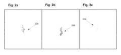
- FIGS. 2 a -2 care illustrations of bubble cloud initiation in water.
- FIG. 3illustrates a focal pressure waveform according to one embodiment.
- FIGS. 4 a -4 eare conceptual drawings that illustrate shock scattering.
- FIGS. 5 a -5 cillustrate various embodiments of pulse sequences that include initiation and scattering pressure waveforms for delivering ultrasound energy to tissue.
- FIG. 6illustrates a system configured to deliver the preferred sequences for treating the tissue with cavitation.
- Cavitation nucleiare individual bubbles formed as a result of the delivery of low pressure to tissue.
- Bubble cloudscan comprise of dense clusters of cavitation nuclei that form at or near the transducer focus. The formation of cavitation nuclei (bubble clouds) are both key components of Histotripsy therapy.
- Cavitation nucleican be formed in tissue if the tissue is subjected to a peak negative (peak rarefaction) pressure approaching or exceeding the pressure level needed to create at least a single cavitation nucleus (bubble). Note that this level is variable and is dependent upon multiple factors including tissue properties (structure and composition, dissolved gas content, and existence of impurities), transducer geometry (focal distance and f number), and sequencing scheme (PRF; number of cycles). The number of cavitation nuclei formed from one acoustic pulse has been shown to be directly related to the peak negative pressure achieved.
- FIGS. 2 a -2 care illustrations showing a typical cavitation time course.
- FIG. 2 aillustrates initiation of cavitation 208 in a medium, such as in tissue, in water, or in gelatin.
- FIG. 2 bshows growth of the cavitation 208 to a maximum size, in which the cavitation bubbles are grouped together in the focal zone.
- FIG. 2 cillustrates collapse of the cavitation 208 where nearly all the cavitation bubbles have collapsed and disappeared.
- FIG. 3shows an example of a shocked focal pressure waveform with a positive half cycle and a negative half cycle. It should be understood that shocked focal pressure waveforms can include a plurality of positive and negative half cycles.
- cavitation nucleican be formed in tissue as a result of shock scattering. Shock scattering occurs when a shocked positive pressure half cycle of an acoustic waveform is reflected, or scattered, off of a pre-existing bubble(s) and the shocked positive pressure half cycle is consequently inverted such that it combines with the incident negative pressure half cycle of the acoustic waveform in an additive fashion. If this combined new negative pressure half cycle produced is large enough (i.e. above the intrinsic threshold for the tissue or medium of interest—greater than 5 MPa peak negative pressure for example), additional cavitation nuclei will form near any preexisting nuclei. This process repeats itself until the combined new negative pressure half cycle is not sufficient in pressure to create new cavitation nuclei.
- FIGS. 4 a -4 eare conceptual drawings illustrating a shock scattering method of Histotripsy therapy.
- the frames on the topshow a pre-existing bubble 408 and a shocked positive pressure half cycle 410
- the frames on the bottomshow the ultrasound pulse pressure distribution 412 (horizontal line 414 indicates a pressure amplitude of zero).
- the pre-existing bubble 408may be formed with an initiation pulse or sequence as described above.
- a shocked pressure waveformcan then be transmitted towards the bubble 408 during a life-cycle of the bubble according to one embodiment of the shock scattering method.
- the incident shocked pressure waveform 412propagates from left to right towards the pre-existing bubble 408 , as indicated by arrows 416 .
- the incident shocked pressure waveformcan be delivered towards and into the bubble during a life-cycle of the bubble, so that the incident shocked pressure waveform interacts with the bubble.
- a single pre-existing bubble 408is shown in FIG. 4 a , having already been generated in the tissue as described above. That bubble can expand in size, as shown in FIG. 4 b , due to the initial negative pressure half cycle of the incident shocked pressure waveform.
- FIG. 4 aA single pre-existing bubble 408 is shown in FIG. 4 a , having already been generated in the tissue as described above. That bubble can expand in size, as shown in FIG. 4 b , due to the initial negative pressure half cycle of the incident shocked pressure waveform.
- a shocked positive pressure half cycle 410 of the incident shocked pressure waveform 412impinges on the bubble 408 and the positive pressure half cycle begins to scatter.
- the scattered shocked positive pressure half cycleinverts and constructively interferes with the shocked negative pressure half cycle 413 of the incident shocked pressure waveform 412 to create a transient, large amplitude, negative pressure half cycle 418 (illustrated as the circular dotted line 418 in FIGS. 4 c -4 e ) that produces additional cavitation nuclei 420 near or behind the bubble 408 .
- the negative pressure half cycle 418propagates from right to left, as indicated by arrows 422 .
- the additional cavitation nuclei 420form in the opposite direction of the shocked positive pressure waveform 410 , until the negative pressure half cycle 418 drops below the threshold for the formation of cavitation nuclei, as shown in FIG. 4 e .
- This processmay be repeated with successive shocked pressure waveforms transmitted towards and into the pre-existing bubble 408 and additional cavitation nuclei 420 .
- Cavitation nuclei formed by this shock scattering methodtend to grow towards the therapy transducer and their extent depends on the number of high pressure cycles in the pulse (waveform) and the pulse repetition frequency (PRF). Minimizing the number of cycles in a shocked waveform or reducing the sequence PRF are effective ways of reducing the length of the bubble cloud and also reducing the time average intensity and therefore the thermal dose.
- a first pulse of the sequencereferred to as an initiation pulse or initiation pressure waveform, configured to form at least one bubble in the tissue
- a second pulse of the sequencereferred to as a scattering pulse or scattering pressure waveform, configured to generate cavitation nuclei near the at least one bubble through shock scattering
- 3) A specific time delay between the initiation and scattering pulsesare: 1) A first pulse of the sequence, referred to as an initiation pulse or initiation pressure waveform, configured to form at least one bubble in the tissue 2) A second pulse of the sequence, referred to as a scattering pulse or scattering pressure waveform, configured to generate cavitation nuclei near the at least one bubble through shock scattering, and 3) A specific time delay between the initiation and scattering pulses.
- the initiation pulseshould be configured to produce at least one bubble in the tissue of interest. This can be achieved with a traditional Histotripsy initiation pulse, as described above, or with other ultrasound techniques that can induce bubble formation in tissue due to boiling such as HIFU or boiling Histotripsy.
- the scattering pulseshould have a peak-to-peak pressure high enough for shock scattering formation of cavitation nuclei.
- the time delay between these pulsescan range between 5 ⁇ s and 200 ⁇ s. In another embodiment, the time delay between these pulses can range between 5 ⁇ s and 40 ms. In another embodiment, the time delay between these pulses can range between 5 ⁇ s and 1 s.
- the pressure amplitude and/or number of cycles used in the initiation pulsecan be increased or decreased. Increasing the pressure amplitude and/or number of cycles in the initiation pulse may increase the probability of creating cavitation in the tissue. However this would also likely increase the time averaged intensity, and thermal dose, delivered to the tissue and the extent of the bubble cloud. Decreasing the pressure amplitude and/or number of cycles of the initiation pulse will reduce the intensity, and thermal dose, of the sequence but may limit the ability of the sequence to generate and/or maintain cavitation.
- the pressure amplitude and/or number of cycles used in the scattering pulse(s)can be increased or decreased. Increasing the pressure amplitude and/or number of cycles in the scattering pulse(s) may increase the probability of creating cavitation in the tissue. However this would also likely increase the time averaged intensity delivered to the tissue, and thermal dose, delivered to the tissue and the extent of the bubble cloud. Decreasing the pressure amplitude and/or number of cycles of the scattering pulse(s) will reduce the intensity, and thermal dose, of the sequence but may limit the ability of the sequence to generate and/or maintain cavitation.
- the sequence PRFcan be as high as 5000 Hz assuming that the time averaged intensity, and resultant thermal dose, are kept within safe limits. The preferred range depends on the tissues being treated. A higher PRF is recommended for more dense and fibrous tissues, and a low PRF is recommended for less dense tissues and for preservation of more fibrous and often vital tissues. Selective treatment of tissues with Histotripsy based on their stiffness can be a probable design and performance consideration for sequence development.
- additional scattering pulses with lower pressure amplitude and/or number of cyclescan be applied in order to reduce the intensity, and thermal dose, of the sequence without reducing the sequence PRF.
- FIGS. 5 a -5 cillustrate three different embodiments for Histotripsy initiation and scattering pulse sequences that can be used to generate and maintain cavitation in tissue during a shocked scattering method of Histotripsy therapy.
- an initiation pulse 524 acomprising a pressure waveform configured to form at least one bubble in the tissue can be transmitted into tissue.
- a scattering pulse 526 acan be transmitted into tissue towards and into the at least one bubble formed by the initiation pulse 524 a .
- the specific time delay between these pulsescan range between 5 ⁇ s and 200 ⁇ s.
- the time delay between these pulsescan range between 5 ⁇ s s and 40 ms.
- the time delay between these pulsescan range between 5 ⁇ s and 1 s.
- the scattering pulse 526 abecomes a shocked focal pressure waveform as it travels through the tissue, and the at least one shocked positive pressure half cycle of the scattering pulse impinges on the at least one bubble and is scattered by the at least one bubble.
- the shocked positive pressure half cycle of the scattering pulseinverts and constructively interferes with the shocked negative pressure half cycle of the scattering pulse to create a transient, large amplitude, negative pressure half cycle that produces additional cavitation nuclei behind the at least one bubble generated by the initiation pulse.
- the pressure amplitudes and/or number of cycles of both the initiation and scattering pulsescan be the same or approximately the same.
- FIG. 5 bshows another embodiment, similar to the embodiment of FIG. 5 a , except the pressure amplitude of the scattering pulses 524 a - 524 n are smaller than the pressure amplitude of the corresponding initiation pulses. Due to the principle of shock, the peak positive wave is amplified relative to the peak negative wave and therefore, the pressure amplitude used to create the scattering pulses can be lowered while still delivering the needed negative pressure with the reflected and inverted positive wave. This embodiment is more efficient than the embodiment of FIG. 5 a and delivers a lower dose of energy into the tissue. In another embodiment, however, the pressure amplitude of the scattering pulses can be greater than the pressure amplitude of the corresponding initiation pulses.
- FIG. 5 cillustrates another embodiment, which is a variation of the embodiment of FIGS. 5 a and 5 b .
- initiation pulse 524 ais followed by a scattering pulse 526 a after a specific time delay, but instead of following that with another initiation/scattering pulse pair as in FIG. 5 a , instead the scattering pulse 526 a is followed with another scattering pulse 526 b after a second time delay.
- a plurality of scattering pulsescan be delivered into tissue after the appropriate time delay to maintain the effectiveness of the bubble cloud (e.g., pulses 526 c , 526 d ) to achieve the desired ablation effect in tissue from the resulting cavitation.
- the pressure amplitudes of the scattering pulsecan be less than, equal to, or greater than the pressure amplitude of the initiation pulse.
- the time delay for subsequent scattering pressure waveformscan be different than the time delay used for the first scattering pressure.
- the first scattering pressure waveformmay be delivered within 5 ⁇ s to 200 ⁇ s of the initiation pressure waveform, but subsequent scattering pressure waveforms may be delivered within 5 ⁇ s to 200 ⁇ s, 5 ⁇ s to 40 ms, or 5 ⁇ s to 1 s.
- the sequencecan be re-started with another initiation/scattering pulse pair, as shown by 524 n / 526 n in FIG. 5 c .
- This embodimentalso uses a lower pressure amplitude scattering pulse, as in the embodiment of FIG. 5 b , but also uses fewer initiation pulses.
- the result of this embodimentis the lowest dose of energy delivered to tissue between the embodiments of FIGS. 5 a -5 c . This strategy has the potential to lower the dose significantly (as much as 50% for example) compared with traditional histotripsy sequences.
- the purpose of the initiation/scattering pairis to generate cavitation in tissue with shock scattering. Once the bubble cloud is generated, and if the focus is not moved, the initiation pulse may no longer be needed to maintain the effectiveness of the bubble cloud.
- the systemcould be designed to first create a bubble cloud with an initiation/scattering pair and follow that with lower pressure amplitude (relative to the initiation pulse pressure amplitude) scattering pulses until the focus is moved. At which point the process is repeated.
- a Histotripsy system and generatoris configured to generate very complex waveforms in order to support the ultrasound pulse sequences described herein.
- a simplified block diagram of system 600is shown in FIG. 6 .
- the main components of the systemare: Computer/controller 602 , USB to Serial Converter 604 , Microcontroller 606 , FPGA (Field Programmable Gate Array) 608 , High Voltage Controller and Power Supply 610 , Amplifier 612 , and Therapy Transducer 614 .
- All controls for the generatorcan be established using “Histotripsy Service Tool” software that can run on the computer/controller 602 (e.g., a standard PC) and communicates to the generator via USB serial communication 604 .
- “Histotripsy Service Tool” softwarecan run on the computer/controller 602 (e.g., a standard PC) and communicates to the generator via USB serial communication 604 .
- the system 600is configured to receive multiple sets of different driving parameters and loop them, which give the ability to the user to create wide range of custom sequences where all parameters (PRF, voltage amplitude, number of cycles, number of pulses per set, frequency, transducer element channels enabled, and time delays) can be set differently for every pulse generated. Time delays between pulses can be specified by the PRF for a parameter set or by specifying zero as the number of cycles per pulse.
- PRFvoltage amplitude, number of cycles, number of pulses per set, frequency, transducer element channels enabled, and time delays
- level of high voltageis changed accordingly through the Microcontroller 606 and HV Controller 610 .
- This methodcannot be used for dynamic voltage amplitude changes between two pulses since it will take too long for all capacitors on the HV line to discharge.
- PWMpulse width modulation
- the FPGA 608where the duty cycle of the pulse is modulated in order to produce the desired pulse voltage and resultant pressure amplitude.
- Histotripsy Service Toolis an application that can be run on any PC and is used for controlling the system.
- the Histotripsy Service Toolcan start/stop the therapy, set and read the level of high voltage, therapy parameters (PRF, number of cycles, duty ratio, channel enabled and delay, etc), and set and read other service and maintenance related items.
- PRFtherapy parameters
- USB to Serial converter 604converts USB combination to serial in order to communicate to the Microcontroller 606 .
- the Microcontroller 606communicates to the computer/controller 602 (Histotripsy Service Tool) to set/read working parameters, start/stop the therapy, etc. It can use internal flash memory to store all the parameters.
- the Microcontrollercommunicates to the FPGA 608 all driving parameters that are necessary to generate complex pulsing. It also communicates using serial communication to the high voltage controller and power supply 610 where it can set/read the proper level of driving voltage.
- the FPGA 608receives the information from the Microcontroller 606 and it generates the complex pulsing sequence that is required to drive the amplifier 612 .
- the FPGAcan run on 100 MHz clock since speed of pulsing is critical to be timed in 10 ns increments.
- the High Voltage Controller and Power Supply 610receives the commands from the Microcontroller 606 regarding the level of DC voltage that needs to be supplied to the amplifier circuitry in order to have an adequate voltage amplitude level at the output of the amplifier.
- the Amplifier 612receives pulses generated by the FPGA and is supplied with high voltage from High Voltage Controller and Power Supply. It generates high voltage amplitude pulses that are fed to the Therapy Transducer 614 through the matching network components which properly matches the impedance of the therapy transducer to the impedance of the amplifier. It is necessary to use a large number of capacitors that can store enough energy to support peak current demand during the generation of high voltage amplitude pulses.
- the data structures and code described in this detailed descriptionare typically stored on a computer-readable storage medium, which may be any device or medium that can store code and/or data for use by a computer system.
- the computer-readable storage mediumincludes, but is not limited to, volatile memory, non-volatile memory, magnetic and optical storage devices such as disk drives, magnetic tape, CDs (compact discs), DVDs (digital versatile discs or digital video discs), or other media capable of storing computer-readable media now known or later developed.
- the methods and processes described in the detailed description sectioncan be embodied as code and/or data, which can be stored in a computer-readable storage medium as described above.
- a computer systemreads and executes the code and/or data stored on the computer-readable storage medium, the computer system performs the methods and processes embodied as data structures and code and stored within the computer-readable storage medium.
- the methods and processes described abovecan be included in hardware modules.
- the hardware modulescan include, but are not limited to, application-specific integrated circuit (ASIC) chips, field-programmable gate arrays (FPGAs), and other programmable-logic devices now known or later developed.
- ASICapplication-specific integrated circuit
- FPGAfield-programmable gate arrays
- the hardware modulesWhen the hardware modules are activated, the hardware modules perform the methods and processes included within the hardware modules.
Landscapes
- Health & Medical Sciences (AREA)
- Life Sciences & Earth Sciences (AREA)
- Engineering & Computer Science (AREA)
- Veterinary Medicine (AREA)
- Animal Behavior & Ethology (AREA)
- Nuclear Medicine, Radiotherapy & Molecular Imaging (AREA)
- Public Health (AREA)
- General Health & Medical Sciences (AREA)
- Biomedical Technology (AREA)
- Surgery (AREA)
- Radiology & Medical Imaging (AREA)
- Heart & Thoracic Surgery (AREA)
- Medical Informatics (AREA)
- Molecular Biology (AREA)
- Vascular Medicine (AREA)
- Orthopedic Medicine & Surgery (AREA)
- Mechanical Engineering (AREA)
- Surgical Instruments (AREA)
- Medicines That Contain Protein Lipid Enzymes And Other Medicines (AREA)
- Vibration Dampers (AREA)
- Investigating Or Analyzing Materials By The Use Of Ultrasonic Waves (AREA)
Abstract
Description
| Delay-time | |||
| Event | In Water (μs) | In Gelatin (μs) | ||
| Initiation | 68 | 68 | ||
| Growth | 149 | 84 | ||
| Collapse | 230 | 100 | ||
Claims (19)
Priority Applications (3)
| Application Number | Priority Date | Filing Date | Title |
|---|---|---|---|
| US14/323,693US10293187B2 (en) | 2013-07-03 | 2014-07-03 | Histotripsy excitation sequences optimized for bubble cloud formation using shock scattering |
| US16/410,821US20190275353A1 (en) | 2013-07-03 | 2019-05-13 | Histotripsy excitation sequences optimized for bubble cloud formation using shock scattering |
| US18/737,746US20240316367A1 (en) | 2013-07-03 | 2024-06-07 | Histotripsy excitation sequences optimized for bubble cloud formation using shock scattering |
Applications Claiming Priority (2)
| Application Number | Priority Date | Filing Date | Title |
|---|---|---|---|
| US201361842820P | 2013-07-03 | 2013-07-03 | |
| US14/323,693US10293187B2 (en) | 2013-07-03 | 2014-07-03 | Histotripsy excitation sequences optimized for bubble cloud formation using shock scattering |
Related Child Applications (1)
| Application Number | Title | Priority Date | Filing Date |
|---|---|---|---|
| US16/410,821ContinuationUS20190275353A1 (en) | 2013-07-03 | 2019-05-13 | Histotripsy excitation sequences optimized for bubble cloud formation using shock scattering |
Publications (2)
| Publication Number | Publication Date |
|---|---|
| US20150011916A1 US20150011916A1 (en) | 2015-01-08 |
| US10293187B2true US10293187B2 (en) | 2019-05-21 |
Family
ID=52133288
Family Applications (3)
| Application Number | Title | Priority Date | Filing Date |
|---|---|---|---|
| US14/323,693Active2035-11-26US10293187B2 (en) | 2013-07-03 | 2014-07-03 | Histotripsy excitation sequences optimized for bubble cloud formation using shock scattering |
| US16/410,821PendingUS20190275353A1 (en) | 2013-07-03 | 2019-05-13 | Histotripsy excitation sequences optimized for bubble cloud formation using shock scattering |
| US18/737,746PendingUS20240316367A1 (en) | 2013-07-03 | 2024-06-07 | Histotripsy excitation sequences optimized for bubble cloud formation using shock scattering |
Family Applications After (2)
| Application Number | Title | Priority Date | Filing Date |
|---|---|---|---|
| US16/410,821PendingUS20190275353A1 (en) | 2013-07-03 | 2019-05-13 | Histotripsy excitation sequences optimized for bubble cloud formation using shock scattering |
| US18/737,746PendingUS20240316367A1 (en) | 2013-07-03 | 2024-06-07 | Histotripsy excitation sequences optimized for bubble cloud formation using shock scattering |
Country Status (9)
| Country | Link |
|---|---|
| US (3) | US10293187B2 (en) |
| EP (2) | EP3016594B1 (en) |
| JP (4) | JP6600304B2 (en) |
| CN (1) | CN105530869B (en) |
| BR (1) | BR112015032926B1 (en) |
| ES (1) | ES2941665T3 (en) |
| IL (1) | IL243001A0 (en) |
| MX (1) | MX369950B (en) |
| WO (1) | WO2015003142A1 (en) |
Cited By (9)
| Publication number | Priority date | Publication date | Assignee | Title |
|---|---|---|---|---|
| US11364042B2 (en) | 2005-09-22 | 2022-06-21 | The Regents Of The University Of Michigan | Histotripsy for thrombolysis |
| US11497465B2 (en) | 2019-10-25 | 2022-11-15 | Bard Peripheral Vascular, Inc. | Method for treatment of a vascular lesion |
| US11648424B2 (en) | 2018-11-28 | 2023-05-16 | Histosonics Inc. | Histotripsy systems and methods |
| US11813485B2 (en) | 2020-01-28 | 2023-11-14 | The Regents Of The University Of Michigan | Systems and methods for histotripsy immunosensitization |
| US11819712B2 (en) | 2013-08-22 | 2023-11-21 | The Regents Of The University Of Michigan | Histotripsy using very short ultrasound pulses |
| WO2024163876A1 (en)* | 2023-02-03 | 2024-08-08 | Sciton, Inc. | Methods and systems for histotripsy |
| US12220602B2 (en) | 2015-06-24 | 2025-02-11 | The Regents Of The University Of Michigan | Histotripsy therapy systems and methods for the treatment of brain tissue |
| US12318636B2 (en) | 2022-10-28 | 2025-06-03 | Histosonics, Inc. | Histotripsy systems and methods |
| US12343568B2 (en) | 2020-08-27 | 2025-07-01 | The Regents Of The University Of Michigan | Ultrasound transducer with transmit-receive capability for histotripsy |
Families Citing this family (9)
| Publication number | Priority date | Publication date | Assignee | Title |
|---|---|---|---|---|
| WO2011022411A2 (en) | 2009-08-17 | 2011-02-24 | Histosonics, Inc. | Disposable acoustic coupling medium container |
| US9144694B2 (en) | 2011-08-10 | 2015-09-29 | The Regents Of The University Of Michigan | Lesion generation through bone using histotripsy therapy without aberration correction |
| US20140100459A1 (en) | 2012-10-05 | 2014-04-10 | The Regents Of The University Of Michigan | Bubble-induced color doppler feedback during histotripsy |
| WO2015003154A1 (en) | 2013-07-03 | 2015-01-08 | Histosonics, Inc. | Articulating arm limiter for cavitational ultrasound therapy system |
| GB201617255D0 (en)* | 2016-10-11 | 2016-11-23 | Oxford University Innovation Limited | Modular ultrasound apparatus and methods |
| CN114746148A (en)* | 2019-10-11 | 2022-07-12 | 医视特有限公司 | Pre-Treatment Tissue Sensitization for Focused Ultrasound Procedures |
| US12053652B2 (en)* | 2021-07-12 | 2024-08-06 | CSW Therapeutics AB | Wearable and automated ultrasound therapy devices and methods |
| US12285636B2 (en) | 2021-08-05 | 2025-04-29 | The University Of Washington | Non-planar holographic beam shaping lenses for acoustics |
| KR102791157B1 (en)* | 2022-10-24 | 2025-04-04 | 경희대학교 산학협력단 | Apparatus and method for adjusting the range of bio tissue destruction |
Citations (323)
| Publication number | Priority date | Publication date | Assignee | Title |
|---|---|---|---|---|
| US3243497A (en) | 1964-12-11 | 1966-03-29 | Dynapower Systems Corp Of Cali | Universal support for electrotherapeutic treatment head |
| US3679021A (en) | 1970-03-25 | 1972-07-25 | Eg & G Inc | Acoustic pulse generating system |
| US4016749A (en) | 1973-07-05 | 1977-04-12 | Wachter William J | Method and apparatus for inspection of nuclear fuel rods |
| US4024501A (en) | 1975-09-03 | 1977-05-17 | Standard Oil Company | Line driver system |
| US4051394A (en) | 1976-03-15 | 1977-09-27 | The Boeing Company | Zero crossing ac relay control circuit |
| US4117446A (en) | 1974-11-28 | 1978-09-26 | Agence Nationale De Valorisation De La Recherche (A N V A R) | Devices for probing by ultrasonic radiation |
| EP0017382A1 (en) | 1979-03-20 | 1980-10-15 | THE GENERAL ELECTRIC COMPANY, p.l.c. | Ultrasonic imaging system |
| US4269174A (en) | 1979-08-06 | 1981-05-26 | Medical Dynamics, Inc. | Transcutaneous vasectomy apparatus and method |
| US4277367A (en) | 1978-10-23 | 1981-07-07 | Wisconsin Alumni Research Foundation | Phantom material and method |
| US4351038A (en) | 1979-12-31 | 1982-09-21 | Agence Nationale De Valorisation De La Recherche (Anvar) | Ultrasonic examination and imaging |
| GB2099582A (en) | 1980-02-08 | 1982-12-08 | Stanford Res Inst Int | Ultrasonic image methods and apparatus |
| US4406153A (en) | 1979-05-04 | 1983-09-27 | Acoustic Standards Corporation | Ultrasonic beam characterization device |
| DE3220751A1 (en) | 1982-06-02 | 1983-12-08 | Jörg Dr. 8022 Grünwald Schüller | Device for crushing concrements, especially renal calculi, in living human or animal bodies |
| US4440025A (en) | 1980-06-27 | 1984-04-03 | Matsushita Electric Industrial Company, Limited | Arc scan transducer array having a diverging lens |
| US4453408A (en) | 1981-03-09 | 1984-06-12 | William Clayman | Device for testing ultrasonic beam profiles |
| US4483345A (en) | 1981-08-08 | 1984-11-20 | Fujitsu Limited | Pressure measuring system with ultrasonic wave |
| JPS6080779A (en) | 1983-10-07 | 1985-05-08 | Matsushita Electric Ind Co Ltd | Magnetic field sensor |
| US4549533A (en) | 1984-01-30 | 1985-10-29 | University Of Illinois | Apparatus and method for generating and directing ultrasound |
| US4550606A (en) | 1982-09-28 | 1985-11-05 | Cornell Research Foundation, Inc. | Ultrasonic transducer array with controlled excitation pattern |
| US4575330A (en) | 1984-08-08 | 1986-03-11 | Uvp, Inc. | Apparatus for production of three-dimensional objects by stereolithography |
| JPS61196718A (en) | 1985-02-22 | 1986-08-30 | 株式会社日立製作所 | Earth fault protection device |
| US4622972A (en) | 1981-10-05 | 1986-11-18 | Varian Associates, Inc. | Ultrasound hyperthermia applicator with variable coherence by multi-spiral focusing |
| US4625731A (en) | 1984-10-10 | 1986-12-02 | Picker International, Inc. | Ultrasonic image display mounting |
| US4641378A (en) | 1984-06-06 | 1987-02-03 | Raycom Systems, Inc. | Fiber optic communication module |
| US4669483A (en) | 1984-07-21 | 1987-06-02 | Dornier System Gmbh | Lithotripsy system having locating and orienting apparatus |
| DE3544628A1 (en) | 1985-12-17 | 1987-06-19 | Eisenmenger Wolfgang | DEVICE FOR MECHANICALLY ACOUSTIC CONNECTION OF PRESSURE SHAFTS, ESPECIALLY OF FOCUSED SHOCK WAVES TO THE BODY OF LIVING BEINGS |
| US4689986A (en) | 1985-03-13 | 1987-09-01 | The University Of Michigan | Variable frequency gas-bubble-manipulating apparatus and method |
| US4757820A (en) | 1985-03-15 | 1988-07-19 | Kabushiki Kaisha Toshiba | Ultrasound therapy system |
| US4791915A (en) | 1986-09-29 | 1988-12-20 | Dynawave Corporation | Ultrasound therapy device |
| US4819621A (en) | 1986-03-11 | 1989-04-11 | Richard Wolf Gmbh | Method for detection of cavitations during medical application of high sonic energy |
| US4829491A (en) | 1984-07-12 | 1989-05-09 | Siemens Aktiengesellschaft | Phased-array equipment |
| EP0320303A2 (en) | 1987-12-11 | 1989-06-14 | General Electric Company | Coherent beam formation |
| US4856107A (en) | 1987-04-28 | 1989-08-08 | Edap International | Acoustic filter for suppressing or attenuating the negative half-waves of an elastic wave and an elastic wave generator comprising such a filter |
| US4865042A (en) | 1985-08-16 | 1989-09-12 | Hitachi, Ltd. | Ultrasonic irradiation system |
| EP0332871A2 (en) | 1988-03-16 | 1989-09-20 | Dornier Medizintechnik Gmbh | Destruction of concretions by combined treatment |
| DE3817094A1 (en) | 1988-04-18 | 1989-11-30 | Schubert Werner | Coupling and adhesive device for shock wave treatment units |
| US4888746A (en) | 1987-09-24 | 1989-12-19 | Richard Wolf Gmbh | Focussing ultrasound transducer |
| US4890267A (en) | 1985-09-24 | 1989-12-26 | Hewlett-Packard Company | Switch matrix |
| US4922917A (en) | 1987-08-14 | 1990-05-08 | Edap International | Ultrasonic tissue characterization |
| US4938217A (en) | 1988-06-21 | 1990-07-03 | Massachusetts Institute Of Technology | Electronically-controlled variable focus ultrasound hyperthermia system |
| JPH02215451A (en) | 1989-02-17 | 1990-08-28 | Toshiba Corp | Calculus crushing device |
| EP0384831A2 (en) | 1989-02-21 | 1990-08-29 | Technomed International | Apparatus for selective destruction of cells including soft tissues and bones inside a living being by implosing of gas bubbles |
| US4957099A (en) | 1988-02-10 | 1990-09-18 | Siemens Aktiengesellschaft | Shock wave source for extracorporeal lithotripsy |
| US4973980A (en) | 1987-09-11 | 1990-11-27 | Dataproducts Corporation | Acoustic microstreaming in an ink jet apparatus |
| US4984575A (en) | 1987-04-16 | 1991-01-15 | Olympus Optical Co., Ltd. | Therapeutical apparatus of extracorporeal type |
| US4991151A (en) | 1987-04-28 | 1991-02-05 | Edap International | Elastic pulse generator having a desired predetermined wave form |
| US5014686A (en) | 1989-08-31 | 1991-05-14 | International Sonic Technologies | Phantom kidney stone system |
| USRE33590E (en) | 1983-12-14 | 1991-05-21 | Edap International, S.A. | Method for examining, localizing and treating with ultrasound |
| US5065751A (en) | 1990-01-03 | 1991-11-19 | Wolf Gerald L | Method and apparatus for reversibly occluding a biological tube |
| US5080102A (en) | 1983-12-14 | 1992-01-14 | Edap International, S.A. | Examining, localizing and treatment with ultrasound |
| US5091893A (en) | 1990-04-05 | 1992-02-25 | General Electric Company | Ultrasonic array with a high density of electrical connections |
| US5092336A (en) | 1989-02-08 | 1992-03-03 | Universite Paris Vii-Bureau De La Valorisation Et De Relations Industrielle | Method and device for localization and focusing of acoustic waves in tissues |
| US5097709A (en) | 1989-02-16 | 1992-03-24 | Hitachi, Ltd. | Ultrasonic imaging system |
| DE4012760A1 (en) | 1990-04-21 | 1992-05-07 | G M T I Ges Fuer Medizintechni | Ultrasonic Doppler method for gallstone lithography - uses analysis of Doppler frequency shift to detect velocity and calculating size of tracked particles |
| US5143074A (en) | 1983-12-14 | 1992-09-01 | Edap International | Ultrasonic treatment device using a focussing and oscillating piezoelectric element |
| US5150711A (en) | 1983-12-14 | 1992-09-29 | Edap International, S.A. | Ultra-high-speed extracorporeal ultrasound hyperthermia treatment device |
| US5158070A (en) | 1983-12-14 | 1992-10-27 | Edap International, S.A. | Method for the localized destruction of soft structures using negative pressure elastic waves |
| US5158071A (en) | 1988-07-01 | 1992-10-27 | Hitachi, Ltd. | Ultrasonic apparatus for therapeutical use |
| US5163421A (en) | 1988-01-22 | 1992-11-17 | Angiosonics, Inc. | In vivo ultrasonic system with angioplasty and ultrasonic contrast imaging |
| US5165412A (en) | 1990-03-05 | 1992-11-24 | Kabushiki Kaisha Toshiba | Shock wave medical treatment apparatus with exchangeable imaging ultrasonic wave probe |
| US5174294A (en) | 1988-10-26 | 1992-12-29 | Kabushiki Kaisha Toshiba | Shockwave treatment apparatus |
| US5209221A (en) | 1988-03-01 | 1993-05-11 | Richard Wolf Gmbh | Ultrasonic treatment of pathological tissue |
| US5215680A (en) | 1990-07-10 | 1993-06-01 | Cavitation-Control Technology, Inc. | Method for the production of medical-grade lipid-coated microbubbles, paramagnetic labeling of such microbubbles and therapeutic uses of microbubbles |
| US5230340A (en) | 1992-04-13 | 1993-07-27 | General Electric Company | Ultrasound imaging system with improved dynamic focusing |
| US5295484A (en) | 1992-05-19 | 1994-03-22 | Arizona Board Of Regents For And On Behalf Of The University Of Arizona | Apparatus and method for intra-cardiac ablation of arrhythmias |
| WO1994006355A1 (en) | 1992-09-14 | 1994-03-31 | Coraje, Inc. | Apparatus and method for enhanced intravascular phonophoresis including dissolution of intravascular blockage and concomitant inhibition of restenosis |
| US5316000A (en) | 1991-03-05 | 1994-05-31 | Technomed International (Societe Anonyme) | Use of at least one composite piezoelectric transducer in the manufacture of an ultrasonic therapy apparatus for applying therapy, in a body zone, in particular to concretions, to tissue, or to bones, of a living being and method of ultrasonic therapy |
| JPH06197907A (en) | 1992-11-16 | 1994-07-19 | Siemens Ag | Therapeutic ultrasonic applicator |
| US5354258A (en) | 1992-01-07 | 1994-10-11 | Edap International | Ultra-high-speed extracorporeal ultrasound hyperthermia treatment method |
| JPH06304178A (en) | 1993-04-02 | 1994-11-01 | Siemens Ag | A therapeutic device for the treatment of pathological tissue by focused ultrasound |
| US5380411A (en) | 1987-12-02 | 1995-01-10 | Schering Aktiengesellschaft | Ultrasound or shock wave work process and preparation for carrying out same |
| US5409002A (en) | 1989-07-12 | 1995-04-25 | Focus Surgery Incorporated | Treatment system with localization |
| JPH07504339A (en) | 1992-03-10 | 1995-05-18 | シーメンス アクチエンゲゼルシヤフト | Tissue treatment method and treatment device using ultrasound |
| US5431621A (en) | 1984-11-26 | 1995-07-11 | Edap International | Process and device of an anatomic anomaly by means of elastic waves, with tracking of the target and automatic triggering of the shootings |
| US5435311A (en) | 1989-06-27 | 1995-07-25 | Hitachi, Ltd. | Ultrasound therapeutic system |
| US5469852A (en) | 1993-03-12 | 1995-11-28 | Kabushiki Kaisha Toshiba | Ultrasound diagnosis apparatus and probe therefor |
| US5501655A (en) | 1992-03-31 | 1996-03-26 | Massachusetts Institute Of Technology | Apparatus and method for acoustic heat generation and hyperthermia |
| JPH0884740A (en) | 1994-09-16 | 1996-04-02 | Toshiba Corp | Treatment equipment |
| JPH08131454A (en) | 1994-09-17 | 1996-05-28 | Toshiba Corp | Ultrasonic treatment device and ultrasonic irradiation device |
| US5520188A (en) | 1994-11-02 | 1996-05-28 | Focus Surgery Inc. | Annular array transducer |
| US5523058A (en) | 1992-09-16 | 1996-06-04 | Hitachi, Ltd. | Ultrasonic irradiation apparatus and processing apparatus based thereon |
| US5524620A (en) | 1991-11-12 | 1996-06-11 | November Technologies Ltd. | Ablation of blood thrombi by means of acoustic energy |
| US5540909A (en) | 1994-09-28 | 1996-07-30 | Alliance Pharmaceutical Corp. | Harmonic ultrasound imaging with microbubbles |
| US5542935A (en) | 1989-12-22 | 1996-08-06 | Imarx Pharmaceutical Corp. | Therapeutic delivery systems related applications |
| US5558092A (en) | 1995-06-06 | 1996-09-24 | Imarx Pharmaceutical Corp. | Methods and apparatus for performing diagnostic and therapeutic ultrasound simultaneously |
| US5563346A (en) | 1994-02-21 | 1996-10-08 | Siemens Aktiengesellschaft | Method and device for imaging an object using a two-dimensional ultrasonic array |
| US5566675A (en) | 1995-06-30 | 1996-10-22 | Siemens Medical Systems, Inc. | Beamformer for phase aberration correction |
| US5573497A (en) | 1994-11-30 | 1996-11-12 | Technomed Medical Systems And Institut National | High-intensity ultrasound therapy method and apparatus with controlled cavitation effect and reduced side lobes |
| US5580575A (en) | 1989-12-22 | 1996-12-03 | Imarx Pharmaceutical Corp. | Therapeutic drug delivery systems |
| US5582578A (en) | 1995-08-01 | 1996-12-10 | Duke University | Method for the comminution of concretions |
| US5590657A (en) | 1995-11-06 | 1997-01-07 | The Regents Of The University Of Michigan | Phased array ultrasound system and method for cardiac ablation |
| EP0755653A1 (en) | 1995-07-27 | 1997-01-29 | Hewlett-Packard GmbH | Patient monitoring module |
| US5601526A (en) | 1991-12-20 | 1997-02-11 | Technomed Medical Systems | Ultrasound therapy apparatus delivering ultrasound waves having thermal and cavitation effects |
| JPH0955571A (en) | 1995-08-11 | 1997-02-25 | Hewlett Packard Japan Ltd | Electronic circuit board with high insulation section and its production |
| US5617862A (en) | 1995-05-02 | 1997-04-08 | Acuson Corporation | Method and apparatus for beamformer system with variable aperture |
| US5648098A (en) | 1995-10-17 | 1997-07-15 | The Board Of Regents Of The University Of Nebraska | Thrombolytic agents and methods of treatment for thrombosis |
| US5676692A (en) | 1996-03-28 | 1997-10-14 | Indianapolis Center For Advanced Research, Inc. | Focussed ultrasound tissue treatment method |
| US5676452A (en) | 1995-03-02 | 1997-10-14 | Gebr. Berchtold Gmbh & Co. | Operating lamp with main bulb and replacement bulb |
| US5678554A (en) | 1996-07-02 | 1997-10-21 | Acuson Corporation | Ultrasound transducer for multiple focusing and method for manufacture thereof |
| US5694936A (en) | 1994-09-17 | 1997-12-09 | Kabushiki Kaisha Toshiba | Ultrasonic apparatus for thermotherapy with variable frequency for suppressing cavitation |
| US5695460A (en) | 1994-09-09 | 1997-12-09 | Coraje, Inc. | Enhancement of ultrasound thrombolysis |
| US5717657A (en) | 1996-06-24 | 1998-02-10 | The United States Of America As Represented By The Secretary Of The Navy | Acoustical cavitation suppressor for flow fields |
| US5724972A (en) | 1996-05-02 | 1998-03-10 | Acuson Corporation | Method and apparatus for distributed focus control with slope tracking |
| US5753929A (en) | 1996-08-28 | 1998-05-19 | Motorola, Inc. | Multi-directional optocoupler and method of manufacture |
| US5766138A (en) | 1996-04-18 | 1998-06-16 | Siemens Aktiengesellschaft | Therapy apparatus with simple setting of a desired distance from a reference point |
| US5769790A (en) | 1996-10-25 | 1998-06-23 | General Electric Company | Focused ultrasound surgery system guided by ultrasound imaging |
| US5797848A (en) | 1997-01-31 | 1998-08-25 | Acuson Corporation | Ultrasonic transducer assembly with improved electrical interface |
| US5823962A (en) | 1996-09-02 | 1998-10-20 | Siemens Aktiengesellschaft | Ultrasound transducer for diagnostic and therapeutic use |
| US5827204A (en) | 1996-11-26 | 1998-10-27 | Grandia; Willem | Medical noninvasive operations using focused modulated high power ultrasound |
| US5836896A (en) | 1996-08-19 | 1998-11-17 | Angiosonics | Method of inhibiting restenosis by applying ultrasonic energy |
| JPH10512477A (en) | 1995-01-20 | 1998-12-02 | メデラ インコーポレイテッド | Apparatus and method for supporting a breast shield and associated pumping equipment |
| US5849727A (en) | 1996-06-28 | 1998-12-15 | Board Of Regents Of The University Of Nebraska | Compositions and methods for altering the biodistribution of biological agents |
| US5873902A (en) | 1995-03-31 | 1999-02-23 | Focus Surgery, Inc. | Ultrasound intensity determining method and apparatus |
| US5879314A (en) | 1997-06-30 | 1999-03-09 | Cybersonics, Inc. | Transducer assembly and method for coupling ultrasonic energy to a body for thrombolysis of vascular thrombi |
| US5932807A (en) | 1994-10-25 | 1999-08-03 | U.S. Philips Corporation | Device for the non-destructive testing of hollow tubular objects by means of ultrasound |
| US5947904A (en) | 1997-08-21 | 1999-09-07 | Acuson Corporation | Ultrasonic method and system for imaging blood flow including disruption or activation of a contrast agent |
| US6001069A (en) | 1997-05-01 | 1999-12-14 | Ekos Corporation | Ultrasound catheter for providing a therapeutic effect to a vessel of a body |
| US6022309A (en) | 1996-04-24 | 2000-02-08 | The Regents Of The University Of California | Opto-acoustic thrombolysis |
| US6036667A (en) | 1996-10-04 | 2000-03-14 | United States Surgical Corporation | Ultrasonic dissection and coagulation system |
| US6088613A (en) | 1989-12-22 | 2000-07-11 | Imarx Pharmaceutical Corp. | Method of magnetic resonance focused surgical and therapeutic ultrasound |
| US6093883A (en) | 1997-07-15 | 2000-07-25 | Focus Surgery, Inc. | Ultrasound intensity determining method and apparatus |
| US6113558A (en) | 1997-09-29 | 2000-09-05 | Angiosonics Inc. | Pulsed mode lysis method |
| US6126607A (en) | 1997-11-03 | 2000-10-03 | Barzell-Whitmore Maroon Bells, Inc. | Ultrasound interface control system |
| US6128958A (en) | 1997-09-11 | 2000-10-10 | The Regents Of The University Of Michigan | Phased array system architecture |
| JP2000300559A (en) | 1999-04-26 | 2000-10-31 | Olympus Optical Co Ltd | Ultrasonic probe and its manufacture |
| US6143018A (en) | 1993-05-14 | 2000-11-07 | Ceramoptec Gmbh | Method and device for thermally obliterating biological tissue |
| US6165144A (en) | 1998-03-17 | 2000-12-26 | Exogen, Inc. | Apparatus and method for mounting an ultrasound transducer |
| US6176842B1 (en) | 1995-03-08 | 2001-01-23 | Ekos Corporation | Ultrasound assembly for use with light activated drugs |
| US6308710B1 (en) | 1999-04-12 | 2001-10-30 | David Silva | Scrotal drape and support |
| US6309355B1 (en) | 1998-12-22 | 2001-10-30 | The Regents Of The University Of Michigan | Method and assembly for performing ultrasound surgery using cavitation |
| US6308585B1 (en) | 2000-02-10 | 2001-10-30 | Ultra Sonus Ab | Method and a device for attaching ultrasonic transducers |
| US20010039420A1 (en) | 1998-04-08 | 2001-11-08 | Senorx, Inc. | Tissue specimen isolating and damaging device and method |
| US20010041163A1 (en) | 2000-03-09 | 2001-11-15 | Nami Sugita | Sensitizer for tumor treatment |
| US6318146B1 (en) | 1999-07-14 | 2001-11-20 | Wisconsin Alumni Research Foundation | Multi-imaging modality tissue mimicking materials for imaging phantoms |
| US6321109B2 (en) | 1996-02-15 | 2001-11-20 | Biosense, Inc. | Catheter based surgery |
| US6338566B1 (en) | 1999-04-28 | 2002-01-15 | Alm | Flexible stop piece for limiting angular travel, articulated system comprising such a stop piece, and medical equipment comprising such an articulated system |
| US6344489B1 (en) | 1991-02-14 | 2002-02-05 | Wayne State University | Stabilized gas-enriched and gas-supersaturated liquids |
| US20020045890A1 (en) | 1996-04-24 | 2002-04-18 | The Regents Of The University O F California | Opto-acoustic thrombolysis |
| WO2002032506A1 (en) | 2000-10-20 | 2002-04-25 | Sunnybrook And Women"S College Health Sciences Centre, | Technique and apparatus for ultrasound therapy |
| US6391020B1 (en) | 1999-10-06 | 2002-05-21 | The Regents Of The Univerity Of Michigan | Photodisruptive laser nucleation and ultrasonically-driven cavitation of tissues and materials |
| US20020078964A1 (en) | 2000-10-09 | 2002-06-27 | American Medical Systems, Inc. | Pelvic surgery drape |
| US6419648B1 (en) | 2000-04-21 | 2002-07-16 | Insightec-Txsonics Ltd. | Systems and methods for reducing secondary hot spots in a phased array focused ultrasound system |
| US20020099356A1 (en) | 2001-01-19 | 2002-07-25 | Unger Evan C. | Transmembrane transport apparatus and method |
| US6470204B1 (en) | 1999-08-25 | 2002-10-22 | Egidijus Edward Uzgiris | Intracavity probe for MR image guided biopsy and delivery of therapy |
| US6488639B1 (en) | 1998-05-13 | 2002-12-03 | Technomed Medical Systems, S.A | Frequency adjustment in high intensity focused ultrasound treatment apparatus |
| US6490469B2 (en) | 2000-03-15 | 2002-12-03 | The Regents Of The University Of California | Method and apparatus for dynamic focusing of ultrasound energy |
| US6500141B1 (en) | 1998-01-08 | 2002-12-31 | Karl Storz Gmbh & Co. Kg | Apparatus and method for treating body tissue, in particular soft surface tissue with ultrasound |
| US6506171B1 (en) | 2000-07-27 | 2003-01-14 | Insightec-Txsonics, Ltd | System and methods for controlling distribution of acoustic energy around a focal point using a focused ultrasound system |
| US6506154B1 (en) | 2000-11-28 | 2003-01-14 | Insightec-Txsonics, Ltd. | Systems and methods for controlling a phased array focused ultrasound system |
| US6508774B1 (en) | 1999-03-09 | 2003-01-21 | Transurgical, Inc. | Hifu applications with feedback control |
| US6511428B1 (en) | 1998-10-26 | 2003-01-28 | Hitachi, Ltd. | Ultrasonic medical treating device |
| US6511444B2 (en) | 1998-02-17 | 2003-01-28 | Brigham And Women's Hospital | Transmyocardial revascularization using ultrasound |
| US6522142B1 (en) | 2001-12-14 | 2003-02-18 | Insightec-Txsonics Ltd. | MRI-guided temperature mapping of tissue undergoing thermal treatment |
| US6524251B2 (en) | 1999-10-05 | 2003-02-25 | Omnisonics Medical Technologies, Inc. | Ultrasonic device for tissue ablation and sheath for use therewith |
| JP2003510159A (en) | 1999-10-05 | 2003-03-18 | オムニソニクス メディカル テクノロジーズ インコーポレイテッド | Ultrasound therapy method and ultrasound therapy device for reducing prostate in particular |
| US6536553B1 (en) | 2000-04-25 | 2003-03-25 | The United States Of America As Represented By The Secretary Of The Army | Method and apparatus using acoustic sensor for sub-surface object detection and visualization |
| US6543272B1 (en) | 2000-04-21 | 2003-04-08 | Insightec-Txsonics Ltd. | Systems and methods for testing and calibrating a focused ultrasound transducer array |
| US6556750B2 (en) | 2000-05-26 | 2003-04-29 | Fairchild Semiconductor Corporation | Bi-directional optical coupler |
| US6559644B2 (en) | 2001-05-30 | 2003-05-06 | Insightec - Txsonics Ltd. | MRI-based temperature mapping with error compensation |
| US20030092982A1 (en) | 1999-08-12 | 2003-05-15 | Eppstein Jonathan A. | Microporation of tissue for delivery of bioactive agents |
| US20030112922A1 (en) | 2001-11-05 | 2003-06-19 | Computerized Medical Systems, Inc. | Apparatus and method for registration, guidance and targeting of external beam radiation therapy |
| US6599288B2 (en) | 2000-05-16 | 2003-07-29 | Atrionix, Inc. | Apparatus and method incorporating an ultrasound transducer onto a delivery member |
| US20030149352A1 (en) | 2002-02-04 | 2003-08-07 | Shen-Min Liang | Automatic stone-tracking system |
| US6607498B2 (en) | 2001-01-03 | 2003-08-19 | Uitra Shape, Inc. | Method and apparatus for non-invasive body contouring by lysing adipose tissue |
| US20030157025A1 (en) | 1995-06-07 | 2003-08-21 | Unger Evan C. | Novel methods of imaging and treatment with targeted compositions |
| US6613004B1 (en) | 2000-04-21 | 2003-09-02 | Insightec-Txsonics, Ltd. | Systems and methods for creating longer necrosed volumes using a phased array focused ultrasound system |
| US6613005B1 (en) | 2000-11-28 | 2003-09-02 | Insightec-Txsonics, Ltd. | Systems and methods for steering a focused ultrasound array |
| US6612988B2 (en) | 2000-08-29 | 2003-09-02 | Brigham And Women's Hospital, Inc. | Ultrasound therapy |
| US20030181833A1 (en) | 2002-03-22 | 2003-09-25 | Fmd, Llc | Apparatus for extracorporeal shock wave lithotripter using at least two shock wave pulses |
| US6626855B1 (en) | 1999-11-26 | 2003-09-30 | Therus Corpoation | Controlled high efficiency lesion formation using high intensity ultrasound |
| US6626854B2 (en) | 2000-12-27 | 2003-09-30 | Insightec - Txsonics Ltd. | Systems and methods for ultrasound assisted lipolysis |
| US20030199857A1 (en) | 2002-04-17 | 2003-10-23 | Dornier Medtech Systems Gmbh | Apparatus and method for manipulating acoustic pulses |
| US6645162B2 (en) | 2000-12-27 | 2003-11-11 | Insightec - Txsonics Ltd. | Systems and methods for ultrasound assisted lipolysis |
| US6648839B2 (en) | 2002-02-28 | 2003-11-18 | Misonix, Incorporated | Ultrasonic medical treatment device for RF cauterization and related method |
| US20030221561A1 (en) | 1999-12-06 | 2003-12-04 | Simcha Milo | Ultrasonic medical device |
| US6666833B1 (en) | 2000-11-28 | 2003-12-23 | Insightec-Txsonics Ltd | Systems and methods for focussing an acoustic energy beam transmitted through non-uniform tissue medium |
| US20030236539A1 (en) | 1999-10-05 | 2003-12-25 | Omnisonics Medical Technologies, Inc. | Apparatus and method for using an ultrasonic probe to clear a vascular access device |
| EP1374785A1 (en) | 2002-06-26 | 2004-01-02 | Dornier MedTech Systems GmbH | Lithotripter with a doppler ultrasound unit for hit/miss monitoring |
| US6685640B1 (en) | 1998-03-30 | 2004-02-03 | Focus Surgery, Inc. | Ablation system |
| US6685657B2 (en) | 1998-11-20 | 2004-02-03 | Joie P. Jones | Methods for selectively dissolving and removing materials using ultra-high frequency ultrasound |
| JP2004505660A (en) | 2000-08-03 | 2004-02-26 | エル.アール. アールアンドディー リミテッド | System for enhanced chemical debridement |
| US6705994B2 (en) | 2002-07-08 | 2004-03-16 | Insightec - Image Guided Treatment Ltd | Tissue inhomogeneity correction in ultrasound imaging |
| US6719449B1 (en) | 1998-10-28 | 2004-04-13 | Covaris, Inc. | Apparatus and method for controlling sonic treatment |
| US6719694B2 (en) | 1999-12-23 | 2004-04-13 | Therus Corporation | Ultrasound transducers for imaging and therapy |
| JP2004512502A (en) | 2000-08-21 | 2004-04-22 | ヴイ−ターゲット テクノロジーズ リミテッド | Radiation radiation detector with position tracking system and its use in medical systems and procedures |
| US6735461B2 (en) | 2001-06-19 | 2004-05-11 | Insightec-Txsonics Ltd | Focused ultrasound system with MRI synchronization |
| US6736814B2 (en) | 2002-02-28 | 2004-05-18 | Misonix, Incorporated | Ultrasonic medical treatment device for bipolar RF cauterization and related method |
| US6750463B1 (en) | 2000-02-29 | 2004-06-15 | Hill-Rom Services, Inc. | Optical isolation apparatus and method |
| US20040127815A1 (en) | 1993-09-24 | 2004-07-01 | Transmedica International, Inc. | Removable tip for laser device |
| US20040138563A1 (en) | 2000-02-09 | 2004-07-15 | Moehring Mark A | Method and apparatus combining diagnostic ultrasound with therapeutic ultrasound to enhance thrombolysis |
| US6770031B2 (en) | 2000-12-15 | 2004-08-03 | Brigham And Women's Hospital, Inc. | Ultrasound therapy |
| US6775438B1 (en) | 1999-07-19 | 2004-08-10 | Thomson Licensing S.A. | Electrical insulation device with optocoupler for bidirectional connecting lines |
| US6788977B2 (en) | 2000-06-20 | 2004-09-07 | Celsion Corporation | System and method for heating the prostate gland to treat and prevent the growth and spread of prostate tumor |
| US6790180B2 (en) | 2001-12-03 | 2004-09-14 | Insightec-Txsonics Ltd. | Apparatus, systems, and methods for measuring power output of an ultrasound transducer |
| US6820160B1 (en) | 2001-08-21 | 2004-11-16 | Cypress Semiconductor Corporation | Apparatus for optically isolating a USB peripheral from a USB host |
| US20040236248A1 (en) | 1992-01-07 | 2004-11-25 | Pat Svedman | Transdermal perfusion of fluids |
| US20040243021A1 (en) | 2001-11-06 | 2004-12-02 | Murphy John C. | Device for thermal stimulation of small neural fibers |
| US6852082B2 (en) | 2002-07-17 | 2005-02-08 | Adam Strickberger | Apparatus and methods for performing non-invasive vasectomies |
| EP1504713A1 (en) | 2003-07-14 | 2005-02-09 | Surgical Navigation Technologies, Inc. | Navigation system for cardiac therapies |
| US20050038361A1 (en) | 2003-08-14 | 2005-02-17 | Duke University | Apparatus for improved shock-wave lithotripsy (SWL) using a piezoelectric annular array (PEAA) shock-wave generator in combination with a primary shock wave source |
| US20050038339A1 (en) | 2002-01-21 | 2005-02-17 | Sunita Chauhan | Ultrasonic treatment of breast cancer |
| WO2005018469A1 (en) | 2003-08-14 | 2005-03-03 | Duke University | Apparatus for improved shock-wave lithotripsy (swl) using a piezoelectric annular array (peaa) shock-wave generator in combination with a primary shock wave |
| US6869439B2 (en) | 1996-09-19 | 2005-03-22 | United States Surgical Corporation | Ultrasonic dissector |
| US6890332B2 (en) | 1999-05-24 | 2005-05-10 | Csaba Truckai | Electrical discharge devices and techniques for medical procedures |
| JP2005167058A (en) | 2003-12-04 | 2005-06-23 | Oval Corp | Explosion-proof insulated separation circuit |
| US20050152561A1 (en) | 2002-01-18 | 2005-07-14 | Spencer Michael E. | Modulator - amplifier |
| US20050154314A1 (en) | 2003-12-30 | 2005-07-14 | Liposonix, Inc. | Component ultrasound transducer |
| US6929609B2 (en) | 2001-01-18 | 2005-08-16 | Hitachi Medical Corporation | Ultrasonic diagnosing/treating device and method therefor |
| US20050283098A1 (en) | 1998-02-06 | 2005-12-22 | Conston Stanley R | Method for ultrasound triggered drug delivery using hollow microbubbles with controlled fragility |
| US7004282B2 (en) | 2002-10-28 | 2006-02-28 | Misonix, Incorporated | Ultrasonic horn |
| US20060060991A1 (en) | 2004-09-21 | 2006-03-23 | Interuniversitair Microelektronica Centrum (Imec) | Method and apparatus for controlled transient cavitation |
| US20060074303A1 (en) | 2004-09-28 | 2006-04-06 | Minnesota Medical Physics Llc | Apparatus and method for conformal radiation brachytherapy for prostate gland and other tumors |
| US7059168B2 (en) | 2002-10-01 | 2006-06-13 | Olympus Corporation | Ultrasound phantom |
| US20060173387A1 (en) | 2004-12-10 | 2006-08-03 | Douglas Hansmann | Externally enhanced ultrasonic therapy |
| US20060206028A1 (en) | 2005-03-11 | 2006-09-14 | Qi Yu | Apparatus and method for ablating deposits from blood vessel |
| US20060241523A1 (en) | 2005-04-12 | 2006-10-26 | Prorhythm, Inc. | Ultrasound generating method, apparatus and probe |
| US20060241466A1 (en) | 1999-08-13 | 2006-10-26 | Point Biomedical Corporation | Hollow microspheres with controlled fragility for medical use |
| US7128711B2 (en) | 2002-03-25 | 2006-10-31 | Insightec, Ltd. | Positioning systems and methods for guided ultrasound therapy systems |
| US20060264760A1 (en) | 2005-02-10 | 2006-11-23 | Board Of Regents, The University Of Texas System | Near infrared transrectal probes for prostate cancer detection and prognosis |
| US20060293630A1 (en) | 2005-06-22 | 2006-12-28 | Misonix Incorporated | Fluid containment apparatus for surgery and method of use |
| US20070010805A1 (en) | 2005-07-08 | 2007-01-11 | Fedewa Russell J | Method and apparatus for the treatment of tissue |
| US20070016039A1 (en) | 2005-06-21 | 2007-01-18 | Insightec-Image Guided Treatment Ltd. | Controlled, non-linear focused ultrasound treatment |
| US7175596B2 (en) | 2001-10-29 | 2007-02-13 | Insightec-Txsonics Ltd | System and method for sensing and locating disturbances in an energy path of a focused ultrasound system |
| US20070044562A1 (en) | 2005-08-26 | 2007-03-01 | The Boeing Company | Rapid prototype integrated matrix ultrasonic transducer array inspection apparatus, systems, and methods |
| US20070065420A1 (en) | 2005-08-23 | 2007-03-22 | Johnson Lanny L | Ultrasound Therapy Resulting in Bone Marrow Rejuvenation |
| US7196313B2 (en) | 2004-04-02 | 2007-03-27 | Fairchild Semiconductor Corporation | Surface mount multi-channel optocoupler |
| US20070083120A1 (en) | 2005-09-22 | 2007-04-12 | Cain Charles A | Pulsed cavitational ultrasound therapy |
| US7223239B2 (en) | 2002-03-22 | 2007-05-29 | Ethicon Endo-Surgery, Inc. | Medical device that removably attaches to a bodily organ |
| US20070161902A1 (en) | 2004-02-06 | 2007-07-12 | Adam Dan | Localized production of microbubbles and control of cavitational and heating effects by use of enhanced ultrasound |
| US20070167764A1 (en) | 2005-11-15 | 2007-07-19 | Kullervo Hynynen | Impedance matching for ultrasound phased array elements |
| US7258674B2 (en) | 2002-02-20 | 2007-08-21 | Liposonix, Inc. | Ultrasonic treatment and imaging of adipose tissue |
| US20070205785A1 (en) | 2004-10-18 | 2007-09-06 | Mobile Robotics Sweden Ab | Robot for ultrasonic examination |
| US20070219448A1 (en) | 2004-05-06 | 2007-09-20 | Focus Surgery, Inc. | Method and Apparatus for Selective Treatment of Tissue |
| US7273458B2 (en) | 1998-01-12 | 2007-09-25 | Georgia Tech Research Corporation | Method of applying acoustic energy effective to alter transport or cell viability |
| US7273459B2 (en) | 2003-03-31 | 2007-09-25 | Liposonix, Inc. | Vortex transducer |
| US7300414B1 (en) | 1999-11-01 | 2007-11-27 | University Of Cincinnati | Transcranial ultrasound thrombolysis system and method of treating a stroke |
| US7311679B2 (en) | 2003-12-30 | 2007-12-25 | Liposonix, Inc. | Disposable transducer seal |
| US20080013593A1 (en) | 2006-06-21 | 2008-01-17 | Ken-Ichi Kawabata | Phantom |
| US7331951B2 (en) | 2002-06-25 | 2008-02-19 | Ultrashape Inc. | Devices and methodologies useful in body aesthetics |
| US20080055003A1 (en) | 2006-09-06 | 2008-03-06 | Texas Instruments Incorporated | Reduction of voltage spikes in switching half-bridge stages |
| US7341569B2 (en) | 2004-01-30 | 2008-03-11 | Ekos Corporation | Treatment of vascular occlusions using ultrasonic energy and microbubbles |
| US7347855B2 (en) | 2001-10-29 | 2008-03-25 | Ultrashape Ltd. | Non-invasive ultrasonic body contouring |
| US20080082026A1 (en) | 2006-04-26 | 2008-04-03 | Rita Schmidt | Focused ultrasound system with far field tail suppression |
| US7359640B2 (en) | 2003-09-30 | 2008-04-15 | Stmicroelectronics Sa | Optical coupling device and method for bidirectional data communication over a common signal line |
| US7358226B2 (en) | 2003-08-27 | 2008-04-15 | The Regents Of The University Of California | Ultrasonic concentration of drug delivery capsules |
| US20080091125A1 (en) | 2006-10-13 | 2008-04-17 | University Of Washington | Method and apparatus to detect the fragmentation of kidney stones by measuring acoustic scatter |
| WO2008051484A2 (en) | 2006-10-19 | 2008-05-02 | Medela Holding Ag | System and device for supporting a breast shield |
| US7367948B2 (en) | 2002-08-29 | 2008-05-06 | The Regents Of The University Of Michigan | Acoustic monitoring method and system in laser-induced optical breakdown (LIOB) |
| US7374551B2 (en) | 2003-02-19 | 2008-05-20 | Pittsburgh Plastic Surgery Research Associates | Minimally invasive fat cavitation method |
| US7377900B2 (en) | 2003-06-02 | 2008-05-27 | Insightec - Image Guided Treatment Ltd. | Endo-cavity focused ultrasound transducer |
| US20080126665A1 (en) | 2006-09-19 | 2008-05-29 | Kent Allan Burr | Apparatus and methods to communicatively couple field devices to controllers in a process control system |
| US20080177180A1 (en) | 2004-08-17 | 2008-07-24 | Technion Research & Development | Ultrasonic Image-Guided Tissue-Damaging Procedure |
| US20080194965A1 (en) | 2007-02-08 | 2008-08-14 | Sliwa John W | Device and method for high intensity focused ultrasound ablation with acoustic lens |
| US20080214964A1 (en) | 2005-03-15 | 2008-09-04 | Edap S.A. | Therapeutic Endocavity Probe Comprising an Image Transducer Integrated Within the Therapy Ultrasonic Transducer |
| US20080262486A1 (en) | 2000-07-31 | 2008-10-23 | Galil Medical Ltd. | Planning and facilitation systems and methods for cryosurgery |
| US20080262345A1 (en) | 2003-07-21 | 2008-10-23 | The John Hopkins University | Image registration of multiple medical imaging modalities using a multiple degree-of-freedom-encoded fiducial device |
| US7442168B2 (en) | 2002-04-05 | 2008-10-28 | Misonix, Incorporated | High efficiency medical transducer with ergonomic shape and method of manufacture |
| US7462488B2 (en) | 2002-10-04 | 2008-12-09 | Wisconsin Alumni Research Foundation | Tissue mimicking elastography phantoms |
| US20080312561A1 (en) | 2004-05-06 | 2008-12-18 | Nanyang Technological University | Mechanical Manipulator for Hifu Transducers |
| US20080319376A1 (en) | 2007-06-22 | 2008-12-25 | Ekos Corporation | Method and apparatus for treatment of intracranial hemorrhages |
| US20090030339A1 (en) | 2006-01-26 | 2009-01-29 | Cheng Wai Sam C | Apparatus and method for motorised placement of needle |
| US20090112098A1 (en) | 2005-09-16 | 2009-04-30 | Shahram Vaezy | Thin-profile therapeutic ultrasound applicators |
| US20090177085A1 (en) | 2005-09-22 | 2009-07-09 | Adam Maxwell | Histotripsy for thrombolysis |
| US7559905B2 (en) | 2006-09-21 | 2009-07-14 | Focus Surgery, Inc. | HIFU probe for treating tissue with in-line degassing of fluid |
| US20090198094A1 (en) | 2004-03-09 | 2009-08-06 | Robarts Research Institute | Apparatus and computing device for performing brachytherapy and methods of imaging using the same |
| US20090211587A1 (en) | 2005-11-30 | 2009-08-27 | Urotech Pty Ltd. | Urology Drape |
| US20090227874A1 (en) | 2007-11-09 | 2009-09-10 | Eigen, Inc. | Holder assembly for a medical imaging instrument |
| US20090230822A1 (en) | 2008-03-13 | 2009-09-17 | Leonid Kushculey | Patterned ultrasonic transducers |
| US20100011845A1 (en) | 1998-10-28 | 2010-01-21 | Covaris, Inc. | Methods and systems for modulating acoustic energy delivery |
| JP2010019554A (en) | 2008-07-08 | 2010-01-28 | Hioki Ee Corp | Circuit board and measuring device |
| US7656638B2 (en) | 2004-05-18 | 2010-02-02 | Abb Oy | Earthing and overvoltage protection arrangement |
| JP2010029650A (en) | 2008-07-01 | 2010-02-12 | Yoshihiro Kagamiyama | Medical ultrasonic phantom |
| US20100059264A1 (en) | 2008-09-10 | 2010-03-11 | Kabushiki Kaisha Toshiba | Electronic apparatus and printed wiring board |
| US20100125225A1 (en) | 2008-11-19 | 2010-05-20 | Daniel Gelbart | System for selective ultrasonic ablation |
| US20100152624A1 (en) | 2005-11-07 | 2010-06-17 | Smith & Nephew, Inc. | Apparatus and method for mounting a therapeutic device |
| US20100163694A1 (en) | 2008-11-27 | 2010-07-01 | Franz Fadler | Imaging system stand |
| JP2010204068A (en) | 2009-03-06 | 2010-09-16 | Hioki Ee Corp | Insulating input type measuring instrument |
| US20100261994A1 (en) | 2009-04-09 | 2010-10-14 | Rafael Davalos | Integration of very short electric pulses for minimally to noninvasive electroporation |
| US20100274136A1 (en) | 2009-04-23 | 2010-10-28 | Marino Cerofolini | Array of electroacoustic transducers and electronic probe for three-dimensional imaging |
| US20100286519A1 (en) | 2009-05-11 | 2010-11-11 | General Electric Company | Ultrasound system and method to automatically identify and treat adipose tissue |
| US20100305432A1 (en) | 2009-05-28 | 2010-12-02 | Edwards Lifesciences Corporation | System and Method for Locating Medical Devices in Vivo Using Ultrasound Doppler Mode |
| US20100317971A1 (en) | 2009-05-04 | 2010-12-16 | Siemens Medical Solutions Usa, Inc. | Feedback in medical ultrasound imaging for high intensity focused ultrasound |
| US20110040190A1 (en) | 2009-08-17 | 2011-02-17 | Jahnke Russell C | Disposable Acoustic Coupling Medium Container |
| US20110054315A1 (en) | 2009-08-26 | 2011-03-03 | Roberts William W | Micromanipulator control arm for therapeutic and imaging ultrasound transducers |
| US20110054363A1 (en) | 2009-08-26 | 2011-03-03 | Cain Charles A | Devices and methods for using controlled bubble cloud cavitation in fractionating urinary stones |
| WO2011040054A1 (en) | 2009-09-30 | 2011-04-07 | 株式会社日立製作所 | Insulation circuit board, and power semiconductor device or inverter module using the same |
| US20110112400A1 (en) | 2009-11-06 | 2011-05-12 | Ardian, Inc. | High intensity focused ultrasound catheter apparatuses, systems, and methods for renal neuromodulation |
| US20110118602A1 (en) | 1999-10-25 | 2011-05-19 | Kona Medical, Inc. | Methods and apparatus for focused ultrasound application |
| US20110172529A1 (en) | 2009-10-12 | 2011-07-14 | Michael Gertner | Flow directed heating of nervous structures |
| US20110178444A1 (en) | 2009-11-24 | 2011-07-21 | Slayton Michael H | Methods and systems for generating thermal bubbles for improved ultrasound imaging and therapy |
| WO2011092683A1 (en) | 2010-02-01 | 2011-08-04 | Livesonics Ltd. | Non-invasive ultrasound treatment of subcostal lesions |
| US20110251528A1 (en) | 2010-04-12 | 2011-10-13 | University Of Washington | Methods and systems for non-invasive treatment of tissue using high intensity focused ultrasound therapy |
| US20110257524A1 (en) | 2009-10-12 | 2011-10-20 | Michael Gertner | External autonomic modulation |
| US20110263967A1 (en) | 2010-04-22 | 2011-10-27 | of higher education having a principal place of bussiness | Ultrasound based method and apparatus for stone detection and to facilitate clearance thereof |
| US8057408B2 (en) | 2005-09-22 | 2011-11-15 | The Regents Of The University Of Michigan | Pulsed cavitational ultrasound therapy |
| WO2011154654A2 (en) | 2010-06-07 | 2011-12-15 | Image Guided Therapy | Ultrasound transducer for medical use |
| EP2397188A1 (en) | 2010-06-15 | 2011-12-21 | Theraclion SAS | Ultrasound probe head comprising an imaging transducer with a shielding element |
| US20120029353A1 (en) | 2010-08-02 | 2012-02-02 | Guided Therapy Systems, Llc | Systems and methods for ultrasound treatment |
| US20120059264A1 (en) | 2009-05-13 | 2012-03-08 | Koninklijke Philips Electronics N.V. | Ultrasonic blood flow doppler audio with pitch shifting |
| US20120059285A1 (en) | 2010-08-27 | 2012-03-08 | Ekos Corporation | Method and apparatus for treatment of intracranial hemorrhages |
| US20120092724A1 (en) | 2010-08-18 | 2012-04-19 | Pettis Nathaniel B | Networked three-dimensional printing |
| US20120130288A1 (en) | 2009-03-20 | 2012-05-24 | University Of Cincinnati | Ultrasound-mediated inducement, detection, and enhancement of stable cavitation |
| CN102481164A (en) | 2009-12-04 | 2012-05-30 | 株式会社日立制作所 | Ultrasonic treatment device |
| US20120189998A1 (en) | 2009-06-29 | 2012-07-26 | Koninklijke Philips Electronics N.V. | Tumor ablation training system |
| US20120232388A1 (en) | 2011-03-10 | 2012-09-13 | University Of Washington Through Its Center For Commercialization | Ultrasound systems and methods for real-time noninvasive spatial temperature estimation |
| US20120259250A1 (en) | 2011-04-11 | 2012-10-11 | University Of Washington | Methods of soft tissue emulsification using a mechanism of ultrasonic atomization inside gas or vapor cavities and associated systems and devices |
| US20120271223A1 (en) | 2006-05-05 | 2012-10-25 | Neurovention, LLC | Central Nervous System Drain |
| US20120271167A1 (en) | 2011-03-01 | 2012-10-25 | University Of Cincinnati | Methods of Enhancing Delivery of Drugs Using Ultrasonic Waves and Systems for Performing The Same |
| US8333115B1 (en) | 2010-08-26 | 2012-12-18 | The Boeing Company | Inspection apparatus and method for irregular shaped, closed cavity structures |
| US20130041293A1 (en) | 2011-08-10 | 2013-02-14 | Charles A. Cain | Lesion generation through bone using histotripsy therapy without aberration correction |
| US20130053691A1 (en) | 2010-04-09 | 2013-02-28 | Kenichi Kawabata | Ultrasound diagnostic and treatment device |
| US20130090579A1 (en) | 2011-10-10 | 2013-04-11 | Charles A. Cain | Pulsed Cavitational Therapeutic Ultrasound With Dithering |
| US20130102932A1 (en) | 2011-10-10 | 2013-04-25 | Charles A. Cain | Imaging Feedback of Histotripsy Treatments with Ultrasound Transient Elastography |
| US20130190623A1 (en) | 2012-01-06 | 2013-07-25 | James A. Bertolina | Histotripsy Therapy Transducer |
| US8539813B2 (en) | 2009-09-22 | 2013-09-24 | The Regents Of The University Of Michigan | Gel phantoms for testing cavitational ultrasound (histotripsy) transducers |
| US20130269982A1 (en) | 2012-04-13 | 2013-10-17 | Dejan Teofilovic | Systems and Methods for Obtaining Large Creepage Isolation on Printed Circuit Boards |
| US20130289593A1 (en) | 2012-04-30 | 2013-10-31 | Timothy L. Hall | Ultrasound Transducer Manufacturing Using Rapid-Prototyping Method |
| US20140058293A1 (en) | 2012-05-23 | 2014-02-27 | Sunnybrook Research Institute | Multi-Frequency Ultrasound Device and Method of Operation |
| US20140073995A1 (en) | 2012-09-11 | 2014-03-13 | Dejan Teofilovic | Histotripsy therapy system |
| US20140100459A1 (en) | 2012-10-05 | 2014-04-10 | The Regents Of The University Of Michigan | Bubble-induced color doppler feedback during histotripsy |
| US20140128734A1 (en) | 2012-11-05 | 2014-05-08 | Ekos Corporation | Catheter systems and methods |
| WO2015000953A1 (en) | 2013-07-03 | 2015-01-08 | Bracco Suisse S.A. | Devices and methods for the ultrasound treatment of ischemic stroke |
| US20160135916A1 (en) | 2013-07-03 | 2016-05-19 | Histosonics, Inc. | Articulating arm limiter for cavitational ultrasound therapy system |
- 2014
- 2014-07-03EPEP14819742.9Apatent/EP3016594B1/enactiveActive
- 2014-07-03JPJP2016524375Apatent/JP6600304B2/enactiveActive
- 2014-07-03CNCN201480038082.7Apatent/CN105530869B/enactiveActive
- 2014-07-03BRBR112015032926-8Apatent/BR112015032926B1/enactiveIP Right Grant
- 2014-07-03ESES14819742Tpatent/ES2941665T3/enactiveActive
- 2014-07-03MXMX2015017596Apatent/MX369950B/enactiveIP Right Grant
- 2014-07-03USUS14/323,693patent/US10293187B2/enactiveActive
- 2014-07-03WOPCT/US2014/045431patent/WO2015003142A1/enactiveApplication Filing
- 2014-07-03EPEP22213038.7Apatent/EP4166194A1/enactivePending
- 2015
- 2015-12-21ILIL243001Apatent/IL243001A0/enunknown
- 2019
- 2019-05-13USUS16/410,821patent/US20190275353A1/enactivePending
- 2019-10-03JPJP2019183193Apatent/JP6977008B2/enactiveActive
- 2021
- 2021-11-10JPJP2021182948Apatent/JP7680939B2/enactiveActive
- 2024
- 2024-06-07USUS18/737,746patent/US20240316367A1/enactivePending
- 2025
- 2025-03-12JPJP2025039165Apatent/JP2025096724A/enactivePending
Patent Citations (344)
| Publication number | Priority date | Publication date | Assignee | Title |
|---|---|---|---|---|
| US3243497A (en) | 1964-12-11 | 1966-03-29 | Dynapower Systems Corp Of Cali | Universal support for electrotherapeutic treatment head |
| US3679021A (en) | 1970-03-25 | 1972-07-25 | Eg & G Inc | Acoustic pulse generating system |
| US4016749A (en) | 1973-07-05 | 1977-04-12 | Wachter William J | Method and apparatus for inspection of nuclear fuel rods |
| US4117446A (en) | 1974-11-28 | 1978-09-26 | Agence Nationale De Valorisation De La Recherche (A N V A R) | Devices for probing by ultrasonic radiation |
| US4024501A (en) | 1975-09-03 | 1977-05-17 | Standard Oil Company | Line driver system |
| US4051394A (en) | 1976-03-15 | 1977-09-27 | The Boeing Company | Zero crossing ac relay control circuit |
| US4277367A (en) | 1978-10-23 | 1981-07-07 | Wisconsin Alumni Research Foundation | Phantom material and method |
| EP0017382A1 (en) | 1979-03-20 | 1980-10-15 | THE GENERAL ELECTRIC COMPANY, p.l.c. | Ultrasonic imaging system |
| US4406153A (en) | 1979-05-04 | 1983-09-27 | Acoustic Standards Corporation | Ultrasonic beam characterization device |
| US4269174A (en) | 1979-08-06 | 1981-05-26 | Medical Dynamics, Inc. | Transcutaneous vasectomy apparatus and method |
| US4351038A (en) | 1979-12-31 | 1982-09-21 | Agence Nationale De Valorisation De La Recherche (Anvar) | Ultrasonic examination and imaging |
| GB2099582A (en) | 1980-02-08 | 1982-12-08 | Stanford Res Inst Int | Ultrasonic image methods and apparatus |
| US4440025A (en) | 1980-06-27 | 1984-04-03 | Matsushita Electric Industrial Company, Limited | Arc scan transducer array having a diverging lens |
| US4453408A (en) | 1981-03-09 | 1984-06-12 | William Clayman | Device for testing ultrasonic beam profiles |
| US4483345A (en) | 1981-08-08 | 1984-11-20 | Fujitsu Limited | Pressure measuring system with ultrasonic wave |
| US4622972A (en) | 1981-10-05 | 1986-11-18 | Varian Associates, Inc. | Ultrasound hyperthermia applicator with variable coherence by multi-spiral focusing |
| DE3220751A1 (en) | 1982-06-02 | 1983-12-08 | Jörg Dr. 8022 Grünwald Schüller | Device for crushing concrements, especially renal calculi, in living human or animal bodies |
| US4550606A (en) | 1982-09-28 | 1985-11-05 | Cornell Research Foundation, Inc. | Ultrasonic transducer array with controlled excitation pattern |
| JPS6080779A (en) | 1983-10-07 | 1985-05-08 | Matsushita Electric Ind Co Ltd | Magnetic field sensor |
| US5080101A (en) | 1983-12-14 | 1992-01-14 | Edap International, S.A. | Method for examining and aiming treatment with untrasound |
| US5143073A (en) | 1983-12-14 | 1992-09-01 | Edap International, S.A. | Wave apparatus system |
| US5080102A (en) | 1983-12-14 | 1992-01-14 | Edap International, S.A. | Examining, localizing and treatment with ultrasound |
| US5150711A (en) | 1983-12-14 | 1992-09-29 | Edap International, S.A. | Ultra-high-speed extracorporeal ultrasound hyperthermia treatment device |
| US5111822A (en) | 1983-12-14 | 1992-05-12 | Edap International, S.A. | Piezoelectric article |
| US5143074A (en) | 1983-12-14 | 1992-09-01 | Edap International | Ultrasonic treatment device using a focussing and oscillating piezoelectric element |
| US5158070A (en) | 1983-12-14 | 1992-10-27 | Edap International, S.A. | Method for the localized destruction of soft structures using negative pressure elastic waves |
| USRE33590E (en) | 1983-12-14 | 1991-05-21 | Edap International, S.A. | Method for examining, localizing and treating with ultrasound |
| US4549533A (en) | 1984-01-30 | 1985-10-29 | University Of Illinois | Apparatus and method for generating and directing ultrasound |
| US4641378A (en) | 1984-06-06 | 1987-02-03 | Raycom Systems, Inc. | Fiber optic communication module |
| US4829491A (en) | 1984-07-12 | 1989-05-09 | Siemens Aktiengesellschaft | Phased-array equipment |
| US4669483A (en) | 1984-07-21 | 1987-06-02 | Dornier System Gmbh | Lithotripsy system having locating and orienting apparatus |
| US4575330B1 (en) | 1984-08-08 | 1989-12-19 | ||
| US4575330A (en) | 1984-08-08 | 1986-03-11 | Uvp, Inc. | Apparatus for production of three-dimensional objects by stereolithography |
| US4625731A (en) | 1984-10-10 | 1986-12-02 | Picker International, Inc. | Ultrasonic image display mounting |
| US5431621A (en) | 1984-11-26 | 1995-07-11 | Edap International | Process and device of an anatomic anomaly by means of elastic waves, with tracking of the target and automatic triggering of the shootings |
| JPS61196718A (en) | 1985-02-22 | 1986-08-30 | 株式会社日立製作所 | Earth fault protection device |
| US4689986A (en) | 1985-03-13 | 1987-09-01 | The University Of Michigan | Variable frequency gas-bubble-manipulating apparatus and method |
| US4757820A (en) | 1985-03-15 | 1988-07-19 | Kabushiki Kaisha Toshiba | Ultrasound therapy system |
| US4865042A (en) | 1985-08-16 | 1989-09-12 | Hitachi, Ltd. | Ultrasonic irradiation system |
| US4890267A (en) | 1985-09-24 | 1989-12-26 | Hewlett-Packard Company | Switch matrix |
| DE3544628A1 (en) | 1985-12-17 | 1987-06-19 | Eisenmenger Wolfgang | DEVICE FOR MECHANICALLY ACOUSTIC CONNECTION OF PRESSURE SHAFTS, ESPECIALLY OF FOCUSED SHOCK WAVES TO THE BODY OF LIVING BEINGS |
| US4819621A (en) | 1986-03-11 | 1989-04-11 | Richard Wolf Gmbh | Method for detection of cavitations during medical application of high sonic energy |
| US4791915A (en) | 1986-09-29 | 1988-12-20 | Dynawave Corporation | Ultrasound therapy device |
| US4984575A (en) | 1987-04-16 | 1991-01-15 | Olympus Optical Co., Ltd. | Therapeutical apparatus of extracorporeal type |
| US4991151A (en) | 1987-04-28 | 1991-02-05 | Edap International | Elastic pulse generator having a desired predetermined wave form |
| US4856107A (en) | 1987-04-28 | 1989-08-08 | Edap International | Acoustic filter for suppressing or attenuating the negative half-waves of an elastic wave and an elastic wave generator comprising such a filter |
| US4995012A (en) | 1987-04-28 | 1991-02-19 | Edap International | Method of generating an elastic wave |
| US4922917A (en) | 1987-08-14 | 1990-05-08 | Edap International | Ultrasonic tissue characterization |
| US4973980A (en) | 1987-09-11 | 1990-11-27 | Dataproducts Corporation | Acoustic microstreaming in an ink jet apparatus |
| US4888746A (en) | 1987-09-24 | 1989-12-19 | Richard Wolf Gmbh | Focussing ultrasound transducer |
| US5380411A (en) | 1987-12-02 | 1995-01-10 | Schering Aktiengesellschaft | Ultrasound or shock wave work process and preparation for carrying out same |
| EP0320303A2 (en) | 1987-12-11 | 1989-06-14 | General Electric Company | Coherent beam formation |
| US5163421A (en) | 1988-01-22 | 1992-11-17 | Angiosonics, Inc. | In vivo ultrasonic system with angioplasty and ultrasonic contrast imaging |
| US4957099A (en) | 1988-02-10 | 1990-09-18 | Siemens Aktiengesellschaft | Shock wave source for extracorporeal lithotripsy |
| US5209221A (en) | 1988-03-01 | 1993-05-11 | Richard Wolf Gmbh | Ultrasonic treatment of pathological tissue |
| EP0332871A2 (en) | 1988-03-16 | 1989-09-20 | Dornier Medizintechnik Gmbh | Destruction of concretions by combined treatment |
| DE3817094A1 (en) | 1988-04-18 | 1989-11-30 | Schubert Werner | Coupling and adhesive device for shock wave treatment units |
| US4938217A (en) | 1988-06-21 | 1990-07-03 | Massachusetts Institute Of Technology | Electronically-controlled variable focus ultrasound hyperthermia system |
| US5158071A (en) | 1988-07-01 | 1992-10-27 | Hitachi, Ltd. | Ultrasonic apparatus for therapeutical use |
| US5174294A (en) | 1988-10-26 | 1992-12-29 | Kabushiki Kaisha Toshiba | Shockwave treatment apparatus |
| US5092336A (en) | 1989-02-08 | 1992-03-03 | Universite Paris Vii-Bureau De La Valorisation Et De Relations Industrielle | Method and device for localization and focusing of acoustic waves in tissues |
| US5097709A (en) | 1989-02-16 | 1992-03-24 | Hitachi, Ltd. | Ultrasonic imaging system |
| JPH02215451A (en) | 1989-02-17 | 1990-08-28 | Toshiba Corp | Calculus crushing device |
| EP0384831A2 (en) | 1989-02-21 | 1990-08-29 | Technomed International | Apparatus for selective destruction of cells including soft tissues and bones inside a living being by implosing of gas bubbles |
| US5219401A (en) | 1989-02-21 | 1993-06-15 | Technomed Int'l | Apparatus for selective destruction of cells by implosion of gas bubbles |
| US5435311A (en) | 1989-06-27 | 1995-07-25 | Hitachi, Ltd. | Ultrasound therapeutic system |
| US5409002A (en) | 1989-07-12 | 1995-04-25 | Focus Surgery Incorporated | Treatment system with localization |
| US5014686A (en) | 1989-08-31 | 1991-05-14 | International Sonic Technologies | Phantom kidney stone system |
| US5580575A (en) | 1989-12-22 | 1996-12-03 | Imarx Pharmaceutical Corp. | Therapeutic drug delivery systems |
| US6088613A (en) | 1989-12-22 | 2000-07-11 | Imarx Pharmaceutical Corp. | Method of magnetic resonance focused surgical and therapeutic ultrasound |
| US5542935A (en) | 1989-12-22 | 1996-08-06 | Imarx Pharmaceutical Corp. | Therapeutic delivery systems related applications |
| US5065751A (en) | 1990-01-03 | 1991-11-19 | Wolf Gerald L | Method and apparatus for reversibly occluding a biological tube |
| US5165412A (en) | 1990-03-05 | 1992-11-24 | Kabushiki Kaisha Toshiba | Shock wave medical treatment apparatus with exchangeable imaging ultrasonic wave probe |
| US5091893A (en) | 1990-04-05 | 1992-02-25 | General Electric Company | Ultrasonic array with a high density of electrical connections |
| DE4012760A1 (en) | 1990-04-21 | 1992-05-07 | G M T I Ges Fuer Medizintechni | Ultrasonic Doppler method for gallstone lithography - uses analysis of Doppler frequency shift to detect velocity and calculating size of tracked particles |
| US5215680A (en) | 1990-07-10 | 1993-06-01 | Cavitation-Control Technology, Inc. | Method for the production of medical-grade lipid-coated microbubbles, paramagnetic labeling of such microbubbles and therapeutic uses of microbubbles |
| US6344489B1 (en) | 1991-02-14 | 2002-02-05 | Wayne State University | Stabilized gas-enriched and gas-supersaturated liquids |
| US5474071A (en) | 1991-03-05 | 1995-12-12 | Technomed Medical Systems | Therapeutic endo-rectal probe and apparatus constituting an application thereof for destroying cancer tissue, in particular of the prostate, and preferably in combination with an imaging endo-cavitary-probe |
| US5666954A (en) | 1991-03-05 | 1997-09-16 | Technomed Medical Systems Inserm-Institut National De La Sante Et De La Recherche Medicale | Therapeutic endo-rectal probe, and apparatus constituting an application thereof for destroying cancer tissue, in particular of the prostate, and preferably in combination with an imaging endo-cavitary-probe |
| US5316000A (en) | 1991-03-05 | 1994-05-31 | Technomed International (Societe Anonyme) | Use of at least one composite piezoelectric transducer in the manufacture of an ultrasonic therapy apparatus for applying therapy, in a body zone, in particular to concretions, to tissue, or to bones, of a living being and method of ultrasonic therapy |
| US5524620A (en) | 1991-11-12 | 1996-06-11 | November Technologies Ltd. | Ablation of blood thrombi by means of acoustic energy |
| US5601526A (en) | 1991-12-20 | 1997-02-11 | Technomed Medical Systems | Ultrasound therapy apparatus delivering ultrasound waves having thermal and cavitation effects |
| US5354258A (en) | 1992-01-07 | 1994-10-11 | Edap International | Ultra-high-speed extracorporeal ultrasound hyperthermia treatment method |
| US20040236248A1 (en) | 1992-01-07 | 2004-11-25 | Pat Svedman | Transdermal perfusion of fluids |
| JPH07504339A (en) | 1992-03-10 | 1995-05-18 | シーメンス アクチエンゲゼルシヤフト | Tissue treatment method and treatment device using ultrasound |
| US5759162A (en) | 1992-03-10 | 1998-06-02 | Siemens Aktiengesellschaft | Method and apparatus for ultrasound tissue therapy |
| US5501655A (en) | 1992-03-31 | 1996-03-26 | Massachusetts Institute Of Technology | Apparatus and method for acoustic heat generation and hyperthermia |
| US5230340A (en) | 1992-04-13 | 1993-07-27 | General Electric Company | Ultrasound imaging system with improved dynamic focusing |
| US5295484A (en) | 1992-05-19 | 1994-03-22 | Arizona Board Of Regents For And On Behalf Of The University Of Arizona | Apparatus and method for intra-cardiac ablation of arrhythmias |
| US5474531A (en) | 1992-09-14 | 1995-12-12 | Coraje, Inc. | Apparatus and method for enhanced intravascular phonophoresis including dissolution of intravascular blockage and concomitant inhibition of restenosis |
| WO1994006355A1 (en) | 1992-09-14 | 1994-03-31 | Coraje, Inc. | Apparatus and method for enhanced intravascular phonophoresis including dissolution of intravascular blockage and concomitant inhibition of restenosis |
| US5523058A (en) | 1992-09-16 | 1996-06-04 | Hitachi, Ltd. | Ultrasonic irradiation apparatus and processing apparatus based thereon |
| US5443069A (en) | 1992-11-16 | 1995-08-22 | Siemens Aktiengesellschaft | Therapeutic ultrasound applicator for the urogenital region |
| JPH06197907A (en) | 1992-11-16 | 1994-07-19 | Siemens Ag | Therapeutic ultrasonic applicator |
| US5743863A (en) | 1993-01-22 | 1998-04-28 | Technomed Medical Systems And Institut National | High-intensity ultrasound therapy method and apparatus with controlled cavitation effect and reduced side lobes |
| US5469852A (en) | 1993-03-12 | 1995-11-28 | Kabushiki Kaisha Toshiba | Ultrasound diagnosis apparatus and probe therefor |
| JPH06304178A (en) | 1993-04-02 | 1994-11-01 | Siemens Ag | A therapeutic device for the treatment of pathological tissue by focused ultrasound |
| US6143018A (en) | 1993-05-14 | 2000-11-07 | Ceramoptec Gmbh | Method and device for thermally obliterating biological tissue |
| US20040127815A1 (en) | 1993-09-24 | 2004-07-01 | Transmedica International, Inc. | Removable tip for laser device |
| US5563346A (en) | 1994-02-21 | 1996-10-08 | Siemens Aktiengesellschaft | Method and device for imaging an object using a two-dimensional ultrasonic array |
| US6576220B2 (en) | 1994-03-11 | 2003-06-10 | Imarx Therapeutics, Inc. | Non-invasive methods for surgery in the vasculature |
| US5695460A (en) | 1994-09-09 | 1997-12-09 | Coraje, Inc. | Enhancement of ultrasound thrombolysis |
| JPH0884740A (en) | 1994-09-16 | 1996-04-02 | Toshiba Corp | Treatment equipment |
| US5694936A (en) | 1994-09-17 | 1997-12-09 | Kabushiki Kaisha Toshiba | Ultrasonic apparatus for thermotherapy with variable frequency for suppressing cavitation |
| JPH08131454A (en) | 1994-09-17 | 1996-05-28 | Toshiba Corp | Ultrasonic treatment device and ultrasonic irradiation device |
| US5540909A (en) | 1994-09-28 | 1996-07-30 | Alliance Pharmaceutical Corp. | Harmonic ultrasound imaging with microbubbles |
| US5932807A (en) | 1994-10-25 | 1999-08-03 | U.S. Philips Corporation | Device for the non-destructive testing of hollow tubular objects by means of ultrasound |
| US5520188A (en) | 1994-11-02 | 1996-05-28 | Focus Surgery Inc. | Annular array transducer |
| US5573497A (en) | 1994-11-30 | 1996-11-12 | Technomed Medical Systems And Institut National | High-intensity ultrasound therapy method and apparatus with controlled cavitation effect and reduced side lobes |
| JPH10512477A (en) | 1995-01-20 | 1998-12-02 | メデラ インコーポレイテッド | Apparatus and method for supporting a breast shield and associated pumping equipment |
| US5676452A (en) | 1995-03-02 | 1997-10-14 | Gebr. Berchtold Gmbh & Co. | Operating lamp with main bulb and replacement bulb |
| US6176842B1 (en) | 1995-03-08 | 2001-01-23 | Ekos Corporation | Ultrasound assembly for use with light activated drugs |
| US5873902A (en) | 1995-03-31 | 1999-02-23 | Focus Surgery, Inc. | Ultrasound intensity determining method and apparatus |
| US5617862A (en) | 1995-05-02 | 1997-04-08 | Acuson Corporation | Method and apparatus for beamformer system with variable aperture |
| US5558092A (en) | 1995-06-06 | 1996-09-24 | Imarx Pharmaceutical Corp. | Methods and apparatus for performing diagnostic and therapeutic ultrasound simultaneously |
| US20030157025A1 (en) | 1995-06-07 | 2003-08-21 | Unger Evan C. | Novel methods of imaging and treatment with targeted compositions |
| US5566675A (en) | 1995-06-30 | 1996-10-22 | Siemens Medical Systems, Inc. | Beamformer for phase aberration correction |
| EP0755653A1 (en) | 1995-07-27 | 1997-01-29 | Hewlett-Packard GmbH | Patient monitoring module |
| US5582578A (en) | 1995-08-01 | 1996-12-10 | Duke University | Method for the comminution of concretions |
| JPH0955571A (en) | 1995-08-11 | 1997-02-25 | Hewlett Packard Japan Ltd | Electronic circuit board with high insulation section and its production |
| US5648098A (en) | 1995-10-17 | 1997-07-15 | The Board Of Regents Of The University Of Nebraska | Thrombolytic agents and methods of treatment for thrombosis |
| US5590657A (en) | 1995-11-06 | 1997-01-07 | The Regents Of The University Of Michigan | Phased array ultrasound system and method for cardiac ablation |
| US6321109B2 (en) | 1996-02-15 | 2001-11-20 | Biosense, Inc. | Catheter based surgery |
| US5676692A (en) | 1996-03-28 | 1997-10-14 | Indianapolis Center For Advanced Research, Inc. | Focussed ultrasound tissue treatment method |
| US5766138A (en) | 1996-04-18 | 1998-06-16 | Siemens Aktiengesellschaft | Therapy apparatus with simple setting of a desired distance from a reference point |
| US20020045890A1 (en) | 1996-04-24 | 2002-04-18 | The Regents Of The University O F California | Opto-acoustic thrombolysis |
| US6022309A (en) | 1996-04-24 | 2000-02-08 | The Regents Of The University Of California | Opto-acoustic thrombolysis |
| US5724972A (en) | 1996-05-02 | 1998-03-10 | Acuson Corporation | Method and apparatus for distributed focus control with slope tracking |
| US5717657A (en) | 1996-06-24 | 1998-02-10 | The United States Of America As Represented By The Secretary Of The Navy | Acoustical cavitation suppressor for flow fields |
| US5849727A (en) | 1996-06-28 | 1998-12-15 | Board Of Regents Of The University Of Nebraska | Compositions and methods for altering the biodistribution of biological agents |
| US5678554A (en) | 1996-07-02 | 1997-10-21 | Acuson Corporation | Ultrasound transducer for multiple focusing and method for manufacture thereof |
| US5836896A (en) | 1996-08-19 | 1998-11-17 | Angiosonics | Method of inhibiting restenosis by applying ultrasonic energy |
| US5753929A (en) | 1996-08-28 | 1998-05-19 | Motorola, Inc. | Multi-directional optocoupler and method of manufacture |
| US5823962A (en) | 1996-09-02 | 1998-10-20 | Siemens Aktiengesellschaft | Ultrasound transducer for diagnostic and therapeutic use |
| US6869439B2 (en) | 1996-09-19 | 2005-03-22 | United States Surgical Corporation | Ultrasonic dissector |
| US6036667A (en) | 1996-10-04 | 2000-03-14 | United States Surgical Corporation | Ultrasonic dissection and coagulation system |
| US5769790A (en) | 1996-10-25 | 1998-06-23 | General Electric Company | Focused ultrasound surgery system guided by ultrasound imaging |
| US5827204A (en) | 1996-11-26 | 1998-10-27 | Grandia; Willem | Medical noninvasive operations using focused modulated high power ultrasound |
| US5797848A (en) | 1997-01-31 | 1998-08-25 | Acuson Corporation | Ultrasonic transducer assembly with improved electrical interface |
| US6001069A (en) | 1997-05-01 | 1999-12-14 | Ekos Corporation | Ultrasound catheter for providing a therapeutic effect to a vessel of a body |
| US5879314A (en) | 1997-06-30 | 1999-03-09 | Cybersonics, Inc. | Transducer assembly and method for coupling ultrasonic energy to a body for thrombolysis of vascular thrombi |
| US6093883A (en) | 1997-07-15 | 2000-07-25 | Focus Surgery, Inc. | Ultrasound intensity determining method and apparatus |
| US5947904A (en) | 1997-08-21 | 1999-09-07 | Acuson Corporation | Ultrasonic method and system for imaging blood flow including disruption or activation of a contrast agent |
| US6128958A (en) | 1997-09-11 | 2000-10-10 | The Regents Of The University Of Michigan | Phased array system architecture |
| US6113558A (en) | 1997-09-29 | 2000-09-05 | Angiosonics Inc. | Pulsed mode lysis method |
| US6126607A (en) | 1997-11-03 | 2000-10-03 | Barzell-Whitmore Maroon Bells, Inc. | Ultrasound interface control system |
| US6500141B1 (en) | 1998-01-08 | 2002-12-31 | Karl Storz Gmbh & Co. Kg | Apparatus and method for treating body tissue, in particular soft surface tissue with ultrasound |
| US7273458B2 (en) | 1998-01-12 | 2007-09-25 | Georgia Tech Research Corporation | Method of applying acoustic energy effective to alter transport or cell viability |
| US20050283098A1 (en) | 1998-02-06 | 2005-12-22 | Conston Stanley R | Method for ultrasound triggered drug delivery using hollow microbubbles with controlled fragility |
| US6511444B2 (en) | 1998-02-17 | 2003-01-28 | Brigham And Women's Hospital | Transmyocardial revascularization using ultrasound |
| US6165144A (en) | 1998-03-17 | 2000-12-26 | Exogen, Inc. | Apparatus and method for mounting an ultrasound transducer |
| US6685640B1 (en) | 1998-03-30 | 2004-02-03 | Focus Surgery, Inc. | Ablation system |
| US20010039420A1 (en) | 1998-04-08 | 2001-11-08 | Senorx, Inc. | Tissue specimen isolating and damaging device and method |
| US6488639B1 (en) | 1998-05-13 | 2002-12-03 | Technomed Medical Systems, S.A | Frequency adjustment in high intensity focused ultrasound treatment apparatus |
| US6511428B1 (en) | 1998-10-26 | 2003-01-28 | Hitachi, Ltd. | Ultrasonic medical treating device |
| US6719449B1 (en) | 1998-10-28 | 2004-04-13 | Covaris, Inc. | Apparatus and method for controlling sonic treatment |
| US20100011845A1 (en) | 1998-10-28 | 2010-01-21 | Covaris, Inc. | Methods and systems for modulating acoustic energy delivery |
| US6685657B2 (en) | 1998-11-20 | 2004-02-03 | Joie P. Jones | Methods for selectively dissolving and removing materials using ultra-high frequency ultrasound |
| US6413216B1 (en) | 1998-12-22 | 2002-07-02 | The Regents Of The University Of Michigan | Method and assembly for performing ultrasound surgery using cavitation |
| US6309355B1 (en) | 1998-12-22 | 2001-10-30 | The Regents Of The University Of Michigan | Method and assembly for performing ultrasound surgery using cavitation |
| US6508774B1 (en) | 1999-03-09 | 2003-01-21 | Transurgical, Inc. | Hifu applications with feedback control |
| US6308710B1 (en) | 1999-04-12 | 2001-10-30 | David Silva | Scrotal drape and support |
| JP2000300559A (en) | 1999-04-26 | 2000-10-31 | Olympus Optical Co Ltd | Ultrasonic probe and its manufacture |
| US6338566B1 (en) | 1999-04-28 | 2002-01-15 | Alm | Flexible stop piece for limiting angular travel, articulated system comprising such a stop piece, and medical equipment comprising such an articulated system |
| US6890332B2 (en) | 1999-05-24 | 2005-05-10 | Csaba Truckai | Electrical discharge devices and techniques for medical procedures |
| US6318146B1 (en) | 1999-07-14 | 2001-11-20 | Wisconsin Alumni Research Foundation | Multi-imaging modality tissue mimicking materials for imaging phantoms |
| US6775438B1 (en) | 1999-07-19 | 2004-08-10 | Thomson Licensing S.A. | Electrical insulation device with optocoupler for bidirectional connecting lines |
| US20030092982A1 (en) | 1999-08-12 | 2003-05-15 | Eppstein Jonathan A. | Microporation of tissue for delivery of bioactive agents |
| US20060241466A1 (en) | 1999-08-13 | 2006-10-26 | Point Biomedical Corporation | Hollow microspheres with controlled fragility for medical use |
| US6470204B1 (en) | 1999-08-25 | 2002-10-22 | Egidijus Edward Uzgiris | Intracavity probe for MR image guided biopsy and delivery of therapy |
| US20030236539A1 (en) | 1999-10-05 | 2003-12-25 | Omnisonics Medical Technologies, Inc. | Apparatus and method for using an ultrasonic probe to clear a vascular access device |
| JP2003510159A (en) | 1999-10-05 | 2003-03-18 | オムニソニクス メディカル テクノロジーズ インコーポレイテッド | Ultrasound therapy method and ultrasound therapy device for reducing prostate in particular |
| US6524251B2 (en) | 1999-10-05 | 2003-02-25 | Omnisonics Medical Technologies, Inc. | Ultrasonic device for tissue ablation and sheath for use therewith |
| US6391020B1 (en) | 1999-10-06 | 2002-05-21 | The Regents Of The Univerity Of Michigan | Photodisruptive laser nucleation and ultrasonically-driven cavitation of tissues and materials |
| US20110118602A1 (en) | 1999-10-25 | 2011-05-19 | Kona Medical, Inc. | Methods and apparatus for focused ultrasound application |
| US7300414B1 (en) | 1999-11-01 | 2007-11-27 | University Of Cincinnati | Transcranial ultrasound thrombolysis system and method of treating a stroke |
| US6626855B1 (en) | 1999-11-26 | 2003-09-30 | Therus Corpoation | Controlled high efficiency lesion formation using high intensity ultrasound |
| US20030221561A1 (en) | 1999-12-06 | 2003-12-04 | Simcha Milo | Ultrasonic medical device |
| US6719694B2 (en) | 1999-12-23 | 2004-04-13 | Therus Corporation | Ultrasound transducers for imaging and therapy |
| US20040138563A1 (en) | 2000-02-09 | 2004-07-15 | Moehring Mark A | Method and apparatus combining diagnostic ultrasound with therapeutic ultrasound to enhance thrombolysis |
| US6308585B1 (en) | 2000-02-10 | 2001-10-30 | Ultra Sonus Ab | Method and a device for attaching ultrasonic transducers |
| US6750463B1 (en) | 2000-02-29 | 2004-06-15 | Hill-Rom Services, Inc. | Optical isolation apparatus and method |
| US20010041163A1 (en) | 2000-03-09 | 2001-11-15 | Nami Sugita | Sensitizer for tumor treatment |
| US6490469B2 (en) | 2000-03-15 | 2002-12-03 | The Regents Of The University Of California | Method and apparatus for dynamic focusing of ultrasound energy |
| US6419648B1 (en) | 2000-04-21 | 2002-07-16 | Insightec-Txsonics Ltd. | Systems and methods for reducing secondary hot spots in a phased array focused ultrasound system |
| US6613004B1 (en) | 2000-04-21 | 2003-09-02 | Insightec-Txsonics, Ltd. | Systems and methods for creating longer necrosed volumes using a phased array focused ultrasound system |
| US6543272B1 (en) | 2000-04-21 | 2003-04-08 | Insightec-Txsonics Ltd. | Systems and methods for testing and calibrating a focused ultrasound transducer array |
| US6536553B1 (en) | 2000-04-25 | 2003-03-25 | The United States Of America As Represented By The Secretary Of The Army | Method and apparatus using acoustic sensor for sub-surface object detection and visualization |
| US6599288B2 (en) | 2000-05-16 | 2003-07-29 | Atrionix, Inc. | Apparatus and method incorporating an ultrasound transducer onto a delivery member |
| US6556750B2 (en) | 2000-05-26 | 2003-04-29 | Fairchild Semiconductor Corporation | Bi-directional optical coupler |
| US6788977B2 (en) | 2000-06-20 | 2004-09-07 | Celsion Corporation | System and method for heating the prostate gland to treat and prevent the growth and spread of prostate tumor |
| US6506171B1 (en) | 2000-07-27 | 2003-01-14 | Insightec-Txsonics, Ltd | System and methods for controlling distribution of acoustic energy around a focal point using a focused ultrasound system |
| US20080262486A1 (en) | 2000-07-31 | 2008-10-23 | Galil Medical Ltd. | Planning and facilitation systems and methods for cryosurgery |
| JP2004505660A (en) | 2000-08-03 | 2004-02-26 | エル.アール. アールアンドディー リミテッド | System for enhanced chemical debridement |
| US7128719B2 (en) | 2000-08-03 | 2006-10-31 | Mediwound Ltd. | System for enhanced chemical debridement |
| JP2004512502A (en) | 2000-08-21 | 2004-04-22 | ヴイ−ターゲット テクノロジーズ リミテッド | Radiation radiation detector with position tracking system and its use in medical systems and procedures |
| US6612988B2 (en) | 2000-08-29 | 2003-09-02 | Brigham And Women's Hospital, Inc. | Ultrasound therapy |
| US20020078964A1 (en) | 2000-10-09 | 2002-06-27 | American Medical Systems, Inc. | Pelvic surgery drape |
| WO2002032506A1 (en) | 2000-10-20 | 2002-04-25 | Sunnybrook And Women"S College Health Sciences Centre, | Technique and apparatus for ultrasound therapy |
| US6666833B1 (en) | 2000-11-28 | 2003-12-23 | Insightec-Txsonics Ltd | Systems and methods for focussing an acoustic energy beam transmitted through non-uniform tissue medium |
| US6613005B1 (en) | 2000-11-28 | 2003-09-02 | Insightec-Txsonics, Ltd. | Systems and methods for steering a focused ultrasound array |
| US6506154B1 (en) | 2000-11-28 | 2003-01-14 | Insightec-Txsonics, Ltd. | Systems and methods for controlling a phased array focused ultrasound system |
| US6770031B2 (en) | 2000-12-15 | 2004-08-03 | Brigham And Women's Hospital, Inc. | Ultrasound therapy |
| US6626854B2 (en) | 2000-12-27 | 2003-09-30 | Insightec - Txsonics Ltd. | Systems and methods for ultrasound assisted lipolysis |
| US6645162B2 (en) | 2000-12-27 | 2003-11-11 | Insightec - Txsonics Ltd. | Systems and methods for ultrasound assisted lipolysis |
| US6607498B2 (en) | 2001-01-03 | 2003-08-19 | Uitra Shape, Inc. | Method and apparatus for non-invasive body contouring by lysing adipose tissue |
| US6929609B2 (en) | 2001-01-18 | 2005-08-16 | Hitachi Medical Corporation | Ultrasonic diagnosing/treating device and method therefor |
| US20020099356A1 (en) | 2001-01-19 | 2002-07-25 | Unger Evan C. | Transmembrane transport apparatus and method |
| US6559644B2 (en) | 2001-05-30 | 2003-05-06 | Insightec - Txsonics Ltd. | MRI-based temperature mapping with error compensation |
| US6735461B2 (en) | 2001-06-19 | 2004-05-11 | Insightec-Txsonics Ltd | Focused ultrasound system with MRI synchronization |
| US6820160B1 (en) | 2001-08-21 | 2004-11-16 | Cypress Semiconductor Corporation | Apparatus for optically isolating a USB peripheral from a USB host |
| US7175596B2 (en) | 2001-10-29 | 2007-02-13 | Insightec-Txsonics Ltd | System and method for sensing and locating disturbances in an energy path of a focused ultrasound system |
| US7347855B2 (en) | 2001-10-29 | 2008-03-25 | Ultrashape Ltd. | Non-invasive ultrasonic body contouring |
| US20030112922A1 (en) | 2001-11-05 | 2003-06-19 | Computerized Medical Systems, Inc. | Apparatus and method for registration, guidance and targeting of external beam radiation therapy |
| US20040243021A1 (en) | 2001-11-06 | 2004-12-02 | Murphy John C. | Device for thermal stimulation of small neural fibers |
| US6790180B2 (en) | 2001-12-03 | 2004-09-14 | Insightec-Txsonics Ltd. | Apparatus, systems, and methods for measuring power output of an ultrasound transducer |
| US6522142B1 (en) | 2001-12-14 | 2003-02-18 | Insightec-Txsonics Ltd. | MRI-guided temperature mapping of tissue undergoing thermal treatment |
| US20050152561A1 (en) | 2002-01-18 | 2005-07-14 | Spencer Michael E. | Modulator - amplifier |
| US20050038339A1 (en) | 2002-01-21 | 2005-02-17 | Sunita Chauhan | Ultrasonic treatment of breast cancer |
| US20030149352A1 (en) | 2002-02-04 | 2003-08-07 | Shen-Min Liang | Automatic stone-tracking system |
| US7258674B2 (en) | 2002-02-20 | 2007-08-21 | Liposonix, Inc. | Ultrasonic treatment and imaging of adipose tissue |
| US6736814B2 (en) | 2002-02-28 | 2004-05-18 | Misonix, Incorporated | Ultrasonic medical treatment device for bipolar RF cauterization and related method |
| US6648839B2 (en) | 2002-02-28 | 2003-11-18 | Misonix, Incorporated | Ultrasonic medical treatment device for RF cauterization and related method |
| US20030181833A1 (en) | 2002-03-22 | 2003-09-25 | Fmd, Llc | Apparatus for extracorporeal shock wave lithotripter using at least two shock wave pulses |
| US7223239B2 (en) | 2002-03-22 | 2007-05-29 | Ethicon Endo-Surgery, Inc. | Medical device that removably attaches to a bodily organ |
| US7128711B2 (en) | 2002-03-25 | 2006-10-31 | Insightec, Ltd. | Positioning systems and methods for guided ultrasound therapy systems |
| US7442168B2 (en) | 2002-04-05 | 2008-10-28 | Misonix, Incorporated | High efficiency medical transducer with ergonomic shape and method of manufacture |
| US20030199857A1 (en) | 2002-04-17 | 2003-10-23 | Dornier Medtech Systems Gmbh | Apparatus and method for manipulating acoustic pulses |
| US7331951B2 (en) | 2002-06-25 | 2008-02-19 | Ultrashape Inc. | Devices and methodologies useful in body aesthetics |
| EP1374785A1 (en) | 2002-06-26 | 2004-01-02 | Dornier MedTech Systems GmbH | Lithotripter with a doppler ultrasound unit for hit/miss monitoring |
| US6705994B2 (en) | 2002-07-08 | 2004-03-16 | Insightec - Image Guided Treatment Ltd | Tissue inhomogeneity correction in ultrasound imaging |
| US6852082B2 (en) | 2002-07-17 | 2005-02-08 | Adam Strickberger | Apparatus and methods for performing non-invasive vasectomies |
| US7367948B2 (en) | 2002-08-29 | 2008-05-06 | The Regents Of The University Of Michigan | Acoustic monitoring method and system in laser-induced optical breakdown (LIOB) |
| US7059168B2 (en) | 2002-10-01 | 2006-06-13 | Olympus Corporation | Ultrasound phantom |
| US7462488B2 (en) | 2002-10-04 | 2008-12-09 | Wisconsin Alumni Research Foundation | Tissue mimicking elastography phantoms |
| US7004282B2 (en) | 2002-10-28 | 2006-02-28 | Misonix, Incorporated | Ultrasonic horn |
| US7374551B2 (en) | 2003-02-19 | 2008-05-20 | Pittsburgh Plastic Surgery Research Associates | Minimally invasive fat cavitation method |
| US7273459B2 (en) | 2003-03-31 | 2007-09-25 | Liposonix, Inc. | Vortex transducer |
| US7377900B2 (en) | 2003-06-02 | 2008-05-27 | Insightec - Image Guided Treatment Ltd. | Endo-cavity focused ultrasound transducer |
| EP1504713A1 (en) | 2003-07-14 | 2005-02-09 | Surgical Navigation Technologies, Inc. | Navigation system for cardiac therapies |
| US20080262345A1 (en) | 2003-07-21 | 2008-10-23 | The John Hopkins University | Image registration of multiple medical imaging modalities using a multiple degree-of-freedom-encoded fiducial device |
| WO2005018469A1 (en) | 2003-08-14 | 2005-03-03 | Duke University | Apparatus for improved shock-wave lithotripsy (swl) using a piezoelectric annular array (peaa) shock-wave generator in combination with a primary shock wave |
| US20050038361A1 (en) | 2003-08-14 | 2005-02-17 | Duke University | Apparatus for improved shock-wave lithotripsy (SWL) using a piezoelectric annular array (PEAA) shock-wave generator in combination with a primary shock wave source |
| US7358226B2 (en) | 2003-08-27 | 2008-04-15 | The Regents Of The University Of California | Ultrasonic concentration of drug delivery capsules |
| US7359640B2 (en) | 2003-09-30 | 2008-04-15 | Stmicroelectronics Sa | Optical coupling device and method for bidirectional data communication over a common signal line |
| JP2005167058A (en) | 2003-12-04 | 2005-06-23 | Oval Corp | Explosion-proof insulated separation circuit |
| US7311679B2 (en) | 2003-12-30 | 2007-12-25 | Liposonix, Inc. | Disposable transducer seal |
| US20050154314A1 (en) | 2003-12-30 | 2005-07-14 | Liposonix, Inc. | Component ultrasound transducer |
| US7341569B2 (en) | 2004-01-30 | 2008-03-11 | Ekos Corporation | Treatment of vascular occlusions using ultrasonic energy and microbubbles |
| JP2007520307A (en) | 2004-02-06 | 2007-07-26 | テクニオン リサーチ アンド ディベロップメント ファウンデーション リミティド | Microbubble local formation method, cavitation effect control and heating effect control by using enhanced ultrasound |
| US20070161902A1 (en) | 2004-02-06 | 2007-07-12 | Adam Dan | Localized production of microbubbles and control of cavitational and heating effects by use of enhanced ultrasound |
| US20090198094A1 (en) | 2004-03-09 | 2009-08-06 | Robarts Research Institute | Apparatus and computing device for performing brachytherapy and methods of imaging using the same |
| US7196313B2 (en) | 2004-04-02 | 2007-03-27 | Fairchild Semiconductor Corporation | Surface mount multi-channel optocoupler |
| US20070219448A1 (en) | 2004-05-06 | 2007-09-20 | Focus Surgery, Inc. | Method and Apparatus for Selective Treatment of Tissue |
| US20080312561A1 (en) | 2004-05-06 | 2008-12-18 | Nanyang Technological University | Mechanical Manipulator for Hifu Transducers |
| US7656638B2 (en) | 2004-05-18 | 2010-02-02 | Abb Oy | Earthing and overvoltage protection arrangement |
| US20080177180A1 (en) | 2004-08-17 | 2008-07-24 | Technion Research & Development | Ultrasonic Image-Guided Tissue-Damaging Procedure |
| US20060060991A1 (en) | 2004-09-21 | 2006-03-23 | Interuniversitair Microelektronica Centrum (Imec) | Method and apparatus for controlled transient cavitation |
| US20060074303A1 (en) | 2004-09-28 | 2006-04-06 | Minnesota Medical Physics Llc | Apparatus and method for conformal radiation brachytherapy for prostate gland and other tumors |
| US20070205785A1 (en) | 2004-10-18 | 2007-09-06 | Mobile Robotics Sweden Ab | Robot for ultrasonic examination |
| US20060173387A1 (en) | 2004-12-10 | 2006-08-03 | Douglas Hansmann | Externally enhanced ultrasonic therapy |
| US20060264760A1 (en) | 2005-02-10 | 2006-11-23 | Board Of Regents, The University Of Texas System | Near infrared transrectal probes for prostate cancer detection and prognosis |
| US20060206028A1 (en) | 2005-03-11 | 2006-09-14 | Qi Yu | Apparatus and method for ablating deposits from blood vessel |
| US20080214964A1 (en) | 2005-03-15 | 2008-09-04 | Edap S.A. | Therapeutic Endocavity Probe Comprising an Image Transducer Integrated Within the Therapy Ultrasonic Transducer |
| US20060241523A1 (en) | 2005-04-12 | 2006-10-26 | Prorhythm, Inc. | Ultrasound generating method, apparatus and probe |
| US20070016039A1 (en) | 2005-06-21 | 2007-01-18 | Insightec-Image Guided Treatment Ltd. | Controlled, non-linear focused ultrasound treatment |
| US20060293630A1 (en) | 2005-06-22 | 2006-12-28 | Misonix Incorporated | Fluid containment apparatus for surgery and method of use |
| US20070010805A1 (en) | 2005-07-08 | 2007-01-11 | Fedewa Russell J | Method and apparatus for the treatment of tissue |
| US20070065420A1 (en) | 2005-08-23 | 2007-03-22 | Johnson Lanny L | Ultrasound Therapy Resulting in Bone Marrow Rejuvenation |
| US20070044562A1 (en) | 2005-08-26 | 2007-03-01 | The Boeing Company | Rapid prototype integrated matrix ultrasonic transducer array inspection apparatus, systems, and methods |
| US20090112098A1 (en) | 2005-09-16 | 2009-04-30 | Shahram Vaezy | Thin-profile therapeutic ultrasound applicators |
| US20100069797A1 (en) | 2005-09-22 | 2010-03-18 | Cain Charles A | Pulsed cavitational ultrasound therapy |
| US8057408B2 (en) | 2005-09-22 | 2011-11-15 | The Regents Of The University Of Michigan | Pulsed cavitational ultrasound therapy |
| US20070083120A1 (en) | 2005-09-22 | 2007-04-12 | Cain Charles A | Pulsed cavitational ultrasound therapy |
| US20120010541A1 (en) | 2005-09-22 | 2012-01-12 | The Regents Of The University Of Michigan | Pulsed Cavitational Ultrasound Therapy |
| US20090177085A1 (en) | 2005-09-22 | 2009-07-09 | Adam Maxwell | Histotripsy for thrombolysis |
| US20100152624A1 (en) | 2005-11-07 | 2010-06-17 | Smith & Nephew, Inc. | Apparatus and method for mounting a therapeutic device |
| US20070167764A1 (en) | 2005-11-15 | 2007-07-19 | Kullervo Hynynen | Impedance matching for ultrasound phased array elements |
| US20090211587A1 (en) | 2005-11-30 | 2009-08-27 | Urotech Pty Ltd. | Urology Drape |
| US20090030339A1 (en) | 2006-01-26 | 2009-01-29 | Cheng Wai Sam C | Apparatus and method for motorised placement of needle |
| US20080082026A1 (en) | 2006-04-26 | 2008-04-03 | Rita Schmidt | Focused ultrasound system with far field tail suppression |
| US20120271223A1 (en) | 2006-05-05 | 2012-10-25 | Neurovention, LLC | Central Nervous System Drain |
| US20080013593A1 (en) | 2006-06-21 | 2008-01-17 | Ken-Ichi Kawabata | Phantom |
| US20080055003A1 (en) | 2006-09-06 | 2008-03-06 | Texas Instruments Incorporated | Reduction of voltage spikes in switching half-bridge stages |
| US20080126665A1 (en) | 2006-09-19 | 2008-05-29 | Kent Allan Burr | Apparatus and methods to communicatively couple field devices to controllers in a process control system |
| US7559905B2 (en) | 2006-09-21 | 2009-07-14 | Focus Surgery, Inc. | HIFU probe for treating tissue with in-line degassing of fluid |
| US20080091125A1 (en) | 2006-10-13 | 2008-04-17 | University Of Washington | Method and apparatus to detect the fragmentation of kidney stones by measuring acoustic scatter |
| WO2008051484A2 (en) | 2006-10-19 | 2008-05-02 | Medela Holding Ag | System and device for supporting a breast shield |
| US20080194965A1 (en) | 2007-02-08 | 2008-08-14 | Sliwa John W | Device and method for high intensity focused ultrasound ablation with acoustic lens |
| US20080319376A1 (en) | 2007-06-22 | 2008-12-25 | Ekos Corporation | Method and apparatus for treatment of intracranial hemorrhages |
| US20090227874A1 (en) | 2007-11-09 | 2009-09-10 | Eigen, Inc. | Holder assembly for a medical imaging instrument |
| US20090230822A1 (en) | 2008-03-13 | 2009-09-17 | Leonid Kushculey | Patterned ultrasonic transducers |
| JP2010029650A (en) | 2008-07-01 | 2010-02-12 | Yoshihiro Kagamiyama | Medical ultrasonic phantom |
| JP2010019554A (en) | 2008-07-08 | 2010-01-28 | Hioki Ee Corp | Circuit board and measuring device |
| US20100059264A1 (en) | 2008-09-10 | 2010-03-11 | Kabushiki Kaisha Toshiba | Electronic apparatus and printed wiring board |
| US20100125225A1 (en) | 2008-11-19 | 2010-05-20 | Daniel Gelbart | System for selective ultrasonic ablation |
| US20100163694A1 (en) | 2008-11-27 | 2010-07-01 | Franz Fadler | Imaging system stand |
| JP2010204068A (en) | 2009-03-06 | 2010-09-16 | Hioki Ee Corp | Insulating input type measuring instrument |
| US20120130288A1 (en) | 2009-03-20 | 2012-05-24 | University Of Cincinnati | Ultrasound-mediated inducement, detection, and enhancement of stable cavitation |
| US20100261994A1 (en) | 2009-04-09 | 2010-10-14 | Rafael Davalos | Integration of very short electric pulses for minimally to noninvasive electroporation |
| US20100274136A1 (en) | 2009-04-23 | 2010-10-28 | Marino Cerofolini | Array of electroacoustic transducers and electronic probe for three-dimensional imaging |
| US20100317971A1 (en) | 2009-05-04 | 2010-12-16 | Siemens Medical Solutions Usa, Inc. | Feedback in medical ultrasound imaging for high intensity focused ultrasound |
| US20100286519A1 (en) | 2009-05-11 | 2010-11-11 | General Electric Company | Ultrasound system and method to automatically identify and treat adipose tissue |
| US20120059264A1 (en) | 2009-05-13 | 2012-03-08 | Koninklijke Philips Electronics N.V. | Ultrasonic blood flow doppler audio with pitch shifting |
| US20100305432A1 (en) | 2009-05-28 | 2010-12-02 | Edwards Lifesciences Corporation | System and Method for Locating Medical Devices in Vivo Using Ultrasound Doppler Mode |
| US20120189998A1 (en) | 2009-06-29 | 2012-07-26 | Koninklijke Philips Electronics N.V. | Tumor ablation training system |
| US20110040190A1 (en) | 2009-08-17 | 2011-02-17 | Jahnke Russell C | Disposable Acoustic Coupling Medium Container |
| US20110054315A1 (en) | 2009-08-26 | 2011-03-03 | Roberts William W | Micromanipulator control arm for therapeutic and imaging ultrasound transducers |
| US20130303906A1 (en) | 2009-08-26 | 2013-11-14 | Charles A. Cain | Devices and Methods for Using Controlled Bubble Cloud Cavitation in Fractionating Urinary Stones |
| US20110054363A1 (en) | 2009-08-26 | 2011-03-03 | Cain Charles A | Devices and methods for using controlled bubble cloud cavitation in fractionating urinary stones |
| US8539813B2 (en) | 2009-09-22 | 2013-09-24 | The Regents Of The University Of Michigan | Gel phantoms for testing cavitational ultrasound (histotripsy) transducers |
| WO2011040054A1 (en) | 2009-09-30 | 2011-04-07 | 株式会社日立製作所 | Insulation circuit board, and power semiconductor device or inverter module using the same |
| US20110257524A1 (en) | 2009-10-12 | 2011-10-20 | Michael Gertner | External autonomic modulation |
| US20110172529A1 (en) | 2009-10-12 | 2011-07-14 | Michael Gertner | Flow directed heating of nervous structures |
| US20110112400A1 (en) | 2009-11-06 | 2011-05-12 | Ardian, Inc. | High intensity focused ultrasound catheter apparatuses, systems, and methods for renal neuromodulation |
| US20110178444A1 (en) | 2009-11-24 | 2011-07-21 | Slayton Michael H | Methods and systems for generating thermal bubbles for improved ultrasound imaging and therapy |
| US20120172720A1 (en) | 2009-12-04 | 2012-07-05 | Hitachi ,Ltd. | Ultrasonic treatment device |
| CN102481164A (en) | 2009-12-04 | 2012-05-30 | 株式会社日立制作所 | Ultrasonic treatment device |
| WO2011092683A1 (en) | 2010-02-01 | 2011-08-04 | Livesonics Ltd. | Non-invasive ultrasound treatment of subcostal lesions |
| US20130053691A1 (en) | 2010-04-09 | 2013-02-28 | Kenichi Kawabata | Ultrasound diagnostic and treatment device |
| US20110251528A1 (en) | 2010-04-12 | 2011-10-13 | University Of Washington | Methods and systems for non-invasive treatment of tissue using high intensity focused ultrasound therapy |
| US20110263967A1 (en) | 2010-04-22 | 2011-10-27 | of higher education having a principal place of bussiness | Ultrasound based method and apparatus for stone detection and to facilitate clearance thereof |
| WO2011154654A2 (en) | 2010-06-07 | 2011-12-15 | Image Guided Therapy | Ultrasound transducer for medical use |
| EP2397188A1 (en) | 2010-06-15 | 2011-12-21 | Theraclion SAS | Ultrasound probe head comprising an imaging transducer with a shielding element |
| US20120029353A1 (en) | 2010-08-02 | 2012-02-02 | Guided Therapy Systems, Llc | Systems and methods for ultrasound treatment |
| US20120092724A1 (en) | 2010-08-18 | 2012-04-19 | Pettis Nathaniel B | Networked three-dimensional printing |
| US8333115B1 (en) | 2010-08-26 | 2012-12-18 | The Boeing Company | Inspection apparatus and method for irregular shaped, closed cavity structures |
| US20120059285A1 (en) | 2010-08-27 | 2012-03-08 | Ekos Corporation | Method and apparatus for treatment of intracranial hemorrhages |
| US20120271167A1 (en) | 2011-03-01 | 2012-10-25 | University Of Cincinnati | Methods of Enhancing Delivery of Drugs Using Ultrasonic Waves and Systems for Performing The Same |
| US20120232388A1 (en) | 2011-03-10 | 2012-09-13 | University Of Washington Through Its Center For Commercialization | Ultrasound systems and methods for real-time noninvasive spatial temperature estimation |
| US20120259250A1 (en) | 2011-04-11 | 2012-10-11 | University Of Washington | Methods of soft tissue emulsification using a mechanism of ultrasonic atomization inside gas or vapor cavities and associated systems and devices |
| US20130041293A1 (en) | 2011-08-10 | 2013-02-14 | Charles A. Cain | Lesion generation through bone using histotripsy therapy without aberration correction |
| US20150375015A1 (en) | 2011-08-10 | 2015-12-31 | Charles A. Cain | Lesion generation through bone using histotripsy therapy without aberration correction |
| US20130090579A1 (en) | 2011-10-10 | 2013-04-11 | Charles A. Cain | Pulsed Cavitational Therapeutic Ultrasound With Dithering |
| US20130102932A1 (en) | 2011-10-10 | 2013-04-25 | Charles A. Cain | Imaging Feedback of Histotripsy Treatments with Ultrasound Transient Elastography |
| US20130190623A1 (en) | 2012-01-06 | 2013-07-25 | James A. Bertolina | Histotripsy Therapy Transducer |
| US20130269982A1 (en) | 2012-04-13 | 2013-10-17 | Dejan Teofilovic | Systems and Methods for Obtaining Large Creepage Isolation on Printed Circuit Boards |
| US20130289593A1 (en) | 2012-04-30 | 2013-10-31 | Timothy L. Hall | Ultrasound Transducer Manufacturing Using Rapid-Prototyping Method |
| US20140058293A1 (en) | 2012-05-23 | 2014-02-27 | Sunnybrook Research Institute | Multi-Frequency Ultrasound Device and Method of Operation |
| US20140073995A1 (en) | 2012-09-11 | 2014-03-13 | Dejan Teofilovic | Histotripsy therapy system |
| US20140100459A1 (en) | 2012-10-05 | 2014-04-10 | The Regents Of The University Of Michigan | Bubble-induced color doppler feedback during histotripsy |
| US20140128734A1 (en) | 2012-11-05 | 2014-05-08 | Ekos Corporation | Catheter systems and methods |
| WO2015000953A1 (en) | 2013-07-03 | 2015-01-08 | Bracco Suisse S.A. | Devices and methods for the ultrasound treatment of ischemic stroke |
| US20160135916A1 (en) | 2013-07-03 | 2016-05-19 | Histosonics, Inc. | Articulating arm limiter for cavitational ultrasound therapy system |
Non-Patent Citations (97)
| Title |
|---|
| Akiyama et al.; Elliptically curved acoustic lens for emitting strongly focused finite-amplitude beams: Application of the spheroidal beam equation model to the theoretical prediction; Acoustical Science and Technology, vol. 26, pp. 279-284, May 2005. |
| Appel et al.; Stereoscopic highspeed recording of bubble filaments; Ultrasonics Sonochemistry; vol. 11(1); pp. 39-42; Jan. 2004. |
| Arani et al.; Transurethral prostate magnetic resonance elestography; prospective imaging requirements; Magn. Reson. Med.; 65(2); pp. 340-349; Feb. 2011. |
| Aschoff et al.; How does alteration of hepatic blood flow affect liver perfusion and radiofrequency-induced thermal lesion size in rabbit liver?; J Magn Reson Imaging; 13(1); pp. 57-63; Jan. 2001. |
| Atchley et al.; Thresholds for cavitation produced in water by pulsed ultrasound; Ultrasonics.⋅, vol. 26(5); pp. 280-285; Sep. 1988. |
| Avago Technologies; ACNV2601 High Insulation Voltage 10 MBd Digital Opotcoupler. Avago Technologies Data Sheet; pp. 1-11; Jul. 29, 2010. |
| Avago Technologies; Avago's ACNV2601 optocoupler is an optically coupled logic gate; Data Sheet; 2 pages; Jul. 29, 2010. |
| Avtech; AVR-8 Data sheet; May 23, 2004; 3 pages; retrieved from the internet (http//www.avtechpulse.com). |
| Billson et al.; Rapid prototyping technologies for ultrasonic beam focussing in NDE; IEEE International Ultrasonic Symposium Proceedings; pp. 2472-2472; Oct. 2011. |
| Bjoerk et al.; Cool/MOS CP-How to make most beneficial use of the generation of super junction technology devices. lnfineon Technologies AG. Feb. 2007 [retrieved Feb. 4, 2014] from the internet (http://www.infineon.com/dgdl/Infineon+-+Application+Note+-+PowerMOSFETs+-+600V+CoolMOS%E284%A2+-+CP+Most+beneficial+use+of+superjunction+technologie+devices.pdf?folderId=db3a304412b407950112b408e8c90004&fileId=db3a304412b407950112b40ac9a40688>pp. 1, 4, 14. |
| Bjoerk et al.; Cool/MOS CP—How to make most beneficial use of the generation of super junction technology devices. lnfineon Technologies AG. Feb. 2007 [retrieved Feb. 4, 2014] from the internet (http://www.infineon.com/dgdl/Infineon+-+Application+Note+-+PowerMOSFETs+-+600V+CoolMOS%E284%A2+-+CP+Most+beneficial+use+of+superjunction+technologie+devices.pdf?folderId=db3a304412b407950112b408e8c90004&fileId=db3a304412b407950112b40ac9a40688>pp. 1, 4, 14. |
| Bland et al.; Surgical Oncology; McGraw Hill; Chap. 5 (Cavitron Ultrasonic Aspirator); pp. 461-462; Jan. 29, 2001. |
| Burdin et al.; Implementation of the laser diffraction technique for cavitation bubble investigations; Particle & Particle Systems Characterization; vol. 19; pp. 73-83; May 2002. |
| Cain et al.; U.S. Appl. No. 14/911,273 entitled "Histotripsy using very short ultrasound pulses," filed Feb. 10, 2016. |
| Cain, Charles A.; Histotripsy: controlled mechanical sub-division of soft tissues by high intensity pulsed ultrasound (conference presentation); American Institute of Physics (AIP) Therapeutic Ultrasound: 5th International Symposium on Therapeutic Ultrasound; 44 pgs.; Oct. 27-29, 2005. |
| Canney et al.; Shock-Induced Heating and Millisecond Boiling in Gels and Tissue Due to High Intensity Focused Ultrasound; Ultrasound in Medicine & Biology, vol. 36, pp. 250-267; Feb. 2010 (author manuscript). |
| Chan et al.; An image-guided high intensity focused ultrasound device for uterine fibroids treatment; Medical Physics, vol. 29, pp. 2611-2620, Nov. 2002. |
| Clasen et al.; MR-guided radiofrequency ablation of hepatocellular carcinoma: Long-term effectiveness; J Vase Intery Radiol; 22(6); pp. 762-770; Jun. 2011. |
| Clement et al.; A hemisphere array for non-invasive ultrasound brain therapy and surgery; Physics in Medicine and Biology, vol. 45, p. 3707-3719, Dec. 2000. |
| Cline et al.; Magnetic resonance-guided thermal surgery; Magnetic Resonance in Medicine; 30(1); pp. 98-106; Jul. 1993. |
| Curiel et al.; Elastography for the follow-up of high-intensity focused ultrasound prostate cancer treatment: Initial comparison with MRI; Ultrasound Med. Biol; 31(11); pp. 1461-1468; Nov. 2005. |
| Desilets et al.; The Design of Efficient Broad-Band Piezoelectric Transducers; Sonics and Ultrasonics, IEEE Transactions on, vol. 25, pp. 115-125, May 1978. |
| Emelianov et al.; Triplex ultrasound: Elasticity imaging to age deep venous thrombosis; Ultrasound Med Biol; 28(6); pp. 757-767; Jun. 2002. |
| Giannatsis et al.; Additive fabrication technologies applied to medicine and health care: a review; The International Journal of Advanced Manufacturing Technology; 40(1-2); pp. 116-127; Jan. 2009. |
| Gudra et al.; Influence of acoustic impedance of multilayer acoustic systems on the transfer function of ultrasonic airborne transducers; Ultrasonics, vol. 40, pp. 457-463, May 2002. |
| Hall et al.; A Low Cost Compact 512 Channel Therapeutic Ultrasound System for Transcutaneous Ultrasound Surgery; AIP Conference Proceedings, Boston, MA; vol. 829, pp. 445-449, Oct. 27-29, 2005. |
| Hall et al.; Acoustic Access to the Prostate for Extracorporeal Ultrasound Ablation; Journal of Endourology, vol. 24, pp. 1875-1881, Nov. 2010. |
| Hall et al.; Histotripsy of the prostate: dose effects in a chronic canine model; Urology; 74(4); pp. 932-937; Oct. 2009 (author manuscript). |
| Hall et al.; Imaging feedback of tissue liquefaction (histotripsy) in ultrasound surgery; IEEE Ultrasonic Symposium, Sep. 18-21, 2005, pp. 1732-1734. |
| Hartmann; Ultrasonic properties of poly(4-methyl pentene-1), Journal of Applied Physics, vol. 51, pp. 310-314, Jan. 1980. |
| Hobarth et al.; Color flow doppler sonography for extracorporal shock wave lithotripsy; Journal of Urology; 150(6); pp. 1768-1770; Dec. 1, 1993. |
| Holland et al.; Thresholds for transient cavitation produced by pulsed ultrasound in a controlled nuclei environment; J. Acoust. Soc. Am.; vol. 88(5); pp. 2059-2069; Nov. 1990. |
| Huber et al.; Influence of shock wave pressure amplitude and pulse repetition frequency on the lifespan, size and number of transient cavities in the field of an electromagnetic lithotripter; Physics in Medicine and Biology; vol. 43 (10); pp. 3113-3128; Oct. 1998. |
| Hynynen et al.; Tissue thermometry during ultrasound exposure; European Urology; 23(Suppl 1); pp. 12-16; 1993 (year of pub. sufficiently earlier than effective US filing date and any foreign priority date). |
| Jahnke et al.; U.S. Appl. No. 14/746,692 entitled "Disposable acoustic coupling medium container," filed Jun. 22, 2015. |
| Kallel et al.; The feasibility of elastographic visualization of HIFU-induced thermal lesions in soft tissues: Image-guided high-intensity focused ultrasound; Ultrasound Med. Biol; 25(4); pp. 641-647; May 1999. |
| Khokhlova et al.; Controlled tissue emulsification produced by high intensity focused ultrasound shock waves and millisecond boiling; J. Acoust. Soc. Am.; 130(5), pt. 2; pp. 3498-3510; Nov. 2011. |
| Kim et al.; Dependence of particle volume fraction on sound velocity and attenuation of EPDM composites; Ultrasonics, vol. 46, pp. 177-183, Feb. 2007. |
| Konofagou; Quo vadis elasticity imaging?; Ultrasonics; 42(1-9); pp. 331-336; Apr. 2004. |
| Krimholtz et al.; New equivalent circuits for elementary piezoelectric transducers; Electronics Letters, vol. 6, pp. 398-399, Jun. 1970. |
| Kruse et al.; Tissue characterization using magnetic resonance elastography: Preliminary results; Phys. Med. Biol; 45(6); pp. 1579-1590; Jun. 2000. |
| Lake et al.; Histotripsy: minimally invasive technology for prostatic tissue ablation in an in vivo canine model; Urology; 72(3); pp. 682-686; Sep. 2008. |
| Lauterborn et al.; Cavitation bubble dynamics studied by high speed photography and holography: part one; Ultrasonics; vol. 23; pp. 260-268; Nov. 1985. |
| Lensing et al.; Deep-vein thrombosis; The Lancet, vol. 353, pp. 479-485, Feb. 6, 1999. |
| Lin et al.; U.S. Appl. No. 14/656,633 entitled "Frequency compounding ultrasound pulses for imaging and therapy," filed Mar. 12, 2015. |
| Lin et al; Dual-beam histotripsy: a low-frequency pump enabling a high-frequency probe for precise lesion formation; IEEE Trans. Ultrason. Ferroelectr. Control; 61(2); pp. 325-340; Feb. 2014; (Author Manuscript; 29 pages). |
| Liu et al.; Real-time 2-D temperature imaging using ultrasound; IEEE Trans Biomed Eng; 57(1); pp. 12-16; Jan. 2010 (author manuscript, 16 pgs.). |
| Liu et al.; Viscoelastic property measurement in thin tissue constructs using ultrasound; IEEE Trans Ultrason Ferroelectr Freq Control; 55(2); pp. 368-383; Feb. 2008 (author manuscript, 37 pgs.). |
| Manes et al.; Design of a Simplified Delay System for Ultrasound Phased Array Imaging; Sonics and Ultrasonics, IEEE Transactions on, vol. 30, pp. 350-354, Nov. 1983. |
| Maréchal et al; Effect of Radial Displacement of Lens on Response of Focused Ultrasonic Transducer; Japanese Journal of Applied Physics, vol. 46, p. 3077-3085; May 15, 2007. |
| Maréchal et al; Lens-focused transducer modeling using an extended KLM model; Ultrasonics, vol. 46, pp. 155-167, May 2007. |
| Martin et al.; Water-cooled, high-intensity ultrasound surgical applicators with frequency tracking; Ultrasonics, Ferroelectrics and Frequency Control, IEEE Transactions on, vol. 50, pp. 1305-1317, Oct. 2003. |
| Maxwell et al., Cavitation clouds created by shock scattering from bubbles during histotripsy, Oct. 2011, J. Acoust. Soc. Am., vol. 130, No. 4, pp. 1888-1898.* |
| Maxwell et al.; In-vivo study of non-invasive thrombolysis by histotripsy in a porcine model; IEEE international Ultrasonics Symposium; IEEE; p. 220-223; Sep. 20, 2009. |
| Maxwell et al.; Noninvasive Thrombolysis Using Pulsed Ultrasound Cavitation Therapy Histotripsy; Ultrasound in Medicine & Biology, vol. 35, pp. 1982-1994, Dec. 2009 (author manuscript). |
| Maxwell; Noninvasive thrombolysis using histotripsy pulsed ultrasound cavitation therapy; PhD Dissertation. University of Michigan, Ann Arbor, Michigan. Jun. 2012. |
| Miller et al.; A review of in vitro bioeffects of inertial ultrasonic cavitation from a mechanistic perspective; Ultrasound in Medicine and Biology; vol. 22; pp. 1131-1154; (year of publication is sufficiently earlier than the effective U.S. filing date and any foreign priority date) 1996. |
| Miller et al.; Investigation of the mechanism of ARFI-based color doppler feedback of histotripsy tissue fractionation; Ultrasonic Symposium (IUS); 2013 IEEE International; 4 pages; Jul. 21-25, 2013. |
| Miller et al.; Real-time elastography-based monitoring of histotripsy tissue fractionation using color doppler; Ultrasonics Symposium (IUS); 2012 IEEE International; 8 pages; Oct. 7-10, 2012. |
| Nightingale et al.; Analysis of contrast in images generated with transient acoustic radiation force; Ultrasound Med Biol; 32(1); pp. 61-72; Jan. 2006. |
| Ohl et al.; Bubble dynamics, shock waves and sonoluminescence; Phil. Trans. R. Soc. Lond. A; vol. 357; pp. 269-294; (year of publication is sufficiently earlier than the effective U.S. filing date and any foreign priority date) 1999. |
| Okada et al.; A case of hepatocellular carcinoma treated by MR-guided focused ultrasound ablation with respiratory gating; Magn Reson Med Sci; 5(3); pp. 167-171; Oct. 2006. |
| Palmeri et al.; Acoustic radiation force-based elasticity imaging methods; Interface Focus; 1; pp. 553-564; Aug. 2011. |
| Parsons et al.; Cost-effective assembly of a basic fiber-optic hydrophone for measurement of high-amplitude therapeutic ultrasound fields; The Journal of the Acoustical Society of America, vol. 119, pp. 1432-1440, Mar. 2006. |
| Parsons et al.; Pulsed cavitational ultrasound therapy for controlled tissue homogenization; Ultrasound in Med. & Biol.; vol. 32(1); pp. 115-129; Jan. 2006. |
| Pishchalnikov et al.; Cavitation Bubble Cluster Activity in the Breakage of Kidney Stones by Lithotripter Shock Waves; J Endourol.; 17(7): 435-446; Sep. 2003. |
| Porter et al.; Reduction in left ventricular cavitary attenuation and improvement in posterior myocardial contrast . . . ; J Am Soc Echocardiography; pp. 437-441; Jul.-Aug. 1996. |
| Roberts et al.; Pulsed cavitational ultrasound: a noninvasive technology for controlled tissue ablation (histotripsy) in the rabbit kidney; Journal of Urology; vol. 175(2); pp. 734-738; Feb. 2006. |
| Rosenschein et al.; Ultrasound Imaging-Guided Noninvasive Ultrasound Thrombolysis: Preclinical Results; Circulation; vol. 102; pp. 238-245, Jul. 11, 2000. |
| Rowland et al.; MRI study of hepatic tumours following high intensity focused ultrasound surgery; British Journal of Radiology; 70; pp. 144-153; Feb. 1997. |
| Roy et al.; A precise technique for the measurement of acoustic cavitation thresholds and some preliminary results; Journal of the Acoustical Society of America; vol. 78(5); pp. 1799-1805; Nov. 1985. |
| Sapareto et al.; Thermal dose determination in cancer therapy; Int J Radiat Oncol Biol Phys; 10(6); pp. 787-800; Apr. 1984. |
| Sapozhnikov et al.; Ultrasound-Guided Localized Detection of Cavitation During Lithotripsy in Pig Kidney in Vivo; IEEE Ultrasonics Symposium, vol. 2; pp. 1347-1350; Oct. 7-10, 2001. |
| Sato et al.; Experimental Investigation of Phased Array Using Tapered Matching Layers. 2002 IEEE Ultrasound Symposium. vol. 2; pp. 1235-1238, Oct. 2002. |
| Simonin et al.; Characterization of heterogeneous structure in a polymer object manufactured by stereolithography with low-frequency microechography; Journal of Materials Chemistry; vol. 6, pp. 1595-1599, Sep. 1996. |
| Sokolov et al.; Use of a dual-pulse lithotripter to generate a localized and intensified cavitation field; Journal of the Acoustical Society of America; vol. 110(3); pp. 1685-1695; Sep. 2001. |
| Song et al.; Feasibility of Using Lateral Mode Coupling Method for a Large Scale Ultrasound Phased Array for Noninvasive Transcranial Therapy; Biomedical Engineering; IEEE Transactions on, vol. 57, pp. 124-133; Jan. 2010 (author manuscript). |
| Souchon et al.; Visualisation of HIFU lesions using elastography of the human prostate in vivo: Preliminary results; Ultrasound Med. Biol; 29(7); pp. 1007-1015; Jul. 2003. |
| Souquet et al.; Design of Low-Loss Wide-Band Ultrasonic Transducers for Noninvasive Medical Application; Sonics and Ultrasonics, IEEE Transactions on, vol. 26, pp. 75-80, Mar. 1979. |
| Therapeutic Ultrasound Group. Non-invasive Ultrasonic Tissue Fraction for Treatment of Benign Disease and Cancer-"Histotripsy". University research [online]. Biomedical Engineering Department, University of Michigan. Jul. 2011[retrieved on Jan. 28, 2014] from: (http://web.archive.org/web/20110720091822/http://www.histotripsy.umich.edu/index.html>.entiredocument). |
| Therapeutic Ultrasound Group. Non-invasive Ultrasonic Tissue Fraction for Treatment of Benign Disease and Cancer—"Histotripsy". University research [online]. Biomedical Engineering Department, University of Michigan. Jul. 2011[retrieved on Jan. 28, 2014] from: (http://web.archive.org/web/20110720091822/http://www.histotripsy.umich.edu/index.html>.entiredocument). |
| Toda; Narrowband impedance matching layer for high efficiency thickness mode ultrasonic transducers; Ultrasonics, Ferroelectrics and Frequency Control, IEEE Transactions on, vol. 49, pp. 299-306, Mar. 2002. |
| Urban et al.; Measurement of prostate viscoelasticity using shearwave dispersion ultrasound vibrometry (SDUV): an in vitro study; IEEE International Ultrasonics Symposium Proceedings (IUS); pp. 1141-1144; Oct. 11, 2010. |
| Van Kervel et al.; A calculation scheme for the optimum design of ultrasonic transducers; Ultrasonics, vol. 21, pp. 134-140, May 1983. |
| Wang et al.; Quantitative ultrasound backscatter for pulsed cavitational ultrasound therapy-histotripsy; Ultrasonics, Ferroelectrics and Frequency Control, IEEE Transactions on, vol. 56, pp. 995-1005, May 2009 (author manuscript). |
| Wikipedia; Medical ultrasound; 15 pages; retrieved from the internet (https://en.wikipedia.org/w/index.php?title=Medical_utrasound&oldid=515340960) on Jan. 12, 2018. |
| Xie et al.; Correspondence of ultrasound elasticity imaging to direct mechanical measurement in aging DVT in rats; Ultrasound Med Biol; 31(10); pp. 1351-1359; Oct. 2005 (author manuscript, 20 pgs.). |
| Xu et al.; A new strategy to enhance cavitational tissue erosion by using a high intensity initiating sequence; IEEE Trans Ultrasonics Ferroelectrics and Freq Control; vol. 53(8); pp. 1412-1424; Aug. 2006. |
| Xu et al.; Controlled ultrasound tissue erosion: the role of dynamic interaction between insonation and microbubble activity; Journal of the Acoustical Society of America; vol. 117(1); pp. 424-435; Jan. 2005. |
| Xu et al.; Controlled ultrasound tissue erosion; IEEE Transaction on Ultrasonics, Ferroelectrics, and Frequency Control; vol. 51 (6); pp. 726-736; Jun. 2004. |
| Xu et al.; Effects of acoustic parameters on bubble cloud dynamics in ultrasound tissue erosion (histotripsy); Journal of the Acoustical Society of America; vol. 122(1); pp. 229-236; Jul. 2007. |
| Xu et al.; High Speed Imaging of Bubble Clouds Generated in Pulsed Ultrasound Cavitational Therapy Histotripsy; IEEE Trans Ultrason Ferroelectr Freq Control; ; vol. 54; No. 10; pp. 2091R2101; Oct. 2007. |
| Xu et al.; Investigation of intensity threshold for ultrasound tissue erosion; Ultrasound in Med. & Biol.; vol. 31(12); pp. 1673-1682; Dec. 2005. |
| Xu et al.; Optical and acoustic monitoring of bubble cloud dynamics at a tissue-fluid interface in ultrasound tissue erosion; Journal of the Acoustical Society of America; vol. 121(4); pp. 2421-2430; Apr. 2007. |
| Yan et al.; A review of rapid prototyping technologies and systems; Computer-Aided Design, vol. 28, pp. 307-318, Apr. 1996. |
| Zhang et al.; A fast tissue stiffness-dependent elastography for HIFU-induced lesions inspection; Ultrasonics; 51(8); pp. 857-869; Dec. 2011. |
| Zheng et al.; An acoustic backscatter-based method for localization of lesions induced by high-intensity focused ultrasound; Ultrasound Med Biol; 36(4); pp. 610-622; Apr. 2010. |
Cited By (17)
| Publication number | Priority date | Publication date | Assignee | Title |
|---|---|---|---|---|
| US12150661B2 (en) | 2005-09-22 | 2024-11-26 | The Regents Of The University Of Michigan | Histotripsy for thrombolysis |
| US11364042B2 (en) | 2005-09-22 | 2022-06-21 | The Regents Of The University Of Michigan | Histotripsy for thrombolysis |
| US12303152B2 (en) | 2005-09-22 | 2025-05-20 | The Regents Of The University Of Michigan | Histotripsy for thrombolysis |
| US11701134B2 (en) | 2005-09-22 | 2023-07-18 | The Regents Of The University Of Michigan | Histotripsy for thrombolysis |
| US11819712B2 (en) | 2013-08-22 | 2023-11-21 | The Regents Of The University Of Michigan | Histotripsy using very short ultrasound pulses |
| US12350525B2 (en) | 2013-08-22 | 2025-07-08 | The Regents Of The University Of Michigan | Histotripsy using very short ultrasound pulses |
| US12220602B2 (en) | 2015-06-24 | 2025-02-11 | The Regents Of The University Of Michigan | Histotripsy therapy systems and methods for the treatment of brain tissue |
| US12420118B2 (en) | 2018-11-28 | 2025-09-23 | Histosonics, Inc. | Histotripsy systems and methods |
| US11813484B2 (en) | 2018-11-28 | 2023-11-14 | Histosonics, Inc. | Histotripsy systems and methods |
| US11648424B2 (en) | 2018-11-28 | 2023-05-16 | Histosonics Inc. | Histotripsy systems and methods |
| US11980778B2 (en) | 2018-11-28 | 2024-05-14 | Histosonics, Inc. | Histotripsy systems and methods |
| US11497465B2 (en) | 2019-10-25 | 2022-11-15 | Bard Peripheral Vascular, Inc. | Method for treatment of a vascular lesion |
| US11813485B2 (en) | 2020-01-28 | 2023-11-14 | The Regents Of The University Of Michigan | Systems and methods for histotripsy immunosensitization |
| US12343568B2 (en) | 2020-08-27 | 2025-07-01 | The Regents Of The University Of Michigan | Ultrasound transducer with transmit-receive capability for histotripsy |
| US12318636B2 (en) | 2022-10-28 | 2025-06-03 | Histosonics, Inc. | Histotripsy systems and methods |
| US12390665B1 (en) | 2022-10-28 | 2025-08-19 | Histosonics, Inc. | Histotripsy systems and methods |
| WO2024163876A1 (en)* | 2023-02-03 | 2024-08-08 | Sciton, Inc. | Methods and systems for histotripsy |
Also Published As
| Publication number | Publication date |
|---|---|
| JP7680939B2 (en) | 2025-05-21 |
| EP3016594A4 (en) | 2017-06-14 |
| US20240316367A1 (en) | 2024-09-26 |
| CN105530869A (en) | 2016-04-27 |
| EP4166194A1 (en) | 2023-04-19 |
| US20190275353A1 (en) | 2019-09-12 |
| CN105530869B (en) | 2019-10-29 |
| IL243001A0 (en) | 2016-02-29 |
| EP3016594B1 (en) | 2023-01-25 |
| JP6600304B2 (en) | 2019-10-30 |
| MX2015017596A (en) | 2016-12-16 |
| WO2015003142A1 (en) | 2015-01-08 |
| JP2016527945A (en) | 2016-09-15 |
| EP3016594A1 (en) | 2016-05-11 |
| JP2020018878A (en) | 2020-02-06 |
| ES2941665T3 (en) | 2023-05-24 |
| MX369950B (en) | 2019-11-27 |
| US20150011916A1 (en) | 2015-01-08 |
| JP6977008B2 (en) | 2021-12-08 |
| BR112015032926A2 (en) | 2017-07-25 |
| JP2022017543A (en) | 2022-01-25 |
| JP2025096724A (en) | 2025-06-27 |
| BR112015032926B1 (en) | 2022-04-05 |
Similar Documents
| Publication | Publication Date | Title |
|---|---|---|
| US20240316367A1 (en) | Histotripsy excitation sequences optimized for bubble cloud formation using shock scattering | |
| US12350525B2 (en) | Histotripsy using very short ultrasound pulses | |
| US10772646B2 (en) | Method for controlling histotripsy using confocal fundamental and harmonic superposition combined with hundred-microsecond ultrasound pulses | |
| US20150258352A1 (en) | Frequency compounding ultrasound pulses for imaging and therapy | |
| EP0955911B1 (en) | Device for the comminution of concretions | |
| JP2022017543A5 (en) | ||
| JP6979882B2 (en) | Tissue disruption therapy systems and methods for the treatment of brain tissue | |
| US20090131837A1 (en) | Ultrasound Device | |
| JP2019513438A5 (en) | ||
| KR20170118745A (en) | Method and system for removing foreign matter from tissue | |
| CN202355446U (en) | Piezoelectric focused ultrasonic lithotrite | |
| US20110230793A1 (en) | Treatment of Alopecia with ultrasound | |
| HK40083139A (en) | Histotripsy excitation sequences optimized for bubble cloud formation using shock scattering | |
| CN119303251A (en) | Focused ultrasonic processing device and method | |
| JPWO2017077605A1 (en) | Ultrasound medical equipment |
Legal Events
| Date | Code | Title | Description |
|---|---|---|---|
| AS | Assignment | Owner name:THE REGENTS OF THE UNIVERSITY OF MICHIGAN, MICHIGA Free format text:ASSIGNMENT OF ASSIGNORS INTEREST;ASSIGNORS:HALL, TIMOTHY L.;MAXWELL, ADAM D.;SIGNING DATES FROM 20140716 TO 20140717;REEL/FRAME:035031/0301 Owner name:HISTOSONICS, INC., MICHIGAN Free format text:ASSIGNMENT OF ASSIGNORS INTEREST;ASSIGNORS:CANNATA, JONATHAN M.;TEOFILOVIC, DEJAN;REEL/FRAME:035031/0257 Effective date:20140715 | |
| STPP | Information on status: patent application and granting procedure in general | Free format text:NOTICE OF ALLOWANCE MAILED -- APPLICATION RECEIVED IN OFFICE OF PUBLICATIONS | |
| STPP | Information on status: patent application and granting procedure in general | Free format text:PUBLICATIONS -- ISSUE FEE PAYMENT RECEIVED | |
| STPP | Information on status: patent application and granting procedure in general | Free format text:PUBLICATIONS -- ISSUE FEE PAYMENT VERIFIED | |
| STCF | Information on status: patent grant | Free format text:PATENTED CASE | |
| MAFP | Maintenance fee payment | Free format text:PAYMENT OF MAINTENANCE FEE, 4TH YR, SMALL ENTITY (ORIGINAL EVENT CODE: M2551); ENTITY STATUS OF PATENT OWNER: SMALL ENTITY Year of fee payment:4 |