US10251576B2 - System and method for ECG data classification for use in facilitating diagnosis of cardiac rhythm disorders with the aid of a digital computer - Google Patents
System and method for ECG data classification for use in facilitating diagnosis of cardiac rhythm disorders with the aid of a digital computerDownload PDFInfo
- Publication number
- US10251576B2 US10251576B2US15/934,888US201815934888AUS10251576B2US 10251576 B2US10251576 B2US 10251576B2US 201815934888 AUS201815934888 AUS 201815934888AUS 10251576 B2US10251576 B2US 10251576B2
- Authority
- US
- United States
- Prior art keywords
- data
- ecg data
- ecg
- noise
- classification
- Prior art date
- Legal status (The legal status is an assumption and is not a legal conclusion. Google has not performed a legal analysis and makes no representation as to the accuracy of the status listed.)
- Active
Links
Images


Classifications
- A61B5/0456—
- A—HUMAN NECESSITIES
- A61—MEDICAL OR VETERINARY SCIENCE; HYGIENE
- A61B—DIAGNOSIS; SURGERY; IDENTIFICATION
- A61B5/00—Measuring for diagnostic purposes; Identification of persons
- A61B5/02—Detecting, measuring or recording for evaluating the cardiovascular system, e.g. pulse, heart rate, blood pressure or blood flow
- A61B5/0205—Simultaneously evaluating both cardiovascular conditions and different types of body conditions, e.g. heart and respiratory condition
- A—HUMAN NECESSITIES
- A61—MEDICAL OR VETERINARY SCIENCE; HYGIENE
- A61B—DIAGNOSIS; SURGERY; IDENTIFICATION
- A61B5/00—Measuring for diagnostic purposes; Identification of persons
- A61B5/02—Detecting, measuring or recording for evaluating the cardiovascular system, e.g. pulse, heart rate, blood pressure or blood flow
- A61B5/024—Measuring pulse rate or heart rate
- A61B5/02405—Determining heart rate variability
- A—HUMAN NECESSITIES
- A61—MEDICAL OR VETERINARY SCIENCE; HYGIENE
- A61B—DIAGNOSIS; SURGERY; IDENTIFICATION
- A61B5/00—Measuring for diagnostic purposes; Identification of persons
- A61B5/02—Detecting, measuring or recording for evaluating the cardiovascular system, e.g. pulse, heart rate, blood pressure or blood flow
- A61B5/024—Measuring pulse rate or heart rate
- A61B5/0245—Measuring pulse rate or heart rate by using sensing means generating electric signals, i.e. ECG signals
- A61B5/04017—
- A61B5/04085—
- A61B5/04087—
- A61B5/0432—
- A61B5/044—
- A61B5/04525—
- A61B5/046—
- A61B5/0464—
- A—HUMAN NECESSITIES
- A61—MEDICAL OR VETERINARY SCIENCE; HYGIENE
- A61B—DIAGNOSIS; SURGERY; IDENTIFICATION
- A61B5/00—Measuring for diagnostic purposes; Identification of persons
- A61B5/24—Detecting, measuring or recording bioelectric or biomagnetic signals of the body or parts thereof
- A61B5/25—Bioelectric electrodes therefor
- A61B5/251—Means for maintaining electrode contact with the body
- A61B5/257—Means for maintaining electrode contact with the body using adhesive means, e.g. adhesive pads or tapes
- A61B5/259—Means for maintaining electrode contact with the body using adhesive means, e.g. adhesive pads or tapes using conductive adhesive means, e.g. gels
- A—HUMAN NECESSITIES
- A61—MEDICAL OR VETERINARY SCIENCE; HYGIENE
- A61B—DIAGNOSIS; SURGERY; IDENTIFICATION
- A61B5/00—Measuring for diagnostic purposes; Identification of persons
- A61B5/24—Detecting, measuring or recording bioelectric or biomagnetic signals of the body or parts thereof
- A61B5/25—Bioelectric electrodes therefor
- A61B5/279—Bioelectric electrodes therefor specially adapted for particular uses
- A61B5/28—Bioelectric electrodes therefor specially adapted for particular uses for electrocardiography [ECG]
- A61B5/282—Holders for multiple electrodes
- A—HUMAN NECESSITIES
- A61—MEDICAL OR VETERINARY SCIENCE; HYGIENE
- A61B—DIAGNOSIS; SURGERY; IDENTIFICATION
- A61B5/00—Measuring for diagnostic purposes; Identification of persons
- A61B5/24—Detecting, measuring or recording bioelectric or biomagnetic signals of the body or parts thereof
- A61B5/30—Input circuits therefor
- A61B5/307—Input circuits therefor specially adapted for particular uses
- A61B5/308—Input circuits therefor specially adapted for particular uses for electrocardiography [ECG]
- A—HUMAN NECESSITIES
- A61—MEDICAL OR VETERINARY SCIENCE; HYGIENE
- A61B—DIAGNOSIS; SURGERY; IDENTIFICATION
- A61B5/00—Measuring for diagnostic purposes; Identification of persons
- A61B5/24—Detecting, measuring or recording bioelectric or biomagnetic signals of the body or parts thereof
- A61B5/316—Modalities, i.e. specific diagnostic methods
- A61B5/318—Heart-related electrical modalities, e.g. electrocardiography [ECG]
- A61B5/333—Recording apparatus specially adapted therefor
- A—HUMAN NECESSITIES
- A61—MEDICAL OR VETERINARY SCIENCE; HYGIENE
- A61B—DIAGNOSIS; SURGERY; IDENTIFICATION
- A61B5/00—Measuring for diagnostic purposes; Identification of persons
- A61B5/24—Detecting, measuring or recording bioelectric or biomagnetic signals of the body or parts thereof
- A61B5/316—Modalities, i.e. specific diagnostic methods
- A61B5/318—Heart-related electrical modalities, e.g. electrocardiography [ECG]
- A61B5/339—Displays specially adapted therefor
- A61B5/341—Vectorcardiography [VCG]
- A—HUMAN NECESSITIES
- A61—MEDICAL OR VETERINARY SCIENCE; HYGIENE
- A61B—DIAGNOSIS; SURGERY; IDENTIFICATION
- A61B5/00—Measuring for diagnostic purposes; Identification of persons
- A61B5/24—Detecting, measuring or recording bioelectric or biomagnetic signals of the body or parts thereof
- A61B5/316—Modalities, i.e. specific diagnostic methods
- A61B5/318—Heart-related electrical modalities, e.g. electrocardiography [ECG]
- A61B5/346—Analysis of electrocardiograms
- A61B5/349—Detecting specific parameters of the electrocardiograph cycle
- A61B5/35—Detecting specific parameters of the electrocardiograph cycle by template matching
- A—HUMAN NECESSITIES
- A61—MEDICAL OR VETERINARY SCIENCE; HYGIENE
- A61B—DIAGNOSIS; SURGERY; IDENTIFICATION
- A61B5/00—Measuring for diagnostic purposes; Identification of persons
- A61B5/24—Detecting, measuring or recording bioelectric or biomagnetic signals of the body or parts thereof
- A61B5/316—Modalities, i.e. specific diagnostic methods
- A61B5/318—Heart-related electrical modalities, e.g. electrocardiography [ECG]
- A61B5/346—Analysis of electrocardiograms
- A61B5/349—Detecting specific parameters of the electrocardiograph cycle
- A61B5/352—Detecting R peaks, e.g. for synchronising diagnostic apparatus; Estimating R-R interval
- A—HUMAN NECESSITIES
- A61—MEDICAL OR VETERINARY SCIENCE; HYGIENE
- A61B—DIAGNOSIS; SURGERY; IDENTIFICATION
- A61B5/00—Measuring for diagnostic purposes; Identification of persons
- A61B5/24—Detecting, measuring or recording bioelectric or biomagnetic signals of the body or parts thereof
- A61B5/316—Modalities, i.e. specific diagnostic methods
- A61B5/318—Heart-related electrical modalities, e.g. electrocardiography [ECG]
- A61B5/346—Analysis of electrocardiograms
- A61B5/349—Detecting specific parameters of the electrocardiograph cycle
- A61B5/361—Detecting fibrillation
- A—HUMAN NECESSITIES
- A61—MEDICAL OR VETERINARY SCIENCE; HYGIENE
- A61B—DIAGNOSIS; SURGERY; IDENTIFICATION
- A61B5/00—Measuring for diagnostic purposes; Identification of persons
- A61B5/24—Detecting, measuring or recording bioelectric or biomagnetic signals of the body or parts thereof
- A61B5/316—Modalities, i.e. specific diagnostic methods
- A61B5/318—Heart-related electrical modalities, e.g. electrocardiography [ECG]
- A61B5/346—Analysis of electrocardiograms
- A61B5/349—Detecting specific parameters of the electrocardiograph cycle
- A61B5/363—Detecting tachycardia or bradycardia
- A—HUMAN NECESSITIES
- A61—MEDICAL OR VETERINARY SCIENCE; HYGIENE
- A61B—DIAGNOSIS; SURGERY; IDENTIFICATION
- A61B5/00—Measuring for diagnostic purposes; Identification of persons
- A61B5/68—Arrangements of detecting, measuring or recording means, e.g. sensors, in relation to patient
- A61B5/6801—Arrangements of detecting, measuring or recording means, e.g. sensors, in relation to patient specially adapted to be attached to or worn on the body surface
- A61B5/6813—Specially adapted to be attached to a specific body part
- A61B5/6823—Trunk, e.g., chest, back, abdomen, hip
- A—HUMAN NECESSITIES
- A61—MEDICAL OR VETERINARY SCIENCE; HYGIENE
- A61B—DIAGNOSIS; SURGERY; IDENTIFICATION
- A61B5/00—Measuring for diagnostic purposes; Identification of persons
- A61B5/72—Signal processing specially adapted for physiological signals or for diagnostic purposes
- A61B5/7203—Signal processing specially adapted for physiological signals or for diagnostic purposes for noise prevention, reduction or removal
- A61B5/0468—
- A—HUMAN NECESSITIES
- A61—MEDICAL OR VETERINARY SCIENCE; HYGIENE
- A61B—DIAGNOSIS; SURGERY; IDENTIFICATION
- A61B5/00—Measuring for diagnostic purposes; Identification of persons
- A61B5/24—Detecting, measuring or recording bioelectric or biomagnetic signals of the body or parts thereof
- A61B5/316—Modalities, i.e. specific diagnostic methods
- A61B5/318—Heart-related electrical modalities, e.g. electrocardiography [ECG]
- A61B5/346—Analysis of electrocardiograms
- A61B5/349—Detecting specific parameters of the electrocardiograph cycle
- A61B5/364—Detecting abnormal ECG interval, e.g. extrasystoles, ectopic heartbeats
- A—HUMAN NECESSITIES
- A61—MEDICAL OR VETERINARY SCIENCE; HYGIENE
- A61B—DIAGNOSIS; SURGERY; IDENTIFICATION
- A61B5/00—Measuring for diagnostic purposes; Identification of persons
- A61B5/74—Details of notification to user or communication with user or patient; User input means
- A61B5/742—Details of notification to user or communication with user or patient; User input means using visual displays
Definitions
- This applicationrelates in general to electrocardiographic monitoring and, in particular, to a system and method for ECG data classification for use in facilitating diagnosis of cardiac rhythm disorders with the aid of a digital computer.
- An ECG proceduremeasures cardiac electrical potentials that can be graphed to visually depict the electrical activity of the heart over time and allows physicians to diagnose cardiac function by visually tracing the cutaneous electrical signals (action potentials) that are generated by the propagation of the transmembrane ionic currents that trigger the depolarization of cardiac fibers.
- action potentialscutaneous electrical signals
- An ECG tracecontains alphabetically-labeled waveform deflections, PQRSTU, that represent distinct features within the cyclic cardiac activation sequence and that can be interpreted post-ECG recordation to derive heart rate and physiology and for use in medical diagnosis and treatment.
- the P-waverepresents atrial depolarization, which causes atrial contraction.
- the QRS-complexrepresents ventricular depolarization and is the largest electrical signal of the heart.
- the T-wave and U-waverepresents ventricular repolarization. The T and U-waves are not usually used in diagnosing most cardiac rhythm disorders and are included for completeness.
- the focusis around the R-wave, which is often used as an abbreviation for the QRS-complex.
- the R-R interval span between successive R-waves, in a normal heartis 600 milliseconds (ms) to 1000 ms (i.e., one second) long, which respectively corresponds to 100 to 60 beats per minute (bpm).
- cardiac physiology datamay be embedded to provide information that allows a physician to understand, at a glance, the context of the associated ECG rhythm both before and after a suspected rhythm abnormality and can be of confirmational and collaborative value in cardiac arrhythmia diagnosis and treatment.
- the R-wave datacan be provided to analysis software for identification of cardiac events and patient diagnosis.
- analysis softwaregenerally operates best on datasets that represent a few minutes to two days of ECG data.
- newer ECG monitoring devices enter the marketmany include longer data recording periods. Accordingly, such analysis programs are overloaded with data, which can hinder effectiveness of the analysis, leading to an incorrect patient diagnosis.
- noisecan mask portions of the ECG data, which can prevent analysis software or a medical professional from identifying patterns of cardiac events, as well as lead to an incorrect diagnosis or lack of a diagnosis.
- Such noisecan be recognized by differentiating regions of valid ECG data from noise using an adaptive noise detector and the regions of noise can be removed from the valid ECG data.
- ECG datais recorded for a patient via an ECG ambulatory monitor over an extended period of time.
- the recorded ECG datais provided to an adaptive noise detector prior to differentiating valid ECG data from noise.
- the noisecan appear as device-related events and lead to an inaccurate diagnosis.
- the ECG data identified as noisecan be removed and the remaining valid ECG data is then provided to a medical professional or analysis software for analysis and patient diagnosis.
- Patent feedback received during recordingcan be helpful to identify cardiac events. For instance, patients are instructed to press a tactile feedback button on the ECG ambulatory monitor when they feel discomfort, ill, or that a cardiac event may be occurring. However, pressure on the button can often appear as noise and may be removed. To prevent removal of the data associated with the button press, a window surrounding the button press is designated. Any ECG data classified as noise and overlapping the window is removed from the noise classification and remains with the ECG data for analysis via software or a medical professional.
- ECG datais obtained via an electrocardiography monitor with a patient button.
- a press of the patient buttonis identified within the ECG data.
- a button window with a segment of data prior to and after the patient button pressis defined within the ECG data.
- the ECG datais divided into blocks and noise detection analysis is applied to the blocks of ECG data.
- a classification of one of noise and valid datais assigned to each ECG data block based on the noise detection analysis.
- At least one block of ECG data that overlaps the button windowis assigned the noise classification.
- the at least one block of ECG datais trimmed to align with the start or end of the button window. The trimmed blocks assigned with the noise classification are removed from the ECG data.
- FIG. 1is a graph showing, by way of example, a single ECG waveform.
- FIG. 2is a graph showing, by way of example, a prior art Poincaré R-R interval plot.
- FIG. 3is a flow diagram showing a method for facilitating diagnosis of cardiac rhythm disorders with the aid of a digital computer in accordance with one embodiment.
- FIG. 4is a flow diagram showing a routine for constructing and displaying a diagnostic composite plot for use in the method of FIG. 3 .
- FIG. 5is a flow diagram showing a routine for constructing an extended-duration R-R interval plot for use in the routine of FIG. 4 .
- FIG. 6is a diagram showing, by way of example, a diagnostic composite plot generated by the method of FIG. 3 .
- FIG. 7is a diagram showing, by way of example, a diagnostic composite plot for facilitating the diagnosis of sinus rhythm (SR) transitioning into atrial fibrillation (AF).
- SRsinus rhythm
- AFatrial fibrillation
- FIG. 8is a diagram showing, by way of example, a diagnostic composite plot for facilitating the diagnosis of 3:1 atrial flutter (AFL) transitioning into SR.
- AFLatrial flutter
- FIG. 9is a diagram showing, by way of example, a diagnostic composite plot for facilitating the diagnosis of atrial trigeminy.
- FIG. 10is a diagram showing, by way of example, a diagnostic composite plot for facilitating the diagnosis of maximum heart rate in an episode of AF during exercise.
- FIG. 11is a diagram showing, by way of example, a diagnostic composite plot for facilitating the diagnosis of SR transitioning into AFL transitioning into AF.
- FIG. 12is a diagram showing, by way of example, a diagnostic composite plot for facilitating the diagnosis of sinus tachycardia and palpitations that occurred during exercise accompanied by a jump in heart rate.
- FIG. 13is a diagram showing, by way of example, a diagnostic composite plot for facilitating the diagnosis of bradycardia.
- FIG. 14is a block diagram showing a system for facilitating diagnosis of cardiac rhythm disorders with the aid of a digital computer in accordance with one embodiment.
- FIG. 15is a perspective view showing an ECG ambulatory monitor having an extended wear electrode patch with a monitor recorder inserted.
- FIG. 16is a perspective view showing the monitor recorder of FIG. 15 .
- FIG. 17is a perspective view showing the extended wear electrode patch of FIG. 15 without a monitor recorder inserted.
- FIG. 18is a bottom plan view of the monitor recorder of FIG. 15 .
- FIG. 19is a flow diagram showing a method for ECG data classification for use in facilitating diagnosis of cardiac rhythm disorders with the aid of a digital computer.
- FIG. 20is a flow diagram showing a method for noise detection.
- FIG. 21is a flow diagram showing, by way of example, a method for training an adaptive noise detector.
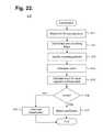
- FIG. 22is a flow diagram showing, by way of example, a method for classification of ECG data.
- FIG. 23is a flow diagram showing, by way of example, a method for trimming data segments classified as noise.
- FIG. 24is a diagram showing, by way of example, a diagnostic composite plot showing data from a button press window.
- FIG. 25is a diagram showing, by way of example, a summary graph of data press occurrences.
- a normal healthy cardiac cyclerepeats through an expected sequence of events that can be visually traced through an ECG.
- Each cyclestarts with cardiac depolarization originating high in the right atrium in the sinoatrial (SA) node before spreading leftward towards the left atrium and inferiorly towards the atrioventricular (AV) node.
- SAsinoatrial
- AVatrioventricular
- the depolarization impulsetransits the Bundle of His and moves into the right and left bundle branches and Purkinje fibers to activate the right and left ventricles.
- FIG. 1is a graph showing, by way of example, a single ECG waveform 10 .
- the x-axisrepresents approximate time in units of tenths of a second and the y-axis represents approximate cutaneous electrical signal strength in units of millivolts.
- ECGsare typically printed or displayed at an effective paper speed of 25 millimeters (mm) per second.
- an ECGmay be provided to a physician in traditional paper-printed form, in “virtual” electronic display form, or both.
- the term “effective paper speed”is still widely applied as a metric to normalize the recorded ECG signal to a standardized grid of 1 mm squares (omitted for the sake of clarity in FIG. 1 ), whereby each 1 mm horizontal box in the grid corresponds to 0.04 s (40 ms) of recorded time.
- Other effective paper speeds, grid sizes and units of displayare possible.
- a full ECGconsists of a stream of alphabetically-labeled waveforms 10 that collectively cover cardiac performance over a period of observation.
- the P-wave 11will normally have a smooth, normally upward, positive waveform that indicates atrial depolarization.
- the QRS complex 17will usually follow during normal rhythms, often with a downward deflection of a Q-wave 12 , followed by a larger upward deflection of an R-wave 13 , and be terminated with a downward waveform of the S-wave 14 , which are collectively representative of ventricular depolarization. This term, QRS, is often reduced to the “R-wave”.
- reference to the R-R intervalmeans the time from one R-wave (or QRS) to another R-wave.
- T-wave 15which will normally be a modest upward waveform, representative of ventricular repolarization
- U-wave 16which is often not directly observable, will indicate the recovery period of the Purkinje conduction fibers.
- this part of the ECG waveformwill not be pertinent.
- Rhythm disordersoften manifest through R-R interval variability or through the patterns formed by R-R intervals over an extended time period prior to and after a specific arrhythmia onset and offset. Both are important tools in the diagnosis of cardiac rhythm abnormalities.
- atrial fibrillationis the chaotic firing of the atria that leads to an erratic activation of the ventricles.
- AFis initially diagnosed by an absence of organized P-waves 11 and confirmed by erratic ventricular rates that manifest in an ECG R-R interval plot as a cloud-like pattern of irregular R-R intervals due to an abnormal conduction of impulses to the ventricles.
- Gaussian-like distributionto these R-R intervals during AF.
- Atrial flutteris an abnormal heart rhythm in which cardiac impulses travel along pathways within the right atrium in an organized circular motion, causing the atria to beat faster than and out of sync with the ventricles.
- AFLpresents in an ECG in a “sawtooth” pattern
- AFL behaviorcan fluctuate wildly.
- Such fluctuationcan be confirmed in an ECG by characteristic R-R interval patterns that usually manifest as 2:1 atrioventricular (AV) conduction, 4:1 AV conduction or even variable AV conduction, as it frequently can be.
- AVatrioventricular
- the importance of this latter manifestation of AFLrelates to how AFL can be distinguished from AF.
- the R-R interval plotcan clearly distinguish the two unlike algorithms that frequently conflate AF with AFL and variable conduction.
- FIG. 2is a graph showing, by way of example, a prior art Poincaré R-R interval plot 18 .
- the x-axisrepresents the duration of R-R interval n in units of milliseconds (ms).
- the y-axisrepresents the duration of R-R interval n+1 also in units of ms.
- the x-and y-axesuse the same units, so as to form a trend line 19 along the 45-degree angle.
- the dot representing the two intervalsfalls onto the 45-degree trend line 19 .
- the dot representing the two intervalsfalls off the 45-degree trend line 19 and, as the difference between successive R-R intervals increases, the dots fall further away from the trend line 19 .
- the number of dots deviating from the trend line 19 in a Poincaré plotcan indicate the frequency of occurrence of irregular heartbeats when compared to the number of dots on the trend line 19 .
- the distance of the dots to the trend line 19can approximate the extent of heart rate change from one heartbeat to the next.
- heart rate changeis limited to only successively-occurring heartbeats, the linearity of time and associated contextual information over an extended time frame are lost.
- Poincare plotscan be generally predictive but they have limited value when used to understand physiological antecedents to an arrhythmia pre-and post-rhythm events, for example something as simple as exercise.
- a Poincaré plotis more useful as a mathematical tool than a physiological one, and therefore a Poincaré plot cannot truly represent what the heart is doing serially over time with respect to changes in the heart's normal and abnormal physiology on a beat-by-beat basis.
- FIG. 3is a flow diagram showing a method 20 for facilitating diagnosis of cardiac rhythm disorders with the aid of a digital computer in accordance with one embodiment.
- the method 20can be implemented in software and execution of the software can be performed on a computer, such as further described infra with reference to FIG. 14 , as a series of processes or method modules or steps.
- the patient's ECG dataare recorded over a set time period (step 21 ), which can be over a short term or extended time frame.
- ECG recordationas well as other physiological monitoring, can be provided through various kinds of ECG-capable monitoring ensembles, including a standardized 12-lead ECG setup, such as used for clinical ECG monitoring, a portable Holter-type ECG recorder for traditional ambulatory ECG monitoring, or a wearable ambulatory ECG monitor, such as a flexible extended wear electrode patch and a removable reusable (or single use) monitor recorder, such as described in commonly-assigned U.S. Pat. No. 9,345,414, issued May 24, 2016, the disclosure of which is incorporated by reference.
- the wearable ambulatory ECG monitorincludes an electrode patch and monitor recorder that are synergistically optimized to capture electrical signals from the propagation of low amplitude, relatively low frequency content cardiac action potentials, particularly the P-waves, generated during atrial activation, and is described in further detail below with respect to FIG. 15 . Still other forms of ECG monitoring assemblies are possible.
- the ECG and any other physiological dataare downloaded or retrieved into a digital computer, as further described infra with reference to FIG. 14 , with, for instance, the assistance of a download station or similar device, or via wireless connection, if so equipped, and a vector of the downloaded or retrieved ECG data is obtained (step 22 ).
- the vector of ECG datarepresents a 40-minute (or other duration) time span that is used in constructing the plot of R-R interval data, although other pre-event and post-event time spans are possible.
- a potentially-actionable cardiac event within the vector of ECG datacan be identified and the ECG data during, prior to and after the event is selected (step 23 ).
- the eventcould be identified with the assistance of a software package, such as Holter LX Analysis Software, licensed by NorthEast Monitoring, Inc., Maynard, Mass.; Intelli Space Cardiovascular Image and Information management system, licensed Koninklijke Philips N.V., Amsterdam, Netherlands; MoMe System, licensed by InfoBionic, Lowell, Mass.; Pyramis ECG Management, licensed by Mortara Instrument Inc., Milwaukee, Wis.; ICS Clinical Suite, licensed by Spacelabs Healthcare Inc., Snoqualmie, Wash.; or a customized software package.
- a software packagesuch as Holter LX Analysis Software, licensed by NorthEast Monitoring, Inc., Maynard, Mass.; Intelli Space Cardiovascular Image and Information management system, licensed Koninklijke Philips N.V., Amsterdam, Netherlands; MoMe System, licensed by InfoBionic, Lowell, Mass.; Pyramis ECG Management, licensed by Mortara Instrument Inc., Milwaukee, Wis.; ICS Clinical Suite, licensed by Spacelabs Healthcare Inc., Snoqualmie, Wash.; or
- a diagnostic composite plotis constructed that includes one or more temporal points of reference into the ECG data, which provide important diagnostic context, and a plot of R-R interval data is constructed based on the vector of ECG data (step 24 ), as further described infra with reference to FIG. 4 .
- both near field and far field contextual views of the ECG dataare constructed and displayed. Both views are temporally keyed to an extended duration R-R interval data view that, in one embodiment, is scaled non-linearly (for practical viewing reasons) to maximize the visual differentiation for frequently-occurring heart rate ranges, such that a single glance allows the physician to make a diagnosis.
- findings made through interpretation of heart rate variability patterns in the diagnostic composite plotcan be analyzed to form a diagnosis of a cardiac rhythm disorder (step 25 ), such as the cardiac rhythm disorders listed, by way of example, in Table 1.
- a cardiac rhythm disordersuch as the cardiac rhythm disorders listed, by way of example, in Table 1.
- the heart rate variability patterns in the diagnostic composite plotcould be provided to a system that programmatically detects AF by virtue of looking for the classic Gaussian-type distribution on the “cloud” of heart rate variability formed in the plot of R-R interval data, which can be corroborated by the accompanying contextual ECG data.
- a cardiac rhythm therapy delivery devicesuch as an implantable medical device (IMD) (not shown), including a pacemaker, implantable cardioverter defibrillator (ICD), or similar devices.
- IMDimplantable medical device
- ICDimplantable cardioverter defibrillator
- a diagnostic composite plotis constructed and displayed to help physicians identify and diagnose temporally-related cardiac dysrhythmic patterns.
- the diagnostic composite plotincludes ECG traces from two or more temporal points of reference and a plot of R-R interval data, although other configurations of ECG data plots when combined with the R-R interval plot will also provide critical information.
- FIG. 4is a flow diagram showing a routine 30 for constructing and displaying a diagnostic composite plot for use in the method 20 of FIG. 3 . Specific examples of diagnostic composite plots are discussed in detail infra with reference to FIGS. 7-13 .
- R-R interval datais presented to physicians in a format that includes views of relevant near field and far field ECG data, which together provide contextual information that improves diagnostic accuracy.
- other views of ECG datacan be provided in addition to or in lieu of the near field and far field ECG data views.
- the near field (or short duration) ECG dataprovides a “pinpoint” classical view of an ECG at traditional recording speed in a manner that is known to and widely embraced by physicians.
- This near field ECG datais coupled to a far field (or medium duration) ECG data view that provides an “intermediate” lower resolution, offering pre-and post-event contextual ECG data.
- the extended-duration R-R interval plotis first constructed (step 31 ), as further described infra with reference to FIG. 5 .
- noisecan be filtered from the R-R interval plot (step 32 ), which is then displayed (step 33 ).
- noisecan be filtered from the ECG data prior to constructing the R-R interval plot, as further described below with reference to FIG. 19 .
- Noise filteringcan include low-pass or high-pass filtering or other forms of signal processing, including automatic gain control, such as described in commonly-assigned U.S. Pat. No. 9,345,414, issued May 24, 2016, cited supra, as well as identification via an adaptive noise detector, as further described in detail below with respect to FIGS. 19-25 .
- Rhythm disordershave different weightings depending upon the context with which they occur.
- the R-R interval data view and the multiple views of the ECG data around which the R-R data embraceprovide that necessary context.
- the short and medium duration ECG data that accompanies the extended-duration R-R interval plotrepresents the ECG data “zoomed” in around a temporal point of reference identified in the center (or other location) of the R-R interval plot, thereby providing a short-term, medium-term and long-term visual context to the physician that allows temporal assessment of cardiac rhythm changes in various complementary views of the heart's behavior.
- the diagnostic composite plotdisplays R-R interval data over a forty-minute duration and ECG data over short and medium durations (steps 34 and 35 ), such as four-second and 24-second durations that provide two-and 12-second segments of the ECG data before and after the RR interval plot's temporal point of reference, which is generally in the center of the R-R interval plot, although other locations in the R-R interval plot could be identified as the temporal point of reference.
- the pinpoint “snapshot” and intermediate views of ECG data with the extended term R-R interval datacomparatively depicts heart rate context and patterns of behavior prior to and after a clinically meaningful arrhythmia or patient concern, thereby enhancing diagnostic specificity of cardiac rhythm disorders and providing physiological context to improve diagnostic ability.
- diagnostically relevant cardiac eventscan be identified and the R-R interval plot can be constructed with a cardiac event centered in the middle (or other location) of the plot, which thereby allows pre-and post-event heart rhythm data to be contextually “framed” through the pinpoint and intermediate ECG data views.
- Other durations, intervals and presentations of ECG dataare possible, including allowing the physician or viewer to select or scroll through the raw ECG data to advance the ECG data together with the R-R interval data, allowing context to be selected by the viewer.
- the extended-duration R-R interval plotpresents beat-to-beat heart rate variability in a format that is intuitive and contextual, yet condensed.
- the format of the R-R interval plotis selected to optimize visualization of cardiac events in a compressed, yet understandable field of view, that allows for compact presentation of the data akin to a cardiologists understanding of clinical events.
- FIG. 5is a flow diagram showing a routine 40 for constructing an extended-duration R-R interval plot for use in the routine 30 of FIG. 4 .
- the duration of the R-R interval plotcan vary from less than one minute to the entire duration of the recording.
- a plurality of R-wave peaksis first selected out of the vector of ECG data (step 41 ) appropriate to the duration of the R-R interval plot to be constructed.
- each recording time differencerepresents the length of one heartbeat.
- the heart rate associated with the recording time differenceis determined by taking an inverse of the recording time difference and normalizing the inverse to beats per minute (step 44 ). Taking the inverse of the recording time difference yields a heart rate expressed in beats per second, which can be adjusted by a factor of 60 to provide a heart rate expressed in bpm. Calculation of the differences between the recording times and the associated heart rate continues for all of the remaining pairs of the R-wave peaks (step 44 ).
- the pairings of R-R intervals and associated heart ratesare formed into a two-dimensional plot.
- R-R intervalsare plotted along the x-axis and associated heart rates are plotted along the y-axis.
- the range and scale of the y-axiscan be adjusted according to the range and frequency of normal or patient-specific heart rates, so as to increase the visual distinctions between the heart rates that correspond to different R-R intervals.
- the y-axis of the R-R interval plothas a range of 20 to 300 beats per minute and R-R intervals corresponding to heart rates falling extremely outside of this range are excluded to allow easy visualization of 99+% of the heart rate possibilities.
- 0 to 350 bpmwould alter the practical visibility of the interval range for the vast majority of events. (Note that these extreme rates remain visible in the ECG.)
- they-axishas a non-linear scale that is calculated as a function of the x-axis (R-R interval), such that:
- y( x - min ⁇ ⁇ bpm max ⁇ ⁇ bpm - min ⁇ ⁇ bpm ) m
- xis the time difference
- min bpmis the minimum heart rate
- max bpmis the maximum heart rate
- the overall effectis to accentuate the spatial differences in frequently-occurring ranges of heart rate and de-emphasize the spatial differential in ranges of heart rate where a deviation from norm would have been apparent, thus maximizing the spatial efficiency in data presentation.
- the goalis to show cardiac events in a simple, small visual contextual format. Larger scales and larger formats bely the practical limits of single-page presentations for the easy visualization at a glance by the busy physician.
- the visual distinctions between the heart rates that correspond to different R-R intervalsstand out, especially when plotted on a non-linear scale. In this configuration, three distinct arrhythmia events can be presented in standard 8.5 by 11 inch page formats. However, other formats are possible. Also, other y-axis ranges and scales are possible as may be selected by distinct clinical needs and specific diagnostic requirements.
- the diagnostic composite plotincludes a single, long range view of R-R interval data and a pair of pinpoint ECG data views that together help to facilitate rhythm disorder diagnosis by placing focused long-term heart rate information alongside short-term and medium-term ECG information.
- Such pairing of ECG and R-R interval datais unique in its ability to inform the physician of events prior to, during and after a cardiovascular event.
- FIG. 6is a diagram showing, by way of example, a diagnostic composite plot 50 generated by the method 30 of FIG. 3 . Note that the diagnostic composite plot can be tailored to include more than one view of R-R interval data and as many views of contextual ECG data as needed.
- a background information plot presenting an extended far field of related informationcan be included, such as activity amount, activity intensity, posture, syncope impulse detection, respiratory rate, blood pressure, oxygen saturation (SpO 2 ), blood carbon dioxide level (pCO 2 ), glucose, lung wetness, and temperature.
- Other forms of background informationare possible.
- background informationcan be layered on top of or keyed to the diagnostic composite plot 50 , particularly at key points of time in the R-R interval data plot, so that the context provided by each item of background information can be readily accessed by the reviewing physician.
- the diagnostic composite plot 50includes an ECG plot presenting a near field (short duration) view 51 , an ECG plot presenting an intermediate field (medium duration) view 52 , and an R-R interval data plot presenting a far field (extended duration) view 53 .
- the three views 51 , 52 , 53are juxtaposed alongside one other to allow quick back and forth referencing of the full context of the heart's normal and abnormal physiology.
- a temporal point of referencewhich could be a diagnostically relevant cardiac event, patient concern or other indicia, would be identified and centered on the x-axis in all three views.
- the placement of the temporal point of reference in the middle of all three x-axesenables the ECG data to be temporally keyed to the R-R interval data appearing in the center 60 of the R-R interval data view 53 , with a near field view 51 of an ECG displayed at normal (paper-based) recording speed and a far field view 52 that presents the ECG data occurring before and after the center 60 .
- the near field view 51provides the ECG data corresponding to the R-R interval data at the center 60 (or other location) in a format that is familiar to all physicians, while the intermediate field view 52 enables presentation of the broader ECG data context going beyond the borders of the near field view 51 .
- the center 60can be slidably adjusted backwards and forwards in time, with the near field view 51 and the far field view 52 of the ECG data automatically or manually adjusting accordingly to stay in context with the R-R interval data view 51 .
- multiple temporal points of referencecan be identified with each temporal point of reference being optionally accompanied by one or more dedicated sets of ECG data views.
- the collection of plotsare conveniently arranged close enough to one another to facilitate printing on a single page of standard sized paper (or physical paper substitute, such as a PDF file), although other layouts of the plots are possible.
- the far field view 53is plotted with time in the x-axis and heart rate in the y-axis.
- the R-R intervalsare calculated by measuring the time occurring between successive R-wave peaks.
- the far field view 53presents R-R interval data (expressed as heart rate in bpm) that begins about 20 minutes prior to and ends about 20 minutes following the center 60 , although other durations are possible.
- the near field view 51 and intermediate field view 52present ECG data relative to the center 60 of the far field view 53 .
- the near field view 51provides a pinpoint or short duration view of the ECG data.
- the near field view 51presents ECG data 55 that begins about two seconds prior to and ends about two seconds following the center 60 , although other durations are possible.
- the intermediate field view 52provides additional contextual ECG information allowing the physician to assess the ECG itself and gather a broader view of the rhythm before and after a “blow-up” of the specific arrhythmia of interest.
- the intermediate field view 52presents ECG data 56 that begins about 12 seconds prior to and ends about 12 seconds following the center 60 , although other durations are possible.
- the eight-second interval of the ECG data 56 in the intermediate field view 52 that makes up the ECG data 56 in the near field view 51is visually highlighted, here, with a surrounding box 57 .
- other views of the ECG dataeither in addition to or in lieu of the near field view 51 and the far field view 52 are possible.
- an ECG plot presenting an extended far field view 54 of the background informationcan be included in the diagnostic composite plot 50 .
- the background informationis presented as average heart rate with day and night periods 58 alternately shaded along the x-axis.
- Other types of background informationsuch as activity amount, activity intensity, posture, syncope impulse detection, respiratory rate, blood pressure, oxygen saturation (SpO 2 ), blood carbon dioxide level (pCO 2 ), glucose, lung wetness, and temperature, are possible.
- FIG. 7is a diagram showing, by way of example, a diagnostic composite plot 70 for facilitating the diagnosis of sinus rhythm (SR) transitioning into AF.
- SRsinus rhythm
- SRis indicated through the presence of a reasonably steady baseline, but with subsidiary lines of premature beats and their compensatory pauses.
- SRmanifests as a shadowing 71 of a high heart rate line and a low heart rate line.
- AFis characterized by irregular heartbeats with a somewhat random variation of R-R intervals, although within a limited range and concentrating in a Gaussian-like distribution pattern around a mean that varies over time. Such characteristics can also be distinct from the more random nature of noise.
- AFcan be diagnosed by viewing a near field view 51 of ECG data showing heartbeats with reversed P-wave and irregular R-R intervals, this approach may be unclear when viewing “snippets” of ECG data, especially when associated with poor quality ECG signals.
- AFcan also be confirmed through a far field view 53 of R-R interval data, in which the R-R intervals assume superficially appearing disorganized, spread-out and decentralized scattered cloud 72 along the x-axis, in comparison to a concentrated, darkened line typical of a more organized cardiac rhythm.
- FIG. 8is a diagram showing, by way of example, a diagnostic composite plot 80 for facilitating the diagnosis of 3:1 atrial flutter (AFL) transitioning into SR with frequent premature ectopic atrial beats.
- AFL3:1 atrial flutter
- the R-R intervalshave a discernible aggregated line in the middle of the cloud 81 when the rhythm has yet to stabilize into a set pattern, not quite AF and not quite AFL.
- a dense line representing firm 3:1 atrial flutterstabilizes the rhythm prior to the transition into SR associated with the presence of two seesawing baselines that result from frequent atrial ectopy causing short coupling intervals and then compensatory long coupling intervals.
- SRis indicated by the middle of the three lines with a low heart rate line consistent with the compensatory pause (long coupling interval) and a high heart rate line with the shortest coupling interval representing the series of atrial premature beats 82 , and thus, at a faster heart rate.
- FIG. 9is a diagram showing, by way of example, a diagnostic composite plot 90 for facilitating the diagnosis of atrial trigeminy.
- Atrial trigeminyis characterized by three heartbeat rates appearing intermittently yet reasonably regularly.
- atrial trigeminycan be diagnosed by viewing a near field view 51 of ECG data, the pattern and its clinical impact and frequency is significantly more recognizable in a far field view 53 of R-R interval data, in which a repeating pattern of three distinct heartbeat lines are persistently present and clearly visible 91 .
- This viewalso provides the physician with a qualitative feel for the frequency of the event troubling the patient that is not discernible from a single ECG strip.
- FIG. 10is a diagram showing, by way of example, a diagnostic composite plot 100 for facilitating the diagnosis of maximum heart rate in an episode of AF during exercise.
- AFmanifests through a dispersed cloud of dots (Gaussian-like distribution) without a discernible main heart rate line representing regular heartbeats 101 .
- the maximum heartbeatcan be located by an increase in heart rate clustered about the cloud 102 .
- individual dots above the 200 bpm range throughout the entire 40-minute rangeindicates the maximum heart rate during exercise.
- the very rapid rise in heart ratecan be critical to patient management, as such bumps in rate by exercise can prove dangerous and even trigger cardiac arrest by inducing ventricular fibrillation. Their very presence is easily visualized in the R-R interval data plot, thereby allowing the physician to alter therapy sufficiently to control such potentially damaging rises in heart rate.
- FIG. 11is a diagram showing, by way of example, a diagnostic composite plot 110 for facilitating the diagnosis of SR transitioning into AFL transitioning into AF.
- SRmanifests as an uneven main heart rate line with a fluctuating height 111 .
- the main heart rate linebreaks away at a lower heart rate than the SR main heart rate line 112 .
- the episode of AFLfurther evolves into AF as characterized by a dispersed cloud of irregular heartbeats without concentrated heart rate lines 113 .
- This viewprovides critical information to the physician managing AF patients in that, at a glance, the view provides data that tells the physician that the patient's AF may be the consequence of AFL.
- Such knowledgemay alter both drug and procedure therapies, like catheter ablation details of intervention. These two disorders are not interchangeable and generally demand different drug and different catheter ablation approaches.
- FIG. 12is a diagram showing, by way of example, a diagnostic composite plot 120 for facilitating the diagnosis of sinus tachycardia and palpitations that occurred during exercise accompanied by a jump in heart rate.
- sinus tachycardiais indicated by the presence of a baseline heart rate of about 60 bpm 121 that spikes up to around 100 bpm 122 and gradually slopes down with a wide tail 123 , reflecting a sharp rise of heart rates followed by a gradual decline.
- the associated ECG data in the near field and intermediate field viewscan confirm the rhythm as sinus rhythm and a normal response to exercise.
- FIG. 13is a diagram showing, by way of example, a diagnostic composite plot 90 for facilitating the diagnosis of bradycardia during sleep and a R-R interval pattern characteristic of sleep.
- Bradycardiarefers to a resting heart rate of under 60 bpm. Bradycardia during sleep is often tempered with occasional spikes of rapid heart rate, which can be a secondary compensatory response to dreaming, snoring or sleep apnea.
- bradycardiamanifests as the presence of a base line heart rate in the range of about 50 bpm 131 , coupled with multiple spikes of dots 132 representing intermittent episodes of elevated heart rate. Such elevations in heart rate during a pre-dominantly slower rate may be signs of a cardio-respiratory disorder. Still other applications of the diagnostic composite plot 80 are possible.
- the diagnostic composite plotsare a tool used by physicians as part of a continuum of cardiac care provisioning that begins with ECG monitoring, continues through diagnostic overread and finally, if medically appropriate, concludes with cardiac rhythm disorder treatment. Each of these steps involve different physical components that collaboratively allow physicians to acquire and visualize R-R interval and ECG data in a way that accurately depicts heart rate variability over time. Further, the data represented by the diagnostic composite plots can be provided to analysis software for cardiac event detection, and patient diagnosis or confirmation.
- FIG. 14is a block diagram showing a system 140 for facilitating diagnosis of cardiac rhythm disorders with the aid of a digital computer 150 in accordance with one embodiment.
- Each diagnostic composite plot 151is based on ECG data 166 that has either been recorded by a conventional electrocardiograph (not shown) or retrieved or obtained from some other type of ECG monitoring and recording device. Following completion of the ECG monitoring, the ECG data is assembled into a diagnostic composite plot 151 , which can be used by a physician to diagnosis and, if required, treat a cardiac rhythm disorder, or for other health care or related purposes.
- Each diagnostic composite plot 151is based on ECG data 166 that has been recorded over a period of observation, which can be for just a short term, such as during a clinic appointment, or over an extended time frame of months.
- ECG recordation and, in some cases, physiological monitoringcan be provided through various types of ECG-capable monitoring ensembles, including a standardized 12-lead ECG setup (not shown), such as used for clinical ECG monitoring, a portable Holter-type ECG recorder for traditional ambulatory ECG monitoring (also not shown), or a wearable ambulatory ECG monitor.
- One form of ambulatory ECG monitor 142 particularly suited to monitoring and recording ECG and physiological dataemploys an electrode patch 143 and a removable reusable (or single use) monitor recorder 144 , such as described in commonly-assigned U.S. Pat. No. 9,345,414, issued May 24, 2016, cited supra, and also, further described in detail below with respect to FIGS. 15-18 .
- the electrode patch 143 and monitor recorder 144are synergistically optimized to capture electrical signals from the propagation of low amplitude, relatively low frequency content cardiac action potentials, particularly the P-waves generated during atrial activation.
- the ECG monitor 142sits centrally (in the midline) on the patient's chest along the sternum 169 oriented top-to-bottom.
- the ECG monitor 142interfaces to a pair of cutaneous electrodes (not shown) on the electrode patch 143 that are adhered to the patient's skin along the sternal midline (or immediately to either side of the sternum 169 ).
- the ECG monitor 142has a unique narrow “hourglass”-like shape that significantly improves the ability of the monitor to be comfortably worn by the patient 141 for an extended period of time and to cutaneously sense cardiac electric signals, particularly the P-wave (or atrial activity) and, to a lesser extent, the QRS interval signals in the ECG waveforms indicating ventricular activity.
- the electrode patch 143itself is shaped to conform to the contours of the patient's chest approximately centered on the sternal midline.
- a layer of non-irritating adhesivesuch as hydrocolloid, is provided at least partially on the underside, or contact, surface of the electrode patch, but only on the electrode patch's distal and proximal ends.
- a strain reliefcan be defined in the electrode patch's flexible circuit using cutouts partially extending transversely from each opposite side of the flexible circuit and continuing longitudinally towards each other to define in ‘S’-shaped pattern.
- the electrode patch 143is made from a type of stretchable spunlace fabric.
- the outward-facing aspect of the backingto which a (non-stretchable) flexible circuit is fixedly attached, stretches at a different rate than the backing's skin-facing aspect, where a skin adhesive removably affixes the electrode patch 143 to the skin.
- a skin adhesiveremovably affixes the electrode patch 143 to the skin.
- the monitor recorder 142senses and records the patient's ECG data 166 and physiological data (not shown) into a memory onboard the monitor recorder 144 .
- the recorded datacan be downloaded using a download station 147 , which could be a dedicated download station 145 that permits the retrieval of stored ECG data 166 and physiological data, if applicable, execution of diagnostics on or programming of the monitor recorder 144 , or performance of other functions.
- the monitor recorder 144has a set of electrical contacts (not shown) that enable the monitor recorder 144 to physically interface to a set of terminals 148 .
- the download station 145can be operated through user controls 149 to execute a communications or data download program 146 (“Download”) or similar program that interacts with the monitor recorder 144 via the physical interface to retrieve the stored ECG data 166 .
- the download station 145could alternatively be a server, personal computer, tablet or handheld computer, smart mobile device, or purpose-built device designed specific to the task of interfacing with a monitor recorder 144 . Still other forms of download station 145 are possible.
- the ECG data 166 from the monitor recorder 144can be offloaded wirelessly.
- the ECG data 166can be retrieved from the download station 145 using a control program 157 (“Ctl”) or analogous application executing on a personal digital computer 156 or other connectable computing device, via a hard wired link 158 , wireless link (not shown), or by physical transfer of storage media (not shown).
- the personal digital computer 156may also execute middleware (not shown) that converts the ECG data 166 into a format suitable for use by a third-party post-monitoring analysis program.
- the personal digital computer 156stores the ECG data 166 along with each patient's electronic medical records (EMRs) 165 in the secure database 64 , as further discussed infra.
- the download station 145is able to directly interface with other devices over a computer communications network 155 , which could be a combination of local area and wide area networks, including the Internet or another telecommunications network, over wired or wireless connections.
- a client-server modelcan be employed for ECG data 166 analysis.
- a server 62executes a patient management program 160 (“Mgt”) or similar application that accesses the retrieved ECG data 166 and other information in the secure database 164 cataloged with each patient's EMRs 165 .
- the patients' EMRscan be supplemented with other information (not shown), such as medical history, testing results, and so forth, which can be factored into automated diagnosis and treatment.
- the patient management program 160also maintains and safeguards the secure database 164 to limit access to patient EMRs 165 to only authorized parties for appropriate medical or other uses, such as mandated by state or federal law, such as under the Health Insurance Portability and Accountability Act (HIPAA) or per the European Union's Data Protection Directive.
- HIPAAHealth Insurance Portability and Accountability Act
- Other schemes and safeguards to protect and maintain the integrity of patient EMRs 165are possible.
- the wearable monitor 142can interoperate wirelessly with other wearable or implantable physiology monitors and activity sensors 152 , such as activity trackers worn on the wrist or body, and with mobile devices 153 , including smart watches and smartphones.
- Wearable or implantable physiology monitors and activity sensors 152encompass a wide range of wirelessly interconnectable devices that measure or monitor a patient's physiological data, such as heart rate, temperature, blood pressure, respiratory rate, blood pressure, blood sugar (with or without an appropriate subcutaneous probe), oxygen saturation, minute ventilation, and so on; physical states, such as movement, sleep, footsteps, and the like; and performance, including calories burned or estimated blood glucose level.
- wearable and implantable physiology monitors and activity sensors 152are capable of wirelessly interfacing with mobile devices 153 , particularly smart mobile devices, including so-called “smartphones” and “smart watches,” as well as with personal computers and tablet or handheld computers, to download monitoring data either in real-time or in batches through an application (“App”) or similar program.
- mobile devices 153particularly smart mobile devices, including so-called “smartphones” and “smart watches,” as well as with personal computers and tablet or handheld computers, to download monitoring data either in real-time or in batches through an application (“App”) or similar program.
- Appapplication
- ECG data 166Based on the ECG data 166 , physicians can rely on the data as medically certifiable and are able to directly proceed with diagnosing cardiac rhythm disorders and determining the appropriate course of treatment for the patient 141 , including undertaking further medical interventions as appropriate.
- the ECG data 166can be retrieved by a digital computer 150 over the network 155 .
- a diagnostic composite plot 151 that includes multiple temporal points of reference and a plot of R-R interval datais then constructed based on the ECG data 166 , as discussed in detail supra with reference to FIG. 3 , and displayed or, alternatively, printed, for use by a physician.
- the server 159executes a patient diagnosis program 161 (“Dx”) or similar application that can evaluate the ECG data 166 to form a diagnosis of a cardiac rhythm disorder.
- the patient diagnosis program 161compares and evaluates the ECG data 166 to a set of medical diagnostic criteria 167 , from which a diagnostic overread 162 (“diagnosis”) is generated.
- Each diagnostic overread 162can include one or more diagnostic findings 168 that can be rated by degree of severity, such as with the automated diagnosis of atrial fibrillation.
- diagnostic findings 168 for a patientexceed a threshold level of tolerance, which may be tailored to a specific client, disease or medical condition group, or applied to a general patient population, in a still further embodiment, therapeutic treatment (“Therapy”) to address diagnosed disorder findings can be generated and, optionally, programmed into a cardiac rhythm therapy delivery device, such as an IMD (not shown), including a pacemaker, implantable cardioverter defibrillator (ICD), or similar devices.
- IMDimplantable cardioverter defibrillator
- FIG. 15is a perspective view showing an ambulatory ECG monitor 212 having an extended wear electrode patch 215 with a monitor recorder 214 inserted.
- the body of the electrode patch 215is preferably constructed using a flexible backing 220 formed as an elongated strip 221 of wrap knit or similar stretchable material about 145 mm long and 32 mm at the widest point with a narrow longitudinal mid-section 223 evenly tapering inward from both sides.
- a pair of cut-outs 222 between the distal and proximal ends of the electrode patch 15create a narrow longitudinal midsection 223 or “isthmus” and defines an elongated “hourglass”-like shape, when viewed from above, such as described in commonly-assigned U.S. Design Pat. No. D744659, issued Dec. 1, 2015, the disclosure of which is incorporated by reference.
- the upper part of the “hourglass”is sized to allow an electrically non-conductive receptacle 225 , that sits on top of the outward-facing surface of the electrode patch 215 , to be affixed to the electrode patch 215 with an ECG electrode placed underneath on the patient-facing underside, or contact, surface of the electrode patch 215 ; the upper part of the “hourglass” has a longer and wider profile (but still rounded and tapered to fit comfortably between the breasts) than the lower part of the “hourglass,” which is sized primarily to allow just the placement of an ECG electrode of appropriate shape and surface area to record the P-wave and the QRS signals sufficiently given the inter-electrode spacing.
- the electrode patch 215incorporates features that significantly improve wearability, performance, and patient comfort throughout an extended monitoring period.
- the entire electrode patch 215is lightweight in construction, which allows the patch to be resilient to disadhesing or falling off and, critically, to avoid creating distracting discomfort to the patient, even when the patient is asleep.
- the weight of a heavy ECG monitorimpedes patient mobility and will cause the monitor to constantly tug downwards and press on the patient's body that can generate skin inflammation with frequent adjustments by the patient needed to maintain comfort.
- the electrode patch 215is subjected to pushing, pulling, and torsional movements, including compressional and torsional forces when the patient bends forward, or tensile and torsional forces when the patient leans backwards.
- the electrode patch 215incorporates crimp and strain reliefs, such as described in commonly-assigned U.S. Pat. No. 9,545,204, issued Jan. 17, 2017, the disclosure of which is incorporated by reference.
- tabs 224can respectively extend an additional 8 mm to 12 mm beyond the distal and proximal ends of the flexible backing 220 to facilitate with adhering the electrode patch 215 to or removing the electrode patch 215 from the sternum 213 .
- These tabs 224preferably lack adhesive on the underside, or contact, surface of the electrode patch 215 . Still other shapes, cut-outs and conformities to the electrode patch 215 are possible.
- the monitor recorder 214removably and reusably snaps into an electrically non-conductive receptacle 225 during use.
- the monitor recorder 214contains electronic circuitry for recording and storing the patient's electrocardiography as sensed via a pair of ECG electrodes provided on the electrode patch 215 .
- the non-conductive receptacle 225is provided on the top surface of the flexible backing 220 with a retention catch 226 and tension clip 227 molded into the non-conductive receptacle 225 to conformably receive and securely hold the monitor recorder 214 in place.
- the electrode patch 215is generally intended for a single use and is meant to be replaced periodically throughout an extended period of monitoring. However, some types of monitoring may not extend over a period of time long enough to necessitate replacement of the electrode patch 215 . In those situations, the monitor recorder 214 and electrode patch 215 can be combined into a single integral assembly.
- the monitor recorder 214includes a sealed housing that snaps into place in the non-conductive receptacle 225 .
- FIG. 16is a perspective view showing the monitor recorder 214 of FIG. 15 .
- the sealed housing 250 of the monitor recorder 214can have a rounded isosceles trapezoidal-like shape, when viewed from above, such as described in commonly-assigned U.S. Design Pat. No. D717955, issued Nov. 18, 2014, the disclosure of which is incorporated by reference.
- the edges 251 along the top and bottom surfacesare rounded for patient comfort.
- the sealed housing 250is approximately 47 mm long, 23 mm wide at the widest point, and 7 mm high, excluding a patient-operable tactile-feedback button 255 .
- other sizes of the sealed housingare possible.
- the sealed housing 250can be molded out of polycarbonate, ABS, or an alloy of those two materials.
- the button 255is waterproof and the button's top outer surface is molded silicon rubber or similar soft pliable material.
- a retention detent 253 and tension detent 254are molded along the edges of the top surface of the housing 250 to respectively engage the retention catch 226 and the tension clip 227 molded into non-conductive receptacle 225 .
- Other shapes, features, and conformities of the sealed housing 250are possible.
- the electrode patch 215is intended to be disposable, while the monitor recorder 214 is designed for reuse and can be transferred to successive electrode patches 215 to ensure continuity of monitoring, if so desired.
- the monitor recorder 14can be used only once, but single use effectively wastes the synergistic benefits provided by the combination of the disposable electrode patch and reusable monitor recorder.
- the placement of the wearable monitor 212 in a location at the sternal midline (or immediately to either side of the sternum)benefits long-term extended wear by removing the requirement that ECG electrodes be continually placed in the same spots on the skin throughout the monitoring period. Instead, the patient is free to place an electrode patch 215 anywhere within the general region of the sternum.
- a monitor recorder 214is merely unsnapped from a worn out electrode patch 215 , the worn out electrode patch 215 is removed from the skin, a new electrode patch 215 is adhered to the skin, possibly in a new spot immediately adjacent to the earlier location, and the same monitor recorder 214 is snapped into the new electrode patch 215 to reinitiate and continue the ECG monitoring.
- FIG. 17is a perspective view showing the extended wear electrode patch 215 of FIG. 15 without a monitor recorder 214 inserted.
- a flexible circuit 232is adhered to each end of the flexible backing 220 .
- a distal circuit trace 233 from the distal end 230 of the flexible backing 220 and a proximal circuit trace (not shown) from the proximal end 231 of the flexible backing 220electrically couple ECG electrodes (not shown) with a pair of electrical pads 234 .
- the distal and proximal circuit tracesare replaced with interlaced or sewn-in flexible wires.
- the electrical pads 234are provided within a moisture-resistant seal 235 formed on the bottom surface of the non-conductive receptacle 225 .
- the electrical pads 234interface to electrical contacts (not shown) protruding from the bottom surface of the monitor recorder 214 .
- the moisture-resistant seal 235enables the monitor recorder 214 to be worn at all times, even during showering or other activities that could expose the monitor recorder 214 to moisture or adverse conditions.
- a battery compartment 236is formed on the bottom surface of the non-conductive receptacle 225 .
- the battery contained within the battery compartment 235is a direct current (DC) power cell and can be replaceable, rechargeable or disposable.
- FIG. 18is a bottom plan view of the monitor recorder 214 of FIG. 15 .
- a cavity 258is formed on the bottom surface of the sealed housing 250 to accommodate the upward projection of the battery compartment 236 from the bottom surface of the non-conductive receptacle 225 , when the monitor recorder 214 is secured in place on the non-conductive receptacle 225 .
- a set of electrical contacts 256protrude from the bottom surface of the sealed housing 250 and are arranged in alignment with the electrical pads 234 provided on the bottom surface of the non-conductive receptacle 225 to establish electrical connections between the electrode patch 215 and the monitor recorder 214 .
- a seal coupling 257circumferentially surrounds the set of electrical contacts 256 and securely mates with the moisture-resistant seal 235 formed on the bottom surface of the non-conductive receptacle 225 .
- the battery contained within the battery compartment 236can be replaceable, rechargeable or disposable.
- the ECG sensing circuitry of the monitor recorder 214can be supplemented with additional sensors, including an SpO 2 sensor, a blood pressure sensor, a temperature sensor, respiratory rate sensor, a glucose sensor, an air flow sensor, and a volumetric pressure sensor, which can be incorporated directly into the monitor recorder 214 or onto the non-conductive receptacle 225 .
- additional sensorsincluding an SpO 2 sensor, a blood pressure sensor, a temperature sensor, respiratory rate sensor, a glucose sensor, an air flow sensor, and a volumetric pressure sensor, which can be incorporated directly into the monitor recorder 214 or onto the non-conductive receptacle 225 .
- ECG data collected via the ambulatory ECG monitor, such as described above, or another monitorcan be provided to a post-monitoring analysis program for cardiac event detection and patient diagnosis.
- noisecan optionally be removed to refine the ECG data for more accurate detection and diagnosis results.
- ECG datais often interspersed with events that occur while recording and represent device-related events, such as low battery warnings or reset events, which are not interesting to the analysis of the data. Detection and removal of noise not only increases data quality, but reduces an amount of data necessary for analysis, which can result in more accurate and quicker results.
- areas of recorded data near a period of time when the patient presses the feedback buttontends to look like noise, but is extremely important for consideration and analysis since the patient believes a cardiac event may be occurring.
- FIG. 19is a flow diagram showing a method 270 for ECG data classification for facilitating diagnosis of cardiac rhythm disorders with the aid of a digital computer. Cutaneous action potentials of a patient are monitored and recorded (block 271 ) as ECG data over a set time period. Upon completion of the monitoring period, the ECG and any physiological data are downloaded or retrieved into a digital computer (block 272 ).
- automatic gain controlcan be applied (block 273 ) to the recorded signals to place as many as the ECG signals as possible within a normal range, such as from 0.5 to 1.8 mV peak-to-peak, such as described in commonly-assigned U.S. Pat. No. 9,345,414, issued May 24, 2016, the disclosure of which is incorporated by reference.
- Automatic gain controlis generally provided through an AGC module that is implemented as part of a suite of post-processing modules, although automatic gain control could also be provided through standalone software or as part of other forms of ECG visualization and interpretation software.
- the automatic gain controlcan be run prior to or after noise detection. If prior, gain control is used to regularize the signal amplitude for noise detection; however, if run after, gain control is used to control the signal amplitude seen by the noise detection analysis software. Alternatively, the signal amplitude is controlled manually by a reading technician.
- the gaincan be determined by defining a temporal window for automatic gain control.
- the temporal windowhas to be long enough to capture at least one heart beat; a five-second temporal window is used, although other durations are possible.
- the peak-to-peak voltage of each ECG data segmentis computed and a single gain factor is applied to the entire signal such that the average signal falls in the center of the preferred range.
- other statistical valuescould be used to represent the correlation of the peak-to-peak voltages in the preferred range, either in lieu of or in addition to the average voltage, such as maximum or minimum observed voltage, mean voltage, and so on.
- Each ECG value in a segmentis then processed in an iterative loop.
- the ECG valuesare dynamically gained. In one embodiment, the preferred range falls from about 2 mV to 10 mV, although other values could be chosen.
- noise detectioncan optionally be performed (block 274 ) to refine the ECG data for analysis and use in patient diagnosis.
- the noise detectioncan include classifying segments of the ECG data as noise or valid data, as further described below with reference to FIGS. 19-25 .
- a potentially-actionable cardiac event within a vector of the ECG datacan be optionally identified and the ECG data during, prior to and after the event can be selected (block 275 ), as described supra with respect to FIG. 3 .
- the vector of ECG datacan represent a 40-minute (or other duration) time span that is used in constructing the plot of R-R interval data, although other pre-event and post-event time spans are possible.
- the eventcould be identified with the assistance of a software package, such as Holter LX Analysis Software, licensed by NorthEast Monitoring, Inc., Maynard, Mass.; IntelliSpace Cardiovascular Image and Information management system, licensed Koninklijke Philips N.V., Amsterdam, Netherlands; MoMe System, licensed by InfoBionic, Lowell, Mass.; Pyramis ECG Management, licensed by Mortara Instrument Inc., Milwaukee, Wis.; ICS Clinical Suite, licensed by Spacelabs Healthcare Inc., Snoqualmie, Wash.; or a customized software package.
- a software packagesuch as Holter LX Analysis Software, licensed by NorthEast Monitoring, Inc., Maynard, Mass.; IntelliSpace Cardiovascular Image and Information management system, licensed Koninklijke Philips N.V., Amsterdam, Netherlands; MoMe System, licensed by InfoBionic, Lowell, Mass.; Pyramis ECG Management, licensed by Mortara Instrument Inc., Milwaukee, Wis.; ICS Clinical Suite, licensed by Spacelabs Healthcare Inc., Snoqualmie, Wash.; or
- a plot of R-R interval datais constructed (block 276 ) and can be displayed with both near field and far field contextual views of the ECG data, as a diagnostic composite plot, as described supra with respect to FIG. 6 .
- Both viewsare temporally keyed to an extended duration R-R interval data view that, in one embodiment, is scaled non-linearly to maximize the visual differentiation for frequently-occurring heart rate ranges, such that a single glance allows the physician to make a diagnosis. All three views are presented simultaneously, thereby allowing an interpreting physician to diagnose rhythm and the pre-and post-contextual events leading up to a cardiac rhythm of interest.
- noise detectioncan optionally be performed (block 277 ) on the R-R interval data to remove data segments classified as noise, as further described below with respect to FIG. 20 , to refine the data for further analysis.
- a diagnosis of a cardiac rhythm disordercan optionally be formed (block 278 ) based on findings made through interpretation of heart rate variability patterns in the diagnostic composite plot.
- the heart rate variability patterns in the diagnostic composite plotcould be provided to a system that programmatically detects AF by virtue of looking for the classic Gaussian-type distribution on the “cloud” of heart rate variability formed in the plot of R-R interval data, which can be corroborated by the accompanying contextual ECG data.
- a cardiac rhythm therapy delivery devicesuch as an implantable medical device (IMD) (not shown), including a pacemaker, implantable cardioverter defibrillator (ICD), or similar devices.
- IMDimplantable medical device
- ICDimplantable cardioverter defibrillator
- FIG. 20is a flow diagram showing a method 290 for noise detection.
- an adaptive noise detectorPrior to classification of ECG data, an adaptive noise detector is trained (block 291 ).
- the adaptive noise detectorcan be implemented by a convolutional neural network that represents a processing device, such as an algorithm executed by a computer processor or actual hardware. Other types of systems are possible.
- segments of the ECG dataare annotated with a classification of noise or valid data by software analysis or human review. Training is further described in detail below with respect to FIG. 21 .
- further ECG data collected from a patient via an ambulatory ECG monitorsuch as the monitor described above with respect to FIGS. 15-18 , is segmented and provided (block 292 ) to the trained adaptive noise detector for classification (block 293 ).
- data from other types of ECG monitorscan be used.
- Classification of the ECG data segmentsincludes assigning a designation of valid data or noise to each segment. Subsequently, the ECG data segments that correspond with a time frame during which a patient feedback button on the ECG ambulatory monitor is pressed are identified. Those segments classified as noise and that overlap with the timeframe are trimmed (block 294 ) to remove the data corresponding with the button press. Defining the time frame is further described in detail below with reference to FIG. 23 . Finally, all segments classified as noise can be removed from the ECG data prior to further processing. In a further embodiment, the noise data segments can remain with the ECG data; however, during review by analysis software, those segments classified as noise can be ignored.
- FIG. 21is a flow diagram showing, by way of example, a method 300 for training an adaptive noise detector.
- ECG training datais obtained (block 301 ) as data files from ambulatory ECG monitors associated with a group of patients. The patients can be selected randomly or identified based on patient condition. In one embodiment, between 200 and 250 files can be collected from different ECG ambulatory monitors and used as training data.
- the ambulatory monitors used to collect the ECG datacan include the monitor described above with respect to FIGS. 15-18 , as well as other types of monitors.
- the data file collected from each ECG ambulatory monitorcan each include up to or more than 64 MB of data.
- the training datais divided (block 302 ) into segments, and regions of the training data are annotated (block 303 ) with markers for valid data or noise by software analysis, human review, or a combination of software analysis and human review.
- each file from one of the ambulatory ECG monitorsis divided into segments.
- the ECG data from a single filecan be divided into over 1500 segments, such that each segment includes between 9-10 seconds of data.
- other numbers of segments and data times represented by each segmentare possible, such as segments of data between 2 and 12 seconds.
- the datacan be divided into contiguous segments or alternatively, into overlapping segments.
- a neural networkviews only one segment at a time for detection and thus, each segment should include enough data for a determination of noise.
- the periodicity of heart activityis an important indicator of non-noise so each segment should include at least a few beats. However, the larger the segment becomes the more good signal will be mis-marked as noise on either side of a real noise identification.
- each training segmentcan be removed (block 304 ) since such data often includes or resembles noise, which could lead to misclassification of the data in that segment.
- the training data segmentsare then provided (block 305 ) to the adaptive noise detector.
- the segments of the training dataare each provided and run through the adaptive noise detector once.
- the training segmentsare run multiple times.
- testingcan be performed (block 306 ) to determine classification accuracy of the adaptive noise detector.
- a set of testing datasuch as collected from ECG ambulatory monitors, is run through the trained adaptive noise detector, which classifies the testing data. Additionally, the testing data is annotated manually or via a different program with classifications for noise or valid data. A comparison is made between the annotation of the data and the results of the adaptive noise detector. A number of samples correctly classified is determined and an average accuracy of the testing data is determined. In one embodiment, the average accuracy can be determined and reported after every n number of batches of ECG data. For example, n can represent 100 batches; however, other numbers of batches are possible.
- the batchescan be determined based on an amount of ECG data, a number of ECG ambulatory monitors providing the data, as well as by other metrics. In one embodiment, the batch should be small enough to fit in the memory available, but large enough to amortize startup time.
- An accuracy thresholdcan be applied to the average accuracy values and if an average accuracy value or a predetermined number of average accuracy values are below the threshold, further training can be performed (block 307 ) to increase the accuracy of the adaptive noise detector.
- FIG. 22is a flow diagram showing, by way of example, a method 310 for classification of ECG data.
- the collected ECG datais segmented into blocks, such as described above with respect to the training data of FIG. 21 .
- the ECG data segmentsare then received (block 311 ) by the adaptive noise detector, which can be implemented by a convolutional neural network utilizing, for example, a one dimensional formulation for use with ECG data.
- the adaptive noise detectorcan include hidden layers for performing the classification. In the example described below, two convolutional or pooling hidden layers, and two fully-connected hidden layers are utilized. However, other number of layers are possible.
- ECG trace featuresare identified (block 312 ) using, for example, sliding filters.
- ECG trace featurescan include two R waves with three P waves located between the R waves and R wave spikes, as well as other types of features that are indicative of a cardiac event, such as an arrhythmia.
- the featurescan be automatically learned during the training process and take any form that is learned to be associated with noise or the ECG.
- filters for at least 32 featuresare run against the ECG data.
- repeating patterns of the featuresare identified (block 313 ), including, for example, identifying high numbers of successive R wave spikes.
- the data obtained from the second convolution layeris provided to a first fully connected cross-connection layer, which builds (block 314 ) a matrix with the repeating features representing the columns and matrix multipliers representing rows.
- An intersection value for each combination of the repeating features and matrix multipliersare listed in the matrix as cross connection weights. Specifically, the intersection value can be determined by multiplying each repeating feature value with a matrix multiplier and scaling the product by a predetermined factor. However, other methods for determining the intersection values are possible.
- the second fully connected cross-connection layerutilizes the cross-connection weights from the first fully connected cross-connection layer and multiplies the cross-connection weights by further weights to calculate (block 315 ) final cross-connection values for each ECG data segment.
- the final cross-connection valuesinclude a noise classification value and a valid data classification value.
- the noise classification value and the valid data classification valueindicate a likelihood that the data segment, respectively, includes noise or does not include noise.
- the segments classified as noiseare removed from the ECG data and the remaining valid data segments are provided for further analysis and patient diagnosis.
- Patient identified cardiac eventsare important for use in diagnosis since the patient generally provides feedback when she feels uncomfortable, ill, or is experiencing symptoms resembling a cardiac event. Such feedback is obtained when the patient applies physical pressure to the feedback button located on the monitor recorder of the ECG ambulatory monitor, as shown in FIG. 16 .
- the physical pressure, from the patientcauses the button press to appear as noise in the recorded data. Due to the importance of the data prior to, during, and after the button press, if the data collected at the time of the button press is marked as noise, some or all of the related data will be removed from further analysis. Thus, to maintain data related to the button press, segments with data that overlap a button press event and that are classified as noise are trimmed to remove the data related to the button from being classified as noise.
- FIG. 23is a flow diagram showing, by way of example, a method 320 for trimming data segments classified as noise.
- presses of the patient feedback buttonare identified (block 321 ) and marked.
- a button windowis generated (block 322 ) by identifying a predetermined time before and after each button press event and designating the span of time as a button window.
- the predetermined times before and after a button press eventcan be the same, such as five minutes for a 10 minute window, or a different amount of time.
- each of the predetermined time before and aftercan differ.
- the data segments classified as noiseare identified (block 323 ) and compared with the button press windows to further identify (block 324 ) those noise data segments that overlap a button press window.
- the overlapping noise data segmentsare trimmed (block 325 ) to align with a start or end of the button press window, such that the noise data segment includes no ECG data within the button press window.
- the data removed from the noise data segmentcan be assigned a valid data classification to ensure the data is later reviewed. Further, a noise classification assigned to any data segment that is fully encompassed by the button window is removed so that the data can be considered.
- the data segments classified as noiseare removed from the ECG data, leaving only the data segments classified as valid and the data associated with the button presses for further analysis and use in patient diagnosis.
- FIG. 24is a diagram showing, by way of example, a diagnostic composite plot 340 showing data from a button press window.
- the diagnostic composite plot 340includes an ECG plot presenting a near field (short duration) view 51 , an ECG plot presenting an intermediate field (medium duration) view 52 , and an R-R interval data plot presenting a far field (extended duration) view 53 .
- a temporal point of reference, such as a button press eventis identified and centered on the x-axis in all three views.
- the button press eventis associated with a button press window 341 , which is displayed on the intermediate field view.
- FIG. 25is a diagram showing, by way of example, a summary graph 350 , showing instances of button presses.
- a summary graph 350maps all ECG data recorded by the ECG ambulatory monitor over a period up to seven days. The data is displayed and button presses can be identified via an icon 351 . Identifying all instances of button presses over a recording period can be useful to identify patterns of symptoms experienced by the patient, which can assist in making a diagnosis.
- the placement of a single button press window in the middle of the x-axes on all three viewsenables the ECG data to be temporally keyed to the R-R interval data appearing in the center of the R-R interval data view 53 , with a near field view 51 of an ECG displayed at normal (paper-based) recording speed and an intermediate field view 52 that presents the ECG data occurring before and after the center.
- the near field view 51provides the ECG data corresponding to the R-R interval data at the center (or another location), while the intermediate field view 52 enables presentation of the broader ECG data context going beyond the borders of the near field view 51 .
- the centercan be slidably adjusted backwards and forwards in time, with the near field view 51 and the far field view 52 of the ECG data automatically adjusting accordingly to stay in context with the R-R interval data view 51 .
- multiple temporal points of referencecan be identified with each temporal point of reference being optionally accompanied by one or more dedicated sets of ECG data views.
- the intermediate field view 52also displays data segments 342 , d n , which each represent a grouping of ECG data that has been classified as valid or noise.
- the classification for each data segment 342can also be displayed.
- the segments 342 that are assigned a noise classificationare compared with the button press window 341 .
- Those noise data segments that overlap with the button press window 341are trimmed.
- data segment d 8overlaps with the button press window 341 .
- the overlapping data 343 at the beginning of the button press windowis removed from the data segment that is classified as noise, thus trimming the noise data segment.
- the trimmed datacan be added to the next consecutive segment or form a new segment.
- a noise thresholdcan be applied to the data segments classified as noise to prevent shorts bursts of noise within one such segment from requiring all data in that segment to be removed, since some types of arrhythmias can resemble noise. If the data identified as noise within the noise segment occurs over a period that meets or exceeds the noise threshold, then that noise data segment is removed from the ECG data prior to analysis. However, if the data identified as noise within the noise data segment occurs over a period less than the noise threshold, the noise classification can be removed and the data remains with the ECG data.
- each segmentis assigned a noise determination of yes or no, with a yes determination indicating noise is present and a no determination indicating no noise is present in the segment.
- the thresholdcan be applied to one or more segments and can, in one instance, be equal to the length of a predetermined number of segments. For example, the threshold can be set to 20 seconds based on two 10 second segments of data and can be applied to multiple consecutive segments of data.
- the data segments classified as noiseare removed from the ECG data, leaving only the valid data segments and data associated with the button press windows for further analysis.
- datais provided for software analysis or human review for patient diagnosis, including identification of one or more cardiac events.
Landscapes
- Health & Medical Sciences (AREA)
- Life Sciences & Earth Sciences (AREA)
- Cardiology (AREA)
- Engineering & Computer Science (AREA)
- Animal Behavior & Ethology (AREA)
- Veterinary Medicine (AREA)
- Biophysics (AREA)
- Biomedical Technology (AREA)
- Heart & Thoracic Surgery (AREA)
- Medical Informatics (AREA)
- Molecular Biology (AREA)
- Surgery (AREA)
- Physics & Mathematics (AREA)
- General Health & Medical Sciences (AREA)
- Public Health (AREA)
- Pathology (AREA)
- Physiology (AREA)
- Signal Processing (AREA)
- Artificial Intelligence (AREA)
- Computer Vision & Pattern Recognition (AREA)
- Psychiatry (AREA)
- Pulmonology (AREA)
- Chemical & Material Sciences (AREA)
- Dispersion Chemistry (AREA)
- Measurement And Recording Of Electrical Phenomena And Electrical Characteristics Of The Living Body (AREA)
Abstract
Description
| TABLE 1 |
| Cardiac Rhythm Disorders |
| Normal sinus rhythm | ||
| Sinus Bradycardia | ||
| Sinus Tachycardia | ||
| Premature atrial and ventricular beats | ||
| Ectopic atrial tachycardia | ||
| Atrial fibrillation | ||
| Atrial flutter | ||
| Atrial or ventricular bigeminy, trigeminy or quadrigeminy | ||
| Sinus Bradycardia | ||
| Fusion beats | ||
| Interpolated ventricular premature beats | ||
| Intraventricular conduction delay | ||
| Junctional rhythm | ||
| AV Nodal re-entrant tachycardia | ||
| AV re-entrant tachycardia | ||
| Wolff-Parkinson-White Syndrome and Pre-excitation | ||
| Ventricular tachycardia | ||
| Accelerated idioventricular rhythm | ||
| AV Wenckebach block | ||
| AV Type II block | ||
| Sinoatrial block | ||
where x is the time difference, min bpm is the minimum heart rate, max bpm is the maximum heart rate, and n<1. The non-linear scale of the y-axis accentuates the spatial distance between successive heart rates when heart rate is low. For example, when n=2, the spatial difference between 50 and 60 bpm is 32% larger than the spatial difference between 90 bpm and 100 bpm, and 68% larger than the spatial difference between 150 bpm and 160 bpm. As a result the overall effect is to accentuate the spatial differences in frequently-occurring ranges of heart rate and de-emphasize the spatial differential in ranges of heart rate where a deviation from norm would have been apparent, thus maximizing the spatial efficiency in data presentation. The goal is to show cardiac events in a simple, small visual contextual format. Larger scales and larger formats bely the practical limits of single-page presentations for the easy visualization at a glance by the busy physician. The visual distinctions between the heart rates that correspond to different R-R intervals stand out, especially when plotted on a non-linear scale. In this configuration, three distinct arrhythmia events can be presented in standard 8.5 by 11 inch page formats. However, other formats are possible. Also, other y-axis ranges and scales are possible as may be selected by distinct clinical needs and specific diagnostic requirements.
Claims (18)
Priority Applications (5)
| Application Number | Priority Date | Filing Date | Title |
|---|---|---|---|
| US15/934,888US10251576B2 (en) | 2013-09-25 | 2018-03-23 | System and method for ECG data classification for use in facilitating diagnosis of cardiac rhythm disorders with the aid of a digital computer |
| EP18208987.0AEP3488772B1 (en) | 2017-11-28 | 2018-11-28 | Ecg data classification for use in facilitating diagnosis of cardiac rhythm disorders with the aid of a digital computer |
| US16/378,312US10849523B2 (en) | 2013-09-25 | 2019-04-08 | System and method for ECG data classification for use in facilitating diagnosis of cardiac rhythm disorders |
| US17/107,797US11826151B2 (en) | 2013-09-25 | 2020-11-30 | System and method for physiological data classification for use in facilitating diagnosis |
| US18/493,413US20240050019A1 (en) | 2013-09-25 | 2023-10-24 | System and method for physiological data classification for use in facilitating diagnosis |
Applications Claiming Priority (13)
| Application Number | Priority Date | Filing Date | Title |
|---|---|---|---|
| US201361882403P | 2013-09-25 | 2013-09-25 | |
| US14/080,725US9730593B2 (en) | 2013-09-25 | 2013-11-14 | Extended wear ambulatory electrocardiography and physiological sensor monitor |
| US14/488,230US9700227B2 (en) | 2013-09-25 | 2014-09-16 | Ambulatory electrocardiography monitoring patch optimized for capturing low amplitude cardiac action potential propagation |
| US14/614,265US9408545B2 (en) | 2013-09-25 | 2015-02-04 | Method for efficiently encoding and compressing ECG data optimized for use in an ambulatory ECG monitor |
| US201562132497P | 2015-03-12 | 2015-03-12 | |
| US14/875,622US9504423B1 (en) | 2015-10-05 | 2015-10-05 | Method for addressing medical conditions through a wearable health monitor with the aid of a digital computer |
| US14/997,416US9345414B1 (en) | 2013-09-25 | 2016-01-15 | Method for providing dynamic gain over electrocardiographic data with the aid of a digital computer |
| US15/066,883US9408551B2 (en) | 2013-11-14 | 2016-03-10 | System and method for facilitating diagnosis of cardiac rhythm disorders with the aid of a digital computer |
| US15/231,752US10045709B2 (en) | 2013-09-25 | 2016-08-08 | System and method for facilitating a cardiac rhythm disorder diagnosis with the aid of a digital computer |
| US15/362,743US9788722B2 (en) | 2015-10-05 | 2016-11-28 | Method for addressing medical conditions through a wearable health monitor with the aid of a digital computer |
| US15/785,317US9936875B2 (en) | 2015-10-05 | 2017-10-16 | Health monitoring apparatus for initiating a treatment of a patient with the aid of a digital computer |
| US201762591715P | 2017-11-28 | 2017-11-28 | |
| US15/934,888US10251576B2 (en) | 2013-09-25 | 2018-03-23 | System and method for ECG data classification for use in facilitating diagnosis of cardiac rhythm disorders with the aid of a digital computer |
Related Parent Applications (2)
| Application Number | Title | Priority Date | Filing Date |
|---|---|---|---|
| US15/231,752Continuation-In-PartUS10045709B2 (en) | 2013-09-25 | 2016-08-08 | System and method for facilitating a cardiac rhythm disorder diagnosis with the aid of a digital computer |
| US15/785,317Continuation-In-PartUS9936875B2 (en) | 2013-09-25 | 2017-10-16 | Health monitoring apparatus for initiating a treatment of a patient with the aid of a digital computer |
Related Child Applications (2)
| Application Number | Title | Priority Date | Filing Date |
|---|---|---|---|
| US14/488,230Continuation-In-PartUS9700227B2 (en) | 2013-09-25 | 2014-09-16 | Ambulatory electrocardiography monitoring patch optimized for capturing low amplitude cardiac action potential propagation |
| US16/378,312ContinuationUS10849523B2 (en) | 2013-09-25 | 2019-04-08 | System and method for ECG data classification for use in facilitating diagnosis of cardiac rhythm disorders |
Publications (2)
| Publication Number | Publication Date |
|---|---|
| US20180206752A1 US20180206752A1 (en) | 2018-07-26 |
| US10251576B2true US10251576B2 (en) | 2019-04-09 |
Family
ID=62905383
Family Applications (4)
| Application Number | Title | Priority Date | Filing Date |
|---|---|---|---|
| US15/934,888ActiveUS10251576B2 (en) | 2013-09-25 | 2018-03-23 | System and method for ECG data classification for use in facilitating diagnosis of cardiac rhythm disorders with the aid of a digital computer |
| US16/378,312ActiveUS10849523B2 (en) | 2013-09-25 | 2019-04-08 | System and method for ECG data classification for use in facilitating diagnosis of cardiac rhythm disorders |
| US17/107,797Active2034-10-02US11826151B2 (en) | 2013-09-25 | 2020-11-30 | System and method for physiological data classification for use in facilitating diagnosis |
| US18/493,413PendingUS20240050019A1 (en) | 2013-09-25 | 2023-10-24 | System and method for physiological data classification for use in facilitating diagnosis |
Family Applications After (3)
| Application Number | Title | Priority Date | Filing Date |
|---|---|---|---|
| US16/378,312ActiveUS10849523B2 (en) | 2013-09-25 | 2019-04-08 | System and method for ECG data classification for use in facilitating diagnosis of cardiac rhythm disorders |
| US17/107,797Active2034-10-02US11826151B2 (en) | 2013-09-25 | 2020-11-30 | System and method for physiological data classification for use in facilitating diagnosis |
| US18/493,413PendingUS20240050019A1 (en) | 2013-09-25 | 2023-10-24 | System and method for physiological data classification for use in facilitating diagnosis |
Country Status (1)
| Country | Link |
|---|---|
| US (4) | US10251576B2 (en) |
Cited By (19)
| Publication number | Priority date | Publication date | Assignee | Title |
|---|---|---|---|---|
| US20190110711A1 (en)* | 2017-10-12 | 2019-04-18 | Nihon Kohden Corporation | Apparatus and method for processing physiological information |
| US20190231210A1 (en)* | 2013-09-25 | 2019-08-01 | Bardy Diagnostics, Inc. | System And Method For ECG Data Classification For Use In Facilitating Diagnosis Of Cardiac Rhythm Disorders |
| US10426364B2 (en) | 2015-10-27 | 2019-10-01 | Cardiologs Technologies Sas | Automatic method to delineate or categorize an electrocardiogram |
| US10779744B2 (en) | 2015-10-27 | 2020-09-22 | Cardiologs Technologies Sas | Automatic method to delineate or categorize an electrocardiogram |
| US10813565B2 (en) | 2014-10-31 | 2020-10-27 | Irhythm Technologies, Inc. | Wearable monitor |
| US10827938B2 (en) | 2018-03-30 | 2020-11-10 | Cardiologs Technologies Sas | Systems and methods for digitizing electrocardiograms |
| US11083371B1 (en) | 2020-02-12 | 2021-08-10 | Irhythm Technologies, Inc. | Methods and systems for processing data via an executable file on a monitor to reduce the dimensionality of the data and encrypting the data being transmitted over the wireless network |
| US11141091B2 (en) | 2010-05-12 | 2021-10-12 | Irhythm Technologies, Inc. | Device features and design elements for long-term adhesion |
| US11246523B1 (en) | 2020-08-06 | 2022-02-15 | Irhythm Technologies, Inc. | Wearable device with conductive traces and insulator |
| US11331034B2 (en) | 2015-10-27 | 2022-05-17 | Cardiologs Technologies Sas | Automatic method to delineate or categorize an electrocardiogram |
| US11350865B2 (en) | 2020-08-06 | 2022-06-07 | Irhythm Technologies, Inc. | Wearable device with bridge portion |
| EP4027620A1 (en) | 2021-01-10 | 2022-07-13 | Bardy Diagnostics, Inc. | System and method for long-term patient monitoring of continuous ecg and physiological data |
| US11627902B2 (en) | 2013-01-24 | 2023-04-18 | Irhythm Technologies, Inc. | Physiological monitoring device |
| US11672464B2 (en) | 2015-10-27 | 2023-06-13 | Cardiologs Technologies Sas | Electrocardiogram processing system for delineation and classification |
| US11678831B2 (en) | 2020-08-10 | 2023-06-20 | Cardiologs Technologies Sas | Electrocardiogram processing system for detecting and/or predicting cardiac events |
| US20230352180A1 (en)* | 2021-12-10 | 2023-11-02 | Huazhong University Of Science And Technology | Atrial fibrillation risk prediction system based on heartbeat rhythm signals and application thereof |
| US11826150B2 (en) | 2017-08-25 | 2023-11-28 | Koninklijke Philips N.V. | User interface for analysis of electrocardiograms |
| US12016694B2 (en) | 2019-02-04 | 2024-06-25 | Cardiologs Technologies Sas | Electrocardiogram processing system for delineation and classification |
| USD1063079S1 (en) | 2021-08-06 | 2025-02-18 | Irhythm Technologies, Inc. | Physiological monitoring device |
Families Citing this family (11)
| Publication number | Priority date | Publication date | Assignee | Title |
|---|---|---|---|---|
| CN107945844B (en)* | 2017-11-27 | 2021-07-16 | 上海乐普云智科技股份有限公司 | Test report data generation method |
| US11357452B2 (en)* | 2018-07-24 | 2022-06-14 | Medtronic Monitoring, Inc. | System and method of patient monitoring and signal quality analysis |
| US11903683B2 (en) | 2018-08-03 | 2024-02-20 | Chelak Medical Solutions Inc | Non-barometric determination of hemodynamic effects of cardiac arrhythmias using signals sensed by an implantable device |
| EP3829426A1 (en)* | 2018-08-03 | 2021-06-09 | Chelak Medical Solution Inc. | Non-barometric determination of hemodynamic effects of cardiac arrhythmias using signals sensed by an implantable device |
| US11839479B2 (en)* | 2018-11-19 | 2023-12-12 | Boston Scientific Cardiac Diagnostic Technologies, Inc. | True onset identification |
| CN110288028B (en)* | 2019-06-27 | 2021-11-02 | 北京邮电大学 | ECG detection method, system, device and computer-readable storage medium |
| CN110916649B (en)* | 2019-12-25 | 2022-09-27 | 深圳市博英医疗仪器科技有限公司 | Processing device, processing method and detection device for long-range electrocardiogram scatter diagram |
| LU101763B1 (en)* | 2020-05-04 | 2021-11-05 | Microsoft Technology Licensing Llc | Microsegment secure speech transcription |
| TWI797690B (en)* | 2021-07-26 | 2023-04-01 | 大立雲康科技股份有限公司 | Multiple physiological information collection device |
| CN115804581B (en)* | 2021-09-15 | 2023-12-15 | 深圳先进技术研究院 | Measuring method of heart rate characteristics, symptom detecting method and related equipment |
| CN119452234A (en) | 2022-04-29 | 2025-02-14 | 瑟拉克医疗解决方案股份有限公司 | Miniaturized systems, devices and methods for interrogation of fiber Bragg gratings integrated into implantable devices |
Citations (346)
| Publication number | Priority date | Publication date | Assignee | Title |
|---|---|---|---|---|
| US3215136A (en) | 1962-07-06 | 1965-11-02 | Holter Res Foundation Inc | Electrocardiographic means |
| US3569852A (en) | 1969-01-23 | 1971-03-09 | American Optical Corp | Frequency selective variable gain amplifier |
| US3699948A (en) | 1970-05-16 | 1972-10-24 | Jeol Ltd | Recording device with automatic gain range changer and d.c. correction means |
| US3893453A (en) | 1974-02-11 | 1975-07-08 | American Optical Corp | Compressed data display system |
| US4123785A (en) | 1976-08-25 | 1978-10-31 | Del Mar Avionics | Recorder for cardiac signals with manually actuated event marking |
| US4328814A (en) | 1980-06-04 | 1982-05-11 | The Kendall Company | Precordial ECG strip |
| US4441500A (en) | 1980-04-17 | 1984-04-10 | Ferris Manufacturing Corp. | EKG Electrode |
| US4532934A (en) | 1978-11-01 | 1985-08-06 | Del Mar Avionics | Pacemaker monitoring recorder and malfunction analyzer |
| US4546342A (en) | 1983-12-14 | 1985-10-08 | Digital Recording Research Limited Partnership | Data compression method and apparatus |
| US4550502A (en) | 1983-04-15 | 1985-11-05 | Joseph Grayzel | Device for analysis of recorded electrocardiogram |
| US4580572A (en) | 1983-06-01 | 1986-04-08 | Bio-Stimu Trend Corp. | Garment apparatus for delivering or receiving electric impulses |
| US4635646A (en)* | 1983-11-10 | 1987-01-13 | Ascher Gilles | Portable apparatus for monitoring heart activity |
| US4653022A (en)* | 1984-08-09 | 1987-03-24 | Kabushiki Kaisha Tatebe Seishudo | Portable electrocardiogram storing apparatus |
| US4716903A (en) | 1986-10-06 | 1988-01-05 | Telectronics N.V. | Storage in a pacemaker memory |
| US4809705A (en) | 1984-10-11 | 1989-03-07 | Gilles Ascher | Portable electrocardiogram monitor |
| US4915656A (en) | 1988-10-21 | 1990-04-10 | Physio-Control Corporation | Discriminating medical electrode connector |
| US5007429A (en) | 1987-09-21 | 1991-04-16 | Pulsetrend, Inc. | Interface using 12-digit keypad for programming parameters in ambulatory blood pressure monitor |
| US5025794A (en) | 1988-08-30 | 1991-06-25 | Corazonix Corporation | Method for analysis of electrocardiographic signal QRS complex |
| US5107480A (en) | 1989-10-23 | 1992-04-21 | U.S. Philips Corporation | Device for reading digital information recorded on an information carrier, and peak detector and information carrier for such a device |
| US5168876A (en) | 1990-10-30 | 1992-12-08 | American Home Products Corporation | Connector device for use in monitoring fetal heart rate |
| US5215098A (en) | 1991-08-12 | 1993-06-01 | Telectronics Pacing Systems, Inc. | Data compression of cardiac electrical signals using scanning correlation and temporal data compression |
| USD341423S (en) | 1991-02-14 | 1993-11-16 | Chris Bible | Ambulatory cardiac monitor |
| US5265579A (en) | 1992-09-21 | 1993-11-30 | Ferrari R Keith | X-ray transparent monitoring electrode and method for making |
| US5333615A (en) | 1992-06-22 | 1994-08-02 | William Craelius | Apparatus for digitally recording and analyzing electrocardial and other bioelectric signals |
| US5341806A (en) | 1991-04-18 | 1994-08-30 | Physio-Control Corporation | Multiple electrode strip |
| US5355891A (en) | 1992-01-13 | 1994-10-18 | Oxford Medical Limited | ECG analyzer |
| JPH06319711A (en) | 1993-05-13 | 1994-11-22 | Sharp Corp | Portable electrocardiograph |
| US5365935A (en)* | 1991-09-10 | 1994-11-22 | Ralin, Inc. | Portable, multi-channel ECG data monitor/recorder |
| US5365934A (en) | 1991-06-28 | 1994-11-22 | Life Fitness | Apparatus and method for measuring heart rate |
| US5392784A (en) | 1993-08-20 | 1995-02-28 | Hewlett-Packard Company | Virtual right leg drive and augmented right leg drive circuits for common mode voltage reduction in ECG and EEG measurements |
| USD357069S (en) | 1993-08-25 | 1995-04-04 | Quinton Instrument Company | Medical electrode |
| US5402884A (en) | 1992-09-24 | 1995-04-04 | Surviva Link Corporation | Medical electrode packaging technology |
| US5402780A (en) | 1993-09-02 | 1995-04-04 | Faasse, Jr.; Adrian L. | Medical electrode with offset contact stud |
| US5450845A (en) | 1993-01-11 | 1995-09-19 | Axelgaard; Jens | Medical electrode system |
| US5458141A (en) | 1993-08-04 | 1995-10-17 | Quinton Instrument Company | Abrasive skin electrode |
| US5473537A (en) | 1993-07-30 | 1995-12-05 | Psychresources Development Company | Method for evaluating and reviewing a patient's condition |
| US5483969A (en) | 1994-09-21 | 1996-01-16 | Medtronic, Inc. | Method and apparatus for providing a respiratory effort waveform for the treatment of obstructive sleep apnea |
| US5511553A (en) | 1989-02-15 | 1996-04-30 | Segalowitz; Jacob | Device-system and method for monitoring multiple physiological parameters (MMPP) continuously and simultaneously |
| US5540733A (en) | 1994-09-21 | 1996-07-30 | Medtronic, Inc. | Method and apparatus for detecting and treating obstructive sleep apnea |
| US5546952A (en) | 1994-09-21 | 1996-08-20 | Medtronic, Inc. | Method and apparatus for detection of a respiratory waveform |
| US5549655A (en) | 1994-09-21 | 1996-08-27 | Medtronic, Inc. | Method and apparatus for synchronized treatment of obstructive sleep apnea |
| US5582181A (en) | 1994-09-10 | 1996-12-10 | Hewlett-Packard Company | Device and a method for equalizing a patient's potential with respect to medical equipment |
| USD377983S (en) | 1995-09-13 | 1997-02-11 | Mohamed Sabri | Cardiac monitor |
| US5601089A (en) | 1993-03-12 | 1997-02-11 | Hewlett-Packard Company | Method and apparatus for boosting the amplitude of ECG signals within a predetermined frequency range |
| US5623935A (en) | 1994-07-07 | 1997-04-29 | Ela Medical, S.A. | Data compression methods and apparatus for use with physiological data |
| US5682901A (en) | 1993-08-03 | 1997-11-04 | Kamen; Peter Walter | Method and apparatus for measuring autonomic activity of a patient |
| US5697955A (en) | 1996-05-10 | 1997-12-16 | Survivalink Corporation | Defibrillator electrodes and date code detector circuit |
| US5749902A (en) | 1996-05-22 | 1998-05-12 | Survivalink Corporation | Recorded data correction method and apparatus for isolated clock systems |
| US5788633A (en) | 1997-01-28 | 1998-08-04 | Hewlett-Packard Company | ECG electrode strip with elongated slots |
| US5817151A (en) | 1996-06-04 | 1998-10-06 | Survivalink Corporation | Circuit detectable packaged medical electrodes |
| US5819741A (en) | 1994-10-07 | 1998-10-13 | Ortivus Medical Ab | Cardiac monitoring system and method |
| US5876351A (en) | 1997-04-10 | 1999-03-02 | Mitchell Rohde | Portable modular diagnostic medical device |
| USD407159S (en) | 1998-04-30 | 1999-03-23 | Anne-Marie Roberg | Pre-natal heartbeat monitor |
| US5906583A (en) | 1997-08-20 | 1999-05-25 | R.Z. Comparative Diagnostics Ltd. | Automatic cardiometer |
| JPH11188015A (en) | 1997-12-25 | 1999-07-13 | Suzuki Motor Corp | Biological signal measuring apparatus |
| US5951598A (en) | 1997-01-14 | 1999-09-14 | Heartstream, Inc. | Electrode system |
| US5957857A (en) | 1998-05-07 | 1999-09-28 | Cardiac Pacemakers, Inc. | Apparatus and method for automatic sensing threshold determination in cardiac pacemakers |
| US5984102A (en) | 1992-09-24 | 1999-11-16 | Survivalink Corporation | Medical electrode packaging technology |
| US6032064A (en) | 1996-10-11 | 2000-02-29 | Aspect Medical Systems, Inc. | Electrode array system for measuring electrophysiological signals |
| US6038469A (en) | 1994-10-07 | 2000-03-14 | Ortivus Ab | Myocardial ischemia and infarction analysis and monitoring method and apparatus |
| US6101413A (en) | 1996-06-04 | 2000-08-08 | Survivalink Corporation | Circuit detectable pediatric defibrillation electrodes |
| US6115638A (en) | 1998-05-04 | 2000-09-05 | Survivalink Corporation | Medical electrode with conductive release liner |
| US6117077A (en) | 1999-01-22 | 2000-09-12 | Del Mar Medical Systems, Llc | Long-term, ambulatory physiological recorder |
| US6134479A (en) | 1998-07-09 | 2000-10-17 | Survivalink Corporation | Electrode triad for external defibrillation |
| US6148233A (en) | 1997-03-07 | 2000-11-14 | Cardiac Science, Inc. | Defibrillation system having segmented electrodes |
| US6149781A (en) | 1994-01-10 | 2000-11-21 | Forand; James L. | Method and apparatus for electrochemical processing |
| US6149602A (en) | 1996-09-10 | 2000-11-21 | Arcelus; Almudena | User-worn electrocardiogram viewer device |
| WO2000078213A2 (en) | 1999-06-09 | 2000-12-28 | Aspect Medical Systems, Inc. | A smart electrophysiological sensor system with automatic authentication and validation |
| US6188407B1 (en) | 1998-03-04 | 2001-02-13 | Critikon Company, Llc | Reconfigurable user interface for modular patient monitor |
| USD443063S1 (en) | 2000-08-25 | 2001-05-29 | Nexan Limited | Chest multisensor array |
| DE19955211A1 (en) | 1999-11-17 | 2001-05-31 | Siemens Ag | Patient referral method for referring patient to other medical department |
| US6245025B1 (en) | 1995-08-10 | 2001-06-12 | Toeroek Miklos | Method and apparatus for measuring fetal heart rate and an electroacoustic sensor for receiving fetal heart sounds |
| US6246330B1 (en) | 1998-05-29 | 2001-06-12 | Wyn Y. Nielsen | Elimination-absorber monitoring system |
| US6249696B1 (en) | 1999-01-15 | 2001-06-19 | Medtronic Physio-Control Manufacturing Corp. | Method and apparatus for increasing the low frequency dynamic range of a digital ECG measuring system |
| USD445507S1 (en) | 2000-08-25 | 2001-07-24 | Nexan Limited | Electronics unit for chest multisensor array |
| US6269267B1 (en) | 1998-09-02 | 2001-07-31 | Agilent Technologies, Inc. | Configurable arrhythmia analysis algorithm with security interface |
| US6272385B1 (en) | 1998-09-01 | 2001-08-07 | Agilent Technologies, Inc. | Independently deployable sealed defibrillator electrode pad and method of use |
| US6304783B1 (en) | 1999-10-14 | 2001-10-16 | Heartstream, Inc. | Defibrillator system including a removable monitoring electrodes adapter and method of detecting the monitoring adapter |
| US6304773B1 (en) | 1998-05-21 | 2001-10-16 | Medtronic Physio-Control Manufacturing Corp. | Automatic detection and reporting of cardiac asystole |
| US20020013538A1 (en) | 1997-09-30 | 2002-01-31 | David Teller | Method and apparatus for health signs monitoring |
| US20020016798A1 (en) | 2000-07-25 | 2002-02-07 | Kabushiki Kaisha Toshiba | Text information analysis apparatus and method |
| US6381482B1 (en) | 1998-05-13 | 2002-04-30 | Georgia Tech Research Corp. | Fabric or garment with integrated flexible information infrastructure |
| US6416471B1 (en) | 1999-04-15 | 2002-07-09 | Nexan Limited | Portable remote patient telemonitoring system |
| US6424860B1 (en) | 1994-10-07 | 2002-07-23 | Ortivus Ab | Myocardial ischemia and infarction analysis and monitoring method and apparatus |
| US20020103422A1 (en) | 2001-02-01 | 2002-08-01 | Martin Harder | Apparatus for the implementation of a physiologically controlled measurement at a living subject |
| US20020109621A1 (en) | 2000-04-18 | 2002-08-15 | Motorola, Inc. | Wireless system protocol for telemetry monitoring |
| US20020120310A1 (en) | 2000-08-22 | 2002-08-29 | Linden Gregory J. | Medical device systems implemented network system for remote patient management |
| US20020128686A1 (en) | 1999-01-11 | 2002-09-12 | Minogue Michael Conor | Electrotherapy device and method |
| US6454708B1 (en) | 1999-04-15 | 2002-09-24 | Nexan Limited | Portable remote patient telemonitoring system using a memory card or smart card |
| US6456872B1 (en) | 1999-06-23 | 2002-09-24 | Ela Medical S.A. | Holter-type apparatus for recording physiological signals indicative of cardiac activity |
| US6463320B1 (en) | 1999-12-22 | 2002-10-08 | Ge Medical Systems Information Technologies, Inc. | Clinical research workstation |
| US20020193668A1 (en) | 2001-04-30 | 2002-12-19 | Dave Munneke | Method and system for compressing and storing data in a medical device having limited storage |
| US20030073916A1 (en) | 2000-04-28 | 2003-04-17 | Cardiac Pacemakers, Inc. | Automatic input impedance balancing for electrocardiogram (ECG) sensing applications |
| WO2003032192A1 (en) | 2001-10-12 | 2003-04-17 | Studico Pty Ltd | Service provider selection and management system and method |
| US20030083559A1 (en) | 2001-10-31 | 2003-05-01 | Thompson David L. | Non-contact monitor |
| US20030097078A1 (en) | 2001-11-16 | 2003-05-22 | Parama Tech | Portable biological data measuring apparatus |
| US20030139785A1 (en) | 1999-12-24 | 2003-07-24 | Medtronic, Inc. | Method and a system for using implanted medical device data for accessing therapies |
| US6605046B1 (en) | 1991-06-03 | 2003-08-12 | Del Mar Medical Systems, Llc | Ambulatory physio-kinetic monitor with envelope enclosure |
| US6607485B2 (en) | 1999-06-03 | 2003-08-19 | Cardiac Intelligence Corporation | Computer readable storage medium containing code for automated collection and analysis of patient information retrieved from an implantable medical device for remote patient care |
| US6611705B2 (en) | 2000-07-18 | 2003-08-26 | Motorola, Inc. | Wireless electrocardiograph system and method |
| US20030176802A1 (en) | 2002-03-14 | 2003-09-18 | Inovise Medical, Inc. | Color-coded ECG |
| US20030211797A1 (en) | 2002-05-10 | 2003-11-13 | Hill Ian Gregory | Plural layer woven electronic textile, article and method |
| US6671547B2 (en) | 2001-06-13 | 2003-12-30 | Koninklijke Philips Electronics N.V. | Adaptive analysis method for an electrotherapy device and apparatus |
| US20040008123A1 (en) | 2002-07-15 | 2004-01-15 | Battelle Memorial Institute | System and method for tracking medical devices |
| US20040019288A1 (en) | 2002-07-23 | 2004-01-29 | Eric Kinast | Patient-worn medical monitoring device |
| US6694186B2 (en) | 1999-11-16 | 2004-02-17 | Cardiac Intelligence Corporation | Automated collection and analysis patient care system for managing the pathophysiological outcomes of atrial fibrillation |
| US20040034284A1 (en) | 2002-04-10 | 2004-02-19 | Aversano Thomas R. | Patient initiated emergency response system |
| US20040049132A1 (en) | 2000-06-15 | 2004-03-11 | The Procter & Gamble Company | Device for body activity detection and processing |
| US6719701B2 (en) | 2002-01-28 | 2004-04-13 | Pacesetter, Inc. | Implantable syncope monitor and method of using the same |
| US20040073127A1 (en) | 2001-07-17 | 2004-04-15 | Gmp Companies, Inc. | Wireless ECG system |
| JP2004129788A (en) | 2002-10-09 | 2004-04-30 | Nippon Koden Corp | Biological information processing device |
| US20040088019A1 (en) | 2002-10-31 | 2004-05-06 | Rueter John C. | Atrial capture detection via atrial-ventricular conduction |
| US20040087836A1 (en) | 2002-10-31 | 2004-05-06 | Green Michael R. | Computer system and method for closed-loop support of patient self-testing |
| US20040093192A1 (en) | 2001-03-19 | 2004-05-13 | Assaf Hasson | Apparatus and method for effecient representation of periodic and nearly periodic signals for analysis |
| US6754523B2 (en) | 2000-11-28 | 2004-06-22 | J. Gerald Toole | Method of analysis of the electrocardiogram |
| US20040148194A1 (en) | 2003-01-27 | 2004-07-29 | Wellons David L. | Virtual physician office systems and methods |
| US20040163034A1 (en) | 2002-10-17 | 2004-08-19 | Sean Colbath | Systems and methods for labeling clusters of documents |
| US6782293B2 (en) | 2001-09-14 | 2004-08-24 | Zoll Medical Corporation | Defibrillation electrode assembly including CPR pad |
| US20040207530A1 (en) | 2001-06-11 | 2004-10-21 | Nielsen Wyn Y. | Elimination - absorber monitoring system |
| US20040236202A1 (en) | 2003-05-22 | 2004-11-25 | Burton Steven Angell | Expandable strap for use in electrical impedance tomography |
| US20040243435A1 (en) | 2003-05-29 | 2004-12-02 | Med-Sched, Inc. | Medical information management system |
| US20040260192A1 (en) | 2003-06-12 | 2004-12-23 | Norihito Yamamoto | Electrocardiograph and method of displaying electrocardiographic wave |
| US20040260188A1 (en) | 2003-06-17 | 2004-12-23 | The General Hospital Corporation | Automated auscultation system |
| US20040256453A1 (en) | 2003-06-23 | 2004-12-23 | Robert Lammle | Method and system for providing pharmaceutical product information to a patient |
| US6856832B1 (en) | 1997-12-25 | 2005-02-15 | Nihon Kohden Corporation | Biological signal detection apparatus Holter electrocardiograph and communication system of biological signals |
| US6860897B2 (en) | 1999-06-03 | 2005-03-01 | Cardiac Intelligence Corporation | System and method for providing feedback to an individual patient for automated remote patient care |
| US6866629B2 (en) | 1999-07-26 | 2005-03-15 | Cardiac Intelligence Corporation | Automated system and method for establishing a patient status reference baseline |
| US6887201B2 (en) | 1999-07-26 | 2005-05-03 | Cardiac Intelligence Corporation | System and method for determining a reference baseline of regularly retrieved patient information for automated remote patient care |
| US20050096717A1 (en) | 1999-12-01 | 2005-05-05 | Bishay Jon M. | Apparatus and method for coupling therapeutic and/or monitoring equipment to a patient |
| US20050108055A1 (en) | 2002-11-13 | 2005-05-19 | Biomedical Systems Corporation | Method and system for collecting and analyzing holter data employing a web site |
| US6913577B2 (en) | 1999-11-16 | 2005-07-05 | Cardiac Intelligence Corporation | System and method for diagnosing and monitoring myocardial ischemia for automated remote patient care |
| US20050154267A1 (en) | 1999-06-03 | 2005-07-14 | Bardy Gust H. | System and method for providing voice feedback for automated remote patient care |
| US20050215918A1 (en) | 2004-03-24 | 2005-09-29 | Frantz Ann K | Thoracic impedance monitor and electrode array and method of use |
| US20050222513A1 (en) | 2004-03-30 | 2005-10-06 | Hadley David M | Methods for quantifying the morphology and amplitude of cardiac action potential alternans |
| US20050245839A1 (en) | 2002-08-22 | 2005-11-03 | John Stivoric | Non-invasive temperature monitoring device |
| US6970731B1 (en) | 1998-09-21 | 2005-11-29 | Georgia Tech Research Corp. | Fabric-based sensor for monitoring vital signs |
| US6978169B1 (en) | 2002-04-04 | 2005-12-20 | Guerra Jim J | Personal physiograph |
| WO2006009767A1 (en) | 2004-06-18 | 2006-01-26 | Neuronetrix, Inc | Wireless electrode for biopotential measurement |
| US6993377B2 (en) | 2002-02-22 | 2006-01-31 | The Board Of Trustees Of The University Of Arkansas | Method for diagnosing heart disease, predicting sudden death, and analyzing treatment response using multifractal analysis |
| US20060025696A1 (en) | 2004-05-25 | 2006-02-02 | Kurzweil Raymond C | Cooperative processing with mobile monitoring device and computer system |
| US20060025824A1 (en) | 2004-05-12 | 2006-02-02 | Freeman Gary A | Automatic therapy advisor |
| WO2006014806A2 (en) | 2004-07-26 | 2006-02-09 | Dennis Garboski | Medical monitoring system |
| US20060030767A1 (en) | 2002-12-13 | 2006-02-09 | Burrhus Lang | Medical electrode |
| US20060041201A1 (en) | 2004-08-23 | 2006-02-23 | Khosrow Behbehani | System, software, and method for detection of sleep-disordered breathing using an electrocardiogram |
| US7020508B2 (en) | 2002-08-22 | 2006-03-28 | Bodymedia, Inc. | Apparatus for detecting human physiological and contextual information |
| US7027864B2 (en) | 2002-04-17 | 2006-04-11 | Koninklijke Philips Electronics N.V. | Defibrillation system and method designed for rapid attachment |
| US20060122469A1 (en) | 2004-11-16 | 2006-06-08 | Martel Normand M | Remote medical monitoring system |
| US20060124193A1 (en) | 2004-12-13 | 2006-06-15 | Orr Lawrence W | Elastic fabric with sinusoidally disposed wires |
| US7065401B2 (en) | 2002-05-08 | 2006-06-20 | Michael Worden | Method of applying electrical signals to a patient and automatic wearable external defibrillator |
| US7085601B1 (en) | 1999-11-17 | 2006-08-01 | Koninklijke Philips Electronics N.V. | External atrial defibrillator and method for personal termination of atrial fibrillation |
| US7104955B2 (en) | 1999-06-03 | 2006-09-12 | Cardiac Intelligence Corporation | System and method for collection and analysis of regularly retrieved patient information for automated remote patient care |
| US20060224072A1 (en) | 2005-03-31 | 2006-10-05 | Cardiovu, Inc. | Disposable extended wear heart monitor patch |
| US20060235320A1 (en) | 2004-05-12 | 2006-10-19 | Zoll Medical Corporation | ECG rhythm advisory method |
| US7134996B2 (en) | 1999-06-03 | 2006-11-14 | Cardiac Intelligence Corporation | System and method for collection and analysis of patient information for automated remote patient care |
| US7137389B2 (en) | 1996-09-23 | 2006-11-21 | Resmed Limited | Method and apparatus for determining instantaneous inspired volume of a subject during ventilatory assistance |
| US20060264767A1 (en) | 2005-05-17 | 2006-11-23 | Cardiovu, Inc. | Programmable ECG sensor patch |
| US20070003115A1 (en) | 2005-06-30 | 2007-01-04 | Eastman Kodak Company | Remote diagnostic device for medical testing |
| US20070038057A1 (en) | 2005-08-12 | 2007-02-15 | Nam Seung H | Garment for measuring physiological signal |
| US20070050209A1 (en) | 2004-11-08 | 2007-03-01 | Paul Yered | Method for Providing Prescriptions and Additional Services at Lower Costs Using an Ethnic and Demographic Prescription Program |
| US20070078324A1 (en) | 2005-09-30 | 2007-04-05 | Textronics, Inc. | Physiological Monitoring Wearable Having Three Electrodes |
| US20070078354A1 (en) | 2005-10-04 | 2007-04-05 | Welch Allyn, Inc. | Method and apparatus for removing baseline wander from an ECG signal |
| JP2007082938A (en) | 2005-09-26 | 2007-04-05 | Ritsumeikan | ECG measurement device |
| US20070089800A1 (en) | 2005-10-24 | 2007-04-26 | Sensatex, Inc. | Fabrics and Garments with Information Infrastructure |
| US20070093719A1 (en) | 2005-10-20 | 2007-04-26 | Nichols Allen B Jr | Personal heart rhythm recording device |
| US20070100248A1 (en) | 2005-10-31 | 2007-05-03 | Van Dam Peter M | Method of and apparatus for classifying arrhythmias using scatter plot analysis |
| US7215991B2 (en) | 1993-09-04 | 2007-05-08 | Motorola, Inc. | Wireless medical diagnosis and monitoring equipment |
| US20070123801A1 (en) | 2005-11-28 | 2007-05-31 | Daniel Goldberger | Wearable, programmable automated blood testing system |
| US20070131595A1 (en) | 2005-12-13 | 2007-06-14 | Gambro Lundia Ab | Method for conductivity calculation in a treatment fluid upstream and downstream a filtration unit in apparatuses for the blood treatment |
| WO2007066270A2 (en) | 2005-12-08 | 2007-06-14 | Koninklijke Philips Electronics N.V. | Medical sensor having electrodes and a motion sensor |
| US20070136091A1 (en) | 2005-12-13 | 2007-06-14 | Mctaggart Ryan | Method and system for patient transfers and referrals |
| US20070185390A1 (en) | 2003-08-19 | 2007-08-09 | Welch Allyn, Inc. | Information workflow for a medical diagnostic workstation |
| WO2007092543A2 (en) | 2006-02-06 | 2007-08-16 | The Board Of Trustees Of The Leland Stanford Junior University | Non-invasive cardiac monitor and methods of using continuously recorded cardiac data |
| US20070203423A1 (en) | 1999-11-16 | 2007-08-30 | Bardy Gust H | System and method for managing respiratory insufficiency through remote patient care |
| US20070208232A1 (en) | 2006-03-03 | 2007-09-06 | Physiowave Inc. | Physiologic monitoring initialization systems and methods |
| US20070208233A1 (en) | 2006-03-03 | 2007-09-06 | Physiowave Inc. | Integrated physiologic monitoring systems and methods |
| US20070208266A1 (en) | 2006-03-03 | 2007-09-06 | Cardiac Science Corporation | Methods for quantifying the risk of cardiac death using exercise induced heart rate variability metrics |
| US7277752B2 (en) | 2002-06-11 | 2007-10-02 | Matos Jeffrey A | System for cardiac resuscitation |
| US20070244405A1 (en) | 2006-04-17 | 2007-10-18 | Xue Joel Q | Multi-tier system for cardiology and patient monitoring data analysis |
| EP1859833A2 (en) | 1997-08-01 | 2007-11-28 | Harbinger Medical Inc. | System and method of non-invasively determining a patient's susceptibility to arrhythmia |
| US20070276270A1 (en) | 2006-05-24 | 2007-11-29 | Bao Tran | Mesh network stroke monitoring appliance |
| US20070276275A1 (en) | 2006-05-24 | 2007-11-29 | University Of Miami | Screening method and system to estimate the severity of injury in critically ill patients |
| US20070299617A1 (en) | 2006-06-27 | 2007-12-27 | Willis John P | Biofouling self-compensating biosensor |
| US20070299325A1 (en) | 2004-08-20 | 2007-12-27 | Brian Farrell | Physiological status monitoring system |
| USD558882S1 (en) | 2006-03-14 | 2008-01-01 | Unomedical Limited | Biomedical electrode for attachment to skin |
| WO2008010216A2 (en) | 2006-07-18 | 2008-01-24 | Biopad Ltd | Fetal motor activity monitoring apparatus and pad therfor |
| US20080027339A1 (en) | 2004-12-22 | 2008-01-31 | Dainippon Sumitomo Pharma Co., Ltd. | Electrocardiogram Waveform Correction Display and Electrocardiogram Waveform Correction Display Method |
| US7328061B2 (en) | 2000-10-06 | 2008-02-05 | Ge Medical Systems Information Technologies | Method and apparatus for perioperative assessment of cardiovascular risk |
| US20080091097A1 (en) | 2004-06-23 | 2008-04-17 | Ditf Deutsche Institute Fur Textil-Und Faserforschung | Garment With Integrated Sensor System |
| US20080108890A1 (en) | 2006-11-07 | 2008-05-08 | Leadtek Research Inc. | Physiological data measuring apparatus and measuring strip |
| US20080114232A1 (en) | 2004-11-16 | 2008-05-15 | Tapuz Medical Technologies (T.M.T. 2004) Ltd. | Ergometry Belt |
| WO2008057884A2 (en) | 2006-11-01 | 2008-05-15 | Welch Allyn, Inc. | Body worn physiological sensor device having a disposable electrode module |
| US20080143080A1 (en) | 2006-10-27 | 2008-06-19 | Textronics, Inc. | Wearable article with band portion adapted to include textile-based electrodes and method of making such article |
| US20080177168A1 (en) | 2006-12-05 | 2008-07-24 | Mark Callahan | Ecg lead wire organizer and dispenser |
| US7412395B2 (en) | 2000-12-29 | 2008-08-12 | Ge Medical Systems Information Technologies, Inc. | Automated scheduling of emergency procedure based on identification of high-risk patient |
| US20080194927A1 (en) | 1999-06-03 | 2008-08-14 | Kenknight Bruce | System and method for processing directly-collected normalized voice feedback for use in automated patient care |
| US7429938B1 (en) | 2007-03-26 | 2008-09-30 | Medtronic, Inc. | Method of compressing waveform data with differential entropy based compression |
| US20080288026A1 (en) | 2005-11-30 | 2008-11-20 | Koninklijke Philips Electronics N. V. | Electro-Mechanical Connector for Thin Medical Monitoring Patch |
| US20080284599A1 (en) | 2005-04-28 | 2008-11-20 | Proteus Biomedical, Inc. | Pharma-Informatics System |
| US20080294024A1 (en) | 2007-05-24 | 2008-11-27 | Cosentino Daniel L | Glucose meter system and monitor |
| US20080306359A1 (en) | 2005-09-01 | 2008-12-11 | Zdeblick Mark J | Medical Diagnostic and Treatment Platform Using Near-Field Wireless Communication of Information Within a Patient's Body |
| US20080312522A1 (en) | 2007-06-15 | 2008-12-18 | Gordon Ian Rowlandson | System and apparatus for collecting physiological signals from a plurality of electrodes |
| US20090012979A1 (en) | 2007-07-02 | 2009-01-08 | Arash Bateni | Methods and systems for partitioning of datasets for retail sales and demand chain management analysis |
| US20090054952A1 (en) | 2007-08-23 | 2009-02-26 | Arkady Glukhovsky | System for transmitting electrical current to a bodily tissue |
| US20090062897A1 (en) | 2007-09-04 | 2009-03-05 | Jens Axelgaard | Differential diameter electrode |
| WO2009036306A1 (en) | 2007-09-14 | 2009-03-19 | Corventis, Inc. | Adherent cardiac monitor with advanced sensing capabilities |
| US20090076341A1 (en) | 2007-09-14 | 2009-03-19 | Corventis, Inc. | Adherent Athletic Monitor |
| WO2009036327A1 (en) | 2007-09-14 | 2009-03-19 | Corventis, Inc. | Adherent device for respiratory monitoring and sleep disordered breathing |
| US20090076336A1 (en) | 2007-09-14 | 2009-03-19 | Corventis, Inc. | Medical Device Automatic Start-up Upon Contact to Patient Tissue |
| US20090076559A1 (en) | 2007-09-14 | 2009-03-19 | Corventis, Inc. | Adherent Device for Cardiac Rhythm Management |
| US20090073991A1 (en) | 2007-09-14 | 2009-03-19 | Corventis, Inc. | Dynamic Pairing of Patients to Data Collection Gateways |
| WO2009036313A1 (en) | 2007-09-14 | 2009-03-19 | Corventis, Inc. | Adherent device with multiple physiological sensors |
| US20090076401A1 (en) | 2007-09-14 | 2009-03-19 | Corventis, Inc. | Injectable Physiological Monitoring System |
| US20090076397A1 (en) | 2007-09-14 | 2009-03-19 | Corventis, Inc. | Adherent Emergency Patient Monitor |
| US20090076349A1 (en) | 2007-09-14 | 2009-03-19 | Corventis, Inc. | Adherent Multi-Sensor Device with Implantable Device Communication Capabilities |
| US20090076342A1 (en) | 2007-09-14 | 2009-03-19 | Corventis, Inc. | Adherent Multi-Sensor Device with Empathic Monitoring |
| US20090088652A1 (en) | 2007-09-28 | 2009-04-02 | Kathleen Tremblay | Physiological sensor placement and signal transmission device |
| US20090112116A1 (en) | 2003-09-18 | 2009-04-30 | Kent Lee | System And Method For Discrimination Of Central And Obstructive Disordered Breathing Events |
| US20090131759A1 (en) | 2003-11-04 | 2009-05-21 | Nathaniel Sims | Life sign detection and health state assessment system |
| US20090156908A1 (en) | 2007-12-14 | 2009-06-18 | Transoma Medical, Inc. | Deriving Patient Activity Information from Sensed Body Electrical Information |
| US7552031B2 (en) | 2000-12-15 | 2009-06-23 | Apple Inc. | Personal items network, and associated methods |
| US20090216132A1 (en) | 2005-03-21 | 2009-08-27 | Tuvi Orbach | System for Continuous Blood Pressure Monitoring |
| WO2009112976A1 (en) | 2008-03-10 | 2009-09-17 | Koninklijke Philips Electronics N.V. | Cellphone handset with cover for an ecg monitoring system |
| WO2009112979A1 (en) | 2008-03-10 | 2009-09-17 | Koninklijke Philips Electronics N.V. | Cellphone handset with a custom control program for an egg monitoring system |
| WO2009112978A1 (en) | 2008-03-10 | 2009-09-17 | Koninklijke Philips Electronics N.V. | A method for refurbishing ecg monitoring systems |
| JP2009219554A (en) | 2008-03-13 | 2009-10-01 | Denso Corp | Electrocardiographic waveform measuring apparatus |
| US20090270708A1 (en) | 2008-04-25 | 2009-10-29 | Taiwan Textile Research Institute | Sports clothes |
| US20090270747A1 (en) | 2008-04-24 | 2009-10-29 | Peter Van Dam | Template Matching Method for Monitoring of ECG Morphology Changes |
| WO2009142975A1 (en) | 2008-05-23 | 2009-11-26 | Medtronic, Inc. | Fall detection algorithm utilizing a three-axis accelerometer |
| US20090292194A1 (en) | 2008-05-23 | 2009-11-26 | Corventis, Inc. | Chiropractic Care Management Systems and Methods |
| USD606656S1 (en) | 2008-04-04 | 2009-12-22 | Seiko Epson Corporation | Wrist watch type purse sensor |
| US20100007413A1 (en) | 2006-11-10 | 2010-01-14 | Koninklijke Philips Electronics N.V. | Ecg electrode contact quality measurement system |
| US20100022897A1 (en) | 2006-05-24 | 2010-01-28 | Dawood Parker | Heart Monitor |
| US20100056881A1 (en) | 2008-08-29 | 2010-03-04 | Corventis, Inc. | Method and Apparatus For Acute Cardiac Monitoring |
| US20100081913A1 (en) | 2006-12-07 | 2010-04-01 | Koninklijke Philips Electronics N.V. | Handheld, repositionable ecg detector |
| US7706870B2 (en) | 2007-07-10 | 2010-04-27 | Yuan Ze University | Method for analyzing irreversible apneic coma (IAC) |
| WO2010066507A1 (en) | 2008-12-10 | 2010-06-17 | Robert Bosch Gmbh | Method for remote diagnostics monitoring and support of patients and device and telemedical center |
| US20100174229A1 (en) | 2009-01-06 | 2010-07-08 | Hospira, Inc. | System and method for authorized medication delivery |
| US20100177100A1 (en) | 2009-01-09 | 2010-07-15 | Tony Carnes | System and method for customized display of physiological parameters |
| US20100185076A1 (en) | 2007-07-20 | 2010-07-22 | Electronics And Telecommunications Research Institute | Garment for measuring physiological signal and system for processing physiological signal |
| US20100191154A1 (en) | 2007-07-17 | 2010-07-29 | Biopad, Ltd. | Fetal wellbeing monitoring apparatus and pad therefor |
| US20100191310A1 (en) | 2008-07-29 | 2010-07-29 | Corventis, Inc. | Communication-Anchor Loop For Injectable Device |
| US7787943B2 (en) | 2007-07-25 | 2010-08-31 | Mcdonough Daniel K | Heart rate monitor for swimmers |
| US20100223020A1 (en) | 2007-10-24 | 2010-09-02 | Medtronic, Inc. | Remote calibration of an implantable patient sensor |
| WO2010105045A2 (en) | 2009-03-11 | 2010-09-16 | Corventis, Inc. | Method and apparatus for fall prevention and monitoring |
| US20100234716A1 (en) | 2009-03-12 | 2010-09-16 | Corventis, Inc. | Method and Apparatus for Monitoring Fluid Content within Body Tissues |
| US20100234715A1 (en) | 2007-08-03 | 2010-09-16 | Electronics And Telecommunications Research Institute | Garment for measuring physiological signals and method of fabricating the same |
| US20100280366A1 (en) | 2008-05-13 | 2010-11-04 | Lawrence Arne | Continuous field tomography systems and methods of using the same |
| US20100312188A1 (en) | 2008-12-15 | 2010-12-09 | Timothy Robertson | Body-Associated Receiver and Method |
| US20100324384A1 (en) | 2009-06-17 | 2010-12-23 | Jim Moon | Body-worn pulse oximeter |
| US7874993B2 (en) | 1999-11-16 | 2011-01-25 | Cardiac Pacemakers, Inc. | System and method for diagnosing and monitoring congestive heart failure |
| US20110021937A1 (en) | 2008-03-10 | 2011-01-27 | Koninklijke Philips Electronics N.V. | Watertight cardiac monitoring system |
| US7881785B2 (en) | 2008-03-26 | 2011-02-01 | Cardiac Science Corporation | Method and apparatus for defrosting a defibrillation electrode |
| US20110054286A1 (en) | 2009-08-27 | 2011-03-03 | William Crosby | Expandable Electrode Pad |
| US20110060215A1 (en) | 2009-03-30 | 2011-03-10 | Tupin Jr Joe Paul | Apparatus and method for continuous noninvasive measurement of respiratory function and events |
| US20110066041A1 (en) | 2009-09-15 | 2011-03-17 | Texas Instruments Incorporated | Motion/activity, heart-rate and respiration from a single chest-worn sensor, circuits, devices, processes and systems |
| US20110077497A1 (en) | 2008-05-01 | 2011-03-31 | Oster Craig D | Biomedical sensor system |
| WO2011047207A2 (en) | 2009-10-15 | 2011-04-21 | Masimo Corporation | Acoustic respiratory monitoring sensor having multiple sensing elements |
| US20110105861A1 (en) | 2009-09-01 | 2011-05-05 | Adidas Ag World Of Sports | Physiological Monitoring Garment |
| USD639437S1 (en) | 2010-10-08 | 2011-06-07 | Cardiac Science Corporation | Wearable ambulatory electrocardiographic monitor |
| US20110144470A1 (en) | 2009-12-14 | 2011-06-16 | Corventis, Inc. | Body adherent patch with electronics for physiologic monitoring |
| US20110160548A1 (en) | 2009-12-30 | 2011-06-30 | Avery Dennison Corporation | Apparatus and method for monitoring physiological parameters using electrical measurements |
| US20110224564A1 (en) | 2010-03-10 | 2011-09-15 | Sotera Wireless, Inc. | Body-worn vital sign monitor |
| US20110237922A1 (en) | 2003-10-30 | 2011-09-29 | Halthion Medical Technologies, Inc. | Physiological sensor device |
| US20110237924A1 (en) | 2003-10-30 | 2011-09-29 | Halthion Medical Technologies, Inc. | Physiological data collection system |
| US20110245699A1 (en) | 2010-03-30 | 2011-10-06 | Pacesetter, Inc. | Systems and methods related to st segment monitoring by an implantable medical device |
| US20110245711A1 (en) | 2010-04-05 | 2011-10-06 | Corventis, Inc. | Method and apparatus for personalized physiologic parameters |
| US20110288605A1 (en) | 2010-05-18 | 2011-11-24 | Zoll Medical Corporation | Wearable ambulatory medical device with multiple sensing electrodes |
| US20120029315A1 (en) | 2010-07-27 | 2012-02-02 | Carefusion 303, Inc. | System and method for conserving battery power in a patient monitoring system |
| US20120029306A1 (en) | 2010-07-27 | 2012-02-02 | Carefusion 303, Inc. | Vital-signs monitor with encapsulation arrangement |
| US20120029316A1 (en) | 2010-07-27 | 2012-02-02 | Carefusion 303, Inc. | System and method for saving battery power in a patient monitoring system |
| US20120035432A1 (en) | 2010-08-03 | 2012-02-09 | Rodolphe Katra | Medical device and methods of monitoring a patient with renal dysfunction |
| US20120078127A1 (en) | 2010-09-23 | 2012-03-29 | Mcdonald David | Physiological status monitoring system |
| EP2438852A2 (en) | 2010-10-08 | 2012-04-11 | Cardiac Science Corporation | Microcontrolled electrocardiographic monitoring circuit with feedback control |
| EP2438851A2 (en) | 2010-10-08 | 2012-04-11 | Cardiac Science Corporation | Microcontrolled electrocardiographic monitoring circuit with differential voltage encoding |
| US20120088999A1 (en) | 2010-10-08 | 2012-04-12 | Jon Mikalson Bishay | Ambulatory Electrocardiographic Monitor With Jumpered Sensing Electrode For Providing Ease Of Use In Women And Method Of Use |
| US20120088998A1 (en) | 2010-10-08 | 2012-04-12 | Bardy Gust H | Computer-Implemented System And Method For Evaluating Ambulatory Electrocardiographic Monitoring Of Cardiac Rhythm Disorders |
| US20120089037A1 (en) | 2010-10-08 | 2012-04-12 | Jon Mikalson Bishay | Ambulatory Electrocardiographic Monitor With Jumpered Sensing Electrode And Method Of Use |
| US20120089417A1 (en) | 2010-10-08 | 2012-04-12 | Bardy Gust H | Computer-Implemented System And Method For Mediating Patient-Initiated Physiological Monitoring |
| US20120089412A1 (en) | 2010-10-08 | 2012-04-12 | Bardy Gust H | Computer-Implemented System And Method For Facilitating Patient Advocacy Through Online Healthcare Provisioning |
| US20120089001A1 (en) | 2010-10-08 | 2012-04-12 | Jon Mikalson Bishay | Ambulatory Electrocardiographic Monitor And Method Of Use |
| US20120095352A1 (en) | 2006-05-16 | 2012-04-19 | Bao Tran | Health monitoring appliance |
| US20120101358A1 (en) | 2010-09-16 | 2012-04-26 | Bonniejean Boettcher | Apparatus and method for the automated measurement of sural nerve conduction velocity and amplitude |
| US20120101396A1 (en) | 2008-03-10 | 2012-04-26 | Koninklijke Philips Electronics N.V. | Continuous outpatinet ecg monitoring system |
| US8172761B1 (en) | 2004-09-28 | 2012-05-08 | Impact Sports Technologies, Inc. | Monitoring device with an accelerometer, method and system |
| EP2465415A1 (en) | 2010-12-20 | 2012-06-20 | General Electric Company | Single-use biomedical sensors |
| US20120165645A1 (en) | 2010-12-23 | 2012-06-28 | Zephyr Technology Corporation | System method and device for monitoring physiological parameters of a person |
| US20120172695A1 (en) | 2011-01-03 | 2012-07-05 | Samsung Electronics Co., Ltd. | Electrode member and apparatus for measuring biosignal including the electrode member |
| US8266008B1 (en) | 2006-08-15 | 2012-09-11 | Amazon Technologies, Inc. | Facilitating a supply of used items |
| US20120238910A1 (en) | 2011-03-18 | 2012-09-20 | Nike, Inc. | Apparel for physiological telemetry during athletics |
| WO2012140559A1 (en) | 2011-04-11 | 2012-10-18 | Medic4All Ag | Pulse oximetry measurement triggering ecg measurement |
| WO2012146957A1 (en) | 2011-04-29 | 2012-11-01 | Koninklijke Philips Electronics N.V. | An apparatus for use in a fall detector or fall detection system, and a method of operating the same |
| US20120306662A1 (en) | 2011-06-06 | 2012-12-06 | Halthion Medical Technologies, Inc. | Data collection module for a physiological data collection system |
| US20120330126A1 (en) | 2009-12-23 | 2012-12-27 | Delta, Dansk Elektronik, Lys Og Akustik | Monitoring device for attachment to a surface of a subject |
| US8374688B2 (en) | 2007-09-14 | 2013-02-12 | Corventis, Inc. | System and methods for wireless body fluid monitoring |
| US20130041272A1 (en) | 2010-04-20 | 2013-02-14 | Wearable Information Technologies, S.L. (Weartech) | Sensor apparatus adapted to be incorporated in a garment |
| US20130077263A1 (en) | 2011-09-27 | 2013-03-28 | Under Armour, Inc. | Electronic Housing and Sensor Connection Arrangement |
| US20130079611A1 (en) | 2011-09-22 | 2013-03-28 | Nellcor Puritan Bennett Llc | Technique for remanufacturing a medical sensor |
| US8412317B2 (en) | 2008-04-18 | 2013-04-02 | Corventis, Inc. | Method and apparatus to measure bioelectric impedance of patient tissue |
| US20130085403A1 (en) | 2011-09-30 | 2013-04-04 | Medtronic, Inc. | Electrogram summary |
| EP2589333A1 (en) | 2011-11-04 | 2013-05-08 | BIOTRONIK SE & Co. KG | Apparatus and system for long-term cutaneous cardiac monitoring |
| US20130158361A1 (en) | 1999-11-16 | 2013-06-20 | Cardiac Pacemakers, Inc. | System and method for prioritizing medical conditions |
| US8473047B2 (en) | 2010-08-03 | 2013-06-25 | Corventis, Inc. | Multifrequency bioimpedence device and related methods |
| US8478418B2 (en) | 2011-04-15 | 2013-07-02 | Infobionic, Inc. | Remote health monitoring system |
| US20130197380A1 (en) | 2012-01-10 | 2013-08-01 | The Regents Of The University Of Michigan | Atrial Fibrillation Classification Using Power Measurement |
| US20130225963A1 (en) | 2011-11-11 | 2013-08-29 | Georgia Tech Research Corporation | Automated Cell Patch Clamping Method and Apparatus |
| US20130225966A1 (en) | 2010-11-17 | 2013-08-29 | Agustin Maciá Barber | Sensor for Acquiring Physiological Signals |
| US20130243105A1 (en) | 2012-03-16 | 2013-09-19 | National Cheng Kung University | Electrocardiogram signal compression and de-compression system |
| US8554311B2 (en) | 2011-06-17 | 2013-10-08 | General Electric Company | System and method of noise reduction in an electrocardiology study |
| US20130274584A1 (en) | 2012-03-02 | 2013-10-17 | Corventis, Inc. | Heuristic management of physiological data |
| US8594763B1 (en) | 2010-05-25 | 2013-11-26 | Neurowave Systems Inc. | Physiological electrode assembly for fast application |
| US8600486B2 (en) | 2011-03-25 | 2013-12-03 | Zoll Medical Corporation | Method of detecting signal clipping in a wearable ambulatory medical device |
| US20130324855A1 (en) | 2012-05-31 | 2013-12-05 | Nellcor Puritan Bennett Llc | Methods and systems for power optimization in a medical device |
| US20130325359A1 (en) | 2011-03-18 | 2013-12-05 | St. Jude Medical Ab | Hemodynamic status assessment |
| US20130324856A1 (en) | 2012-05-31 | 2013-12-05 | Nellcor Puritan Bennett Llc | Methods and systems for power optimization in a medical device |
| US20130324809A1 (en) | 2012-05-31 | 2013-12-05 | Nellcor Puritan Bennett Llc | Methods and systems for power optimization in a medical device |
| US20130338472A1 (en) | 2010-11-17 | 2013-12-19 | Smart Solutions Technologies, S.L. | Sensors |
| US8620418B1 (en) | 2013-01-04 | 2013-12-31 | Infobionic, Inc. | Systems and methods for processing and displaying patient electrocardiograph data |
| US8628020B2 (en) | 2007-10-24 | 2014-01-14 | Hmicro, Inc. | Flexible wireless patch for physiological monitoring and methods of manufacturing the same |
| US20140056452A1 (en) | 2012-08-21 | 2014-02-27 | Analog Devices, Inc. | Portable Device with Power Management Controls |
| US8668653B2 (en) | 2004-03-24 | 2014-03-11 | Nihon Kohden Corporation | Biological information measuring garment having sensor, biological information measuring system and equipment, and control method of equipment |
| US8718752B2 (en) | 2008-03-12 | 2014-05-06 | Corventis, Inc. | Heart failure decompensation prediction based on cardiac rhythm |
| US20140140359A1 (en) | 2012-08-13 | 2014-05-22 | Gurulogic Microsystems Oy | Encoder and method |
| US20140180027A1 (en) | 2012-12-20 | 2014-06-26 | U.S. Government, As Represented By The Secretary Of The Army | Estimation of Human Core Temperature based on Heart Rate System and Method |
| US20140189928A1 (en) | 2011-09-27 | 2014-07-10 | Under Armour, Inc. | Garment with Receptacle and Electronic Module |
| US20140206977A1 (en) | 2013-01-24 | 2014-07-24 | Irhythm Technologies, Inc. | Physiological monitoring device |
| US8790259B2 (en) | 2009-10-22 | 2014-07-29 | Corventis, Inc. | Method and apparatus for remote detection and monitoring of functional chronotropic incompetence |
| US20140215246A1 (en) | 2013-01-31 | 2014-07-31 | Salutron, Inc. | Ultra Low Power Actigraphy Based On Dynamic Threshold |
| US8818478B2 (en) | 2011-03-31 | 2014-08-26 | Adidas Ag | Sensor garment |
| US8823490B2 (en) | 2008-12-15 | 2014-09-02 | Corventis, Inc. | Patient monitoring systems and methods |
| US20140249852A1 (en) | 2013-03-04 | 2014-09-04 | Hello Inc. | Methods using patient monitoring devices with unique patient IDs and a telemetry system |
| US20140296651A1 (en) | 2013-04-01 | 2014-10-02 | Robert T. Stone | System and Method for Monitoring Physiological Characteristics |
| US20140358193A1 (en) | 2011-11-11 | 2014-12-04 | Declan Lyons | Apparatus and methods for prevention of syncope |
| US20140364756A1 (en)* | 2009-11-03 | 2014-12-11 | Vivaquant Llc | Ecg sensing apparatuses, systems and methods |
| US20150048836A1 (en) | 2012-03-30 | 2015-02-19 | Lifescan Scotland Limited | Battery status detection and storage method and system in medical monitoring |
| US20150065842A1 (en) | 2013-09-05 | 2015-03-05 | Industry-Academic Cooperation Foundation, Yonsei University | Clothing with minimized motion artifact having textile electrode kit mounted thereto |
| US20150165211A1 (en) | 2013-12-16 | 2015-06-18 | Atcor Medical Pty Ltd | Optimization of Pacemaker Settings with R-Wave Detection |
| US20150177175A1 (en) | 2013-12-23 | 2015-06-25 | Cilag Gmbh International | Multi-orientation test stripe |
| US9066664B2 (en) | 2009-03-13 | 2015-06-30 | Polar Electro Oy | Data transfer |
| US20150250422A1 (en) | 2012-10-12 | 2015-09-10 | Delta, Dansk Elektronik, Lys Og Akustik | Monitoring device |
| US20150257670A1 (en) | 2012-10-02 | 2015-09-17 | Xsynchro, Inc. | Ventricular Pacing In Cardiac-Related Applications |
| US20150305676A1 (en) | 2012-11-24 | 2015-10-29 | Healthwatch Ltd. | Vertical conductive textile traces and methods of knitting thereof |
| US20150359489A1 (en) | 2013-01-25 | 2015-12-17 | Vanderbilt University | Smart mobile health monitoring system and related methods |
| US9277864B2 (en) | 2012-05-24 | 2016-03-08 | Vital Connect, Inc. | Modular wearable sensor device |
| US9339202B2 (en) | 2009-11-03 | 2016-05-17 | Vivaquant Llc | System for processing physiological data |
| US20160217691A1 (en) | 2013-11-13 | 2016-07-28 | Empire Technology Development Llc | Dispatch of automated external defibrillators |
| US9439566B2 (en) | 2008-12-15 | 2016-09-13 | Proteus Digital Health, Inc. | Re-wearable wireless device |
Family Cites Families (167)
| Publication number | Priority date | Publication date | Assignee | Title |
|---|---|---|---|---|
| US3266008A (en) | 1964-01-13 | 1966-08-09 | Essex Wire Corp | Electrical terminal and method of applying same to conductors |
| US3602215A (en) | 1968-09-16 | 1971-08-31 | Honeywell Inc | Electrode failure detection device |
| US3718772A (en) | 1971-12-09 | 1973-02-27 | Del Mar Eng Lab | Dynamic egg presentation |
| US4151513A (en) | 1975-03-06 | 1979-04-24 | Medtronic, Inc. | Apparatus for sensing and transmitting a pacemaker's stimulating pulse |
| US4506678A (en) | 1982-06-07 | 1985-03-26 | Healthdyne, Inc. | Patient monitor for providing respiration and electrocardiogram signals |
| US4788983A (en) | 1985-07-31 | 1988-12-06 | Brink Loren S | Pulse rate controlled entertainment device |
| WO1992017240A1 (en) | 1991-04-05 | 1992-10-15 | Medtronic, Inc. | Subcutaneous multi-electrode sensing system |
| US5353793A (en) | 1991-11-25 | 1994-10-11 | Oishi-Kogyo Company | Sensor apparatus |
| US5314453A (en) | 1991-12-06 | 1994-05-24 | Spinal Cord Society | Position sensitive power transfer antenna |
| US5956013A (en) | 1992-02-28 | 1999-09-21 | Hewlett-Packard Company | Method and apparatus for synchronizing a continuous ECG waveform display with a display of superimposed heartbeats |
| US5263481A (en) | 1992-05-21 | 1993-11-23 | Jens Axelgaard | Electrode system with disposable gel |
| US5231990A (en) | 1992-07-09 | 1993-08-03 | Spacelabs, Medical, Inc. | Application specific integrated circuit for physiological monitoring |
| US5312446A (en) | 1992-08-26 | 1994-05-17 | Medtronic, Inc. | Compressed storage of data in cardiac pacemakers |
| US5297557A (en) | 1992-10-14 | 1994-03-29 | Del Mar Avionics | Stress test system with bidirectional filter |
| US5451876A (en) | 1993-10-18 | 1995-09-19 | General Electric Company | MRI system with dynamic receiver gain |
| US5724967A (en) | 1995-11-21 | 1998-03-10 | Nellcor Puritan Bennett Incorporated | Noise reduction apparatus for low level analog signals |
| EP0944414B1 (en) | 1996-07-11 | 2005-11-09 | Medtronic, Inc. | Minimally invasive implantable device for monitoring physiologic events |
| US5860918A (en) | 1996-11-22 | 1999-01-19 | Hewlett-Packard Company | Representation of a review of a patent's physiological parameters |
| WO1998037926A1 (en) | 1997-02-26 | 1998-09-03 | Alfred E. Mann Foundation For Scientific Research | Battery-powered patient implantable device |
| US7756721B1 (en) | 1997-03-14 | 2010-07-13 | Best Doctors, Inc. | Health care management system |
| US5983127A (en) | 1997-05-21 | 1999-11-09 | Quinton Instruments Company | ECG noise detection system |
| US6434410B1 (en) | 1998-06-19 | 2002-08-13 | Aspect Medical Systems, Inc. | Electrode for measuring electrophysiological signals using liquid electrolytic gel with a high salt concentration |
| US8636648B2 (en) | 1999-03-01 | 2014-01-28 | West View Research, Llc | Endoscopic smart probe |
| US6427085B1 (en) | 1999-05-11 | 2002-07-30 | Cardiac Pacemakers, Inc. | Cardiac sense amplifier for capture verification |
| US9183351B2 (en) | 2000-05-30 | 2015-11-10 | Vladimir Shusterman | Mobile system with network-distributed data processing for biomedical applications |
| US20030028811A1 (en) | 2000-07-12 | 2003-02-06 | Walker John David | Method, apparatus and system for authenticating fingerprints, and communicating and processing commands and information based on the fingerprint authentication |
| US6895261B1 (en) | 2000-07-13 | 2005-05-17 | Thomas R. Palamides | Portable, wireless communication apparatus integrated with garment |
| US6858006B2 (en) | 2000-09-08 | 2005-02-22 | Wireless Medical, Inc. | Cardiopulmonary monitoring |
| US6647292B1 (en) | 2000-09-18 | 2003-11-11 | Cameron Health | Unitary subcutaneous only implantable cardioverter-defibrillator and optional pacer |
| US20060100530A1 (en)* | 2000-11-28 | 2006-05-11 | Allez Physionix Limited | Systems and methods for non-invasive detection and monitoring of cardiac and blood parameters |
| US20020013717A1 (en) | 2000-12-28 | 2002-01-31 | Masahiro Ando | Exercise body monitor with functions to verify individual policy holder and wear of the same, and a business model for a discounted insurance premium for policy holder wearing the same |
| US20020184055A1 (en) | 2001-05-29 | 2002-12-05 | Morteza Naghavi | System and method for healthcare specific operating system |
| US6456256B1 (en) | 2001-08-03 | 2002-09-24 | Cardiac Pacemakers, Inc. | Circumferential antenna for an implantable medical device |
| US20050101875A1 (en) | 2001-10-04 | 2005-05-12 | Right Corporation | Non-invasive body composition monitor, system and method |
| US20090182204A1 (en) | 2001-10-04 | 2009-07-16 | Semler Herbert J | Body composition, circulation, and vital signs monitor and method |
| US6755795B2 (en) | 2001-10-26 | 2004-06-29 | Koninklijke Philips Electronics N.V. | Selectively applied wearable medical sensors |
| US20030149349A1 (en) | 2001-12-18 | 2003-08-07 | Jensen Thomas P. | Integral patch type electronic physiological sensor |
| ATE427699T1 (en) | 2002-02-07 | 2009-04-15 | Ecole Polytech | BODY MOVEMENT MONITORING DEVICE |
| US7016529B2 (en) | 2002-03-15 | 2006-03-21 | Microsoft Corporation | System and method facilitating pattern recognition |
| US7027858B2 (en)* | 2002-09-11 | 2006-04-11 | Medtronic, Inc. | Methods and apparatus for cardiac R-wave sensing in a subcutaneous ECG waveform |
| US8672852B2 (en) | 2002-12-13 | 2014-03-18 | Intercure Ltd. | Apparatus and method for beneficial modification of biorhythmic activity |
| US7052472B1 (en) | 2002-12-18 | 2006-05-30 | Dsp Diabetes Sentry Products, Inc. | Systems and methods for detecting symptoms of hypoglycemia |
| US20050058701A1 (en) | 2003-01-29 | 2005-03-17 | Yossi Gross | Active drug delivery in the gastrointestinal tract |
| US7035684B2 (en) | 2003-02-26 | 2006-04-25 | Medtronic, Inc. | Method and apparatus for monitoring heart function in a subcutaneously implanted device |
| US20050043640A1 (en) | 2003-08-21 | 2005-02-24 | Chang Alexander C. | Remote electrocardiogram for early detection of coronary heart disease |
| EP1681010B1 (en) | 2003-10-27 | 2012-10-10 | Olympus Corporation | Capsule type medical device |
| US20050113661A1 (en) | 2003-11-21 | 2005-05-26 | Alireza Nazeri | EKG recording accessory system (EKG RAS) |
| US7242978B2 (en) | 2003-12-03 | 2007-07-10 | Medtronic, Inc. | Method and apparatus for generating a template for arrhythmia detection and electrogram morphology classification |
| US7301451B2 (en) | 2003-12-31 | 2007-11-27 | Ge Medical Systems Information Technologies, Inc. | Notification alarm transfer methods, system, and device |
| KR100592934B1 (en) | 2004-05-21 | 2006-06-23 | 한국전자통신연구원 | Wearable biosignal detection module and measuring device including the same |
| US7761167B2 (en) | 2004-06-10 | 2010-07-20 | Medtronic Urinary Solutions, Inc. | Systems and methods for clinician control of stimulation systems |
| US7173437B2 (en) | 2004-06-10 | 2007-02-06 | Quantum Applied Science And Research, Inc. | Garment incorporating embedded physiological sensors |
| EP1781162A1 (en) | 2004-07-09 | 2007-05-09 | Tadiran Spectralink Ltd. | Wearable device, system and method for measuring vital parameters |
| US20060030781A1 (en) | 2004-08-05 | 2006-02-09 | Adnan Shennib | Emergency heart sensor patch |
| US7890180B2 (en) | 2004-08-09 | 2011-02-15 | Cardiac Pacemakers, Inc. | Secure remote access for an implantable medical device |
| US7546975B2 (en) | 2004-09-14 | 2009-06-16 | The Boeing Company | Tandem rotor wing rotational position control system |
| WO2006045065A2 (en) | 2004-10-19 | 2006-04-27 | University Of Washington | Long-term monitoring for detection of atrial fibrillation |
| US20060111943A1 (en) | 2004-11-15 | 2006-05-25 | Wu Harry C | Method and system to edit and analyze longitudinal personal health data using a web-based application |
| US7570989B2 (en) | 2004-11-22 | 2009-08-04 | Cardiodynamics International Corporation | Method and apparatus for signal assessment including event rejection |
| US7996072B2 (en) | 2004-12-21 | 2011-08-09 | Cardiac Pacemakers, Inc. | Positionally adaptable implantable cardiac device |
| US7294108B1 (en) | 2005-01-27 | 2007-11-13 | Pacesetter, Inc. | Cardiac event microrecorder and method for implanting same |
| JP4731936B2 (en) | 2005-02-09 | 2011-07-27 | 本田技研工業株式会社 | Rotary variable resistor |
| US7706868B2 (en) | 2005-02-25 | 2010-04-27 | Joseph Wiesel | Detecting atrial fibrillation, method of and apparatus for |
| US20060229522A1 (en) | 2005-04-08 | 2006-10-12 | Exelys, Llc | Portable cardiac monitor including RF communication |
| GB2420628B (en) | 2005-09-27 | 2006-11-01 | Toumaz Technology Ltd | Monitoring method and apparatus |
| US20070088419A1 (en) | 2005-10-13 | 2007-04-19 | Fiorina Mark A | Conductive pad assembly for electrical therapy device |
| US8545416B1 (en) | 2005-11-04 | 2013-10-01 | Cleveland Medical Devices Inc. | Integrated diagnostic and therapeutic system and method for improving treatment of subject with complex and central sleep apnea |
| US7742812B2 (en) | 2006-03-29 | 2010-06-22 | Medtronic, Inc. | Method and apparatus for detecting arrhythmias in a medical device |
| US8781568B2 (en) | 2006-06-23 | 2014-07-15 | Brian M. Dugan | Systems and methods for heart rate monitoring, data transmission, and use |
| US7880626B2 (en) | 2006-10-12 | 2011-02-01 | Masimo Corporation | System and method for monitoring the life of a physiological sensor |
| US20080091089A1 (en) | 2006-10-12 | 2008-04-17 | Kenneth Shane Guillory | Single use, self-contained surface physiological monitor |
| US8108035B1 (en) | 2006-10-18 | 2012-01-31 | Pacesetter, Inc. | Systems and methods for detecting and compensating for changes in posture during ischemia detection a using an implantable medical device |
| EP2109397B1 (en) | 2007-01-25 | 2020-02-19 | LifeSync Corporation | Radiolucent assembly for connecting electrode or sensor |
| CN101686800A (en) | 2007-02-01 | 2010-03-31 | 普罗秋斯生物医学公司 | Ingestible Event Marker System |
| US20090093687A1 (en) | 2007-03-08 | 2009-04-09 | Telfort Valery G | Systems and methods for determining a physiological condition using an acoustic monitor |
| US20080243012A1 (en) | 2007-03-29 | 2008-10-02 | Nihon Kohden Corporation | Method of compressing electrocardiogram data and electrocardiogram telemetry system using the same |
| US20090327715A1 (en) | 2007-05-04 | 2009-12-31 | Smith Kevin W | System and Method for Cryptographic Identification of Interchangeable Parts |
| FI20075426A0 (en) | 2007-06-08 | 2007-06-08 | Polar Electro Oy | Performance meter, transmission method and computer program product |
| JP5213097B2 (en) | 2007-06-15 | 2013-06-19 | 株式会社日立製作所 | Sensor node and sensor network system |
| EP2173244A2 (en) | 2007-07-26 | 2010-04-14 | Koninklijke Philips Electronics N.V. | Electrode for acquiring physiological signals of a recipient |
| US8926509B2 (en) | 2007-08-24 | 2015-01-06 | Hmicro, Inc. | Wireless physiological sensor patches and systems |
| US20090062670A1 (en) | 2007-08-30 | 2009-03-05 | Gary James Sterling | Heart monitoring body patch and system |
| US8126539B2 (en)* | 2007-10-12 | 2012-02-28 | Medtronic, Inc. | Method and apparatus for monitoring T-wave alternans |
| US8082160B2 (en) | 2007-10-26 | 2011-12-20 | Hill-Rom Services, Inc. | System and method for collection and communication of data from multiple patient care devices |
| US9521960B2 (en) | 2007-10-31 | 2016-12-20 | The Nielsen Company (Us), Llc | Systems and methods providing en mass collection and centralized processing of physiological responses from viewers |
| US9510755B2 (en) | 2008-03-10 | 2016-12-06 | Koninklijke Philips N.V. | ECG monitoring sytstem with docking station |
| WO2009134246A1 (en) | 2008-04-29 | 2009-11-05 | Lotus Magnus, Llc. | Systems, devices, and methods for monitoring a subject |
| US9883809B2 (en) | 2008-05-01 | 2018-02-06 | Earlysense Ltd. | Monitoring, predicting and treating clinical episodes |
| US8688654B2 (en) | 2009-10-06 | 2014-04-01 | International Business Machines Corporation | Data compression algorithm selection and tiering |
| US9095274B2 (en) | 2008-08-31 | 2015-08-04 | Empire Technology Development Llc | Real time medical data analysis system |
| US9026212B2 (en) | 2008-09-23 | 2015-05-05 | Incube Labs, Llc | Energy harvesting mechanism for medical devices |
| EP2373375A4 (en) | 2008-12-02 | 2014-03-12 | Purdue Research Foundation | TRANSPARENT RADIO DETECTOR IMPLANT BOX |
| WO2010104952A2 (en) | 2009-03-10 | 2010-09-16 | Corventis, Inc. | Display systems for body-worn health monitoring devices |
| US8108311B2 (en) | 2009-04-09 | 2012-01-31 | General Electric Company | Systems and methods for constructing a local electronic medical record data store using a remote personal health record server |
| US20100298720A1 (en) | 2009-04-16 | 2010-11-25 | Potkay Joseph Allen | In Situ Energy Harvesting Systems for Implanted Medical Devices |
| US10588527B2 (en) | 2009-04-16 | 2020-03-17 | Braemar Manufacturing, Llc | Cardiac arrhythmia report |
| US20100317957A1 (en) | 2009-06-16 | 2010-12-16 | Tex-Ray Industrial Co., Ltd. | Three-dimensional wearable electrode set |
| US9603542B2 (en) | 2009-07-13 | 2017-03-28 | Koninklijke Philips N.V. | Electro-physiological measurement with reduced motion artifacts |
| US8688202B2 (en) | 2009-11-03 | 2014-04-01 | Vivaquant Llc | Method and apparatus for identifying cardiac risk |
| US9414786B1 (en) | 2009-11-03 | 2016-08-16 | Vivaquant Llc | ECG sensing with noise filtering |
| US8986207B2 (en) | 2009-11-12 | 2015-03-24 | Covidien Lp | Systems and methods for providing sensor arrays for detecting physiological characteristics |
| US20110160601A1 (en) | 2009-12-30 | 2011-06-30 | Yang Wang | Wire Free Self-Contained Single or Multi-Lead Ambulatory ECG Recording and Analyzing Device, System and Method Thereof |
| US11253159B2 (en) | 2010-01-31 | 2022-02-22 | Vladimir Shusterman | Tracking cardiac forces and arterial blood pressure using accelerometers |
| WO2011129816A1 (en) | 2010-04-13 | 2011-10-20 | Empire Technology Development Llc | Semantic compression |
| DK2568878T3 (en) | 2010-05-12 | 2018-10-29 | Irhythm Tech Inc | Interior features and design elements for long-term adhesion |
| US8565863B2 (en) | 2010-06-17 | 2013-10-22 | General Electric Company | ECG front end and method for acquiring ECG signals |
| US8907782B2 (en) | 2010-06-30 | 2014-12-09 | Welch Allyn, Inc. | Medical devices with proximity detection |
| US20120029300A1 (en) | 2010-07-27 | 2012-02-02 | Carefusion 303, Inc. | System and method for reducing false alarms and false negatives based on motion and position sensing |
| US9055925B2 (en) | 2010-07-27 | 2015-06-16 | Carefusion 303, Inc. | System and method for reducing false alarms associated with vital-signs monitoring |
| US9585620B2 (en) | 2010-07-27 | 2017-03-07 | Carefusion 303, Inc. | Vital-signs patch having a flexible attachment to electrodes |
| US8650045B2 (en) | 2010-09-02 | 2014-02-11 | Medical Management International, Inc. | Electronic health record sharing using hybrid architecture |
| JP6405090B2 (en) | 2010-09-23 | 2018-10-17 | シー・アール・バード・インコーポレーテッドC R Bard Incorporated | Medical system for tracking the position of a medical device within a patient's vasculature and method for operating the medical system |
| US20120108993A1 (en) | 2010-10-27 | 2012-05-03 | Medtronic, Inc. | Electrode shapes and positions for reducing loss of contact in an implantable ecg recorder |
| US9375179B2 (en) | 2010-12-23 | 2016-06-28 | Biosense Webster, Inc. | Single radio-transparent connector for multi-functional reference patch |
| WO2012094569A2 (en) | 2011-01-07 | 2012-07-12 | Access Business Group International Llc | Health monitoring system |
| US8818260B2 (en) | 2011-01-14 | 2014-08-26 | Covidien, LP | Wireless relay module for remote monitoring systems |
| GB201101858D0 (en) | 2011-02-03 | 2011-03-23 | Isansys Lifecare Ltd | Health monitoring |
| GB2487758A (en) | 2011-02-03 | 2012-08-08 | Isansys Lifecare Ltd | Health monitoring electrode assembly |
| WO2012112407A1 (en) | 2011-02-14 | 2012-08-23 | Chung Wayne | Wireless physiological sensor system and method |
| US20140213937A1 (en) | 2011-02-15 | 2014-07-31 | The General Hospital Corporation | Systems and methods to monitor and quantify physiological stages |
| CA2829256C (en) | 2011-03-09 | 2022-05-03 | Humetrix.Com, Inc. | Mobile device-based system for automated, real time health record exchange |
| US20120253847A1 (en) | 2011-03-31 | 2012-10-04 | General Electric Company | Health information telecommunications system and method |
| US20120265080A1 (en) | 2011-04-15 | 2012-10-18 | Xiong Yu | Non-contact sensing of physiological signals |
| US10335519B2 (en) | 2011-04-20 | 2019-07-02 | Trustees Of Tufts College | Dynamic silk coatings for implantable devices |
| US20130087609A1 (en) | 2011-06-17 | 2013-04-11 | The University of Washington through its Center for Commercialization, a public Institution of Hig | Medical Device Tracking System and Method |
| US20130124891A1 (en) | 2011-07-15 | 2013-05-16 | Aliphcom | Efficient control of power consumption in portable sensing devices |
| WO2013036718A1 (en)* | 2011-09-08 | 2013-03-14 | Isis Innovation Ltd. | Determining acceptability of physiological signals |
| US9220436B2 (en) | 2011-09-26 | 2015-12-29 | Covidien Lp | Technique for remanufacturing a BIS sensor |
| US8996109B2 (en) | 2012-01-17 | 2015-03-31 | Pacesetter, Inc. | Leadless intra-cardiac medical device with dual chamber sensing through electrical and/or mechanical sensing |
| US9700222B2 (en) | 2011-12-02 | 2017-07-11 | Lumiradx Uk Ltd | Health-monitor patch |
| US8606351B2 (en) | 2011-12-28 | 2013-12-10 | General Electric Company | Compression of electrocardiograph signals |
| US10034614B2 (en) | 2012-02-29 | 2018-07-31 | General Electric Company | Fractional flow reserve estimation |
| US20140214134A1 (en) | 2012-03-12 | 2014-07-31 | Valencia Technologies Corporation | Closed Loop Chronic Electroacupuncture System Using Changes in Body Temperature or Impedance |
| WO2013155503A1 (en) | 2012-04-13 | 2013-10-17 | Langer Alois A | Outpatient health emergency warning system |
| US10650917B2 (en) | 2012-07-02 | 2020-05-12 | Carefusion 303, Inc. | Patient-device association system |
| US9555234B2 (en) | 2012-07-26 | 2017-01-31 | Medtronic, Inc. | Implantable medical leads |
| US8945328B2 (en) | 2012-09-11 | 2015-02-03 | L.I.F.E. Corporation S.A. | Methods of making garments having stretchable and conductive ink |
| EP2710953B1 (en) | 2012-09-21 | 2015-03-04 | BIOTRONIK SE & Co. KG | Method of enhancing the signal-to-noise ratio (SNR) of measured electrocardiogram (ECG) signals and a cardiac device for use in detecting heartbeats |
| US20140107509A1 (en) | 2012-10-08 | 2014-04-17 | Perminova Inc | Internet-based system for collecting and analyzing data before, during, and after a cardiovascular procedure |
| CA2890268C (en) | 2012-10-31 | 2024-02-20 | The Board Of Trustees Of The Leland Stanford Junior University | Wireless implantable sensing devices |
| KR101759806B1 (en) | 2012-11-01 | 2017-07-19 | 블루 스파크 테크놀러지스, 인크. | Body temperature logging patch |
| US9282911B2 (en) | 2012-11-27 | 2016-03-15 | Physio-Control, Inc. | Linear display of ECG signals |
| US8948935B1 (en) | 2013-01-02 | 2015-02-03 | Google Inc. | Providing a medical support device via an unmanned aerial vehicle |
| WO2014107700A1 (en) | 2013-01-07 | 2014-07-10 | Alivecor, Inc. | Methods and systems for electrode placement |
| WO2014165997A1 (en) | 2013-04-10 | 2014-10-16 | Omsignal Inc. | Textile blank with seamless knitted electrode system |
| US10251573B2 (en) | 2013-05-03 | 2019-04-09 | Medtronic, Inc. | Electrogram summary |
| US20150018660A1 (en) | 2013-07-11 | 2015-01-15 | Alivecor, Inc. | Apparatus for Coupling to Computing Devices and Measuring Physiological Data |
| WO2015030712A1 (en) | 2013-08-26 | 2015-03-05 | Bodhi Technology Ventures Llc | Method of detecting the wearing limb of a wearable electronic device |
| US10251576B2 (en)* | 2013-09-25 | 2019-04-09 | Bardy Diagnostics, Inc. | System and method for ECG data classification for use in facilitating diagnosis of cardiac rhythm disorders with the aid of a digital computer |
| US20150094559A1 (en) | 2013-09-27 | 2015-04-02 | Covidien Lp | Modular physiological sensing patch |
| EP4537748A1 (en) | 2013-12-12 | 2025-04-16 | Alivecor, Inc. | Methods and systems for arrhythmia tracking and scoring |
| US9636505B2 (en) | 2014-11-24 | 2017-05-02 | AtaCor Medical, Inc. | Cardiac pacing sensing and control |
| WO2016070128A1 (en) | 2014-10-31 | 2016-05-06 | Irhythm Technologies, Inc. | Wireless physiological monitoring device and systems |
| US10350417B2 (en) | 2014-11-26 | 2019-07-16 | Medtronic, Inc. | Atrial synchronized ventricular pacing system using intracardiac pacemaker and extracardiac atrial sensing |
| CA2872754A1 (en) | 2014-12-02 | 2016-06-02 | Carre Technologies Inc. | Low-power respiratory inductance plethysmography device, intelligent garments or wearable items equipped therewith and a method for respiratory activity analysis |
| US9901276B2 (en) | 2015-02-18 | 2018-02-27 | Medtronic, Inc. | Method and apparatus for identifying sick sinus syndrome in an implantable cardiac monitoring device |
| US11154228B2 (en) | 2015-08-31 | 2021-10-26 | Zoll Medical Corporation | Electrode use indication |
| EP3365666A4 (en)* | 2015-10-23 | 2019-05-22 | The Regents of The University of California | CHARACTERIZING GASTROINTESTINAL FUNCTIONS FROM SURFACE ELECTROPHYSIOLOGY RECORDING |
| US10362948B2 (en) | 2015-10-23 | 2019-07-30 | Cardiac Pacemakers, Inc. | Multi-vector sensing in cardiac devices with detection combinations |
| LT3367897T (en) | 2015-10-27 | 2021-07-26 | CardioLogs Technologies | An automatic method to delineate or categorize an electrocardiogram |
| US20170156592A1 (en)* | 2015-12-02 | 2017-06-08 | Mediatek Inc. | Healthcare systems and monitoring method for physiological signals |
| US9949660B2 (en) | 2016-03-29 | 2018-04-24 | Pacesetter, Inc. | Method and system to discriminate rhythm patterns in cardiac activity |
| US10045710B2 (en) | 2016-03-30 | 2018-08-14 | Medtronic, Inc. | Atrial arrhythmia episode detection in a cardiac medical device |
| US10321292B2 (en) | 2016-05-20 | 2019-06-11 | Pacesetter, Inc. | Dynamic announcing for creation of wireless communication connections |
| US10463295B2 (en) | 2016-06-13 | 2019-11-05 | Medtronic, Inc. | Multi-parameter prediction of acute cardiac episodes and attacks |
| US10994145B2 (en) | 2016-09-21 | 2021-05-04 | Cardiac Pacemakers, Inc. | Implantable cardiac monitor |
| US10602942B2 (en) | 2017-08-25 | 2020-03-31 | Cambridge Heartwear Limited | Method of detecting abnormalities in ECG signals |
- 2018
- 2018-03-23USUS15/934,888patent/US10251576B2/enactiveActive
- 2019
- 2019-04-08USUS16/378,312patent/US10849523B2/enactiveActive
- 2020
- 2020-11-30USUS17/107,797patent/US11826151B2/enactiveActive
- 2023
- 2023-10-24USUS18/493,413patent/US20240050019A1/enactivePending
Patent Citations (434)
| Publication number | Priority date | Publication date | Assignee | Title |
|---|---|---|---|---|
| US3215136A (en) | 1962-07-06 | 1965-11-02 | Holter Res Foundation Inc | Electrocardiographic means |
| US3569852A (en) | 1969-01-23 | 1971-03-09 | American Optical Corp | Frequency selective variable gain amplifier |
| US3699948A (en) | 1970-05-16 | 1972-10-24 | Jeol Ltd | Recording device with automatic gain range changer and d.c. correction means |
| US3893453A (en) | 1974-02-11 | 1975-07-08 | American Optical Corp | Compressed data display system |
| US4123785A (en) | 1976-08-25 | 1978-10-31 | Del Mar Avionics | Recorder for cardiac signals with manually actuated event marking |
| US4532934A (en) | 1978-11-01 | 1985-08-06 | Del Mar Avionics | Pacemaker monitoring recorder and malfunction analyzer |
| US4441500A (en) | 1980-04-17 | 1984-04-10 | Ferris Manufacturing Corp. | EKG Electrode |
| US4328814A (en) | 1980-06-04 | 1982-05-11 | The Kendall Company | Precordial ECG strip |
| US4550502A (en) | 1983-04-15 | 1985-11-05 | Joseph Grayzel | Device for analysis of recorded electrocardiogram |
| US4580572A (en) | 1983-06-01 | 1986-04-08 | Bio-Stimu Trend Corp. | Garment apparatus for delivering or receiving electric impulses |
| US4635646A (en)* | 1983-11-10 | 1987-01-13 | Ascher Gilles | Portable apparatus for monitoring heart activity |
| US4546342A (en) | 1983-12-14 | 1985-10-08 | Digital Recording Research Limited Partnership | Data compression method and apparatus |
| US4653022A (en)* | 1984-08-09 | 1987-03-24 | Kabushiki Kaisha Tatebe Seishudo | Portable electrocardiogram storing apparatus |
| US4809705A (en) | 1984-10-11 | 1989-03-07 | Gilles Ascher | Portable electrocardiogram monitor |
| US4716903A (en) | 1986-10-06 | 1988-01-05 | Telectronics N.V. | Storage in a pacemaker memory |
| US5007429A (en) | 1987-09-21 | 1991-04-16 | Pulsetrend, Inc. | Interface using 12-digit keypad for programming parameters in ambulatory blood pressure monitor |
| US5025794A (en) | 1988-08-30 | 1991-06-25 | Corazonix Corporation | Method for analysis of electrocardiographic signal QRS complex |
| US4915656A (en) | 1988-10-21 | 1990-04-10 | Physio-Control Corporation | Discriminating medical electrode connector |
| US5511553A (en) | 1989-02-15 | 1996-04-30 | Segalowitz; Jacob | Device-system and method for monitoring multiple physiological parameters (MMPP) continuously and simultaneously |
| US5107480A (en) | 1989-10-23 | 1992-04-21 | U.S. Philips Corporation | Device for reading digital information recorded on an information carrier, and peak detector and information carrier for such a device |
| US5168876A (en) | 1990-10-30 | 1992-12-08 | American Home Products Corporation | Connector device for use in monitoring fetal heart rate |
| USD341423S (en) | 1991-02-14 | 1993-11-16 | Chris Bible | Ambulatory cardiac monitor |
| US5341806A (en) | 1991-04-18 | 1994-08-30 | Physio-Control Corporation | Multiple electrode strip |
| US6605046B1 (en) | 1991-06-03 | 2003-08-12 | Del Mar Medical Systems, Llc | Ambulatory physio-kinetic monitor with envelope enclosure |
| US5365934A (en) | 1991-06-28 | 1994-11-22 | Life Fitness | Apparatus and method for measuring heart rate |
| US5215098A (en) | 1991-08-12 | 1993-06-01 | Telectronics Pacing Systems, Inc. | Data compression of cardiac electrical signals using scanning correlation and temporal data compression |
| US5365935A (en)* | 1991-09-10 | 1994-11-22 | Ralin, Inc. | Portable, multi-channel ECG data monitor/recorder |
| US5355891A (en) | 1992-01-13 | 1994-10-18 | Oxford Medical Limited | ECG analyzer |
| US5333615A (en) | 1992-06-22 | 1994-08-02 | William Craelius | Apparatus for digitally recording and analyzing electrocardial and other bioelectric signals |
| US5265579A (en) | 1992-09-21 | 1993-11-30 | Ferrari R Keith | X-ray transparent monitoring electrode and method for making |
| US5579919A (en) | 1992-09-24 | 1996-12-03 | Survivalink Corporation | Medical electrode packaging technology |
| US5850920A (en) | 1992-09-24 | 1998-12-22 | Survivalink Corporation | Medical electrode packaging technology |
| US5402884A (en) | 1992-09-24 | 1995-04-04 | Surviva Link Corporation | Medical electrode packaging technology |
| US5984102A (en) | 1992-09-24 | 1999-11-16 | Survivalink Corporation | Medical electrode packaging technology |
| US5450845A (en) | 1993-01-11 | 1995-09-19 | Axelgaard; Jens | Medical electrode system |
| US5601089A (en) | 1993-03-12 | 1997-02-11 | Hewlett-Packard Company | Method and apparatus for boosting the amplitude of ECG signals within a predetermined frequency range |
| JPH06319711A (en) | 1993-05-13 | 1994-11-22 | Sharp Corp | Portable electrocardiograph |
| US5473537A (en) | 1993-07-30 | 1995-12-05 | Psychresources Development Company | Method for evaluating and reviewing a patient's condition |
| US5682901A (en) | 1993-08-03 | 1997-11-04 | Kamen; Peter Walter | Method and apparatus for measuring autonomic activity of a patient |
| US5458141A (en) | 1993-08-04 | 1995-10-17 | Quinton Instrument Company | Abrasive skin electrode |
| US5392784A (en) | 1993-08-20 | 1995-02-28 | Hewlett-Packard Company | Virtual right leg drive and augmented right leg drive circuits for common mode voltage reduction in ECG and EEG measurements |
| USD357069S (en) | 1993-08-25 | 1995-04-04 | Quinton Instrument Company | Medical electrode |
| US5402780A (en) | 1993-09-02 | 1995-04-04 | Faasse, Jr.; Adrian L. | Medical electrode with offset contact stud |
| US7215991B2 (en) | 1993-09-04 | 2007-05-08 | Motorola, Inc. | Wireless medical diagnosis and monitoring equipment |
| US6149781A (en) | 1994-01-10 | 2000-11-21 | Forand; James L. | Method and apparatus for electrochemical processing |
| US5623935A (en) | 1994-07-07 | 1997-04-29 | Ela Medical, S.A. | Data compression methods and apparatus for use with physiological data |
| US5582181A (en) | 1994-09-10 | 1996-12-10 | Hewlett-Packard Company | Device and a method for equalizing a patient's potential with respect to medical equipment |
| US5540733A (en) | 1994-09-21 | 1996-07-30 | Medtronic, Inc. | Method and apparatus for detecting and treating obstructive sleep apnea |
| US5483969A (en) | 1994-09-21 | 1996-01-16 | Medtronic, Inc. | Method and apparatus for providing a respiratory effort waveform for the treatment of obstructive sleep apnea |
| US5549655A (en) | 1994-09-21 | 1996-08-27 | Medtronic, Inc. | Method and apparatus for synchronized treatment of obstructive sleep apnea |
| US5546952A (en) | 1994-09-21 | 1996-08-20 | Medtronic, Inc. | Method and apparatus for detection of a respiratory waveform |
| US6424860B1 (en) | 1994-10-07 | 2002-07-23 | Ortivus Ab | Myocardial ischemia and infarction analysis and monitoring method and apparatus |
| US5819741A (en) | 1994-10-07 | 1998-10-13 | Ortivus Medical Ab | Cardiac monitoring system and method |
| US6038469A (en) | 1994-10-07 | 2000-03-14 | Ortivus Ab | Myocardial ischemia and infarction analysis and monitoring method and apparatus |
| US6245025B1 (en) | 1995-08-10 | 2001-06-12 | Toeroek Miklos | Method and apparatus for measuring fetal heart rate and an electroacoustic sensor for receiving fetal heart sounds |
| USD377983S (en) | 1995-09-13 | 1997-02-11 | Mohamed Sabri | Cardiac monitor |
| US5697955A (en) | 1996-05-10 | 1997-12-16 | Survivalink Corporation | Defibrillator electrodes and date code detector circuit |
| US5749902A (en) | 1996-05-22 | 1998-05-12 | Survivalink Corporation | Recorded data correction method and apparatus for isolated clock systems |
| US6101413A (en) | 1996-06-04 | 2000-08-08 | Survivalink Corporation | Circuit detectable pediatric defibrillation electrodes |
| US5817151A (en) | 1996-06-04 | 1998-10-06 | Survivalink Corporation | Circuit detectable packaged medical electrodes |
| US6149602A (en) | 1996-09-10 | 2000-11-21 | Arcelus; Almudena | User-worn electrocardiogram viewer device |
| US7137389B2 (en) | 1996-09-23 | 2006-11-21 | Resmed Limited | Method and apparatus for determining instantaneous inspired volume of a subject during ventilatory assistance |
| US6032064A (en) | 1996-10-11 | 2000-02-29 | Aspect Medical Systems, Inc. | Electrode array system for measuring electrophysiological signals |
| US5951598A (en) | 1997-01-14 | 1999-09-14 | Heartstream, Inc. | Electrode system |
| US5788633A (en) | 1997-01-28 | 1998-08-04 | Hewlett-Packard Company | ECG electrode strip with elongated slots |
| US6148233A (en) | 1997-03-07 | 2000-11-14 | Cardiac Science, Inc. | Defibrillation system having segmented electrodes |
| US6304780B1 (en) | 1997-03-07 | 2001-10-16 | Cardiac Science Inc. | External defibrillator system with diagnostic module |
| US6944498B2 (en) | 1997-03-07 | 2005-09-13 | Cardiac Science, Inc. | Method of utilizing an external defibrillator by replacing its electrodes |
| US6301502B1 (en) | 1997-03-07 | 2001-10-09 | Cardiac Science Inc. | Defibrillation system |
| US6427083B1 (en) | 1997-03-07 | 2002-07-30 | Cardiac Science, Inc. | Defibrillation system |
| US6418342B1 (en) | 1997-03-07 | 2002-07-09 | Cardiac Science Inc. | Defibrillation system |
| US6546285B1 (en) | 1997-03-07 | 2003-04-08 | Cardiac Science, Inc. | Long term wear electrode for defibrillation system |
| US20030004547A1 (en) | 1997-03-07 | 2003-01-02 | Owen James M. | Defibrillation system |
| US6671545B2 (en) | 1997-03-07 | 2003-12-30 | Cardiac Science, Inc. | Defibrillator with controller operating in a low power mode |
| US6374138B1 (en) | 1997-03-07 | 2002-04-16 | Cardiac Science Inc. | Defibrillation system |
| US5876351A (en) | 1997-04-10 | 1999-03-02 | Mitchell Rohde | Portable modular diagnostic medical device |
| EP1859833A2 (en) | 1997-08-01 | 2007-11-28 | Harbinger Medical Inc. | System and method of non-invasively determining a patient's susceptibility to arrhythmia |
| US5906583A (en) | 1997-08-20 | 1999-05-25 | R.Z. Comparative Diagnostics Ltd. | Automatic cardiometer |
| US20020013538A1 (en) | 1997-09-30 | 2002-01-31 | David Teller | Method and apparatus for health signs monitoring |
| US6856832B1 (en) | 1997-12-25 | 2005-02-15 | Nihon Kohden Corporation | Biological signal detection apparatus Holter electrocardiograph and communication system of biological signals |
| JPH11188015A (en) | 1997-12-25 | 1999-07-13 | Suzuki Motor Corp | Biological signal measuring apparatus |
| US6188407B1 (en) | 1998-03-04 | 2001-02-13 | Critikon Company, Llc | Reconfigurable user interface for modular patient monitor |
| USD407159S (en) | 1998-04-30 | 1999-03-23 | Anne-Marie Roberg | Pre-natal heartbeat monitor |
| US6115638A (en) | 1998-05-04 | 2000-09-05 | Survivalink Corporation | Medical electrode with conductive release liner |
| US5957857A (en) | 1998-05-07 | 1999-09-28 | Cardiac Pacemakers, Inc. | Apparatus and method for automatic sensing threshold determination in cardiac pacemakers |
| US6381482B1 (en) | 1998-05-13 | 2002-04-30 | Georgia Tech Research Corp. | Fabric or garment with integrated flexible information infrastructure |
| US6304773B1 (en) | 1998-05-21 | 2001-10-16 | Medtronic Physio-Control Manufacturing Corp. | Automatic detection and reporting of cardiac asystole |
| US6246330B1 (en) | 1998-05-29 | 2001-06-12 | Wyn Y. Nielsen | Elimination-absorber monitoring system |
| US6134479A (en) | 1998-07-09 | 2000-10-17 | Survivalink Corporation | Electrode triad for external defibrillation |
| US6272385B1 (en) | 1998-09-01 | 2001-08-07 | Agilent Technologies, Inc. | Independently deployable sealed defibrillator electrode pad and method of use |
| US6269267B1 (en) | 1998-09-02 | 2001-07-31 | Agilent Technologies, Inc. | Configurable arrhythmia analysis algorithm with security interface |
| US6970731B1 (en) | 1998-09-21 | 2005-11-29 | Georgia Tech Research Corp. | Fabric-based sensor for monitoring vital signs |
| US20020128686A1 (en) | 1999-01-11 | 2002-09-12 | Minogue Michael Conor | Electrotherapy device and method |
| US6249696B1 (en) | 1999-01-15 | 2001-06-19 | Medtronic Physio-Control Manufacturing Corp. | Method and apparatus for increasing the low frequency dynamic range of a digital ECG measuring system |
| US6117077A (en) | 1999-01-22 | 2000-09-12 | Del Mar Medical Systems, Llc | Long-term, ambulatory physiological recorder |
| US6416471B1 (en) | 1999-04-15 | 2002-07-09 | Nexan Limited | Portable remote patient telemonitoring system |
| US6454708B1 (en) | 1999-04-15 | 2002-09-24 | Nexan Limited | Portable remote patient telemonitoring system using a memory card or smart card |
| US20070293739A1 (en) | 1999-06-03 | 2007-12-20 | Bardy Gust H | System and method for processing voice feedback in conjunction with heart failure assessment |
| US6607485B2 (en) | 1999-06-03 | 2003-08-19 | Cardiac Intelligence Corporation | Computer readable storage medium containing code for automated collection and analysis of patient information retrieved from an implantable medical device for remote patient care |
| US7134996B2 (en) | 1999-06-03 | 2006-11-14 | Cardiac Intelligence Corporation | System and method for collection and analysis of patient information for automated remote patient care |
| US20080208014A1 (en) | 1999-06-03 | 2008-08-28 | Kenknight Bruce | System and method for soliciting directly-collected normalized voice feedback for use in automated patient care |
| US20060253006A1 (en) | 1999-06-03 | 2006-11-09 | Bardy Gust H | System and method for generating feeback on physiometry analyzed during automated patient management |
| US20090069867A1 (en) | 1999-06-03 | 2009-03-12 | Kenknight Bruce | apparatus for providing on-patient communication |
| US7104955B2 (en) | 1999-06-03 | 2006-09-12 | Cardiac Intelligence Corporation | System and method for collection and analysis of regularly retrieved patient information for automated remote patient care |
| US20070293740A1 (en) | 1999-06-03 | 2007-12-20 | Bardy Gust H | System and method for evaluating a patient status for use in heart failure assessment |
| US20070293738A1 (en) | 1999-06-03 | 2007-12-20 | Bardy Gust H | System and method for evaluating a patient status for use in heart failure assessment |
| US20080194927A1 (en) | 1999-06-03 | 2008-08-14 | Kenknight Bruce | System and method for processing directly-collected normalized voice feedback for use in automated patient care |
| US20100185063A1 (en) | 1999-06-03 | 2010-07-22 | Cardiac Pacemakers, Inc. | System and Method for Providing Voice Feedback for Automated Remote Patient Care |
| US6705991B2 (en) | 1999-06-03 | 2004-03-16 | Cardiac Intelligence Corporation | System and method for providing patient status diagnosis for use in automated patient care |
| US20070293772A1 (en) | 1999-06-03 | 2007-12-20 | Bardy Gust H | System and method for processing voice feedback in conjunction with heart failure assessment |
| US20050228243A1 (en) | 1999-06-03 | 2005-10-13 | Bardy Gust H | System and method for providing feedback to an individual patient |
| US8277378B2 (en) | 1999-06-03 | 2012-10-02 | Cardiac Pacemakers, Inc | System and method for collection and analysis of patient information for automated remote patient care |
| US20050182309A1 (en) | 1999-06-03 | 2005-08-18 | Bardy Gust H. | Product and method for analyzing normalized patient voice feedback in an automated collection and analysis patient care system |
| US20050154267A1 (en) | 1999-06-03 | 2005-07-14 | Bardy Gust H. | System and method for providing voice feedback for automated remote patient care |
| US6908431B2 (en) | 1999-06-03 | 2005-06-21 | Cardiac Intelligence Corporation | System and method for providing feedback to an individual patient for automated remote patient care |
| US20070100667A1 (en) | 1999-06-03 | 2007-05-03 | Bardy Gust H | System and method for analyzing data during automated patient management |
| US6860897B2 (en) | 1999-06-03 | 2005-03-01 | Cardiac Intelligence Corporation | System and method for providing feedback to an individual patient for automated remote patient care |
| US6298255B1 (en) | 1999-06-09 | 2001-10-02 | Aspect Medical Systems, Inc. | Smart electrophysiological sensor system with automatic authentication and validation and an interface for a smart electrophysiological sensor system |
| WO2000078213A2 (en) | 1999-06-09 | 2000-12-28 | Aspect Medical Systems, Inc. | A smart electrophysiological sensor system with automatic authentication and validation |
| US6456872B1 (en) | 1999-06-23 | 2002-09-24 | Ela Medical S.A. | Holter-type apparatus for recording physiological signals indicative of cardiac activity |
| US6887201B2 (en) | 1999-07-26 | 2005-05-03 | Cardiac Intelligence Corporation | System and method for determining a reference baseline of regularly retrieved patient information for automated remote patient care |
| US6866629B2 (en) | 1999-07-26 | 2005-03-15 | Cardiac Intelligence Corporation | Automated system and method for establishing a patient status reference baseline |
| US6960167B2 (en) | 1999-07-26 | 2005-11-01 | Cardiac Intelligence Corporation | System and method for patient monitoring using a reference baseline for use in automated patient care |
| US20070293741A1 (en) | 1999-07-26 | 2007-12-20 | Bardy Gust H | System and method for determining a reference baseline for use in heart failure assessment |
| US8308650B2 (en) | 1999-07-26 | 2012-11-13 | Cardiac Pacemakers, Inc. | System and method for generating baseline data for automated management of cardiovascular pressure |
| US7248916B2 (en) | 1999-07-26 | 2007-07-24 | Cardiac Intelligence Corporation | Automated system and method for establishing a patient status reference baseline |
| US7147600B2 (en) | 1999-07-26 | 2006-12-12 | Cardiac Intelligence Corporation | System and method for determining a reference baseline of patient information |
| US20070179357A1 (en) | 1999-07-26 | 2007-08-02 | Bardy Gust H | System and method for providing baseline data for automated patient management |
| US20050182308A1 (en) | 1999-07-26 | 2005-08-18 | Bardy Gust H. | System and method for determining a reference baseline record |
| US20070265510A1 (en) | 1999-07-26 | 2007-11-15 | Bardy Gust H | System and method for determining a reference baseline for use in cardiac decompensation assessment |
| US6893397B2 (en) | 1999-07-26 | 2005-05-17 | Cardiac Intelligence Corporation | System and method for determining a reference baseline of patient information for automated remote patient care |
| US6304783B1 (en) | 1999-10-14 | 2001-10-16 | Heartstream, Inc. | Defibrillator system including a removable monitoring electrodes adapter and method of detecting the monitoring adapter |
| US20130158361A1 (en) | 1999-11-16 | 2013-06-20 | Cardiac Pacemakers, Inc. | System and method for prioritizing medical conditions |
| US7874993B2 (en) | 1999-11-16 | 2011-01-25 | Cardiac Pacemakers, Inc. | System and method for diagnosing and monitoring congestive heart failure |
| US20080058661A1 (en) | 1999-11-16 | 2008-03-06 | Bardy Gust H | System And Method For Automated Diagnosis Of Myocardial Ischemia Through Remote Monitoring |
| US20080051668A1 (en) | 1999-11-16 | 2008-02-28 | Bardy Gust H | System And Method For Automated Diagnosis Of Atrial Fibrillation Through Remote Monitoring |
| US6704595B2 (en) | 1999-11-16 | 2004-03-09 | Cardiac Intelligence Corporation | Automated method for diagnosing and monitoring the outcomes of atrial fibrillation |
| US7959574B2 (en) | 1999-11-16 | 2011-06-14 | Cardiac Pacemakers, Inc. | System and method for analyzing a patient status for respiratory insufficiency for use in automated patient care |
| US6694186B2 (en) | 1999-11-16 | 2004-02-17 | Cardiac Intelligence Corporation | Automated collection and analysis patient care system for managing the pathophysiological outcomes of atrial fibrillation |
| US20130123651A1 (en) | 1999-11-16 | 2013-05-16 | Cardiac Pacemakers, Inc. | System and method for analyzing a patient status for congestive heart failure for use in automated patient care |
| US20070203423A1 (en) | 1999-11-16 | 2007-08-30 | Bardy Gust H | System and method for managing respiratory insufficiency through remote patient care |
| US8231540B2 (en) | 1999-11-16 | 2012-07-31 | Cardiac Pacemakers, Inc. | System and method for managing respiratory insufficiency in conjunction with heart failure assessment |
| US8231539B2 (en) | 1999-11-16 | 2012-07-31 | Cardiac Pacemakers, Inc. | Automated patient care system and method for diagnosing and monitoring respiratory insufficiency |
| US8366629B2 (en) | 1999-11-16 | 2013-02-05 | Cardiac Pacemakers, Inc. | System and method for diagnosing and monitoring congestive heart failure |
| US6904312B2 (en) | 1999-11-16 | 2005-06-07 | Cardiac Intelligence Corporation | System and method for diagnosing and monitoring outcomes of atrial fibrillation for automated remote patient care |
| US20070203415A1 (en) | 1999-11-16 | 2007-08-30 | Bardy Gust H | System and method for determining edema through remote patient monitoring |
| US6913577B2 (en) | 1999-11-16 | 2005-07-05 | Cardiac Intelligence Corporation | System and method for diagnosing and monitoring myocardial ischemia for automated remote patient care |
| US7085601B1 (en) | 1999-11-17 | 2006-08-01 | Koninklijke Philips Electronics N.V. | External atrial defibrillator and method for personal termination of atrial fibrillation |
| DE19955211A1 (en) | 1999-11-17 | 2001-05-31 | Siemens Ag | Patient referral method for referring patient to other medical department |
| US20050096717A1 (en) | 1999-12-01 | 2005-05-05 | Bishay Jon M. | Apparatus and method for coupling therapeutic and/or monitoring equipment to a patient |
| US6463320B1 (en) | 1999-12-22 | 2002-10-08 | Ge Medical Systems Information Technologies, Inc. | Clinical research workstation |
| US20030139785A1 (en) | 1999-12-24 | 2003-07-24 | Medtronic, Inc. | Method and a system for using implanted medical device data for accessing therapies |
| US20020109621A1 (en) | 2000-04-18 | 2002-08-15 | Motorola, Inc. | Wireless system protocol for telemetry monitoring |
| US20030073916A1 (en) | 2000-04-28 | 2003-04-17 | Cardiac Pacemakers, Inc. | Automatic input impedance balancing for electrocardiogram (ECG) sensing applications |
| US20040049132A1 (en) | 2000-06-15 | 2004-03-11 | The Procter & Gamble Company | Device for body activity detection and processing |
| US6611705B2 (en) | 2000-07-18 | 2003-08-26 | Motorola, Inc. | Wireless electrocardiograph system and method |
| US20020016798A1 (en) | 2000-07-25 | 2002-02-07 | Kabushiki Kaisha Toshiba | Text information analysis apparatus and method |
| US20020120310A1 (en) | 2000-08-22 | 2002-08-29 | Linden Gregory J. | Medical device systems implemented network system for remote patient management |
| USD445507S1 (en) | 2000-08-25 | 2001-07-24 | Nexan Limited | Electronics unit for chest multisensor array |
| USD443063S1 (en) | 2000-08-25 | 2001-05-29 | Nexan Limited | Chest multisensor array |
| US7328061B2 (en) | 2000-10-06 | 2008-02-05 | Ge Medical Systems Information Technologies | Method and apparatus for perioperative assessment of cardiovascular risk |
| US6754523B2 (en) | 2000-11-28 | 2004-06-22 | J. Gerald Toole | Method of analysis of the electrocardiogram |
| US7552031B2 (en) | 2000-12-15 | 2009-06-23 | Apple Inc. | Personal items network, and associated methods |
| US7412395B2 (en) | 2000-12-29 | 2008-08-12 | Ge Medical Systems Information Technologies, Inc. | Automated scheduling of emergency procedure based on identification of high-risk patient |
| US20020103422A1 (en) | 2001-02-01 | 2002-08-01 | Martin Harder | Apparatus for the implementation of a physiologically controlled measurement at a living subject |
| US20040093192A1 (en) | 2001-03-19 | 2004-05-13 | Assaf Hasson | Apparatus and method for effecient representation of periodic and nearly periodic signals for analysis |
| US20020193668A1 (en) | 2001-04-30 | 2002-12-19 | Dave Munneke | Method and system for compressing and storing data in a medical device having limited storage |
| US20040207530A1 (en) | 2001-06-11 | 2004-10-21 | Nielsen Wyn Y. | Elimination - absorber monitoring system |
| US6671547B2 (en) | 2001-06-13 | 2003-12-30 | Koninklijke Philips Electronics N.V. | Adaptive analysis method for an electrotherapy device and apparatus |
| US20040073127A1 (en) | 2001-07-17 | 2004-04-15 | Gmp Companies, Inc. | Wireless ECG system |
| US6782293B2 (en) | 2001-09-14 | 2004-08-24 | Zoll Medical Corporation | Defibrillation electrode assembly including CPR pad |
| WO2003032192A1 (en) | 2001-10-12 | 2003-04-17 | Studico Pty Ltd | Service provider selection and management system and method |
| US20030083559A1 (en) | 2001-10-31 | 2003-05-01 | Thompson David L. | Non-contact monitor |
| US20030097078A1 (en) | 2001-11-16 | 2003-05-22 | Parama Tech | Portable biological data measuring apparatus |
| US6719701B2 (en) | 2002-01-28 | 2004-04-13 | Pacesetter, Inc. | Implantable syncope monitor and method of using the same |
| US6993377B2 (en) | 2002-02-22 | 2006-01-31 | The Board Of Trustees Of The University Of Arkansas | Method for diagnosing heart disease, predicting sudden death, and analyzing treatment response using multifractal analysis |
| US20030176802A1 (en) | 2002-03-14 | 2003-09-18 | Inovise Medical, Inc. | Color-coded ECG |
| US6978169B1 (en) | 2002-04-04 | 2005-12-20 | Guerra Jim J | Personal physiograph |
| US20040034284A1 (en) | 2002-04-10 | 2004-02-19 | Aversano Thomas R. | Patient initiated emergency response system |
| US7027864B2 (en) | 2002-04-17 | 2006-04-11 | Koninklijke Philips Electronics N.V. | Defibrillation system and method designed for rapid attachment |
| US7065401B2 (en) | 2002-05-08 | 2006-06-20 | Michael Worden | Method of applying electrical signals to a patient and automatic wearable external defibrillator |
| US20030211797A1 (en) | 2002-05-10 | 2003-11-13 | Hill Ian Gregory | Plural layer woven electronic textile, article and method |
| US7277752B2 (en) | 2002-06-11 | 2007-10-02 | Matos Jeffrey A | System for cardiac resuscitation |
| US20040008123A1 (en) | 2002-07-15 | 2004-01-15 | Battelle Memorial Institute | System and method for tracking medical devices |
| US20040019288A1 (en) | 2002-07-23 | 2004-01-29 | Eric Kinast | Patient-worn medical monitoring device |
| US7257438B2 (en) | 2002-07-23 | 2007-08-14 | Datascope Investment Corp. | Patient-worn medical monitoring device |
| US7020508B2 (en) | 2002-08-22 | 2006-03-28 | Bodymedia, Inc. | Apparatus for detecting human physiological and contextual information |
| US20050245839A1 (en) | 2002-08-22 | 2005-11-03 | John Stivoric | Non-invasive temperature monitoring device |
| US20060264730A1 (en) | 2002-08-22 | 2006-11-23 | Bodymedia, Inc. | Apparatus for detecting human physiological and contextual information |
| JP2004129788A (en) | 2002-10-09 | 2004-04-30 | Nippon Koden Corp | Biological information processing device |
| US20040163034A1 (en) | 2002-10-17 | 2004-08-19 | Sean Colbath | Systems and methods for labeling clusters of documents |
| US20040088019A1 (en) | 2002-10-31 | 2004-05-06 | Rueter John C. | Atrial capture detection via atrial-ventricular conduction |
| US20040087836A1 (en) | 2002-10-31 | 2004-05-06 | Green Michael R. | Computer system and method for closed-loop support of patient self-testing |
| US20050108055A1 (en) | 2002-11-13 | 2005-05-19 | Biomedical Systems Corporation | Method and system for collecting and analyzing holter data employing a web site |
| US20060030767A1 (en) | 2002-12-13 | 2006-02-09 | Burrhus Lang | Medical electrode |
| US20040148194A1 (en) | 2003-01-27 | 2004-07-29 | Wellons David L. | Virtual physician office systems and methods |
| US20040236202A1 (en) | 2003-05-22 | 2004-11-25 | Burton Steven Angell | Expandable strap for use in electrical impedance tomography |
| US20040243435A1 (en) | 2003-05-29 | 2004-12-02 | Med-Sched, Inc. | Medical information management system |
| US20040260192A1 (en) | 2003-06-12 | 2004-12-23 | Norihito Yamamoto | Electrocardiograph and method of displaying electrocardiographic wave |
| US20040260188A1 (en) | 2003-06-17 | 2004-12-23 | The General Hospital Corporation | Automated auscultation system |
| US20040256453A1 (en) | 2003-06-23 | 2004-12-23 | Robert Lammle | Method and system for providing pharmaceutical product information to a patient |
| US20070185390A1 (en) | 2003-08-19 | 2007-08-09 | Welch Allyn, Inc. | Information workflow for a medical diagnostic workstation |
| US20090112116A1 (en) | 2003-09-18 | 2009-04-30 | Kent Lee | System And Method For Discrimination Of Central And Obstructive Disordered Breathing Events |
| US20110237922A1 (en) | 2003-10-30 | 2011-09-29 | Halthion Medical Technologies, Inc. | Physiological sensor device |
| US20110237924A1 (en) | 2003-10-30 | 2011-09-29 | Halthion Medical Technologies, Inc. | Physiological data collection system |
| US20090131759A1 (en) | 2003-11-04 | 2009-05-21 | Nathaniel Sims | Life sign detection and health state assessment system |
| US8668653B2 (en) | 2004-03-24 | 2014-03-11 | Nihon Kohden Corporation | Biological information measuring garment having sensor, biological information measuring system and equipment, and control method of equipment |
| US20050215918A1 (en) | 2004-03-24 | 2005-09-29 | Frantz Ann K | Thoracic impedance monitor and electrode array and method of use |
| US20050222513A1 (en) | 2004-03-30 | 2005-10-06 | Hadley David M | Methods for quantifying the morphology and amplitude of cardiac action potential alternans |
| US20060235320A1 (en) | 2004-05-12 | 2006-10-19 | Zoll Medical Corporation | ECG rhythm advisory method |
| US20060025824A1 (en) | 2004-05-12 | 2006-02-02 | Freeman Gary A | Automatic therapy advisor |
| US20060025696A1 (en) | 2004-05-25 | 2006-02-02 | Kurzweil Raymond C | Cooperative processing with mobile monitoring device and computer system |
| WO2006009767A1 (en) | 2004-06-18 | 2006-01-26 | Neuronetrix, Inc | Wireless electrode for biopotential measurement |
| US20080091097A1 (en) | 2004-06-23 | 2008-04-17 | Ditf Deutsche Institute Fur Textil-Und Faserforschung | Garment With Integrated Sensor System |
| WO2006014806A2 (en) | 2004-07-26 | 2006-02-09 | Dennis Garboski | Medical monitoring system |
| US20070299325A1 (en) | 2004-08-20 | 2007-12-27 | Brian Farrell | Physiological status monitoring system |
| US20060041201A1 (en) | 2004-08-23 | 2006-02-23 | Khosrow Behbehani | System, software, and method for detection of sleep-disordered breathing using an electrocardiogram |
| US8172761B1 (en) | 2004-09-28 | 2012-05-08 | Impact Sports Technologies, Inc. | Monitoring device with an accelerometer, method and system |
| US20070050209A1 (en) | 2004-11-08 | 2007-03-01 | Paul Yered | Method for Providing Prescriptions and Additional Services at Lower Costs Using an Ethnic and Demographic Prescription Program |
| US20060122469A1 (en) | 2004-11-16 | 2006-06-08 | Martel Normand M | Remote medical monitoring system |
| US20080114232A1 (en) | 2004-11-16 | 2008-05-15 | Tapuz Medical Technologies (T.M.T. 2004) Ltd. | Ergometry Belt |
| US20060124193A1 (en) | 2004-12-13 | 2006-06-15 | Orr Lawrence W | Elastic fabric with sinusoidally disposed wires |
| US20080027339A1 (en) | 2004-12-22 | 2008-01-31 | Dainippon Sumitomo Pharma Co., Ltd. | Electrocardiogram Waveform Correction Display and Electrocardiogram Waveform Correction Display Method |
| US20090216132A1 (en) | 2005-03-21 | 2009-08-27 | Tuvi Orbach | System for Continuous Blood Pressure Monitoring |
| US20060224072A1 (en) | 2005-03-31 | 2006-10-05 | Cardiovu, Inc. | Disposable extended wear heart monitor patch |
| US20080284599A1 (en) | 2005-04-28 | 2008-11-20 | Proteus Biomedical, Inc. | Pharma-Informatics System |
| US20060264767A1 (en) | 2005-05-17 | 2006-11-23 | Cardiovu, Inc. | Programmable ECG sensor patch |
| US20070003115A1 (en) | 2005-06-30 | 2007-01-04 | Eastman Kodak Company | Remote diagnostic device for medical testing |
| US20070038057A1 (en) | 2005-08-12 | 2007-02-15 | Nam Seung H | Garment for measuring physiological signal |
| US20080306359A1 (en) | 2005-09-01 | 2008-12-11 | Zdeblick Mark J | Medical Diagnostic and Treatment Platform Using Near-Field Wireless Communication of Information Within a Patient's Body |
| JP2007082938A (en) | 2005-09-26 | 2007-04-05 | Ritsumeikan | ECG measurement device |
| US20070078324A1 (en) | 2005-09-30 | 2007-04-05 | Textronics, Inc. | Physiological Monitoring Wearable Having Three Electrodes |
| US20070078354A1 (en) | 2005-10-04 | 2007-04-05 | Welch Allyn, Inc. | Method and apparatus for removing baseline wander from an ECG signal |
| US20070093719A1 (en) | 2005-10-20 | 2007-04-26 | Nichols Allen B Jr | Personal heart rhythm recording device |
| US20070089800A1 (en) | 2005-10-24 | 2007-04-26 | Sensatex, Inc. | Fabrics and Garments with Information Infrastructure |
| US20070100248A1 (en) | 2005-10-31 | 2007-05-03 | Van Dam Peter M | Method of and apparatus for classifying arrhythmias using scatter plot analysis |
| US20070123801A1 (en) | 2005-11-28 | 2007-05-31 | Daniel Goldberger | Wearable, programmable automated blood testing system |
| US20080288026A1 (en) | 2005-11-30 | 2008-11-20 | Koninklijke Philips Electronics N. V. | Electro-Mechanical Connector for Thin Medical Monitoring Patch |
| WO2007066270A2 (en) | 2005-12-08 | 2007-06-14 | Koninklijke Philips Electronics N.V. | Medical sensor having electrodes and a motion sensor |
| US20070131595A1 (en) | 2005-12-13 | 2007-06-14 | Gambro Lundia Ab | Method for conductivity calculation in a treatment fluid upstream and downstream a filtration unit in apparatuses for the blood treatment |
| US20070136091A1 (en) | 2005-12-13 | 2007-06-14 | Mctaggart Ryan | Method and system for patient transfers and referrals |
| US20070249946A1 (en) | 2006-02-06 | 2007-10-25 | Kumar Uday N | Non-invasive cardiac monitor and methods of using continuously recorded cardiac data |
| US20070255153A1 (en) | 2006-02-06 | 2007-11-01 | Kumar Uday N | Non-invasive cardiac monitor and methods of using continuously recorded cardiac data |
| US20070225611A1 (en) | 2006-02-06 | 2007-09-27 | Kumar Uday N | Non-invasive cardiac monitor and methods of using continuously recorded cardiac data |
| WO2007092543A2 (en) | 2006-02-06 | 2007-08-16 | The Board Of Trustees Of The Leland Stanford Junior University | Non-invasive cardiac monitor and methods of using continuously recorded cardiac data |
| US20070208233A1 (en) | 2006-03-03 | 2007-09-06 | Physiowave Inc. | Integrated physiologic monitoring systems and methods |
| US20070208232A1 (en) | 2006-03-03 | 2007-09-06 | Physiowave Inc. | Physiologic monitoring initialization systems and methods |
| US20070208266A1 (en) | 2006-03-03 | 2007-09-06 | Cardiac Science Corporation | Methods for quantifying the risk of cardiac death using exercise induced heart rate variability metrics |
| US8200320B2 (en) | 2006-03-03 | 2012-06-12 | PhysioWave, Inc. | Integrated physiologic monitoring systems and methods |
| USD558882S1 (en) | 2006-03-14 | 2008-01-01 | Unomedical Limited | Biomedical electrode for attachment to skin |
| US20070244405A1 (en) | 2006-04-17 | 2007-10-18 | Xue Joel Q | Multi-tier system for cardiology and patient monitoring data analysis |
| US20120095352A1 (en) | 2006-05-16 | 2012-04-19 | Bao Tran | Health monitoring appliance |
| US20100022897A1 (en) | 2006-05-24 | 2010-01-28 | Dawood Parker | Heart Monitor |
| US20070276270A1 (en) | 2006-05-24 | 2007-11-29 | Bao Tran | Mesh network stroke monitoring appliance |
| US20070276275A1 (en) | 2006-05-24 | 2007-11-29 | University Of Miami | Screening method and system to estimate the severity of injury in critically ill patients |
| US20070299617A1 (en) | 2006-06-27 | 2007-12-27 | Willis John P | Biofouling self-compensating biosensor |
| WO2008010216A2 (en) | 2006-07-18 | 2008-01-24 | Biopad Ltd | Fetal motor activity monitoring apparatus and pad therfor |
| US8266008B1 (en) | 2006-08-15 | 2012-09-11 | Amazon Technologies, Inc. | Facilitating a supply of used items |
| US20080143080A1 (en) | 2006-10-27 | 2008-06-19 | Textronics, Inc. | Wearable article with band portion adapted to include textile-based electrodes and method of making such article |
| WO2008057884A2 (en) | 2006-11-01 | 2008-05-15 | Welch Allyn, Inc. | Body worn physiological sensor device having a disposable electrode module |
| US20080139953A1 (en) | 2006-11-01 | 2008-06-12 | Welch Allyn, Inc. | Body worn physiological sensor device having a disposable electrode module |
| US9155484B2 (en) | 2006-11-01 | 2015-10-13 | Welch Allyn, Inc. | Body worn physiological sensor device having a disposable electrode module |
| US8965492B2 (en) | 2006-11-01 | 2015-02-24 | Welch Allyn, Inc. | Body worn physiological sensor device having a disposable electrode module |
| US20080108890A1 (en) | 2006-11-07 | 2008-05-08 | Leadtek Research Inc. | Physiological data measuring apparatus and measuring strip |
| US20100007413A1 (en) | 2006-11-10 | 2010-01-14 | Koninklijke Philips Electronics N.V. | Ecg electrode contact quality measurement system |
| US20080177168A1 (en) | 2006-12-05 | 2008-07-24 | Mark Callahan | Ecg lead wire organizer and dispenser |
| US8180425B2 (en) | 2006-12-05 | 2012-05-15 | Tyco Healthcare Group Lp | ECG lead wire organizer and dispenser |
| US20100081913A1 (en) | 2006-12-07 | 2010-04-01 | Koninklijke Philips Electronics N.V. | Handheld, repositionable ecg detector |
| US7429938B1 (en) | 2007-03-26 | 2008-09-30 | Medtronic, Inc. | Method of compressing waveform data with differential entropy based compression |
| US20080294024A1 (en) | 2007-05-24 | 2008-11-27 | Cosentino Daniel L | Glucose meter system and monitor |
| US20080312522A1 (en) | 2007-06-15 | 2008-12-18 | Gordon Ian Rowlandson | System and apparatus for collecting physiological signals from a plurality of electrodes |
| US20090012979A1 (en) | 2007-07-02 | 2009-01-08 | Arash Bateni | Methods and systems for partitioning of datasets for retail sales and demand chain management analysis |
| US7706870B2 (en) | 2007-07-10 | 2010-04-27 | Yuan Ze University | Method for analyzing irreversible apneic coma (IAC) |
| US20100191154A1 (en) | 2007-07-17 | 2010-07-29 | Biopad, Ltd. | Fetal wellbeing monitoring apparatus and pad therefor |
| US20100185076A1 (en) | 2007-07-20 | 2010-07-22 | Electronics And Telecommunications Research Institute | Garment for measuring physiological signal and system for processing physiological signal |
| US7787943B2 (en) | 2007-07-25 | 2010-08-31 | Mcdonough Daniel K | Heart rate monitor for swimmers |
| US20100234715A1 (en) | 2007-08-03 | 2010-09-16 | Electronics And Telecommunications Research Institute | Garment for measuring physiological signals and method of fabricating the same |
| US20090054952A1 (en) | 2007-08-23 | 2009-02-26 | Arkady Glukhovsky | System for transmitting electrical current to a bodily tissue |
| US20090062897A1 (en) | 2007-09-04 | 2009-03-05 | Jens Axelgaard | Differential diameter electrode |
| US8591430B2 (en) | 2007-09-14 | 2013-11-26 | Corventis, Inc. | Adherent device for respiratory monitoring |
| US20090076342A1 (en) | 2007-09-14 | 2009-03-19 | Corventis, Inc. | Adherent Multi-Sensor Device with Empathic Monitoring |
| US20130331665A1 (en) | 2007-09-14 | 2013-12-12 | Corventis, Inc. | System and methods for wireless body fluid monitoring |
| US8688190B2 (en) | 2007-09-14 | 2014-04-01 | Corventis, Inc. | Adherent device for sleep disordered breathing |
| WO2009036306A1 (en) | 2007-09-14 | 2009-03-19 | Corventis, Inc. | Adherent cardiac monitor with advanced sensing capabilities |
| US8818481B2 (en) | 2007-09-14 | 2014-08-26 | Corventis, Inc. | Adherent device with multiple physiological sensors |
| US8116841B2 (en) | 2007-09-14 | 2012-02-14 | Corventis, Inc. | Adherent device with multiple physiological sensors |
| US8795174B2 (en) | 2007-09-14 | 2014-08-05 | Corventis, Inc. | Adherent device with multiple physiological sensors |
| US8790257B2 (en) | 2007-09-14 | 2014-07-29 | Corventis, Inc. | Multi-sensor patient monitor to detect impending cardiac decompensation |
| US20090076341A1 (en) | 2007-09-14 | 2009-03-19 | Corventis, Inc. | Adherent Athletic Monitor |
| US8460189B2 (en) | 2007-09-14 | 2013-06-11 | Corventis, Inc. | Adherent cardiac monitor with advanced sensing capabilities |
| WO2009036327A1 (en) | 2007-09-14 | 2009-03-19 | Corventis, Inc. | Adherent device for respiratory monitoring and sleep disordered breathing |
| US8684925B2 (en) | 2007-09-14 | 2014-04-01 | Corventis, Inc. | Injectable device for physiological monitoring |
| US20130085347A1 (en) | 2007-09-14 | 2013-04-04 | Corventis, Inc. | Adherent device with multiple physiological sensors |
| US8374688B2 (en) | 2007-09-14 | 2013-02-12 | Corventis, Inc. | System and methods for wireless body fluid monitoring |
| US20130338448A1 (en) | 2007-09-14 | 2013-12-19 | Corventis, Inc. | Adherent cardiac monitor with advanced sensing capabilities |
| US20090076349A1 (en) | 2007-09-14 | 2009-03-19 | Corventis, Inc. | Adherent Multi-Sensor Device with Implantable Device Communication Capabilities |
| US20090076343A1 (en) | 2007-09-14 | 2009-03-19 | Corventis, Inc. | Energy Management for Adherent Patient Monitor |
| US8285356B2 (en) | 2007-09-14 | 2012-10-09 | Corventis, Inc. | Adherent device with multiple physiological sensors |
| US20090076397A1 (en) | 2007-09-14 | 2009-03-19 | Corventis, Inc. | Adherent Emergency Patient Monitor |
| US20090076401A1 (en) | 2007-09-14 | 2009-03-19 | Corventis, Inc. | Injectable Physiological Monitoring System |
| US8249686B2 (en) | 2007-09-14 | 2012-08-21 | Corventis, Inc. | Adherent device for sleep disordered breathing |
| WO2009036313A1 (en) | 2007-09-14 | 2009-03-19 | Corventis, Inc. | Adherent device with multiple physiological sensors |
| US20090076346A1 (en) | 2007-09-14 | 2009-03-19 | Corventis, Inc. | Tracking and Security for Adherent Patient Monitor |
| US20090073991A1 (en) | 2007-09-14 | 2009-03-19 | Corventis, Inc. | Dynamic Pairing of Patients to Data Collection Gateways |
| US20090076559A1 (en) | 2007-09-14 | 2009-03-19 | Corventis, Inc. | Adherent Device for Cardiac Rhythm Management |
| US20090076336A1 (en) | 2007-09-14 | 2009-03-19 | Corventis, Inc. | Medical Device Automatic Start-up Upon Contact to Patient Tissue |
| US20090088652A1 (en) | 2007-09-28 | 2009-04-02 | Kathleen Tremblay | Physiological sensor placement and signal transmission device |
| US20100223020A1 (en) | 2007-10-24 | 2010-09-02 | Medtronic, Inc. | Remote calibration of an implantable patient sensor |
| US8628020B2 (en) | 2007-10-24 | 2014-01-14 | Hmicro, Inc. | Flexible wireless patch for physiological monitoring and methods of manufacturing the same |
| US20090156908A1 (en) | 2007-12-14 | 2009-06-18 | Transoma Medical, Inc. | Deriving Patient Activity Information from Sensed Body Electrical Information |
| WO2009112976A1 (en) | 2008-03-10 | 2009-09-17 | Koninklijke Philips Electronics N.V. | Cellphone handset with cover for an ecg monitoring system |
| US20120101396A1 (en) | 2008-03-10 | 2012-04-26 | Koninklijke Philips Electronics N.V. | Continuous outpatinet ecg monitoring system |
| WO2009112979A1 (en) | 2008-03-10 | 2009-09-17 | Koninklijke Philips Electronics N.V. | Cellphone handset with a custom control program for an egg monitoring system |
| US20110021937A1 (en) | 2008-03-10 | 2011-01-27 | Koninklijke Philips Electronics N.V. | Watertight cardiac monitoring system |
| WO2009112978A1 (en) | 2008-03-10 | 2009-09-17 | Koninklijke Philips Electronics N.V. | A method for refurbishing ecg monitoring systems |
| US8718752B2 (en) | 2008-03-12 | 2014-05-06 | Corventis, Inc. | Heart failure decompensation prediction based on cardiac rhythm |
| JP2009219554A (en) | 2008-03-13 | 2009-10-01 | Denso Corp | Electrocardiographic waveform measuring apparatus |
| US7881785B2 (en) | 2008-03-26 | 2011-02-01 | Cardiac Science Corporation | Method and apparatus for defrosting a defibrillation electrode |
| US8260414B2 (en) | 2008-03-26 | 2012-09-04 | Cardiac Science Corporation | Method and apparatus for defrosting a defibrillation electrode |
| USD606656S1 (en) | 2008-04-04 | 2009-12-22 | Seiko Epson Corporation | Wrist watch type purse sensor |
| US20140012154A1 (en) | 2008-04-18 | 2014-01-09 | Corventis, Inc. | Method and apparatus to measure bioelectric impedance of patient tissue |
| US8412317B2 (en) | 2008-04-18 | 2013-04-02 | Corventis, Inc. | Method and apparatus to measure bioelectric impedance of patient tissue |
| US20090270747A1 (en) | 2008-04-24 | 2009-10-29 | Peter Van Dam | Template Matching Method for Monitoring of ECG Morphology Changes |
| US20090270708A1 (en) | 2008-04-25 | 2009-10-29 | Taiwan Textile Research Institute | Sports clothes |
| US20110077497A1 (en) | 2008-05-01 | 2011-03-31 | Oster Craig D | Biomedical sensor system |
| US20100280366A1 (en) | 2008-05-13 | 2010-11-04 | Lawrence Arne | Continuous field tomography systems and methods of using the same |
| US20090292194A1 (en) | 2008-05-23 | 2009-11-26 | Corventis, Inc. | Chiropractic Care Management Systems and Methods |
| WO2009142975A1 (en) | 2008-05-23 | 2009-11-26 | Medtronic, Inc. | Fall detection algorithm utilizing a three-axis accelerometer |
| US20100191310A1 (en) | 2008-07-29 | 2010-07-29 | Corventis, Inc. | Communication-Anchor Loop For Injectable Device |
| US20100056881A1 (en) | 2008-08-29 | 2010-03-04 | Corventis, Inc. | Method and Apparatus For Acute Cardiac Monitoring |
| WO2010066507A1 (en) | 2008-12-10 | 2010-06-17 | Robert Bosch Gmbh | Method for remote diagnostics monitoring and support of patients and device and telemedical center |
| US8823490B2 (en) | 2008-12-15 | 2014-09-02 | Corventis, Inc. | Patient monitoring systems and methods |
| US9439566B2 (en) | 2008-12-15 | 2016-09-13 | Proteus Digital Health, Inc. | Re-wearable wireless device |
| US20100312188A1 (en) | 2008-12-15 | 2010-12-09 | Timothy Robertson | Body-Associated Receiver and Method |
| US20100174229A1 (en) | 2009-01-06 | 2010-07-08 | Hospira, Inc. | System and method for authorized medication delivery |
| US20100177100A1 (en) | 2009-01-09 | 2010-07-15 | Tony Carnes | System and method for customized display of physiological parameters |
| WO2010105045A2 (en) | 2009-03-11 | 2010-09-16 | Corventis, Inc. | Method and apparatus for fall prevention and monitoring |
| US20100234716A1 (en) | 2009-03-12 | 2010-09-16 | Corventis, Inc. | Method and Apparatus for Monitoring Fluid Content within Body Tissues |
| US9066664B2 (en) | 2009-03-13 | 2015-06-30 | Polar Electro Oy | Data transfer |
| US20110060215A1 (en) | 2009-03-30 | 2011-03-10 | Tupin Jr Joe Paul | Apparatus and method for continuous noninvasive measurement of respiratory function and events |
| US20100324384A1 (en) | 2009-06-17 | 2010-12-23 | Jim Moon | Body-worn pulse oximeter |
| US20110054286A1 (en) | 2009-08-27 | 2011-03-03 | William Crosby | Expandable Electrode Pad |
| US20110105861A1 (en) | 2009-09-01 | 2011-05-05 | Adidas Ag World Of Sports | Physiological Monitoring Garment |
| US20110066041A1 (en) | 2009-09-15 | 2011-03-17 | Texas Instruments Incorporated | Motion/activity, heart-rate and respiration from a single chest-worn sensor, circuits, devices, processes and systems |
| WO2011047207A2 (en) | 2009-10-15 | 2011-04-21 | Masimo Corporation | Acoustic respiratory monitoring sensor having multiple sensing elements |
| US8790259B2 (en) | 2009-10-22 | 2014-07-29 | Corventis, Inc. | Method and apparatus for remote detection and monitoring of functional chronotropic incompetence |
| US20140364756A1 (en)* | 2009-11-03 | 2014-12-11 | Vivaquant Llc | Ecg sensing apparatuses, systems and methods |
| US9339202B2 (en) | 2009-11-03 | 2016-05-17 | Vivaquant Llc | System for processing physiological data |
| US20110144470A1 (en) | 2009-12-14 | 2011-06-16 | Corventis, Inc. | Body adherent patch with electronics for physiologic monitoring |
| US20120330126A1 (en) | 2009-12-23 | 2012-12-27 | Delta, Dansk Elektronik, Lys Og Akustik | Monitoring device for attachment to a surface of a subject |
| US20110160548A1 (en) | 2009-12-30 | 2011-06-30 | Avery Dennison Corporation | Apparatus and method for monitoring physiological parameters using electrical measurements |
| US20110224564A1 (en) | 2010-03-10 | 2011-09-15 | Sotera Wireless, Inc. | Body-worn vital sign monitor |
| US20110245699A1 (en) | 2010-03-30 | 2011-10-06 | Pacesetter, Inc. | Systems and methods related to st segment monitoring by an implantable medical device |
| US20130096395A1 (en) | 2010-04-05 | 2013-04-18 | Corventis, Inc. | Method and apparatus for personalized physiologic parameters |
| US20110245711A1 (en) | 2010-04-05 | 2011-10-06 | Corventis, Inc. | Method and apparatus for personalized physiologic parameters |
| US20130041272A1 (en) | 2010-04-20 | 2013-02-14 | Wearable Information Technologies, S.L. (Weartech) | Sensor apparatus adapted to be incorporated in a garment |
| US20110288605A1 (en) | 2010-05-18 | 2011-11-24 | Zoll Medical Corporation | Wearable ambulatory medical device with multiple sensing electrodes |
| US8594763B1 (en) | 2010-05-25 | 2013-11-26 | Neurowave Systems Inc. | Physiological electrode assembly for fast application |
| US20120029316A1 (en) | 2010-07-27 | 2012-02-02 | Carefusion 303, Inc. | System and method for saving battery power in a patient monitoring system |
| US20120029306A1 (en) | 2010-07-27 | 2012-02-02 | Carefusion 303, Inc. | Vital-signs monitor with encapsulation arrangement |
| US20120029315A1 (en) | 2010-07-27 | 2012-02-02 | Carefusion 303, Inc. | System and method for conserving battery power in a patient monitoring system |
| US20120035432A1 (en) | 2010-08-03 | 2012-02-09 | Rodolphe Katra | Medical device and methods of monitoring a patient with renal dysfunction |
| US8473047B2 (en) | 2010-08-03 | 2013-06-25 | Corventis, Inc. | Multifrequency bioimpedence device and related methods |
| US20120101358A1 (en) | 2010-09-16 | 2012-04-26 | Bonniejean Boettcher | Apparatus and method for the automated measurement of sural nerve conduction velocity and amplitude |
| US20120078127A1 (en) | 2010-09-23 | 2012-03-29 | Mcdonald David | Physiological status monitoring system |
| US8613709B2 (en) | 2010-10-08 | 2013-12-24 | Cardiac Science Corporation | Ambulatory electrocardiographic monitor for providing ease of use in women |
| US8613708B2 (en) | 2010-10-08 | 2013-12-24 | Cardiac Science Corporation | Ambulatory electrocardiographic monitor with jumpered sensing electrode |
| US20120089001A1 (en) | 2010-10-08 | 2012-04-12 | Jon Mikalson Bishay | Ambulatory Electrocardiographic Monitor And Method Of Use |
| USD639437S1 (en) | 2010-10-08 | 2011-06-07 | Cardiac Science Corporation | Wearable ambulatory electrocardiographic monitor |
| US20120302906A1 (en) | 2010-10-08 | 2012-11-29 | Jason Felix | Computer-Implemented Electrocardiographic Data Processor With Time Stamp Correlation |
| US20120089412A1 (en) | 2010-10-08 | 2012-04-12 | Bardy Gust H | Computer-Implemented System And Method For Facilitating Patient Advocacy Through Online Healthcare Provisioning |
| US20120089417A1 (en) | 2010-10-08 | 2012-04-12 | Bardy Gust H | Computer-Implemented System And Method For Mediating Patient-Initiated Physiological Monitoring |
| EP2438851A2 (en) | 2010-10-08 | 2012-04-11 | Cardiac Science Corporation | Microcontrolled electrocardiographic monitoring circuit with differential voltage encoding |
| US8938287B2 (en) | 2010-10-08 | 2015-01-20 | Cardiac Science Corporation | Computer-implemented electrocardiograhic data processor with time stamp correlation |
| US8239012B2 (en) | 2010-10-08 | 2012-08-07 | Cardiac Science Corporation | Microcontrolled electrocardiographic monitoring circuit with differential voltage encoding |
| US20120088999A1 (en) | 2010-10-08 | 2012-04-12 | Jon Mikalson Bishay | Ambulatory Electrocardiographic Monitor With Jumpered Sensing Electrode For Providing Ease Of Use In Women And Method Of Use |
| US8285370B2 (en) | 2010-10-08 | 2012-10-09 | Cardiac Science Corporation | Microcontrolled electrocardiographic monitoring circuit with feedback control |
| EP2438852A2 (en) | 2010-10-08 | 2012-04-11 | Cardiac Science Corporation | Microcontrolled electrocardiographic monitoring circuit with feedback control |
| US20120089000A1 (en) | 2010-10-08 | 2012-04-12 | Jon Mikalson Bishay | Ambulatory Electrocardiographic Monitor For Providing Ease Of Use In Women And Method Of Use |
| US8626277B2 (en) | 2010-10-08 | 2014-01-07 | Cardiac Science Corporation | Computer-implemented electrocardiographic data processor with time stamp correlation |
| US20120089037A1 (en) | 2010-10-08 | 2012-04-12 | Jon Mikalson Bishay | Ambulatory Electrocardiographic Monitor With Jumpered Sensing Electrode And Method Of Use |
| US20120088998A1 (en) | 2010-10-08 | 2012-04-12 | Bardy Gust H | Computer-Implemented System And Method For Evaluating Ambulatory Electrocardiographic Monitoring Of Cardiac Rhythm Disorders |
| US20130338472A1 (en) | 2010-11-17 | 2013-12-19 | Smart Solutions Technologies, S.L. | Sensors |
| US20130225966A1 (en) | 2010-11-17 | 2013-08-29 | Agustin Maciá Barber | Sensor for Acquiring Physiological Signals |
| EP2465415A1 (en) | 2010-12-20 | 2012-06-20 | General Electric Company | Single-use biomedical sensors |
| US20120165645A1 (en) | 2010-12-23 | 2012-06-28 | Zephyr Technology Corporation | System method and device for monitoring physiological parameters of a person |
| US20120172695A1 (en) | 2011-01-03 | 2012-07-05 | Samsung Electronics Co., Ltd. | Electrode member and apparatus for measuring biosignal including the electrode member |
| US20120238910A1 (en) | 2011-03-18 | 2012-09-20 | Nike, Inc. | Apparel for physiological telemetry during athletics |
| US20130325359A1 (en) | 2011-03-18 | 2013-12-05 | St. Jude Medical Ab | Hemodynamic status assessment |
| US9204813B2 (en) | 2011-03-25 | 2015-12-08 | Zoll Medical Corporation | Method of detecting signal clipping in a wearable ambulatory medical device |
| US8600486B2 (en) | 2011-03-25 | 2013-12-03 | Zoll Medical Corporation | Method of detecting signal clipping in a wearable ambulatory medical device |
| US8798729B2 (en) | 2011-03-25 | 2014-08-05 | Zoll Medical Corporation | Method of detecting signal clipping in a wearable ambulatory medical device |
| US8818478B2 (en) | 2011-03-31 | 2014-08-26 | Adidas Ag | Sensor garment |
| WO2012140559A1 (en) | 2011-04-11 | 2012-10-18 | Medic4All Ag | Pulse oximetry measurement triggering ecg measurement |
| US20130275158A1 (en) | 2011-04-15 | 2013-10-17 | Infobionic, Inc. | Remote health monitoring system |
| US8478418B2 (en) | 2011-04-15 | 2013-07-02 | Infobionic, Inc. | Remote health monitoring system |
| US8744561B2 (en) | 2011-04-15 | 2014-06-03 | Infobionic, Inc. | Remote health monitoring system |
| US8774932B2 (en) | 2011-04-15 | 2014-07-08 | Infobionic, Inc. | Remote health monitoring system |
| WO2012146957A1 (en) | 2011-04-29 | 2012-11-01 | Koninklijke Philips Electronics N.V. | An apparatus for use in a fall detector or fall detection system, and a method of operating the same |
| US20120306662A1 (en) | 2011-06-06 | 2012-12-06 | Halthion Medical Technologies, Inc. | Data collection module for a physiological data collection system |
| US8554311B2 (en) | 2011-06-17 | 2013-10-08 | General Electric Company | System and method of noise reduction in an electrocardiology study |
| US20130079611A1 (en) | 2011-09-22 | 2013-03-28 | Nellcor Puritan Bennett Llc | Technique for remanufacturing a medical sensor |
| US20140189928A1 (en) | 2011-09-27 | 2014-07-10 | Under Armour, Inc. | Garment with Receptacle and Electronic Module |
| US20130077263A1 (en) | 2011-09-27 | 2013-03-28 | Under Armour, Inc. | Electronic Housing and Sensor Connection Arrangement |
| US20130085403A1 (en) | 2011-09-30 | 2013-04-04 | Medtronic, Inc. | Electrogram summary |
| EP2589333A1 (en) | 2011-11-04 | 2013-05-08 | BIOTRONIK SE & Co. KG | Apparatus and system for long-term cutaneous cardiac monitoring |
| US20130116533A1 (en) | 2011-11-04 | 2013-05-09 | Biotronik Se & Co. Kg | Long-term cutaneous cardiac monitoring |
| US20140358193A1 (en) | 2011-11-11 | 2014-12-04 | Declan Lyons | Apparatus and methods for prevention of syncope |
| US20130225963A1 (en) | 2011-11-11 | 2013-08-29 | Georgia Tech Research Corporation | Automated Cell Patch Clamping Method and Apparatus |
| US20130197380A1 (en) | 2012-01-10 | 2013-08-01 | The Regents Of The University Of Michigan | Atrial Fibrillation Classification Using Power Measurement |
| US20130274584A1 (en) | 2012-03-02 | 2013-10-17 | Corventis, Inc. | Heuristic management of physiological data |
| US20130243105A1 (en) | 2012-03-16 | 2013-09-19 | National Cheng Kung University | Electrocardiogram signal compression and de-compression system |
| US20150048836A1 (en) | 2012-03-30 | 2015-02-19 | Lifescan Scotland Limited | Battery status detection and storage method and system in medical monitoring |
| US9277864B2 (en) | 2012-05-24 | 2016-03-08 | Vital Connect, Inc. | Modular wearable sensor device |
| US20130324809A1 (en) | 2012-05-31 | 2013-12-05 | Nellcor Puritan Bennett Llc | Methods and systems for power optimization in a medical device |
| US20130324855A1 (en) | 2012-05-31 | 2013-12-05 | Nellcor Puritan Bennett Llc | Methods and systems for power optimization in a medical device |
| US20130324856A1 (en) | 2012-05-31 | 2013-12-05 | Nellcor Puritan Bennett Llc | Methods and systems for power optimization in a medical device |
| US20140140359A1 (en) | 2012-08-13 | 2014-05-22 | Gurulogic Microsystems Oy | Encoder and method |
| US20140056452A1 (en) | 2012-08-21 | 2014-02-27 | Analog Devices, Inc. | Portable Device with Power Management Controls |
| US20150257670A1 (en) | 2012-10-02 | 2015-09-17 | Xsynchro, Inc. | Ventricular Pacing In Cardiac-Related Applications |
| US20150250422A1 (en) | 2012-10-12 | 2015-09-10 | Delta, Dansk Elektronik, Lys Og Akustik | Monitoring device |
| US20150305676A1 (en) | 2012-11-24 | 2015-10-29 | Healthwatch Ltd. | Vertical conductive textile traces and methods of knitting thereof |
| US20140180027A1 (en) | 2012-12-20 | 2014-06-26 | U.S. Government, As Represented By The Secretary Of The Army | Estimation of Human Core Temperature based on Heart Rate System and Method |
| US8620418B1 (en) | 2013-01-04 | 2013-12-31 | Infobionic, Inc. | Systems and methods for processing and displaying patient electrocardiograph data |
| US8798734B2 (en) | 2013-01-04 | 2014-08-05 | Infobionic Inc. | Systems and methods for processing and displaying patient electrocardiograph data |
| US20140206977A1 (en) | 2013-01-24 | 2014-07-24 | Irhythm Technologies, Inc. | Physiological monitoring device |
| US20150359489A1 (en) | 2013-01-25 | 2015-12-17 | Vanderbilt University | Smart mobile health monitoring system and related methods |
| US20140215246A1 (en) | 2013-01-31 | 2014-07-31 | Salutron, Inc. | Ultra Low Power Actigraphy Based On Dynamic Threshold |
| US20140249852A1 (en) | 2013-03-04 | 2014-09-04 | Hello Inc. | Methods using patient monitoring devices with unique patient IDs and a telemetry system |
| US20140296651A1 (en) | 2013-04-01 | 2014-10-02 | Robert T. Stone | System and Method for Monitoring Physiological Characteristics |
| US20150065842A1 (en) | 2013-09-05 | 2015-03-05 | Industry-Academic Cooperation Foundation, Yonsei University | Clothing with minimized motion artifact having textile electrode kit mounted thereto |
| US20160217691A1 (en) | 2013-11-13 | 2016-07-28 | Empire Technology Development Llc | Dispatch of automated external defibrillators |
| US20150165211A1 (en) | 2013-12-16 | 2015-06-18 | Atcor Medical Pty Ltd | Optimization of Pacemaker Settings with R-Wave Detection |
| US20150177175A1 (en) | 2013-12-23 | 2015-06-25 | Cilag Gmbh International | Multi-orientation test stripe |
Non-Patent Citations (45)
| Title |
|---|
| 15 of the Hottest Wearable Gadgets, URL http://thehottestgadgets.com/2008/09/the-15-hottest-wearable-gadgets-001253. |
| A Voss et al., "Linear and Nonlinear Methods for Analyses of Cardiovascular Variability in Bipolar Disorders," Bipolar Disorders, votl. 8, No. 5p1, Oct. 1, 2006, pp. 441-452, XP55273826, DK ISSN: 1398-5647, DOI: 10.1111/i.1399-5618.2006.00364.x. |
| A VOSS, V BAIER, S SCHULZ, KJ BAR: "Linear and nonlinear methods for analyses of cardiovascular variability in bipolar disorders", BIPOLAR DISORDERS, BLACKWELL MUNKSGAARD, DK, vol. 8, no. 5p1, 1 October 2006 (2006-10-01), DK, pages 441 - 452, XP055273826, ISSN: 1398-5647, DOI: 10.1111/j.1399-5618.2006.00364.x |
| Actigraphy/ Circadian Rhythm SOMNOwatch, URL <http://www.somnomedics.eu/news-events/publications/somnowatchtm.html> (Web page cached on Jan. 23, 2010). |
| ADINSTRUMENTS:ECG Analysis Module for LabChart & PowerLab, 2008. |
| Alivecor, URL <http://www.businesswire.com/news/home/20121203005545/en/AliveCor%E2%80%99s-Heart-Monitor-Phone-Receives-FDA-Clearance#.U7rtq7FVTyF> (Dec. 3, 2012). |
| Anonymous. "Omegawave Launches Consumer App 2.0 in U.S. Endurance Sportswire—Endurance Sportswire." Jul. 11, 2013. URL:http://endurancesportswire.com/omegawave-launches-consumer-app-2-0-in-u-s/. |
| Bharadwaj et al., Techniques for Accurate ECG signal processing, EE Times, URL <www.eetimes.com/document.asp?doc_id=1278571> (Feb. 14, 2011). |
| BIOPAC Systems, Inc. #AS148-Automated ECG Analysis , Mar. 24, 2006. |
| Chan et al. "Wireless Patch Sensor for Remote Monitoring of Heart Rate, Respiration, Activity, and Falls." pp. 6115-6118. 2013 35th Annual International Conference of the IEEE Engineering in Medical and Biology Society. Jul. 1, 2013. |
| Chen et al. "Monitoring Body Temperature of Newborn Infants at Neonatal Intensive Care Units Using Wearable Sensors," BodyNets 2010, Corfu Island, Greece. Sep. 10-12, 1210. |
| Daoud et al. "Fall Detection Using Shimmer Technology and Multiresolution Analysis." Aug. 2, 2013. URL: https://decibel.ni.com/content/docs/DOC-26652. |
| Dolcourt, See the Samsung Galaxy S5's Heart rate monitor in action, URL <http://www.cnet.com/news/see-the-samsung-galaxy-s5s-heart-rate-monitor-in-action> (Feb. 25, 2014). |
| Duttweiler et al., "Probability Estimation in Arithmetic and Adaptive-Huffman Entropy Coders," IEEE Transactions on Image Processing. vol. 4, No. 3, Mar. 1, 1995, pp. 237-246. |
| Epstein, Andrew E. et al.; ACC/AHA/HRS 2008 Guidelines for Device-Based Therapy of Cardiac Rhythm Abnormalities. J. Am. Coll. Cardiol. 2008; 51; el-e62, 66 Pgs. |
| Fitbit Tracker, URL <http://www.fitbit.com/> (Web page cached on Sep. 10, 2008.). |
| Gupta et al., "An ECG Compression Technique for Telecardiology Application," India Conference (INDICON), 2011 Annual IEEE, Dec. 16, 2011, pp. 1-4. |
| Health Research—Hexoskin Biometric Shirt | Hexoskin URL:http://www.hexoskin.com/pages/health-research (Web page cached on Dec. 2, 2014). |
| http://www.gtec.at/Products/Software/g.BSanalyze-Specs-Features (2014). |
| http://www.originlab.com/origin#Data_Exploration 2015. |
| https://web.archive.org/web/20130831204020/http://www.biopac.com/research.asp?CatID=37&Main=Software (Aug. 2013). |
| Jacob Kastrenakes, "Apple Watch uses four sensors to detect your pulse," Sep. 9, 2014. URL: http://www.theverge.com/2014/9/9/6126991/apple-watch-four-back-sensors-detect-activity. |
| Kligfield, Paul et al., Recommendations for the Standardization and Interpretation of the Electrocardiogram: Part I. J.Am.Coll. Cardiol; 2007; 49; 1109-27, 75 Pgs. |
| Lauren Gravitz, "When Your Diet Needs a Band-Aid," Technology Review, MIT. (May 1, 2009). |
| Libbus. "Adherent Cardiac Monitor With Wireless Fall Detection for Patients With Unexplained Syncope." Abstracts of the First AMA-IEEE Medical Technology Conference on Individualized Healthcare. May 22, 2010. |
| LIEBERMAN JONATHON: "How telemedicine is aiding prompt ECG diagnosis in primary care.", BRITISH JOURNAL OF COMMUNITY NURSING, MARK ALLEN PUBLISHING,, GB, vol. 13, no. 3, 1 March 2008 (2008-03-01), GB, pages 123 - 126, XP009155082, ISSN: 1462-4753 |
| Lieberman, Jonathan, "How Telemedicine Is Aiding Prompt ECG Diagnosis in Primary Care," British Journal of Community Nursing, vol. 13, No. 3, Mar. 1, 2008 (Mar. 1, 2008), pp. 123-126, XP009155082, ISSN: 1462-4753. |
| McManus et al., "A Novel Application for the Detection of an Irregular Pulse using an iPhone 4S in Patients with Atrial Fibrillation," vol. 10(3), pp. 315-319 (Mar. 2013.). |
| Nave et al., "ECG Compression Using Long-Term Prediction," IEEE Transactions on Biomedical Engineering, IEEE Service Center, NY, USA, vol. 40, No. 9, Sep. 1, 1993, pp. 877-885. |
| Nicole Lee, "Samsung Gear S review: an ambitious and painfully flawed smartwatch," Dec. 1, 2014. URL: http://www.engadget.com/2014/12/01/samsung-gear-s-review/. |
| Nike+ Fuel Band, URL <http://www.nike.com/us/en_us/c/nikeplus-fuelband> (Web page cached on Jan. 11, 2013.). |
| P. Libby et al.,"Braunwald's Heart Disease—A Textbook of Cardiovascular Medicine," Chs. 11, pp. 125-148 and 12, pp. 149-193 (8th ed. 2008), American Heart Association. |
| Polar Loop, URL <http://www.dcrainmaker.com/2013/09/polar-loop-firstlook.html>. |
| Saadi et al. "Heart Rhythm Analysis Using ECG Recorded With a Novel Sternum Based Patch Technology—A Pilot Study." Cardio technix 2013—Proceedings of the International Congress on Cardiovascular Technologies, Sep. 20, 2013. |
| Seifert, Dan, Samsung dives into fitness wearable with the Gear Fit/ the Verge, URL <http://www.theverge.com/2014/2/24/5440310/samsung-dives-into-fitness-wearables-with-the-gear-fit> (Feb. 24, 2014). |
| Sittig et al., "A Computer-Based Outpatient Clinical Referral System," International Journal of Medical Informatics, Shannon, IR, vol. 55, No. 2, Aug. 1, 1999, pp. 149-158, XO004262434, ISSN: 1386-5056(99)00027-1. |
| Skretting et al., "Improved Huffman Coding Using Recursive Splitting," NORSIG, Jan. 1, 1999. |
| Sleepview, URL <http://www.clevemed.com/sleepview/overview.shtml> (Web page cached on Sep. 4, 2013.). |
| Smith, Jawbone Up, URL <http://www.businessinsider.com/fitbit-flex-vs-jawbone-up-2013-5?op=1> (Jun. 1, 2013). |
| Soper, Taylor, Samsung's new Galaxy S5 flagship phone has fingerprint reader, heart rate monitor, URL <http://www.geekwire.com/2014/samsung-galaxy-s5-fingerprint> (Feb. 24, 2014). |
| Varicrad-Kardi Software User's Manual Rev. 1.1, Jul. 8, 2009 (Jul. 8, 2009), XP002757888, retrieved from the Internet: URL:http://www.ehrlich.tv/KARDiVAR-Software.pdf [retrieved on May 20, 2016]. |
| Vedapulse UK, Jan. 1, 2014 (Jan. 1, 2014), XP002757887, Retrieved from the Internet: URL:http://www.vedapulseuk.com/diagnostic/ [retrieved on May 19, 2016]. |
| Wei et al. "A Stretchable and Flexible System for Skin-Mounted Measurement of Motion Tracking and Physiological Signals." pp. 5772-5775. 2014 36th Annual International Conference of the IEEE Engineering in Medicine and Biology Society. Aug. 26, 2014. |
| Zio Event Card, URL <http://www.irhythmtech.com/zio-solution/zio-event/> (Web page cached on Mar. 11, 2013.). |
| Zio Patch System, URL <http://www.irhythmtech.com/zio-solution/zio-system/index.html> (Web page cached on Sep. 8, 2013). |
Cited By (56)
| Publication number | Priority date | Publication date | Assignee | Title |
|---|---|---|---|---|
| US12408856B1 (en) | 2010-05-12 | 2025-09-09 | Irhythm Technologies, Inc. | Device features and design elements for long-term adhesion |
| US12133734B2 (en) | 2010-05-12 | 2024-11-05 | Irhythm Technologies, Inc. | Device features and design elements for long-term adhesion |
| US12274554B2 (en) | 2010-05-12 | 2025-04-15 | Irhythm Technologies, Inc. | Device features and design elements for long-term adhesion |
| US12303277B2 (en) | 2010-05-12 | 2025-05-20 | Irhythm Technologies, Inc. | Device features and design elements for long-term adhesion |
| US12324668B2 (en) | 2010-05-12 | 2025-06-10 | Irhythm Technologies, Inc. | Device features and design elements for long-term adhesion |
| US11141091B2 (en) | 2010-05-12 | 2021-10-12 | Irhythm Technologies, Inc. | Device features and design elements for long-term adhesion |
| US12303275B2 (en) | 2013-01-24 | 2025-05-20 | Irhythm Technologies, Inc. | Physiological monitoring device |
| US12402819B1 (en) | 2013-01-24 | 2025-09-02 | Irhythm Technologies, Inc. | Physiological monitoring device |
| US12245859B2 (en) | 2013-01-24 | 2025-03-11 | Irhythm Technologies, Inc. | Physiological monitoring device |
| US12245860B2 (en) | 2013-01-24 | 2025-03-11 | Irhythm Technologies, Inc. | Physiological monitoring device |
| US11627902B2 (en) | 2013-01-24 | 2023-04-18 | Irhythm Technologies, Inc. | Physiological monitoring device |
| US12357212B2 (en) | 2013-01-24 | 2025-07-15 | Irhythm Technologies, Inc. | Physiological monitoring device |
| US20190231210A1 (en)* | 2013-09-25 | 2019-08-01 | Bardy Diagnostics, Inc. | System And Method For ECG Data Classification For Use In Facilitating Diagnosis Of Cardiac Rhythm Disorders |
| US10849523B2 (en)* | 2013-09-25 | 2020-12-01 | Bardy Diagnostics, Inc. | System and method for ECG data classification for use in facilitating diagnosis of cardiac rhythm disorders |
| US11289197B1 (en) | 2014-10-31 | 2022-03-29 | Irhythm Technologies, Inc. | Wearable monitor |
| US10813565B2 (en) | 2014-10-31 | 2020-10-27 | Irhythm Technologies, Inc. | Wearable monitor |
| US11605458B2 (en) | 2014-10-31 | 2023-03-14 | Irhythm Technologies, Inc | Wearable monitor |
| US11756684B2 (en) | 2014-10-31 | 2023-09-12 | Irhythm Technologies, Inc. | Wearable monitor |
| US11134880B2 (en) | 2015-10-27 | 2021-10-05 | Cardiologs Technologies Sas | Automatic method to delineate or categorize an electrocardiogram |
| US11331034B2 (en) | 2015-10-27 | 2022-05-17 | Cardiologs Technologies Sas | Automatic method to delineate or categorize an electrocardiogram |
| US10758139B2 (en) | 2015-10-27 | 2020-09-01 | Cardiologs Technologies Sas | Automatic method to delineate or categorize an electrocardiogram |
| US11147500B2 (en) | 2015-10-27 | 2021-10-19 | Cardiologs Technologies Sas | Electrocardiogram processing system for delineation and classification |
| US10426364B2 (en) | 2015-10-27 | 2019-10-01 | Cardiologs Technologies Sas | Automatic method to delineate or categorize an electrocardiogram |
| US10959660B2 (en) | 2015-10-27 | 2021-03-30 | Cardiologs Technologies Sas | Electrocardiogram processing system for delineation and classification |
| US10779744B2 (en) | 2015-10-27 | 2020-09-22 | Cardiologs Technologies Sas | Automatic method to delineate or categorize an electrocardiogram |
| US11672464B2 (en) | 2015-10-27 | 2023-06-13 | Cardiologs Technologies Sas | Electrocardiogram processing system for delineation and classification |
| US11826150B2 (en) | 2017-08-25 | 2023-11-28 | Koninklijke Philips N.V. | User interface for analysis of electrocardiograms |
| US11284829B2 (en)* | 2017-10-12 | 2022-03-29 | Nihon Kohden Corporation | Apparatus and method for processing physiological information |
| US20190110711A1 (en)* | 2017-10-12 | 2019-04-18 | Nihon Kohden Corporation | Apparatus and method for processing physiological information |
| US10827938B2 (en) | 2018-03-30 | 2020-11-10 | Cardiologs Technologies Sas | Systems and methods for digitizing electrocardiograms |
| US12016694B2 (en) | 2019-02-04 | 2024-06-25 | Cardiologs Technologies Sas | Electrocardiogram processing system for delineation and classification |
| US11253185B2 (en) | 2020-02-12 | 2022-02-22 | Irhythm Technologies, Inc. | Methods and systems for processing data via an executable file on a monitor to reduce the dimensionality of the data and encrypting the data being transmitted over the wireless network |
| US11998342B2 (en) | 2020-02-12 | 2024-06-04 | Irhythm Technologies, Inc. | Methods and systems for processing data via an executable file on a monitor to reduce the dimensionality of the data and encrypting the data being transmitted over the wireless network |
| US11083371B1 (en) | 2020-02-12 | 2021-08-10 | Irhythm Technologies, Inc. | Methods and systems for processing data via an executable file on a monitor to reduce the dimensionality of the data and encrypting the data being transmitted over the wireless network |
| US11246524B2 (en) | 2020-02-12 | 2022-02-15 | Irhythm Technologies, Inc. | Non-invasive cardiac monitor and methods of using recorded cardiac data to infer a physiological characteristic of a patient |
| US11253186B2 (en) | 2020-02-12 | 2022-02-22 | Irhythm Technologies, Inc. | Methods and systems for processing data via an executable file on a monitor to reduce the dimensionality of the data and encrypting the data being transmitted over the wireless network |
| US11375941B2 (en) | 2020-02-12 | 2022-07-05 | Irhythm Technologies, Inc. | Methods and systems for processing data via an executable file on a monitor to reduce the dimensionality of the data and encrypting the data being transmitted over the wireless network |
| US11382555B2 (en) | 2020-02-12 | 2022-07-12 | Irhythm Technologies, Inc. | Non-invasive cardiac monitor and methods of using recorded cardiac data to infer a physiological characteristic of a patient |
| US11497432B2 (en) | 2020-02-12 | 2022-11-15 | Irhythm Technologies, Inc. | Methods and systems for processing data via an executable file on a monitor to reduce the dimensionality of the data and encrypting the data being transmitted over the wireless |
| US11925469B2 (en) | 2020-02-12 | 2024-03-12 | Irhythm Technologies, Inc. | Non-invasive cardiac monitor and methods of using recorded cardiac data to infer a physiological characteristic of a patient |
| US12213791B2 (en) | 2020-08-06 | 2025-02-04 | Irhythm Technologies, Inc. | Wearable device |
| US11337632B2 (en) | 2020-08-06 | 2022-05-24 | Irhythm Technologies, Inc. | Electrical components for physiological monitoring device |
| US11246523B1 (en) | 2020-08-06 | 2022-02-15 | Irhythm Technologies, Inc. | Wearable device with conductive traces and insulator |
| US12133731B2 (en) | 2020-08-06 | 2024-11-05 | Irhythm Technologies, Inc. | Adhesive physiological monitoring device |
| US11589792B1 (en) | 2020-08-06 | 2023-02-28 | Irhythm Technologies, Inc. | Wearable device with bridge portion |
| US11504041B2 (en) | 2020-08-06 | 2022-11-22 | Irhythm Technologies, Inc. | Electrical components for physiological monitoring device |
| US11806150B2 (en) | 2020-08-06 | 2023-11-07 | Irhythm Technologies, Inc. | Wearable device with bridge portion |
| US11751789B2 (en) | 2020-08-06 | 2023-09-12 | Irhythm Technologies, Inc. | Wearable device with conductive traces and insulator |
| US11350864B2 (en) | 2020-08-06 | 2022-06-07 | Irhythm Technologies, Inc. | Adhesive physiological monitoring device |
| US11350865B2 (en) | 2020-08-06 | 2022-06-07 | Irhythm Technologies, Inc. | Wearable device with bridge portion |
| US11399760B2 (en) | 2020-08-06 | 2022-08-02 | Irhythm Technologies, Inc. | Wearable device with conductive traces and insulator |
| US11678831B2 (en) | 2020-08-10 | 2023-06-20 | Cardiologs Technologies Sas | Electrocardiogram processing system for detecting and/or predicting cardiac events |
| EP4027620A1 (en) | 2021-01-10 | 2022-07-13 | Bardy Diagnostics, Inc. | System and method for long-term patient monitoring of continuous ecg and physiological data |
| USD1083114S1 (en) | 2021-08-06 | 2025-07-08 | Irhythm Technologies, Inc. | Physiological monitoring device |
| USD1063079S1 (en) | 2021-08-06 | 2025-02-18 | Irhythm Technologies, Inc. | Physiological monitoring device |
| US20230352180A1 (en)* | 2021-12-10 | 2023-11-02 | Huazhong University Of Science And Technology | Atrial fibrillation risk prediction system based on heartbeat rhythm signals and application thereof |
Also Published As
| Publication number | Publication date |
|---|---|
| US20240050019A1 (en) | 2024-02-15 |
| US20190231210A1 (en) | 2019-08-01 |
| US11826151B2 (en) | 2023-11-28 |
| US10849523B2 (en) | 2020-12-01 |
| US20180206752A1 (en) | 2018-07-26 |
| US20210076965A1 (en) | 2021-03-18 |
Similar Documents
| Publication | Publication Date | Title |
|---|---|---|
| US11826151B2 (en) | System and method for physiological data classification for use in facilitating diagnosis | |
| US11647939B2 (en) | System and method for facilitating a cardiac rhythm disorder diagnosis with the aid of a digital computer | |
| US11647941B2 (en) | System and method for facilitating a cardiac rhythm disorder diagnosis with the aid of a digital computer | |
| US11653870B2 (en) | System and method for display of subcutaneous cardiac monitoring data | |
| AU2020213276B2 (en) | Wireless physiological monitoring device and systems | |
| CA2978636C (en) | Display arrangement for diagnosis of cardiac rhythm disorders | |
| EP3488772B1 (en) | Ecg data classification for use in facilitating diagnosis of cardiac rhythm disorders with the aid of a digital computer | |
| EP3488771B1 (en) | System and method for machine-learning-based atrial fibrillation detection | |
| US20240112800A1 (en) | System and method for long-term patient monitoring of continuous ecg and physiological data | |
| CA3146940A1 (en) | Facilitating a cardiac rhythm disorder diagnosis with the aid of a digital computer |
Legal Events
| Date | Code | Title | Description |
|---|---|---|---|
| FEPP | Fee payment procedure | Free format text:ENTITY STATUS SET TO UNDISCOUNTED (ORIGINAL EVENT CODE: BIG.); ENTITY STATUS OF PATENT OWNER: LARGE ENTITY Free format text:ENTITY STATUS SET TO UNDISCOUNTED (ORIGINAL EVENT CODE: BIG.); ENTITY STATUS OF PATENT OWNER: SMALL ENTITY | |
| AS | Assignment | Owner name:BARDY DIAGNOSTICS, INC., WASHINGTON Free format text:ASSIGNMENT OF ASSIGNORS INTEREST;ASSIGNORS:BARDY, GUST H.;DREISBACH, EZRA M.;SIGNING DATES FROM 20180314 TO 20180315;REEL/FRAME:045348/0917 | |
| FEPP | Fee payment procedure | Free format text:ENTITY STATUS SET TO SMALL (ORIGINAL EVENT CODE: SMAL); ENTITY STATUS OF PATENT OWNER: LARGE ENTITY Free format text:ENTITY STATUS SET TO SMALL (ORIGINAL EVENT CODE: SMAL); ENTITY STATUS OF PATENT OWNER: SMALL ENTITY | |
| STCF | Information on status: patent grant | Free format text:PATENTED CASE | |
| AS | Assignment | Owner name:JPMORGAN CHASE BANK, N.A., ILLINOIS Free format text:SECURITY AGREEMENT SUPPLEMENT;ASSIGNOR:BARDY DIAGNOSTICS, INC.;REEL/FRAME:058567/0677 Effective date:20211022 | |
| FEPP | Fee payment procedure | Free format text:ENTITY STATUS SET TO UNDISCOUNTED (ORIGINAL EVENT CODE: BIG.); ENTITY STATUS OF PATENT OWNER: LARGE ENTITY | |
| AS | Assignment | Owner name:HILL-ROM HOLDINGS, INC., ILLINOIS Free format text:RELEASE OF SECURITY INTEREST (SENT FOR RECORDAL OCTOBER 25, 2021);ASSIGNOR:JPMORGAN CHASE BANK, N.A.;REEL/FRAME:058516/0312 Effective date:20211213 Owner name:BARDY DIAGNOSTICS, INC., ILLINOIS Free format text:RELEASE OF SECURITY INTEREST (SENT FOR RECORDAL OCTOBER 25, 2021);ASSIGNOR:JPMORGAN CHASE BANK, N.A.;REEL/FRAME:058516/0312 Effective date:20211213 Owner name:VOALTE, INC., FLORIDA Free format text:RELEASE OF SECURITY INTEREST (SENT FOR RECORDAL OCTOBER 25, 2021);ASSIGNOR:JPMORGAN CHASE BANK, N.A.;REEL/FRAME:058516/0312 Effective date:20211213 Owner name:HILL-ROM, INC., ILLINOIS Free format text:RELEASE OF SECURITY INTEREST (SENT FOR RECORDAL OCTOBER 25, 2021);ASSIGNOR:JPMORGAN CHASE BANK, N.A.;REEL/FRAME:058516/0312 Effective date:20211213 Owner name:WELCH ALLYN, INC., NEW YORK Free format text:RELEASE OF SECURITY INTEREST (SENT FOR RECORDAL OCTOBER 25, 2021);ASSIGNOR:JPMORGAN CHASE BANK, N.A.;REEL/FRAME:058516/0312 Effective date:20211213 Owner name:ALLEN MEDICAL SYSTEMS, INC., ILLINOIS Free format text:RELEASE OF SECURITY INTEREST (SENT FOR RECORDAL OCTOBER 25, 2021);ASSIGNOR:JPMORGAN CHASE BANK, N.A.;REEL/FRAME:058516/0312 Effective date:20211213 Owner name:HILL-ROM SERVICES, INC., ILLINOIS Free format text:RELEASE OF SECURITY INTEREST (SENT FOR RECORDAL OCTOBER 25, 2021);ASSIGNOR:JPMORGAN CHASE BANK, N.A.;REEL/FRAME:058516/0312 Effective date:20211213 Owner name:BREATHE TECHNOLOGIES, INC., CALIFORNIA Free format text:RELEASE OF SECURITY INTEREST (SENT FOR RECORDAL OCTOBER 25, 2021);ASSIGNOR:JPMORGAN CHASE BANK, N.A.;REEL/FRAME:058516/0312 Effective date:20211213 | |
| MAFP | Maintenance fee payment | Free format text:PAYMENT OF MAINTENANCE FEE, 4TH YEAR, LARGE ENTITY (ORIGINAL EVENT CODE: M1551); ENTITY STATUS OF PATENT OWNER: LARGE ENTITY Year of fee payment:4 |