US10226164B2 - Dental scanner device - Google Patents
Dental scanner deviceDownload PDFInfo
- Publication number
- US10226164B2 US10226164B2US14/723,258US201514723258AUS10226164B2US 10226164 B2US10226164 B2US 10226164B2US 201514723258 AUS201514723258 AUS 201514723258AUS 10226164 B2US10226164 B2US 10226164B2
- Authority
- US
- United States
- Prior art keywords
- dental
- scanner
- patient
- scanning
- bite fixture
- Prior art date
- Legal status (The legal status is an assumption and is not a legal conclusion. Google has not performed a legal analysis and makes no representation as to the accuracy of the status listed.)
- Active, expires
Links
- 239000000523sampleSubstances0.000claimsdescription18
- 238000003384imaging methodMethods0.000claimsdescription8
- 238000000034methodMethods0.000claimsdescription8
- 210000002455dental archAnatomy0.000claimsdescription7
- 239000012780transparent materialSubstances0.000claimsdescription2
- 210000003128headAnatomy0.000description14
- 210000000214mouthAnatomy0.000description12
- 230000033001locomotionEffects0.000description5
- 238000001514detection methodMethods0.000description2
- 239000000463materialSubstances0.000description2
- 230000008878couplingEffects0.000description1
- 238000010168coupling processMethods0.000description1
- 238000005859coupling reactionMethods0.000description1
- 210000004195gingivaAnatomy0.000description1
- 238000004519manufacturing processMethods0.000description1
- 239000011159matrix materialSubstances0.000description1
- 230000003287optical effectEffects0.000description1
- 230000001681protective effectEffects0.000description1
- 230000001225therapeutic effectEffects0.000description1
- 238000003325tomographyMethods0.000description1
- 230000001960triggered effectEffects0.000description1
Images


Classifications
- A—HUMAN NECESSITIES
- A61—MEDICAL OR VETERINARY SCIENCE; HYGIENE
- A61B—DIAGNOSIS; SURGERY; IDENTIFICATION
- A61B1/00—Instruments for performing medical examinations of the interior of cavities or tubes of the body by visual or photographical inspection, e.g. endoscopes; Illuminating arrangements therefor
- A61B1/00163—Optical arrangements
- A61B1/00172—Optical arrangements with means for scanning
- A—HUMAN NECESSITIES
- A61—MEDICAL OR VETERINARY SCIENCE; HYGIENE
- A61B—DIAGNOSIS; SURGERY; IDENTIFICATION
- A61B1/00—Instruments for performing medical examinations of the interior of cavities or tubes of the body by visual or photographical inspection, e.g. endoscopes; Illuminating arrangements therefor
- A61B1/24—Instruments for performing medical examinations of the interior of cavities or tubes of the body by visual or photographical inspection, e.g. endoscopes; Illuminating arrangements therefor for the mouth, i.e. stomatoscopes, e.g. with tongue depressors; Instruments for opening or keeping open the mouth
- A61B1/247—Instruments for performing medical examinations of the interior of cavities or tubes of the body by visual or photographical inspection, e.g. endoscopes; Illuminating arrangements therefor for the mouth, i.e. stomatoscopes, e.g. with tongue depressors; Instruments for opening or keeping open the mouth with means for viewing areas outside the direct line of sight, e.g. dentists' mirrors
- A—HUMAN NECESSITIES
- A61—MEDICAL OR VETERINARY SCIENCE; HYGIENE
- A61C—DENTISTRY; APPARATUS OR METHODS FOR ORAL OR DENTAL HYGIENE
- A61C13/00—Dental prostheses; Making same
- A61C13/0003—Making bridge-work, inlays, implants or the like
- A61C13/0004—Computer-assisted sizing or machining of dental prostheses
- A—HUMAN NECESSITIES
- A61—MEDICAL OR VETERINARY SCIENCE; HYGIENE
- A61C—DENTISTRY; APPARATUS OR METHODS FOR ORAL OR DENTAL HYGIENE
- A61C13/00—Dental prostheses; Making same
- A61C13/0003—Making bridge-work, inlays, implants or the like
- A61C13/0006—Production methods
- A61C13/0019—Production methods using three dimensional printing
- A—HUMAN NECESSITIES
- A61—MEDICAL OR VETERINARY SCIENCE; HYGIENE
- A61C—DENTISTRY; APPARATUS OR METHODS FOR ORAL OR DENTAL HYGIENE
- A61C9/00—Impression cups, i.e. impression trays; Impression methods
- A61C9/004—Means or methods for taking digitized impressions
- A61C9/0046—Data acquisition means or methods
- A61C9/0053—Optical means or methods, e.g. scanning the teeth by a laser or light beam
- G—PHYSICS
- G16—INFORMATION AND COMMUNICATION TECHNOLOGY [ICT] SPECIALLY ADAPTED FOR SPECIFIC APPLICATION FIELDS
- G16H—HEALTHCARE INFORMATICS, i.e. INFORMATION AND COMMUNICATION TECHNOLOGY [ICT] SPECIALLY ADAPTED FOR THE HANDLING OR PROCESSING OF MEDICAL OR HEALTHCARE DATA
- G16H20/00—ICT specially adapted for therapies or health-improving plans, e.g. for handling prescriptions, for steering therapy or for monitoring patient compliance
- G16H20/40—ICT specially adapted for therapies or health-improving plans, e.g. for handling prescriptions, for steering therapy or for monitoring patient compliance relating to mechanical, radiation or invasive therapies, e.g. surgery, laser therapy, dialysis or acupuncture
- Y—GENERAL TAGGING OF NEW TECHNOLOGICAL DEVELOPMENTS; GENERAL TAGGING OF CROSS-SECTIONAL TECHNOLOGIES SPANNING OVER SEVERAL SECTIONS OF THE IPC; TECHNICAL SUBJECTS COVERED BY FORMER USPC CROSS-REFERENCE ART COLLECTIONS [XRACs] AND DIGESTS
- Y10—TECHNICAL SUBJECTS COVERED BY FORMER USPC
- Y10T—TECHNICAL SUBJECTS COVERED BY FORMER US CLASSIFICATION
- Y10T29/00—Metal working
- Y10T29/49—Method of mechanical manufacture
- Y10T29/49567—Dental appliance making
Definitions
- a dental scanner deviceto be applied in general both in the field of stomatology, and dental prosthetics manufacturing.
- Teeth and gingivae 3D diagnostic and therapeutic imageshave been traditionally obtained by using replicas or models from alginate-impressed molds. Such replicas get gingiva and tooth negative images, which are later on converted into positive, and then scanned.
- these mainstream techniquespose a double problem: the patient is uncomfortable; and they are not very reliable and accurate; thus, the process is slow and costly.
- Modeular intra-oral imaging system video camerafor instance, mainly provides a I-hand-held video camera to take images of the patient's inner part of the mouth.
- the camerahas a socket; in the inner part of the socket there is a base, and a visualizing device, as well as a socket's long axis optically aligned sensor that converts into data the images taken by the camera.
- US 2006154198 3D dental scanneris both an imaging method and a system to get images of the dental structure in the inner part of the oral cavity, through the motion of at least one image capturer set on an fixed-reference-system coupled arm, external to the mouth, to generate a 3D model of the structure, based on the images captured.
- ES2 383 220“Intraoral dental imaging sensor and X-ray system, using such sensor” is an intraoral dental radiological system equipped with a mouth-insertable x-ray imaging sensor. It is made up of an image-detection matrix to provide electronic signals.
- the systemcomprises a light source to receive the matrix-generated signals; it emits binary light impulses corresponding to a digital image to be transmitted; it also has a light receiver placed at a certain distance from the patient; it can detect a light modulation triggered by the light source; it can transmit to the image-treating device the signal corresponding to such modulation.
- ES2324658“Laser-digitalizing system for dental applications” is a laser digitilizer that has a light source with collimation optics to generate a collimated light beam; a scanner optically coupled with the light source (configured to scan the collimated beam towards the object to be optically represented in a predetermined pattern); an imaging instrument with an optical axis in a e angle with the scanner; this is set up to detect a pattern reflection from the object, and generate the object surface representative data, based on the pattern reflection; and a scanner-coupled processor, and [Sic.] the imaging system, configured to render a data-based 3D image of the object; it is characterized by the fact that the scanner is set up to scan the collimated beam all along at least to axes in the predetermined pattern. Such pattern encompasses a series of curvilinear segment”.
- This inventionis a dental scanning device without any of the flaws of the previously known state-of-the-art systems; it captures 3D images with respect to a fixed-reference system, i.e., the patient's mouth; thus, images are accurate, as the device moves along with the patient, i.e., teeth-position point of reference, for instance, as related to the device, is preserved at all moments, and remains in place while scanning.
- this dental scanning deviceis made up of two embodiments; one of them lodges the scanner as such, as well as a mobile head probe; the other one is to be held when the patient bites, and is to correspondingly house the mobile head probe.
- the head probe partOnce the head probe part is introduced in the bite fixture—and therefore is held by the patient's mouth-the head probe sweeps in at least two directions.
- the aforementioned embodimentscan be coupled to one another, and may be disassembled and joined again; in this case, either the patient's mouth-held piece is made of an easy-to-disinfect I sterilize material, or is disposed after use.
- the scanning embodiment ( 1 ) and the bite fixture ( 2 )area easily coupled to one another through specially placed devices in the coupling ends.
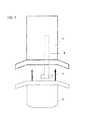
- FIG. 1Scanner device as per one version of the invention, as seen from above.
- FIG. 2Side view of the scanner device in FIG. 1 , showing how it is placed in the patient's mouth.
- FIG. 3Schematization of FIG. 1 , showing device scanning directions.
- the dental scanneris made up of two embodiments.
- the first onethe scanning embodiment, lodges a mobile scan head probe ( 3 ) on a mobile long element at the end of which is the scan head probe ( 4 ) as such.
- This mobile element ( 3 , 4 )is partially kept inside the scanner embodiment ( 1 ).
- the scanner head probe ( 4 )juts out the embodiment ( 1 ) and is lodged in the second embodiment or bite fixture ( 2 ) by way of a pocket.
- the bite fixture ( 2 )is, therefore, a scanner head probe lodging pocket ( 4 ).
- the bite fixture ( 2 )is introduced in the patient's mouth; and it is held in place when the patient bites it, so that the scanner head probe ( 4 ) is introduced in the mouth to scan the corresponding dental arch.
- the bite fixture ( 2 )is mouth shaped to make it easier to hold.
- Such bite fixture ( 2 )is basically a protective pocket for the scanner head probe ( 4 ), and can be bitten by the user. This is why the bite fixture ( 2 ) is made of a suitable transparent material.
- the scanner head probesweeps all along in at least two directions; one following the long axis of the long element ( 3 ), while the other one goes perpendicular to it in both directional senses, in such a way that scanning is the result of combining both side and deep motions with regard the dental arch, as shown in FIG. 3 .
- the deviceFor the intraoral scanning, the device includes detection sensors, laser sensors or similar devices in the scanner head probe ( 4 ), as well as cameras to capture tooth-by-tooth sweep images from the dental arch and gingivae. These are automatically generated and exact 3D images, as a result of their fixed and constant reference system.
Landscapes
- Health & Medical Sciences (AREA)
- Life Sciences & Earth Sciences (AREA)
- Surgery (AREA)
- Animal Behavior & Ethology (AREA)
- General Health & Medical Sciences (AREA)
- Public Health (AREA)
- Veterinary Medicine (AREA)
- Oral & Maxillofacial Surgery (AREA)
- Dentistry (AREA)
- Physics & Mathematics (AREA)
- Engineering & Computer Science (AREA)
- Optics & Photonics (AREA)
- Biophysics (AREA)
- Biomedical Technology (AREA)
- Nuclear Medicine, Radiotherapy & Molecular Imaging (AREA)
- Pathology (AREA)
- Radiology & Medical Imaging (AREA)
- Heart & Thoracic Surgery (AREA)
- Medical Informatics (AREA)
- Molecular Biology (AREA)
- Epidemiology (AREA)
- Manufacturing & Machinery (AREA)
- Dental Tools And Instruments Or Auxiliary Dental Instruments (AREA)
- Apparatus For Radiation Diagnosis (AREA)
- General Physics & Mathematics (AREA)
Abstract
Description
Claims (6)
Applications Claiming Priority (2)
| Application Number | Priority Date | Filing Date | Title |
|---|---|---|---|
| ES201231853AES2472640B1 (en) | 2012-11-28 | 2012-11-28 | DENTAL SCANNING DEVICE |
| PCT/ES2012/070834WO2014083211A1 (en) | 2012-11-28 | 2012-11-28 | Dental scanning device |
Related Parent Applications (1)
| Application Number | Title | Priority Date | Filing Date |
|---|---|---|---|
| PCT/ES2012/070834ContinuationWO2014083211A1 (en) | 2012-11-28 | 2012-11-28 | Dental scanning device |
Publications (2)
| Publication Number | Publication Date |
|---|---|
| US20150250379A1 US20150250379A1 (en) | 2015-09-10 |
| US10226164B2true US10226164B2 (en) | 2019-03-12 |
Family
ID=50827211
Family Applications (3)
| Application Number | Title | Priority Date | Filing Date |
|---|---|---|---|
| US14/286,650ActiveUS8989567B1 (en) | 2012-11-28 | 2014-05-23 | Dental scanner device and system and methods of use |
| US14/666,229Active2032-12-05US9301672B2 (en) | 2012-11-28 | 2015-03-23 | Dental scanner device and system and methods of use |
| US14/723,258Active2033-01-28US10226164B2 (en) | 2012-11-28 | 2015-05-27 | Dental scanner device |
Family Applications Before (2)
| Application Number | Title | Priority Date | Filing Date |
|---|---|---|---|
| US14/286,650ActiveUS8989567B1 (en) | 2012-11-28 | 2014-05-23 | Dental scanner device and system and methods of use |
| US14/666,229Active2032-12-05US9301672B2 (en) | 2012-11-28 | 2015-03-23 | Dental scanner device and system and methods of use |
Country Status (13)
| Country | Link |
|---|---|
| US (3) | US8989567B1 (en) |
| EP (1) | EP2929855B1 (en) |
| JP (1) | JP2016501592A (en) |
| KR (1) | KR20150089030A (en) |
| CN (1) | CN104853692A (en) |
| AU (1) | AU2012395512A1 (en) |
| BR (1) | BR112015012332A2 (en) |
| CA (1) | CA2893035C (en) |
| ES (2) | ES2878141T3 (en) |
| IL (1) | IL239000A0 (en) |
| PL (1) | PL2929855T3 (en) |
| SG (1) | SG11201504207SA (en) |
| WO (1) | WO2014083211A1 (en) |
Families Citing this family (24)
| Publication number | Priority date | Publication date | Assignee | Title |
|---|---|---|---|---|
| KR20150089030A (en)* | 2012-11-28 | 2015-08-04 | 아폴로 오랄 스캐너, 엘엘씨 | Dental scanning device |
| JP2017516619A (en)* | 2014-05-23 | 2017-06-22 | アポロ オーラル スキャナー, エルエルシー | New dental scanner device and system and method of use |
| FR3027205B1 (en)* | 2014-10-20 | 2020-07-17 | Modjaw | METHOD AND SYSTEM FOR MODELING THE MANDIBULAR KINEMATICS OF A PATIENT |
| WO2016176556A1 (en) | 2015-04-29 | 2016-11-03 | University Of Maryland, Baltimore | Apparatus and method for recording digital images and presenting 3d models of a body lumen |
| ES2865298T3 (en)* | 2015-05-19 | 2021-10-15 | Tyto Care Ltd | Throat Imaging Systems and Methods |
| US20160338804A1 (en)* | 2015-05-20 | 2016-11-24 | Vatech Co., Ltd. | Mouthpiece-type intraoral scanner |
| EP3410919B1 (en)* | 2016-02-01 | 2023-12-06 | Martin, Marco | Dental imager and method for recording photographic impressions |
| WO2018031003A1 (en) | 2016-08-10 | 2018-02-15 | Carestream Health, Inc. | Automatic intraoral 3d scanner with low coherence ranging |
| WO2018162641A1 (en)* | 2017-03-09 | 2018-09-13 | N-Lab Marcin Binkowski | Intra-oral scanning device, method of operating such a device and scanner system |
| WO2018183514A2 (en)* | 2017-03-28 | 2018-10-04 | Scientific Intake Limited Co. | Systems including removable oral devices |
| CN111050693B (en)* | 2017-08-16 | 2022-02-18 | 盖百加有限公司 | System and method for manufacturing tooth socket |
| ES2922493T3 (en)* | 2018-04-05 | 2022-09-15 | Tech Xika Ptt S L | Dental parameter measurement device |
| US10499802B1 (en)* | 2018-08-31 | 2019-12-10 | Kaohsiung Medical University | Mouth-opening device custom-made through 3-dimensional printing |
| FR3087644B1 (en)* | 2018-10-30 | 2022-06-10 | Dental Monitoring | ARTICULATED DENTAL PHOTO TAKING KIT |
| US11478132B2 (en)* | 2019-04-05 | 2022-10-25 | Align Technology. Inc. | Intraoral scanner sleeve authentication and identification |
| GB201913469D0 (en)* | 2019-09-18 | 2019-10-30 | Univ Leeds Innovations Ltd | Three-dimensional dental scanning system and method of scanning |
| US20210177266A1 (en)* | 2019-12-17 | 2021-06-17 | Clayton Adams Teufel | Intraoral scanning with raw depth data |
| US12144661B2 (en)* | 2019-12-31 | 2024-11-19 | Align Technology, Inc. | Gesture control using an intraoral scanner |
| CN111513680B (en)* | 2020-04-30 | 2021-08-03 | 四川大学 | intraoral scanner |
| US11633108B2 (en) | 2020-10-15 | 2023-04-25 | Sean M. Langton | Trans-illuminative intraoral diagnostic lighting system and method of using |
| CA3152850A1 (en) | 2021-03-23 | 2022-09-23 | Tactile Robotics Ltd. | Automated measurement apparatus and method for quantifying dimensions of dental preparation |
| US11382727B1 (en)* | 2021-05-19 | 2022-07-12 | Thamer Marghalani | Three-dimensional oral imaging system and method |
| EP4181062A1 (en)* | 2021-11-11 | 2023-05-17 | DENTSPLY SIRONA Inc. | Computer-assisted medical procedures |
| CN115281866B (en)* | 2022-08-15 | 2023-11-17 | 北京美立刻医疗器械有限公司 | Tooth model acquisition auxiliary device, acquisition method, system, medium and equipment |
Citations (5)
| Publication number | Priority date | Publication date | Assignee | Title |
|---|---|---|---|---|
| US4575805A (en) | 1980-12-24 | 1986-03-11 | Moermann Werner H | Method and apparatus for the fabrication of custom-shaped implants |
| US20020058229A1 (en)* | 2000-10-25 | 2002-05-16 | Orange House Co., Ltd. | Dentition image reading apparatus |
| US20030049585A1 (en)* | 2001-09-12 | 2003-03-13 | Severance Gary L. | System and method for scanning oral environment |
| US20050019732A1 (en) | 2003-07-23 | 2005-01-27 | Orametrix, Inc. | Automatic crown and gingiva detection from three-dimensional virtual model of teeth |
| US20060154198A1 (en) | 2005-01-11 | 2006-07-13 | Duane Durbin | 3D dental scanner |
Family Cites Families (12)
| Publication number | Priority date | Publication date | Assignee | Title |
|---|---|---|---|---|
| US3382781A (en)* | 1965-02-10 | 1968-05-14 | William L. Hamilton | Camera |
| DE3932151A1 (en)* | 1989-09-22 | 1991-04-04 | Peter Rohleder | DEVICE FOR SCANNING DETECTION OF AN INTERIOR |
| US5702249A (en) | 1995-05-19 | 1997-12-30 | Cooper; David H. | Modular intra-oral imaging system video camera |
| US6386867B1 (en)* | 2000-11-30 | 2002-05-14 | Duane Milford Durbin | Method and system for imaging and modeling dental structures |
| US6592371B2 (en)* | 2000-10-25 | 2003-07-15 | Duane Durbin | Method and system for imaging and modeling a three dimensional structure |
| EP1252859A3 (en)* | 2001-04-27 | 2003-12-17 | Firma Ivoclar Vivadent AG | Dental camera with mouthpiece |
| JP2004033465A (en)* | 2002-07-03 | 2004-02-05 | Mitsutoyo Corp | Tooth row image reading apparatus |
| AU2003300135B2 (en) | 2002-12-31 | 2009-07-16 | D4D Technologies, Llc | Laser digitizer system for dental applications |
| FR2883719B1 (en) | 2005-04-01 | 2007-06-01 | Atmel Grenoble Soc Par Actions | INTRAORAL DENTAL IMAGE SENSOR AND RADIOLOGICAL SYSTEM USING THE SENSOR |
| US8105233B2 (en)* | 2007-10-24 | 2012-01-31 | Tarek Ahmed Nabil Abou El Kheir | Endoscopic system and method for therapeutic applications and obtaining 3-dimensional human vision simulated imaging with real dynamic convergence |
| US8998609B2 (en)* | 2012-02-11 | 2015-04-07 | The Board Of Trustees Of The Leland Stanford Jr. University | Techniques for standardized imaging of oral cavity |
| KR20150089030A (en)* | 2012-11-28 | 2015-08-04 | 아폴로 오랄 스캐너, 엘엘씨 | Dental scanning device |
- 2012
- 2012-11-28KRKR1020157015674Apatent/KR20150089030A/ennot_activeWithdrawn
- 2012-11-28CACA2893035Apatent/CA2893035C/enactiveActive
- 2012-11-28JPJP2015544510Apatent/JP2016501592A/enactivePending
- 2012-11-28ESES12889074Tpatent/ES2878141T3/enactiveActive
- 2012-11-28SGSG11201504207SApatent/SG11201504207SA/enunknown
- 2012-11-28BRBR112015012332Apatent/BR112015012332A2/ennot_activeIP Right Cessation
- 2012-11-28EPEP12889074.6Apatent/EP2929855B1/enactiveActive
- 2012-11-28ESES201231853Apatent/ES2472640B1/enactiveActive
- 2012-11-28PLPL12889074Tpatent/PL2929855T3/enunknown
- 2012-11-28WOPCT/ES2012/070834patent/WO2014083211A1/enactiveApplication Filing
- 2012-11-28AUAU2012395512Apatent/AU2012395512A1/ennot_activeAbandoned
- 2012-11-28CNCN201280077303.2Apatent/CN104853692A/enactivePending
- 2014
- 2014-05-23USUS14/286,650patent/US8989567B1/enactiveActive
- 2015
- 2015-03-23USUS14/666,229patent/US9301672B2/enactiveActive
- 2015-05-25ILIL239000Apatent/IL239000A0/enunknown
- 2015-05-27USUS14/723,258patent/US10226164B2/enactiveActive
Patent Citations (7)
| Publication number | Priority date | Publication date | Assignee | Title |
|---|---|---|---|---|
| US4575805A (en) | 1980-12-24 | 1986-03-11 | Moermann Werner H | Method and apparatus for the fabrication of custom-shaped implants |
| US20020058229A1 (en)* | 2000-10-25 | 2002-05-16 | Orange House Co., Ltd. | Dentition image reading apparatus |
| US20030049585A1 (en)* | 2001-09-12 | 2003-03-13 | Severance Gary L. | System and method for scanning oral environment |
| US6821116B2 (en)* | 2001-09-12 | 2004-11-23 | Ivoclar Vivadent, Inc. | System for scanning oral environment |
| US20050019732A1 (en) | 2003-07-23 | 2005-01-27 | Orametrix, Inc. | Automatic crown and gingiva detection from three-dimensional virtual model of teeth |
| US20060154198A1 (en) | 2005-01-11 | 2006-07-13 | Duane Durbin | 3D dental scanner |
| US7494338B2 (en)* | 2005-01-11 | 2009-02-24 | Duane Durbin | 3D dental scanner |
Non-Patent Citations (5)
| Title |
|---|
| English Translation of International Search Report for PCT Application No. PCT/ES2012/070834 dated Aug. 16, 2013. |
| English Translation of Written Opinion for PCT Application No. PCT/ES2012/070834 dated Aug. 16, 2013. |
| International Search Report for PCT Application No. PCT/ES2012/070834 dated Aug. 16, 2013. |
| Logozzo, S., et al., A Comparative Analysis of Intraoral 3d Digital Scanners for Restorative Dentistry, The International Journal of Medical Technology, vol. 5, No. 1 (2011). |
| Written Opinion for PCT Application No. PCT/ES2012/070834 dated Aug. 16, 2013. |
Also Published As
| Publication number | Publication date |
|---|---|
| CA2893035C (en) | 2019-11-19 |
| EP2929855B1 (en) | 2021-04-07 |
| US20150238287A1 (en) | 2015-08-27 |
| PL2929855T3 (en) | 2021-11-08 |
| AU2012395512A1 (en) | 2015-07-02 |
| US8989567B1 (en) | 2015-03-24 |
| WO2014083211A1 (en) | 2014-06-05 |
| EP2929855A1 (en) | 2015-10-14 |
| EP2929855A4 (en) | 2016-07-27 |
| IL239000A0 (en) | 2015-07-30 |
| JP2016501592A (en) | 2016-01-21 |
| ES2472640B1 (en) | 2015-04-08 |
| KR20150089030A (en) | 2015-08-04 |
| US9301672B2 (en) | 2016-04-05 |
| CA2893035A1 (en) | 2014-06-05 |
| SG11201504207SA (en) | 2015-07-30 |
| CN104853692A (en) | 2015-08-19 |
| BR112015012332A2 (en) | 2017-07-11 |
| ES2472640A1 (en) | 2014-07-01 |
| US20150250379A1 (en) | 2015-09-10 |
| ES2878141T3 (en) | 2021-11-18 |
Similar Documents
| Publication | Publication Date | Title |
|---|---|---|
| US10226164B2 (en) | Dental scanner device | |
| JP7209039B2 (en) | Dental imager and method for recording photographic impressions | |
| KR102050547B1 (en) | Device and method for subgingival measurement | |
| CN110621259B (en) | Intraoral scanning device, method of operating such a device and scanner system | |
| CN103462584B (en) | Multifunctional stick for intraoral imaging system | |
| CA2950090C (en) | Novel dental scanner device and system and methods of use | |
| KR20170113412A (en) | Dental intraoral scanner system | |
| US20120288819A1 (en) | Dental imaging system with orientation detector | |
| JP5891080B2 (en) | Jaw movement simulation method, jaw movement simulation apparatus, and jaw movement simulation system | |
| JP6774365B2 (en) | Tip member that can be attached to and detached from the image pickup device and the housing of the image pickup device. | |
| US20240252131A1 (en) | Digital dental xray sensor, method and system |
Legal Events
| Date | Code | Title | Description |
|---|---|---|---|
| AS | Assignment | Owner name:CARNOJAAL, S.L., SPAIN Free format text:ASSIGNMENT OF ASSIGNORS INTEREST;ASSIGNORS:FERNANDEZ PULIDO, ALFONSO;DE PABLOS GARCIA, DAVID;VALDES TAMAMES, JAVIER;REEL/FRAME:039071/0551 Effective date:20140522 Owner name:APOLLO ORAL SCANNER, LLC., FLORIDA Free format text:ASSIGNMENT OF ASSIGNORS INTEREST;ASSIGNOR:CARNOJAAL, S.L.;REEL/FRAME:039071/0603 Effective date:20040523 | |
| AS | Assignment | Owner name:APOLLO ORAL SCANNER, LLC, FLORIDA Free format text:CORRECTIVE ASSIGNMENT TO CORRECT THE EXECUTION DATE PREVIOUSLY RECORDED ON REEL 039071 FRAME 0603. ASSIGNOR(S) HEREBY CONFIRMS THE ASSIGNMENT;ASSIGNOR:CARNOJAAL, S.L.;REEL/FRAME:040264/0107 Effective date:20140523 | |
| FEPP | Fee payment procedure | Free format text:ENTITY STATUS SET TO MICRO (ORIGINAL EVENT CODE: MICR); ENTITY STATUS OF PATENT OWNER: SMALL ENTITY | |
| STCF | Information on status: patent grant | Free format text:PATENTED CASE | |
| AS | Assignment | Owner name:CARNOJAAL, S.L., SPAIN Free format text:ASSIGNMENT OF ASSIGNORS INTEREST;ASSIGNOR:APOLLO ORAL SCANNER, LLC;REEL/FRAME:060127/0469 Effective date:20220321 | |
| MAFP | Maintenance fee payment | Free format text:PAYMENT OF MAINTENANCE FEE, 4TH YR, SMALL ENTITY (ORIGINAL EVENT CODE: M2551); ENTITY STATUS OF PATENT OWNER: SMALL ENTITY Year of fee payment:4 |