US10214580B2 - Constructs and libraries comprising antibody surrogate light chain sequences - Google Patents
Constructs and libraries comprising antibody surrogate light chain sequencesDownload PDFInfo
- Publication number
- US10214580B2 US10214580B2US14/047,301US201314047301AUS10214580B2US 10214580 B2US10214580 B2US 10214580B2US 201314047301 AUS201314047301 AUS 201314047301AUS 10214580 B2US10214580 B2US 10214580B2
- Authority
- US
- United States
- Prior art keywords
- light chain
- sequence
- antibody
- heavy chain
- surrogate light
- Prior art date
- Legal status (The legal status is an assumption and is not a legal conclusion. Google has not performed a legal analysis and makes no representation as to the accuracy of the status listed.)
- Active, expires
Links
- 108010018324Surrogate Immunoglobulin Light ChainsProteins0.000titleclaimsabstractdescription151
- 102000002663Surrogate Immunoglobulin Light ChainsHuman genes0.000titleclaimsabstractdescription151
- 108090000765processed proteins & peptidesProteins0.000claimsabstractdescription116
- 102000004196processed proteins & peptidesHuman genes0.000claimsabstractdescription104
- 229920001184polypeptidePolymers0.000claimsabstractdescription101
- 230000004927fusionEffects0.000claimsdescription98
- 230000027455bindingEffects0.000claimsdescription67
- 125000003275alpha amino acid groupChemical group0.000claimsdescription37
- 241000712431Influenza A virusSpecies0.000claims2
- 101000854886Homo sapiens Immunoglobulin iota chainProteins0.000abstractdescription83
- 102100020744Immunoglobulin iota chainHuman genes0.000abstractdescription83
- 108090000623proteins and genesProteins0.000description83
- 235000001014amino acidNutrition0.000description56
- 229940024606amino acidDrugs0.000description54
- 102000004169proteins and genesHuman genes0.000description54
- 150000001413amino acidsChemical class0.000description51
- 235000018102proteinsNutrition0.000description50
- 210000004027cellAnatomy0.000description42
- 239000000427antigenSubstances0.000description32
- 238000000034methodMethods0.000description32
- 102000036639antigensHuman genes0.000description30
- 108091007433antigensProteins0.000description30
- 239000012634fragmentSubstances0.000description27
- 238000002965ELISAMethods0.000description23
- 125000000539amino acid groupChemical group0.000description22
- 108020004414DNAProteins0.000description18
- 230000014509gene expressionEffects0.000description17
- 230000004048modificationEffects0.000description14
- 238000012986modificationMethods0.000description14
- 238000011282treatmentMethods0.000description13
- 241000588724Escherichia coliSpecies0.000description12
- 238000003780insertionMethods0.000description12
- 230000037431insertionEffects0.000description12
- 238000002823phage displayMethods0.000description12
- 229960000723ampicillinDrugs0.000description11
- AVKUERGKIZMTKX-NJBDSQKTSA-NampicillinChemical compoundC1([C@@H](N)C(=O)N[C@H]2[C@H]3SC([C@@H](N3C2=O)C(O)=O)(C)C)=CC=CC=C1AVKUERGKIZMTKX-NJBDSQKTSA-N0.000description11
- 239000003623enhancerSubstances0.000description11
- 239000013604expression vectorSubstances0.000description11
- 102000037865fusion proteinsHuman genes0.000description11
- 108020001507fusion proteinsProteins0.000description11
- 150000007523nucleic acidsChemical class0.000description11
- 108020004707nucleic acidsProteins0.000description10
- 102000039446nucleic acidsHuman genes0.000description10
- 239000013598vectorSubstances0.000description10
- 108091026890Coding regionProteins0.000description9
- 239000006137Luria-Bertani brothSubstances0.000description9
- 210000004962mammalian cellAnatomy0.000description9
- DHMQDGOQFOQNFH-UHFFFAOYSA-NGlycineChemical compoundNCC(O)=ODHMQDGOQFOQNFH-UHFFFAOYSA-N0.000description8
- 108010021625Immunoglobulin FragmentsProteins0.000description8
- 102000008394Immunoglobulin FragmentsHuman genes0.000description8
- 241000699666Mus <mouse, genus>Species0.000description8
- 210000003719b-lymphocyteAnatomy0.000description8
- 230000001580bacterial effectEffects0.000description8
- 238000001514detection methodMethods0.000description8
- 239000006166lysateSubstances0.000description8
- 239000002245particleSubstances0.000description8
- 230000003612virological effectEffects0.000description8
- 241000894006BacteriaSpecies0.000description7
- WHUUTDBJXJRKMK-UHFFFAOYSA-NGlutamic acidNatural productsOC(=O)C(N)CCC(O)=OWHUUTDBJXJRKMK-UHFFFAOYSA-N0.000description7
- XUJNEKJLAYXESH-REOHCLBHSA-NL-CysteineChemical compoundSC[C@H](N)C(O)=OXUJNEKJLAYXESH-REOHCLBHSA-N0.000description7
- CKLJMWTZIZZHCS-REOHCLBHSA-NL-aspartic acidChemical compoundOC(=O)[C@@H](N)CC(O)=OCKLJMWTZIZZHCS-REOHCLBHSA-N0.000description7
- WHUUTDBJXJRKMK-VKHMYHEASA-NL-glutamic acidChemical compoundOC(=O)[C@@H](N)CCC(O)=OWHUUTDBJXJRKMK-VKHMYHEASA-N0.000description7
- COLNVLDHVKWLRT-QMMMGPOBSA-NL-phenylalanineChemical compoundOC(=O)[C@@H](N)CC1=CC=CC=C1COLNVLDHVKWLRT-QMMMGPOBSA-N0.000description7
- 241000829100Macaca mulatta polyomavirus 1Species0.000description7
- 101710125418Major capsid proteinProteins0.000description7
- 102000001708Protein IsoformsHuman genes0.000description7
- 108010029485Protein IsoformsProteins0.000description7
- 238000011161developmentMethods0.000description7
- 238000006467substitution reactionMethods0.000description7
- 230000001225therapeutic effectEffects0.000description7
- MTCFGRXMJLQNBG-REOHCLBHSA-N(2S)-2-Amino-3-hydroxypropansäureChemical compoundOC[C@H](N)C(O)=OMTCFGRXMJLQNBG-REOHCLBHSA-N0.000description6
- 101710132601Capsid proteinProteins0.000description6
- 101710094648Coat proteinProteins0.000description6
- 241000724791Filamentous phageSpecies0.000description6
- WQZGKKKJIJFFOK-GASJEMHNSA-NGlucoseNatural productsOC[C@H]1OC(O)[C@H](O)[C@@H](O)[C@@H]1OWQZGKKKJIJFFOK-GASJEMHNSA-N0.000description6
- 102100021181Golgi phosphoprotein 3Human genes0.000description6
- 101000840267Homo sapiens Immunoglobulin lambda-like polypeptide 1Proteins0.000description6
- 108060003951ImmunoglobulinProteins0.000description6
- 102100029616Immunoglobulin lambda-like polypeptide 1Human genes0.000description6
- AYFVYJQAPQTCCC-GBXIJSLDSA-NL-threonineChemical compoundC[C@@H](O)[C@H](N)C(O)=OAYFVYJQAPQTCCC-GBXIJSLDSA-N0.000description6
- OUYCCCASQSFEME-QMMMGPOBSA-NL-tyrosineChemical compoundOC(=O)[C@@H](N)CC1=CC=C(O)C=C1OUYCCCASQSFEME-QMMMGPOBSA-N0.000description6
- KDXKERNSBIXSRK-UHFFFAOYSA-NLysineNatural productsNCCCCC(N)C(O)=OKDXKERNSBIXSRK-UHFFFAOYSA-N0.000description6
- 101710141454NucleoproteinProteins0.000description6
- 102000003992PeroxidasesHuman genes0.000description6
- 101710083689Probable capsid proteinProteins0.000description6
- 241000700605VirusesSpecies0.000description6
- 239000000539dimerSubstances0.000description6
- 239000008103glucoseSubstances0.000description6
- 102000018358immunoglobulinHuman genes0.000description6
- 238000011534incubationMethods0.000description6
- 238000002703mutagenesisMethods0.000description6
- 231100000350mutagenesisToxicity0.000description6
- 108040007629peroxidase activity proteinsProteins0.000description6
- 239000006228supernatantSubstances0.000description6
- 238000005406washingMethods0.000description6
- UAIUNKRWKOVEES-UHFFFAOYSA-N3,3',5,5'-tetramethylbenzidineChemical compoundCC1=C(N)C(C)=CC(C=2C=C(C)C(N)=C(C)C=2)=C1UAIUNKRWKOVEES-UHFFFAOYSA-N0.000description5
- DCXYFEDJOCDNAF-REOHCLBHSA-NL-asparagineChemical compoundOC(=O)[C@@H](N)CC(N)=ODCXYFEDJOCDNAF-REOHCLBHSA-N0.000description5
- ZDXPYRJPNDTMRX-VKHMYHEASA-NL-glutamineChemical compoundOC(=O)[C@@H](N)CCC(N)=OZDXPYRJPNDTMRX-VKHMYHEASA-N0.000description5
- AGPKZVBTJJNPAG-WHFBIAKZSA-NL-isoleucineChemical compoundCC[C@H](C)[C@H](N)C(O)=OAGPKZVBTJJNPAG-WHFBIAKZSA-N0.000description5
- ROHFNLRQFUQHCH-YFKPBYRVSA-NL-leucineChemical compoundCC(C)C[C@H](N)C(O)=OROHFNLRQFUQHCH-YFKPBYRVSA-N0.000description5
- 206010028980NeoplasmDiseases0.000description5
- 108091028043Nucleic acid sequenceProteins0.000description5
- 108010076504Protein Sorting SignalsProteins0.000description5
- KZSNJWFQEVHDMF-UHFFFAOYSA-NValineNatural productsCC(C)C(N)C(O)=OKZSNJWFQEVHDMF-UHFFFAOYSA-N0.000description5
- 239000011248coating agentSubstances0.000description5
- 238000000576coating methodMethods0.000description5
- 238000012217deletionMethods0.000description5
- 230000037430deletionEffects0.000description5
- 230000000694effectsEffects0.000description5
- 230000006870functionEffects0.000description5
- 206010022000influenzaDiseases0.000description5
- 230000010076replicationEffects0.000description5
- 239000000523sampleSubstances0.000description5
- 210000002966serumAnatomy0.000description5
- 241000894007speciesSpecies0.000description5
- 238000001262western blotMethods0.000description5
- YBJHBAHKTGYVGT-ZKWXMUAHSA-N(+)-BiotinChemical compoundN1C(=O)N[C@@H]2[C@H](CCCCC(=O)O)SC[C@@H]21YBJHBAHKTGYVGT-ZKWXMUAHSA-N0.000description4
- DTHNMHAUYICORS-KTKZVXAJSA-NGlucagon-like peptide 1Chemical compoundC([C@@H](C(=O)N[C@@H]([C@@H](C)CC)C(=O)N[C@@H](C)C(=O)N[C@@H](CC=1C2=CC=CC=C2NC=1)C(=O)N[C@@H](CC(C)C)C(=O)N[C@@H](C(C)C)C(=O)N[C@@H](CCCCN)C(=O)NCC(=O)N[C@@H](CCCNC(N)=N)C(N)=O)NC(=O)[C@H](CCC(O)=O)NC(=O)[C@H](CCCCN)NC(=O)[C@H](C)NC(=O)[C@H](C)NC(=O)[C@H](CCC(N)=O)NC(=O)CNC(=O)[C@H](CCC(O)=O)NC(=O)[C@H](CC(C)C)NC(=O)[C@H](CC=1C=CC(O)=CC=1)NC(=O)[C@H](CO)NC(=O)[C@H](CO)NC(=O)[C@@H](NC(=O)[C@H](CC(O)=O)NC(=O)[C@H](CO)NC(=O)[C@@H](NC(=O)[C@H](CC=1C=CC=CC=1)NC(=O)[C@@H](NC(=O)CNC(=O)[C@H](CCC(O)=O)NC(=O)[C@H](C)NC(=O)[C@@H](N)CC=1N=CNC=1)[C@@H](C)O)[C@@H](C)O)C(C)C)C1=CC=CC=C1DTHNMHAUYICORS-KTKZVXAJSA-N0.000description4
- 101000611183Homo sapiens Tumor necrosis factorProteins0.000description4
- 125000000174L-prolyl groupChemical group[H]N1C([H])([H])C([H])([H])C([H])([H])[C@@]1([H])C(*)=O0.000description4
- -1LeuChemical group0.000description4
- PXHVJJICTQNCMI-UHFFFAOYSA-NNickelChemical compound[Ni]PXHVJJICTQNCMI-UHFFFAOYSA-N0.000description4
- 108091034117OligonucleotideProteins0.000description4
- 230000008901benefitEffects0.000description4
- 230000000903blocking effectEffects0.000description4
- 210000004899c-terminal regionAnatomy0.000description4
- 102000057041human TNFHuman genes0.000description4
- 238000010348incorporationMethods0.000description4
- NOESYZHRGYRDHS-UHFFFAOYSA-NinsulinChemical compoundN1C(=O)C(NC(=O)C(CCC(N)=O)NC(=O)C(CCC(O)=O)NC(=O)C(C(C)C)NC(=O)C(NC(=O)CN)C(C)CC)CSSCC(C(NC(CO)C(=O)NC(CC(C)C)C(=O)NC(CC=2C=CC(O)=CC=2)C(=O)NC(CCC(N)=O)C(=O)NC(CC(C)C)C(=O)NC(CCC(O)=O)C(=O)NC(CC(N)=O)C(=O)NC(CC=2C=CC(O)=CC=2)C(=O)NC(CSSCC(NC(=O)C(C(C)C)NC(=O)C(CC(C)C)NC(=O)C(CC=2C=CC(O)=CC=2)NC(=O)C(CC(C)C)NC(=O)C(C)NC(=O)C(CCC(O)=O)NC(=O)C(C(C)C)NC(=O)C(CC(C)C)NC(=O)C(CC=2NC=NC=2)NC(=O)C(CO)NC(=O)CNC2=O)C(=O)NCC(=O)NC(CCC(O)=O)C(=O)NC(CCCNC(N)=N)C(=O)NCC(=O)NC(CC=3C=CC=CC=3)C(=O)NC(CC=3C=CC=CC=3)C(=O)NC(CC=3C=CC(O)=CC=3)C(=O)NC(C(C)O)C(=O)N3C(CCC3)C(=O)NC(CCCCN)C(=O)NC(C)C(O)=O)C(=O)NC(CC(N)=O)C(O)=O)=O)NC(=O)C(C(C)CC)NC(=O)C(CO)NC(=O)C(C(C)O)NC(=O)C1CSSCC2NC(=O)C(CC(C)C)NC(=O)C(NC(=O)C(CCC(N)=O)NC(=O)C(CC(N)=O)NC(=O)C(NC(=O)C(N)CC=1C=CC=CC=1)C(C)C)CC1=CN=CN1NOESYZHRGYRDHS-UHFFFAOYSA-N0.000description4
- 229930027917kanamycinNatural products0.000description4
- 229960000318kanamycinDrugs0.000description4
- SBUJHOSQTJFQJX-NOAMYHISSA-NkanamycinChemical compoundO[C@@H]1[C@@H](O)[C@H](O)[C@@H](CN)O[C@@H]1O[C@H]1[C@H](O)[C@@H](O[C@@H]2[C@@H]([C@@H](N)[C@H](O)[C@@H](CO)O2)O)[C@H](N)C[C@@H]1NSBUJHOSQTJFQJX-NOAMYHISSA-N0.000description4
- 229930182823kanamycin ANatural products0.000description4
- 210000003292kidney cellAnatomy0.000description4
- 239000003550markerSubstances0.000description4
- 239000013612plasmidSubstances0.000description4
- 108091033319polynucleotideProteins0.000description4
- 102000040430polynucleotideHuman genes0.000description4
- 239000002157polynucleotideSubstances0.000description4
- 230000004481post-translational protein modificationEffects0.000description4
- 230000000644propagated effectEffects0.000description4
- 102000005962receptorsHuman genes0.000description4
- 108020003175receptorsProteins0.000description4
- 230000028327secretionEffects0.000description4
- 238000002741site-directed mutagenesisMethods0.000description4
- 239000000126substanceSubstances0.000description4
- 108091032973(ribonucleotides)n+mProteins0.000description3
- 241000193388Bacillus thuringiensisSpecies0.000description3
- 241000701022CytomegalovirusSpecies0.000description3
- 102400000322Glucagon-like peptide 1Human genes0.000description3
- 108010093488His-His-His-His-His-HisProteins0.000description3
- 125000000393L-methionino groupChemical group[H]OC(=O)[C@@]([H])(N([H])[*])C([H])([H])C(SC([H])([H])[H])([H])[H]0.000description3
- QIVBCDIJIAJPQS-VIFPVBQESA-NL-tryptophaneChemical compoundC1=CC=C2C(C[C@H](N)C(O)=O)=CNC2=C1QIVBCDIJIAJPQS-VIFPVBQESA-N0.000description3
- ROHFNLRQFUQHCH-UHFFFAOYSA-NLeucineNatural productsCC(C)CC(N)C(O)=OROHFNLRQFUQHCH-UHFFFAOYSA-N0.000description3
- 101150012195PREB geneProteins0.000description3
- 102000000434Pre-B Cell ReceptorsHuman genes0.000description3
- 108010016231Pre-B Cell ReceptorsProteins0.000description3
- 102000007056Recombinant Fusion ProteinsHuman genes0.000description3
- 108010008281Recombinant Fusion ProteinsProteins0.000description3
- 240000004808Saccharomyces cerevisiaeSpecies0.000description3
- 235000014680Saccharomyces cerevisiaeNutrition0.000description3
- QIVBCDIJIAJPQS-UHFFFAOYSA-NTryptophanNatural productsC1=CC=C2C(CC(N)C(O)=O)=CNC2=C1QIVBCDIJIAJPQS-UHFFFAOYSA-N0.000description3
- 238000005273aerationMethods0.000description3
- 230000002788anti-peptideEffects0.000description3
- 229940097012bacillus thuringiensisDrugs0.000description3
- 230000009286beneficial effectEffects0.000description3
- 239000011230binding agentSubstances0.000description3
- 230000015572biosynthetic processEffects0.000description3
- 210000001185bone marrowAnatomy0.000description3
- 238000010367cloningMethods0.000description3
- 230000000295complement effectEffects0.000description3
- 238000004132cross linkingMethods0.000description3
- 238000010586diagramMethods0.000description3
- 230000002255enzymatic effectEffects0.000description3
- RAXXELZNTBOGNW-UHFFFAOYSA-NimidazoleNatural productsC1=CNC=N1RAXXELZNTBOGNW-UHFFFAOYSA-N0.000description3
- 238000000338in vitroMethods0.000description3
- 230000003993interactionEffects0.000description3
- BPHPUYQFMNQIOC-NXRLNHOXSA-Nisopropyl beta-D-thiogalactopyranosideChemical compoundCC(C)S[C@@H]1O[C@H](CO)[C@H](O)[C@H](O)[C@H]1OBPHPUYQFMNQIOC-NXRLNHOXSA-N0.000description3
- 210000004698lymphocyteAnatomy0.000description3
- 239000000463materialSubstances0.000description3
- 229940092253ovalbuminDrugs0.000description3
- 238000004091panningMethods0.000description3
- 210000001322periplasmAnatomy0.000description3
- 125000002924primary amino groupChemical group[H]N([H])*0.000description3
- 230000008569processEffects0.000description3
- 238000000746purificationMethods0.000description3
- 238000012552reviewMethods0.000description3
- 238000012216screeningMethods0.000description3
- 238000012360testing methodMethods0.000description3
- 238000013518transcriptionMethods0.000description3
- 230000035897transcriptionEffects0.000description3
- 239000013638trimerSubstances0.000description3
- FWMNVWWHGCHHJJ-SKKKGAJSSA-N4-amino-1-[(2r)-6-amino-2-[[(2r)-2-[[(2r)-2-[[(2r)-2-amino-3-phenylpropanoyl]amino]-3-phenylpropanoyl]amino]-4-methylpentanoyl]amino]hexanoyl]piperidine-4-carboxylic acidChemical compoundC([C@H](C(=O)N[C@H](CC(C)C)C(=O)N[C@H](CCCCN)C(=O)N1CCC(N)(CC1)C(O)=O)NC(=O)[C@H](N)CC=1C=CC=CC=1)C1=CC=CC=C1FWMNVWWHGCHHJJ-SKKKGAJSSA-N0.000description2
- 239000004475ArginineSubstances0.000description2
- DCXYFEDJOCDNAF-UHFFFAOYSA-NAsparagineNatural productsOC(=O)C(N)CC(N)=ODCXYFEDJOCDNAF-UHFFFAOYSA-N0.000description2
- 244000063299Bacillus subtilisSpecies0.000description2
- 235000014469Bacillus subtilisNutrition0.000description2
- 241000282693CercopithecidaeSpecies0.000description2
- 102000053602DNAHuman genes0.000description2
- BWGNESOTFCXPMA-UHFFFAOYSA-NDihydrogen disulfideChemical compoundSSBWGNESOTFCXPMA-UHFFFAOYSA-N0.000description2
- 239000006144Dulbecco’s modified Eagle's mediumSubstances0.000description2
- 102000004190EnzymesHuman genes0.000description2
- 108090000790EnzymesProteins0.000description2
- 102000003951ErythropoietinHuman genes0.000description2
- 108090000394ErythropoietinProteins0.000description2
- 241000206602EukaryotaSpecies0.000description2
- 101710198884GATA-type zinc finger protein 1Proteins0.000description2
- 108700028146Genetic Enhancer ElementsProteins0.000description2
- 239000004471GlycineSubstances0.000description2
- 241000701024Human betaherpesvirus 5Species0.000description2
- 102000004877InsulinHuman genes0.000description2
- 108090001061InsulinProteins0.000description2
- QNAYBMKLOCPYGJ-REOHCLBHSA-NL-alanineChemical compoundC[C@H](N)C(O)=OQNAYBMKLOCPYGJ-REOHCLBHSA-N0.000description2
- FFEARJCKVFRZRR-BYPYZUCNSA-NL-methionineChemical compoundCSCC[C@H](N)C(O)=OFFEARJCKVFRZRR-BYPYZUCNSA-N0.000description2
- 125000000510L-tryptophano groupChemical group[H]C1=C([H])C([H])=C2N([H])C([H])=C(C([H])([H])[C@@]([H])(C(O[H])=O)N([H])[*])C2=C1[H]0.000description2
- KZSNJWFQEVHDMF-BYPYZUCNSA-NL-valineChemical compoundCC(C)[C@H](N)C(O)=OKZSNJWFQEVHDMF-BYPYZUCNSA-N0.000description2
- 239000004472LysineSubstances0.000description2
- 241000699670Mus sp.Species0.000description2
- 108091007491NSP3 Papain-like protease domainsProteins0.000description2
- ONIBWKKTOPOVIA-UHFFFAOYSA-NProlineNatural productsOC(=O)C1CCCN1ONIBWKKTOPOVIA-UHFFFAOYSA-N0.000description2
- 108020005091Replication OriginProteins0.000description2
- MTCFGRXMJLQNBG-UHFFFAOYSA-NSerineNatural productsOCC(N)C(O)=OMTCFGRXMJLQNBG-UHFFFAOYSA-N0.000description2
- AYFVYJQAPQTCCC-UHFFFAOYSA-NThreonineNatural productsCC(O)C(N)C(O)=OAYFVYJQAPQTCCC-UHFFFAOYSA-N0.000description2
- 239000004473ThreonineSubstances0.000description2
- 102000006601Thymidine KinaseHuman genes0.000description2
- 108020004440Thymidine kinaseProteins0.000description2
- RHQDFWAXVIIEBN-UHFFFAOYSA-NTrifluoroethanolChemical compoundOCC(F)(F)FRHQDFWAXVIIEBN-UHFFFAOYSA-N0.000description2
- 235000004279alanineNutrition0.000description2
- 238000013459approachMethods0.000description2
- ODKSFYDXXFIFQN-UHFFFAOYSA-NarginineNatural productsOC(=O)C(N)CCCNC(N)=NODKSFYDXXFIFQN-UHFFFAOYSA-N0.000description2
- 235000009582asparagineNutrition0.000description2
- 229960001230asparagineDrugs0.000description2
- 235000003704aspartic acidNutrition0.000description2
- 238000002819bacterial displayMethods0.000description2
- OQFSQFPPLPISGP-UHFFFAOYSA-Nbeta-carboxyaspartic acidNatural productsOC(=O)C(N)C(C(O)=O)C(O)=OOQFSQFPPLPISGP-UHFFFAOYSA-N0.000description2
- 102000023732binding proteinsHuman genes0.000description2
- 108091008324binding proteinsProteins0.000description2
- 229960002685biotinDrugs0.000description2
- 235000020958biotinNutrition0.000description2
- 239000011616biotinSubstances0.000description2
- 210000002421cell wallAnatomy0.000description2
- 230000008859changeEffects0.000description2
- 239000013522chelantSubstances0.000description2
- 238000004587chromatography analysisMethods0.000description2
- 238000011509clonal analysisMethods0.000description2
- 230000021615conjugationEffects0.000description2
- 238000010276constructionMethods0.000description2
- 238000012258culturingMethods0.000description2
- 235000018417cysteineNutrition0.000description2
- XUJNEKJLAYXESH-UHFFFAOYSA-NcysteineNatural productsSCC(N)C(O)=OXUJNEKJLAYXESH-UHFFFAOYSA-N0.000description2
- 230000001419dependent effectEffects0.000description2
- 239000003085diluting agentSubstances0.000description2
- 239000012636effectorSubstances0.000description2
- 238000005516engineering processMethods0.000description2
- 229940105423erythropoietinDrugs0.000description2
- 210000003527eukaryotic cellAnatomy0.000description2
- 230000002068genetic effectEffects0.000description2
- 210000004602germ cellAnatomy0.000description2
- 235000013922glutamic acidNutrition0.000description2
- 239000004220glutamic acidSubstances0.000description2
- ZDXPYRJPNDTMRX-UHFFFAOYSA-NglutamineNatural productsOC(=O)C(N)CCC(N)=OZDXPYRJPNDTMRX-UHFFFAOYSA-N0.000description2
- 230000036541healthEffects0.000description2
- HNDVDQJCIGZPNO-UHFFFAOYSA-NhistidineNatural productsOC(=O)C(N)CC1=CN=CN1HNDVDQJCIGZPNO-UHFFFAOYSA-N0.000description2
- 238000009396hybridizationMethods0.000description2
- 229940072221immunoglobulinsDrugs0.000description2
- 238000001727in vivoMethods0.000description2
- 229940125396insulinDrugs0.000description2
- 238000002955isolationMethods0.000description2
- AGPKZVBTJJNPAG-UHFFFAOYSA-NisoleucineNatural productsCCC(C)C(N)C(O)=OAGPKZVBTJJNPAG-UHFFFAOYSA-N0.000description2
- 229960000310isoleucineDrugs0.000description2
- 210000003734kidneyAnatomy0.000description2
- 210000005229liver cellAnatomy0.000description2
- 238000002824mRNA displayMethods0.000description2
- 239000012528membraneSubstances0.000description2
- MYWUZJCMWCOHBA-VIFPVBQESA-NmethamphetamineChemical compoundCN[C@@H](C)CC1=CC=CC=C1MYWUZJCMWCOHBA-VIFPVBQESA-N0.000description2
- 229930182817methionineNatural products0.000description2
- 239000011325microbeadSubstances0.000description2
- 230000035772mutationEffects0.000description2
- 230000003472neutralizing effectEffects0.000description2
- 229910052759nickelInorganic materials0.000description2
- 238000005457optimizationMethods0.000description2
- COLNVLDHVKWLRT-UHFFFAOYSA-NphenylalanineNatural productsOC(=O)C(N)CC1=CC=CC=C1COLNVLDHVKWLRT-UHFFFAOYSA-N0.000description2
- 239000006187pillSubstances0.000description2
- 230000008488polyadenylationEffects0.000description2
- OXCMYAYHXIHQOA-UHFFFAOYSA-Npotassium;[2-butyl-5-chloro-3-[[4-[2-(1,2,4-triaza-3-azanidacyclopenta-1,4-dien-5-yl)phenyl]phenyl]methyl]imidazol-4-yl]methanolChemical compound[K+].CCCCC1=NC(Cl)=C(CO)N1CC1=CC=C(C=2C(=CC=CC=2)C2=N[N-]N=N2)C=C1OXCMYAYHXIHQOA-UHFFFAOYSA-N0.000description2
- 230000036515potencyEffects0.000description2
- 238000002360preparation methodMethods0.000description2
- 210000001948pro-b lymphocyteAnatomy0.000description2
- 239000000047productSubstances0.000description2
- 230000008707rearrangementEffects0.000description2
- 238000005215recombinationMethods0.000description2
- 230000006798recombinationEffects0.000description2
- 238000002702ribosome displayMethods0.000description2
- 230000003248secreting effectEffects0.000description2
- 230000009870specific bindingEffects0.000description2
- 238000013519translationMethods0.000description2
- OUYCCCASQSFEME-UHFFFAOYSA-NtyrosineNatural productsOC(=O)C(N)CC1=CC=C(O)C=C1OUYCCCASQSFEME-UHFFFAOYSA-N0.000description2
- 239000004474valineSubstances0.000description2
- JNYAEWCLZODPBN-JGWLITMVSA-N(2r,3r,4s)-2-[(1r)-1,2-dihydroxyethyl]oxolane-3,4-diolChemical compoundOC[C@@H](O)[C@H]1OC[C@H](O)[C@H]1OJNYAEWCLZODPBN-JGWLITMVSA-N0.000description1
- MZOFCQQQCNRIBI-VMXHOPILSA-N(3s)-4-[[(2s)-1-[[(2s)-1-[[(1s)-1-carboxy-2-hydroxyethyl]amino]-4-methyl-1-oxopentan-2-yl]amino]-5-(diaminomethylideneamino)-1-oxopentan-2-yl]amino]-3-[[2-[[(2s)-2,6-diaminohexanoyl]amino]acetyl]amino]-4-oxobutanoic acidChemical compoundOC[C@@H](C(O)=O)NC(=O)[C@H](CC(C)C)NC(=O)[C@H](CCCN=C(N)N)NC(=O)[C@H](CC(O)=O)NC(=O)CNC(=O)[C@@H](N)CCCCNMZOFCQQQCNRIBI-VMXHOPILSA-N0.000description1
- UKAUYVFTDYCKQA-UHFFFAOYSA-N-2-Amino-4-hydroxybutanoic acidNatural productsOC(=O)C(N)CCOUKAUYVFTDYCKQA-UHFFFAOYSA-N0.000description1
- OWEGMIWEEQEYGQ-UHFFFAOYSA-N100676-05-9Natural productsOC1C(O)C(O)C(CO)OC1OCC1C(O)C(O)C(O)C(OC2C(OC(O)C(O)C2O)CO)O1OWEGMIWEEQEYGQ-UHFFFAOYSA-N0.000description1
- 102000007469ActinsHuman genes0.000description1
- 108010085238ActinsProteins0.000description1
- 229920001817AgarPolymers0.000description1
- 101100136076Aspergillus oryzae (strain ATCC 42149 / RIB 40) pel1 geneProteins0.000description1
- 101710192393Attachment protein G3PProteins0.000description1
- 206010006187Breast cancerDiseases0.000description1
- 125000001433C-terminal amino-acid groupChemical group0.000description1
- 241000282465CanisSpecies0.000description1
- 101710169873Capsid protein G8PProteins0.000description1
- 241000282552Chlorocebus aethiopsSpecies0.000description1
- 108091062157Cis-regulatory elementProteins0.000description1
- 108010047041Complementarity Determining RegionsProteins0.000description1
- 241000699800CricetinaeSpecies0.000description1
- 102000004127CytokinesHuman genes0.000description1
- 108090000695CytokinesProteins0.000description1
- 230000004544DNA amplificationEffects0.000description1
- 108090000204Dipeptidase 1Proteins0.000description1
- 241000701867Enterobacteria phage T7Species0.000description1
- 241000701959Escherichia virus LambdaSpecies0.000description1
- 241000654838ExosporiumSpecies0.000description1
- 108700039691Genetic Promoter RegionsProteins0.000description1
- 101800000224Glucagon-like peptide 1Proteins0.000description1
- 102000003886GlycoproteinsHuman genes0.000description1
- 108090000288GlycoproteinsProteins0.000description1
- 102000002812Heat-Shock ProteinsHuman genes0.000description1
- 108010004889Heat-Shock ProteinsProteins0.000description1
- 101710154606HemagglutininProteins0.000description1
- 241000282412HomoSpecies0.000description1
- 101000808011Homo sapiens Vascular endothelial growth factor AProteins0.000description1
- 241000701109Human adenovirus 2Species0.000description1
- 108700005091Immunoglobulin GenesProteins0.000description1
- 102100039352Immunoglobulin heavy constant muHuman genes0.000description1
- 208000002979Influenza in BirdsDiseases0.000description1
- 102000003746Insulin ReceptorHuman genes0.000description1
- 108010001127Insulin ReceptorProteins0.000description1
- 102000014150InterferonsHuman genes0.000description1
- 108010050904InterferonsProteins0.000description1
- 101150008942J geneProteins0.000description1
- 108010025815Kanamycin KinaseProteins0.000description1
- SNDPXSYFESPGGJ-BYPYZUCNSA-NL-2-aminopentanoic acidChemical compoundCCC[C@H](N)C(O)=OSNDPXSYFESPGGJ-BYPYZUCNSA-N0.000description1
- AHLPHDHHMVZTML-BYPYZUCNSA-NL-OrnithineChemical compoundNCCC[C@H](N)C(O)=OAHLPHDHHMVZTML-BYPYZUCNSA-N0.000description1
- 125000000998L-alanino groupChemical group[H]N([*])[C@](C([H])([H])[H])([H])C(=O)O[H]0.000description1
- UKAUYVFTDYCKQA-VKHMYHEASA-NL-homoserineChemical compoundOC(=O)[C@@H](N)CCOUKAUYVFTDYCKQA-VKHMYHEASA-N0.000description1
- SNDPXSYFESPGGJ-UHFFFAOYSA-NL-norVal-OHNatural productsCCCC(N)C(O)=OSNDPXSYFESPGGJ-UHFFFAOYSA-N0.000description1
- LRQKBLKVPFOOQJ-YFKPBYRVSA-NL-norleucineChemical compoundCCCC[C@H]([NH3+])C([O-])=OLRQKBLKVPFOOQJ-YFKPBYRVSA-N0.000description1
- GUBGYTABKSRVRQ-QKKXKWKRSA-NLactoseNatural productsOC[C@H]1O[C@@H](O[C@H]2[C@H](O)[C@@H](O)C(O)O[C@@H]2CO)[C@H](O)[C@@H](O)[C@H]1OGUBGYTABKSRVRQ-QKKXKWKRSA-N0.000description1
- 101710156564Major tail protein Gp23Proteins0.000description1
- GUBGYTABKSRVRQ-PICCSMPSSA-NMaltoseNatural productsO[C@@H]1[C@@H](O)[C@H](O)[C@@H](CO)O[C@@H]1O[C@@H]1[C@@H](CO)OC(O)[C@H](O)[C@H]1OGUBGYTABKSRVRQ-PICCSMPSSA-N0.000description1
- 241001465754MetazoaSpecies0.000description1
- 102000005431Molecular ChaperonesHuman genes0.000description1
- 108010006519Molecular ChaperonesProteins0.000description1
- 102000007474Multiprotein ComplexesHuman genes0.000description1
- 108010085220Multiprotein ComplexesProteins0.000description1
- MSFSPUZXLOGKHJ-UHFFFAOYSA-NMuraminsaeureNatural productsOC(=O)C(C)OC1C(N)C(O)OC(CO)C1OMSFSPUZXLOGKHJ-UHFFFAOYSA-N0.000description1
- 125000001429N-terminal alpha-amino-acid groupChemical group0.000description1
- 229930193140NeomycinNatural products0.000description1
- IOVCWXUNBOPUCH-UHFFFAOYSA-NNitrous acidChemical compoundON=OIOVCWXUNBOPUCH-UHFFFAOYSA-N0.000description1
- 108091060545Nonsense suppressorProteins0.000description1
- AHLPHDHHMVZTML-UHFFFAOYSA-NOrn-delta-NH2Natural productsNCCCC(N)C(O)=OAHLPHDHHMVZTML-UHFFFAOYSA-N0.000description1
- UTJLXEIPEHZYQJ-UHFFFAOYSA-NOrnithineNatural productsOC(=O)C(C)CCCNUTJLXEIPEHZYQJ-UHFFFAOYSA-N0.000description1
- 241000283973Oryctolagus cuniculusSpecies0.000description1
- 101710093908Outer capsid protein VP4Proteins0.000description1
- 101710135467Outer capsid protein sigma-1Proteins0.000description1
- 108010058846OvalbuminProteins0.000description1
- 238000012408PCR amplificationMethods0.000description1
- 108091005804PeptidasesProteins0.000description1
- 108010067902Peptide LibraryProteins0.000description1
- 108010013639PeptidoglycanProteins0.000description1
- 102000009658Peptidylprolyl IsomeraseHuman genes0.000description1
- 108010020062Peptidylprolyl IsomeraseProteins0.000description1
- 239000002202Polyethylene glycolSubstances0.000description1
- 239000004365ProteaseSubstances0.000description1
- 101710176177Protein A56Proteins0.000description1
- 102000006010Protein Disulfide-IsomeraseHuman genes0.000description1
- 102000002067Protein SubunitsHuman genes0.000description1
- 108010001267Protein SubunitsProteins0.000description1
- 241000700157Rattus norvegicusSpecies0.000description1
- 108020004511Recombinant DNAProteins0.000description1
- 102100037486Reverse transcriptase/ribonuclease HHuman genes0.000description1
- 108020004682Single-Stranded DNAProteins0.000description1
- 108020005038Terminator CodonProteins0.000description1
- 239000004098TetracyclineSubstances0.000description1
- 108010022394Threonine synthaseProteins0.000description1
- 108060008682Tumor Necrosis FactorProteins0.000description1
- 102000000852Tumor Necrosis Factor-alphaHuman genes0.000description1
- 102100033732Tumor necrosis factor receptor superfamily member 1AHuman genes0.000description1
- 101710187743Tumor necrosis factor receptor superfamily member 1AProteins0.000description1
- 241000347391Umbrina cirrosaSpecies0.000description1
- 101150117115V geneProteins0.000description1
- 244000000188Vaccinium ovalifoliumSpecies0.000description1
- 238000005411Van der Waals forceMethods0.000description1
- 241000251539Vertebrata <Metazoa>Species0.000description1
- 108010093857Viral HemagglutininsProteins0.000description1
- JLCPHMBAVCMARE-UHFFFAOYSA-N[3-[[3-[[3-[[3-[[3-[[3-[[3-[[3-[[3-[[3-[[3-[[5-(2-amino-6-oxo-1H-purin-9-yl)-3-[[3-[[3-[[3-[[3-[[3-[[5-(2-amino-6-oxo-1H-purin-9-yl)-3-[[5-(2-amino-6-oxo-1H-purin-9-yl)-3-hydroxyoxolan-2-yl]methoxy-hydroxyphosphoryl]oxyoxolan-2-yl]methoxy-hydroxyphosphoryl]oxy-5-(5-methyl-2,4-dioxopyrimidin-1-yl)oxolan-2-yl]methoxy-hydroxyphosphoryl]oxy-5-(6-aminopurin-9-yl)oxolan-2-yl]methoxy-hydroxyphosphoryl]oxy-5-(6-aminopurin-9-yl)oxolan-2-yl]methoxy-hydroxyphosphoryl]oxy-5-(6-aminopurin-9-yl)oxolan-2-yl]methoxy-hydroxyphosphoryl]oxy-5-(6-aminopurin-9-yl)oxolan-2-yl]methoxy-hydroxyphosphoryl]oxyoxolan-2-yl]methoxy-hydroxyphosphoryl]oxy-5-(5-methyl-2,4-dioxopyrimidin-1-yl)oxolan-2-yl]methoxy-hydroxyphosphoryl]oxy-5-(4-amino-2-oxopyrimidin-1-yl)oxolan-2-yl]methoxy-hydroxyphosphoryl]oxy-5-(5-methyl-2,4-dioxopyrimidin-1-yl)oxolan-2-yl]methoxy-hydroxyphosphoryl]oxy-5-(5-methyl-2,4-dioxopyrimidin-1-yl)oxolan-2-yl]methoxy-hydroxyphosphoryl]oxy-5-(6-aminopurin-9-yl)oxolan-2-yl]methoxy-hydroxyphosphoryl]oxy-5-(6-aminopurin-9-yl)oxolan-2-yl]methoxy-hydroxyphosphoryl]oxy-5-(4-amino-2-oxopyrimidin-1-yl)oxolan-2-yl]methoxy-hydroxyphosphoryl]oxy-5-(4-amino-2-oxopyrimidin-1-yl)oxolan-2-yl]methoxy-hydroxyphosphoryl]oxy-5-(4-amino-2-oxopyrimidin-1-yl)oxolan-2-yl]methoxy-hydroxyphosphoryl]oxy-5-(6-aminopurin-9-yl)oxolan-2-yl]methoxy-hydroxyphosphoryl]oxy-5-(4-amino-2-oxopyrimidin-1-yl)oxolan-2-yl]methyl [5-(6-aminopurin-9-yl)-2-(hydroxymethyl)oxolan-3-yl] hydrogen phosphatePolymersCc1cn(C2CC(OP(O)(=O)OCC3OC(CC3OP(O)(=O)OCC3OC(CC3O)n3cnc4c3nc(N)[nH]c4=O)n3cnc4c3nc(N)[nH]c4=O)C(COP(O)(=O)OC3CC(OC3COP(O)(=O)OC3CC(OC3COP(O)(=O)OC3CC(OC3COP(O)(=O)OC3CC(OC3COP(O)(=O)OC3CC(OC3COP(O)(=O)OC3CC(OC3COP(O)(=O)OC3CC(OC3COP(O)(=O)OC3CC(OC3COP(O)(=O)OC3CC(OC3COP(O)(=O)OC3CC(OC3COP(O)(=O)OC3CC(OC3COP(O)(=O)OC3CC(OC3COP(O)(=O)OC3CC(OC3COP(O)(=O)OC3CC(OC3COP(O)(=O)OC3CC(OC3COP(O)(=O)OC3CC(OC3COP(O)(=O)OC3CC(OC3CO)n3cnc4c(N)ncnc34)n3ccc(N)nc3=O)n3cnc4c(N)ncnc34)n3ccc(N)nc3=O)n3ccc(N)nc3=O)n3ccc(N)nc3=O)n3cnc4c(N)ncnc34)n3cnc4c(N)ncnc34)n3cc(C)c(=O)[nH]c3=O)n3cc(C)c(=O)[nH]c3=O)n3ccc(N)nc3=O)n3cc(C)c(=O)[nH]c3=O)n3cnc4c3nc(N)[nH]c4=O)n3cnc4c(N)ncnc34)n3cnc4c(N)ncnc34)n3cnc4c(N)ncnc34)n3cnc4c(N)ncnc34)O2)c(=O)[nH]c1=OJLCPHMBAVCMARE-UHFFFAOYSA-N0.000description1
- 239000002253acidSubstances0.000description1
- 230000003213activating effectEffects0.000description1
- 238000001042affinity chromatographyMethods0.000description1
- 239000008272agarSubstances0.000description1
- 230000004075alterationEffects0.000description1
- 150000001408amidesChemical class0.000description1
- 238000004458analytical methodMethods0.000description1
- 230000001188anti-phageEffects0.000description1
- 230000000259anti-tumor effectEffects0.000description1
- 238000003556assayMethods0.000description1
- 206010064097avian influenzaDiseases0.000description1
- 102000006635beta-lactamaseHuman genes0.000description1
- GUBGYTABKSRVRQ-QUYVBRFLSA-Nbeta-maltoseChemical compoundOC[C@H]1O[C@H](O[C@H]2[C@H](O)[C@@H](O)[C@H](O)O[C@@H]2CO)[C@H](O)[C@@H](O)[C@@H]1OGUBGYTABKSRVRQ-QUYVBRFLSA-N0.000description1
- 238000012575bio-layer interferometryMethods0.000description1
- 238000012742biochemical analysisMethods0.000description1
- 230000006287biotinylationEffects0.000description1
- 238000007413biotinylationMethods0.000description1
- 201000011510cancerDiseases0.000description1
- 125000003178carboxy groupChemical group[H]OC(*)=O0.000description1
- 230000007541cellular toxicityEffects0.000description1
- 208000019065cervical carcinomaDiseases0.000description1
- 210000004978chinese hamster ovary cellAnatomy0.000description1
- 230000009918complex formationEffects0.000description1
- 150000001875compoundsChemical class0.000description1
- 230000008878couplingEffects0.000description1
- 238000010168coupling processMethods0.000description1
- 238000005859coupling reactionMethods0.000description1
- 239000003431cross linking reagentSubstances0.000description1
- 238000013461designMethods0.000description1
- 102000004419dihydrofolate reductaseHuman genes0.000description1
- 238000010790dilutionMethods0.000description1
- 239000012895dilutionSubstances0.000description1
- 201000010099diseaseDiseases0.000description1
- 208000037265diseases, disorders, signs and symptomsDiseases0.000description1
- 238000010494dissociation reactionMethods0.000description1
- 230000005593dissociationsEffects0.000description1
- 239000003814drugSubstances0.000description1
- 230000007613environmental effectEffects0.000description1
- 230000007717exclusionEffects0.000description1
- 230000001747exhibiting effectEffects0.000description1
- 238000002474experimental methodMethods0.000description1
- 239000013613expression plasmidSubstances0.000description1
- 238000000684flow cytometryMethods0.000description1
- 238000004108freeze dryingMethods0.000description1
- 125000000524functional groupChemical group0.000description1
- 229930182830galactoseNatural products0.000description1
- 239000003102growth factorSubstances0.000description1
- 239000001963growth mediumSubstances0.000description1
- 206010073071hepatocellular carcinomaDiseases0.000description1
- 239000000833heterodimerSubstances0.000description1
- 102000058223human VEGFAHuman genes0.000description1
- 230000002209hydrophobic effectEffects0.000description1
- 230000001900immune effectEffects0.000description1
- 238000012750in vivo screeningMethods0.000description1
- 230000001939inductive effectEffects0.000description1
- 230000003914insulin secretionEffects0.000description1
- 229940047124interferonsDrugs0.000description1
- 238000005304joiningMethods0.000description1
- 239000008101lactoseSubstances0.000description1
- 210000005265lung cellAnatomy0.000description1
- 238000004519manufacturing processMethods0.000description1
- 239000011159matrix materialSubstances0.000description1
- 239000002609mediumSubstances0.000description1
- 230000011987methylationEffects0.000description1
- 238000007069methylation reactionMethods0.000description1
- 230000000813microbial effectEffects0.000description1
- 230000003278mimic effectEffects0.000description1
- 238000002156mixingMethods0.000description1
- 238000010369molecular cloningMethods0.000description1
- 230000012666negative regulation of transcription by glucoseEffects0.000description1
- 229960004927neomycinDrugs0.000description1
- 230000009635nitrosylationEffects0.000description1
- 230000000269nucleophilic effectEffects0.000description1
- 125000003729nucleotide groupChemical group0.000description1
- 238000011275oncology therapyMethods0.000description1
- 229960003104ornithineDrugs0.000description1
- 230000036542oxidative stressEffects0.000description1
- 210000000496pancreasAnatomy0.000description1
- 101150040383pel2 geneProteins0.000description1
- 101150050446pelB geneProteins0.000description1
- 150000002978peroxidesChemical class0.000description1
- CMFNMSMUKZHDEY-UHFFFAOYSA-Nperoxynitrous acidChemical compoundOON=OCMFNMSMUKZHDEY-UHFFFAOYSA-N0.000description1
- 230000026731phosphorylationEffects0.000description1
- 238000006366phosphorylation reactionMethods0.000description1
- 230000007505plaque formationEffects0.000description1
- 238000013492plasmid preparationMethods0.000description1
- 229920001223polyethylene glycolPolymers0.000description1
- 239000002243precursorSubstances0.000description1
- 230000002035prolonged effectEffects0.000description1
- 108020003519protein disulfide isomeraseProteins0.000description1
- 238000002818protein evolutionMethods0.000description1
- 230000017854proteolysisEffects0.000description1
- 230000002797proteolythic effectEffects0.000description1
- 210000001938protoplastAnatomy0.000description1
- 230000005180public healthEffects0.000description1
- 239000007845reactive nitrogen speciesSubstances0.000description1
- 239000003642reactive oxygen metaboliteSubstances0.000description1
- 230000001105regulatory effectEffects0.000description1
- 230000004044responseEffects0.000description1
- 230000000717retained effectEffects0.000description1
- 230000001177retroviral effectEffects0.000description1
- 229940119265seppDrugs0.000description1
- 210000000717sertoli cellAnatomy0.000description1
- 239000004017serum-free culture mediumSubstances0.000description1
- 230000000392somatic effectEffects0.000description1
- 210000000952spleenAnatomy0.000description1
- 238000010561standard procedureMethods0.000description1
- 239000000758substrateSubstances0.000description1
- 230000004083survival effectEffects0.000description1
- 238000004114suspension cultureMethods0.000description1
- 238000003786synthesis reactionMethods0.000description1
- 238000010189synthetic methodMethods0.000description1
- 230000008685targetingEffects0.000description1
- 229960002180tetracyclineDrugs0.000description1
- 229930101283tetracyclineNatural products0.000description1
- 235000019364tetracyclineNutrition0.000description1
- 150000003522tetracyclinesChemical class0.000description1
- 229940124597therapeutic agentDrugs0.000description1
- RTKIYNMVFMVABJ-UHFFFAOYSA-LthimerosalChemical compound[Na+].CC[Hg]SC1=CC=CC=C1C([O-])=ORTKIYNMVFMVABJ-UHFFFAOYSA-L0.000description1
- 230000005026transcription initiationEffects0.000description1
- 230000005030transcription terminationEffects0.000description1
- 230000010474transient expressionEffects0.000description1
- 238000003146transient transfectionMethods0.000description1
- 208000001072type 2 diabetes mellitusDiseases0.000description1
- 241000701161unidentified adenovirusSpecies0.000description1
- 241001430294unidentified retrovirusSpecies0.000description1
- 238000011144upstream manufacturingMethods0.000description1
- 238000012800visualizationMethods0.000description1
Images


Classifications
- C—CHEMISTRY; METALLURGY
- C07—ORGANIC CHEMISTRY
- C07K—PEPTIDES
- C07K16/00—Immunoglobulins [IGs], e.g. monoclonal or polyclonal antibodies
- C—CHEMISTRY; METALLURGY
- C07—ORGANIC CHEMISTRY
- C07K—PEPTIDES
- C07K16/00—Immunoglobulins [IGs], e.g. monoclonal or polyclonal antibodies
- C07K16/18—Immunoglobulins [IGs], e.g. monoclonal or polyclonal antibodies against material from animals or humans
- C07K16/28—Immunoglobulins [IGs], e.g. monoclonal or polyclonal antibodies against material from animals or humans against receptors, cell surface antigens or cell surface determinants
- C—CHEMISTRY; METALLURGY
- C07—ORGANIC CHEMISTRY
- C07K—PEPTIDES
- C07K16/00—Immunoglobulins [IGs], e.g. monoclonal or polyclonal antibodies
- C07K16/005—Immunoglobulins [IGs], e.g. monoclonal or polyclonal antibodies constructed by phage libraries
- C—CHEMISTRY; METALLURGY
- C07—ORGANIC CHEMISTRY
- C07K—PEPTIDES
- C07K16/00—Immunoglobulins [IGs], e.g. monoclonal or polyclonal antibodies
- C07K16/08—Immunoglobulins [IGs], e.g. monoclonal or polyclonal antibodies against material from viruses
- C—CHEMISTRY; METALLURGY
- C07—ORGANIC CHEMISTRY
- C07K—PEPTIDES
- C07K16/00—Immunoglobulins [IGs], e.g. monoclonal or polyclonal antibodies
- C07K16/08—Immunoglobulins [IGs], e.g. monoclonal or polyclonal antibodies against material from viruses
- C07K16/10—Immunoglobulins [IGs], e.g. monoclonal or polyclonal antibodies against material from viruses from RNA viruses
- C07K16/1018—Orthomyxoviridae, e.g. influenza virus
- C—CHEMISTRY; METALLURGY
- C07—ORGANIC CHEMISTRY
- C07K—PEPTIDES
- C07K16/00—Immunoglobulins [IGs], e.g. monoclonal or polyclonal antibodies
- C07K16/18—Immunoglobulins [IGs], e.g. monoclonal or polyclonal antibodies against material from animals or humans
- C—CHEMISTRY; METALLURGY
- C07—ORGANIC CHEMISTRY
- C07K—PEPTIDES
- C07K16/00—Immunoglobulins [IGs], e.g. monoclonal or polyclonal antibodies
- C07K16/18—Immunoglobulins [IGs], e.g. monoclonal or polyclonal antibodies against material from animals or humans
- C07K16/22—Immunoglobulins [IGs], e.g. monoclonal or polyclonal antibodies against material from animals or humans against growth factors ; against growth regulators
- C—CHEMISTRY; METALLURGY
- C07—ORGANIC CHEMISTRY
- C07K—PEPTIDES
- C07K16/00—Immunoglobulins [IGs], e.g. monoclonal or polyclonal antibodies
- C07K16/18—Immunoglobulins [IGs], e.g. monoclonal or polyclonal antibodies against material from animals or humans
- C07K16/24—Immunoglobulins [IGs], e.g. monoclonal or polyclonal antibodies against material from animals or humans against cytokines, lymphokines or interferons
- C07K16/241—Tumor Necrosis Factors
- C—CHEMISTRY; METALLURGY
- C07—ORGANIC CHEMISTRY
- C07K—PEPTIDES
- C07K19/00—Hybrid peptides, i.e. peptides covalently bound to nucleic acids, or non-covalently bound protein-protein complexes
- C—CHEMISTRY; METALLURGY
- C40—COMBINATORIAL TECHNOLOGY
- C40B—COMBINATORIAL CHEMISTRY; LIBRARIES, e.g. CHEMICAL LIBRARIES
- C40B40/00—Libraries per se, e.g. arrays, mixtures
- C40B40/04—Libraries containing only organic compounds
- C40B40/10—Libraries containing peptides or polypeptides, or derivatives thereof
- C—CHEMISTRY; METALLURGY
- C07—ORGANIC CHEMISTRY
- C07K—PEPTIDES
- C07K2317/00—Immunoglobulins specific features
- C07K2317/30—Immunoglobulins specific features characterized by aspects of specificity or valency
- C07K2317/34—Identification of a linear epitope shorter than 20 amino acid residues or of a conformational epitope defined by amino acid residues
- C—CHEMISTRY; METALLURGY
- C07—ORGANIC CHEMISTRY
- C07K—PEPTIDES
- C07K2317/00—Immunoglobulins specific features
- C07K2317/50—Immunoglobulins specific features characterized by immunoglobulin fragments
- C07K2317/52—Constant or Fc region; Isotype
- C—CHEMISTRY; METALLURGY
- C07—ORGANIC CHEMISTRY
- C07K—PEPTIDES
- C07K2317/00—Immunoglobulins specific features
- C07K2317/50—Immunoglobulins specific features characterized by immunoglobulin fragments
- C07K2317/56—Immunoglobulins specific features characterized by immunoglobulin fragments variable (Fv) region, i.e. VH and/or VL
- C—CHEMISTRY; METALLURGY
- C07—ORGANIC CHEMISTRY
- C07K—PEPTIDES
- C07K2317/00—Immunoglobulins specific features
- C07K2317/60—Immunoglobulins specific features characterized by non-natural combinations of immunoglobulin fragments
- C07K2317/62—Immunoglobulins specific features characterized by non-natural combinations of immunoglobulin fragments comprising only variable region components
- C07K2317/622—Single chain antibody (scFv)
- C—CHEMISTRY; METALLURGY
- C07—ORGANIC CHEMISTRY
- C07K—PEPTIDES
- C07K2319/00—Fusion polypeptide
Definitions
- the present inventionconcerns constructs and libraries comprising antibody surrogate light chain sequences.
- the inventionconcerns constructs comprising VpreB sequences, optionally partnered with another polypeptide, such as, for example, antibody heavy chain variable domain sequences, and libraries containing the same.
- Antibody (Ig) molecules produced by B-lymphocytesare built of heavy (H) and light (L) chains.
- the amino acid sequences of the amino terminal domains of the H and L chainsare variable (V H and V L ), especially at the three hypervariable regions (CDR1, CDR2, CDR3) that form the antigen combining site.
- the assembly of the H and L chainsis stabilized by a disulfide bond between the constant region of the L chain (C L ) and the first constant region of the heavy chain (C.sub.H1) and by non-covalent interactions between the V H and V L domains.
- the genes encoding the antibody H and L chainsare assembled by stepwise somatic rearrangements of gene fragments encoding parts of the V regions.
- Various stages of B lymphocyte developmentare characterized by the rearrangement status of the Ig gene loci (see, e.g. Melchers, F. & Rolink, A., B-Lymphocyte Development and Biology, Paul, W. E., ed., 1999, Lippincott, Philadephia).
- Precursors of B cellshave been identified in the bone marrow as lymphocytes that produce ⁇ heavy chains but instead of the fully developed light chains express a set of B lineage-specific genes called VpreB(1-3) and ⁇ 5, respectively.
- VpreB1The main isoform of human VpreB1 (CAG30495) is a 145 aa-long polypeptide (SEQ ID NO: 1). It has an Ig V domain-like structure, but lacks the last ⁇ -strand ( ⁇ 7) of a typical V domain, and has a carboxyl terminal end that shows no sequence homologies to any other proteins.
- VpreB2has several isoforms, including a 142-amino acid mouse VpreB2 polypeptide (P13373; SEQ ID NO: 2), and a 171-amino acid long splice variant of the mouse VPreB2 sequence (CAA019641 SEQ ID NO: 3).
- VpreB1 and VpreB2 sequenceshave been disclosed in EP 0 269 127 and U.S. Pat. No. 5,182,205; Collins et al., Genome Biol. 5(10):R84 (2004); and Hollins et al., Proc. Natl. Acad. Sci. USA 86(14):5552-5556 (1989).
- the main isoform of human VpreB3(SEQ ID NO: 4) is a 123 amino acid long protein (CAG30496), disclosed in Collins et. al., Genome Biol. 5(10):R84 (2004).
- VpreB(1-3)are non-covalently associated with another protein, ⁇ 5.
- the human ⁇ 5is a 209-amino acid polypeptide (CAA01962; SEQ ID NO: 5), that carries an Ig C domain-like structure with strong homologies to antibody light chains and, towards its amino terminal end, two functionally distinct regions, one of which shows strong homology to the ⁇ 7 strand of the V ⁇ domains.
- a human ⁇ 5-like proteinhas 213 amino acids (NP_064455; SEQ ID NO: 6) and shows about 84% sequence identity to the antibody ⁇ light chain constant region.
- the VpreB and ⁇ 5 polypeptidestogether form a non-covalently associated, Ig light chain-like structure, which is called the surrogate light chain or pseudo light chain.
- the surrogate light chainis disulfide-linked to membrane-bound Ig ⁇ heavy chain in association with a signal transducer CD79a/CD79b heterodimer to form a B cell receptor-like structure, the so-called preB cell receptor (preBCR).
- the inventionconcerns polypeptides comprising a VpreB sequence or a ⁇ 5 sequence conjugated to a heterogeneous amino acid sequence, wherein the polypeptides are capable of binding to a target.
- the polypeptidecomprises a VpreB sequence, where VpreB may be any native VpreB, including human VpreB1 (SEQ ID NO: 1), mouse VpreB2 (SEQ ID NO: 2 and 3) and human VpreB3 (SEQ ID NO: 4), or a homologue thereof in another mammalian species, or a fragment or variant thereof, provided that the polypeptide retains the ability to bind to a target.
- VpreBmay be any native VpreB, including human VpreB1 (SEQ ID NO: 1), mouse VpreB2 (SEQ ID NO: 2 and 3) and human VpreB3 (SEQ ID NO: 4), or a homologue thereof in another mammalian species, or a fragment or variant thereof, provided that the polypeptide retains the ability to bind to a target.
- the heterogeneous amino acid sequenceis a ⁇ 5 sequence, which may be any native ⁇ 5 sequence, or any fragment or variant thereof, including the native human ⁇ 5 sequence of SEQ ID NO: 5, the human ⁇ 5-like sequence of SEQ ID NO: 6, and fragments and variants thereof.
- VpreB sequence and the heterogeneous amino acid sequencemay be directly fused to each other, or may be non-covalently associated.
- the fusionpreferably takes place at or around the CDR3 analogous regions of VpreB and ⁇ 5, respectively.
- the heterogeneous amino acid sequenceis or comprises an antibody chain variable region sequence.
- the antibody light chain variable region sequenceis fused to the VpreB sequence at a site analogous to an antibody light chain CDR3 region.
- the fusionis between the CDR3 region of an antibody light chain and the CDR3 analogous region of a VpreB.
- the antibody light chaincan be a ⁇ chain or a ⁇ chain.
- the polypeptides hereinincluding, without limitation VpreB- ⁇ 5 conjugates (including fusions, other covalent linkage, and non-covalent associations), and VpreB-antibody light chain conjugates, may be further associated with a sequence comprising an antibody heavy chain variable region sequence, such as an antibody heavy chain variable region, or a complete antibody heavy chain, including a variable region.
- VpreB- ⁇ 5 conjugatesincluding fusions, other covalent linkage, and non-covalent associations
- VpreB-antibody light chain conjugatesmay be further associated with a sequence comprising an antibody heavy chain variable region sequence, such as an antibody heavy chain variable region, or a complete antibody heavy chain, including a variable region.
- ⁇ 5may be any native ⁇ 5, including human ⁇ 5 of SEQ ID NO: 5 and human ⁇ 5-like protein of SEQ ID NO: 6, or a homologue in another mammalian species, or any fragment or variant thereof, provided that the polypeptide retains the ability to bind to a target.
- the heterogenous amino acid sequence conjugated to the ⁇ 5 sequenceis a VpreB sequence.
- the VpreB and ⁇ 5 sequencesmay be conjugates by any means, including direct fusion, covalent linkage by a linker sequence (e.g. a peptide linker), and non-covalent association.
- a linker sequencee.g. a peptide linker
- a fusion of VpreB sequence and a ⁇ 5 sequenceis conjugated town antibody heavy chain sequence by non-covalent association, to form a dimeric complex.
- a trimeric complexis formed by non-covalent association of a VpreB sequence, a ⁇ 5 sequence and an antibody heavy chain sequence.
- these structureswhich are also referred to as variant surrogate light chain structures or “SURROBODYTM variants,” the characteristic tails (appendages) of one or both of the VpreB and ⁇ 5 portions may be (but do not have to be) retained. It is possible to attach additional functionalities to such appendages.
- beneficial appendage fusionscan be designed and made in order to improve various properties of the constructs, such as PK and/or potency.
- the polypeptide of the present invention and the antibody heavy chain variable region sequencesmay bind to the same or to different targets.
- the inventionconcerns a library of such polypeptides.
- the inventionconcerns a library of such polypeptides associated with antibody heavy chains or fragments thereof comprising variable region sequences.
- the inventionconcerns a library comprising a collection of surrogate light chain sequences optionally associated with antibody heavy chain variable region sequences.
- the librarymay be in the form of a display, such as, for example, a phage display, bacterial display, yeast display, ribosome display, mRNA display, DNA display, display on mammalian cells, spore display, viral display, display based on protein-DNA linkage, or microbead display.
- a displaysuch as, for example, a phage display, bacterial display, yeast display, ribosome display, mRNA display, DNA display, display on mammalian cells, spore display, viral display, display based on protein-DNA linkage, or microbead display.
- the inventionfurther concerns various uses of such polypeptides and libraries containing such polypeptides, for example, to design or select antibody-like molecules with desired binding specificities and/or binding affinities, which have important therapeutic applications.
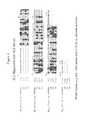
- FIG. 1shows the alignment of human VpreB1 (SEQ ID NO: 1) and human ⁇ 5 (SEQ ID NO: 5) with antibody ⁇ chain variable (SEQ ID NO: 27) and constant regions (SEQ ID NO: 28).
- VpreB1shares same sequence similarity to antibody ⁇ chain variable regions, while ⁇ 5 shares some similarly to antibody ⁇ chain constant regions and framework region 4.
- the boxed regionsidentify VpreB1 and ⁇ 5 sequences that are similar to antibody ⁇ chain CDR1, CDR2 and CDR3 regions, respectively.
- FIG. 2is a schematic illustration of a surrogate light chain formed by VpreB and ⁇ 5 sequences, illustrative fusion polypeptides comprising surrogate light chain sequences, and an antibody light chain structure derived from V-J joining.
- FIG. 3is a schematic illustration of various surrogate light chain deletion and single chain constructs.
- FIG. 4schematically illustrates the incorporation of combinational functional diversity into surrogate light chain constructs.
- FIG. 5shows the gene and protein structures of various illustrative surrogate light chain constructs including an SLC domain protein, a single chain fusion protein (comprising an SLC domain protein fused to a heavy chain variable domain by a (Gly 4 Ser) 3 linker (SEQ ID NO: 35)), a VpreB protein fusion-dimeric complex, and a VpreB and lambda 5-trimeric complex.
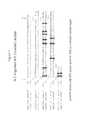
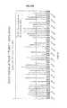
- FIG. 6is the alignment of human VpreB1 (SEQ ID NO: 1, top sequence) with antibody ⁇ 5 light chain variable region germline sequences (SEQ ID NOs: 29 (second from top), 30 (second from bottom), and 31 (bottom sequence), respectively). Regions with the highest degree of sequence similarity are boxed. As shown in the figure, VpreB1 shows only 56%-62% (amino acids 2 to 97) sequence identity to the ⁇ 5 light chain variable region germline sequences.
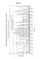
- FIG. 7is the alignment of a ⁇ 5 sequence (SEQ ID NO: 5) with an antibody ⁇ light chain constant region sequence (SEQ ID NO: 32). As shown in the figure, the aligned ⁇ 5sequence shows only 62% (amino acids 97 to 209) sequence identity to the corresponding antibody ⁇ light chain constant region sequences.
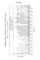
- FIG. 8is the alignment of a ⁇ 5 sequence (SEQ ID NO: 5) with antibody ⁇ light chain constant region sequence (SEQ ID NO: 33). As shown in the figure, the aligned ⁇ 5 sequence shows only 35% (amino acids 105 to 209) sequence identity to the corresponding antibody ⁇ light chain constant region sequence.
- FIG. 9A - FIG. 9Dillustrate various representative ways of adding functionality to surrogate light chain (SLC) components.
- FIGS. 10A-10Dshow the human VpreB1 sequence of SEQ ID NO: 1, the mouse VpreB2 sequences of SEQ ID NOS: 2 and 3; the human VpreB3 sequence of SEQ ID NO: 4, the human ⁇ 5 sequence of SEQ D NO: 5 and the human ⁇ 5-like protein sequence of SEQ ID NO: 6, and sequences of various constructs used in the examples.
- FIG. 11illustrates various trimeric and dimeric surrogate light chain constructs of the invention.
- FIG. 12Detection of surrogate light chains and conjugated heavy chains.
- Lane 1Full Length; Lane 2: Lambda 5 dT; Lane 3: VpreB dt; Lane 4; Short; Lane 5: SCL fusion 1; Lane 6: SLC fusion 2; Lane 7: Antibody.
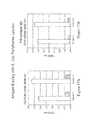
- FIG. 13A - FIG. 13BSLC fusion proteins express and secrete well into the periplasm of E. coli and are best partnered with heavy chain CH1 from IgG1 rather than 1 gM.
- FIG. 13ASCL fusion protein expression in E. coli .
- FIG. 13BIgG1 gamma chain partners and purifies better than 1 gM ⁇ chain with an SLC fusion.
- FIG. 14A - FIG. 14BPhage surrogate light chain construct capture ELISA via anti-phage detection.
- FIG. 15Purified surrogate light chain constructs expressed in mammalian cells bind viral target.
- FIG. 16A - FIG. 16BPurified surrogate light chain constructs expressed in mammalian cells contain stable complexes that bind viral antigen.
- FIG. 17A - FIG. 17BAntigen binding with E. coli periplasmic lysates.
- FIG. 18Surrogate light chain fusion construct phage paired with neutralizing heavy chain readily binds H5 HA antigen.
- FIG. 19Surrogate light chain construct phage paired with neutralizing heavy chain binds antigen.
- FIG. 20Table summarizing the results of phage display experiments.
- FIGS. 21A-21D and FIGS. 22A-22DResults of clonal analysis of rounds 1 and 2 of surrogate light chain fusions 1 and 2.
- surrogate light chainrefers to a dimer formed by the non-covalent association of a VpreB and a ⁇ 5 protein.
- VpreBis used herein in the broadest sense and refers to any native sequence or variant VpreB polypeptide, specifically including, without limitation, human VpreB1 of SEQ ID NO: 1, mouse VpreB2 of SEQ ID NOS: 2 and 3, human VpreB3 of SEQ ID NO: 4 and isoforms, including splice variants and variants formed by posttranslational modifications, other mammalian homologues thereof, as well as variants of such native sequence polypeptides.
- ⁇ 5is used herein in the broadest sense and refers to any native sequence or variant ⁇ 5 polypeptide, specifically including, without limitation, human ⁇ 5 of SEQ ID NO: 5, human ⁇ 5-like protein of SEQ ID NO: 6, and their isoforms, including splice variants and variants formed by posttranslational modifications, other mammalian homologous thereof, as well a variants of such native sequence polypeptides.
- variable VpreB polypeptideand “a variant of a VpreB polypeptide” are used interchangeably, and are defined herein as a polypeptide differing from a native sequence VpreB polypeptide at one or more amino acid positions as a result of an amino acid modification.
- the “variant VpreB polypeptide,” as defined herein,will be different from a native antibody ⁇ or ⁇ light chain sequence, or a fragment thereof.
- the “variant VpreB polypeptide”will preferably retain at least about 65%, or at least about 70%, or at least about 75%, or at least about 80%, or at least about 85%, or at least about 90%, or at least about 95%, or at least about 98% sequence identity with a native sequence VpreB polypeptide.
- variable VpreB polypeptidewill be less then 95%, or less than 90%, or less then 85%, or less than 80%, or less than 75%, or less then 70%, or less than 65%, or less than 60% identical in its amino acid sequence to a native antibody ⁇ or ⁇ light chain sequence.
- Variant VpreB polypeptidesspecifically include, without limitation, VpreB polypeptides in which the non-Ig-like unique tail at the C-terminus of the VpreB sequence is partially or completely removed.
- variant ⁇ 5 polypeptideand “a variant of a ⁇ 5 polypeptide” are used interchangeably, and are defined herein as a polypeptide differing from a native sequence ⁇ 5 polypeptide at one or more amino acid positions as a result of an amino acid modification.
- the “variant ⁇ 5 polypeptide,” as defined herein,will be different from a native antibody ⁇ or ⁇ light chain sequence, or a fragment thereof.
- the “variant ⁇ 5 polypeptide”will preferably retain at least about 65%, or at least about 70%, or at least about 75%, or at least about 80%, or at least about 85%, or at least about 90%, or at least about 95%, or at least about 98% sequence identity with a native sequence ⁇ 5 polypeptide.
- the “variant ⁇ 5 polypeptide”will be less than 95%, or less than 90%, or less than 85%, or less than 80%, or less than 75%, or less than 70%, or less than 65%, or less than 60% identical in its amino acid sequence to a native antibody ⁇ or ⁇ light chain sequence.
- Variant ⁇ 5 polypeptidesspecifically include, without limitation, ⁇ 5 polypeptides in which the unique tail at the N-terminus of the ⁇ 5 sequence is partially or completely removed.
- Percent amino acid sequence identitymay be determined using the sequence comparison program NCBI-BLAST2 (Altschul et al., Nucleic Acids Res. 25:3389-3402 (1997)).
- NCBI-BLAST2 sequence comparison programmay be downloaded from website of the National Center for Biotechnology Information (NCBI) or otherwise obtained from the National Institute of Health, Bethesda, Md.
- VpreB sequenceis used herein to refer to the sequence of “VpreB,” as hereinabove defined, or a fragment thereof.
- ⁇ 5 sequenceis used herein to refers to the sequence of “ ⁇ 5,” as hereinabove defined, or a fragment thereof.
- surrogate light chain sequencemeans any polypeptide sequence that comprises a “VpreB sequence” and/or a “ ⁇ 5 sequence,” as hereinabove defined.
- the “surrogate light chain sequence,” as defined herein,specifically includes, without limitation, the human VpreB1 sequence of SEQ ID NO 1, the mouse VpreB2 sequences of SEQ ID NOS: 2 and 3, and the human VpreB3 sequence of SEQ ID NO: 4, and their various isoforms, including splice variants and variants formed by posttranslational modifications, homologues thereof in other mammalian species, as well as fragments and variants thereof.
- surrogate light chain sequenceadditionally includes, without limitation, the human ⁇ 5 sequence of SEQ ID NO: 5, the human ⁇ 5-like sequence of SEQ ID NO: 6, and their isoforms, including splice variants and variants formed by posttranslational modifications, homologues thereof in other mammalian species, as well as fragments and variants thereof.
- surrogate light chain sequenceadditionally includes a sequence comprising both VpreB and ⁇ 5 sequences as hereinabove defined.
- preBCRdie pre-B-cell receptor
- SCLsurrogate light chain
- the “surrogate light chain sequence”may be optionally conjugated to a heterogeneous amino acid sequence, or any other heterogeneous component, to form a “surrogate light chain construct” herein.
- the term, “surrogate light chain construct”is used in the broadest sense and includes any and all additional heterogeneous components, including a heterogeneous amino acid sequence, nucleic acid, and other molecules conjugated to a surrogate light chain sequence, wherein “conjugation” is defined below.
- a “surrogate light chain construct”is also referred herein as a “SURROBODYTM,” and the two terms are used interchangeably.
- heterogeneous amino acid sequencerelative to a first amino acid sequence, is used to refer to an amino acid sequence not naturally associated with the first amino acid sequence, at least not in the form it is present in the surrogate light chain constructs herein.
- a “heterogenous amino acid sequence” relative to a VpreBis any amino acid sequence not associated with native VpreB in its native environment, including, without limitation, ⁇ 5 sequences that are different from those ⁇ 5 sequences that, together with VpreB, form the surrogate light chain on developing B cells, such as amino acid sequence variants, e.g. truncated and/or derivatized ⁇ 5 sequences.
- a “heterogeneous amino acid sequence” relative to a VpreBalso includes ⁇ 5 sequences covalently associated with, e.g. fused to, VpreB, including native sequence ⁇ 5, since in their native environment, the VpreB and ⁇ 5 sequences are not covalently associated, e.g. fused, to each other.
- Heterogeneous amino acid sequencesalso include, without limitation, antibody sequences, including antibody and heavy chain sequences and fragments thereof, such as, for example, antibody light and heavy chain variable region sequences, and antibody light and heavy chain constant region sequences.
- conjugationrefers to any and all forms of covalent or non-covalent linkage, and include, without limitation, direct genetic or chemical fusion, coupling through a linker or a cross-linking agent, and non-covalent association, for example through Van der Waals forces, or by using a leucine zipper.
- fusionis used herein to refer to the combination of amino acid sequences of different origin in one polypeptide chain by in-frame combination of their coding nucleotide sequences.
- fusionexplicitly encompasses internal fusions, i.e., insertion of sequences of different origin within a polypeptide chain, in addition to fusion to one of its termini.
- targetis a substance that interacts with a polypeptide herein.
- Targetsspecifically include antigens with which the VpreB-containing constructs of the present invention interact. Preferably, interaction takes place by direct binding.
- peptideAs used herein, the terms “peptide,” “polypeptide” and “protein” all refer to a primary sequence of amino acids that are joined by covalent “peptide linkages.” In general, a peptide consists of a few amino acids, typically from about 2 to about 50 amino acids, and is shorter than a protein.
- polypeptideas defined herein, encompasses peptides and proteins.
- amino acidtypically refers to an amino acid having its art recognized definition such as an amino acid selected from the group consisting of alanine (Ala); arginine (Arg); asparagine (Asn); aspartic acid (Asp); cysteine (Cys); glutamine (Gln); glutamic acid (Glu); glycine (Gly); histidine (His); isoleucine (Ile); leucine (Leu); lysine (Lys); methionine (Met); phenylalanine (Phe); proline (Pro); serine (Ser); threonine (Thr); tryptophan (Trp); tyrosine (Tyr); and valine (Val) although modified, synthetic, or rare amino acids may be used as desired.
- amino acidscan be subdivided into various sub-groups.
- amino acidscan be grouped as having a nonpolar side chain (e.g., Ala, Cys, Ile, Leu, Met, Phe, Pro, Val); a negatively charged side chain (e.g., Asp, Glu); a positively charged side chain (e.g., Arg, His, Lys); or an uncharged polar side chain (e.g., Asn, Cys, Gln, Gly, His, Met, Phe, Ser, Thr, Trp, and Tyr).
- a nonpolar side chaine.g., Ala, Cys, Ile, Leu, Met, Phe, Pro, Val
- a negatively charged side chaine.g., Asp, Glu
- a positively charged side chaine.g., Arg, His, Lys
- an uncharged polar side chaine.g., Asn, Cys, Gln, Gly, His, Met, Phe, Ser,
- Amino acidscan also be grouped as small amino acids (Gly, Ala), nucleophilic amino acids (Ser, His, Thr, Cys), hydrophobic amino acids (Val, Leu, Ile, Met, Pro), aromatic amino acids (Phe, Tyr, Trp, Asp, Glu), amides (Asp, Glu), and basic amino acids (Lys, Arg).
- polynucleotide(s)refers to nucleic acids such as DNA molecules and RNA molecules and analogs thereof (e.g., DNA or RNA generated using nucleotide analogs or using nucleic acid chemistry).
- the polynucleotidesmay be made synthetically, e.g., using art-recognized nucleic acid chemistry or enzymatically using, e.g., a polymerase, and, if desired, be modified. Typical modifications include methylation, biotinylation, and other art-known modifications.
- the nucleic acid moleculecan be single-stranded or double-stranded and, where desired, linked to a detectable moiety.
- variantrefers to a polypeptide that possesses at least one amino acid mutation or modification (i.e., alteration) as compared to a native polypeptide.
- variants generated by “amino acid modifications”can be produced, for example, by substituting, deleting, inserting and/or chemically modifying at least one amino acid in the native amino acid sequence.
- amino acid modificationrefers to a change in the amino acid sequence of a predetermined amino acid sequence.
- exemplary modificationsinclude an amino acid substitution, insertion and/or deletion.
- amino acid modification atrefers to the substitution or deletion of the specified residue, or the insertion of at least one amino acid residue adjacent the specified residue.
- insertion “adjacent” a specified residueis meant insertion within one to two residues thereof. The insertion may be N-terminal or C-terminal to the specified residue.
- amino acid substitutionrefers to the replacement of at least one existing amino acid residue in a predetermined amino acid sequence with another different “replacement” amino acid residue.
- the replacement residue or residuesmay be “naturally occurring amino acid residues” (i.e. encoded by the genetic code) and selected from the group consisting of: alanine (Ala); arginine (Arg); asparagine (Asn); aspartic acid (Asp); cysteine (Cys); glutamine (Gln); glutamic acid (Glu); glycine (Gly); histidine (His); isoleucine (Ile): leucine (Leu); lysine (Lys); methionine (Met); phenylalanine (Phe); proline (Pro); serine (Ser); threonine (Thr); tryptophan (Trp); tyrosine (Tyr); and valine (Val). Substitution with one or more non-naturally occurring amino acid residues is also encompasse
- non-naturally occurring amino acid residuerefers to a residue, other than those naturally occurring amino acid residues listed above, which is able to covalently bind adjacent amino acid residues(s) in a polypeptide chain.
- non-naturally occurring amino acid residuesinclude norleucine, ornithine, norvaline, homoserine and other amino acid residue analogues such as those described in Ellman et al. Meth. Enzym. 202:301 336 (1991).
- the procedures of Noren et al. Science 244:182 (1989) and Ellman et al., supracan be used. Briefly, these procedures involve chemically activating a suppressor tRNA with a non-naturally occurring amino acid residue followed by in vitro transcription and translation of the RNA.
- amino acid insertionrefers to the incorporation of at least one amino acid into a predetermined amino acid sequence. While the insertion will usually consist of the insertion of one or two amino acid residues, the present application contemplates larger “peptide insertions”, e.g. insertion of about three to about five or even up to about ten amino acid residues.
- the inserted residue(s)may be naturally occurring or non-naturally occurring as disclosed above.
- amino acid deletionrefers to the removal of at least one amino acid residue from a predetermined amino acid sequence.
- mutagenesisrefers to, unless otherwise specified, any art recognized technique for altering a polynucleotide polypeptide sequence. Preferred types of mutagenesis include error prone PCR mutagenesis, saturation mutagenesis, or other site directed mutagenesis.
- Site-directed mutagenesisis a technique standard in the art, and is conducted using a synthetic oligonucleotide primer complementary to a single-stranded phage DNA to be mutagenized except for limited mismatching, representing the desired mutation. Briefly, the synthetic oligonucleotide is used as a primer to direct synthesis of a strand complementary to the single-stranded phage DNA, and the resulting double-stranded DNA is transformed into a phage-supporting host bacterium. Cultures of the transformed bacteria are plated in top agar, permitting plaque formation from single cells that harbor the phage.
- Plaques of interestare selected by hybridizing with kinased synthetic primer at a temperature that permits hybridization of an exact match, but at which the mismatches with the original strand are sufficient to prevent hybridization. Plaques that hybridize with the probe are then selected, sequenced and cultured, and the DNA is recovered.
- antibodyis used to refer to a native antibody from a classically recombined heavy chain derived from V(D)J gene recombination and a classically recombined light chain also derived from VJ gene recombination, or a fragment thereof.
- a “native antibody”is heterotetrameric glycoprotein of about 150,000 daltons, composed of two identical light (L) chains and two identical heavy (H) chains. Each light chain is linked to a heavy chain by covalent disulfide bond(s), while the number of disulfide linkages varies between the heavy chains of different immunoglobulin isotypes. Each heavy and light chain also has regularly spaced intrachain disulfide bridges. Each heavy chain has, at one end, a variable domain (V H ) followed by a number of constant domains.
- V Hvariable domain
- Each light chainhas a variable domain at one end (V L ) and a constant domain at its other end; the constant domain of the light chain is aligned with the first constant domain of the heavy chain, and the light chain variable domain is aligned with the variable domain of the heavy chain.
- Particular amino acid residuesare believed to form an interface between the light- and heavy-chain variable domains, Chothia et al., J. Mol. Biol. 186:651 (1985); Novotny and Haber, Proc. Natl. Acad. Sci. U.S.A. 82:4592 (1985).
- variablewith reference to antibody chains is used to refer to portions of the antibody chains which differ extensively in sequence among antibodies and participate in the binding and specificity of each particular antibody for its particular antigen. Such variability is concentrated in three segments called hypervariable regions both in the light chain and the heavy chain variable domains. The more highly conserved portions of variable domains are called the framework region (FR).
- the variable domains of native heavy and light chainseach comprise four FRs (FR1, FR2, FR3 and FR4, respectively), largely adopting a ⁇ -sheet configuration, connected by three hypervariable regions, which form loops connecting, and in some cases forming part of, the ⁇ -sheet structure.
- the hypervariable regions in each chainare held together in close proximity by the FRs and, with the hypervariable regions from the other chain, contribute to the formation of the antigen-binding site of antibodies (see Kabat et al., Sequences of Proteins of Immunological Interest, 5th Ed. Public Health Service, National Institutes of Health, Bethesda, Md. (1991), pages 647-669).
- the constant domainsare not involved directly in binding an antibody to an antigen, but exhibit various effector functions, such as participation of the antibody in antibody-dependent cellular toxicity.
- hypervariable regionwhen used herein refers to the amino acid residues of an antibody which are responsible for antigen-binding.
- the hypervariable regioncomprises amino acid residues from a “complementarity determining region” or “CDR” (i.e., residues 30-36 (L1), 46-55 (L2) and 86-96 (L3) in the light chain variable domain and 30-35 (H1), 47-58 (H2) and 93-101 (H3) in the heavy chain variable domain; MacCallum et al., J Mol Biol. 262(5):732-45 (1996).
- CDRcomplementarity determining region
- framework regionrefers to the art recognized portions of an antibody variable region that exist between the more divergent CDR regions. Such framework regions are typically referred to as frameworks 1 through 4 (FR1, FR2, FR3, and FR4) and provide a scaffold for holding, in three-dimensional space, the three CDRs found in a heavy or light chain antibody variable region, such that the CDRs can form an antigen-binding surface.
- frameworks 1 through 4FR1, FR2, FR3, and FR4
- antibodiescan be assigned to different classes. There are five major classes of antibodies IgA, IgD, IgE, IgG, and IgM, and several of these may be further divided into subclasses (isotypes), e.g., IgG1, IgG2, IgG3, IgG4, IgA, and IgA2.
- the heavy-chain constant domains that correspond to the different classes of immunoglobulinsare called ⁇ , ⁇ , ⁇ , ⁇ , and ⁇ , respectively.
- the “light chains” of antibodies from any vertebrate speciescan be assigned to one of two clearly distinct types, called kappa ( ⁇ ) and lambda ( ⁇ ), based on the amino acid sequences of their constant domains. Any reference to an antibody light chain herein includes both ⁇ and ⁇ light chains.
- Antibody fragmentscomprise a portion of a full length antibody, generally the antigen binding or a variable domain thereof.
- antibody fragmentsinclude, but are not limited to, Fab, Fab′, F(ab′) 2 , scFv, and (scFv) 2 fragments.
- antibody binding regionrefers to one or more portions of an immunoglobulin or antibody variable region capable of binding an antigen(s).
- the antibody binding regionis, for example, an antibody light chain (VL) (or variable region thereof), an antibody heavy chain (VH) (or variable region thereof), a heavy chain Fd region, a combined antibody light and heavy chain (or variable region thereof) such as a Fab, F(ab′) 2 , single domain, or single chain antibody (scFv), or a full length antibody, for example, an IgG (e.g., an IgG1, IgG2, IgG3, or IgG4 subtype), IgA1, IgA2, IgD, IgE, or IgM antibody.
- VLantibody light chain
- VHantibody heavy chain
- a heavy chain Fd regiona combined antibody light and heavy chain (or variable region thereof) such as a Fab, F(ab′) 2 , single domain, or single chain antibody (scFv)
- an IgG
- epitoperefers to a sequence of at least about 3 to 5, preferably at least about 5 to 10, or at least about 5 to 15 amino acids, and typically not more than about 500, or about 1,000 amino acids, which define a sequence that by itself, or as part of a larger sequence, binds to an antibody generated in response to such sequence.
- An epitopeis not limited to a polypeptide having a sequence identical to the portion of the parent protein from which it is derived. Indeed, viral genomes are in a state of constant change and exhibit relatively high degrees of variability between isolates.
- epitopeencompasses sequences identical to the native sequence, as well as modifications, such as deletions, substitutions and/or insertions to the native sequence.
- modificationsare conservative in nature but non-conservative modifications are also contemplated.
- the termspecifically includes “mimotopes,” i.e. sequences that do not identify a continuous linear native sequence or do not necessarily occur in a native protein, but functionally mimic an epitope on a native protein.
- epitopespecifically includes linear and conformational epitopes.
- vectoris used to refer to a rDNA molecule capable of autonomous replication in a cell and to which a DNA segment, e.g., gene or polynucleotide, can be operatively linked so as to bring about replication of the attached segment.
- vectorscapable of directing the expression of genes encoding for one or more polypeptides are referred to herein as “expression vectors.”
- control sequencesrefers to DNA sequences necessary for the expression of an operably linked coding sequence in a particular host organism.
- the control sequences that are suitable for prokaryotes, for example,include a promoter, optionally an operator sequence, and a ribosome binding site.
- Eukaryotic cellsare known to utilize promoters, polyadenylation signals, and enhancers.
- Nucleic acidis “operably linked” when it is placed into a functional relationship with another nucleic acid sequence.
- DNA for a presequence or secretory leaderis operably linked to DNA for a polypeptide if it is expressed as a preprotein that participates in the secretion of the polypeptide;
- a promoter or enhanceris operably linked to a coding sequence if it affects the transcription of the sequence; or
- a ribosome binding siteis operably linked to a coding sequence if it is positioned so as to facilitate translation.
- “operably linked”means that the DNA sequences being linked are contiguous, and, in the case of a secretory leader, contiguous and in reading phase. However, enhancers do not have to be contiguous. Linking is accomplished by ligation at convenient restriction sites. If such sites do not exist, the synthetic oligonucleotide adaptors or linkers are used in accordance with conventional practice.
- a “phage display library”is a protein expression library that expresses a collection of cloned protein sequences as fusions with a phage coat protein.
- phage display libraryrefers herein to a collection of phage (e.g., filamentous phage) wherein the phage express an external (typically heterologous) protein. The external protein is free to interact with (bind to) other moieties with which the phage are contacted.
- Each phage displaying an external proteinis a “member” of the phage display library.
- filamentous phagerefers to a viral particle capable of displaying a heterogenous polypeptide on its surface, and includes, without limitation, fl, fd, Pfl, and M13.
- the filamentous phagemay contain a selectable marker such as tetracycline (e.g., “fd-tet”).
- fd-tettetracycline
- Various filamentous phage display systemsare well known to those of skill in the art (see, e.g., Zacher et al. Gene 9: 127-140 (1980), Smith et al. Science 228: 1315-1317 (1985); and Parmley and Smith Gene 73: 305-318 (1988)).
- panningis used to refer to the multiple rounds of screening process in identification and isolation of phages carrying compounds, such as antibodies, with high affinity and specificity to a target.
- Mutagenesiscan, for example, be performed using site-directed mutagenesis (Kunkel et al., Proc. Natl. Acad. Sci USA 82:488-492 (1985)).
- PCR amplification methodsare described in U.S. Pat. Nos. 4,683,192, 4,683,202, 4,800,159, and 4,965,188, and in several textbooks including “PCR Technology: Principles and Applications for DNA Amplification”, H. Erlich, ed., Stockton Press, New York (1989); and PCR Protocols: A Guide to Methods and Applications, Innis et al., eds., Academic Press, San Diego, Calif. (1990).
- the present inventionconcerns constructs and libraries comprising antibody surrogate light chain sequences.
- pre-B cellshave been identified in the bone marrow as lymphocytes that produce ⁇ heavy chains but instead of the fully developed light chains express a set of B lineage-specific genes called VpreB(1-3) and ⁇ 5, respectively.
- VpreB and ⁇ 5 polypeptidestogether form a non-covalently associated, Ig light chain-like structure, which is called the surrogate light chain.
- the surrogate light chainalthough not an antibody chain, naturally associates with all antibody heavy chains, and surrogate light chain-antibody heavy chain complexes have been shown to bind self-antigens.
- the present inventionprovides polypeptides comprising VpreB and/or ⁇ 5 sequences and having the ability to bind a target.
- the targetcan be any peptide or polypeptide that is a binding partner for the VpreB and/or ⁇ 5 sequence-containing polypeptides of the present invention.
- Targetsspecifically include all types of targets generally referred to as “antigens” in the context of antibody binding.
- polypeptides of the present inventioninclude, without limitation, conjugates of VpreB sequences to heterogeneous amino acid sequences, provided that they retain the ability to bind a desired target.
- the binding of the VpreB sequence to the heterogeneous amino acid sequencecan be either covalent or non-covalent, and may occur directly, or through a linker, including peptide linkers.
- polypeptide constructs hereininclude polypeptides in which a VpreB sequence, such as a VpreB1, VpreB2, or VpreB3 sequence, including fragments and variants of the native sequences, is conjugated to a ⁇ 5 sequence, including fragments and variants of the native sequence. Representative fusions of this type are illustrated in FIGS. 2 and 11 and described in the Examples.
- a VpreB sequencetypically the C-terminus of a VpreB sequence (e.g. a VpreB1, VpreB2 or VpreB3 sequence) is fused to the N-terminus of a ⁇ 5 sequence. While it is possible to fuse the entire length of a native VpreB sequence to a full-length ⁇ 5 sequence (see, e.g. the first diagram in FIG. 3 ), typically the fusion takes place at or around a CDR3 analogous site in each of the two polypeptides. Such CDR3 analogous sites for VpreB1 and ⁇ 5 are illustrated in FIG. 1 , and a representative fusion construct is illustrated in FIG. 2 .
- the fusionmay take place within, or at a location within about 10 amino acid residues at either side of the CDR3 analogous region. In a preferred embodiment, the fusion takes place between about amino acid residues 116-126 of the native human VpreB1 sequence (SEQ ID NO: 1) and between about amino acid residues 82 and 93 of the native human ⁇ 5 sequence (SEQ ID NO: 5).
- VpreB sequenceto the CDR3 region of an antibody ⁇ light chain, as shown in FIG. 2 .
- FIG. 3Further constructs, in which only one of VpreB and ⁇ 5 is truncated are shown in FIG. 3 . Similar constructs can be prepared using antibody ⁇ light chain sequences.
- the structure designated “SLC fusion 1”is a tetramer, composed of two dimers, in which the fusion of a truncated V-preB1 sequence (lacking the characteristic “tail” at the C-terminus of native VpreB1) to a similarly truncated ⁇ 5 sequence is non-covalently associated with an antibody heavy chain.
- the structure designated “SLC fusion 2”is a tetramer, composed of two dimers, in which the fusion of a truncated VpreB1 sequence (lacking the characteristic “tail” at the C-terminus of native VpreB1) to an antibody light chain constant region is non-covalently associated with an antibody heavy chain.
- the structure designated “SLC fusion 3”is a tetramer, composed of two dimers, in which the fusion of an antibody light chain variable region to a truncated ⁇ 5 sequence (lacking the characteristic “tail” at the N-terminus of native ⁇ 5) is non-covalently associated with an antibody heavy chain.
- the polypeptide constructs of the present inventioninclude non-covalent associations of a VpreB sequence (including fragments and variants of a native sequence) with a heterogeneous sequence, such as a ⁇ 5 sequence (including fragments and variants of the native sequence), and/or an antibody sequence.
- a full-length VpreB sequencemay be non-covalently associated with a truncated ⁇ 5 sequence.
- a truncated VpreB sequencemay be non-covalently associated with a full-length ⁇ 5 sequence.
- VpreB1 and ⁇ 5 sequencesSurrogate light chain constructs comprising non-covalently associated VpreB1 and ⁇ 5 sequences, in non-covalent association with an antibody heavy chain, are shown on the left side of FIG. 11 .
- the structuresmay include, for example, full-length VpreB1 and ⁇ 5 sequences, a full-length VpreB1 sequence associated with a truncated ⁇ 5 sequence (“Lambda 5dT”), a truncated V-reB1 sequence associated with a full-length ⁇ 5 sequence (VpreB dT”) and a truncated VpreB1 sequence associated with a truncated ⁇ 5 sequence (“Short”).
- FIG. 11illustrates certain specific constructs
- the structurescan be asymmetrical, comprising different surrogate light chain sequences in each arm, and/or having trimeric or pentameric structures, as opposed to the structures illustrated in FIG. 11 .
- the constructs of the present inventioncan be engineered, for example, by incorporating or appending known sequences or sequence motifs from the CDR1, CDR2 and/or CDR3 regions of antibodies, including known therapeutic antibodies into the CDR1, CDR2 and/or CDR3 analogous regions of the surrogate light chain sequences. This allows the creation of molecules that are not antibodies, but will exhibit binding specificities and affinities very similar to those of a known therapeutic antibody.
- a VpreB- ⁇ 5 fusioncan be linked to an antibody heavy chain variable region sequence by a peptide linker.
- a VpreB- ⁇ 5 fusionis non-covalently associated with an antibody heavy chain, or a fragment thereof including a variable region sequence to form a dimeric complex.
- the VpreB and ⁇ 5 sequencesare non-covalently associated with each other and an antibody heavy chain, or a fragment thereof including a variable region sequence, thereby forming a trimeric complex. Exemplary constructs comprising an antibody heavy chain are illustrated in FIG. 11 .
- the present inventionincludes all constructs that comprise surrogate light chain sequences and have the ability to bind a desired target. In certain embodiment, the constructs also have the ability to associate with antibody heavy chain variable region sequences.
- constructs of the present inventionmay be used to build libraries of surrogate light chain sequences, which can be used for various purposes, similarly to antibody libraries, including selection of constructs with the desired binding specificities and affinities.
- the free ends of one or both componentsare available for incorporating an additional diversity into the library of such sequences.
- a random peptide librarycan be appended or substituted to one of these free ends and panned for specific binding to a particular target.
- the surrogate light chain component with the desired binding specificitycan be combined with an antibody heavy chain or heavy fragment binding to a different target.
- the surrogate light chain componentmay bind a tumor antigen while the antibody heavy chain or heavy chain fragment may bind to effector cells. This way, a single entity with targeting and anti-tumor activity can be created.
- the appendage or the polypeptide that connects the VpreB and ⁇ 5 sequencescan be an antibody or antibody fragments, such as a Fab or a scFv fragment.
- the incorporation of an antibody sequencewill not only create a “chelating” effect but can also generate bispecificity in a single molecule, without the need of a second independent arm, such as that found in bispecific antibodies.
- the two specificitiesmay be to different parts of the same target, to disparate targets, or to a target antibody complex.
- multi-specific constructscan be made with any type of molecule, other than antibodies or antibody fragments, including peptides, proteins, enzymes, and the like.
- the surrogate light chain component with the desired specificitycan be combined with any therapeutic peptide or protein.
- the surrogate light chain constructs of the present inventioncan be prepared by methods known in the art, including well known techniques of recombinant DNA technology.
- Nucleic acid encoding surrogate light chaine.g. VpreB and ⁇ 5 polypeptides
- VpreB and ⁇ 5 polypeptidescan be isolated from natural sources, e.g. developing B cells and/or obtained by synthetic or semi-synthetic methods. Once this DNA has been identified and isolated or otherwise produced, it can be ligated into a replicable vector for further cloning or for expression.
- Cloning and expression vectors that can be used expressing the coding sequences of the polypeptides hereinare well known in the art and are commercially available.
- the vector componentsgenerally include, but are not limited to, one or more of the following: a signal sequence, an origin of replication, one or more marker genes, an enhancer element, a promoter, and, a transcription termination sequence.
- Suitable host cells for cloning or expressing the DNA encoding the surrogate light chain constructs in the vectors hereinare prokaryote, yeast, or higher eukaryote (mammalian) cells, mammalian cells are being preferred.
- suitable mammalian host cell linesinclude, without limitation, monkey kidney CV1 line transformed by SV40 (COS-7, ATCC CRL 1651); human embryonic kidney line 293 (293 cells) subcloned for growth in suspension culture, Graham et al, J. Gen Virol. 36:59 (1977)); baby hamster kidney cells (BHK, ATCC CCL 10); Chinese hamster ovary cells/-DHFR (CHO, Urlaub et al., Proc. Natl. Acad. Sci. USA 77:4216 (1980)); mouse sertoli cells (TM4, Mather, Biol. Reprod.
- COS-7monkey kidney CV1 line transformed by SV40
- human embryonic kidney line 293 (293 cells) subcloned for growth in suspension cultureGraham et al, J. Gen Virol. 36:59 (1977)
- baby hamster kidney cellsBHK, ATCC CCL 10
- Chinese hamster ovary cells/-DHFRCHO, Urlaub et al., Proc. Natl
- monkey kidney cells(CV1 ATCC CCL 70); African green monkey kidney cells (VERO-76, ATCC CRL-1587); human cervical carcinoma cells (HELA, ATCC CCL 2); canine kidney cells (MDCK, ATCC CCL 34); buffalo rat liver cells (BRL 3A, ATCC CRL 1442); human lung cells (W 138, ATCC CCL 75); human liver cells (Hep G2, HB 8065); mouse mammary tumor (MMT 060562, ATCC CCL51); TRI cells (Mather et al., Annals N.Y. Acad. Sci. 383:44-68 (1982)); MRC 5 cells; FS4 cells; and a humn hepatoma line (Hep G2).
- promoterscan be derived from the genomes of polyoma, Adenovirus2, retroviruses, cytomegalovirus, and Simian Virus 40 (SV40).
- promotersinclude, without limitation, the early and late promoters of SV40 virus (Fiers et al., Nature, 273: 113 (1978)), the immediate early promoter of the human cytomegalovirus (Greenaway et al., Gene, 18: 355-360 (1982)), and promoter and/or control sequences normally associated with the desired gene sequence, provided such control sequences are compatible with the host cell system.
- Enhancersare relatively orientation and position independent, but preferably are located upstream of the promoter sequence present in the expression vector.
- the enhancermight originate from the same source as the promoter, such as, for example, from a eukaryotic cell virus, e.g. the SV40 enhancer on the late side of the replication origin (bp 100-270), the cytomegalovirus early promoter enhancer, the polyoma enhancer on the late side of the replication origin, and adenovirus enhancers.
- Expression vectors used in mammalian host cellsalso contain polyadenylation sites, such as those derived from viruses such as, e.g., the SV40 (early and late) or HBV.
- polyadenylation sitessuch as those derived from viruses such as, e.g., the SV40 (early and late) or HBV.
- An origin of replicationmay be provided either by construction of the vector to include an exogenous origin, such as may be derived from SV40 or other viral (e.g., Polyoma, Adeno, VSV, BPV) source, or may be provided by the host cell.
- an exogenous originsuch as may be derived from SV40 or other viral (e.g., Polyoma, Adeno, VSV, BPV) source, or may be provided by the host cell.
- the expression vectorsusually contain a selectable marker that encodes a protein necessary for the survival or growth of a host cell transformed with the vector.
- selectable markers for mammalian cellsinclude dihydrofolate reductase thymidine kinase (TK), and neomycin.
- Suitable mammalian expression vectorsare well known in the art and commercially available.
- the surrogate light chain constructs of the present inventioncan be produced in mammalian host cells using a pCI expression vector (Promega), carrying the human cytomegalovirus (CMV) immediate-early enhancer/promoter region to promote constitutive expression of a DNA insert.
- the vectorcan contain a neomycin phosphotransferase gene as a selectable marker.
- the surrogate light chain constructs of the present inventioncan also be produced in bacterial host cells.
- Control elements for use in bacterial systemsinclude promoters, optionally containing operator sequences, and ribosome binding sites. Suitable promoters include, without limitation, galactose (gal), lactose (lac), maltose, tryptophan (trp), ⁇ -lactamase promoters, bacteriophage ⁇ and T7 promoters. In addition, synthetic promoters can be used, such as the tac promoter. Promoters for use in bacterial systems also generally contain a Shine-Dalgarno (SD) sequence operably linked to the DNA encoding the Fab molecule. The origin of replication from the plasmid pBR322 is suitable for most Gram-negative bacteria.
- SDShine-Dalgarno
- the coding sequences of the individual chains within a multi-chain construct comprising antibody surrogate light chain sequencescan be present in the same expression vector, under control of separate regulatory sequences, or in separate expression vectors, used to cotransfect a desired host cells, including eukaryotic and prokaryotic hosts.
- a desired host cellsincluding eukaryotic and prokaryotic hosts.
- multiple genescan be coexpressed using the DuetTM vectors commercially available from Novagen.
- the transformed host cellsmay be cultured in a variety of media.
- Commercially available media for culturing mammalian host cellsinclude Ham's F10 (Sigma), Minimal Essential Medium ((MEM), (Sigma), RPM1-1640 (Sigma), and Dulbecco's Modified Eagle's Medium ((DMEM), Sigma).
- any of the media described in Ham et al., Meth. Enz. 58:44 (1979) and Barnes et al., Anal. Biochem. 102:255 (1980)may be used as culture media for the host cells.
- the culture conditions, such as temperature, pH, and the like,are those previously used with the host cell selected for expression, and are included in the manufacturer's instructions or will otherwise be apparent to the ordinarily skilled artisan.
- the surrogate antibody moleculesare purified in a 6.times.His-tagged form, using the Ni-NTA purification system (Invitrogen).
- the present inventionfurther concerns various libraries of surrogate light chain sequences and constructs comprising such sequences.
- librariesmay comprise, consist essentially of, or consist of, displays of surrogate light chain sequences, such as the VpreB- and/or ⁇ 5-containing constructs of the present invention, including, without limitation, those specifically described above, illustrated in the figures and/or described in the Examples.
- the libraries of the present inventionare preferably in the form of a display.
- Systems for displaying heterologous proteins, including antibodies and other polypeptides,are well known in the art.
- Antibody fragmentshave been displayed on the surface of filamentous phage that encode the antibody genes (Hoogenboom and Winter J. Mol. Biol., 222:381 388 (1992); McCafferty et al., Nature 348(6301):552 554 (1990); Griffiths et al. EMBO J., 13(14):3245-3260 (1994)).
- HoogenboomHoogenboom, Nature Biotechnol. 23(9):1105-1116 (2005).
- viral displaysuch as retroviral display (Urban et al., Nucleic Acids Res. 33:e35 (2005), display based on protein-DNA linkage (Odegrip et al., Proc. Acad. Natl. Sci. USA 101:2806-2810 (2004); Reiersen et al., Nucleic Acids Res. 33:e10 (2005)), and microbead display (Sepp et al., FEBS Lett. 532:455-458 (2002)).
- retroviral displayUrban et al., Nucleic Acids Res. 33:e35 (2005), display based on protein-DNA linkage (Odegrip et al., Proc. Acad. Natl. Sci. USA 101:2806-2810 (2004); Reiersen et al., Nucleic Acids Res. 33:e10 (2005)
- microbead displaySepp et al., FEBS Lett. 532:455-458 (2002).
- the surrogate light chain-containing librariesmay be advantageously displayed using any display technique, including phage display and spore display.
- the heterologous proteinsuch as a surrogate light chain polypeptide
- a coat protein of a phage particlewhile the DNA sequence from which it was expressed is packaged within the phage coat.
- phage display methodscan be found, for example, McCafferty et al., Nature 348, 552-553 (1990)), describing the production of human antibodies and antibody fragments in vitro, from immunoglobulin variable (V) domain gene repertoires from unimmunized donors.
- antibody V domain genesare cloned in-frame into either a major or minor coat protein gene of a filamentous bacteriophage, such as M13 or fd, and displayed as functional antibody fragments on the surface of the phage particle. Because the filamentous particle contains a single-stranded DNA copy of the phage genome, selections based on the functional properties of the antibody also result in selection of the gene encoding the antibody exhibiting those properties. Thus, the phage mimics some of the properties of the B-cell.
- Phage displaycan be performed in a variety of formats; for their review see, e.g. Johnson, Kevin S. and Chiswell, David J., Current Opinion in Structural Biology 3, 564-571 (1993).
- Several sources of heavy chain V-gene segmentscan be discovered through phage display. Clarkson et al., Nature 352, 624-628 (1991) isolated a diverse array of anti-oxazolone heavy chains and light chains from a small random combinatorial library of V genes derived from the spleens of immunized mice.
- a repertoire of heavy and light chain V genes from unimmunized human donorscan be constructed and recovered specific to a diverse array of antigens (including self-antigens) essentially following the techniques described by Marks et al., J. Mol. Biol. 222, 581-597 (1991), or Griffith et al., EMBO J. 12, 725-734 (1993).
- These, and other techniques known in the artcan be adapted to the display of any polypeptide, including polypeptides and other constructs comprising surrogate light chain sequences.
- the surrogate light chaincan be supplemented with a collection of heavy chains from either a naturally diverse source, such as lymphocytes, or a synthetically generated collection created entirely through techniques of molecular biology.
- Spore display systemsare based on attaching the sequences to be displayed to a coat protein, such as a Bacillus subtilis spore coat protein.
- the spore protoplast (core)is surrounded by the cell wall, the cortex, and the spore coat. Depending on the species, an exosporium may also be present.
- the core wallis composed of the same type of peptidoglycan as the vegetative cell wall.
- Spore displayincluding surface display system using a component of the Bacillus subtilis spore coat (CorB) and Bacillus thuringiensis (Bt) spore display, is described in Isticato et al., J. Bacterial. 183:6294-6301 (2001); Cheng et al., Appl.
- spore display systemsAn advantage of spore display systems is the homogenous particle surface and particle size of non-eukaryotic nature, which is expected to provide an ideal non-reactive background.
- particle size of sporesis sufficient to enable selection by flow cytometry that permits selectable clonal isolation, based upon interactions.
- sporesLeveraging on the stability of spores, it is possible to perform various post-sporulation chemical, enzymatic and/or environmental treatments and modification. Thus, it is possible to stabilize structural helical structures with chemical treatment using trifluoroethanol (TFE), when such structures are displayed.
- TFEtrifluoroethanol
- oxidative stress treatmentssuch as treatments with Reactive Oxygen Species (e.g. peroxide) or reactive Nitrogen Species (e.g. nitrous acid) are possible.
- enzymatic treatmentssuch as proteolytic exposure, other enzymatic processes, phosphorylation, etc.
- Other possible treatmentsinclude, without limitation, nitrosylation by peroxynitrite treatment, proteolysis by recombinant, purified, or serum protease treatment, irradiation, coincubation with known chaperones, such as heat shock proteins (both bacterial and mammalian), treatment with folding proteins, such as protein disulfide isomerase, prolyl isomerase, etc., lyophilization, and preservative-like treatments, such as treatment with thimerosol.
- chaperonessuch as heat shock proteins (both bacterial and mammalian)
- folding proteinssuch as protein disulfide isomerase, prolyl isomerase, etc.
- preservative-like treatmentssuch as treatment with thimerosol.
- Similar techniquescan be used in all spore display systems, including displays where the attachment is to a spore coat protein, including, for example, the spore display systems disclosed in
- the libraries of the present inventioncan be used to identify surrogate light chain sequences and surrogate light chain constructs, such as fusions comprising surrogate light chain sequences, with desired properties. For example, in vitro or in vivo screening of the libraries herein can yield polypeptides comprising surrogate light chain sequences binding to desired targets with high binding specificity and affinity. Thus, the libraries herein can be used to identify molecules for therapeutic and diagnostic purposes, such as polypeptides comprising surrogate light chain sequences that bind to tumor markers or other molecular targets of therapeutic intervention.
- libraries of surrogate light chain polypeptidescan be engineered, including libraries comprising a collection of polypeptides binding to the same target, libraries of polypeptides binding to different targets, libraries of polypeptides with multiple specificities, and the like.
- the antibody surrogate light chain constructs of the present inventioncan be used in analytical and diagnostic assays, to detect the presence of a desired target molecule, such as a tumor antigen or any polypeptide associated with a disease state or condition.
- a desired target moleculesuch as a tumor antigen or any polypeptide associated with a disease state or condition.
- the surrogate light chain constructs of the present inventioncan be used as therapeutic agents, such as, for example, in cancer therapy, to target tumor antigens that have been determined to associate with the development and/or spread of cancer.
- VpreBas a Binding Domain Protein and Fusions Containing It
- the SLC binding domain protein constructis comprised of the amino acids 20 to 121 from VpreB1 and the amino acids 87 to 105 from ⁇ 5. If desired, to create novel and specific binding capabilities, the molecule is reengineered according to structural or sequence evidence. Additionally, or alternatively, a collection of variants is created either randomly, for example by error-prone PCR, or directly by single or multi-site specific mutagenesis with a collection of amino acids. The resulting clones or collections are then cloned in frame with pill for use in phage or phagemid display. This phagemid construct is transformed into TG1 cells.
- a single colonyis propagated in Luria Broth (LB) supplemented with 50 ⁇ g/ml Ampicillin and 2% glucose until it reached OD600 ⁇ 0.3, and infected with MK307 helper phage at 37° C. for 30 minutes without shaking.
- the cellsare then pelleted and then resuspended in LB containing 50 ⁇ g/ml ampicillin and 75 ⁇ g/ml kanamycin and allowed to grow overnight with vigorous aeration at 30° C.
- the next day the supernatant containing phagemid expressed SLC-HC fusion proteinis used in Phage ELISA to determine targeted binding.
- the ELISAentails coating and blocking of an ELISA plate with human TNF- ⁇ , followed by incubation of the SLC-HC phage for 2 hours at 4° C., washing with PBS-polyethylene glycol sorbitan monolaurate-20 (PBS-TWEENTM-20) (0.05%) and direct detection with anti-m13-HRP antibody.
- bindingis assessed by directly amplifying or eluting the bound phage and determining phage titers using XL-1 Blue cells.
- This exampledescribes a SLC binding domain fusion as a single clone, but this SLC can be recombinantly recombined with other heterologous sequences that recognize a common target and screened as a library.
- this SLC binding proteincan be combined with a previously selected collection of heavy chains and screened directly on the same target of interest or a second target of interest to create a bispecific molecule.
- this reinforced binding or bispecific bindingcan be discovered by screening in conjunction with unselected collections of heavy chains.
- this examplerefers to antibody heavy chains, it should be understood that a complete heavy chain is not needed.
- Single-chain fusions comprising heavy chain variable region sequences, in the absence of a heavy chain constant region, or a complete heavy chain constant regioncan be made in an analogous manner and are within the scope of this example.
- VHVariable Heavy Chain
- VpreB protein fusion—dimeric complexA functional VpreB- ⁇ 5 fusion protein shown in the second diagram of FIG. 5 (designated “VpreB protein fusion—dimeric complex”) is recombinantly created.
- the VpreB- ⁇ 5 fusion proteinis comprised of an m13 gene III signal sequence, the amino acids 20 to 115 from VpreB1, and the amino acids 83 to 209 from ⁇ 5.
- This constructis coexpressed with a variable heavy chain-CH1 fusion in frame with pIII for use in phage phagemid display.
- VH coding sequencethe VH coding sequence from the anti-TNF- ⁇ antibody, D2E7, is used, and CH1 is the CH1 region of human IgG1. This phagemid construct is transformed into TG1 cells.
- a single colonyis propagated in Luria Broth (LB) supplemented with 50 ⁇ g/ml Ampicillin and 2% glucose until it reached OD600 ⁇ 0.3, and infected with MK307 helper phage at 37 degrees for 30 minutes, without shaking.
- the cellsare then pelleted and then resuspended in LB Briefly the ELISA entails coating and blocking of an ELISA plate with human TNF- ⁇ , followed by incubation of the SLC-HC phage for 2 hours at 4 degrees, washing with PBS-TWEENTM-20 (0.05%) and direct detection with anti-m13-HRP antibody.
- bindingcan be assessed by directly amplifying or eluting the bound phage and determining phage titers using XL-1 Blue cells.
- This exampledescribes a SLC partnered with a heavy chain variable-CH1 fusion as a single clone, but this SLC can be combined with a focused collection of heavy chain variable regions that recognize a common target and screened as a library. Furthermore, this SLC fusion can be combined with an unselected collection of heavy chains and screened directly on a target of interest.
- VpreB and Lambda5as an Associated Variable Heavy Chain (VH) Partner
- VpreB and lambda 5-trimeric complexThe VpreB- ⁇ 5 coexpressed protein shown in the third diagram of FIG. 5 (designated “VpreB and lambda 5-trimeric complex”) is made of an m13 gene III signal sequence and the corresponding amino acids of the predicted mature, processed VpreB1 (amino acids 20 to 146) and lambda 5 (amino acids 31 to 209). These are coexpressed with a variable heavy chain-CH1 fusion in frame with pIII for use in phage or phagemid display.
- VH coding sequencethe VH coding sequence from the anti-TNF- ⁇ antibody, D2E7, is used, and CH1 is the CH1 region from human IgG1. This phagemid construct is transformed into TG1 cells.
- a single colonyis propagated in Luria Broth (LB) supplemented with 50 ⁇ g/ml Ampicillin and 2% glucose until it reached OD600 ⁇ 0.3, and is then infected with MK307 helper phage at 37 degrees for 30 minutes, without shaking.
- the cellsare then pelleted and then resuspended in LB containing 50 ⁇ g/ml ampicillin and 75 ⁇ /ml kanamycin and allowed to grow overnight with vigorous aeration at 30 degrees.
- the next day the supernatant containing phagemid expressed SLC HC trimeric protein complexesis used in Phage ELISA to determine targeted binding.
- the ELISAentails coating and blocking of an ELISA plate with human TNF- ⁇ , followed by incubation of the SLC-HC phage for 2 hours at 4 degrees, washing with PBS-TWEENTM-20 (0.05%) and direct detection with anti-m13-HRP antibody.
- bindingcan be assessed by directly amplifying or eluting the bound phage and determining phage titers using XL-1Blue cells.
- This exampledescribes a SLC partnered with a heavy chain variable-CH1 fusion as a single clone, but this SLC can be combined with a focused collection of heavy chain variable regions that recognize a common target and screened as a library. Furthermore, this SLC fusion can be combined with an unselected collection of heavy chains and screened directly on a target of interest.
- the CDR analogous regions of the surrogate light chainwill have similar functions to the CDR's of an antibody light chain, it is important to determine the fusion points between the VpreB and ⁇ 5.
- the most suitable fusion point for a particular purposeis determined starting with the CDR3 analogous site containing all VpreB amino acids and incrementally substituting amino acids position by position from ⁇ 5 encoded in clonable oligonucleotides. This incremental substitution continues until the CDR analogous site is entirely composed of a ⁇ 5 source sequence. At some point during this process, it might be desirable to add a complementary heavy chain and allow/facilitate its antigen binding and recognition.
- the SLCis comprised of two independent polypeptides this creates natural opportunities to append or embed secondary functionalities.
- an anti-VEGF scFvis inserted to create a fusion protein linking VpreB and ⁇ 5 ( FIG. 9A ).
- This resulting engineered SLC-constrained scFvis paired with the heavy chain of an anti-TNF- ⁇ antibody.
- the resulting constructis coexpressed with the heavy chain cloned in frame with pill for use in phage or phagemid display.
- This phagemid constructis transformed into TG1 cells and a single colony is propagated in Luria Broth (LB) supplemented with 50 ⁇ g/ml Ampicillin and 2% glucose until it reached OD600 ⁇ 03, and infected with MK307 helper phage at 37° C. for 30 minutes without shaking.
- the cellsare then be pelleted and then resuspended in LB containing 50 ⁇ g/ml ampicillin and 75 ⁇ g/ml kanamycin and allowed to grow overnight with vigorous aeration at 30° C. The next day the supernatant containing phagemid expressed SLC-HC fusion protein is used in Phage ELISA to determine targeted binding.
- the ELISAentails coating and blocking of an ELISA plate with human TNF- ⁇ or human VEGF, followed by incubation of the SLC-HC phage for 2 hours at 4° C., washing with PBS-TWEENTM-20 (0.05%) and direct detection with anti-m13 -HRP antibody.
- an anti-ovalbumin scFvis fused to the amino terminus of ⁇ 5 and the tripartite protein complex tested for binding to both TNF- ⁇ and ovalbumin.
- VpreB-antiVEGF scFv and the ⁇ 5-anti-ovalbuminare combined with the heavy chain of the anti-TNF- ⁇ antibody to create a trispecific molecule, which is then confirmed in phagemid ELISA as described above.
- tandem binderscan either provide reinforced binding or even in some instances cross-linking function.
- Fab cross-linkingwill be beneficial in instances where whole antibodies provide undesirable and prolonged cross-linking. For instance, it may be undesirable for whole immunoglobulin insulin receptor antibodies that act as insulin substitutes to require 3-4 weeks for serum clearance. As insulin usually has a half-life of minutes, a Fab would be more in tune with this scale of half-life and the tandem functionality could appropriately address this application.
- erythropoietin (EPO) mimeticserythropoietin (EPO) mimetics
- receptorse.g., TNF-RI
- binding proteinse.g., IL-Ira
- any therapeutic proteinsuch as interferons, to the appended and constrained constructs to create molecules of similar functions.
- heterodimeric proteinssuch as heavy and light chains to create a secondary Fab-like molecule.
- Coding sequences of the surrogate light chain components of the structures designated in FIG. 11 as “trimers”were cotransfected with a full-length IgG1 antibody heavy chain into CHO-K1 cells (ATCC CCL-61) to transiently produce surrogate light chain constructs for biochemical analysis.
- full length human VpreB1 and ⁇ 5were cloned into the mammalian expression vector pCI (Promega, Madison Wis.). These constructs contained their native predicted secretion signals.
- VpreB1the predicted signal peptide is amino acids 1-20 of SEQ ID NO: 1
- the predicted signal sequenceis amino acids 1-30 of SEQ ID NO: 5.
- VpreB1this included the C-terminal amino acids 122-146 of SEQ ID NO: 1
- lambda 5this included the N-terminal amino acids 30-86 of SEQ ID NO: 5.
- sequence of the truncated ⁇ 5 sequence in the “trimer” designated in FIG. 11 as “Lambda 5 dT”is shown as SEQ ID NO: 7.
- sequence of the truncated VpreB1 sequence in the “trimer” designated in FIG. 11 as “VpreB dT”is shown as SEQ ID NO: 8.
- Each of the four combinatorial surrogate light chain possibilitieswere cotransfected with a known human anti-influenza heavy chain, containing a C-terminal hexahistidine (His6) tag (SEQ ID NO: 9), and expressed according to manufacturer's suggestions (Invitrogen, Carlsbad Calif.) in low serum media. After 3 days the supernatants were collected, filtered, and purified by nickel chelate chromatography (Qiagen, Germany). The purified proteins were then examined by western blot analysis with either anti-peptide rabbit serum (VpreB and ⁇ 5) or anti-histidine antibodies (Scrotec, Raleigh N.C.). Detection of proteins was visualized following anti-rabbit HRP (VpreB and ⁇ 5) or anti-mouse HRP (heavy chain) and colorimetric development with TMB substrate. ( FIG. 12 , lanes 1-4)
- surrogate light chain fusionswere created by engineering a chimeric protein composed of the VpreB1 gene and either the ⁇ 5 gene or the light chain constant lambda domain. Specifically a recombinantly fused protein was produced that contained amino acids 1-87 from VpreB (SEQ ID NO: 1) to ⁇ 5 protein amino acids 121-209 (SEQ ID NO: 5) (SEQ ID NO: 10). Additionally, a second fusion was made that contained amino acids 1-87 from VpreB (SEQ ID NO: 1) to the C-terminal 121 amino acid of the antibody ⁇ light chain constant (SEQ ID NO: 11).
- Each surrogate light chain fusionwas transiently expressed, harvested, purified, and examined by western blot analysis, essentially as described above. Notably, as both fusions contained the epitope to the anti-VpreB1 anti-peptide serum, it was used for western blot analysis. ( FIG. 12 , lanes 5-6).
- the surrogate light chain fusions designated as “dimers” in FIG. 11were clones into E. coli expression/secretion systems.
- a plac repressible expression systemwas used, where the mature mammalian proteins were expressed and secreted into the periplasm by recombinant fusion to prokaryotic leader sequences.
- the surrogate light chain fusionswere directed to the periplasm by fusing the coding sequence of the mature protein to the C-terminus of the m13 gIII leader coding sequence (SEQ ID NOs: 12 and 13).
- the heavy chainwas expressed by fusing the IgG1 heavy chain variable region and heavy chain constant region domain of an anti-influenza antibody to the C-terminus of the pelB leader sequence (SEQ ID NO: 14).
- the plasmids expressing both proteinswere transformed into HB2151 E. coli cells (Stratagene) and expressed overnight in LB media containing 100 mcg/ml ampicillin, and 200 micromolar IPTG at 30 degrees.
- the cellswere harvested and periplasmic lysates were prepared, following methods known in the art.
- the periplasmic lysateswere tested directly by western blot analysis or purified as described above ( FIG. 13 , panel A).
- surrogate light chain fusions(“dimers” in FIG. 11 ) were clones into E. coli expression/secretion systems. For all systems a pLac repressible expression system described above was used. However, in this case we appended an E-tag epitope (GAPVPYPDPLEPR) (SEQ ID NO: 16) to the surrogate light chain fusions, as well as to a light chain control protein.
- GAPVPYPDPLEPRE-tag epitope
- the sequences of the geneIII VpreB1-lambda5-E tag fusion (Fusion 1) and the geneIII VpreB1-C1-E tag fusion (Fusion 2)are shown as SEQ ID NOs: 12 and 13, respectively.
- the heavy chain constructswere recombinantly cloned the variable heavy chains and gamma constant heavy domain 1 regions in frame with the m13 gene III product.
- the recombinant proteinscontained an intervening, a hexahistidine peptide, the peptide epitope for the anti-c-myc antibody (GEQKLISLEEDL) (SEQ ID NO: 17), and amber stop codon.
- GEQKLISLEEDLanti-c-myc antibody
- Phagemid expression of antibodies and surrogate light chain constructswere accomplished by standard methods well known in the art. Essentially, TG-1 cells transformed with expression plasmids were grown to mid log (OD 600 ⁇ 0.3) in 2-YT media supplemented with 100 mcg/ml ampicillin and 2% glucose repression and then infected with m13K07 helper phage and then grown overnight in 2-YT media supplemented with 100 mcg ampicillin, 70 mcg/ml kanamycin, and 200 micromolar IPTG. Phage containing supernatants or precipitated and PBS resuspended phage were used for phage capture ELISA.
- the phage capture ELISAwas accomplished by coating microtiter plates with either anti-histidine (Serotec) or anti-E antibodies (Abcam) and then detecting binding with anti-m13 peroxidase antibodies (Pharmacia), followed by colorimetric visualization with TMB substrate.
- Serotecanti-histidine
- Anti-E antibodiesAnti-E antibodies
- TMB substrateanti-m13 peroxidase antibodies
- periplasmic lysateswere prepared as described above. The lysates were then subjected to ELISA antigen binding, essentially as described above, except binding was detected with either a monoclonal antibody to an appended hexahistidine epitope at the C-terminus of the heavy chain or to an appended E-tag at the C-terminus of the surrogate light chain fusion via polyclonal affinity purified antibodies. Epitope detection was accomplished by either anti-mouse or anti-rabbit peroxidase conjugated antibodies. Finally, binding was colorimetrically visualized and quantitated with TMB substrate development. The results are shown in FIG. 17 .
- combinatorial antibody librariesprepared from the bone marrow of H5N1 avian influenza survivors were created. These libraries were screened against H5N1 viral hemagglutinin protein for two rounds of selection. Next the phagemid plasmid was amplified and purified. Heavy chain variable regions isolated by restriction digest from this plasmid preparation and cloned in frame with the constant heavy domain 1 to form a recombinant fusion to the m13 gene III coat protein for phagemid display.
- soluble surrogate light chain construct fusion proteinse.g., SURROBODYTM fusion proteins.
- HB2151 cloneswere grown and induced to produce soluble surrogate light chain constructs (e.g., soluble SURROBODIESTM). Specifically, colonies were cultured overnight in 2-YT media supplemented with 100 mcg/ml ampicillin and 200 micromolar IPTG overnight at 30 degrees the periplasmic lysates, as described above. The resulting periplasmic lysates were tested by ELISA, essentially as outlined above.
- FIG. 20The number of transformants and percent positive clones for the two fusions, at rounds 1 and 2 of panning are shown in FIG. 20 , and the clonal analysis data for Rounds 1 and 2 of Fusion 1 and Fusion 2 library clones are shown in FIGS. 21A-21D and FIGS. 22A-22D .
- glucagon-like peptide 1(or GLP-1) benefits individuals by inducing glucose-dependent insulin secretion in the pancreas, thereby improving glucose management in those patients.
- GLP-1glucagon-like peptide 1
- a long-lived GLP-1 peptideis a desirable goal.
- the tails of the surrogate light chainsare distinct and accessible, we could accomplish this goal by either recombinantly fusing the active GLP-1 moiety to either the C-terminus of the VpreB1 tail (SEQ ID NOs: 23 and 24) or the N-terminal tail of ⁇ 5 (SEQ ID NOs: 25 and 26).
- VpreB1In the case of a ⁇ 5 fusion we may express this in the presence or absence of vpreB1 and even in the presence or absence of the Variable heavy domain, as depicted in FIG. 11 . Fusions to VpreB1 can similarly be made in the presence or absence of ⁇ 5, and possibly with or without the CH1 domain of the heavy chain. Similarly, other beneficial growth factor, cytokine, receptor, and enzyme fusions may be created. In all of these cases binding is not requisite of the surrogate light chain, or surrogate light chain construct components (e.g., SURROBODYTM components), but rather may be conferred either entirely or in large part by the heterologous surrogate light chain fused element.
- surrogate light chainor surrogate light chain construct components (e.g., SURROBODYTM components), but rather may be conferred either entirely or in large part by the heterologous surrogate light chain fused element.
- SURROBODIESTMfusion surrogate light chain constructs
- FIG. 11To determine the affinities of fusion surrogate light chain constructs (SURROBODIESTM, see, FIG. 11 ) we overexpressed and purified various surrogate light chain constructs (e.g., SURROBODIESTM) in E. coli and compared them to the parental Fabs from which the heavy chains were first identified. Affinities were determined by Bio-Layer interferometry on a BioForte Octet essentially as follows.
- hemagglutinin proteinwas biotinlyated at a 20:1 molar excess using Pierce No-Weigh PEO4 biotin (cat#21329) according to manufacturer's instructions, incubated at room temperature for 1-3 hours with intermittent mixing and then incubated overnight at 4C. The excess biotin was removed by size exclusion spin column and exchanged into PBS.
- HA binding surrogate light chain constructs and Fabswere purified by FPLC using Ni 2+ affinity chromatography, desalted to remove excess imidazole, concentrated, and quantitated by quantitative light chain ELISAs (Bethel Labs, cat#E80-115- ⁇ , and E80-116- ⁇ ) are performed according to the manufacturer's instructions.
- affinitieswere determined by analyzing a range of sample concentrations that are typically 15 nM-500 nM in serial 2 fold dilutions.
- the sampleswere incubated with biosensors coated with HA protein for up to 15 minutes, then incubated in sample diluent for up to 1 hour. All of these steps were done with sample plate rotation at 1500 RPM. Association was measured during the Fab incubation with the HA-coated biosensors and dissociation is measured in the sample diluent incubation following binding. Affinities are shown in the following Table.
Landscapes
- Chemical & Material Sciences (AREA)
- Health & Medical Sciences (AREA)
- Organic Chemistry (AREA)
- Life Sciences & Earth Sciences (AREA)
- Biochemistry (AREA)
- Medicinal Chemistry (AREA)
- Molecular Biology (AREA)
- General Health & Medical Sciences (AREA)
- Genetics & Genomics (AREA)
- Biophysics (AREA)
- Proteomics, Peptides & Aminoacids (AREA)
- Immunology (AREA)
- Virology (AREA)
- Chemical Kinetics & Catalysis (AREA)
- General Chemical & Material Sciences (AREA)
- Communicable Diseases (AREA)
- Pulmonology (AREA)
- Peptides Or Proteins (AREA)
Abstract
Description
| Clone | VpreB1-Lambda5 | VpreB1-constant lambda | Fab |
| F5 | 250-400 | pM | 150-270 | 1 pM |
| B11 | 31-180 | pM | Not determined | 13 pM |
Claims (2)
Priority Applications (1)
| Application Number | Priority Date | Filing Date | Title |
|---|---|---|---|
| US14/047,301US10214580B2 (en) | 2007-03-27 | 2013-10-07 | Constructs and libraries comprising antibody surrogate light chain sequences |
Applications Claiming Priority (4)
| Application Number | Priority Date | Filing Date | Title |
|---|---|---|---|
| US92056807P | 2007-03-27 | 2007-03-27 | |
| US12/056,151US8114967B2 (en) | 2007-03-27 | 2008-03-26 | Constructs and libraries comprising antibody surrogate light chain sequences |
| US13/371,347US20120202713A1 (en) | 2007-03-27 | 2012-02-10 | Constructs and libraries comprising antibody surrogate light chain sequences |
| US14/047,301US10214580B2 (en) | 2007-03-27 | 2013-10-07 | Constructs and libraries comprising antibody surrogate light chain sequences |
Related Parent Applications (1)
| Application Number | Title | Priority Date | Filing Date |
|---|---|---|---|
| US13/371,347ContinuationUS20120202713A1 (en) | 2007-03-27 | 2012-02-10 | Constructs and libraries comprising antibody surrogate light chain sequences |
Publications (2)
| Publication Number | Publication Date |
|---|---|
| US20140228544A1 US20140228544A1 (en) | 2014-08-14 |
| US10214580B2true US10214580B2 (en) | 2019-02-26 |
Family
ID=39765747
Family Applications (4)
| Application Number | Title | Priority Date | Filing Date |
|---|---|---|---|
| US12/056,151Active2029-10-08US8114967B2 (en) | 2007-03-27 | 2008-03-26 | Constructs and libraries comprising antibody surrogate light chain sequences |
| US13/374,202AbandonedUS20120123098A1 (en) | 2007-03-27 | 2011-12-14 | Constructs and libraries comprising antibody surrogate light chain sequences |
| US13/371,347AbandonedUS20120202713A1 (en) | 2007-03-27 | 2012-02-10 | Constructs and libraries comprising antibody surrogate light chain sequences |
| US14/047,301Active2029-12-14US10214580B2 (en) | 2007-03-27 | 2013-10-07 | Constructs and libraries comprising antibody surrogate light chain sequences |
Family Applications Before (3)
| Application Number | Title | Priority Date | Filing Date |
|---|---|---|---|
| US12/056,151Active2029-10-08US8114967B2 (en) | 2007-03-27 | 2008-03-26 | Constructs and libraries comprising antibody surrogate light chain sequences |
| US13/374,202AbandonedUS20120123098A1 (en) | 2007-03-27 | 2011-12-14 | Constructs and libraries comprising antibody surrogate light chain sequences |
| US13/371,347AbandonedUS20120202713A1 (en) | 2007-03-27 | 2012-02-10 | Constructs and libraries comprising antibody surrogate light chain sequences |
Country Status (11)
| Country | Link |
|---|---|
| US (4) | US8114967B2 (en) |
| EP (1) | EP2132312B1 (en) |
| JP (2) | JP5779350B2 (en) |
| KR (1) | KR101540822B1 (en) |
| CN (1) | CN101679974B (en) |
| AU (1) | AU2008230795B2 (en) |
| BR (1) | BRPI0809435A2 (en) |
| CA (1) | CA2680237C (en) |
| IL (1) | IL200716B (en) |
| MX (1) | MX2009009912A (en) |
| WO (1) | WO2008118970A2 (en) |
Families Citing this family (85)
| Publication number | Priority date | Publication date | Assignee | Title |
|---|---|---|---|---|
| WO2006106905A1 (en) | 2005-03-31 | 2006-10-12 | Chugai Seiyaku Kabushiki Kaisha | Process for production of polypeptide by regulation of assembly |
| EP2006381B1 (en) | 2006-03-31 | 2016-02-03 | Chugai Seiyaku Kabushiki Kaisha | Method for controlling blood pharmacokinetics of antibodies |
| US8148085B2 (en) | 2006-05-15 | 2012-04-03 | Sea Lane Biotechnologies, Llc | Donor specific antibody libraries |
| JP5779350B2 (en)* | 2007-03-27 | 2015-09-16 | シー レーン バイオテクノロジーズ, エルエルシー | Constructs and libraries containing antibody surrogate light chain sequences |
| JP5334319B2 (en) | 2007-09-26 | 2013-11-06 | 中外製薬株式会社 | Method for modifying isoelectric point of antibody by amino acid substitution of CDR |
| CN101965403B (en)* | 2008-01-29 | 2013-07-03 | 株式会社抗体研究所 | Composition for neutralizing botulinus toxin type-a, and human anti-botulinus toxin type-a antibody |
| CA2719201A1 (en) | 2008-03-28 | 2009-10-01 | Sea Lane Biotechnologies, Llc. | Neutralizing molecules to viral antigens |
| WO2010006286A2 (en)* | 2008-07-11 | 2010-01-14 | Sea Lane Biotechnologies, Llc | Constructs and libraries comprising antibody surrogate kappa light chain sequences |
| AU2010249046A1 (en) | 2009-05-13 | 2011-12-01 | Sea Lane Biotechnologies, Llc | Neutralizing molecules to influenza viruses |
| KR20180017231A (en)* | 2009-06-26 | 2018-02-20 | 아이2 파마슈티컬스, 인크. | Expression of surrogate light chains |
| US9445581B2 (en) | 2012-03-28 | 2016-09-20 | Kymab Limited | Animal models and therapeutic molecules |
| PT2564695E (en) | 2009-07-08 | 2015-06-03 | Kymab Ltd | Animal models and therapeutic molecules |
| US9493578B2 (en) | 2009-09-02 | 2016-11-15 | Xencor, Inc. | Compositions and methods for simultaneous bivalent and monovalent co-engagement of antigens |
| WO2011062206A1 (en)* | 2009-11-17 | 2011-05-26 | 協和発酵キリン株式会社 | Human artificial chromosome vector |
| US20120269830A1 (en) | 2009-12-07 | 2012-10-25 | Lawrence Horowitz | Conjugates with improved pharmacokinetic properties |
| DK3029066T3 (en) | 2010-07-29 | 2019-05-20 | Xencor Inc | ANTIBODIES WITH MODIFIED ISOELECTRIC ITEMS |
| EP2640831A1 (en) | 2010-11-17 | 2013-09-25 | Sea Lane Biotechnologies,llc. | Influenza virus neutralizing agents that mimic the binding site of an influenza neutralizing antibody |
| KR20240025059A (en) | 2010-11-30 | 2024-02-26 | 추가이 세이야쿠 가부시키가이샤 | Cytotoxicity-inducing therapeutic agent |
| WO2013003647A2 (en) | 2011-06-28 | 2013-01-03 | Sea Lane Biotechnologies, Llc | Multispecific stacked variable domain binding proteins |
| CA2842860A1 (en) | 2011-07-28 | 2013-01-31 | Sea Lane Biotechnologies, Llc | Sur-binding proteins |
| EP2757875B2 (en) | 2011-09-19 | 2023-03-22 | Kymab Limited | Manipulation of immunoglobulin gene diversity and multi-antibody therapeutics |
| GB2495083A (en)* | 2011-09-26 | 2013-04-03 | Kymab Ltd | Human VpreB and chimaeric surrogate light chains in transgenic non-human vertebrates |
| WO2013045916A1 (en)* | 2011-09-26 | 2013-04-04 | Kymab Limited | Chimaeric surrogate light chains (slc) comprising human vpreb |
| US10851178B2 (en) | 2011-10-10 | 2020-12-01 | Xencor, Inc. | Heterodimeric human IgG1 polypeptides with isoelectric point modifications |
| US9253965B2 (en) | 2012-03-28 | 2016-02-09 | Kymab Limited | Animal models and therapeutic molecules |
| WO2013096828A1 (en)* | 2011-12-22 | 2013-06-27 | Sea Lane Biotechnologies, Llc | Surrogate binding proteins |
| EP2804631B1 (en) | 2012-01-20 | 2021-03-17 | i2 Pharmaceuticals, Inc. | Surrobody conjugates |
| GB2502127A (en) | 2012-05-17 | 2013-11-20 | Kymab Ltd | Multivalent antibodies and in vivo methods for their production |
| US10251377B2 (en) | 2012-03-28 | 2019-04-09 | Kymab Limited | Transgenic non-human vertebrate for the expression of class-switched, fully human, antibodies |
| US9969794B2 (en) | 2012-05-10 | 2018-05-15 | Visterra, Inc. | HA binding agents |
| US10131710B2 (en) | 2013-01-14 | 2018-11-20 | Xencor, Inc. | Optimized antibody variable regions |
| US9605084B2 (en) | 2013-03-15 | 2017-03-28 | Xencor, Inc. | Heterodimeric proteins |
| US10487155B2 (en) | 2013-01-14 | 2019-11-26 | Xencor, Inc. | Heterodimeric proteins |
| US9701759B2 (en) | 2013-01-14 | 2017-07-11 | Xencor, Inc. | Heterodimeric proteins |
| US11053316B2 (en) | 2013-01-14 | 2021-07-06 | Xencor, Inc. | Optimized antibody variable regions |
| DK2943511T3 (en) | 2013-01-14 | 2019-10-21 | Xencor Inc | NEW HETERODIMERIC PROTEINS |
| US10968276B2 (en) | 2013-03-12 | 2021-04-06 | Xencor, Inc. | Optimized anti-CD3 variable regions |
| WO2014113510A1 (en) | 2013-01-15 | 2014-07-24 | Xencor, Inc. | Rapid clearance of antigen complexes using novel antibodies |
| ES2887398T3 (en) | 2013-01-24 | 2021-12-22 | Abtlas Co Ltd | Collection of Fv based on combination of proteins and method of preparation thereof |
| US10858417B2 (en) | 2013-03-15 | 2020-12-08 | Xencor, Inc. | Heterodimeric proteins |
| US10519242B2 (en) | 2013-03-15 | 2019-12-31 | Xencor, Inc. | Targeting regulatory T cells with heterodimeric proteins |
| US10106624B2 (en) | 2013-03-15 | 2018-10-23 | Xencor, Inc. | Heterodimeric proteins |
| US10544187B2 (en) | 2013-03-15 | 2020-01-28 | Xencor, Inc. | Targeting regulatory T cells with heterodimeric proteins |
| US9788534B2 (en) | 2013-03-18 | 2017-10-17 | Kymab Limited | Animal models and therapeutic molecules |
| US9783618B2 (en) | 2013-05-01 | 2017-10-10 | Kymab Limited | Manipulation of immunoglobulin gene diversity and multi-antibody therapeutics |
| US9783593B2 (en) | 2013-05-02 | 2017-10-10 | Kymab Limited | Antibodies, variable domains and chains tailored for human use |
| US11707056B2 (en) | 2013-05-02 | 2023-07-25 | Kymab Limited | Animals, repertoires and methods |
| KR102441231B1 (en) | 2013-09-27 | 2022-09-06 | 추가이 세이야쿠 가부시키가이샤 | Method for preparing polypeptide heteromultimers |
| CA2925723A1 (en) | 2013-10-01 | 2015-04-09 | Kymab Limited | Animal models and therapeutic molecules |
| BR112016022385A2 (en) | 2014-03-28 | 2018-06-19 | Xencor, Inc | specific antibodies that bind to cd38 and cd3 |
| MA40764A (en) | 2014-09-26 | 2017-08-01 | Chugai Pharmaceutical Co Ltd | THERAPEUTIC AGENT INDUCING CYTOTOXICITY |
| EA037065B1 (en) | 2014-11-26 | 2021-02-01 | Ксенкор, Инк. | Heterodimeric antibodies that bind cd3 and cd38 |
| ES2886523T3 (en) | 2014-11-26 | 2021-12-20 | Xencor Inc | Heterodimeric antibodies that bind to CD3 and CD20 |
| US10259887B2 (en) | 2014-11-26 | 2019-04-16 | Xencor, Inc. | Heterodimeric antibodies that bind CD3 and tumor antigens |
| WO2016105450A2 (en) | 2014-12-22 | 2016-06-30 | Xencor, Inc. | Trispecific antibodies |
| CA2975346A1 (en) | 2015-02-02 | 2016-08-11 | I2 Pharmaceuticals, Inc. | Anti-surrogate light chain antibodies |
| AU2016215110A1 (en) | 2015-02-05 | 2017-09-21 | Stc.Unm | Anti-pre-BCR antagonists and methods |
| WO2016141387A1 (en) | 2015-03-05 | 2016-09-09 | Xencor, Inc. | Modulation of t cells with bispecific antibodies and fc fusions |
| EP3279216A4 (en) | 2015-04-01 | 2019-06-19 | Chugai Seiyaku Kabushiki Kaisha | PROCESS FOR THE PRODUCTION OF A POLYPEPTIDE HETERO-OLIGOMER |
| DE102015209994A1 (en)* | 2015-05-29 | 2016-12-15 | Lufthansa Technik Ag | Method and device for cleaning a jet engine |
| US10513553B2 (en) | 2015-11-13 | 2019-12-24 | Visterra, Inc. | Compositions and methods for treating and preventing influenza |
| AU2016365742A1 (en) | 2015-12-07 | 2018-06-21 | Xencor, Inc. | Heterodimeric antibodies that bind CD3 and PSMA |
| WO2017147248A1 (en) | 2016-02-24 | 2017-08-31 | Visterra, Inc. | Formulations of antibody molecules to influenza virus |
| SG11201807936VA (en) | 2016-03-14 | 2018-10-30 | Chugai Pharmaceutical Co Ltd | Cell injury inducing therapeutic drug for use in cancer therapy |
| HUE068801T2 (en) | 2016-06-14 | 2025-01-28 | Xencor Inc | Bispecific checkpoint inhibitor antibodies |
| CN109715663B (en) | 2016-06-28 | 2022-11-25 | Xencor股份有限公司 | Heterodimeric antibodies binding to somatostatin receptor 2 |
| US10793632B2 (en) | 2016-08-30 | 2020-10-06 | Xencor, Inc. | Bispecific immunomodulatory antibodies that bind costimulatory and checkpoint receptors |
| MY203000A (en) | 2016-10-14 | 2024-06-01 | Xencor Inc | Il15/il15r� heterodimeric fc-fusion proteins |
| WO2019006472A1 (en) | 2017-06-30 | 2019-01-03 | Xencor, Inc. | Targeted heterodimeric fc fusion proteins containing il-15/il-15ra and antigen binding domains |
| US10981992B2 (en) | 2017-11-08 | 2021-04-20 | Xencor, Inc. | Bispecific immunomodulatory antibodies that bind costimulatory and checkpoint receptors |
| EP3706793A1 (en) | 2017-11-08 | 2020-09-16 | Xencor, Inc. | Bispecific and monospecific antibodies using novel anti-pd-1 sequences |
| SG11202005732XA (en) | 2017-12-19 | 2020-07-29 | Xencor Inc | Engineered il-2 fc fusion proteins |
| EP3773911A2 (en) | 2018-04-04 | 2021-02-17 | Xencor, Inc. | Heterodimeric antibodies that bind fibroblast activation protein |
| JP2021521784A (en) | 2018-04-18 | 2021-08-30 | ゼンコア インコーポレイテッド | PD-1 targeted heterodimer fusion proteins containing IL-15 / IL-15RaFc fusion proteins and PD-1 antigen binding domains and their use |
| KR20210003814A (en) | 2018-04-18 | 2021-01-12 | 젠코어 인코포레이티드 | TIM-3 targeting heterodimer fusion protein containing IL-15/IL-15Rα Fc-fusion protein and TIM-3 antigen binding domain |
| EP3561703B1 (en)* | 2018-04-25 | 2021-01-20 | Bayer AG | Identification of the mating of variable domains from light and heavy chains of antibodies |
| MA53822A (en) | 2018-10-03 | 2021-08-11 | Xencor Inc | IL-12 HETERODIMER FC FUSION PROTEINS |
| CN114173875B (en) | 2019-03-01 | 2025-04-15 | Xencor股份有限公司 | Heterodimeric antibodies that bind ENPP3 and CD3 |
| WO2020198329A1 (en) | 2019-03-25 | 2020-10-01 | Visterra, Inc. | Compositions and methods for treating and preventing influenza |
| WO2021231976A1 (en) | 2020-05-14 | 2021-11-18 | Xencor, Inc. | Heterodimeric antibodies that bind prostate specific membrane antigen (psma) and cd3 |
| IL305188A (en) | 2021-02-16 | 2023-10-01 | Childrens Health Care D/B/A Childrens Minnesota | Methods for treating B-ALL by administering a pre-BCR complex antagonist |
| WO2022192403A1 (en) | 2021-03-09 | 2022-09-15 | Xencor, Inc. | Heterodimeric antibodies that bind cd3 and cldn6 |
| US11859012B2 (en) | 2021-03-10 | 2024-01-02 | Xencor, Inc. | Heterodimeric antibodies that bind CD3 and GPC3 |
| AU2023307151A1 (en)* | 2022-07-12 | 2025-02-13 | Revopsis Therapeutics, Inc. | Ang-2/vegf antibodies and uses thereof |
| WO2025076131A1 (en)* | 2023-10-03 | 2025-04-10 | Revopsis Therapeutics, Inc. | Compostions of multispecific antigen binding polypeptides and methods of use |
Citations (100)
| Publication number | Priority date | Publication date | Assignee | Title |
|---|---|---|---|---|
| US4256746A (en) | 1978-11-14 | 1981-03-17 | Takeda Chemical Industries | Dechloromaytansinoids, their pharmaceutical compositions and method of use |
| US4307016A (en) | 1978-03-24 | 1981-12-22 | Takeda Chemical Industries, Ltd. | Demethyl maytansinoids |
| US4313946A (en) | 1981-01-27 | 1982-02-02 | The United States Of America As Represented By The Secretary Of Agriculture | Chemotherapeutically active maytansinoids from Trewia nudiflora |
| US4315929A (en) | 1981-01-27 | 1982-02-16 | The United States Of America As Represented By The Secretary Of Agriculture | Method of controlling the European corn borer with trewiasine |
| US4322348A (en) | 1979-06-05 | 1982-03-30 | Takeda Chemical Industries, Ltd. | Maytansinoids |
| US4331598A (en) | 1979-09-19 | 1982-05-25 | Takeda Chemical Industries, Ltd. | Maytansinoids |
| US4362663A (en) | 1979-09-21 | 1982-12-07 | Takeda Chemical Industries, Ltd. | Maytansinoid compound |
| US4364866A (en) | 1979-09-21 | 1982-12-21 | Takeda Chemical Industries, Ltd. | Maytansinoids |
| US4371533A (en) | 1980-10-08 | 1983-02-01 | Takeda Chemical Industries, Ltd. | 4,5-Deoxymaytansinoids, their use and pharmaceutical compositions thereof |
| US4424219A (en) | 1981-05-20 | 1984-01-03 | Takeda Chemical Industries, Ltd. | 9-Thiomaytansinoids and their pharmaceutical compositions and use |
| WO1984000687A1 (en) | 1982-08-23 | 1984-03-01 | Scripps Clinic Res | Broad spectrum influenza antisera |
| US4439196A (en) | 1982-03-18 | 1984-03-27 | Merck & Co., Inc. | Osmotic drug delivery system |
| US4447233A (en) | 1981-04-10 | 1984-05-08 | Parker-Hannifin Corporation | Medication infusion pump |
| US4447224A (en) | 1982-09-20 | 1984-05-08 | Infusaid Corporation | Variable flow implantable infusion apparatus |
| US4450254A (en) | 1980-11-03 | 1984-05-22 | Standard Oil Company | Impact improvement of high nitrile resins |
| US4475196A (en) | 1981-03-06 | 1984-10-02 | Zor Clair G | Instrument for locating faults in aircraft passenger reading light and attendant call control system |
| US4486194A (en) | 1983-06-08 | 1984-12-04 | James Ferrara | Therapeutic device for administering medicaments through the skin |
| US4487603A (en) | 1982-11-26 | 1984-12-11 | Cordis Corporation | Implantable microinfusion pump system |
| US4522811A (en) | 1982-07-08 | 1985-06-11 | Syntex (U.S.A.) Inc. | Serial injection of muramyldipeptides and liposomes enhances the anti-infective activity of muramyldipeptides |
| US4563304A (en) | 1981-02-27 | 1986-01-07 | Pharmacia Fine Chemicals Ab | Pyridine compounds modifying proteins, polypeptides or polysaccharides |
| US4596556A (en) | 1985-03-25 | 1986-06-24 | Bioject, Inc. | Hypodermic injection apparatus |
| US4683192A (en) | 1984-04-19 | 1987-07-28 | Fuji Photo Film Co., Ltd. | Silver halide photographic emulsions and process for producing them |
| US4683202A (en) | 1985-03-28 | 1987-07-28 | Cetus Corporation | Process for amplifying nucleic acid sequences |
| EP0269127A2 (en) | 1986-11-27 | 1988-06-01 | F. Hoffmann-La Roche Ag | Nucleotide sequences which are selectively expressed in pre-B cells and probes therefor |
| US4790824A (en) | 1987-06-19 | 1988-12-13 | Bioject, Inc. | Non-invasive hypodermic injection device |
| US4800159A (en) | 1986-02-07 | 1989-01-24 | Cetus Corporation | Process for amplifying, detecting, and/or cloning nucleic acid sequences |
| US4816567A (en) | 1983-04-08 | 1989-03-28 | Genentech, Inc. | Recombinant immunoglobin preparations |
| US4941880A (en) | 1987-06-19 | 1990-07-17 | Bioject, Inc. | Pre-filled ampule and non-invasive hypodermic injection device assembly |
| US4965188A (en) | 1986-08-22 | 1990-10-23 | Cetus Corporation | Process for amplifying, detecting, and/or cloning nucleic acid sequences using a thermostable enzyme |
| US5064413A (en) | 1989-11-09 | 1991-11-12 | Bioject, Inc. | Needleless hypodermic injection device |
| US5182205A (en) | 1986-11-27 | 1993-01-26 | Hoffmann-La Roche Inc. | Nucleotide sequences which are selectively expressed in pre-B cells and probes therefor |
| US5208020A (en) | 1989-10-25 | 1993-05-04 | Immunogen Inc. | Cytotoxic agents comprising maytansinoids and their therapeutic use |
| US5312335A (en) | 1989-11-09 | 1994-05-17 | Bioject Inc. | Needleless hypodermic injection device |
| US5374548A (en) | 1986-05-02 | 1994-12-20 | Genentech, Inc. | Methods and compositions for the attachment of proteins to liposomes using a glycophospholipid anchor |
| US5383851A (en) | 1992-07-24 | 1995-01-24 | Bioject Inc. | Needleless hypodermic injection device |
| US5399331A (en) | 1985-06-26 | 1995-03-21 | The Liposome Company, Inc. | Method for protein-liposome coupling |
| US5416016A (en) | 1989-04-03 | 1995-05-16 | Purdue Research Foundation | Method for enhancing transmembrane transport of exogenous molecules |
| US5427908A (en) | 1990-05-01 | 1995-06-27 | Affymax Technologies N.V. | Recombinant library screening methods |
| US5474765A (en) | 1992-03-23 | 1995-12-12 | Ut Sw Medical Ctr At Dallas | Preparation and use of steroid-polyanionic polymer-based conjugates targeted to vascular endothelial cells |
| US5475982A (en) | 1993-06-21 | 1995-12-19 | Thermique Generale Et Vinicole | Method for storing a coolant fluid in melting equilibrium |
| US5480968A (en) | 1989-12-01 | 1996-01-02 | The United States Of America As Represented By The Department Of Health And Human Services | Isolated polypeptide erbB-3, related to the epidermal growth factor receptor and antibody thereto |
| US5585499A (en) | 1992-03-25 | 1996-12-17 | Immunogen Inc. | Cyclopropylbenzindole-containing cytotoxic drugs |
| US5622929A (en) | 1992-01-23 | 1997-04-22 | Bristol-Myers Squibb Company | Thioether conjugates |
| WO1997016208A1 (en) | 1995-11-01 | 1997-05-09 | Medeva Holdings B.V. | Influenza vaccine compositions |
| US5641869A (en) | 1991-05-24 | 1997-06-24 | Genentech, Inc. | Method for purifying heregulin |
| US5824805A (en) | 1995-12-22 | 1998-10-20 | King; Dalton | Branched hydrazone linkers |
| WO2000073349A1 (en) | 1999-05-28 | 2000-12-07 | Genentech, Inc. | Dr4 antibodies and uses thereof |
| WO2001035993A2 (en) | 1999-10-18 | 2001-05-25 | Chiron Corporation | Compositions and methods for stimulating an immune response against infectious agents |
| WO2001060402A2 (en) | 2000-02-15 | 2001-08-23 | Intellivax International Inc. | Proteosome influenza vaccine |
| US6333410B1 (en) | 2000-08-18 | 2001-12-25 | Immunogen, Inc. | Process for the preparation and purification of thiol-containing maytansinoids |
| US6337070B1 (en) | 1993-04-29 | 2002-01-08 | Takara Shuzo Co., Ltd. | Polypeptides for use in generating anti-human influenza virus antibodies |
| WO2002030463A2 (en) | 2000-10-12 | 2002-04-18 | Genentech, Inc. | Reduced-viscosity concentrated protein formulations |
| US6441163B1 (en) | 2001-05-31 | 2002-08-27 | Immunogen, Inc. | Methods for preparation of cytotoxic conjugates of maytansinoids and cell binding agents |
| WO2002096457A2 (en) | 2001-05-31 | 2002-12-05 | Novartis Ag | Stable liquid formulations of antibodies |
| US20030198637A1 (en) | 2000-05-02 | 2003-10-23 | Tong Zhou | Antibody selective for a tumor necrosis factor-related apoptosis-inducing ligand receptor and uses thereof |
| US20030215453A1 (en) | 2002-05-14 | 2003-11-20 | Dedera Douglas A. | Methods of therapy and diagnosis using immunotargeting of cells expressing VpreB1 protein |
| WO2004003019A2 (en) | 2002-06-28 | 2004-01-08 | Domantis Limited | Immunoglobin single variant antigen-binding domains and dual-specific constructs |
| WO2004010957A2 (en) | 2002-07-31 | 2004-02-05 | Seattle Genetics, Inc. | Drug conjugates and their use for treating cancer, an autoimmune disease or an infectious disease |
| EP1396500A1 (en) | 2001-05-18 | 2004-03-10 | Kirin Beer Kabushiki Kaisha | Anti-trail-r antibodies |
| US6716821B2 (en) | 2001-12-21 | 2004-04-06 | Immunogen Inc. | Cytotoxic agents bearing a reactive polyethylene glycol moiety, cytotoxic conjugates comprising polyethylene glycol linking groups, and methods of making and using the same |
| US20050169933A1 (en) | 2003-10-10 | 2005-08-04 | Immunogen, Inc. | Method of targeting specific cell populations using cell-binding agent maytansinoid conjugates linked via a non-cleavable linker, said conjugates, and methods of making said conjugates |
| US20050238649A1 (en) | 2003-11-06 | 2005-10-27 | Seattle Genetics, Inc. | Monomethylvaline compounds capable of conjugation to ligands |
| US20060024317A1 (en) | 2004-05-19 | 2006-02-02 | Medarex, Inc | Chemical linkers and conjugates thereof |
| US20060147997A1 (en) | 2004-11-30 | 2006-07-06 | Virosys Pharmaceuticals, Inc. | PenetraBodies: receptor-mediated targeted delivery of functionally-active human antibody fragments into cytosol for the treatment of chronic infections and diseases |
| US20070004909A1 (en) | 2005-04-15 | 2007-01-04 | Macrogenics, Inc. | Covalent diabodies and uses thereof |
| WO2007077028A2 (en) | 2005-12-30 | 2007-07-12 | U3 Pharma Ag | Antibodies directed to her-3 and uses thereof |
| US20070191314A1 (en) | 2006-01-13 | 2007-08-16 | Sanofi Pasteur Sa | Thermoreversible Oil-In-Water Emulsion |
| US7276497B2 (en) | 2003-05-20 | 2007-10-02 | Immunogen Inc. | Cytotoxic agents comprising new maytansinoids |
| WO2007134327A2 (en) | 2006-05-15 | 2007-11-22 | Sea Lane Biotechnologies, Llc. | Neutralizing antibodies to influenza viruses |
| WO2008028946A2 (en) | 2006-09-07 | 2008-03-13 | Crucell Holland B.V. | Human binding molecules capable of neutralizing influenza virus h5n1 and uses thereof |
| WO2008089073A2 (en) | 2007-01-12 | 2008-07-24 | Sea Lane Biotechnologies, Llc | Combinatorial libraries of conformationally constrained polypeptide sequences |
| WO2008100624A2 (en) | 2007-02-16 | 2008-08-21 | Merrimack Pharmaceuticals, Inc. | Antibodies against erbb3 and uses thereof |
| WO2008118970A2 (en)* | 2007-03-27 | 2008-10-02 | Sea Lane Biotechnologies, Llc | Constructs and libraries comprising antibody surrogate light chain sequences |
| WO2008153236A1 (en) | 2007-06-15 | 2008-12-18 | Protheon Co., Ltd | An attenuated influenza virus and a live vaccine comprising the same |
| WO2009021754A2 (en) | 2007-08-15 | 2009-02-19 | Bayer Schering Pharma Aktiengesellschaft | Monospecific and multispecific antibodies and method of use |
| US20090082213A1 (en) | 2006-10-02 | 2009-03-26 | Lawrence Horowitz | Construction of diverse synthetic peptide and polypeptide libraries |
| US20090098164A1 (en)* | 2007-08-13 | 2009-04-16 | Ramesh Bhatt | Spore associated display |
| WO2009079259A2 (en) | 2007-12-06 | 2009-06-25 | Dana-Farber Cancer Institute, Inc. | Antibodies against influenza virus and methods of use thereof |
| US20090226455A1 (en) | 2008-03-06 | 2009-09-10 | Genentech, Inc. | Combination therapy with c-met and her antagonists |
| WO2009121004A2 (en) | 2008-03-28 | 2009-10-01 | Sea Lane Biotechnologies, Llc | Neutralizing molecules to viral antigens |
| US7612181B2 (en) | 2005-08-19 | 2009-11-03 | Abbott Laboratories | Dual variable domain immunoglobulin and uses thereof |
| US20090274713A1 (en) | 2008-04-30 | 2009-11-05 | Immunogen Inc. | Cross-linkers and their uses |
| WO2009134976A1 (en) | 2008-04-30 | 2009-11-05 | Immunogen, Inc | Potent conjugates and hydrophilic linkers |
| WO2010006286A2 (en) | 2008-07-11 | 2010-01-14 | Sea Lane Biotechnologies, Llc | Constructs and libraries comprising antibody surrogate kappa light chain sequences |
| US20100210034A1 (en) | 2009-01-15 | 2010-08-19 | Laboratory Corporation Of America Holdings | Methods of determining patient response by measurement of her-3 |
| US20100255010A1 (en) | 2009-03-20 | 2010-10-07 | Genentech, Inc. | Anti-her antibodies |
| WO2010132604A2 (en) | 2009-05-13 | 2010-11-18 | Sea Lane Biotechnologies, Llc | Neutralizing molecules to influenza viruses |
| US20100297174A1 (en) | 2009-03-30 | 2010-11-25 | Adolfo Garcia-Sastre | Influenza virus vaccines and uses thereof |
| WO2010151808A1 (en) | 2009-06-26 | 2010-12-29 | Sea Lane Biotechnologies, Llc | Expression of surrogate light chains |
| WO2011071957A1 (en) | 2009-12-07 | 2011-06-16 | Sea Lane Biotechnologies, Llc | Conjugates comprising an antibody surrogate scaffold with improved pharmacokinetic properties |
| WO2011112955A1 (en) | 2010-03-12 | 2011-09-15 | Merial Limited | Bluetongue virus recombinant vaccines and uses thereof |
| US20110256154A1 (en) | 2010-04-09 | 2011-10-20 | Sylvie Vincent | Anti-erbb3 antibodies |
| WO2011143307A1 (en) | 2010-05-14 | 2011-11-17 | Amgen Inc. | High concentration antibody formulations |
| WO2011153431A2 (en) | 2010-06-03 | 2011-12-08 | Abraxis Bioscience, Llc | Peripheral blood sparc antibodies and uses thereof |
| US20120156217A1 (en) | 2010-10-29 | 2012-06-21 | Immunogen, Inc. | Novel EGFR-Binding Molecules and Immunoconjugates Thereof |
| US20120294853A1 (en) | 2007-10-19 | 2012-11-22 | Seattle Genetics, Inc. | CD19 Binding Agents and Uses Thereof |
| WO2013003652A1 (en) | 2011-06-28 | 2013-01-03 | Sea Lane Biotechnologies, Llc | Multispecific stacked variable domain binding proteins |
| WO2013016714A1 (en) | 2011-07-28 | 2013-01-31 | Sea Lane Biotechnologies | Sur-binding proteins against erbb3 |
| WO2013096828A1 (en) | 2011-12-22 | 2013-06-27 | Sea Lane Biotechnologies, Llc | Surrogate binding proteins |
| WO2013109994A1 (en) | 2012-01-20 | 2013-07-25 | Sea Lane Biotechnologies, Llc | Surrobody cojugates |
Family Cites Families (1)
| Publication number | Priority date | Publication date | Assignee | Title |
|---|---|---|---|---|
| GB8628433D0 (en)* | 1986-11-27 | 1986-12-31 | Hoffmann La Roche | Nucleotide sequence |
- 2008
- 2008-03-26JPJP2010501191Apatent/JP5779350B2/ennot_activeExpired - Fee Related
- 2008-03-26USUS12/056,151patent/US8114967B2/enactiveActive
- 2008-03-26MXMX2009009912Apatent/MX2009009912A/enactiveIP Right Grant
- 2008-03-26CACA2680237Apatent/CA2680237C/enactiveActive
- 2008-03-26AUAU2008230795Apatent/AU2008230795B2/enactiveActive
- 2008-03-26BRBRPI0809435-7Apatent/BRPI0809435A2/ennot_activeIP Right Cessation
- 2008-03-26EPEP08732867.0Apatent/EP2132312B1/enactiveActive
- 2008-03-26KRKR1020097022324Apatent/KR101540822B1/ennot_activeExpired - Fee Related
- 2008-03-26CNCN200880010164.5Apatent/CN101679974B/ennot_activeExpired - Fee Related
- 2008-03-26WOPCT/US2008/058283patent/WO2008118970A2/enactiveApplication Filing
- 2009
- 2009-09-03ILIL200716Apatent/IL200716B/enactiveIP Right Grant
- 2011
- 2011-12-14USUS13/374,202patent/US20120123098A1/ennot_activeAbandoned
- 2012
- 2012-02-10USUS13/371,347patent/US20120202713A1/ennot_activeAbandoned
- 2013
- 2013-08-21JPJP2013171717Apatent/JP2013234201A/enactivePending
- 2013-10-07USUS14/047,301patent/US10214580B2/enactiveActive
Patent Citations (128)
| Publication number | Priority date | Publication date | Assignee | Title |
|---|---|---|---|---|
| US4307016A (en) | 1978-03-24 | 1981-12-22 | Takeda Chemical Industries, Ltd. | Demethyl maytansinoids |
| US4361650A (en) | 1978-03-24 | 1982-11-30 | Takeda Chemical Industries, Ltd. | Fermentation process of preparing demethyl maytansinoids |
| US4256746A (en) | 1978-11-14 | 1981-03-17 | Takeda Chemical Industries | Dechloromaytansinoids, their pharmaceutical compositions and method of use |
| US4322348A (en) | 1979-06-05 | 1982-03-30 | Takeda Chemical Industries, Ltd. | Maytansinoids |
| US4331598A (en) | 1979-09-19 | 1982-05-25 | Takeda Chemical Industries, Ltd. | Maytansinoids |
| US4362663A (en) | 1979-09-21 | 1982-12-07 | Takeda Chemical Industries, Ltd. | Maytansinoid compound |
| US4364866A (en) | 1979-09-21 | 1982-12-21 | Takeda Chemical Industries, Ltd. | Maytansinoids |
| US4371533A (en) | 1980-10-08 | 1983-02-01 | Takeda Chemical Industries, Ltd. | 4,5-Deoxymaytansinoids, their use and pharmaceutical compositions thereof |
| US4450254A (en) | 1980-11-03 | 1984-05-22 | Standard Oil Company | Impact improvement of high nitrile resins |
| US4313946A (en) | 1981-01-27 | 1982-02-02 | The United States Of America As Represented By The Secretary Of Agriculture | Chemotherapeutically active maytansinoids from Trewia nudiflora |
| US4315929A (en) | 1981-01-27 | 1982-02-16 | The United States Of America As Represented By The Secretary Of Agriculture | Method of controlling the European corn borer with trewiasine |
| US4563304A (en) | 1981-02-27 | 1986-01-07 | Pharmacia Fine Chemicals Ab | Pyridine compounds modifying proteins, polypeptides or polysaccharides |
| US4475196A (en) | 1981-03-06 | 1984-10-02 | Zor Clair G | Instrument for locating faults in aircraft passenger reading light and attendant call control system |
| US4447233A (en) | 1981-04-10 | 1984-05-08 | Parker-Hannifin Corporation | Medication infusion pump |
| US4424219A (en) | 1981-05-20 | 1984-01-03 | Takeda Chemical Industries, Ltd. | 9-Thiomaytansinoids and their pharmaceutical compositions and use |
| US4439196A (en) | 1982-03-18 | 1984-03-27 | Merck & Co., Inc. | Osmotic drug delivery system |
| US4522811A (en) | 1982-07-08 | 1985-06-11 | Syntex (U.S.A.) Inc. | Serial injection of muramyldipeptides and liposomes enhances the anti-infective activity of muramyldipeptides |
| WO1984000687A1 (en) | 1982-08-23 | 1984-03-01 | Scripps Clinic Res | Broad spectrum influenza antisera |
| US4447224A (en) | 1982-09-20 | 1984-05-08 | Infusaid Corporation | Variable flow implantable infusion apparatus |
| US4487603A (en) | 1982-11-26 | 1984-12-11 | Cordis Corporation | Implantable microinfusion pump system |
| US4816567A (en) | 1983-04-08 | 1989-03-28 | Genentech, Inc. | Recombinant immunoglobin preparations |
| US4486194A (en) | 1983-06-08 | 1984-12-04 | James Ferrara | Therapeutic device for administering medicaments through the skin |
| US4683192A (en) | 1984-04-19 | 1987-07-28 | Fuji Photo Film Co., Ltd. | Silver halide photographic emulsions and process for producing them |
| US4596556A (en) | 1985-03-25 | 1986-06-24 | Bioject, Inc. | Hypodermic injection apparatus |
| US4683202A (en) | 1985-03-28 | 1987-07-28 | Cetus Corporation | Process for amplifying nucleic acid sequences |
| US4683202B1 (en) | 1985-03-28 | 1990-11-27 | Cetus Corp | |
| US5399331A (en) | 1985-06-26 | 1995-03-21 | The Liposome Company, Inc. | Method for protein-liposome coupling |
| US4800159A (en) | 1986-02-07 | 1989-01-24 | Cetus Corporation | Process for amplifying, detecting, and/or cloning nucleic acid sequences |
| US5374548A (en) | 1986-05-02 | 1994-12-20 | Genentech, Inc. | Methods and compositions for the attachment of proteins to liposomes using a glycophospholipid anchor |
| US4965188A (en) | 1986-08-22 | 1990-10-23 | Cetus Corporation | Process for amplifying, detecting, and/or cloning nucleic acid sequences using a thermostable enzyme |
| EP0269127A2 (en) | 1986-11-27 | 1988-06-01 | F. Hoffmann-La Roche Ag | Nucleotide sequences which are selectively expressed in pre-B cells and probes therefor |
| US5182205A (en) | 1986-11-27 | 1993-01-26 | Hoffmann-La Roche Inc. | Nucleotide sequences which are selectively expressed in pre-B cells and probes therefor |
| US4790824A (en) | 1987-06-19 | 1988-12-13 | Bioject, Inc. | Non-invasive hypodermic injection device |
| US4941880A (en) | 1987-06-19 | 1990-07-17 | Bioject, Inc. | Pre-filled ampule and non-invasive hypodermic injection device assembly |
| US5416016A (en) | 1989-04-03 | 1995-05-16 | Purdue Research Foundation | Method for enhancing transmembrane transport of exogenous molecules |
| US5208020A (en) | 1989-10-25 | 1993-05-04 | Immunogen Inc. | Cytotoxic agents comprising maytansinoids and their therapeutic use |
| US5416064A (en) | 1989-10-25 | 1995-05-16 | Immunogen, Inc. | Cytotoxic agents comprising maytansinoids and their therapeutic use |
| US5064413A (en) | 1989-11-09 | 1991-11-12 | Bioject, Inc. | Needleless hypodermic injection device |
| US5312335A (en) | 1989-11-09 | 1994-05-17 | Bioject Inc. | Needleless hypodermic injection device |
| US5480968A (en) | 1989-12-01 | 1996-01-02 | The United States Of America As Represented By The Department Of Health And Human Services | Isolated polypeptide erbB-3, related to the epidermal growth factor receptor and antibody thereto |
| US5427908A (en) | 1990-05-01 | 1995-06-27 | Affymax Technologies N.V. | Recombinant library screening methods |
| US5641869A (en) | 1991-05-24 | 1997-06-24 | Genentech, Inc. | Method for purifying heregulin |
| US5622929A (en) | 1992-01-23 | 1997-04-22 | Bristol-Myers Squibb Company | Thioether conjugates |
| US5762918A (en) | 1992-03-23 | 1998-06-09 | Board Of Regents The University Of Texas System | Methods of using steroid-polyanionic polymer-based conjugated targeted to vascular endothelial cells |
| US5474765A (en) | 1992-03-23 | 1995-12-12 | Ut Sw Medical Ctr At Dallas | Preparation and use of steroid-polyanionic polymer-based conjugates targeted to vascular endothelial cells |
| US5585499A (en) | 1992-03-25 | 1996-12-17 | Immunogen Inc. | Cyclopropylbenzindole-containing cytotoxic drugs |
| US5846545A (en) | 1992-03-25 | 1998-12-08 | Immunogen, Inc. | Targeted delivery of cyclopropylbenzindole-containing cytotoxic drugs |
| US5383851A (en) | 1992-07-24 | 1995-01-24 | Bioject Inc. | Needleless hypodermic injection device |
| US5399163A (en) | 1992-07-24 | 1995-03-21 | Bioject Inc. | Needleless hypodermic injection methods and device |
| US6720409B2 (en) | 1992-09-17 | 2004-04-13 | Takara Shuzo Co., Ltd. | Anti-human influenza virus antibody |
| US20020054882A1 (en) | 1992-09-17 | 2002-05-09 | Yoshinobu Okuno | Anti-human influenza virus antibody |
| US6337070B1 (en) | 1993-04-29 | 2002-01-08 | Takara Shuzo Co., Ltd. | Polypeptides for use in generating anti-human influenza virus antibodies |
| US5475982A (en) | 1993-06-21 | 1995-12-19 | Thermique Generale Et Vinicole | Method for storing a coolant fluid in melting equilibrium |
| WO1997016208A1 (en) | 1995-11-01 | 1997-05-09 | Medeva Holdings B.V. | Influenza vaccine compositions |
| US5824805A (en) | 1995-12-22 | 1998-10-20 | King; Dalton | Branched hydrazone linkers |
| WO2000073349A1 (en) | 1999-05-28 | 2000-12-07 | Genentech, Inc. | Dr4 antibodies and uses thereof |
| WO2001035993A2 (en) | 1999-10-18 | 2001-05-25 | Chiron Corporation | Compositions and methods for stimulating an immune response against infectious agents |
| WO2001060402A2 (en) | 2000-02-15 | 2001-08-23 | Intellivax International Inc. | Proteosome influenza vaccine |
| US20030198637A1 (en) | 2000-05-02 | 2003-10-23 | Tong Zhou | Antibody selective for a tumor necrosis factor-related apoptosis-inducing ligand receptor and uses thereof |
| US6333410B1 (en) | 2000-08-18 | 2001-12-25 | Immunogen, Inc. | Process for the preparation and purification of thiol-containing maytansinoids |
| WO2002030463A2 (en) | 2000-10-12 | 2002-04-18 | Genentech, Inc. | Reduced-viscosity concentrated protein formulations |
| EP1396500A1 (en) | 2001-05-18 | 2004-03-10 | Kirin Beer Kabushiki Kaisha | Anti-trail-r antibodies |
| US6441163B1 (en) | 2001-05-31 | 2002-08-27 | Immunogen, Inc. | Methods for preparation of cytotoxic conjugates of maytansinoids and cell binding agents |
| WO2002096457A2 (en) | 2001-05-31 | 2002-12-05 | Novartis Ag | Stable liquid formulations of antibodies |
| US6716821B2 (en) | 2001-12-21 | 2004-04-06 | Immunogen Inc. | Cytotoxic agents bearing a reactive polyethylene glycol moiety, cytotoxic conjugates comprising polyethylene glycol linking groups, and methods of making and using the same |
| US20030215453A1 (en) | 2002-05-14 | 2003-11-20 | Dedera Douglas A. | Methods of therapy and diagnosis using immunotargeting of cells expressing VpreB1 protein |
| WO2004003019A2 (en) | 2002-06-28 | 2004-01-08 | Domantis Limited | Immunoglobin single variant antigen-binding domains and dual-specific constructs |
| US20060074008A1 (en) | 2002-07-31 | 2006-04-06 | Senter Peter D | Drug conjugates and their use for treating cancer, an autoimmune disease or an infectious disease |
| WO2004010957A2 (en) | 2002-07-31 | 2004-02-05 | Seattle Genetics, Inc. | Drug conjugates and their use for treating cancer, an autoimmune disease or an infectious disease |
| US7276497B2 (en) | 2003-05-20 | 2007-10-02 | Immunogen Inc. | Cytotoxic agents comprising new maytansinoids |
| US7473796B2 (en) | 2003-05-20 | 2009-01-06 | Immunogen Inc. | Cytotoxic agents comprising new maytansinoids |
| US20050169933A1 (en) | 2003-10-10 | 2005-08-04 | Immunogen, Inc. | Method of targeting specific cell populations using cell-binding agent maytansinoid conjugates linked via a non-cleavable linker, said conjugates, and methods of making said conjugates |
| US8198417B2 (en) | 2003-10-10 | 2012-06-12 | Immunogen Inc. | Method of targeting specific cell populations using cell-binding agent maytansinoid conjugates linked via a non-cleavable linker, said conjugates and methods of making said conjugates |
| US20050238649A1 (en) | 2003-11-06 | 2005-10-27 | Seattle Genetics, Inc. | Monomethylvaline compounds capable of conjugation to ligands |
| US20060024317A1 (en) | 2004-05-19 | 2006-02-02 | Medarex, Inc | Chemical linkers and conjugates thereof |
| US20060147997A1 (en) | 2004-11-30 | 2006-07-06 | Virosys Pharmaceuticals, Inc. | PenetraBodies: receptor-mediated targeted delivery of functionally-active human antibody fragments into cytosol for the treatment of chronic infections and diseases |
| US20070004909A1 (en) | 2005-04-15 | 2007-01-04 | Macrogenics, Inc. | Covalent diabodies and uses thereof |
| US7612181B2 (en) | 2005-08-19 | 2009-11-03 | Abbott Laboratories | Dual variable domain immunoglobulin and uses thereof |
| WO2007077028A2 (en) | 2005-12-30 | 2007-07-12 | U3 Pharma Ag | Antibodies directed to her-3 and uses thereof |
| US20080124345A1 (en) | 2005-12-30 | 2008-05-29 | Mike Rothe | Antibodies directed to HER-3 and uses thereof |
| US20070191314A1 (en) | 2006-01-13 | 2007-08-16 | Sanofi Pasteur Sa | Thermoreversible Oil-In-Water Emulsion |
| WO2007134327A2 (en) | 2006-05-15 | 2007-11-22 | Sea Lane Biotechnologies, Llc. | Neutralizing antibodies to influenza viruses |
| US20080014205A1 (en) | 2006-05-15 | 2008-01-17 | Lawrence Horowitz | Neutralizing Antibodies to Influenza Viruses |
| WO2008028946A2 (en) | 2006-09-07 | 2008-03-13 | Crucell Holland B.V. | Human binding molecules capable of neutralizing influenza virus h5n1 and uses thereof |
| US20090082213A1 (en) | 2006-10-02 | 2009-03-26 | Lawrence Horowitz | Construction of diverse synthetic peptide and polypeptide libraries |
| WO2008089073A2 (en) | 2007-01-12 | 2008-07-24 | Sea Lane Biotechnologies, Llc | Combinatorial libraries of conformationally constrained polypeptide sequences |
| WO2008100624A2 (en) | 2007-02-16 | 2008-08-21 | Merrimack Pharmaceuticals, Inc. | Antibodies against erbb3 and uses thereof |
| US20120123098A1 (en) | 2007-03-27 | 2012-05-17 | Ramesh Bhatt | Constructs and libraries comprising antibody surrogate light chain sequences |
| WO2008118970A2 (en)* | 2007-03-27 | 2008-10-02 | Sea Lane Biotechnologies, Llc | Constructs and libraries comprising antibody surrogate light chain sequences |
| US8114967B2 (en) | 2007-03-27 | 2012-02-14 | Sea Lane Biotechnologies Llc | Constructs and libraries comprising antibody surrogate light chain sequences |
| US20100004139A1 (en) | 2007-03-27 | 2010-01-07 | Ramesh Bhatt | Constructs and libraries comprising antibody surrogate light chain sequences |
| US20120202713A1 (en) | 2007-03-27 | 2012-08-09 | Ramesh Bhatt | Constructs and libraries comprising antibody surrogate light chain sequences |
| WO2008153236A1 (en) | 2007-06-15 | 2008-12-18 | Protheon Co., Ltd | An attenuated influenza virus and a live vaccine comprising the same |
| US20090098164A1 (en)* | 2007-08-13 | 2009-04-16 | Ramesh Bhatt | Spore associated display |
| WO2009021754A2 (en) | 2007-08-15 | 2009-02-19 | Bayer Schering Pharma Aktiengesellschaft | Monospecific and multispecific antibodies and method of use |
| US20120294853A1 (en) | 2007-10-19 | 2012-11-22 | Seattle Genetics, Inc. | CD19 Binding Agents and Uses Thereof |
| WO2009079259A2 (en) | 2007-12-06 | 2009-06-25 | Dana-Farber Cancer Institute, Inc. | Antibodies against influenza virus and methods of use thereof |
| US20090226455A1 (en) | 2008-03-06 | 2009-09-10 | Genentech, Inc. | Combination therapy with c-met and her antagonists |
| WO2009121004A2 (en) | 2008-03-28 | 2009-10-01 | Sea Lane Biotechnologies, Llc | Neutralizing molecules to viral antigens |
| JP2011516423A (en) | 2008-03-28 | 2011-05-26 | シー レーン バイオテクノロジーズ, エルエルシー | Neutralizing molecules against viral antigens |
| US9169318B2 (en) | 2008-03-28 | 2015-10-27 | Sea Lane Biotechnologies, Inc. | Neutralizing molecules to viral antigens |
| US20100040635A1 (en) | 2008-03-28 | 2010-02-18 | Sea Lane Biotechnologies | Neutralizing antibodies to influenza viruses |
| US20090274713A1 (en) | 2008-04-30 | 2009-11-05 | Immunogen Inc. | Cross-linkers and their uses |
| WO2009134976A1 (en) | 2008-04-30 | 2009-11-05 | Immunogen, Inc | Potent conjugates and hydrophilic linkers |
| US20100062950A1 (en) | 2008-07-11 | 2010-03-11 | Bhatt Ramesh R | Constructs and libraries comprising antibody surrogate kappa light chain sequences |
| WO2010006286A2 (en) | 2008-07-11 | 2010-01-14 | Sea Lane Biotechnologies, Llc | Constructs and libraries comprising antibody surrogate kappa light chain sequences |
| US20100210034A1 (en) | 2009-01-15 | 2010-08-19 | Laboratory Corporation Of America Holdings | Methods of determining patient response by measurement of her-3 |
| US20100255010A1 (en) | 2009-03-20 | 2010-10-07 | Genentech, Inc. | Anti-her antibodies |
| US20100297174A1 (en) | 2009-03-30 | 2010-11-25 | Adolfo Garcia-Sastre | Influenza virus vaccines and uses thereof |
| US20160096882A1 (en) | 2009-05-13 | 2016-04-07 | Sea Lane Biotechnologies, Llc | Neutralizing molecules to influenza viruses |
| US20120128671A1 (en) | 2009-05-13 | 2012-05-24 | Lawrence Horowitz | Neutralizing molecules to influenza viruses |
| WO2010132604A2 (en) | 2009-05-13 | 2010-11-18 | Sea Lane Biotechnologies, Llc | Neutralizing molecules to influenza viruses |
| WO2010151808A1 (en) | 2009-06-26 | 2010-12-29 | Sea Lane Biotechnologies, Llc | Expression of surrogate light chains |
| WO2011071957A1 (en) | 2009-12-07 | 2011-06-16 | Sea Lane Biotechnologies, Llc | Conjugates comprising an antibody surrogate scaffold with improved pharmacokinetic properties |
| WO2011112955A1 (en) | 2010-03-12 | 2011-09-15 | Merial Limited | Bluetongue virus recombinant vaccines and uses thereof |
| US20110256154A1 (en) | 2010-04-09 | 2011-10-20 | Sylvie Vincent | Anti-erbb3 antibodies |
| WO2011143307A1 (en) | 2010-05-14 | 2011-11-17 | Amgen Inc. | High concentration antibody formulations |
| WO2011153431A2 (en) | 2010-06-03 | 2011-12-08 | Abraxis Bioscience, Llc | Peripheral blood sparc antibodies and uses thereof |
| US20120156217A1 (en) | 2010-10-29 | 2012-06-21 | Immunogen, Inc. | Novel EGFR-Binding Molecules and Immunoconjugates Thereof |
| WO2013003652A1 (en) | 2011-06-28 | 2013-01-03 | Sea Lane Biotechnologies, Llc | Multispecific stacked variable domain binding proteins |
| US20150045540A1 (en) | 2011-06-28 | 2015-02-12 | Sea Lane Biotechnologies, Llc | Multispecific stacked variable domain binding proteins |
| WO2013016714A1 (en) | 2011-07-28 | 2013-01-31 | Sea Lane Biotechnologies | Sur-binding proteins against erbb3 |
| US20140308287A1 (en) | 2011-07-28 | 2014-10-16 | Sea Lane Biotechnologies, Llc | Sur-binding proteins |
| WO2013096828A1 (en) | 2011-12-22 | 2013-06-27 | Sea Lane Biotechnologies, Llc | Surrogate binding proteins |
| US20150004162A1 (en) | 2011-12-22 | 2015-01-01 | Sea Lane Biotechnologies, Llc | Surrogate binding proteins |
| WO2013109994A1 (en) | 2012-01-20 | 2013-07-25 | Sea Lane Biotechnologies, Llc | Surrobody cojugates |
| US20150011736A1 (en) | 2012-01-20 | 2015-01-08 | Sea Lane Biotechnologies, Llc | Binding molecule conjugates |
| US20160354486A1 (en) | 2012-01-20 | 2016-12-08 | Sea Lane Biotechnologies, Llc | Binding molecule conjugates |
Non-Patent Citations (123)
| Title |
|---|
| "The Protein Protocols Handbook", 1 January 1996, HUMANA PRESS, Totowa, NJ, ISBN: 9781603272599, article GLENN E. MORRIS: "Epitope Mapping of Protein Antigens by Competition ELISA", pages: 595 - 600, XP055007939, DOI: 10.1007/978-1-60327-259-9_96 |
| Ada, G.L. and Jones, P.D. "The Immune response to influenza infection", Current topics in Microbiology and Immunology (1986); 128: 1-54. |
| Adams, Camellia W., et al. "Humanization of a recombinant monoclonal antibody to produce a therapeutic HER dimerization inhibitor, pertuzumab." Cancer Immunology, Immunotherapy (2006); 55.6: 717-727. |
| Ashkenazi, A., Directing cancer cells to self-destruct with pro-apoptotic receptor agonists, Nat. Rev. Drug Discov. (2008); 7: 1001-1012. |
| Bankovich et al. "Structural insight into pre-B cell receptor function", Science 316: 291-294, Apr. 2007. |
| Bendig M. M. (Methods: A Companion to Methods in Enzymology, 1995; 8:83-93).* |
| Bowie, James U., et al. "Deciphering the message in protein sequences: tolerance to amino acid substitutions." Science (1990); 247.4948: 1306-1310. |
| Brummell, David A., et al. "Probing the combining site of an anti-carbohydrate antibody by saturation-mutagenesis: role of the heavy-chain CDR3 residues." Biochemistry (1993); 32.4: 1180-1187. |
| Burks et al, "In vitro scanning saturation mutagenesis of an antibody binding pocket", PNAS 94: 412-417, (1997). |
| Carlsson et al., "Protein Thiolation and Reversible Protein-Protein Conjugation", Biochem. J., 173:723-737 (1978). |
| Casset et al. "A peptide mimetic of an anti-CD4 monoclonal antibody by rational design", Biochemical and Biophysical Research Communications (2003); 307: 198-205. |
| Chen et al. "Selection and analysis of an optimized anti-VEGF antibody: Crystal structure of an affinity-matured Fabin complex with Antigen", Journal of Molecular Biology (1999); 293: 865-881. |
| Chumsae, et al: "Comparison of methionine oxidation in thermal stability and chemically stressed samples of a fully human monoclonal antibody", Journal of Chromatography (2007); 850: 285-294. |
| Collins et al. "A genome annotation-driven approach to cloning the human ORFeome", Genome Biology 5(10)R84, Epub Sep. 30, 2004. |
| Colman et al., "Structure of the catalytic and antigenic sites in influenza virus neuraminidase", Nature (1983); 303: 41-44. |
| Colman, "Effects of amino acid sequence changes on antibody-antigen interactions", Research in Immunol. (1994); 145: 33-36. |
| Couch and Kasel, "Immunity to influenza in man", Annual Reviews in Microbiology (1983); 37.1: 529-549. |
| Daniel, Claude, et al. "Mapping of linear antigenic sites on the S glycoprotein of a neurotropic murine coronavirus with synthetic peptides: a combination of nine prediction algorithms fails to identify relevant epitopes and peptide immunogenicity is drastically influenced by the nature of the protein carrier." Virology (1994); 202.2: 540-549. |
| Database UniProt (online) Immunoglobulin lambda-like polypeptide 1, XP002498605 (1990). |
| Davies and Riechmann, "Affinity improvement of single antibody VH domains: residues in all three hyper variable regions affect antigen binding", Immunotechnology (1996); 2.3: 169-179. |
| De Pascalis et al. "Grafting of "Abbreviated" complementarity-determining regions containing specificity-determining residues essential for ligand contact to engineer a less immunogenic humanized monoclonal antibody", The Journal of Immunology (2002); 169: 3076-3084. |
| Dubowchik and Walker, "Receptor-mediated and enzyme-dependent targeting of cytotoxic anticancer drugs", Pharm. Therapeutics 83:67-123 (1999). |
| Extended European Search Report for European Application No. EP 13177665.0, dated Jan. 16, 2014, 14 pages. |
| Foreman (Molecule Cancer Therapeutics, vol. 11, No. 7, p. 1411-1420, 2012).* |
| Francés et al. "A surrogate 15 kDa JC kappa protein is expressed in combination with mu heavy chain by human B cell precursors", EMBO Journal (1994); 13: 5937-5943. |
| Franklin, Matthew C., et al. "Insights into ErbB signaling from the structure of the ErbB2-pertuzumab complex." Cancer Cell (2004); 5.4: 317-328. |
| Friedman et al. "Engineering and characterization of a bispecific HER2 × EGFR-binding affibody molecule", Biotechnology and Applied Biochemistry (2009); 54(2): 121-131. |
| Gauthier et al. "U-surrogate light chain physicochemical interactions of the human preB cell receptor:implications for VH repertoire selection and cell signaling at the preB cell stage", J. Immunology 162:41-50, 1999. |
| Gocník, et al., "Antibodies specific to the HA2 glycopolypeptide of influenza. A virus haemagglutinin with fusion-inhibition activity contribute to the protection of mice against lethal infection", Journal of General Virology (2007); 88(Part 3): 951-955. |
| Goudsmit, Japp, "Discovery of a unique set of human monoclonal antibodies active against H5N1." Presentation at 5th International Bird Flu Summit, Sep. 27, 2007, URL link http://investors.crucell.com/C/132631/present 2007 v2.html, 35 pages. |
| Goudsmit, Japp, "New Directions in Fighting Flu." Presentation at Symposium for 10th Anniversary of Inflexal V, Apr. 26, 2007, 38 pages. |
| Govorkova, et al., "Immunization with reverse-genetics-produced H5N1 influenza vaccine protects ferrets against homologous and heterologous challenge." Journal of Infectious Diseases (2006); 194.2: 159-167. |
| Graduate School of Infection Control Diseases, et al. "Analysis on epitopes of neutralizing antibodies against a highly pathogenic avian influenza H5N1 and preparation of scFv." BMB2007 (30th Meeting of the Molecular Biology Society of Japan/80th Meeting of the Japanese Biochemical Society Joint Meeting) Lecture Abstracts, 2007, p. 851, #4P-1098 (and English translation), 3 pages. |
| Greenspan and Di Cera "Defining epitopes: It's not as easy as it seems." Nature Biotechnology (1999); 17(10): 936-937. |
| Güssow and Seemann. "[5] Humanization of monoclonal antibodies." Methods in Enzymology (1991); 203: 99-121. |
| Hagiwara, S. "Transgenic expression of VpreB-3 under the control of the immunoglobulin heavy chain enhancer and SV40 promoter", The Kobe Journal of Medical Sciences 42(1): 43-49, Feb. 1996. |
| Hanson, et al., "Passive immunoprophylaxis and therapy with humanized monoclonal antibody specific for influenza A H5 hemagglutinin in mice", Respiratory Research (2006); 7: 126, pp. 1-10. |
| Hashida et al., "More Useful Maleimide Compounds for the Conjugation of Fab' to Horseradish Peroxidase through Thiol Groups in the Hinge", J. Applied Biochem., 56-63 (1984). |
| Hirabayashi et al. Kinetic analysis of the interactions of recombinant human VpreBand Ig V domain, J Immunology 155(3): 1218-1228, 1995. |
| Hollis et al. (PIR database, 1996, accession No. A33911, accessed on Sep. 12, 2012, Score Alignment 3 pages. |
| Hollis et al. "Immunoglobulin lambda light-chain-related genes 14.1 and 16.1 are expressed in pre-B cells and may encode the human immunoglobulin omega light-chain protein", Proc. Natl. Acad. Sci. USA 86(14):5552-5556, 1989. |
| Hollis, Gregory F., et al. "Immunoglobulin lambda light-chain-related genes 14.1 and 16.1 are expressed in pre-B cells and may encode the human immunoglobulin omega light-chain protein." Proceedings of the National Academy of Sciences (1989); 86.14: 5552-5556. |
| Holm et al. "Functional mapping and single chain construction of the anti-cytokeratin 8 monoclonal antibody TSI." Molecular Immunology (2007); 44.6: 1075-1084. |
| Holt, et al., "Domain antibodies: proteins for therapy", Trends in Biotechnology (2003); 21.11: 484-490. |
| Horváth, et al. "A Hemagglutinin-Based Multipeptide Construct Elicits Enhanced Protective Immune Response in Mice Against Influenza A Virus Infection", Immunology Letters (1998); 60.2: 127-136. |
| International Preliminary Report on Patentability for International Application No. PCT/US2008/058283, dated Sep. 29, 2009, 7 pages. |
| International Preliminary Report on Patentability for International Application No. PCT/US2009/038636, dated Sep. 28, 2010, 13 pages. |
| International Preliminary Report on Patentability for International Application No. PCT/US2010/034604, dated Nov. 15, 2011, 13 pages. |
| International Preliminary Report on Patentability for International Application No. PCT/US2012/044746, dated Jan. 7, 2014, 6 pages. |
| International Preliminary Report on Patentability for International Application No. PCT/US2012/048730, dated Jan. 28, 2014, 7 pages. |
| International Preliminary Report on Patentability for International Application No. PCT/US2012/071352, dated Jun. 24, 2014, 9 pages. |
| International Preliminary Report on Patentability for International Application No. PCT/US2013/022308, dated Jul. 22, 2014, 5 pages. |
| International Search Report and the Written Opinion for International Application No. PCT/US2012/048730, dated Nov. 6, 2012, 15 pages. |
| International Search Report and the Written Opinion for International Application No. PCT/US2012/071352, dated May 14, 2013, 15 pages. |
| International Search Report and the Written Opinion for International Application No. PCT/US2013/022308, dated Mar. 8, 2013, 9 pages. |
| International Search Report and Written Opinion for International Application No. PCT/US2008/058283, dated Oct. 30, 2008, 11 pages. |
| International Search Report and Written Opinion for International Application No. PCT/US2009/038636, dated Feb. 8, 2010, 21 pages. |
| International Search Report and Written Opinion for International Application No. PCT/US2010/034604, dated Jan. 26, 2011, 21 pages. |
| International Search Report and Written Opinion for International Application No. PCT/US2012/044746, dated Dec. 4, 2012, 10 pages. |
| Jang et al, "The structural basis for DNA binding by an anti-DNA autoantibody", Molecular immunology (1998); 35.18: 1207-1217. |
| Johnson et al., "Anti-Tumor Activity of CC49-Doxorubicin Immunoconjugates", Anticancer Res. 15:1387-93 (1995). |
| Johnson, George, and Wu, Tai Te. "The Kabat database and a bioinformatics example." Antibody Engineering: Methods and Protocols (2004); 248: 11-25. |
| Kaneko et al., "New Hydrazone Derivatives of Adriamycin and Their Immunoconjugates—a Correlation between Acid Stability and Cytotoxicity", Bioconjugate Chem. 2:133-41 (1991). |
| Karasuyama et al. "Surrogate light chain in B cell development", Adv. Immunol. 63:1-41, 1996. |
| Karasuyama et al. "The proteins encoded by the VpreB and _5 pre-B cell-specific genes can associate with each other and with heavy chain", J of Experimental Medicine 172: 969-972, 1990. |
| Kashap, et al. "Combinatorial, antibody libraries from survivors of the Turkish H5NI avian influenza outbreak reveal virus neutralization strategies", Proceedings of the National Academy of Sciences (2008); 105(16): 5986-5991. |
| Kobayashi et al, "Tryptophan H33 plays an important role in pyrimidine (6-4) pyrimidone photoproduct binding by a high-affinity antibody", Protein Engineering (1999); 12.10: 879-884. |
| Kong, et al., "Successful treatment of avian influenza with convalescent plasma", Hong Kong Med. Journal (2006); 12(6): 489. |
| Kudo et al. (PIR database, 1987 accession No. A26166, accessed on Jul. 19, 2010 Score Alignment , 4 pages. |
| Kumar et al, "Molecular Cloning and Expression of the Fabs of Human Autoantibodies in Escherichia coli. Determination of the heavy or light chain contribution to the anti-DNA/-Cardiolipin activity of the Fab", J. Biol. Chem. (2000); 275: 35129-35136. |
| Lamminmaki and Kankare. "Expanding the conformational diversity by random insertions to CDRH2 results in improved anti-estradiol antibodies", J. Mol. Biol., 291: 589-602, (1999). |
| Lanig et al. "Three dimentional modeling of a pre B-cell receptor", Molecular Immunology 40(17): 1263-1272, 2004. |
| Lau et al., "Conjugation of Doxorubicin to Monoclonal Anti-carcinoembryonic Antigen Antibody via Novel Thiol-directed Cross-linking Reagents", Bioorg-Med-Chem. 3(10):1299-1304 (1995). |
| Lau et al., "Novel Doxorubicin-Monoclonal Anti-carcinoembryonic Antigen Antibody Immunoconjugate Activity in vitro", Bioorg-Med-Chem. 3(10): 1305-12 (1995). |
| Law, et al., "Broadly neutralizing antibodies protect against hepatitis C virus quasispecies challenge", Nature Medicine (2008); 14(1): 25-27. |
| Lee, et al. "Generation of Bivalent and Bispecific Kringle Single Domains by Loop Grafting as Potent Agonists against Death Receptors 4 and 5." Journal of Molecular Biology (2011); 411(1): 201-219. |
| Lee, et al., High-affinity human antibodies from phage-displayed synthetic Fab libraries with a single framework scaffold, J. Mol. Biol., 340: 1073-1093, (2004). |
| Lerner, et al., "Manufacturing immunity to disease in a test tube: the magic bullet realized", Angewandte Chemie International Edition (2006); 45.48: 8106-8125. |
| Lerner, et. al., "Rare antibodies from combinatorial libraries suggests an S.O.S. component of the human immunological repertoire", Molecular BioSystems (2011); 7.4: 1004-1012. |
| Lippincott-Schwartz. "Antibodies as Cell Biological Tools." Current Protocols in Cell Biology (2002); 16.0.1-16.0.2. |
| Liu et al., "New Procedures for Preparation and Isolation of Conjugates of Proteins and a Synthetic Copolymer of D-Amino Acids and Immunochemical Characterization of Such Conjugatest", Biochem., 18:690-697 (1979). |
| Lu, et al., "Passive immunotherapy for influenza A H5NI virus infection with equine hyperimmune globulin F(ab′)2 in mice", Respiratory Research (2006); 7: 43, pp. 1-7. |
| Luke, et al., "Meta-analysis: Convalescent blood products for Spanish influenza pneumonia: A future H5N1 treatment", Annals of Internal Medicine (2006); 145.8: 599-609. |
| MacCallum, et al. "Antibody-antigen interactions: contact analysis and binding site topography." Journal of Molecular Biology (1996); 262.5: 732-745. |
| Mariuzza, et al. "The structural basis of antigen-antibody recognition." Annual Review of Biophysics and Biophysical Chemistry (1987); 16.1: 139-159. |
| Mårtensson, Inga-Lill, et al. "The pre-B cell receptor checkpoint." FEBS Letters (2010); 584.12: 2572-2579. |
| Mateu, et al. "Non-additive effects of multiple amino acid substitutions on antigen-antibody recognition." European Journal of Immunology (1992); 22: 1385-1389. |
| McKeller, Morgan R., and Martinez-Valdez, Hector. "The κ-like pre-B receptor: Surplus biology or a missing link?." Seminars in Immunology (2006); 18(1): 40-43. |
| Melchers et al. "Fit for life in the immune system? Surrogate L chain tests H chains that test L chains", Proc. Natl. Acad. Sci. USA 96:2571-2573, Mar. 1999. |
| Melchers et al. "The surrogate light chain in B-cell development", Immunology Today 14(2):60-68, Feb. 1993. |
| Milutinovic, Snezana, et al. "Development of a novel SurrobodyTM that simultaneously activates both death receptors DR4 and DR5 and induces cancer cell death with high potency." Cancer Research (2013); 73.8 Supplement: 4318-4318. |
| Milutinovic, Snezana, et al. "Dual Agonist Surrobody Simultaneously Activates Death Receptors DR4 and DR5 to Induce Cancer Cell Death." Molecular Cancer Therapeutics (2016); 15.1: 114-124, 11 pages. |
| Minegishi et al. "Novel mechanisms control the folding and assembly of 5/14.1 and VpreB to produce an intact surrogate light chain". PNAS 96:3041-3046. 1999. |
| Morris, Glenn E. "Epitope Mapping of Protein Antigens by Competition ELISA" In: "The Protein Protocols Handbook", Jan. 1, 1996, (Jan. 1, 1996), Humana Press, Totowa, NJ, XP055007939, ISBN: 978-1-60-327259-9, pp. 595-600, DOI: 10.1007/978-1-60327-259-9_96. |
| Neville, Jr. et al., "Enhancement of Immunotoxin Efficacy by Acid-cleavable Cross-linking Agents Utilizing Diphtheria Toxin and Toxin Mutants*", Biol. Chem. 264:14653-14661 (1989). |
| Okuno, et al., "A common neutralizing epitope conserved between the hemagglutinins of influenza A virus H1 and H2 strains." Journal of Virology (1993); 67.5: 2552-2558. |
| Oner, et al. "Avian influenza A (H5N1) infection in eastern Turkey in 2006." New England Journal of Medicine (2006); 355.21: 2179-2185. |
| Palese, P. and Shaw, M.L. "Orthomyxoviridae: The viruses and their replication", Fields Virology (2007); 2: 1647-1689. |
| Pan, et al., "Methionine oxidation in human IgG2 Fe decreases binding affinities to protein A and FcRn", Protein Science (2009); 18.2: 424-433. |
| Paul, Fundamental Immunology, 3rd Edition, 1993, pp. 292-295.* |
| Portolano (The Journal of Immunology, vol. 150, No. 3, p. 880-887, 1993).* |
| Rangel et al. "Assembly of the kappa preB receptor requires a V kappa-like protein encoded by a germline transcript", Journal of Biological Chemistry (2005); 280.18: 17807-17814. |
| Robinson et al., "Targeting ErbB2 and ErbB3 with a blspecific single-chain Fv enhances targeting 39 selectivity and induces a therapeutic effect in vitro", British Journal of Cancer (2008); 99.9: 1415-1425. |
| Rudikoff et al. "Single amino acid substitution altering antigen-binding specificity", Proceedings of the National Academy of Sciences (1982); 79.6: 1979-1983. |
| Simmons, et al., "Prophylactic and therapeutic efficacy of human monoclonal antibodies against H5NI influenza", PLOS Medicine (2007); 4(5): 928-936. |
| Smirnov, et al., "An epitope shared by the hemagglutinins of HI, H2, H5 and H6 subtypes of influenza A virus", Acta Virologica (1999); 43.4: 237-244. |
| Smirnov, et al., "Prevention and treatment of bronchopneumonia in mice caused by mouse-adapted variant of avian H5N2 influenza A virus using monoclonal antibody against conserved epitope in the HA stem region", Archives of Virology (2000); 145.8: 1733-1741. |
| Smith-Gill et al, "Contributions of immunoglobulin heavy and light chains to antibody specificity for lysozyme and two haptens", The Journal of Immunology (1987); 139.12: 4135-4144. |
| Song et al. "Light chain of natural antibody plays a dominant role in protein antigen binding", Biochemical and Biophysical Research Communications (2000); 268.2: 390-394. |
| Thompson et al. "A pro-B-cell stage characterized by germline Ig transcription without surrogate light chain expression." Immunogenetics (1998); 48(5): 305-311. |
| Thorpe et al., "New Coupling Agents for the Synthesis of Immunotoxins Containing a Hindered Disulfide Bond with Improved Stability in Vivo", Cancer Res. 47:5924-5931 (1987). |
| Throsby, et al., "Heterosubtypic neutralizing monoclonal antibodies cross-protective against H5NI recovered from human IgM memory B cells", PLoS ONE (2008); 3.12:: e3942, pp. 1-15. |
| Vajdos et al. "Comprehensive functional maps of the antigen-binding site of an Anti-ErbB2 antibody obtained with shotam scanning mutagenesis", Journal of Molecular Biology (2002); 320.2: 415-428. |
| Vermot-Desroches, C. et al. "Characterization of monoclonal antibodies directed against trail or trail receptors." Cellular Immunology (2005); 236.1: 86-91. |
| Ward et al, "Binding activities of a repertoire of single immunoglobulin variable domains secreted from Escherichia coli", Nature (1989); 341.6242: 544-546. |
| Wawrzynczak et al., "In Immunoconjugates: Antibody Conjugates in Radioimagery and Therapy of Cancer," (C.W. Vogel ed., Oxford U. Press (1987). |
| Wiley and Skehel. "The structure and function of the hemagglutinin membrane glycoprotein of influenza virus." Ann. Rev. Biochem. (1987); 56:365-394. |
| Wu et al. "Humanization of a Murine monoclonal antibody by simultaneous optimization of framework and CDR residues", Journal of Molecular Biology (1999); 294.1: 151-162. |
| Xu (Journal of Molecular Biology, vol. 397, p. 352-360, 2010).* |
| Xu et al. "Combinatorial surrobody libraries", PNAS 105(31):10756-10761, 2008. |
| Yoshitake et al., "Conjugation of Glucose Oxidase from Aspergillus niger and Rabbit Antibodies Using N-Hydroxysuccinimide Ester of N-(4-Carboxycyclohexylmethyl)-Maleimide", Eur. J. Biochem., 101:395-399 (1979). |
| Yuste, L., "Activation of ErbB2 by Overexpression or by Transmembrane Neuregulin Results in Differential Signaling and Sensitivity to Herceptin", Cancer Research (2005); 65.15: 6801-6810. |
| Zhou, et al., "Treatment with convalescent plasma for influenza A (H5NI) infection", New England Journal of Medicine (2007); 357.14: 1450-1451. |
Also Published As
| Publication number | Publication date |
|---|---|
| US20120202713A1 (en) | 2012-08-09 |
| EP2132312A2 (en) | 2009-12-16 |
| WO2008118970A2 (en) | 2008-10-02 |
| CN101679974B (en) | 2015-09-30 |
| US20100004139A1 (en) | 2010-01-07 |
| JP2010522566A (en) | 2010-07-08 |
| KR20100015902A (en) | 2010-02-12 |
| US20140228544A1 (en) | 2014-08-14 |
| US8114967B2 (en) | 2012-02-14 |
| WO2008118970A9 (en) | 2009-04-02 |
| MX2009009912A (en) | 2010-01-18 |
| EP2132312B1 (en) | 2016-01-27 |
| CA2680237A1 (en) | 2008-10-02 |
| BRPI0809435A2 (en) | 2014-09-09 |
| US20120123098A1 (en) | 2012-05-17 |
| AU2008230795A1 (en) | 2008-10-02 |
| CN101679974A (en) | 2010-03-24 |
| IL200716A0 (en) | 2011-08-01 |
| WO2008118970A3 (en) | 2009-01-22 |
| IL200716B (en) | 2018-06-28 |
| JP2013234201A (en) | 2013-11-21 |
| CA2680237C (en) | 2018-11-06 |
| AU2008230795B2 (en) | 2014-05-29 |
| HK1138329A1 (en) | 2010-08-20 |
| JP5779350B2 (en) | 2015-09-16 |
| KR101540822B1 (en) | 2015-07-30 |
Similar Documents
| Publication | Publication Date | Title |
|---|---|---|
| US10214580B2 (en) | Constructs and libraries comprising antibody surrogate light chain sequences | |
| US20100062950A1 (en) | Constructs and libraries comprising antibody surrogate kappa light chain sequences | |
| EP2046831B1 (en) | Novel multivalent immunoglobulins | |
| EP2044117B1 (en) | Method for engineering immunoglobulins | |
| Rothe et al. | The human combinatorial antibody library HuCAL GOLD combines diversification of all six CDRs according to the natural immune system with a novel display method for efficient selection of high-affinity antibodies | |
| US20120269830A1 (en) | Conjugates with improved pharmacokinetic properties | |
| JP2010522566A5 (en) | ||
| JP5850846B2 (en) | Display of disulfide-bonded dimeric proteins on filamentous phage | |
| HK1138329B (en) | Constructs and libraries comprising antibody surrogate light chain sequences | |
| US20100233167A1 (en) | Chain reaction creating oligomers from repeat units of binding molecules | |
| HK1159132A (en) | Constructs and libraries comprising antibody surrogate kappa light chain sequences |
Legal Events
| Date | Code | Title | Description |
|---|---|---|---|
| AS | Assignment | Owner name:SEA LANE BIOTECHNOLOGIES, LLC., CALIFORNIA Free format text:ASSIGNMENT OF ASSIGNORS INTEREST;ASSIGNORS:HOROWITZ, LAWRENCE;BHATT, RAMESH;XU, LI;REEL/FRAME:041130/0872 Effective date:20071004 Owner name:ALTUS BIOPHARMA INC., COLORADO Free format text:ASSIGNMENT OF ASSIGNORS INTEREST;ASSIGNOR:SEA LANE BIOTECHNOLOGIES, LLC.;REEL/FRAME:041154/0070 Effective date:20161214 Owner name:I2 PHARMACEUTICALS, INC., COLORADO Free format text:ASSIGNMENT OF ASSIGNORS INTEREST;ASSIGNOR:ALTUS BIOPHARMA INC.;REEL/FRAME:041154/0613 Effective date:20161216 | |
| STCF | Information on status: patent grant | Free format text:PATENTED CASE | |
| MAFP | Maintenance fee payment | Free format text:PAYMENT OF MAINTENANCE FEE, 4TH YR, SMALL ENTITY (ORIGINAL EVENT CODE: M2551); ENTITY STATUS OF PATENT OWNER: SMALL ENTITY Year of fee payment:4 | |
| AS | Assignment | Owner name:BIOASSETS, LLC, COLORADO Free format text:ASSIGNMENT OF ASSIGNORS INTEREST;ASSIGNOR:I2 PHARMACEUTICALS, INC.;REEL/FRAME:064003/0129 Effective date:20211008 | |
| AS | Assignment | Owner name:REVOPSIS THERAPEUTICS, INC., ILLINOIS Free format text:ASSIGNMENT OF ASSIGNORS INTEREST;ASSIGNOR:BIOASSETS, LLC;REEL/FRAME:066055/0107 Effective date:20230707 |