US10130372B2 - Occlusion Device - Google Patents
Occlusion DeviceDownload PDFInfo
- Publication number
- US10130372B2 US10130372B2US14/699,188US201514699188AUS10130372B2US 10130372 B2US10130372 B2US 10130372B2US 201514699188 AUS201514699188 AUS 201514699188AUS 10130372 B2US10130372 B2US 10130372B2
- Authority
- US
- United States
- Prior art keywords
- aneurysm
- occlusion device
- marker
- occlusion
- mesh
- Prior art date
- Legal status (The legal status is an assumption and is not a legal conclusion. Google has not performed a legal analysis and makes no representation as to the accuracy of the status listed.)
- Active
Links
Images


Classifications
- A—HUMAN NECESSITIES
- A61—MEDICAL OR VETERINARY SCIENCE; HYGIENE
- A61B—DIAGNOSIS; SURGERY; IDENTIFICATION
- A61B17/00—Surgical instruments, devices or methods
- A61B17/12—Surgical instruments, devices or methods for ligaturing or otherwise compressing tubular parts of the body, e.g. blood vessels or umbilical cord
- A61B17/12022—Occluding by internal devices, e.g. balloons or releasable wires
- A61B17/12027—Type of occlusion
- A61B17/12031—Type of occlusion complete occlusion
- A—HUMAN NECESSITIES
- A61—MEDICAL OR VETERINARY SCIENCE; HYGIENE
- A61B—DIAGNOSIS; SURGERY; IDENTIFICATION
- A61B17/00—Surgical instruments, devices or methods
- A61B17/12—Surgical instruments, devices or methods for ligaturing or otherwise compressing tubular parts of the body, e.g. blood vessels or umbilical cord
- A61B17/12022—Occluding by internal devices, e.g. balloons or releasable wires
- A61B17/12099—Occluding by internal devices, e.g. balloons or releasable wires characterised by the location of the occluder
- A61B17/12109—Occluding by internal devices, e.g. balloons or releasable wires characterised by the location of the occluder in a blood vessel
- A61B17/12113—Occluding by internal devices, e.g. balloons or releasable wires characterised by the location of the occluder in a blood vessel within an aneurysm
- A—HUMAN NECESSITIES
- A61—MEDICAL OR VETERINARY SCIENCE; HYGIENE
- A61B—DIAGNOSIS; SURGERY; IDENTIFICATION
- A61B17/00—Surgical instruments, devices or methods
- A61B17/12—Surgical instruments, devices or methods for ligaturing or otherwise compressing tubular parts of the body, e.g. blood vessels or umbilical cord
- A61B17/12022—Occluding by internal devices, e.g. balloons or releasable wires
- A61B17/12131—Occluding by internal devices, e.g. balloons or releasable wires characterised by the type of occluding device
- A61B17/12168—Occluding by internal devices, e.g. balloons or releasable wires characterised by the type of occluding device having a mesh structure
- A61B17/12172—Occluding by internal devices, e.g. balloons or releasable wires characterised by the type of occluding device having a mesh structure having a pre-set deployed three-dimensional shape
- A—HUMAN NECESSITIES
- A61—MEDICAL OR VETERINARY SCIENCE; HYGIENE
- A61B—DIAGNOSIS; SURGERY; IDENTIFICATION
- A61B90/00—Instruments, implements or accessories specially adapted for surgery or diagnosis and not covered by any of the groups A61B1/00 - A61B50/00, e.g. for luxation treatment or for protecting wound edges
- A61B90/39—Markers, e.g. radio-opaque or breast lesions markers
- A—HUMAN NECESSITIES
- A61—MEDICAL OR VETERINARY SCIENCE; HYGIENE
- A61B—DIAGNOSIS; SURGERY; IDENTIFICATION
- A61B17/00—Surgical instruments, devices or methods
- A61B2017/00831—Material properties
- A61B2017/00867—Material properties shape memory effect
- A—HUMAN NECESSITIES
- A61—MEDICAL OR VETERINARY SCIENCE; HYGIENE
- A61B—DIAGNOSIS; SURGERY; IDENTIFICATION
- A61B17/00—Surgical instruments, devices or methods
- A61B17/12—Surgical instruments, devices or methods for ligaturing or otherwise compressing tubular parts of the body, e.g. blood vessels or umbilical cord
- A61B17/12022—Occluding by internal devices, e.g. balloons or releasable wires
- A61B2017/1205—Introduction devices
- A61B2017/12054—Details concerning the detachment of the occluding device from the introduction device
- A61B2017/12063—Details concerning the detachment of the occluding device from the introduction device electrolytically detachable
- A—HUMAN NECESSITIES
- A61—MEDICAL OR VETERINARY SCIENCE; HYGIENE
- A61B—DIAGNOSIS; SURGERY; IDENTIFICATION
- A61B90/00—Instruments, implements or accessories specially adapted for surgery or diagnosis and not covered by any of the groups A61B1/00 - A61B50/00, e.g. for luxation treatment or for protecting wound edges
- A61B90/39—Markers, e.g. radio-opaque or breast lesions markers
- A61B2090/3937—Visible markers
- A—HUMAN NECESSITIES
- A61—MEDICAL OR VETERINARY SCIENCE; HYGIENE
- A61B—DIAGNOSIS; SURGERY; IDENTIFICATION
- A61B90/00—Instruments, implements or accessories specially adapted for surgery or diagnosis and not covered by any of the groups A61B1/00 - A61B50/00, e.g. for luxation treatment or for protecting wound edges
- A61B90/39—Markers, e.g. radio-opaque or breast lesions markers
- A61B2090/3966—Radiopaque markers visible in an X-ray image
Definitions
- the present inventionrelates generally to the field of occlusion devices and/or occlusion device systems and/or implantable occlusion devices and the use of the same for the treatment and/or amelioration of aneurysms.
- an aneurysmforms when a dilated portion of an artery is stretched thin from the pressure of the blood.
- the weakened part of the arteryforms a bulge, or a ballooning area, that risks leak and/or rupture.
- a neurovascular aneurysmruptures, it causes bleeding into the compartment surrounding the brain, the subarachnoid space, causing a subarachnoid hemorrhage.
- Subarachnoid hemorrhage from a ruptured neurovascular aneurysmcan lead to a hemorrhagic stroke, brain damage, and death.
- Approximately 25 percent of all patients with a neurovascular aneurysmsuffer a subarachnoid hemorrhage.
- Neurovascular aneurysmsoccur in two to five percent of the population and more commonly in women than men. It is estimated that as many as 18 million people currently living in the United States will develop a neurovascular aneurysm during their lifetime. Annually, the incidence of subarachnoid hemorrhage in the United States exceeds 30,000 people. Ten to fifteen percent of these patients die before reaching the hospital and over 50 percent die within the first thirty days after rupture. Of those who survive, about half suffer some permanent neurological deficit.
- EDSEhlers-Danlos syndrome
- unruptured aneurysmsare asymptomatic. Some people with unruptured aneurysms experience some or all of the following symptoms: peripheral vision deficits, thinking or processing problems, speech complications, perceptual problems, sudden changes in behavior, loss of balance and coordination, decreased concentration, short term memory difficulty, and fatigue. Symptoms of a ruptured neurovascular aneurysm include nausea and vomiting, stiff neck or neck pain, blurred or double vision, pain above and behind the eye, dilated pupils, sensitivity to light, and loss of sensation. Sometimes patients describing “the worst headache of my life” are experiencing one of the symptoms of a ruptured neurovascular aneurysm.
- CSFcerebrospinal fluid
- Cerebral angiographythe traditional method, involves introducing a catheter into an artery (usually in the leg) and steering it through the blood vessels of the body to the artery involved by the aneurysm.
- a special dyecalled a contrast agent, is injected into the patient's artery and its distribution is shown on X-ray projections. This method may not detect some aneurysms due to overlapping structures or spasm.
- Computed Tomographic Angiographyis an alternative to the traditional method and can be performed without the need for arterial catheterization.
- This testcombines a regular CT scan with a contrast dye injected into a vein. Once the dye is injected into a vein, it travels to the brain arteries, and images are created using a CT scan. These images show exactly how blood flows into the brain arteries.
- New diagnostic modalitiespromise to supplement both classical and conventional diagnostic studies with less-invasive imaging and possibly provide more accurate 3-dimensional anatomic information relative to aneurismal pathology. Better imaging, combined with the development of improved minimally invasive treatments, will enable physicians to increasingly detect, and treat, more silent aneurysms before problems arise.
- open craniotomyis a procedure by which an aneurysm is located, and treated, extravascularly.
- This type of procedurehas significant disadvantages.
- the patientundergoes a great deal of trauma in the area of the aneurysm by virtue of the fact that the surgeon must sever various tissues in order to reach the aneurysm.
- the surgeonIn treating cerebral aneurysms extravascularly, for instance, the surgeon must typically remove a portion of the patient's skull, and must also traumatize brain tissue in order to reach the aneurysm. As such, there is a potential for the development of epilepsy in the patients due to the surgery.
- embolic materialincludes, for example, detachable coils or an embolic agent, such as a liquid polymer.
- the injection of these types of embolic materialssuffers from disadvantages, most of which are associated with migration of the embolic material out of the aneurysm into the parent artery. This can cause permanent and irreversible occlusion of the parent artery.
- the detachable coilswhen detachable coils are used to occlude an aneurysm which does not have a well-defined neck region, the detachable coils can migrate out of the sac of the aneurysm and into the parent artery. Further, it is at times difficult to gauge exactly how full the sac of the aneurysm is when detachable coils are deployed. Therefore, there is a risk of overfilling the aneurysm in which case the detachable coils also spill out into the parent artery.
- detachable coilsinvolves coil compaction over time. After filling the aneurysm, there remains space between the coils. Continued hemodynamic forces from the circulation act to compact the coil mass resulting in a cavity in the aneurysm neck. Thus, the aneurysm can recanalize.
- Embolic agent migrationis also a problem. For instance, where a liquid polymer is injected into the sac of the aneurysm, it can migrate out of the sac of the aneurysm due to the hemodynamics of the system. This can also lead to irreversible occlusion of the parent vessel.
- Such techniquesare, without limitation, temporary flow arrest and parent vessel occlusion, and typically involve temporarily occluding the parent vessel proximal of the aneurysm, so that no blood flow occurs through the parent vessel, until a thrombotic mass has formed in the sac of the aneurysm.
- thishelps reduce the tendency of the embolic material to migrate out of the aneurysm sac.
- a thrombotic masscan dissolve through normal lysis of blood.
- even occluding the parent vesselmay not prevent all embolic material migration into the parent vessel.
- Another endovascular technique for treating aneurysmsinvolves inserting a detachable balloon into the sac of the aneurysm using a microcatheter.
- the detachable balloonis then inflated using saline and/or contrast fluid.
- the balloonis then detached from the microcatheter and left within the sac of the aneurysm in an attempt to fill the sac of the aneurysm.
- detachable balloonsalso suffer disadvantages and as such this practice has all but been superseded by the current practice of deployment of coils or other types of occlusion devices.
- detachable balloonswhen inflated, typically will not conform to the interior configuration of the aneurysm sac.
- the detachable balloonrequires the aneurysm sac to conform to the exterior surface of the detachable balloon. Thus, there is an increased risk that the detachable balloon will rupture the sac of the aneurysm. Further, detachable balloons can rupture and migrate out of the aneurysm.
- Another endovascular technique for treating aneurysmsinvolves occlusion devices having two expandable lobes and a waist, or an expandable body portion, a neck portion, and a base portion.
- Still another endovascular technique for treating aneurysmsinvolves occlusion devices for intrasaccular implantation having a body portion designed to fill and/or expand radially into the space within the sac of the aneurysm.
- the present inventionprovides innovative improvements and several advantages in the field of vascular occlusion devices because the occlusion device disclosed herein provides aneurysm treatment and/or amelioration, particularly for neurovascular aneurysms, via the use of a minimum amount of fully-retrievable deployable material.
- the configuration of such an oversized occlusion deviceeliminates the need for additional material for pinning the aneurysm neck and/or for an anchoring mechanism in the parent vessel adjacent to the aneurysm and/or for spherical, radial expansion of the body portion of the device into the sac of the aneurysm.
- the present inventorhas designed an occlusion device for providing aneurysm treatment and/or amelioration through the use of a minimum amount of fully-retrievable deployable low profile resilient mesh material which is oversized to the diameter of the aneurysm.
- an occlusion devicehaving less material than the current standard device, minimizes the need for anti-coagulation therapy and/or lessens the risk of clot emboli formation which could flow deeper into the vascular tree inducing stroke.
- Such an implantable occlusion deviceis also used for treatment of vessel occlusion and/or peripheral vascular embolization.
- an occlusion devicefor intrasaccular implantation comprising: (a) a substantially solid marker having a proximal end, and a distal end; and (b) a low profile resilient mesh body attached to the distal end of the marker, the body having a delivery shape and a deployed shape capable of conforming to aneurysm walls; wherein the body has a diameter greater than a diameter of an aneurysm to be treated.
- the resilient mesh body of the occlusion deviceis single-layer mesh.
- the resilient mesh body of the occlusion deviceis a dual or double layer mesh.
- the dual layer of meshcomprises a single layer of mesh folded circumferentially.
- the deployed shape of the resilient mesh body of the occlusion deviceis capable of apposing an aneurysm dome.
- the proximal end of the marker of the occlusion deviceis capable of sealing an aneurysm neck.
- the markeris a radiopaque marker
- the markeris a detachment junction to deploy the occlusion device
- the markeris an attachment junction to retrieve the occlusion device
- the markercomprises a rigid member
- the markeris a solid ring.
- kitscomprising the occlusion device disclosed herein and a delivery means for deploying the occlusion device.
- an implantable device for vessel occlusioncomprising: (a) a substantially solid marker having a proximal end, and a distal end; and (b) a low profile resilient mesh body attached to the distal end of the marker, the body having a delivery shape and a deployed shape capable of conforming to vessel walls; wherein the body has a diameter greater than a diameter of a vessel to be treated.
- the body of the occlusion deviceis a single layer of mesh.
- the body of the occlusion deviceis a dual or double layer of mesh.
- the dual layer of meshcomprises a single layer of mesh folded circumferentially.
- an implantable device for vessel occlusioncomprising: (a) a substantially solid marker having a proximal end, and a distal end; and (b) a resilient mesh body attached to the distal end of the marker, the body having a delivery shape and a deployed shape capable of conforming to vessel walls; wherein the body is a dual layer of mesh comprising a circumferential fold line.
- the resilient mesh body of the occlusion deviceis a low profile resilient mesh body.
- an occlusion devicecomprising: (a) a substantially solid marker having a proximal end, and a distal end; and (b) a resilient mesh body attached to the distal end of the marker, the body having a delivery shape and a deployed shape capable of conforming to vessel or aneurysm walls; wherein the body has a diameter greater than a diameter of an aneurysm or vessel to be treated; and wherein the body has a height that is between about 10-20% of its width.
- the occlusion device in the preceding paragraphsmay incorporate any of the preceding or subsequently disclosed embodiments.
- FIG. 1A-1Billustrates perspective views of an embodiment of an occlusion device disclosed herein.
- FIG. 1Ashows the diameter (x) of the occlusion device in free air.
- FIG. 1Bshows a cross-sectional view of the occlusion device deployed in an aneurysm having a diameter (y).
- FIG. 2A-2Cillustrates perspective views of an embodiment of the delivery and/or deployment of an occlusion device disclosed herein.
- FIG. 2Ashows the device in its delivery shape.
- FIG. 2Bshows the device deploying in a manner in which the compacted ends of the mesh material open in an outward manner.
- FIG. 2Cshows the device in its deployed shape.
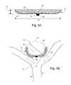
- FIG. 3A-3Billustrates perspective views of an embodiment of the occlusion device disclosed herein.
- FIG. 3Ashows the diameter (x) of the occlusion device having a circumferential fold line and a dual or double layer of mesh material.
- FIG. 3Bshows a dual layer occlusion device disclosed herein deployed in an aneurysm having a diameter (y).
- FIG. 4A-4Cillustrates perspective views of an embodiment of the delivery and/or deployment of an occlusion device disclosed herein.
- FIG. 4Ashows a dual layer occlusion device in its delivery shape.
- FIG. 4Bshows the dual layer occlusion device deploying in a manner in which the compacted ends of the device open in an outward manner.
- FIG. 4Cshows the flattening effect/increase in width/increase in diameter of the dual/double layer of mesh material of the device in its deployed state.
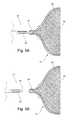
- FIG. 5A-5Billustrates perspective views of an embodiment of the electrolytic delivery and/or deployment and/or detachment of an occlusion device disclosed herein.
- FIG. 5Ashows the delivery of a dual layer occlusion device via a catheter and/or guide wire having electrolytic means.
- FIG. 5Bshows electrolytic detachment of the core wire or guide wire from the occlusion device.
- FIGS. 1-5Exemplary embodiments of the present invention are depicted in FIGS. 1-5 .
- an occlusion device delivery systemcorresponds to (or is compatible with) an occlusion device for deployment thereof.
- occlusion devicemeans and/or may be interchangeable with terminology such as, without limitation, “device” or “occlusion device system” or “occlusion system” or “system” or “occlusion device implant” or “implant” or “intrasaccular implant” and the like.
- Occlusion device delivery systemsare well known and readily available in the art. For example, such delivery technologies may be found, without limitation, in U.S. Pat. Nos. 4,991,602; 5,067,489; 6,833,003; 2006/0167494; and 2007/0288083; each of the teachings of which are incorporated herein.
- any type of occlusion device delivery means and/or delivery system and/or delivery technology and/or delivery mechanism and/or detachment (and/or attachment) means and/or detachment system and/or detachment technology and/or detachment mechanismmay be utilized and/or modified in such a manner as to make compatible (so as to correspond) with the occlusion device disclosed herein.
- Exemplary occlusion device delivery mechanisms and/or systemsinclude, without limitation, guide wires, pusher wires, catheters, micro-catheters, and the like.
- Exemplary occlusion device detachment mechanismsinclude, without limitation, fluid pressure, electrolytic mechanisms, hydraulic mechanisms, interlocking mechanisms, and the like.
- the occlusion device disclosed hereinis used in a method of electrolytic detachment. Electrolytic detachment is well known in the art and can be found, for example, in U.S. Pat. Nos. 5,122,136; 5,423,829; 5,624,449; 5,891,128; 6,123,714; 6,589,230; and 6,620,152.
- FIG. 1A and FIG. 3Ashow an embodiment of an occlusion device as disclosed herein for intrasaccular implantation within an 10 aneurysm to be treated.
- FIG. 1A and FIG. 3Aalso shows the diameter (x) of the 14 resilient mesh body of such an occlusion device in “free air”. As is accepted in the art, the diameter of such an occlusion device is measured in free air. Accordingly, for the purposes of the present invention, and in one embodiment, the 14 resilient mesh body of the occlusion device is “oversized” relative to the 10 aneurysm and therefore has a diameter (x) greater than the diameter (y) of the 10 aneurysm (i.e., ⁇ x> ⁇ y) to be treated as shown in FIGS.
- diameter (y)is the greatest diameter of the 10 aneurysm to be treated or is one of the greater diameters of the 10 aneurysm so long as the 14 mesh body is oversized in such a manner so as to sufficiently seal the 22 neck of the 10 aneurysm to trigger clot formation and/or healing of the 10 aneurysm.
- An exemplary range of the diameter (x) of the occlusion device disclosed hereinis approximately 6-30 millimeters (mm) and an exemplary diameter (y) of the aneurysm to be treated is less than the value of x.
- the diameter (x) of the occlusion deviceis any of 7 mm, 11 mm, and/or 14 mm.
- the position of the 34 distal end of the substantially solid 16 markeris attached approximately equidistantly from the opposing ends of the 14 resilient mesh body. Such a positioning of the 16 marker on the intrasaccular 14 resilient mesh body confers full retrievability of the occlusion device disclosed herein.
- the occlusion device disclosed hereinis “oversized” relative to any vessel to be treated, such as, in pathological conditions in which vessel occlusion is desired, e.g, in peripheral vascular disease.
- the diameter (x) of the occlusion deviceis greater than the diameter (z) of any vessel to be treated so long as the 14 body of the occlusion device is capable of conforming to vessel walls and promoting clot formation.
- FIG. 1B and FIG. 3Bshow an embodiment of an occlusion device as disclosed herein deployed within an 10 aneurysm to be treated.
- FIG. 1B and FIG. 3Bshow the diameter (y) of such an 10 aneurysm to be treated and also shows the blood flow (arrows) in the 12 parent vessel (and basilar artery) adjacent to the 10 aneurysm and its 22 neck.
- the 14 resilient mesh body of the occlusion devicewhen in free air and when deployed, is a “low profile” configuration.
- the terminology “low profile”means that the 14 resilient mesh body, in free air, has a 32 height that is between about 10-20% of its width, and therefore in its deployed shape the 14 resilient mesh body lays flush, in a flattened manner, up against the 10 aneurysm walls and is positioned to cover at least the interior surface of the 20 lower portion of the 10 aneurysm and seal the 22 neck of the 10 aneurysm.
- the occlusion device disclosed hereinis lower and/or slimmer than occlusion devices readily available in the art which expand to fill the space of the 10 aneurysm dome (fully and/or partially with respect to the majority of the space in the 10 aneurysm) and which expand radially and/or which expand in a spherical manner.
- the 14 resilient mesh body, in free airhas a 32 height between about 12-18% of its width.
- the 14 resilient mesh body, in free airhas a 32 height between about 14-16% of its width.
- the 14 resilient mesh body, in free airhas a 32 height of about 15% of its width.
- the deployed shape of the low profile 14 resilient mesh bodycovers between about 40%-80% of the inner surface area of the 10 aneurysm dome. In another embodiment, the deployed shape of the low profile 14 resilient mesh body covers between about 50%-70% of the inner surface area of the 10 aneurysm dome. In another embodiment, the deployed shape of the low profile 14 resilient mesh body covers about 60% of the inner surface area of the 10 aneurysm dome.
- the low profile, winged shaped and/or open-ended expanded spread configuration of the 14 bodyis a single layer of resilient mesh material.
- the low profile, expanded spread configurationis a 24 dual (or double) layer of resilient mesh material.
- such a 14 resilient mesh bodyis “oversized” in comparison to the 10 aneurysm to be treated; and therefore the 14 mesh body has a diameter (x) greater than the diameter (y) of the 10 aneurysm to be treated (i.e., the greatest diameter or one of the greater diameters of the 10 aneurysm to be treated so long as the 14 mesh body is oversized in such a manner so as to sufficiently seal the 22 neck of the 10 aneurysm to trigger clot formation and/or healing of the 10 aneurysm).
- the low profile and oversizing attributes of the 14 resilient mesh bodyconfer its capabilities for conforming to the inner surface of the walls of the 10 aneurysm (via the opposing pressure of the 14 body against the 10 aneurysm walls) and therefore the occlusion device expands in only at least the 20 lower portion (i.e., in a low volume flattened manner) of the 10 aneurysm along the 10 aneurysm walls, thereby eliminating the need for material to pin the 22 neck of the 10 aneurysm and/or to anchor within the 12 parent vessel (and thereby minimizing the need for anti-coagulation therapy).
- the wing-span and/or expanded spread of the 14 bodyconforms to the interior surface of the 10 aneurysm and apposes the 10 aneurysm dome.
- Such a configurationfacilitates sealing of the 22 neck of the 10 aneurysm and therefore clot formation and/or healing and/or shrinkage of the 10 aneurysm which is particularly advantageous if the size or mass of the 10 aneurysm is causing pain or other side effects within the patient.
- Such a configurationis also advantageous because it requires a minimum amount of resilient mesh material thereby eliminating the need to fill or substantially fill, in a spherical, radially expanded manner, the space in the 10 aneurysm dome.
- Such an occlusion deviceis well suited for conformability across a broad range of 10 aneurysm morphologies, particularly since it is well known and generally accepted that 10 aneurysms are not perfectly round in shape. It is also advantageous because an occlusion device as disclosed herein, having a “minimum of” or less material than the current standard devices, minimizes the need for anti-coagulation therapy and/or lessens the risk of clot emboli formation which could flow deeper into the vascular tree inducing stroke.
- the single layer or 24 dual layer of resilient mesh material of the low profile devicecomprises a relatively uniform distribution of wire mesh strands or braids such as, without limitation, a 72 nitinol (NiTi) wire mesh strand braided configuration.
- the occlusion devicecomprises wire mesh strands or braids that range from 36 to 144 NiTi strand braided configuration.
- a 24 dual layer occlusion device disclosed hereinis a configuration of wire mesh which is folded circumferentially ( 26 circumferential fold line) and therefore doubled back on itself.
- the ends of the 24 dual or doubled back layerintersect with the 16 marker positioned approximately at the core of the 14 body of the device.
- the deviceis constructed by circumferentially folding a single layer of mesh material over itself on a preferential 26 fold line effectively resulting in an occlusion device comprising a 24 dual layer of wire mesh material, i.e., the 24 dual layer of mesh comprises a single layer of mesh folded circumferentially ( 26 circumferential fold line).
- this 24 doubled or dual layer of wire mesh materialtriggers a mechanism of action believed to contribute to the enhanced acute thrombogenicity of the device in animal studies. It is believed that the localizing of a small volume of clot between the 24 dual/double layers, which have a high surface area contribution from the wire strands, facilitates nucleating and stabilizing thrombus.
- the 14 body having a folded back 24 dual layeris deeper when compared to a non-deployed 24 dual layer occlusion device accounting for a change in width of approximately 15% which translates to an increase in the diameter (x) of the device when pressure is applied at the 16 marker.
- This change in width/increase in diameter (x)is an effective anchoring feature of the deployed device as blood applies pressure to the mesh 14 body distributed across the 22 neck of the 10 aneurysm.
- Such a configurationalso provides sufficient apposition of the 14 body of the device against the 10 aneurysm wall or vessel wall for peripheral arterial or venous occlusion.
- the device disclosed hereinprovides sufficient mesh density to confer stasis acutely. It is further known, based on analyzing the device in post-deployment that the wire mesh/braid distribution remains relatively uniform.
- FIG. 1B and FIG. 4Balso show the position of the 16 marker, having a 36 proximal end and a 34 distal end, on an occlusion device of the present invention.
- the 34 distal end of the 16 markeris attached to the 14 resilient mesh body of the occlusion device.
- the 36 proximal end of the 16 markeris shown resting across, in the manner of a bridge-like mechanism, the 22 neck of the 10 aneurysm to be treated, which when combined with the properties of the low profile 14 resilient mesh body, eliminates the need for incorporating additional material to pin the 22 neck of the 10 aneurysm and/or as an anchor within the 12 parent vessel and advantageously provides for full retrievability of the device.
- the 16 marker of the occlusion device disclosed hereinis a substantially solid collar or rigid member such as, without limitation a solid ring comprised of materials such as, without limitation, gold, platinum, stainless steel, and/or combinations thereof.
- radiopaque materialssuch as, without limitation, gold, platinum, platinum/iridium alloy, and/or combinations thereof, can be used.
- Such a 16 markerprovides visualization of the device during delivery and placement. The 16 marker is positioned within the occlusion device so that the 36 proximal end of the 16 marker is capable of resting above the 22 neck of an 10 aneurysm.
- the solidness of the 16 markerhelps confer stability of the device within the 10 aneurysm and prevents movement or the transfer of forces through the resilient mesh of the 14 body thereby preventing misplacement or accidental movement of the device.
- the 16 markeris also configured with a junction to cooperate and release from/attach to a corresponding delivery means such as, without limitation, a delivery catheter or guide wire and/or 18 pusher wire technologies. It also advantageously provides for full retrievability of the device disclosed herein.
- the substantially solid 16 markercomprises a radiopaque material (such as for example, without limitation, platinum, gold, platinum/iridium alloy, and/or combinations thereof) to facilitate visualization of the occlusion device under fluoroscopy during delivery, placement and/or deployment.
- the 16 markercomprises a 36 proximal end and a 34 distal end.
- a 14 resilient mesh bodyis attached to the 34 distal end and the 36 proximal end of the 16 marker may be configured to influence shape, diameter, and/or curvature of the 14 resilient mesh body upon expansion of the occlusion device.
- the 16 markermay be designed in various shapes to influence the overall profile of the occlusion device to ensure a proper fit of the expanded/deployed occlusion device within the 10 aneurysm sac.
- FIG. 2A-2C and FIG. 4A-Cshow an exemplary means for delivery and/or deployment, through an 12 artery and/or vessel adjacent to the 10 aneurysm, of the occlusion device disclosed herein.
- the occlusion deviceis delivered in a closed, compacted position (delivery shape) as shown in FIGS. 2A and 4A , such that the low profile 14 body is closed inward on itself or compressed, via a 18 pusher wire mechanism.
- the ends of the low profile 14 resilient mesh bodyopen outward in the manner of the opening of a flower (as shown in FIG. 2B and FIG.
- the opened 14 bodythen conforms to the walls of the 10 aneurysm permitting the 16 marker to rest across the 22 neck and the low profile 14 body to lay flush in a flattened manner (deployed shape) covering at least the 20 lower portion of the 10 aneurysm and sealing the 22 neck of the 10 aneurysm.
- the deviceshows the deepening and/or flattening of the either the single layer ( FIG. 2C ) or 24 dual layer ( FIG. 4C ) device accounting for a change in width and an increase in the diameter (x) of the device when pressure is applied at the 16 marker.
- This change in width/increase in diameter (x)is an effective anchoring feature of the deployed device as blood applies pressure to the 14 body distributed across the 22 neck of the 10 aneurysm.
- Results of animal studies provided hereinsupport that the circumferentially 24 doubled-over/dual-layer configuration provides efficient apposition of the mesh 14 body of the device against the 10 aneurysm wall or vessel for peripheral arterial or venous occlusion.
- FIG. 5A-5Bshow an exemplary means for electrolytic delivery and/or deployment and/or detachment of the occlusion device disclosed herein through an 12 artery and/or vessel adjacent to the 10 aneurysm.
- Electrolytic detachment means and methodssuch as U.S. Pat. No. 5,122,136 are well known in the art.
- a 28 coil-wound core wire (or guide wire) of the catheter (or micro-catheter)is attached inside the 16 marker at its 34 distal end to the 24 dual layer occlusion device disclosed herein (as shown in FIG. 5A ).
- the coil windmaintains a constant diameter ( ⁇ ) so as not to impact upon flexibility or stiffness of the delivery catheter or micro-catheter or guide wire.
- FEPFluorinated Ethylene Propylene heat shrink tubing encases the 28 coil-wound portion of the core wire.
- Numerous readily available and well known attachment techniques in the medical device artscan be used to attach the distal end of the core wire inside the 16 marker band and to the occlusion device or implant. Such attachment techniques include, without limitation, adhesives, laser melting, laser tack, spot, and/or continuous welding.
- an adhesiveis used to attach the distal end of the core wire inside the 16 marker band.
- the adhesiveis an epoxy material which is cured or hardened through the application of heat or UV (ultra-violet) radiation.
- the epoxyis a thermal cured, two-part epoxy such as EPO-TEK® 353ND-4 available from Epoxy Technology, Inc., 14 Fortune Drive, Billerica, Mass.
- EPO-TEK® 353ND-4available from Epoxy Technology, Inc., 14 Fortune Drive, Billerica, Mass.
- Such an adhesive or epoxy materialencapsulates the junction of the core wire inside the marker band and increases its mechanical stability.
- the 28 coil-wound core wiredetaches the 24 dual layer occlusion device disclosed herein at an 30 electrolytic detachment site (or zone) on the core wire itself in such a manner so that the core wire is severed and/or dissolved through electrolytic action at the base of the 16 marker band (as shown in FIG. 5B ). Such action then releases and/or places the 24 dual layer occlusion device into an aneurysm or vessel to be treated.
- the low profile 14 resilient mesh body of the occlusion device disclosed hereincan be filled with an embolic material to promote clotting and closure of the 10 aneurysm.
- the oversized occlusion device disclosed hereinmay further incorporate adjunctive elements and/or members such as coiling techniques, framing coils, embolic agents, additional markers, polymers, resorbent polymers and/or a combination thereof.
- Resilient mesh materials for design and/or manufacture of occlusion devicesare readily available and well known by those skilled in the relevant art.
- resilient mesh materialsrange from a wide variety of available materials such as, without limitation, nickel titanium (nitinol or otherwise known as NiTi), stainless steel, polymers, and/or combinations thereof.
- Exemplary known biomedical polymeric familiesinclude, without limitation, polymers such as polyphosphazenes, polyanhydrides, polyacetals, poly(ortho esters), polyphosphoesters, polycaprolactones, polyurethanes, polylactides, polycarbonates, polyamides, and/or a combination thereof (See, e.g., J Polym Sci B Polym Phys. Author manuscript; available in PMC 2012 Jun. 15.)
- the resilient mesh materialis formed of woven strands of polymer material, such as, without limitation, nylon, polypropylene or polyester.
- the polymer strandscan be filled with a radiopaque material which allows the physician treating the aneurysm to fluoroscopically visualize the location of the device within the vasculature.
- Radiopaque filler materialspreferably include bismuth trioxide, tungsten, titanium dioxide or barium sulfate, or radiopaque dyes such as iodine.
- the resilient mesh materialcan be formed by strands of radiopaque material. The radiopaque strands allow the physician and/or radiologist to fluoroscopically visualize the location of the mesh, without the use of filled polymer materials.
- radiopaque strandsmay be formed with materials such as, without limitation, gold, platinum, a platinum/iridium alloy, and/or a combination thereof.
- the resilient mesh materialis constructed of 10%-20% platinum core NiTi.
- the resilient mesh materialis constructed of 10% platinum core NiTi, 15% platinum core NiTi, or 20% platinum core NiTi. 10% platinum core NiTi construction is sufficient to provide a ghost image of the occlusion device under x-ray.
- DFT® wireis a metal-to-metal composite constructed to combine the desired physical and mechanical attributes of two or more materials into a single wire.
- the NiTi outer layeris able to provide the resulting composite wire with similar mechanical properties of a 100% NiTi wire.
- DFT® wiresare available from Fort Wayne Metals Corp., Fort Wayne, Ind., U.S.A. See also, for example, the journal article entitled Biocompatible Wire by Schaffer in Advanced Materials & Processes, October 2002, pages 51-54, incorporated herein by reference.
- the strandsmay be covered with a polymer coating or extrusion.
- the coating or extrusion over the radiopaque wire strandsprovides fluoroscopic visualization but also increases the resistance of the strands to bending fatigue and may also increase lubricity of the strands.
- the polymer coating or extrusionin one embodiment, is coated or treated with an agent which tends to resist clotting, such as heparin. Such clot resistant coatings are generally known.
- the polymer coating or extrusioncan be any suitable extrudable polymer, or any polymer that can be applied in a thin coating, such as Teflon® or polyurethane.
- the strands of the resilient mesh materialare formed using both metal and polymer braided strands. Combining the metal strands with the polymer strands into a braid changes the flexibility characteristics of mesh. The force required to deploy and/or collapse such a mesh portion is significantly reduced over that required for a mesh portion that includes only metal mesh strands. However, the radiopaque characteristics of the mesh for fluoroscopic visualization are retained.
- Metal strands forming such a deviceincludes, without limitation, stainless steel, gold, platinum, platinum/iridium, nitinol, and/or combinations thereof.
- Polymer strands forming the devicecan include nylon, polypropylene, polyester, Teflon®, and/or combinations thereof.
- polymer strands of the mesh materialcan be chemically modified to make them radiopaque with known techniques such as, without limitation, by using gold deposition onto the polymer strands, or by using ion beam plasma deposition of suitable metal ions onto the polymer strands.
- the resilient mesh materialcan also be formed with filaments or strands of varying diameter and/or varying flexibility. By varying the size or flexibility of the polymer strands, the flexibility characteristics of the mesh, upon deployment, can also be varied. By varying the flexibility characteristics, both the deployed and collapsed configuration of the 14 resilient mesh body can be varied or changed to substantially any desired shape.
- the meshbe formed of both polymer strands or filaments and metal strands or filaments, but it can be formed using filaments of different polymer materials.
- different polymer materialshaving different flexibility characteristics can be used in forming the mesh. This alters the flexibility characteristics to change the resultant configuration of the 14 mesh body in both the deployed and the collapsed positions.
- biomedical polymersare readily known and available in the art and can be derived from polymeric families such as, without limitation, polyphosphazenes, polyanhydrides, polyacetals, poly (ortho esters), polyphosphoesters, polycaprolactones, polyurethanes, polylactides, polycarbonates, polyamides, and/or a combination thereof.
- Resilient mesh materials suitable for use within the 14 mesh bodymay take the form of a flat woven sheet, knitted sheet, or a laser cut wire mesh.
- the materialshould include two or more sets of substantially parallel strands, with one set of parallel strands being at a pitch of between 45 degrees and 135 degrees with respect to the other set of parallel strands.
- the two sets of parallel strands forming the mesh materialare substantially perpendicular to each other.
- the pitch and general construction of the mesh materialmay be optimized to meet the performance needs of the occlusion device.
- the wire strands of the metal fabric used in the present inventionshould be formed of a material which is both resilient and can be heat-treated to substantially set a desired shape.
- Materials which are believed to be suitable for this purposeinclude a cobalt-based low thermal expansion alloy referred to in the field of occlusion devices as Elgiloy®, nickel-based high-temperature high-strength “superalloys” commercially available from Haynes International under the trade name Hastelloy®, nickel-based heat treatable alloys sold under the name Incoloy® by International Nickel, and a number of different grades of stainless steel.
- the important factor in choosing a suitable material for the wiresis that the wires retain a suitable amount of the deformation induced by the molding surface (or shape memory, as described below) when subjected to a predetermined heat treatment.
- shape memory alloystend to have a temperature induced phase change which will cause the material to have a preferred configuration which can be fixed by heating the material above a certain transition temperature to induce a change in the phase of the material. When the alloy is cooled, the alloy will “remember” the shape it was in during the heat treatment and will tend to assume that same and/or similar configuration unless constrained from doing so.
- NiTi alloyssuch as nitinol, including appropriate compositions and handling requirements, are well known in the art and such alloys need not be discussed in detail here.
- U.S. Pat. Nos. 5,067,489 and 4,991, 602the teachings of which are incorporated herein by reference, discuss the use of shape memory NiTi alloys in guide wire-based technologies.
- Such NiTi alloysare preferred, at least in part, because they are commercially available and more is known about handling such alloys than other known shape memory alloys.
- NiTi alloysare also very elastic. Indeed, they are said to be known as “superelastic” or “pseudoelastic.” This elasticity will help an occlusion device as disclosed herein return to prior expanded configuration for deployment thereof.
- the wire strandscan comprise a standard monofilament of the selected material, i.e., a standard wire stock may be used. In some embodiments, 72 wire strands and/or 72 strand braid configuration may be used. In other embodiments, the occlusion device comprises wire mesh strands or braids that range from 36 to 144 NiTi strand braided configurations. If so desired, though, the individual wire strands may be formed from “cables” made up of a plurality of individual wires. For example, cables formed of metal wires where several wires are helically wrapped about a central wire are commercially available and NiTi cables having an outer diameter of 0.003 inches or less can be purchased. One advantage of certain cables is that they tend to be “softer” than the monofilament wires having the same diameter and formed of same material. Additionally, the use of a cable can increase the effective surface area of the wire strand, which will tend to promote thrombosis.
- An occlusion device disclosed hereinis configured with low profile resilient mesh material of a mesh density sufficient for functioning in such a manner as an endothelial cell scaffold within a vessel or across the 22 neck of the 10 aneurysm and thereby reducing blood flow by about 60% to trigger clot formation and/or healing of the 10 aneurysm.
- the terminology “mesh density”means the level of porosity or the ratio of metal to open area of the 14 mesh body. Mesh density relates to the number and size of the openings or pores of the mesh and by the extent that the pores are open or closed in situations where opening or pore openness varies between delivery and deployment.
- a high mesh density region of a resilient mesh materialhas approximately about 40% or more metal area and about 60% or less open area.
- the 14 resilient mesh bodymay be formed uniformly of the same material; however such material may have different knitted, stitched, braided, and/or cut construction.
- the implantable occlusion device disclosed hereincan be used for the process of peripheral vascular embolization (a process well known in the art and known to involve the shutdown of blood flow distal to a specified vascular point), for example, in the treatment and/or amelioration of peripheral arterial or venous pathologies and/or any related pathologies requiring vessel occlusion for the treatment thereof.
- the occlusion device of the present inventionmay incorporate reasonable design parameters, features, modifications, advantages, and variations that are readily apparent to those skilled in the art in the field of occlusion devices.
- the rabbit elastase aneurysm modelis a well-accepted and art-recognized model for testing novel neurointerventional devices and has been the subject of a number of clinical publications regarding efficacy and similarity to human response. (See, e.g., Altes et al. Creation of Saccular Aneurysms in the Rabbit: A Model Suitable for Testing Endovascular Devices. AJR 2000; 174: 349-354.) It therefore is readily accepted by the regulatory agencies as an appropriate test model. The model's coagulation system is highly similar to that of humans.
- the modelhas advantageous anatomical aspects in that the diameters of the rabbits' extra-cranial carotid arteries are highly similar to the diameter of extra-cranial carotid arteries in humans. Moreover, elastase-induced aneurysms have been shown to behave in a histologically similar manner as human aneurysms.
- Non-detachable occlusion device lot 30680Detachable occlusion device lot 30676, Aneurysm size 4.5 millimeters (mm) (height) ⁇ 2.5 width
- the non-detachable occlusion devicewas positioned into the aneurysm with the marker at the neck of the aneurysm and contrast runs performed at timed intervals. Immediately after deployment of the device, it was observed to be in a good position in the aneurysm and there was some slowing of flow. At 5 minutes post deployment, there was some stasis observed in the aneurysm. At 10 minutes post deployment, further stagnation in the aneurysm was observed and the device was repositioned closer to the aneurysm neck. At 15 minutes post deployment, stagnation of flow in the aneurysm was observed. When the device was removed from the aneurysm and heparin was given, flow into the aneurysm returned to pre-deployment status.
- the non-detachable occlusion devicewas then removed and a 7 mm diameter occlusion device advanced into a 0.027′′ lumen microcatheter (ExcelsiorXT27, Stryker) using a 0.014′′ guide wire (Synchro2, Stryker). Advancement of the device in the catheter was noted to be smooth with low friction.
- the occlusion devicewas advanced to the neck of the aneurysm and deployed. Timed angiographic runs were performed. Immediately post deployment, there was stasis of flow observed in the aneurysm. At 5 minutes post deployment, a filling defect in the aneurysm was observed. At 10 minutes post deployment, thrombus in the aneurysm was observed. At 20 minutes post deployment, the 5F catheter removed. On completion of the procedure the animal was euthanized in accordance with the Standard Operating Procedure (SOP).
- SOPStandard Operating Procedure
- Non-detachable occlusion device lot 30680Detachable occlusion device lot 30676, Aneurysm size 10 mm height ⁇ 4 mm wide ⁇ 3 mm neck
- Example IIA similar procedure to Example I was performed with the placement of the non-detachable device into the aneurysm neck.
- a 4F systemwas used to introduce the device into the 5F sheath and a “step” on the internal hub of the catheter was noted to cause the device to catch.
- the devicewas placed and timed angiographic runs obtained as before.
- Immediately post deploymentsome flow reduction was observed in the aneurysm.
- a filling defectwas observed in the aneurysm sac.
- an increase in size of the filling defectwas observed.
- This devicewas removed, angiography demonstrated that flow had returned to pre-deployment status in the aneurysm, and an implantable (detached) device was deployed using the same method as previously.
- the implantrequired some force initially to transition into the microcatheter from the loading sheath (possibly due to poor sheath to hub lumen compatibility) but once inserted, the microcatheter advanced freely.
- the devicewas noted to have reasonable deployment control despite not being fixed to a detachment mechanism. Positioning was achieved to cover the neck of the aneurysm and timed angiographic runs obtained. Immediately post deployment, thrombosis was observed at a distal portion of the aneurysm. At 5 minutes post deployment, virtual occlusion of the aneurysm sac was observed. At 10 minutes post deployment, complete occlusion of the aneurysm sac was observed. At 15 minutes post deployment, occlusion of the aneurysm distal to the device marker was observed.
- ACTActivated Clotting Time
- Detachable device lot 30676Aneurysm size 6.5 mm ⁇ 3.1 mm width ⁇ 2.4 mm neck
- This series of angiogramsconfirm the wire mesh braid configuration of the occlusion device disclosed herein is sufficiently dense to reduce blood flow in the aneurysm leading to stasis of blood and thrombosis in the aneurysm sac.
- Manipulation and deployment control of the occlusion devicewere carried out while visualizing the proximal radiopaque marker of the device in relation to the catheter tip.
- Device developmentwill entail the incorporation of radiopaque struts of platinum core NiTi wire to aid in visibility.
- the occlusion device in the animal studieswas of limited expansion (7 mm). Device development will incorporate increased diameters of greater than 7 mm. Accordingly, such devices have been designed with diameters of 11 mm and 14 mm. Even so, despite limitations with expansion spread of the 7 mm devices, all deployments promoted stasis in the aneurysms and all devices were easy to manipulate with pinpoint accuracy, particularly in relation to guidance through the parent arteries and neck of the aneurysms and placement within the aneurysms across the neck of the aneurysms.
Landscapes
- Health & Medical Sciences (AREA)
- Surgery (AREA)
- Life Sciences & Earth Sciences (AREA)
- Heart & Thoracic Surgery (AREA)
- Molecular Biology (AREA)
- Veterinary Medicine (AREA)
- Engineering & Computer Science (AREA)
- Biomedical Technology (AREA)
- Public Health (AREA)
- Medical Informatics (AREA)
- Nuclear Medicine, Radiotherapy & Molecular Imaging (AREA)
- Animal Behavior & Ethology (AREA)
- General Health & Medical Sciences (AREA)
- Reproductive Health (AREA)
- Vascular Medicine (AREA)
- Neurosurgery (AREA)
- Oral & Maxillofacial Surgery (AREA)
- Pathology (AREA)
- Surgical Instruments (AREA)
Abstract
Description
Claims (15)
Priority Applications (5)
| Application Number | Priority Date | Filing Date | Title |
|---|---|---|---|
| US14/699,188US10130372B2 (en) | 2014-04-30 | 2015-04-29 | Occlusion Device |
| US16/172,157US11284901B2 (en) | 2014-04-30 | 2018-10-26 | Occlusion device |
| US16/590,821US11389174B2 (en) | 2014-04-30 | 2019-10-02 | Occlusion device |
| US17/847,856US12029431B2 (en) | 2014-04-30 | 2022-06-23 | Occlusion device |
| US18/739,062US12414775B1 (en) | 2014-04-30 | 2024-06-10 | Occlusion device |
Applications Claiming Priority (3)
| Application Number | Priority Date | Filing Date | Title |
|---|---|---|---|
| US201461986369P | 2014-04-30 | 2014-04-30 | |
| US201462083672P | 2014-11-24 | 2014-11-24 | |
| US14/699,188US10130372B2 (en) | 2014-04-30 | 2015-04-29 | Occlusion Device |
Related Child Applications (1)
| Application Number | Title | Priority Date | Filing Date |
|---|---|---|---|
| US16/172,157ContinuationUS11284901B2 (en) | 2014-04-30 | 2018-10-26 | Occlusion device |
Publications (2)
| Publication Number | Publication Date |
|---|---|
| US20150313605A1 US20150313605A1 (en) | 2015-11-05 |
| US10130372B2true US10130372B2 (en) | 2018-11-20 |
Family
ID=53055025
Family Applications (5)
| Application Number | Title | Priority Date | Filing Date |
|---|---|---|---|
| US14/699,188ActiveUS10130372B2 (en) | 2014-04-30 | 2015-04-29 | Occlusion Device |
| US16/172,157Active2035-05-31US11284901B2 (en) | 2014-04-30 | 2018-10-26 | Occlusion device |
| US16/590,821Active2036-09-07US11389174B2 (en) | 2014-04-30 | 2019-10-02 | Occlusion device |
| US17/847,856Active2035-06-30US12029431B2 (en) | 2014-04-30 | 2022-06-23 | Occlusion device |
| US18/739,062ActiveUS12414775B1 (en) | 2014-04-30 | 2024-06-10 | Occlusion device |
Family Applications After (4)
| Application Number | Title | Priority Date | Filing Date |
|---|---|---|---|
| US16/172,157Active2035-05-31US11284901B2 (en) | 2014-04-30 | 2018-10-26 | Occlusion device |
| US16/590,821Active2036-09-07US11389174B2 (en) | 2014-04-30 | 2019-10-02 | Occlusion device |
| US17/847,856Active2035-06-30US12029431B2 (en) | 2014-04-30 | 2022-06-23 | Occlusion device |
| US18/739,062ActiveUS12414775B1 (en) | 2014-04-30 | 2024-06-10 | Occlusion device |
Country Status (9)
| Country | Link |
|---|---|
| US (5) | US10130372B2 (en) |
| EP (3) | EP3970635A1 (en) |
| JP (5) | JP6571760B2 (en) |
| CN (1) | CN106456183B (en) |
| CA (1) | CA2946078C (en) |
| ES (2) | ES2732752T3 (en) |
| IL (1) | IL248515A0 (en) |
| MX (2) | MX381828B (en) |
| WO (1) | WO2015166013A1 (en) |
Cited By (45)
| Publication number | Priority date | Publication date | Assignee | Title |
|---|---|---|---|---|
| US20200113576A1 (en)* | 2018-10-12 | 2020-04-16 | DePuy Synthes Products, Inc. | Folded aneurysm treatment device and delivery method |
| US10905430B2 (en) | 2018-01-24 | 2021-02-02 | DePuy Synthes Products, Inc. | Aneurysm device and delivery system |
| US10939915B2 (en) | 2018-05-31 | 2021-03-09 | DePuy Synthes Products, Inc. | Aneurysm device and delivery system |
| US11058430B2 (en) | 2018-05-25 | 2021-07-13 | DePuy Synthes Products, Inc. | Aneurysm device and delivery system |
| US11076860B2 (en) | 2014-03-31 | 2021-08-03 | DePuy Synthes Products, Inc. | Aneurysm occlusion device |
| WO2021165443A2 (en) | 2020-02-20 | 2021-08-26 | Cerus Endovascular Limited | Clot removal distal protection methods |
| US11123077B2 (en) | 2018-09-25 | 2021-09-21 | DePuy Synthes Products, Inc. | Intrasaccular device positioning and deployment system |
| US11134953B2 (en) | 2019-02-06 | 2021-10-05 | DePuy Synthes Products, Inc. | Adhesive cover occluding device for aneurysm treatment |
| US11154302B2 (en) | 2014-03-31 | 2021-10-26 | DePuy Synthes Products, Inc. | Aneurysm occlusion device |
| US11272939B2 (en) | 2018-12-18 | 2022-03-15 | DePuy Synthes Products, Inc. | Intrasaccular flow diverter for treating cerebral aneurysms |
| US11278292B2 (en) | 2019-05-21 | 2022-03-22 | DePuy Synthes Products, Inc. | Inverting braided aneurysm treatment system and method |
| US11284901B2 (en) | 2014-04-30 | 2022-03-29 | Cerus Endovascular Limited | Occlusion device |
| US11304700B2 (en)* | 2017-08-22 | 2022-04-19 | Covidien Lp | Devices, systems, and methods for the treatment of vascular defects |
| US11337706B2 (en) | 2019-03-27 | 2022-05-24 | DePuy Synthes Products, Inc. | Aneurysm treatment device |
| US11357511B2 (en) | 2008-05-01 | 2022-06-14 | Aneuclose Llc | Intrasacular aneurysm occlusion device with globular first configuration and bowl-shaped second configuration |
| US11406392B2 (en) | 2018-12-12 | 2022-08-09 | DePuy Synthes Products, Inc. | Aneurysm occluding device for use with coagulating agents |
| US11413046B2 (en) | 2019-05-21 | 2022-08-16 | DePuy Synthes Products, Inc. | Layered braided aneurysm treatment device |
| US11457926B2 (en) | 2019-12-18 | 2022-10-04 | DePuy Synthes Products, Inc. | Implant having an intrasaccular section and intravascular section |
| US11464518B2 (en) | 2008-05-01 | 2022-10-11 | Aneuclose Llc | Proximal concave neck bridge with central lumen and distal net for occluding cerebral aneurysms |
| US11471163B2 (en) | 2008-05-01 | 2022-10-18 | Aneuclose Llc | Intrasaccular aneurysm occlusion device with net or mesh expanded by string-of-pearls embolies |
| US11471164B2 (en) | 2008-05-01 | 2022-10-18 | Aneuclose Llc | Methods of occluding a cerebral aneurysm by inserting embolic members or material into an intrasacular implant |
| US11484322B2 (en) | 2018-01-03 | 2022-11-01 | Aneuclose Llc | Aneurysm neck bridge with a closeable opening or lumen through which embolic material is inserted into the aneurysm sac |
| US11497504B2 (en) | 2019-05-21 | 2022-11-15 | DePuy Synthes Products, Inc. | Aneurysm treatment with pushable implanted braid |
| US11504816B2 (en) | 2019-11-04 | 2022-11-22 | Covidien Lp | Systems and methods for treating aneurysms |
| US11583288B2 (en) | 2018-08-08 | 2023-02-21 | DePuy Synthes Products, Inc. | Delivery of embolic braid |
| US11583289B2 (en) | 2008-05-01 | 2023-02-21 | Aneuclose Llc | Aneurysm-occluding mesh ribbon with a series of loops or segments having distal-to-proximal variation in size, shape, and/or orientation |
| US11583282B2 (en) | 2019-05-21 | 2023-02-21 | DePuy Synthes Products, Inc. | Layered braided aneurysm treatment device |
| US11596412B2 (en)* | 2018-05-25 | 2023-03-07 | DePuy Synthes Products, Inc. | Aneurysm device and delivery system |
| US11602350B2 (en) | 2019-12-05 | 2023-03-14 | DePuy Synthes Products, Inc. | Intrasaccular inverting braid with highly flexible fill material |
| US11607226B2 (en) | 2019-05-21 | 2023-03-21 | DePuy Synthes Products, Inc. | Layered braided aneurysm treatment device with corrugations |
| US11648013B2 (en) | 2016-03-11 | 2023-05-16 | Cerus Endovascular Limited | Occlusion device |
| US11672542B2 (en) | 2019-05-21 | 2023-06-13 | DePuy Synthes Products, Inc. | Aneurysm treatment with pushable ball segment |
| US11672543B2 (en) | 2017-02-23 | 2023-06-13 | DePuy Synthes Products, Inc. | Aneurysm method and system |
| US11690628B2 (en) | 2012-11-13 | 2023-07-04 | Covidien Lp | Occlusive devices |
| US11813282B2 (en) | 2016-10-21 | 2023-11-14 | Covidien Lp | Injectable scaffold for treatment of intracranial aneurysms and related technology |
| US11812971B2 (en) | 2017-08-21 | 2023-11-14 | Cerus Endovascular Limited | Occlusion device |
| US12004750B2 (en) | 2008-05-01 | 2024-06-11 | Aneuclose Llc | Methods for creating an expandable two-part intrasacular aneurysm occlusion device from a tubular mesh |
| US12023034B2 (en) | 2020-03-11 | 2024-07-02 | Microvention, Inc. | Devices for treatment of vascular defects |
| US12064364B2 (en) | 2018-09-18 | 2024-08-20 | Nanostructures, Inc. | Catheter based methods and devices for obstructive blood flow restriction |
| US12070220B2 (en) | 2020-03-11 | 2024-08-27 | Microvention, Inc. | Devices having multiple permeable shells for treatment of vascular defects |
| US12082821B2 (en) | 2008-05-02 | 2024-09-10 | Microvention, Inc. | Filamentary devices for treatment of vascular defects |
| US12082819B2 (en) | 2019-03-15 | 2024-09-10 | Microvention, Inc. | Filamentary devices for treatment of vascular defects |
| US12096940B2 (en) | 2013-08-16 | 2024-09-24 | Microvention, Inc. | Filamentary devices for treatment of vascular defects |
| US12303136B2 (en) | 2016-05-26 | 2025-05-20 | Nanostructures, Inc. | System and methods for embolized occlusion of neurovascular aneurysms |
| US12408925B2 (en) | 2020-03-11 | 2025-09-09 | Microvention, Inc. | Multiple layer devices for treatment of vascular defects |
Families Citing this family (49)
| Publication number | Priority date | Publication date | Assignee | Title |
|---|---|---|---|---|
| US10716573B2 (en) | 2008-05-01 | 2020-07-21 | Aneuclose | Janjua aneurysm net with a resilient neck-bridging portion for occluding a cerebral aneurysm |
| US9526503B2 (en)* | 2013-08-12 | 2016-12-27 | W. L. Gore & Associates, Inc. | Lumbar ostia occlusion devices and methods of deploying the same |
| US11484319B2 (en) | 2015-01-20 | 2022-11-01 | Neurogami Medical, Inc. | Delivery system for micrograft for treating intracranial aneurysms |
| CA2972620C (en) | 2015-01-20 | 2023-08-01 | Neurogami Medical, Inc. | Micrograft for the treatment of intracranial aneurysms and method for use |
| US10736730B2 (en) | 2015-01-20 | 2020-08-11 | Neurogami Medical, Inc. | Vascular implant |
| US10857012B2 (en) | 2015-01-20 | 2020-12-08 | Neurogami Medical, Inc. | Vascular implant |
| US10925611B2 (en) | 2015-01-20 | 2021-02-23 | Neurogami Medical, Inc. | Packaging for surgical implant |
| CA2976260C (en) | 2015-02-25 | 2024-02-06 | Galaxy Therapeutics, Llc | System for and method of treating aneurysms |
| US10548579B2 (en)* | 2015-07-29 | 2020-02-04 | Cardiac Pacemakers, Inc. | Left atrial appendage implant |
| EP4011303B1 (en) | 2015-12-07 | 2024-06-12 | Cerus Endovascular Limited | Occlusion device |
| CN108472042B (en)* | 2015-12-18 | 2021-03-16 | 斯瑞克公司 | Vascular occlusion devices and delivery assemblies |
| AU2017218115B2 (en)* | 2016-02-10 | 2020-03-05 | Microvention, Inc. | Devices for vascular occlusion |
| WO2017214431A2 (en)* | 2016-06-10 | 2017-12-14 | Stryker Corporation | Braided medical devices |
| JP6946450B2 (en)* | 2017-03-13 | 2021-10-06 | ボストン サイエンティフィック サイムド,インコーポレイテッドBoston Scientific Scimed,Inc. | Occlusion medical device system |
| US10751065B2 (en)* | 2017-12-22 | 2020-08-25 | DePuy Synthes Products, Inc. | Aneurysm device and delivery system |
| US11185335B2 (en)* | 2018-01-19 | 2021-11-30 | Galaxy Therapeutics Inc. | System for and method of treating aneurysms |
| CN111936063B (en)* | 2018-01-31 | 2024-08-20 | 纳米结构公司 | Vascular occlusion device utilizing thin film nitinol foil |
| EP3773251A1 (en) | 2018-04-10 | 2021-02-17 | Medstar Health | Embolization scaffold devices |
| IL269005A (en)* | 2018-09-12 | 2019-10-31 | Depuy Synthes Products Inc | Improved aneurysm occlusion device |
| US11730485B2 (en) | 2018-12-17 | 2023-08-22 | Covidien Lp | Devices, systems, and methods for the treatment of vascular defects |
| CN109745094B (en)* | 2018-12-29 | 2021-09-03 | 先健科技(深圳)有限公司 | Plugging device |
| WO2020150023A1 (en)* | 2019-01-18 | 2020-07-23 | Neurogami Medical, Inc. | Vascular implant |
| CN109965930B (en)* | 2019-04-29 | 2024-05-31 | 北京久事神康医疗科技有限公司 | Aneurysm embolism device and aneurysm embolism system |
| KR20200134171A (en)* | 2019-05-21 | 2020-12-01 | 디퍼이 신테스 프로덕츠, 인코포레이티드 | Layered braided aneurysm treatment device |
| US11202636B2 (en) | 2019-05-25 | 2021-12-21 | Galaxy Therapeutics Inc. | Systems and methods for treating aneurysms |
| US12102327B2 (en) | 2019-05-25 | 2024-10-01 | Galaxy Therapeutics, Inc. | Systems and methods for treating aneurysms |
| CN114126540A (en) | 2019-07-17 | 2022-03-01 | 波士顿科学医学有限公司 | Continuously covered left atrial appendage implant |
| EP3771437A1 (en)* | 2019-07-29 | 2021-02-03 | Université de Montpellier | Intra-aneurysm device |
| EP4563099A3 (en) | 2019-08-30 | 2025-08-06 | Boston Scientific Scimed, Inc. | Left atrial appendage implant with sealing disk |
| WO2021195085A1 (en) | 2020-03-24 | 2021-09-30 | Boston Scientific Scimed, Inc. | Medical system for treating a left atrial appendage |
| US11931041B2 (en) | 2020-05-12 | 2024-03-19 | Covidien Lp | Devices, systems, and methods for the treatment of vascular defects |
| JP7627761B2 (en) | 2020-11-30 | 2025-02-06 | ボストン サイエンティフィック サイムド,インコーポレイテッド | Implantable passive mean pressure sensor |
| WO2022149141A1 (en) | 2021-01-10 | 2022-07-14 | Luseed Vascular Ltd. | Intravascular device |
| WO2022164957A1 (en)* | 2021-01-27 | 2022-08-04 | Galaxy Therapeutics, Inc. | Systems and methods for treating aneurysms |
| US12383201B2 (en) | 2021-02-03 | 2025-08-12 | Boston Scientific Scimed, Inc. | Medical system for treating a left atrial appendage |
| CN113017950A (en)* | 2021-02-08 | 2021-06-25 | 北京联合大学 | Hemispherical braided stent and manufacturing method thereof |
| EP4308013A1 (en) | 2021-03-16 | 2024-01-24 | Covidien LP | Injectable biopolymer compositions and associated systems and methods |
| CN113069168B (en)* | 2021-04-07 | 2024-01-23 | 上海微密医疗科技有限公司 | Aneurysm plugging device |
| CN113069170A (en)* | 2021-04-30 | 2021-07-06 | 上海暖阳医疗器械有限公司 | Aneurysm occlusion device |
| CN113274087B (en)* | 2021-05-24 | 2022-08-26 | 北京泰杰伟业科技有限公司 | Flow disturbing device in tumor |
| EP4358872A1 (en) | 2021-06-22 | 2024-05-01 | Boston Scientific Scimed, Inc. | Left atrial appendage implant |
| CN117615720A (en) | 2021-07-08 | 2024-02-27 | 波士顿科学医学有限公司 | Left auricle closing device |
| WO2023038929A1 (en) | 2021-09-08 | 2023-03-16 | Boston Scientific Scimed, Inc. | Occlusive implant with multi-sharpness split tip soft tissue anchors |
| CN113855141A (en)* | 2021-09-28 | 2021-12-31 | 北京泰杰伟业科技有限公司 | A tumor neck closure device |
| CN113796911A (en)* | 2021-10-09 | 2021-12-17 | 上海励楷科技有限公司 | Bifurcation aneurysm occlusion device |
| CN113951964B (en)* | 2021-11-05 | 2023-11-17 | 聚辉医疗科技(深圳)有限公司 | Embolic coil and coil system |
| CN114129217A (en)* | 2021-12-31 | 2022-03-04 | 江苏暖阳医疗器械有限公司 | Aneurysm internal blocking device of tectorial membrane |
| US12070221B2 (en)* | 2022-07-30 | 2024-08-27 | Covidien Lp | Devices, systems, and methods for treating aneurysms |
| CN115607221B (en)* | 2022-12-19 | 2023-03-03 | 上海微密医疗科技有限公司 | Aneurysm internal plugging device and aneurysm internal plugging system |
Citations (149)
| Publication number | Priority date | Publication date | Assignee | Title |
|---|---|---|---|---|
| US2849002A (en) | 1956-03-12 | 1958-08-26 | Vincent J Oddo | Haemostatic catheter |
| US3480017A (en) | 1966-04-27 | 1969-11-25 | Wallace B Shute | Cervical dilator |
| US4364392A (en) | 1980-12-04 | 1982-12-21 | Wisconsin Alumni Research Foundation | Detachable balloon catheter |
| US4395806A (en) | 1980-05-08 | 1983-08-02 | Sorenson Research Co., Inc. | Method of manufacturing a detachable balloon catheter assembly |
| US4545367A (en) | 1982-07-16 | 1985-10-08 | Cordis Corporation | Detachable balloon catheter and method of use |
| US4836204A (en) | 1987-07-06 | 1989-06-06 | Landymore Roderick W | Method for effecting closure of a perforation in the septum of the heart |
| US4991602A (en) | 1989-06-27 | 1991-02-12 | Flexmedics Corporation | Flexible guide wire with safety tip |
| US5002556A (en) | 1986-11-29 | 1991-03-26 | Terumo Kabushiki Kaisha | Balloon catheter assembly |
| US5025060A (en) | 1990-10-15 | 1991-06-18 | Kansai Paint Co., Ltd. | Dispersion of fine particles of a polymer |
| US5065772A (en) | 1989-10-13 | 1991-11-19 | Inamed Corporation | Inflatable cerivical pessary |
| US5067489A (en) | 1988-08-16 | 1991-11-26 | Flexmedics Corporation | Flexible guide with safety tip |
| US5122136A (en) | 1990-03-13 | 1992-06-16 | The Regents Of The University Of California | Endovascular electrolytically detachable guidewire tip for the electroformation of thrombus in arteries, veins, aneurysms, vascular malformations and arteriovenous fistulas |
| US5192301A (en) | 1989-01-17 | 1993-03-09 | Nippon Zeon Co., Ltd. | Closing plug of a defect for medical use and a closing plug device utilizing it |
| US5423829A (en) | 1993-11-03 | 1995-06-13 | Target Therapeutics, Inc. | Electrolytically severable joint for endovascular embolic devices |
| US5624449A (en) | 1993-11-03 | 1997-04-29 | Target Therapeutics | Electrolytically severable joint for endovascular embolic devices |
| US5733294A (en) | 1996-02-28 | 1998-03-31 | B. Braun Medical, Inc. | Self expanding cardiovascular occlusion device, method of using and method of making the same |
| US5891128A (en) | 1994-12-30 | 1999-04-06 | Target Therapeutics, Inc. | Solderless electrolytically severable joint for detachable devices placed within the mammalian body |
| US5928260A (en) | 1997-07-10 | 1999-07-27 | Scimed Life Systems, Inc. | Removable occlusion system for aneurysm neck |
| US6007573A (en) | 1996-09-18 | 1999-12-28 | Microtherapeutics, Inc. | Intracranial stent and method of use |
| US6024756A (en) | 1996-03-22 | 2000-02-15 | Scimed Life Systems, Inc. | Method of reversibly closing a septal defect |
| EP1003422A1 (en) | 1997-08-05 | 2000-05-31 | Boston Scientific Limited | Detachable aneurysm neck bridge |
| US6080191A (en) | 1992-06-18 | 2000-06-27 | American Biomed, Inc. | Method for making a stent |
| US6096021A (en) | 1998-03-30 | 2000-08-01 | The University Of Virginia Patent Foundation | Flow arrest, double balloon technique for occluding aneurysms or blood vessels |
| US6113609A (en) | 1998-05-26 | 2000-09-05 | Scimed Life Systems, Inc. | Implantable tissue fastener and system for treating gastroesophageal reflux disease |
| US6168622B1 (en) | 1996-01-24 | 2001-01-02 | Microvena Corporation | Method and apparatus for occluding aneurysms |
| US6221086B1 (en) | 1998-03-13 | 2001-04-24 | B. Braun Medical Sas | Covered self-expanding vascular occlusion device |
| US6270515B1 (en) | 1995-02-06 | 2001-08-07 | Scimed Life Systems, Inc. | Device for closing a septal defect |
| US6315787B1 (en) | 1998-11-24 | 2001-11-13 | Embol-X, Inc. | Sutureless vessel plug and methods of use |
| US6334048B1 (en) | 1998-05-18 | 2001-12-25 | Allgon Ab | Antenna system and a radio communication device including an antenna system |
| US20020082638A1 (en) | 2000-12-27 | 2002-06-27 | Porter Stephen Christopher | Selectively permeable highly distensible occlusion balloon |
| US6419686B1 (en) | 1997-08-05 | 2002-07-16 | Pearsalls Limited | Occlusion device |
| US6454780B1 (en) | 2001-06-21 | 2002-09-24 | Scimed Life Systems, Inc. | Aneurysm neck obstruction device |
| US20020143349A1 (en) | 1999-06-02 | 2002-10-03 | Concentric Medical, Inc. | Devices and methods for treating vascular malformations |
| US6463317B1 (en) | 1998-05-19 | 2002-10-08 | Regents Of The University Of Minnesota | Device and method for the endovascular treatment of aneurysms |
| US20020188314A1 (en) | 2001-06-07 | 2002-12-12 | Microvena Corporation | Radiopaque distal embolic protection device |
| US6527919B1 (en) | 1998-07-17 | 2003-03-04 | Micro Therapeutics, Inc. | Thin film stent |
| US6551303B1 (en) | 1999-10-27 | 2003-04-22 | Atritech, Inc. | Barrier device for ostium of left atrial appendage |
| US6569190B2 (en) | 2000-10-11 | 2003-05-27 | Micro Therapeutics, Inc. | Methods for treating aneurysms |
| US6605102B1 (en) | 1994-07-08 | 2003-08-12 | Ev3, Inc. | Intravascular trap and method of trapping particles in bodily fluids |
| US6620152B2 (en) | 1990-03-13 | 2003-09-16 | The Regents Of The University Of California | Method and apparatus for fast electrolyitic detachment of an implant |
| US20030176884A1 (en)* | 2002-03-12 | 2003-09-18 | Marwane Berrada | Everted filter device |
| US20030181927A1 (en) | 2001-06-21 | 2003-09-25 | Wallace Michael P. | Aneurysm neck obstruction device |
| US20030195553A1 (en) | 2002-04-12 | 2003-10-16 | Scimed Life Systems, Inc. | System and method for retaining vaso-occlusive devices within an aneurysm |
| US6669719B2 (en) | 1996-12-09 | 2003-12-30 | Microtherapeutics, Inc. | Intracranial stent and method of use |
| US6689159B2 (en) | 1991-10-28 | 2004-02-10 | Advanced Cardiovascular Systems, Inc. | Expandable stents and method for making same |
| US20040034366A1 (en) | 1999-11-08 | 2004-02-19 | Ev3 Sunnyvale, Inc., A California Corporation | Device for containing embolic material in the LAA having a plurality of tissue retention structures |
| US20040044391A1 (en)* | 2002-08-29 | 2004-03-04 | Stephen Porter | Device for closure of a vascular defect and method of treating the same |
| US20040087998A1 (en) | 2002-08-29 | 2004-05-06 | Scimed Life Systems, Inc. | Device and method for treatment of a vascular defect |
| US20040098027A1 (en) | 2001-07-31 | 2004-05-20 | Scimed Life Systems, Inc. | Expandable body cavity liner device |
| US20040127935A1 (en) | 1999-10-27 | 2004-07-01 | Atritech, Inc. | Filter apparatus for ostium of left atrial appendage |
| US20040133222A1 (en) | 2003-01-07 | 2004-07-08 | Scimed Life Systems, Inc. | Occlusive cinching devices and methods of use |
| EP0836450B1 (en) | 1995-06-13 | 2004-11-10 | WILLIAM COOK EUROPE ApS | A device for implantation in a vessel or hollow organ lumen |
| US20040254594A1 (en) | 2003-01-24 | 2004-12-16 | Arthur Alfaro | Cardiac defect occlusion device |
| US6833003B2 (en) | 2002-06-24 | 2004-12-21 | Cordis Neurovascular | Expandable stent and delivery system |
| US20050021016A1 (en) | 2003-03-27 | 2005-01-27 | Cierra, Inc. | Energy based devices and methods for treatment of anatomic tissue defects |
| US6855154B2 (en) | 2000-08-11 | 2005-02-15 | University Of Louisville Research Foundation, Inc. | Endovascular aneurysm treatment device and method |
| US6949116B2 (en) | 1996-05-08 | 2005-09-27 | Carag Ag | Device for plugging an opening such as in a wall of a hollow or tubular organ including biodegradable elements |
| EP1295563B1 (en) | 2001-09-20 | 2005-11-16 | Cordis Neurovascular, Inc. | Endovascular aneurysm embolization device |
| US20060052816A1 (en) | 2004-08-31 | 2006-03-09 | Cook Incorporated | Device for treating an aneurysm |
| EP1633275A2 (en) | 2003-05-15 | 2006-03-15 | Biomerix Corporation | Reticulated elastomeric matrices, their manufacture and use in implantable devices |
| US20060058735A1 (en) | 2004-09-16 | 2006-03-16 | Lesh Michael D | Systems and devices for soft tissue augmentation |
| US20060064151A1 (en)* | 2004-09-22 | 2006-03-23 | Guterman Lee R | Cranial aneurysm treatment arrangement |
| WO2006034149A2 (en) | 2004-09-17 | 2006-03-30 | Cordis Neurovascular, Inc. | Expandable vascular occlusion device |
| US7044134B2 (en) | 1999-11-08 | 2006-05-16 | Ev3 Sunnyvale, Inc | Method of implanting a device in the left atrial appendage |
| US20060155367A1 (en) | 2005-01-07 | 2006-07-13 | Hines Richard A | Micro-pleated stent assembly |
| US20060155323A1 (en) | 2005-01-07 | 2006-07-13 | Porter Stephen C | Intra-aneurysm devices |
| US20060167494A1 (en) | 2005-01-21 | 2006-07-27 | Loubert Suddaby | Aneurysm repair method and apparatus |
| US7093527B2 (en) | 2003-06-10 | 2006-08-22 | Surpass Medical Ltd. | Method and apparatus for making intraluminal implants and construction particularly useful in such method and apparatus |
| US7128736B1 (en) | 1998-09-04 | 2006-10-31 | Boston Scientific Scimed, Inc. | Detachable aneurysm neck closure patch |
| EP1651117B1 (en) | 2003-08-22 | 2007-01-24 | Occlutech GmbH | Occlusion device and method for producing the same |
| US7195636B2 (en) | 2001-11-15 | 2007-03-27 | Cordis Neurovascular, Inc. | Aneurysm neck cover for sealing an aneurysm |
| US20070173928A1 (en) | 2006-01-23 | 2007-07-26 | Hesham Morsi | Aneurysm sealing device |
| US20070208376A1 (en)* | 2006-03-03 | 2007-09-06 | Jian Meng | Occlusion device with internal fastener |
| EP1862122A1 (en) | 1997-09-08 | 2007-12-05 | Aga Medical Corporation | Percutaneous catheter directed occlusion devices |
| US20070288083A1 (en) | 2006-05-12 | 2007-12-13 | Hines Richard A | Exclusion Device and System For Delivery |
| EP1527753B1 (en) | 2003-10-29 | 2008-02-27 | Cordis Neurovascular, Inc. | Neck covering device for an aneurysm |
| US20080097495A1 (en) | 2004-09-17 | 2008-04-24 | Feller Lll Frederick R | Thin Film Metallic Device for Plugging Aneurysms or Vessels |
| US7371249B2 (en) | 2001-04-03 | 2008-05-13 | Medtronic Vascular, Inc. | Temporary intraluminal filter guidewire and methods of use |
| EP1569565B1 (en) | 2002-12-13 | 2008-07-23 | Boston Scientific Limited | Detachable aneurysm neck bridge |
| EP2063791A1 (en) | 2006-09-22 | 2009-06-03 | Gore Enterprise Holdings, Inc. | Cerebral vasculature device |
| US7597704B2 (en) | 2003-04-28 | 2009-10-06 | Atritech, Inc. | Left atrial appendage occlusion device with active expansion |
| WO2009132045A2 (en) | 2008-04-21 | 2009-10-29 | Nfocus Neuromedical, Inc. | Braid-ball embolic devices and delivery systems |
| US20090281557A1 (en) | 2008-05-12 | 2009-11-12 | Xlumena, Inc. | Tissue anchor for securing tissue layers |
| EP1054635B1 (en) | 1998-02-10 | 2010-01-06 | Artemis Medical, Inc. | Occlusion, anchoring, tensioning or flow direction apparatus |
| EP1659988B1 (en) | 2003-08-15 | 2010-02-17 | Atritech, Inc. | System and method for delivering a left atrial appendage blood clot barrier device |
| US20100069948A1 (en) | 2008-09-12 | 2010-03-18 | Micrus Endovascular Corporation | Self-expandable aneurysm filling device, system and method of placement |
| US7695488B2 (en) | 2002-03-27 | 2010-04-13 | Boston Scientific Scimed, Inc. | Expandable body cavity liner device |
| EP1923005B1 (en) | 2004-03-19 | 2010-07-14 | Aga Medical Corporation | Multi-Layer braided structures for occluding vascular defects |
| EP2207500A1 (en) | 2007-10-12 | 2010-07-21 | AGA Medical Corporation | Multi-component vascular device |
| EP2244666A1 (en) | 2008-02-19 | 2010-11-03 | AGA Medical Corporation | Medical devices for treating a target site and associated method |
| EP2279023A2 (en) | 2008-05-02 | 2011-02-02 | Sequent Medical, Inc. | Filamentary devices for treatment of vascular defects |
| US20110046658A1 (en) | 2008-05-01 | 2011-02-24 | Aneuclose Llc | Aneurysm occlusion device |
| DE102008015781B4 (en) | 2008-03-26 | 2011-09-29 | Malte Neuss | Device for sealing defects in the vascular system |
| US8034061B2 (en) | 2007-07-12 | 2011-10-11 | Aga Medical Corporation | Percutaneous catheter directed intravascular occlusion devices |
| US20110319978A1 (en) | 2010-06-25 | 2011-12-29 | Fort Wayne Metals Research Products Corporation | Biodegradable composite wire for medical devices |
| EP2460476A2 (en) | 2007-04-16 | 2012-06-06 | Occlutech Holding AG | Occluder for closing a cardiac auricle and manufacturing method therefor |
| EP2324775B1 (en) | 2007-08-02 | 2012-06-20 | Occlutech Holding AG | Method of producing a medical implantable device |
| EP2468349A1 (en) | 2004-09-17 | 2012-06-27 | Codman & Shurtleff, Inc. | Thin film metallic devices for plugging aneurysms or vessels |
| US8221483B2 (en) | 2007-05-18 | 2012-07-17 | Stryker Corporation | Medical implant detachment systems and methods |
| WO2012099909A2 (en) | 2011-01-17 | 2012-07-26 | Novita Therapeutics, Llc | Detachable metal balloon delivery device and method |
| US8261648B1 (en) | 2011-10-17 | 2012-09-11 | Sequent Medical Inc. | Braiding mechanism and methods of use |
| EP2496299A2 (en) | 2009-11-05 | 2012-09-12 | Sequent Medical, Inc. | Multiple layer filamentary devices or treatment of vascular defects |
| US20120283768A1 (en) | 2011-05-05 | 2012-11-08 | Sequent Medical Inc. | Method and apparatus for the treatment of large and giant vascular defects |
| DE102011102955A1 (en) | 2011-05-31 | 2012-12-06 | Acandis Gmbh & Co. Kg | Medical implant for arranging a hollow body, in particular an aneurysm, and method for producing a medical implant |
| EP2387951B1 (en) | 2010-05-23 | 2012-12-26 | Occlutech Holding AG | Braided medical device and manufacturing method therefore |
| US20120330341A1 (en) | 2011-06-22 | 2012-12-27 | Becking Frank P | Folded-Flat Aneurysm Embolization Devices |
| EP2543345A1 (en) | 2010-03-02 | 2013-01-09 | Microport Medical Shanghai Co., Ltd. | Surgical apparatus for aneurysms |
| US8361106B2 (en) | 2006-03-14 | 2013-01-29 | Thermopeutix, Inc. | Aneurysm coil delivery system |
| US8361138B2 (en) | 2007-07-25 | 2013-01-29 | Aga Medical Corporation | Braided occlusion device having repeating expanded volume segments separated by articulation segments |
| WO2013016618A2 (en) | 2011-07-27 | 2013-01-31 | The Cleveland Clinic Foundation | Apparatus, system, and method for treating a regurgitant heart valve |
| US20130035712A1 (en) | 2011-08-04 | 2013-02-07 | Cook Medical Technologies Llc | Cerebral aneurysm closure device |
| US8372114B2 (en) | 2006-11-13 | 2013-02-12 | Electroformed Stents, Inc. | Over-the-wire exclusion device and system for delivery |
| EP2567663A1 (en) | 2011-09-09 | 2013-03-13 | Occlutech Holding AG | A collapsible medical closing device, a method and a medical system for delivering an object |
| US20130066357A1 (en) | 2010-09-10 | 2013-03-14 | Maria Aboytes | Devices and methods for the treatment of vascular defects |
| US8398671B2 (en) | 2009-04-16 | 2013-03-19 | Stryker Corporation | Electrical contact for occlusive device delivery system |
| EP2613709A1 (en) | 2010-09-06 | 2013-07-17 | NonWoTecc Medical GmbH | Device for closing openings or cavities in blood vessels |
| EP2617386A1 (en) | 2010-09-16 | 2013-07-24 | Lifetech Scientific (Shenzhen) Co., Ltd. | Occlusion device and method for its manufacture |
| WO2013109309A1 (en) | 2012-01-17 | 2013-07-25 | Novita Therapeutics, Llc | Expandable body device and method of use |
| US8523897B2 (en) | 1998-11-06 | 2013-09-03 | Atritech, Inc. | Device for left atrial appendage occlusion |
| JP2013537069A (en) | 2010-09-10 | 2013-09-30 | メディナ メディカル,インコーポレイテッド | Device and method for treating vascular abnormalities |
| EP2647343A2 (en) | 2012-04-02 | 2013-10-09 | Acandis GmbH & Co. KG | Occluding device for implantation within an aneurysm |
| EP2677944A1 (en) | 2011-02-22 | 2014-01-01 | Phenox GmbH | Implant, especially for the occlusion of bifurcation aneurysms |
| EP2266465B1 (en) | 2006-03-24 | 2014-05-21 | Occlutech Holding AG | Occlusion instrument and method for its production |
| EP2744412A1 (en) | 2011-08-15 | 2014-06-25 | W.L. Gore & Associates, Inc. | Heart occlusion devices |
| DE102009058132B4 (en) | 2009-12-12 | 2014-07-24 | Bentley Surgical Gmbh | Cylindrical occluder for sealing hollow cylindrical body vessels |
| US20140257360A1 (en)* | 2013-03-05 | 2014-09-11 | Aga Medical Corporation | Medical device for treating a target site |
| US20140343602A1 (en)* | 2011-08-19 | 2014-11-20 | Brian J. Cox | Expandable occlusion device and methods |
| EP2848211A1 (en) | 2013-09-13 | 2015-03-18 | Covidien LP | Endovascular engagement device |
| DE202008018523U1 (en) | 2007-09-26 | 2015-04-29 | Aga Medical Corporation | Braided vascular devices without end clamps |
| EP2887887A1 (en) | 2012-08-22 | 2015-07-01 | Phenox GmbH | Implant |
| DE102013106031B4 (en) | 2013-06-11 | 2015-07-02 | Acandis Gmbh & Co. Kg | Medical implant and system with such an implant |
| EP2506808B1 (en) | 2009-12-01 | 2015-07-22 | Acandis GmbH & Co. KG | Medical device for insertion into a hollow organ and method for producing such a device |
| DE102012107175B4 (en) | 2012-08-06 | 2015-08-13 | Acandis Gmbh & Co. Kg | Medical closure device and system with such a closure device |
| EP2926744A1 (en) | 2014-03-31 | 2015-10-07 | Depuy Synthes Products, Inc. | Improved aneurysm occlusion device |
| EP1483009B1 (en) | 2002-03-08 | 2015-10-21 | Covidien LP | Distal protection devices having controllable wire motion |
| EP2943152A1 (en) | 2013-01-14 | 2015-11-18 | Microvention, Inc. | Occlusive device |
| WO2015187196A1 (en) | 2014-06-04 | 2015-12-10 | Nfinium Vascular Technologies, Llc | Low radial force vascular device and method of occlusion |
| EP1725185B1 (en) | 2004-01-22 | 2016-04-27 | Microvention, Inc. | Aneurysm treatment device |
| WO2016107357A1 (en) | 2014-12-30 | 2016-07-07 | 先健科技(深圳)有限公司 | Occluder, manufacturing method thereof and woven mesh pipe for manufacturing occluder |
| WO2016137997A1 (en) | 2015-02-25 | 2016-09-01 | Galaxy Therapeutics, Llc | System for and method of treating aneurysms |
| EP3068337A1 (en) | 2013-11-13 | 2016-09-21 | Covidien LP | Galvanically assisted attachment of medical devices to thrombus |
| EP2367482B1 (en) | 2009-12-31 | 2016-10-12 | Covidien LP | Device for blood flow restoration and thrombus management |
| EP2923674B1 (en) | 2004-12-22 | 2016-12-07 | W.L. Gore & Associates, Inc. | Filament-wound implantable devices |
| EP2854704B1 (en) | 2012-06-04 | 2016-12-21 | Penumbra, Inc. | Aneurysm occlusion system |
| EP1574169B1 (en) | 1999-02-01 | 2017-01-18 | Board Of Regents, The University Of Texas System | Woven intravascular devices |
| EP3131515A1 (en) | 2014-04-14 | 2017-02-22 | Sequent Medical, Inc. | Devices for therapeutic vascular procedures |
| EP3136986A1 (en) | 2014-04-30 | 2017-03-08 | Cerus Endovascular Limited | Occlusion device |
| EP2157937B1 (en) | 2007-06-04 | 2017-03-22 | Sequent Medical, Inc. | Devices for treatment of vascular defects |
| EP3148481A1 (en) | 2014-05-28 | 2017-04-05 | Stryker European Holdings I, LLC | Vaso-occlusive devices and methods of use |
Family Cites Families (173)
| Publication number | Priority date | Publication date | Assignee | Title |
|---|---|---|---|---|
| US602A (en) | 1838-02-15 | Inclined elevating box-wheel fob excavating eakth | ||
| US4991A (en) | 1847-02-27 | bagley | ||
| JP2656818B2 (en) | 1988-12-02 | 1997-09-24 | 三井東圧化学株式会社 | Adhesive polypropylene composition |
| US5221261A (en) | 1990-04-12 | 1993-06-22 | Schneider (Usa) Inc. | Radially expandable fixation member |
| JPH0447415A (en) | 1990-06-14 | 1992-02-17 | Amada Co Ltd | Method and device for controlling linear motor in work carrying robot |
| JPH0546421Y2 (en)* | 1990-08-23 | 1993-12-06 | ||
| DE69419097T2 (en) | 1993-04-09 | 1999-11-11 | Viskase Corp., Chicago | CHEESE PACKING, FILM, BAG AND METHOD FOR PACKING CO2 BREATHING FOODSTUFFS |
| DK177010B1 (en) | 1996-09-03 | 2010-11-29 | Cook William Europ | Embolization device for placement in a blood vessel |
| US20010041900A1 (en) | 1999-12-21 | 2001-11-15 | Ovion, Inc. | Occluding device and method of use |
| US7569066B2 (en) | 1997-07-10 | 2009-08-04 | Boston Scientific Scimed, Inc. | Methods and devices for the treatment of aneurysms |
| US9498604B2 (en) | 1997-11-12 | 2016-11-22 | Genesis Technologies Llc | Medical device and method |
| DE69839888D1 (en) | 1997-11-12 | 2008-09-25 | Genesis Technologies Llc | DEVICE FOR REMOVING OCCLUSIONS IN BIOLOGICAL PASSES |
| US20100030256A1 (en) | 1997-11-12 | 2010-02-04 | Genesis Technologies Llc | Medical Devices and Methods |
| AU756080B2 (en) | 1998-06-04 | 2003-01-02 | New York University | Endovascular thin film devices and methods for treating and preventing stroke |
| US6152144A (en) | 1998-11-06 | 2000-11-28 | Appriva Medical, Inc. | Method and device for left atrial appendage occlusion |
| US20020169473A1 (en) | 1999-06-02 | 2002-11-14 | Concentric Medical, Inc. | Devices and methods for treating vascular malformations |
| US6168579B1 (en) | 1999-08-04 | 2001-01-02 | Scimed Life Systems, Inc. | Filter flush system and methods of use |
| US6589265B1 (en) | 2000-10-31 | 2003-07-08 | Endovascular Technologies, Inc. | Intrasaccular embolic device |
| TW491214U (en) | 2000-12-12 | 2002-06-11 | Pei-Shiou Huang | Improved shelf for soft drinks |
| US8252040B2 (en) | 2001-07-20 | 2012-08-28 | Microvention, Inc. | Aneurysm treatment device and method of use |
| US7572288B2 (en) | 2001-07-20 | 2009-08-11 | Microvention, Inc. | Aneurysm treatment device and method of use |
| US20040172056A1 (en) | 2002-07-12 | 2004-09-02 | Guterman Lee R. | Bifurcated aneurysm buttress arrangement |
| US20050119684A1 (en) | 2002-07-12 | 2005-06-02 | Guterman Lee R. | Aneurysm buttress arrangement |
| AU2003258124A1 (en) | 2002-08-05 | 2004-02-23 | Miravant Medical Technologies | Light delivery catheter |
| US20040167597A1 (en) | 2002-10-23 | 2004-08-26 | Costantino Peter D. | Aneurysm treatment devices and methods |
| US20040193246A1 (en) | 2003-03-25 | 2004-09-30 | Microvention, Inc. | Methods and apparatus for treating aneurysms and other vascular defects |
| JP4242749B2 (en) | 2003-06-19 | 2009-03-25 | 株式会社クレハ | Coextruded laminated film for deep drawing |
| US8777974B2 (en) | 2004-03-19 | 2014-07-15 | Aga Medical Corporation | Multi-layer braided structures for occluding vascular defects |
| US8747453B2 (en) | 2008-02-18 | 2014-06-10 | Aga Medical Corporation | Stent/stent graft for reinforcement of vascular abnormalities and associated method |
| US9039724B2 (en) | 2004-03-19 | 2015-05-26 | Aga Medical Corporation | Device for occluding vascular defects |
| US8500751B2 (en) | 2004-03-31 | 2013-08-06 | Merlin Md Pte Ltd | Medical device |
| US20090297582A1 (en) | 2004-11-26 | 2009-12-03 | Biomerix Corporation | Vascular occlusion devices and methods |
| US8771294B2 (en) | 2004-11-26 | 2014-07-08 | Biomerix Corporation | Aneurysm treatment devices and methods |
| US20060116709A1 (en) | 2004-11-26 | 2006-06-01 | Ivan Sepetka | Aneurysm treatment devices and methods |
| US20060116713A1 (en) | 2004-11-26 | 2006-06-01 | Ivan Sepetka | Aneurysm treatment devices and methods |
| CN100389732C (en) | 2005-01-28 | 2008-05-28 | 先健科技(深圳)有限公司 | Cardiac septal defect occluder with self-adjusting function |
| US20060206199A1 (en) | 2005-03-12 | 2006-09-14 | Churchwell Stacey D | Aneurysm treatment devices |
| JP4576328B2 (en) | 2005-12-14 | 2010-11-04 | 株式会社リコー | Facsimile machine |
| WO2007076480A2 (en) | 2005-12-23 | 2007-07-05 | Levy Elad I | Bifurcated aneurysm treatment arrangement |
| WO2007079402A2 (en) | 2005-12-31 | 2007-07-12 | Levy Elad I | Bifurcation aneurysm treatment stent with distal web |
| AU2007309715A1 (en) | 2006-03-24 | 2008-05-02 | Biomerix Corp | Self-expandable endovascular device for aneurysm occlusion |
| US9615832B2 (en) | 2006-04-07 | 2017-04-11 | Penumbra, Inc. | Aneurysm occlusion system and method |
| US20120150147A1 (en) | 2010-12-08 | 2012-06-14 | Penumbra, Inc. | System and method for treating ischemic stroke |
| DE102006036844B3 (en) | 2006-08-07 | 2008-01-03 | Kuhne Anlagenbau Gmbh | Multi-layered, flat or tube-like plastic food casing or covering, useful as a food packaging, comprises nine different layers from polyethylene terephthalate, adhesion promoter, polyethylene, polyamide and ethyl vinyl alcohol |
| CA2660851A1 (en) | 2006-08-17 | 2008-02-21 | Nfocus Neuromedical, Inc. | Isolation devices for the treatment of aneurysms |
| JP5148094B2 (en) | 2006-09-27 | 2013-02-20 | 株式会社東芝 | Ultrasonic diagnostic apparatus, medical image processing apparatus, and program |
| EP2263605A1 (en) | 2006-11-20 | 2010-12-22 | SeptRx, Inc. | Device and method for preventing the undesired passage of emboli from a venous blood pool to an arterial blood pool |
| EP2190380A1 (en) | 2007-09-11 | 2010-06-02 | NFocus Neuromedical, Inc. | Aneurysm cover device for embolic delivery and retention |
| US11337714B2 (en) | 2007-10-17 | 2022-05-24 | Covidien Lp | Restoring blood flow and clot removal during acute ischemic stroke |
| US9198687B2 (en) | 2007-10-17 | 2015-12-01 | Covidien Lp | Acute stroke revascularization/recanalization systems processes and products thereby |
| US20100174309A1 (en) | 2008-05-19 | 2010-07-08 | Mindframe, Inc. | Recanalization/revascularization and embolus addressing systems including expandable tip neuro-microcatheter |
| US20100256600A1 (en) | 2009-04-04 | 2010-10-07 | Ferrera David A | Neurovascular otw pta balloon catheter and delivery system |
| US10123803B2 (en) | 2007-10-17 | 2018-11-13 | Covidien Lp | Methods of managing neurovascular obstructions |
| US8585713B2 (en) | 2007-10-17 | 2013-11-19 | Covidien Lp | Expandable tip assembly for thrombus management |
| US8926680B2 (en) | 2007-11-12 | 2015-01-06 | Covidien Lp | Aneurysm neck bridging processes with revascularization systems methods and products thereby |
| US8088140B2 (en) | 2008-05-19 | 2012-01-03 | Mindframe, Inc. | Blood flow restorative and embolus removal methods |
| US9220522B2 (en) | 2007-10-17 | 2015-12-29 | Covidien Lp | Embolus removal systems with baskets |
| DE102008028308A1 (en) | 2007-10-18 | 2009-04-23 | Acandis Gmbh & Co. Kg | Vascular implant for treating cerebral posterior circulation aneurysm, has lattice structure with deflection unit forming cone with apex that faces structure or three-dimensional curvature that extends towards axial end of structure |
| US20090192455A1 (en) | 2008-01-07 | 2009-07-30 | David Ferrera | Novel enhanced ptna rapid exchange type of catheter system |
| US20090228029A1 (en) | 2008-03-05 | 2009-09-10 | Neuro Vasx, Inc. | Aneurysm shield anchoring device |
| US20130165967A1 (en) | 2008-03-07 | 2013-06-27 | W.L. Gore & Associates, Inc. | Heart occlusion devices |
| US9119607B2 (en) | 2008-03-07 | 2015-09-01 | Gore Enterprise Holdings, Inc. | Heart occlusion devices |
| WO2009124288A1 (en) | 2008-04-04 | 2009-10-08 | Reverse Medical Corporation | Multi-utilitarian microcatheter system and method of use |
| CN101977650A (en) | 2008-04-11 | 2011-02-16 | 曼德弗雷姆公司 | Monorail neural microcatheter for delivery of medical device for treatment of stroke, methods and products thereof |
| US20200100795A1 (en) | 2008-05-01 | 2020-04-02 | Aneuclose Llc | "Bowl-of-Spaghetti" Type Intrasacular Aneurysm Occlusion Device |
| US20180125500A1 (en) | 2008-05-01 | 2018-05-10 | Aneuclose Llc | Intrasacular Aneurysm Occlusion Device with Mesh-Filled Loops |
| US11471163B2 (en) | 2008-05-01 | 2022-10-18 | Aneuclose Llc | Intrasaccular aneurysm occlusion device with net or mesh expanded by string-of-pearls embolies |
| US20160206321A1 (en) | 2008-05-01 | 2016-07-21 | Aneuclose Llc | Aneurysm Occlusion Device with Sequence of Shape-Changing Embolic Members |
| US20190365472A1 (en) | 2010-10-21 | 2019-12-05 | Aneuclose Llc | Using 3D Imaging and 3D Printing to Occlude a Cerebral Aneurysm |
| US10028747B2 (en) | 2008-05-01 | 2018-07-24 | Aneuclose Llc | Coils with a series of proximally-and-distally-connected loops for occluding a cerebral aneurysm |
| US20160374690A9 (en) | 2010-10-21 | 2016-12-29 | Robert A. Connor | Devices and Methods for Occluding a Cerebral Aneurysm |
| US10716573B2 (en) | 2008-05-01 | 2020-07-21 | Aneuclose | Janjua aneurysm net with a resilient neck-bridging portion for occluding a cerebral aneurysm |
| US9179918B2 (en) | 2008-07-22 | 2015-11-10 | Covidien Lp | Vascular remodeling device |
| US8262692B2 (en) | 2008-09-05 | 2012-09-11 | Merlin Md Pte Ltd | Endovascular device |
| US8470013B2 (en) | 2008-10-20 | 2013-06-25 | Imds Corporation | Systems and methods for aneurysm treatment and vessel occlusion |
| US9717500B2 (en) | 2009-04-15 | 2017-08-01 | Microvention, Inc. | Implant delivery system |
| US20120071911A1 (en) | 2009-05-20 | 2012-03-22 | University Of Miami | Spherical helix embolic coils for the treatment of cerebral aneurysms |
| US9693781B2 (en) | 2009-06-17 | 2017-07-04 | Coherex Medical, Inc. | Medical device for modification of left atrial appendage and related systems and methods |
| US8740961B2 (en) | 2009-08-13 | 2014-06-03 | Richard Eustis Fulton, III | Method for treating a target site in a vascular body channel |
| US9095342B2 (en) | 2009-11-09 | 2015-08-04 | Covidien Lp | Braid ball embolic device features |
| EP2399524A1 (en) | 2010-06-22 | 2011-12-28 | Occlutech Holding AG | Medical implant and manufacturing method thereof |
| JP2012030497A (en) | 2010-07-30 | 2012-02-16 | Hosokawa Yoko Co Ltd | Co-extruded film and bag using the same |
| GB2483885B (en) | 2010-09-23 | 2014-12-24 | Dyson Technology Ltd | A filter assembly for a vacuum cleaning appliance |
| US20120172973A1 (en) | 2010-12-30 | 2012-07-05 | Cook Medical Technologies Llc | Self-expanding occlusion device |
| JP5868432B2 (en) | 2011-02-11 | 2016-02-24 | コヴィディエン リミテッド パートナーシップ | Two-stage deployed aneurysm embolization device |
| US10398444B2 (en) | 2011-03-30 | 2019-09-03 | Noha, Llc | Advanced endovascular clip and method of using same |
| US10028745B2 (en) | 2011-03-30 | 2018-07-24 | Noha, Llc | Advanced endovascular clip and method of using same |
| WO2013005195A1 (en) | 2011-07-07 | 2013-01-10 | Jayandiran Pillai | Aneurysm occluder |
| JP6100454B2 (en) | 2011-07-29 | 2017-03-22 | アクセスポイント テクノロジーズ有限会社 | Biological lumen occlusion device |
| EP4101399B1 (en) | 2011-08-05 | 2025-04-09 | Route 92 Medical, Inc. | System for treatment of acute ischemic stroke |
| US9216076B2 (en) | 2011-09-09 | 2015-12-22 | Endoluminal Sciences Pty. Ltd. | Means for controlled sealing of endovascular devices |
| US9119625B2 (en) | 2011-10-05 | 2015-09-01 | Pulsar Vascular, Inc. | Devices, systems and methods for enclosing an anatomical opening |
| EP2596754A1 (en) | 2011-11-23 | 2013-05-29 | Occlutech Holding AG | Medical implant and manufacturing method thereof |
| BR112014016789A8 (en) | 2012-01-06 | 2017-07-04 | Inceptus Medical LLC | expandable occlusion devices and methods of use |
| US9113890B2 (en)* | 2012-02-06 | 2015-08-25 | Aga Medical Corporation | Devices and methods for occluding vascular abnormalities |
| US10987208B2 (en) | 2012-04-06 | 2021-04-27 | Merlin Md Pte Ltd. | Devices and methods for treating an aneurysm |
| WO2013159065A1 (en) | 2012-04-20 | 2013-10-24 | Paul Lubock | Expandable occlusion devices and methods of use |
| DE102012111223A1 (en) | 2012-11-21 | 2014-05-22 | Jotec Gmbh | Vascular implant with asymmetric stent springs |
| CN104905828B (en) | 2013-02-04 | 2018-08-07 | 先健科技(深圳)有限公司 | A kind of plugging device of the flat disk with included-angle-changeable |
| CN103099652B (en) | 2013-02-19 | 2015-08-12 | 湖南埃普特医疗器械有限公司 | A kind of left atrial appendage occlusion device and a kind of induction system |
| US9681861B2 (en) | 2013-03-11 | 2017-06-20 | St. Jude Medical, Cardiology Division, Inc. | Percutaneous catheter directed collapsible medical closure device |
| US8715314B1 (en) | 2013-03-15 | 2014-05-06 | Insera Therapeutics, Inc. | Vascular treatment measurement methods |
| US9955976B2 (en) | 2013-08-16 | 2018-05-01 | Sequent Medical, Inc. | Filamentary devices for treatment of vascular defects |
| US9078658B2 (en) | 2013-08-16 | 2015-07-14 | Sequent Medical, Inc. | Filamentary devices for treatment of vascular defects |
| JP6661539B2 (en) | 2013-12-20 | 2020-03-11 | テルモ株式会社 | Vessel closure |
| US11076860B2 (en) | 2014-03-31 | 2021-08-03 | DePuy Synthes Products, Inc. | Aneurysm occlusion device |
| WO2015160721A1 (en) | 2014-04-14 | 2015-10-22 | Sequent Medical Inc. | Devices for therapeutic vascular procedures |
| US9060777B1 (en) | 2014-05-28 | 2015-06-23 | Tw Medical Technologies, Llc | Vaso-occlusive devices and methods of use |
| CN111248966B (en) | 2014-08-14 | 2023-09-12 | 后续医疗股份有限公司 | Filiform device for treating vascular defects |
| WO2016067646A1 (en) | 2014-10-27 | 2016-05-06 | テルモ株式会社 | Medical device |
| CA2972620C (en) | 2015-01-20 | 2023-08-01 | Neurogami Medical, Inc. | Micrograft for the treatment of intracranial aneurysms and method for use |
| US20200138422A1 (en) | 2015-01-20 | 2020-05-07 | Neurogami Medical, Inc. | Vascular implant |
| US20160213380A1 (en) | 2015-01-22 | 2016-07-28 | Boston Scientific Scimed, Inc. | Occlusion device having spherical secondary shape and mandrel for forming same |
| US10159490B2 (en) | 2015-05-08 | 2018-12-25 | Stryker European Holdings I, Llc | Vaso-occlusive devices |
| CN108992120B (en) | 2015-07-28 | 2022-01-07 | 杭州德诺电生理医疗科技有限公司 | Improved left auricle occluder |
| CN104958087B (en) | 2015-07-28 | 2018-09-25 | 杭州诺茂医疗科技有限公司 | A kind of occluder for left auricle |
| CN204971420U (en) | 2015-08-12 | 2016-01-20 | 上海形状记忆合金材料有限公司 | Patent blocking device for arterial duct |
| CN204931771U (en) | 2015-09-18 | 2016-01-06 | 王奎重 | A kind of intracranial aneurysm intervention embolization device |
| US10478194B2 (en) | 2015-09-23 | 2019-11-19 | Covidien Lp | Occlusive devices |
| EP4011303B1 (en) | 2015-12-07 | 2024-06-12 | Cerus Endovascular Limited | Occlusion device |
| WO2017106567A1 (en) | 2015-12-15 | 2017-06-22 | Nsvascular, Inc. | Intrasaccular thin-film flow diverters and related methods |
| AU2017218115B2 (en) | 2016-02-10 | 2020-03-05 | Microvention, Inc. | Devices for vascular occlusion |
| US10952739B2 (en) | 2016-03-11 | 2021-03-23 | Sequent Medical, Inc. | Systems and methods for delivery of stents and stent-like devices |
| WO2017153603A1 (en) | 2016-03-11 | 2017-09-14 | Cerus Endovascular Limited | Occlusion device |
| JP6937327B2 (en) | 2016-03-17 | 2021-09-22 | スワミナサン ジャヤラマン | Anatomical blockage |
| EP3457954A4 (en) | 2016-05-18 | 2020-01-08 | Microvention, Inc. | EMBOLIC INCLUSION |
| EP3505091B1 (en) | 2016-08-29 | 2022-09-21 | Terumo Kabushiki Kaisha | Medical device |
| AU2017328979A1 (en) | 2016-09-14 | 2019-03-14 | Medinol Ltd. | Aneurysm closure device |
| US20180206851A1 (en) | 2016-10-19 | 2018-07-26 | Daniel E. Walzman | Hydrogel intrasaccular occlusion device |
| FI3528717T3 (en) | 2016-10-24 | 2024-08-09 | Inari Medical Inc | Devices for treating vascular occlusion |
| US11045177B2 (en) | 2016-11-02 | 2021-06-29 | Daniel Ezra Walzman | Orientable intracranial occlusion device and method |
| US12426885B2 (en) | 2016-12-05 | 2025-09-30 | Daniel Ezra Walzman | Mesh cap for ameliorating outpouchings |
| US10543015B2 (en) | 2016-12-05 | 2020-01-28 | Daniel Ezra Walzman | Mesh disc for saccular aneurysms and cover for saccular out-pouching |
| US10617428B2 (en) | 2016-12-05 | 2020-04-14 | Daniel Ezra Walzman | Complex coil with mesh cap |
| US10448970B2 (en) | 2016-12-05 | 2019-10-22 | Daniel E. Walzman | Alternative use for hydrogel intrasaccular occlusion device with telescoping central support element |
| US10561441B2 (en) | 2016-12-05 | 2020-02-18 | Daniel E. Walzman | Alternative use for hydrogel intrasaccular occlusion device with an umbrella member for structural support |
| FR3061647B1 (en) | 2017-01-11 | 2020-06-19 | Universite De Montpellier | INTRA-ANEVRISMAL DEVICE |
| CN110545739A (en) | 2017-02-23 | 2019-12-06 | 德普伊新特斯产品公司 | aneurysm devices and delivery systems |
| WO2018175221A1 (en) | 2017-03-24 | 2018-09-27 | Sequent Medical, Inc. | Systems and methods for embolization of body structures |
| JP7414710B2 (en) | 2017-08-21 | 2024-01-16 | シーラス エンドバスキュラー リミテッド | occlusion device |
| US10716574B2 (en) | 2017-12-22 | 2020-07-21 | DePuy Synthes Products, Inc. | Aneurysm device and delivery method |
| US10751065B2 (en) | 2017-12-22 | 2020-08-25 | DePuy Synthes Products, Inc. | Aneurysm device and delivery system |
| US20190216467A1 (en) | 2018-01-12 | 2019-07-18 | Mg Stroke Analytics Inc. | Apparatus and Methods for Intravascular Treatment of Aneurysms |
| US11185335B2 (en) | 2018-01-19 | 2021-11-30 | Galaxy Therapeutics Inc. | System for and method of treating aneurysms |
| US10905430B2 (en) | 2018-01-24 | 2021-02-02 | DePuy Synthes Products, Inc. | Aneurysm device and delivery system |
| US20190262002A1 (en) | 2018-02-23 | 2019-08-29 | Neurvana Medical Llc | Novel enhanced orb-like intrasacular device |
| US11596412B2 (en) | 2018-05-25 | 2023-03-07 | DePuy Synthes Products, Inc. | Aneurysm device and delivery system |
| US11058430B2 (en) | 2018-05-25 | 2021-07-13 | DePuy Synthes Products, Inc. | Aneurysm device and delivery system |
| US10939915B2 (en) | 2018-05-31 | 2021-03-09 | DePuy Synthes Products, Inc. | Aneurysm device and delivery system |
| IL269005A (en) | 2018-09-12 | 2019-10-31 | Depuy Synthes Products Inc | Improved aneurysm occlusion device |
| US11123077B2 (en) | 2018-09-25 | 2021-09-21 | DePuy Synthes Products, Inc. | Intrasaccular device positioning and deployment system |
| US11076861B2 (en) | 2018-10-12 | 2021-08-03 | DePuy Synthes Products, Inc. | Folded aneurysm treatment device and delivery method |
| US11272939B2 (en) | 2018-12-18 | 2022-03-15 | DePuy Synthes Products, Inc. | Intrasaccular flow diverter for treating cerebral aneurysms |
| US10807605B2 (en) | 2018-12-19 | 2020-10-20 | Waymo Llc | Systems and methods for detecting and dynamically mitigating driver fatigue |
| WO2020139544A2 (en) | 2018-12-27 | 2020-07-02 | Stryker Corporation | Shape adaptable multi-layered intra-saccular flow reduction device and methods of manufacturing same |
| WO2020150023A1 (en) | 2019-01-18 | 2020-07-23 | Neurogami Medical, Inc. | Vascular implant |
| CN113573650B (en) | 2019-03-15 | 2024-05-28 | 后续医疗股份有限公司 | Silk device with flexible connector for treating vascular defects |
| CN113573765B (en) | 2019-03-15 | 2024-08-13 | 美科微先股份有限公司 | Silk device for treating vascular defects |
| US11559309B2 (en) | 2019-03-15 | 2023-01-24 | Sequent Medical, Inc. | Filamentary devices for treatment of vascular defects |
| US11337706B2 (en) | 2019-03-27 | 2022-05-24 | DePuy Synthes Products, Inc. | Aneurysm treatment device |
| US11202636B2 (en) | 2019-05-25 | 2021-12-21 | Galaxy Therapeutics Inc. | Systems and methods for treating aneurysms |
| US11523838B2 (en) | 2019-06-12 | 2022-12-13 | Covidien Lp | Retrieval of material from corporeal lumens |
| US10561411B1 (en) | 2019-07-26 | 2020-02-18 | Little Engine, LLC | Flush anchor snap-off apparatus |
| DE102019121546B4 (en) | 2019-08-09 | 2024-02-08 | Acandis Gmbh | Medical set and medical system for the treatment of aneurysms |
| DE102019121554B4 (en) | 2019-08-09 | 2025-07-10 | Acandis Gmbh | Medical set for the treatment of aneurysms, manufacturing method and medical system for the treatment of aneurysms |
| CN120131118A (en) | 2019-09-12 | 2025-06-13 | 丹尼尔·以斯拉·沃尔兹曼 | Systems for handling extrusions |
| WO2021092618A1 (en) | 2019-11-04 | 2021-05-14 | Covidien Lp | Devices, systems, and methods for treatment of intracranial aneurysms |
| CA3157491A1 (en) | 2019-11-06 | 2021-05-14 | Mg Stroke Analytics Inc. | Stent and catheter systems for treatment of unstable plaque and cerebral aneurysm |
| US11376013B2 (en) | 2019-11-18 | 2022-07-05 | DePuy Synthes Products, Inc. | Implant delivery system with braid cup formation |
| US11457926B2 (en) | 2019-12-18 | 2022-10-04 | DePuy Synthes Products, Inc. | Implant having an intrasaccular section and intravascular section |
| US11432822B2 (en) | 2020-02-14 | 2022-09-06 | DePuy Synthes Products, Inc. | Intravascular implant deployment system |
| US11406404B2 (en) | 2020-02-20 | 2022-08-09 | Cerus Endovascular Limited | Clot removal distal protection methods |
| US12023034B2 (en) | 2020-03-11 | 2024-07-02 | Microvention, Inc. | Devices for treatment of vascular defects |
- 2015
- 2015-04-29USUS14/699,188patent/US10130372B2/enactiveActive
- 2015-04-29JPJP2017508761Apatent/JP6571760B2/enactiveActive
- 2015-04-29WOPCT/EP2015/059429patent/WO2015166013A1/enactiveApplication Filing
- 2015-04-29ESES15720947Tpatent/ES2732752T3/enactiveActive
- 2015-04-29CACA2946078Apatent/CA2946078C/enactiveActive
- 2015-04-29EPEP21206876.1Apatent/EP3970635A1/enactivePending
- 2015-04-29EPEP19159876.2Apatent/EP3510945B1/enactiveActive
- 2015-04-29ESES19159876Tpatent/ES2905419T3/enactiveActive
- 2015-04-29EPEP15720947.9Apatent/EP3136986B1/enactiveActive
- 2015-04-29MXMX2016014236Apatent/MX381828B/enunknown
- 2015-04-29CNCN201580035663.XApatent/CN106456183B/enactiveActive
- 2016
- 2016-10-26ILIL248515Apatent/IL248515A0/enactiveIP Right Grant
- 2016-10-28MXMX2021004542Apatent/MX2021004542A/enunknown
- 2018
- 2018-10-26USUS16/172,157patent/US11284901B2/enactiveActive
- 2019
- 2019-08-08JPJP2019145966Apatent/JP6841874B2/enactiveActive
- 2019-10-02USUS16/590,821patent/US11389174B2/enactiveActive
- 2021
- 2021-02-18JPJP2021024226Apatent/JP7160973B2/enactiveActive
- 2022
- 2022-06-23USUS17/847,856patent/US12029431B2/enactiveActive
- 2022-10-13JPJP2022164527Apatent/JP2022188259A/enactivePending
- 2024
- 2024-06-10USUS18/739,062patent/US12414775B1/enactiveActive
- 2024-06-20JPJP2024099257Apatent/JP2024125345A/enactivePending
Patent Citations (182)
| Publication number | Priority date | Publication date | Assignee | Title |
|---|---|---|---|---|
| US2849002A (en) | 1956-03-12 | 1958-08-26 | Vincent J Oddo | Haemostatic catheter |
| US3480017A (en) | 1966-04-27 | 1969-11-25 | Wallace B Shute | Cervical dilator |
| US4395806A (en) | 1980-05-08 | 1983-08-02 | Sorenson Research Co., Inc. | Method of manufacturing a detachable balloon catheter assembly |
| US4364392A (en) | 1980-12-04 | 1982-12-21 | Wisconsin Alumni Research Foundation | Detachable balloon catheter |
| US4545367A (en) | 1982-07-16 | 1985-10-08 | Cordis Corporation | Detachable balloon catheter and method of use |
| US5002556A (en) | 1986-11-29 | 1991-03-26 | Terumo Kabushiki Kaisha | Balloon catheter assembly |
| US4836204A (en) | 1987-07-06 | 1989-06-06 | Landymore Roderick W | Method for effecting closure of a perforation in the septum of the heart |
| US5067489A (en) | 1988-08-16 | 1991-11-26 | Flexmedics Corporation | Flexible guide with safety tip |
| US5192301A (en) | 1989-01-17 | 1993-03-09 | Nippon Zeon Co., Ltd. | Closing plug of a defect for medical use and a closing plug device utilizing it |
| US4991602A (en) | 1989-06-27 | 1991-02-12 | Flexmedics Corporation | Flexible guide wire with safety tip |
| US5065772A (en) | 1989-10-13 | 1991-11-19 | Inamed Corporation | Inflatable cerivical pessary |
| US5122136A (en) | 1990-03-13 | 1992-06-16 | The Regents Of The University Of California | Endovascular electrolytically detachable guidewire tip for the electroformation of thrombus in arteries, veins, aneurysms, vascular malformations and arteriovenous fistulas |
| US6620152B2 (en) | 1990-03-13 | 2003-09-16 | The Regents Of The University Of California | Method and apparatus for fast electrolyitic detachment of an implant |
| US5025060A (en) | 1990-10-15 | 1991-06-18 | Kansai Paint Co., Ltd. | Dispersion of fine particles of a polymer |
| US6689159B2 (en) | 1991-10-28 | 2004-02-10 | Advanced Cardiovascular Systems, Inc. | Expandable stents and method for making same |
| US6080191A (en) | 1992-06-18 | 2000-06-27 | American Biomed, Inc. | Method for making a stent |
| US5423829A (en) | 1993-11-03 | 1995-06-13 | Target Therapeutics, Inc. | Electrolytically severable joint for endovascular embolic devices |
| US5624449A (en) | 1993-11-03 | 1997-04-29 | Target Therapeutics | Electrolytically severable joint for endovascular embolic devices |
| US6605102B1 (en) | 1994-07-08 | 2003-08-12 | Ev3, Inc. | Intravascular trap and method of trapping particles in bodily fluids |
| EP0902704B1 (en) | 1994-07-08 | 2003-08-20 | Microvena Corporation | Apparatus for occluding aneurysms and method of forming said apparatus |
| US6589230B2 (en) | 1994-12-30 | 2003-07-08 | Target Therapeutics, Inc. | System for detaching an occlusive device within a mammalian body using a solderless, electrolytically severable joint |
| US5891128A (en) | 1994-12-30 | 1999-04-06 | Target Therapeutics, Inc. | Solderless electrolytically severable joint for detachable devices placed within the mammalian body |
| US6123714A (en) | 1994-12-30 | 2000-09-26 | Target Therapeutics, Inc. | System for detaching an occlusive device within a body using a solderless, electrolytically severable joint |
| US6270515B1 (en) | 1995-02-06 | 2001-08-07 | Scimed Life Systems, Inc. | Device for closing a septal defect |
| EP0836450B1 (en) | 1995-06-13 | 2004-11-10 | WILLIAM COOK EUROPE ApS | A device for implantation in a vessel or hollow organ lumen |
| US6168622B1 (en) | 1996-01-24 | 2001-01-02 | Microvena Corporation | Method and apparatus for occluding aneurysms |
| US6506204B2 (en) | 1996-01-24 | 2003-01-14 | Aga Medical Corporation | Method and apparatus for occluding aneurysms |
| US5733294A (en) | 1996-02-28 | 1998-03-31 | B. Braun Medical, Inc. | Self expanding cardiovascular occlusion device, method of using and method of making the same |
| US6024756A (en) | 1996-03-22 | 2000-02-15 | Scimed Life Systems, Inc. | Method of reversibly closing a septal defect |
| US6949116B2 (en) | 1996-05-08 | 2005-09-27 | Carag Ag | Device for plugging an opening such as in a wall of a hollow or tubular organ including biodegradable elements |
| US6007573A (en) | 1996-09-18 | 1999-12-28 | Microtherapeutics, Inc. | Intracranial stent and method of use |
| US6669719B2 (en) | 1996-12-09 | 2003-12-30 | Microtherapeutics, Inc. | Intracranial stent and method of use |
| US7229461B2 (en) | 1997-07-10 | 2007-06-12 | Boston Scientific Scimed, Inc. | Removable occlusion system for aneurysm neck |
| US6780196B2 (en) | 1997-07-10 | 2004-08-24 | Scimed Life Systems, Inc. | Removable occlusion system for aneurysm neck |
| US5928260A (en) | 1997-07-10 | 1999-07-27 | Scimed Life Systems, Inc. | Removable occlusion system for aneurysm neck |
| US6419686B1 (en) | 1997-08-05 | 2002-07-16 | Pearsalls Limited | Occlusion device |
| EP1003422A1 (en) | 1997-08-05 | 2000-05-31 | Boston Scientific Limited | Detachable aneurysm neck bridge |
| EP1862122A1 (en) | 1997-09-08 | 2007-12-05 | Aga Medical Corporation | Percutaneous catheter directed occlusion devices |
| EP1054635B1 (en) | 1998-02-10 | 2010-01-06 | Artemis Medical, Inc. | Occlusion, anchoring, tensioning or flow direction apparatus |
| US6589256B2 (en) | 1998-03-13 | 2003-07-08 | B. Braun Medical Sas | Covered self-expanding vascular occlusion device |
| US6221086B1 (en) | 1998-03-13 | 2001-04-24 | B. Braun Medical Sas | Covered self-expanding vascular occlusion device |
| US6096021A (en) | 1998-03-30 | 2000-08-01 | The University Of Virginia Patent Foundation | Flow arrest, double balloon technique for occluding aneurysms or blood vessels |
| US6334048B1 (en) | 1998-05-18 | 2001-12-25 | Allgon Ab | Antenna system and a radio communication device including an antenna system |
| US6463317B1 (en) | 1998-05-19 | 2002-10-08 | Regents Of The University Of Minnesota | Device and method for the endovascular treatment of aneurysms |
| US6113609A (en) | 1998-05-26 | 2000-09-05 | Scimed Life Systems, Inc. | Implantable tissue fastener and system for treating gastroesophageal reflux disease |
| US6527919B1 (en) | 1998-07-17 | 2003-03-04 | Micro Therapeutics, Inc. | Thin film stent |
| US20130190800A1 (en)* | 1998-09-04 | 2013-07-25 | Stryker Nv Operations Limited | Detachable aneurysm neck bridge |
| US7128736B1 (en) | 1998-09-04 | 2006-10-31 | Boston Scientific Scimed, Inc. | Detachable aneurysm neck closure patch |
| US7410482B2 (en) | 1998-09-04 | 2008-08-12 | Boston Scientific-Scimed, Inc. | Detachable aneurysm neck bridge |
| US7152605B2 (en) | 1998-11-06 | 2006-12-26 | Ev3 Endovascular, Inc. | Adjustable left atrial appendage implant deployment system |
| US8523897B2 (en) | 1998-11-06 | 2013-09-03 | Atritech, Inc. | Device for left atrial appendage occlusion |
| US6315787B1 (en) | 1998-11-24 | 2001-11-13 | Embol-X, Inc. | Sutureless vessel plug and methods of use |
| EP1574169B1 (en) | 1999-02-01 | 2017-01-18 | Board Of Regents, The University Of Texas System | Woven intravascular devices |
| US20020143349A1 (en) | 1999-06-02 | 2002-10-03 | Concentric Medical, Inc. | Devices and methods for treating vascular malformations |
| US6551303B1 (en) | 1999-10-27 | 2003-04-22 | Atritech, Inc. | Barrier device for ostium of left atrial appendage |
| US20030120337A1 (en) | 1999-10-27 | 2003-06-26 | Atritech, Inc. | Barrier device for ostium of left atrial appendage |
| US20040127935A1 (en) | 1999-10-27 | 2004-07-01 | Atritech, Inc. | Filter apparatus for ostium of left atrial appendage |
| US7044134B2 (en) | 1999-11-08 | 2006-05-16 | Ev3 Sunnyvale, Inc | Method of implanting a device in the left atrial appendage |
| US20040034366A1 (en) | 1999-11-08 | 2004-02-19 | Ev3 Sunnyvale, Inc., A California Corporation | Device for containing embolic material in the LAA having a plurality of tissue retention structures |
| US6855154B2 (en) | 2000-08-11 | 2005-02-15 | University Of Louisville Research Foundation, Inc. | Endovascular aneurysm treatment device and method |
| US6569190B2 (en) | 2000-10-11 | 2003-05-27 | Micro Therapeutics, Inc. | Methods for treating aneurysms |
| US6547804B2 (en) | 2000-12-27 | 2003-04-15 | Scimed Life Systems, Inc. | Selectively permeable highly distensible occlusion balloon |
| US20020082638A1 (en) | 2000-12-27 | 2002-06-27 | Porter Stephen Christopher | Selectively permeable highly distensible occlusion balloon |
| US7371249B2 (en) | 2001-04-03 | 2008-05-13 | Medtronic Vascular, Inc. | Temporary intraluminal filter guidewire and methods of use |
| US20020188314A1 (en) | 2001-06-07 | 2002-12-12 | Microvena Corporation | Radiopaque distal embolic protection device |
| US6454780B1 (en) | 2001-06-21 | 2002-09-24 | Scimed Life Systems, Inc. | Aneurysm neck obstruction device |
| US20030181927A1 (en) | 2001-06-21 | 2003-09-25 | Wallace Michael P. | Aneurysm neck obstruction device |
| US20040098027A1 (en) | 2001-07-31 | 2004-05-20 | Scimed Life Systems, Inc. | Expandable body cavity liner device |
| EP1295563B1 (en) | 2001-09-20 | 2005-11-16 | Cordis Neurovascular, Inc. | Endovascular aneurysm embolization device |
| EP1441649B1 (en) | 2001-10-19 | 2011-08-17 | ev3 Endovascular, Inc. | Adjustable left atrial appendage occlusion device |
| US7195636B2 (en) | 2001-11-15 | 2007-03-27 | Cordis Neurovascular, Inc. | Aneurysm neck cover for sealing an aneurysm |
| EP1483009B1 (en) | 2002-03-08 | 2015-10-21 | Covidien LP | Distal protection devices having controllable wire motion |
| US20030176884A1 (en)* | 2002-03-12 | 2003-09-18 | Marwane Berrada | Everted filter device |
| US7695488B2 (en) | 2002-03-27 | 2010-04-13 | Boston Scientific Scimed, Inc. | Expandable body cavity liner device |
| US20030195553A1 (en) | 2002-04-12 | 2003-10-16 | Scimed Life Systems, Inc. | System and method for retaining vaso-occlusive devices within an aneurysm |
| EP1494619B1 (en) | 2002-04-12 | 2009-08-12 | Boston Scientific Limited | Devices for retaining vaso-occlussive devices within an aneurysm |
| US20070106311A1 (en) | 2002-04-12 | 2007-05-10 | Boston Scirntific Place | System and method for retaining vaso-occlusive devices within an aneurysm |
| US6833003B2 (en) | 2002-06-24 | 2004-12-21 | Cordis Neurovascular | Expandable stent and delivery system |
| US8075585B2 (en) | 2002-08-29 | 2011-12-13 | Stryker Corporation | Device and method for treatment of a vascular defect |
| US20050251200A1 (en) | 2002-08-29 | 2005-11-10 | Stephen Porter | Device for closure of a vascular defect and method for treating the same |
| US20040044391A1 (en)* | 2002-08-29 | 2004-03-04 | Stephen Porter | Device for closure of a vascular defect and method of treating the same |
| US20040087998A1 (en) | 2002-08-29 | 2004-05-06 | Scimed Life Systems, Inc. | Device and method for treatment of a vascular defect |
| EP1569565B1 (en) | 2002-12-13 | 2008-07-23 | Boston Scientific Limited | Detachable aneurysm neck bridge |
| US7229454B2 (en) | 2003-01-07 | 2007-06-12 | Boston Scientific Scimed, Inc. | Occlusive cinching devices and methods of use |
| US20040133222A1 (en) | 2003-01-07 | 2004-07-08 | Scimed Life Systems, Inc. | Occlusive cinching devices and methods of use |
| US20040254594A1 (en) | 2003-01-24 | 2004-12-16 | Arthur Alfaro | Cardiac defect occlusion device |
| US20050021016A1 (en) | 2003-03-27 | 2005-01-27 | Cierra, Inc. | Energy based devices and methods for treatment of anatomic tissue defects |
| US7597704B2 (en) | 2003-04-28 | 2009-10-06 | Atritech, Inc. | Left atrial appendage occlusion device with active expansion |
| EP1633275A2 (en) | 2003-05-15 | 2006-03-15 | Biomerix Corporation | Reticulated elastomeric matrices, their manufacture and use in implantable devices |
| US7093527B2 (en) | 2003-06-10 | 2006-08-22 | Surpass Medical Ltd. | Method and apparatus for making intraluminal implants and construction particularly useful in such method and apparatus |
| EP1659988B1 (en) | 2003-08-15 | 2010-02-17 | Atritech, Inc. | System and method for delivering a left atrial appendage blood clot barrier device |
| EP1651117B1 (en) | 2003-08-22 | 2007-01-24 | Occlutech GmbH | Occlusion device and method for producing the same |
| EP1527753B1 (en) | 2003-10-29 | 2008-02-27 | Cordis Neurovascular, Inc. | Neck covering device for an aneurysm |
| EP1725185B1 (en) | 2004-01-22 | 2016-04-27 | Microvention, Inc. | Aneurysm treatment device |
| EP1923005B1 (en) | 2004-03-19 | 2010-07-14 | Aga Medical Corporation | Multi-Layer braided structures for occluding vascular defects |
| US20060052816A1 (en) | 2004-08-31 | 2006-03-09 | Cook Incorporated | Device for treating an aneurysm |
| US20060058735A1 (en) | 2004-09-16 | 2006-03-16 | Lesh Michael D | Systems and devices for soft tissue augmentation |
| WO2006034149A2 (en) | 2004-09-17 | 2006-03-30 | Cordis Neurovascular, Inc. | Expandable vascular occlusion device |
| US20080097495A1 (en) | 2004-09-17 | 2008-04-24 | Feller Lll Frederick R | Thin Film Metallic Device for Plugging Aneurysms or Vessels |
| EP2468349A1 (en) | 2004-09-17 | 2012-06-27 | Codman & Shurtleff, Inc. | Thin film metallic devices for plugging aneurysms or vessels |
| EP1804719A2 (en) | 2004-09-22 | 2007-07-11 | Lee R. Guterman | Cranial aneurysm treatment arrangement |
| US20060064151A1 (en)* | 2004-09-22 | 2006-03-23 | Guterman Lee R | Cranial aneurysm treatment arrangement |
| EP2923674B1 (en) | 2004-12-22 | 2016-12-07 | W.L. Gore & Associates, Inc. | Filament-wound implantable devices |
| US20060155367A1 (en) | 2005-01-07 | 2006-07-13 | Hines Richard A | Micro-pleated stent assembly |
| US20060155323A1 (en) | 2005-01-07 | 2006-07-13 | Porter Stephen C | Intra-aneurysm devices |
| US20060167494A1 (en) | 2005-01-21 | 2006-07-27 | Loubert Suddaby | Aneurysm repair method and apparatus |
| US7744652B2 (en) | 2006-01-23 | 2010-06-29 | Hesham Morsi | Aneurysm sealing device |
| US20070173928A1 (en) | 2006-01-23 | 2007-07-26 | Hesham Morsi | Aneurysm sealing device |
| US20070208376A1 (en)* | 2006-03-03 | 2007-09-06 | Jian Meng | Occlusion device with internal fastener |
| US8361106B2 (en) | 2006-03-14 | 2013-01-29 | Thermopeutix, Inc. | Aneurysm coil delivery system |
| EP2266465B1 (en) | 2006-03-24 | 2014-05-21 | Occlutech Holding AG | Occlusion instrument and method for its production |
| US20070288083A1 (en) | 2006-05-12 | 2007-12-13 | Hines Richard A | Exclusion Device and System For Delivery |
| EP2063791A1 (en) | 2006-09-22 | 2009-06-03 | Gore Enterprise Holdings, Inc. | Cerebral vasculature device |
| US8372114B2 (en) | 2006-11-13 | 2013-02-12 | Electroformed Stents, Inc. | Over-the-wire exclusion device and system for delivery |
| EP2460476A3 (en) | 2007-04-16 | 2015-06-10 | Occlutech Holding AG | Occluder for closing a cardiac auricle and manufacturing method therefor |
| EP2460476A2 (en) | 2007-04-16 | 2012-06-06 | Occlutech Holding AG | Occluder for closing a cardiac auricle and manufacturing method therefor |
| EP2134263B1 (en) | 2007-04-16 | 2012-12-05 | Occlutech Holding AG | Occluder for occluding an atrial appendage |
| US8221483B2 (en) | 2007-05-18 | 2012-07-17 | Stryker Corporation | Medical implant detachment systems and methods |
| EP2157937B1 (en) | 2007-06-04 | 2017-03-22 | Sequent Medical, Inc. | Devices for treatment of vascular defects |
| US8034061B2 (en) | 2007-07-12 | 2011-10-11 | Aga Medical Corporation | Percutaneous catheter directed intravascular occlusion devices |
| US8454633B2 (en) | 2007-07-12 | 2013-06-04 | Aga Medical Corporation | Percutaneous catheter directed intravascular occlusion devices |
| US8361138B2 (en) | 2007-07-25 | 2013-01-29 | Aga Medical Corporation | Braided occlusion device having repeating expanded volume segments separated by articulation segments |
| EP2324775B1 (en) | 2007-08-02 | 2012-06-20 | Occlutech Holding AG | Method of producing a medical implantable device |
| DE202008018523U1 (en) | 2007-09-26 | 2015-04-29 | Aga Medical Corporation | Braided vascular devices without end clamps |
| EP2207500A1 (en) | 2007-10-12 | 2010-07-21 | AGA Medical Corporation | Multi-component vascular device |
| EP2244666A1 (en) | 2008-02-19 | 2010-11-03 | AGA Medical Corporation | Medical devices for treating a target site and associated method |
| DE102008015781B4 (en) | 2008-03-26 | 2011-09-29 | Malte Neuss | Device for sealing defects in the vascular system |
| US8142456B2 (en) | 2008-04-21 | 2012-03-27 | Nfocus Neuromedical, Inc. | Braid-ball embolic devices |
| WO2009132045A2 (en) | 2008-04-21 | 2009-10-29 | Nfocus Neuromedical, Inc. | Braid-ball embolic devices and delivery systems |
| EP2265193B1 (en) | 2008-04-21 | 2011-11-16 | NFocus Neuromedical, Inc. | Braid-ball embolic devices and delivery systems |
| US20110046658A1 (en) | 2008-05-01 | 2011-02-24 | Aneuclose Llc | Aneurysm occlusion device |
| EP2279023A2 (en) | 2008-05-02 | 2011-02-02 | Sequent Medical, Inc. | Filamentary devices for treatment of vascular defects |
| US20090281557A1 (en) | 2008-05-12 | 2009-11-12 | Xlumena, Inc. | Tissue anchor for securing tissue layers |
| EP2349024A1 (en) | 2008-09-12 | 2011-08-03 | Micrus Endovascular LLC | Self- expandable aneurysm filling device and system |
| US20100069948A1 (en) | 2008-09-12 | 2010-03-18 | Micrus Endovascular Corporation | Self-expandable aneurysm filling device, system and method of placement |
| US8398671B2 (en) | 2009-04-16 | 2013-03-19 | Stryker Corporation | Electrical contact for occlusive device delivery system |
| EP2496299A2 (en) | 2009-11-05 | 2012-09-12 | Sequent Medical, Inc. | Multiple layer filamentary devices or treatment of vascular defects |
| EP2506808B1 (en) | 2009-12-01 | 2015-07-22 | Acandis GmbH & Co. KG | Medical device for insertion into a hollow organ and method for producing such a device |
| DE102009058132B4 (en) | 2009-12-12 | 2014-07-24 | Bentley Surgical Gmbh | Cylindrical occluder for sealing hollow cylindrical body vessels |
| EP2367482B1 (en) | 2009-12-31 | 2016-10-12 | Covidien LP | Device for blood flow restoration and thrombus management |
| EP2543345A1 (en) | 2010-03-02 | 2013-01-09 | Microport Medical Shanghai Co., Ltd. | Surgical apparatus for aneurysms |
| EP2387951B1 (en) | 2010-05-23 | 2012-12-26 | Occlutech Holding AG | Braided medical device and manufacturing method therefore |
| US20110319978A1 (en) | 2010-06-25 | 2011-12-29 | Fort Wayne Metals Research Products Corporation | Biodegradable composite wire for medical devices |
| EP2613709A1 (en) | 2010-09-06 | 2013-07-17 | NonWoTecc Medical GmbH | Device for closing openings or cavities in blood vessels |
| US20130066357A1 (en) | 2010-09-10 | 2013-03-14 | Maria Aboytes | Devices and methods for the treatment of vascular defects |
| JP2013537069A (en) | 2010-09-10 | 2013-09-30 | メディナ メディカル,インコーポレイテッド | Device and method for treating vascular abnormalities |
| EP2617386A1 (en) | 2010-09-16 | 2013-07-24 | Lifetech Scientific (Shenzhen) Co., Ltd. | Occlusion device and method for its manufacture |
| WO2012099909A2 (en) | 2011-01-17 | 2012-07-26 | Novita Therapeutics, Llc | Detachable metal balloon delivery device and method |
| US20140012363A1 (en) | 2011-01-17 | 2014-01-09 | Novita Therapeutics, Llc | Ballstent device and methods of use |
| US20140012307A1 (en) | 2011-01-17 | 2014-01-09 | Novita Therapeutics, Llc | Detachable metal balloon delivery device and method |
| US20140018838A1 (en) | 2011-01-17 | 2014-01-16 | Novita Therapeutics, Llc | Blockstent device and methods of use |
| WO2012099704A2 (en) | 2011-01-17 | 2012-07-26 | Novita Therapeutics, Llc | Blockstent device and methods of use |
| EP2677944A1 (en) | 2011-02-22 | 2014-01-01 | Phenox GmbH | Implant, especially for the occlusion of bifurcation aneurysms |
| US20120283768A1 (en) | 2011-05-05 | 2012-11-08 | Sequent Medical Inc. | Method and apparatus for the treatment of large and giant vascular defects |
| DE102011102955A1 (en) | 2011-05-31 | 2012-12-06 | Acandis Gmbh & Co. Kg | Medical implant for arranging a hollow body, in particular an aneurysm, and method for producing a medical implant |
| US20120330341A1 (en) | 2011-06-22 | 2012-12-27 | Becking Frank P | Folded-Flat Aneurysm Embolization Devices |
| WO2013016618A2 (en) | 2011-07-27 | 2013-01-31 | The Cleveland Clinic Foundation | Apparatus, system, and method for treating a regurgitant heart valve |
| US20130035712A1 (en) | 2011-08-04 | 2013-02-07 | Cook Medical Technologies Llc | Cerebral aneurysm closure device |
| EP2744412A1 (en) | 2011-08-15 | 2014-06-25 | W.L. Gore & Associates, Inc. | Heart occlusion devices |
| US20140343602A1 (en)* | 2011-08-19 | 2014-11-20 | Brian J. Cox | Expandable occlusion device and methods |
| EP2567663A1 (en) | 2011-09-09 | 2013-03-13 | Occlutech Holding AG | A collapsible medical closing device, a method and a medical system for delivering an object |
| US8430012B1 (en) | 2011-10-17 | 2013-04-30 | Sequent Medical Inc. | Braiding mechanism and methods of use |
| US8261648B1 (en) | 2011-10-17 | 2012-09-11 | Sequent Medical Inc. | Braiding mechanism and methods of use |
| WO2013109309A1 (en) | 2012-01-17 | 2013-07-25 | Novita Therapeutics, Llc | Expandable body device and method of use |
| EP2647343A2 (en) | 2012-04-02 | 2013-10-09 | Acandis GmbH & Co. KG | Occluding device for implantation within an aneurysm |
| EP2647343A3 (en) | 2012-04-02 | 2015-06-17 | Acandis GmbH & Co. KG | Occluding device for implantation within an aneurysm |
| EP2854704B1 (en) | 2012-06-04 | 2016-12-21 | Penumbra, Inc. | Aneurysm occlusion system |
| DE102012107175B4 (en) | 2012-08-06 | 2015-08-13 | Acandis Gmbh & Co. Kg | Medical closure device and system with such a closure device |
| EP2887887A1 (en) | 2012-08-22 | 2015-07-01 | Phenox GmbH | Implant |
| EP2943152A1 (en) | 2013-01-14 | 2015-11-18 | Microvention, Inc. | Occlusive device |
| US20140257360A1 (en)* | 2013-03-05 | 2014-09-11 | Aga Medical Corporation | Medical device for treating a target site |
| EP2964105A1 (en) | 2013-03-05 | 2016-01-13 | AGA Medical Corporation | Medical device for treating a target site |
| DE102013106031B4 (en) | 2013-06-11 | 2015-07-02 | Acandis Gmbh & Co. Kg | Medical implant and system with such an implant |
| EP2848211A1 (en) | 2013-09-13 | 2015-03-18 | Covidien LP | Endovascular engagement device |
| EP3068337A1 (en) | 2013-11-13 | 2016-09-21 | Covidien LP | Galvanically assisted attachment of medical devices to thrombus |
| EP2926744A1 (en) | 2014-03-31 | 2015-10-07 | Depuy Synthes Products, Inc. | Improved aneurysm occlusion device |
| EP3131515A1 (en) | 2014-04-14 | 2017-02-22 | Sequent Medical, Inc. | Devices for therapeutic vascular procedures |
| EP3136986A1 (en) | 2014-04-30 | 2017-03-08 | Cerus Endovascular Limited | Occlusion device |
| EP3148481A1 (en) | 2014-05-28 | 2017-04-05 | Stryker European Holdings I, LLC | Vaso-occlusive devices and methods of use |
| WO2015187196A1 (en) | 2014-06-04 | 2015-12-10 | Nfinium Vascular Technologies, Llc | Low radial force vascular device and method of occlusion |
| WO2016107357A1 (en) | 2014-12-30 | 2016-07-07 | 先健科技(深圳)有限公司 | Occluder, manufacturing method thereof and woven mesh pipe for manufacturing occluder |
| WO2016137997A1 (en) | 2015-02-25 | 2016-09-01 | Galaxy Therapeutics, Llc | System for and method of treating aneurysms |
Non-Patent Citations (3)
| Title |
|---|
| Altes et al., Creation of Saccular Aneurysms in the Rabbit: A Model Suitable for Testing Endovascular Devices. AJR 2000; 174: 349-354. |
| International Search Report which dated Jul. 6, 2015 in corresponding application No. PCT/EP2015/059429. |
| Schaffer, Advanced Materials & Processes, Oct 2002, pp. 51-54. |
Cited By (70)
| Publication number | Priority date | Publication date | Assignee | Title |
|---|---|---|---|---|
| US11471164B2 (en) | 2008-05-01 | 2022-10-18 | Aneuclose Llc | Methods of occluding a cerebral aneurysm by inserting embolic members or material into an intrasacular implant |
| US11464518B2 (en) | 2008-05-01 | 2022-10-11 | Aneuclose Llc | Proximal concave neck bridge with central lumen and distal net for occluding cerebral aneurysms |
| US12004750B2 (en) | 2008-05-01 | 2024-06-11 | Aneuclose Llc | Methods for creating an expandable two-part intrasacular aneurysm occlusion device from a tubular mesh |
| US11357511B2 (en) | 2008-05-01 | 2022-06-14 | Aneuclose Llc | Intrasacular aneurysm occlusion device with globular first configuration and bowl-shaped second configuration |
| US11471163B2 (en) | 2008-05-01 | 2022-10-18 | Aneuclose Llc | Intrasaccular aneurysm occlusion device with net or mesh expanded by string-of-pearls embolies |
| US11583289B2 (en) | 2008-05-01 | 2023-02-21 | Aneuclose Llc | Aneurysm-occluding mesh ribbon with a series of loops or segments having distal-to-proximal variation in size, shape, and/or orientation |
| US12082821B2 (en) | 2008-05-02 | 2024-09-10 | Microvention, Inc. | Filamentary devices for treatment of vascular defects |
| US11690628B2 (en) | 2012-11-13 | 2023-07-04 | Covidien Lp | Occlusive devices |
| US11786253B2 (en) | 2012-11-13 | 2023-10-17 | Covidien Lp | Occlusive devices |
| US12193675B2 (en) | 2012-11-13 | 2025-01-14 | Covidien Lp | Occlusive devices |
| US12096940B2 (en) | 2013-08-16 | 2024-09-24 | Microvention, Inc. | Filamentary devices for treatment of vascular defects |
| US12318091B2 (en) | 2013-08-16 | 2025-06-03 | Microvention, Inc. | Filamentary devices for treatment of vascular defects |
| US11154302B2 (en) | 2014-03-31 | 2021-10-26 | DePuy Synthes Products, Inc. | Aneurysm occlusion device |
| US11076860B2 (en) | 2014-03-31 | 2021-08-03 | DePuy Synthes Products, Inc. | Aneurysm occlusion device |
| US12029431B2 (en) | 2014-04-30 | 2024-07-09 | Stryker Ireland Technology, Ltd. | Occlusion device |
| US11389174B2 (en) | 2014-04-30 | 2022-07-19 | Cerus Endovascular Limited | Occlusion device |
| US12414775B1 (en) | 2014-04-30 | 2025-09-16 | Stryker Ireland Technology LTD | Occlusion device |
| US11284901B2 (en) | 2014-04-30 | 2022-03-29 | Cerus Endovascular Limited | Occlusion device |
| US11648013B2 (en) | 2016-03-11 | 2023-05-16 | Cerus Endovascular Limited | Occlusion device |
| US12285175B2 (en) | 2016-03-11 | 2025-04-29 | Stryker Ireland Technology Ltd. | Occlusion device |
| US12303136B2 (en) | 2016-05-26 | 2025-05-20 | Nanostructures, Inc. | System and methods for embolized occlusion of neurovascular aneurysms |
| US12364708B2 (en) | 2016-10-21 | 2025-07-22 | Covidien Lp | Injectable scaffold for treatment of intracranial aneurysms and related technology |
| US11813282B2 (en) | 2016-10-21 | 2023-11-14 | Covidien Lp | Injectable scaffold for treatment of intracranial aneurysms and related technology |
| US11890020B2 (en) | 2017-02-23 | 2024-02-06 | DePuy Synthes Products, Inc. | Intrasaccular aneurysm treatment device with varying coatings |
| US11672543B2 (en) | 2017-02-23 | 2023-06-13 | DePuy Synthes Products, Inc. | Aneurysm method and system |
| US12251112B2 (en) | 2017-08-21 | 2025-03-18 | Stryker Ireland Technology Ltd. | Occlusion device |
| US11812971B2 (en) | 2017-08-21 | 2023-11-14 | Cerus Endovascular Limited | Occlusion device |
| US11304700B2 (en)* | 2017-08-22 | 2022-04-19 | Covidien Lp | Devices, systems, and methods for the treatment of vascular defects |
| US11484322B2 (en) | 2018-01-03 | 2022-11-01 | Aneuclose Llc | Aneurysm neck bridge with a closeable opening or lumen through which embolic material is inserted into the aneurysm sac |
| US10905430B2 (en) | 2018-01-24 | 2021-02-02 | DePuy Synthes Products, Inc. | Aneurysm device and delivery system |
| US11672540B2 (en) | 2018-01-24 | 2023-06-13 | DePuy Synthes Products, Inc. | Aneurysm device and delivery system |
| US11058430B2 (en) | 2018-05-25 | 2021-07-13 | DePuy Synthes Products, Inc. | Aneurysm device and delivery system |
| US11596412B2 (en)* | 2018-05-25 | 2023-03-07 | DePuy Synthes Products, Inc. | Aneurysm device and delivery system |
| US10939915B2 (en) | 2018-05-31 | 2021-03-09 | DePuy Synthes Products, Inc. | Aneurysm device and delivery system |
| US11583288B2 (en) | 2018-08-08 | 2023-02-21 | DePuy Synthes Products, Inc. | Delivery of embolic braid |
| US12150650B2 (en) | 2018-08-08 | 2024-11-26 | DePuy Synthes Products, Inc. | Delivery of embolic braid |
| US12064364B2 (en) | 2018-09-18 | 2024-08-20 | Nanostructures, Inc. | Catheter based methods and devices for obstructive blood flow restriction |
| US11123077B2 (en) | 2018-09-25 | 2021-09-21 | DePuy Synthes Products, Inc. | Intrasaccular device positioning and deployment system |
| US11633191B2 (en) | 2018-10-12 | 2023-04-25 | DePuy Synthes Products, Inc. | Folded aneurysm treatment device and delivery method |
| US11076861B2 (en)* | 2018-10-12 | 2021-08-03 | DePuy Synthes Products, Inc. | Folded aneurysm treatment device and delivery method |
| US20200113576A1 (en)* | 2018-10-12 | 2020-04-16 | DePuy Synthes Products, Inc. | Folded aneurysm treatment device and delivery method |
| US11406392B2 (en) | 2018-12-12 | 2022-08-09 | DePuy Synthes Products, Inc. | Aneurysm occluding device for use with coagulating agents |
| US11272939B2 (en) | 2018-12-18 | 2022-03-15 | DePuy Synthes Products, Inc. | Intrasaccular flow diverter for treating cerebral aneurysms |
| US11134953B2 (en) | 2019-02-06 | 2021-10-05 | DePuy Synthes Products, Inc. | Adhesive cover occluding device for aneurysm treatment |
| US12082819B2 (en) | 2019-03-15 | 2024-09-10 | Microvention, Inc. | Filamentary devices for treatment of vascular defects |
| US11337706B2 (en) | 2019-03-27 | 2022-05-24 | DePuy Synthes Products, Inc. | Aneurysm treatment device |
| US11672542B2 (en) | 2019-05-21 | 2023-06-13 | DePuy Synthes Products, Inc. | Aneurysm treatment with pushable ball segment |
| US11278292B2 (en) | 2019-05-21 | 2022-03-22 | DePuy Synthes Products, Inc. | Inverting braided aneurysm treatment system and method |
| US11607226B2 (en) | 2019-05-21 | 2023-03-21 | DePuy Synthes Products, Inc. | Layered braided aneurysm treatment device with corrugations |
| US11413046B2 (en) | 2019-05-21 | 2022-08-16 | DePuy Synthes Products, Inc. | Layered braided aneurysm treatment device |
| US11497504B2 (en) | 2019-05-21 | 2022-11-15 | DePuy Synthes Products, Inc. | Aneurysm treatment with pushable implanted braid |
| US11583282B2 (en) | 2019-05-21 | 2023-02-21 | DePuy Synthes Products, Inc. | Layered braided aneurysm treatment device |
| US12295582B2 (en) | 2019-11-04 | 2025-05-13 | Covidien Lp | Devices, systems, and methods for treating aneurysms |
| US11685007B2 (en) | 2019-11-04 | 2023-06-27 | Covidien Lp | Devices, systems, and methods for treatment of intracranial aneurysms |
| US11504816B2 (en) | 2019-11-04 | 2022-11-22 | Covidien Lp | Systems and methods for treating aneurysms |
| US11541490B2 (en) | 2019-11-04 | 2023-01-03 | Covidien Lp | Aneurysm treatment device |
| US11717924B2 (en) | 2019-11-04 | 2023-08-08 | Covidien Lp | Devices, systems, and methods for treatment of intracranial aneurysms |
| US11679458B2 (en) | 2019-11-04 | 2023-06-20 | Covidien Lp | Devices, systems, and methods for treating aneurysms |
| US11633818B2 (en) | 2019-11-04 | 2023-04-25 | Covidien Lp | Devices, systems, and methods for treatment of intracranial aneurysms |
| US12251110B2 (en) | 2019-11-04 | 2025-03-18 | Covidien Lp | Systems and methods for treating aneurysms |
| US12256936B2 (en) | 2019-11-04 | 2025-03-25 | Covidien Lp | Devices, systems, and methods for treatment of intracranial aneurysms |
| US11826863B2 (en) | 2019-11-04 | 2023-11-28 | Covidien Lp | Systems and methods for treating aneurysms |
| US11602350B2 (en) | 2019-12-05 | 2023-03-14 | DePuy Synthes Products, Inc. | Intrasaccular inverting braid with highly flexible fill material |
| US11457926B2 (en) | 2019-12-18 | 2022-10-04 | DePuy Synthes Products, Inc. | Implant having an intrasaccular section and intravascular section |
| US11406404B2 (en) | 2020-02-20 | 2022-08-09 | Cerus Endovascular Limited | Clot removal distal protection methods |
| US12303153B2 (en) | 2020-02-20 | 2025-05-20 | Stryker Ireland Technology Ltd. | Clot removal distal protection methods |
| WO2021165443A2 (en) | 2020-02-20 | 2021-08-26 | Cerus Endovascular Limited | Clot removal distal protection methods |
| US12070220B2 (en) | 2020-03-11 | 2024-08-27 | Microvention, Inc. | Devices having multiple permeable shells for treatment of vascular defects |
| US12023034B2 (en) | 2020-03-11 | 2024-07-02 | Microvention, Inc. | Devices for treatment of vascular defects |
| US12408925B2 (en) | 2020-03-11 | 2025-09-09 | Microvention, Inc. | Multiple layer devices for treatment of vascular defects |
Also Published As
| Publication number | Publication date |
|---|---|
| JP2017516605A (en) | 2017-06-22 |
| US20200038035A1 (en) | 2020-02-06 |
| ES2732752T3 (en) | 2019-11-25 |
| WO2015166013A1 (en) | 2015-11-05 |
| EP3136986B1 (en) | 2019-04-17 |
| CA2946078C (en) | 2023-03-14 |
| JP6841874B2 (en) | 2021-03-10 |
| MX381828B (en) | 2025-03-13 |
| JP2021090803A (en) | 2021-06-17 |
| JP6571760B2 (en) | 2019-09-04 |
| US11284901B2 (en) | 2022-03-29 |
| IL248515A0 (en) | 2016-12-29 |
| ES2905419T3 (en) | 2022-04-08 |
| EP3510945A1 (en) | 2019-07-17 |
| US20220323084A1 (en) | 2022-10-13 |
| CN106456183B (en) | 2019-09-20 |
| JP7160973B2 (en) | 2022-10-25 |
| JP2019205893A (en) | 2019-12-05 |
| US20190059909A1 (en) | 2019-02-28 |
| MX2021004542A (en) | 2021-06-15 |
| MX2016014236A (en) | 2017-05-30 |
| US20150313605A1 (en) | 2015-11-05 |
| JP2024125345A (en) | 2024-09-18 |
| EP3510945B1 (en) | 2021-11-10 |
| US12414775B1 (en) | 2025-09-16 |
| JP2022188259A (en) | 2022-12-20 |
| CA2946078A1 (en) | 2015-11-05 |
| EP3970635A1 (en) | 2022-03-23 |
| CN106456183A (en) | 2017-02-22 |
| US11389174B2 (en) | 2022-07-19 |
| US12029431B2 (en) | 2024-07-09 |
| EP3136986A1 (en) | 2017-03-08 |
Similar Documents
| Publication | Publication Date | Title |
|---|---|---|
| US12414775B1 (en) | Occlusion device | |
| US12251112B2 (en) | Occlusion device | |
| US12076022B2 (en) | Occlusion device | |
| HK40068168A (en) | Occlusion device | |
| HK40011380A (en) | Occlusion device | |
| HK40011380B (en) | Occlusion device | |
| HK40028805B (en) | Occlusion device | |
| HK40028805A (en) | Occlusion device | |
| HK1261731B (en) | Occlusion device | |
| HK1261731A1 (en) | Occlusion device | |
| HK1234631B (en) | Occlusion device |
Legal Events
| Date | Code | Title | Description |
|---|---|---|---|
| AS | Assignment | Owner name:CERUS ENDOVASCULAR LIMITED, UNITED KINGDOM Free format text:ASSIGNMENT OF ASSIGNORS INTEREST;ASSIGNOR:GRIFFIN, STEPHEN;REEL/FRAME:035548/0033 Effective date:20150421 | |
| STCF | Information on status: patent grant | Free format text:PATENTED CASE | |
| MAFP | Maintenance fee payment | Free format text:PAYMENT OF MAINTENANCE FEE, 4TH YR, SMALL ENTITY (ORIGINAL EVENT CODE: M2551); ENTITY STATUS OF PATENT OWNER: SMALL ENTITY Year of fee payment:4 | |
| AS | Assignment | Owner name:MIDCAP FINANCIAL TRUST, MARYLAND Free format text:SECURITY INTEREST;ASSIGNORS:CERUS ENDOVASCULAR LIMITED;CERUS ENDOVASCULAR, INC.;REEL/FRAME:062665/0626 Effective date:20230203 | |
| AS | Assignment | Owner name:CERUS ENDOVASCULAR LIMITED, GREAT BRITAIN Free format text:RELEASE BY SECURED PARTY;ASSIGNOR:MIDCAP FINANCIAL TRUST;REEL/FRAME:063515/0943 Effective date:20230502 Owner name:CERUS ENDOVASCULAR, INC., CALIFORNIA Free format text:RELEASE BY SECURED PARTY;ASSIGNOR:MIDCAP FINANCIAL TRUST;REEL/FRAME:063515/0943 Effective date:20230502 | |
| AS | Assignment | Owner name:STRYKER IRELAND TECHNOLOGY LTD., IRELAND Free format text:ASSIGNMENT OF ASSIGNORS INTEREST;ASSIGNOR:CERUS ENDOVASCULAR LTD.;REEL/FRAME:066667/0328 Effective date:20240222 | |
| RR | Request for reexamination filed | Effective date:20240418 |