US10117705B2 - Optical recognition of tissue and vessels - Google Patents
Optical recognition of tissue and vesselsDownload PDFInfo
- Publication number
- US10117705B2 US10117705B2US13/108,196US201113108196AUS10117705B2US 10117705 B2US10117705 B2US 10117705B2US 201113108196 AUS201113108196 AUS 201113108196AUS 10117705 B2US10117705 B2US 10117705B2
- Authority
- US
- United States
- Prior art keywords
- tissue
- light
- wavelength
- vessel
- scattered
- Prior art date
- Legal status (The legal status is an assumption and is not a legal conclusion. Google has not performed a legal analysis and makes no representation as to the accuracy of the status listed.)
- Expired - Fee Related, expires
Links
Images


Classifications
- A—HUMAN NECESSITIES
- A61—MEDICAL OR VETERINARY SCIENCE; HYGIENE
- A61B—DIAGNOSIS; SURGERY; IDENTIFICATION
- A61B18/00—Surgical instruments, devices or methods for transferring non-mechanical forms of energy to or from the body
- A61B18/04—Surgical instruments, devices or methods for transferring non-mechanical forms of energy to or from the body by heating
- A61B18/12—Surgical instruments, devices or methods for transferring non-mechanical forms of energy to or from the body by heating by passing a current through the tissue to be heated, e.g. high-frequency current
- A61B18/14—Probes or electrodes therefor
- A61B18/1442—Probes having pivoting end effectors, e.g. forceps
- A61B18/1445—Probes having pivoting end effectors, e.g. forceps at the distal end of a shaft, e.g. forceps or scissors at the end of a rigid rod
- A—HUMAN NECESSITIES
- A61—MEDICAL OR VETERINARY SCIENCE; HYGIENE
- A61B—DIAGNOSIS; SURGERY; IDENTIFICATION
- A61B90/00—Instruments, implements or accessories specially adapted for surgery or diagnosis and not covered by any of the groups A61B1/00 - A61B50/00, e.g. for luxation treatment or for protecting wound edges
- A61B90/30—Devices for illuminating a surgical field, the devices having an interrelation with other surgical devices or with a surgical procedure
- A—HUMAN NECESSITIES
- A61—MEDICAL OR VETERINARY SCIENCE; HYGIENE
- A61B—DIAGNOSIS; SURGERY; IDENTIFICATION
- A61B1/00—Instruments for performing medical examinations of the interior of cavities or tubes of the body by visual or photographical inspection, e.g. endoscopes; Illuminating arrangements therefor
- A61B1/00131—Accessories for endoscopes
- A61B1/00137—End pieces at either end of the endoscope, e.g. caps, seals or forceps plugs
- A—HUMAN NECESSITIES
- A61—MEDICAL OR VETERINARY SCIENCE; HYGIENE
- A61B—DIAGNOSIS; SURGERY; IDENTIFICATION
- A61B17/00—Surgical instruments, devices or methods
- A61B17/28—Surgical forceps
- A61B17/2812—Surgical forceps with a single pivotal connection
- A—HUMAN NECESSITIES
- A61—MEDICAL OR VETERINARY SCIENCE; HYGIENE
- A61B—DIAGNOSIS; SURGERY; IDENTIFICATION
- A61B17/00—Surgical instruments, devices or methods
- A61B2017/00017—Electrical control of surgical instruments
- A61B2017/00022—Sensing or detecting at the treatment site
- A61B2017/00057—Light
- A61B2017/00061—Light spectrum
- A—HUMAN NECESSITIES
- A61—MEDICAL OR VETERINARY SCIENCE; HYGIENE
- A61B—DIAGNOSIS; SURGERY; IDENTIFICATION
- A61B18/00—Surgical instruments, devices or methods for transferring non-mechanical forms of energy to or from the body
- A61B2018/00315—Surgical instruments, devices or methods for transferring non-mechanical forms of energy to or from the body for treatment of particular body parts
- A61B2018/00345—Vascular system
- A61B2018/00404—Blood vessels other than those in or around the heart
- A—HUMAN NECESSITIES
- A61—MEDICAL OR VETERINARY SCIENCE; HYGIENE
- A61B—DIAGNOSIS; SURGERY; IDENTIFICATION
- A61B18/00—Surgical instruments, devices or methods for transferring non-mechanical forms of energy to or from the body
- A61B2018/00571—Surgical instruments, devices or methods for transferring non-mechanical forms of energy to or from the body for achieving a particular surgical effect
- A61B2018/00589—Coagulation
- A—HUMAN NECESSITIES
- A61—MEDICAL OR VETERINARY SCIENCE; HYGIENE
- A61B—DIAGNOSIS; SURGERY; IDENTIFICATION
- A61B18/00—Surgical instruments, devices or methods for transferring non-mechanical forms of energy to or from the body
- A61B2018/00571—Surgical instruments, devices or methods for transferring non-mechanical forms of energy to or from the body for achieving a particular surgical effect
- A61B2018/00595—Cauterization
- A—HUMAN NECESSITIES
- A61—MEDICAL OR VETERINARY SCIENCE; HYGIENE
- A61B—DIAGNOSIS; SURGERY; IDENTIFICATION
- A61B18/00—Surgical instruments, devices or methods for transferring non-mechanical forms of energy to or from the body
- A61B2018/00571—Surgical instruments, devices or methods for transferring non-mechanical forms of energy to or from the body for achieving a particular surgical effect
- A61B2018/00607—Coagulation and cutting with the same instrument
- A—HUMAN NECESSITIES
- A61—MEDICAL OR VETERINARY SCIENCE; HYGIENE
- A61B—DIAGNOSIS; SURGERY; IDENTIFICATION
- A61B18/00—Surgical instruments, devices or methods for transferring non-mechanical forms of energy to or from the body
- A61B2018/00571—Surgical instruments, devices or methods for transferring non-mechanical forms of energy to or from the body for achieving a particular surgical effect
- A61B2018/0063—Sealing
- A—HUMAN NECESSITIES
- A61—MEDICAL OR VETERINARY SCIENCE; HYGIENE
- A61B—DIAGNOSIS; SURGERY; IDENTIFICATION
- A61B18/00—Surgical instruments, devices or methods for transferring non-mechanical forms of energy to or from the body
- A61B2018/00636—Sensing and controlling the application of energy
- A61B2018/00696—Controlled or regulated parameters
- A61B2018/00702—Power or energy
- A—HUMAN NECESSITIES
- A61—MEDICAL OR VETERINARY SCIENCE; HYGIENE
- A61B—DIAGNOSIS; SURGERY; IDENTIFICATION
- A61B18/00—Surgical instruments, devices or methods for transferring non-mechanical forms of energy to or from the body
- A61B2018/00636—Sensing and controlling the application of energy
- A61B2018/00696—Controlled or regulated parameters
- A61B2018/00726—Duty cycle
- A—HUMAN NECESSITIES
- A61—MEDICAL OR VETERINARY SCIENCE; HYGIENE
- A61B—DIAGNOSIS; SURGERY; IDENTIFICATION
- A61B18/00—Surgical instruments, devices or methods for transferring non-mechanical forms of energy to or from the body
- A61B2018/00636—Sensing and controlling the application of energy
- A61B2018/00696—Controlled or regulated parameters
- A61B2018/00732—Frequency
- A—HUMAN NECESSITIES
- A61—MEDICAL OR VETERINARY SCIENCE; HYGIENE
- A61B—DIAGNOSIS; SURGERY; IDENTIFICATION
- A61B18/00—Surgical instruments, devices or methods for transferring non-mechanical forms of energy to or from the body
- A61B2018/00636—Sensing and controlling the application of energy
- A61B2018/00773—Sensed parameters
- A61B2018/00791—Temperature
- A—HUMAN NECESSITIES
- A61—MEDICAL OR VETERINARY SCIENCE; HYGIENE
- A61B—DIAGNOSIS; SURGERY; IDENTIFICATION
- A61B18/00—Surgical instruments, devices or methods for transferring non-mechanical forms of energy to or from the body
- A61B2018/00636—Sensing and controlling the application of energy
- A61B2018/00773—Sensed parameters
- A61B2018/00827—Current
- A—HUMAN NECESSITIES
- A61—MEDICAL OR VETERINARY SCIENCE; HYGIENE
- A61B—DIAGNOSIS; SURGERY; IDENTIFICATION
- A61B18/00—Surgical instruments, devices or methods for transferring non-mechanical forms of energy to or from the body
- A61B2018/00636—Sensing and controlling the application of energy
- A61B2018/00773—Sensed parameters
- A61B2018/00875—Resistance or impedance
- A—HUMAN NECESSITIES
- A61—MEDICAL OR VETERINARY SCIENCE; HYGIENE
- A61B—DIAGNOSIS; SURGERY; IDENTIFICATION
- A61B18/00—Surgical instruments, devices or methods for transferring non-mechanical forms of energy to or from the body
- A61B2018/00636—Sensing and controlling the application of energy
- A61B2018/00773—Sensed parameters
- A61B2018/00892—Voltage
- A—HUMAN NECESSITIES
- A61—MEDICAL OR VETERINARY SCIENCE; HYGIENE
- A61B—DIAGNOSIS; SURGERY; IDENTIFICATION
- A61B18/00—Surgical instruments, devices or methods for transferring non-mechanical forms of energy to or from the body
- A61B2018/00982—Surgical instruments, devices or methods for transferring non-mechanical forms of energy to or from the body combined with or comprising means for visual or photographic inspections inside the body, e.g. endoscopes
- A—HUMAN NECESSITIES
- A61—MEDICAL OR VETERINARY SCIENCE; HYGIENE
- A61B—DIAGNOSIS; SURGERY; IDENTIFICATION
- A61B90/00—Instruments, implements or accessories specially adapted for surgery or diagnosis and not covered by any of the groups A61B1/00 - A61B50/00, e.g. for luxation treatment or for protecting wound edges
- A61B90/39—Markers, e.g. radio-opaque or breast lesions markers
- A61B2090/3937—Visible markers
- A61B2090/3941—Photoluminescent markers
- A—HUMAN NECESSITIES
- A61—MEDICAL OR VETERINARY SCIENCE; HYGIENE
- A61B—DIAGNOSIS; SURGERY; IDENTIFICATION
- A61B2562/00—Details of sensors; Constructional details of sensor housings or probes; Accessories for sensors
- A61B2562/02—Details of sensors specially adapted for in-vivo measurements
- A61B2562/0233—Special features of optical sensors or probes classified in A61B5/00
- A61B2562/0238—Optical sensor arrangements for performing transmission measurements on body tissue
- A—HUMAN NECESSITIES
- A61—MEDICAL OR VETERINARY SCIENCE; HYGIENE
- A61B—DIAGNOSIS; SURGERY; IDENTIFICATION
- A61B2562/00—Details of sensors; Constructional details of sensor housings or probes; Accessories for sensors
- A61B2562/04—Arrangements of multiple sensors of the same type
- A61B2562/046—Arrangements of multiple sensors of the same type in a matrix array
- A—HUMAN NECESSITIES
- A61—MEDICAL OR VETERINARY SCIENCE; HYGIENE
- A61B—DIAGNOSIS; SURGERY; IDENTIFICATION
- A61B5/00—Measuring for diagnostic purposes; Identification of persons
- A61B5/0059—Measuring for diagnostic purposes; Identification of persons using light, e.g. diagnosis by transillumination, diascopy, fluorescence
- A61B5/0071—Measuring for diagnostic purposes; Identification of persons using light, e.g. diagnosis by transillumination, diascopy, fluorescence by measuring fluorescence emission
- A—HUMAN NECESSITIES
- A61—MEDICAL OR VETERINARY SCIENCE; HYGIENE
- A61B—DIAGNOSIS; SURGERY; IDENTIFICATION
- A61B5/00—Measuring for diagnostic purposes; Identification of persons
- A61B5/0059—Measuring for diagnostic purposes; Identification of persons using light, e.g. diagnosis by transillumination, diascopy, fluorescence
- A61B5/0082—Measuring for diagnostic purposes; Identification of persons using light, e.g. diagnosis by transillumination, diascopy, fluorescence adapted for particular medical purposes
- A61B5/0084—Measuring for diagnostic purposes; Identification of persons using light, e.g. diagnosis by transillumination, diascopy, fluorescence adapted for particular medical purposes for introduction into the body, e.g. by catheters
- A—HUMAN NECESSITIES
- A61—MEDICAL OR VETERINARY SCIENCE; HYGIENE
- A61N—ELECTROTHERAPY; MAGNETOTHERAPY; RADIATION THERAPY; ULTRASOUND THERAPY
- A61N7/00—Ultrasound therapy
- A61N2007/0004—Applications of ultrasound therapy
- A61N2007/0017—Wound healing
Definitions
- the present disclosurerelates to methods and apparatus for recognizing tissue parameters during a surgical procedure, and more particularly to methods and apparatus of energy-based tissue sealing that employ optical components for recognizing tissue parameters.
- Existing energy-based tissue-sealing devicesuse different types of energy to heat tissue.
- the different types of energy used to heat tissueinclude direct heat conduction from a heating element (see, e.g., U.S. Pat. No. 6,220,346), RF current (see, e.g., U.S. Pat. No. 7,384,420), and ultrasound (see, e.g., U.S. Publication No. 2007/10179379).
- a typical energy-based tissue-sealing deviceincludes jaw members for grasping and compressing the tissue and applying energy to the tissue.
- tissue-sealing procedureit is important for a surgeon to be able to determine the exact location of and the type of structures within tissue. For example, when performing a tissue-sealing procedure, it is important for a surgeon to be able to determine the exact location of and the type of vessel within tissue. This information allows a surgeon to correctly position the jaw members of a tissue-sealing instrument with respect to the vessel so that the surgeon can create a high-quality tissue seal. Correctly positioning the tissue-sealing instrument is especially important during laparoscopic operations when the surgeon's field of view may be limited.
- tissue-sealing instrumentsmay not provide sufficient information about the location of vessels within tissue or other information about the vessels within tissue.
- typical tissue-sealing instrument designsinclude jaw members that are not transparent. As a result, the jaw members tend to block or obscure the surgeon's view of tissue grasped by the jaw members.
- the method and apparatus of the present disclosureenables a surgeon to view or recognize tissue parameters while manipulating the tissue with a surgical instrument during a surgical procedure.
- the present disclosurefeatures a method of recognizing tissue during an energy-based tissue-sealing procedure.
- the methodincludes grasping tissue with an energy-based tissue-sealing instrument, illuminating the grasped tissue or tissue adjacent to the grasped tissue with light, forming a spatial distribution of the light transmitted, scattered, or reflected by the tissue into an image of the tissue, and recognizing the tissue based on the image of the tissue.
- the methodmay further include analyzing a spectral distribution of the light transmitted, scattered, or reflected by the tissue, and recognizing the tissue based on the result of analyzing the spectral distribution of the light transmitted, scattered, or reflected by the tissue.
- illuminating the tissue with lightincludes illuminating the tissue with light having a wavelength selected so that the difference between the absorption or scattering of the light by the vessel and the absorption or scattering of the light by tissue surrounding the vessel is sufficient for recognizing the vessel based on the light transmitted, scattered, or reflected by the tissue.
- illuminating the tissue with lightincludes illuminating the tissue with light having at least a first wavelength and a second wavelength.
- the first wavelengthis selected so that the difference between absorption or scattering of the light of the first wavelength by the vessel and absorption or scattering of the light of the first wavelength by tissue surrounding the vessel is sufficient to recognize the vessel based on the light transmitted, scattered, or reflected by the tissue.
- the second wavelengthis selected so that the magnitude of the absorption or scattering of the light of the second wavelength by the vessel is approximately equal to the magnitude of the absorption or scattering of the light of the second wavelength by the tissue surrounding the vessel.
- the methodmay further include correlating the light of the first wavelength transmitted, scattered, or reflected by the tissue with the light of the second wavelength transmitted, scattered, or reflected by the tissue to determine a position of the vessel in the tissue surrounding the vessel.
- grasping tissueincludes applying time-varying force to the tissue to vary the amount of fluid in the vessel.
- the methodmay further include introducing a marker into fluid flowing in the vessel.
- the markermay be a luminescent marker.
- the methodmay also include analyzing luminescent light emitted from the marker to determine a parameter of the vessel.
- the parameter of the vesselincludes a size of the vessel, further comprising generating an alarm signal when the size of the vessel reaches a predetermined size.
- illuminating the tissue with lightincludes illuminating the tissue with light having a wavelength selected so that the difference between the absorption or scattering of the light by the marker and the absorption or scattering of the light by the tissue is sufficient to distinguish between the marker and the tissue.
- illuminating the tissue with lightincludes illuminating the tissue with light having at least one wavelength absorbable by the marker to cause the marker to emit luminescent light.
- illuminating the tissue with lightincludes illuminating the tissue with light having at least a first wavelength and a second wavelength.
- the first wavelengthis selected so that the difference between the absorption or scattering of the light of the first wavelength by the marker and the absorption or scattering of the light of the first wavelength by the tissue is sufficient to distinguish between the marker and the tissue in the light transmitted, scattered, or reflected by the tissue.
- the second wavelengthis selected so that the magnitude of absorption or scattering of the light of the second wavelength by the marker and the magnitude of absorption or scattering of the light of the second wavelength by the tissue are substantially equal.
- the methodmay further include correlating the light of the first wavelength transmitted, scattered, or reflected by the tissue with the light of the second wavelength transmitted, scattered, or reflected by the tissue, and determining the position of the vessel in tissue surrounding the vessel based on the result of correlating the light of the first wavelength transmitted, scattered, or reflected by the tissue with the light of the second wavelength transmitted, scattered, or reflected by the tissue.
- the present disclosurefeatures an energy-based tissue-sealing instrument.
- the energy-based tissue-sealing instrumentincludes a first jaw member made of a transparent or semitransparent material, a second jaw member disposed opposite the first jaw member, a transparent or semi-transparent contact coupled to the first jaw member, and an optical system coupled to the second jaw member.
- the first jaw member and the second jaw memberare operable to move in opposite directions to grasp tissue.
- the first jaw memberis configured to transmit the light transmitted or scattered by the tissue to an exterior surface of the first jaw member.
- the transparent or semi-transparent contactis configured to apply energy to the tissue to seal the tissue.
- the optical systemis configured to illuminate tissue with a light beam.
- the optical systemmay include a light source configured to generate light and a beam former configured to form the light into the light beam and to illuminate the tissue with the light beam.
- the energy-based tissue-sealing instrumentmay further include a second transparent or semi-transparent contact coupled to the second jaw member. The second transparent or semi-transparent contact is configured to apply energy to the tissue to seal the tissue.
- the optical systemmay be disposed between the second transparent or semi-transparent contact and at least a portion of the second jaw member.
- the energy-based tissue-sealing instrumentmay further include an optical sensor coupled to the exterior surface of the first jaw member.
- the optical sensoris configured to sense the light transmitted to the exterior surface of the first jaw member.
- the optical sensormay include a matrix of optical detectors.
- the first jaw memberis further configured to project the light transmitted or scattered by the tissue onto at least one eye of a user of the energy-based tissue-sealing instrument.
- the first jaw membermay include a lens configured to project the light transmitted or scattered by the tissue onto at least one eye of a user of the energy-based tissue-sealing instrument.
- FIG. 1is a block diagram of a tissue-sealing system including optical components for detecting tissue parameters according to embodiments of the present disclosure
- FIG. 2is a cross-sectional side view of a portion of a tissue-sealing instrument including optical components for detecting tissue parameters according to some embodiments of the present disclosure
- FIG. 3is a cross-sectional side view of a portion of a tissue-sealing instrument using a matrix of photo detectors to sense the transparency image of the tissue according to other embodiments of the present disclosure
- FIG. 4is a diagram of the matrix of optical detectors used in the tissue-sealing instrument of FIG. 3 according to embodiments of the present disclosure
- FIG. 5is a cross-sectional front view of a tissue-sealing instrument without optically transparent jaw members according to yet other embodiments of the present disclosure.
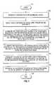
- FIGS. 6 and 7are flow diagrams of methods for recognizing tissue during a tissue-sealing procedure according to embodiments of the present disclosure.
- the method and apparatus of the present disclosurehelps a surgeon to view and recognize tissue parameters while the tissue is grasped by a surgical instrument.
- the methodincludes the steps of grasping tissue with an energy-based surgical instrument, illuminating the grasped tissue with light, and analyzing the light that is transmitted and/or scattered by the tissue.
- the transmitted and/or scattered lightis analyzed to determine parameters associated with the tissue or any vessels disposed within the tissue.
- the transmitted and/or scattered lightmay indicate whether there is a vessel located within a volume of tissue.
- the lightmay also indicate the spatial distribution of vessels within a volume of tissue.
- the lightmay further indicate the type of tissue or the type of vessel within tissue. A surgeon can use all or a portion of this information to more accurately position the tissue-sealing instrument and achieve a high-quality tissue seal and cut.
- FIG. 1illustrates an energy-based tissue-sealing system 100 according to embodiments of the present disclosure.
- the energy-based tissue-sealing system 100(and the methods described below) may use any type of energy to seal tissue including mechanical energy, acoustical energy, thermal energy, electric energy, or electromagnetic energy (e.g., optical energy or radio frequency (RF) energy).
- mechanical energye.g., mechanical energy, acoustical energy, thermal energy, electric energy, or electromagnetic energy (e.g., optical energy or radio frequency (RF) energy).
- RFradio frequency
- the energy-based tissue-sealing system 100includes a power supply 122 , an energy output stage 124 , and an energy-based instrument having a handpiece 104 and transparent contacts 105 , 106 .
- the power supply 122provides power to the energy output stage 124 , which, in turn, provides energy to the tissue 101 via the handpiece 104 and the transparent contacts 105 , 106 .
- the energy output stage 124provides RF energy to the tissue 101 via at least one contact 105 , 106 of the energy-based instrument 126 to seal the tissue 101 .
- the tissue-sealing system 100also includes a sensor 112 , an analog-to-digital converter (ADC) 113 , a microprocessor 114 , a user interface 116 , and a display 118 .
- the sensor 112senses various parameters or properties of tissue 101 at the operating site and transmits analog sensor signals representing the sensed parameters or properties of the tissue 101 to the ADC 113 .
- the ADC 113converts the analog sensor signals into digital sensor data and transmits the digital sensor data to the microprocessor 114 .
- the microprocessor 114processes the digital sensor data and generates control signals based on the processed digital sensor data to control the power supply 122 and/or the energy output stage 124 . For example, the microprocessor 114 may regulate the voltage or current output from the power supply 122 or the energy output stage 124 based on the processed digital sensor data.
- the sensor 112may be configured to measure or sense various electrical or electromechanical conditions at the operating site such as tissue impedance, changes in tissue impedance, tissue temperature, changes in tissue temperature, leakage current, applied voltage, and applied current.
- the sensor 112continuously measures one or more of these conditions so that the microprocessor 114 can continually adjust the energy output from the power supply 122 and/or the energy output stage 124 .
- the user interface 116is coupled to the microprocessor 114 allowing a user to control various parameters of the energy applied to the tissue 101 during a surgical procedure.
- the user interface 116may allow a user to manually set, regulate and/or control one or more parameters of the energy delivered to the tissue, such as voltage, current, power, frequency, and/or pulse parameters, e.g., pulse width, duty cycle, crest factor, and/or repetition rate.
- the microprocessor 114is capable of executing software instructions for processing data received from the user interface 116 and the ADC 113 and for outputting control signals to the power supply 122 and/or the energy output stage 124 .
- the software instructionsare stored in an internal memory of the microprocessor 114 , an internal or external memory bank accessible by the microprocessor 114 and/or an external memory, e.g., an external hard drive, floppy diskette, or CD-ROM.
- Control signals generated by the microprocessor 114may be converted to analog signals by a digital-to-analog converter (DAC) (not shown) before being applied to the power supply 122 and/or energy output stage 124 .
- DACdigital-to-analog converter
- the power supply 122may be a high-voltage DC power supply that produces RF current.
- the microprocessor 114generates control signals to control the magnitude of the voltage and current output by the DC power supply 122 .
- the energy output stage 124receives the output current from the DC power supply 122 and generates one or more pulses via a waveform generator (not shown).
- the microprocessor 114generates control signals to regulate the pulse parameters, such as pulse width, duty cycle, crest factor, and repetition rate.
- the power supply 122may be an AC power supply, and the energy output stage 124 may vary the waveform of the signal provided by the power supply 122 to achieve a desired waveform.
- the user interface 116may be local to or remote from the microprocessor 114 .
- a usermay enter data, such as the type of instrument, the type of procedure, and/or the type of tissue.
- the usermay enter commands, such as a target effective voltage, current or power level.
- the usermay also enter commands for controlling parameters of the energy that is delivered from the energy output stage 124 to the handpiece 104 and the contacts 105 , 106 .
- the energy-based tissue-sealing system 100also includes an optical system for allowing a surgeon to view parameters of the tissue 101 and the vessels 102 within the tissue 101 .
- the optical systemincludes a light source 108 , a beam former 107 , transparent contacts 105 , 106 , and an optional optical sensor 119 .
- the light source 108supplies light to a beam former 107 , which forms the light into a light beam 110 .
- the light beam 110propagates through the first transparent contact 105 , the tissue 101 , and the second transparent contact 106 .
- the optical sensor 119senses the light beam transmitted through the tissue 101 and provides optical sensor signals to the microprocessor 114 via the communications link 109 .
- the optical sensor 119may include a matrix of detectors as described in more detail below.
- the microprocessor 114analyzes the optical sensor signals to determine parameters of the tissue 101 and/or the vessel 102 .
- the microprocessor 114also processes the optical sensor signals and transmits the processed optical sensor signals to the display 118 so that a user may view a transparency image of the vessels 102 within the tissue 101 .
- the tissue-sealing system 100may not include the optical sensor 119 .
- the light beam 110is simply transmitted through the contacts 105 , 106 and the tissue 101 and projected onto a surgeon's eyes 210 .
- the surgeoncan view a transparency image of the vessels 102 or other parameters associated with the tissue 101 or the vessels 102 .
- the surgeoncan recognize or identify the type of the tissue 101 or the vessels 102 within the tissue 101 .
- the surgeoncan also monitor the tissue 101 or the vessels 102 within the tissue 101 while performing a surgical procedure.
- FIG. 2is a cross-sectional side view of a portion of an RF-based tissue-sealing instrument 200 that may be used in the tissue sealing system of FIG. 1 .
- the RF-based tissue-sealing instrument 200includes an upper jaw member 203 and a lower jaw member 204 that are mechanically coupled together (e.g., by a hinge) so that the upper jaw member 203 can move with respect to the lower jaw member 204 and the jaw members 203 , 204 can grasp the tissue 101 .
- the upper jaw member 203is made of a material that is optically transparent or semi-transparent to light having a particular wavelength or a spectrum of wavelengths.
- the upper jaw member 203includes an optically transparent or semi-transparent contact plate 105 .
- the lower jaw member 204includes an optically transparent or semi-transparent contact plate 106 .
- the beam former 107is disposed beneath the contact plate 106 in the lower jaw member 204 .
- the light 202 generated by the light source 108propagates into the beam former 107 , which forms the light 202 into a light beam 110 .
- the beam former 107projects the light beam 110 onto the tissue 101 through the transparent or semi-transparent contact plate 106 .
- the beam former 107may include different optical refracting, reflecting, and guiding components to guide the light from the light source and to form the light beam 110 .
- the beam former 107may include optical fibers and prisms as disclosed in commonly-owned U.S. patent application Ser. No. 12/757,340, entitled “Optical Hydrology Arrays and System and Method for Monitoring Water Displacement During Treatment of Patient Tissue,” the entire contents of which are incorporated by reference herein.
- the beam former 107may form the light 202 into a light spot that is smaller than the light beam 110 .
- the beam former 107may include optical components configured to scan the tissue 101 with the light spot.
- the light beam 110is transmitted through the tissue 101 and is selectively absorbed and/or scattered by vessels 102 and by the surrounding tissue. After passing through the tissue 101 , the light beam 110 passes through the transparent or semitransparent contact plate 105 and the upper jaw member 203 to the surgeon's eyes 210 .
- Luminescent markersmay be introduced into the tissue 101 or the vessels 102 to highlight a parameter of the tissue 101 or to increase the contrast between the tissue 101 and the vessels 101 .
- the upper jaw member 203may be made of a material that is optically transparent at the wavelength of the luminescent light to allow the luminescent light to pass through the upper jaw member 203 .
- the upper jaw member 203may be configured to form the luminescent light into an image and to project the image onto the surgeon's eyes 210 so that the surgeon can view, among other parameters, the positions of the vessels 102 within the tissue 101 .
- the tissue 101is grasped between the jaw members 203 , 204 and RF energy is applied to the grasped tissue 101 through the contact plates 105 , 106 .
- Contact plates 105 , 106are formed of a material that is optically transparent or semi-transparent at the wavelength of the light beam 110 . If luminescent markers are introduced into tissue 101 or the vessels 102 , the contact plate material may also be made optically transparent or semi-transparent at the wavelength of luminescent light.
- the transparent contact plates 105 , 106can be fabricated, for example, by depositing Indium Tin Oxide on a transparent dielectric substrate.
- the contact plates 105 , 106can be made semi-transparent, for example, by depositing a conducting metal mesh or grid on a transparent dielectric substrate. Contact plates 105 , 106 are electrically coupled to the energy output stage 124 of FIG. 1 via electrical conductors or leads 115 .
- additional optical elementscan be placed on the outer surface of the transparent upper jaw member 203 to modify the image that is projected onto the surgeon's eyes 210 .
- lensescan be disposed on the outer surface of the transparent upper jaw member 203 to form an image that meets the needs of the surgeon during a surgical procedure.
- the jaw member 203may be shaped so as to modify the light beam 110 .
- the outer surface of the jaw member 203may have a concave or convex shape to modify the transmitted light 110 in a way that provides more convenient conditions for the surgeon to observe the image created by the transmitted light 110 .
- Grasping tissue with the surgical instrument 200creates favorable conditions for viewing tissue structures and parameters of those structures, such as the density of vessels 102 within tissue 101 .
- tissue 101When tissue 101 is grasped with the surgical instrument 200 , the jaw members 203 , 204 may move towards each other and apply an appropriate amount of pressure on the tissue to leave a sufficient amount of biological fluid within the vessels 102 to achieve the best image quality.
- the applied pressuremay be less than that used for a tissue-sealing procedure.
- the applied pressuremay also be varied during a vessel recognition procedure to provide variations in the amount of biological fluid within the vessels 102 . These variations cause variations in the vessel image intensity, which can improve the recognition of vessels 102 or other parameters of the tissue 101 relative to surrounding tissue.
- the wavelength of the light beam 110may be selected from that part of the spectrum where the difference in absorption and/or scattering spectra between the vessel 102 and surrounding tissue is relatively high or at least sufficient to distinguish the vessel 102 from the surrounding tissue.
- the optical contrast between the images of tissue structurese.g., between the vessels 102 and the tissue 101 surrounding the vessels 102 , and the level of detail of the images depends on the optical properties of the tissue 101 and vessels 102 .
- the optical contrast between the images of tissue structuresmay not be enough.
- the difference between absorption and/or scattering of the light by the vessel 102 and absorption and/or scattering of the light by the tissue 101 surrounding the vessel 102may not be enough to detect a vessel in a transparency image of the tissue.
- a markercan be added to the biological fluid circulating or flowing through the vessels 102 .
- the markermay be a substance that has high optical absorption and/or luminescent properties.
- the marker substanceis selected to have a predetermined absorption, scattering, or luminescence spectrum that is different from the absorption spectrum of the tissue surrounding the vessel so that the marker substance can be detected in the light transmitted, scattered, or reflected by the tissue.
- a marker substancemay be introduced into the cholecystis to facilitate the detection of bile ducts in tissue surrounding the bile ducts.
- the marker substancemay include a fluorescence agent.
- vascular fluorescence agentsare PerkinElmer Inc.'s GenhanceTM 680 and GenhanceTM 750.
- FIG. 3illustrates a portion of an instrument 300 that includes a light detector matrix 301 (e.g., a CCD matrix) for detecting the transparency image of tissue 101 .
- the light detector matrix 301is disposed on the outer surface of the upper jaw member 203 .
- An optical element 302may be disposed between the detector matrix 301 and the transparent contact plate 106 to transform the light 110 , which is transmitted through the tissue 101 , to form a detectable distribution of light at the detector matrix 301 .
- the optical element 302is a bundle of optical fibers.
- the optical element 302is a lens.
- the detector matrix 301detects the distribution of the light beam 110 and transmits a detection signal to the microprocessor 114 of FIG. 1 via the communications link 109 .
- the microprocessor 114processes the detection signal to obtain an image, which is transmitted to a display 118 via user interface 116 so that a user can view the image.
- the display 118may include a camera monitor used in laparoscopic surgical procedures. Accordingly, a surgeon can have a clear and unobstructed view of the surgical site even while using a surgical instrument during a laparoscopic surgical procedure.
- a detector matrix similar to detector matrix 301is positioned in the lower jaw member 204 to obtain an image of the vessels 102 based on light reflected or scattered from the tissue 101 .
- the detector matrixmay be positioned between the contact plate 106 and the beam former 107 .
- the light source 108 and/or the beam former 107can generate two or more wavelengths of light.
- One of the wavelengthscan be selected from that part of the electromagnetic spectrum where the difference between the magnitude of absorption and/or scattering of light by the vessel 102 and the magnitude of absorption and/or scattering of light by the surrounding tissue is large.
- the other wavelength(s)can be selected from that part of the electromagnetic spectrum where the difference between the magnitude of absorption and/or scattering of light by the vessel 102 and the magnitude of absorption and/or scattering of light by the surrounding tissue is small.
- the light source 108may include two light sources (e.g., LEDs) that generate light at two different wavelengths of about 530 nm (green) and 630 nm (red). Accordingly, the detector matrix 301 can detect multiple transparency images of the tissue 101 for different wavelengths.
- two light sourcese.g., LEDs
- the detector matrix 301can detect multiple transparency images of the tissue 101 for different wavelengths.
- the detector matrix 301may transmit the multiple images of the tissue 101 to the microprocessor 114 , which may process the transparency images to find the correlation between them.
- This correlation informationmay be used, for example, to determine the position of a vessel 102 in the surrounding tissue.
- the microprocessor 114may use any one of a number of image processing techniques known to those skilled in the art for finding the correlation between the transparency images.
- the correlation analysis of the transparency imagesmay exclude intensity variations related to varying tissue thickness and other inhomogeneities of the tissue structure that are unrelated to the vessels to be identified.
- FIG. 4is a diagram of the detector matrix 301 used in the tissue-sealing instrument of FIG. 3 according to certain embodiments of the present disclosure.
- the detector matrix 301includes multiple rows 401 a - 401 n and multiple columns 402 a - 402 n of optical detectors.
- Each optical detector in the detector matrix 301e.g., the optical detector located in row 401 a and column 402 b
- Each optical detector in the detector matrix 301is configured to detect one or more parameters (e.g., intensity, polarization, and frequency spectrum) of a portion of the light beam 110 .
- the parameters of each portion of the light beam 110 that are detected by the detector matrix 301are then transmitted to the microprocessor 114 for analysis (e.g., spectral analysis) and processing.
- FIG. 5is a cross-sectional front view of an energy-based sealing instrument 500 that does not have optically transparent jaw members.
- the energy-based sealing systemincludes a lower jaw member 504 that is wider than the upper jaw member 503 .
- Those portions of the lower jaw member 504 that extend outside of the footprint of the upper jaw member 503each have a light source 508 a , 508 b and a corresponding beam former 507 a , 507 b .
- the lower jaw member 504may extend beyond only one side of the footprint of the upper jaw member 503 , in which case the lower jaw member 504 would include a single light source and corresponding beam former.
- the light beams 110 produced by the light sources 508 a , 508 b and the beam formers 507 a , 507 billuminate those portions of tissue near the edges of the upper jaw member 503 .
- the light beams 110 that are transmitted and/or scattered by that portion of the tissue 101 and vessel 102 that is adjacent to the tissue 101 and vessel 102 grasped by the jaw members 503 , 504can be directly observed by a surgeon.
- the light sources 508 a , 508 b , the beam formers 507 a , 507 b , and the other components needed to view or to detect a transparency image of the tissue 101 and vessel 102may be disposed on an instrument or probe that is separate from the tissue-sealing instrument.
- the separate instrument or probemay fit around the tissue-sealing instrument.
- the separate instrument or probemay be a standalone instrument or probe that may be used together with the tissue-sealing instrument.
- Embodiments of the present disclosuremay employ different types of energy or combinations of different types of energy to create an image of the tissue 101 and vessel 102 and to recognize parameters of the tissue 101 and vessel 102 .
- light in the visible light spectramay be used together with ultrasonic energy to recognize a tissue structure.
- electromagnetic energy outside of the visible light rangesuch as in the Terahertz range, may be used with an appropriate detector to recognize a particular tissue structure.
- FIG. 6is a flow diagram of a method for recognizing tissue during a tissue-sealing procedure.
- tissueis grasped with a tissue-sealing instrument, in step 602 .
- the grasped tissue or tissue adjacent to the grasped tissueis illuminated with light.
- a spatial distribution of the light that is transmitted, scattered, and/or reflected by the tissueis formed into an image of the tissue.
- a spectral distribution of the light that is transmitted, scattered, and/or reflected by the tissueis analyzed.
- the tissueis recognized based upon the image of the tissue and/or the result of analyzing the spectral distribution of the light in step 608 .
- optical components of the tissue-sealing instrument or a separate instrument(1) form an image of the tissue based upon the light that is transmitted, scattered, or reflected by the tissue, and (2) project this image onto the surgeon's eyes 210 .
- a surgeoncan continually direct her eyes towards a surgical site within a surgical field of view without needing to redirect her eyes to a separate monitor or display located outside of the surgical field of view.
- the surgeoncan grasp tissue with a tissue sealing instrument and immediately see from the image of the tissue that a vessel is not properly positioned within the jaw members of the tissue-sealing instrument or that a vessel is not contained within the grasped tissue.
- the surgeoncan move the tissue-sealing instrument while visually tracking the vessel within the image of the tissue to properly position the jaw members with respect to the vessel.
- FIG. 7is a flow diagram of another method for recognizing tissue during a tissue-sealing procedure according to other embodiments that use a marker to increase the optical contrast between vessels and surrounding tissue.
- a markeris introduced into fluid flowing in a vessel in step 702 .
- tissue containing the vesselis grasped with a tissue-sealing instrument. While the tissue is grasped with the tissue-sealing instrument, the tissue is illuminated with light having two different wavelengths.
- the tissueis illuminated with light having a first wavelength selected so that the difference between the absorption or scattering of the light of the first wavelength by the marker and the tissue is sufficient to distinguish between the marker and the tissue in the light transmitted, scattered, or reflected by the tissue.
- the tissueis illuminated with light having a second wavelength selected so that the magnitude of absorption or scattering of the light of the second wavelength by the marker and the tissue are substantially equal.
- the tissue that is illuminated with lighttransmits, scatters, and/or reflects the light of the first and second wavelengths.
- step 706the light of the first wavelength and the light of the second wavelength that are transmitted, scattered, and/or reflected by the tissue are measured and correlated.
- step 708before the process ends in step 709 , the position of the vessel within the tissue surrounding the vessel is determined based on the result of correlation.
- the spatial and spectral distribution of the light that is transmitted, scattered, and/or reflected by the tissueis analyzed and formed into an image of the illuminated tissue.
- the optical sensor 119 of FIG. 1may detect the spatial and spectral distribution of the light transmitted and/or scattered by the tissue and the microprocessor 114 may process this information and transmit a transparency image of the tissue to the user display 118 .
- the transparent upper jaw member 203 of the tissue-sealing instrument 200 shown in FIG. 2may be configured as a lens or other similar optical element to form an image of the illuminated tissue on the surgeon's eyes 210 .
- the lightmay illuminate tissue adjacent to the tissue grasped by the tissue-sealing instrument as illustrated in FIG. 5 . In this case, the light passes through the tissue and forms an image on the surgeon's eyes 210 .
- the energy-based tissue sealing system 100 of FIG. 1may include circuitry and other hardware, rather than, or in combination with, programmable instructions executed by the microprocessor 114 for processing the digital sensor data and determining the control signals to transmit to the power supply 122 and the energy output stage 124 .
Landscapes
- Health & Medical Sciences (AREA)
- Surgery (AREA)
- Life Sciences & Earth Sciences (AREA)
- Engineering & Computer Science (AREA)
- Veterinary Medicine (AREA)
- Molecular Biology (AREA)
- Nuclear Medicine, Radiotherapy & Molecular Imaging (AREA)
- Public Health (AREA)
- Biomedical Technology (AREA)
- Heart & Thoracic Surgery (AREA)
- Medical Informatics (AREA)
- General Health & Medical Sciences (AREA)
- Animal Behavior & Ethology (AREA)
- Otolaryngology (AREA)
- Plasma & Fusion (AREA)
- Physics & Mathematics (AREA)
- Oral & Maxillofacial Surgery (AREA)
- Pathology (AREA)
- Surgical Instruments (AREA)
- Investigating Or Analysing Materials By Optical Means (AREA)
Abstract
Description
Claims (16)
Priority Applications (3)
| Application Number | Priority Date | Filing Date | Title |
|---|---|---|---|
| US13/108,196US10117705B2 (en) | 2011-05-16 | 2011-05-16 | Optical recognition of tissue and vessels |
| PCT/US2012/038104WO2012158774A1 (en) | 2011-05-16 | 2012-05-16 | Optical recognition of tissue and vessels |
| EP12724795.5AEP2709548B1 (en) | 2011-05-16 | 2012-05-16 | Optical recognition of tissue and vessels |
Applications Claiming Priority (1)
| Application Number | Priority Date | Filing Date | Title |
|---|---|---|---|
| US13/108,196US10117705B2 (en) | 2011-05-16 | 2011-05-16 | Optical recognition of tissue and vessels |
Publications (2)
| Publication Number | Publication Date |
|---|---|
| US20120296205A1 US20120296205A1 (en) | 2012-11-22 |
| US10117705B2true US10117705B2 (en) | 2018-11-06 |
Family
ID=46197689
Family Applications (1)
| Application Number | Title | Priority Date | Filing Date |
|---|---|---|---|
| US13/108,196Expired - Fee RelatedUS10117705B2 (en) | 2011-05-16 | 2011-05-16 | Optical recognition of tissue and vessels |
Country Status (3)
| Country | Link |
|---|---|
| US (1) | US10117705B2 (en) |
| EP (1) | EP2709548B1 (en) |
| WO (1) | WO2012158774A1 (en) |
Cited By (39)
| Publication number | Priority date | Publication date | Assignee | Title |
|---|---|---|---|---|
| US10271897B2 (en) | 2012-05-01 | 2019-04-30 | Covidien Lp | Surgical instrument with stamped double-flange jaws and actuation mechanism |
| US11076997B2 (en) | 2017-07-25 | 2021-08-03 | Smith & Nephew Plc | Restriction of sensor-monitored region for sensor-enabled wound dressings |
| NL2025324B1 (en)* | 2020-04-09 | 2021-10-26 | Academisch Ziekenhuis Leiden | A Surgical Tool, System and Method for Tissue Characterisation |
| US11241275B2 (en) | 2018-03-21 | 2022-02-08 | Covidien Lp | Energy-based surgical instrument having multiple operational configurations |
| US11324424B2 (en) | 2017-03-09 | 2022-05-10 | Smith & Nephew Plc | Apparatus and method for imaging blood in a target region of tissue |
| US11559438B2 (en) | 2017-11-15 | 2023-01-24 | Smith & Nephew Plc | Integrated sensor enabled wound monitoring and/or therapy dressings and systems |
| US11596553B2 (en) | 2017-09-27 | 2023-03-07 | Smith & Nephew Plc | Ph sensing for sensor enabled negative pressure wound monitoring and therapy apparatuses |
| US11612428B2 (en) | 2013-08-07 | 2023-03-28 | Covidien Lp | Bipolar surgical instrument |
| US11633147B2 (en) | 2017-09-10 | 2023-04-25 | Smith & Nephew Plc | Sensor enabled wound therapy dressings and systems implementing cybersecurity |
| US11633153B2 (en) | 2017-06-23 | 2023-04-25 | Smith & Nephew Plc | Positioning of sensors for sensor enabled wound monitoring or therapy |
| US11638664B2 (en) | 2017-07-25 | 2023-05-02 | Smith & Nephew Plc | Biocompatible encapsulation and component stress relief for sensor enabled negative pressure wound therapy dressings |
| US11690570B2 (en) | 2017-03-09 | 2023-07-04 | Smith & Nephew Plc | Wound dressing, patch member and method of sensing one or more wound parameters |
| US11707313B2 (en) | 2012-03-29 | 2023-07-25 | Covidien Lp | Electrosurgical forceps and method of manufacturing the same |
| US11717447B2 (en) | 2016-05-13 | 2023-08-08 | Smith & Nephew Plc | Sensor enabled wound monitoring and therapy apparatus |
| US11759144B2 (en) | 2017-09-10 | 2023-09-19 | Smith & Nephew Plc | Systems and methods for inspection of encapsulation and components in sensor equipped wound dressings |
| US11791030B2 (en) | 2017-05-15 | 2023-10-17 | Smith & Nephew Plc | Wound analysis device and method |
| US11806038B2 (en) | 2019-10-11 | 2023-11-07 | Covidien Lp | Surgical instrument and method facilitating testing jaw force of the surgical instrument |
| US11832916B2 (en) | 2020-01-29 | 2023-12-05 | Covidien Lp | System and methods for identifying vessels within tissue |
| US11839464B2 (en) | 2017-09-28 | 2023-12-12 | Smith & Nephew, Plc | Neurostimulation and monitoring using sensor enabled wound monitoring and therapy apparatus |
| US11883262B2 (en) | 2017-04-11 | 2024-01-30 | Smith & Nephew Plc | Component positioning and stress relief for sensor enabled wound dressings |
| US11903635B2 (en) | 2020-02-28 | 2024-02-20 | Covidien Lp | Electrosurgical forceps including tissue indication |
| US11925735B2 (en) | 2017-08-10 | 2024-03-12 | Smith & Nephew Plc | Positioning of sensors for sensor enabled wound monitoring or therapy |
| US11925406B2 (en) | 2020-09-14 | 2024-03-12 | Covidien Lp | End effector assemblies for surgical instruments |
| US11931165B2 (en) | 2017-09-10 | 2024-03-19 | Smith & Nephew Plc | Electrostatic discharge protection for sensors in wound therapy |
| US11944418B2 (en) | 2018-09-12 | 2024-04-02 | Smith & Nephew Plc | Device, apparatus and method of determining skin perfusion pressure |
| US11957545B2 (en) | 2017-09-26 | 2024-04-16 | Smith & Nephew Plc | Sensor positioning and optical sensing for sensor enabled wound therapy dressings and systems |
| US11969538B2 (en) | 2018-12-21 | 2024-04-30 | T.J.Smith And Nephew, Limited | Wound therapy systems and methods with multiple power sources |
| US12011942B2 (en) | 2019-03-18 | 2024-06-18 | Smith & Nephew Plc | Rules for sensor integrated substrates |
| US12016994B2 (en) | 2019-10-07 | 2024-06-25 | Smith & Nephew Plc | Sensor enabled negative pressure wound monitoring apparatus with different impedances inks |
| US12033738B2 (en) | 2017-05-15 | 2024-07-09 | Smith & Nephew Plc | Negative pressure wound therapy system using eulerian video magnification |
| US12156691B2 (en) | 2020-04-22 | 2024-12-03 | Covidien Lp | Surgical system and methods for treating tissue |
| US12161386B2 (en) | 2020-09-11 | 2024-12-10 | Covidien Lp | Surgical instruments having an articulating section such as for use in robotic surgical systems |
| US12178597B2 (en) | 2017-03-09 | 2024-12-31 | Smith & Nephew Plc | Device, apparatus and method of determining skin perfusion pressure |
| US12186165B2 (en) | 2018-09-28 | 2025-01-07 | T.J.Smith And Nephew, Limited | Optical fibers for optically sensing through wound dressings |
| US12185964B2 (en) | 2020-09-10 | 2025-01-07 | Covidien Lp | End effector assemblies for surgical instruments such as for use in robotic surgical systems |
| US12186164B2 (en) | 2018-10-16 | 2025-01-07 | Smith & Nephew Plc | Systems and method for applying biocompatible encapsulation to sensor enabled wound monitoring and therapy dressings |
| US12299772B2 (en) | 2020-04-21 | 2025-05-13 | T.J.Smith And Nephew, Limited | Wound treatment management using augmented reality overlay |
| US12409071B2 (en) | 2018-08-29 | 2025-09-09 | Smith & Nephew Plc | Component positioning and encapsulation for sensor enabled wound dressings |
| US12426941B2 (en) | 2020-03-20 | 2025-09-30 | Covidien Lp | Surgical instruments and systems configured to detect, analyze, and/or distinguish smoke and steam during a surgical procedure |
Families Citing this family (85)
| Publication number | Priority date | Publication date | Assignee | Title |
|---|---|---|---|---|
| US7364577B2 (en) | 2002-02-11 | 2008-04-29 | Sherwood Services Ag | Vessel sealing system |
| ES2262639T3 (en) | 2001-04-06 | 2006-12-01 | Sherwood Services Ag | SHUTTER AND DIVIDER OF GLASSES WITH BUMPER MEMBERS N OCONDUCTIVES. |
| US7628791B2 (en) | 2005-08-19 | 2009-12-08 | Covidien Ag | Single action tissue sealer |
| US8298232B2 (en) | 2006-01-24 | 2012-10-30 | Tyco Healthcare Group Lp | Endoscopic vessel sealer and divider for large tissue structures |
| US8114122B2 (en) | 2009-01-13 | 2012-02-14 | Tyco Healthcare Group Lp | Apparatus, system, and method for performing an electrosurgical procedure |
| US8187273B2 (en) | 2009-05-07 | 2012-05-29 | Tyco Healthcare Group Lp | Apparatus, system, and method for performing an electrosurgical procedure |
| US8430876B2 (en) | 2009-08-27 | 2013-04-30 | Tyco Healthcare Group Lp | Vessel sealer and divider with knife lockout |
| US8133254B2 (en) | 2009-09-18 | 2012-03-13 | Tyco Healthcare Group Lp | In vivo attachable and detachable end effector assembly and laparoscopic surgical instrument and methods therefor |
| US8112871B2 (en) | 2009-09-28 | 2012-02-14 | Tyco Healthcare Group Lp | Method for manufacturing electrosurgical seal plates |
| US8939972B2 (en) | 2011-05-06 | 2015-01-27 | Covidien Lp | Surgical forceps |
| US8685009B2 (en) | 2011-05-16 | 2014-04-01 | Covidien Lp | Thread-like knife for tissue cutting |
| US8852185B2 (en) | 2011-05-19 | 2014-10-07 | Covidien Lp | Apparatus for performing an electrosurgical procedure |
| US9615877B2 (en) | 2011-06-17 | 2017-04-11 | Covidien Lp | Tissue sealing forceps |
| US8745840B2 (en) | 2011-07-11 | 2014-06-10 | Covidien Lp | Surgical forceps and method of manufacturing thereof |
| US9039732B2 (en) | 2011-07-11 | 2015-05-26 | Covidien Lp | Surgical forceps |
| US8852186B2 (en) | 2011-08-09 | 2014-10-07 | Covidien Lp | Microwave sensing for tissue sealing |
| US8845636B2 (en) | 2011-09-16 | 2014-09-30 | Covidien Lp | Seal plate with insulation displacement connection |
| US8864795B2 (en) | 2011-10-03 | 2014-10-21 | Covidien Lp | Surgical forceps |
| US8968309B2 (en) | 2011-11-10 | 2015-03-03 | Covidien Lp | Surgical forceps |
| US8968310B2 (en) | 2011-11-30 | 2015-03-03 | Covidien Lp | Electrosurgical instrument with a knife blade lockout mechanism |
| US9113897B2 (en) | 2012-01-23 | 2015-08-25 | Covidien Lp | Partitioned surgical instrument |
| US8968360B2 (en) | 2012-01-25 | 2015-03-03 | Covidien Lp | Surgical instrument with resilient driving member and related methods of use |
| US8747434B2 (en) | 2012-02-20 | 2014-06-10 | Covidien Lp | Knife deployment mechanisms for surgical forceps |
| US11399898B2 (en)* | 2012-03-06 | 2022-08-02 | Briteseed, Llc | User interface for a system used to determine tissue or artifact characteristics |
| US8961514B2 (en)* | 2012-03-06 | 2015-02-24 | Covidien Lp | Articulating surgical apparatus |
| US20150066000A1 (en)* | 2012-03-06 | 2015-03-05 | Briteseed Llc | Surgical Tool With Integrated Sensor |
| US9375282B2 (en) | 2012-03-26 | 2016-06-28 | Covidien Lp | Light energy sealing, cutting and sensing surgical device |
| US9668807B2 (en) | 2012-05-01 | 2017-06-06 | Covidien Lp | Simplified spring load mechanism for delivering shaft force of a surgical instrument |
| US9820765B2 (en) | 2012-05-01 | 2017-11-21 | Covidien Lp | Surgical instrument with stamped double-flange jaws |
| US9039731B2 (en) | 2012-05-08 | 2015-05-26 | Covidien Lp | Surgical forceps including blade safety mechanism |
| US9375258B2 (en) | 2012-05-08 | 2016-06-28 | Covidien Lp | Surgical forceps |
| US10231782B2 (en) | 2012-09-06 | 2019-03-19 | Covidien Lp | Medical devices and methods incorporating frustrated total internal reflection for energy-efficient sealing and cutting of tissue using light energy |
| US10226297B2 (en) | 2012-09-06 | 2019-03-12 | Covidien Lp | Medical devices and methods incorporating frustrated total internal reflection for energy-efficient sealing and cutting of tissue using light energy |
| US9265566B2 (en) | 2012-10-16 | 2016-02-23 | Covidien Lp | Surgical instrument |
| US10722292B2 (en) | 2013-05-31 | 2020-07-28 | Covidien Lp | Surgical device with an end-effector assembly and system for monitoring of tissue during a surgical procedure |
| EP2815713B1 (en) | 2013-06-20 | 2015-08-12 | Erbe Elektromedizin GmbH | Electrosurgical instrument with light guide |
| AU2013375909B2 (en) | 2013-08-07 | 2015-07-30 | Covidien Lp | Bipolar surgical instrument |
| USD788302S1 (en) | 2013-10-01 | 2017-05-30 | Covidien Lp | Knife for endoscopic electrosurgical forceps |
| WO2015148504A1 (en) | 2014-03-25 | 2015-10-01 | Briteseed Llc | Vessel detector and method of detection |
| US9918785B2 (en) | 2014-09-17 | 2018-03-20 | Covidien Lp | Deployment mechanisms for surgical instruments |
| US9987076B2 (en) | 2014-09-17 | 2018-06-05 | Covidien Lp | Multi-function surgical instruments |
| US10039592B2 (en) | 2014-09-17 | 2018-08-07 | Covidien Lp | Deployment mechanisms for surgical instruments |
| US10080605B2 (en) | 2014-09-17 | 2018-09-25 | Covidien Lp | Deployment mechanisms for surgical instruments |
| US9877777B2 (en) | 2014-09-17 | 2018-01-30 | Covidien Lp | Surgical instrument having a bipolar end effector assembly and a deployable monopolar assembly |
| AU2016200084B2 (en)* | 2015-01-16 | 2020-01-16 | Covidien Lp | Powered surgical stapling device |
| US10172612B2 (en) | 2015-01-21 | 2019-01-08 | Covidien Lp | Surgical instruments with force applier and methods of use |
| ES2892526T3 (en)* | 2015-02-19 | 2022-02-04 | Briteseed Llc | System for determining the size of a vessel by light absorption |
| EP3545830B1 (en) | 2015-02-19 | 2022-01-05 | Briteseed, LLC | System for determining vessel edge |
| KR101684781B1 (en)* | 2015-05-11 | 2016-12-08 | 고려대학교 산학협력단 | X-ray imaging apparatus for minimally invasive surgery |
| USD844139S1 (en) | 2015-07-17 | 2019-03-26 | Covidien Lp | Monopolar assembly of a multi-function surgical instrument |
| USD844138S1 (en) | 2015-07-17 | 2019-03-26 | Covidien Lp | Handle assembly of a multi-function surgical instrument |
| US10987159B2 (en) | 2015-08-26 | 2021-04-27 | Covidien Lp | Electrosurgical end effector assemblies and electrosurgical forceps configured to reduce thermal spread |
| WO2017062720A1 (en) | 2015-10-08 | 2017-04-13 | Briteseed Llc | System and method for determining vessel size |
| KR101835043B1 (en)* | 2015-11-03 | 2018-03-08 | (주) 인텍플러스 | Dissection tool And Dissection system |
| US10357162B1 (en)* | 2015-12-30 | 2019-07-23 | Banpil Photonics, Inc. | Imaging system for screening and diagnosis of breast cancer |
| US10426543B2 (en) | 2016-01-23 | 2019-10-01 | Covidien Lp | Knife trigger for vessel sealer |
| US11992235B2 (en) | 2016-02-12 | 2024-05-28 | Briteseed, Llc | System to differentiate and identify types of tissue within a region proximate to a working end of a surgical instrument |
| US10537381B2 (en) | 2016-02-26 | 2020-01-21 | Covidien Lp | Surgical instrument having a bipolar end effector assembly and a deployable monopolar assembly |
| US10631887B2 (en) | 2016-08-15 | 2020-04-28 | Covidien Lp | Electrosurgical forceps for video assisted thoracoscopic surgery and other surgical procedures |
| EP4026489B1 (en) | 2016-08-30 | 2025-07-30 | Briteseed, LLC | System for determining vessel size with angular distortion compensation |
| US10813695B2 (en) | 2017-01-27 | 2020-10-27 | Covidien Lp | Reflectors for optical-based vessel sealing |
| US10973567B2 (en) | 2017-05-12 | 2021-04-13 | Covidien Lp | Electrosurgical forceps for grasping, treating, and/or dividing tissue |
| US11172980B2 (en) | 2017-05-12 | 2021-11-16 | Covidien Lp | Electrosurgical forceps for grasping, treating, and/or dividing tissue |
| USD843574S1 (en) | 2017-06-08 | 2019-03-19 | Covidien Lp | Knife for open vessel sealer |
| USD854684S1 (en) | 2017-06-08 | 2019-07-23 | Covidien Lp | Open vessel sealer with mechanical cutter |
| USD854149S1 (en) | 2017-06-08 | 2019-07-16 | Covidien Lp | End effector for open vessel sealer |
| WO2019032984A2 (en)* | 2017-08-11 | 2019-02-14 | Intuitive Surgical Operations, Inc. | Medical apparatus with optical sensing, and related devices and methods |
| US11154348B2 (en) | 2017-08-29 | 2021-10-26 | Covidien Lp | Surgical instruments and methods of assembling surgical instruments |
| US11723600B2 (en) | 2017-09-05 | 2023-08-15 | Briteseed, Llc | System and method used to determine tissue and/or artifact characteristics |
| US11696777B2 (en) | 2017-12-22 | 2023-07-11 | Briteseed, Llc | Compact system used to determine tissue or artifact characteristics |
| US11123132B2 (en) | 2018-04-09 | 2021-09-21 | Covidien Lp | Multi-function surgical instruments and assemblies therefor |
| US10780544B2 (en) | 2018-04-24 | 2020-09-22 | Covidien Lp | Systems and methods facilitating reprocessing of surgical instruments |
| WO2019208011A1 (en)* | 2018-04-24 | 2019-10-31 | オリンパス株式会社 | Endoscope system |
| US10828756B2 (en) | 2018-04-24 | 2020-11-10 | Covidien Lp | Disassembly methods facilitating reprocessing of multi-function surgical instruments |
| US11471211B2 (en) | 2018-10-12 | 2022-10-18 | Covidien Lp | Electrosurgical forceps |
| US11376062B2 (en) | 2018-10-12 | 2022-07-05 | Covidien Lp | Electrosurgical forceps |
| US11350982B2 (en) | 2018-12-05 | 2022-06-07 | Covidien Lp | Electrosurgical forceps |
| EP3902471B1 (en) | 2018-12-30 | 2024-09-18 | Briteseed, LLC | A system used to detect or differentiate tissue or an artifact |
| US11523861B2 (en) | 2019-03-22 | 2022-12-13 | Covidien Lp | Methods for manufacturing a jaw assembly for an electrosurgical forceps |
| US12402934B2 (en) | 2019-09-15 | 2025-09-02 | Covidien Lp | Electrosurgical instrument for grasping, treating, and/or dividing tissue incorporating thermal management feature |
| US11622804B2 (en) | 2020-03-16 | 2023-04-11 | Covidien Lp | Forceps with linear trigger mechanism |
| US12295641B2 (en) | 2020-07-01 | 2025-05-13 | Covidien Lp | Electrosurgical forceps with swivel action nerve probe |
| US11660109B2 (en) | 2020-09-08 | 2023-05-30 | Covidien Lp | Cutting elements for surgical instruments such as for use in robotic surgical systems |
| US20230096074A1 (en)* | 2021-09-30 | 2023-03-30 | Cilag Gmbh International | Electrosurgical instrument with light accumulator end effector and fiber optics |
| CN120168052A (en)* | 2025-05-06 | 2025-06-20 | 首都医科大学附属北京安贞医院 | Auxiliary device for vena cava cuff and control method thereof |
Citations (151)
| Publication number | Priority date | Publication date | Assignee | Title |
|---|---|---|---|---|
| SU401367A1 (en) | 1971-10-05 | 1973-10-12 | Тернопольский государственный медицинский институт | BIAKTIVNYE ELECTRO SURGICAL INSTRUMENT |
| DE2415263A1 (en) | 1974-03-29 | 1975-10-02 | Aesculap Werke Ag | Surgical H.F. coagulation probe has electrode tongs - with exposed ends of insulated conductors forming tong-jaws |
| DE2514501A1 (en) | 1975-04-03 | 1976-10-21 | Karl Storz | Bipolar coagulation instrument for endoscopes - has two high frequency electrodes looped over central insulating piece |
| DE2627679A1 (en) | 1975-06-26 | 1977-01-13 | Marcel Lamidey | HEMATISTIC HIGH FREQUENCY EXTRACTOR FORCEPS |
| USD249549S (en) | 1976-10-22 | 1978-09-19 | Aspen Laboratories, Inc. | Electrosurgical handle |
| US4126136A (en) | 1976-02-09 | 1978-11-21 | Research Corporation | Photocoagulating scalpel system |
| USD263020S (en) | 1980-01-22 | 1982-02-16 | Rau Iii David M | Retractable knife |
| DE3423356A1 (en) | 1984-06-25 | 1986-01-02 | Berchtold Medizin-Elektronik GmbH & Co, 7200 Tuttlingen | ELECTROSURGICAL HIGH-FREQUENCY CUTTING INSTRUMENT |
| JPS61501068A (en) | 1984-01-30 | 1986-05-29 | ハルコフスキイ ナウチノ−イススレドワテルスキイ インスチチユ−ト オブスチエイ イ ネオトロジノイ ヒルルギイ | bipolar electrosurgical instrument |
| DE3612646A1 (en) | 1985-04-16 | 1987-04-30 | Ellman International | Electrosurgical handle piece for blades, needles and forceps |
| DE8712328U1 (en) | 1987-09-11 | 1988-02-18 | Jakoubek, Franz, 7201 Emmingen-Liptingen | Endoscopy forceps |
| USD295894S (en) | 1985-09-26 | 1988-05-24 | Acme United Corporation | Disposable surgical scissors |
| USD295893S (en) | 1985-09-25 | 1988-05-24 | Acme United Corporation | Disposable surgical clamp |
| USD298353S (en) | 1986-05-06 | 1988-11-01 | Vitalmetrics, Inc. | Handle for surgical instrument |
| USD299413S (en) | 1985-07-17 | 1989-01-17 | The Stanley Works | Folding pocket saw handle |
| EP0480293A1 (en) | 1990-10-11 | 1992-04-15 | LaserSurge, Inc. | Clamp for approximating tissue sections |
| US5147356A (en) | 1991-04-16 | 1992-09-15 | Microsurge, Inc. | Surgical instrument |
| JPH055106A (en) | 1990-07-31 | 1993-01-14 | Matsushita Electric Works Ltd | Production of alloy sintered body |
| JPH0540112A (en) | 1991-02-08 | 1993-02-19 | Tokico Ltd | Sample liquid component analyzer |
| USD343453S (en) | 1993-05-05 | 1994-01-18 | Laparomed Corporation | Handle for laparoscopic surgical instrument |
| JPH06502328A (en) | 1990-10-17 | 1994-03-17 | ボストン サイエンティフィック コーポレイション | Surgical instruments and methods |
| JPH06121797A (en) | 1992-02-27 | 1994-05-06 | United States Surgical Corp | Equipment and method for performing intracutaneous stapling of body tissue |
| USD348930S (en) | 1991-10-11 | 1994-07-19 | Ethicon, Inc. | Endoscopic stapler |
| USD349341S (en) | 1992-10-28 | 1994-08-02 | Microsurge, Inc. | Endoscopic grasper |
| US5336221A (en) | 1992-10-14 | 1994-08-09 | Premier Laser Systems, Inc. | Method and apparatus for applying thermal energy to tissue using a clamp |
| DE4303882A1 (en) | 1993-02-10 | 1994-08-18 | Kernforschungsz Karlsruhe | Combined instrument for separating and coagulating in minimally invasive surgery |
| JPH06285078A (en) | 1993-04-05 | 1994-10-11 | Olympus Optical Co Ltd | Forceps |
| JPH06343644A (en) | 1993-05-04 | 1994-12-20 | Gyrus Medical Ltd | Surgical peritoneoscope equipment |
| JPH06511401A (en) | 1991-06-07 | 1994-12-22 | バイタル メディカル プロダクツ コーポレイション | Bipolar electrosurgical endoscopic instrument and its method of use |
| USD354564S (en) | 1993-06-25 | 1995-01-17 | Richard-Allan Medical Industries, Inc. | Surgical clip applier |
| USD358887S (en) | 1993-12-02 | 1995-05-30 | Cobot Medical Corporation | Combined cutting and coagulating forceps |
| DE4403252A1 (en) | 1994-02-03 | 1995-08-10 | Michael Hauser | Instrument shaft for min. invasive surgery |
| JPH07265328A (en) | 1993-11-01 | 1995-10-17 | Gyrus Medical Ltd | Electrode assembly for electric surgery device and electric surgery device using it |
| US5470331A (en) | 1990-01-22 | 1995-11-28 | S.L.T. Japan Co., Ltd. | Laser light irradiation apparatus for medical treatment |
| JPH0856955A (en) | 1994-06-29 | 1996-03-05 | Gyrus Medical Ltd | Electric surgical apparatus |
| DE19515914C1 (en) | 1995-05-02 | 1996-07-25 | Aesculap Ag | Tong or scissor-shaped surgical instrument |
| DE19506363A1 (en) | 1995-02-24 | 1996-08-29 | Frost Lore Geb Haupt | Non-invasive thermometry in organs under hyperthermia and coagulation conditions |
| JPH08252263A (en) | 1994-12-21 | 1996-10-01 | Gyrus Medical Ltd | Electronic surgical incision instrument and electronic surgical incision device using the same |
| DE29616210U1 (en) | 1996-09-18 | 1996-11-14 | Olympus Winter & Ibe Gmbh, 22045 Hamburg | Handle for surgical instruments |
| JPH08317934A (en) | 1995-04-12 | 1996-12-03 | Ethicon Endo Surgery Inc | Hemostatic device for electric surgery with adaptable electrode |
| JPH0910223A (en) | 1995-06-23 | 1997-01-14 | Gyrus Medical Ltd | Generator and system for electric operation |
| DE19608716C1 (en) | 1996-03-06 | 1997-04-17 | Aesculap Ag | Bipolar surgical holding instrument |
| JPH09122138A (en) | 1995-10-20 | 1997-05-13 | Ethicon Endo Surgery Inc | Apparatus for operation |
| USD384413S (en) | 1994-10-07 | 1997-09-30 | United States Surgical Corporation | Endoscopic suturing instrument |
| JPH1024051A (en) | 1995-09-20 | 1998-01-27 | Olympus Optical Co Ltd | Coagulation forceps with separating function |
| DE19751106A1 (en) | 1996-11-27 | 1998-05-28 | Eastman Kodak Co | Laser printer with array of laser diodes |
| US5762609A (en) | 1992-09-14 | 1998-06-09 | Sextant Medical Corporation | Device and method for analysis of surgical tissue interventions |
| JPH10155798A (en) | 1996-12-04 | 1998-06-16 | Asahi Optical Co Ltd | Hot biopsy forceps for endoscopes |
| US5769791A (en)* | 1992-09-14 | 1998-06-23 | Sextant Medical Corporation | Tissue interrogating device and methods |
| USH1745H (en) | 1995-09-29 | 1998-08-04 | Paraschac; Joseph F. | Electrosurgical clamping device with insulation limited bipolar electrode |
| USD402028S (en) | 1997-10-10 | 1998-12-01 | Invasatec, Inc. | Hand controller for medical system |
| JPH1147150A (en) | 1997-08-06 | 1999-02-23 | Olympus Optical Co Ltd | Endoscopic surgery appliance |
| JPH1170124A (en) | 1997-05-14 | 1999-03-16 | Ethicon Endo Surgery Inc | Improved electrosurgical hemostatic apparatus having anvil |
| USD408018S (en) | 1996-03-12 | 1999-04-13 | Mcnaughton Patrick J | Switch guard |
| DE19751108A1 (en) | 1997-11-18 | 1999-05-20 | Beger Frank Michael Dipl Desig | Electrosurgical operation tool, especially for diathermy |
| JPH11169381A (en) | 1997-12-15 | 1999-06-29 | Olympus Optical Co Ltd | High frequency treating device |
| JPH11192238A (en) | 1997-10-10 | 1999-07-21 | Ethicon Endo Surgery Inc | Ultrasonic forceps coagulation device improved of pivot-attaching of forceps arm |
| JPH11244298A (en) | 1997-12-19 | 1999-09-14 | Gyrus Medical Ltd | Electric surgical instrument |
| USD416089S (en) | 1996-04-08 | 1999-11-02 | Richard-Allan Medical Industries, Inc. | Endoscopic linear stapling and dividing surgical instrument |
| US6039729A (en) | 1997-08-08 | 2000-03-21 | Cynosure, Inc. | Portable cautery system |
| JP2000102545A (en) | 1997-06-18 | 2000-04-11 | Eggers & Associates Inc | Electric tweezers for surgery |
| USD424694S (en) | 1998-10-23 | 2000-05-09 | Sherwood Services Ag | Forceps |
| USD425201S (en) | 1998-10-23 | 2000-05-16 | Sherwood Services Ag | Disposable electrode assembly |
| WO2000036986A1 (en) | 1998-12-18 | 2000-06-29 | Karl Storz Gmbh & Co. Kg | Bipolar medical instrument |
| US6086586A (en) | 1998-09-14 | 2000-07-11 | Enable Medical Corporation | Bipolar tissue grasping apparatus and tissue welding method |
| USH1904H (en) | 1997-05-14 | 2000-10-03 | Ethicon Endo-Surgery, Inc. | Electrosurgical hemostatic method and device |
| JP2000342599A (en) | 1999-05-21 | 2000-12-12 | Gyrus Medical Ltd | Generator for electrosurgical operation, electrosurgical operation system, method for operating this system and method for performing amputation and resection of tissue by electrosurgical operation |
| JP2000350732A (en) | 1999-05-21 | 2000-12-19 | Gyrus Medical Ltd | Electrosurgical system, generator for electrosurgery, and method for cutting or excising tissue by electrosurgery |
| JP2001008944A (en) | 1999-05-28 | 2001-01-16 | Gyrus Medical Ltd | Electric surgical signal generator and electric surgical system |
| JP2001029356A (en) | 1999-06-11 | 2001-02-06 | Gyrus Medical Ltd | Electric and surgical signal generator |
| WO2001015614A1 (en) | 1999-08-27 | 2001-03-08 | Karl Storz Gmbh & Co. Kg | Bipolar medical instrument |
| JP2001128990A (en) | 1999-05-28 | 2001-05-15 | Gyrus Medical Ltd | Electro surgical instrument and electrosurgical tool converter |
| US6248117B1 (en) | 1999-04-16 | 2001-06-19 | Vital Access Corp | Anastomosis apparatus for use in intraluminally directed vascular anastomosis |
| JP2001190564A (en) | 2000-01-12 | 2001-07-17 | Olympus Optical Co Ltd | Medical treatment instrument |
| WO2001054604A1 (en) | 2000-01-25 | 2001-08-02 | Aesculap Ag & Co. Kg | Bipolar gripping device |
| USD449886S1 (en) | 1998-10-23 | 2001-10-30 | Sherwood Services Ag | Forceps with disposable electrode |
| EP1159926A2 (en) | 2000-06-03 | 2001-12-05 | Aesculap Ag | Scissor- or forceps-like surgical instrument |
| USD453923S1 (en) | 2000-11-16 | 2002-02-26 | Carling Technologies, Inc. | Electrical rocker switch guard |
| USD454951S1 (en) | 2001-02-27 | 2002-03-26 | Visionary Biomedical, Inc. | Steerable catheter |
| DE10045375A1 (en) | 2000-09-14 | 2002-04-11 | Aesculap Ag & Co Kg | Medical instrument comprises temperature and pressure condition sensor and modification device for influencing transmitting device |
| USD457958S1 (en) | 2001-04-06 | 2002-05-28 | Sherwood Services Ag | Vessel sealer and divider |
| USD457959S1 (en) | 2001-04-06 | 2002-05-28 | Sherwood Services Ag | Vessel sealer |
| USD465281S1 (en) | 1999-09-21 | 2002-11-05 | Karl Storz Gmbh & Co. Kg | Endoscopic medical instrument |
| USD466209S1 (en) | 2001-02-27 | 2002-11-26 | Visionary Biomedical, Inc. | Steerable catheter |
| US6611320B1 (en)* | 1999-09-08 | 2003-08-26 | Optoq Ab | Method and apparatus |
| US6623494B1 (en) | 1999-04-16 | 2003-09-23 | Integrated Vascular Interventional Technologies, L.C. (Ivit, Lc) | Methods and systems for intraluminally directed vascular anastomosis |
| US20040106880A1 (en)* | 1999-10-25 | 2004-06-03 | Therus Corporation (Legal) | Use of focused ultrasound for vascular sealing |
| JP2004517668A (en) | 2000-10-20 | 2004-06-17 | オーナックス・メディカル・インコーポレーテッド | Surgical suturing instrument and method of use |
| USD493888S1 (en) | 2003-02-04 | 2004-08-03 | Sherwood Services Ag | Electrosurgical pencil with pistol grip |
| US20040176752A1 (en) | 2003-03-06 | 2004-09-09 | Alfano Robert R. | System and methods for laser treatment of ocular tissue |
| JP2004528869A (en) | 2001-01-26 | 2004-09-24 | エシコン・エンド−サージェリィ・インコーポレイテッド | Electrosurgical instruments for coagulation and cutting |
| USD496997S1 (en) | 2003-05-15 | 2004-10-05 | Sherwood Services Ag | Vessel sealer and divider |
| USD499181S1 (en) | 2003-05-15 | 2004-11-30 | Sherwood Services Ag | Handle for a vessel sealer and divider |
| USD502994S1 (en) | 2003-05-21 | 2005-03-15 | Blake, Iii Joseph W | Repeating multi-clip applier |
| USD509297S1 (en) | 2003-10-17 | 2005-09-06 | Tyco Healthcare Group, Lp | Surgical instrument |
| US20050234437A1 (en) | 1999-07-14 | 2005-10-20 | Cardiofocus, Inc. | Deflectable sheath catheters with out-of-plane bent tip |
| WO2005110264A2 (en) | 2004-05-14 | 2005-11-24 | Erbe Elektromedizin Gmbh | Electrosurgical instrument |
| USD525361S1 (en) | 2004-10-06 | 2006-07-18 | Sherwood Services Ag | Hemostat style elongated dissecting and dividing instrument |
| US20060173355A1 (en)* | 2003-04-17 | 2006-08-03 | Alfano Robert R | Detecting human cancer through spectral optical imaging using key water absorption wavelengths |
| USD531311S1 (en) | 2004-10-06 | 2006-10-31 | Sherwood Services Ag | Pistol grip style elongated dissecting and dividing instrument |
| US20060258629A1 (en)* | 2002-10-18 | 2006-11-16 | Freeman William R | Photodynamic therapy for ocular neovascularization |
| USD533274S1 (en) | 2004-10-12 | 2006-12-05 | Allegiance Corporation | Handle for surgical suction-irrigation device |
| USD533942S1 (en) | 2004-06-30 | 2006-12-19 | Sherwood Services Ag | Open vessel sealer with mechanical cutter |
| USD535027S1 (en) | 2004-10-06 | 2007-01-09 | Sherwood Services Ag | Low profile vessel sealing and cutting mechanism |
| US20070032723A1 (en)* | 2005-06-21 | 2007-02-08 | Glossop Neil D | System, method and apparatus for navigated therapy and diagnosis |
| US20070038209A1 (en) | 1998-10-23 | 2007-02-15 | Buysse Steven P | Method and system for controlling output of RF medical generator |
| USD538932S1 (en) | 2005-06-30 | 2007-03-20 | Medical Action Industries Inc. | Surgical needle holder |
| USD541418S1 (en) | 2004-10-06 | 2007-04-24 | Sherwood Services Ag | Lung sealing device |
| USD541938S1 (en) | 2004-04-09 | 2007-05-01 | Sherwood Services Ag | Open vessel sealer with mechanical cutter |
| USD541611S1 (en) | 2006-01-26 | 2007-05-01 | Robert Bosch Gmbh | Cordless screwdriver |
| US7217266B2 (en) | 2001-05-30 | 2007-05-15 | Anderson R Rox | Apparatus and method for laser treatment with spectroscopic feedback |
| USD545432S1 (en) | 2003-08-08 | 2007-06-26 | Olympus Corporation | Distal portion of hemostatic forceps for endoscope |
| USD547154S1 (en) | 2006-09-08 | 2007-07-24 | Winsource Industries Limited | Rotary driving tool |
| US20070179484A1 (en) | 2006-01-30 | 2007-08-02 | Sharon Sade | Temperature Controlled Multi-Wavelength Laser Welding And Heating System |
| DE202007009317U1 (en) | 2007-06-26 | 2007-08-30 | Aesculap Ag & Co. Kg | Surgical instrument |
| DE202007009165U1 (en) | 2007-06-29 | 2007-08-30 | Kls Martin Gmbh + Co. Kg | Surgical instrument e.g. tube shaft, for use in e.g. high frequency coagulation instrument, has separator inserted through opening such that largest extension of opening transverse to moving direction corresponds to dimension of separator |
| US20070260231A1 (en)* | 2005-04-21 | 2007-11-08 | Ondine International, Ltd. | Optical probe for delivery of light |
| US20080009860A1 (en) | 2006-07-07 | 2008-01-10 | Sherwood Services Ag | System and method for controlling electrode gap during tissue sealing |
| DE202007016233U1 (en) | 2007-11-20 | 2008-01-31 | Aesculap Ag & Co. Kg | Surgical forceps |
| USD564662S1 (en) | 2004-10-13 | 2008-03-18 | Sherwood Services Ag | Hourglass-shaped knife for electrosurgical forceps |
| USD567943S1 (en) | 2004-10-08 | 2008-04-29 | Sherwood Services Ag | Over-ratchet safety for a vessel sealing instrument |
| USD575395S1 (en) | 2007-02-15 | 2008-08-19 | Tyco Healthcare Group Lp | Hemostat style elongated dissecting and dividing instrument |
| USD575401S1 (en) | 2007-06-12 | 2008-08-19 | Tyco Healthcare Group Lp | Vessel sealer |
| US20080221409A1 (en) | 2007-03-09 | 2008-09-11 | Nellcor Puritan Bennett Llc | System and method for controlling tissue treatment |
| USD582038S1 (en) | 2004-10-13 | 2008-12-02 | Medtronic, Inc. | Transurethral needle ablation device |
| DE19738457B4 (en) | 1997-09-03 | 2009-01-02 | Celon Ag Medical Instruments | Method and device for in vivo deep coagulation of biological tissue volumes while sparing the tissue surface with high frequency alternating current |
| WO2009005850A1 (en)* | 2007-06-29 | 2009-01-08 | Tyco Healthcare Group, Lp | Method and system for monitoring tissue during an electrosurgical procedure |
| US20090024034A1 (en)* | 2006-10-19 | 2009-01-22 | Romain Moreau-Gobard | Relative position determination medical ultrasound scans |
| DE102008018406B3 (en) | 2008-04-10 | 2009-07-23 | Bowa-Electronic Gmbh & Co. Kg | Electrosurgical device |
| CN201299462Y (en) | 2008-10-28 | 2009-09-02 | 宋洪海 | Multi-layer metal composite pot |
| US20090287194A1 (en) | 2006-04-06 | 2009-11-19 | Yissum Research Development Company Of The Hebrew University Of Jerusalem | Device for irradiating an internal body surface |
| US20090322907A1 (en)* | 2008-06-26 | 2009-12-31 | Fujifilm Corporation | Image obtainment method and apparatus |
| US20100049187A1 (en) | 2008-08-21 | 2010-02-25 | Carlton John D | Electrosurgical Instrument Including a Sensor |
| US7684846B2 (en) | 1992-10-14 | 2010-03-23 | Techniscan, Inc | Apparatus and method for imaging objects with wavefields |
| USD617903S1 (en) | 2009-05-13 | 2010-06-15 | Tyco Healthcare Group Lp | End effector pointed tip |
| USD617901S1 (en) | 2009-05-13 | 2010-06-15 | Tyco Healthcare Group Lp | End effector chamfered tip |
| USD617900S1 (en) | 2009-05-13 | 2010-06-15 | Tyco Healthcare Group Lp | End effector tip with undercut bottom jaw |
| USD617902S1 (en) | 2009-05-13 | 2010-06-15 | Tyco Healthcare Group Lp | End effector tip with undercut top jaw |
| US20100160791A1 (en) | 2007-05-21 | 2010-06-24 | Board Of Regents, The University Of Texas System | Porcine biliary tract imaging |
| US20100160904A1 (en) | 2008-11-24 | 2010-06-24 | Gradiant Research, Llc | Photothermal treatment of soft tissues |
| USD618798S1 (en) | 2009-05-13 | 2010-06-29 | Tyco Healthcare Group Lp | Vessel sealing jaw seal plate |
| USD621503S1 (en) | 2009-04-28 | 2010-08-10 | Tyco Healthcare Group Ip | Pistol grip laparoscopic sealing and dissection device |
| US20100274178A1 (en)* | 2007-07-30 | 2010-10-28 | Lepivert Patrick | Fluid flowing device and method for tissue diagnosis or therapy |
| USD627462S1 (en) | 2009-09-09 | 2010-11-16 | Tyco Healthcare Group Lp | Knife channel of a jaw device |
| USD628290S1 (en) | 2009-11-30 | 2010-11-30 | Tyco Healthcare Group Lp | Surgical instrument handle |
| USD628289S1 (en) | 2009-11-30 | 2010-11-30 | Tyco Healthcare Group Lp | Surgical instrument handle |
| USD630324S1 (en) | 2009-08-05 | 2011-01-04 | Tyco Healthcare Group Lp | Dissecting surgical jaw |
| US20110098531A1 (en)* | 2009-04-20 | 2011-04-28 | Spine View, Inc. | Dilator with direct visualization |
| US20110125165A1 (en)* | 2008-05-16 | 2011-05-26 | The Johns Hopkins University | System and method for macro-micro distal dexterity enhancement in micro-surgery of the eye |
| US20110160570A1 (en)* | 2009-12-31 | 2011-06-30 | Itay Kariv | Prolapse detection and tool dislodgement detection |
| US20120078117A1 (en)* | 2009-05-13 | 2012-03-29 | Kyoto University | Blood vessel inner wall analyzing device and blood vessel inner wall analyzing method |
Family Cites Families (2)
| Publication number | Priority date | Publication date | Assignee | Title |
|---|---|---|---|---|
| US6220346B1 (en) | 1999-05-29 | 2001-04-24 | Halliburton Energy Services, Inc. | Thermal insulation vessel |
| US7101372B2 (en) | 2001-04-06 | 2006-09-05 | Sherwood Sevices Ag | Vessel sealer and divider |
- 2011
- 2011-05-16USUS13/108,196patent/US10117705B2/ennot_activeExpired - Fee Related
- 2012
- 2012-05-16WOPCT/US2012/038104patent/WO2012158774A1/enactiveApplication Filing
- 2012-05-16EPEP12724795.5Apatent/EP2709548B1/enactiveActive
Patent Citations (155)
| Publication number | Priority date | Publication date | Assignee | Title |
|---|---|---|---|---|
| SU401367A1 (en) | 1971-10-05 | 1973-10-12 | Тернопольский государственный медицинский институт | BIAKTIVNYE ELECTRO SURGICAL INSTRUMENT |
| DE2415263A1 (en) | 1974-03-29 | 1975-10-02 | Aesculap Werke Ag | Surgical H.F. coagulation probe has electrode tongs - with exposed ends of insulated conductors forming tong-jaws |
| DE2514501A1 (en) | 1975-04-03 | 1976-10-21 | Karl Storz | Bipolar coagulation instrument for endoscopes - has two high frequency electrodes looped over central insulating piece |
| DE2627679A1 (en) | 1975-06-26 | 1977-01-13 | Marcel Lamidey | HEMATISTIC HIGH FREQUENCY EXTRACTOR FORCEPS |
| US4126136A (en) | 1976-02-09 | 1978-11-21 | Research Corporation | Photocoagulating scalpel system |
| USD249549S (en) | 1976-10-22 | 1978-09-19 | Aspen Laboratories, Inc. | Electrosurgical handle |
| USD263020S (en) | 1980-01-22 | 1982-02-16 | Rau Iii David M | Retractable knife |
| JPS61501068A (en) | 1984-01-30 | 1986-05-29 | ハルコフスキイ ナウチノ−イススレドワテルスキイ インスチチユ−ト オブスチエイ イ ネオトロジノイ ヒルルギイ | bipolar electrosurgical instrument |
| DE3423356A1 (en) | 1984-06-25 | 1986-01-02 | Berchtold Medizin-Elektronik GmbH & Co, 7200 Tuttlingen | ELECTROSURGICAL HIGH-FREQUENCY CUTTING INSTRUMENT |
| DE3612646A1 (en) | 1985-04-16 | 1987-04-30 | Ellman International | Electrosurgical handle piece for blades, needles and forceps |
| USD299413S (en) | 1985-07-17 | 1989-01-17 | The Stanley Works | Folding pocket saw handle |
| USD295893S (en) | 1985-09-25 | 1988-05-24 | Acme United Corporation | Disposable surgical clamp |
| USD295894S (en) | 1985-09-26 | 1988-05-24 | Acme United Corporation | Disposable surgical scissors |
| USD298353S (en) | 1986-05-06 | 1988-11-01 | Vitalmetrics, Inc. | Handle for surgical instrument |
| DE8712328U1 (en) | 1987-09-11 | 1988-02-18 | Jakoubek, Franz, 7201 Emmingen-Liptingen | Endoscopy forceps |
| US5470331A (en) | 1990-01-22 | 1995-11-28 | S.L.T. Japan Co., Ltd. | Laser light irradiation apparatus for medical treatment |
| JPH055106A (en) | 1990-07-31 | 1993-01-14 | Matsushita Electric Works Ltd | Production of alloy sintered body |
| EP0480293A1 (en) | 1990-10-11 | 1992-04-15 | LaserSurge, Inc. | Clamp for approximating tissue sections |
| JPH06502328A (en) | 1990-10-17 | 1994-03-17 | ボストン サイエンティフィック コーポレイション | Surgical instruments and methods |
| JPH0540112A (en) | 1991-02-08 | 1993-02-19 | Tokico Ltd | Sample liquid component analyzer |
| US5147356A (en) | 1991-04-16 | 1992-09-15 | Microsurge, Inc. | Surgical instrument |
| JPH06511401A (en) | 1991-06-07 | 1994-12-22 | バイタル メディカル プロダクツ コーポレイション | Bipolar electrosurgical endoscopic instrument and its method of use |
| USD348930S (en) | 1991-10-11 | 1994-07-19 | Ethicon, Inc. | Endoscopic stapler |
| JPH06121797A (en) | 1992-02-27 | 1994-05-06 | United States Surgical Corp | Equipment and method for performing intracutaneous stapling of body tissue |
| US5762609A (en) | 1992-09-14 | 1998-06-09 | Sextant Medical Corporation | Device and method for analysis of surgical tissue interventions |
| US5769791A (en)* | 1992-09-14 | 1998-06-23 | Sextant Medical Corporation | Tissue interrogating device and methods |
| US5772597A (en)* | 1992-09-14 | 1998-06-30 | Sextant Medical Corporation | Surgical tool end effector |
| US5336221A (en) | 1992-10-14 | 1994-08-09 | Premier Laser Systems, Inc. | Method and apparatus for applying thermal energy to tissue using a clamp |
| US7684846B2 (en) | 1992-10-14 | 2010-03-23 | Techniscan, Inc | Apparatus and method for imaging objects with wavefields |
| USD349341S (en) | 1992-10-28 | 1994-08-02 | Microsurge, Inc. | Endoscopic grasper |
| DE4303882A1 (en) | 1993-02-10 | 1994-08-18 | Kernforschungsz Karlsruhe | Combined instrument for separating and coagulating in minimally invasive surgery |
| JPH06285078A (en) | 1993-04-05 | 1994-10-11 | Olympus Optical Co Ltd | Forceps |
| JPH06343644A (en) | 1993-05-04 | 1994-12-20 | Gyrus Medical Ltd | Surgical peritoneoscope equipment |
| USD343453S (en) | 1993-05-05 | 1994-01-18 | Laparomed Corporation | Handle for laparoscopic surgical instrument |
| USD354564S (en) | 1993-06-25 | 1995-01-17 | Richard-Allan Medical Industries, Inc. | Surgical clip applier |
| JPH07265328A (en) | 1993-11-01 | 1995-10-17 | Gyrus Medical Ltd | Electrode assembly for electric surgery device and electric surgery device using it |
| USD358887S (en) | 1993-12-02 | 1995-05-30 | Cobot Medical Corporation | Combined cutting and coagulating forceps |
| DE4403252A1 (en) | 1994-02-03 | 1995-08-10 | Michael Hauser | Instrument shaft for min. invasive surgery |
| JPH0856955A (en) | 1994-06-29 | 1996-03-05 | Gyrus Medical Ltd | Electric surgical apparatus |
| USD384413S (en) | 1994-10-07 | 1997-09-30 | United States Surgical Corporation | Endoscopic suturing instrument |
| JPH08252263A (en) | 1994-12-21 | 1996-10-01 | Gyrus Medical Ltd | Electronic surgical incision instrument and electronic surgical incision device using the same |
| DE19506363A1 (en) | 1995-02-24 | 1996-08-29 | Frost Lore Geb Haupt | Non-invasive thermometry in organs under hyperthermia and coagulation conditions |
| JPH08317934A (en) | 1995-04-12 | 1996-12-03 | Ethicon Endo Surgery Inc | Hemostatic device for electric surgery with adaptable electrode |
| DE19515914C1 (en) | 1995-05-02 | 1996-07-25 | Aesculap Ag | Tong or scissor-shaped surgical instrument |
| JPH0910223A (en) | 1995-06-23 | 1997-01-14 | Gyrus Medical Ltd | Generator and system for electric operation |
| JPH1024051A (en) | 1995-09-20 | 1998-01-27 | Olympus Optical Co Ltd | Coagulation forceps with separating function |
| USH1745H (en) | 1995-09-29 | 1998-08-04 | Paraschac; Joseph F. | Electrosurgical clamping device with insulation limited bipolar electrode |
| JPH09122138A (en) | 1995-10-20 | 1997-05-13 | Ethicon Endo Surgery Inc | Apparatus for operation |
| DE19608716C1 (en) | 1996-03-06 | 1997-04-17 | Aesculap Ag | Bipolar surgical holding instrument |
| USD408018S (en) | 1996-03-12 | 1999-04-13 | Mcnaughton Patrick J | Switch guard |
| USD416089S (en) | 1996-04-08 | 1999-11-02 | Richard-Allan Medical Industries, Inc. | Endoscopic linear stapling and dividing surgical instrument |
| DE29616210U1 (en) | 1996-09-18 | 1996-11-14 | Olympus Winter & Ibe Gmbh, 22045 Hamburg | Handle for surgical instruments |
| DE19751106A1 (en) | 1996-11-27 | 1998-05-28 | Eastman Kodak Co | Laser printer with array of laser diodes |
| JPH10155798A (en) | 1996-12-04 | 1998-06-16 | Asahi Optical Co Ltd | Hot biopsy forceps for endoscopes |
| USH1904H (en) | 1997-05-14 | 2000-10-03 | Ethicon Endo-Surgery, Inc. | Electrosurgical hemostatic method and device |
| JPH1170124A (en) | 1997-05-14 | 1999-03-16 | Ethicon Endo Surgery Inc | Improved electrosurgical hemostatic apparatus having anvil |
| USH2037H1 (en) | 1997-05-14 | 2002-07-02 | David C. Yates | Electrosurgical hemostatic device including an anvil |
| JP2000102545A (en) | 1997-06-18 | 2000-04-11 | Eggers & Associates Inc | Electric tweezers for surgery |
| JPH1147150A (en) | 1997-08-06 | 1999-02-23 | Olympus Optical Co Ltd | Endoscopic surgery appliance |
| US6039729A (en) | 1997-08-08 | 2000-03-21 | Cynosure, Inc. | Portable cautery system |
| DE19738457B4 (en) | 1997-09-03 | 2009-01-02 | Celon Ag Medical Instruments | Method and device for in vivo deep coagulation of biological tissue volumes while sparing the tissue surface with high frequency alternating current |
| USD402028S (en) | 1997-10-10 | 1998-12-01 | Invasatec, Inc. | Hand controller for medical system |
| JPH11192238A (en) | 1997-10-10 | 1999-07-21 | Ethicon Endo Surgery Inc | Ultrasonic forceps coagulation device improved of pivot-attaching of forceps arm |
| DE19751108A1 (en) | 1997-11-18 | 1999-05-20 | Beger Frank Michael Dipl Desig | Electrosurgical operation tool, especially for diathermy |
| JPH11169381A (en) | 1997-12-15 | 1999-06-29 | Olympus Optical Co Ltd | High frequency treating device |
| JPH11244298A (en) | 1997-12-19 | 1999-09-14 | Gyrus Medical Ltd | Electric surgical instrument |
| US6086586A (en) | 1998-09-14 | 2000-07-11 | Enable Medical Corporation | Bipolar tissue grasping apparatus and tissue welding method |
| USD449886S1 (en) | 1998-10-23 | 2001-10-30 | Sherwood Services Ag | Forceps with disposable electrode |
| USD424694S (en) | 1998-10-23 | 2000-05-09 | Sherwood Services Ag | Forceps |
| US20070038209A1 (en) | 1998-10-23 | 2007-02-15 | Buysse Steven P | Method and system for controlling output of RF medical generator |
| USD425201S (en) | 1998-10-23 | 2000-05-16 | Sherwood Services Ag | Disposable electrode assembly |
| WO2000036986A1 (en) | 1998-12-18 | 2000-06-29 | Karl Storz Gmbh & Co. Kg | Bipolar medical instrument |
| US6623494B1 (en) | 1999-04-16 | 2003-09-23 | Integrated Vascular Interventional Technologies, L.C. (Ivit, Lc) | Methods and systems for intraluminally directed vascular anastomosis |
| US6248117B1 (en) | 1999-04-16 | 2001-06-19 | Vital Access Corp | Anastomosis apparatus for use in intraluminally directed vascular anastomosis |
| JP2000350732A (en) | 1999-05-21 | 2000-12-19 | Gyrus Medical Ltd | Electrosurgical system, generator for electrosurgery, and method for cutting or excising tissue by electrosurgery |
| JP2000342599A (en) | 1999-05-21 | 2000-12-12 | Gyrus Medical Ltd | Generator for electrosurgical operation, electrosurgical operation system, method for operating this system and method for performing amputation and resection of tissue by electrosurgical operation |
| JP2001128990A (en) | 1999-05-28 | 2001-05-15 | Gyrus Medical Ltd | Electro surgical instrument and electrosurgical tool converter |
| JP2001008944A (en) | 1999-05-28 | 2001-01-16 | Gyrus Medical Ltd | Electric surgical signal generator and electric surgical system |
| JP2001029356A (en) | 1999-06-11 | 2001-02-06 | Gyrus Medical Ltd | Electric and surgical signal generator |
| US20050234437A1 (en) | 1999-07-14 | 2005-10-20 | Cardiofocus, Inc. | Deflectable sheath catheters with out-of-plane bent tip |
| WO2001015614A1 (en) | 1999-08-27 | 2001-03-08 | Karl Storz Gmbh & Co. Kg | Bipolar medical instrument |
| US6611320B1 (en)* | 1999-09-08 | 2003-08-26 | Optoq Ab | Method and apparatus |
| USD465281S1 (en) | 1999-09-21 | 2002-11-05 | Karl Storz Gmbh & Co. Kg | Endoscopic medical instrument |
| US20040106880A1 (en)* | 1999-10-25 | 2004-06-03 | Therus Corporation (Legal) | Use of focused ultrasound for vascular sealing |
| JP2001190564A (en) | 2000-01-12 | 2001-07-17 | Olympus Optical Co Ltd | Medical treatment instrument |
| WO2001054604A1 (en) | 2000-01-25 | 2001-08-02 | Aesculap Ag & Co. Kg | Bipolar gripping device |
| EP1159926A2 (en) | 2000-06-03 | 2001-12-05 | Aesculap Ag | Scissor- or forceps-like surgical instrument |
| DE10045375A1 (en) | 2000-09-14 | 2002-04-11 | Aesculap Ag & Co Kg | Medical instrument comprises temperature and pressure condition sensor and modification device for influencing transmitting device |
| JP2004517668A (en) | 2000-10-20 | 2004-06-17 | オーナックス・メディカル・インコーポレーテッド | Surgical suturing instrument and method of use |
| USD453923S1 (en) | 2000-11-16 | 2002-02-26 | Carling Technologies, Inc. | Electrical rocker switch guard |
| JP2004528869A (en) | 2001-01-26 | 2004-09-24 | エシコン・エンド−サージェリィ・インコーポレイテッド | Electrosurgical instruments for coagulation and cutting |
| USD454951S1 (en) | 2001-02-27 | 2002-03-26 | Visionary Biomedical, Inc. | Steerable catheter |
| USD466209S1 (en) | 2001-02-27 | 2002-11-26 | Visionary Biomedical, Inc. | Steerable catheter |
| USD457959S1 (en) | 2001-04-06 | 2002-05-28 | Sherwood Services Ag | Vessel sealer |
| USD457958S1 (en) | 2001-04-06 | 2002-05-28 | Sherwood Services Ag | Vessel sealer and divider |
| US7217266B2 (en) | 2001-05-30 | 2007-05-15 | Anderson R Rox | Apparatus and method for laser treatment with spectroscopic feedback |
| US20060258629A1 (en)* | 2002-10-18 | 2006-11-16 | Freeman William R | Photodynamic therapy for ocular neovascularization |
| USD493888S1 (en) | 2003-02-04 | 2004-08-03 | Sherwood Services Ag | Electrosurgical pencil with pistol grip |
| US20040176752A1 (en) | 2003-03-06 | 2004-09-09 | Alfano Robert R. | System and methods for laser treatment of ocular tissue |
| US20060173355A1 (en)* | 2003-04-17 | 2006-08-03 | Alfano Robert R | Detecting human cancer through spectral optical imaging using key water absorption wavelengths |
| USD499181S1 (en) | 2003-05-15 | 2004-11-30 | Sherwood Services Ag | Handle for a vessel sealer and divider |
| USD496997S1 (en) | 2003-05-15 | 2004-10-05 | Sherwood Services Ag | Vessel sealer and divider |
| USD502994S1 (en) | 2003-05-21 | 2005-03-15 | Blake, Iii Joseph W | Repeating multi-clip applier |
| USD545432S1 (en) | 2003-08-08 | 2007-06-26 | Olympus Corporation | Distal portion of hemostatic forceps for endoscope |
| USD509297S1 (en) | 2003-10-17 | 2005-09-06 | Tyco Healthcare Group, Lp | Surgical instrument |
| USD541938S1 (en) | 2004-04-09 | 2007-05-01 | Sherwood Services Ag | Open vessel sealer with mechanical cutter |
| DE102004026179A1 (en) | 2004-05-14 | 2005-12-08 | Erbe Elektromedizin Gmbh | Electrosurgical instrument |
| WO2005110264A2 (en) | 2004-05-14 | 2005-11-24 | Erbe Elektromedizin Gmbh | Electrosurgical instrument |
| USD533942S1 (en) | 2004-06-30 | 2006-12-19 | Sherwood Services Ag | Open vessel sealer with mechanical cutter |
| USD535027S1 (en) | 2004-10-06 | 2007-01-09 | Sherwood Services Ag | Low profile vessel sealing and cutting mechanism |
| USD531311S1 (en) | 2004-10-06 | 2006-10-31 | Sherwood Services Ag | Pistol grip style elongated dissecting and dividing instrument |
| USD525361S1 (en) | 2004-10-06 | 2006-07-18 | Sherwood Services Ag | Hemostat style elongated dissecting and dividing instrument |
| USD541418S1 (en) | 2004-10-06 | 2007-04-24 | Sherwood Services Ag | Lung sealing device |
| USD567943S1 (en) | 2004-10-08 | 2008-04-29 | Sherwood Services Ag | Over-ratchet safety for a vessel sealing instrument |
| USD533274S1 (en) | 2004-10-12 | 2006-12-05 | Allegiance Corporation | Handle for surgical suction-irrigation device |
| USD564662S1 (en) | 2004-10-13 | 2008-03-18 | Sherwood Services Ag | Hourglass-shaped knife for electrosurgical forceps |
| USD582038S1 (en) | 2004-10-13 | 2008-12-02 | Medtronic, Inc. | Transurethral needle ablation device |
| US20070260231A1 (en)* | 2005-04-21 | 2007-11-08 | Ondine International, Ltd. | Optical probe for delivery of light |
| US20070032723A1 (en)* | 2005-06-21 | 2007-02-08 | Glossop Neil D | System, method and apparatus for navigated therapy and diagnosis |
| USD538932S1 (en) | 2005-06-30 | 2007-03-20 | Medical Action Industries Inc. | Surgical needle holder |
| USD541611S1 (en) | 2006-01-26 | 2007-05-01 | Robert Bosch Gmbh | Cordless screwdriver |
| US20070179484A1 (en) | 2006-01-30 | 2007-08-02 | Sharon Sade | Temperature Controlled Multi-Wavelength Laser Welding And Heating System |
| US20090287194A1 (en) | 2006-04-06 | 2009-11-19 | Yissum Research Development Company Of The Hebrew University Of Jerusalem | Device for irradiating an internal body surface |
| US20080009860A1 (en) | 2006-07-07 | 2008-01-10 | Sherwood Services Ag | System and method for controlling electrode gap during tissue sealing |
| USD547154S1 (en) | 2006-09-08 | 2007-07-24 | Winsource Industries Limited | Rotary driving tool |
| US20090024034A1 (en)* | 2006-10-19 | 2009-01-22 | Romain Moreau-Gobard | Relative position determination medical ultrasound scans |
| USD575395S1 (en) | 2007-02-15 | 2008-08-19 | Tyco Healthcare Group Lp | Hemostat style elongated dissecting and dividing instrument |
| US20080221409A1 (en) | 2007-03-09 | 2008-09-11 | Nellcor Puritan Bennett Llc | System and method for controlling tissue treatment |
| US20100160791A1 (en) | 2007-05-21 | 2010-06-24 | Board Of Regents, The University Of Texas System | Porcine biliary tract imaging |
| USD575401S1 (en) | 2007-06-12 | 2008-08-19 | Tyco Healthcare Group Lp | Vessel sealer |
| DE202007009317U1 (en) | 2007-06-26 | 2007-08-30 | Aesculap Ag & Co. Kg | Surgical instrument |
| DE202007009165U1 (en) | 2007-06-29 | 2007-08-30 | Kls Martin Gmbh + Co. Kg | Surgical instrument e.g. tube shaft, for use in e.g. high frequency coagulation instrument, has separator inserted through opening such that largest extension of opening transverse to moving direction corresponds to dimension of separator |
| WO2009005850A1 (en)* | 2007-06-29 | 2009-01-08 | Tyco Healthcare Group, Lp | Method and system for monitoring tissue during an electrosurgical procedure |
| US20100217258A1 (en) | 2007-06-29 | 2010-08-26 | Tyco Healthcare Group ,LP | Method and system for monitoring tissue during an electrosurgical procedure |
| US20100274178A1 (en)* | 2007-07-30 | 2010-10-28 | Lepivert Patrick | Fluid flowing device and method for tissue diagnosis or therapy |
| DE202007016233U1 (en) | 2007-11-20 | 2008-01-31 | Aesculap Ag & Co. Kg | Surgical forceps |
| DE102008018406B3 (en) | 2008-04-10 | 2009-07-23 | Bowa-Electronic Gmbh & Co. Kg | Electrosurgical device |
| US20110125165A1 (en)* | 2008-05-16 | 2011-05-26 | The Johns Hopkins University | System and method for macro-micro distal dexterity enhancement in micro-surgery of the eye |
| US20090322907A1 (en)* | 2008-06-26 | 2009-12-31 | Fujifilm Corporation | Image obtainment method and apparatus |
| US20100049187A1 (en) | 2008-08-21 | 2010-02-25 | Carlton John D | Electrosurgical Instrument Including a Sensor |
| CN201299462Y (en) | 2008-10-28 | 2009-09-02 | 宋洪海 | Multi-layer metal composite pot |
| US20100160904A1 (en) | 2008-11-24 | 2010-06-24 | Gradiant Research, Llc | Photothermal treatment of soft tissues |
| US20110098531A1 (en)* | 2009-04-20 | 2011-04-28 | Spine View, Inc. | Dilator with direct visualization |
| USD621503S1 (en) | 2009-04-28 | 2010-08-10 | Tyco Healthcare Group Ip | Pistol grip laparoscopic sealing and dissection device |
| USD618798S1 (en) | 2009-05-13 | 2010-06-29 | Tyco Healthcare Group Lp | Vessel sealing jaw seal plate |
| USD617900S1 (en) | 2009-05-13 | 2010-06-15 | Tyco Healthcare Group Lp | End effector tip with undercut bottom jaw |
| USD617902S1 (en) | 2009-05-13 | 2010-06-15 | Tyco Healthcare Group Lp | End effector tip with undercut top jaw |
| USD617903S1 (en) | 2009-05-13 | 2010-06-15 | Tyco Healthcare Group Lp | End effector pointed tip |
| USD617901S1 (en) | 2009-05-13 | 2010-06-15 | Tyco Healthcare Group Lp | End effector chamfered tip |
| US20120078117A1 (en)* | 2009-05-13 | 2012-03-29 | Kyoto University | Blood vessel inner wall analyzing device and blood vessel inner wall analyzing method |
| USD630324S1 (en) | 2009-08-05 | 2011-01-04 | Tyco Healthcare Group Lp | Dissecting surgical jaw |
| USD627462S1 (en) | 2009-09-09 | 2010-11-16 | Tyco Healthcare Group Lp | Knife channel of a jaw device |
| USD628290S1 (en) | 2009-11-30 | 2010-11-30 | Tyco Healthcare Group Lp | Surgical instrument handle |
| USD628289S1 (en) | 2009-11-30 | 2010-11-30 | Tyco Healthcare Group Lp | Surgical instrument handle |
| US20110160570A1 (en)* | 2009-12-31 | 2011-06-30 | Itay Kariv | Prolapse detection and tool dislodgement detection |
Non-Patent Citations (270)
| Title |
|---|
| "Electrosurgery: A Historical Overview" Innovations in Electrosurgery; Sales/Product Literature; Dec. 31, 2000. |
| "Reducing Needlestick Injuries in the Operating Room" Sales/Product Literature 2001. |
| Barbara Levy, "Use of a New Vessel Ligation Device During Vaginal Hysterectomy" FIGO 2000, Washington, D.C. |
| Benaron et al., "Optical Time-Of-Flight and Absorbance Imaging of Biologic Media", Science, American Association for the Advancement of Science, Washington, DC, vol. 259, Mar. 5, 1993, pp. 1463-1466. |
| Bergdahl et al. "Studies on Coagulation and the Development of an Automatic Computerized Bipolar Coagulator" J.Neurosurg, vol. 75, Jul. 1991, pp. 148-151. |
| Burdette et al. "In Vivo Probe Measurement Technique for Determining Dielectric Properties At VHF Through Microwave Frequencies", IEEE Transactions on Microwave Theory and Techniques, vol. MTT-28, No. 4, Apr. 1980 pp. 414-427. |
| Carbonell et al., "Comparison of theGyrus PlasmaKinetic Sealer and the Valleylab LigaSure Device in the Hemostasis of Small, Medium, and Large-Sized Arteries" Carolinas Laparoscopic and Advanced Surgery Program, Carolinas Medical Center, Charlotte, NC; Date: Aug. 2003. |
| Carus et al., "Initial Experience With the LigaSure Vessel Sealing System in Abdominal Surgery" Innovations That Work, Jun. 2002. |
| Chung et al., "Clinical Experience of Sutureless Closed Hemorrhoidectomy with LigaSure" Diseases of the Colon & Rectum vol. 46, No. 1 Jan. 2003. |
| Craig Johnson, "Use of the LigaSure Vessel Sealing System in Bloodless Hemorrhoidectomy" Innovations That Work, Mar. 2000. |
| Crawford et al. "Use of the LigaSure Vessel Sealing System in Urologic Cancer Surgery" Grand Rounds in Urology 1999 vol. 1 Issue 4 pp. 10-17. |
| Crouch et al. "A Velocity-Dependent Model for Needle Insertion in Soft Tissue" MICCAI 2005; LNCS 3750 pp. 624-632, Dated: 2005. |
| Dulemba et al. "Use of a Bipolar Electrothermal Vessel Sealer in Laparoscopically Assisted Vaginal Hysterectomy" Sales/Product Literature; Jan. 2004. |
| E. David Crawford "Evaluation of a New Vessel Sealing Device in Urologic Cancer Surgery" Sales/Product Literature 2000. |
| E. David Crawford "Use of a Novel Vessel Sealing Technology in Management of the Dorsal Veinous Complex" Sales/Product Literature 2000. |
| European Office Action for EP 12724795 dated Jul. 27, 2015. |
| Examination Report issued in corresponding EP Patent Application No. 12724795.5 dated May 30, 2016, 5 pages. |
| Heniford et al. "Initial Research and Clinical Results with an Electrothermal Bipolar Vessel Sealer" Oct. 1999. |
| Heniford et al. "Initial Results with an Electrothermal Bipolar Vessel Sealer" Surgical Endoscopy (2000) 15:799-801. |
| Herman et al., "Laparoscopic Intestinal Resection With the LigaSure Vessel Sealing System: A Case Report"; Innovations That Work, Feb. 2002. |
| International Search Report PCT/US2012/038112, dated Jul. 16, 2012. |
| International Search Report PCT/US2012/038126, dated Jul. 16, 2012. |
| Int'l Search Report EP 04013772.1 dated Apr. 1, 2005. |
| Int'l Search Report EP 04027314.6 dated Mar. 10, 2005. |
| Int'l Search Report EP 04027479.7 dated Mar. 8, 2005. |
| Int'l Search Report EP 04027705.5 dated Feb. 3, 2005. |
| Int'l Search Report EP 04709033.7 dated Dec. 8, 2010. |
| Int'l Search Report EP 04752343.6 dated Jul. 20, 2007. |
| Int'l Search Report EP 05002671.5 dated Dec. 22, 2008. |
| Int'l Search Report EP 05002674.9 dated Jan. 16, 2009. |
| Int'l Search Report EP 05013463.4 dated Oct. 7, 2005. |
| Int'l Search Report EP 05013894 dated Feb. 3, 2006. |
| Int'l Search Report EP 05013895.7 dated Oct. 21, 2005. |
| Int'l Search Report EP 05016399.7 dated Jan. 13, 2006. |
| Int'l Search Report EP 05017281.6 dated Nov. 24, 2005. |
| Int'l Search Report EP 05019130.3 dated Oct. 27, 2005. |
| Int'l Search Report EP 05019429.9 dated May 6, 2008. |
| Int'l Search Report EP 05020532 dated Jan. 10, 2006. |
| Int'l Search Report EP 05020665.5 dated Feb. 27, 2006. |
| Int'l Search Report EP 05020666.3 dated Feb. 27, 2006. |
| Int'l Search Report EP 05021197.8 dated Feb. 20, 2006. |
| Int'l Search Report EP 05021779.3 dated Feb. 2, 2006. |
| Int'l Search Report EP 05021780.1 dated Feb. 23, 2006. |
| Int'l Search Report EP 05021937.7 dated Jan. 23, 2006. |
| Int'l Search Report EP 05023017.6 dated Feb. 24, 2006. |
| Int'l Search Report EP 06 024122.1 dated Apr. 16, 2007. |
| Int'l Search Report EP 06002279.5 dated Mar. 30, 2006. |
| Int'l Search Report EP 06005185.1 dated May 10, 2006. |
| Int'l Search Report EP 06006716.2 dated Aug. 4, 2006. |
| Int'l Search Report EP 06008515.6 dated Jan. 8, 2009. |
| Int'l Search Report EP 06008779.8 dated Jul. 13, 2006. |
| Int'l Search Report EP 06014461.5 dated Oct. 31, 2006. |
| Int'l Search Report EP 06020574.7 dated Oct. 2, 2007. |
| Int'l Search Report EP 06020583.8 dated Feb. 7, 2007. |
| Int'l Search Report EP 06020584.6 dated Feb. 1, 2007. |
| Int'l Search Report EP 06020756.0 dated Feb. 16, 2007. |
| Int'l Search Report EP 06024123.9 dated Mar. 6, 2007. |
| Int'l Search Report EP 07 001480.8 dated Apr. 19, 2007. |
| Int'l Search Report EP 07 001488.1 dated Jun. 5, 2007. |
| Int'l Search Report EP 07 004429.2 dated Nov. 2, 2010. |
| Int'l Search Report EP 07 009026.1 dated Oct. 8, 2007. |
| Int'l Search Report EP 07 009321.6 dated Aug. 28, 2007. |
| Int'l Search Report EP 07 010672.9 dated Oct. 16, 2007. |
| Int'l Search Report EP 07 013779.9 dated Oct. 26, 2007. |
| Int'l Search Report EP 07 014016 dated Jan. 28, 2008. |
| Int'l Search Report EP 07 015191.5 dated Jan. 23, 2008. |
| Int'l Search Report EP 07 015601.3 dated Jan. 4, 2008. |
| Int'l Search Report EP 07 016911 dated May 28, 2010. |
| Int'l Search Report EP 07 020283.3 dated Feb. 5, 2008. |
| Int'l Search Report EP 07 021646.0 dated Jul. 9, 2008. |
| Int'l Search Report EP 07 021646.0 dated Mar. 20, 2008. |
| Int'l Search Report EP 07 021647.8 dated May 2, 2008. |
| Int'l Search Report EP 08 002692.5 dated Dec. 12, 2008. |
| Int'l Search Report EP 08 004655.0 dated Jun. 24, 2008. |
| Int'l Search Report EP 08 006732.5 dated Jul. 29, 2008. |
| Int'l Search Report EP 08 006917.2 dated Jul. 3, 2008. |
| Int'l Search Report EP 08 016539.2 dated Jan. 8, 2009. |
| Int'l Search Report EP 08 020807.7 dated Apr. 24, 2009. |
| Int'l Search Report EP 09 003677.3 dated May 4, 2009. |
| Int'l Search Report EP 09 003813.4 dated Aug. 3, 2009. |
| Int'l Search Report EP 09 004491.8 dated Sep. 9, 2009. |
| Int'l Search Report EP 09 005051.9 dated Jul. 6, 2009. |
| Int'l Search Report EP 09 005575.7 dated Sep. 9, 2009. |
| Int'l Search Report EP 09 010521.4 dated Dec. 16, 2009. |
| Int'l Search Report EP 09 011745.8 dated Jan. 5, 2010. |
| Int'l Search Report EP 09 012629.3 dated Dec. 8, 2009. |
| Int'l Search Report EP 09 012687.1 dated Dec. 23, 2009. |
| Int'l Search Report EP 09 012688.9 dated Dec. 28, 2009. |
| Int'l Search Report EP 09 152267.2 dated Jun. 15, 2009. |
| Int'l Search Report EP 09 152898.4 dated Jun. 10, 2009. |
| Int'l Search Report EP 09 154850.3 dated Jul. 20, 2009. |
| Int'l Search Report EP 09 160476.9 dated Aug. 4, 2009. |
| Int'l Search Report EP 09 164903.8 dated Aug. 21, 2009. |
| Int'l Search Report EP 09 165753.6 dated Nov. 11, 2009. |
| Int'l Search Report EP 09 168153.6 dated Jan. 14, 2010. |
| Int'l Search Report EP 09 168810.1 dated Feb. 2, 2010. |
| Int'l Search Report EP 09 172749.5 dated Dec. 4, 2009. |
| Int'l Search Report EP 10 000259.1 dated Jun. 30, 2010. |
| Int'l Search Report EP 10 011750.6 dated Feb. 1, 2011. |
| Int'l Search Report EP 10 157500.9 dated Jul. 30, 2010. |
| Int'l Search Report EP 10 159205.3 dated Jul. 7, 2010. |
| Int'l Search Report EP 10 160870,1 dated Aug. 9, 2010. |
| Int'l Search Report EP 10 161596.1 dated Jul. 28, 2010. |
| Int'l Search Report EP 10 168705.1 dated Oct. 4, 2010. |
| Int'l Search Report EP 10 169647.4 dated Oct. 29, 2010. |
| Int'l Search Report EP 10 172005.0 dated Sep. 30, 2010. |
| Int'l Search Report EP 10 175956.1 dated Nov. 12, 2010. |
| Int'l Search Report EP 10 181034.9 dated Jan. 26, 2011. |
| Int'l Search Report EP 10 181575.1 dated Apr. 5, 2011. |
| Int'l Search Report EP 10 181969.6 dated Feb. 4, 2011. |
| Int'l Search Report EP 10 182022.3 dated Mar. 11, 2011. |
| Int'l Search Report EP 10 185386.9 dated Jan. 10, 2011. |
| Int'l Search Report EP 10 185405.7 dated Jan. 5, 2011. |
| Int'l Search Report EP 10 189206.5 dated Mar. 17, 2011. |
| Int'l Search Report EP 10 191320.0 dated Feb. 15, 2011. |
| Int'l Search Report EP 11 151509.4 dated Jun. 6, 2011. |
| Int'l Search Report EP 11 152220.7 dated May 19, 2011. |
| Int'l Search Report EP 11 152360.1 dated Jun. 6, 2011. |
| Int'l Search Report EP 11 161117.4 dated Jun. 30, 2011. |
| Int'l Search Report EP 98944778.4 dated Oct. 31, 2000. |
| Int'l Search Report EP 98957771 dated Aug. 9, 2001. |
| Int'l Search Report EP 98957773 dated Aug. 1, 2001. |
| Int'l Search Report EP 98958575.7 dated Sep. 20, 2002. |
| Int'l Search Report Extended—EP 07 009029.5 dated Jul. 20, 2007. |
| Int'l Search Report PCT/US01/11218 dated Aug. 14, 2001. |
| Int'l Search Report PCT/US01/11224 dated Nov. 13, 2001. |
| Int'l Search Report PCT/US01/11340 dated Aug. 16, 2001. |
| Int'l Search Report PCT/US01/11420 dated Oct. 16, 2001. |
| Int'l Search Report PCT/US02/01890 dated Jul. 25, 2002. |
| Int'l Search Report PCT/US02/11100 dated Jul. 16, 2002. |
| Int'l Search Report PCT/US03/08146 dated Aug. 8, 2003. |
| Int'l Search Report PCT/US03/18674 dated Sep. 18, 2003. |
| Int'l Search Report PCT/US03/18676 dated Sep. 19, 2003. |
| Int'l Search Report PCT/US03/28534 dated Dec. 19, 2003. |
| Int'l Search Report PCT/US04/13273 dated Dec. 15, 2004. |
| Int'l Search Report PCT/US04/15311 dated Jan. 12, 2005. |
| Int'l Search Report PCT/US07/021438 dated Apr. 1, 2008. |
| Int'l Search Report PCT/US07/021440 dated Apr. 8, 2008. |
| Int'l Search Report PCT/US08/52460 dated Apr. 24, 2008. |
| Int'l Search Report PCT/US08/61498 dated Sep. 22, 2008. |
| Int'l Search Report PCT/US09/032690 dated Jun. 16, 2009. |
| Int'l Search Report PCT/US98/18640 dated Jan. 29, 1999. |
| Int'l Search Report PCT/US98/23950 dated Jan. 14, 1999. |
| Int'l Search Report PCT/US98/24281 dated Feb. 22, 1999. |
| Int'l Search Report PCT/US99/24869 dated Feb. 3, 2000. |
| Int'l Search Report PCT/USO4/03436 dated Mar. 3, 2005. |
| Int'l Search Report—extended—EP 05021937.7 dated Mar. 15, 2006. |
| Jarrett et al., "Use of the LigaSure Vessel Sealing System for Peri-Hilar Vessels in Laparoscopic Nephrectomy" Sales/Product Literature 2000. |
| Johnson et al. "Evaluation of a Bipolar Electrothermal Vessel Sealing Device in Hemorrhoidectomy" Sales/Product Literature; Jan. 2004. |
| Johnson et al. "Evaluation of the LigaSure Vessel Sealing System in Hemorrhoidectormy" American College of Surgeons (ACS) Clinicla Congress Poster (2000). |
| Joseph Ortenberg "LigaSure System Used in Laparoscopic 1st and 2nd Stage Orchiopexy" Innovations That Work, Nov. 2002. |
| Kennedy et al. "High-burst-strength, feedback-controlled bipolar vessel sealing" Surgical Endoscopy (1998) 12: 876-878. |
| Koyle et al., "Laparoscopic Palomo Varicocele Ligation in Children and Adolescents" Pediatric Endosurgery & Innovative Techniques, vol. 6, No. 1, 2002. |
| Levy et al. "Randomized Trial of Suture Versus Electrosurgical Bipolar Vessel Sealing in Vaginal Hysterectomy" Obstetrics & Gynecology, vol. 102, No. 1, Jul. 2003. |
| Levy et al. "Use of a New Energy-based Vessel Ligation Device During Vaginal Hysterectomy" Int'l Federation of Gynecology and Obstetrics (FIGO) World Congress 1999. |
| Levy et al., "Update on Hysterectomy—New Technologies and Techniques" OBG Management, Feb. 2003. |
| LigaSure Vessel Sealing System, the Seal of Confidence in General, Gynecologic, Urologic, and Laparaoscopic Surgery; Sales/Product Literature; Apr. 2002. |
| McLellan et al. "Vessel Sealing for Hemostasis During Gynecologic Surgery" Sales/Product Literature 1999. |
| McLellan et al. "Vessel Sealing for Hemostasis During Pelvic Surgery" Int'l Federation of Gynecology and Obstetrics FIGO World Congress 2000, Washington, D.C. |
| Michael Choti, "Abdominoperineal Resection with the LigaSure Vessel Sealing System and LigaSure Atlas 20 cm Open Instrument" ; Innovations That Work, Jun. 2003. |
| Muller et al., "Extended Left Hemicolectomy Using the LigaSure Vessel Sealing System" Innovations That Work, Sep. 1999. |
| Olsson et al. "Radical Cystectomy in Females" Current Surgical Techniques in Urology, vol. 14, Issue 3, 2001. |
| Palazzo et al. "Randomized clinical trial of Ligasure versus open haemorrhoidectomy" British Journal of Surgery 2002, 89, 154-157. |
| Paul G. Horgan, "A Novel Technique for Parenchymal Division During Hepatectomy" The American Journal of Surgery, vol. 181, No. 3, Apr. 2001 pp. 236-237. |
| PCT Search Report for International Application PCT/US2012/038104 dated Jul. 10, 2012. |
| Peterson et al. "Comparison of Healing Process Following Ligation with Sutures and Bipolar Vessel Sealing" Surgical Technology International (2001). |
| Rothenberg et al. "Use of the LigaSure Vessel Sealing System in Minimally Invasive Surgery in Children" Int'l Pediatric Endosurgery Group (IPEG) 2000. |
| Sampayan et al, "Multilayer Ultra-High Gradient Insulator Technology" Discharges and Electrical Insulation in Vacuum, 1998. Netherlands Aug. 17-21, 1998; vol. 2, pp. 740-743. |
| Sayfan et al. "Sutureless Closed Hemorrhoidectomy: A New Technique" Annals of Surgery vol. 234 No. 1 Jul. 2001; pp. 21-24. |
| Sengupta et al., "Use of a Computer-Controlled Bipolar Diathermy System in Radical Prostatectomies and Other Open Urological Surgery" ANZ Journal of Surgery (2001) 71.9 pp. 538-540. |
| Sigel et al. "The Mechanism of Blood Vessel Closure by High Frequency Electrocoagulation" Surgery Gynecology & Obstetrics, Oct. 1965 pp. 823-831. |
| Strasberg et al. "A Phase I Study of the LigaSure Vessel Sealing System in Hepatic Surgery" Section of HPB Surger, Washington University School of Medicine, St. Louis MO, Presented at AHPBA, Feb. 2001. |
| Strasberg et al., "Use of a Bipolar Vessel-Sealing Device for Parenchymal Transection During Liver Surgery" Journal of Gastrointestinal Surgery, vol. 6, No. 4, Jul./Aug. 2002 pp. 569-574. |
| Tinkcler L.F., "Combined Diathermy and Suction Forceps" , Feb. 6, 1967 (Feb. 6, 1965), British Medical Journal Feb. 6, 1976, vol. 1, nr. 5431 p. 361, ISSN: 0007-1447. |
| U.S. Appl. No. 08/926,869, filed Sep. 10, 1997, James G. Chandler. |
| U.S. Appl. No. 09/177,950, filed Oct. 23, 1998, Randel A. Frazier. |
| U.S. Appl. No. 09/387,883, filed Sep. 1, 1999, Dale F. Schmaltz. |
| U.S. Appl. No. 09/591,328, filed Jun. 9, 2000, Thomas P. Ryan. |
| U.S. Appl. No. 12/336,970, filed Dec. 17, 2008, Paul R. Sremeich. |
| U.S. Appl. No. 12/692,414, filed Jan. 22, 2010, Peter M. Mueller. |
| U.S. Appl. No. 12/696,592, filed Jan. 29, 2010, Jennifer S. Harper. |
| U.S. Appl. No. 12/696,857, filed Jan. 29, 2010, Edward M. Chojin. |
| U.S. Appl. No. 12/700,856, filed Feb. 5, 2010, James E. Krapohl. |
| U.S. Appl. No. 12/719,407, filed Mar. 8, 2010, Arlen J. Reschke. |
| U.S. Appl. No. 12/728,994, filed Mar. 22, 2010, Edward M. Chojin. |
| U.S. Appl. No. 12/748,028, filed Mar. 26, 2010, Jessica E.C. Olson. |
| U.S. Appl. No. 12/757,340, filed Apr. 9, 2010, Carine Hoarau. |
| U.S. Appl. No. 12/758,524, filed Apr. 12, 2010, Duane E. Kerr. |
| U.S. Appl. No. 12/759,551, filed Apr. 13, 2010, Glenn A. Horner. |
| U.S. Appl. No. 12/769,444, filed Apr. 28, 2010, Glenn A. Norner. |
| U.S. Appl. No. 12/770,369, filed Apr. 29, 2010, Glennn A. Horner. |
| U.S. Appl. No. 12/770,380, filed Apr. 29, 2010, Glenn A. Horner. |
| U.S. Appl. No. 12/770,387, filed Apr. 29, 2010, Glenn A. Horner. |
| U.S. Appl. No. 12/773,526, filed May 4, 2010, Duane E. Kerr. |
| U.S. Appl. No. 12/773,644, filed May 4, 2010, Thomas J. Gerhardt. |
| U.S. Appl. No. 12/786,589, filed May 25, 2010, Duane E. Kerr. |
| U.S. Appl. No. 12/791,112, filed Jun. 1, 2010, David M. Garrison. |
| U.S. Appl. No. 12/792,001, filed Jun. 2, 2010, Duane E. Kerr. |
| U.S. Appl. No. 12/792,008, filed Jun. 2, 2010, Duane E. Kerr. |
| U.S. Appl. No. 12/792,019, filed Jun. 2, 2010, Duane E. Kerr. |
| U.S. Appl. No. 12/792,038, filed Jun. 2, 2010, Glenn A. Horner. |
| U.S. Appl. No. 12/792,051, filed Jun. 2, 2010, David M. Garrison. |
| U.S. Appl. No. 12/792,068, filed Jun. 2, 2010, Glenn A. Horner. |
| U.S. Appl. No. 12/792,097, filed Jun. 2, 2010, Duane E. Kerr. |
| U.S. Appl. No. 12/792,262, filed Jun. 2, 2010, Jeffrey M. Roy. |
| U.S. Appl. No. 12/792,299, filed Jun. 2, 2010, Jeffrey M. Roy. |
| U.S. Appl. No. 12/792,330, filed Jun. 2, 2010, David M. Garrison. |
| U.S. Appl. No. 12/821,253, filed Jun. 23, 2010, Edward M. Chojin. |
| U.S. Appl. No. 12/822,024, filed Jun. 23, 2010, Peter M. Mueller. |
| U.S. Appl. No. 12/832,772, filed Jul. 8, 2010, Gary M. Couture. |
| U.S. Appl. No. 12/843,384, filed Jul. 26, 2010, David M. Garrison. |
| U.S. Appl. No. 12/845,203, filed Jul. 28, 2010, Gary M. Couture. |
| U.S. Appl. No. 12/853,896, filed Aug. 10, 2010, William H. Nau, Jr. |
| U.S. Appl. No. 12/859,896, filed Aug. 20, 2010, Peter M. Mueller. |
| U.S. Appl. No. 12/861,198, filed Aug. 23, 2010, James A. Gilbert. |
| U.S. Appl. No. 12/861,209, filed Aug. 23, 2010, William H. Nau, Jr. |
| U.S. Appl. No. 12/876,668, filed Sep. 7, 2010, Sara E. Anderson. |
| U.S. Appl. No. 12/876,680, filed Sep. 7, 2010, Peter M. Mueller. |
| U.S. Appl. No. 12/876,705, filed Sep. 7, 2010, Kristin D. Johnson. |
| U.S. Appl. No. 12/876,731, filed Sep. 7, 2010, Kristin D. Johnson. |
| U.S. Appl. No. 12/877,199, filed Sep. 8, 2010, Arlen J. Reschke. |
| U.S. Appl. No. 12/877,482, filed Sep. 8, 2010, Gary M. Couture. |
| U.S. Appl. No. 12/895,020, filed Sep. 30, 2010, Jeffrey M. Roy. |
| U.S. Appl. No. 12/896,100, filed Oct. 1, 2010, Ryan Artale. |
| U.S. Appl. No. 12/897,346, filed Oct. 4, 2010, Ryan Artale. |
| U.S. Appl. No. 12/906,672, filed Oct. 18, 2010, Kathy E. Rooks. |
| U.S. Appl. No. 12/915,809, filed Oct. 29, 2010, Thomas J. Gerhardt, Jr. |
| U.S. Appl. No. 12/947,352, filed Nov. 16, 2010, Jason L. Craig. |
| U.S. Appl. No. 12/947,420, filed Nov. 16, 2010, Jason L. Craig. |
| U.S. Appl. No. 12/948,081, filed Nov. 17, 2010, Boris Chernov. |
| U.S. Appl. No. 12/948,144, filed Nov. 17, 2010, Boris Chernov. |
| U.S. Appl. No. 12/950,505, filed Nov. 19, 2010, David M. Garrison. |
| U.S. Appl. No. 12/955,010, filed Nov. 29, 2010, Paul R. Romero. |
| U.S. Appl. No. 12/955,042, filed Nov. 29, 2010, Steven C. Rupp. |
| U.S. Appl. No. 12/981,771, filed Dec. 30, 2010, James D. Allen, IV. |
| U.S. Appl. No. 12/981,787, filed Dec. 30, 2010, John R. Twomey. |
| U.S. Appl. No. 13/006,538, filed Jan. 14, 2011, John W. Twomey. |
| U.S. Appl. No. 13/029,390, filed Feb. 17, 2011, Michael C. Moses. |
| U.S. Appl. No. 13/030,231, filed Feb. 18, 2011, Jeffrey M. Roy. |
| U.S. Appl. No. 13/050,182, filed Mar. 17, 2011, Glenn A. Horner. |
| U.S. Appl. No. 13/072,945, filed Mar. 28, 2011, Patrick L. Dumbauld. |
| U.S. Appl. No. 13/075,847, filed Mar. 30, 2011, Gary M. Couture. |
| U.S. Appl. No. 13/080,383, filed Apr. 5, 2011, David M. Garrison. |
| U.S. Appl. No. 13/083,962, filed Apr. 11, 2011, Michael C. Moses. |
| U.S. Appl. No. 13/085,144, filed Apr. 12, 2011, Keir Hart. |
| U.S. Appl. No. 13/089,779, filed Apr. 19, 2011, Yevgeniy Fedotov. |
| U.S. Appl. No. 13/091,331, filed Apr. 21, 2011, Jeffrey R. Townsend. |
| U.S. Appl. No. 13/102,573, filed May 6, 2011, John R. Twomey. |
| U.S. Appl. No. 13/102,604, filed May 6, 2011, Paul E. Ourada. |
| U.S. Appl. No. 13/108,093, filed May 16, 2011, Boris Chernov. |
| U.S. Appl. No. 13/108,129, filed May 16, 2011, Boris Chernov. |
| U.S. Appl. No. 13/108,152, filed May 16, 2011, Boris Chernov. |
| U.S. Appl. No. 13/108,177, filed May 16, 2011, Boris Chernov. |
| U.S. Appl. No. 13/108,196, filed May 16, 2011, Boris Chernov. |
| U.S. Appl. No. 13/108,441, filed May 16, 2011, Boris Chernov. |
| U.S. Appl. No. 13/108,468, filed May 16, 2011, Boris Chernov. |
| U.S. Appl. No. 13/111,642, filed May 19, 2011, John R. Twomey. |
| U.S. Appl. No. 13/111,678, filed May 19, 2011, Nikolay Kharin. |
| U.S. Appl. No. 13/113,231, filed May 23, 2011, David M. Garrison. |
| U.S. Appl. No. 13/157,047, filed Jun. 9, 2011, John R. Twomey. |
| U.S. Appl. No. 13/162,814, filed Jun. 17, 2011, Barbara R. Tyrrell. |
| U.S. Appl. No. 13/166,477, filed Jun. 22, 2011, Daniel A. Joseph. |
| U.S. Appl. No. 13/166,497, filed Jun. 22, 2011, Daniel A. Joseph. |
| U.S. Appl. No. 13/179,919, filed Jul. 11, 2011, Russell D. Hempstead. |
| U.S. Appl. No. 13/179,960, filed Jul. 11, 2011, Boris Chernov. |
| U.S. Appl. No. 13/179,975, filed Jul. 11, 2011, Grant T. Sims. |
| U.S. Appl. No. 13/180,018, filed Jul. 11, 2011, Chase Collings. |
| U.S. Appl. No. 13/183,856, filed Jul. 15, 2011, John R. Twomey. |
| U.S. Appl. No. 13/185,593, filed Jul. 19, 2011, James D. Allen, IV. |
| W. Scott Helton, "LigaSure Vessel Sealing System: Revolutionary Hemostasis Product for General Surgery"; Sales/Product Literature 1999. |
Cited By (50)
| Publication number | Priority date | Publication date | Assignee | Title |
|---|---|---|---|---|
| US11707313B2 (en) | 2012-03-29 | 2023-07-25 | Covidien Lp | Electrosurgical forceps and method of manufacturing the same |
| US11672592B2 (en) | 2012-05-01 | 2023-06-13 | Covidien Lp | Electrosurgical instrument |
| US11219482B2 (en) | 2012-05-01 | 2022-01-11 | Covidien Lp | Surgical instrument with stamped double-flange jaws and actuation mechanism |
| US10271897B2 (en) | 2012-05-01 | 2019-04-30 | Covidien Lp | Surgical instrument with stamped double-flange jaws and actuation mechanism |
| US11612428B2 (en) | 2013-08-07 | 2023-03-28 | Covidien Lp | Bipolar surgical instrument |
| US11717447B2 (en) | 2016-05-13 | 2023-08-08 | Smith & Nephew Plc | Sensor enabled wound monitoring and therapy apparatus |
| US12178597B2 (en) | 2017-03-09 | 2024-12-31 | Smith & Nephew Plc | Device, apparatus and method of determining skin perfusion pressure |
| US11324424B2 (en) | 2017-03-09 | 2022-05-10 | Smith & Nephew Plc | Apparatus and method for imaging blood in a target region of tissue |
| US11690570B2 (en) | 2017-03-09 | 2023-07-04 | Smith & Nephew Plc | Wound dressing, patch member and method of sensing one or more wound parameters |
| US12245920B2 (en) | 2017-04-11 | 2025-03-11 | Smith & Nephew Plc | Component positioning and stress relief for sensor enabled wound dressings |
| US11883262B2 (en) | 2017-04-11 | 2024-01-30 | Smith & Nephew Plc | Component positioning and stress relief for sensor enabled wound dressings |
| US11791030B2 (en) | 2017-05-15 | 2023-10-17 | Smith & Nephew Plc | Wound analysis device and method |
| US12033738B2 (en) | 2017-05-15 | 2024-07-09 | Smith & Nephew Plc | Negative pressure wound therapy system using eulerian video magnification |
| US12424307B2 (en) | 2017-05-15 | 2025-09-23 | Smith & Nephew Plc | Wound analysis device and method |
| US11633153B2 (en) | 2017-06-23 | 2023-04-25 | Smith & Nephew Plc | Positioning of sensors for sensor enabled wound monitoring or therapy |
| US12102447B2 (en) | 2017-06-23 | 2024-10-01 | Smith & Nephew Plc | Positioning of sensors for sensor enabled wound monitoring or therapy |
| US11076997B2 (en) | 2017-07-25 | 2021-08-03 | Smith & Nephew Plc | Restriction of sensor-monitored region for sensor-enabled wound dressings |
| US11638664B2 (en) | 2017-07-25 | 2023-05-02 | Smith & Nephew Plc | Biocompatible encapsulation and component stress relief for sensor enabled negative pressure wound therapy dressings |
| US11925735B2 (en) | 2017-08-10 | 2024-03-12 | Smith & Nephew Plc | Positioning of sensors for sensor enabled wound monitoring or therapy |
| US12214111B2 (en) | 2017-08-10 | 2025-02-04 | Smith & Nephew Plc | Positioning of sensors for sensor enabled wound monitoring or therapy |
| US11759144B2 (en) | 2017-09-10 | 2023-09-19 | Smith & Nephew Plc | Systems and methods for inspection of encapsulation and components in sensor equipped wound dressings |
| US11633147B2 (en) | 2017-09-10 | 2023-04-25 | Smith & Nephew Plc | Sensor enabled wound therapy dressings and systems implementing cybersecurity |
| US12114994B2 (en) | 2017-09-10 | 2024-10-15 | Smith & Nephew Plc | Sensor enabled wound therapy dressings and systems implementing cybersecurity |
| US11931165B2 (en) | 2017-09-10 | 2024-03-19 | Smith & Nephew Plc | Electrostatic discharge protection for sensors in wound therapy |
| US11957545B2 (en) | 2017-09-26 | 2024-04-16 | Smith & Nephew Plc | Sensor positioning and optical sensing for sensor enabled wound therapy dressings and systems |
| US12097092B2 (en) | 2017-09-27 | 2024-09-24 | Smith & Nephew Plc | pH sensing for sensor enabled negative pressure wound monitoring and therapy apparatuses |
| US11596553B2 (en) | 2017-09-27 | 2023-03-07 | Smith & Nephew Plc | Ph sensing for sensor enabled negative pressure wound monitoring and therapy apparatuses |
| US12290352B2 (en) | 2017-09-28 | 2025-05-06 | Smith & Nephew Plc | Neurostimulation and monitoring using sensor enabled wound monitoring and therapy apparatus |
| US11839464B2 (en) | 2017-09-28 | 2023-12-12 | Smith & Nephew, Plc | Neurostimulation and monitoring using sensor enabled wound monitoring and therapy apparatus |
| US11559438B2 (en) | 2017-11-15 | 2023-01-24 | Smith & Nephew Plc | Integrated sensor enabled wound monitoring and/or therapy dressings and systems |
| US11241275B2 (en) | 2018-03-21 | 2022-02-08 | Covidien Lp | Energy-based surgical instrument having multiple operational configurations |
| US12409071B2 (en) | 2018-08-29 | 2025-09-09 | Smith & Nephew Plc | Component positioning and encapsulation for sensor enabled wound dressings |
| US11944418B2 (en) | 2018-09-12 | 2024-04-02 | Smith & Nephew Plc | Device, apparatus and method of determining skin perfusion pressure |
| US12186165B2 (en) | 2018-09-28 | 2025-01-07 | T.J.Smith And Nephew, Limited | Optical fibers for optically sensing through wound dressings |
| US12186164B2 (en) | 2018-10-16 | 2025-01-07 | Smith & Nephew Plc | Systems and method for applying biocompatible encapsulation to sensor enabled wound monitoring and therapy dressings |
| US11969538B2 (en) | 2018-12-21 | 2024-04-30 | T.J.Smith And Nephew, Limited | Wound therapy systems and methods with multiple power sources |
| US12226565B2 (en) | 2018-12-21 | 2025-02-18 | T.J.Smith And Nephew, Limited | Wound therapy systems and methods with multiple power sources |
| US12011942B2 (en) | 2019-03-18 | 2024-06-18 | Smith & Nephew Plc | Rules for sensor integrated substrates |
| US12016994B2 (en) | 2019-10-07 | 2024-06-25 | Smith & Nephew Plc | Sensor enabled negative pressure wound monitoring apparatus with different impedances inks |
| US11806038B2 (en) | 2019-10-11 | 2023-11-07 | Covidien Lp | Surgical instrument and method facilitating testing jaw force of the surgical instrument |
| US11832916B2 (en) | 2020-01-29 | 2023-12-05 | Covidien Lp | System and methods for identifying vessels within tissue |
| US11903635B2 (en) | 2020-02-28 | 2024-02-20 | Covidien Lp | Electrosurgical forceps including tissue indication |
| US12426941B2 (en) | 2020-03-20 | 2025-09-30 | Covidien Lp | Surgical instruments and systems configured to detect, analyze, and/or distinguish smoke and steam during a surgical procedure |
| EP4132387A1 (en)* | 2020-04-09 | 2023-02-15 | Academisch Ziekenhuis Leiden (h.o.d.n. LUMC) | A surgical tool, system and method for tissue characterisation |
| NL2025324B1 (en)* | 2020-04-09 | 2021-10-26 | Academisch Ziekenhuis Leiden | A Surgical Tool, System and Method for Tissue Characterisation |
| US12299772B2 (en) | 2020-04-21 | 2025-05-13 | T.J.Smith And Nephew, Limited | Wound treatment management using augmented reality overlay |
| US12156691B2 (en) | 2020-04-22 | 2024-12-03 | Covidien Lp | Surgical system and methods for treating tissue |
| US12185964B2 (en) | 2020-09-10 | 2025-01-07 | Covidien Lp | End effector assemblies for surgical instruments such as for use in robotic surgical systems |
| US12161386B2 (en) | 2020-09-11 | 2024-12-10 | Covidien Lp | Surgical instruments having an articulating section such as for use in robotic surgical systems |
| US11925406B2 (en) | 2020-09-14 | 2024-03-12 | Covidien Lp | End effector assemblies for surgical instruments |
Also Published As
| Publication number | Publication date |
|---|---|
| US20120296205A1 (en) | 2012-11-22 |
| EP2709548B1 (en) | 2017-08-30 |
| WO2012158774A1 (en) | 2012-11-22 |
| EP2709548A1 (en) | 2014-03-26 |
Similar Documents
| Publication | Publication Date | Title |
|---|---|---|
| US10117705B2 (en) | Optical recognition of tissue and vessels | |
| AU2023222972B2 (en) | Feedback detection for a treatment device | |
| US20190388142A1 (en) | System and methods for identifying tissue and vessels | |
| EP3302327B1 (en) | Surgical device with an end effector assembly and system for monitoring of tissue before and after a surgical procedure | |
| US11730530B2 (en) | Device for tissue coagulation | |
| JP7368597B2 (en) | Endoscopic laser system with laser interlock |
Legal Events
| Date | Code | Title | Description |
|---|---|---|---|
| AS | Assignment | Owner name:TYCO HEALTHCARE GROUP LP, COLORADO Free format text:ASSIGNMENT OF ASSIGNORS INTEREST;ASSIGNOR:CHERNOV, BORIS;REEL/FRAME:026284/0396 Effective date:20110506 | |
| AS | Assignment | Owner name:TYCO HEALTHCARE GROUP LP, COLORADO Free format text:ASSIGNMENT OF ASSIGNORS INTEREST;ASSIGNORS:MISUCHENKO, IGORIS;MARTSINOVSKIY, GEORGY;VERBITSKY, MIKHAIL;SIGNING DATES FROM 20110420 TO 20110511;REEL/FRAME:026297/0604 | |
| AS | Assignment | Owner name:COVIDIEN LP, MASSACHUSETTS Free format text:CHANGE OF NAME;ASSIGNOR:TYCO HEALTHCARE GROUP LP;REEL/FRAME:029065/0403 Effective date:20120928 | |
| STCF | Information on status: patent grant | Free format text:PATENTED CASE | |
| FEPP | Fee payment procedure | Free format text:MAINTENANCE FEE REMINDER MAILED (ORIGINAL EVENT CODE: REM.); ENTITY STATUS OF PATENT OWNER: LARGE ENTITY | |
| LAPS | Lapse for failure to pay maintenance fees | Free format text:PATENT EXPIRED FOR FAILURE TO PAY MAINTENANCE FEES (ORIGINAL EVENT CODE: EXP.); ENTITY STATUS OF PATENT OWNER: LARGE ENTITY | |
| STCH | Information on status: patent discontinuation | Free format text:PATENT EXPIRED DUE TO NONPAYMENT OF MAINTENANCE FEES UNDER 37 CFR 1.362 | |
| FP | Lapsed due to failure to pay maintenance fee | Effective date:20221106 |