US10076330B2 - Tissue anchor for securing tissue layers - Google Patents
Tissue anchor for securing tissue layersDownload PDFInfo
- Publication number
- US10076330B2 US10076330B2US13/892,958US201313892958AUS10076330B2US 10076330 B2US10076330 B2US 10076330B2US 201313892958 AUS201313892958 AUS 201313892958AUS 10076330 B2US10076330 B2US 10076330B2
- Authority
- US
- United States
- Prior art keywords
- tissue anchor
- proximal
- tissue
- saddle region
- configuration
- Prior art date
- Legal status (The legal status is an assumption and is not a legal conclusion. Google has not performed a legal analysis and makes no representation as to the accuracy of the status listed.)
- Active
Links
Images


Classifications
- A—HUMAN NECESSITIES
- A61—MEDICAL OR VETERINARY SCIENCE; HYGIENE
- A61B—DIAGNOSIS; SURGERY; IDENTIFICATION
- A61B17/00—Surgical instruments, devices or methods
- A61B17/11—Surgical instruments, devices or methods for performing anastomosis; Buttons for anastomosis
- A—HUMAN NECESSITIES
- A61—MEDICAL OR VETERINARY SCIENCE; HYGIENE
- A61B—DIAGNOSIS; SURGERY; IDENTIFICATION
- A61B17/00—Surgical instruments, devices or methods
- A61B17/12—Surgical instruments, devices or methods for ligaturing or otherwise compressing tubular parts of the body, e.g. blood vessels or umbilical cord
- A61B17/12009—Implements for ligaturing other than by clamps or clips, e.g. using a loop with a slip knot
- A—HUMAN NECESSITIES
- A61—MEDICAL OR VETERINARY SCIENCE; HYGIENE
- A61B—DIAGNOSIS; SURGERY; IDENTIFICATION
- A61B17/00—Surgical instruments, devices or methods
- A61B17/34—Trocars; Puncturing needles
- A61B17/3468—Trocars; Puncturing needles for implanting or removing devices, e.g. prostheses, implants, seeds, wires
- A—HUMAN NECESSITIES
- A61—MEDICAL OR VETERINARY SCIENCE; HYGIENE
- A61M—DEVICES FOR INTRODUCING MEDIA INTO, OR ONTO, THE BODY; DEVICES FOR TRANSDUCING BODY MEDIA OR FOR TAKING MEDIA FROM THE BODY; DEVICES FOR PRODUCING OR ENDING SLEEP OR STUPOR
- A61M25/00—Catheters; Hollow probes
- A61M25/10—Balloon catheters
- A61M25/1002—Balloon catheters characterised by balloon shape
- A—HUMAN NECESSITIES
- A61—MEDICAL OR VETERINARY SCIENCE; HYGIENE
- A61B—DIAGNOSIS; SURGERY; IDENTIFICATION
- A61B17/00—Surgical instruments, devices or methods
- A61B17/11—Surgical instruments, devices or methods for performing anastomosis; Buttons for anastomosis
- A61B17/1114—Surgical instruments, devices or methods for performing anastomosis; Buttons for anastomosis of the digestive tract, e.g. bowels or oesophagus
- A—HUMAN NECESSITIES
- A61—MEDICAL OR VETERINARY SCIENCE; HYGIENE
- A61B—DIAGNOSIS; SURGERY; IDENTIFICATION
- A61B17/00—Surgical instruments, devices or methods
- A61B17/34—Trocars; Puncturing needles
- A61B17/3478—Endoscopic needles, e.g. for infusion
- A—HUMAN NECESSITIES
- A61—MEDICAL OR VETERINARY SCIENCE; HYGIENE
- A61B—DIAGNOSIS; SURGERY; IDENTIFICATION
- A61B17/00—Surgical instruments, devices or methods
- A61B17/00234—Surgical instruments, devices or methods for minimally invasive surgery
- A61B2017/00238—Type of minimally invasive operation
- A61B2017/00278—Transorgan operations, e.g. transgastric
- A—HUMAN NECESSITIES
- A61—MEDICAL OR VETERINARY SCIENCE; HYGIENE
- A61B—DIAGNOSIS; SURGERY; IDENTIFICATION
- A61B17/00—Surgical instruments, devices or methods
- A61B17/00234—Surgical instruments, devices or methods for minimally invasive surgery
- A61B2017/00292—Surgical instruments, devices or methods for minimally invasive surgery mounted on or guided by flexible, e.g. catheter-like, means
- A61B2017/0034—Surgical instruments, devices or methods for minimally invasive surgery mounted on or guided by flexible, e.g. catheter-like, means adapted to be inserted through a working channel of an endoscope
- A—HUMAN NECESSITIES
- A61—MEDICAL OR VETERINARY SCIENCE; HYGIENE
- A61B—DIAGNOSIS; SURGERY; IDENTIFICATION
- A61B17/00—Surgical instruments, devices or methods
- A61B17/11—Surgical instruments, devices or methods for performing anastomosis; Buttons for anastomosis
- A61B2017/1139—Side-to-side connections, e.g. shunt or X-connections
- A—HUMAN NECESSITIES
- A61—MEDICAL OR VETERINARY SCIENCE; HYGIENE
- A61B—DIAGNOSIS; SURGERY; IDENTIFICATION
- A61B17/00—Surgical instruments, devices or methods
- A61B17/34—Trocars; Puncturing needles
- A61B2017/348—Means for supporting the trocar against the body or retaining the trocar inside the body
- A61B2017/3482—Means for supporting the trocar against the body or retaining the trocar inside the body inside
- A61B2017/3484—Anchoring means, e.g. spreading-out umbrella-like structure
- A61B2017/3486—Balloon
- A—HUMAN NECESSITIES
- A61—MEDICAL OR VETERINARY SCIENCE; HYGIENE
- A61F—FILTERS IMPLANTABLE INTO BLOOD VESSELS; PROSTHESES; DEVICES PROVIDING PATENCY TO, OR PREVENTING COLLAPSING OF, TUBULAR STRUCTURES OF THE BODY, e.g. STENTS; ORTHOPAEDIC, NURSING OR CONTRACEPTIVE DEVICES; FOMENTATION; TREATMENT OR PROTECTION OF EYES OR EARS; BANDAGES, DRESSINGS OR ABSORBENT PADS; FIRST-AID KITS
- A61F2/00—Filters implantable into blood vessels; Prostheses, i.e. artificial substitutes or replacements for parts of the body; Appliances for connecting them with the body; Devices providing patency to, or preventing collapsing of, tubular structures of the body, e.g. stents
- A61F2/82—Devices providing patency to, or preventing collapsing of, tubular structures of the body, e.g. stents
- A—HUMAN NECESSITIES
- A61—MEDICAL OR VETERINARY SCIENCE; HYGIENE
- A61M—DEVICES FOR INTRODUCING MEDIA INTO, OR ONTO, THE BODY; DEVICES FOR TRANSDUCING BODY MEDIA OR FOR TAKING MEDIA FROM THE BODY; DEVICES FOR PRODUCING OR ENDING SLEEP OR STUPOR
- A61M25/00—Catheters; Hollow probes
- A61M25/0067—Catheters; Hollow probes characterised by the distal end, e.g. tips
- A61M25/0082—Catheter tip comprising a tool
- A61M2025/0096—Catheter tip comprising a tool being laterally outward extensions or tools, e.g. hooks or fibres
- A—HUMAN NECESSITIES
- A61—MEDICAL OR VETERINARY SCIENCE; HYGIENE
- A61M—DEVICES FOR INTRODUCING MEDIA INTO, OR ONTO, THE BODY; DEVICES FOR TRANSDUCING BODY MEDIA OR FOR TAKING MEDIA FROM THE BODY; DEVICES FOR PRODUCING OR ENDING SLEEP OR STUPOR
- A61M25/00—Catheters; Hollow probes
- A61M25/01—Introducing, guiding, advancing, emplacing or holding catheters
- A61M25/09—Guide wires
- A61M2025/09125—Device for locking a guide wire in a fixed position with respect to the catheter or the human body
- A—HUMAN NECESSITIES
- A61—MEDICAL OR VETERINARY SCIENCE; HYGIENE
- A61M—DEVICES FOR INTRODUCING MEDIA INTO, OR ONTO, THE BODY; DEVICES FOR TRANSDUCING BODY MEDIA OR FOR TAKING MEDIA FROM THE BODY; DEVICES FOR PRODUCING OR ENDING SLEEP OR STUPOR
- A61M25/00—Catheters; Hollow probes
- A61M25/10—Balloon catheters
- A61M2025/1043—Balloon catheters with special features or adapted for special applications
- A61M2025/1059—Balloon catheters with special features or adapted for special applications having different inflatable sections mainly depending on the response to the inflation pressure, e.g. due to different material properties
Definitions
- US 2003/069533describes an endoscopic transduodenal biliary drainage system which is introduced through a penetration, made by a trans-orally advanced catheter having a needle which is advanced from the duodenum into the gall bladder.
- U.S. Pat. No. 6,620,122describes a system for placing a self-expanding stent from the stomach into a pseudocyst using a needle and an endoscope.
- US 2005/0228413, commonly assigned with the present applicationdescribes a tissue-penetrating device for endoscopy or endosonography-guided (ultrasonic) procedures where an anchor may be placed to form an anastomosis between body lumens, including the intestine, stomach, and gallbladder. See also U.S.
- the strength of the double-walled flanged structureswill depend on the number, size, stiffness, and weave pattern(s) of the individual wires used to form the tubular anchor body. For example, a design with a large number of nitinol wires, for example 48, but a relatively small wire diameter, for example 0.006 inches, will form a braid structure with a saddle region which remains flexible and double-walled flanges which are relatively firm. Use of fewer wires, for example 16, and a larger wire diameter, for example 0.016 inches, will form a braid structure with a relatively rigid saddle region and relatively stiff, non-flexible flanges. Usually, the more flexible design is desirable.
- the bodywill be foreshortened to a degree selected to apply sufficient pressure to the tissues to hold them together without causing significant tissue injury or necrosis.
- the applied pressurewill be in the range from 0.005 g/mm 2 to 5 g/mm 2 , usually from 0.2 g/mm 2 to 1 g/mm 2 .
- foreshorteningmay comprise releasing the elongated tubular body from constraint so that the flanges self-expand.
- the foreshorteningmay comprise applying an axial tension to the anchor body to draw the ends closer, thus deploying the flanges radially outwardly.
- FIGS. 12A-12Gillustrate the method of the present invention for establishing a flow path between the gallbladder and the intestines in accordance with the principles of the present invention.
- the tissue anchorcan be deployed through a tissue tract while the exterior is radially constrained or the or the ends axially lengthened.
- the elastic tethersWhen released from the radial constraint, or axial tension, the elastic tethers will foreshorten the ends, forming double walled flanges where the saddle size (flange diameter, saddle length, saddle diameter) will conform exactly to the anatomy.
- the geometrywill be “self-adjusting”. Reinforcement over the central saddle region is not necessary but could be utilized if desired for other purposes.
Landscapes
- Health & Medical Sciences (AREA)
- Life Sciences & Earth Sciences (AREA)
- Surgery (AREA)
- Heart & Thoracic Surgery (AREA)
- Veterinary Medicine (AREA)
- Public Health (AREA)
- General Health & Medical Sciences (AREA)
- Engineering & Computer Science (AREA)
- Biomedical Technology (AREA)
- Animal Behavior & Ethology (AREA)
- Molecular Biology (AREA)
- Medical Informatics (AREA)
- Nuclear Medicine, Radiotherapy & Molecular Imaging (AREA)
- Child & Adolescent Psychology (AREA)
- Biophysics (AREA)
- Pulmonology (AREA)
- Anesthesiology (AREA)
- Hematology (AREA)
- Pathology (AREA)
- Vascular Medicine (AREA)
- Reproductive Health (AREA)
- Prostheses (AREA)
- Surgical Instruments (AREA)
Abstract
Description
This application is a divisional of U.S. application Ser. No. 12/427,215 filed on Apr. 21, 2009, entitled “Tissue Anchor for Securing Tissue Layers”, which claims priority to Application Ser. No. 61/052,460 filed on May 12, 2008, entitled “Methods and Systems for Endoscopically Placing a Gall Bladder Drain”, the disclosures of each of which are incorporated by reference in their entirety. The disclosure is also related to application Ser. No. 12/427,254 filed on Apr. 21, 2009.
1. Field of the Invention
The present invention relates generally to medical methods and devices. In particular, the present invention relates to tissue anchors and methods for their use in fastening adjacent tissue layers in medical procedures.
Tissue approximation is useful in many medical procedures for a variety of purposes. In the broadest definition, tissue apposition may be performed by a number of conventional procedures, such as suturing, gluing, energy-mediated fusion, and the like. Of particular interest to the present invention, however, is the use of tissue fasteners which are positioned through penetrations in adjacent tissue layers and deployed to physically hold or anchor the tissue layers together.
A number of tissue-anchoring systems have been devised over the years. Many prior art tissue anchors include expandable cage structures, often referred to as malecotts, or “molybolts,” at opposite ends of a shaft, where the cages are expanded and deployed on each side of the layered tissues to be anchored together. One exemplary tissue anchor employing expandable structural elements on each side of a shaft for anchoring the esophagus to the stomach wall is described in commonly-owned, copending U.S. patent publication no. 2005/0228413. In some instances, the mechanical tissue fasteners may provide or define a central lumen or passage, typically to allow for drainage from one body lumen or cavity into another. Such fasteners are often referred to as “stents,” with an exemplary stent for draining a pseudocyst described in U.S. Pat. No. 6,620,122. The '122 stent has a barbell-like configuration with open cuffs at each end. The cuffs are not reinforced and do not provide significant strength for holding adjacent tissue structures together, particularly when the tissue structures tend to separate as the patient moves about.
While usable for many purposes, the tissue anchors of the prior art have often been either too rigid, providing good attachment but presenting substantial risk of tissue necrosis or adhesion, or too weak, presenting little risk of tissue damage but allowing leakage and movement at the point of tissue penetration.
Thus, for these reasons, it would be beneficial to provide alternative or improved tissue anchors and methods for their deployment and use, where the anchors can provide firm attachment of tissue while minimizing the risk of necrosis and other damage to the tissue. The tissue anchors should preferably be suitable for attachment both with and without a central lumen for fistula formation. The tissue anchors should be deliverable endoscopically to a wide variety of body lumens for a wide variety of purposes. Additionally, it would be desirable if the tissue anchors were removable, both during initial implantation procedures as well as in a subsequent procedure(s) many weeks, months, or even years following the initial implantation. At least some of these objectives will be met by the inventions described hereinbelow.
2. Description of the Background Art
US 2003/069533 describes an endoscopic transduodenal biliary drainage system which is introduced through a penetration, made by a trans-orally advanced catheter having a needle which is advanced from the duodenum into the gall bladder. U.S. Pat. No. 6,620,122 describes a system for placing a self-expanding stent from the stomach into a pseudocyst using a needle and an endoscope. US 2005/0228413, commonly assigned with the present application, describes a tissue-penetrating device for endoscopy or endosonography-guided (ultrasonic) procedures where an anchor may be placed to form an anastomosis between body lumens, including the intestine, stomach, and gallbladder. See also U.S. Pat. No. 5,458,131; U.S. Pat. No. 5,495,851; U.S. Pat. No. 5,944,738; U.S. Pat. No. 6,007,522; U.S. Pat. No. 6,231,587; U.S. Pat. No. 6,655,386; U.S. Pat. No. 7,273,451; U.S. Pat. No. 7,309,341; US 2004/0243122; US 2004/0249985; US 2007/0123917; WO 2006/062996; EP 1314404 Kahaleh et al. (2006)Gastrointestinal Endoscopy64:52-59; and Kwan et al. (2007)Gastrointestinal Endoscopy66:582-586.
Tissue anchors according to the present invention comprise a body formed from a woven filament braid. The filament will typically be a metal wire, more typically being a nickel-titanium or other super-elastic or shape memory metal wire. Alternatively, in cases where elasticity is less critical, a filament could be formed from a polymeric material, such as polypropylene, polyethylene, polyester, nylon, PTFE, or the like. In some cases, a bio-absorbable or bio-degradable material, typically a biodegradable polymer, such as poly-L-lactic acid (PLLA), could find use.
The body will have both an elongated tubular configuration and a foreshortened configuration where proximal and distal ends of the body expand radially (as the body is foreshortened) into double-walled flange structures. Such “double-walled flange structures” are formed as a portion of the body, typically an end-most portion but optionally some portion spaced inwardly from the end, moves inwardly (toward the middle) so that a pair of adjacent body segments within the portion are drawn together at their bases so that a midline or a crest line bends and expands radially to form a pair of adjacent annular rings which define the double-walled flange structure. After such foreshortening and deployment of the double-walled flange structures, the body will further have a cylindrical saddle region between the flange structures. When the anchor is deployed in tissue, the flange structures engage the outer surfaces of adjacent tissue layers and the saddle region typically resides within a penetration through the tissue layers.
When formed from shaped memory metal wires, such as nitinol or eligiloy, the wires will have a relatively small diameter, typically in the range from 0.001 inch to 0.02 inch, usually from 0.002 inch to 0.01 inch, where the braid will include from as few as 10 to as many as 200 wires, more commonly being from 20 wires to 100 wires. In exemplary cases, the wires will be round having diameters in the range from 0.003 into the 0.007 inch with a total of from 24 to 60 wires. The wires are braided into a tubular geometry by conventional techniques, and the tubular geometry will be heat-treated to impart the desired shape memory. Usually, the braided tube will be formed into the desired final (deployed) configuration with the flanges at each end. Such a flanged configuration will then be heat set or formed into the braid so that, in the absence of a radially constraining or axially elongating force, the anchor will assume the foreshortened configuration with the flanges at each end. Such foreshortened-memory configurations will allow the anchor to be delivered in a constrained configuration (either radially or axially elongated) and thereafter released from constraint so that the body assumes the flanged configuration at the target site.
In alternative embodiments, however, the woven filament braid will be heatset into the elongated tubular configuration and shifted into the foreshortened, flanged configuration by applying an axial compressive force. Such axial compression will foreshorten and radially expand the flanges. The flanges may be preferentially formed by providing sleeves, tubes, rods, filaments, tethers, or the like, which apply force to the tube to create the flanges while leaving the cylindrical saddle region unexpanded or expanded to a lesser degree. Optionally, the body may have weakened regions, reinforced regions, or be otherwise modified so that the desired flange geometries are formed when a force is applied to cause axial foreshortening.
The tissue anchors will be adapted to be delivered by a delivery device, typically an endoscopic delivery catheter, usually having a small diameter in the range from 1 mm to 8 mm, usually from 2 mm to 5 mm. Thus, the elongated tubular configuration of the anchor body will usually have a diameter less than that of the catheter diameter, usually from 0.8 mm to 7.5 mm, more usually from 0.8 mm to 4.5 mm, where the double-walled flanged structures will be expandable significantly, usually being in the range from 3 mm to 70 mm, more usually in the range from 5 mm to 40 mm. The cylindrical saddle region of the anchor will often not increase in diameter during deployment, but may optionally increase to a diameter from 2 mm to 50 mm, more usually from 5 mm to 20 mm. When present, the lumen or passage through the deployed tissue anchor can have a variety of diameters, typically from as small as 0.2 mm to as large as 40 mm, more usually being in the range from 1 mm to 20 mm, and typically having a diameter which is slightly smaller than the expanded diameter of the cylindrical saddle region. The length of the body may also vary significantly. Typically, when in the elongated tubular configuration, the body will have a length in the range from 7 mm to 100 mm, usually from 12 mm to 70 mm. When deployed, the body will be foreshortened, typically by at least 20%, more typically by at least 40% and often by 70% or greater. Thus, the foreshortened length will typically be in the range from 2 mm to 80 mm, usually in the range from 2.5 mm to 60 mm, and more usually being in the range from 3 mm to 40 mm.
The body of the tissue anchor may consist of the woven filament braid with no other coverings or layers. In other instances, however, the tissue anchor may further comprise a membrane or other covering formed over at least a portion of the body. Often, the membrane is intended to prevent or inhibit tissue ingrowth to allow the device to be removed after having been implanted for weeks, months, or longer. Suitable membrane materials include polytetrafluoroethylene (PTFE), expanded PTFE (ePTFE), silicone, polypropylene, urethane polyether block amides (PEBA), polyethyleneterephthalate (PET), polyethylene, C-Flex® thermoplastic elastomer, Krator® SEBS and SBS polymers, and the like.
Such membranes may be formed over the entire portion of the anchor body or only a portion thereof, may be formed over the exterior or interior of the body, and will typically be elastomeric so that the membrane conforms to the body in both the elongated and foreshortened configurations. Optionally, the membrane may be formed over only the central saddle region, in which case it would not have to be elastomeric when the central saddle region does not radially expand.
The strength of the double-walled flanged structures will depend on the number, size, stiffness, and weave pattern(s) of the individual wires used to form the tubular anchor body. For example, a design with a large number of nitinol wires, for example 48, but a relatively small wire diameter, for example 0.006 inches, will form a braid structure with a saddle region which remains flexible and double-walled flanges which are relatively firm. Use of fewer wires, for example 16, and a larger wire diameter, for example 0.016 inches, will form a braid structure with a relatively rigid saddle region and relatively stiff, non-flexible flanges. Usually, the more flexible design is desirable. In particular, it is preferred that the double-walled flange structures have a preselected bending stiffness in the range from 1 g/mm to 100 g/mm, preferably in the range from 4 g/mm to 40 g/mm. Similarly, it is preferred that the central saddle region have a preselected bending stiffness in the range from 1 g/mm to 100 g/mm, preferably from 10 g/mm to 100 g/mm.
The bending stiffness of the flange can be determined by the following test. The distal flange is secured in a fixture. The outer diameter of the flange is pulled in a direction parallel to the axis of the tissue anchor using a hook attached to a Chatillon force gage. The saddle of anchor is held in a hole in a fixture and force (grams) and deflection (mm) are measured and recorded. The bending stiffness of the flange can be determined by the following test. The distal flange is secured in a fixture. The outer diameter of the flange is pulled in a direction perpendicular to axis of the tissue anchor using a hook attached to a Chatillon force gage. The saddle of anchor is held in a hole in a fixture and force (grams) and deflection (mm) are measured and recorded.
While it will usually be preferred to form the self-expanding anchor bodies from shape memory alloys, other designs could employ elastic tethers which join the ends of the body together. Thus, the bodies could have a low elasticity, where the force for axially compressing the ends comes from the elastic tethers. Such designs may be particularly suitable when polymeric or other less elastic materials are being used for the body of the anchor.
In still other embodiments, the tissue anchors may comprise a lock which maintains the body in a foreshortened configuration. For example, the lock may comprise a rod or a cylinder within the body which latches to both ends of the body when the body is foreshortened. Alternatively, the lock could comprise one, two, or more axial members which clamp over the lumen of the anchor body when the body is foreshortened.
As a still further option, the tissue anchor could comprise a sleeve formed over a portion of the cylindrical saddle region. The sleeve will both maintain the diameter of the central saddle region and will limit the inward extension of the flanges, help forming the flanges as the anchor body is axially foreshortened.
In still other embodiments, the body of the tissue anchor will be expanded by applying an axial compression to the ends of the body (i.e., drawing the ends toward each other, not by self-expansion). Usually, the body in such embodiments will be pre-shaped or pre-formed to assume its elongated tubular configuration when not subjected to axial compression. Only by applying an axially compressive force will the flanges be formed at the ends. The force may be applied in a variety of ways. Most commonly, at least one axial member will be attached to one end of the body, where the axial member can be pulled to foreshorten the body. The axial member may comprise a plurality of tethers. In a particular example, the tethers will lie over the exterior of the body in the saddle region lying within a lumen of the body within the flange regions. Alternatively, the axial member may comprise a rod or cylinder which is disposed within the lumen of the body. In particular, the cylinder may be attached at one end of the body and pulled toward the other end to deploy the flanges. When the body is fully deployed, the cylinder may be attached to the other end of the body, thus providing an open lumen through the body. In those embodiments where the flanges are deployed by applying an axial compression to the body, it will usually be necessary to provide a lock to hold the body in the foreshortened configuration. A variety of specific lock structures are described hereinbelow.
In another aspect of the present invention, systems for delivering the tissue anchor are provided. The self-expanding tissue anchors may be delivered using a delivery catheter comprising a sheath which covers the tissue anchor body, or a mandrel which extends through a central lumen of the anchor body, to hold the body in its elongated tubular configuration. By then retracting the sheath or advancing the tissue anchor relative to the sheath, the body of the anchor is released from constraint and the flanges are allowed to radially expand. For use with the tissue anchors which require the application of an axial force for deployment, the delivery catheter will comprise an actuator which releasably holds the tissue anchor and which includes a mechanism for engaging and pulling (axially tensioning) the axial member to expand the flanges and deploy the anchor.
In still other aspects of the present invention, methods for approximating tissue comprising forming aligned penetrations in two or more adjacent tissue layers. The tissue anchor is then advanced through the penetrations, where the tissue anchor comprises a body formed from the woven filament braid. The body is in an elongated tubular configuration while being advanced and is subsequently foreshortened to cause a distal end and a proximal end of the body to each deform into double-walled flange structures on opposite sides of the adjacent tissue layers. A cylindrical saddle region remains on the anchor body between the deployed flanges, where the flanges are able to press against the tissue layers to provide the approximating force. Typically, the body will be foreshortened to a degree selected to apply sufficient pressure to the tissues to hold them together without causing significant tissue injury or necrosis. Usually, the applied pressure will be in the range from 0.005 g/mm2to 5 g/mm2, usually from 0.2 g/mm2to 1 g/mm2.
The methods of the present invention are useful for holding a wide variety of adjacent tissue layers together, where the tissues are typically selected from the group consisting of the esophagus, stomach, duodenum, small intestine, large intestine, bile duct, pancreatic duct, gallbladder, pancreas, pancreatic pseudocyst, liver, diaphragm, and cms muscle and adjoining tissues. The anchor is typically formed and advanced by positioning a catheter near a target location on the tissue wall within a body lumen. The penetrating element is then advanced from the catheter to form the penetrations, and the catheter is advanced through the penetrations to position the tissue anchor therethrough prior to foreshortening. Foreshortening may comprise either of the approaches described above. That is, foreshortening may comprise releasing the elongated tubular body from constraint so that the flanges self-expand. Alternatively, the foreshortening may comprise applying an axial tension to the anchor body to draw the ends closer, thus deploying the flanges radially outwardly.
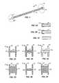
As shown inFIG. 1 ,tissue anchor 10 constructed in accordance with the principles of the present invention comprises abody 12 havingcaps 14 at each end thereof. Thebody 12 comprises a woven filament braid, as discussed in more detail above, and is illustrated in its elongated tubular configuration. In this particular embodiment, thebody 12 will be formed from a super elastic material, typically nitinol or eligiloy, and will be heat set, so that in the absence of radial constraint or an axially elongating force, the body will revert to its memory configuration having double-walled flanges formed at each end. One of theflanges 16 is shown in broken line inFIG. 1 , whileFIGS. 3A-3F illustrate various configurations of theflanges 16.
Referring now toFIGS. 1 A-IC, the double-walled flange structure 16 forms as the end oftubular body 12 axially foreshortens. Initially, the end of thetubular body 12 is maintained in its tubular configuration, as shown inFIG. 1A . Maintenance of the tubular configuration can be achieved in various ways, such as using an external tubular sheath or other restraint (not shown), by using a mandrel or other elongate structure which is advanced through an inside lumen orpassage 18 of the body to engage the end and/or occupy the entire lumen in order to maintain the tubular configuration, or the like. Once the constraint is removed, the pre-formed or memory shape of the double-walled flange structure will begin to form, as shown in Fig. IB. The end of thetubular structure 12 will move toward the middle of the tubular structure, as shown by the arrow in Fig. IB, and apre-formed ridge 17 will appear, eventually collapsing into the double-embodiments where thetubular body 12 has been pre-formed to have the double-walled flange structure 16 as part of its memory, the general change in geometry will also be true for those anchors where an axially compressive force must be applied in order to deploy the flanges. In such cases, the tubular body may have preformed scoring or other weakened regions which preferentially allow the woven braid to bend in the manner illustrated inFIGS. 1A-1C .
The end caps14 will be provided when it is desired to constrain the end of theanchor body 12 to prevent the end(s) from expanding. In some instances, theend cap 14 will have a solid face, as shown inFIG. 2A , which will close the inside lumen orpassage 18 to prevent or inhibit the flow of body fluids therethrough. Alternatively, theend cap 14amay be formed with a passage20 therethrough to allow for its flow through lumen18 (FIG. 2B ). In a still further alternate embodiment, as shown inFIG. 2C , thetissue anchor 10 may not include any end caps, allowing the end to expand in certain embodiments.
Referring now toFIGS. 3 A through3F, various deployment configurations for thetissue anchor 10 will be described (where thetissue anchor 10 is assumed to have the same elongated starting length in each illustrated deployment). InFIG. 3 A, theflanges 16 expand radially while acentral saddle region 22 does not expand. InFIG. 3B , thecentral saddle region 22 does not significantly expand but has a somewhat greater deployed length than that of the embodiment ofFIG. 3 A, resulting inflanges 16 having a slightly smaller diameters.FIG. 3B also illustrates a covering or membrane24 over the entire exterior of thetissue anchor 10, thus inhibiting tissue ingrowth and/or minimizing fluid leakage when the anchor is implanted. InFIG. 3C ,tissue anchor 10 includes the open end caps14aproviding anopen lumen 18 therethrough. InFIG. 3D , atissue anchor 10 having acentral saddle region 22 with a significantly expanded diameter is illustrated. InFIG. 3E , thetissue anchor 10 having open ends26 (that is, they are free from the end cap as illustrated in Fig. C) is illustrated.Passages 26 are shown to have generally the same diameter as thetubular body 22 in its non-deployed configuration. In contrast, inFIG. 3F , open ends28 are shown having diameters which are significantly greater than the non-deployed diameter of the anchor body. Similarly, thecentral saddle region 22 ofFIG. 3F is also significantly greater than the diameter of the non-deployed tissue anchor. It will be appreciated that the tissue anchors of the present invention may have a wide variety of configurations with different lengths, saddle region diameters, flange diameters, open lumens, closed lumens, membrane-covered surfaces, partially membrane-covered surfaces, and the like.
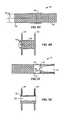
Referring now toFIGS. 4A and 4B , atissue anchor 30 having an alternative construction is illustrated. Thebody 32 oftissue anchor 30 is not pre-shaped, forming the enlarged flanges as a result of axial shortening. For example,elastic tethers 34 are provided which apply the axially compressive force to foreshorten the ends and form double-walled flanges 36, as illustrated inFIG. 4B . The resulting shape may be controlled by providing reinforcement over acentral saddle region 38 to prevent that region from axially foreshortening and/or radially expanding. Alternatively, thecentral saddle region 38 could be fused together to prevent deformation. In the device ofFIGS. 4 A and4B, the tissue anchor can be deployed through a tissue tract while the exterior is radially constrained or the or the ends axially lengthened. When released from the radial constraint, or axial tension, the elastic tethers will foreshorten the ends, forming double walled flanges where the saddle size (flange diameter, saddle length, saddle diameter) will conform exactly to the anatomy. Thus, the geometry will be “self-adjusting”. Reinforcement over the central saddle region is not necessary but could be utilized if desired for other purposes.
Referring now toFIGS. 5A and 5B , atissue anchor 40 comprises atubular body 42 which has both an elongated tubular configuration (as shown inFIG. 5 A) and an axially foreshortened configuration with double-walled flanges 44, as shown inFIG. 5B . Thetubular body 44 could either be of the self-expanding type or, alternatively, could require an axial compressive force to foreshorten the body into the configuration ofFIG. 5B . In either case, thetissue anchor 40 will be provided with a locking structure including a plurality ofaxial bars 46 which lock over the ends of the deployedtissue anchor 40, as illustrated inFIG. 5B .
Referring now toFIGS. 6A and 6B , atissue anchor 50 comprises ananchor body 52 which requires an axially compressive force in order to foreshorten the body to form the double-walled flanges 54, as shown inFIG. 6B . The axially compressive force is provided by a plurality oftethers 56 which extend through a lumen orcentral passage 58 of thebody 52 through the flange region and which then extend outwardly over thecentral saddle region 60 before passing back into the interior of the body. By then pulling on thetethers 56 relative to thebody 52, the flange regions will be axially compressed to radially expand, as shown inFIG. 6B , while and thecentral saddle region 60 may radially expand to an extent which depends on the braid configuration, the size and compliance of the lumen through which the device passes, and the force applied to the tethers. After the flanges have been deployed, the tethers may be locked in place, typically by alocking device 64, such as crimping pledgets, use of a unidirectional slide or other ratcheting lock device, or use of a slip-knot or a sliding element that relies on friction to secure its position.
Atissue anchor 70, as illustrated inFIGS. 7A and 7B , comprises ananchor body 72 having a lockingcylinder 74 in one end of the lumen or central passage76. Theanchor body 72 may be axially foreshortened by drawing on the free end of the lockingcylinder 74 and pulling the cylinder in the direction of the arrow until a lockingend 78 of the cylinder engages the far end of the deployedflange 80. Not only does thecylinder 74 act as an element to foreshorten theanchor body 72, it also acts as the lock to hold the anchor body open and provides a smooth cylindrical surface for the lumen to permit fluid flow or provide other access. Conveniently, the lockingend 78 of thecylinder 74 may be provided with notches or other apertures to allow that end to be collapsed within the lumen76 and to snap back open as it is pulled past theflange 80 to which it will lock.
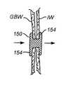
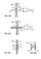
Referring now toFIGS. 8A and 8B , an exemplary tissue anchor orstent 150 comprises a counterwound, braided stent body, typically formed from a polymer such as polypropylene, polyester, nylon, or PEEK; a metal, such stainless steel, nitinol, or eligiloy; a bioabsorbable material, such as polyglycolic acid, lactic acid, caprolactone, polydioxanone, cat or bovine intestine; a natural fiber, such as silk or cotton; or mixtures, composites, or co-constructions of any of the above.Tethers 166 are provided which are connected at theremote end 168 of the stent, and which, when drawn in the direction away from the duodenum or other originating body lumen, will foreshorten the stent to create theflanges 154, as described previously. Drawing thetethers 166 in the proximal direction opens and maintains thecentral lumen 172 of deployedstent 150 to provide the luminal conduit which allows flow between anatomical lumens, such as the gallbladder, GB and intestine. A reduced diametercentral region 170 is located between theflanges 154. The width of thecentral region 170 may be controlled optionally by placing a restraining element, such as a cylinder or struts172 over the stent to prevent radial expansion. Thus, thestent 150 will automatically adjust to the thickness of the luminal walls. A restraint is not needed since the tissue geometry, particularly the tract dilation either before or after anchor placement, will provide a barrier which will restrain expansion of the central region and determine the length of the saddle region.
In another embodiment (not illustrated), thestent 150 can have proximal and distal ends connected centrally by an extensible material allowing the deployed stent to facilitate apposition of opposing luminal walls and minimize pressure necrosis.
Referring now toFIG. 9 , in some instances it will be desirable to provide a one-way flow element 180, such as a flat valve, within the interior of thestent 150. By properly orienting the stent, the one-way flow control element can then allow drainage from the gallbladder into the intestines while substantially inhibiting or blocking reflux flow back from the stomach into the gallbladder. Additionally, theflow control element 180 could serve as a restraint to define thecentral region 170 of the stent when expanded. Alternative valve designs include a sock valve placed within the interior or at the proximal end of the foreshortened anchor, a “duck bill” valve, a flapper valve, a spring-loaded ball valve, or other spring-loaded element, such as a tapered pin or plug.
Use of the tissue anchors of the present invention for draining a gallbladder will now be described. The biliary system of a patient (FIG. 10 ) includes the gallbladder GB which is connected to the cystic duct CD which feeds into the common bile duct CBD. The common bile duct, in turn, feeds bile into the descending part of the duodenum DD. While the present invention will be described with particular reference to attachment between the gallbladder GB and the descending duodenum DD, the principles apply to connecting a variety of other luminal structures, including the esophagus, the cms, the fundus, the bile duct, the intestines, and the like.
Referring now toFIG. 11 , a system for connecting luminal walls and placing a stent to establish a flow path therebetween is illustrated. Thissystem 100 is particularly useful for connecting a wall of the gallbladder to an intestinal wall, such as the duodenal wall or a stomach wall, but it will be appreciated that the system can find other uses in establishing other anastomotic connections, such as between the biliary duct including but not limited to the common bile duct, cystic duct and/or the pancreatic duct and the stomach, intestine or any part of the gastrointestinal tract.System 100 can also be used to create a connection to any fluid collection including but not limited to a pancreatic pseudocyst, abdominal fluid collections including ascites, pleural effusions and abscesses in any accessible location or the like.System 100 is also useful to create a connection between the urinary bladder and any part of the gastrointestinal tract.
The luminal wall connection system of the present invention comprises acatheter 112 including acatheter body 114 having adistal end 116 and aproximal end 118. Thecatheter body 114 has a lumen extending therethrough, with adistal port 120 of the lumen being visible inFIG. 11 . Aninflatable balloon 122 is mounted on the distal end of thecatheter body 114 and an inflation lumen (not shown) is provided in the wall of the catheter body and connected to aninflation port 124 near the proximal end of thecatheter body 114.
Aneedle 126 having a sharpeneddistal tip 128 is received within the lumen of thecatheter body 114 and is slidably received so that it can be selectively advanced from and/or retracted into thedistal port 120, as illustrated inFIG. 11 . A handle orgrip 130 is provided at the proximal end of theneedle 126 to facilitate manipulation.
An outertubular member 136 is coaxially received over thecatheter body 114 and includes adistal end 138 having adistal port 140 through which thecatheter body 114 projects.Proximal end 142 of the outertubular body 136 is connected to handle144.Catheter body 114 extends through the handle, allowing the catheter ofballoon 122 to be selectively extended and retracted relative to both theouter tube 136 andneedle 126.
The expandable tissue anchor/stent 150 is carried near thedistal end 138 of the outertubular body 136. The stent is optionally expanded in a variety of ways, including balloon expansion, self-expansion (where the stent would be released from constraint), heat-induced expansion of heat-sensitive alloy, such as nitinol, or the like. In the presently preferred embodiment, thestent 150 will comprise a polymer braid which may be foreshortened to induce radial expansion. This particular design was described in more detail above with reference toFIGS. 8A and 8B . The handle144 will include athumb slide 152 for effecting expansion of thestent 150 typically by pulling on tethers attached to the stent, as described below. A variety of other expansion mechanisms could be employed, for example, by pushing on the proximal end of the stents with rods or other pushing elements while a distal portion of the stent remains constrained.
Referring now toFIGS. 12A-12E , deployment of thestent 150 to attach gallbladder wall GBW to an intestinal wall IW will be described. Initially, an endoscope E will usually be trans-orally introduced so that it is within the intestines and can image the gallbladder to locate a target site for the anastomotic connection, as illustrated inFIG. 12 A. The endoscope will usually include at least a light source LS and a fiber optic image cable, or in some instances a CCD or other miniature camera, or ultrasound transducer. The endoscope will also include a conventional working channel WC, as illustrated in broken line inFIG. 12A .
Referring now toFIG. 12B , the luminalwall connection system 100 will be introduced through the working channel WC so that thedistal end 116 of thecatheter 114 is brought adjacent to the walls GBW and IW. Theneedle 126 may then be advanced through the walls to form an initial penetration.
Theuninflated balloon 122 will be advanced into the penetration, usually over theneedle 126, as shown inFIG. 12C . The balloon may then be inflated, typically assuming a standard hot dog, top hat or dog bone pattern, said top hat having the distal end where the proximal and distal ends are wider (e.g., have a larger diameter) than the central and distal regions. Proximal movement of the top-hat balloon will pull the GBW and the IW walls together. The penetration P is thus expanded prior to placement of thestent 150.
Referring toFIG. 12D , as an alternative to usingballoon 122, the penetration P can be expanded using a tapereddilator 160 which may be advanced directly over the needle through the endoscope. Optionally, a tapered dilator may be formed as a distal extension of the outer tubular member136 (not shown).
Referring now toFIG. 12E , after the penetration P has been expanded, the outertubular member 136 will be advanced so that thestent 150 is located in the expanded penetration. Astent 150 is then expanded, as shown inFIG. 12F , typically by foreshortening, as will be described in more detail below. Preferably, the proximal and distal ends of thestent 150 will be expanded or flared to form relativelylarge flange regions 154 which act to tightly hold the gallbladder wall GBW and intestinal wall IW together to promote tissue knitting or ingrowth to inhibit leakage from either the gallbladder or the intestines. Once in place, thestent 150 forms a central lumen152 (FIG. 12G ) which provides a flow path as indicated by the arrows inFIG. 12F from the gallbladder to the intestines. Following formation of a fistula or anastomosis the stent can optionally be removed, the flow now being through a tissue fistula orifice.
An alternate method is to follow theneedle 126 with the simultaneous movement of the outertubular member 136 withstent 150 andballoon 122. The stent is then released from constraint, with proximal and distal flanges now expanding and holding the lumens together, this followed by balloon expansion of the saddle region of the stent byballoon 122 which is inside the partially collapsed saddle region. This post-expansion method allows the anchor stent to hold the tissues together during tract dilation which is desirable.FIG. 11 new version will have stent on balloon Balloon is under saddle only.
While the above is a complete description of the preferred embodiments of the invention, various alternatives, modifications, and equivalents may be used. Therefore, the above description should not be taken as limiting the scope of the invention which is defined by the appended claims.
Claims (25)
1. A tissue anchor to appose adjacent tissue layers of a first luminal body and a second body in a patient, comprising a body formed of a woven filament braid, said body having an elongated tubular configuration and a foreshortened configuration where a proximal end of the body radially expands into only one proximal flange structure and a distal end of the body radially expands into only one distal flange structure leaving a cylindrical saddle region therebetween, wherein the adjacent tissue layers are apposed between the radially expanded proximal and distal ends and along the cylindrical saddle region, wherein a diameter of the cylindrical saddle region is greater than a diameter of the body in the elongated tubular configuration, wherein the proximal and distal flange structures have a diameter that is at least substantially twice the diameter of the cylindrical saddle region, wherein an opening of the proximal flange structure and an opening of the distal flange structure include a diameter that is greater than a diameter of the body in the elongated tubular configuration, wherein the tissue anchor is configured in the foreshortened configuration to allow fluid flow through the cylindrical saddle region, and an elastomeric membrane is formed over an entire length of the tissue anchor so that the membrane conforms to the tissue anchor in both the elongated tubular configuration and the foreshortened configuration.
2. A tissue anchor as inclaim 1 , wherein the radially expanded proximal and distal flange structures have preselected bending stiffnesses in the range from 1 g/mm to 100 g/mm.
3. A tissue anchor as inclaim 2 , wherein the radially expanded proximal and distal flange structures have preselected bending stiffnesses in the range from 4 g/mm to 40 g/mm.
4. A tissue anchor as inclaim 1 , wherein the body in the elongated tubular configuration has a diameter in the range from 7 mm to 100 mm.
5. A tissue anchor as inclaim 4 , wherein the proximal and distal flange structures have diameters in the range from 3 mm to 70 mm and the cylindrical saddle region has a diameter in the range from 1 mm to 40 mm in the foreshortened configuration.
6. A tissue anchor as inclaim 1 , wherein the proximal and distal flange structures are self-expanding when the body is released from constraint.
7. A tissue anchor as inclaim 6 , wherein the proximal and distal flange structures expand to a pre-shaped body configuration.
8. A tissue anchor as inclaim 1 , wherein the tissue anchor has a length of from 3 mm to 40 mm in the foreshortened configuration.
9. A tissue anchor as inclaim 1 , wherein the proximal and distal flange structures are double-walled flange structures.
10. A tissue anchor as inclaim 1 , wherein the tissue anchor in the foreshortened configuration is capable of maintaining the fluid flow through the cylindrical saddle region for a week or more.
11. A tissue anchor comprising a body formed of a woven filament braid, said body having an elongated tubular configuration and a foreshortened configuration, where proximal and distal ends of the body expand radially into respective proximal and distal flange structures leaving a cylindrical saddle region therebetween, the cylindrical saddle region having a longitudinal axis, wherein a length of the cylindrical saddle region is at least 40% less than a length of the body in the elongated configuration, wherein the proximal and distal flange structures have a diameter that is at least substantially twice the diameter of the cylindrical saddle region, wherein an opening of the proximal flange structure and an opening of the distal flange structure include a diameter that is greater than a diameter of the body in the elongated tubular configuration, wherein the proximal flange structure includes a single tissue apposing surface adjacent to the cylindrical saddle region and the distal flange structure includes a single tissue apposing surface adjacent to the cylindrical saddle region to appose adjacent tissue layers along the cylindrical saddle, wherein the tissue anchor is configured in the foreshortened configuration to allow fluid flow through an interior of the cylindrical saddle region, and an elastomeric membrane is formed over an entire length of the tissue anchor so that the membrane conforms to the tissue anchor in both the elongated tubular configuration and the foreshortened configuration.
12. A tissue anchor as inclaim 11 , wherein the body defines an open passage along a central axis in the elongated tubular configuration.
13. A tissue anchor as inclaim 11 , wherein the proximal and distal flange structures have preselected bending stiffnesses in the range from 1 g/mm to 100 g/mm.
14. A tissue anchor as inclaim 13 , wherein the proximal and distal flange structures have preselected bending stiffnesses in the range from 4 g/mm to 40 g/mm.
15. A tissue anchor as inclaim 11 , wherein the body in the elongated tubular configuration has a diameter in the range from 7 mm to 100 mm.
16. A tissue anchor as inclaim 15 , wherein the proximal and distal flange structures have diameters in the range from 3 mm to 70 mm and the saddle region has a diameter in the range from 1 mm to 40 mm in the foreshortened configuration.
17. A tissue anchor as inclaim 11 , wherein the proximal and distal flange structures are self-expanding when the body is released from constraint.
18. A tissue anchor as inclaim 11 , wherein the tissue anchor is configured to be removable during or after implantation.
19. A tissue anchor as inclaim 11 , wherein the tissue anchor has a length of from 3 mm to 40 mm in the foreshortened configuration.
20. A tissue anchor as inclaim 11 , wherein the cylindrical saddle region includes a longitudinal axis, and wherein the proximal and distal flange structures each expand into a double-walled flange structure.
21. A tissue anchor as inclaim 20 , wherein each of the double-walled flange structures includes a pair of walls, wherein each of the pair of walls are aligned substantially perpendicular to the longitudinal axis of the cylindrical saddle region when in the foreshortened configuration.
22. A tissue anchor as inclaim 11 , wherein the tissue anchor in the foreshortened configuration is capable of maintaining the fluid flow through the cylindrical saddle region for a week or more.
23. A tissue anchor to appose adjacent tissue layers of a first luminal body and a second body in a patient, comprising a body formed of a woven filament braid, said body having an elongated tubular configuration and a foreshortened configuration where proximal and distal ends of the body expand radially into respective proximal and distal flange structures leaving a cylindrical saddle region therebetween, wherein the proximal flange structure includes a single tissue opposing surface adjacent to the cylindrical saddle region and the distal flange structure includes a single tissue opposing surface adjacent to the cylindrical saddle region to appose the adjacent tissue layers along the cylindrical saddle region, wherein a diameter of the cylindrical saddle region is greater than a diameter of the body in the elongated tubular configuration, wherein the proximal and distal flange structures have a diameter that is at least substantially twice the diameter of the cylindrical saddle region, wherein an opening of the proximal flange structure and an opening of the distal flange structure include a diameter that is greater than a diameter of the body in the elongated tubular configuration, wherein the tissue anchor is configured in the foreshortened configuration to allow fluid flow through the cylindrical saddle region, and an elastomeric membrane is formed over an entire length of the tissue anchor so that the membrane conforms to the tissue anchor in both the elongated tubular configuration and the foreshortened configuration.
24. A tissue anchor as inclaim 23 , wherein the proximal and distal flange structures are double-walled flange structures.
25. A tissue anchor as inclaim 23 , wherein the tissue anchor in the foreshortened configuration is capable of maintaining the fluid flow through the cylindrical saddle region for a week or more.
Priority Applications (5)
| Application Number | Priority Date | Filing Date | Title |
|---|---|---|---|
| US13/892,958US10076330B2 (en) | 2008-05-12 | 2013-05-13 | Tissue anchor for securing tissue layers |
| US15/331,249US10390833B2 (en) | 2008-05-12 | 2016-10-21 | Tissue anchor for securing tissue layers |
| US16/107,067US11129617B2 (en) | 2008-05-12 | 2018-08-21 | Tissue anchor for securing tissue layers |
| US16/693,732US11129618B2 (en) | 2008-05-12 | 2019-11-25 | Tissue anchor for securing tissue layers |
| US17/412,360US20210378672A1 (en) | 2008-05-12 | 2021-08-26 | Tissue anchor for securing tissue layers |
Applications Claiming Priority (4)
| Application Number | Priority Date | Filing Date | Title |
|---|---|---|---|
| US5246008P | 2008-05-12 | 2008-05-12 | |
| US12/427,215US8454632B2 (en) | 2008-05-12 | 2009-04-21 | Tissue anchor for securing tissue layers |
| PCT/US2009/043445WO2009140195A1 (en) | 2008-05-12 | 2009-05-11 | Tissue anchor for securing tissue layers |
| US13/892,958US10076330B2 (en) | 2008-05-12 | 2013-05-13 | Tissue anchor for securing tissue layers |
Related Parent Applications (1)
| Application Number | Title | Priority Date | Filing Date |
|---|---|---|---|
| US12/427,215DivisionUS8454632B2 (en) | 2008-05-12 | 2009-04-21 | Tissue anchor for securing tissue layers |
Related Child Applications (1)
| Application Number | Title | Priority Date | Filing Date |
|---|---|---|---|
| US15/331,249ContinuationUS10390833B2 (en) | 2008-05-12 | 2016-10-21 | Tissue anchor for securing tissue layers |
Publications (2)
| Publication Number | Publication Date |
|---|---|
| US20130253546A1 US20130253546A1 (en) | 2013-09-26 |
| US10076330B2true US10076330B2 (en) | 2018-09-18 |
Family
ID=54013355
Family Applications (2)
| Application Number | Title | Priority Date | Filing Date |
|---|---|---|---|
| US12/427,215Active2029-05-11US8454632B2 (en) | 2008-05-12 | 2009-04-21 | Tissue anchor for securing tissue layers |
| US13/892,958ActiveUS10076330B2 (en) | 2008-05-12 | 2013-05-13 | Tissue anchor for securing tissue layers |
Family Applications Before (1)
| Application Number | Title | Priority Date | Filing Date |
|---|---|---|---|
| US12/427,215Active2029-05-11US8454632B2 (en) | 2008-05-12 | 2009-04-21 | Tissue anchor for securing tissue layers |
Country Status (4)
| Country | Link |
|---|---|
| US (2) | US8454632B2 (en) |
| EP (1) | EP2273942B1 (en) |
| JP (2) | JP6026741B2 (en) |
| WO (1) | WO2009140195A1 (en) |
Cited By (10)
| Publication number | Priority date | Publication date | Assignee | Title |
|---|---|---|---|---|
| US11304795B2 (en) | 2017-10-25 | 2022-04-19 | Boston Scientific Scimed, Inc. | Stent with atraumatic spacer |
| US11419741B2 (en) | 2019-06-17 | 2022-08-23 | Boston Scientific Scimed, Inc. | Covered endoprosthesis with improved branch access |
| US11517319B2 (en) | 2017-09-23 | 2022-12-06 | Universität Zürich | Medical occluder device |
| US11564787B2 (en) | 2019-11-18 | 2023-01-31 | Boston Scientific Scimed, Inc. | Stent with improved anti-migration properties |
| US11678970B2 (en) | 2018-12-04 | 2023-06-20 | Boston Scientific Scimed, Inc. | Device for anastomotic bypass |
| US11752314B2 (en) | 2019-02-07 | 2023-09-12 | Nxt Biomedical, Llc | Rivet shunt and method of deployment |
| US11944315B2 (en) | 2019-09-26 | 2024-04-02 | Universität Zürich | Left atrial appendage occlusion devices |
| US12303105B2 (en) | 2004-04-12 | 2025-05-20 | Boston Scientific Scimed, Inc. | Luminal structure anchoring devices and methods |
| US12343072B2 (en) | 2012-05-17 | 2025-07-01 | Boston Scientific Scimed, Inc. | Methods and devices for access across adjacent tissue layers |
| US12402885B2 (en) | 2017-09-23 | 2025-09-02 | Universität Zürich | Medical occlusion device |
Families Citing this family (123)
| Publication number | Priority date | Publication date | Assignee | Title |
|---|---|---|---|---|
| US8425539B2 (en) | 2004-04-12 | 2013-04-23 | Xlumena, Inc. | Luminal structure anchoring devices and methods |
| JP5111112B2 (en) | 2004-12-08 | 2012-12-26 | エックスルミナ, インコーポレイテッド | Device for performing needle-guided therapy |
| US8777967B2 (en) | 2005-06-09 | 2014-07-15 | Xlumena, Inc. | Methods and devices for anchoring to tissue |
| US8784437B2 (en) | 2005-06-09 | 2014-07-22 | Xlumena, Inc. | Methods and devices for endosonography-guided fundoplexy |
| US9232997B2 (en) | 2006-11-07 | 2016-01-12 | Corvia Medical, Inc. | Devices and methods for retrievable intra-atrial implants |
| US20110257723A1 (en) | 2006-11-07 | 2011-10-20 | Dc Devices, Inc. | Devices and methods for coronary sinus pressure relief |
| US10413284B2 (en) | 2006-11-07 | 2019-09-17 | Corvia Medical, Inc. | Atrial pressure regulation with control, sensing, monitoring and therapy delivery |
| EP2097012A4 (en) | 2006-11-07 | 2012-08-15 | David Stephen Celermajer | Devices and methods for the treatment of heart failure |
| US8372114B2 (en)* | 2006-11-13 | 2013-02-12 | Electroformed Stents, Inc. | Over-the-wire exclusion device and system for delivery |
| WO2009124148A1 (en)* | 2008-04-04 | 2009-10-08 | Curaseal, Inc. | Implantable fistula closure device |
| US8454632B2 (en) | 2008-05-12 | 2013-06-04 | Xlumena, Inc. | Tissue anchor for securing tissue layers |
| US20090281379A1 (en)* | 2008-05-12 | 2009-11-12 | Xlumena, Inc. | System and method for transluminal access |
| EP2330985A4 (en) | 2008-09-04 | 2015-11-18 | Curaseal Inc | Inflatable devices for enteric fistula treatment |
| US9782565B2 (en) | 2008-10-01 | 2017-10-10 | Covidien Lp | Endoscopic ultrasound-guided biliary access system |
| US9186128B2 (en) | 2008-10-01 | 2015-11-17 | Covidien Lp | Needle biopsy device |
| US8968210B2 (en) | 2008-10-01 | 2015-03-03 | Covidien LLP | Device for needle biopsy with integrated needle protection |
| US9332973B2 (en) | 2008-10-01 | 2016-05-10 | Covidien Lp | Needle biopsy device with exchangeable needle and integrated needle protection |
| US11298113B2 (en) | 2008-10-01 | 2022-04-12 | Covidien Lp | Device for needle biopsy with integrated needle protection |
| US9364259B2 (en) | 2009-04-21 | 2016-06-14 | Xlumena, Inc. | System and method for delivering expanding trocar through a sheath |
| US20120130417A1 (en)* | 2010-10-25 | 2012-05-24 | Keke Lepulu | Apparatus and method for penetrating and enlarging adjacent tissue layers |
| CA2761871C (en)* | 2009-05-15 | 2013-12-10 | Cook Medical Technologies Llc | Delivery system for magnetic anastomosis device |
| JP5535313B2 (en) | 2009-05-29 | 2014-07-02 | エックスルミナ, インコーポレイテッド | Device and method for deploying a stent across adjacent tissue layers |
| US20110184504A1 (en)* | 2010-01-22 | 2011-07-28 | Medtronic Vascular, Inc. | Methods and Apparatus for Providing an Arteriovenous Fistula |
| EP2528646A4 (en) | 2010-01-29 | 2017-06-28 | DC Devices, Inc. | Devices and systems for treating heart failure |
| US10568628B2 (en) | 2017-05-23 | 2020-02-25 | Muffin Incorporated | Closing device for tissue openings |
| WO2011143443A2 (en)* | 2010-05-12 | 2011-11-17 | Medical Device Innovations Llc | Method and device for treatment of arrhythmias and other maladies |
| WO2012058244A2 (en)* | 2010-10-25 | 2012-05-03 | Xlumena, Inc. | Apparatus and method for penetrating and enlarging adjacent tissue layers |
| WO2012071031A1 (en)* | 2010-11-23 | 2012-05-31 | Treus Medical, Inc | Biliary shunts, delivery systems, and methods of using the same |
| US12303119B2 (en) | 2011-02-10 | 2025-05-20 | Corvia Medical, Inc. | Apparatus and methods to create and maintain an intra-atrial pressure relief opening |
| WO2012109557A2 (en) | 2011-02-10 | 2012-08-16 | Dc Devices, Inc. | Apparatus and methods to create and maintain an intra-atrial pressure relief opening |
| US9393133B2 (en) | 2011-02-18 | 2016-07-19 | Piolax Medical Devices, Inc. | Abdominal cavity-vein shunt stent |
| AU2012225575B9 (en) | 2011-03-08 | 2015-08-20 | W. L. Gore & Associates, Inc. | Medical device for use with a stoma |
| WO2012149167A2 (en) | 2011-04-26 | 2012-11-01 | Christopher Gerard Kunis | Method and device for treatment of hypertension and other maladies |
| JP6122424B2 (en) | 2011-06-16 | 2017-04-26 | キュラシール インコーポレイテッド | Device for fistula treatment and related method |
| CN107137114A (en) | 2011-06-17 | 2017-09-08 | 库拉希尔公司 | The device and method treated for fistula |
| KR101311756B1 (en) | 2011-07-06 | 2013-09-26 | 신경민 | A Stent |
| US8951223B2 (en) | 2011-12-22 | 2015-02-10 | Dc Devices, Inc. | Methods and devices for intra-atrial shunts having adjustable sizes |
| ES2980140T3 (en)* | 2013-02-21 | 2024-09-30 | Boston Scient Scimed Inc | Devices for forming an anastomosis |
| US10675450B2 (en)* | 2014-03-12 | 2020-06-09 | Corvia Medical, Inc. | Devices and methods for treating heart failure |
| US11076860B2 (en) | 2014-03-31 | 2021-08-03 | DePuy Synthes Products, Inc. | Aneurysm occlusion device |
| US11154302B2 (en) | 2014-03-31 | 2021-10-26 | DePuy Synthes Products, Inc. | Aneurysm occlusion device |
| US10130372B2 (en) | 2014-04-30 | 2018-11-20 | Cerus Endovascular Limited | Occlusion Device |
| US11712230B2 (en) | 2014-05-02 | 2023-08-01 | W. L. Gore & Associates, Inc. | Occluder and anastomosis devices |
| US10363040B2 (en)* | 2014-05-02 | 2019-07-30 | W. L. Gore & Associates, Inc. | Anastomosis devices |
| US11439396B2 (en) | 2014-05-02 | 2022-09-13 | W. L. Gore & Associates, Inc. | Occluder and anastomosis devices |
| EP3360488B1 (en) | 2014-05-28 | 2020-03-18 | Boston Scientific Scimed, Inc. | Catheter with radiofrequency cutting tip and heated balloon |
| EP3160362B1 (en) | 2014-06-30 | 2023-01-04 | Cook Medical Technologies LLC | Expandable mesh with locking feature |
| JP6799526B2 (en) | 2014-07-23 | 2020-12-16 | コルヴィア メディカル インコーポレイテッド | Equipment and methods for the treatment of heart failure |
| KR101696810B1 (en)* | 2015-02-04 | 2017-02-01 | 주식회사 엠아이텍 | Stent for connecting adjacent tissues and manufacturing method thereof |
| WO2016141295A1 (en) | 2015-03-05 | 2016-09-09 | Merit Medical Systems, Inc. | Vascular prosthesis deployment device and method of use |
| KR20170005661A (en) | 2015-07-06 | 2017-01-16 | 주식회사 비씨엠 | Medical stent enhanced the resistance of the end portions |
| KR101691118B1 (en) | 2015-07-06 | 2016-12-29 | 주식회사 비씨엠 | The resistance of the end-enhanced method for manufacturing a medical stent and a stent |
| US10470906B2 (en) | 2015-09-15 | 2019-11-12 | Merit Medical Systems, Inc. | Implantable device delivery system |
| EP4011303B1 (en) | 2015-12-07 | 2024-06-12 | Cerus Endovascular Limited | Occlusion device |
| JP6659845B2 (en)* | 2015-12-18 | 2020-03-04 | ボストン サイエンティフィック サイムド,インコーポレイテッドBoston Scientific Scimed,Inc. | Cardiac tissue anchor |
| WO2017153603A1 (en) | 2016-03-11 | 2017-09-14 | Cerus Endovascular Limited | Occlusion device |
| US10299795B2 (en)* | 2016-04-28 | 2019-05-28 | Mayo Foundation For Medical Education And Research | Devices and methods for esophageal lengthening and anastomosis formation |
| US20190216603A1 (en)* | 2016-06-30 | 2019-07-18 | Tel Hashomer Medical Research, Infrastructure And Services Ltd. | Apparatus and method for accessing aorta |
| US10702384B2 (en) | 2016-08-16 | 2020-07-07 | Boston Scientific Scimed, Inc. | Heart valve regurgitation anchor and delivery tool |
| CN115054413A (en) | 2016-09-29 | 2022-09-16 | 美国医疗设备有限公司 | Method of adjusting effective length of stent and prosthesis delivery catheter assembly |
| CN110114027B (en)* | 2016-11-01 | 2022-09-06 | 贝利斯医疗公司 | Method and apparatus for puncturing tissue |
| EP3556326A4 (en)* | 2016-12-15 | 2020-07-08 | BCM Co., Ltd. | Method for fabricating medical stent having resistance-reinforced end parts, and stent fabricated thereby |
| JP2018099266A (en)* | 2016-12-20 | 2018-06-28 | ビーシーエム カンパニー,リミテッド | Medical stent with distal resistance force being strengthened |
| US11857745B2 (en)* | 2017-02-07 | 2024-01-02 | ewimed | Method and apparatus for treating fluid build-up in a body cavity, including method and apparatus for treating ascites and pleural effusions |
| CN110545739A (en) | 2017-02-23 | 2019-12-06 | 德普伊新特斯产品公司 | aneurysm devices and delivery systems |
| US11628078B2 (en) | 2017-03-15 | 2023-04-18 | Merit Medical Systems, Inc. | Transluminal delivery devices and related kits and methods |
| EP4467111A3 (en) | 2017-03-15 | 2025-03-05 | Merit Medical Systems, Inc. | Transluminal stents |
| USD836194S1 (en) | 2017-03-21 | 2018-12-18 | Merit Medical Systems, Inc. | Stent deployment device |
| CA3049473A1 (en)* | 2017-03-30 | 2018-10-04 | Boston Scientific Scimed, Inc. | Stents with dual tissue-wall anchoring features |
| US11724075B2 (en)* | 2017-04-18 | 2023-08-15 | W. L. Gore & Associates, Inc. | Deployment constraining sheath that enables staged deployment by device section |
| US11589884B2 (en) | 2017-04-28 | 2023-02-28 | W. L. Gore & Associates, Inc. | Endoscopic transluminal stent access and delivery system |
| CA3063418A1 (en) | 2017-05-17 | 2018-11-22 | Massachusetts Institute Of Technology | Self-actuating articles |
| US11541015B2 (en) | 2017-05-17 | 2023-01-03 | Massachusetts Institute Of Technology | Self-righting systems, methods, and related components |
| JP7068349B2 (en)* | 2017-06-20 | 2022-05-16 | ボストン サイエンティフィック サイムド,インコーポレイテッド | Systems and methods for creating permanent drainage fistulas |
| KR101976743B1 (en)* | 2017-07-14 | 2019-05-09 | 주식회사 비씨엠 | Stent insertion device for human digestive organ connection |
| WO2019032816A1 (en)* | 2017-08-10 | 2019-02-14 | St. Jude Medical, Cardiology Division, Inc. | Collapsible medical device for atrial sealing and trans-septal access |
| WO2019036541A2 (en) | 2017-08-17 | 2019-02-21 | Boston Scientific Scimed, Inc. | Anchor delivery system and methods for valve repair |
| JP7414710B2 (en) | 2017-08-21 | 2024-01-16 | シーラス エンドバスキュラー リミテッド | occlusion device |
| EP3684296B1 (en) | 2017-09-19 | 2024-02-14 | Boston Scientific Scimed, Inc. | Percutaneous repair of mitral prolapse |
| US11998707B2 (en) | 2017-10-03 | 2024-06-04 | Boston Scientific Scimed, Inc. | Flow control stent |
| CN111315319B (en) | 2017-11-01 | 2022-10-18 | 波士顿科学国际有限公司 | Esophageal stent comprising valve member |
| US10905430B2 (en) | 2018-01-24 | 2021-02-02 | DePuy Synthes Products, Inc. | Aneurysm device and delivery system |
| CN111787871B (en)* | 2018-03-29 | 2023-12-12 | 波士顿科学国际有限公司 | flow control valve |
| US10888691B2 (en) | 2018-04-24 | 2021-01-12 | Olympus Corporation | Stent delivery method |
| CA3100710A1 (en) | 2018-05-17 | 2019-11-21 | Massachusetts Institute Of Technology | Systems for electrical stimulation |
| US11058430B2 (en) | 2018-05-25 | 2021-07-13 | DePuy Synthes Products, Inc. | Aneurysm device and delivery system |
| US11596412B2 (en) | 2018-05-25 | 2023-03-07 | DePuy Synthes Products, Inc. | Aneurysm device and delivery system |
| US10939915B2 (en) | 2018-05-31 | 2021-03-09 | DePuy Synthes Products, Inc. | Aneurysm device and delivery system |
| US11051825B2 (en) | 2018-08-08 | 2021-07-06 | DePuy Synthes Products, Inc. | Delivery system for embolic braid |
| KR102146913B1 (en)* | 2018-08-14 | 2020-08-24 | 울산대학교 산학협력단 | Biodegradable stent and using method thereof |
| US11123077B2 (en) | 2018-09-25 | 2021-09-21 | DePuy Synthes Products, Inc. | Intrasaccular device positioning and deployment system |
| US11076861B2 (en) | 2018-10-12 | 2021-08-03 | DePuy Synthes Products, Inc. | Folded aneurysm treatment device and delivery method |
| US11737745B2 (en) | 2018-10-24 | 2023-08-29 | Mayo Foundation For Medical Education And Research | Medical devices and methods for body conduit lengthening and anastomosis formation |
| CN109260577B (en)* | 2018-11-22 | 2021-10-26 | 上海市东方医院 | Bidirectional displacement-preventing pancreatic duct bracket and implanting device |
| JP7229737B2 (en)* | 2018-11-30 | 2023-02-28 | ビーシーエム カンパニー,リミテッド | Stent insertion device for connecting human digestive organs |
| US11406392B2 (en) | 2018-12-12 | 2022-08-09 | DePuy Synthes Products, Inc. | Aneurysm occluding device for use with coagulating agents |
| US11272939B2 (en) | 2018-12-18 | 2022-03-15 | DePuy Synthes Products, Inc. | Intrasaccular flow diverter for treating cerebral aneurysms |
| US11998467B2 (en) | 2019-01-28 | 2024-06-04 | Tensor Flow Ventures Llc | Stent delivery for vascular surgery |
| US11666464B2 (en) | 2019-01-28 | 2023-06-06 | Tensor Flow Ventures Llc | Magnetic stent and stent delivery |
| JP2022523121A (en) | 2019-02-01 | 2022-04-21 | マサチューセッツ インスティテュート オブ テクノロジー | Systems and methods for liquid injection |
| US11134953B2 (en) | 2019-02-06 | 2021-10-05 | DePuy Synthes Products, Inc. | Adhesive cover occluding device for aneurysm treatment |
| US11337706B2 (en) | 2019-03-27 | 2022-05-24 | DePuy Synthes Products, Inc. | Aneurysm treatment device |
| US11672542B2 (en) | 2019-05-21 | 2023-06-13 | DePuy Synthes Products, Inc. | Aneurysm treatment with pushable ball segment |
| US11607226B2 (en) | 2019-05-21 | 2023-03-21 | DePuy Synthes Products, Inc. | Layered braided aneurysm treatment device with corrugations |
| US10653425B1 (en) | 2019-05-21 | 2020-05-19 | DePuy Synthes Products, Inc. | Layered braided aneurysm treatment device |
| US11497504B2 (en) | 2019-05-21 | 2022-11-15 | DePuy Synthes Products, Inc. | Aneurysm treatment with pushable implanted braid |
| US11413046B2 (en) | 2019-05-21 | 2022-08-16 | DePuy Synthes Products, Inc. | Layered braided aneurysm treatment device |
| US11278292B2 (en) | 2019-05-21 | 2022-03-22 | DePuy Synthes Products, Inc. | Inverting braided aneurysm treatment system and method |
| US11602350B2 (en) | 2019-12-05 | 2023-03-14 | DePuy Synthes Products, Inc. | Intrasaccular inverting braid with highly flexible fill material |
| JP7631632B2 (en)* | 2019-09-04 | 2025-02-19 | Sbカワスミ株式会社 | Stents |
| JP7648607B2 (en)* | 2019-09-09 | 2025-03-18 | シファメド・ホールディングス・エルエルシー | ADJUSTABLE SHUNTS AND ASSOCIATED SYSTEMS AND METHODS - Patent application |
| US11541216B2 (en) | 2019-11-21 | 2023-01-03 | Massachusetts Institute Of Technology | Methods for manufacturing tissue interfacing components |
| US11457926B2 (en) | 2019-12-18 | 2022-10-04 | DePuy Synthes Products, Inc. | Implant having an intrasaccular section and intravascular section |
| US11406404B2 (en) | 2020-02-20 | 2022-08-09 | Cerus Endovascular Limited | Clot removal distal protection methods |
| EP4185239A4 (en) | 2020-07-24 | 2024-08-07 | Merit Medical Systems, Inc. | ESOPHAGEAL STENT PROSTHESES AND RELATED METHODS |
| AU2021342292B2 (en)* | 2020-09-14 | 2024-07-18 | Boston Scientific Scimed, Inc. | Stents and methods for use and manufacture of stents with improved retention members |
| CA3194910A1 (en) | 2020-10-26 | 2022-05-05 | Tiffany ETHRIDGE | Esophageal stents with helical thread |
| AU2022222662A1 (en)* | 2021-02-16 | 2023-09-07 | Andrew Howard Schulick | Endoleak device |
| US20230026939A1 (en) | 2021-07-26 | 2023-01-26 | Tensor Flow Ventures Llc | Dual stent and delivery system, delivery tool apparatus, and method of delivery of dual stents |
| US20230277348A1 (en) | 2021-07-26 | 2023-09-07 | Tensor Flow Ventures Llc | Dual stent and delivery system, delivery tool apparatus, and method of delivery of dual stents |
| CN118234456A (en) | 2021-11-15 | 2024-06-21 | 波士顿科学国际有限公司 | Devices, systems, and methods for pyloric occlusion |
| CN118302135A (en) | 2021-11-29 | 2024-07-05 | 波士顿科学国际有限公司 | Devices, systems, and methods for pyloric occlusion |
| US20230380837A1 (en)* | 2022-05-26 | 2023-11-30 | Vasorum Ltd. | Wound closure and tissue coupling systems and methods |
Citations (389)
| Publication number | Priority date | Publication date | Assignee | Title |
|---|---|---|---|---|
| US2127903A (en) | 1936-05-05 | 1938-08-23 | Davis & Geck Inc | Tube for surgical purposes and method of preparing and using the same |
| US3039468A (en) | 1959-01-07 | 1962-06-19 | Joseph L Price | Trocar and method of treating bloat |
| US3717151A (en) | 1971-03-11 | 1973-02-20 | R Collett | Flesh penetrating apparatus |
| US3874388A (en) | 1973-02-12 | 1975-04-01 | Ochsner Med Found Alton | Shunt defect closure system |
| US3970090A (en) | 1975-02-03 | 1976-07-20 | Physio Medics, Inc. | Catheter |
| US4173392A (en) | 1977-07-20 | 1979-11-06 | American Hospital Supply Corporation | Glass fiber light guide and method of making the same |
| GB2020557A (en) | 1978-05-13 | 1979-11-21 | Ruesch Gmbh & Co Kg Willy | Medical instrument for removing foreign bodies |
| US4235238A (en) | 1978-05-11 | 1980-11-25 | Olympus Optical Co., Ltd. | Apparatus for suturing coeliac tissues |
| JPS5835219A (en) | 1981-08-27 | 1983-03-01 | Toyota Motor Corp | Manifold converter holder |
| US4580568A (en) | 1984-10-01 | 1986-04-08 | Cook, Incorporated | Percutaneous endovascular stent and method for insertion thereof |
| US4587972A (en) | 1984-07-16 | 1986-05-13 | Morantte Jr Bernardo D | Device for diagnostic and therapeutic intravascular intervention |
| US4608965A (en) | 1985-03-27 | 1986-09-02 | Anspach Jr William E | Endoscope retainer and tissue retracting device |
| JPS62233168A (en) | 1986-04-03 | 1987-10-13 | オリンパス光学工業株式会社 | Catheter |
| US4705040A (en) | 1985-11-18 | 1987-11-10 | Medi-Tech, Incorporated | Percutaneous fixation of hollow organs |
| US4790813A (en) | 1984-12-17 | 1988-12-13 | Intravascular Surgical Instruments, Inc. | Method and apparatus for surgically removing remote deposits |
| US4869263A (en) | 1988-02-04 | 1989-09-26 | Cardiometrics, Inc. | Device and method for measuring volumetric blood flow in a vessel |
| US4896678A (en) | 1986-12-12 | 1990-01-30 | Olympus Optical Co., Ltd. | Endoscopic treating tool |
| US4917097A (en) | 1987-10-27 | 1990-04-17 | Endosonics Corporation | Apparatus and method for imaging small cavities |
| US4920967A (en) | 1986-07-18 | 1990-05-01 | Pfizer Hospital Products Group, Inc. | Doppler tip wire guide |
| US4950285A (en) | 1989-11-27 | 1990-08-21 | Wilk Peter J | Suture device |
| US4973317A (en) | 1989-07-14 | 1990-11-27 | Bobrove Arthur M | Automatic sheath protection of hypodermic needle |
| US4990139A (en) | 1986-09-10 | 1991-02-05 | Jang G David | Tandem independently inflatable/deflatable multiple diameter balloon angioplasty catheter systems |
| US5024655A (en) | 1989-09-05 | 1991-06-18 | Freeman Andrew B | Epidural catheter apparatus and associated method |
| US5061275A (en) | 1986-04-21 | 1991-10-29 | Medinvent S.A. | Self-expanding prosthesis |
| US5064435A (en) | 1990-06-28 | 1991-11-12 | Schneider (Usa) Inc. | Self-expanding prosthesis having stable axial length |
| US5180392A (en) | 1988-02-01 | 1993-01-19 | Einar Skeie | Anastomotic device |
| US5183464A (en) | 1991-05-17 | 1993-02-02 | Interventional Thermodynamics, Inc. | Radially expandable dilator |
| US5197971A (en) | 1990-03-02 | 1993-03-30 | Bonutti Peter M | Arthroscopic retractor and method of using the same |
| US5207229A (en) | 1989-12-21 | 1993-05-04 | Advanced Biomedical Devices, Inc. | Flexibility steerable guidewire with inflatable balloon |
| US5209727A (en) | 1992-01-29 | 1993-05-11 | Interventional Technologies, Inc. | Guide wire with integral angioplasty balloon |
| US5211651A (en) | 1989-08-18 | 1993-05-18 | Evi Corporation | Catheter atherotome |
| JPH05137794A (en) | 1990-10-04 | 1993-06-01 | Schneider Europ Ag | Balloon expansion catheter |
| US5221258A (en) | 1991-01-22 | 1993-06-22 | Shturman Technologies, Inc. | Introduction balloon catheter |
| US5224945A (en) | 1992-01-13 | 1993-07-06 | Interventional Technologies, Inc. | Compressible/expandable atherectomy cutter |
| US5226421A (en) | 1992-03-06 | 1993-07-13 | Cardiometrics, Inc. | Doppler elongate flexible member having an inflatable balloon mounted thereon |
| JPH05192407A (en) | 1991-07-22 | 1993-08-03 | Dow Corning Wright Corp | Extirpating device for distention atheroma |
| US5234447A (en) | 1990-08-28 | 1993-08-10 | Robert L. Kaster | Side-to-end vascular anastomotic staple apparatus |
| US5246445A (en) | 1990-04-19 | 1993-09-21 | Instent Inc. | Device for the treatment of constricted ducts in human bodies |
| US5246007A (en) | 1992-03-13 | 1993-09-21 | Cardiometrics, Inc. | Vascular catheter for measuring flow characteristics and method |
| US5257990A (en) | 1992-02-24 | 1993-11-02 | Kensey Nash Corporation | Electrosurgical catheter instrument with impacting working head and method of use |
| US5258000A (en) | 1991-11-25 | 1993-11-02 | Cook Incorporated | Tissue aperture repair device |
| US5261920A (en) | 1992-08-21 | 1993-11-16 | Ethicon, Inc. | Anvil bushing for circular stapler |
| JPH05508563A (en) | 1990-05-07 | 1993-12-02 | ケンゼー・ナッシュ・コーポレーション | A device for sealing an opening in tissue that separates one part of a living body from another part of the body. |
| JPH05329165A (en) | 1991-11-25 | 1993-12-14 | Cook Inc | Tissue opening medical treatment device |
| US5275610A (en) | 1991-05-13 | 1994-01-04 | Cook Incorporated | Surgical retractors and method of use |
| US5275611A (en) | 1990-11-20 | 1994-01-04 | Innerdyne Medical, Inc. | Tension guide and dilator |
| US5282824A (en) | 1990-10-09 | 1994-02-01 | Cook, Incorporated | Percutaneous stent assembly |
| US5290249A (en) | 1990-10-09 | 1994-03-01 | Vance Products Incorporated | Surgical access sheath |
| US5304198A (en) | 1992-11-13 | 1994-04-19 | Target Therapeutics | Single-lumen balloon catheter having a directional valve |
| US5330497A (en) | 1989-11-22 | 1994-07-19 | Dexide, Inc. | Locking trocar sleeve |
| US5353785A (en) | 1991-07-15 | 1994-10-11 | Wilk Peter J | Method for lifting abdominal wall during laparoscopic surgery |
| US5368595A (en) | 1990-09-06 | 1994-11-29 | United States Surgical Corporation | Implant assist apparatus |
| US5372588A (en) | 1992-11-24 | 1994-12-13 | Farley; Kevin | Trocar having blunt tip |
| US5381788A (en) | 1991-08-05 | 1995-01-17 | United States Surgical Corporation | Surgical retractor |
| US5387235A (en) | 1991-10-25 | 1995-02-07 | Cook Incorporated | Expandable transluminal graft prosthesis for repair of aneurysm |
| EP0637431A1 (en) | 1993-08-05 | 1995-02-08 | VODA, Jan | Suture device |
| US5395349A (en) | 1991-12-13 | 1995-03-07 | Endovascular Technologies, Inc. | Dual valve reinforced sheath and method |
| JPH0796038A (en) | 1994-05-02 | 1995-04-11 | Olympus Optical Co Ltd | Balloon catheter |
| US5425739A (en) | 1989-03-09 | 1995-06-20 | Avatar Design And Development, Inc. | Anastomosis stent and stent selection system |
| US5443484A (en) | 1992-06-16 | 1995-08-22 | Loma Linda University Medical Center | Trocar and method for endoscopic surgery |
| US5458131A (en) | 1992-08-25 | 1995-10-17 | Wilk; Peter J. | Method for use in intra-abdominal surgery |
| US5470337A (en) | 1994-05-17 | 1995-11-28 | Moss; Gerald | Surgical fastener |
| US5495851A (en) | 1995-03-23 | 1996-03-05 | Roanoke Gastroenterology, P.C. | Use of endoscopic ultrasound and stimulated bilary drainage in the diagnosis of cholecystitis and microlithiasis |
| US5496311A (en) | 1988-10-28 | 1996-03-05 | Boston Scientific Corporation | Physiologic low stress angioplasty |
| JPH0871158A (en) | 1994-09-08 | 1996-03-19 | Nippon Zeon Co Ltd | Anchor type guide wire |
| JPH08504940A (en) | 1991-10-29 | 1996-05-28 | パーストープ、アナリティカル、アクチボラグ | Luminescence measurement system and luminometer device |
| US5520700A (en) | 1992-11-13 | 1996-05-28 | Technion Research & Development Foundation, Ltd. | Stapler device particularly useful in medical suturing |
| US5531699A (en) | 1994-09-19 | 1996-07-02 | Abbott Laboratories | Spring-loaded reciprocable stylet holder |
| US5536248A (en) | 1992-05-11 | 1996-07-16 | Arrow Precision Products, Inc. | Method and apparatus for electrosurgically obtaining access to the biliary tree and placing a stent therein |
| JPH08509639A (en) | 1993-04-29 | 1996-10-15 | シメッド ライフ システムズ インコーポレイテッド | Expandable vascular obstruction remover |
| JPH08299455A (en) | 1994-03-30 | 1996-11-19 | Corvita Corp | Method and device for introducing stent or stent transplant piece |
| US5588432A (en) | 1988-03-21 | 1996-12-31 | Boston Scientific Corporation | Catheters for imaging, sensing electrical potentials, and ablating tissue |
| JPH09500047A (en) | 1993-11-24 | 1997-01-07 | ヴァリーラブ・インコーポレーテッド | Retrograde high-frequency tissue separator |
| US5601588A (en) | 1994-09-29 | 1997-02-11 | Olympus Optical Co., Ltd. | Endoscopic puncture needle |
| US5603698A (en) | 1993-04-13 | 1997-02-18 | Boston Scientific Corporation | Prosthesis delivery system |
| US5620457A (en) | 1994-11-23 | 1997-04-15 | Medinol Ltd. | Catheter balloon |
| US5620456A (en) | 1995-10-20 | 1997-04-15 | Lasersurge, Inc. | Trocar assembly |
| JPH09504186A (en) | 1993-04-30 | 1997-04-28 | メディカル サイエンティフィク インコーポレイテッド | Bipole electrosurgical trocar |
| JPH09140804A (en) | 1995-11-14 | 1997-06-03 | Schneider Europ Ag | Stent delivery device |
| WO1997027898A1 (en) | 1996-02-02 | 1997-08-07 | Transvascular, Inc. | Methods and apparatus for connecting openings formed in adjacent blood vessels or other anatomical structures |
| US5662664A (en) | 1992-09-04 | 1997-09-02 | Laurus Medical Corporation | Endoscopic suture system |
| US5681345A (en) | 1995-03-01 | 1997-10-28 | Scimed Life Systems, Inc. | Sleeve carrying stent |
| US5688247A (en) | 1992-06-30 | 1997-11-18 | Hans Haindl | Port catheter |
| US5697944A (en) | 1995-11-15 | 1997-12-16 | Interventional Technologies Inc. | Universal dilator with expandable incisor |
| US5709707A (en) | 1995-10-30 | 1998-01-20 | Children's Medical Center Corporation | Self-centering umbrella-type septal closure device |
| US5709671A (en) | 1995-10-16 | 1998-01-20 | Ethicon Endo-Surgery, Inc. | Trocar having an improved tip configuration |
| US5709713A (en)* | 1995-03-31 | 1998-01-20 | Cardiovascular Concepts, Inc. | Radially expansible vascular prosthesis having reversible and other locking structures |
| US5713870A (en) | 1991-11-27 | 1998-02-03 | Yoon; Inbae | Retractable safety penetrating instrument with laterally extendable spring strip |
| US5725552A (en) | 1994-07-08 | 1998-03-10 | Aga Medical Corporation | Percutaneous catheter directed intravascular occlusion devices |
| JPH1094543A (en) | 1996-09-13 | 1998-04-14 | Interventional Technol Inc | Apparatus and method for excising or expanding constricted tissue of blood vessel |
| JPH10155799A (en) | 1996-12-04 | 1998-06-16 | Olympus Optical Co Ltd | Organ lifting device |
| US5817062A (en) | 1996-03-12 | 1998-10-06 | Heartport, Inc. | Trocar |
| US5827276A (en) | 1995-03-24 | 1998-10-27 | Board Of Regents Of Univ Of Nebraksa | Apparatus for volumetric tissue ablation |
| US5830222A (en) | 1995-10-13 | 1998-11-03 | Transvascular, Inc. | Device, system and method for intersititial transvascular intervention |
| US5843127A (en) | 1994-08-22 | 1998-12-01 | Le Medical Technologies, Inc. | Fixation device and method for installing same |
| US5843116A (en) | 1996-05-02 | 1998-12-01 | Cardiovascular Dynamics, Inc. | Focalized intraluminal balloons |
| US5853422A (en) | 1996-03-22 | 1998-12-29 | Scimed Life Systems, Inc. | Apparatus and method for closing a septal defect |
| US5853421A (en) | 1995-06-07 | 1998-12-29 | Datascope Investment Corp. | Guide wire with releasable barb anchor |
| US5857999A (en) | 1995-05-05 | 1999-01-12 | Imagyn Medical Technologies, Inc. | Small diameter introducer for laparoscopic instruments |
| US5858006A (en) | 1992-11-06 | 1999-01-12 | Texas Instruments Incorporated | Hypodermic needle with a protrusion |
| US5882340A (en) | 1992-04-15 | 1999-03-16 | Yoon; Inbae | Penetrating instrument having an expandable anchoring portion for triggering protrusion of a safety member and/or retraction of a penetrating member |
| US5893856A (en) | 1996-06-12 | 1999-04-13 | Mitek Surgical Products, Inc. | Apparatus and method for binding a first layer of material to a second layer of material |
| US5897567A (en) | 1993-04-29 | 1999-04-27 | Scimed Life Systems, Inc. | Expandable intravascular occlusion material removal devices and methods of use |
| WO1999023952A1 (en) | 1997-11-12 | 1999-05-20 | William Dubrul | Biological passageway occlusion removal |
| US5935107A (en) | 1996-10-07 | 1999-08-10 | Applied Medical Resources Corporation | Apparatus and method for surgically accessing a body cavity |
| US5944738A (en) | 1998-02-06 | 1999-08-31 | Aga Medical Corporation | Percutaneous catheter directed constricting occlusion device |
| US5951588A (en) | 1996-02-29 | 1999-09-14 | Moenning; Stephen P. | Apparatus and method for protecting a port site opening in the wall of a body cavity |
| US5951576A (en) | 1998-03-02 | 1999-09-14 | Wakabayashi; Akio | End-to-side vascular anastomosing stapling device |
| US5957363A (en) | 1994-06-17 | 1999-09-28 | Elf Atochem S.A. | Method of performing vascular anastomosis |
| JPH11512318A (en) | 1995-09-12 | 1999-10-26 | ボストン サイエンティフィック コーポレイション | Stent delivery system |
| US5993447A (en) | 1996-08-16 | 1999-11-30 | United States Surgical | Apparatus for thermal treatment of tissue |
| US6007544A (en) | 1996-06-14 | 1999-12-28 | Beth Israel Deaconess Medical Center | Catheter apparatus having an improved shape-memory alloy cuff and inflatable on-demand balloon for creating a bypass graft in-vivo |
| US6007522A (en) | 1996-09-13 | 1999-12-28 | Boston Scientific Corporation | Single operator exchange biliary catheter |
| JP2000500045A (en) | 1995-11-09 | 2000-01-11 | ヘモダイナミクス,インコーポレイテッド | Orifice stent balloon |
| US6015431A (en) | 1996-12-23 | 2000-01-18 | Prograft Medical, Inc. | Endolumenal stent-graft with leak-resistant seal |
| US6017352A (en) | 1997-09-04 | 2000-01-25 | Kensey Nash Corporation | Systems for intravascular procedures and methods of use |
| US6022359A (en) | 1999-01-13 | 2000-02-08 | Frantzen; John J. | Stent delivery system featuring a flexible balloon |
| US6036698A (en) | 1998-10-30 | 2000-03-14 | Vivant Medical, Inc. | Expandable ring percutaneous tissue removal device |
| WO2000024449A1 (en) | 1998-10-27 | 2000-05-04 | Transvascular, Inc. | Systems and methods for directing and snaring guidewires |
| US6063113A (en) | 1995-06-13 | 2000-05-16 | William Cook Europe Aps | Device for implantation in a vessel or hollow organ lumen |
| US6074416A (en) | 1997-10-09 | 2000-06-13 | St. Jude Medical Cardiovascular Group, Inc. | Wire connector structures for tubular grafts |
| US6080174A (en) | 1993-03-05 | 2000-06-27 | Innerdyne, Inc. | Trocar system having expandable port |
| US6099547A (en) | 1997-02-13 | 2000-08-08 | Scimed Life Systems, Inc. | Method and apparatus for minimally invasive pelvic surgery |
| US6113609A (en) | 1998-05-26 | 2000-09-05 | Scimed Life Systems, Inc. | Implantable tissue fastener and system for treating gastroesophageal reflux disease |
| US6113611A (en) | 1998-05-28 | 2000-09-05 | Advanced Vascular Technologies, Llc | Surgical fastener and delivery system |
| JP2000237303A (en) | 1999-02-25 | 2000-09-05 | Asahi Optical Co Ltd | Drainage tube indwelling device for endoscope |
| US6152144A (en) | 1998-11-06 | 2000-11-28 | Appriva Medical, Inc. | Method and device for left atrial appendage occlusion |
| US6156064A (en)* | 1998-08-14 | 2000-12-05 | Schneider (Usa) Inc | Stent-graft-membrane and method of making the same |
| WO2000072909A1 (en) | 1999-06-02 | 2000-12-07 | Concentric Medical, Inc. | Devices and methods for treating vascular malformations |
| US6190353B1 (en) | 1995-10-13 | 2001-02-20 | Transvascular, Inc. | Methods and apparatus for bypassing arterial obstructions and/or performing other transvascular procedures |
| WO2001021247A1 (en) | 1999-09-20 | 2001-03-29 | Appriva Medical, Inc. | Method and apparatus for closing a body lumen |
| US6228039B1 (en) | 1997-05-07 | 2001-05-08 | Kenneth F. Binmoeller | Biopsy device |
| US6231515B1 (en) | 1999-01-13 | 2001-05-15 | Scimed Life Systems, Inc. | Safety mechanism and method to prevent rotating imaging guide device from exiting a catheter |
| US6241757B1 (en) | 1997-02-04 | 2001-06-05 | Solco Surgical Instrument Co., Ltd. | Stent for expanding body's lumen |
| US6241758B1 (en) | 1999-05-28 | 2001-06-05 | Advanced Cardiovascular Systems, Inc. | Self-expanding stent delivery system and method of use |
| US6251084B1 (en) | 1989-08-09 | 2001-06-26 | Medtronic Ave, Inc. | Guide catheter and guidewires for effecting rapid catheter exchange |
| US6264675B1 (en) | 2000-02-04 | 2001-07-24 | Gregory R. Brotz | Single suture structure |
| US20010011170A1 (en) | 1998-08-20 | 2001-08-02 | Endius Incorporated | Method for performing a surgical procedure and a cannula for use in performing the surgical procedure |
| JP2001511658A (en) | 1996-05-08 | 2001-08-14 | カラグ アーゲー | Implant device for closing an opening in a wall of a body organ |
| US6290485B1 (en) | 1995-03-02 | 2001-09-18 | Lixiao Wang | Mold for forming a balloon catheter having stepped compliance curve |
| WO2001072367A1 (en) | 2000-03-27 | 2001-10-04 | Aga Medical Corporation | Retrievable self expanding shunt |
| JP2001275947A (en) | 2000-03-29 | 2001-10-09 | Olympus Optical Co Ltd | Puncture needle control tool for endoscope |
| JP2001517524A (en) | 1997-09-26 | 2001-10-09 | クリオライフ,インコーポレイティド | Technique for anastomosis using bioadhesive without suturing and device therefor |
| US6309415B1 (en) | 1993-12-28 | 2001-10-30 | Endovascular Technologies, Inc. | Stent |
| US6315708B1 (en) | 2000-03-31 | 2001-11-13 | Cordis Corporation | Stent with self-expanding end sections |
| US6319272B1 (en) | 1996-11-08 | 2001-11-20 | Boston Scientific Corporation | Transvaginal anchor implantation device and method of use |
| US6322495B1 (en) | 1998-01-15 | 2001-11-27 | Scimed Life Systems, Inc. | Method for placing a feeding tube inside a gastro-intestinal tract |
| US6325798B1 (en) | 1998-02-19 | 2001-12-04 | Curon Medical, Inc. | Vacuum-assisted systems and methods for treating sphincters and adjoining tissue regions |
| US6334446B1 (en) | 1992-11-13 | 2002-01-01 | American Medical Systems, Inc. | Medical sling procedures and anchor insertion methods and devices |
| US20020004663A1 (en) | 1999-01-15 | 2002-01-10 | Ventrica, Inc. | Methods and devices for placing a conduit in fluid communication with a target vessel |
| US6348064B1 (en) | 2000-09-01 | 2002-02-19 | Angiolink Corporation | Wound site management and wound closure device |
| US6358264B2 (en) | 1996-07-24 | 2002-03-19 | Surgical Design Corporation | Surgical instruments with movable member |
| US6371964B1 (en) | 1999-05-18 | 2002-04-16 | Vascular Innovations, Inc. | Trocar for use in deploying an anastomosis device and method of performing anastomosis |
| US6371965B2 (en) | 1995-02-24 | 2002-04-16 | Gifford, Iii Hanson S. | Devices and methods for performing a vascular anastomosis |
| JP2002119516A (en) | 2000-04-26 | 2002-04-23 | Microvena Corp | Septum defect closing device |
| US6391036B1 (en) | 1998-01-30 | 2002-05-21 | St. Jude Medical Atg Inc. | Medical graft connector or plug structures, and methods of making and installing same |
| US6402770B1 (en) | 1998-06-01 | 2002-06-11 | Avatar Design & Development, Inc. | Method and apparatus for placing and maintaining a percutaneous tube into a body cavity |
| JP2002524196A (en) | 1998-09-10 | 2002-08-06 | パーカーディア,インコーポレイティド | Transmyocardial shunt for left ventricular revascularization and its mounting mechanism |
| US6432040B1 (en) | 2000-09-14 | 2002-08-13 | Nizam N. Meah | Implantable esophageal sphincter apparatus for gastroesophageal reflux disease and method |
| US6436119B1 (en) | 1999-09-30 | 2002-08-20 | Raymedica, Inc. | Adjustable surgical dilator |
| US6447524B1 (en) | 2000-10-19 | 2002-09-10 | Ethicon Endo-Surgery, Inc. | Fastener for hernia mesh fixation |
| JP2002534208A (en) | 1999-01-15 | 2002-10-15 | ベントリカ, インコーポレイテッド | Methods and devices for forming a vascular anastomosis |
| US6475185B1 (en) | 2000-02-24 | 2002-11-05 | Scimed Life Systems, Inc. | Occlusion device |
| US6475222B1 (en) | 1998-11-06 | 2002-11-05 | St. Jude Medical Atg, Inc. | Minimally invasive revascularization apparatus and methods |
| US6475168B1 (en) | 2000-11-10 | 2002-11-05 | Scimed Life Systems, Inc. | Guide wire having x-ray transparent window for x-ray catheter |
| US6485496B1 (en) | 1997-10-24 | 2002-11-26 | Wilhelmus Joseph Leonardus Suyker | Mechanical anastomosis system for hollow structures |
| US6488653B1 (en) | 1999-08-12 | 2002-12-03 | Wilson-Cook Medical Incorporated | Dilation balloon having multiple diameters |
| US20020183787A1 (en) | 2001-06-01 | 2002-12-05 | Velocimed, L.L.C. | Closure devices, related delivery methods and tools, and related methods of use |
| US20020188301A1 (en) | 2001-06-11 | 2002-12-12 | Dallara Mark Douglas | Tissue anchor insertion system |
| JP2002542872A (en) | 1999-05-03 | 2002-12-17 | ベントリカ, インコーポレイテッド | Methods and apparatus for placing a conduit in fluid communication with a target vessel |
| US6503247B2 (en) | 1997-06-27 | 2003-01-07 | Daig Corporation | Process and device for the treatment of atrial arrhythmia |
| US20030014063A1 (en) | 1996-11-08 | 2003-01-16 | Houser Russell A. | Percutaneous bypass graft and securing system |
| US6508252B1 (en) | 1998-11-06 | 2003-01-21 | St. Jude Medical Atg, Inc. | Medical grafting methods and apparatus |
| US20030032975A1 (en) | 1999-01-06 | 2003-02-13 | Bonutti Peter M. | Arthroscopic retractors |
| US6520908B1 (en) | 1999-09-30 | 2003-02-18 | Olympus Optical Co., Ltd. | Electronic endoscope |
| US20030040803A1 (en) | 2001-08-23 | 2003-02-27 | Rioux Robert F. | Maintaining an open passageway through a body lumen |
| US20030045893A1 (en) | 2001-09-06 | 2003-03-06 | Integrated Vascular Systems, Inc. | Clip apparatus for closing septal defects and methods of use |
| US20030050665A1 (en) | 2001-09-07 | 2003-03-13 | Integrated Vascular Systems, Inc. | Needle apparatus for closing septal defects and methods for using such apparatus |
| WO2003020106A2 (en) | 2001-08-28 | 2003-03-13 | Ethicon, Inc. | Composite staple for completing an anastomosis |
| US6535764B2 (en) | 2001-05-01 | 2003-03-18 | Intrapace, Inc. | Gastric treatment and diagnosis device and method |
| WO2003024305A2 (en) | 2001-09-14 | 2003-03-27 | Arthrocare Corporation | Electrosurgical apparatus and methods for tissue treatment & removal |
| US20030069533A1 (en) | 2001-10-10 | 2003-04-10 | Hiroshi Kakutani | Endoscopic transduodenal biliary drainage system |
| US6547776B1 (en) | 2000-01-03 | 2003-04-15 | Curon Medical, Inc. | Systems and methods for treating tissue in the crura |
| US20030073979A1 (en) | 2001-10-15 | 2003-04-17 | Wendy Naimark | Medical device for delivering patches |
| US20030078604A1 (en) | 1998-05-21 | 2003-04-24 | Walshe Christopher J. | Tissue anchor system |
| US20030088256A1 (en) | 2001-10-03 | 2003-05-08 | Conston Stanley R. | Devices and methods for interconnecting vessels |
| US20030093118A1 (en) | 1999-03-01 | 2003-05-15 | Coalescent Surgical, Inc. | Tissue connector apparatus with cable release |
| US6575967B1 (en) | 1995-03-24 | 2003-06-10 | The Board Of Regents Of The University Of Nebraska | Method and systems for volumetric tissue ablation |
| US20030109900A1 (en) | 2000-09-15 | 2003-06-12 | Jonathan Martinek | Knotless tissue anchor |
| US20030120292A1 (en) | 2001-06-20 | 2003-06-26 | Park Medical, Llc | Anastomotic device |
| US6610100B2 (en) | 1998-09-10 | 2003-08-26 | Percardia, Inc. | Designs for left ventricular conduit |
| US20030163017A1 (en) | 1998-02-19 | 2003-08-28 | Tam Lisa A. | Multi layer radiation delivery balloon |
| US6614595B2 (en) | 2001-02-16 | 2003-09-02 | Olympus Optical Co., Ltd. | Stereo endoscope |
| WO2003071962A2 (en) | 2002-02-25 | 2003-09-04 | Yeung Jeffrey E | Expandable fastener with compressive grips |
| JP2003526448A (en) | 2000-03-10 | 2003-09-09 | パラコー サージカル インコーポレイテッド | Inflatable cardiac harness for treating congestive heart failure |
| US6620122B2 (en) | 2001-04-26 | 2003-09-16 | Scimed Life Systems, Inc. | Gastric pseudocyst drainage and stent delivery system for use therein |
| US6626919B1 (en) | 1997-12-29 | 2003-09-30 | Lee L. Swanstrom | Method and apparatus for attaching or locking an implant to an anatomic vessel or hollow organ wall |
| US6626917B1 (en) | 1999-10-26 | 2003-09-30 | H. Randall Craig | Helical suture instrument |
| US6632197B2 (en) | 1999-04-16 | 2003-10-14 | Thomas R. Lyon | Clear view cannula |
| US6635068B1 (en) | 1998-02-10 | 2003-10-21 | Artemis Medical, Inc. | Occlusion, anchoring, tensioning and flow direction apparatus and methods for use |
| US20030199991A1 (en) | 2001-08-27 | 2003-10-23 | Stack Richard S. | Satiation devices and methods |
| US6638213B2 (en) | 2000-10-02 | 2003-10-28 | Olympus Optical Co., Ltd. | Endoscope |
| US6645205B2 (en) | 2001-08-15 | 2003-11-11 | Core Medical, Inc. | Apparatus and methods for reducing lung volume |
| US6656182B1 (en) | 1982-05-20 | 2003-12-02 | John O. Hayhurst | Tissue manipulation |
| US6656206B2 (en) | 1999-05-13 | 2003-12-02 | Cardia, Inc. | Occlusion device with non-thrombogenic properties |
| US20030236536A1 (en) | 2002-06-20 | 2003-12-25 | Scimed Life Systems, Inc. | Endoscopic fundoplication devices and methods for treatment of gastroesophageal reflux disease |
| US6669708B1 (en) | 1999-12-09 | 2003-12-30 | Michael Nissenbaum | Devices, systems and methods for creating sutureless on-demand vascular anastomoses and hollow organ communication channels |
| US6682536B2 (en) | 2000-03-22 | 2004-01-27 | Advanced Stent Technologies, Inc. | Guidewire introducer sheath |
| US20040019322A1 (en) | 2002-07-23 | 2004-01-29 | Hoffmann Gerard Von | Intracranial aspiration catheter |
| US20040034371A1 (en) | 2001-05-18 | 2004-02-19 | Glen Lehman | Method of promoting tissue adhesion |
| US20040044364A1 (en) | 2002-08-29 | 2004-03-04 | Devries Robert | Tissue fasteners and related deployment systems and methods |
| US20040049157A1 (en) | 2001-09-13 | 2004-03-11 | Michael Plishka | Paracentesis device having multiple detachable components |
| US20040073108A1 (en) | 2002-05-29 | 2004-04-15 | Zahid Saeed | Device for directing a wire guide |
| JP2004512153A (en) | 2000-11-07 | 2004-04-22 | カラグ・アクチェンゲゼルシャフト | Device for closing holes, such as walls of hollow or tubular organs |
| US6736828B1 (en) | 2000-09-29 | 2004-05-18 | Scimed Life Systems, Inc. | Method for performing endoluminal fundoplication and apparatus for use in the method |
| US6746489B2 (en) | 1998-08-31 | 2004-06-08 | Wilson-Cook Medical Incorporated | Prosthesis having a sleeve valve |
| US6746472B2 (en) | 1999-09-20 | 2004-06-08 | Ev3 Sunnyvale, Inc. | Endoluminal anchor |
| US6749621B2 (en) | 2002-02-21 | 2004-06-15 | Integrated Vascular Systems, Inc. | Sheath apparatus and methods for delivering a closure device |
| US20040122456A1 (en) | 2002-12-11 | 2004-06-24 | Saadat Vahid C. | Methods and apparatus for gastric reduction |
| JP2004216192A (en) | 1999-06-22 | 2004-08-05 | Ndo Surgical Inc | Device and medical appliance |
| US6773440B2 (en) | 2002-07-02 | 2004-08-10 | Satiety, Inc. | Method and device for use in tissue approximation and fixation |
| US20040199087A1 (en) | 2003-04-03 | 2004-10-07 | Swain Paul Christopher | Guide wire structure for insertion into an internal space |
| US20040215220A1 (en) | 2003-04-24 | 2004-10-28 | Dolan Mark J. | Anastomotic stent, apparatus and methods of use thereof |
| US20040236346A1 (en) | 2003-04-25 | 2004-11-25 | Cook Incorporated | Delivery catheter and method of manufacture |
| US20040243122A1 (en) | 2003-02-13 | 2004-12-02 | Coaptus Medical Corporation | Transseptal closure of a patent foramen ovale and other cardiac defects |
| US20040249985A1 (en) | 2001-10-18 | 2004-12-09 | Toshiaki Mori | Host network interface device and drive network interface device |
| US20040249335A1 (en)* | 2003-04-08 | 2004-12-09 | Faul John L. | Implantable arteriovenous shunt device |
| US6835189B2 (en) | 2002-10-15 | 2004-12-28 | Scimed Life Systems, Inc. | Controlled deployment balloon |
| US6846323B2 (en) | 2003-05-15 | 2005-01-25 | Advanced Cardiovascular Systems, Inc. | Intravascular stent |
| US20050022843A1 (en) | 2003-06-11 | 2005-02-03 | Policicchio Nicola John | Cleaning pads |
| US20050033327A1 (en) | 1999-09-07 | 2005-02-10 | John Gainor | Retrievable septal defect closure device |
| US20050043781A1 (en) | 1998-02-13 | 2005-02-24 | Mark Foley | Methods and devices providing transmyocardial blood flow to the arterial vascular system of the heart |
| US20050043759A1 (en) | 2003-07-14 | 2005-02-24 | Nmt Medical, Inc. | Tubular patent foramen ovale (PFO) closure device with catch system |
| US20050059890A1 (en) | 2003-07-31 | 2005-03-17 | Wislon-Cook Medical Inc. | System and method for introducing multiple medical devices |
| EP1520532A2 (en) | 2003-09-30 | 2005-04-06 | Ethicon Endo-Surgery, Inc. | Applier having automated release of surgical device |
| EP1520526A1 (en) | 2003-09-30 | 2005-04-06 | Ethicon Endo-Surgery, Inc. | Applier for a surgical device |
| US20050075654A1 (en) | 2003-10-06 | 2005-04-07 | Brian Kelleher | Methods and devices for soft tissue securement |
| US20050096685A1 (en) | 2003-10-31 | 2005-05-05 | Medtronic, Inc. | Surgical instrument with adjustable rotary cutting tool and method of cutting |
| US20050113868A1 (en) | 2003-11-20 | 2005-05-26 | Devellian Carol A. | Device, with electrospun fabric, for a percutaneous transluminal procedure, and methods thereof |
| US6902535B2 (en) | 2002-08-26 | 2005-06-07 | Kansey Nash Corporation | Guide-wire mounted balloon modulation device and methods of use |
| US6916332B2 (en) | 2001-05-23 | 2005-07-12 | Scimed Life Systems, Inc. | Endoluminal fundoplication device and related method for installing tissue fastener |
| US6921387B2 (en) | 2001-05-01 | 2005-07-26 | Mayo Foundation For Medical Education And Research | Vascular needle |
| US6921361B2 (en) | 2000-07-24 | 2005-07-26 | Olympus Corporation | Endoscopic instrument for forming an artificial valve |
| US20050187567A1 (en) | 2004-02-20 | 2005-08-25 | Esophyx, Inc. | Tissue fixation devices and assemblies for deploying the same |
| JP2005525865A (en) | 2002-05-17 | 2005-09-02 | タイコ ヘルスケア グループ エルピー | Endoscopic organ traction system and method of use thereof |
| US6942678B2 (en) | 2001-11-06 | 2005-09-13 | Possis Medical, Inc. | Gas inflation/evacuation system and sealing system for guidewire assembly having occlusive device |
| US20050228413A1 (en) | 2004-04-12 | 2005-10-13 | Binmoeller Kenneth F | Automated transluminal tissue targeting and anchoring devices and methods |
| WO2005096953A1 (en) | 2004-03-31 | 2005-10-20 | Wilson-Cook Medical Inc. | Biopsy needle system |
| US6960233B1 (en) | 2002-12-10 | 2005-11-01 | Torax Medical, Inc. | Methods and apparatus for improving the function of biological passages |
| US20050251159A1 (en) | 2004-05-07 | 2005-11-10 | Usgi Medical Inc. | Methods and apparatus for grasping and cinching tissue anchors |
| US20050251208A1 (en) | 2004-05-07 | 2005-11-10 | Usgi Medical Inc. | Linear anchors for anchoring to tissue |
| US6966917B1 (en) | 2000-11-09 | 2005-11-22 | Innovation Interventional Technologies B.V. | Deformable connector for mechanically connecting hollow structures |
| US6974467B1 (en) | 2002-11-04 | 2005-12-13 | Gonzales Jr Antonio | Method and apparatus for making a precise surgical incision |
| US20050277965A1 (en) | 2004-06-14 | 2005-12-15 | Rox Medical, Inc. | Devices for arterio-venous fistula creation |
| US20050277981A1 (en) | 2004-06-09 | 2005-12-15 | Usgi Medical Inc. | Apparatus and methods for optimizing anchoring force |
| US6979290B2 (en) | 2002-05-30 | 2005-12-27 | The Board Of Trustees Of The Leland Stanford Junior University | Apparatus and methods for coronary sinus access |
| US20060015006A1 (en) | 2004-06-01 | 2006-01-19 | Laurence Bernard H | System and method for accessing a body cavity |
| US20060047337A1 (en) | 2004-08-27 | 2006-03-02 | Brenneman Rodney A | Device and method for establishing an artificial arterio-venous fistula |
| US20060062996A1 (en) | 2004-09-22 | 2006-03-23 | Yun-Chao Yeh | Resin matrix composite with aluminum for lubrication in drilling |
| US7018401B1 (en) | 1999-02-01 | 2006-03-28 | Board Of Regents, The University Of Texas System | Woven intravascular devices and methods for making the same and apparatus for delivery of the same |
| US20060111672A1 (en) | 2002-05-28 | 2006-05-25 | Mercator Medsystems, Inc. | Methods and apparatus for aspiration and priming of inflatable structures in catheters |
| US20060111704A1 (en) | 2004-11-22 | 2006-05-25 | Rox Medical, Inc. | Devices, systems, and methods for energy assisted arterio-venous fistula creation |
| US20060116697A1 (en) | 2004-11-30 | 2006-06-01 | Esophyx, Inc. | Flexible transoral endoscopic gastroesophageal flap valve restoration device and method |
| US7056325B1 (en) | 1997-06-28 | 2006-06-06 | Medtronic Vascular, Inc. | Transluminal methods and devices for closing, forming attachments to, and/or forming anastomotic junctions in, luminal anatomical structures |
| US20060142790A1 (en) | 2004-03-23 | 2006-06-29 | Michael Gertner | Methods and devices to facilitate connections between body lumens |
| US20060142703A1 (en) | 2004-12-07 | 2006-06-29 | Cook Incorporated | Catheter aperture with related structures and method |
| US7077850B2 (en) | 2002-05-01 | 2006-07-18 | Scimed Life Systems, Inc. | Tissue fastening devices and related insertion tools and methods |
| US20060167482A1 (en) | 2003-04-04 | 2006-07-27 | Swain Christopher P | Device for transfixing and joining tissue |
| US20060190021A1 (en) | 2005-02-18 | 2006-08-24 | Michael Hausman | Cutting device for subcutaneous incisions |
| US20060190075A1 (en) | 2005-01-28 | 2006-08-24 | Boston Scientific Scimed, Inc. | Stent retrieval member and devices and methods for retrieving or repositioning a stent |
| US20060200177A1 (en) | 2003-04-16 | 2006-09-07 | Tyco Healthcare Group Lp | Method and apparatus for radical prostatectomy anastomosis including an anchor for engaging a body vessel and deployable sutures |
| US20060217748A1 (en) | 2003-09-30 | 2006-09-28 | Ethicon Endo-Surgery, Inc. | Method for Hybrid Gastro-Jejunostomy |
| US20060217762A1 (en) | 2004-06-09 | 2006-09-28 | Usgi Medical, Inc. | Compressible tissue anchor assemblies |
| US20060224183A1 (en) | 2003-01-21 | 2006-10-05 | Franz Freudenthal | Implantable device |
| WO2006115811A1 (en) | 2005-04-22 | 2006-11-02 | Abbott Cardiovascular Systems Inc. | A dual needle delivery system |
| US7131948B2 (en) | 1986-02-28 | 2006-11-07 | Scimed Life Systems, Inc. | Method and apparatus for intravascular two-dimensional ultrasonography |
| US20060259074A1 (en) | 2005-02-22 | 2006-11-16 | Brian Kelleher | Methods and devices for anchoring to soft tissue |
| US20060259051A1 (en) | 2005-04-08 | 2006-11-16 | Alveolus, Inc. | Duodenum stent and associated method |
| US20060282087A1 (en) | 2005-06-09 | 2006-12-14 | Binmoeller Kenneth F | Methods and devices for endosonography-guided fundopexy |
| US7150723B2 (en) | 2001-11-29 | 2006-12-19 | C-I-Medic Co., Ltd. | Medical device including guide wire and balloon catheter for curing a coronary artery |
| US7156857B2 (en) | 2001-04-04 | 2007-01-02 | Olympus Optical Co., Ltd. | Endoscopic instruments |
| US7169161B2 (en) | 2001-11-06 | 2007-01-30 | Possis Medical, Inc. | Guidewire having occlusive device and repeatably crimpable proximal end |
| US20070027534A1 (en) | 2005-07-27 | 2007-02-01 | Bjarne Bergheim | Methods and systems for cardiac valve delivery |
| US7175646B2 (en) | 1995-09-15 | 2007-02-13 | Boston Scientific Scimed, Inc. | Apparatus and method for percutaneous sealing of blood vessel punctures |
| US7182771B1 (en) | 2001-12-20 | 2007-02-27 | Russell A. Houser | Vascular couplers, techniques, methods, and accessories |
| US20070066863A1 (en) | 2005-08-31 | 2007-03-22 | Medtronic Vascular, Inc. | Device for treating mitral valve regurgitation |
| US7204842B2 (en) | 2000-05-10 | 2007-04-17 | Boston Scientific Scimed, Inc. | Devices and related methods for securing a tissue fold |
| WO2007047151A1 (en) | 2005-10-14 | 2007-04-26 | Boston Scientific Limited | Bronchoscopic lung volume reduction valve |
| US20070112383A1 (en) | 2005-11-15 | 2007-05-17 | Conlon Sean P | Self-shielding suture anchor |
| US20070112380A1 (en) | 2005-11-14 | 2007-05-17 | Jen.Meditec Gmbh | Occlusion device for occluding an atrial auricula and method for producing same |
| US20070112363A1 (en) | 2005-11-15 | 2007-05-17 | Endogastric Solutions, Inc. | Apparatus including multiple invaginators for restoring a gastroesophageal flap valve and method |
| US20070123934A1 (en) | 2005-09-26 | 2007-05-31 | Whisenant Brian K | Delivery system for patent foramen ovale closure device |
| US20070123840A1 (en) | 2005-10-18 | 2007-05-31 | Usgi Medical, Inc. | Instrument assisted abdominal access |
| US20070123917A1 (en) | 2005-11-29 | 2007-05-31 | Ortiz Mark S | Anastomotic device promoting tissue necrosis |
| JP2007514462A (en) | 2003-10-14 | 2007-06-07 | エンドガストリック ソリューションズ インコーポレイテッド | Transesophageal gastric reduction apparatus, system and method |
| US20070135825A1 (en) | 2005-06-09 | 2007-06-14 | Binmoeller Kenneth F | Methods and devices for anchoring to tissue |
| US20070179426A1 (en) | 2004-05-11 | 2007-08-02 | Selden Nathan R | Interfacial stent and method of maintaining patency of surgical fenestrations |
| US20070197862A1 (en) | 2004-06-18 | 2007-08-23 | Jacques Deviere | Endoscopic device |
| US20070213812A1 (en) | 2002-11-15 | 2007-09-13 | Webler William E | Apparatuses and methods for delivering/deploying a medical device in a vessel |
| US7273451B2 (en) | 2003-03-04 | 2007-09-25 | Olympus Corporation | Endoscopic treatment system and anastomotic method using this system |
| WO2007115117A1 (en) | 2006-03-31 | 2007-10-11 | Nmt Medical, Inc. | Adjustable length patent foramen ovale (pfo) occluder and catch system |
| US20070260273A1 (en) | 2006-05-08 | 2007-11-08 | Ethicon Endo-Surgery, Inc. | Endoscopic Translumenal Surgical Systems |
| US20070265656A1 (en)* | 2004-03-19 | 2007-11-15 | Aga Medical Corporation | Multi-layer braided structures for occluding vascular defects |
| EP1857135A2 (en) | 2006-05-17 | 2007-11-21 | St. Jude Medical, Atrial Fibrillation Division, Inc. | Transseptal catheterization assembly and method |
| US7303531B2 (en) | 1998-09-03 | 2007-12-04 | Rubicor Medical, Inc. | Excisional biopsy devices and methods |
| US7309341B2 (en) | 2003-09-30 | 2007-12-18 | Ethicon Endo-Surgery, Inc. | Single lumen anastomosis applier for self-deploying fastener |
| US20080009888A1 (en) | 2006-07-07 | 2008-01-10 | Usgi Medical, Inc. | Low profile tissue anchors, tissue anchor systems, and methods for their delivery and use |
| WO2008005888A2 (en) | 2006-06-30 | 2008-01-10 | Atheromed, Inc. | Atherectomy devices and methods |
| WO2008005510A2 (en) | 2006-07-06 | 2008-01-10 | Synecor, Llc | Systems and methods for restoring function of diseased bowel |
| US20080045989A1 (en) | 2006-08-18 | 2008-02-21 | Welborn Kenneth W | Endoscopic surgical tool |
| EP1894514A2 (en) | 2006-08-28 | 2008-03-05 | Olympus Medical Systems Corp. | Ultrasonic endoscope, catheter with balloon, magnet indwelling device, and magnet set |
| US20080065012A1 (en) | 2006-05-09 | 2008-03-13 | Stephen Hebert | Clot retrieval device |
| US20080071301A1 (en) | 1999-09-20 | 2008-03-20 | Nuvasive, Inc. | Annulotomy closure device and related methods |
| US20080077180A1 (en) | 2006-09-26 | 2008-03-27 | Nmt Medical, Inc. | Scaffold for tubular septal occluder device and techniques for attachment |
| EP1908421A1 (en) | 2006-10-06 | 2008-04-09 | Ethicon Endo-Surgery, Inc. | A carrier member, anastomotic device and instrumentation for performing endoluminal and/or transluminal anastomosis |
| DE102006050385A1 (en) | 2006-10-05 | 2008-04-10 | pfm Produkte für die Medizin AG | Implantable mechanism for use in human and/or animal body for e.g. closing atrium septum defect, has partial piece that is folded back on another partial piece from primary form into secondary form of carrying structure |
| US7361180B2 (en) | 2004-05-07 | 2008-04-22 | Usgi Medical, Inc. | Apparatus for manipulating and securing tissue |
| US7377897B1 (en) | 2002-05-02 | 2008-05-27 | Kunkel Sanford S | Portal device |
| US20080132999A1 (en)* | 2004-07-09 | 2008-06-05 | Mericle Robert A | Tubular Polymer Stent Coverings |
| US20080140172A1 (en) | 2004-12-13 | 2008-06-12 | Robert Hunt Carpenter | Multi-Wall Expandable Device Capable Of Drug Delivery Related Applications |
| US7390323B2 (en) | 2005-02-15 | 2008-06-24 | Humed Co., Ltd. | Catheter |
| US20080154153A1 (en) | 2004-08-25 | 2008-06-26 | Heuser Richard R | Multiple-wire systems and methods for ablation of occlusions within blood vessels |
| US20080161645A1 (en) | 2005-02-10 | 2008-07-03 | G.I. View Ltd. | Advancement Techniques For Gastrointestinal Tool With Guiding Element |
| US20080167524A1 (en) | 2003-08-13 | 2008-07-10 | G.I View Ltd. | Gastrointestinal Tool Over Guiding Element |
| US20080171944A1 (en) | 2005-07-26 | 2008-07-17 | Rox Medical, Inc. | Devices, systems, and methods for peripheral arteriovenous fistula creation |
| US20080183080A1 (en) | 2006-10-12 | 2008-07-31 | Innoscion, Llc | Image guided catheter having deployable balloons and pericardial access procedure |
| US7416554B2 (en) | 2002-12-11 | 2008-08-26 | Usgi Medical Inc | Apparatus and methods for forming and securing gastrointestinal tissue folds |
| JP2008534029A (en) | 2005-01-27 | 2008-08-28 | ウィルソン−クック・メディカル・インコーポレーテッド | Endoscope cutting device |
| US20080215089A1 (en) | 2006-09-21 | 2008-09-04 | Williams Michael S | Stomach wall closure devices |
| US7429264B2 (en) | 2004-06-15 | 2008-09-30 | Warsaw Orthopedic, Inc. | Minimally invasive deployable cutting instrument |
| US20080249562A1 (en) | 2007-04-05 | 2008-10-09 | Nmt Medical, Inc. | Septal closure device with centering mechanism |
| US20080249481A1 (en) | 2006-12-15 | 2008-10-09 | Lawrence Crainich | Devices and Methods for Vertebrostenting |
| JP2009500051A (en) | 2005-06-10 | 2009-01-08 | アクラレント インコーポレイテッド | Apparatus and system for treating sinusitis |
| US20090024149A1 (en) | 2007-07-17 | 2009-01-22 | Wilson-Cook Medical Inc. | Rivet introduction system |
| US20090062841A1 (en)* | 2004-03-19 | 2009-03-05 | Aga Medical Corporation | Device for occluding vascular defects |
| US20090082803A1 (en)* | 2007-09-26 | 2009-03-26 | Aga Medical Corporation | Braided vascular devices having no end clamps |
| US20090105733A1 (en) | 2007-10-22 | 2009-04-23 | Coleman James E | Anastomosis devices and methods |
| US7534247B2 (en) | 2005-05-03 | 2009-05-19 | Ethicon Endo-Surgery, Inc. | Sheathless anastomotic ring applier device |
| US20090138071A1 (en) | 2005-11-16 | 2009-05-28 | Yingsheng Cheng | Cardia Stent |
| US20090143713A1 (en) | 2007-11-30 | 2009-06-04 | Jacques Van Dam | Biliary Shunts, Delivery Systems, Methods of Using the Same and Kits Therefor |
| US20090177288A1 (en) | 2005-10-29 | 2009-07-09 | Pnn Medical S.A. | Stent for Use in a Tubular Organ of a Body |
| US20090187214A1 (en) | 2008-01-18 | 2009-07-23 | Aga Medical Corporation | Percutaneous catheter directed intravascular occlusion device |
| US20090210048A1 (en) | 2008-02-18 | 2009-08-20 | Aga Medical Corporation | Stent/stent graft for reinforcement of vascular abnormalities and associated method |
| US20090227835A1 (en) | 2005-08-08 | 2009-09-10 | Smart Medical Systems Ltd | Balloon Guided Endoscopy |
| US7591828B2 (en) | 2005-07-22 | 2009-09-22 | Ethicon Endo-Surgery, Inc. | Resposable anastomotic ring applier device |
| US20090259288A1 (en) | 2005-05-27 | 2009-10-15 | Bandula Wijay | Catheter device for delivery of stents to bifurcated arteries |
| US7614999B2 (en) | 1997-02-13 | 2009-11-10 | Boston Scientific Scimed, Inc. | Systems, devices, and methods for minimally invasive pelvic surgery |
| US20090281379A1 (en) | 2008-05-12 | 2009-11-12 | Xlumena, Inc. | System and method for transluminal access |
| US7637919B2 (en) | 2002-01-30 | 2009-12-29 | Olympus Corporation | Anastomosis system for performing anastomosis in body |
| WO2010011445A1 (en) | 2008-07-24 | 2010-01-28 | Aga Medical Corporation | Multi-layered medical device for treating a target site and associated method |
| US20100048990A1 (en) | 2008-08-25 | 2010-02-25 | Ethicon Endo-Surgery, Inc. | Endoscopic needle for natural orifice translumenal endoscopic surgery |
| US20100105983A1 (en) | 2006-01-30 | 2010-04-29 | Vision - Sciences Inc. | System and method for navigating a tool within a body conduit |
| US20100130835A1 (en) | 2008-09-30 | 2010-05-27 | Rox Medical, Inc. | Methods for screening and treating patients with compromised cardiopulmonary function |
| US20100130993A1 (en) | 2005-07-25 | 2010-05-27 | Adrian Paz | Anastomosis device and system |
| US7731693B2 (en) | 2005-10-27 | 2010-06-08 | Cook Incorporated | Coupling wire guide |
| US20100168557A1 (en) | 2008-12-30 | 2010-07-01 | Deno D Curtis | Multi-electrode ablation sensing catheter and system |
| US7753872B2 (en) | 1998-05-01 | 2010-07-13 | Boston Scientific Scimed, Inc. | Device, system and method for improving delivery of hemostatic material |
| US7758565B2 (en) | 2005-10-18 | 2010-07-20 | Cook Incorporated | Identifiable wire guide |
| US20100191167A1 (en) | 2006-03-02 | 2010-07-29 | Lytn | Gastrointestinal implant and methods for use |
| US7785275B2 (en) | 2006-01-31 | 2010-08-31 | Cook Incorporated | Wire guide having distal coupling tip |
| US20100241218A1 (en) | 2009-03-23 | 2010-09-23 | Medtronic Vascular, Inc. | Branch Vessel Prosthesis With a Roll-Up Sealing Assembly |
| US20100256775A1 (en) | 2009-04-03 | 2010-10-07 | Metamodix, Inc. | Modular gastrointestinal prostheses |
| US20100261962A1 (en) | 2007-03-21 | 2010-10-14 | Friedberg Joseph S | Natural orifice transluminal endoscopic surgery overtube and method of introducing multiple endoscopes |
| US20100268175A1 (en) | 2009-04-21 | 2010-10-21 | Xlumena, Inc. | System and method for delivering expanding trocar through a sheath |
| US20100268029A1 (en) | 2009-04-21 | 2010-10-21 | Xlumena, Inc. | Methods and apparatus for advancing a device from one body lumen to another |
| US20100268316A1 (en) | 2004-08-27 | 2010-10-21 | Rox Medical, Inc. | Device and method for establishing an artificial arterio-venous fistula |
| US7828814B2 (en) | 2004-08-27 | 2010-11-09 | Rox Medical, Inc. | Device and method for establishing an artificial arterio-venous fistula |
| US7845536B2 (en) | 2004-10-18 | 2010-12-07 | Tyco Healthcare Group Lp | Annular adhesive structure |
| US20110060398A1 (en) | 2009-09-10 | 2011-03-10 | Boston Scientific Scimed, Inc. | Endoprosthesis with filament repositioning or retrieval member and guard structure |
| US7914552B2 (en) | 2006-11-09 | 2011-03-29 | Ethicon Endo-Surgery, Inc. | Method of performing an end-to-end anastomosis using a stent and an adhesive |
| US20110087146A1 (en) | 2009-10-09 | 2011-04-14 | Boston Scientific Scimed, Inc. | Stomach bypass for the treatment of obesity |
| US20110098531A1 (en) | 2009-04-20 | 2011-04-28 | Spine View, Inc. | Dilator with direct visualization |
| US7942890B2 (en) | 2005-03-15 | 2011-05-17 | Tyco Healthcare Group Lp | Anastomosis composite gasket |
| US20110137394A1 (en) | 2009-05-29 | 2011-06-09 | Xlumena, Inc. | Methods and systems for penetrating adjacent tissue layers |
| US7998155B2 (en) | 2002-06-20 | 2011-08-16 | Tyco Healthcare Group Lp | Method and apparatus for anastomosis including annular joining member |
| US8034063B2 (en) | 2007-07-13 | 2011-10-11 | Xlumena, Inc. | Methods and systems for treating hiatal hernias |
| US8088171B2 (en) | 2004-08-27 | 2012-01-03 | Rox Medical, Inc. | Device and method for establishing an artificial arterio-venous fistula |
| US20120109277A1 (en) | 2010-10-25 | 2012-05-03 | Keke Lepulu | Apparatus and method for penetrating and enlarging adjacent tissue layers |
| US20120130417A1 (en) | 2010-10-25 | 2012-05-24 | Keke Lepulu | Apparatus and method for penetrating and enlarging adjacent tissue layers |
| US8187289B2 (en) | 2004-11-05 | 2012-05-29 | Ethicon Endo-Surgery, Inc. | Device and method for the therapy of obesity |
| US20120136426A1 (en) | 2009-05-29 | 2012-05-31 | Hoang Phan | Apparatus and method for deploying stent across adjacent tissue layers |
| US8197498B2 (en) | 2008-11-06 | 2012-06-12 | Trinitas Ventures Ltd. | Gastric bypass devices and procedures |
| US8226592B2 (en) | 2004-12-15 | 2012-07-24 | Rox Medical, Inc. | Method of treating COPD with artificial arterio-venous fistula and flow mediating systems |
| US8328837B2 (en) | 2004-12-08 | 2012-12-11 | Xlumena, Inc. | Method and apparatus for performing needle guided interventions |
| US20130006347A1 (en) | 2011-06-30 | 2013-01-03 | Cook Medical Technologies Llc | Stent Delivery System |
| US20130012969A1 (en) | 2011-07-06 | 2013-01-10 | Kyong-Min Shin | Stent for connecting adjacent tissues of organs |
| US8425539B2 (en) | 2004-04-12 | 2013-04-23 | Xlumena, Inc. | Luminal structure anchoring devices and methods |
| US8454632B2 (en) | 2008-05-12 | 2013-06-04 | Xlumena, Inc. | Tissue anchor for securing tissue layers |
| US20160095599A1 (en) | 2013-04-24 | 2016-04-07 | Trustees Of Tufts College | Bioresorbable biopolymer anastomosis devices |
Family Cites Families (5)
| Publication number | Priority date | Publication date | Assignee | Title |
|---|---|---|---|---|
| US259051A (en)* | 1882-06-06 | Washing-machine | ||
| US24149A (en)* | 1859-05-24 | Improvement in machines for stripping and cutting sugar-cane for grinding | ||
| US177288A (en)* | 1876-05-09 | Improvement in shoes | ||
| US224183A (en)* | 1880-02-03 | Type-writing machine | ||
| US5345927A (en) | 1990-03-02 | 1994-09-13 | Bonutti Peter M | Arthroscopic retractors |
- 2009
- 2009-04-21USUS12/427,215patent/US8454632B2/enactiveActive
- 2009-05-11EPEP09747279.9Apatent/EP2273942B1/ennot_activeNot-in-force
- 2009-05-11WOPCT/US2009/043445patent/WO2009140195A1/enactiveApplication Filing
- 2009-05-11JPJP2011509586Apatent/JP6026741B2/enactiveActive
- 2013
- 2013-05-13USUS13/892,958patent/US10076330B2/enactiveActive
- 2015
- 2015-05-01JPJP2015093807Apatent/JP2015142790A/enactivePending
Patent Citations (410)
| Publication number | Priority date | Publication date | Assignee | Title |
|---|---|---|---|---|
| US2127903A (en) | 1936-05-05 | 1938-08-23 | Davis & Geck Inc | Tube for surgical purposes and method of preparing and using the same |
| US3039468A (en) | 1959-01-07 | 1962-06-19 | Joseph L Price | Trocar and method of treating bloat |
| US3717151A (en) | 1971-03-11 | 1973-02-20 | R Collett | Flesh penetrating apparatus |
| US3874388A (en) | 1973-02-12 | 1975-04-01 | Ochsner Med Found Alton | Shunt defect closure system |
| US3970090A (en) | 1975-02-03 | 1976-07-20 | Physio Medics, Inc. | Catheter |
| US4173392A (en) | 1977-07-20 | 1979-11-06 | American Hospital Supply Corporation | Glass fiber light guide and method of making the same |
| US4235238A (en) | 1978-05-11 | 1980-11-25 | Olympus Optical Co., Ltd. | Apparatus for suturing coeliac tissues |
| GB2020557A (en) | 1978-05-13 | 1979-11-21 | Ruesch Gmbh & Co Kg Willy | Medical instrument for removing foreign bodies |
| JPS5835219A (en) | 1981-08-27 | 1983-03-01 | Toyota Motor Corp | Manifold converter holder |
| US6656182B1 (en) | 1982-05-20 | 2003-12-02 | John O. Hayhurst | Tissue manipulation |
| US4587972A (en) | 1984-07-16 | 1986-05-13 | Morantte Jr Bernardo D | Device for diagnostic and therapeutic intravascular intervention |
| US4580568A (en) | 1984-10-01 | 1986-04-08 | Cook, Incorporated | Percutaneous endovascular stent and method for insertion thereof |
| US4790813A (en) | 1984-12-17 | 1988-12-13 | Intravascular Surgical Instruments, Inc. | Method and apparatus for surgically removing remote deposits |
| US4608965A (en) | 1985-03-27 | 1986-09-02 | Anspach Jr William E | Endoscope retainer and tissue retracting device |
| US4705040A (en) | 1985-11-18 | 1987-11-10 | Medi-Tech, Incorporated | Percutaneous fixation of hollow organs |
| US7131948B2 (en) | 1986-02-28 | 2006-11-07 | Scimed Life Systems, Inc. | Method and apparatus for intravascular two-dimensional ultrasonography |
| JPS62233168A (en) | 1986-04-03 | 1987-10-13 | オリンパス光学工業株式会社 | Catheter |
| US5061275A (en) | 1986-04-21 | 1991-10-29 | Medinvent S.A. | Self-expanding prosthesis |
| US4920967A (en) | 1986-07-18 | 1990-05-01 | Pfizer Hospital Products Group, Inc. | Doppler tip wire guide |
| US4990139A (en) | 1986-09-10 | 1991-02-05 | Jang G David | Tandem independently inflatable/deflatable multiple diameter balloon angioplasty catheter systems |
| US4896678A (en) | 1986-12-12 | 1990-01-30 | Olympus Optical Co., Ltd. | Endoscopic treating tool |
| US4917097A (en) | 1987-10-27 | 1990-04-17 | Endosonics Corporation | Apparatus and method for imaging small cavities |
| US5180392A (en) | 1988-02-01 | 1993-01-19 | Einar Skeie | Anastomotic device |
| US4869263A (en) | 1988-02-04 | 1989-09-26 | Cardiometrics, Inc. | Device and method for measuring volumetric blood flow in a vessel |
| US5588432A (en) | 1988-03-21 | 1996-12-31 | Boston Scientific Corporation | Catheters for imaging, sensing electrical potentials, and ablating tissue |
| US5496311A (en) | 1988-10-28 | 1996-03-05 | Boston Scientific Corporation | Physiologic low stress angioplasty |
| US5425739A (en) | 1989-03-09 | 1995-06-20 | Avatar Design And Development, Inc. | Anastomosis stent and stent selection system |
| US4973317A (en) | 1989-07-14 | 1990-11-27 | Bobrove Arthur M | Automatic sheath protection of hypodermic needle |
| US6251084B1 (en) | 1989-08-09 | 2001-06-26 | Medtronic Ave, Inc. | Guide catheter and guidewires for effecting rapid catheter exchange |
| US5211651A (en) | 1989-08-18 | 1993-05-18 | Evi Corporation | Catheter atherotome |
| US5024655A (en) | 1989-09-05 | 1991-06-18 | Freeman Andrew B | Epidural catheter apparatus and associated method |
| US5330497A (en) | 1989-11-22 | 1994-07-19 | Dexide, Inc. | Locking trocar sleeve |
| US4950285A (en) | 1989-11-27 | 1990-08-21 | Wilk Peter J | Suture device |
| US5207229A (en) | 1989-12-21 | 1993-05-04 | Advanced Biomedical Devices, Inc. | Flexibility steerable guidewire with inflatable balloon |
| US5197971A (en) | 1990-03-02 | 1993-03-30 | Bonutti Peter M | Arthroscopic retractor and method of using the same |
| US5246445A (en) | 1990-04-19 | 1993-09-21 | Instent Inc. | Device for the treatment of constricted ducts in human bodies |
| JPH05508563A (en) | 1990-05-07 | 1993-12-02 | ケンゼー・ナッシュ・コーポレーション | A device for sealing an opening in tissue that separates one part of a living body from another part of the body. |
| US5064435A (en) | 1990-06-28 | 1991-11-12 | Schneider (Usa) Inc. | Self-expanding prosthesis having stable axial length |
| US5234447A (en) | 1990-08-28 | 1993-08-10 | Robert L. Kaster | Side-to-end vascular anastomotic staple apparatus |
| US5368595A (en) | 1990-09-06 | 1994-11-29 | United States Surgical Corporation | Implant assist apparatus |
| JPH05137794A (en) | 1990-10-04 | 1993-06-01 | Schneider Europ Ag | Balloon expansion catheter |
| US5282824A (en) | 1990-10-09 | 1994-02-01 | Cook, Incorporated | Percutaneous stent assembly |
| US5290249A (en) | 1990-10-09 | 1994-03-01 | Vance Products Incorporated | Surgical access sheath |
| US5275611A (en) | 1990-11-20 | 1994-01-04 | Innerdyne Medical, Inc. | Tension guide and dilator |
| US5221258A (en) | 1991-01-22 | 1993-06-22 | Shturman Technologies, Inc. | Introduction balloon catheter |
| US5275610A (en) | 1991-05-13 | 1994-01-04 | Cook Incorporated | Surgical retractors and method of use |
| US5183464A (en) | 1991-05-17 | 1993-02-02 | Interventional Thermodynamics, Inc. | Radially expandable dilator |
| US5353785A (en) | 1991-07-15 | 1994-10-11 | Wilk Peter J | Method for lifting abdominal wall during laparoscopic surgery |
| JPH05192407A (en) | 1991-07-22 | 1993-08-03 | Dow Corning Wright Corp | Extirpating device for distention atheroma |
| US5381788A (en) | 1991-08-05 | 1995-01-17 | United States Surgical Corporation | Surgical retractor |
| US5387235A (en) | 1991-10-25 | 1995-02-07 | Cook Incorporated | Expandable transluminal graft prosthesis for repair of aneurysm |
| JPH08504940A (en) | 1991-10-29 | 1996-05-28 | パーストープ、アナリティカル、アクチボラグ | Luminescence measurement system and luminometer device |
| US5258000A (en) | 1991-11-25 | 1993-11-02 | Cook Incorporated | Tissue aperture repair device |
| JPH05329165A (en) | 1991-11-25 | 1993-12-14 | Cook Inc | Tissue opening medical treatment device |
| US5713870A (en) | 1991-11-27 | 1998-02-03 | Yoon; Inbae | Retractable safety penetrating instrument with laterally extendable spring strip |
| US5395349A (en) | 1991-12-13 | 1995-03-07 | Endovascular Technologies, Inc. | Dual valve reinforced sheath and method |
| US5224945A (en) | 1992-01-13 | 1993-07-06 | Interventional Technologies, Inc. | Compressible/expandable atherectomy cutter |
| US5209727A (en) | 1992-01-29 | 1993-05-11 | Interventional Technologies, Inc. | Guide wire with integral angioplasty balloon |
| US5257990A (en) | 1992-02-24 | 1993-11-02 | Kensey Nash Corporation | Electrosurgical catheter instrument with impacting working head and method of use |
| US5226421A (en) | 1992-03-06 | 1993-07-13 | Cardiometrics, Inc. | Doppler elongate flexible member having an inflatable balloon mounted thereon |
| US5246007A (en) | 1992-03-13 | 1993-09-21 | Cardiometrics, Inc. | Vascular catheter for measuring flow characteristics and method |
| US5882340A (en) | 1992-04-15 | 1999-03-16 | Yoon; Inbae | Penetrating instrument having an expandable anchoring portion for triggering protrusion of a safety member and/or retraction of a penetrating member |
| US5536248A (en) | 1992-05-11 | 1996-07-16 | Arrow Precision Products, Inc. | Method and apparatus for electrosurgically obtaining access to the biliary tree and placing a stent therein |
| US5443484A (en) | 1992-06-16 | 1995-08-22 | Loma Linda University Medical Center | Trocar and method for endoscopic surgery |
| US5688247A (en) | 1992-06-30 | 1997-11-18 | Hans Haindl | Port catheter |
| US5261920A (en) | 1992-08-21 | 1993-11-16 | Ethicon, Inc. | Anvil bushing for circular stapler |
| US5458131A (en) | 1992-08-25 | 1995-10-17 | Wilk; Peter J. | Method for use in intra-abdominal surgery |
| US5662664A (en) | 1992-09-04 | 1997-09-02 | Laurus Medical Corporation | Endoscopic suture system |
| US5858006A (en) | 1992-11-06 | 1999-01-12 | Texas Instruments Incorporated | Hypodermic needle with a protrusion |
| US5304198A (en) | 1992-11-13 | 1994-04-19 | Target Therapeutics | Single-lumen balloon catheter having a directional valve |
| US6334446B1 (en) | 1992-11-13 | 2002-01-01 | American Medical Systems, Inc. | Medical sling procedures and anchor insertion methods and devices |
| US5520700A (en) | 1992-11-13 | 1996-05-28 | Technion Research & Development Foundation, Ltd. | Stapler device particularly useful in medical suturing |
| US5372588A (en) | 1992-11-24 | 1994-12-13 | Farley; Kevin | Trocar having blunt tip |
| US6080174A (en) | 1993-03-05 | 2000-06-27 | Innerdyne, Inc. | Trocar system having expandable port |
| US5603698A (en) | 1993-04-13 | 1997-02-18 | Boston Scientific Corporation | Prosthesis delivery system |
| JPH08509639A (en) | 1993-04-29 | 1996-10-15 | シメッド ライフ システムズ インコーポレイテッド | Expandable vascular obstruction remover |
| US5897567A (en) | 1993-04-29 | 1999-04-27 | Scimed Life Systems, Inc. | Expandable intravascular occlusion material removal devices and methods of use |
| JPH09504186A (en) | 1993-04-30 | 1997-04-28 | メディカル サイエンティフィク インコーポレイテッド | Bipole electrosurgical trocar |
| EP0637431A1 (en) | 1993-08-05 | 1995-02-08 | VODA, Jan | Suture device |
| US5462561A (en) | 1993-08-05 | 1995-10-31 | Voda; Jan K. | Suture device |
| US5797906A (en) | 1993-11-24 | 1998-08-25 | Valleylab Inc | Retrograde tissue splitter and method |
| JPH09500047A (en) | 1993-11-24 | 1997-01-07 | ヴァリーラブ・インコーポレーテッド | Retrograde high-frequency tissue separator |
| US6309415B1 (en) | 1993-12-28 | 2001-10-30 | Endovascular Technologies, Inc. | Stent |
| JPH08299455A (en) | 1994-03-30 | 1996-11-19 | Corvita Corp | Method and device for introducing stent or stent transplant piece |
| JPH0796038A (en) | 1994-05-02 | 1995-04-11 | Olympus Optical Co Ltd | Balloon catheter |
| US5470337A (en) | 1994-05-17 | 1995-11-28 | Moss; Gerald | Surgical fastener |
| US5957363A (en) | 1994-06-17 | 1999-09-28 | Elf Atochem S.A. | Method of performing vascular anastomosis |
| US5725552A (en) | 1994-07-08 | 1998-03-10 | Aga Medical Corporation | Percutaneous catheter directed intravascular occlusion devices |
| US5843127A (en) | 1994-08-22 | 1998-12-01 | Le Medical Technologies, Inc. | Fixation device and method for installing same |
| JPH0871158A (en) | 1994-09-08 | 1996-03-19 | Nippon Zeon Co Ltd | Anchor type guide wire |
| US5531699A (en) | 1994-09-19 | 1996-07-02 | Abbott Laboratories | Spring-loaded reciprocable stylet holder |
| US5601588A (en) | 1994-09-29 | 1997-02-11 | Olympus Optical Co., Ltd. | Endoscopic puncture needle |
| US5620457A (en) | 1994-11-23 | 1997-04-15 | Medinol Ltd. | Catheter balloon |
| US6371965B2 (en) | 1995-02-24 | 2002-04-16 | Gifford, Iii Hanson S. | Devices and methods for performing a vascular anastomosis |
| US5681345A (en) | 1995-03-01 | 1997-10-28 | Scimed Life Systems, Inc. | Sleeve carrying stent |
| US6290485B1 (en) | 1995-03-02 | 2001-09-18 | Lixiao Wang | Mold for forming a balloon catheter having stepped compliance curve |
| US5495851A (en) | 1995-03-23 | 1996-03-05 | Roanoke Gastroenterology, P.C. | Use of endoscopic ultrasound and stimulated bilary drainage in the diagnosis of cholecystitis and microlithiasis |
| US6575967B1 (en) | 1995-03-24 | 2003-06-10 | The Board Of Regents Of The University Of Nebraska | Method and systems for volumetric tissue ablation |
| US6454765B1 (en) | 1995-03-24 | 2002-09-24 | The Board Of Regents Of The University Of Nebraska | Methods for volumetric tissue ablation |
| US5827276A (en) | 1995-03-24 | 1998-10-27 | Board Of Regents Of Univ Of Nebraksa | Apparatus for volumetric tissue ablation |
| US5855576A (en) | 1995-03-24 | 1999-01-05 | Board Of Regents Of University Of Nebraska | Method for volumetric tissue ablation |
| US5709713A (en)* | 1995-03-31 | 1998-01-20 | Cardiovascular Concepts, Inc. | Radially expansible vascular prosthesis having reversible and other locking structures |
| US5857999A (en) | 1995-05-05 | 1999-01-12 | Imagyn Medical Technologies, Inc. | Small diameter introducer for laparoscopic instruments |
| US5853421A (en) | 1995-06-07 | 1998-12-29 | Datascope Investment Corp. | Guide wire with releasable barb anchor |
| US6063113A (en) | 1995-06-13 | 2000-05-16 | William Cook Europe Aps | Device for implantation in a vessel or hollow organ lumen |
| JPH11512318A (en) | 1995-09-12 | 1999-10-26 | ボストン サイエンティフィック コーポレイション | Stent delivery system |
| US7175646B2 (en) | 1995-09-15 | 2007-02-13 | Boston Scientific Scimed, Inc. | Apparatus and method for percutaneous sealing of blood vessel punctures |
| US5830222A (en) | 1995-10-13 | 1998-11-03 | Transvascular, Inc. | Device, system and method for intersititial transvascular intervention |
| US7134438B2 (en) | 1995-10-13 | 2006-11-14 | Medtronic Vascular, Inc. | Methods and apparatus for bypassing arterial obstructions and/or performing other transvascular procedures |
| US6231587B1 (en) | 1995-10-13 | 2001-05-15 | Transvascular, Inc. | Devices for connecting anatomical conduits such as vascular structures |
| US6190353B1 (en) | 1995-10-13 | 2001-02-20 | Transvascular, Inc. | Methods and apparatus for bypassing arterial obstructions and/or performing other transvascular procedures |
| US5709671A (en) | 1995-10-16 | 1998-01-20 | Ethicon Endo-Surgery, Inc. | Trocar having an improved tip configuration |
| US5620456A (en) | 1995-10-20 | 1997-04-15 | Lasersurge, Inc. | Trocar assembly |
| US5709707A (en) | 1995-10-30 | 1998-01-20 | Children's Medical Center Corporation | Self-centering umbrella-type septal closure device |
| JP2000500045A (en) | 1995-11-09 | 2000-01-11 | ヘモダイナミクス,インコーポレイテッド | Orifice stent balloon |
| JPH09140804A (en) | 1995-11-14 | 1997-06-03 | Schneider Europ Ag | Stent delivery device |
| US5697944A (en) | 1995-11-15 | 1997-12-16 | Interventional Technologies Inc. | Universal dilator with expandable incisor |
| US6616675B1 (en) | 1996-02-02 | 2003-09-09 | Transvascular, Inc. | Methods and apparatus for connecting openings formed in adjacent blood vessels or other anatomical structures |
| WO1997027898A1 (en) | 1996-02-02 | 1997-08-07 | Transvascular, Inc. | Methods and apparatus for connecting openings formed in adjacent blood vessels or other anatomical structures |
| US5951588A (en) | 1996-02-29 | 1999-09-14 | Moenning; Stephen P. | Apparatus and method for protecting a port site opening in the wall of a body cavity |
| US5817062A (en) | 1996-03-12 | 1998-10-06 | Heartport, Inc. | Trocar |
| US5853422A (en) | 1996-03-22 | 1998-12-29 | Scimed Life Systems, Inc. | Apparatus and method for closing a septal defect |
| US5843116A (en) | 1996-05-02 | 1998-12-01 | Cardiovascular Dynamics, Inc. | Focalized intraluminal balloons |
| JP2001511658A (en) | 1996-05-08 | 2001-08-14 | カラグ アーゲー | Implant device for closing an opening in a wall of a body organ |
| US5893856A (en) | 1996-06-12 | 1999-04-13 | Mitek Surgical Products, Inc. | Apparatus and method for binding a first layer of material to a second layer of material |
| US6007544A (en) | 1996-06-14 | 1999-12-28 | Beth Israel Deaconess Medical Center | Catheter apparatus having an improved shape-memory alloy cuff and inflatable on-demand balloon for creating a bypass graft in-vivo |
| US6358264B2 (en) | 1996-07-24 | 2002-03-19 | Surgical Design Corporation | Surgical instruments with movable member |
| US5993447A (en) | 1996-08-16 | 1999-11-30 | United States Surgical | Apparatus for thermal treatment of tissue |
| US6007522A (en) | 1996-09-13 | 1999-12-28 | Boston Scientific Corporation | Single operator exchange biliary catheter |
| JPH1094543A (en) | 1996-09-13 | 1998-04-14 | Interventional Technol Inc | Apparatus and method for excising or expanding constricted tissue of blood vessel |
| US5935107A (en) | 1996-10-07 | 1999-08-10 | Applied Medical Resources Corporation | Apparatus and method for surgically accessing a body cavity |
| US6319272B1 (en) | 1996-11-08 | 2001-11-20 | Boston Scientific Corporation | Transvaginal anchor implantation device and method of use |
| US20030014063A1 (en) | 1996-11-08 | 2003-01-16 | Houser Russell A. | Percutaneous bypass graft and securing system |
| JPH10155799A (en) | 1996-12-04 | 1998-06-16 | Olympus Optical Co Ltd | Organ lifting device |
| US6015431A (en) | 1996-12-23 | 2000-01-18 | Prograft Medical, Inc. | Endolumenal stent-graft with leak-resistant seal |
| US6241757B1 (en) | 1997-02-04 | 2001-06-05 | Solco Surgical Instrument Co., Ltd. | Stent for expanding body's lumen |
| US7614999B2 (en) | 1997-02-13 | 2009-11-10 | Boston Scientific Scimed, Inc. | Systems, devices, and methods for minimally invasive pelvic surgery |
| US6099547A (en) | 1997-02-13 | 2000-08-08 | Scimed Life Systems, Inc. | Method and apparatus for minimally invasive pelvic surgery |
| US6228039B1 (en) | 1997-05-07 | 2001-05-08 | Kenneth F. Binmoeller | Biopsy device |
| US6503247B2 (en) | 1997-06-27 | 2003-01-07 | Daig Corporation | Process and device for the treatment of atrial arrhythmia |
| US7056325B1 (en) | 1997-06-28 | 2006-06-06 | Medtronic Vascular, Inc. | Transluminal methods and devices for closing, forming attachments to, and/or forming anastomotic junctions in, luminal anatomical structures |
| US6017352A (en) | 1997-09-04 | 2000-01-25 | Kensey Nash Corporation | Systems for intravascular procedures and methods of use |
| JP2001517524A (en) | 1997-09-26 | 2001-10-09 | クリオライフ,インコーポレイティド | Technique for anastomosis using bioadhesive without suturing and device therefor |
| US6074416A (en) | 1997-10-09 | 2000-06-13 | St. Jude Medical Cardiovascular Group, Inc. | Wire connector structures for tubular grafts |
| US6485496B1 (en) | 1997-10-24 | 2002-11-26 | Wilhelmus Joseph Leonardus Suyker | Mechanical anastomosis system for hollow structures |
| WO1999023952A1 (en) | 1997-11-12 | 1999-05-20 | William Dubrul | Biological passageway occlusion removal |
| US20040260332A1 (en) | 1997-11-12 | 2004-12-23 | Genesis Technologies Llc | Body passageway occluder and method |
| US6626919B1 (en) | 1997-12-29 | 2003-09-30 | Lee L. Swanstrom | Method and apparatus for attaching or locking an implant to an anatomic vessel or hollow organ wall |
| US6322495B1 (en) | 1998-01-15 | 2001-11-27 | Scimed Life Systems, Inc. | Method for placing a feeding tube inside a gastro-intestinal tract |
| US6391036B1 (en) | 1998-01-30 | 2002-05-21 | St. Jude Medical Atg Inc. | Medical graft connector or plug structures, and methods of making and installing same |
| US5944738A (en) | 1998-02-06 | 1999-08-31 | Aga Medical Corporation | Percutaneous catheter directed constricting occlusion device |
| US6635068B1 (en) | 1998-02-10 | 2003-10-21 | Artemis Medical, Inc. | Occlusion, anchoring, tensioning and flow direction apparatus and methods for use |
| US20050043781A1 (en) | 1998-02-13 | 2005-02-24 | Mark Foley | Methods and devices providing transmyocardial blood flow to the arterial vascular system of the heart |
| US20030163017A1 (en) | 1998-02-19 | 2003-08-28 | Tam Lisa A. | Multi layer radiation delivery balloon |
| US6325798B1 (en) | 1998-02-19 | 2001-12-04 | Curon Medical, Inc. | Vacuum-assisted systems and methods for treating sphincters and adjoining tissue regions |
| US5951576A (en) | 1998-03-02 | 1999-09-14 | Wakabayashi; Akio | End-to-side vascular anastomosing stapling device |
| US7753872B2 (en) | 1998-05-01 | 2010-07-13 | Boston Scientific Scimed, Inc. | Device, system and method for improving delivery of hemostatic material |
| US20030078604A1 (en) | 1998-05-21 | 2003-04-24 | Walshe Christopher J. | Tissue anchor system |
| US6113609A (en) | 1998-05-26 | 2000-09-05 | Scimed Life Systems, Inc. | Implantable tissue fastener and system for treating gastroesophageal reflux disease |
| US6113611A (en) | 1998-05-28 | 2000-09-05 | Advanced Vascular Technologies, Llc | Surgical fastener and delivery system |
| US6402770B1 (en) | 1998-06-01 | 2002-06-11 | Avatar Design & Development, Inc. | Method and apparatus for placing and maintaining a percutaneous tube into a body cavity |
| US6156064A (en)* | 1998-08-14 | 2000-12-05 | Schneider (Usa) Inc | Stent-graft-membrane and method of making the same |
| US20010011170A1 (en) | 1998-08-20 | 2001-08-02 | Endius Incorporated | Method for performing a surgical procedure and a cannula for use in performing the surgical procedure |
| US6746489B2 (en) | 1998-08-31 | 2004-06-08 | Wilson-Cook Medical Incorporated | Prosthesis having a sleeve valve |
| US7303531B2 (en) | 1998-09-03 | 2007-12-04 | Rubicor Medical, Inc. | Excisional biopsy devices and methods |
| US6610100B2 (en) | 1998-09-10 | 2003-08-26 | Percardia, Inc. | Designs for left ventricular conduit |
| JP2002524196A (en) | 1998-09-10 | 2002-08-06 | パーカーディア,インコーポレイティド | Transmyocardial shunt for left ventricular revascularization and its mounting mechanism |
| WO2000024449A1 (en) | 1998-10-27 | 2000-05-04 | Transvascular, Inc. | Systems and methods for directing and snaring guidewires |
| US6036698A (en) | 1998-10-30 | 2000-03-14 | Vivant Medical, Inc. | Expandable ring percutaneous tissue removal device |
| US6475222B1 (en) | 1998-11-06 | 2002-11-05 | St. Jude Medical Atg, Inc. | Minimally invasive revascularization apparatus and methods |
| US6508252B1 (en) | 1998-11-06 | 2003-01-21 | St. Jude Medical Atg, Inc. | Medical grafting methods and apparatus |
| US6152144A (en) | 1998-11-06 | 2000-11-28 | Appriva Medical, Inc. | Method and device for left atrial appendage occlusion |
| US20030032975A1 (en) | 1999-01-06 | 2003-02-13 | Bonutti Peter M. | Arthroscopic retractors |
| US6022359A (en) | 1999-01-13 | 2000-02-08 | Frantzen; John J. | Stent delivery system featuring a flexible balloon |
| US6231515B1 (en) | 1999-01-13 | 2001-05-15 | Scimed Life Systems, Inc. | Safety mechanism and method to prevent rotating imaging guide device from exiting a catheter |
| US20020004663A1 (en) | 1999-01-15 | 2002-01-10 | Ventrica, Inc. | Methods and devices for placing a conduit in fluid communication with a target vessel |
| JP2002534208A (en) | 1999-01-15 | 2002-10-15 | ベントリカ, インコーポレイテッド | Methods and devices for forming a vascular anastomosis |
| US7018401B1 (en) | 1999-02-01 | 2006-03-28 | Board Of Regents, The University Of Texas System | Woven intravascular devices and methods for making the same and apparatus for delivery of the same |
| JP2000237303A (en) | 1999-02-25 | 2000-09-05 | Asahi Optical Co Ltd | Drainage tube indwelling device for endoscope |
| US20030093118A1 (en) | 1999-03-01 | 2003-05-15 | Coalescent Surgical, Inc. | Tissue connector apparatus with cable release |
| US6632197B2 (en) | 1999-04-16 | 2003-10-14 | Thomas R. Lyon | Clear view cannula |
| JP2002542872A (en) | 1999-05-03 | 2002-12-17 | ベントリカ, インコーポレイテッド | Methods and apparatus for placing a conduit in fluid communication with a target vessel |
| US6656206B2 (en) | 1999-05-13 | 2003-12-02 | Cardia, Inc. | Occlusion device with non-thrombogenic properties |
| US6371964B1 (en) | 1999-05-18 | 2002-04-16 | Vascular Innovations, Inc. | Trocar for use in deploying an anastomosis device and method of performing anastomosis |
| US6241758B1 (en) | 1999-05-28 | 2001-06-05 | Advanced Cardiovascular Systems, Inc. | Self-expanding stent delivery system and method of use |
| WO2000072909A1 (en) | 1999-06-02 | 2000-12-07 | Concentric Medical, Inc. | Devices and methods for treating vascular malformations |
| JP2004216192A (en) | 1999-06-22 | 2004-08-05 | Ndo Surgical Inc | Device and medical appliance |
| US7153314B2 (en) | 1999-06-22 | 2006-12-26 | Ndo Surgical | Tissue reconfiguration |
| US6488653B1 (en) | 1999-08-12 | 2002-12-03 | Wilson-Cook Medical Incorporated | Dilation balloon having multiple diameters |
| US20050033327A1 (en) | 1999-09-07 | 2005-02-10 | John Gainor | Retrievable septal defect closure device |
| US6746472B2 (en) | 1999-09-20 | 2004-06-08 | Ev3 Sunnyvale, Inc. | Endoluminal anchor |
| WO2001021247A1 (en) | 1999-09-20 | 2001-03-29 | Appriva Medical, Inc. | Method and apparatus for closing a body lumen |
| US20080071301A1 (en) | 1999-09-20 | 2008-03-20 | Nuvasive, Inc. | Annulotomy closure device and related methods |
| US6520908B1 (en) | 1999-09-30 | 2003-02-18 | Olympus Optical Co., Ltd. | Electronic endoscope |
| US6436119B1 (en) | 1999-09-30 | 2002-08-20 | Raymedica, Inc. | Adjustable surgical dilator |
| US6626917B1 (en) | 1999-10-26 | 2003-09-30 | H. Randall Craig | Helical suture instrument |
| US6669708B1 (en) | 1999-12-09 | 2003-12-30 | Michael Nissenbaum | Devices, systems and methods for creating sutureless on-demand vascular anastomoses and hollow organ communication channels |
| US6547776B1 (en) | 2000-01-03 | 2003-04-15 | Curon Medical, Inc. | Systems and methods for treating tissue in the crura |
| US6264675B1 (en) | 2000-02-04 | 2001-07-24 | Gregory R. Brotz | Single suture structure |
| US6475185B1 (en) | 2000-02-24 | 2002-11-05 | Scimed Life Systems, Inc. | Occlusion device |
| JP2003526448A (en) | 2000-03-10 | 2003-09-09 | パラコー サージカル インコーポレイテッド | Inflatable cardiac harness for treating congestive heart failure |
| US6682536B2 (en) | 2000-03-22 | 2004-01-27 | Advanced Stent Technologies, Inc. | Guidewire introducer sheath |
| WO2001072367A1 (en) | 2000-03-27 | 2001-10-04 | Aga Medical Corporation | Retrievable self expanding shunt |
| US6468303B1 (en)* | 2000-03-27 | 2002-10-22 | Aga Medical Corporation | Retrievable self expanding shunt |
| JP2001275947A (en) | 2000-03-29 | 2001-10-09 | Olympus Optical Co Ltd | Puncture needle control tool for endoscope |
| US6315708B1 (en) | 2000-03-31 | 2001-11-13 | Cordis Corporation | Stent with self-expanding end sections |
| JP2002119516A (en) | 2000-04-26 | 2002-04-23 | Microvena Corp | Septum defect closing device |
| US7204842B2 (en) | 2000-05-10 | 2007-04-17 | Boston Scientific Scimed, Inc. | Devices and related methods for securing a tissue fold |
| US6921361B2 (en) | 2000-07-24 | 2005-07-26 | Olympus Corporation | Endoscopic instrument for forming an artificial valve |
| US6348064B1 (en) | 2000-09-01 | 2002-02-19 | Angiolink Corporation | Wound site management and wound closure device |
| US6432040B1 (en) | 2000-09-14 | 2002-08-13 | Nizam N. Meah | Implantable esophageal sphincter apparatus for gastroesophageal reflux disease and method |
| US20030109900A1 (en) | 2000-09-15 | 2003-06-12 | Jonathan Martinek | Knotless tissue anchor |
| US6736828B1 (en) | 2000-09-29 | 2004-05-18 | Scimed Life Systems, Inc. | Method for performing endoluminal fundoplication and apparatus for use in the method |
| US6638213B2 (en) | 2000-10-02 | 2003-10-28 | Olympus Optical Co., Ltd. | Endoscope |
| US6447524B1 (en) | 2000-10-19 | 2002-09-10 | Ethicon Endo-Surgery, Inc. | Fastener for hernia mesh fixation |
| JP2004512153A (en) | 2000-11-07 | 2004-04-22 | カラグ・アクチェンゲゼルシャフト | Device for closing holes, such as walls of hollow or tubular organs |
| US6966917B1 (en) | 2000-11-09 | 2005-11-22 | Innovation Interventional Technologies B.V. | Deformable connector for mechanically connecting hollow structures |
| US6475168B1 (en) | 2000-11-10 | 2002-11-05 | Scimed Life Systems, Inc. | Guide wire having x-ray transparent window for x-ray catheter |
| US6614595B2 (en) | 2001-02-16 | 2003-09-02 | Olympus Optical Co., Ltd. | Stereo endoscope |
| US7156857B2 (en) | 2001-04-04 | 2007-01-02 | Olympus Optical Co., Ltd. | Endoscopic instruments |
| US6620122B2 (en) | 2001-04-26 | 2003-09-16 | Scimed Life Systems, Inc. | Gastric pseudocyst drainage and stent delivery system for use therein |
| US6535764B2 (en) | 2001-05-01 | 2003-03-18 | Intrapace, Inc. | Gastric treatment and diagnosis device and method |
| US6921387B2 (en) | 2001-05-01 | 2005-07-26 | Mayo Foundation For Medical Education And Research | Vascular needle |
| US20040034371A1 (en) | 2001-05-18 | 2004-02-19 | Glen Lehman | Method of promoting tissue adhesion |
| US6916332B2 (en) | 2001-05-23 | 2005-07-12 | Scimed Life Systems, Inc. | Endoluminal fundoplication device and related method for installing tissue fastener |
| US20020183787A1 (en) | 2001-06-01 | 2002-12-05 | Velocimed, L.L.C. | Closure devices, related delivery methods and tools, and related methods of use |
| US20020188301A1 (en) | 2001-06-11 | 2002-12-12 | Dallara Mark Douglas | Tissue anchor insertion system |
| US20030120292A1 (en) | 2001-06-20 | 2003-06-26 | Park Medical, Llc | Anastomotic device |
| US6645205B2 (en) | 2001-08-15 | 2003-11-11 | Core Medical, Inc. | Apparatus and methods for reducing lung volume |
| US20030040803A1 (en) | 2001-08-23 | 2003-02-27 | Rioux Robert F. | Maintaining an open passageway through a body lumen |
| US20030199991A1 (en) | 2001-08-27 | 2003-10-23 | Stack Richard S. | Satiation devices and methods |
| WO2003020106A2 (en) | 2001-08-28 | 2003-03-13 | Ethicon, Inc. | Composite staple for completing an anastomosis |
| US20030045893A1 (en) | 2001-09-06 | 2003-03-06 | Integrated Vascular Systems, Inc. | Clip apparatus for closing septal defects and methods of use |
| US20030050665A1 (en) | 2001-09-07 | 2003-03-13 | Integrated Vascular Systems, Inc. | Needle apparatus for closing septal defects and methods for using such apparatus |
| US20040049157A1 (en) | 2001-09-13 | 2004-03-11 | Michael Plishka | Paracentesis device having multiple detachable components |
| WO2003024305A2 (en) | 2001-09-14 | 2003-03-27 | Arthrocare Corporation | Electrosurgical apparatus and methods for tissue treatment & removal |
| US20030088256A1 (en) | 2001-10-03 | 2003-05-08 | Conston Stanley R. | Devices and methods for interconnecting vessels |
| US20030069533A1 (en) | 2001-10-10 | 2003-04-10 | Hiroshi Kakutani | Endoscopic transduodenal biliary drainage system |
| EP1314404A2 (en) | 2001-10-10 | 2003-05-28 | Medico's Hirata Inc. | Endoscopic transduodenal biliary drainage system |
| US20030073979A1 (en) | 2001-10-15 | 2003-04-17 | Wendy Naimark | Medical device for delivering patches |
| US20040249985A1 (en) | 2001-10-18 | 2004-12-09 | Toshiaki Mori | Host network interface device and drive network interface device |
| US6942678B2 (en) | 2001-11-06 | 2005-09-13 | Possis Medical, Inc. | Gas inflation/evacuation system and sealing system for guidewire assembly having occlusive device |
| US7169161B2 (en) | 2001-11-06 | 2007-01-30 | Possis Medical, Inc. | Guidewire having occlusive device and repeatably crimpable proximal end |
| US7150723B2 (en) | 2001-11-29 | 2006-12-19 | C-I-Medic Co., Ltd. | Medical device including guide wire and balloon catheter for curing a coronary artery |
| US7182771B1 (en) | 2001-12-20 | 2007-02-27 | Russell A. Houser | Vascular couplers, techniques, methods, and accessories |
| US7637919B2 (en) | 2002-01-30 | 2009-12-29 | Olympus Corporation | Anastomosis system for performing anastomosis in body |
| US6749621B2 (en) | 2002-02-21 | 2004-06-15 | Integrated Vascular Systems, Inc. | Sheath apparatus and methods for delivering a closure device |
| WO2003071962A2 (en) | 2002-02-25 | 2003-09-04 | Yeung Jeffrey E | Expandable fastener with compressive grips |
| US7077850B2 (en) | 2002-05-01 | 2006-07-18 | Scimed Life Systems, Inc. | Tissue fastening devices and related insertion tools and methods |
| US7377897B1 (en) | 2002-05-02 | 2008-05-27 | Kunkel Sanford S | Portal device |
| JP2005525865A (en) | 2002-05-17 | 2005-09-02 | タイコ ヘルスケア グループ エルピー | Endoscopic organ traction system and method of use thereof |
| US20060111672A1 (en) | 2002-05-28 | 2006-05-25 | Mercator Medsystems, Inc. | Methods and apparatus for aspiration and priming of inflatable structures in catheters |
| US20040073108A1 (en) | 2002-05-29 | 2004-04-15 | Zahid Saeed | Device for directing a wire guide |
| US6979290B2 (en) | 2002-05-30 | 2005-12-27 | The Board Of Trustees Of The Leland Stanford Junior University | Apparatus and methods for coronary sinus access |
| US20030236536A1 (en) | 2002-06-20 | 2003-12-25 | Scimed Life Systems, Inc. | Endoscopic fundoplication devices and methods for treatment of gastroesophageal reflux disease |
| US7998155B2 (en) | 2002-06-20 | 2011-08-16 | Tyco Healthcare Group Lp | Method and apparatus for anastomosis including annular joining member |
| US6773440B2 (en) | 2002-07-02 | 2004-08-10 | Satiety, Inc. | Method and device for use in tissue approximation and fixation |
| US20040019322A1 (en) | 2002-07-23 | 2004-01-29 | Hoffmann Gerard Von | Intracranial aspiration catheter |
| US6902535B2 (en) | 2002-08-26 | 2005-06-07 | Kansey Nash Corporation | Guide-wire mounted balloon modulation device and methods of use |
| US20040044364A1 (en) | 2002-08-29 | 2004-03-04 | Devries Robert | Tissue fasteners and related deployment systems and methods |
| US6835189B2 (en) | 2002-10-15 | 2004-12-28 | Scimed Life Systems, Inc. | Controlled deployment balloon |
| US6974467B1 (en) | 2002-11-04 | 2005-12-13 | Gonzales Jr Antonio | Method and apparatus for making a precise surgical incision |
| US20070213812A1 (en) | 2002-11-15 | 2007-09-13 | Webler William E | Apparatuses and methods for delivering/deploying a medical device in a vessel |
| US6960233B1 (en) | 2002-12-10 | 2005-11-01 | Torax Medical, Inc. | Methods and apparatus for improving the function of biological passages |
| US7416554B2 (en) | 2002-12-11 | 2008-08-26 | Usgi Medical Inc | Apparatus and methods for forming and securing gastrointestinal tissue folds |
| US20040122456A1 (en) | 2002-12-11 | 2004-06-24 | Saadat Vahid C. | Methods and apparatus for gastric reduction |
| US20060224183A1 (en) | 2003-01-21 | 2006-10-05 | Franz Freudenthal | Implantable device |
| US20040243122A1 (en) | 2003-02-13 | 2004-12-02 | Coaptus Medical Corporation | Transseptal closure of a patent foramen ovale and other cardiac defects |
| US7273451B2 (en) | 2003-03-04 | 2007-09-25 | Olympus Corporation | Endoscopic treatment system and anastomotic method using this system |
| US20040199087A1 (en) | 2003-04-03 | 2004-10-07 | Swain Paul Christopher | Guide wire structure for insertion into an internal space |
| US20060167482A1 (en) | 2003-04-04 | 2006-07-27 | Swain Christopher P | Device for transfixing and joining tissue |
| US20040249335A1 (en)* | 2003-04-08 | 2004-12-09 | Faul John L. | Implantable arteriovenous shunt device |
| US7628768B2 (en) | 2003-04-08 | 2009-12-08 | Rox Medical, Inc. | Implantable arterio-venous shunt devices and methods for their use |
| US20060200177A1 (en) | 2003-04-16 | 2006-09-07 | Tyco Healthcare Group Lp | Method and apparatus for radical prostatectomy anastomosis including an anchor for engaging a body vessel and deployable sutures |
| US20040215220A1 (en) | 2003-04-24 | 2004-10-28 | Dolan Mark J. | Anastomotic stent, apparatus and methods of use thereof |
| US20040236346A1 (en) | 2003-04-25 | 2004-11-25 | Cook Incorporated | Delivery catheter and method of manufacture |
| US6846323B2 (en) | 2003-05-15 | 2005-01-25 | Advanced Cardiovascular Systems, Inc. | Intravascular stent |
| US20050022843A1 (en) | 2003-06-11 | 2005-02-03 | Policicchio Nicola John | Cleaning pads |
| WO2005011463A2 (en) | 2003-07-01 | 2005-02-10 | Usgi Medical Inc. | Methods and apparatus for gastric reduction |
| US20050043759A1 (en) | 2003-07-14 | 2005-02-24 | Nmt Medical, Inc. | Tubular patent foramen ovale (PFO) closure device with catch system |
| US20050059990A1 (en) | 2003-07-31 | 2005-03-17 | Ayala Juan Carlos | System and method for introducing multiple medical devices |
| US20050059890A1 (en) | 2003-07-31 | 2005-03-17 | Wislon-Cook Medical Inc. | System and method for introducing multiple medical devices |
| US20080167524A1 (en) | 2003-08-13 | 2008-07-10 | G.I View Ltd. | Gastrointestinal Tool Over Guiding Element |
| US20060217748A1 (en) | 2003-09-30 | 2006-09-28 | Ethicon Endo-Surgery, Inc. | Method for Hybrid Gastro-Jejunostomy |
| US7309341B2 (en) | 2003-09-30 | 2007-12-18 | Ethicon Endo-Surgery, Inc. | Single lumen anastomosis applier for self-deploying fastener |
| EP1520532A2 (en) | 2003-09-30 | 2005-04-06 | Ethicon Endo-Surgery, Inc. | Applier having automated release of surgical device |
| EP1520526A1 (en) | 2003-09-30 | 2005-04-06 | Ethicon Endo-Surgery, Inc. | Applier for a surgical device |
| US20050075654A1 (en) | 2003-10-06 | 2005-04-07 | Brian Kelleher | Methods and devices for soft tissue securement |
| JP2007514462A (en) | 2003-10-14 | 2007-06-07 | エンドガストリック ソリューションズ インコーポレイテッド | Transesophageal gastric reduction apparatus, system and method |
| US20050096685A1 (en) | 2003-10-31 | 2005-05-05 | Medtronic, Inc. | Surgical instrument with adjustable rotary cutting tool and method of cutting |
| US20050113868A1 (en) | 2003-11-20 | 2005-05-26 | Devellian Carol A. | Device, with electrospun fabric, for a percutaneous transluminal procedure, and methods thereof |
| US20050187567A1 (en) | 2004-02-20 | 2005-08-25 | Esophyx, Inc. | Tissue fixation devices and assemblies for deploying the same |
| US20070265656A1 (en)* | 2004-03-19 | 2007-11-15 | Aga Medical Corporation | Multi-layer braided structures for occluding vascular defects |
| US20090062841A1 (en)* | 2004-03-19 | 2009-03-05 | Aga Medical Corporation | Device for occluding vascular defects |
| US20060142790A1 (en) | 2004-03-23 | 2006-06-29 | Michael Gertner | Methods and devices to facilitate connections between body lumens |
| WO2005096953A1 (en) | 2004-03-31 | 2005-10-20 | Wilson-Cook Medical Inc. | Biopsy needle system |
| US20050228413A1 (en) | 2004-04-12 | 2005-10-13 | Binmoeller Kenneth F | Automated transluminal tissue targeting and anchoring devices and methods |
| US8425539B2 (en) | 2004-04-12 | 2013-04-23 | Xlumena, Inc. | Luminal structure anchoring devices and methods |
| US7361180B2 (en) | 2004-05-07 | 2008-04-22 | Usgi Medical, Inc. | Apparatus for manipulating and securing tissue |
| US20050251159A1 (en) | 2004-05-07 | 2005-11-10 | Usgi Medical Inc. | Methods and apparatus for grasping and cinching tissue anchors |
| US20050251208A1 (en) | 2004-05-07 | 2005-11-10 | Usgi Medical Inc. | Linear anchors for anchoring to tissue |
| US20070179426A1 (en) | 2004-05-11 | 2007-08-02 | Selden Nathan R | Interfacial stent and method of maintaining patency of surgical fenestrations |
| US20060015006A1 (en) | 2004-06-01 | 2006-01-19 | Laurence Bernard H | System and method for accessing a body cavity |
| US20060217762A1 (en) | 2004-06-09 | 2006-09-28 | Usgi Medical, Inc. | Compressible tissue anchor assemblies |
| US20050277981A1 (en) | 2004-06-09 | 2005-12-15 | Usgi Medical Inc. | Apparatus and methods for optimizing anchoring force |
| US8016782B2 (en) | 2004-06-14 | 2011-09-13 | Rox Medical, Inc. | Methods for providing oxygenated blood to venous circulation |
| US20050277965A1 (en) | 2004-06-14 | 2005-12-15 | Rox Medical, Inc. | Devices for arterio-venous fistula creation |
| US8236014B2 (en) | 2004-06-14 | 2012-08-07 | Rox Medical, Inc. | Methods for arterio-venous fistula creation |
| US7429264B2 (en) | 2004-06-15 | 2008-09-30 | Warsaw Orthopedic, Inc. | Minimally invasive deployable cutting instrument |
| US20070197862A1 (en) | 2004-06-18 | 2007-08-23 | Jacques Deviere | Endoscopic device |
| US20080132999A1 (en)* | 2004-07-09 | 2008-06-05 | Mericle Robert A | Tubular Polymer Stent Coverings |
| US20080154153A1 (en) | 2004-08-25 | 2008-06-26 | Heuser Richard R | Multiple-wire systems and methods for ablation of occlusions within blood vessels |
| US8088171B2 (en) | 2004-08-27 | 2012-01-03 | Rox Medical, Inc. | Device and method for establishing an artificial arterio-venous fistula |
| US20060047337A1 (en) | 2004-08-27 | 2006-03-02 | Brenneman Rodney A | Device and method for establishing an artificial arterio-venous fistula |
| US20100268316A1 (en) | 2004-08-27 | 2010-10-21 | Rox Medical, Inc. | Device and method for establishing an artificial arterio-venous fistula |
| US7828814B2 (en) | 2004-08-27 | 2010-11-09 | Rox Medical, Inc. | Device and method for establishing an artificial arterio-venous fistula |
| US20060062996A1 (en) | 2004-09-22 | 2006-03-23 | Yun-Chao Yeh | Resin matrix composite with aluminum for lubrication in drilling |
| US7845536B2 (en) | 2004-10-18 | 2010-12-07 | Tyco Healthcare Group Lp | Annular adhesive structure |
| US8187289B2 (en) | 2004-11-05 | 2012-05-29 | Ethicon Endo-Surgery, Inc. | Device and method for the therapy of obesity |
| US20060111704A1 (en) | 2004-11-22 | 2006-05-25 | Rox Medical, Inc. | Devices, systems, and methods for energy assisted arterio-venous fistula creation |
| EP1824404B1 (en) | 2004-11-22 | 2012-08-15 | Rox Medical Inc. | Devices for energy assisted arterio-venous fistula creation |
| US20060116697A1 (en) | 2004-11-30 | 2006-06-01 | Esophyx, Inc. | Flexible transoral endoscopic gastroesophageal flap valve restoration device and method |
| US20060142703A1 (en) | 2004-12-07 | 2006-06-29 | Cook Incorporated | Catheter aperture with related structures and method |
| US8328837B2 (en) | 2004-12-08 | 2012-12-11 | Xlumena, Inc. | Method and apparatus for performing needle guided interventions |
| US20080140172A1 (en) | 2004-12-13 | 2008-06-12 | Robert Hunt Carpenter | Multi-Wall Expandable Device Capable Of Drug Delivery Related Applications |
| US8226592B2 (en) | 2004-12-15 | 2012-07-24 | Rox Medical, Inc. | Method of treating COPD with artificial arterio-venous fistula and flow mediating systems |
| JP2008534029A (en) | 2005-01-27 | 2008-08-28 | ウィルソン−クック・メディカル・インコーポレーテッド | Endoscope cutting device |
| US20060190075A1 (en) | 2005-01-28 | 2006-08-24 | Boston Scientific Scimed, Inc. | Stent retrieval member and devices and methods for retrieving or repositioning a stent |
| US20080161645A1 (en) | 2005-02-10 | 2008-07-03 | G.I. View Ltd. | Advancement Techniques For Gastrointestinal Tool With Guiding Element |
| US7390323B2 (en) | 2005-02-15 | 2008-06-24 | Humed Co., Ltd. | Catheter |
| US20060190021A1 (en) | 2005-02-18 | 2006-08-24 | Michael Hausman | Cutting device for subcutaneous incisions |
| US20060259074A1 (en) | 2005-02-22 | 2006-11-16 | Brian Kelleher | Methods and devices for anchoring to soft tissue |
| US7942890B2 (en) | 2005-03-15 | 2011-05-17 | Tyco Healthcare Group Lp | Anastomosis composite gasket |
| US20060259051A1 (en) | 2005-04-08 | 2006-11-16 | Alveolus, Inc. | Duodenum stent and associated method |
| WO2006115811A1 (en) | 2005-04-22 | 2006-11-02 | Abbott Cardiovascular Systems Inc. | A dual needle delivery system |
| US7534247B2 (en) | 2005-05-03 | 2009-05-19 | Ethicon Endo-Surgery, Inc. | Sheathless anastomotic ring applier device |
| US20090259288A1 (en) | 2005-05-27 | 2009-10-15 | Bandula Wijay | Catheter device for delivery of stents to bifurcated arteries |
| US20070135825A1 (en) | 2005-06-09 | 2007-06-14 | Binmoeller Kenneth F | Methods and devices for anchoring to tissue |
| US20060282087A1 (en) | 2005-06-09 | 2006-12-14 | Binmoeller Kenneth F | Methods and devices for endosonography-guided fundopexy |
| JP2009500051A (en) | 2005-06-10 | 2009-01-08 | アクラレント インコーポレイテッド | Apparatus and system for treating sinusitis |
| US7591828B2 (en) | 2005-07-22 | 2009-09-22 | Ethicon Endo-Surgery, Inc. | Resposable anastomotic ring applier device |
| US20100130993A1 (en) | 2005-07-25 | 2010-05-27 | Adrian Paz | Anastomosis device and system |
| US20080171944A1 (en) | 2005-07-26 | 2008-07-17 | Rox Medical, Inc. | Devices, systems, and methods for peripheral arteriovenous fistula creation |
| US20070027534A1 (en) | 2005-07-27 | 2007-02-01 | Bjarne Bergheim | Methods and systems for cardiac valve delivery |
| US20090227835A1 (en) | 2005-08-08 | 2009-09-10 | Smart Medical Systems Ltd | Balloon Guided Endoscopy |
| US20070066863A1 (en) | 2005-08-31 | 2007-03-22 | Medtronic Vascular, Inc. | Device for treating mitral valve regurgitation |
| US20070123934A1 (en) | 2005-09-26 | 2007-05-31 | Whisenant Brian K | Delivery system for patent foramen ovale closure device |
| WO2007047151A1 (en) | 2005-10-14 | 2007-04-26 | Boston Scientific Limited | Bronchoscopic lung volume reduction valve |
| US20070096048A1 (en) | 2005-10-14 | 2007-05-03 | Claude Clerc | Bronchoscopic lung volume reduction valve |
| US20070123840A1 (en) | 2005-10-18 | 2007-05-31 | Usgi Medical, Inc. | Instrument assisted abdominal access |
| US7758565B2 (en) | 2005-10-18 | 2010-07-20 | Cook Incorporated | Identifiable wire guide |
| US7731693B2 (en) | 2005-10-27 | 2010-06-08 | Cook Incorporated | Coupling wire guide |
| US20090177288A1 (en) | 2005-10-29 | 2009-07-09 | Pnn Medical S.A. | Stent for Use in a Tubular Organ of a Body |
| US20070112380A1 (en) | 2005-11-14 | 2007-05-17 | Jen.Meditec Gmbh | Occlusion device for occluding an atrial auricula and method for producing same |
| US20070112363A1 (en) | 2005-11-15 | 2007-05-17 | Endogastric Solutions, Inc. | Apparatus including multiple invaginators for restoring a gastroesophageal flap valve and method |
| US20070112383A1 (en) | 2005-11-15 | 2007-05-17 | Conlon Sean P | Self-shielding suture anchor |
| US20090138071A1 (en) | 2005-11-16 | 2009-05-28 | Yingsheng Cheng | Cardia Stent |
| US20070123917A1 (en) | 2005-11-29 | 2007-05-31 | Ortiz Mark S | Anastomotic device promoting tissue necrosis |
| US20100105983A1 (en) | 2006-01-30 | 2010-04-29 | Vision - Sciences Inc. | System and method for navigating a tool within a body conduit |
| US7785275B2 (en) | 2006-01-31 | 2010-08-31 | Cook Incorporated | Wire guide having distal coupling tip |
| US20100191167A1 (en) | 2006-03-02 | 2010-07-29 | Lytn | Gastrointestinal implant and methods for use |
| WO2007115117A1 (en) | 2006-03-31 | 2007-10-11 | Nmt Medical, Inc. | Adjustable length patent foramen ovale (pfo) occluder and catch system |
| US20070260273A1 (en) | 2006-05-08 | 2007-11-08 | Ethicon Endo-Surgery, Inc. | Endoscopic Translumenal Surgical Systems |
| US20080065012A1 (en) | 2006-05-09 | 2008-03-13 | Stephen Hebert | Clot retrieval device |
| EP1857135A2 (en) | 2006-05-17 | 2007-11-21 | St. Jude Medical, Atrial Fibrillation Division, Inc. | Transseptal catheterization assembly and method |
| WO2008005888A2 (en) | 2006-06-30 | 2008-01-10 | Atheromed, Inc. | Atherectomy devices and methods |
| WO2008005510A2 (en) | 2006-07-06 | 2008-01-10 | Synecor, Llc | Systems and methods for restoring function of diseased bowel |
| US20080009888A1 (en) | 2006-07-07 | 2008-01-10 | Usgi Medical, Inc. | Low profile tissue anchors, tissue anchor systems, and methods for their delivery and use |
| US20080045989A1 (en) | 2006-08-18 | 2008-02-21 | Welborn Kenneth W | Endoscopic surgical tool |
| EP1894514A2 (en) | 2006-08-28 | 2008-03-05 | Olympus Medical Systems Corp. | Ultrasonic endoscope, catheter with balloon, magnet indwelling device, and magnet set |
| US20080215089A1 (en) | 2006-09-21 | 2008-09-04 | Williams Michael S | Stomach wall closure devices |
| US20080077180A1 (en) | 2006-09-26 | 2008-03-27 | Nmt Medical, Inc. | Scaffold for tubular septal occluder device and techniques for attachment |
| DE102006050385A1 (en) | 2006-10-05 | 2008-04-10 | pfm Produkte für die Medizin AG | Implantable mechanism for use in human and/or animal body for e.g. closing atrium septum defect, has partial piece that is folded back on another partial piece from primary form into secondary form of carrying structure |
| EP1908421A1 (en) | 2006-10-06 | 2008-04-09 | Ethicon Endo-Surgery, Inc. | A carrier member, anastomotic device and instrumentation for performing endoluminal and/or transluminal anastomosis |
| US20080183080A1 (en) | 2006-10-12 | 2008-07-31 | Innoscion, Llc | Image guided catheter having deployable balloons and pericardial access procedure |
| US7914552B2 (en) | 2006-11-09 | 2011-03-29 | Ethicon Endo-Surgery, Inc. | Method of performing an end-to-end anastomosis using a stent and an adhesive |
| US20080249481A1 (en) | 2006-12-15 | 2008-10-09 | Lawrence Crainich | Devices and Methods for Vertebrostenting |
| US20100261962A1 (en) | 2007-03-21 | 2010-10-14 | Friedberg Joseph S | Natural orifice transluminal endoscopic surgery overtube and method of introducing multiple endoscopes |
| US20080249562A1 (en) | 2007-04-05 | 2008-10-09 | Nmt Medical, Inc. | Septal closure device with centering mechanism |
| US8034063B2 (en) | 2007-07-13 | 2011-10-11 | Xlumena, Inc. | Methods and systems for treating hiatal hernias |
| US20090024149A1 (en) | 2007-07-17 | 2009-01-22 | Wilson-Cook Medical Inc. | Rivet introduction system |
| US20090082803A1 (en)* | 2007-09-26 | 2009-03-26 | Aga Medical Corporation | Braided vascular devices having no end clamps |
| US20090105733A1 (en) | 2007-10-22 | 2009-04-23 | Coleman James E | Anastomosis devices and methods |
| US20090143713A1 (en) | 2007-11-30 | 2009-06-04 | Jacques Van Dam | Biliary Shunts, Delivery Systems, Methods of Using the Same and Kits Therefor |
| US20090143759A1 (en) | 2007-11-30 | 2009-06-04 | Jacques Van Dam | Methods, Devices, Kits and Systems for Defunctionalizing the Cystic Duct |
| US20090143760A1 (en) | 2007-11-30 | 2009-06-04 | Jacques Van Dam | Methods, Devices, Kits and Systems for Defunctionalizing the Gallbladder |
| US20090187214A1 (en) | 2008-01-18 | 2009-07-23 | Aga Medical Corporation | Percutaneous catheter directed intravascular occlusion device |
| US20090210048A1 (en) | 2008-02-18 | 2009-08-20 | Aga Medical Corporation | Stent/stent graft for reinforcement of vascular abnormalities and associated method |
| US8454632B2 (en) | 2008-05-12 | 2013-06-04 | Xlumena, Inc. | Tissue anchor for securing tissue layers |
| US20090281379A1 (en) | 2008-05-12 | 2009-11-12 | Xlumena, Inc. | System and method for transluminal access |
| WO2010011445A1 (en) | 2008-07-24 | 2010-01-28 | Aga Medical Corporation | Multi-layered medical device for treating a target site and associated method |
| US20100048990A1 (en) | 2008-08-25 | 2010-02-25 | Ethicon Endo-Surgery, Inc. | Endoscopic needle for natural orifice translumenal endoscopic surgery |
| US20100130835A1 (en) | 2008-09-30 | 2010-05-27 | Rox Medical, Inc. | Methods for screening and treating patients with compromised cardiopulmonary function |
| US8197498B2 (en) | 2008-11-06 | 2012-06-12 | Trinitas Ventures Ltd. | Gastric bypass devices and procedures |
| US20100168557A1 (en) | 2008-12-30 | 2010-07-01 | Deno D Curtis | Multi-electrode ablation sensing catheter and system |
| US20100241218A1 (en) | 2009-03-23 | 2010-09-23 | Medtronic Vascular, Inc. | Branch Vessel Prosthesis With a Roll-Up Sealing Assembly |
| US20100256775A1 (en) | 2009-04-03 | 2010-10-07 | Metamodix, Inc. | Modular gastrointestinal prostheses |
| US20110098531A1 (en) | 2009-04-20 | 2011-04-28 | Spine View, Inc. | Dilator with direct visualization |
| US20100268175A1 (en) | 2009-04-21 | 2010-10-21 | Xlumena, Inc. | System and method for delivering expanding trocar through a sheath |
| US20100268029A1 (en) | 2009-04-21 | 2010-10-21 | Xlumena, Inc. | Methods and apparatus for advancing a device from one body lumen to another |
| US20120136426A1 (en) | 2009-05-29 | 2012-05-31 | Hoang Phan | Apparatus and method for deploying stent across adjacent tissue layers |
| US8357193B2 (en) | 2009-05-29 | 2013-01-22 | Xlumena, Inc. | Apparatus and method for deploying stent across adjacent tissue layers |
| US20110137394A1 (en) | 2009-05-29 | 2011-06-09 | Xlumena, Inc. | Methods and systems for penetrating adjacent tissue layers |
| US20110060398A1 (en) | 2009-09-10 | 2011-03-10 | Boston Scientific Scimed, Inc. | Endoprosthesis with filament repositioning or retrieval member and guard structure |
| US20110087146A1 (en) | 2009-10-09 | 2011-04-14 | Boston Scientific Scimed, Inc. | Stomach bypass for the treatment of obesity |
| US20120130417A1 (en) | 2010-10-25 | 2012-05-24 | Keke Lepulu | Apparatus and method for penetrating and enlarging adjacent tissue layers |
| US20120109277A1 (en) | 2010-10-25 | 2012-05-03 | Keke Lepulu | Apparatus and method for penetrating and enlarging adjacent tissue layers |
| US20130006347A1 (en) | 2011-06-30 | 2013-01-03 | Cook Medical Technologies Llc | Stent Delivery System |
| US20130012969A1 (en) | 2011-07-06 | 2013-01-10 | Kyong-Min Shin | Stent for connecting adjacent tissues of organs |
| US20160095599A1 (en) | 2013-04-24 | 2016-04-07 | Trustees Of Tufts College | Bioresorbable biopolymer anastomosis devices |
Non-Patent Citations (17)
| Title |
|---|
| Binmoeller et al.; Silicone-covered expanadable metallic stents in the esophagus: an experimental study; Endoscopy; 24; pp. 416-420; Jun. 1992. |
| Binmoeller et al.; U.S. Appl. No. 13/865,098 entitled "Luminal Structure Anchoring Devices and Methods," filed Apr. 17, 2013. |
| Binmoeller et al.; U.S. Appl. No. 14/186,994 entitled "Devices and methods for forming an anastomosis," filed Feb. 21, 2014. |
| Binmoeller, Kenneth F.; U.S. Appl. No. 13/709,960 entitled "Method and Apparatus for Performing Needle Guided Interventions," filed Dec. 10, 2012. |
| Blum et al.; Endoluminal stent-grafts for infrarenal abdominal aortic aneurysms; NEJM; 336(1); pp. 13-20; Jan. 2, 1996. |
| Brown et al.; U.S. Appl. No. 13/871,978 entitled "Methods and devices for access across adjacent tissue layers," filed Apr. 26, 2013. |
| Chopita et al.; Endoscopic gastroenteric anastomosis using magnets; Endoscopy; 37(4); pp. 313-317; Apr. 2005. |
| Davies et al.; Percutaneous cystogastrostomy with a new catheter for drainage of pancreatic pseudocysts and fluid collections; Cardiovascular and Interventional Radiology; 19; pp. 128-131; Mar. 1996. |
| Fritscher-Ravens et al.; A through-the-scope device for suturing and tissue approximation under EUS control; Gastro Endo; 56(5); pp. 737-742; Nov. 2002. |
| Fritscher-Ravens et al.; Transgastric gastropexy and hiatal hernia repair for GERD under EUS control: A porcine model; Castro Endo; 59(1); pp. 89-95; Jan. 2004. |
| Kahaleh et al.; Interventional EUS-guided cholangiography: evaluation of a technique in evolution; Gastrointestinal Endoscopy; 64(1); pp. 52-59; Jul. 2006. |
| Kwan et al.; EUS-guided cholecystenterostomy: a new technique; Gastrointestinal Endoscopy; 66(3); pp. 582-586; Sep. 2007. |
| Maisin et al.; Patency of endoscopic cystoduodenostomy maintained by a Z stent; Gastrointestinal Endoscopy; 40(6); pp. 765-768; Nov. 1994. |
| Rieder, E. et al., "Endoscopic suture fixation of gastrointestinal stents: proof of biomechanical principles and early experience," Endoscopy, 2012, vol. 44, No. 12, pp. 1121-1126. |
| Schaer et al.; Treatment of malignant esophageal obstruction with silicon-coated metallic self-expanding stents; Gastrointestinal Endoscopy; 38(1); pp. 7-11; Jan. 1992. |
| Spillner et al.; Initial clainical experiences with endovascular stent-grafts for treatment of infrarenal abdominal aortic aneurysm (in German w/ English Summary); Zentralbl Chir.; 121(9); pp. 727-733; 1996 (year of pub. sufficiently earlier than effective US filed and any foreign priority date). |
| Swain et al.; Knot tying at flexible endoscopy; gastro endo; 40(6); pp. 722-729; Nov. 1994. |
Cited By (14)
| Publication number | Priority date | Publication date | Assignee | Title |
|---|---|---|---|---|
| US12303105B2 (en) | 2004-04-12 | 2025-05-20 | Boston Scientific Scimed, Inc. | Luminal structure anchoring devices and methods |
| US12343072B2 (en) | 2012-05-17 | 2025-07-01 | Boston Scientific Scimed, Inc. | Methods and devices for access across adjacent tissue layers |
| US12402885B2 (en) | 2017-09-23 | 2025-09-02 | Universität Zürich | Medical occlusion device |
| US11517319B2 (en) | 2017-09-23 | 2022-12-06 | Universität Zürich | Medical occluder device |
| US11304795B2 (en) | 2017-10-25 | 2022-04-19 | Boston Scientific Scimed, Inc. | Stent with atraumatic spacer |
| US12064333B2 (en) | 2017-10-25 | 2024-08-20 | Boston Scientific Scimed, Inc. | Stent with atraumatic spacer |
| US11678970B2 (en) | 2018-12-04 | 2023-06-20 | Boston Scientific Scimed, Inc. | Device for anastomotic bypass |
| US11752314B2 (en) | 2019-02-07 | 2023-09-12 | Nxt Biomedical, Llc | Rivet shunt and method of deployment |
| US12083299B2 (en) | 2019-02-07 | 2024-09-10 | Nxt Biomedical, Llc | Rivet shunt and method of deployment |
| US11419741B2 (en) | 2019-06-17 | 2022-08-23 | Boston Scientific Scimed, Inc. | Covered endoprosthesis with improved branch access |
| US11944315B2 (en) | 2019-09-26 | 2024-04-02 | Universität Zürich | Left atrial appendage occlusion devices |
| US12426887B2 (en) | 2019-09-26 | 2025-09-30 | Universität Zürich | Left atrial appendage occlusion devices |
| US11564787B2 (en) | 2019-11-18 | 2023-01-31 | Boston Scientific Scimed, Inc. | Stent with improved anti-migration properties |
| US12268589B2 (en) | 2019-11-18 | 2025-04-08 | Boston Scientific Scimed, Inc. | Stent with improved anti-migration properties |
Also Published As
| Publication number | Publication date |
|---|---|
| US20130253546A1 (en) | 2013-09-26 |
| JP2015142790A (en) | 2015-08-06 |
| JP6026741B2 (en) | 2016-11-16 |
| JP2011519709A (en) | 2011-07-14 |
| EP2273942B1 (en) | 2016-04-20 |
| EP2273942A4 (en) | 2012-08-15 |
| US8454632B2 (en) | 2013-06-04 |
| EP2273942A1 (en) | 2011-01-19 |
| US20090281557A1 (en) | 2009-11-12 |
| WO2009140195A1 (en) | 2009-11-19 |
Similar Documents
| Publication | Publication Date | Title |
|---|---|---|
| US11129618B2 (en) | Tissue anchor for securing tissue layers | |
| US10076330B2 (en) | Tissue anchor for securing tissue layers | |
| US12310832B2 (en) | Biliary stents and methods | |
| US9119715B2 (en) | Collapsible/expandable prosthetic heart valve delivery system and methods | |
| US10420661B2 (en) | Stents and stent deployment devices | |
| US20240189122A1 (en) | Stent graft device with anchoring members having adjustable geometries | |
| WO2024091719A1 (en) | Methods and devices for delivering dynamic gastric bypass devices |
Legal Events
| Date | Code | Title | Description |
|---|---|---|---|
| AS | Assignment | Owner name:XLUMENA, INC., CALIFORNIA Free format text:ASSIGNMENT OF ASSIGNORS INTEREST;ASSIGNORS:SANDER, FIONA M.;ALLEN, MICHAEL P.;YUREK, MATTHEW;AND OTHERS;SIGNING DATES FROM 20090423 TO 20131111;REEL/FRAME:031954/0842 | |
| AS | Assignment | Owner name:BOSTON SCIENTIFIC SCIMED, INC., MINNESOTA Free format text:ASSIGNMENT OF ASSIGNORS INTEREST;ASSIGNOR:XLUMENA, INC.;REEL/FRAME:044339/0512 Effective date:20150402 | |
| FEPP | Fee payment procedure | Free format text:ENTITY STATUS SET TO UNDISCOUNTED (ORIGINAL EVENT CODE: BIG.); ENTITY STATUS OF PATENT OWNER: LARGE ENTITY | |
| STCF | Information on status: patent grant | Free format text:PATENTED CASE | |
| MAFP | Maintenance fee payment | Free format text:PAYMENT OF MAINTENANCE FEE, 4TH YEAR, LARGE ENTITY (ORIGINAL EVENT CODE: M1551); ENTITY STATUS OF PATENT OWNER: LARGE ENTITY Year of fee payment:4 |