US10058240B2 - Systems, implants, tools, and methods for treatments of pelvic conditions - Google Patents
Systems, implants, tools, and methods for treatments of pelvic conditionsDownload PDFInfo
- Publication number
- US10058240B2 US10058240B2US13/537,977US201213537977AUS10058240B2US 10058240 B2US10058240 B2US 10058240B2US 201213537977 AUS201213537977 AUS 201213537977AUS 10058240 B2US10058240 B2US 10058240B2
- Authority
- US
- United States
- Prior art keywords
- expansion member
- panel
- distal end
- introducer
- piece
- Prior art date
- Legal status (The legal status is an assumption and is not a legal conclusion. Google has not performed a legal analysis and makes no representation as to the accuracy of the status listed.)
- Active, expires
Links
Images


Classifications
- A—HUMAN NECESSITIES
- A61—MEDICAL OR VETERINARY SCIENCE; HYGIENE
- A61B—DIAGNOSIS; SURGERY; IDENTIFICATION
- A61B1/00—Instruments for performing medical examinations of the interior of cavities or tubes of the body by visual or photographical inspection, e.g. endoscopes; Illuminating arrangements therefor
- A61B1/06—Instruments for performing medical examinations of the interior of cavities or tubes of the body by visual or photographical inspection, e.g. endoscopes; Illuminating arrangements therefor with illuminating arrangements
- A61B1/0661—Endoscope light sources
- A61B1/0676—Endoscope light sources at distal tip of an endoscope
- A—HUMAN NECESSITIES
- A61—MEDICAL OR VETERINARY SCIENCE; HYGIENE
- A61B—DIAGNOSIS; SURGERY; IDENTIFICATION
- A61B1/00—Instruments for performing medical examinations of the interior of cavities or tubes of the body by visual or photographical inspection, e.g. endoscopes; Illuminating arrangements therefor
- A61B1/06—Instruments for performing medical examinations of the interior of cavities or tubes of the body by visual or photographical inspection, e.g. endoscopes; Illuminating arrangements therefor with illuminating arrangements
- A61B1/0661—Endoscope light sources
- A61B1/0684—Endoscope light sources using light emitting diodes [LED]
- A—HUMAN NECESSITIES
- A61—MEDICAL OR VETERINARY SCIENCE; HYGIENE
- A61B—DIAGNOSIS; SURGERY; IDENTIFICATION
- A61B1/00—Instruments for performing medical examinations of the interior of cavities or tubes of the body by visual or photographical inspection, e.g. endoscopes; Illuminating arrangements therefor
- A61B1/303—Instruments for performing medical examinations of the interior of cavities or tubes of the body by visual or photographical inspection, e.g. endoscopes; Illuminating arrangements therefor for the vagina, i.e. vaginoscopes
- A—HUMAN NECESSITIES
- A61—MEDICAL OR VETERINARY SCIENCE; HYGIENE
- A61B—DIAGNOSIS; SURGERY; IDENTIFICATION
- A61B1/00—Instruments for performing medical examinations of the interior of cavities or tubes of the body by visual or photographical inspection, e.g. endoscopes; Illuminating arrangements therefor
- A61B1/32—Devices for opening or enlarging the visual field, e.g. of a tube of the body
Definitions
- the inventionrelates generally to systems, tools, and related methods for treating pelvic conditions including but not limited to prolapse conditions, for example treatment of vaginal and vaginal vault prolapse by transvaginal, abdominal, and laparoscopic procedures, such as by a transvaginal, abdominal, or laparoscopic sacral colpopexy procedure.
- prolapse conditionsfor example treatment of vaginal and vaginal vault prolapse by transvaginal, abdominal, and laparoscopic procedures, such as by a transvaginal, abdominal, or laparoscopic sacral colpopexy procedure.
- Pelvic health for men and womenis a medical area of increasing importance, at least in part due to an aging population.
- pelvic ailmentsinclude incontinence (e.g., fecal and urinary), pelvic tissue prolapse (e.g., female vaginal prolapse), and conditions of the pelvic floor.
- vaginal vault prolapsecan result in the distension of the vaginal apex outside of the vagina.
- An enteroceleis a vaginal hernia in which the peritoneal sac containing a portion of the small bowel extends into the rectovaginal space. Vaginal vault prolapse and enterocele represent challenging forms of pelvic disorders for surgeons. These procedures often involve lengthy surgical procedure times.
- SCPAbdominal sacralcolpopexy
- Devices, systems, and methods as describedcan be used in the treatment of pelvic conditions such as vaginal prolapse (including various forms such as enterocele, cystocele, rectocele, apical or vault prolapse, uterine descent, etc.), and other conditions caused by muscle and ligament weakness, hysterectomies, and the like, in a male and female patients.
- vaginal prolapseincluding various forms such as enterocele, cystocele, rectocele, apical or vault prolapse, uterine descent, etc.
- other conditions caused by muscle and ligament weakness, hysterectomies, and the likein a male and female patients.
- Various surgical tools, structures, implants, expansion members (“retractors”), and procedural improvementsare described herein for use in treating pelvic conditions. These items can be used in procedures for placing a pelvic implant in a therapeutic location in a male or a female patient, by a method of accessing pelvic tissue, the method involving a vaginal incision or an abdominal incision such as a laparoscopic incision.
- Certain described embodimentsrelate generally to surgical methods and apparatus and, more specifically, to surgical tools having surfaces capable of retracting tissue (a retractor, such as an expansion member), and adapted to provide access and guidance to a surgical site.
- a retractorsuch as an expansion member
- These embodimentsinvolve various surgical tools and related methods designed to provide improved and safer access to a surgical site or anatomy, for example so that sharp objects and tools can be passed to a surgical location without having to make multiple attempts from an incision to an anatomical target area.
- Certain of these described embodimentsrelate generally to various means, devices, and techniques for providing a clear view and unobstructed access to a sacrum, through a vaginal incision or an abdominal incision (e.g., laparoscopically).
- desired utility of a retractor and methodscan be provided by way of an expandable device, or other devices capable of being used to retract tissue, that can be inserted into a vaginal, laparoscopic, or abdominal incision and then expanded, dilated, or otherwise used for retraction.
- Certain preferred versions of these toolscan include distal end functionality to add efficiency to a surgical procedure, e.g., for performing a transvaginal transabdominal, or laparoscopic sacral colpopexy.
- the inventionrelates to an expansion member that includes a proximal end, a distal end, and a length from the proximal end to the distal end.
- the distal endcan be placed through a surgical incision to provide access to a region of sacral anatomy.
- the distal endcomprises functionality selected from: viewing functionality, lighting functionality, size adjustability, suction, dissection, anchor delivery, implant delivery, fluid delivery, absorption functionality, cauterization functionality, an expandable surface, a suture needle holder, a dissolvable coating at the distal end, and combinations thereof.
- the inventionin another aspect relates to an expansion member that includes an expansion member piece and a handle piece.
- the expansion member pieceincludes a proximal end, a distal end, and a length from the proximal end to the distal end.
- the distal endcan be placed through a surgical incision to provide access to a region of sacral anatomy.
- the pieceis removably engagable with the proximal end.
- the inventionin another aspect relates to a expansion member that includes an expansion member piece and an introducer piece.
- the expansion member pieceincludes a proximal end, a distal end, and a length from the proximal end to the distal end.
- the distal endcan be placed through a surgical incision to provide access to a region of sacral anatomy.
- the inventionin yet another aspect relates to a method of performing pelvic surgery to support a vaginal apex.
- the methodincludes: providing an expansion member as described; inserting the distal end through a surgical incision selected from a vaginal incision and an abdominal incision, and using the expansion member to provide access to a region of sacral anatomy.
- the inventionin another aspect relates to a method of performing pelvic surgery to support a vaginal apex.
- the methodincludes: providing an expansion member comprising an expansion member piece and a handle piece.
- the expansion member pieceincludes a proximal end, a distal end, and a length from the proximal end to the distal end.
- the distal endcan be placed through a surgical incision to provide access to a region of sacral anatomy.
- the handle pieceis removably engagable with the proximal end.
- the methodincludes inserting the distal end through a surgical incision selected from a vaginal incision and an abdominal incision; using the expansion member to provide access to a region of sacral anatomy; and removing the handle piece from the expansion member piece.
- the inventionin another aspect relates to a method of performing pelvic surgery to support a vaginal apex.
- the methodcomprising includes providing an expansion member comprising an expansion member piece and an introducer piece.
- the expansion member pieceincludes a proximal end, a distal end, and a length from the proximal end to the distal end.
- the distal endcan be placed through a surgical incision to provide access to a region of sacral anatomy.
- the methodincludes inserting the introducer piece through a surgical incision selected from a vaginal incision and an abdominal incision; inserting the distal end of the expansion member piece into the introducer piece placed in the surgical incision; removing the introducer piece from the incision, and using the expansion member to provide access to a region of sacral anatomy.
- the inventionin yet another aspect relates to a blunt dissection device that includes a proximal end, a distal end, a shaft extending along a length between the proximal end to the distal end, a handle at the proximal end.
- a paddle at the distal endincludes a surface capable of moving tissue laterally, and a blunt distal edge.
- the deviceincludes a light at the distal end.
- the inventionin another aspect relates to a method of performing pelvic surgery to support a vaginal apex.
- the methodincludes: providing a blunt dissection device as described herein, inserting the blunt dissection device through a surgical incision selected from a vaginal incision and an abdominal incision, and using the blunt dissection device to retract tissue to provide access to a region of sacral anatomy.
- FIGS. 1A, 1B, 1C, and 1Dare end perspective, top, side perspective, and end views of an expansion member.
- FIGS. 2A, 2B, and 2Care end perspective, end, and end perspective views of an expansion member.
- FIGS. 3A and 3Bare side and end views of an expansion member.
- FIG. 3Cis a side view of an expansion member.
- FIG. 4is a side view of distal end features of an expansion member.
- FIGS. 5A, 5B, 5C, and 5Dare side views of an expansion member piece and an introducer piece.
- FIGS. 6A and 6Bare side views of an introducer piece.
- FIG. 6Cis a side view of an expansion member piece and an introducer piece.
- FIGS. 7A and 7Bare side views of an expansion member system including an introducer piece and an expansion member piece.
- FIGS. 7C, 7D, 7Eare end views of the system.
- FIG. 8Ais a side perspective view of a dissection device.
- FIGS. 8B and 8Care top and side views of a distal end of a dissection device.
- FIGS. 8D, 8G, and 8Hare top views of distal ends of dissection devices.
- FIG. 8Eis a side view of a dissection device.
- FIG. 8Fis a top and bottom view of a dissection device.
- FIG. 8Iis a top view of a dissection device.
- FIG. 8Jis a side view of a rectal probe.
- FIG. 9is a series of panels illustrating steps of using a blunt dissection device to retract or dissect tissue.
- Pelvic floor disordersinclude cystocele, rectocele, enterocele, incontinence (e.g., urinary and fecal incontinence), and uterine and vaginal vault prolapse, among others. These disorders typically result from weakness or damage to normal pelvic support systems. The most common etiologies include childbearing, removal of the uterus, connective tissue defects, prolonged heavy physical labor and postmenopausal atrophy.
- Vaginal vault prolapseis often associated with a rectocele, cystocele, or enterocele. It is known to repair vaginal vault prolapse by suturing to the supraspinous ligament or to attach the vaginal vault through mesh or fascia to the sacrum. Many patients suffering from vaginal vault prolapse also require a surgical procedure to correct stress urinary incontinence that is either symptomatic or latent.
- a sacral colpopexyis a procedure for providing vaginal vault suspension. It may be performed through an abdominal incision, a vaginal incision, or laparoscopically, and entails suspension (by use of an implant such as a strip of mesh) of the vaginal cuff to a region of sacral anatomy such as the sacrum (bone itself), a nearby sacrospinous ligament, uterosacral ligament, or anterior longitudinal ligament at the sacral promontory.
- an implantcan attach to posterior vaginal tissue remaining after removal of the uterus and cervix, and attaches also to anatomy to support the vaginal tissue, at or around the sacrum such as to uterosacral ligaments or to the sacrum itself (i.e., to a component of the sacral anatomy).
- anchorrefers non-specifically to any structure that can connect an implant to tissue of a pelvic region, to secure the implant to that tissue.
- the tissuemay be bone or a soft tissue such as a muscle, fascia, ligament, tendon, or the like.
- the anchormay be any known or future-developed structure, or a structure described herein, useful to connect an implant to such tissue, including but not limited to a clamp, a suture, a soft tissue anchor such as a self-fixating tip, a helical anchor such as a screw-type or corkscrew-type anchor that can be driven into bone or soft tissue using rotation, a bone anchor (e.g., screw), or other structures known or later developed for connecting an implant to soft tissue or bone of a pelvic region.
- a clampe.g., a suture
- a soft tissue anchorsuch as a self-fixating tip
- a helical anchorsuch as a screw-type or corkscrew-type anchor that can be driven into bone or soft tissue using rotation
- a bone anchore.g., screw
- Pelvic implant installation proceduresmay be performed through an abdominal opening, laparoscopically (through a laparoscopic incision in an abdomen), or transvaginally.
- a tissue expander, expansion member, or other retractor device(these terms being used herein interchangeably) can be used to improve access to a surgical site of a pelvic implant installation procedure.
- a retractor devicecan be used in the present methods in a minimally invasive transvaginal SCP procedure, or in an abdominal SCP procedure. Examples of methods, tools, expansion members, and soft tissue anchors useful in pelvic procedures are described in Assignee's copending International Patent Application number PCT/US2010/062577, filed Dec. 30, 2010, the entirety of which is incorporated by reference.
- a retractor or expansion memberincludes a distal end and a proximal end, the distal end may include distal end functionality such as a lighting functionality, size adjustability, suction, dissection, anchor delivery, implant delivery, fluid delivery, absorption functionality, cauterization functionality, an expandable surface, a suture needle holder, a dissolvable coating at the distal end, among others.
- distal end functionalitysuch as a lighting functionality, size adjustability, suction, dissection, anchor delivery, implant delivery, fluid delivery, absorption functionality, cauterization functionality, an expandable surface, a suture needle holder, a dissolvable coating at the distal end, among others.
- a physicianis able to guide a distal end or shaft of an implant delivery tool (i.e., “needle”) with direct viewing, visually identify potential areas of risk, and guide or steer the end of the delivery tool to a desired target tissue site for placing an anchor, implant, or implant component.
- an implant delivery tooli.e., “needle”
- a visualization featurea faster learning curve is provided for physicians to safely pass the needle with the aid of a scope and optical viewing, and the knowledge from scope usage in surgery is applied to and benefits surgical procedures.
- an expansion member(a.k.a., “retractor,” “speculum,” or the like) can be useful for accessing a male or female pelvic anatomy during a transvaginal (in female patients) or trans-abdominal pelvic procedure, especially a female pelvic anatomy, transvaginally, to access tissue of the posterior pelvic region such as to perform an SCP procedure.
- An expansion membercan have a length to allow such access when placed transabdominally or transvaginally, e.g., a length to allow a distal end of the expansion member to provide access to pelvic tissue, e.g., posterior pelvic tissue, while a proximal end of the tool extends through an abdominal or vaginal incision and to a location external to the patient.
- the proximal end of the expansion memberremains external to the patient during use to allow a surgeon or other user to access and manipulate the proximal end and access a surgical site at the distal end through an inner opening or channel in the expansion member that extends between the proximal end and the distal end.
- the expansion memberincludes body wall or sidewall portions that extend lengthwise between the distal end and the proximal end, and may optionally form a full or partial enclosure or tube along some or all of a length between the proximal end and the distal end.
- Exemplary lengths of an expansion member (or an expansion member piece, or an introducer piece) between a proximal end and a distal endmay be in the range from 13 to 18 centimeters, especially for use in a female patient to transabdominally or transvaginally access a posterior location of a pelvic region such as a region of sacral anatomy.
- a retractor or expansion membercan include body wall portions (e.g., sidewalls) that make up a full or partial enclosure or “tube” (whether a partial tube or complete tube).
- the body wall portionsprovide partial or continuous structure and support along a length of the expansion member between the distal end and proximal end, to separate tissue from an inner opening or channel defined at the interior of the body wall portions (e.g., sidewalls).
- the body wall structuremay extend lengthwise along a partial or complete length of the device, and at lateral locations (around a circumference relative to a length-wise axis) the body wall structure can be a complete or partial structure; the body wall structure may form a tube (e.g., circular, rectangular, or of any other desired cross-section) having structure extending around a complete circumference, e.g., a circular or non-circular “tube”; or a body wall structure may extend partially around a circumference, such as in the form of a partial circular or non-circular “tube.”
- the cross-section when viewed along a longitudinal axismay be round or angular, circular, partially-circular, square, rectangular, etc., and may be continuous over the cross-sectional shape or interrupted by on or more openings (longitudinal openings) in the body walls.
- a cross-sectional diameter or other dimension of such a structurecan be useful to allow the device to be inserted and placed with reduced trauma through a vaginal or an abdominal incision.
- a cross-sectional dimension (e.g., diameter) of the tubecan be variable, such as by being expandable (then retractable) after placement of the expansion member within a patient incision, to allow increased and expanded access to tissue at a surgical site.
- Preferred expansion memberscan include one or more functional features at a distal end that allow the tool to be useful to carry out functions such as dissection (a mechanical dissection using a sharp blade, a blunt dissection device using an expandable structure such as a balloon, or hydrodissection), blunt dissection, viewing (visualization) of a surgical location, illumination of a surgical location, fluid delivery at a surgical location, irrigation at a surgical location, suction at a surgical location, expandability, and placing anchors (bone anchors, soft tissue anchors such as a self-fixating tip, sutures, etc.) into a desired target tissue at a surgical location.
- dissectiona mechanical dissection using a sharp blade, a blunt dissection device using an expandable structure such as a balloon, or hydrodissection
- blunt dissectionviewing (visualization) of a surgical location, illumination of a surgical location, fluid delivery at a surgical location, irrigation at a surgical location, suction at a surgical location, expandability, and placing anchors (bone anchors, soft tissue anchors
- expansion membersmay have general structural and operational features that allow one or more flexible, rigid, or semi-rigid, distal retracting structure to be introduced through an incision (e.g., a vaginal or abdominal incision), to retract internal tissue.
- the expansion membercan be introduced through an incision in a closed, compressed, or reduced-size or reduced-diameter state, then be moved, assembled, or expanded to enlarge a cross-sectional size or related space or inner opening defined by the expansion member structure, such as sidewalls, to push tissue aside to create space in and access to a pelvic region with access to desired anatomy.
- a preferred size of an expansion membercan include a cross sectional dimension (e.g., a width or diameter associated with an inner opening along a length of the device) in the range from 1 to 5 centimeters, such as from 2 to 4 centimeters (these are referred to herein as diameter ranges d 1 ), when an expansion member is in a reduced-size (e.g., closed) configuration.
- a cross sectional dimensione.g., a width or diameter associated with an inner opening along a length of the device
- a preferred dimensione.g., a width or diameter associated with an inner opening or cross section along a length of the expansion member
- a preferred dimensioncan be in the range from 2 to 10 centimeters, such as from 3 to 7 centimeters (these are referred to herein as diameter ranges d 2 ).
- an expansion member (or expansion member piece)can include a length dimension (from a proximal to a distal end) that can be selected to work with a particular anatomy (male or female) and procedure (anterior repair, posterior repair, etc.).
- a length of an expansion member (or expansion member piece) useful in a transvaginal or abdominal method of treating a posterior pelvic conditioncan be sufficient to allow the distal end to reach a region of sacral anatomy as a proximal end remains at or outside of a vaginal introitus or an abdominal incision.
- a related dimensionis the “working depth” of such a device, which is the distance between the distal end of the expansion member and the vaginal introitus or abdominal incision when the expansion member is installed, and which can be any dimension useful or desired, e.g., from 13 to 18 centimeters.
- a distance by which a portion of an expansion member extends proximally, away from a patient, out of the vaginal introitus or abdominal incision,is preferably minimized.
- another relevant dimensionis a “working space” dimension, which is a lateral dimension of an inner opening at a proximal end of the device, such as a diameter, which may preferably be in a range from 3 to 8 centimeters; in a transvaginal or abdominal method, this is an approximate diameter of a vaginal introitus or abdominal incision held open by a proximal end of the expansion member.
- Various embodiments of devicesare contemplated for use in providing access to internal tissue of a pelvic region through an incision in a male or female patient, e.g., as a tissue retractor used to gain access (e.g., transvaginal or trans-abdominal access) to a posterior region of a female pelvic anatomy. Any of these may be useful according to methods for placing an implant to support pelvic tissue, for example a SCP procedure, using any desired or useful implant, insertions tool, multi-functional tool, anchor, etc.
- an expansion membercan be designed to have a reduced cross-sectional size and profile in a closed or compressed state for easy entry into a patient (e.g., vaginally or abdominally), and the expansion member can be opened or expanded to open and retract the surrounding tissue between the surgical incision and the surgical area (e.g., posterior pelvic region or a region of sacral anatomy) for improved viewing of the surgical area to keep tissue from interfering with the procedure.
- a patiente.g., vaginally or abdominally
- the expansion membercan be opened or expanded to open and retract the surrounding tissue between the surgical incision and the surgical area (e.g., posterior pelvic region or a region of sacral anatomy) for improved viewing of the surgical area to keep tissue from interfering with the procedure.
- an expansion membercan be constructed of multiple (e.g., two, three, four, or more) longitudinal panels, sections, or segments, each having a retraction surface, and each connected by a along a longitudinal-extending side or edge, such as by a hinge or other connection.
- the hinged (or otherwise connected) edgecan be curved or straight.
- the connectione.g., hinge
- a hingemay be a “piano”-style hinge, a flexible adhesive, a flexible polymeric connective material or membrane, a moveable mechanical connection, a flexible adhesive weld, etc.
- FIGS. 1A, 1B, 1C, 1D, 2A, 2B, and 2Cshow examples of expansion members that include a light at a distal end for illuminating a surgical area (e.g., surgical site) at a pelvic region, when the expansion member is placed within a patient.
- a surgical areae.g., surgical site
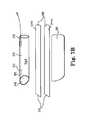
- expansion member 100includes proximal end 102 , frame 101 at proximal end 102 , distal end 104 , blades (or sidewalls) 106 extending from proximal end 102 to distal end 104 , and inner opening 105 .
- Upper and lower longitudinal openings 109extend between top and bottom edges of blades 106 .
- a batterye.g., a 3 volt “coin” battery
- 110is located at a proximal portion of one of blades 106 , and connects to a light 112 (light emitting diode, or otherwise) at a location on or toward distal end 104 .
- Light 112can be any desired light source, such as a light emitting diode (LED).
- LEDlight emitting diode
- a specific examplecan be an “ultrabright” 3 millimeter LED, powered by a lithium ion 3 volt battery (e.g., a 20 mm coin type battery).
- Expansion member 100 and components thereofe.g., blades 106 and frame 101
- Each blade 106may be flat or curved (e.g., to form a structure that defines a complete or partial tube (see FIG. 2A )).
- the use of a light source at a distal end of an expansion membercan simplify a surgical procedure by allowing a surgeon to see a targeted tissue without holding a separate light and without moving his or her head to see tissue using a headlamp.
- expansion member 120includes proximal end 122 , distal end 124 , sidewalls 126 extending from proximal end 122 to distal end 124 , and inner opening 125 .
- Longitudinal opening 129extends between of opposing sidewalls 126 , along a bottom or a top of the length of the device.
- a batterye.g., a 3 volt “coin” battery
- 210is located at a proximal portion of one of the sidewalls 126 and connects to lights 132 (light emitting diode, or otherwise) at a location on or toward distal end 124 .
- Lights 132can be any desired light source, such as a light emitting diode (LED).
- Expansion member 120e.g., sidewalls 126
- expansion member 120is in the form of a tube having a fixed cross section and length, but other embodiments are also possible, as described.
- expansion membersmay be incorporated, alone or together, with any of the features described elsewhere herein, for use with a method or a device as described.
- One featureis adjustability of one or more dimension (length, width (e.g., diameter), height) of an expansion member.
- An expansion memberhas a cross-sectional width that defines an inner opening, channel, or tube (complete or partial) used to create access to and working space within a pelvic location, such as access to a posterior pelvic location near a sacrum and sacral promontory.
- the widthcan be uniform along a length of an expansion member; non-uniform along the length (e.g., wider at a proximal end and narrower at a distal end); or variable at any location along a length, for adjustment before, during, or after a surgical procedure; e.g., a cross-sectional width of an inner opening at a proximal end, at one or more location along a length between a proximal and a distal end, or at a distal end, may be adjustable, each independently of one another, before, during, or after insertion for use during a surgical procedure.
- a lengthis a distance between a distal end and a proximal end and may be fixed or may be variable either before, during, or after a surgical procedure (e.g., a length may be adjustable before, during, or after insertion into a patient for use during a surgical procedure).
- a heightis a dimension in the same cross-sectional plane as a width, and can also be fixed or variable at any one or more location along a length of an expansion member, either before, during, or after a surgical procedure.
- an expansion membermay not fully enclose a working space along the length or around a circumference of the device between the distal and proximal ends, but may leave one or more portion or side (along the partial or total length of the expansion member) open, giving access to tissue.
- a “bottom” side of an expansion membermay lack structure, leaving an opening along a length of the device (e.g., at the bottom or top; see FIGS. 1A and 2A ), to allow access to a peritoneum and fixation of mesh, and anchor, or other implant material, at locations of exposed tissue, e.g., by suturing or use of an anchor.
- a tapere.g., a narrowing of the shape from the proximal end to the distal end 2 ; ribs for retention of an expansion member within the patient; sacral mating geometry (shaping of the distal end to match a shape or angle of a sacrum) or materials (e.g., conforming materials such as “tissue wipers”) at the far end or tips of one or more segments of a tool; a longitudinal (parallel or approximately parallel to a longitudinal axis or longitudinal dimension of the expansion member) (straight or curved) hinge between moveable segments; selective expansion (adjustment of a width dimension) of an expansion member at different locations along a length of an expansion member, e.g., to match the anatomy of a patient; selective adjustment of a length dimension of an expansion member, e.g., to match the anatomy of a patient; a handle at a proximal end, which may be removable; a lock or ratchet mechanism to maintain one or more selectively set
- a transvaginal vaginal process to reach a sacral promontorycan include:
- FIG. 4illustrates an expansion member that may have any one or more features as described, and that includes a delivery tool that can be engaged and disengaged to the retractor at a proximal end.
- the delivery toolcan be used to engage the retractor at a proximal end, manipulate the retractor to place the retractor into a patient, and adjust the retractor such as by increasing a diameter (expanding a cross-sectional dimension and inner opening of the retractor).
- the delivery toolcan be disengaged and removed from the proximal end of the retractor. Removing the delivery tool allows improved access to the inner opening and workspace created by the expanded retractor.
- removing the removable delivery tool from the proximal endwill reduce the size and profile of the retractor (proximal end) by removing the handles and delivery mechanism, once the retractor is deployed, giving improved access to the retractor portion placed in the patient.
- expansion member 140includes expansion member piece 160 and detachable handle piece 162 .
- Expansion member piece 160includes proximal end 142 , distal end 144 , and sidewalls 146 extending from proximal end 142 to distal end 144 .
- Sidewalls 146are made of multiple panels 147 held together by longitudinal hinges 149 , which, as described herein, may be of various types of hinge construction such as a “piano”-style hinge, an adhesive, a flexible polymeric connective material (e.g., membrane or a “living hinge”), a moveable mechanical connection, a flexible adhesive weld, etc.
- Inner opening 141extends lengthwise between proximal end 142 and distal end 144 .
- Bottom opening 143extends at a bottom of expansion member piece 160 , from a location about mid-way along the length of expansion member piece 160 , to the distal end of expansion member piece 160 .
- the absence of structure at the bottom of the distal endallows access to tissue of a sacrum, perineum, vaginal tissue, or the like, that will become located at that portion of the expansion member piece 160 when it is installed in a patient.
- expansion member piece 160is in the form of a partial, expandable and retractable tube having a variable cross-sectional dimensions.
- FIG. 3Bshows an unfolded or opened cross section with panels 147 being unfolded and arranged to form a cross-sectional, circular, inner opening 141 .
- panels 147are folded against each other to form a folded or closed configuration of smaller cross sectional dimensions, and a to smaller or absent inner opening 141 between panels.
- the cross-sectional dimensions of inner opening 141 of expansion member piece 140can be increased and decreased, i.e., panels 147 can be transitioned between the folded (closed) and unfolded (opened) configurations, by movement of panels 147 about longitudinal hinges 149 , by use of handle piece 162 .
- Handle piece 162includes handles 164 and body 166 .
- Body 166includes distal end 168 having engagement surfaces that are capable of releasably engaging proximal end 142 of expansion member piece 160 .
- Engagement surfacesmay be any useful surface capable of moving panels 147 between a folded and an unfolded configuration. As illustrated, engagement surfaces are in the form of distal ends of panels 147 a , but other forms of engagement surfaces will also be useful.
- handle piece 162can be used to manipulate expansion member piece 160 ; for example, handle piece 162 engaged with expansion member piece 160 can be used to insert a folded expansion member piece 160 into a patient incision (e.g., vaginal, abdominal, etc.) and place distal end 144 at a region of sacral anatomy or other desired surgical or pelvic location.
- a patient incisione.g., vaginal, abdominal, etc.
- handles 164can be manipulated to cause expansion member piece 160 to become unfolded, retracting tissue at the patient incision and at the surgical or pelvic location such as at a region of a sacrum.
- handle piece 162can be disengaged from proximal end 142 of expansion member piece 160 , providing improved access to inner opening 141 at proximal end 142 , for use by a surgeon of installed expansion member piece 160 during a surgical procedure.
- handle piece 162can be re-engaged with proximal end 142 , handle piece 162 can be used to re-configure expansion member piece 160 in a folded configuration, and the assembly of handle piece 162 and expansion portion piece 160 can be removed from the patient.
- hinges 149may be straight or curved and may be capable of ratcheting or locking to allow expansion piece 160 to be locked into an opened (unfolded) or partially opened configuration during a surgical procedure.
- a ratchet or lockmay be releasable by a release mechanism on expansion portion piece 160 or handle piece 162 .
- Panels 147may be rounded or flat and may be constructed of plastic (e.g., transparent or clear polyacrylate) or metal (e.g., stainless steel).
- Distal end 144may include any of the optional features described herein such as a light, irrigation feature, suction, anchor placement feature, dissolvable coating, or the like.
- a length dimensionmay be fixed or variable.
- FIG. 3Cshows an alternate embodiment of an expansion member 140 that includes expansion member piece 160 and handle piece 162 .
- distal end 144include an optional, expandable tissue approximating structure 170 .
- Tissue approximating structure of a expandable balloon 170can be any expandable structure at a distal end that can be expanded (e.g., inflated) to retract tissue situated adjacent to tissue approximating structure 170 at distal end 144 .
- tissue approximating structuremay be in the form of an expandable metal cage, moveable paddles or other moveable surfaces, or any other moveable structure capable of approximating tissue adjacent to distal end 144 .
- tissue approximating structure 170can be actuated and expanded to move tissue away from distal end 144 .
- the expandable structuremay be, e.g., an inflatable balloon, and can include a “gauzy” surface that is capable of retracting (pushing away) tissue, e.g., viscera, from a surgical site at the distal end of the device when installed through a surgical incision (transvaginally or trans-abdominally) in a patient to access a posterior pelvic location such as a sacrum.
- FIG. 4illustrates an embodiment of a distal end of a retractor (expansion member) that includes an elongate, needle-like extension or tip.
- the tipcan engage tissue and can be coated with a dissolvable or bioabsorbable material that will dissolve in a short time (e.g., 2 minutes or less) when exposed to body fluids.
- dissolvable coating materialsinclude types of material that dissolve upon exposure to body fluids, specific types being used in the field of dissolvable coatings used on cardiac electrodes. The dissolvable materials are described, for example in the following United States patent documents, the entire contents of which are incorporated herein by reference: U.S. Pat. Nos. 4,827,940; 6,304,786; and 7,218,971.
- distal end 144 of an expansion memberincludes extension 170 , which is an elongate, straight, curved, and optionally sharp, or to otherwise shaped, lead or frictional surface that can be contacted with or inserted into tissue at a surgical site.
- Extension 170when in contact with tissue, can prevent movement of distal end 144 relative to the tissue, to secure the position of distal end 144 during a surgical procedure.
- extension 170including coating 172 , is placed to contact or penetrate tissue. Fluid from the tissue causes coating 172 to dissolve or to be absorbed, leaving an uncotated extension 170 in contact with the tissue.
- FIGS. 5A, 5B, 5C, and 5Dshow an example of an expansion member that can be expanded (and retracted) by use of an electroactive polymer, inside of a patient, to retract tissue.
- the expansion membercan be placed within a patient, optionally by use of a separate introducer.
- the expansion membercan be placed in a patient in a closed configuration (and an optional introducer removed). Once placed, voltage can be applied to the expansion member to cause the expansion member to expand within the patient to cause retraction of tissue, especially when placed vaginally or transabdominally to access a posterior pelvic location.
- An electroactive polymeris capable of changing shape upon application of an electric field to the electroactive polymer.
- an electroactive polymercan be applied to or placed into contact with one or more surface of an expansion member, in a manner that exposure of the electroactive polymer to an electric field (e.g., current or voltage) causes the expansion member to change shape between a rolled or “closed” configuration, and an unrolled or “opened” configuration.
- an electric fielde.g., current or voltage
- a application of and removal of current or voltage to the electroactive polymerwill cause the expansion member to transition between opened and closed configurations. During such transformation from a closed to an opened configuration the expansion member piece can produce a force against tissue that is sufficient to retract the tissue.
- Introducer piece 262has a size and a shape that allow expansion member piece 260 to fit within the introducer piece 262 , when expansion member piece 260 is in a rolled or closed configuration. Introducer piece 262 can be first introduced into a surgical incision (see FIG. 5B ). Expansion member piece 260 in a closed configuration can be introduced into introducer piece 262 , and introducer piece 262 can be removed from the patient (see FIG. 5C ), leaving expansion member piece 260 in the incision (see FIG. 5D ).
- An electroactive polymeris located on the expansion member piece 260 at locations of the surface (inner surface, outer surface, or both) such that when the electroactive polymer is exposed to or stimulated by an electric field, e.g., a current or voltage is applied to (or removed from) the electroactive polymer, the electroactive polymer changes shape; the expansion member piece 260 changes shape in response to the electroactive polymer changing shape.
- an electric fielde.g., a current or voltage is applied to (or removed from) the electroactive polymer
- the electroactive polymerchanges shape
- the expansion member piece 260changes shape in response to the electroactive polymer changing shape.
- application or removal of a current or voltage, to the electroactive polymer or other structure (e.g., an electrode) of expansion member piece 260can transform the shape of expansion member piece 260 from a rolled up or closed form as shown in FIGS. 5A, 5B, and 5C , to an opened or unrolled form as shown at FIG. 5D .
- expansion member piece 260can be used alone or in combination with introducer piece 262 .
- expansion member piece 260can be inserted into a patient incision (e.g., an abdominal or vaginal) in the closed configuration and then expanded into the opened configuration to retract tissue.
- a surgeoncan perform a surgical procedure in the pelvic region, such as an SCP procedure with the expansion member placed in the incision and in the opened configuration to retract tissue and allow access to a surgical site.
- the expansion member piececan be placed in the closed configuration (by application of or removal of an electric current or voltage) and then removed.
- the electroactive polymermay be activated or deactivated, e.g., current or voltage can be applied or removed, to cause the expansion member piece to change between the closed and the opened, then the closed, configurations.
- introducer piece 262When optional introducer piece 262 is used in combination with expansion member piece 260 , introducer piece 262 can be first introduced into a surgical incision such as a vaginal or abdominal incision. A distal end of introducer piece 262 can be placed at a desired surgical site such as at a region of sacral anatomy. Expansion member piece 260 in a closed configuration, can be introduced into introducer piece 262 so that distal end 244 is placed at the surgical site, e.g., a region of sacral anatomy. Introducer piece 262 can be removed from the patient, leaving expansion member piece 260 in the incision, which can be expanded to an opened configuration. A surgical procedure can be performed, and expansion member piece 260 can be removed, optionally by first transforming expansion member piece 260 to the closed configuration.
- a surgical incisionsuch as a vaginal or abdominal incision.
- a distal end of introducer piece 262can be placed at a desired surgical site such as at a region of sacral anatomy.
- FIGS. 6A, 6B, 6C, 7A, 7B, 7C, 7D, and 7Eillustrate examples of expansion member systems that include an introducer piece and an expansion member piece (as do FIGS. 5A, 5B, 5C, and 5 d ).
- the expansion member pieceincludes a generally rigid tube or other form of expansion member, for retracting tissue such as by maintaining the position of tissue that is first retracted using the introducer piece.
- the introducer pieceincludes a proximal end, a distal end, and two opposing side sections (e.g., sidewalls, such as blades, or other moveable surfaces).
- side sectionsare in the form of two generally flat blades (sidewalls) joined together at their opposing edges by a flexible plastic (e.g., polyethylene, polypropylene, or other polyolefin) or fiber (e.g., a continuous fabric, woven, non-woven, knit, etc.) connective sheet or film material (sheathing, membrane) that can be folded between or adjacent to the blades to allow the blades to take on a “butted,” “closed,” or “collapsed” state. See FIG. 6A .
- a flexible plastice.g., polyethylene, polypropylene, or other polyolefin
- fibere.g., a continuous fabric, woven, non-woven, knit, etc.
- the bladeswhich may be substantially flat (as illustrated), or somewhat curved, have lengths between the proximal and distal ends that can provide an inner opening or workspace between a vaginal introitus or abdominal incision, and a posterior pelvic region, including a sacrum and sacral promontory.
- exemplary introducer piece 300can be useful in a transvaginal or transabdominal sacrocolpopexy procedure to allow fixation of an implant to a region of sacral anatomy such as an anterior longitudinal ligament of a sacrum.
- the bladescan be introduced into a vaginal introitus and through a posterior vaginal incision of open vaginal cuff, into the peritoneal cavity, to locate and identify a sacral promontory by tactile feedback.
- the bladescan be introduced into an abdominal incision and advanced to a posterior location of a pelvic region (e.g., into the peritoneal cavity), to locate and identify a sacral promontory by tactile feedback.
- introducer piece 300includes proximal end 308 , distal end 310 , opposing sidewalls or blades 302 , and membrane 304 connecting opposing upper and lower edges of opposing blades 302 .
- Distal end 310includes a slant or taper 311 designed to match a shape of a sacrum when introducer piece 300 is placed transvaginally or transabdominally within a vaginal incision or an abdominal incision, respectively, with distal end 310 located at a region of a sacrum.
- introducer piece 300can be first introduced into a surgical incision such as a vaginal or abdominal incision. With blades 302 butted together and membrane 304 folded between or adjacent to blades 302 , distal end 310 can be introduced through a surgical incision and advanced toward and placed at a desired surgical site such as at a region of a sacrum. Distal end 310 can be used to deflect the sigmoid colon of the sacral promontory and then become located over the sacral promontory to provide access to the sacral promontory. With introducer piece 300 placed as desired, expansion member piece 316 can be inserted into proximal end 308 between blades 302 and advanced distally toward distal end 310 .
- FIGS. 7A, 7B, 7C, 7D, and 7Eillustrate another example of an expansion member system that includes an expansion member piece 312 (retractor or retractor component or tube) and introducer piece 300 .
- introducer piece 300includes three separable items or pieces including a tubular membrane pouch 305 (for producing dilation) and two separable (e.g., malleable) blades 302 .
- Tubular membrane pouch 305is a flexible fabric or polymeric tube that can fit (e.g., loosely) around expansion member piece 312 , and that can be alternately folded or collapsed then opened or expanded by inserting blades 302 into an inner channel of membrane pouch 305 .
- Membrane pouch 305may be made of, e.g., a flexible polymer or fabric (woven, non-woven, knit, etc.) material such as polyethylene, polyurethane, silicone, or a textile material and can optionally include pockets on two sides to house blades 302 .
- Membrane pouch 305can be flexible to allow folding between or adjacent to the blades. Blades 302 can fit within the membrane pouch 305 along with expansion member piece 312 .
- Expansion member piece 312can be as described herein, such as in the form of a clear, transparent, tubular rigid polymeric member having distal end 318 , proximal end 316 , and a length and sidewalls 322 therebetween, with distal end 318 shaped (angled) for close contact with the sacrum when expansion member piece 312 is placed within a vaginal or abdominal incision.
- the length of body sidewall 322 extending between proximal end 316 and distal end 318can provide an inner opening and workspace 315 between a surgical incision at a location of an abdominal incision or a location of a vaginal introitus, and a posterior pelvic region such as a location of a sacrum, sacral promontory, or other sacral anatomy.
- Expansion member 312can include an optional lighting feature (not illustrated), for example in the form of a solid optical fiber extending inside of the tube or at an interior of the tube sidewall.
- an introducer piece 300 and an expansion member 312 as illustrated at FIGS. 7A, 7B, and 7Ccan be used together by steps that include: inserting and using inserter device 300 (including an assembly of membrane pouch 305 and blades 302 ) through a surgical incision, with distal end 310 located at a region of sacral anatomy and used to deflect the sigmoid colon; palpating and identifying the sacral promontory; and clearing the adjacent area (making sure no bowel interferes with access).
- the dilating component(membrane pouch 305 ) is expanded by separating the malleable blades at proximal end 308 , which is external to the patient, to allow insertion of the retraction tube (expansion member piece 312 ) into membrane pouch 305 .
- the expansion member piece 312is advanced slowly into the central inner lumen of membrane pouch 305 , between opposing blades 302 , and advanced distally to a region of the sacrum.
- Leading edge 320 of expansion member piece 312can be keyed (angled) such that the top edge of the angled distal end sits on top of the promontory for easy identification of the structure.
- expansion member piece 312Once expansion member piece 312 has been advanced to a desired distal location, malleable blades 302 can be withdrawn to allow for a less crowded proximal end, for easy access to the workspace and easy introduction of instruments through inner opening 315 of expansion member piece 312 .
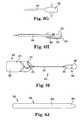
- FIGS. 8A, 8B, 8C, 8D, 8E, 8F, 8G, 8H, and 8Iillustrate examples of surgical tools in the form of “blunt dissection devices” 350 , which include proximal end 352 , shaft 354 , distal end 356 , and handle 358 at proximal end 352 .
- a distal end 356includes a blunt dissection feature or surface such as a blunt (e.g., rounded, dull, or slightly sharpened) distal edge or distal surface 362 of a paddle 360 .
- Paddle 360 or a surface thereofcan be used to contact tissue (e.g., at or proximal to a region of sacral anatomy) to cause dissection or otherwise move or retract tissue by manipulation of handle 358 at proximal end 352 .
- the blunt dissection surfacemay include one or a combination of a rigid, curved or flat paddle, and an optional expandable structure or surface such as a balloon, expandable metal cage, or other surface moveable relative to a surface of the paddle.
- a blunt dissection surface or paddlemay include one or more of the following optional features: a cutting feature such as a blade or pressurized air, an irrigation feature, a suction feature, a lighting feature (e.g., light 112 and battery 110 of certain figures), or an ultrasonic energy or other vibration feature.
- blunt dissection device 350includes proximal end 352 , shaft 354 , distal end 356 , and handle 358 at proximal end 352 .
- Paddle 360may be flat ( FIG. 8A ) or curved about a longitudinal axis ( FIG. 8C ).
- Light 112is energized by battery 110 .
- Shaft 354has a low profile or cross section to allow use through a vaginal or abdominal incision, or through an opening in an expansion member, without obstruction a view of a distal surgical site.
- Shaft 354may be a straight, rigid metal (e.g., stainless steel, titanium) or rigid plastic shaft of small cross-sectional dimension (e.g., diameter), such as having a diameter of 1 to 10 millimeters, e.g., 2 to 6 millimeters.
- rigid metale.g., stainless steel, titanium
- rigid plastic shaftof small cross-sectional dimension (e.g., diameter), such as having a diameter of 1 to 10 millimeters, e.g., 2 to 6 millimeters.
- Blunt dissection device 350 of FIGS. 8D and 8Eis similar to device 350 of FIG. 8A , except that shaft 354 includes bends or curves toward distal end 356 to stagger or offset the position of paddle 360 relative to a proximal portion ( 355 ) of shaft 354 .
- Paddle 360is parallel to proximal portion 355 of shaft 354 , but offset.
- handle 358is angled (e.g., at an angle in a range from 10 to 80 degrees, such as from 30 to 60 degrees) relative to proximal portion 355 of shaft 354 .
- the offset paddle and the angled handlecan be featured alone or separately to allow to manipulation of tissue by paddle 360 with open viewing of an adjacent surgical site.
- FIG. 8Fshows a top view and a bottom view of blunt dissection device 350 , which is an elongate flat blade having other features as presented.
- ultrasonic energycan emanate from paddle 360 , which can vibrates either laterally (as illustrated) or longitudinally.
- pressurized aircan flow along or through shaft 354 and be ejected through orifices (not shown) located on paddle 360 , either at a flat or curved surface 365 of paddle 360 , or at an edge such as distal edge 362 or at one or both of side edges 363 .
- Ultrasonic energycan emanate from paddle 360 , which can vibrate laterally (as illustrated), longitudinally, or both.
- device 350includes proximal end 352 , shaft 354 , distal end 356 , and handle 358 .
- Distal end 356includes paddle 360 , which includes expandable surface (e.g., balloon) 380 .
- Expandable surface 380can be located at a surface or distal or side edge of paddle 360 .
- Handle 358includes moveable or flexible bellows or flexible air reservoir or chamber 370 , which can be compressed to force air through valve 372 along shaft 354 and into an interior space of expandable surface (e.g., balloon) 380 .
- a switch at valve 372can be moved to connect the interior space of expandable surface 380 to outlet 374 .
- blunt dissection devices 350can include optional features such as an expandable structure (e.g., balloon 380 ), paddles curved laterally (e.g., about a lengthwise direction, see FIGS. 8B and 8C ), a longitudinal axis of shaft 354 aligned with a longitudinal axis of handle 358 , or a longitudinal axis of shaft 354 bent relative to a longitudinal axis of handle 358 (see FIG. 8E ).
- an expandable structuree.g., balloon 380
- paddles curved laterallye.g., about a lengthwise direction, see FIGS. 8B and 8C
- a longitudinal axis of shaft 354 aligned with a longitudinal axis of handle 358e.g., a longitudinal axis of shaft 354 bent relative to a longitudinal axis of handle 358 (see FIG. 8E ).
- a preferred flat or curved (laterally) paddlemay have an elongate but narrow size and shape, such as about a width (a major dimension of a paddle extending in a direction perpendicular to a length) of a finger (e.g., 1.5 to 2.5 centimeters) or somewhat wider (e.g., from 1.5 to 3 or 4 centimeters).
- a preferred expandable structuremay be a fluid-inflatable (e.g., air, carbon dioxide, nitrogen, etc.) balloon, which in use can be placed in contact with tissue and expanded to adjust the position of tissue, e.g., retract the tissue during a surgical procedure.
- the distal endcan also optionally and preferably include a light for better viewing of a to surgical space by a surgeon.
- the dissection toolcan be used in combination with a rectal probe 580 (see FIG. 8J ).
- FIG. 9includes panels that illustrates a series of steps for using a blunt dissection tool 350 , such as the one shown at FIG. 8I that includes an expandable surface (e.g., balloon) 380 as part of paddle 356 .
- FIG. 9shows a distal end 356 and paddle 360 advancing distally between two opposing tissue surfaces 380 and 382 .
- Panels 2 and 3show paddle 356 progressing distally between the tissue surfaces.
- balloon 380begins expansion and at panel 5 expansion continues in a manner such that the expanding surface of balloon 380 presses against the tissue surfaces to increase separation between the tissue surfaces.
- tool 350is advanced farther distally (in the direction of the arrow), with balloon 380 in an expanded configuration.
- balloon 380is reduced, e.g., by deflation, and at panel 8 tool 350 can be advanced farther distally with balloon 380 in a deflated configuration. Subsequently, balloon 380 can be re-inflated (as at FIGS. 4 and 5 ) and then steps of balloon inflation, distal advancement of tool 350 , deflation, etc., can be repeated.
- FIG. 8Jshows rectal probe 580 having proximal end 582 , distal end 584 with a rounded bulbous tip, and shaft 586 extending therebetween.
- Elongate shaft 586is substantially straight and includes a substantially circular cross section and a relatively smooth outer surface.
- Shaft 586can be made of a substantially rigid material such as plastic, metal, or transparent or translucent plastic.
- Shaft 586can contain a light source and may be made of a clear or transparent material to produce illumination when placed in a body lumen such as a rectum or colon. According to methods of using the expansion members of the invention, for use in treating a pelvic condition, a rectal probe 580 can be inserted into a rectum, and illuminated.
- a tube or retractorcan be placed transvaginally or transabdominally, e.g., in a non-expanded, closed, or collapsed state.
- the expansion membercan then be expanded while in place in the patient incision, e.g., transvaginally or transabdominally, to create access through an inner opening of the expansion member to desired anatomy such as the posterior of a pelvic region, e.g., to gain access to a region of sacral anatomy.
- the expansion membercreates an inner opening or workspace extending between a vaginal introitus or an abdominal incision and a region of sacral anatomy, such as an anterior longitudinal ligament.
- a surgeoncan perform a surgical procedure by use of the access, which provides working space to pelvic anatomy such as the sacrum.
- the methodcan optionally also involve a tool, multi-functional tool, implant, adjustable implant, anchor, or other device or method, e.g., as described at Applicant's co-pending International Patent Application number PCT/US2010/062577, filed Dec. 30, 2010, the entirety of which is incorporated by reference.
- an expansion member or dissection tool as describedcan include features and structures (e.g., fiber optics) to allow viewing or illumination of a surgical site. Illumination can be accomplished by any of various lighting techniques and structures. If a structural component of an expansion member or dissection tool is made of a plastic or polymeric light-conductive material, light can be transmitted through that material from a proximal end to a distal end at the surgical site. Alternately, a fiber optic cable can be incorporated into a length of the device, extending from a proximal to or toward a distal end, to allow light to be transmitted from the proximal end to the distal end. Light could alternately be generated and shone from the distal end.
- a structural component of an expansion member or dissection toolis made of a plastic or polymeric light-conductive material, light can be transmitted through that material from a proximal end to a distal end at the surgical site.
- a fiber optic cablecan be incorporated into a length of the device, extending from
- Tools and devices as describedcan be made from any suitable material or combination of materials. Examples include any material save for less than 24 hour contact with tissue, such as stainless steel, nitinol, polycarbonate, polypropylene, polyethylene, fluoropolymer, PET, polyurethane, silicone, polysulphone, and ultem. Any structure of an expansion member, retractor, tube, dissection tool, or other identified component may be capable of conducting light between a proximal and distal end, or alternately one or more fiber optics cable may be incorporated into an expansion member, retractor, tube, or component thereof, to provide lighting.
- tissuesuch as stainless steel, nitinol, polycarbonate, polypropylene, polyethylene, fluoropolymer, PET, polyurethane, silicone, polysulphone, and ultem.
- Any structure of an expansion member, retractor, tube, dissection tool, or other identified componentmay be capable of conducting light between a proximal and distal end, or alternately one or more fiber optics cable
- Implantstheir various components, structures, features, materials and methods may have a number of suitable configurations as shown and described in the previously-incorporated references, or as described herein.
- Various methods and tools for introducing, deploying, anchoring and manipulating implants to treat prolapse or another pelvic conditions, as disclosed in the previously-incorporated references,are envisioned for use with the present invention as well as those methods and tools identified and described herein.
- any of the described toolscan be used for placing any desired pelvic implant in a male or a female patient, and for any of a large variety of conditions, such as a pelvic condition.
- the implantcan include any structural features useful for such treatment, including any desired size, shape, and optional features such as adjustability and anchoring systems. Any of these features may be previously known, future developed, described herein, or described in documents incorporated herein, for any particular implant and method.
- some figures and discussionsinclude examples of features of “anchors” (e.g., soft tissue or bone anchors, as these terms are generically and inclusively used) that can be useful according to the methods of placing a surgical implant.
- an implant that includes or is otherwise secured by any of the anchors describedcan be useful to treat a pelvic condition in a male or a female patient; as a single and non-limiting example, an implant that includes or uses an anchor as described can be used in a transvaginal or transabdominal SCP procedure to provide support to a vaginal cuff, through an implant that includes the anchor, the anchor being attached at a region of sacral anatomy such as a sacral ligament (e.g., anterior longitudinal ligament, a.k.a. the “anterior ligament” or “longitudinal ligament”).
- a sacral ligamente.g., anterior longitudinal ligament, a.k.a. the “anterior ligament” or “longitudinal ligament”.
Landscapes
- Health & Medical Sciences (AREA)
- Life Sciences & Earth Sciences (AREA)
- Surgery (AREA)
- Physics & Mathematics (AREA)
- Engineering & Computer Science (AREA)
- Optics & Photonics (AREA)
- Biomedical Technology (AREA)
- Molecular Biology (AREA)
- Pathology (AREA)
- Nuclear Medicine, Radiotherapy & Molecular Imaging (AREA)
- Biophysics (AREA)
- Heart & Thoracic Surgery (AREA)
- Medical Informatics (AREA)
- Radiology & Medical Imaging (AREA)
- Animal Behavior & Ethology (AREA)
- General Health & Medical Sciences (AREA)
- Public Health (AREA)
- Veterinary Medicine (AREA)
- Microelectronics & Electronic Packaging (AREA)
- Gynecology & Obstetrics (AREA)
- Reproductive Health (AREA)
- Surgical Instruments (AREA)
Abstract
Description
- 1—Complete an incision through the vaginal apex (or posterior to the apex) and the peritoneum;
- 2—Place the retractor introducer through the small bowel (optional step);
- 3—Confirm sacral promontory (bone=firm feel, promontory=increased depth with minimal anterior movement);
- 4—If the introducer was used, place the retractor (expansion member) over the introducer and push until the tip of the distal end meets the sacrum;
- 5—Open the retractor
- 6—Connect a light source (if an external source is used).
- 7—A working space to the sacrum has been opened and is (optionally) lighted.
When the procedure is performed using an abdominal incision (trans-abdominally) instead of a vaginal incision (transvaginally), the steps can be similar, except that step 1 above is to complete an incision through an abdomen and not a vagina. Optionally, during a procedure, a rectal probe580 (seeFIG. 8J ) can be inserted into a rectum of the patient and illuminated to improve viewing of the posterior pelvic region, especially the rectum, colon, and adjacent tissue, to allow avoidance of or manipulation of those tissues during the procedure.
Claims (10)
Priority Applications (1)
| Application Number | Priority Date | Filing Date | Title |
|---|---|---|---|
| US13/537,977US10058240B2 (en) | 2011-06-29 | 2012-06-29 | Systems, implants, tools, and methods for treatments of pelvic conditions |
Applications Claiming Priority (2)
| Application Number | Priority Date | Filing Date | Title |
|---|---|---|---|
| US201161502694P | 2011-06-29 | 2011-06-29 | |
| US13/537,977US10058240B2 (en) | 2011-06-29 | 2012-06-29 | Systems, implants, tools, and methods for treatments of pelvic conditions |
Publications (2)
| Publication Number | Publication Date |
|---|---|
| US20130006061A1 US20130006061A1 (en) | 2013-01-03 |
| US10058240B2true US10058240B2 (en) | 2018-08-28 |
Family
ID=47391311
Family Applications (1)
| Application Number | Title | Priority Date | Filing Date |
|---|---|---|---|
| US13/537,977Active2033-05-17US10058240B2 (en) | 2011-06-29 | 2012-06-29 | Systems, implants, tools, and methods for treatments of pelvic conditions |
Country Status (1)
| Country | Link |
|---|---|
| US (1) | US10058240B2 (en) |
Cited By (1)
| Publication number | Priority date | Publication date | Assignee | Title |
|---|---|---|---|---|
| US11445901B2 (en) | 2018-08-10 | 2022-09-20 | The Moein Family Trust | One-time use expandable speculum |
Families Citing this family (14)
| Publication number | Priority date | Publication date | Assignee | Title |
|---|---|---|---|---|
| US9421032B2 (en)* | 2010-06-16 | 2016-08-23 | Covidien Lp | Seal port with blood collector |
| US9289114B2 (en)* | 2010-07-30 | 2016-03-22 | Nilesh R. Vasan | Disposable, self-contained laryngoscope and method of using same |
| WO2012050973A1 (en)* | 2010-09-29 | 2012-04-19 | Ams Research Corporation | Systems, tools, and methods for treatments of pelvic conditions |
| US9861349B2 (en) | 2011-09-29 | 2018-01-09 | Proa Medical, Inc. | Speculum for obstetrical and gynecological exams and related procedures |
| US9622779B2 (en) | 2011-10-27 | 2017-04-18 | DePuy Synthes Products, Inc. | Method and devices for a sub-splenius / supra-levator scapulae surgical access technique |
| US9649114B2 (en) | 2012-07-24 | 2017-05-16 | Ams Research Corporation | Systems, tools, and methods for connecting to tissue |
| CA2905345A1 (en) | 2013-03-15 | 2014-09-25 | Ams Research Corporation | Systems, tools, and methods for connecting to tissue |
| US9808231B2 (en)* | 2013-07-09 | 2017-11-07 | Edwards Lifesciences Corporation | Tissue retractor |
| US10064614B2 (en)* | 2014-03-18 | 2018-09-04 | The Nemours Foundation | Depressor/retractor |
| WO2017200555A1 (en)* | 2016-05-20 | 2017-11-23 | Choicespine, Lp | Access instruments to extend a surgical working channel |
| EP4223210A1 (en)* | 2017-11-10 | 2023-08-09 | Hegenbergerspeculum APS | Device |
| USD876625S1 (en) | 2018-08-07 | 2020-02-25 | Adroit Surgical, Llc | Laryngoscope |
| WO2021053392A2 (en)* | 2019-09-20 | 2021-03-25 | Axis Spine Technologies Ltd. | Radiolucent surgical retractor |
| WO2022006528A1 (en)* | 2020-07-02 | 2022-01-06 | Norris Jeffrey P | Apparatus and system for absorbable surgical button and methods thereof |
Citations (338)
| Publication number | Priority date | Publication date | Assignee | Title |
|---|---|---|---|---|
| US2738790A (en) | 1954-08-12 | 1956-03-20 | George P Pilling & Son Company | Suturing instrument |
| US3124136A (en) | 1964-03-10 | Method of repairing body tissue | ||
| US3182662A (en) | 1962-07-25 | 1965-05-11 | Vithal N Shirodkar | Plastic prosthesis useful in gynaecological surgery |
| US3311110A (en) | 1964-07-15 | 1967-03-28 | American Cyanamid Co | Flexible composite suture having a tandem linkage |
| US3384073A (en) | 1964-04-21 | 1968-05-21 | Ethicon Inc | Surgical device for correction of urinary incontinence |
| US3472232A (en) | 1967-05-31 | 1969-10-14 | Abbott Lab | Catheter insertion device |
| US3580313A (en) | 1969-01-07 | 1971-05-25 | Mcknight Charles A | Surgical instrument |
| US3763860A (en) | 1971-08-26 | 1973-10-09 | H Clarke | Laparoscopy instruments and method for suturing and ligation |
| US3789828A (en) | 1972-09-01 | 1974-02-05 | Heyer Schulte Corp | Urethral prosthesis |
| US3815576A (en) | 1973-01-26 | 1974-06-11 | D Balaban | Artificial sphincter |
| DE2305815A1 (en) | 1973-02-07 | 1974-08-08 | Kurt Seuberth | DEVICE FOR SEPARATING SURGICAL FEEDS |
| US3858783A (en) | 1972-11-20 | 1975-01-07 | Nikolai Nikolaevich Kapitanov | Surgical instrument for stitching up tissues with lengths of suture wire |
| US3924633A (en) | 1974-01-31 | 1975-12-09 | Cook Inc | Apparatus and method for suprapubic catheterization |
| US3995619A (en) | 1975-10-14 | 1976-12-07 | Glatzer Stephen G | Combination subcutaneous suture remover, biopsy sampler and syringe |
| US4019499A (en) | 1976-04-22 | 1977-04-26 | Heyer-Schulte Corporation | Compression implant for urinary incontinence |
| US4037603A (en) | 1975-05-13 | 1977-07-26 | Wendorff Erwin R | Metallic surgical suture |
| US4128100A (en) | 1976-10-08 | 1978-12-05 | Wendorff Erwin R | Suture |
| US4172458A (en) | 1977-11-07 | 1979-10-30 | Pereyra Armand J | Surgical ligature carrier |
| US4235238A (en) | 1978-05-11 | 1980-11-25 | Olympus Optical Co., Ltd. | Apparatus for suturing coeliac tissues |
| US4246660A (en) | 1978-12-26 | 1981-01-27 | Queen's University At Kingston | Artificial ligament |
| US4441497A (en) | 1982-10-21 | 1984-04-10 | Paudler Franklin T | Universal suture passer |
| US4509516A (en) | 1983-02-24 | 1985-04-09 | Stryker Corporation | Ligament tunneling instrument |
| US4548202A (en) | 1983-06-20 | 1985-10-22 | Ethicon, Inc. | Mesh tissue fasteners |
| SU1225547A1 (en) | 1984-08-03 | 1986-04-23 | Московский Городской Ордена Ленина И Ордена Трудового Красного Знамени Научно-Исследовательский Институт Скорой Помощи Им.Н.В.Склифосовского | Surgical instrument |
| US4585437A (en)* | 1983-03-17 | 1986-04-29 | Simms Mark D | Introducer for an umbilical artery catheter |
| US4632100A (en) | 1985-08-29 | 1986-12-30 | Marlowe E. Goble | Suture anchor assembly |
| SU1342486A1 (en) | 1982-06-29 | 1987-10-07 | М.А. Мороз | Needle holder |
| EP0248544A1 (en) | 1986-05-07 | 1987-12-09 | National Research Development Corporation | Urinary incontinence prostheses |
| US4775380A (en) | 1985-10-17 | 1988-10-04 | Seedhom Bahaa B | Surgical replacement of ligaments |
| US4865031A (en) | 1985-07-12 | 1989-09-12 | Keeffe Paul J O | Fabric and method of use for treatment of scars |
| US4873976A (en) | 1984-02-28 | 1989-10-17 | Schreiber Saul N | Surgical fasteners and method |
| US4920986A (en) | 1986-10-14 | 1990-05-01 | Zedlani Pty. Limited | Urinary incontinence device |
| US4932962A (en) | 1989-05-16 | 1990-06-12 | Inbae Yoon | Suture devices particularly useful in endoscopic surgery and methods of suturing |
| US4938760A (en) | 1989-03-29 | 1990-07-03 | American Medical Systems, Inc. | Female suspension procedure |
| US5007894A (en) | 1989-02-10 | 1991-04-16 | Goran Enhorning | Female incontinence device |
| US5013292A (en) | 1989-02-24 | 1991-05-07 | R. Laborie Medical Corporation | Surgical correction of female urinary stress incontinence and kit therefor |
| US5013316A (en) | 1990-03-26 | 1991-05-07 | Marlowe Goble E | Soft tissue anchor system |
| US5012822A (en) | 1988-10-11 | 1991-05-07 | Schwarz Gerald R | Method for controlling urinary incontinence |
| US5019032A (en) | 1990-04-03 | 1991-05-28 | Robertson Jack R | Refined suspension procedure with implement for treating female stress incontinence |
| US5032508A (en) | 1988-09-08 | 1991-07-16 | Marrow-Tech, Inc. | Three-dimensional cell and tissue culture system |
| US5053043A (en) | 1990-09-28 | 1991-10-01 | Vance Products Incorporated | Suture guide and method of placing sutures through a severed duct |
| US5085661A (en) | 1990-10-29 | 1992-02-04 | Gerald Moss | Surgical fastener implantation device |
| US5112344A (en) | 1988-10-04 | 1992-05-12 | Petros Peter E | Surgical instrument and method of utilization of such |
| US5123428A (en) | 1988-10-11 | 1992-06-23 | Schwarz Gerald R | Laparoscopically implanting bladder control apparatus |
| US5141520A (en) | 1991-10-29 | 1992-08-25 | Marlowe Goble E | Harpoon suture anchor |
| US5149329A (en) | 1990-12-12 | 1992-09-22 | Wayne State University | Surgical suture carrier and method for urinary bladder neck suspension |
| US5188636A (en) | 1992-05-07 | 1993-02-23 | Ethicon, Inc. | Purse string suture instrument |
| US5209756A (en) | 1989-11-03 | 1993-05-11 | Bahaa Botros Seedhom | Ligament fixation staple |
| WO1993017635A1 (en) | 1992-03-04 | 1993-09-16 | C.R. Bard, Inc. | Composite prosthesis and method for limiting the incidence of postoperative adhesions |
| US5250033A (en) | 1992-10-28 | 1993-10-05 | Interventional Thermodynamics, Inc. | Peel-away introducer sheath having proximal fitting |
| WO1993019678A2 (en) | 1991-12-03 | 1993-10-14 | Vesitec Medical, Inc. | Surgical treatment of stress urinary incontinence |
| US5256133A (en) | 1990-09-05 | 1993-10-26 | Spitz Robert M | Device for correcting stress urinary incontinence |
| US5269783A (en) | 1991-05-13 | 1993-12-14 | United States Surgical Corporation | Device and method for repairing torn tissue |
| GB2268690A (en) | 1992-07-15 | 1994-01-19 | Lopez Francisco Garcia | Vaginal autosuture device |
| US5281237A (en) | 1992-09-25 | 1994-01-25 | Gimpelson Richard J | Surgical stitching device and method of use |
| DE4220283C2 (en) | 1992-06-20 | 1994-05-19 | Singer Spezialnadelfab | Surgical needle-thread combination |
| US5328077A (en) | 1992-11-19 | 1994-07-12 | Lou Ek Seng | Method and apparatus for treating female urinary incontinence |
| US5337736A (en) | 1992-09-30 | 1994-08-16 | Reddy Pratap K | Method of using a laparoscopic retractor |
| US5362294A (en) | 1992-09-25 | 1994-11-08 | Seitzinger Michael R | Sling for positioning internal organ during laparoscopic surgery and method of use |
| US5368595A (en) | 1990-09-06 | 1994-11-29 | United States Surgical Corporation | Implant assist apparatus |
| US5370662A (en) | 1993-06-23 | 1994-12-06 | Kevin R. Stone | Suture anchor assembly |
| US5370650A (en) | 1992-02-24 | 1994-12-06 | United States Surgical Corporation | Articulating mesh deployment apparatus |
| US5376097A (en) | 1991-04-05 | 1994-12-27 | Phillips; Edward H. | Surgical fastener system |
| EP0632999A1 (en) | 1993-07-01 | 1995-01-11 | United States Surgical Corporation | Soft tissue repair system and method |
| US5383904A (en) | 1992-10-13 | 1995-01-24 | United States Surgical Corporation | Stiffened surgical device |
| US5386836A (en) | 1986-10-14 | 1995-02-07 | Zedlani Pty Limited | Urinary incontinence device |
| EP0643945A2 (en) | 1993-08-20 | 1995-03-22 | United States Surgical Corporation | Apparatus and method for applying and adjusting an anchoring device |
| US5403328A (en) | 1992-04-22 | 1995-04-04 | United States Surgical Corporation | Surgical apparatus and method for suturing body tissue |
| US5404870A (en)* | 1993-05-28 | 1995-04-11 | Ethicon, Inc. | Method of using a transanal inserter |
| EP0650703A1 (en) | 1993-07-06 | 1995-05-03 | Antoine Jean Henri Robert | Adjustable periurethral sphincter |
| WO1995011631A1 (en) | 1993-10-28 | 1995-05-04 | Javin Pierce | A suture anchor |
| US5413598A (en) | 1993-03-25 | 1995-05-09 | C. R. Bard, Inc. | Vascular graft |
| US5439467A (en) | 1991-12-03 | 1995-08-08 | Vesica Medical, Inc. | Suture passer |
| WO1995025469A1 (en) | 1994-03-21 | 1995-09-28 | The Anspach Effort, Inc. | Fastener for attaching objects to bones |
| US5474518A (en) | 1992-10-05 | 1995-12-12 | Farrer Velazquez; Francisco | Corrective device of urinary incontinence in women |
| US5474543A (en) | 1993-05-17 | 1995-12-12 | Mckay; Hunter A. | Single needle apparatus and method for performing retropublic urethropexy |
| US5518504A (en) | 1993-12-28 | 1996-05-21 | American Medical Systems, Inc. | Implantable sphincter system utilizing lifting means |
| US5520700A (en) | 1992-11-13 | 1996-05-28 | Technion Research & Development Foundation, Ltd. | Stapler device particularly useful in medical suturing |
| US5520703A (en) | 1993-06-07 | 1996-05-28 | Essig; Mitchell N. | Laparoscopic deschamp and associated suturing technique |
| US5527342A (en) | 1993-12-14 | 1996-06-18 | Pietrzak; William S. | Method and apparatus for securing soft tissues, tendons and ligaments to bone |
| US5571139A (en) | 1995-05-19 | 1996-11-05 | Jenkins, Jr.; Joseph R. | Bidirectional suture anchor |
| US5582188A (en) | 1991-12-03 | 1996-12-10 | Benderev; Theodore V. | Method of tensioning a suspended tissue mass |
| US5591206A (en) | 1993-09-30 | 1997-01-07 | Moufarr+E,Gra E+Ee Ge; Richard | Method and device for closing wounds |
| US5591163A (en) | 1995-06-14 | 1997-01-07 | Incont, Inc. | Apparatus and method for laparoscopic urethropexy |
| DE19544162C1 (en) | 1995-11-17 | 1997-04-24 | Ethicon Gmbh | Implant for suspension of the bladder in urinary incontinence in women |
| WO1997016121A1 (en) | 1995-10-31 | 1997-05-09 | Karl Christopher Texler | Surgical instruments |
| US5628756A (en) | 1993-01-06 | 1997-05-13 | Smith & Nephew Richards Inc. | Knotted cable attachment apparatus formed of braided polymeric fibers |
| US5633286A (en) | 1977-03-17 | 1997-05-27 | Applied Elastomerics, Inc. | Gelatinous elastomer articles |
| US5643320A (en) | 1995-03-13 | 1997-07-01 | Depuy Inc. | Soft tissue anchor and method |
| WO1997030638A1 (en) | 1996-02-23 | 1997-08-28 | Influence, Inc. | Non-linear anchor inserter device and bone anchors |
| US5669935A (en) | 1995-07-28 | 1997-09-23 | Ethicon, Inc. | One-way suture retaining device for braided sutures |
| US5683349A (en) | 1993-02-22 | 1997-11-04 | Valleylab Inc | Laparoscopic dissection tension retractor device and method |
| US5697931A (en) | 1995-06-14 | 1997-12-16 | Incont, Inc. | Apparatus and method for laparoscopic urethopexy |
| WO1997047244A1 (en) | 1996-06-12 | 1997-12-18 | Mitek Surgical Products, Inc. | Apparatus and method for binding a first layer of material to a second layer of material |
| US5709708A (en) | 1997-01-31 | 1998-01-20 | Thal; Raymond | Captured-loop knotless suture anchor assembly |
| US5725541A (en) | 1996-01-22 | 1998-03-10 | The Anspach Effort, Inc. | Soft tissue fastener device |
| US5741282A (en) | 1996-01-22 | 1998-04-21 | The Anspach Effort, Inc. | Soft tissue fastener device |
| WO1998019606A1 (en) | 1996-11-08 | 1998-05-14 | Boston Scientific Corporation | Transvaginal anchor implantation device |
| US5782916A (en) | 1996-08-13 | 1998-07-21 | Galt Laboratories, Inc. | Device for maintaining urinary continence |
| US5785640A (en) | 1996-05-23 | 1998-07-28 | Kresch; Arnold J. | Method for treating female incontinence |
| WO1998035632A1 (en) | 1997-02-13 | 1998-08-20 | Boston Scientific Ireland Limited | Stabilization sling for use in minimally invasive pelvic surgery |
| WO1998035616A1 (en) | 1997-02-13 | 1998-08-20 | Boston Scientific Ireland Limited, Barbados Head Office | Percutaneous and hiatal devices and methods for use in minimally invasive pelvic surgery |
| WO1998035606A2 (en) | 1997-02-13 | 1998-08-20 | Boston Scientific Ireland Limited, Barbados Head Office | Method and apparatus for minimally invasive pelvic surgery |
| WO1998042261A1 (en) | 1997-03-25 | 1998-10-01 | Ethicon, Inc. | System for anchoring tissue to bone |
| WO1998053746A1 (en) | 1997-06-02 | 1998-12-03 | Martello Jeannette M D | Soft tissue securing anchor |
| WO1999016381A1 (en) | 1997-10-01 | 1999-04-08 | Boston Scientific Limited | Pelvic floor reconstruction |
| US5899909A (en) | 1994-08-30 | 1999-05-04 | Medscand Medical Ab | Surgical instrument for treating female urinary incontinence |
| US5919232A (en) | 1994-05-27 | 1999-07-06 | Cogent | Prosthetic fabric |
| US5922026A (en) | 1997-05-01 | 1999-07-13 | Origin Medsystems, Inc. | Surgical method and prosthetic strip therefor |
| US5925047A (en) | 1998-10-19 | 1999-07-20 | Third Millennium Engineering, Llc | Coupled rod, anterior vertebral body screw, and staple assembly |
| WO1999037217A1 (en) | 1998-01-27 | 1999-07-29 | Scimed Life Systems, Inc. | Bone anchor with implantation device |
| US5934283A (en) | 1997-04-15 | 1999-08-10 | Uroplasty, Inc. | Pubovaginal sling device |
| US5935122A (en) | 1991-12-13 | 1999-08-10 | Endovascular Technologies, Inc. | Dual valve, flexible expandable sheath and method |
| US5944732A (en) | 1997-08-27 | 1999-08-31 | Medical Components, Inc. | Subcutaneous tunnelling device and methods of forming a subcutaneous tunnel |
| US5954057A (en) | 1997-02-12 | 1999-09-21 | Li Medical Technologies, Inc. | Soft tissue suspension clip, clip assembly, emplacement tool and method |
| WO1999052450A1 (en) | 1998-04-16 | 1999-10-21 | Axya Medical, Inc. | A suture tensioning and fixation device |
| WO1999053844A1 (en) | 1998-04-21 | 1999-10-28 | Tornier S.A. | Suture anchor with reversible expansion |
| US5976146A (en)* | 1997-07-11 | 1999-11-02 | Olympus Optical Co., Ltd. | Surgical operation system and method of securing working space for surgical operation in body |
| US5980558A (en) | 1997-09-30 | 1999-11-09 | Biomet Inc. | Suture anchor system |
| US5984927A (en) | 1998-03-03 | 1999-11-16 | Ethicon, Inc. | Device for sutureless attachment of soft tissue to bone |
| US5988171A (en) | 1997-06-26 | 1999-11-23 | Influence Medical Technologies, Ltd. | Methods and devices for the treatment of airway obstruction, sleep apnea and snoring |
| WO1999059477A1 (en) | 1998-05-21 | 1999-11-25 | Walshe Christopher J | A tissue anchor system |
| US5997554A (en) | 1995-06-14 | 1999-12-07 | Medworks Corporation | Surgical template and surgical method employing same |
| US6010447A (en) | 1998-07-31 | 2000-01-04 | Kardjian; Paul M. | Bladder sling |
| US6027523A (en) | 1997-10-06 | 2000-02-22 | Arthrex, Inc. | Suture anchor with attached disk |
| IT1299162B1 (en) | 1998-04-17 | 2000-02-29 | Mauro Cervigni | PROSTHETIC ASSEMBLY TO BE USED IN SURGICAL THERAPY OF UROGENITAL PROLAPSE |
| US6030393A (en) | 1998-09-15 | 2000-02-29 | Corlew; Earvin L. | Needle and procedure for relieving urinary incontinence |
| US6031148A (en) | 1990-12-06 | 2000-02-29 | W. L. Gore & Associates, Inc. | Implantable bioabsorbable article |
| US6036701A (en) | 1994-01-13 | 2000-03-14 | Ethicon, Inc. | Spiral surgical tack |
| WO2000013601A1 (en) | 1998-09-08 | 2000-03-16 | Pierson Raymond H Iii | Medical tensioning system |
| US6039686A (en) | 1997-03-18 | 2000-03-21 | Kovac; S. Robert | System and a method for the long term cure of recurrent urinary female incontinence |
| US6042536A (en) | 1998-08-13 | 2000-03-28 | Contimed, Inc. | Bladder sling |
| US6042583A (en) | 1995-06-14 | 2000-03-28 | Medworks Corporation | Bone anchor-insertion tool and surgical method employing same |
| WO2000018319A1 (en) | 1998-09-30 | 2000-04-06 | Burger, Nicolaas, Daniel, Lombard | Distensible sling for urinary incontinence |
| US6048351A (en) | 1992-09-04 | 2000-04-11 | Scimed Life Systems, Inc. | Transvaginal suturing system |
| US6050937A (en) | 1998-09-21 | 2000-04-18 | Benderev; Theodore V. | Surgical tension/pressure monitor |
| WO2000027304A1 (en) | 1998-11-10 | 2000-05-18 | Sofradim Production | Suspension device for treating prolapsus and urinary incontinence |
| US6068591A (en) | 1998-02-17 | 2000-05-30 | Bruckner; Norman I. | Pubo-urethral support harness apparatus for percutaneous treatment of female stress urinary incontinence |
| US6071290A (en) | 1995-09-26 | 2000-06-06 | Compton; Jeffrey Spencer | Surgical clip set |
| US6074341A (en) | 1998-06-09 | 2000-06-13 | Timm Medical Technologies, Inc. | Vessel occlusive apparatus and method |
| FR2787990A1 (en) | 1998-12-30 | 2000-07-07 | Medical Research & Management | Prosthesis for correcting female urinary incontinence made in two halves connecting vaginal wall on either side of bladder to abdominal muscles |
| WO2000040158A2 (en) | 1999-01-08 | 2000-07-13 | Influence Medical Technologies, Ltd. | Surgical tack |
| US6099538A (en) | 1999-02-02 | 2000-08-08 | T.A.G. Medical Products | Set of surgical tools and surgical method for connecting soft bone parts to one another or to connective tissue |
| US6099552A (en) | 1997-11-12 | 2000-08-08 | Boston Scientific Corporation | Gastrointestinal copression clips |
| US6099551A (en) | 1998-03-12 | 2000-08-08 | Shelhigh, Inc. | Pericardial strip and stapler assembly for dividing and sealing visceral tissues and method of use thereof |
| US6117067A (en) | 1998-03-10 | 2000-09-12 | Gil-Vernet; Josema | Device for the height-adjustable fixing and support of internal anatomical organs |
| US6127597A (en) | 1997-03-07 | 2000-10-03 | Discotech N.V. | Systems for percutaneous bone and spinal stabilization, fixation and repair |
| WO2000057812A1 (en) | 1999-03-26 | 2000-10-05 | Ethicon, Inc. | Knitted surgical mesh |
| WO2000064370A1 (en) | 1999-04-27 | 2000-11-02 | Sofradim Production | Device for treating a prolapse by vaginal suspension |
| WO2000066030A1 (en) | 1999-04-30 | 2000-11-09 | Uromedica, Inc. | Method and apparatus for adjustable sling for treatment of urinary stress incontinence |
| WO2000074613A1 (en) | 1999-06-09 | 2000-12-14 | Ethicon, Inc. | Visually-directed surgical instrument and method for treating female urinary incontinence |
| WO2000074594A1 (en) | 1999-06-09 | 2000-12-14 | Ethicon, Inc. | Surgical instrument for treating female urinary incontinence |
| WO2000074633A2 (en) | 1999-06-09 | 2000-12-14 | Ethicon, Inc. | Method and apparatus for adjusting flexible areal polymer implants |
| DE20016866U1 (en) | 2000-09-30 | 2000-12-14 | Wendt, Adalbert, 50127 Bergheim | Tool to unlock a lock cylinder |
| EP1060714A2 (en) | 1999-06-08 | 2000-12-20 | Ethicon, Inc. | Knitted surgical mesh |
| US6168611B1 (en) | 1999-09-08 | 2001-01-02 | Syed Rizvi | Suturing needle assemblies and methods of use thereof |
| WO2001006951A1 (en) | 1999-07-27 | 2001-02-01 | Angiologica B.M. S.R.L. | Corrective mesh for body tissues |
| GB2353220A (en) | 1999-10-22 | 2001-02-21 | George Kaladelfos | A surgical instrument and method of treating female urinary incontinence |
| US6200330B1 (en) | 1998-11-23 | 2001-03-13 | Theodore V. Benderev | Systems for securing sutures, grafts and soft tissue to bone and periosteum |
| WO2001026581A1 (en) | 1999-10-11 | 2001-04-19 | Uromedica, Inc. | Adjustable implantable genitourinary device |
| US6221005B1 (en) | 1998-02-17 | 2001-04-24 | Norman I. Bruckner | Pubo-urethral support harness apparatus for percutaneous treatment of female stress urinary incontinence with urethal hypemobility |
| US6241736B1 (en) | 1998-05-12 | 2001-06-05 | Scimed Life Systems, Inc. | Manual bone anchor placement devices |
| WO2001039670A1 (en) | 1999-11-30 | 2001-06-07 | Shlomo Gabbay | Implantable support apparatus and method of using same |
| WO2001045589A1 (en) | 1999-12-22 | 2001-06-28 | Promedon | Sling with pad treatment of urinary incontinence |
| WO2001045588A2 (en) | 1999-12-07 | 2001-06-28 | Trabucco Arnaldo F | Mesh pubovaginal sling |
| US20010007073A1 (en)* | 1997-01-02 | 2001-07-05 | St. Francis Medical Technologies, Inc. | Spine distraction implant and method |
| US6264676B1 (en) | 1996-11-08 | 2001-07-24 | Scimed Life Systems, Inc. | Protective sheath for transvaginal anchor implantation devices |
| US20010011170A1 (en)* | 1998-08-20 | 2001-08-02 | Endius Incorporated | Method for performing a surgical procedure and a cannula for use in performing the surgical procedure |
| WO2001056499A1 (en) | 2000-01-31 | 2001-08-09 | Ethicon Gmbh | Areal implant with x-ray-visible elements |
| US6302840B1 (en) | 1998-09-21 | 2001-10-16 | Theodore V. Benderev | Surgical monitor |
| US6328744B1 (en) | 1999-06-04 | 2001-12-11 | American Medical Systems, Inc. | Bone suturing device |
| US20020007222A1 (en) | 2000-04-11 | 2002-01-17 | Ashvin Desai | Method and apparatus for supporting a body organ |
| US6352553B1 (en) | 1995-12-14 | 2002-03-05 | Gore Enterprise Holdings, Inc. | Stent-graft deployment apparatus and method |
| US20020028980A1 (en) | 2000-09-07 | 2002-03-07 | American Medical Systems | Implantable article and method |
| US6364832B1 (en)* | 1999-12-23 | 2002-04-02 | Tri-State Hospital Supply Corporation | Vaginal lateral walls retractor for use in combination with vaginal specula and method of performing vaginal/cervical examination |
| WO2002028312A1 (en) | 2000-10-05 | 2002-04-11 | Sofradim Production | Sub-urethral supporting assembly for treating female stress urinary incontinence |
| WO2002028315A2 (en) | 2000-10-06 | 2002-04-11 | American Medical Systems, Inc. | Sling adjustment and tensioning accessory |
| WO2002030293A1 (en) | 2000-10-12 | 2002-04-18 | Gyne Ideas Limited | Apparatus and method for treating female urinary incontinence |
| WO2002032284A2 (en) | 2000-10-18 | 2002-04-25 | Ethicon, Inc. | Apparatus and method for treating female urinary incontinence |
| WO2002034124A2 (en) | 2000-10-20 | 2002-05-02 | Ethicon Gmbh | System with a surgical needle and a handle |
| US6383191B1 (en)* | 2000-03-15 | 2002-05-07 | Sdgi Holdings, Inc. | Laparoscopic instrument sleeve |
| US6382214B1 (en) | 1998-04-24 | 2002-05-07 | American Medical Systems, Inc. | Methods and apparatus for correction of urinary and gynecological pathologies including treatment of male incontinence and female cystocele |
| US6387041B1 (en) | 1999-01-08 | 2002-05-14 | Boaz Harari | Tack device |
| WO2002038079A2 (en) | 2000-11-13 | 2002-05-16 | Ethicon Gmbh | Implant for holding the female bladder |
| WO2002039890A2 (en) | 2000-11-20 | 2002-05-23 | Ethicon, Inc. | Surgical instrument and method for treating female urinary incontinence |
| US6406423B1 (en) | 2000-01-21 | 2002-06-18 | Sofradim Production | Method for surgical treatment of urinary incontinence and device for carrying out said method |
| US6406480B1 (en) | 1992-11-13 | 2002-06-18 | American Med Syst | Bone anchor inserter with retractable shield |
| US6414179B1 (en) | 2000-02-18 | 2002-07-02 | Bristol-Myers Squibb Company | Alpha-and beta-substituted trifluoromethyl ketones as phospholipase inhibitors |
| WO2002058563A1 (en) | 2001-01-23 | 2002-08-01 | American Medical Systems, Inc. | Implantable sling |
| US20020128670A1 (en) | 2000-11-22 | 2002-09-12 | Ulf Ulmsten | Surgical instrument and method for treating female urinary incontinence |
| US6451024B1 (en) | 1995-06-14 | 2002-09-17 | Dexterity Surgical, Inc. | Surgical method for treating urinary incontinence, and apparatus for use in same |
| WO2002071953A2 (en) | 2001-03-09 | 2002-09-19 | Scimed Life Systems, Inc. | System for implanting an implant and method thereof |
| US20020147382A1 (en) | 2001-01-23 | 2002-10-10 | Neisz Johann J. | Surgical articles and methods |
| WO2002078552A1 (en) | 2001-03-28 | 2002-10-10 | George Kaladelfos | Treatment of vault prolapse |
| US20020151909A1 (en) | 2001-03-09 | 2002-10-17 | Gellman Barry N. | System for implanting an implant and method thereof |
| US20020161382A1 (en) | 2001-03-29 | 2002-10-31 | Neisz Johann J. | Implant inserted without bone anchors |
| US6478727B2 (en) | 2000-01-21 | 2002-11-12 | Sofradim Production | Percutaneous device and method for treating urinary stress incontinence in women using a sub-urethral tape |
| WO2002089704A2 (en) | 2001-05-07 | 2002-11-14 | Bernard Guerquin | Intra-vaginal device for preventing female stress incontinence |
| US6482214B1 (en) | 2000-04-27 | 2002-11-19 | Medtronic, Inc. | Intravascular seal with mesh reinforcement and method for using same |
| US6491703B1 (en) | 1995-10-09 | 2002-12-10 | Ethicon, Inc. | Surgical instrument for treating female urinary incontinence |
| US6494906B1 (en) | 2000-07-25 | 2002-12-17 | Advanced Cardiovascular Systems, Inc. | Stent fold clip |
| US20030004581A1 (en) | 2001-06-27 | 2003-01-02 | Rousseau Robert A. | Implantable prosthetic mesh system |
| WO2003003778A1 (en) | 2001-06-29 | 2003-01-09 | Siemens Mobile Communications S.P.A. | Method to control the packet switching traffic in digital cellular communication systems |
| WO2003017848A1 (en) | 2001-08-23 | 2003-03-06 | Ethicon Gmbh | System with a surgical needle and a handle |
| US6530943B1 (en) | 1999-12-15 | 2003-03-11 | Ethicon, Gmbh | Surgical needle for implanting a tape |
| US20030065402A1 (en) | 2001-10-03 | 2003-04-03 | Ameican Medical Systems | Implantable article |
| WO2003028585A2 (en) | 2001-10-04 | 2003-04-10 | Ethicon, Inc. | Mesh for pelvic floor repair |
| WO2003037215A2 (en) | 2001-10-29 | 2003-05-08 | Ethicon Gmbh | Areal implant |
| WO2003041613A1 (en) | 2001-11-14 | 2003-05-22 | Ethicon Gmbh | Areal implant |
| WO2003047435A1 (en) | 2001-12-03 | 2003-06-12 | Ethicon Gmbh | Surgical auxiliary instrument |
| US6582443B2 (en) | 2000-12-27 | 2003-06-24 | Ams Research Corporation | Apparatus and methods for enhancing the functional longevity and for facilitating the implantation of medical devices |
| US6596001B2 (en) | 2000-05-01 | 2003-07-22 | Ethicon, Inc. | Aiming device for surgical instrument and method for use for treating female urinary incontinence |
| US6599235B2 (en) | 1997-03-18 | 2003-07-29 | American Medical Systems Inc. | Transvaginal bone anchor implantation device |
| US6599323B2 (en) | 2000-12-21 | 2003-07-29 | Ethicon, Inc. | Reinforced tissue implants and methods of manufacture and use |
| US6602260B2 (en) | 2001-02-02 | 2003-08-05 | Ams Research Corporation | Powered bone screw device |
| WO2003068107A1 (en) | 2002-02-14 | 2003-08-21 | Ethicon, Inc. | Surgical instrument and method for treating organ prolapse conditions |
| WO2003075792A1 (en) | 2002-03-07 | 2003-09-18 | Ams Research Corporation | Transobturator surgical articles and methods |
| US20030176875A1 (en) | 2002-03-07 | 2003-09-18 | Anderson Kimberly A. | Transobturator surgical articles and methods |
| DE10211360A1 (en) | 2002-03-14 | 2003-10-09 | Ethicon Gmbh | Implantate band and surgical needle system to support female urethra has distal end region of needle narrower than implantate band |
| US6638211B2 (en) | 2000-07-05 | 2003-10-28 | Mentor Corporation | Method for treating urinary incontinence in women and implantable device intended to correct urinary incontinence |
| US6638210B2 (en) | 2000-09-26 | 2003-10-28 | Ethicon, Inc. | Surgical apparatus and methods for delivery of a sling in the treatment of female urinary incontinence |
| US6641525B2 (en) | 2001-01-23 | 2003-11-04 | Ams Research Corporation | Sling assembly with secure and convenient attachment |
| WO2003092546A2 (en) | 2002-04-30 | 2003-11-13 | Cook Urological Inc. | Sling for supporting tissue |
| US6652450B2 (en) | 2001-01-23 | 2003-11-25 | American Medical Systems, Inc. | Implantable article and method for treating urinary incontinence using means for repositioning the implantable article |
| WO2003096929A1 (en) | 2002-05-15 | 2003-11-27 | American Medical Systems, Inc. | Improved pelvic health implants and methods |
| US6673010B2 (en) | 2001-10-22 | 2004-01-06 | T. A. G. Medical Products Ltd. | Biological vessel suspending assembly and systems and methods utilizing same |
| US6689047B2 (en) | 2000-11-15 | 2004-02-10 | Scimed Life Systems, Inc. | Treating urinary incontinence |
| WO2004012626A1 (en) | 2002-08-01 | 2004-02-12 | Fin Med S.A.S. Di Grondelli & C. | Device for the surgical treatment of female prolapse |
| WO2004016196A2 (en) | 2002-08-14 | 2004-02-26 | Boston Scientific Limited | Systems, methods and devices relating to delivery of medical implants |
| US6699175B2 (en) | 2000-10-23 | 2004-03-02 | Ethicon, Inc. | Apparatus and method for the measurement of the resistance of the urethral sphincter |
| WO2004017862A2 (en) | 2002-08-22 | 2004-03-04 | Ams Research Corporation | Composite sling and sling assembly tool for treating incontinence |
| US20040073235A1 (en) | 2001-10-01 | 2004-04-15 | Lund Robert E. | Surgical article |
| WO2004034912A1 (en) | 2002-10-17 | 2004-04-29 | Ams Research Corporation | Surgical instruments |
| US20040116777A1 (en)* | 2002-12-13 | 2004-06-17 | Jeffrey Larson | Guided retractor and methods of use |
| US6755781B2 (en) | 2001-07-27 | 2004-06-29 | Scimed Life Systems, Inc. | Medical slings |
| FR2852813A1 (en) | 2003-03-28 | 2004-10-01 | Analytic Biosurgical Solutions | Reinforcement band placing device for treating cystocele, has cutting unit, cutting body of introducer into two parts by traction exerted on traction unit, having two openings in cavity for passage of cutting tool |
| US20040225181A1 (en) | 2003-04-25 | 2004-11-11 | Scimed Life Systems, Inc. | Systems and methods for sling delivery and placement |
| US6830052B2 (en) | 2001-10-03 | 2004-12-14 | Solarant Medical, Inc. | Urethral support for incontinence |
| US20050004427A1 (en) | 2003-04-30 | 2005-01-06 | Mauro Cervigni | Prosthesis for use in the surgical treatment of urogenital prolapse and female urinary incontinence |
| US20050004576A1 (en) | 1998-11-23 | 2005-01-06 | Benderev Theodore V. | System for securing sutures, grafts and soft tissue to bone and periosteum |
| US20050038451A1 (en) | 2003-08-14 | 2005-02-17 | Scimed Life Systems, Inc. | Medical slings |
| US20050055104A1 (en) | 2003-04-24 | 2005-03-10 | Ams Research Corporation | Male urethral prosthesis |
| US6881184B2 (en) | 2002-07-16 | 2005-04-19 | Stephen M. Zappala | Absorbable pubovaginal sling system and method |
| WO2005037132A2 (en) | 2003-10-14 | 2005-04-28 | Caldera Medical, Inc. | Implantable sling having bladder support |
| US20050131391A1 (en) | 2001-03-09 | 2005-06-16 | Scimed Life Systems, Inc. | Systems, methods and devices relating to delivery of medical implants |
| US20050131393A1 (en) | 2001-03-09 | 2005-06-16 | Scimed Life Systems, Inc. | Systems, methods and devices relating to delivery of medical implants |
| US6908425B2 (en) | 1999-06-09 | 2005-06-21 | Ethicon Inc. | Surgical instrument and method for treating female urinary incontinence |
| US6908473B2 (en) | 2000-04-14 | 2005-06-21 | Jeffry B. Skiba | Tissue anchoring devices, biological vessel suspending devices and systems and methods utilizing same |
| US6911003B2 (en) | 2002-03-07 | 2005-06-28 | Ams Research Corporation | Transobturator surgical articles and methods |
| US6932759B2 (en) | 1999-06-09 | 2005-08-23 | Gene W. Kammerer | Surgical instrument and method for treating female urinary incontinence |
| WO2005079702A1 (en) | 2004-02-19 | 2005-09-01 | Ams Research Corporation | Prolapse repair |
| US20050199249A1 (en) | 2004-03-15 | 2005-09-15 | Karram Mickey M. | Apparatus and method for incision-free vaginal prolapse repair |
| US20050245787A1 (en) | 2004-04-30 | 2005-11-03 | Ams Research Corporation | Method and apparatus for treating pelvic organ prolapse |
| US20050256530A1 (en) | 2002-08-13 | 2005-11-17 | Petros Peter E | Anchoring device and its implementation |
| US6974462B2 (en) | 2001-12-19 | 2005-12-13 | Boston Scientific Scimed, Inc. | Surgical anchor implantation device |
| US20050277806A1 (en) | 2004-06-10 | 2005-12-15 | Cristalli Bernard G R | Prosthetic implant for sub-urethral support, an instrument, an insertion kit, and a surgical method for implanting it |
| US20050278037A1 (en) | 2004-06-11 | 2005-12-15 | Analytic Biosurgical Solutions-Abiss | Implant for the treatment of cystocele and rectocele |
| US20050283189A1 (en) | 1999-03-31 | 2005-12-22 | Rosenblatt Peter L | Systems and methods for soft tissue reconstruction |
| WO2005122954A1 (en) | 2004-06-14 | 2005-12-29 | Boston Scientific Scimed, Inc. | Systems, methods and devices relating to implantable supportive slings |
| US6981983B1 (en) | 1999-03-31 | 2006-01-03 | Rosenblatt Peter L | System and methods for soft tissue reconstruction |
| US6981944B2 (en) | 2003-07-07 | 2006-01-03 | Ethicon, Inc. | Implantable surgical mesh having a lubricious coating |
| US20060015010A1 (en) | 2000-04-03 | 2006-01-19 | Ross Jaffe | Endoscope having a guide tube |
| US20060028828A1 (en) | 2003-10-31 | 2006-02-09 | Phillips Todd L | Light fixture candle assembly |
| WO2006015031A2 (en) | 2004-07-28 | 2006-02-09 | Ethicon, Inc. | Minimally invasive medical implant and insertion device and method for using the same |
| US20060058578A1 (en) | 2002-04-11 | 2006-03-16 | Gyne Ideas Limited | Apparatus and method for treating female urinary incontinence |
| US7025063B2 (en) | 2000-09-07 | 2006-04-11 | Ams Research Corporation | Coated sling material |
| US20060089524A1 (en) | 2004-10-25 | 2006-04-27 | Chu Michael S H | Systems and methods for sling delivery and placement |
| US20060106416A1 (en)* | 2004-10-29 | 2006-05-18 | Douglas Raymond | Expandable ports and methods for minimally invasive surgery |
| US20060122457A1 (en) | 2004-10-05 | 2006-06-08 | Kovac S R | Method for supporting vaginal cuff |
| US7070558B2 (en) | 2001-07-27 | 2006-07-04 | Boston Scientific Scimed, Inc. | Medical slings |
| US20060167487A1 (en)* | 2002-10-25 | 2006-07-27 | Hamada James S | Minimal access lumbar diskectomy instrumentation and method |
| US20060173237A1 (en) | 2002-03-01 | 2006-08-03 | Bernard Jacquetin | Method and apparatus for treating pelvic organ prolapses in female patients |
| US20060195011A1 (en) | 2005-02-04 | 2006-08-31 | Arnal Kevin R | Pelvic implants and related methods |
| US20060195007A1 (en) | 2002-03-07 | 2006-08-31 | Anderson Kimberly A | Transobturator surgical articles and methods |
| US20060217589A1 (en) | 2005-03-22 | 2006-09-28 | Wan Shaw P | Pubovaginal sling implanter and procedure for the usage |
| US20060229493A1 (en) | 2005-04-06 | 2006-10-12 | Boston Scientific Scimed, Inc. | Systems, devices, and methods for sub-urethral support |
| US20060252980A1 (en) | 2005-05-04 | 2006-11-09 | Arnal Kevin R | Methods and Apparatus for Securing and Tensioning a Urethral Sling to Pubic Bone |
| US20060287571A1 (en) | 2005-02-04 | 2006-12-21 | Christian Gozzi | Transobturator methods for installing sling to treat incontinence, and related devices |
| EP1342450B1 (en) | 2002-03-08 | 2007-01-10 | Cl Medical | Surgical instrument and tape for treating female urinary stress incontinence |
| US20070015953A1 (en) | 2005-07-13 | 2007-01-18 | Boston Scientific Scimed, Inc. | Snap fit sling anchor system and related methods |
| WO2007011341A1 (en) | 2005-07-15 | 2007-01-25 | Boston Scientific Scimed, Inc. | A tension-adjustable surgical sling assembly |
| WO2007014241A1 (en) | 2005-07-25 | 2007-02-01 | Boston Scientific Scimed, Inc. | Pelvic floor repair system |
| WO2007016083A1 (en) | 2005-07-26 | 2007-02-08 | Ams Research Corporation | Methods and systems for treatment of prolapse |
| US7179263B2 (en)* | 2000-03-15 | 2007-02-20 | Sdgi Holdings, Inc. | Methods and instruments for laparoscopic spinal surgery |
| WO2007027592A1 (en) | 2005-08-29 | 2007-03-08 | Ams Research Corporation | System for positioning support mesh in a patient |
| US20070073110A1 (en)* | 2003-11-26 | 2007-03-29 | Synthes Spine Company, Lp | Guided retractor and methods of use |
| US7198597B2 (en) | 2002-05-07 | 2007-04-03 | Ams Research Corporation | Male urethral prosthesis with tensioning member |
| US20070078295A1 (en) | 2003-04-03 | 2007-04-05 | Susanne Landgrebe | Surgical implant system for treating female urinary incontinence |
| WO2007059199A2 (en) | 2005-11-14 | 2007-05-24 | C.R. Bard, Inc. | Sling anchor system |
| US7226407B2 (en) | 1999-06-09 | 2007-06-05 | Ethicon, Inc. | Surgical instrument and method for treating female urinary incontinence |
| US7229404B2 (en) | 2002-08-30 | 2007-06-12 | Bernard Bouffier | Surgical prosthesis-forming device used to implant an organ support in a mammal |
| WO2007081955A1 (en) | 2006-01-10 | 2007-07-19 | American Medical Systems, Inc. | Levator for repair of perineal prolapse |
| US20070173864A1 (en) | 2006-01-13 | 2007-07-26 | Boston Scientific Scimed, Inc. | Placing fixation devices |
| US7261723B2 (en) | 2003-11-12 | 2007-08-28 | Ethicon, Inc. | Surgical instrument and method for the treatment of urinary incontinence |
| WO2007097994A2 (en) | 2006-02-16 | 2007-08-30 | Ams Research Corporation | Surgical articles and methods for treating pelvic conditions |
| US20070219416A1 (en)* | 2006-03-16 | 2007-09-20 | Perez-Cruet Mick J | Minimally invasive surgical access device |
| US7299803B2 (en) | 2000-10-09 | 2007-11-27 | Ams Research Corporation | Pelvic surgery drape |
| WO2007137226A2 (en) | 2006-05-19 | 2007-11-29 | Ams Research Corporation | Method and articles for treatment of stress urinary incontinence |
| US7303525B2 (en) | 2003-08-22 | 2007-12-04 | Ams Research Corporation | Surgical article and methods for treating female urinary incontinence |
| WO2007146784A2 (en) | 2006-06-08 | 2007-12-21 | Ams Research Corporation | Method and apparatus for levator distension repair |
| WO2007149555A2 (en) | 2006-06-22 | 2007-12-27 | Ams Research Corporation | Adjustable tension incontinence sling assemblies |
| WO2007149348A2 (en) | 2006-06-16 | 2007-12-27 | Ams Research Corporation | Surgical implants and tools for treating pelvic conditions |
| US20080039678A1 (en) | 2004-05-07 | 2008-02-14 | Ams Research Corporation | Method and Apparatus for Treatment of Vaginal Anterior Repairs |
| US7347812B2 (en) | 2003-09-22 | 2008-03-25 | Ams Research Corporation | Prolapse repair |
| US7351197B2 (en) | 2004-05-07 | 2008-04-01 | Ams Research Corporation | Method and apparatus for cystocele repair |
| US7357773B2 (en) | 2002-03-07 | 2008-04-15 | Ams Research Corporation | Handle and surgical article |
| US7364541B2 (en) | 2001-03-09 | 2008-04-29 | Boston Scientific Scimed, Inc. | Systems, methods and devices relating to delivery of medical implants |
| US7371245B2 (en) | 2002-08-02 | 2008-05-13 | C R Bard, Inc | Transobturator introducer system for sling suspension system |
| WO2008057261A2 (en) | 2006-10-26 | 2008-05-15 | Ams Research Corporation | Surgical articles for treating pelvic conditions |
| US7374534B2 (en)* | 2005-03-09 | 2008-05-20 | Dalton Brian E | Retractor and method for percutaneous tissue retraction and surgery |
| US20080132764A1 (en)* | 2002-10-25 | 2008-06-05 | Hamada James S | Minimal access lumbar diskectomy instrumentation and method |
| US7387634B2 (en) | 1998-11-23 | 2008-06-17 | Benderev Theodore V | System for securing sutures, grafts and soft tissue to bone and periosteum |
| US7393320B2 (en) | 2005-04-29 | 2008-07-01 | Ams Research Corporation | Pelvic floor health articles and procedures |
| US7410460B2 (en) | 1998-11-23 | 2008-08-12 | Benderev Theodore V | System for securing sutures, grafts and soft tissue to bone and periosteum |
| US7431690B2 (en) | 2005-04-30 | 2008-10-07 | Coloplast A/S | Implantable sling for the treatment of male incontinence and method of using the same |
| WO2008124056A1 (en) | 2007-04-04 | 2008-10-16 | Ams Research Corporation | Kit for levator avulsion repair |
| US20080300607A1 (en) | 2005-12-28 | 2008-12-04 | C.R. Bard, Inc. | Apparatus and Method for Introducing Implants |
| US20090005634A1 (en) | 2006-01-10 | 2009-01-01 | Ajay Rane | Multi-Leveled Transgluteal Tension-Free Levatorplasty For Treatment of Rectocele |
| WO2009005714A2 (en) | 2007-06-29 | 2009-01-08 | Ams Research Corporation | Surgical articles and methods for treating pelvic conditions |
| US20090012353A1 (en) | 2006-01-10 | 2009-01-08 | Beyer Roger D | Apparatus for Posterior Pelvic Floor Repair |
| WO2009017680A2 (en) | 2007-07-27 | 2009-02-05 | Ams Research Corporation | Pelvic floor treatments and related tools and implants |
| US7494495B2 (en) | 2003-03-28 | 2009-02-24 | Coloplast A/S | Method and implant for curing cystocele |
| US7513865B2 (en) | 2005-12-20 | 2009-04-07 | Boston Scientific Scimed, Inc. | Flattened tubular mesh sling and related methods |
| US7527588B2 (en) | 2004-09-15 | 2009-05-05 | Ethicon, Inc. | System and method for surgical implant placement |
| US7588598B2 (en) | 2003-03-28 | 2009-09-15 | Coloplast A/S | Implant for treating rectocele and a device for putting said implant into place |
| US7611454B2 (en) | 2002-08-29 | 2009-11-03 | Universite De Liege | Surgical procedure for the treatment of female urinary incontinence: tension-free inside-out transobturator urethral suspension |
| US7637860B2 (en) | 2005-11-16 | 2009-12-29 | Boston Scientific Scimed, Inc. | Devices for minimally invasive pelvic surgery |
| US7722527B2 (en) | 2004-05-03 | 2010-05-25 | Ams Research Corporation | Surgical implants and related methods |
| US7740576B2 (en) | 2005-04-05 | 2010-06-22 | Ams Research Corporation | Articles, devices, and methods for pelvic surgery |
| US20100179575A1 (en) | 2008-11-10 | 2010-07-15 | Von Pechmann Walter | Adjustable sacral mesh fixation device and method |
| US20100280627A1 (en) | 2009-04-30 | 2010-11-04 | Boston Scientific Scimed, Inc. | Dissection and retraction device for vaginal sacral colpopexy |
| WO2011082350A1 (en) | 2009-12-30 | 2011-07-07 | Ams Research Corporation | Systems, implants, tools, and methods for treatments of pelvic conditions |
| US7981024B2 (en) | 2003-11-17 | 2011-07-19 | Prolapse Inc. | Pelvic implant with anchoring frame |
| US20110174313A1 (en) | 2008-12-12 | 2011-07-21 | Von Pechmann Walter | Endoscopic mesh delivery system with integral mesh stabilizer and vaginal probe |
| US20110301424A1 (en)* | 2010-06-07 | 2011-12-08 | Steigerwald James J | Vaginal Cuff Closure Systems, And Related Method For Knot-Free Laparoscopic Hysterectomy |
| US20120016185A1 (en) | 2010-07-16 | 2012-01-19 | Charles Sherts | Vaginal Manipulator Tips and Related Systems and Methods |
| US8172745B2 (en) | 2004-12-20 | 2012-05-08 | Ams Research Corporation | Treatment of anal incontinence and defecatory dysfunction |
| US20130296686A1 (en)* | 2011-01-25 | 2013-11-07 | Surbhi Sarna | Systems and Methods for Maintaining a Narrow Body Lumen |
- 2012
- 2012-06-29USUS13/537,977patent/US10058240B2/enactiveActive
Patent Citations (438)
| Publication number | Priority date | Publication date | Assignee | Title |
|---|---|---|---|---|
| US3124136A (en) | 1964-03-10 | Method of repairing body tissue | ||
| US2738790A (en) | 1954-08-12 | 1956-03-20 | George P Pilling & Son Company | Suturing instrument |
| US3182662A (en) | 1962-07-25 | 1965-05-11 | Vithal N Shirodkar | Plastic prosthesis useful in gynaecological surgery |
| US3384073A (en) | 1964-04-21 | 1968-05-21 | Ethicon Inc | Surgical device for correction of urinary incontinence |
| US3311110A (en) | 1964-07-15 | 1967-03-28 | American Cyanamid Co | Flexible composite suture having a tandem linkage |
| US3472232A (en) | 1967-05-31 | 1969-10-14 | Abbott Lab | Catheter insertion device |
| US3580313A (en) | 1969-01-07 | 1971-05-25 | Mcknight Charles A | Surgical instrument |
| US3763860A (en) | 1971-08-26 | 1973-10-09 | H Clarke | Laparoscopy instruments and method for suturing and ligation |
| US3789828A (en) | 1972-09-01 | 1974-02-05 | Heyer Schulte Corp | Urethral prosthesis |
| US3858783A (en) | 1972-11-20 | 1975-01-07 | Nikolai Nikolaevich Kapitanov | Surgical instrument for stitching up tissues with lengths of suture wire |
| US3815576A (en) | 1973-01-26 | 1974-06-11 | D Balaban | Artificial sphincter |
| DE2305815A1 (en) | 1973-02-07 | 1974-08-08 | Kurt Seuberth | DEVICE FOR SEPARATING SURGICAL FEEDS |
| US3924633A (en) | 1974-01-31 | 1975-12-09 | Cook Inc | Apparatus and method for suprapubic catheterization |
| US4037603A (en) | 1975-05-13 | 1977-07-26 | Wendorff Erwin R | Metallic surgical suture |
| US3995619A (en) | 1975-10-14 | 1976-12-07 | Glatzer Stephen G | Combination subcutaneous suture remover, biopsy sampler and syringe |
| US4019499A (en) | 1976-04-22 | 1977-04-26 | Heyer-Schulte Corporation | Compression implant for urinary incontinence |
| US4128100A (en) | 1976-10-08 | 1978-12-05 | Wendorff Erwin R | Suture |
| US5633286B1 (en) | 1977-03-17 | 2000-10-10 | Applied Elastomerics Inc | Gelatinous elastomer articles |
| US5633286A (en) | 1977-03-17 | 1997-05-27 | Applied Elastomerics, Inc. | Gelatinous elastomer articles |
| US4172458A (en) | 1977-11-07 | 1979-10-30 | Pereyra Armand J | Surgical ligature carrier |
| US4235238A (en) | 1978-05-11 | 1980-11-25 | Olympus Optical Co., Ltd. | Apparatus for suturing coeliac tissues |
| US4246660A (en) | 1978-12-26 | 1981-01-27 | Queen's University At Kingston | Artificial ligament |
| SU1342486A1 (en) | 1982-06-29 | 1987-10-07 | М.А. Мороз | Needle holder |
| US4441497A (en) | 1982-10-21 | 1984-04-10 | Paudler Franklin T | Universal suture passer |
| US4509516A (en) | 1983-02-24 | 1985-04-09 | Stryker Corporation | Ligament tunneling instrument |
| US4585437A (en)* | 1983-03-17 | 1986-04-29 | Simms Mark D | Introducer for an umbilical artery catheter |
| US4548202A (en) | 1983-06-20 | 1985-10-22 | Ethicon, Inc. | Mesh tissue fasteners |
| US4873976A (en) | 1984-02-28 | 1989-10-17 | Schreiber Saul N | Surgical fasteners and method |
| SU1225547A1 (en) | 1984-08-03 | 1986-04-23 | Московский Городской Ордена Ленина И Ордена Трудового Красного Знамени Научно-Исследовательский Институт Скорой Помощи Им.Н.В.Склифосовского | Surgical instrument |
| US4865031A (en) | 1985-07-12 | 1989-09-12 | Keeffe Paul J O | Fabric and method of use for treatment of scars |
| US4632100A (en) | 1985-08-29 | 1986-12-30 | Marlowe E. Goble | Suture anchor assembly |
| US4775380A (en) | 1985-10-17 | 1988-10-04 | Seedhom Bahaa B | Surgical replacement of ligaments |
| EP0248544A1 (en) | 1986-05-07 | 1987-12-09 | National Research Development Corporation | Urinary incontinence prostheses |
| US4857041A (en) | 1986-05-07 | 1989-08-15 | National Research Development Corporation | Urinary incontinence prostheses |
| US4920986A (en) | 1986-10-14 | 1990-05-01 | Zedlani Pty. Limited | Urinary incontinence device |
| US5386836A (en) | 1986-10-14 | 1995-02-07 | Zedlani Pty Limited | Urinary incontinence device |
| US5036867A (en) | 1986-10-14 | 1991-08-06 | Zedlani Pty. Limited | Urinary incontinence device |
| US5032508A (en) | 1988-09-08 | 1991-07-16 | Marrow-Tech, Inc. | Three-dimensional cell and tissue culture system |
| US5112344A (en) | 1988-10-04 | 1992-05-12 | Petros Peter E | Surgical instrument and method of utilization of such |
| US5123428A (en) | 1988-10-11 | 1992-06-23 | Schwarz Gerald R | Laparoscopically implanting bladder control apparatus |
| US5012822A (en) | 1988-10-11 | 1991-05-07 | Schwarz Gerald R | Method for controlling urinary incontinence |
| US5007894A (en) | 1989-02-10 | 1991-04-16 | Goran Enhorning | Female incontinence device |
| EP0470308A1 (en) | 1989-02-24 | 1992-02-12 | R. Laborie Medical Corporation | Surgical correction of female urinary stress incontinence and kit therefor |
| US5013292A (en) | 1989-02-24 | 1991-05-07 | R. Laborie Medical Corporation | Surgical correction of female urinary stress incontinence and kit therefor |
| US4969892A (en) | 1989-03-29 | 1990-11-13 | Ams, Inc. | Suturing anchoring device for use in a female suspension procedure |
| US4938760A (en) | 1989-03-29 | 1990-07-03 | American Medical Systems, Inc. | Female suspension procedure |
| US4932962A (en) | 1989-05-16 | 1990-06-12 | Inbae Yoon | Suture devices particularly useful in endoscopic surgery and methods of suturing |
| US5209756A (en) | 1989-11-03 | 1993-05-11 | Bahaa Botros Seedhom | Ligament fixation staple |
| US5013316A (en) | 1990-03-26 | 1991-05-07 | Marlowe Goble E | Soft tissue anchor system |
| US5019032A (en) | 1990-04-03 | 1991-05-28 | Robertson Jack R | Refined suspension procedure with implement for treating female stress incontinence |
| US5256133A (en) | 1990-09-05 | 1993-10-26 | Spitz Robert M | Device for correcting stress urinary incontinence |
| US5368595A (en) | 1990-09-06 | 1994-11-29 | United States Surgical Corporation | Implant assist apparatus |
| US5053043A (en) | 1990-09-28 | 1991-10-01 | Vance Products Incorporated | Suture guide and method of placing sutures through a severed duct |
| US5085661A (en) | 1990-10-29 | 1992-02-04 | Gerald Moss | Surgical fastener implantation device |
| US6031148A (en) | 1990-12-06 | 2000-02-29 | W. L. Gore & Associates, Inc. | Implantable bioabsorbable article |
| US5149329A (en) | 1990-12-12 | 1992-09-22 | Wayne State University | Surgical suture carrier and method for urinary bladder neck suspension |
| US5376097A (en) | 1991-04-05 | 1994-12-27 | Phillips; Edward H. | Surgical fastener system |
| US5269783A (en) | 1991-05-13 | 1993-12-14 | United States Surgical Corporation | Device and method for repairing torn tissue |
| US5141520A (en) | 1991-10-29 | 1992-08-25 | Marlowe Goble E | Harpoon suture anchor |
| US6077216A (en) | 1991-12-03 | 2000-06-20 | Boston Scientific Technology, Inc. | Device for transvaginally suspending the bladder neck |
| US5836314A (en) | 1991-12-03 | 1998-11-17 | Boston Scientific Technology, Inc. | Surgical treatment of stress urinary incontinence |
| US6056688A (en) | 1991-12-03 | 2000-05-02 | Boston Scientific Technology, Inc. | Device and method for transvaginally suspending the bladder neck |
| US5439467A (en) | 1991-12-03 | 1995-08-08 | Vesica Medical, Inc. | Suture passer |
| WO1993019678A2 (en) | 1991-12-03 | 1993-10-14 | Vesitec Medical, Inc. | Surgical treatment of stress urinary incontinence |
| US5836315A (en) | 1991-12-03 | 1998-11-17 | Boston Scientific Technology, Inc. | Method of tensioning a suspended tissue mass |
| US5860425A (en) | 1991-12-03 | 1999-01-19 | Boston Scientific Technology, Inc. | Bladder neck suspension procedure |
| US5611515A (en) | 1991-12-03 | 1997-03-18 | Boston Scientic Corporation | Bladder neck suspension procedure |
| US5842478A (en) | 1991-12-03 | 1998-12-01 | Boston Scientific Technology, Inc. | Method of securing a bone anchor |
| US5582188A (en) | 1991-12-03 | 1996-12-10 | Benderev; Theodore V. | Method of tensioning a suspended tissue mass |
| US5544664A (en) | 1991-12-03 | 1996-08-13 | Benderev; Theodore V. | Method of advancing a suture through tissue |
| US5935122A (en) | 1991-12-13 | 1999-08-10 | Endovascular Technologies, Inc. | Dual valve, flexible expandable sheath and method |
| US5370650A (en) | 1992-02-24 | 1994-12-06 | United States Surgical Corporation | Articulating mesh deployment apparatus |
| WO1993017635A1 (en) | 1992-03-04 | 1993-09-16 | C.R. Bard, Inc. | Composite prosthesis and method for limiting the incidence of postoperative adhesions |
| US5403328A (en) | 1992-04-22 | 1995-04-04 | United States Surgical Corporation | Surgical apparatus and method for suturing body tissue |
| US5188636A (en) | 1992-05-07 | 1993-02-23 | Ethicon, Inc. | Purse string suture instrument |
| DE4220283C2 (en) | 1992-06-20 | 1994-05-19 | Singer Spezialnadelfab | Surgical needle-thread combination |
| GB2268690A (en) | 1992-07-15 | 1994-01-19 | Lopez Francisco Garcia | Vaginal autosuture device |
| US6048351A (en) | 1992-09-04 | 2000-04-11 | Scimed Life Systems, Inc. | Transvaginal suturing system |
| US5281237A (en) | 1992-09-25 | 1994-01-25 | Gimpelson Richard J | Surgical stitching device and method of use |
| US5362294A (en) | 1992-09-25 | 1994-11-08 | Seitzinger Michael R | Sling for positioning internal organ during laparoscopic surgery and method of use |
| US5337736A (en) | 1992-09-30 | 1994-08-16 | Reddy Pratap K | Method of using a laparoscopic retractor |
| US5474518A (en) | 1992-10-05 | 1995-12-12 | Farrer Velazquez; Francisco | Corrective device of urinary incontinence in women |
| US5383904A (en) | 1992-10-13 | 1995-01-24 | United States Surgical Corporation | Stiffened surgical device |
| US5250033A (en) | 1992-10-28 | 1993-10-05 | Interventional Thermodynamics, Inc. | Peel-away introducer sheath having proximal fitting |
| US6592610B2 (en) | 1992-11-13 | 2003-07-15 | Ams Research Corporation | Medical sling procedures and anchor insertion methods and devices |
| US5972000A (en) | 1992-11-13 | 1999-10-26 | Influence Medical Technologies, Ltd. | Non-linear anchor inserter device and bone anchors |
| US5520700A (en) | 1992-11-13 | 1996-05-28 | Technion Research & Development Foundation, Ltd. | Stapler device particularly useful in medical suturing |
| US6406480B1 (en) | 1992-11-13 | 2002-06-18 | American Med Syst | Bone anchor inserter with retractable shield |
| US5807403A (en) | 1992-11-13 | 1998-09-15 | Technion Research And Development Foundation, Ltd. | Medical anchor device with suture thread and method for implantation into bone |
| US6334446B1 (en) | 1992-11-13 | 2002-01-01 | American Medical Systems, Inc. | Medical sling procedures and anchor insertion methods and devices |
| US5328077A (en) | 1992-11-19 | 1994-07-12 | Lou Ek Seng | Method and apparatus for treating female urinary incontinence |
| US5628756A (en) | 1993-01-06 | 1997-05-13 | Smith & Nephew Richards Inc. | Knotted cable attachment apparatus formed of braided polymeric fibers |
| US5683349A (en) | 1993-02-22 | 1997-11-04 | Valleylab Inc | Laparoscopic dissection tension retractor device and method |
| US5413598A (en) | 1993-03-25 | 1995-05-09 | C. R. Bard, Inc. | Vascular graft |
| US5474543A (en) | 1993-05-17 | 1995-12-12 | Mckay; Hunter A. | Single needle apparatus and method for performing retropublic urethropexy |
| US5404870A (en)* | 1993-05-28 | 1995-04-11 | Ethicon, Inc. | Method of using a transanal inserter |
| US5520703A (en) | 1993-06-07 | 1996-05-28 | Essig; Mitchell N. | Laparoscopic deschamp and associated suturing technique |
| US5370662A (en) | 1993-06-23 | 1994-12-06 | Kevin R. Stone | Suture anchor assembly |
| EP0632999A1 (en) | 1993-07-01 | 1995-01-11 | United States Surgical Corporation | Soft tissue repair system and method |
| EP0650703A1 (en) | 1993-07-06 | 1995-05-03 | Antoine Jean Henri Robert | Adjustable periurethral sphincter |
| US5562689A (en) | 1993-08-20 | 1996-10-08 | United States Surgical Corporation | Apparatus and method for applying and adjusting an anchoring device |
| EP0643945A2 (en) | 1993-08-20 | 1995-03-22 | United States Surgical Corporation | Apparatus and method for applying and adjusting an anchoring device |
| US5591206A (en) | 1993-09-30 | 1997-01-07 | Moufarr+E,Gra E+Ee Ge; Richard | Method and device for closing wounds |
| WO1995011631A1 (en) | 1993-10-28 | 1995-05-04 | Javin Pierce | A suture anchor |
| US5527342A (en) | 1993-12-14 | 1996-06-18 | Pietrzak; William S. | Method and apparatus for securing soft tissues, tendons and ligaments to bone |
| US5518504A (en) | 1993-12-28 | 1996-05-21 | American Medical Systems, Inc. | Implantable sphincter system utilizing lifting means |
| US6036701A (en) | 1994-01-13 | 2000-03-14 | Ethicon, Inc. | Spiral surgical tack |
| WO1995025469A1 (en) | 1994-03-21 | 1995-09-28 | The Anspach Effort, Inc. | Fastener for attaching objects to bones |
| US5919232A (en) | 1994-05-27 | 1999-07-06 | Cogent | Prosthetic fabric |
| US5899909A (en) | 1994-08-30 | 1999-05-04 | Medscand Medical Ab | Surgical instrument for treating female urinary incontinence |
| US5643320A (en) | 1995-03-13 | 1997-07-01 | Depuy Inc. | Soft tissue anchor and method |
| US5571139A (en) | 1995-05-19 | 1996-11-05 | Jenkins, Jr.; Joseph R. | Bidirectional suture anchor |
| US5997554A (en) | 1995-06-14 | 1999-12-07 | Medworks Corporation | Surgical template and surgical method employing same |
| US5697931A (en) | 1995-06-14 | 1997-12-16 | Incont, Inc. | Apparatus and method for laparoscopic urethopexy |
| US5591163A (en) | 1995-06-14 | 1997-01-07 | Incont, Inc. | Apparatus and method for laparoscopic urethropexy |
| US6451024B1 (en) | 1995-06-14 | 2002-09-17 | Dexterity Surgical, Inc. | Surgical method for treating urinary incontinence, and apparatus for use in same |
| US6042583A (en) | 1995-06-14 | 2000-03-28 | Medworks Corporation | Bone anchor-insertion tool and surgical method employing same |
| US5669935A (en) | 1995-07-28 | 1997-09-23 | Ethicon, Inc. | One-way suture retaining device for braided sutures |
| US6071290A (en) | 1995-09-26 | 2000-06-06 | Compton; Jeffrey Spencer | Surgical clip set |
| US6491703B1 (en) | 1995-10-09 | 2002-12-10 | Ethicon, Inc. | Surgical instrument for treating female urinary incontinence |
| WO1997016121A1 (en) | 1995-10-31 | 1997-05-09 | Karl Christopher Texler | Surgical instruments |
| US5840011A (en) | 1995-11-17 | 1998-11-24 | Ethicon, Inc. | Implant for suspension of the urinary bladder in cases of incontinence of urine in women |
| DE19544162C1 (en) | 1995-11-17 | 1997-04-24 | Ethicon Gmbh | Implant for suspension of the bladder in urinary incontinence in women |
| US6352553B1 (en) | 1995-12-14 | 2002-03-05 | Gore Enterprise Holdings, Inc. | Stent-graft deployment apparatus and method |
| US5741282A (en) | 1996-01-22 | 1998-04-21 | The Anspach Effort, Inc. | Soft tissue fastener device |
| US5725541A (en) | 1996-01-22 | 1998-03-10 | The Anspach Effort, Inc. | Soft tissue fastener device |
| WO1997030638A1 (en) | 1996-02-23 | 1997-08-28 | Influence, Inc. | Non-linear anchor inserter device and bone anchors |
| US5785640A (en) | 1996-05-23 | 1998-07-28 | Kresch; Arnold J. | Method for treating female incontinence |
| WO1997047244A1 (en) | 1996-06-12 | 1997-12-18 | Mitek Surgical Products, Inc. | Apparatus and method for binding a first layer of material to a second layer of material |
| US5782916A (en) | 1996-08-13 | 1998-07-21 | Galt Laboratories, Inc. | Device for maintaining urinary continence |
| WO1998019606A1 (en) | 1996-11-08 | 1998-05-14 | Boston Scientific Corporation | Transvaginal anchor implantation device |
| US6264676B1 (en) | 1996-11-08 | 2001-07-24 | Scimed Life Systems, Inc. | Protective sheath for transvaginal anchor implantation devices |
| US6053935A (en) | 1996-11-08 | 2000-04-25 | Boston Scientific Corporation | Transvaginal anchor implantation device |
| US20010007073A1 (en)* | 1997-01-02 | 2001-07-05 | St. Francis Medical Technologies, Inc. | Spine distraction implant and method |
| US5709708A (en) | 1997-01-31 | 1998-01-20 | Thal; Raymond | Captured-loop knotless suture anchor assembly |
| US5954057A (en) | 1997-02-12 | 1999-09-21 | Li Medical Technologies, Inc. | Soft tissue suspension clip, clip assembly, emplacement tool and method |
| US7413540B2 (en) | 1997-02-13 | 2008-08-19 | Boston Scientific Scimed, Inc. | Devices for minimally invasive pelvic surgery |
| US6042534A (en) | 1997-02-13 | 2000-03-28 | Scimed Life Systems, Inc. | Stabilization sling for use in minimally invasive pelvic surgery |
| US6752814B2 (en) | 1997-02-13 | 2004-06-22 | Scimed Life Systems, Inc. | Devices for minimally invasive pelvic surgery |
| WO1998035606A2 (en) | 1997-02-13 | 1998-08-20 | Boston Scientific Ireland Limited, Barbados Head Office | Method and apparatus for minimally invasive pelvic surgery |
| US6423080B1 (en) | 1997-02-13 | 2002-07-23 | Scimed Life Systems, Inc. | Percutaneous and hiatal devices and methods for use in minimally invasive pelvic surgery |
| WO1998035616A1 (en) | 1997-02-13 | 1998-08-20 | Boston Scientific Ireland Limited, Barbados Head Office | Percutaneous and hiatal devices and methods for use in minimally invasive pelvic surgery |
| WO1998035632A1 (en) | 1997-02-13 | 1998-08-20 | Boston Scientific Ireland Limited | Stabilization sling for use in minimally invasive pelvic surgery |
| US7691050B2 (en) | 1997-02-13 | 2010-04-06 | Boston Scientific Scimed, Inc. | Devices for minimally invasive pelvic surgery |
| US6127597A (en) | 1997-03-07 | 2000-10-03 | Discotech N.V. | Systems for percutaneous bone and spinal stabilization, fixation and repair |
| US6599235B2 (en) | 1997-03-18 | 2003-07-29 | American Medical Systems Inc. | Transvaginal bone anchor implantation device |
| US6641524B2 (en) | 1997-03-18 | 2003-11-04 | Ams Research Corporation | Sling system for treating incontinence |
| US6322492B1 (en) | 1997-03-18 | 2001-11-27 | American Medical Systems, Inc. | Transvaginal method for securing a bone anchor |
| US6328686B1 (en) | 1997-03-18 | 2001-12-11 | American Medical Systems, Inc. | Transvaginal system and method for treating female urinary incontinence |
| US6039686A (en) | 1997-03-18 | 2000-03-21 | Kovac; S. Robert | System and a method for the long term cure of recurrent urinary female incontinence |
| WO1998042261A1 (en) | 1997-03-25 | 1998-10-01 | Ethicon, Inc. | System for anchoring tissue to bone |
| US5934283A (en) | 1997-04-15 | 1999-08-10 | Uroplasty, Inc. | Pubovaginal sling device |
| US5922026A (en) | 1997-05-01 | 1999-07-13 | Origin Medsystems, Inc. | Surgical method and prosthetic strip therefor |
| WO1998053746A1 (en) | 1997-06-02 | 1998-12-03 | Martello Jeannette M D | Soft tissue securing anchor |
| US5988171A (en) | 1997-06-26 | 1999-11-23 | Influence Medical Technologies, Ltd. | Methods and devices for the treatment of airway obstruction, sleep apnea and snoring |
| US5976146A (en)* | 1997-07-11 | 1999-11-02 | Olympus Optical Co., Ltd. | Surgical operation system and method of securing working space for surgical operation in body |
| US5944732A (en) | 1997-08-27 | 1999-08-31 | Medical Components, Inc. | Subcutaneous tunnelling device and methods of forming a subcutaneous tunnel |
| US5980558A (en) | 1997-09-30 | 1999-11-09 | Biomet Inc. | Suture anchor system |
| WO1999016381A1 (en) | 1997-10-01 | 1999-04-08 | Boston Scientific Limited | Pelvic floor reconstruction |
| US6027523A (en) | 1997-10-06 | 2000-02-22 | Arthrex, Inc. | Suture anchor with attached disk |
| US6099552A (en) | 1997-11-12 | 2000-08-08 | Boston Scientific Corporation | Gastrointestinal copression clips |
| WO1999037217A1 (en) | 1998-01-27 | 1999-07-29 | Scimed Life Systems, Inc. | Bone anchor with implantation device |
| US6068591A (en) | 1998-02-17 | 2000-05-30 | Bruckner; Norman I. | Pubo-urethral support harness apparatus for percutaneous treatment of female stress urinary incontinence |
| US6221005B1 (en) | 1998-02-17 | 2001-04-24 | Norman I. Bruckner | Pubo-urethral support harness apparatus for percutaneous treatment of female stress urinary incontinence with urethal hypemobility |
| US5984927A (en) | 1998-03-03 | 1999-11-16 | Ethicon, Inc. | Device for sutureless attachment of soft tissue to bone |
| US6117067A (en) | 1998-03-10 | 2000-09-12 | Gil-Vernet; Josema | Device for the height-adjustable fixing and support of internal anatomical organs |
| US6099551A (en) | 1998-03-12 | 2000-08-08 | Shelhigh, Inc. | Pericardial strip and stapler assembly for dividing and sealing visceral tissues and method of use thereof |
| US6106545A (en) | 1998-04-16 | 2000-08-22 | Axya Medical, Inc. | Suture tensioning and fixation device |
| WO1999052450A1 (en) | 1998-04-16 | 1999-10-21 | Axya Medical, Inc. | A suture tensioning and fixation device |
| IT1299162B1 (en) | 1998-04-17 | 2000-02-29 | Mauro Cervigni | PROSTHETIC ASSEMBLY TO BE USED IN SURGICAL THERAPY OF UROGENITAL PROLAPSE |
| WO1999053844A1 (en) | 1998-04-21 | 1999-10-28 | Tornier S.A. | Suture anchor with reversible expansion |
| US6691711B2 (en) | 1998-04-24 | 2004-02-17 | Ams Research Corporation | Method for correction of urinary and gynecological pathologies including treatment of incontinence |
| US6502578B2 (en) | 1998-04-24 | 2003-01-07 | Ams Research Corporation | Method and apparatus for correction for gynecological pathologies including treatment of female cystocele |
| US6382214B1 (en) | 1998-04-24 | 2002-05-07 | American Medical Systems, Inc. | Methods and apparatus for correction of urinary and gynecological pathologies including treatment of male incontinence and female cystocele |
| US6241736B1 (en) | 1998-05-12 | 2001-06-05 | Scimed Life Systems, Inc. | Manual bone anchor placement devices |
| US20100022822A1 (en) | 1998-05-21 | 2010-01-28 | Walshe Christopher J | Tissue Anchor System |
| US6506190B1 (en) | 1998-05-21 | 2003-01-14 | Christopher J. Walshe | Tissue anchor system |
| US7056333B2 (en) | 1998-05-21 | 2006-06-06 | Walshe Christopher J | Tissue anchor system |
| WO1999059477A1 (en) | 1998-05-21 | 1999-11-25 | Walshe Christopher J | A tissue anchor system |
| US6074341A (en) | 1998-06-09 | 2000-06-13 | Timm Medical Technologies, Inc. | Vessel occlusive apparatus and method |
| US6010447A (en) | 1998-07-31 | 2000-01-04 | Kardjian; Paul M. | Bladder sling |
| US6042536A (en) | 1998-08-13 | 2000-03-28 | Contimed, Inc. | Bladder sling |
| US6110101A (en) | 1998-08-13 | 2000-08-29 | Conticare Medical, Inc. | Bladder sling |
| US20010011170A1 (en)* | 1998-08-20 | 2001-08-02 | Endius Incorporated | Method for performing a surgical procedure and a cannula for use in performing the surgical procedure |
| WO2000013601A1 (en) | 1998-09-08 | 2000-03-16 | Pierson Raymond H Iii | Medical tensioning system |
| US6030393A (en) | 1998-09-15 | 2000-02-29 | Corlew; Earvin L. | Needle and procedure for relieving urinary incontinence |
| US6302840B1 (en) | 1998-09-21 | 2001-10-16 | Theodore V. Benderev | Surgical monitor |
| US6050937A (en) | 1998-09-21 | 2000-04-18 | Benderev; Theodore V. | Surgical tension/pressure monitor |
| WO2000018319A1 (en) | 1998-09-30 | 2000-04-06 | Burger, Nicolaas, Daniel, Lombard | Distensible sling for urinary incontinence |
| US5925047A (en) | 1998-10-19 | 1999-07-20 | Third Millennium Engineering, Llc | Coupled rod, anterior vertebral body screw, and staple assembly |
| US6575897B1 (en) | 1998-11-10 | 2003-06-10 | Sofradim Production | Suspension device for treating prolapse and urinary incontinence |
| WO2000027304A1 (en) | 1998-11-10 | 2000-05-18 | Sofradim Production | Suspension device for treating prolapsus and urinary incontinence |
| US6200330B1 (en) | 1998-11-23 | 2001-03-13 | Theodore V. Benderev | Systems for securing sutures, grafts and soft tissue to bone and periosteum |
| US20050004576A1 (en) | 1998-11-23 | 2005-01-06 | Benderev Theodore V. | System for securing sutures, grafts and soft tissue to bone and periosteum |
| US7387634B2 (en) | 1998-11-23 | 2008-06-17 | Benderev Theodore V | System for securing sutures, grafts and soft tissue to bone and periosteum |
| US7410460B2 (en) | 1998-11-23 | 2008-08-12 | Benderev Theodore V | System for securing sutures, grafts and soft tissue to bone and periosteum |
| US7326213B2 (en) | 1998-11-23 | 2008-02-05 | Benderev Theodore V | Systems for securing sutures, grafts and soft tissue to bone and periosteum |
| FR2787990A1 (en) | 1998-12-30 | 2000-07-07 | Medical Research & Management | Prosthesis for correcting female urinary incontinence made in two halves connecting vaginal wall on either side of bladder to abdominal muscles |
| WO2000040158A2 (en) | 1999-01-08 | 2000-07-13 | Influence Medical Technologies, Ltd. | Surgical tack |
| US7226408B2 (en) | 1999-01-08 | 2007-06-05 | Ams Research Corporation | Tack device with shield |
| US6387041B1 (en) | 1999-01-08 | 2002-05-14 | Boaz Harari | Tack device |
| US6099538A (en) | 1999-02-02 | 2000-08-08 | T.A.G. Medical Products | Set of surgical tools and surgical method for connecting soft bone parts to one another or to connective tissue |
| WO2000057812A1 (en) | 1999-03-26 | 2000-10-05 | Ethicon, Inc. | Knitted surgical mesh |
| US6981983B1 (en) | 1999-03-31 | 2006-01-03 | Rosenblatt Peter L | System and methods for soft tissue reconstruction |
| US20050283189A1 (en) | 1999-03-31 | 2005-12-22 | Rosenblatt Peter L | Systems and methods for soft tissue reconstruction |
| WO2000064370A1 (en) | 1999-04-27 | 2000-11-02 | Sofradim Production | Device for treating a prolapse by vaginal suspension |
| WO2000066030A1 (en) | 1999-04-30 | 2000-11-09 | Uromedica, Inc. | Method and apparatus for adjustable sling for treatment of urinary stress incontinence |
| US6328744B1 (en) | 1999-06-04 | 2001-12-11 | American Medical Systems, Inc. | Bone suturing device |
| EP1060714A2 (en) | 1999-06-08 | 2000-12-20 | Ethicon, Inc. | Knitted surgical mesh |
| EP1060714A3 (en) | 1999-06-08 | 2002-10-09 | Ethicon, Inc. | Knitted surgical mesh |
| US6638284B1 (en) | 1999-06-08 | 2003-10-28 | Ethicon, Inc. | Knitted surgical mesh |
| US20010049467A1 (en) | 1999-06-09 | 2001-12-06 | Jorn Lehe | Surgical instrument and method for treating female urinary incontinence |
| WO2000074613A1 (en) | 1999-06-09 | 2000-12-14 | Ethicon, Inc. | Visually-directed surgical instrument and method for treating female urinary incontinence |
| US7226407B2 (en) | 1999-06-09 | 2007-06-05 | Ethicon, Inc. | Surgical instrument and method for treating female urinary incontinence |
| US6908425B2 (en) | 1999-06-09 | 2005-06-21 | Ethicon Inc. | Surgical instrument and method for treating female urinary incontinence |
| US7083637B1 (en) | 1999-06-09 | 2006-08-01 | Tannhauser Robert J | Method and apparatus for adjusting flexible areal polymer implants |
| US6273852B1 (en) | 1999-06-09 | 2001-08-14 | Ethicon, Inc. | Surgical instrument and method for treating female urinary incontinence |
| US6932759B2 (en) | 1999-06-09 | 2005-08-23 | Gene W. Kammerer | Surgical instrument and method for treating female urinary incontinence |
| WO2000074594A1 (en) | 1999-06-09 | 2000-12-14 | Ethicon, Inc. | Surgical instrument for treating female urinary incontinence |
| US6475139B1 (en) | 1999-06-09 | 2002-11-05 | Ethicon, Inc. | Visually-directed surgical instrument and method for treating female urinary incontinence |
| WO2000074633A2 (en) | 1999-06-09 | 2000-12-14 | Ethicon, Inc. | Method and apparatus for adjusting flexible areal polymer implants |
| US7121997B2 (en) | 1999-06-09 | 2006-10-17 | Ethicon, Inc. | Surgical instrument and method for treating female urinary incontinence |
| WO2001006951A1 (en) | 1999-07-27 | 2001-02-01 | Angiologica B.M. S.R.L. | Corrective mesh for body tissues |
| US6168611B1 (en) | 1999-09-08 | 2001-01-02 | Syed Rizvi | Suturing needle assemblies and methods of use thereof |
| WO2001026581A1 (en) | 1999-10-11 | 2001-04-19 | Uromedica, Inc. | Adjustable implantable genitourinary device |
| EP1093758A1 (en) | 1999-10-22 | 2001-04-25 | Kaladelfos, George | Suture insertion device for the treatment of urinary stress incontinence |
| GB2353220A (en) | 1999-10-22 | 2001-02-21 | George Kaladelfos | A surgical instrument and method of treating female urinary incontinence |
| WO2001039670A1 (en) | 1999-11-30 | 2001-06-07 | Shlomo Gabbay | Implantable support apparatus and method of using same |
| WO2001045588A2 (en) | 1999-12-07 | 2001-06-28 | Trabucco Arnaldo F | Mesh pubovaginal sling |
| US6306079B1 (en) | 1999-12-07 | 2001-10-23 | Arnaldo F. Trabucco | Mesh pubovaginal sling |
| US6530943B1 (en) | 1999-12-15 | 2003-03-11 | Ethicon, Gmbh | Surgical needle for implanting a tape |
| WO2001045589A1 (en) | 1999-12-22 | 2001-06-28 | Promedon | Sling with pad treatment of urinary incontinence |
| US6911002B2 (en) | 1999-12-22 | 2005-06-28 | Promedon S.A. | Sling with pad for treatment of urinary incontinence |
| US6364832B1 (en)* | 1999-12-23 | 2002-04-02 | Tri-State Hospital Supply Corporation | Vaginal lateral walls retractor for use in combination with vaginal specula and method of performing vaginal/cervical examination |
| US6406423B1 (en) | 2000-01-21 | 2002-06-18 | Sofradim Production | Method for surgical treatment of urinary incontinence and device for carrying out said method |
| US20030036676A1 (en) | 2000-01-21 | 2003-02-20 | Sofradim Production | Percutaneous device and method for treating urinary stress incontinence in women using a sub-urethral tape |
| US6478727B2 (en) | 2000-01-21 | 2002-11-12 | Sofradim Production | Percutaneous device and method for treating urinary stress incontinence in women using a sub-urethral tape |
| WO2001056499A1 (en) | 2000-01-31 | 2001-08-09 | Ethicon Gmbh | Areal implant with x-ray-visible elements |
| US6414179B1 (en) | 2000-02-18 | 2002-07-02 | Bristol-Myers Squibb Company | Alpha-and beta-substituted trifluoromethyl ketones as phospholipase inhibitors |
| US20040267088A1 (en) | 2000-03-09 | 2004-12-30 | Kammerer Gene W | Surgical instrument and method for treating organ prolapse conditions |
| US7131943B2 (en) | 2000-03-09 | 2006-11-07 | Ethicon, Inc. | Surgical instrument and method for treating organ prolapse conditions |
| US6383191B1 (en)* | 2000-03-15 | 2002-05-07 | Sdgi Holdings, Inc. | Laparoscopic instrument sleeve |
| US7179263B2 (en)* | 2000-03-15 | 2007-02-20 | Sdgi Holdings, Inc. | Methods and instruments for laparoscopic spinal surgery |
| US20060015010A1 (en) | 2000-04-03 | 2006-01-19 | Ross Jaffe | Endoscope having a guide tube |
| US20020007222A1 (en) | 2000-04-11 | 2002-01-17 | Ashvin Desai | Method and apparatus for supporting a body organ |
| US6908473B2 (en) | 2000-04-14 | 2005-06-21 | Jeffry B. Skiba | Tissue anchoring devices, biological vessel suspending devices and systems and methods utilizing same |
| US6482214B1 (en) | 2000-04-27 | 2002-11-19 | Medtronic, Inc. | Intravascular seal with mesh reinforcement and method for using same |
| US6596001B2 (en) | 2000-05-01 | 2003-07-22 | Ethicon, Inc. | Aiming device for surgical instrument and method for use for treating female urinary incontinence |
| US6638211B2 (en) | 2000-07-05 | 2003-10-28 | Mentor Corporation | Method for treating urinary incontinence in women and implantable device intended to correct urinary incontinence |
| US7621864B2 (en) | 2000-07-05 | 2009-11-24 | Coloplast A/S | Method for treating urinary incontinence in women and implantable device intended to correct urinary incontinence |
| US6494906B1 (en) | 2000-07-25 | 2002-12-17 | Advanced Cardiovascular Systems, Inc. | Stent fold clip |
| US6884212B2 (en) | 2000-09-07 | 2005-04-26 | Ams Research Corporation | Implantable article and method |
| US6592515B2 (en) | 2000-09-07 | 2003-07-15 | Ams Research Corporation | Implantable article and method |
| US7025063B2 (en) | 2000-09-07 | 2006-04-11 | Ams Research Corporation | Coated sling material |
| US20020028980A1 (en) | 2000-09-07 | 2002-03-07 | American Medical Systems | Implantable article and method |
| US6638210B2 (en) | 2000-09-26 | 2003-10-28 | Ethicon, Inc. | Surgical apparatus and methods for delivery of a sling in the treatment of female urinary incontinence |
| DE20016866U1 (en) | 2000-09-30 | 2000-12-14 | Wendt, Adalbert, 50127 Bergheim | Tool to unlock a lock cylinder |
| US6685629B2 (en) | 2000-10-05 | 2004-02-03 | Sofradim Production | Suburethral support assembly in treatment of female urinary stress incontinence |
| WO2002028312A1 (en) | 2000-10-05 | 2002-04-11 | Sofradim Production | Sub-urethral supporting assembly for treating female stress urinary incontinence |
| US6702827B1 (en) | 2000-10-06 | 2004-03-09 | American Medical Systems | Sling adjustment and tensioning accessory |
| WO2002028315A2 (en) | 2000-10-06 | 2002-04-11 | American Medical Systems, Inc. | Sling adjustment and tensioning accessory |
| US7299803B2 (en) | 2000-10-09 | 2007-11-27 | Ams Research Corporation | Pelvic surgery drape |
| US6960160B2 (en) | 2000-10-12 | 2005-11-01 | Gyneideas, Limited | Apparatus and method for treating female urinary incontinence |
| WO2002030293A1 (en) | 2000-10-12 | 2002-04-18 | Gyne Ideas Limited | Apparatus and method for treating female urinary incontinence |
| US7789821B2 (en) | 2000-10-12 | 2010-09-07 | Gyne Ideas Ltd. | Apparatus and method for treating female urinary incontinence |
| WO2002032284A2 (en) | 2000-10-18 | 2002-04-25 | Ethicon, Inc. | Apparatus and method for treating female urinary incontinence |
| WO2002034124A2 (en) | 2000-10-20 | 2002-05-02 | Ethicon Gmbh | System with a surgical needle and a handle |
| US6699175B2 (en) | 2000-10-23 | 2004-03-02 | Ethicon, Inc. | Apparatus and method for the measurement of the resistance of the urethral sphincter |
| WO2002038079A2 (en) | 2000-11-13 | 2002-05-16 | Ethicon Gmbh | Implant for holding the female bladder |
| US7014607B2 (en) | 2000-11-15 | 2006-03-21 | Boston Scientific Scimed, Inc. | Treating urinary incontinence |
| US6689047B2 (en) | 2000-11-15 | 2004-02-10 | Scimed Life Systems, Inc. | Treating urinary incontinence |
| WO2002039890A2 (en) | 2000-11-20 | 2002-05-23 | Ethicon, Inc. | Surgical instrument and method for treating female urinary incontinence |
| WO2002069781A2 (en) | 2000-11-22 | 2002-09-12 | Ethicon, Inc. | Improved surgical instrument and method for treating female urinary incontinence |
| US7112210B2 (en) | 2000-11-22 | 2006-09-26 | Ethicon, Inc. | Surgical instrument and method for treating female urinary incontinence |
| US20020128670A1 (en) | 2000-11-22 | 2002-09-12 | Ulf Ulmsten | Surgical instrument and method for treating female urinary incontinence |
| US6599323B2 (en) | 2000-12-21 | 2003-07-29 | Ethicon, Inc. | Reinforced tissue implants and methods of manufacture and use |
| US6582443B2 (en) | 2000-12-27 | 2003-06-24 | Ams Research Corporation | Apparatus and methods for enhancing the functional longevity and for facilitating the implantation of medical devices |
| WO2002058563A1 (en) | 2001-01-23 | 2002-08-01 | American Medical Systems, Inc. | Implantable sling |
| US6802807B2 (en) | 2001-01-23 | 2004-10-12 | American Medical Systems, Inc. | Surgical instrument and method |
| US7762942B2 (en) | 2001-01-23 | 2010-07-27 | Ams Research Corporation | Implantable article for the treatment of incontinence |
| CA2404459C (en) | 2001-01-23 | 2005-08-30 | American Medical Systems, Inc. | Surgical sling delivery system |
| AU2002241673B2 (en) | 2001-01-23 | 2005-08-11 | American Medical Systems, Inc. | Surgical sling delivery system |
| US6641525B2 (en) | 2001-01-23 | 2003-11-04 | Ams Research Corporation | Sling assembly with secure and convenient attachment |
| US7229453B2 (en) | 2001-01-23 | 2007-06-12 | Ams Research Corporation | Pelvic floor implant system and method of assembly |
| US7083568B2 (en) | 2001-01-23 | 2006-08-01 | American Medical Systems | Implantable article for treatment of urinary incontinence |
| US6652450B2 (en) | 2001-01-23 | 2003-11-25 | American Medical Systems, Inc. | Implantable article and method for treating urinary incontinence using means for repositioning the implantable article |
| US20020147382A1 (en) | 2001-01-23 | 2002-10-10 | Neisz Johann J. | Surgical articles and methods |
| US6612977B2 (en) | 2001-01-23 | 2003-09-02 | American Medical Systems Inc. | Sling delivery system and method of use |
| US7048682B2 (en) | 2001-01-23 | 2006-05-23 | American Medical Systems, Inc. | Surgical articles and methods |
| US20040015057A1 (en) | 2001-01-23 | 2004-01-22 | Ams Research Corporation | Sling assembly with secure and convenient attachment |
| US6971986B2 (en) | 2001-01-23 | 2005-12-06 | American Medical Systems, Inc. | Sling delivery system and method of use |
| WO2002062237A1 (en) | 2001-01-23 | 2002-08-15 | American Medical Systems, Inc. | Surgical sling delivery system |
| US6602260B2 (en) | 2001-02-02 | 2003-08-05 | Ams Research Corporation | Powered bone screw device |
| WO2002071953A2 (en) | 2001-03-09 | 2002-09-19 | Scimed Life Systems, Inc. | System for implanting an implant and method thereof |
| US7025772B2 (en) | 2001-03-09 | 2006-04-11 | Scimed Life Systems, Inc. | System for implanting an implant and method thereof |
| US7364541B2 (en) | 2001-03-09 | 2008-04-29 | Boston Scientific Scimed, Inc. | Systems, methods and devices relating to delivery of medical implants |
| US20050131391A1 (en) | 2001-03-09 | 2005-06-16 | Scimed Life Systems, Inc. | Systems, methods and devices relating to delivery of medical implants |
| US20050131393A1 (en) | 2001-03-09 | 2005-06-16 | Scimed Life Systems, Inc. | Systems, methods and devices relating to delivery of medical implants |
| US20020151909A1 (en) | 2001-03-09 | 2002-10-17 | Gellman Barry N. | System for implanting an implant and method thereof |
| US6991597B2 (en) | 2001-03-09 | 2006-01-31 | Boston Scientific Scimed, Inc. | System for implanting an implant and method thereof |
| US6936052B2 (en) | 2001-03-09 | 2005-08-30 | Boston Scientific Scimed, Inc. | System for implanting an implant and method thereof |
| US7235043B2 (en) | 2001-03-09 | 2007-06-26 | Boston Scientific Scimed Inc. | System for implanting an implant and method thereof |
| US6953428B2 (en) | 2001-03-09 | 2005-10-11 | Boston Scientific Scimed, Inc. | Medical slings |
| US7175591B2 (en) | 2001-03-28 | 2007-02-13 | George Kaladelfos | Treatment of vault prolapse |
| WO2002078552A1 (en) | 2001-03-28 | 2002-10-10 | George Kaladelfos | Treatment of vault prolapse |
| US20020161382A1 (en) | 2001-03-29 | 2002-10-31 | Neisz Johann J. | Implant inserted without bone anchors |
| WO2002089704A2 (en) | 2001-05-07 | 2002-11-14 | Bernard Guerquin | Intra-vaginal device for preventing female stress incontinence |
| US20030004581A1 (en) | 2001-06-27 | 2003-01-02 | Rousseau Robert A. | Implantable prosthetic mesh system |
| WO2003003778A1 (en) | 2001-06-29 | 2003-01-09 | Siemens Mobile Communications S.P.A. | Method to control the packet switching traffic in digital cellular communication systems |
| US7037255B2 (en) | 2001-07-27 | 2006-05-02 | Ams Research Corporation | Surgical instruments for addressing pelvic disorders |
| US7407480B2 (en) | 2001-07-27 | 2008-08-05 | Ams Research Corporation | Method and apparatus for correction of urinary and gynecological pathologies, including treatment of incontinence cystocele |
| US7070558B2 (en) | 2001-07-27 | 2006-07-04 | Boston Scientific Scimed, Inc. | Medical slings |
| US20080140218A1 (en) | 2001-07-27 | 2008-06-12 | Ams Research Corporation | Pelvic health implants and methods |
| US6755781B2 (en) | 2001-07-27 | 2004-06-29 | Scimed Life Systems, Inc. | Medical slings |
| WO2003017848A1 (en) | 2001-08-23 | 2003-03-06 | Ethicon Gmbh | System with a surgical needle and a handle |
| US20040073235A1 (en) | 2001-10-01 | 2004-04-15 | Lund Robert E. | Surgical article |
| US6648921B2 (en) | 2001-10-03 | 2003-11-18 | Ams Research Corporation | Implantable article |
| US6830052B2 (en) | 2001-10-03 | 2004-12-14 | Solarant Medical, Inc. | Urethral support for incontinence |
| US20030065402A1 (en) | 2001-10-03 | 2003-04-03 | Ameican Medical Systems | Implantable article |
| US7087065B2 (en) | 2001-10-04 | 2006-08-08 | Ethicon, Inc. | Mesh for pelvic floor repair |
| WO2003028585A2 (en) | 2001-10-04 | 2003-04-10 | Ethicon, Inc. | Mesh for pelvic floor repair |
| US6673010B2 (en) | 2001-10-22 | 2004-01-06 | T. A. G. Medical Products Ltd. | Biological vessel suspending assembly and systems and methods utilizing same |
| WO2003037215A2 (en) | 2001-10-29 | 2003-05-08 | Ethicon Gmbh | Areal implant |
| WO2003041613A1 (en) | 2001-11-14 | 2003-05-22 | Ethicon Gmbh | Areal implant |
| WO2003047435A1 (en) | 2001-12-03 | 2003-06-12 | Ethicon Gmbh | Surgical auxiliary instrument |
| US7686759B2 (en) | 2001-12-19 | 2010-03-30 | Boston Scientific Scimed, Inc. | Sling assemblies for treating urinary incontinence and methods related thereto |
| US6974462B2 (en) | 2001-12-19 | 2005-12-13 | Boston Scientific Scimed, Inc. | Surgical anchor implantation device |
| WO2003068107A1 (en) | 2002-02-14 | 2003-08-21 | Ethicon, Inc. | Surgical instrument and method for treating organ prolapse conditions |
| US20060173237A1 (en) | 2002-03-01 | 2006-08-03 | Bernard Jacquetin | Method and apparatus for treating pelvic organ prolapses in female patients |
| US7131944B2 (en) | 2002-03-01 | 2006-11-07 | Ethicon, Inc. | Method and apparatus for treating pelvic organ prolapses in female patients |
| US6911003B2 (en) | 2002-03-07 | 2005-06-28 | Ams Research Corporation | Transobturator surgical articles and methods |
| US7357773B2 (en) | 2002-03-07 | 2008-04-15 | Ams Research Corporation | Handle and surgical article |
| US20060195007A1 (en) | 2002-03-07 | 2006-08-31 | Anderson Kimberly A | Transobturator surgical articles and methods |
| US20030176875A1 (en) | 2002-03-07 | 2003-09-18 | Anderson Kimberly A. | Transobturator surgical articles and methods |
| US7070556B2 (en) | 2002-03-07 | 2006-07-04 | Ams Research Corporation | Transobturator surgical articles and methods |
| WO2003075792A1 (en) | 2002-03-07 | 2003-09-18 | Ams Research Corporation | Transobturator surgical articles and methods |
| EP1342450B1 (en) | 2002-03-08 | 2007-01-10 | Cl Medical | Surgical instrument and tape for treating female urinary stress incontinence |
| DE10211360A1 (en) | 2002-03-14 | 2003-10-09 | Ethicon Gmbh | Implantate band and surgical needle system to support female urethra has distal end region of needle narrower than implantate band |
| US20060058578A1 (en) | 2002-04-11 | 2006-03-16 | Gyne Ideas Limited | Apparatus and method for treating female urinary incontinence |
| US7766926B2 (en) | 2002-04-30 | 2010-08-03 | Vance Products Incorporated | Sling for supporting tissue |
| WO2003092546A2 (en) | 2002-04-30 | 2003-11-13 | Cook Urological Inc. | Sling for supporting tissue |
| US7753839B2 (en) | 2002-05-07 | 2010-07-13 | Ams Research Corporation | Male urethral prosthesis with tensioning member |
| US7198597B2 (en) | 2002-05-07 | 2007-04-03 | Ams Research Corporation | Male urethral prosthesis with tensioning member |
| WO2003096929A1 (en) | 2002-05-15 | 2003-11-27 | American Medical Systems, Inc. | Improved pelvic health implants and methods |
| US6881184B2 (en) | 2002-07-16 | 2005-04-19 | Stephen M. Zappala | Absorbable pubovaginal sling system and method |
| WO2004012626A1 (en) | 2002-08-01 | 2004-02-12 | Fin Med S.A.S. Di Grondelli & C. | Device for the surgical treatment of female prolapse |
| US7371245B2 (en) | 2002-08-02 | 2008-05-13 | C R Bard, Inc | Transobturator introducer system for sling suspension system |
| US20050256530A1 (en) | 2002-08-13 | 2005-11-17 | Petros Peter E | Anchoring device and its implementation |
| WO2004016196A2 (en) | 2002-08-14 | 2004-02-26 | Boston Scientific Limited | Systems, methods and devices relating to delivery of medical implants |
| WO2004017862A2 (en) | 2002-08-22 | 2004-03-04 | Ams Research Corporation | Composite sling and sling assembly tool for treating incontinence |
| US7611454B2 (en) | 2002-08-29 | 2009-11-03 | Universite De Liege | Surgical procedure for the treatment of female urinary incontinence: tension-free inside-out transobturator urethral suspension |
| US7229404B2 (en) | 2002-08-30 | 2007-06-12 | Bernard Bouffier | Surgical prosthesis-forming device used to implant an organ support in a mammal |
| WO2004034912A1 (en) | 2002-10-17 | 2004-04-29 | Ams Research Corporation | Surgical instruments |
| US20080132764A1 (en)* | 2002-10-25 | 2008-06-05 | Hamada James S | Minimal access lumbar diskectomy instrumentation and method |
| US20060167487A1 (en)* | 2002-10-25 | 2006-07-27 | Hamada James S | Minimal access lumbar diskectomy instrumentation and method |
| US20040116777A1 (en)* | 2002-12-13 | 2004-06-17 | Jeffrey Larson | Guided retractor and methods of use |
| US20050000523A1 (en) | 2003-03-28 | 2005-01-06 | Analytic Biosurgical Solutions- Abiss | Introducer and perforator guide for placing a tape in the human body |
| FR2852813A1 (en) | 2003-03-28 | 2004-10-01 | Analytic Biosurgical Solutions | Reinforcement band placing device for treating cystocele, has cutting unit, cutting body of introducer into two parts by traction exerted on traction unit, having two openings in cavity for passage of cutting tool |
| US7494495B2 (en) | 2003-03-28 | 2009-02-24 | Coloplast A/S | Method and implant for curing cystocele |
| US7588598B2 (en) | 2003-03-28 | 2009-09-15 | Coloplast A/S | Implant for treating rectocele and a device for putting said implant into place |
| US20070078295A1 (en) | 2003-04-03 | 2007-04-05 | Susanne Landgrebe | Surgical implant system for treating female urinary incontinence |
| US20050055104A1 (en) | 2003-04-24 | 2005-03-10 | Ams Research Corporation | Male urethral prosthesis |
| US20040225181A1 (en) | 2003-04-25 | 2004-11-11 | Scimed Life Systems, Inc. | Systems and methods for sling delivery and placement |
| US20050004427A1 (en) | 2003-04-30 | 2005-01-06 | Mauro Cervigni | Prosthesis for use in the surgical treatment of urogenital prolapse and female urinary incontinence |
| US6981944B2 (en) | 2003-07-07 | 2006-01-03 | Ethicon, Inc. | Implantable surgical mesh having a lubricious coating |
| US20050038451A1 (en) | 2003-08-14 | 2005-02-17 | Scimed Life Systems, Inc. | Medical slings |
| US7303525B2 (en) | 2003-08-22 | 2007-12-04 | Ams Research Corporation | Surgical article and methods for treating female urinary incontinence |
| US7347812B2 (en) | 2003-09-22 | 2008-03-25 | Ams Research Corporation | Prolapse repair |
| WO2005037132A2 (en) | 2003-10-14 | 2005-04-28 | Caldera Medical, Inc. | Implantable sling having bladder support |
| US20060028828A1 (en) | 2003-10-31 | 2006-02-09 | Phillips Todd L | Light fixture candle assembly |
| US7261723B2 (en) | 2003-11-12 | 2007-08-28 | Ethicon, Inc. | Surgical instrument and method for the treatment of urinary incontinence |
| US7981024B2 (en) | 2003-11-17 | 2011-07-19 | Prolapse Inc. | Pelvic implant with anchoring frame |
| US20070073110A1 (en)* | 2003-11-26 | 2007-03-29 | Synthes Spine Company, Lp | Guided retractor and methods of use |
| WO2005079702A1 (en) | 2004-02-19 | 2005-09-01 | Ams Research Corporation | Prolapse repair |
| US20050199249A1 (en) | 2004-03-15 | 2005-09-15 | Karram Mickey M. | Apparatus and method for incision-free vaginal prolapse repair |
| US20050245787A1 (en) | 2004-04-30 | 2005-11-03 | Ams Research Corporation | Method and apparatus for treating pelvic organ prolapse |
| US7500945B2 (en) | 2004-04-30 | 2009-03-10 | Ams Research Corporation | Method and apparatus for treating pelvic organ prolapse |
| US7722527B2 (en) | 2004-05-03 | 2010-05-25 | Ams Research Corporation | Surgical implants and related methods |
| US7351197B2 (en) | 2004-05-07 | 2008-04-01 | Ams Research Corporation | Method and apparatus for cystocele repair |
| US20080039678A1 (en) | 2004-05-07 | 2008-02-14 | Ams Research Corporation | Method and Apparatus for Treatment of Vaginal Anterior Repairs |
| US20050277806A1 (en) | 2004-06-10 | 2005-12-15 | Cristalli Bernard G R | Prosthetic implant for sub-urethral support, an instrument, an insertion kit, and a surgical method for implanting it |
| US20050278037A1 (en) | 2004-06-11 | 2005-12-15 | Analytic Biosurgical Solutions-Abiss | Implant for the treatment of cystocele and rectocele |
| WO2005122954A1 (en) | 2004-06-14 | 2005-12-29 | Boston Scientific Scimed, Inc. | Systems, methods and devices relating to implantable supportive slings |
| US20060089525A1 (en) | 2004-06-14 | 2006-04-27 | Boston Scientific Scimed, Inc. | Systems, methods and devices relating to implantable supportive slings |
| US7601118B2 (en) | 2004-07-28 | 2009-10-13 | Ethicon, Inc. | Minimally invasive medical implant and insertion device and method for using the same |
| WO2006015031A2 (en) | 2004-07-28 | 2006-02-09 | Ethicon, Inc. | Minimally invasive medical implant and insertion device and method for using the same |
| US7297102B2 (en) | 2004-07-28 | 2007-11-20 | Ethicon, Inc. | Minimally invasive medical implant and insertion device and method for using the same |
| US7527588B2 (en) | 2004-09-15 | 2009-05-05 | Ethicon, Inc. | System and method for surgical implant placement |
| US20060122457A1 (en) | 2004-10-05 | 2006-06-08 | Kovac S R | Method for supporting vaginal cuff |
| US20060089524A1 (en) | 2004-10-25 | 2006-04-27 | Chu Michael S H | Systems and methods for sling delivery and placement |
| US20060106416A1 (en)* | 2004-10-29 | 2006-05-18 | Douglas Raymond | Expandable ports and methods for minimally invasive surgery |
| US8172745B2 (en) | 2004-12-20 | 2012-05-08 | Ams Research Corporation | Treatment of anal incontinence and defecatory dysfunction |
| US20060287571A1 (en) | 2005-02-04 | 2006-12-21 | Christian Gozzi | Transobturator methods for installing sling to treat incontinence, and related devices |
| US20060195011A1 (en) | 2005-02-04 | 2006-08-31 | Arnal Kevin R | Pelvic implants and related methods |
| US7722528B2 (en) | 2005-02-04 | 2010-05-25 | Ams Research Corporation | Surgical implants and related methods and systems |
| US7422557B2 (en) | 2005-02-04 | 2008-09-09 | Ams Research Corporation | Needle design for male transobturator sling |
| US7374534B2 (en)* | 2005-03-09 | 2008-05-20 | Dalton Brian E | Retractor and method for percutaneous tissue retraction and surgery |
| US20060217589A1 (en) | 2005-03-22 | 2006-09-28 | Wan Shaw P | Pubovaginal sling implanter and procedure for the usage |
| US7740576B2 (en) | 2005-04-05 | 2010-06-22 | Ams Research Corporation | Articles, devices, and methods for pelvic surgery |
| WO2006108145A1 (en) | 2005-04-06 | 2006-10-12 | Boston Scientific Scimed, Inc. | Systems, devices, and methods for treating pelvic floor disorders |
| US20060229493A1 (en) | 2005-04-06 | 2006-10-12 | Boston Scientific Scimed, Inc. | Systems, devices, and methods for sub-urethral support |
| US20060229596A1 (en) | 2005-04-06 | 2006-10-12 | Boston Scientific Scimed, Inc. | Systems, devices, and methods for treating pelvic floor disorders |
| US7393320B2 (en) | 2005-04-29 | 2008-07-01 | Ams Research Corporation | Pelvic floor health articles and procedures |
| US7431690B2 (en) | 2005-04-30 | 2008-10-07 | Coloplast A/S | Implantable sling for the treatment of male incontinence and method of using the same |
| US20060252980A1 (en) | 2005-05-04 | 2006-11-09 | Arnal Kevin R | Methods and Apparatus for Securing and Tensioning a Urethral Sling to Pubic Bone |
| US20070015953A1 (en) | 2005-07-13 | 2007-01-18 | Boston Scientific Scimed, Inc. | Snap fit sling anchor system and related methods |
| WO2007011341A1 (en) | 2005-07-15 | 2007-01-25 | Boston Scientific Scimed, Inc. | A tension-adjustable surgical sling assembly |
| WO2007014241A1 (en) | 2005-07-25 | 2007-02-01 | Boston Scientific Scimed, Inc. | Pelvic floor repair system |
| WO2007016083A1 (en) | 2005-07-26 | 2007-02-08 | Ams Research Corporation | Methods and systems for treatment of prolapse |
| WO2007027592A1 (en) | 2005-08-29 | 2007-03-08 | Ams Research Corporation | System for positioning support mesh in a patient |
| WO2007059199A2 (en) | 2005-11-14 | 2007-05-24 | C.R. Bard, Inc. | Sling anchor system |
| US20090221868A1 (en) | 2005-11-14 | 2009-09-03 | Evans Douglas G | Sling anchor system |
| US7637860B2 (en) | 2005-11-16 | 2009-12-29 | Boston Scientific Scimed, Inc. | Devices for minimally invasive pelvic surgery |
| US7513865B2 (en) | 2005-12-20 | 2009-04-07 | Boston Scientific Scimed, Inc. | Flattened tubular mesh sling and related methods |
| US20080300607A1 (en) | 2005-12-28 | 2008-12-04 | C.R. Bard, Inc. | Apparatus and Method for Introducing Implants |
| WO2007081955A1 (en) | 2006-01-10 | 2007-07-19 | American Medical Systems, Inc. | Levator for repair of perineal prolapse |
| US20090005634A1 (en) | 2006-01-10 | 2009-01-01 | Ajay Rane | Multi-Leveled Transgluteal Tension-Free Levatorplasty For Treatment of Rectocele |
| US20090012353A1 (en) | 2006-01-10 | 2009-01-08 | Beyer Roger D | Apparatus for Posterior Pelvic Floor Repair |
| US20070173864A1 (en) | 2006-01-13 | 2007-07-26 | Boston Scientific Scimed, Inc. | Placing fixation devices |
| WO2007097994A2 (en) | 2006-02-16 | 2007-08-30 | Ams Research Corporation | Surgical articles and methods for treating pelvic conditions |
| US20070219416A1 (en)* | 2006-03-16 | 2007-09-20 | Perez-Cruet Mick J | Minimally invasive surgical access device |
| WO2007137226A2 (en) | 2006-05-19 | 2007-11-29 | Ams Research Corporation | Method and articles for treatment of stress urinary incontinence |
| WO2007146784A2 (en) | 2006-06-08 | 2007-12-21 | Ams Research Corporation | Method and apparatus for levator distension repair |
| WO2007149348A2 (en) | 2006-06-16 | 2007-12-27 | Ams Research Corporation | Surgical implants and tools for treating pelvic conditions |
| US20110124954A1 (en) | 2006-06-22 | 2011-05-26 | Ams Research Corporation | Adjustable tension incontinence sling assemblies |
| WO2007149555A2 (en) | 2006-06-22 | 2007-12-27 | Ams Research Corporation | Adjustable tension incontinence sling assemblies |
| US20100261950A1 (en) | 2006-06-22 | 2010-10-14 | Ams Research Corporation | Adjustable Tension Incontinence Sling Assemblies |
| WO2008057261A2 (en) | 2006-10-26 | 2008-05-15 | Ams Research Corporation | Surgical articles for treating pelvic conditions |
| WO2008124056A1 (en) | 2007-04-04 | 2008-10-16 | Ams Research Corporation | Kit for levator avulsion repair |
| WO2009005714A2 (en) | 2007-06-29 | 2009-01-08 | Ams Research Corporation | Surgical articles and methods for treating pelvic conditions |
| WO2009017680A2 (en) | 2007-07-27 | 2009-02-05 | Ams Research Corporation | Pelvic floor treatments and related tools and implants |
| US20100179575A1 (en) | 2008-11-10 | 2010-07-15 | Von Pechmann Walter | Adjustable sacral mesh fixation device and method |
| US20110174313A1 (en) | 2008-12-12 | 2011-07-21 | Von Pechmann Walter | Endoscopic mesh delivery system with integral mesh stabilizer and vaginal probe |
| US20100280627A1 (en) | 2009-04-30 | 2010-11-04 | Boston Scientific Scimed, Inc. | Dissection and retraction device for vaginal sacral colpopexy |
| WO2011082350A1 (en) | 2009-12-30 | 2011-07-07 | Ams Research Corporation | Systems, implants, tools, and methods for treatments of pelvic conditions |
| US20110301424A1 (en)* | 2010-06-07 | 2011-12-08 | Steigerwald James J | Vaginal Cuff Closure Systems, And Related Method For Knot-Free Laparoscopic Hysterectomy |
| US20120016185A1 (en) | 2010-07-16 | 2012-01-19 | Charles Sherts | Vaginal Manipulator Tips and Related Systems and Methods |
| US20130296686A1 (en)* | 2011-01-25 | 2013-11-07 | Surbhi Sarna | Systems and Methods for Maintaining a Narrow Body Lumen |
Non-Patent Citations (199)
| Title |
|---|
| "Access Instrument System with AlloSling Fascia" (5 pages with two pages of Instructions for Use). |
| "Introducing: AlloSling Fascia The Natural Choice for Suburethral Sling Procedures", Advertisement from UroMed Corporation (1 page). |
| "We're staying ahead of the curve" lntroducing the IVS Tunneller Device for Tension Free Procedures, Tyco Healthcare, 3 pages (2002). |
| Advantage A/T™, Surgical Mesh Sling Kit, Boston Scientific, 6 pages (2002). |
| Albert H. Aldridge, B.S., M.D., F.A.C.S., Transplantation of Fascia for Relief of Urinary Stress Incontience, American Journal of Obstetrics and Gynecology, V. 44, pp. 398-411, (1948). |
| AlloSource product literature (11pages). |
| Amundsen, Cindy L. et al., Anatomical Correction of Vaginal Vault Prolapse by Uterosacral Ligament Fixation in Women who also Require a Pubovaginal Sling, The Journal of Urology, vol. 169, pp. 1770-1774, (May 2003). |
| Araki, Tohru et al., The Loop-Loosening Procedure for Urination Difficulties After Stamey Suspension of the Vesical Neck, The Journal of Urology, vol. 144, pp. 319-323 (Aug. 1990). |
| Asmussen, M. et al., Simultaneous Urethro-Cystometry With a New Technique, Scand J Urol Nephrol 10, p. 7-11 (1976). |
| Beck, Peter R. et al., Treatment of Urinary Stress Incontinence With Anterior Colporrhaphy, Obstetrics and Gynecology, vol. 59, (No. 3), pp. 269-274 (Mar. 1982). |
| Benderev, Theodore V., MD, A Modified Percutaneous Outpatient Bladder Neck Suspension System, Journal of Urology, vol. 152, pp. 2316-2320 (Dec. 1994). |
| Benderev, Theodore V., MD, Anchor Fixation and Other Modifications of Endoscopic Bladder Neck Suspension, Urology, vol. 40, No. 5, pp. 409-418 (Nov. 1992). |
| Bergman, Arieh et al., Three Surgical Procedures for Genuine Stress Incontinence: Five-Year Follow-Up of a Prospective Randomized Study, Am J Obstet Gynecol, vol. 173 No. 1, pp. 66-71 (Jul. 1995). |
| Blaivas, Jerry et al., Pubovaginal Fascial Sling for the Treatment of Complicated Stress Urinary Incontinence, The Journal of Urology, vol. 145, pp. 1214-1218 (Jun. 1991). |
| Blaivas, Jerry et al., Type III Stress Urinary Incontinence: Importance of Proper Diagnosis and Treatment, Surgical Forum, pp. 473-475, (1984). |
| Blaivas, Jerry, Commentary: Pubovaginal Sling Procedure, Experience with Pubovaginal Slings, pp. 93-101 (1990). |
| Boyles, Sarah Hamilton et al., Procedures for Urinary Incontinence in the United States, 1979-1997, Am J Obstet Gynecol, vol. 189, n. 1, pp. 70-75 (Jul. 2003). |
| Bryans, Fred E., Marlex Gauze Hammock Sling Operation With Cooper's Ligament Attachment in the Management of Recurrent Urinary Stress Incontinence, American Journal of Obstetrics and Gynecology, vol. 133, pp. 292-294 (Feb. 1979). |
| Burch, John C., Urethrovaginal Fixation to Cooper's Ligament for Correction of Stress Incontinence, Cystocele, and Prolapse, Am. J. Obst. & Gyn. vol. 31, pp. 281-290 (1961). |
| Capio™ CL—Transvaginal Suture Capturing Device—Transvaginal Suture Fixation to Cooper's Ligament for Sling Procedures, Boston Scientific, Microvasive®, 8 pages, (2002). |
| Cervigni, Mauro et al., The use of Synthetics in the Treatment of Pelvic Organ Prolapse, Voiding Dysfunction and Female Urology, vol. 11, pp. 429-435 (2001). |
| Choe, Jong M. et al., Gore-Tex Patch Sling: 7 Years Later, Urology, vol. 54, pp. 641-646 (1999). |
| Comparison of Tissue Reaction of Monofilament and Multifilament Polypropylene Mesh—A Case Report, Tyco Healthcare, United States Surgical, 4 pages (no date). |
| Cook/Ob Gyn®, Urogynecology, Copyright Cook Urological Inc., pp. 1-36 (1996). |
| Dargent, D. et al., Insertion of a Suburethral Sling Through the Obturator Membrane in the Treatment of Female Urinary Incontinence, Gynecol Obstet Fertil, vol. 30, pp. 576-582 (2002). |
| Das, Sakti et al., Laparoscopic Colpo-Suspension, The Journal of Urology, vol. 154, pp. 1119-1121 (Sep. 1995). |
| Debodiance, Philipp et al., "Tolerance of Synthetic Tissues in Touch With Vaginal Scars: Review to the Point of 287 Cases", Europeon Journal of Obstetrics & Gynecology and Reproductive Biology 87 (1999) pp. 23-30. |
| Decter, Ross M., Use of the Fascial Sling for Neurogenic Incontinence: Lessons Learned, The Journal of Urology, vol. 150, pp. 683-686 (Aug. 1993). |
| Delancey, John, MD, Structural Support of the Urethra as it Relates to Stress Urinary Incontinence: The Hammock Hypothesis, Am J Obstet Gynecol, vol. 170, No. 6, pp. 1713-1723 (Jun. 1994). |
| Delorme, Emmanuel, Trans-Obturator Sling: A Minimal Invasive Procedure to Treat Female Stress Urinary Incontinence, Progres en Urologie, vol. 11, pp. 1306-1313 (2001) English Abstract attached. |
| Diana, et al., Treatment of Vaginal Vault Prolapse With Abdominal Sacral Colpopexy Using Prolene Mesh, American Journal of Surgery, vol. 179, pp. 126-128, (Feb. 2000). |
| Drutz, H.P. et al., Clinical and Urodynamic Re-Evaluation of Combined Abdominovaginal Marlex Sling Operations for Recurrent Stress Urinary Incontinence, International Urogynecology Journal, vol. 1, pp. 70-73 (1990). |
| Eglin et al., Transobturator Subvesical Mesh, Tolerance and short-term results of a 103 case continuous series, Gynecologie Obstetrique & Fertilite, vol. 31, Issue 1, pp. 14-19 (Jan. 2003). |
| Enzelsberger, H. et al., Urodynamic and Radiologic Parameters Before and After Loop Surgery for Recurrent Urinary Stress Incontinence, Acta Obstet Gynecol Scand, 69, pp. 51-54 (1990). |
| Eriksen, Bjarne C. et al., Long-Term Effectiveness of the Burch Colposuspension in Female Urinary Stress Incontinence, Acta Obstet Gynecol Scand, 69, pp. 45-50 (1990). |
| Falconer, C. et al., Clinical Outcome and Changes in Connective Tissue Metabolism After Intravaginal Slingplasty in Stress Incontinence Women, International Urogynecology Journal, pp. 133-137 (1966). |
| Falconer, C. et al., Influence of Different Sling Materials of Connective Tissue Metabolism in Stress Urinary Incontinent Women, International Urogynecology Journal, Supp. 2, pp. S19-S23 (2001). |
| Farnsworth, B.N., Posterior Intravaginal Slingplasty (Infracoccygeal Sacropexy) for Sever Posthysterectomy Vaginal Vault Prolapse—A Preliminary Report on Efficacy and Safety, Int Urogynecology J, vol. 13, pp. 4-8 (2002). |
| Farquhar, Cynthia M. et al., Hysterectomy Rates in the United States 1990-1997, Obstetrics & Gynecology, vol. 99, n. 2, pp. 229-234 (Feb. 2002). |
| Fidela, Marie R. et al., Pelvic Support Defects and Visceral and Sexual Function in Women Treated With Sacrospinous Ligament Suspension and Pelvic Reconstruction, Am J Obstet Gynecol, vol. 175, n. 6 (Dec. 1996). |
| Flood, C.G. et al., Anterior Colporrhaphy Reinforce With Marlex Mesh for the Treatment of Cystoceles, International Urogynecology Journal, vol. 9, pp. 200-204 (1998). |
| Gilja, Ivan et al., A Modified Raz Bladder Neck Suspension Operation (Transvaginal Burch), The Journal of Urology, vol. 153, pp. 1455-1457 (May 1995). |
| Gittes, Ruben F. et al., No-Incision Pubovaginal Suspension for Stress Incontinence, The Journal of Urology, vol. 138 (Sep. 1987). |
| Guner, et al., Transvaginal Sacrospinous Colpopexy for Marked Uterovaginal and Vault Prolapse, Inter J of Gynec & Obstetrics, vol. 74, pp. 165-170 (2001). |
| Gynecare TVT Tension-Free Support for Incontinence, The tension-free solution to female Incontinence, Gynecare Worldwide,6 pages, (2002). |
| Handa, Victoria L. et al, Banked Human Fascia Lata for the Suburethral Sling Procedure: A Preliminary Report, Obstetrics & Gynecology, vol. 88 No. 6, 5 pages (Dec. 1996). |
| Heit, Michael et al., Predicting Treatment Choice for Patients With Pelvic Organ Prolapse, Obstetrics & Gynecology, vol. 101, n. 6, pp. 1279-1284 (Jun. 2003). |
| Henriksson, L., et al., "A Urodynamic Evaluation of the Effects of Abdominal Urethrocystopexy and Vaginal Sling Urethroplasty in Women With Stress Incontinence", Am. J. Obstet. Gynecol, vol. 131, No. 1, Mar. 1, 1978, pp. 77-82. |
| Hodgkinson, C. Paul et.al., Urinary Stress Incontinence in the Female, Department of Gynecology and Obstetrics, Henry Ford Hospital, vol. 10, No. 5, p. 493-499, (Nov. 1957). |
| Holschneider, C. H., et al., The Modified Pereyra Procedure in Recurrent Stress Urinary Incontience: A 15-year Review, Obstetrics & Gynecology, vol. 83, No. 4, pp. 573-578 (Apr. 1994). |
| Horbach, Nicollette S., et al., Instruments and Methods, A Suburethral Sling Procedure with Polytetrafluoroethylene for the Treatment of Genuine Stress Incontinence in Patients with Low Urethral Closure Pressure, Obstetric & Gynecology, vol. 71, No. 4, pp. 648-652 (Apr. 1998). |
| Horbach, Nicollette, Suburethral Sling Procedures, Genuine Stress Incontinence, Chapter 42, pp. 569-579. |
| http://www.thefreedictionary.com, definition of "membrane", accessed Jan. 9, 2015.* |
| Ingelman-Sunberg, A. et al., Surgical Treatment of Female Urinary Stress Incontinence, Contr. Gynec. Obstet., vol. 10, pp. 51-69 (1983). |
| Intramesh L.I.F.T. Siliconized polyester, Cousin Biotech, 1 page (no date). |
| Intramesh L.I.F.T.® Polypropylene Less invasive Free tape, Cousin Biotech, 2 pages (no date). |
| IVS Tunneller, AMA, (no date) 4 pages. |
| IVS Tunneller, Australian Medical Desgin Breakthrough for GSI, mixed incontinence and vault prolapse, AMA Medical Products, 4 pages (no date). |
| IVS Tunneller—A Universal Instrument for anterior and posterior intra-vaginal tape placement, Tyco Healthcare, 4 pages (Aug. 2002). |
| IVS Tunneller—ein universeiles Instrument fur die Intra Vaginal Schlingenplastik, Tyco Healthcare, 4 pages (2001). |
| Jeffcoate, T.N.A. et al., The Results of the Aldridge Sling Operation for Stress Incontinence, Journal of Obstetrics and Gynaecology, pp. 36-39 (1956). |
| Jones, N.H.J. Reay et al., Pelvic Connective Tissue Resilience Decreases With Vaginal Delivery, Menopause and Uterine Prolapse, Br J Surg, vol. 90, n. 4, pp. 466-472 (Apr. 2003). |
| Julian, Thomas, The Efficacy of Marlex Mesh in the Repair of Sever Recurrent Vaginal Prolapse of the Anterior Midvaginal Wall, Am J Obstet Gynecol, vol. 175, n. 6, pp. 1472-1475 (Dec. 1996). |
| Karram, Mickey et al., Patch Procedure: Modified Transvaginal Fascia Lata Sling for Recurrent for Severe Stress Urinary Incontinence, vol. 75, pp. 461-463 (Mar. 1990). |
| Karram, Mickey M. et al., Chapter 19 Surgical Treatment of Vaginal Vault Prolapse, Urogynecology and Reconstructive Pelvic Surgety, (Walter & Karram eds.) pp. 235-256 (Mosby 1999). |
| Kersey, J., The Gauze Hammock Sling Operation in the Treatment of Stress Incontintence, British Journal of Obstetrics and Gynaecology, vol. 90, pp. 945-949 (Oct. 1983). |
| Klutke, Carl et al., The Anatomy of Stress Incontinence: Magentic Resonance Imaging of the Female Bladder Neck and Urethra, The Journal of Urology, vol. 143, pp. 563-566 (Mar. 1990). |
| Klutke, John James et al., Transvaginal Bladder Neck Suspension to Cooper's Ligament: A Modified Pereyra Procedure, Obstetrics & Gynecology, vol. 88, No. 2 pp. 294-296 (Aug. 1996). |
| Klutke, John M.D. et al, The promise of tension-free vaginal tape for female SUI, Contemporary Urology, 7 pages. (Oct. 2000). |
| Korea, A. et al., Experience With Silastic Slings for Female Urinary Incontience, Aust NZ J. Obstet Gynaecol, vol. 29, pp. 150-154 (May 1989). |
| Kova, S. Robert, et al, Pubic Bone Suburethral Stabilization Sling: A Long Term Cure for SUI?, Contemporary OB/GYN, 10 pages (Feb. 1998). |
| Kovac, S. Robert, et al, Pubic Bone Suburethral Stabilization Sling for Recurrent Urinary Incontinence, Obstetrics & Gynecology, vol. 89, No. 4, pp. 624-627 (Apr. 1997). |
| Kovac, S. Robert, Follow-up of the Pubic Bone Suburethral Stabilization Sling Operation for Recurrent Urinary Incontinence (Kovac Procedure), Journal of Pelvic Surgery, pp. 156-160 (May 1999). |
| Leach, Gary E., et al., Female Stress Urinary Incontinence Clinical Guidelines Panel Report on Surgical Management of Female Stress Urinary Incontinence, American Urological Association, vol. 158, 875-880 (Sep. 1997). |
| Leach, Gary E., MD, Bone Fixation Technique for Transvaginal Needle Suspension, Urology vol. XXXI, No. 5, pp. 388-390 (May 1986). |
| Lichtenstein, Irving L. et al, The Tension Free Hernioplasty, The American Journal of Surgery. vol. 157 pp. 188-193 (Feb. 1989). |
| LigiSure Atlas™, Tyco Healthcare, Valleylab®, 2 pages (no date). |
| Loughlin, Kevin R. et al., Review of an 8-Year Experience With Modifications of Endoscopic Suspension of the Bladder Neck for Female Stress Incontinence, The Journal of Uroloyg, vol. 143, pp. 44-45 (1990). |
| Luber, Karl M. et al., The Demographics of Pelvic Floor Disorders; Current Observations and Future Projections, Am J Obstet Gynecol, vol. 184, n. 7, pp. 1496-1503 (Jun. 2001). |
| Mage, Technique Chirurgicale, L'Interpostion D'un Trellis Synthetique Dans La Cure Par Voie Vaginale Des Prolapsus Genitaux, J Gynecol Obstet Biol Reprod, vol. 28, pp, 825-829 (1999). |
| Marchionni, Mauro et el., True Incidence of Vaginal Vault Prolapse—Thirteen Years of Experience, Journal of Reproductive Medicine, vol. 44, n. 8, pp. 679-684 (Aug. 199). |
| Marinkovic, Serge Peter et al., Triple Compartment Prolapse: Sacrocolpopexy With Anterior and Posterior Mesh Extensions, Br J Obstet Gynaecol, vol. 110, pp. 323-326 (Mar. 2003). |
| Marshall, Victor Fray et al, The Correction of Stress Incontinence by Simple Vesicourethral Suspension, Surgery, Gynecology and Obstetrics, vol. 88, pp. 509-518 (1949). |
| McGuire, Edward J. et al., Experience With Pubovaginal Slings for Urinary Incontinence at the University of Michigan, Journal of Urology, vol. 138, pp. 90-93(1987). |
| McGuire, Edward J. et al., Pubovaginal Sling Procedure for Stress Incontinence, The Journal of Urology, vol. 119, pp. 82-84 (Jan. 1978). |
| McGuire, Edwared J. et al., Abdominal Fascial Slings, Slings, Raz Female Urology, p. 369-375 (1996). |
| McGuire, Edwared J., M.D., The Sling Procedure for Urinary Stress Incontinence, Profiles in Urology, pp. 3-18. |
| McGuire., Edward J. et al., Abdominal Procedures for Stress Incontinence, Urologic Clinics of North America, pp. 285-290, vol. 12, No. 2 (May 1985). |
| McGuire™ Suture Buide, The McGuire™ Suture Guide, a single use instrument designed for the placement of a suburethral sling, Bard, 2 pages (2001). |
| McIndoe, G. A. et al., The Aldridge Sling Procedure in the Treatment of Urinary Stress Incontinence, Aust. N Z Journal of Obstet Gynecology, pp. 238-239 (Aug. 1987). |
| McKiel, Charles F. Jr., et al, Marshall-Marchetti Procedure Modification, vol. 96, pp. 737-739 (Nov. 1966). |
| Migliari, Roberto et al., Tension-Free Vaginal Mesh Repair for Anterior Vaginal Wall Prolapse, Eur Urol, vol. 38, pp. 151-155 (Oct. 1999). |
| Migliari, Roberto et al., Treatment Results Using a Mixed Fiber Mesh in Patients With Grade IV Cystocele, Journal of Urology, vol. 161, pp. 1255-1258 (Apr. 1999). |
| Mitek Brochure, Therapy of Urinary Stress Incontinence in Women Using Mitek Glil Anchors, By Valenzio C. Mascio, MD. |
| Moir, J. Chassar et.al., The Gauze-Hammock Operation, The Journal of Obstetrics and Gynaecology of British Commonwealth, vol. 75 No. 1, pp. 1-9 (Jan. 1968). |
| Morgan, J. E. et al., The Marlex Sling Operation for the Treatment of Recurrent Stress Urinary Incontinence: A 16-Year Review, American Obstetrics Gynecology, vol. 151, No. 2, pp. 224-226 (Jan. 1998). |
| Morgan, J. E., A Sling Operation, Using Marlex Polypropylene Mesh, for the Treatment of Recurrent Stress Incontinence, Am. J. Obst. & Gynecol, pp. 369-377 (Feb. 1970). |
| Morley, George W. et al., Sacrospinous Ligament Fixations for Eversion of the Vagina, Am J Obstet Gyn, vol. 158, n. 4, pp. 872-881 (Apr. 1988). |
| Mouly, Patrick et al., Vaginal Reconstruction of a Complete Vaginal Prolapse: The Trans Obturator Repair, Journal of Urology, vol. 169, p. 183 (Apr. 2003). |
| Narik, G. et.al., A Simplified Sling Operation Suitable for Routine Use, Gynecological and Obstetrical Clinic, University of Vienna, vol. 84, No. 3, p. 400-405 (Aug. 1, 1962). |
| Natale, F. et al., Tension Free Cystocele Repair (TCR): Long-Term Follow-Up, International Urogynecology Journal, vol. 11, supp. 1, p. S51 (Oct. 2000). |
| Nichols, David H., The Mersilene Mesh Gauze-Hammock for Severe Urinary Stress Incontinence, Obstetrics and Gynecology, vol. 41, pp. 88-93 (Jan. 1973). |
| Nicita, Giulio, A New Operation for Genitourinary Prolapse, Journal of Urology, vol. 160, pp. 741-745 (Sep. 1998). |
| Niknejad, Kathleen et al., Autologous and Synthetic Urethral Slings for Female Incontinence, Urol Clin N Am, vol. 29, pp. 597-611 (2002). |
| Norris, Jeffrey P. et al., Use of Synthetic Material in Sling Surgery: A Minimally Invasive Approach, Journal of Endourology, vol. 10, pp. 227-230 (Jun. 1996). |
| O'Donnell, Pat, Combined Raz Urethral Suspension and McGuire Pubovaginal Sling for Treatment of Complicated Stress Urinary Incontinence, Journal Arkansas Medical Society, vol. 88, pp. 389-392 (Jan. 1992). |
| Ostergard, Donald R. et al., Urogynecology and Urodynamics Theory and Practice, pp. 569-579 (1996). |
| Paraiso et al., Laparoscopic Surgery for Enterocele, Vaginal Apex Prolapse and Rectocele, Int. Urogynecol J, vol. 10, pp. 223-229 (1999). |
| Parra, R. O., et al, Experience With a Simplified Technique for the Treatment of Female Stress Urinary Incontinence, British Journal of Urology, pp. 615-617 (1990). |
| Pelosi, Marco Antonio III et al., Pubic Bone Suburethral Stabilization Sling: Laparoscopic Assessment of a Transvaginal Operation for the Treatment of Stress Urinary Incontinence, Journal of Laparoendoscopic & Advanced Surgical Techniques, vol. 9, No. 1, pp. 45-50 (1999). |
| Pereyra, Armand J. et al, Pubourethral Supports in Perspective: Modified Pereyra Procedure for Urinary Incontinence, Obstetrics and Gynecology, vol. 59, No. 5, pp. 643-648 (May 1982). |
| Pereyra, Armand J., M.D., F.A.C.S., A Simplified Surgical Procedure for Correction of Stress Incontinence in Women, West.J.Surg., Obst. & Gynec, p. 223-226, (Jul.-Aug. 1959). |
| Peter E. Papa Petros et al., Cure of Stress Incontinence by Repair of External Anal Sphincter, Acta Obstet Gynecol Scand, vol. 69, Sup 153, p. 75 (1990). |
| Peter Petros et al., Anchoring the Midurethra Restores Bladder-Neck Anatomy and Continence, The Lancet, vol. 354, pp. 997-998 (Sep. 18, 1999). |
| Petros, et al., "The Autogenic Ligament Procedure: A Technique for Planned Formation of an Artificial Neo-Ligament", Acta Obstet Gynecol Scand, vol. 69, Sup 153, 1990, pp. 43-51. |
| Petros, Papa PE et al., An Integral Theory and Its Method for the Diagnosis and Management of Female Urinary Incontinence, Scandinavian Journal of Urology and Nephrology Supplement 153: p. 1 (1993). |
| Petros, Peter E. Papa et al., An Analysis of Rapid Pad Testing and the History for the Diagnosis of Stress Incontinence, Acta Obstet Gynecol Scand, vol. 71, pp. 529-536 (1992). |
| Petros, Peter E. Papa et al., An Anatomical Basis for Success and Failure of Female Incontinence Surgery, Scandinavian Journal of Neurourology and Urodynamics, Sup 153, pp. 55-60 (1993). |
| Petros, Peter E. Papa et al., An Integral Therory of Female Urinary Incontinence, Acta Obstetricia et Gynecologica Scandinavica, vol. 69 Sup. 153, pp. 7-31 (1990). |
| Petros, Peter E. Papa et al., Bladder Instability in Women: A Premature Activation of the Micturition Reflex, Scandinavian Journal of Neurourology and Urodynamics, Sup 153, pp. 235-239 (1993). |
| Petros, Peter E. Papa et al., Cough Transmission Ratio: An Indicator of Suburethral Vaginal Wall Tension Rather Than Urethral Closure, Acta Obstet Gynecol Scand, vol. 69, Sup 153, pp. 37-39 (1990). |
| Petros, Peter E. Papa et al., Cure of Urge Incontinence by the Combined Intravaginal Sling and Tuck Operation, Acta Obstet Gynecol Scand, vol. 69, Sup 153, pp. 61-62 (1990). |
| Petros, Peter E. Papa et al., Further Development of the Intravaginal Slingplasty Procedure—IVS III—(With Midline "Tuck"), Scandinavian Journal of Neurourology and Urodynamics, Sup 153, p. 69-71 (1993). |
| Petros, Peter E. Papa et al., Medium-Term Follow-Up of the Intravaginal Slingplasty Operation Indicates Minimal Deterioration of Urinary Continence With Time, (3 pages) (1999). |
| Petros, Peter E. Papa et al., Non Stress Non Urge Female Urinary Incontinence—Diagnosis and Cure: A Preliminary Report, Acta Obstet Gynecol Scand, vol. 69, Sup 153, pp. 69-70 (1990). |
| Petros, Peter E. Papa et al., Part I: Theoretical, Morphological, Radiographical Correlations and Clinical Perspective, Scandinavian Journal of Neurourology and Urodynamics, Sup 153, pp. 5-28 (1993). |
| Petros, Peter E. Papa et al., Part III: Surgical Principles Deriving From the Theory, Scandinavian Journal of Neurourology and Urodynamics, Sup 153, pp. 41-52 (1993). |
| Petros, Peter E. Papa et al., Part IV: Surgical Appliations of the Theory—Development of the Intravaginal Sling Pklasty (IVS) Procedure, Scandinavian Journal of Neurourology and Urodynamics, Sup 153, pp. 53-54 (1993). |
| Petros, Peter E. Papa et al., Pelvic Floor Rehabilitation According to the Integrated Theory of Female Urinary Incontinence, Chapter 7, pp. 249-258 (book chapter). |
| Petros, Peter E. Papa et al., Pregnancy Effects on the Intravaginal Sling Operation, Acta Obstet Gynecol Scand, vol. 69 Sup 153, pp. 77-79(1990). |
| Petros, Peter E. Papa et al., The Combined Intravaginal Sling and Tuck Operation an Ambulatory Procedure for Cure of Stress and Urge Incontinence, Acta Obstet Gynecol Scand, vol. 69, Sup 153 pp. 53-59 (1990). |
| Petros, Peter E. Papa et al., The Development of the Intravaginal Slingplasty Procedure: IVS II—(With Bilateral "Tucks"), Scandinavian Journal of Neurourology and Urodynamics, Sup 153, pp. 61-67 (1993). |
| Petros, Peter E. Papa et al., The Free Graft Procedure for Cure of the Tethered Vagina Syndrome, Scandinavian Journal of Neurourology and Urodynamics, Sup 153 pp. 85-87(1993). |
| Petros, Peter E. Papa et al., The Further Development of the Intravaginal Slingplasty Procedure—IVS IV—(With "Double Breasted" Unattached Vaginal Flap Repair and "Free" Vaginal Tapes), Scandinavian Journal of Neurourology and Urodynamics, Sup 153, p. 73-75 (1993). |
| Petros, Peter E. Papa et al., The Further Development of the Intravaginal Slingplasty Procedure—IVS V—(With "Double Breasted" Unattached Vaginal Flap Repair and Permanent Sling)., Scandinavian Journal of Neurourology and Urodynamics Sup 153, pp. 77-79 (1993). |
| Petros, Peter E. Papa et al., The Intravaginal Slingplasty Operation, A Minimally Invasive Technique for Cure of Urinary Incontinence in the Female, Aust. NZ J Obstet Gynaecol, vol. 36, n. 4, pp. 453-461 (1996). |
| Petros, Peter E. Papa et al., The Intravaginal Slingplasty Procedure: IVS VI—Further Development of the "Double Breasted" Vaginal Flap Repair—Attached Flap, Scandinavian Journal of Neurourology and Urodynamics, Sup 153, pp. 81-84 (1993). |
| Petros, Peter E. Papa et al., The Posterior Fornix Syndrome: A Multiple Symptom Complex of Pelvic Pain and Abnormal Urinary Symptoms Deriving From Laxity in the Posterior Fornix of Vagina, Scandinavian Journal of Neurourology and Urodynamics, Sup 153, pp. 89-93 (1993). |
| Petros, Peter E. Papa et al., The Role of a Lax Posterior Vaginal Fornix in the Causation of Stress and Urgency Symptoms: A Preliminary Report, Acta Obstet Gynecol Scand, vol. 69, Sup 153, pp. 71-73 (1990). |
| Petros, Peter E. Papa et al., The Tethered Vagina Syndrome, Post Surgical Incontinence and I-Plasty Operation for Cure, Acta Obstet Gynecol Scand, vol. 69, Sup 153 pp. 63-67 (1990). |
| Petros, Peter E. Papa et al., The Tuck Procedure: A Simplified Vaginal Repair for Treatment of Female Urinary Incontinence, Acta Obstet Gynecol Scand, vol. 69, Sup 153, pp. 41-42 (1990). |
| Petros, Peter E. Papa et al., Urethral Pressure Increase on Effort Originates From Within the Urethra, and Continence From Musculovaginal Closure, Scandinavian Journal of Neurourology and Urodynamics, pp. 337-350 (1995). |
| Petros, Peter E. Papa of al., Part II: The Biomechanics of Vaginal Tissue and Supporting Ligaments With Special Relevance to the Pathogenesis of Female Urinary Incontinence, Scandinavian Journal of Neurourology and Urodynamics, Sup 153, pp. 29-40 plus cover sheet (1993). |
| Petros, Peter E. Papa, Development of Generic Models for Ambulatory Vaginal Surgery—Preliminary Report,International Urogynecology Journal, pp. 20-27 (1998). |
| Petros, Peter E. Papa, New Ambulatory Surgical Methods Using an Anatomical Classification of Urinary Dysfunction Improve Stress, Urge and Abnormal Emptying, Int. Urogynecology Journal Pelvic Floor Dystfunction, vol. 8 (5), pp. 270-278, (1997). |
| Petros, Peter E. Papa, Vault Prolapse II, Restoration of Dynamic Vaginal Supports by Infracoccygeal Sacropexy, an Axial Day-Case Vaginal Procedure, Int Urogynecol J, vol. 12 pp. 296-303 (2001). |
| Pourdeyhimi, B, Porosity of Surgical Mesh Fabrics: New Technology, J. Biomed. Mater. Res.: Applied Biomaterials, vol. 23, No. A1, pp. 145-152 (1989). |
| Rackley, Raymond R. et al., Tension-Free Vaginal Tape and Percutaneous Vaginal Tape Sling Procedures, Techniques in Urology, vol. 7, No. 2, pp. 90-100 (2001). |
| Rackley, Raymond R. M.D., Synthetic Slings: Five Steps for Successful Placement, Urology Times, p. 46,48,49 (Jun. 2000). |
| Raz, Shlomo et al., The Raz Bladder Neck Suspension Results in 206 Patients, The Journal of Urology, pp. 845-846 (1992). |
| Raz, Shlomo, Female Urology, pp. 80-86, 369-398, 435-442 (1996). |
| Raz, Shlomo, MD, Modified Bladder Neck Suspension for Female Stress Incontinence, Urology, vol. XVII, No. 1, pp. 82-85 (Jan. 1981). |
| Readjustable REMEEX® system, Neomedic International, 8 pages (no date). |
| Richardson, David A. et al., Delayed Reaction to the Dacron Buttress Used in Urethropexy, The Journal of Reproductive Medicine, pp. 689-692, vol. 29, No. 9 (Sep. 1984). |
| Richter, K., Massive Eversion of the Vagina: Pathogenesis, Diagnosis and Therapy of the "True" Prolapse of the Vaginal Stump, Clin obstet gynecol, vol. 25, pp. 897-912 (1982). |
| Ridley, John H., Appraisal of the Goebell-Frangenheim-Stoeckel Sling Procedure, American Journal Obst & Gynec., vol. 95, No. 5, pp. 741-721 (Jul. 1, 1986). |
| Roberts, Henry, M.D., Cystourethrography in Women, Deptment of Obstetrics and Gynaecology, University of Liverpool, May 1952, vol. XXXV, No. 293, pp. 253-259. |
| Sabre™ Bioabsorbable Sling, Generation Now, Mentor 4 pages (May 2002). |
| Sabre™ Surgical Procedure, Mentor, 6 pages (Aug. 2002). |
| Sanz, Luis E. et al., Modification of Abominal Sacrocolpopexy Using a Suture Anchor System, The Journal of Reproductive Medicine, vol. 48, n. 7, pp. 496-500 (Jul. 2003). |
| Seim, Arnfinn et al., A Study of Female Urinary Incontinence in General Practice—Demography, Medical History, and Clinical Findings, Scand J Urol Nephrol, vol. 30, pp. 465-472 (1996). |
| Sergent, F. et al., Prosthetic Restoration of the Pelvic Diaphragm in Genital Urinary Prolapse Surgery: Transobturator and Infacoccygeal Hammock Technique, J Gynecol Obstet Biol Reprod, vol. 32, pp. 120-126 (Apr. 2003). |
| Sloan W. R. et al., Stress Incontinence of Urine: A Retrospective Study of the Complications and Late Results of Simple Suprapubic Suburethral Fascial Slings, The Journal of Urology, vol. 110, pp. 533-536 (Nov. 1973). |
| Spencer, Julia R. et al., A Comparison of Endoscopic Suspension of the Vesical Neck With Suprapubic Vesicourethropexy for Treatment of Stress Urinary Incontinence, The Journal of Urology, vol. 137, pp. 411-415 (Mar. 1987). |
| Stamey, Thomas A., M.D., Endoscopic Suspension of the Vesical Neck for Urinary Incontinence in Females, Ann. Surgery, vol. 192 No. 4, pp. 465-471 (Oct. 1980). |
| Stanton, Stuart L., Suprapubic Approaches for Stress Incontinence in Women, Journal of American Geriatrics Society, vol. 38, No. 3, pp. 348-351 (Mar. 1990). |
| Stanton, Stuart, Springer-Veglag, Surgery of Female Incontinence, pp. 105-113 (1986). |
| Staskin et al., A Comparison Tensile Strength among Three Preparations of Irradiated and Non-Irradiated Human Fascia Lata Allografts (2 pages). |
| Staskin, David R. et al., The Gore-Tex Sling Procedure for Female Sphincteric Incontinence: Indications, Technique, and Results, World Jornal of Urology, vol. 15, pp. 295-299 (1997). |
| Studdiford, William E., Transplantation of Abdominal Fascia for the Relief of Urinary Stress Incontinence, American Journal of Obstetrics and Gynecology, pp. 764-775 (1994). |
| Subak, Leslee L. et al. Cost of Pelvic Organ Prolapse Surgery in the United States, Obstetrics & Gynecology, vol. 98, n. 4, pp. 646-651 (Oct. 2001). |
| Sullivan, Eugene S. et al., Total Pelvic Mesh Repair a Ten-Year Experience, Dis. Colon Rectum, vol. 44, No. 6, pp. 857-863 (Jun. 2001). |
| Suport™, Sub-Urethral Perineal Retro-Pubic Tensionless Sling. Matrix Medical (Pty) Ltd, (no date), 1 pg. |
| Swift, S.E., et al., Case-Control Study of Etiologic Factors in the Development of Sever Pelvic Organ Prolapse, Int Urogynecol J, vol. 12, pp. 187-192 (2001). |
| T-Sling® (Totally Tension-free) Urinary Incontinence Procedure, Herniamesh, 2 pages (no date). |
| TVT Tension-free Vaginal Tape, Gynecare, Ethicon, Inc., 23 pages (1999). |
| Ulmsten, U. et al., A Multicenter Study of Tension-Free Vaginal Tape (TVT) for Surgical Treatment of Stress Urinary Incontinence, International Urogynecology Journal, vol. 9, pp. 210-213 (1998). |
| Ulmsten, U. et al., An Ambulatory Surgical Procedure Under Local Anesthesia for Treatment of Female Urinary Incontinence, International Urogynecology Journal, vol. 7, pp. 81-86 (May 1996). |
| Ulmsten, U., Female Urinary Incontinence—a Sympton, not a Urodynamic Disease. Some Theoretical and Practical Aspects on the Diagnosis a Treatment of Female Urinary Incontinence, International Urogynecology Journal, vol 6, pp. 2-3 (1995). |
| Ulmsten, Ulf et al., A Three Year Follow Up of Tension Free Vaginal Tape for Surgical Treatment of Female Stress Urinary Incontinence, British Journal of Obstetrics and Gynaecology, vol. 106, pp. 345-350 (1999). |
| Ulmsten, Ulf et al., Different Biochemical Composition of Connective Tissue in Continent, Acta Obstet Gynecol Scand, pp. 455-457 (1987). |
| Ulmsten, Ulf et al., Intravaginal Slinplasty (IVS): An Ambulatory Surgical Procedure for Treatment of Female Urinary Incontinence, Scand J Urol Nephrol, vol. 29, pp. 75-82 (1995). |
| Ulmsten, Ulf et al., The Unstable Female Urethra, Am. J. Obstet. Gynecol., vol. 144 No. 1 pp. 93-97 (Sep. 1, 1982). |
| UroMed Access Instrument System for the Sub-urethral Sling Procedure Catalog No. 120235, Directions for Use, (3 pages). |
| Vesica® Percutaneous Bladder Neck Stabilization Kit, A New Approach to Bladder Neck Suspenison, Microvasive® Boston Scientic Corporation, 4 pages (1995). |
| Vesica® Sling Kits, Simplifying Sling Procedures, Microvasive® Bostion Scientific Corporation, 4 pages (1998). |
| Villet, R., Reponse De R. Villet À L'Article De D. Dargent et al., Gynécolgie Obstétrique & Fertilité, vol. 31, p. 96 (2003). |
| Visco, Anthony G. et al., Vaginal Mesh Erosion After Abdominal Sacral Colpopexy, Am J Obstet Gynecol, vol. 184, n. 3, pp. 297-302 (297-302). |
| Walters, Mark D., Percutaneous Subrethral Slings: State of the Art, Presented at the conference of the American Urogynecologic Society, Chicago, 29 pages (Oct. 2001). |
| Waxman, Steve et al., Advanced Urology Surgery for Urinary Incontinence, The Female Patient, pp. 93-100, vol. 21, (Mar. 1996). |
| Weber, Anne M. et al., Anterior Vaginal Prolapse. Review of Anatomy and Techniques of Surgical Repair: Obstetrics and Gynecology, vol. 89, n. 2, pp. 311-318 (Feb. 1997). |
| Webster, George D., Female Urinary Incontinence, Urologic Surgery, pp. 665-679. |
| Webster, George et al., Voiding Dysfunction Following Cystourethropexy: Its Evaluation and Management, The Journal of Urology, vol. 144, pp. 670-673 (Sep. 1990). |
| Winter, Chester C., Peripubic Urethropexy for Urinary Stress Incontinence in Women, Urology, vol. XX, No. 4, pp. 408-411 (Oct. 1982). |
| Winters et al., Abdominal Sacral Colpopexy and Abdominal Enterocele Repair in the Management of Vaginal Vault Prolapse, Urology, vol. 56, supp. 6A, pp. 55-63 (2000). |
| Woodside, Jeffrey R. et al., Suprapubic Endoscopic Vesical Neck Suspension for the Management of Urinary Incontinence in Myelodysplastic Girls, The Journal of Urology vol. 135 pp. 97-99 (Jan. 1986). |
| Zacharin, Robert et al., Pulsion Enterocele: Long-Term Results of an Abdominoperineal Technique, Obstetrics & Gynecology, vol. 55 No. 2, pp. 141-148 (Feb. 1980). |
| Zacharin, Robert, The Suspensory Mechanism of the Female Urethra, Journal of Anatomy, vol. 97, Part 3, pp. 423-427 (1963). |
| Zimmern, Phillippe E. et al., Four-Corner Bladder Neck Suspension, Vaginal Surgery for the Urologist vol. 2, No. 1, pp. 29-36 (Apr. 1994). |
Cited By (1)
| Publication number | Priority date | Publication date | Assignee | Title |
|---|---|---|---|---|
| US11445901B2 (en) | 2018-08-10 | 2022-09-20 | The Moein Family Trust | One-time use expandable speculum |
Also Published As
| Publication number | Publication date |
|---|---|
| US20130006061A1 (en) | 2013-01-03 |
Similar Documents
| Publication | Publication Date | Title |
|---|---|---|
| US10058240B2 (en) | Systems, implants, tools, and methods for treatments of pelvic conditions | |
| US9949814B2 (en) | Systems, tools, and methods for treatments of pelvic conditions | |
| EP2519160B1 (en) | Tools for treatments of pelvic conditions | |
| US9968346B2 (en) | Systems, tools, and methods for treatments of pelvic conditions | |
| US9888912B2 (en) | Collapsible tissue anchor device and method | |
| US8998803B2 (en) | Dissection and retraction device for vaginal sacral colpopexy | |
| US10390813B2 (en) | Systems, implants, tools, and methods for treatments of pelvic conditions | |
| EP2120784B1 (en) | Apparatus for organ suspension | |
| US20170027603A1 (en) | Flexible devices for blunt dissection and related methods | |
| US20070088189A1 (en) | Pelvic implant with anchoring frame | |
| EP3154443B1 (en) | Tissue repair device | |
| US11039909B2 (en) | Implants, tools, and methods for treatments of pelvic conditions | |
| AU2014237186B2 (en) | Systems, tools, and methods for connecting to tissue | |
| AU2016273873B2 (en) | Systems, implants, tools, and methods for treatments of pelvic conditions | |
| AU2015201806A1 (en) | Systems, implants, tools, and methods for treatments of pelvic conditions |
Legal Events
| Date | Code | Title | Description |
|---|---|---|---|
| AS | Assignment | Owner name:AMS RESEARCH CORPORATION, MINNESOTA Free format text:ASSIGNMENT OF ASSIGNORS INTEREST;ASSIGNORS:ALEXANDER, JAMES A.;ARCAND, BENJAMIN Y.;HUELMAN, JUSTIN H.;AND OTHERS;SIGNING DATES FROM 20120726 TO 20120730;REEL/FRAME:028943/0946 | |
| AS | Assignment | Owner name:DEUTSCHE BANK AG NEW YORK BRANCH, AS COLLATERAL AGENT, NEW YORK Free format text:GRANT OF SECURITY INTEREST IN PATENTS;ASSIGNORS:ENDO PHARMACEUTICALS SOLUTIONS, INC.;ENDO PHARMACEUTICALS, INC.;AMS RESEARCH CORPORATION;AND OTHERS;REEL/FRAME:032491/0440 Effective date:20140228 Owner name:DEUTSCHE BANK AG NEW YORK BRANCH, AS COLLATERAL AG Free format text:GRANT OF SECURITY INTEREST IN PATENTS;ASSIGNORS:ENDO PHARMACEUTICALS SOLUTIONS, INC.;ENDO PHARMACEUTICALS, INC.;AMS RESEARCH CORPORATION;AND OTHERS;REEL/FRAME:032491/0440 Effective date:20140228 | |
| AS | Assignment | Owner name:AMS RESEARCH, LLC, MINNESOTA Free format text:RELEASE BY SECURED PARTY;ASSIGNOR:DEUTSCHE BANK AG NEW YORK BRANCH;REEL/FRAME:036285/0146 Effective date:20150803 Owner name:LASERSCOPE, CALIFORNIA Free format text:RELEASE BY SECURED PARTY;ASSIGNOR:DEUTSCHE BANK AG NEW YORK BRANCH;REEL/FRAME:036285/0146 Effective date:20150803 Owner name:AMERICAN MEDICAL SYSTEMS, LLC, MINNESOTA Free format text:RELEASE BY SECURED PARTY;ASSIGNOR:DEUTSCHE BANK AG NEW YORK BRANCH;REEL/FRAME:036285/0146 Effective date:20150803 | |
| AS | Assignment | Owner name:AMS RESEARCH, LLC, MINNESOTA Free format text:CHANGE OF NAME;ASSIGNOR:AMS RESEARCH CORPATION;REEL/FRAME:037300/0728 Effective date:20141217 | |
| AS | Assignment | Owner name:ASTORA WOMEN'S HEALTH, LLC, MINNESOTA Free format text:CHANGE OF NAME;ASSIGNOR:APHRODITE WOMEN'S HEALTH, LLC;REEL/FRAME:037551/0749 Effective date:20150929 Owner name:APHRODITE WOMEN'S HEALTH, LLC, MINNESOTA Free format text:ASSIGNMENT OF ASSIGNORS INTEREST;ASSIGNOR:AMS RESEARCH, LLC;REEL/FRAME:037551/0626 Effective date:20150227 | |
| AS | Assignment | Owner name:AMS RESEARCH, LLC, MINNESOTA Free format text:CORRECTIVE ASSIGNMENT TO CORRECT THE CONVEYING PARTY DATA NAME PREVIOUSLY RECORDED AT REEL: 037300 FRAME: 0728. ASSIGNOR(S) HEREBY CONFIRMS THE CHANGE OF NAME;ASSIGNOR:AMS RESEARCH CORPORATION;REEL/FRAME:042308/0630 Effective date:20141217 | |
| AS | Assignment | Owner name:WILMINGTON TRUST, NATIONAL ASSOCIATION, AS COLLATERAL TRUSTEE, DELAWARE Free format text:SECURITY INTEREST;ASSIGNOR:ASTORA WOMEN'S HEALTH, LLC;REEL/FRAME:042743/0278 Effective date:20170427 Owner name:WILMINGTON TRUST, NATIONAL ASSOCIATION, AS COLLATE Free format text:SECURITY INTEREST;ASSIGNOR:ASTORA WOMEN'S HEALTH, LLC;REEL/FRAME:042743/0278 Effective date:20170427 | |
| AS | Assignment | Owner name:BOSTON SCIENTIFIC CORPORATION, MASSACHUSETTS Free format text:ASSIGNMENT OF ASSIGNORS INTEREST;ASSIGNORS:ASTORA WOMEN'S HEALTH, LLC;ENDO HEALTH SOLUTIONS INC.;ASTORA WOMEN'S HEALTH HOLDINGS, LLC;REEL/FRAME:043777/0895 Effective date:20161222 Owner name:BOSTON SCIENTIFIC SCIMED, INC., MINNESOTA Free format text:ASSIGNMENT OF ASSIGNORS INTEREST;ASSIGNOR:BOSTON SCIENTIFIC CORPORATION;REEL/FRAME:043778/0167 Effective date:20161222 | |
| STCF | Information on status: patent grant | Free format text:PATENTED CASE | |
| MAFP | Maintenance fee payment | Free format text:PAYMENT OF MAINTENANCE FEE, 4TH YEAR, LARGE ENTITY (ORIGINAL EVENT CODE: M1551); ENTITY STATUS OF PATENT OWNER: LARGE ENTITY Year of fee payment:4 | |
| AS | Assignment | Owner name:VINTAGE PHARMACEUTICALS, LLC, NEW YORK Free format text:RELEASE BY SECURED PARTY;ASSIGNOR:WILMINGTON TRUST, NATIONAL ASSOCIATION;REEL/FRAME:067240/0001 Effective date:20240423 Owner name:SLATE PHARMACEUTICALS, LLC, PENNSYLVANIA Free format text:RELEASE BY SECURED PARTY;ASSIGNOR:WILMINGTON TRUST, NATIONAL ASSOCIATION;REEL/FRAME:067240/0001 Effective date:20240423 Owner name:QUARTZ SPECIALTY PHARMACEUTICALS, LLC, NEW YORK Free format text:RELEASE BY SECURED PARTY;ASSIGNOR:WILMINGTON TRUST, NATIONAL ASSOCIATION;REEL/FRAME:067240/0001 Effective date:20240423 Owner name:PAR STERILE PRODUCTS, LLC (FORMERLY KNOWN AS JHP PHARMACEUTICALS, LLC), NEW YORK Free format text:RELEASE BY SECURED PARTY;ASSIGNOR:WILMINGTON TRUST, NATIONAL ASSOCIATION;REEL/FRAME:067240/0001 Effective date:20240423 Owner name:PAR PHARMACEUTICAL, INC., NEW YORK Free format text:RELEASE BY SECURED PARTY;ASSIGNOR:WILMINGTON TRUST, NATIONAL ASSOCIATION;REEL/FRAME:067240/0001 Effective date:20240423 Owner name:GENERICS BIDCO I, LLC, NEW YORK Free format text:RELEASE BY SECURED PARTY;ASSIGNOR:WILMINGTON TRUST, NATIONAL ASSOCIATION;REEL/FRAME:067240/0001 Effective date:20240423 Owner name:ENDO PHARMACEUTICALS SOLUTIONS INC. (FORMERLY KNOWN AS INDEVUS PHARMACEUTICALS, INC.), PENNSYLVANIA Free format text:RELEASE BY SECURED PARTY;ASSIGNOR:WILMINGTON TRUST, NATIONAL ASSOCIATION;REEL/FRAME:067240/0001 Effective date:20240423 Owner name:ENDO PHARMACEUTICALS INC., PENNSYLVANIA Free format text:RELEASE BY SECURED PARTY;ASSIGNOR:WILMINGTON TRUST, NATIONAL ASSOCIATION;REEL/FRAME:067240/0001 Effective date:20240423 Owner name:ENDO GENERIC HOLDINGS, INC. (FORMERLY KNOWN AS PAR PHARMACEUTICALS COMPANIES, INC.), PENNSYLVANIA Free format text:RELEASE BY SECURED PARTY;ASSIGNOR:WILMINGTON TRUST, NATIONAL ASSOCIATION;REEL/FRAME:067240/0001 Effective date:20240423 Owner name:DAVA PHARMACEUTICALS, LLC, NEW YORK Free format text:RELEASE BY SECURED PARTY;ASSIGNOR:WILMINGTON TRUST, NATIONAL ASSOCIATION;REEL/FRAME:067240/0001 Effective date:20240423 Owner name:DAVA INTERNATIONAL, LLC, PENNSYLVANIA Free format text:RELEASE BY SECURED PARTY;ASSIGNOR:WILMINGTON TRUST, NATIONAL ASSOCIATION;REEL/FRAME:067240/0001 Effective date:20240423 Owner name:BIOSPECIFICS TECHNOLOGIES LLC, PENNSYLVANIA Free format text:RELEASE BY SECURED PARTY;ASSIGNOR:WILMINGTON TRUST, NATIONAL ASSOCIATION;REEL/FRAME:067240/0001 Effective date:20240423 Owner name:BIOSPECIFICS TECHNOLOGIES CORP., PENNSYLVANIA Free format text:RELEASE BY SECURED PARTY;ASSIGNOR:WILMINGTON TRUST, NATIONAL ASSOCIATION;REEL/FRAME:067240/0001 Effective date:20240423 Owner name:AUXILIUM US HOLDINGS, LLC, PENNSYLVANIA Free format text:RELEASE BY SECURED PARTY;ASSIGNOR:WILMINGTON TRUST, NATIONAL ASSOCIATION;REEL/FRAME:067240/0001 Effective date:20240423 Owner name:AUXILIUM PHARMACEUTICALS, LLC, PENNSYLVANIA Free format text:RELEASE BY SECURED PARTY;ASSIGNOR:WILMINGTON TRUST, NATIONAL ASSOCIATION;REEL/FRAME:067240/0001 Effective date:20240423 Owner name:ASTORA WOMEN'S HEALTH LLC, PENNSYLVANIA Free format text:RELEASE BY SECURED PARTY;ASSIGNOR:WILMINGTON TRUST, NATIONAL ASSOCIATION;REEL/FRAME:067240/0001 Effective date:20240423 Owner name:ASTORA WOMEN'S HEALTH HOLDINGS, LLC, MINNESOTA Free format text:RELEASE BY SECURED PARTY;ASSIGNOR:WILMINGTON TRUST, NATIONAL ASSOCIATION;REEL/FRAME:067240/0001 Effective date:20240423 Owner name:ACTIENT PHARMACEUTICALS LLC, PENNSYLVANIA Free format text:RELEASE BY SECURED PARTY;ASSIGNOR:WILMINGTON TRUST, NATIONAL ASSOCIATION;REEL/FRAME:067240/0001 Effective date:20240423 Owner name:GENERICS INTERNATIONAL (US), INC., NEW YORK Free format text:RELEASE BY SECURED PARTY;ASSIGNOR:WILMINGTON TRUST, NATIONAL ASSOCIATION;REEL/FRAME:067240/0001 Effective date:20240423 |