US10045686B2 - Tissue visualization and modification device - Google Patents
Tissue visualization and modification deviceDownload PDFInfo
- Publication number
- US10045686B2 US10045686B2US13/739,664US201313739664AUS10045686B2US 10045686 B2US10045686 B2US 10045686B2US 201313739664 AUS201313739664 AUS 201313739664AUS 10045686 B2US10045686 B2US 10045686B2
- Authority
- US
- United States
- Prior art keywords
- elongated member
- distal end
- tissue
- modification device
- access body
- Prior art date
- Legal status (The legal status is an assumption and is not a legal conclusion. Google has not performed a legal analysis and makes no representation as to the accuracy of the status listed.)
- Active, expires
Links
Images


Classifications
- A—HUMAN NECESSITIES
- A61—MEDICAL OR VETERINARY SCIENCE; HYGIENE
- A61B—DIAGNOSIS; SURGERY; IDENTIFICATION
- A61B1/00—Instruments for performing medical examinations of the interior of cavities or tubes of the body by visual or photographical inspection, e.g. endoscopes; Illuminating arrangements therefor
- A61B1/04—Instruments for performing medical examinations of the interior of cavities or tubes of the body by visual or photographical inspection, e.g. endoscopes; Illuminating arrangements therefor combined with photographic or television appliances
- A61B1/042—Instruments for performing medical examinations of the interior of cavities or tubes of the body by visual or photographical inspection, e.g. endoscopes; Illuminating arrangements therefor combined with photographic or television appliances characterised by a proximal camera, e.g. a CCD camera
- A—HUMAN NECESSITIES
- A61—MEDICAL OR VETERINARY SCIENCE; HYGIENE
- A61B—DIAGNOSIS; SURGERY; IDENTIFICATION
- A61B1/00—Instruments for performing medical examinations of the interior of cavities or tubes of the body by visual or photographical inspection, e.g. endoscopes; Illuminating arrangements therefor
- A61B1/00064—Constructional details of the endoscope body
- A61B1/00071—Insertion part of the endoscope body
- A61B1/0008—Insertion part of the endoscope body characterised by distal tip features
- A61B1/00087—Tools
- A—HUMAN NECESSITIES
- A61—MEDICAL OR VETERINARY SCIENCE; HYGIENE
- A61B—DIAGNOSIS; SURGERY; IDENTIFICATION
- A61B1/00—Instruments for performing medical examinations of the interior of cavities or tubes of the body by visual or photographical inspection, e.g. endoscopes; Illuminating arrangements therefor
- A61B1/00147—Holding or positioning arrangements
- A61B1/00154—Holding or positioning arrangements using guiding arrangements for insertion
- A—HUMAN NECESSITIES
- A61—MEDICAL OR VETERINARY SCIENCE; HYGIENE
- A61B—DIAGNOSIS; SURGERY; IDENTIFICATION
- A61B1/00—Instruments for performing medical examinations of the interior of cavities or tubes of the body by visual or photographical inspection, e.g. endoscopes; Illuminating arrangements therefor
- A61B1/00163—Optical arrangements
- A61B1/00174—Optical arrangements characterised by the viewing angles
- A61B1/00181—Optical arrangements characterised by the viewing angles for multiple fixed viewing angles
- A—HUMAN NECESSITIES
- A61—MEDICAL OR VETERINARY SCIENCE; HYGIENE
- A61B—DIAGNOSIS; SURGERY; IDENTIFICATION
- A61B1/00—Instruments for performing medical examinations of the interior of cavities or tubes of the body by visual or photographical inspection, e.g. endoscopes; Illuminating arrangements therefor
- A61B1/012—Instruments for performing medical examinations of the interior of cavities or tubes of the body by visual or photographical inspection, e.g. endoscopes; Illuminating arrangements therefor characterised by internal passages or accessories therefor
- A61B1/0125—Endoscope within endoscope
- A—HUMAN NECESSITIES
- A61—MEDICAL OR VETERINARY SCIENCE; HYGIENE
- A61B—DIAGNOSIS; SURGERY; IDENTIFICATION
- A61B5/00—Measuring for diagnostic purposes; Identification of persons
- A61B5/0059—Measuring for diagnostic purposes; Identification of persons using light, e.g. diagnosis by transillumination, diascopy, fluorescence
- A61B5/0082—Measuring for diagnostic purposes; Identification of persons using light, e.g. diagnosis by transillumination, diascopy, fluorescence adapted for particular medical purposes
- A61B5/0084—Measuring for diagnostic purposes; Identification of persons using light, e.g. diagnosis by transillumination, diascopy, fluorescence adapted for particular medical purposes for introduction into the body, e.g. by catheters
- A—HUMAN NECESSITIES
- A61—MEDICAL OR VETERINARY SCIENCE; HYGIENE
- A61B—DIAGNOSIS; SURGERY; IDENTIFICATION
- A61B1/00—Instruments for performing medical examinations of the interior of cavities or tubes of the body by visual or photographical inspection, e.g. endoscopes; Illuminating arrangements therefor
- A61B1/00064—Constructional details of the endoscope body
- A61B1/00071—Insertion part of the endoscope body
- A61B1/0008—Insertion part of the endoscope body characterised by distal tip features
- A61B1/00094—Suction openings
- A—HUMAN NECESSITIES
- A61—MEDICAL OR VETERINARY SCIENCE; HYGIENE
- A61B—DIAGNOSIS; SURGERY; IDENTIFICATION
- A61B1/00—Instruments for performing medical examinations of the interior of cavities or tubes of the body by visual or photographical inspection, e.g. endoscopes; Illuminating arrangements therefor
- A61B1/00163—Optical arrangements
- A61B1/00193—Optical arrangements adapted for stereoscopic vision
- A—HUMAN NECESSITIES
- A61—MEDICAL OR VETERINARY SCIENCE; HYGIENE
- A61B—DIAGNOSIS; SURGERY; IDENTIFICATION
- A61B1/00—Instruments for performing medical examinations of the interior of cavities or tubes of the body by visual or photographical inspection, e.g. endoscopes; Illuminating arrangements therefor
- A61B1/012—Instruments for performing medical examinations of the interior of cavities or tubes of the body by visual or photographical inspection, e.g. endoscopes; Illuminating arrangements therefor characterised by internal passages or accessories therefor
- A61B1/015—Control of fluid supply or evacuation
- A—HUMAN NECESSITIES
- A61—MEDICAL OR VETERINARY SCIENCE; HYGIENE
- A61B—DIAGNOSIS; SURGERY; IDENTIFICATION
- A61B17/00—Surgical instruments, devices or methods
- A61B17/00234—Surgical instruments, devices or methods for minimally invasive surgery
- A61B2017/00292—Surgical instruments, devices or methods for minimally invasive surgery mounted on or guided by flexible, e.g. catheter-like, means
- A61B2017/00296—Surgical instruments, devices or methods for minimally invasive surgery mounted on or guided by flexible, e.g. catheter-like, means mounted on an endoscope
- A—HUMAN NECESSITIES
- A61—MEDICAL OR VETERINARY SCIENCE; HYGIENE
- A61B—DIAGNOSIS; SURGERY; IDENTIFICATION
- A61B18/00—Surgical instruments, devices or methods for transferring non-mechanical forms of energy to or from the body
- A61B2018/00982—Surgical instruments, devices or methods for transferring non-mechanical forms of energy to or from the body combined with or comprising means for visual or photographic inspections inside the body, e.g. endoscopes
Definitions
- tissuemay press against (or “impinge on”) one or more otherwise normal tissues or organs.
- a cancerous tumormay press against an adjacent organ and adversely affect the functioning and/or the health of that organ.
- bony growthsor “bone spurs”
- arthritic changes in bone and/or soft tissueor “bone spurs”
- Other examples of tissues which may grow or move to press against adjacent tissuesinclude ligaments, tendons, cysts, cartilage, scar tissue, blood vessels, adipose tissue, tumor, hematoma, and inflammatory tissue.
- the intervertebral disc 10is composed of a thick outer ring of cartilage (annulus fibrosus, 12 ) and an inner gel-like substance (nucleus pulposus 14 ).
- a three-dimensional view of an intervertebral discis provided in FIG. 1 .
- the annulus 10contains collagen fibers that form concentric lamellae 16 that surround the nucleus and insert into the endplates of the adjacent vertebral bodies.
- the nucleus pulposus 14comprises proteoglycans entrapped by a network of collagen and elastin fibers which has the capacity to bind water.
- the intervertebral disckeeps the spine flexible and serves as a shock absorber by allowing the body to accept and dissipate loads across multiple levels in the spine.
- nucleus pulposusbecomes less fluid and more viscous as a result of age, normal wear and tear, and damage caused from an injury.
- the proteoglycan and water from within the nucleusdecreases which in turn results in the nucleus drying out and becoming smaller and compressed.
- the annulustends to thicken, desiccate, and become more rigid, lessening its ability to elastically deform under load and making it susceptible to disc fissures.
- a fissureoccurs when the fibrous components of the annulus become separated in particular areas, creating a tear within the annulus.
- the most common type of fissureis a radial fissure in which the tear is perpendicular to the direction of the fibers.
- a fissure associated with disc herniationgenerally falls into three types of categories: 1) contained disc herniation (also known as contained disc protrusion); 2) extruded disc herniation; and 3) sequestered disc herniation (also known as a free fragment.) In a contained herniation, a portion of the disc protrudes or bulges from a normal boundary of the disc but does not breach the outer annulus fibrosis.
- the annulusIn an extruded herniation, the annulus is disrupted and a segment of the nucleus protrudes/extrudes from the disc. However, in this condition, the nucleus within the disc remains contiguous with the extruded fragment. With a sequestered disc herniation, a nucleus fragment separates from the nucleus and disc.
- the nucleus pulposusprogresses into the fissure from the nucleus in a posteriorly or posterolateral direction. Additionally, biochemicals contained within the nucleus pulposus may escape through the annulus causing inflammation and irritating adjacent nerves. Symptoms of a herniated disc generally include sharp back or neck pain which radiates into the extremities, numbness, muscle weakness, and in late stages, paralysis, muscle atrophy and bladder and bowel incontinence.
- Conservative therapyis the first line of treating a herniated disc which includes bed rest, medications to reduce inflammation and pain, physical therapy, patient education on proper body mechanics and weight control.
- Open discectomyis the most common surgical treatment for ruptured or herniated discs.
- the procedureinvolves an incision in the skin over the spine to remove the herniated disc material so it no longer presses on the nerves and spinal cord. Before the disc material is removed, some of the bone from the affected vertebra may be removed using a laminotomy or laminectomy to allow the surgeon to better see the area.
- minimally invasive techniqueshave been rapidly replacing open surgery in treating herniated discs. Minimally invasive surgery utilizes small skin incisions, thereby minimizing the damaging effects of large muscle retraction and offering rapid recovery, less post-operative pain and small incisional scars.
- Systems according to embodiments of the inventioninclude: an access device having a proximal end and distal end and an internal passageway extending from the proximal to distal end; and an elongated member dimensioned to be slidably moved through the internal passageway of the access device and having a proximal and distal end.
- at least one of multiple visualization elements and multiple illumination elementsare positioned among the distal ends of the access device and the elongated member.
- methods of using the systems in imaging applications, as well as kits for performing the methodsare also provided.
- FIG. 1provides a three-dimensional view of an intervertebral disc according to one embodiment of the invention.
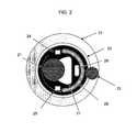
- FIG. 2provides a view of a cross section of the proximal end of a surgical device configured to remove the nucleus pulposus of an intervertebral disc (IVD) according to an embodiment of the invention.
- IVDintervertebral disc
- FIG. 3provides a view of an access device according to an embodiment of the invention.
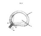
- FIG. 4illustrates a visualization device according to one embodiment of the invention viewing the nucleus pulposus of an intervertebral disc through an access port provided by a access device, such as a retractor tube.
- a access devicesuch as a retractor tube.
- FIG. 5provides a diagrammatic view of the positioning of two imaging sensors to provide a stereoscopic view of an internal target tissue site.
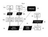
- FIG. 6provides a schematic representation of the operational framework of a processor that may be present in a device according to embodiments of the invention.
- Systems according to embodiments of the inventioninclude: an access device having a proximal end and distal end and an internal passageway extending from the proximal to distal end; and an elongated member dimensioned to be slidably moved through the internal passageway of the access device and having a proximal end and a distal end.
- at least one of multiple visualization elements and multiple illumination elementsare positioned among the distal ends of the access device and the elongated member.
- methods of using the systems in imaging applications, as well as kits for performing the methodsare also provided.
- aspects of the inventioninclude minimally invasive imaging systems.
- the imaging systems of the inventionare minimally invasive, such that they may be introduced to an internal target site of a patient, e.g., a spinal location that is near or inside of an intervertebral disc, through a minimal incision, e.g., one that is less than the size of an incision employed for an access device having a outer diameter of 20 mm or larger, e.g., less than 75% the size of such an incision, such as less than 50% of the size of such an incision, or smaller.
- Imaging systems of the inventioninclude both an access device and an elongate member.
- the access deviceis a device having a proximal end and a distal end and an internal passageway extending from the proximal to distal end.
- the elongated memberhas a proximal end and a distal end and is dimensioned to be slidably moved through the internal passageway of the access device.
- aspects of the inventioninclude at least one of multiple visualization elements and multiple illumination elements that are positioned among the distal ends of the access device and the elongated member.
- Access devices of the inventionare elongated elements having an internal passageway that are configured to provide access to a user (e.g., a health care professional, such as a surgeon) from an extra-corporeal location to an internal target tissue site, e.g., a location near or in the spine or component thereof, e.g., near or in an intervertebral disc, inside of the disc, etc., through a minimally invasive incision.
- Access devices of the inventionmay be cannulas, components of retractor tube systems, etc.
- the access devicesare elongate, they have a length that is 1.5 times or longer than their width, such as 2 times or longer than their width, including 5 or even 10 times or longer than their width, e.g., 20 times longer than its width, 30 times longer than its width, or longer.
- the longest cross-sectional outer dimension of the access devicesranges in certain instances from 5 mm to 50 mm, such as 10 to 20 mm.
- this passageis dimensioned to provide passage of the tools, e.g., imaging devices, tissue modifiers, etc., from an extra-corporeal site to the internal target tissue location.
- the longest cross-sectional dimension of the internal passagewaye.g., the inner diameter of a tubular shaped access device
- the access devicesranges in length from 5 to 30 mm, such as 5 to 25 mm, including 5 to 20 mm, e.g., 7 to 18 mm.
- the access devicesare sufficiently rigid to maintain mechanical separation of tissue, e.g., muscle, and may be fabricated from any convenient material. Materials of interest from which the access devices may be fabricated include, but are not limited to: metals, such as stainless steel and other medical grade metallic materials, plastics, and the like.
- the systems of the inventionfurther include an elongate member having a proximal and distal end, where the elongate member is dimensioned to be slidably moved through the internal passageway of the access device.
- this component of the systemsis elongate, it has a length that is 1.5 times or longer than its width, such as 2 times or longer than its width, including 5 or even 10 times or longer than its width, e.g., 20 times longer than its width, 30 times longer than its width, or longer.
- the elongate memberis dimensioned to access an intervertebral disc.
- dimensioned to access an intervertebral discis meant that at least the distal end of the device has a longest cross-sectional dimension that is 10 mm or less, such as 8 mm or less and including 7 mm or less, where in certain embodiments the longest cross-sectional dimension has a length ranging from 5 to 10 mm, such as 6 to 9 mm, and including 6 to 8 mm.
- the elongate membermay be solid or include one or more lumens, such that it may be viewed as a catheter.
- catheteris employed in its conventional sense to refer to a hollow, flexible or semi-rigid tube configured to be inserted into a body.
- Catheters of the inventionmay include a single lumen, or two or more lumens, e.g., three or more lumens, etc, as desired.
- the elongate membersmay be flexible or rigid, and may be fabricated from any convenient material.
- aspects of the inventioninclude at least one of multiple visualization elements and multiple illumination elements that are positioned among the distal ends of the access device and the elongated member.
- at least one of multiple visualization elements and multiple illumination elementsis meant that, over all, the system includes two or more visualization elements and/or two or more illumination elements that are located among the distal ends of access device and elongated member.
- embodiments of the systemsinclude those systems where two or more visualization elements 38 , examples of which are depicted in FIG. 3 , are located at the distal end of the elongated member.
- embodiments of the systemsalso include those systems where one visualization element is located at the distal end of the elongated member and another visualization element is located at the distal end of the access device.
- embodiments of the systemsinclude those systems where two or more visualization elements are located at the distal end of the access device.
- embodiments of the systemsinclude those systems where two or more illumination elements are located at the distal end of the elongated member.
- Embodiments of the systemsalso include those systems where one illumination element is located at the distal end of the elongated member and another illumination element is located at the distal end of the access device.
- embodiments of the systemsinclude those systems where two or more illumination elements are located at the distal end of the access device.
- the phrase “among the distal ends of the access device and elongated member”means that between the two distal ends, there is positioned at least one of multiple visualization elements and multiple illumination elements.
- located among the distal endsis meant that the item of interest (e.g., the visualization element, the illumination element) is present at the distal end of the elongate member and/or access device, or near the distal end of the elongate member and/or access device, e.g., within 10 mm or closer to the distal end, such as within 5 mm or closer to the distal end and including within 3 mm or closer to the distal end.
- Imaging sensors of interestare miniature in size so as to be positionable at the distal end of the elongate member or the access device.
- Miniature imaging sensors of interestare those that, when integrated at the distal end of an elongated structure along with an illumination source, e.g., such as an LED as reviewed below, can be positioned on a probe having a longest cross section dimension of 6 mm or less, such as 5 mm or less, including 4 mm or less, and even 3 mm or less.
- the miniature imaging sensorshave a longest cross-section dimension (such as a diagonal dimension) of 5 mm or less, such 3 mm or less, where in certain instances the sensors may have a longest cross-sectional dimension ranging from 2 to 3 mm.
- the miniature imaging sensorshave a cross-sectional area that is sufficiently small for its intended use and yet retain a sufficiently high matrix resolution.
- Certain imaging sensors of the inventionhave a cross-sectional area (i.e. an x-y dimension, also known as packaged chip size) that is 2 mm.times.2 mm or less, such as 1.8 mm.times.1.8 mm or less, and yet have a matrix resolution of 400.times.400 or greater, such as 640.times.480 or greater.
- the imaging sensorshave a sensitivity that is 500 mV/Lux-sec or greater, such as 700 mV/Lux-Sec or greater, including 1000 mV/Lux-Sec or greater, where in some instances the sensitivity of the sensor is 2000 mV/Lux-Sec or greater, such as 3000 mV/Lux-Sec or greater.
- the imaging sensors of interestare those that include a photosensitive component, e.g., array of photosensitive elements, coupled to an integrated circuit, where the integrated circuit is configured to obtain and integrate the signals from the photosensitive array and output the analog data to a backend processor.
- the image sensors of interestmay be viewed as integrated circuit image sensors, and include complementary metal-oxide-semiconductor (CMOS) sensors and charge-coupled device (CCD) sensors.
- CMOScomplementary metal-oxide-semiconductor
- CCDcharge-coupled device
- the image sensorsmay further include a lens positioned relative to the photosensitive component so as to focus images on the photosensitive component.
- a signal conductormay be present to connect the image sensor at the distal and to a device at the proximal end of the elongate member, e.g., in the form of one or more wires running along the length of the elongate member from the distal to the proximal end.
- Imaging sensors of interestinclude, but are not limited to, those obtainable from: OminVision Technologies Inc., Sony Corporation, Cypress Semiconductors.
- the imaging sensorsmay be integrated with the component of interest, e.g., the access device or the elongated structure. As the imaging sensor(s) is integrated at the distal end of the component, it cannot be removed from the remainder of the component without significantly compromising the structure of component. As such, the integrated visualization element is not readily removable from the remainder of the component, such that the visualization element and remainder of the component form an inter-related whole.
- CMOS sensorsare the OmniPixel line of CMOS sensors available from OmniVision (Sunnyvale, Calif.), including the OmniPixel, OmniPixel2, OmniPixel3, OmniPixel3-HS and OmniBSI lines of CMOS sensors. These sensors may be either frontside or backside illumination sensors, and have sufficiently small dimensions while maintaining sufficient functionality to be positioned at the distal end of the minimally invasive devices of the invention. Aspects of these sensors are further described in one or more the following U.S. patents, the disclosures of which are herein incorporated by reference: U.S. Pat. Nos.
- a variety of different types of lights sourcesmay be employed as illumination elements, so long as their dimensions are such that they can be positioned at the distal end of the access device and/or elongated member.
- the light sourcesmay be integrated with a given component (e.g., access device, elongated member) such that they are configured relative to the component such that the light source element cannot be removed from the remainder of the component without significantly compromising the structure of the component.
- the integrated illumination element of these embodimentsis not readily removable from the remainder of the component, such that the illumination element and remainder of the component form an inter-related whole.
- the light sourcesmay be light emitting diodes configured to emit light of the desired wavelength range, or optical conveyance elements, e.g., optical fibers, configured to convey light of the desired wavelength range from a location other than the distal end of the elongate member, e.g., a location at the proximal end of the elongate member, to the distal end of the elongate member.
- the light sourcesmay include a conductive element, e.g., wire, optical fiber, which runs the length of the elongate member to provide for control of the light sources from a location outside the body, e.g., an extracorporeal control device.
- the light sourcesmay include a diffusion element 40 , an example of which is depicted in FIG. 3 , to provide for uniform illumination of the target tissue site.
- a diffusion elementmay be employed, including but not limited to a translucent cover or layer (fabricated from any convenient translucent material) through which light from the light source passes and is thus diffused.
- the illumination elementsmay emit light of the same wavelength or they may be spectrally distinct light sources, where by “spectrally distinct” is meant that the light sources emit light at wavelengths that do not substantially overlap, such as white light and infra-red light, such as the spectrally distinct light sources described in copending U.S. application Ser. No.
- the elongate member of the systemfurther includes a tissue modifier.
- Tissue modifiersare components or sub-devices that interact with tissue in some manner to modify the tissue in a desired way.
- the term modifyis used broadly to refer to changing in some way, including cutting the tissue, ablating the tissue, delivering an agent(s) to the tissue, freezing the tissue, etc.
- tissue modifiersare tissue cutters, tissue ablators, tissue freezing/heating elements, agent delivery devices, etc.
- Tissue cutters of interestinclude, but are not limited to: blades, liquid jet devices, lasers and the like.
- Tissue ablators of interestinclude, but are not limited to ablation devices, such as devices for delivery ultrasonic energy (e.g., as employed in ultrasonic ablation), devices for delivering plasma energy, devices for delivery radiofrequency (RF) energy, devices for delivering microwave energy, etc.
- Energy transfer devices of interestinclude, but are not limited to: devices for modulating the temperature of tissue, e.g., freezing or heating devices, etc.
- the elongated membermay further include one or more lumens that run at least the substantial length of the device, e.g., for performing a variety of different functions.
- the elongated membermay include both an irrigation and aspiration lumen.
- the irrigation lumenis operatively connected to a fluid source (e.g., physiologically acceptable fluid, such as saline) at the proximal end of the device, where the fluid source is configured to introduce fluid into the lumen under positive pressure, e.g., at a pressure ranging from 0 to 500 mm Hg, so that fluid is conveyed along the irrigation lumen and out the distal end.
- a fluid sourcee.g., physiologically acceptable fluid, such as saline
- positive pressuree.g., at a pressure ranging from 0 to 500 mm Hg
- the dimensions of the irrigating lumenmay vary, in certain embodiments the longest cross-sectional dimension of the irrigation lumen ranges from 1 to 3 mm.
- the aspiration lumenis operatively connected to a source of negative pressure (e.g., vacuum source) at the proximal end of the device, where the negative pressure source is configured to draw fluid from the tissue location at the distal end into the irrigation lumen under positive pressure, e.g., at a pressure ranging from 50 to 600 mm Hg, so that fluid is removed from the tissue site and conveyed along the irrigation lumen and out the proximal end, e.g., into a waste reservoir.
- a source of negative pressuree.g., vacuum source
- the negative pressure sourceis configured to draw fluid from the tissue location at the distal end into the irrigation lumen under positive pressure, e.g., at a pressure ranging from 50 to 600 mm Hg, so that fluid is removed from the tissue site and conveyed along the irrigation lumen and out the proximal end, e.g., into a waste reservoir.
- positive pressuree.g., at a pressure ranging from 50 to 600 mm H
- the systems of the inventionare used in conjunction with a controller 36 , an example of which is depicted in FIG. 3 , configured to control illumination of the illumination elements and/or capture of images (e.g., as still imaged or video output) from the image sensors.
- This controllermay take a variety of different formats, including hardware, software and combinations thereof.
- the controllermay be physically located relative to the elongated member and/or access device at any convenient location, where the controller may be present at the distal end of the system components, at some point between the distal and proximal ends or at the proximal ends of the system components, as desired.
- the controllermay be distinct from the system components, i.e., access device and elongated member, such the access device and/or elongated member includes a controller interface for operatively coupling to the distinct controller, or the controller may be integral with the device.
- FIG. 2provides a cross-sectional view of the distal ends of the elongated member and access device of a system according to one embodiment of the invention, where the system is configured to be employed in the surgical removal of the nucleus pulposus of an intervertebral disc.
- distal end of elongated member 20in this embodiment a catheter
- distal end of access device 22includes a second imaging sensor 23 .
- first and second LEDs, 24 and 25are also shown at the distal end of elongated member 20 .
- an irrigation lumen 26 and aspiration lumen 27is also shown.
- the deviceincludes a tissue modifier in the form of a dissection electrode 28 .
- the first imaging sensor 21provides visualization of the target tissue site.
- the second imaging sensor 23is positioned on the access device (although it could be positioned at a variety of locations on the access device or the elongated member).
- the orientation of second imaging sensor 23is such that imaging sensor 23 provides imaged data of the elongated member, e.g., of the distal end of the elongated member during placement, etc. Any convenient positioning as use may be achieved.
- FIG. 3provides different views of an access device according to an embodiment of the invention.
- access device 30includes a distal end 31 .
- two cameras 32 A and 32 BPositioned at distal end 31 are two cameras 32 A and 32 B and two illumination sources, e.g., LEDs or light fibers, 33 A and 33 B.
- the multiple visualization and/or illumination elements of the devicesmay be positioned relative to each other in a variety of different ways. By selective positioning of these elements coupled, as desired, with specific image data processing techniques, unique views of the target tissue site may be obtained.
- two cameras 42 and 44may be positioned in the same cross-section of the distal end of the imaging device. Image data from the two cameras can, in such an embodiment, be combined to obtain a panoramic view of the target tissue site, in this case the nucleus pulposus.
- This configurationalso allows one to obtain a stereoscopic view of the target tissue site, as illustrated in FIG. 5 , e.g., by synchronizing the image data from the two cameras.
- FIG. 5e.g., by synchronizing the image data from the two cameras.
- the depth of the circle objectcan be distinguished from the square object.
- the ratio of object distance (i.e., distance of object of interest from the camera) to stereo baseline (i.e., camera to camera distance)may vary, and in certain instances ranges from 10 to 30, such as 15 to 25, e.g., 20 (e.g., where the object depth is 20 mm and the two cameras are 1 mm apart).
- FIG. 2provides an illustration of a distal end of a system made up of a catheter visualization device slidably positioned within an internal passageway of an access device, such as a retractor tube.
- the primary camera 21is on the cross section of the catheter
- the secondary camera 23is on the wall of the access device. Both cameras can be arranged to have certain orientations, as desired, such as forward viewing or angled or side viewing. Illuminations can also be arranged such that different views of the same object can be revealed.
- the light sourcecan be somewhat collimated or focused in a certain direction to give a better view of the surgical blades, electrodes or the local tissue appearance.
- the devices or components thereofmay be configured for one time use (i.e., disposable) or re-usable, e.g., where the components are configured to be used two or more times before disposal, e.g., where the device components are sterilizable.
- aspects of the inventionfurther include methods of imaging an internal tissue site with imaging devices of the invention.
- a variety of internal tissue sitescan be imaged with devices of the invention.
- the methodsare methods of imaging an intervertebral disc in a minimally invasive manner.
- the methodsare now primarily described further in terms of imaging IVD target tissue sites.
- the inventionis not so limited, as the devices may be used to image a variety of distinct target tissue sites.
- embodiments of such methodsinclude positioning a distal end of a minimally invasive intervertebral disc imaging device of the invention in viewing relationship to an intervertebral disc or portion of there, e.g., nucleus pulposus, internal site of nucleus pulposus, etc.
- viewing relationshipis meant that the distal end is positioned within 40 mm, such as within 10 mm, of the target tissue site of interest.
- Positioning the distal end in viewing device in relation to the desired target tissuemay be accomplished using any convenient approach, including through use of an access device, such as a cannula or retractor tube, which may or may not be fitted with a trocar, as desired.
- an access devicesuch as a cannula or retractor tube, which may or may not be fitted with a trocar, as desired.
- the target tissuee.g., intervertebral disc or portion thereof
- Image data obtained according to the methods of the inventionis output to a user in the form of an image, e.g., using a monitor or other convenient medium as a display means.
- the imageis a still image, while in other embodiments the image may be a video.
- the methodsinclude a step of tissue modification in addition to the tissue viewing.
- the methodsmay include a step of tissue removal, e.g., using a combination of tissue cutting and irrigation or flushing.
- the methodsmay include cutting a least a portion of the tissue and then removing the cut tissue from the site, e.g., by flushing at least a portion of the imaged tissue location using a fluid introduce by an irrigation lumen and removed by an aspiration lumen.
- FIG. 4provides a view of one embodiment of a method of visualizing an intervertebral disc.
- an access devicee.g., cannula, trocar, etc. is employed to provide access of the device to the internal body site, e.g., via a minimally sized incision.
- FIG. 4shows a visualization device according to an embodiment of the invention viewing the nucleus pulposus of an intervertebral disc through an access port provided by an access device, such as a cannula.
- the visualization elementsare positioned at the distal end of a catheter member, and are located in the same cross-sectional plane.
- Image data from the two visualization elementsmay be obtained and processed to provide for an enhanced field of view, e.g., a panoramic view, where the enhanced field of view may be one that is wider than the view obtained from a signal visualization element and/or provide for stereoscopic view, as illustrated in FIG. 5 .
- an enhanced field of viewe.g., a panoramic view
- the enhanced field of viewmay be one that is wider than the view obtained from a signal visualization element and/or provide for stereoscopic view, as illustrated in FIG. 5 .
- FIG. 6provides a flow chart representation of a stereoscopic image processing algorithm according to an embodiment of the invention, where the algorithm is configured to derived depth or “range” map on a two-dimensional scene.
- left and right images 61 a and 61 bobtained by two distinct visualization elements, e.g., sensors 42 and 44 as depicted in FIG. 5 , are first warped as shown at 62 a and 62 b via calibration element 63 to remove lens distortion.
- the resultant undistorted left and right images 64 a and 64 bare then processed with stereo and image fusion algorithms 65 and 66 to derive a disparity line 67 .
- triangulation computations 68are applied to derive range data.
- the range (or depth) map 69can be overlay on the image display, as desired.
- Methods of inventionmay find use in any convenient application, including diagnostic and therapeutic applications.
- Specific applications of interestinclude, but are not limited to, intervertebral disc diagnostic and therapeutic applications.
- methods of the inventioninclude diagnostic applications, where a disc is viewed to determine any problems with the disc, if present.
- Methods of the inventionalso include treatment methods, e.g., where a disc is modified in some manner to treat and existing medical condition.
- Treatment methods of interestinclude, but are not limited to: annulotomy, nucleotomy, discectomy, annulus replacement, nucleus replacement, and decompression due to a bulging or extruded disc. Additional methods in which the imaging devices find use include those described in United States Published Application No. 20080255563.
- the subjectis an animal, where in certain embodiments the animal is a “mammal” or “mammalian.”
- mammal and mammalianare used broadly to describe organisms which are within the class mammalia, including the orders carnivore (e.g., dogs and cats), rodentia (e.g., mice, guinea pigs, and rats), lagomorpha (e.g. rabbits) and primates (e.g., humans, chimpanzees, and monkeys).
- the subjectsi.e., patients are humans.
- kits for use in practicing the subject methodsmay include one or more of the above devices, and/or components of the subject systems, as described above.
- a kitmay include a visualization device and an access device, e.g., a cannula configured to be employed with the visualization device.
- the kitmay further include other components, e.g., guidewires, stylets, etc., which may find use in practicing the subject methods.
- Various componentsmay be packaged as desired, e.g., together or separately.
- the subject kitsmay further include instructions for using the components of the kit to practice the subject methods.
- the instructions for practicing the subject methodsare generally recorded on a suitable recording medium.
- the instructionsmay be printed on a substrate, such as paper or plastic, etc.
- the instructionsmay be present in the kits as a package insert, in the labeling of the container of the kit or components thereof (i.e., associated with the packaging or subpackaging) etc.
- the instructionsare present as an electronic storage data file present on a suitable computer readable storage medium, e.g. CD-ROM, diskette, etc.
- the actual instructionsare not present in the kit, but means for obtaining the instructions from a remote source, e.g. via the internet, are provided.
- An example of this embodimentis a kit that includes a web address where the instructions can be viewed and/or from which the instructions can be downloaded. As with the instructions, this means for obtaining the instructions is recorded on a suitable substrate.
- Also of interestis programming that is configured for operating a visualization device according to methods of invention, where the programming is recorded on physical computer readable media, e.g. any medium that can be read and accessed directly by a computer.
- Such mediainclude, but are not limited to: magnetic storage media, such as floppy discs, hard disc storage medium, and magnetic tape; optical storage media such as CD-ROM; electrical storage media such as RAM and ROM; and hybrids of these categories such as magnetic/optical storage media.
- magnetic storage mediasuch as floppy discs, hard disc storage medium, and magnetic tape
- optical storage mediasuch as CD-ROM
- electrical storage mediasuch as RAM and ROM
- hybrids of these categoriessuch as magnetic/optical storage media.
- Programming of the inventionincludes instructions for operating a device of the invention, such that upon execution by the programming, the executed instructions result in execution of the imaging device to: illuminate a target tissue site, such as an intervertebral disc or portion thereof; and capture one or more image frames of the illuminated target tissue site with the imaging sensor.
- a target tissue sitesuch as an intervertebral disc or portion thereof
Landscapes
- Health & Medical Sciences (AREA)
- Life Sciences & Earth Sciences (AREA)
- Surgery (AREA)
- General Health & Medical Sciences (AREA)
- Medical Informatics (AREA)
- Veterinary Medicine (AREA)
- Pathology (AREA)
- Public Health (AREA)
- Biophysics (AREA)
- Engineering & Computer Science (AREA)
- Biomedical Technology (AREA)
- Heart & Thoracic Surgery (AREA)
- Physics & Mathematics (AREA)
- Molecular Biology (AREA)
- Animal Behavior & Ethology (AREA)
- Nuclear Medicine, Radiotherapy & Molecular Imaging (AREA)
- Radiology & Medical Imaging (AREA)
- Optics & Photonics (AREA)
- Endoscopes (AREA)
- Surgical Instruments (AREA)
Abstract
Description
Claims (10)
Priority Applications (2)
| Application Number | Priority Date | Filing Date | Title |
|---|---|---|---|
| US13/739,664US10045686B2 (en) | 2008-11-12 | 2013-01-11 | Tissue visualization and modification device |
| US16/102,463US20190117050A1 (en) | 2008-11-12 | 2018-08-13 | Tissue visualization and modification device |
Applications Claiming Priority (2)
| Application Number | Priority Date | Filing Date | Title |
|---|---|---|---|
| US12/269,770US20100121139A1 (en) | 2008-11-12 | 2008-11-12 | Minimally Invasive Imaging Systems |
| US13/739,664US10045686B2 (en) | 2008-11-12 | 2013-01-11 | Tissue visualization and modification device |
Related Parent Applications (1)
| Application Number | Title | Priority Date | Filing Date |
|---|---|---|---|
| US12/269,770ContinuationUS20100121139A1 (en) | 2008-11-12 | 2008-11-12 | Minimally Invasive Imaging Systems |
Related Child Applications (1)
| Application Number | Title | Priority Date | Filing Date |
|---|---|---|---|
| US16/102,463ContinuationUS20190117050A1 (en) | 2008-11-12 | 2018-08-13 | Tissue visualization and modification device |
Publications (2)
| Publication Number | Publication Date |
|---|---|
| US20130296648A1 US20130296648A1 (en) | 2013-11-07 |
| US10045686B2true US10045686B2 (en) | 2018-08-14 |
Family
ID=42165839
Family Applications (3)
| Application Number | Title | Priority Date | Filing Date |
|---|---|---|---|
| US12/269,770AbandonedUS20100121139A1 (en) | 2008-11-12 | 2008-11-12 | Minimally Invasive Imaging Systems |
| US13/739,664Active2029-11-25US10045686B2 (en) | 2008-11-12 | 2013-01-11 | Tissue visualization and modification device |
| US16/102,463AbandonedUS20190117050A1 (en) | 2008-11-12 | 2018-08-13 | Tissue visualization and modification device |
Family Applications Before (1)
| Application Number | Title | Priority Date | Filing Date |
|---|---|---|---|
| US12/269,770AbandonedUS20100121139A1 (en) | 2008-11-12 | 2008-11-12 | Minimally Invasive Imaging Systems |
Family Applications After (1)
| Application Number | Title | Priority Date | Filing Date |
|---|---|---|---|
| US16/102,463AbandonedUS20190117050A1 (en) | 2008-11-12 | 2018-08-13 | Tissue visualization and modification device |
Country Status (1)
| Country | Link |
|---|---|
| US (3) | US20100121139A1 (en) |
Cited By (18)
| Publication number | Priority date | Publication date | Assignee | Title |
|---|---|---|---|---|
| US10278572B1 (en) | 2017-10-19 | 2019-05-07 | Obp Medical Corporation | Speculum |
| US10368733B2 (en) | 2010-09-28 | 2019-08-06 | Obp Medical Corporation | Speculum |
| US10420538B2 (en) | 2015-02-05 | 2019-09-24 | Obp Medical Corporation | Illuminated surgical retractor |
| US10420540B2 (en) | 2015-02-05 | 2019-09-24 | Obp Medical Corporation | Illuminated surgical retractor |
| US10512519B2 (en) | 2018-02-20 | 2019-12-24 | Obp Medical Corporation | Illuminated medical devices |
| US10687793B2 (en) | 2017-07-18 | 2020-06-23 | Obp Medical Corporation | Minimally invasive no touch (MINT) procedure for harvesting the great saphenous vein (GSV) and venous hydrodissector and retractor for use during the MINT procedure |
| US10722621B2 (en) | 2016-07-11 | 2020-07-28 | Obp Medical Corporation | Illuminated suction device |
| US10799229B2 (en) | 2018-02-20 | 2020-10-13 | Obp Medical Corporation | Illuminated medical devices |
| USD904607S1 (en) | 2019-05-07 | 2020-12-08 | Obp Medical Corporation | Nasal retractor |
| US10881387B2 (en) | 2015-06-03 | 2021-01-05 | Obp Medical Corporation | Retractor |
| USD911521S1 (en) | 2019-02-19 | 2021-02-23 | Obp Medical Corporation | Handle for medical devices including surgical retractors |
| US10939899B2 (en) | 2015-06-03 | 2021-03-09 | Obp Medical Corporation | End cap assembly for retractor and other medical devices |
| US10945588B2 (en) | 2015-08-11 | 2021-03-16 | Trice Medical, Inc. | Fully integrated, disposable tissue visualization device |
| US10952712B2 (en) | 2015-06-03 | 2021-03-23 | Obp Medical Corporation | Retractor |
| US10959609B1 (en) | 2020-01-31 | 2021-03-30 | Obp Medical Corporation | Illuminated suction device |
| US10966702B1 (en) | 2020-02-25 | 2021-04-06 | Obp Medical Corporation | Illuminated dual-blade retractor |
| US11547446B2 (en) | 2014-01-13 | 2023-01-10 | Trice Medical, Inc. | Fully integrated, disposable tissue visualization device |
| US12318080B2 (en) | 2023-07-21 | 2025-06-03 | Coopersurgical, Inc. | Illuminated surgical retractor capable of hand-held operation and of being mounted to a fixed frame |
Families Citing this family (53)
| Publication number | Priority date | Publication date | Assignee | Title |
|---|---|---|---|---|
| US10595710B2 (en) | 2001-10-19 | 2020-03-24 | Visionscope Technologies Llc | Portable imaging system employing a miniature endoscope |
| US20070167681A1 (en) | 2001-10-19 | 2007-07-19 | Gill Thomas J | Portable imaging system employing a miniature endoscope |
| US20100022824A1 (en) | 2008-07-22 | 2010-01-28 | Cybulski James S | Tissue modification devices and methods of using the same |
| US20100121139A1 (en) | 2008-11-12 | 2010-05-13 | Ouyang Xiaolong | Minimally Invasive Imaging Systems |
| US8672837B2 (en) | 2010-06-24 | 2014-03-18 | Hansen Medical, Inc. | Methods and devices for controlling a shapeable medical device |
| MX350734B (en) | 2010-09-08 | 2017-09-15 | Covidien Lp | Catheter with imaging assembly. |
| HK1198738A1 (en) | 2011-05-03 | 2015-06-05 | Endosee股份有限公司 | Method and apparatus for hysteroscopy and endometrial biopsy |
| CN102303538B (en)* | 2011-05-26 | 2014-07-02 | 奇瑞汽车股份有限公司 | Method and device for displaying residual driving range of electromobile |
| US9387048B2 (en) | 2011-10-14 | 2016-07-12 | Intuitive Surgical Operations, Inc. | Catheter sensor systems |
| US9452276B2 (en) | 2011-10-14 | 2016-09-27 | Intuitive Surgical Operations, Inc. | Catheter with removable vision probe |
| JP6430831B2 (en)* | 2011-10-14 | 2018-11-28 | インテュイティブ サージカル オペレーションズ, インコーポレイテッド | Catheter system |
| US20130303944A1 (en) | 2012-05-14 | 2013-11-14 | Intuitive Surgical Operations, Inc. | Off-axis electromagnetic sensor |
| US10238837B2 (en) | 2011-10-14 | 2019-03-26 | Intuitive Surgical Operations, Inc. | Catheters with control modes for interchangeable probes |
| US20130096385A1 (en)* | 2011-10-14 | 2013-04-18 | Intuitive Surgical Operations, Inc. | Vision probe and catheter systems |
| US9622646B2 (en) | 2012-06-25 | 2017-04-18 | Coopersurgical, Inc. | Low-cost instrument for endoscopically guided operative procedures |
| US9198835B2 (en) | 2012-09-07 | 2015-12-01 | Covidien Lp | Catheter with imaging assembly with placement aid and related methods therefor |
| USD717340S1 (en) | 2012-09-07 | 2014-11-11 | Covidien Lp | Display screen with enteral feeding icon |
| US9517184B2 (en) | 2012-09-07 | 2016-12-13 | Covidien Lp | Feeding tube with insufflation device and related methods therefor |
| USD735343S1 (en) | 2012-09-07 | 2015-07-28 | Covidien Lp | Console |
| USD716841S1 (en) | 2012-09-07 | 2014-11-04 | Covidien Lp | Display screen with annotate file icon |
| US9057600B2 (en) | 2013-03-13 | 2015-06-16 | Hansen Medical, Inc. | Reducing incremental measurement sensor error |
| US9014851B2 (en) | 2013-03-15 | 2015-04-21 | Hansen Medical, Inc. | Systems and methods for tracking robotically controlled medical instruments |
| US9271663B2 (en) | 2013-03-15 | 2016-03-01 | Hansen Medical, Inc. | Flexible instrument localization from both remote and elongation sensors |
| US9629595B2 (en) | 2013-03-15 | 2017-04-25 | Hansen Medical, Inc. | Systems and methods for localizing, tracking and/or controlling medical instruments |
| US11020016B2 (en) | 2013-05-30 | 2021-06-01 | Auris Health, Inc. | System and method for displaying anatomy and devices on a movable display |
| US9370295B2 (en) | 2014-01-13 | 2016-06-21 | Trice Medical, Inc. | Fully integrated, disposable tissue visualization device |
| US10342579B2 (en) | 2014-01-13 | 2019-07-09 | Trice Medical, Inc. | Fully integrated, disposable tissue visualization device |
| JP6824967B2 (en) | 2015-09-18 | 2021-02-03 | オーリス ヘルス インコーポレイテッド | Tubular net navigation |
| US10143526B2 (en) | 2015-11-30 | 2018-12-04 | Auris Health, Inc. | Robot-assisted driving systems and methods |
| WO2017161177A1 (en) | 2016-03-17 | 2017-09-21 | Trice Medical, Inc. | Clot evacuation and visualization devices and methods of use |
| US10702305B2 (en) | 2016-03-23 | 2020-07-07 | Coopersurgical, Inc. | Operative cannulas and related methods |
| US10244926B2 (en) | 2016-12-28 | 2019-04-02 | Auris Health, Inc. | Detecting endolumenal buckling of flexible instruments |
| WO2018183727A1 (en) | 2017-03-31 | 2018-10-04 | Auris Health, Inc. | Robotic systems for navigation of luminal networks that compensate for physiological noise |
| US10022192B1 (en) | 2017-06-23 | 2018-07-17 | Auris Health, Inc. | Automatically-initialized robotic systems for navigation of luminal networks |
| US10555778B2 (en) | 2017-10-13 | 2020-02-11 | Auris Health, Inc. | Image-based branch detection and mapping for navigation |
| US11058493B2 (en) | 2017-10-13 | 2021-07-13 | Auris Health, Inc. | Robotic system configured for navigation path tracing |
| US11510736B2 (en) | 2017-12-14 | 2022-11-29 | Auris Health, Inc. | System and method for estimating instrument location |
| WO2019125964A1 (en) | 2017-12-18 | 2019-06-27 | Auris Health, Inc. | Methods and systems for instrument tracking and navigation within luminal networks |
| US20190200906A1 (en)* | 2017-12-28 | 2019-07-04 | Ethicon Llc | Dual cmos array imaging |
| WO2019191144A1 (en) | 2018-03-28 | 2019-10-03 | Auris Health, Inc. | Systems and methods for registration of location sensors |
| JP7225259B2 (en) | 2018-03-28 | 2023-02-20 | オーリス ヘルス インコーポレイテッド | Systems and methods for indicating probable location of instruments |
| EP3773235B1 (en) | 2018-03-29 | 2023-07-19 | Trice Medical, Inc. | Fully integrated endoscope with biopsy capabilities |
| US10905499B2 (en) | 2018-05-30 | 2021-02-02 | Auris Health, Inc. | Systems and methods for location sensor-based branch prediction |
| KR102567087B1 (en) | 2018-05-31 | 2023-08-17 | 아우리스 헬스, 인코포레이티드 | Robotic systems and methods for navigation of luminal networks detecting physiological noise |
| JP7371026B2 (en) | 2018-05-31 | 2023-10-30 | オーリス ヘルス インコーポレイテッド | Path-based navigation of tubular networks |
| MX2020012904A (en) | 2018-05-31 | 2021-02-26 | Auris Health Inc | Image-based airway analysis and mapping. |
| CN112804959B (en) | 2018-09-28 | 2025-01-28 | 奥瑞斯健康公司 | Robotic systems and methods for accompanying endoscopic and percutaneous medical procedures |
| US11207141B2 (en) | 2019-08-30 | 2021-12-28 | Auris Health, Inc. | Systems and methods for weight-based registration of location sensors |
| US11147633B2 (en) | 2019-08-30 | 2021-10-19 | Auris Health, Inc. | Instrument image reliability systems and methods |
| WO2021137108A1 (en) | 2019-12-31 | 2021-07-08 | Auris Health, Inc. | Alignment interfaces for percutaneous access |
| EP4084721B1 (en) | 2019-12-31 | 2025-10-01 | Auris Health, Inc. | Anatomical feature identification and targeting |
| WO2021137109A1 (en) | 2019-12-31 | 2021-07-08 | Auris Health, Inc. | Alignment techniques for percutaneous access |
| US11737663B2 (en) | 2020-03-30 | 2023-08-29 | Auris Health, Inc. | Target anatomical feature localization |
Citations (340)
| Publication number | Priority date | Publication date | Assignee | Title |
|---|---|---|---|---|
| US3835842A (en)* | 1972-07-03 | 1974-09-17 | J Iglesias | Endoscope with continuous irrigation |
| US3871358A (en)* | 1972-08-04 | 1975-03-18 | Olympus Optical Co | Guiding tube for the insertion of an admissible medical implement into a human body |
| US4069823A (en) | 1976-04-19 | 1978-01-24 | Viktor Leonidovich Isakov | Apparatus for laser therapy |
| US4519391A (en) | 1981-10-20 | 1985-05-28 | Fuji Photo Film Co., Ltd. | Endoscope with signal transmission system and method of operating same |
| US4624243A (en) | 1985-04-08 | 1986-11-25 | American Hospital Supply Corp. | Endoscope having a reusable eyepiece and a disposable distal section |
| US4646738A (en) | 1985-12-05 | 1987-03-03 | Concept, Inc. | Rotary surgical tool |
| US4651201A (en) | 1984-06-01 | 1987-03-17 | Arnold Schoolman | Stereoscopic endoscope arrangement |
| US4678459A (en) | 1984-07-23 | 1987-07-07 | E-Z-Em, Inc. | Irrigating, cutting and aspirating system for percutaneous surgery |
| US4697210A (en) | 1984-08-20 | 1987-09-29 | Fuji Photo Optical Co., Ltd. | Endoscope for displaying a normal image |
| US4700702A (en) | 1985-12-09 | 1987-10-20 | Tatiana Nilsson | Instrument for cutting tissues in surgery |
| US4750902A (en) | 1985-08-28 | 1988-06-14 | Sonomed Technology, Inc. | Endoscopic ultrasonic aspirators |
| US4845555A (en) | 1987-02-13 | 1989-07-04 | Olympus Optical Co., Ltd. | Electronic endoscope apparatus |
| US4919112A (en) | 1989-04-07 | 1990-04-24 | Schott Fiber Optics | Low-cost semi-disposable endoscope |
| US5088676A (en) | 1988-06-30 | 1992-02-18 | Ncr Corporation | Compact height adjustable base for a display |
| US5131382A (en) | 1989-03-27 | 1992-07-21 | Meyer William F | Endoscopic percutaneous discectomy device |
| US5170775A (en) | 1990-06-20 | 1992-12-15 | Olympus Optical Co., Ltd. | Endoscope |
| US5178130A (en)* | 1990-04-04 | 1993-01-12 | Olympus Optical Co., Ltd. | Parent-and-son type endoscope system for making a synchronized field sequential system illumination |
| US5188093A (en) | 1991-02-04 | 1993-02-23 | Citation Medical Corporation | Portable arthroscope with periscope optics |
| US5190028A (en) | 1991-02-04 | 1993-03-02 | Citation Medical Corporation | Method for manufacturing a disposable arthroscopic probe |
| US5195541A (en) | 1991-10-18 | 1993-03-23 | Obenchain Theodore G | Method of performing laparoscopic lumbar discectomy |
| US5228430A (en) | 1989-08-04 | 1993-07-20 | Kabushiki Kaisha Toshiba | Electronic endoscope apparatus including easy focusing distal end |
| US5242441A (en) | 1992-02-24 | 1993-09-07 | Boaz Avitall | Deflectable catheter with rotatable tip electrode |
| US5269785A (en) | 1990-06-28 | 1993-12-14 | Bonutti Peter M | Apparatus and method for tissue removal |
| US5291010A (en)* | 1990-10-04 | 1994-03-01 | Olympus Optical Co., Ltd. | Solid state imaging device having a chambered imaging chip corner |
| US5312407A (en) | 1992-12-28 | 1994-05-17 | Carter L Philip | Rongeur apparatus having an offset bayonet and method of use with microscope during microsurgery |
| US5313962A (en) | 1991-10-18 | 1994-05-24 | Obenchain Theodore G | Method of performing laparoscopic lumbar discectomy |
| US5318589A (en) | 1992-04-15 | 1994-06-07 | Microsurge, Inc. | Surgical instrument for endoscopic surgery |
| US5334183A (en) | 1985-08-28 | 1994-08-02 | Valleylab, Inc. | Endoscopic electrosurgical apparatus |
| US5351678A (en) | 1992-09-01 | 1994-10-04 | Citation Medical Corporation | Endoscope scope assembly for full hemisphere view |
| US5354302A (en) | 1992-11-06 | 1994-10-11 | Ko Sung Tao | Medical device and method for facilitating intra-tissue visual observation and manipulation of distensible tissues |
| US5365267A (en)* | 1992-06-19 | 1994-11-15 | Linvatec Corporation | White balance target |
| US5369525A (en) | 1992-12-02 | 1994-11-29 | United States Surgical Corporation | Ring lens assembly for an optical viewing device |
| US5368015A (en) | 1991-03-18 | 1994-11-29 | Wilk; Peter J. | Automated surgical system and apparatus |
| US5373392A (en) | 1993-07-28 | 1994-12-13 | Aotec, Inc. | Photochromic light control mirror |
| US5373312A (en) | 1989-10-19 | 1994-12-13 | Seiko Epson Corporation | Ink jet printer |
| US5373317A (en) | 1993-05-28 | 1994-12-13 | Welch Allyn, Inc. | Control and display section for borescope or endoscope |
| US5383888A (en) | 1992-02-12 | 1995-01-24 | United States Surgical Corporation | Articulating endoscopic surgical apparatus |
| US5395312A (en) | 1991-10-18 | 1995-03-07 | Desai; Ashvin | Surgical tool |
| US5395313A (en) | 1993-08-13 | 1995-03-07 | Naves; Neil H. | Reciprocating arthroscopic shaver |
| US5403342A (en) | 1992-04-23 | 1995-04-04 | United States Surgical Corporation | Articulating endoscopic surgical apparatus |
| US5403276A (en) | 1993-02-16 | 1995-04-04 | Danek Medical, Inc. | Apparatus for minimally invasive tissue removal |
| US5406940A (en) | 1992-09-02 | 1995-04-18 | Olympus Winter & Ibe Gmbh | Medical instrument for creating a tissue canal |
| US5423312A (en) | 1992-12-18 | 1995-06-13 | Schott Fiber Optics, Inc. | Rigid endoscope having modified high refractive index tunnel rod for image transmission and method of manufacture thereof |
| US5425355A (en) | 1991-01-28 | 1995-06-20 | Laserscope | Energy discharging surgical probe and surgical process having distal energy application without concomitant proximal movement |
| US5476473A (en) | 1993-01-05 | 1995-12-19 | Richard Wolf Gmbh | Instrument for surgically cutting tissue |
| US5484433A (en) | 1993-12-30 | 1996-01-16 | The Spectranetics Corporation | Tissue ablating device having a deflectable ablation area and method of using same |
| US5494483A (en) | 1992-09-30 | 1996-02-27 | Adair; Edwin L. | Stereoscopic endoscope with miniaturized electronic imaging chip |
| US5500012A (en) | 1992-07-15 | 1996-03-19 | Angeion Corporation | Ablation catheter system |
| US5512036A (en) | 1994-03-15 | 1996-04-30 | Welch Allyn, Inc. | Dental imaging system |
| US5518502A (en) | 1994-06-08 | 1996-05-21 | The United States Surgical Corporation | Compositions, methods and apparatus for inhibiting fogging of endoscope lenses |
| US5547455A (en) | 1994-03-30 | 1996-08-20 | Medical Media Systems | Electronically steerable endoscope |
| US5569158A (en) | 1993-10-15 | 1996-10-29 | Fuji Photo Optical Co. Ltd. | Shielding structure of electronic endoscope apparatus |
| US5577992A (en)* | 1993-10-05 | 1996-11-26 | Asahi Kogaku Kogyo Kabushiki Kaisha | Bendable portion of endoscope |
| US5582575A (en) | 1994-02-23 | 1996-12-10 | Richard Wolf Gmbh | Instrument for endoscopic therapy |
| US5591192A (en) | 1995-02-01 | 1997-01-07 | Ethicon Endo-Surgery, Inc. | Surgical penetration instrument including an imaging element |
| US5601525A (en) | 1994-04-11 | 1997-02-11 | Olympus Optical Co., Ltd. | Hard-type endoscope apparatus |
| US5630837A (en) | 1993-07-01 | 1997-05-20 | Boston Scientific Corporation | Acoustic ablation |
| US5643294A (en) | 1993-03-01 | 1997-07-01 | United States Surgical Corporation | Surgical apparatus having an increased range of operability |
| US5647840A (en) | 1994-09-14 | 1997-07-15 | Circon Corporation | Endoscope having a distally heated distal lens |
| US5674191A (en) | 1994-05-09 | 1997-10-07 | Somnus Medical Technologies, Inc. | Ablation apparatus and system for removal of soft palate tissue |
| US5695513A (en) | 1996-03-01 | 1997-12-09 | Metagen, Llc | Flexible cutting tool and methods for its use |
| US5704534A (en) | 1994-12-19 | 1998-01-06 | Ethicon Endo-Surgery, Inc. | Articulation assembly for surgical instruments |
| US5707382A (en) | 1995-03-24 | 1998-01-13 | Ethicon Endo Surgery, Inc. | Surgical dissector |
| US5713505A (en) | 1996-05-13 | 1998-02-03 | Ethicon Endo-Surgery, Inc. | Articulation transmission mechanism for surgical instruments |
| JPH1033462A (en) | 1996-05-24 | 1998-02-10 | Fuji Photo Film Co Ltd | Variable diaphragm and endoscope |
| US5722403A (en) | 1996-10-28 | 1998-03-03 | Ep Technologies, Inc. | Systems and methods using a porous electrode for ablating and visualizing interior tissue regions |
| US5735792A (en) | 1992-11-25 | 1998-04-07 | Clarus Medical Systems, Inc. | Surgical instrument including viewing optics and an atraumatic probe |
| US5757458A (en) | 1991-11-12 | 1998-05-26 | Pilkington Barnes Hind, Inc. | Annular mask contact lenses |
| US5766200A (en) | 1993-05-06 | 1998-06-16 | Linvatec Corporation | Rotatable endoscopic shaver with polymeric blades |
| US5766194A (en) | 1996-12-23 | 1998-06-16 | Georgia Skin And Cancer Clinic, Pc | Surgical apparatus for tissue removal |
| US5766199A (en) | 1996-04-10 | 1998-06-16 | Linvatec Corporation | Endoscopic shaver blade with resilient cutting edges |
| US5785647A (en) | 1996-07-31 | 1998-07-28 | United States Surgical Corporation | Surgical instruments useful for spinal surgery |
| US5788636A (en) | 1997-02-25 | 1998-08-04 | Acuson Corporation | Method and system for forming an ultrasound image of a tissue while simultaneously ablating the tissue |
| US5807240A (en)* | 1996-09-24 | 1998-09-15 | Circon Corporation | Continuous flow endoscope with enlarged outflow channel |
| US5818527A (en)* | 1994-12-21 | 1998-10-06 | Olympus Optical Co., Ltd. | Image processor for correcting distortion of central portion of image and preventing marginal portion of the image from protruding |
| US5836943A (en) | 1996-08-23 | 1998-11-17 | Team Medical, L.L.C. | Electrosurgical generator |
| US5857961A (en) | 1995-06-07 | 1999-01-12 | Clarus Medical Systems, Inc. | Surgical instrument for use with a viewing system |
| US5864359A (en)* | 1995-05-30 | 1999-01-26 | Smith & Nephew, Inc. | Stereoscopic autofocusing based on comparing the left and right eye images |
| US5868664A (en) | 1996-02-23 | 1999-02-09 | Envision Medical Corporation | Electrically isolated sterilizable endoscopic video camera head |
| US5873817A (en) | 1997-05-12 | 1999-02-23 | Circon Corporation | Endoscope with resilient deflectable section |
| US5873814A (en) | 1996-07-12 | 1999-02-23 | Adair; Edwin L. | Sterile encapsulated endoscopic video monitor and method |
| US5873816A (en) | 1994-11-02 | 1999-02-23 | Olympus Optical Co., Ltd. | Electronic endoscope having an insertional portion a part of which is a conductive armor |
| US5879285A (en) | 1995-09-28 | 1999-03-09 | Olympus Optical Co., Ltd. | Aligning means attaching a cable in an imaging apparatus |
| US5888193A (en) | 1996-02-22 | 1999-03-30 | Precision Optics Corporation | Endoscope with curved optical axis |
| US5902272A (en) | 1992-01-07 | 1999-05-11 | Arthrocare Corporation | Planar ablation probe and method for electrosurgical cutting and ablation |
| US5916146A (en) | 1995-12-22 | 1999-06-29 | Bieffe Medital S.P.A. | System for support and actuation with vertebrae in particular for surgical and diagnostic instruments |
| US5929901A (en) | 1997-10-06 | 1999-07-27 | Adair; Edwin L. | Reduced area imaging devices incorporated within surgical instruments |
| US5928137A (en) | 1996-05-03 | 1999-07-27 | Green; Philip S. | System and method for endoscopic imaging and endosurgery |
| US5941817A (en) | 1996-11-14 | 1999-08-24 | Vista Medical Technologies, Inc. | Endoscope wherein electrical components are electrically isolated from patient-engaging components |
| US5976075A (en) | 1997-12-15 | 1999-11-02 | University Of Massachusetts | Endoscope deployment apparatus |
| US5976076A (en) | 1995-02-22 | 1999-11-02 | Kolff; Jack | Stereo laparoscope with synchronized optics |
| US5976077A (en) | 1996-08-06 | 1999-11-02 | Olympus Winter & Ibe Gmbh | Surgical endoscopic instrument |
| US5986693A (en) | 1997-10-06 | 1999-11-16 | Adair; Edwin L. | Reduced area imaging devices incorporated within surgical instruments |
| US6001084A (en) | 1995-12-18 | 1999-12-14 | Riek; Siegfried | Medical needle for endoscopic surgery |
| WO2000009001A1 (en) | 1998-08-12 | 2000-02-24 | Inbae Yoon | Surgical instrument endoscope with cmos image sensor and physical parameter sensor |
| US6043839A (en) | 1997-10-06 | 2000-03-28 | Adair; Edwin L. | Reduced area imaging devices |
| US6045549A (en) | 1997-09-30 | 2000-04-04 | Somnus Medical Technologies, Inc. | Tissue ablation apparatus and device for use therein and method |
| US6053923A (en) | 1998-03-17 | 2000-04-25 | Arthrotek, Inc. | Method and apparatus for abrading tissue |
| US6068603A (en) | 1998-02-17 | 2000-05-30 | Olympus Optical Co., Ltd. | Medical instrument for use in combination with an endoscope |
| US6080101A (en) | 1998-08-26 | 2000-06-27 | Olympus Optical Co. Ltd. | Endoscope video camera head which can be autoclaved |
| US6086528A (en) | 1997-09-11 | 2000-07-11 | Adair; Edwin L. | Surgical devices with removable imaging capability and methods of employing same |
| US6099465A (en) | 1996-12-04 | 2000-08-08 | Fuji Photo Optical Co., Ltd. | Electromagnetically coupled electronic endoscope system |
| US6112123A (en) | 1998-07-28 | 2000-08-29 | Endonetics, Inc. | Device and method for ablation of tissue |
| US6110127A (en) | 1998-02-17 | 2000-08-29 | Olympus Optical, Co., Ltd. | Medical instrument for use in combination with an endoscope |
| US6113614A (en) | 1998-05-05 | 2000-09-05 | Ensurg, Inc. | Medical device for dissolution of tissue within the human body |
| US6126592A (en) | 1998-09-12 | 2000-10-03 | Smith & Nephew, Inc. | Endoscope cleaning and irrigation sheath |
| US6129662A (en) | 1996-06-03 | 2000-10-10 | Cogent Light Technologies, Inc. | Surgical tool with surgical field illuminator |
| US6142957A (en) | 1993-09-20 | 2000-11-07 | Boston Scientific Corporation | Multiple biopsy sampling device |
| US6149646A (en) | 1999-02-02 | 2000-11-21 | Linvatec Corporation | Monopolar tissue ablator |
| US6156033A (en) | 1997-05-15 | 2000-12-05 | Tu; Hosheng | Ablation catheter having electrode means and methods thereof |
| US6211904B1 (en) | 1997-09-11 | 2001-04-03 | Edwin L. Adair | Surgical devices incorporating reduced area imaging devices |
| US6234955B1 (en) | 1998-12-11 | 2001-05-22 | Enteric Medical Technologies, Inc. | Method for vascular occulusive therapy in gastrointestinal system |
| US6241727B1 (en) | 1998-05-27 | 2001-06-05 | Irvine Biomedical, Inc. | Ablation catheter system having circular lesion capabilities |
| JP2001161630A (en) | 1999-12-09 | 2001-06-19 | Olympus Optical Co Ltd | Endoscope |
| US6251120B1 (en) | 1998-11-03 | 2001-06-26 | Karl Storz Gmbh & Co., Kg | Medical instrument for removing tissue |
| US6264670B1 (en) | 1995-04-12 | 2001-07-24 | Origin Medsystems, Inc. | Tissue dissection method |
| US6310642B1 (en) | 1997-11-24 | 2001-10-30 | Micro-Medical Devices, Inc. | Reduced area imaging devices incorporated within surgical instruments |
| US20010036015A1 (en)* | 2000-03-31 | 2001-11-01 | Asahi Kogaku Kogyo Kabushiki Kaisha | Illumination optical system and an illumination lens element |
| US6315712B1 (en) | 1998-10-27 | 2001-11-13 | Tokendo (Sarl) | Video endoscopic probe with a distal color CCD sensor |
| US6316215B1 (en) | 1999-12-27 | 2001-11-13 | Edwin L. Adair | Methods of cancer screening utilizing fluorescence detection techniques and selectable imager charge integration periods |
| US6322494B1 (en) | 1998-04-03 | 2001-11-27 | Gyrus Medical Limited | Endoscope |
| US6331165B1 (en) | 1996-11-25 | 2001-12-18 | Scimed Life Systems, Inc. | Biopsy instrument having irrigation and aspiration capabilities |
| US20010053873A1 (en) | 1999-11-24 | 2001-12-20 | Hansgeorg Schaaf | Device for improving drainage of the aqueous humor within the eye of a living being |
| US6332881B1 (en) | 1999-09-01 | 2001-12-25 | Cardima, Inc. | Surgical ablation tool |
| US20020007110A1 (en) | 1992-11-12 | 2002-01-17 | Ing. Klaus Irion | Endoscope, in particular, having stereo-lateral-view optics |
| US6371909B1 (en) | 1998-02-19 | 2002-04-16 | California Institute Of Technology | Apparatus and method for providing spherical viewing during endoscopic procedures |
| US6387043B1 (en)* | 1998-05-13 | 2002-05-14 | Inbae Yoon | Penetrating endoscope and endoscopic surgical instrument with CMOS image sensor and display |
| US20020072651A1 (en)* | 1997-04-01 | 2002-06-13 | George A. Vilos | Debris aspirating resectoscope |
| US20020087047A1 (en) | 1999-09-13 | 2002-07-04 | Visionscope, Inc. | Miniature endoscope system |
| US6419654B1 (en) | 2000-02-01 | 2002-07-16 | Jeffrey S. Kadan | Diagnostic needle arthroscopy and lavage system |
| US6419627B1 (en) | 1997-08-01 | 2002-07-16 | One Way Ocular Technology, Ltd. | Ophthalmic endoscope and endoscope attachments for use therewith |
| US6424369B1 (en) | 1997-10-06 | 2002-07-23 | Edwin L. Adair | Hand-held computers incorporating reduced area imaging devices |
| US6447445B1 (en)* | 1999-09-14 | 2002-09-10 | Fuji Photo Optical Co., Ltd. | Endoscopic insertion instrument |
| US6452626B1 (en) | 1997-10-06 | 2002-09-17 | Edwin L. Adair | Communication devices incorporating reduced area imaging devices |
| US6458140B2 (en) | 1999-07-28 | 2002-10-01 | Vasconnect, Inc. | Devices and methods for interconnecting vessels |
| US6459481B1 (en)* | 1999-05-06 | 2002-10-01 | David F. Schaack | Simple system for endoscopic non-contact three-dimentional measurement |
| US6464711B1 (en) | 1999-03-19 | 2002-10-15 | Medtronic Xomed, Inc. | Articulating mechanism for steerable surgical cutting instruments |
| US6464633B1 (en) | 1999-08-23 | 2002-10-15 | Olympus Optical Co., Ltd. | Light source device for endoscope using DMD |
| US6468274B1 (en) | 1996-07-16 | 2002-10-22 | Arthrocare Corporation | Systems and methods for treating spinal pain |
| EP1252859A2 (en) | 2001-04-27 | 2002-10-30 | Firma Ivoclar Vivadent AG | Dental camera with mouthpiece |
| US6478730B1 (en) | 1998-09-09 | 2002-11-12 | Visionscope, Inc. | Zoom laparoscope |
| US20020177847A1 (en) | 2001-03-30 | 2002-11-28 | Long Gary L. | Endoscopic ablation system with flexible coupling |
| US6491690B1 (en) | 1997-07-18 | 2002-12-10 | Gyrus Medical Limited | Electrosurgical instrument |
| US20030040668A1 (en) | 2001-08-03 | 2003-02-27 | Olympus Optical Co., Ltd. | Endoscope apparatus |
| US6527753B2 (en)* | 2000-02-29 | 2003-03-04 | Olympus Optical Co., Ltd. | Endoscopic treatment system |
| US6533749B1 (en) | 1999-09-24 | 2003-03-18 | Medtronic Xomed, Inc. | Angled rotary tissue cutting instrument with flexible inner member |
| US6544194B1 (en) | 1996-11-25 | 2003-04-08 | Symbiosis Corporation | Proximal actuation handle for a biopsy forceps instrument having irrigation and aspiration capabilities |
| US6561973B1 (en) | 2000-07-25 | 2003-05-13 | John L. Bala | Micro-endoscopic system |
| US6569085B2 (en) | 2001-08-16 | 2003-05-27 | Syntheon, Llc | Methods and apparatus for delivering a medical instrument over an endoscope while the endoscope is in a body lumen |
| US6572578B1 (en) | 2000-08-25 | 2003-06-03 | Patrick A. Blanchard | Fluid-jet catheter and its application to flexible endoscopy |
| US6583240B2 (en) | 2001-05-23 | 2003-06-24 | Equistar Chemicals, Lp | Ethylene polymerization process |
| CN2557085Y (en) | 2002-04-24 | 2003-06-25 | 杜明国 | Endoscopic proctopolypus cutter |
| US20030120156A1 (en) | 2001-12-26 | 2003-06-26 | Forrester Kevin R. | Motion measuring device |
| US6585734B2 (en) | 2001-09-17 | 2003-07-01 | Melvin Levinson | Tissue cutting and retrieval device and method |
| US6602247B2 (en) | 1997-02-27 | 2003-08-05 | Cryocath Technologies Inc. | Apparatus and method for performing a treatment on a selected tissue region |
| US6602248B1 (en) | 1995-06-07 | 2003-08-05 | Arthro Care Corp. | Methods for repairing damaged intervertebral discs |
| US6605087B2 (en) | 1997-07-21 | 2003-08-12 | St. Jude Medical, Daig Division | Ablation catheter |
| US6607529B1 (en) | 1995-06-19 | 2003-08-19 | Medtronic Vidamed, Inc. | Electrosurgical device |
| US6623437B2 (en) | 2001-08-28 | 2003-09-23 | Rex Medical, L.P. | Tissue biopsy apparatus |
| US20030181905A1 (en) | 2002-03-25 | 2003-09-25 | Long Gary L. | Endoscopic ablation system with a distally mounted image sensor |
| US6632227B2 (en) | 2001-08-24 | 2003-10-14 | Scimed Life Systems, Inc. | Endoscopic resection devices |
| US6652522B2 (en) | 1990-12-14 | 2003-11-25 | Robert L. Cucin | Power-assisted tissue aspiration instrument with cauterizing cannula assembly |
| US20030220574A1 (en)* | 2002-03-18 | 2003-11-27 | Sarcos Investments Lc. | Miniaturized imaging device including utility aperture and SSID |
| US6656195B2 (en) | 2000-09-22 | 2003-12-02 | Medtronic Xomed, Inc. | Flexible inner tubular members and rotary tissue cutting instruments having flexible inner tubular members |
| US6656132B1 (en) | 1999-11-29 | 2003-12-02 | Pentex Corporation | Endoscopic tissue collecting instrument |
| US20030233024A1 (en) | 2002-06-14 | 2003-12-18 | Fuji Photo Optical Co., Ltd. | Electronic endoscope for stereoscopic endoscope system |
| US6675033B1 (en) | 1999-04-15 | 2004-01-06 | Johns Hopkins University School Of Medicine | Magnetic resonance imaging guidewire probe |
| US6679838B2 (en) | 2000-07-25 | 2004-01-20 | Micro-Invasive Technology, Inc. | Micro-endoscopic system |
| US6682535B2 (en) | 1999-06-16 | 2004-01-27 | Thomas Hoogland | Apparatus for decompressing herniated intervertebral discs |
| US6689130B2 (en) | 2001-06-04 | 2004-02-10 | Olympus Corporation | Treatment apparatus for endoscope |
| US6692432B1 (en) | 1996-07-15 | 2004-02-17 | East Giant Limited | Hand-held portable camera for producing video images of an object |
| US6695772B1 (en) | 2001-11-26 | 2004-02-24 | Visionary Biomedical, Inc. | Small diameter cannula devices, systems and methods |
| US6726684B1 (en) | 1996-07-16 | 2004-04-27 | Arthrocare Corporation | Methods for electrosurgical spine surgery |
| US20040102772A1 (en) | 2000-03-09 | 2004-05-27 | Baxter Jeffrey W. | Apparatus and method for minimally invasive surgery using rotational cutting tool |
| US6750037B2 (en) | 1999-12-27 | 2004-06-15 | Edwin L. Adair | Method of cancer screening primarily utilizing non-invasive cell collection, fluorescence detection techniques, and radio tracing detection techniques |
| US20040162572A1 (en) | 2003-02-13 | 2004-08-19 | Sauer Jude S. | Instrument for surgically cutting tissue and method of use |
| US20040162554A1 (en) | 2002-12-20 | 2004-08-19 | Manoa Medical, Inc., A Delaware Corporation | Systems and methods for cutting tissue |
| US6805715B2 (en) | 2001-10-09 | 2004-10-19 | Pmt Corporation | Method and device for treating intervertebral disc herniations |
| US20040215061A1 (en)* | 2003-04-28 | 2004-10-28 | Zebadiah Kimmel | Visualization stylet for endotracheal intubation |
| US6820791B2 (en) | 1998-06-19 | 2004-11-23 | Scimed Life Systems, Inc. | Non-circular resection device and endoscope |
| US6832984B2 (en) | 1997-08-21 | 2004-12-21 | Paul Stelzer | Minimally invasive surgery device |
| US6837887B2 (en) | 1995-06-07 | 2005-01-04 | Arthrocare Corporation | Articulated electrosurgical probe and methods |
| US20050038317A1 (en)* | 2004-10-11 | 2005-02-17 | Nitesh Ratnakar | Dual View Endoscope |
| US6863651B2 (en) | 2001-10-19 | 2005-03-08 | Visionscope, Llc | Miniature endoscope with imaging fiber system |
| US6885801B1 (en) | 2001-12-06 | 2005-04-26 | Clear Image Technology Llc | Enhancement of fiber based images |
| US20050090762A1 (en) | 1998-03-03 | 2005-04-28 | Senorx, Inc. | Electrosurgical biopsy device and method |
| CN1612708A (en) | 2002-07-31 | 2005-05-04 | 奥林巴斯株式会社 | Endoscope |
| US6893436B2 (en) | 2002-01-03 | 2005-05-17 | Afx, Inc. | Ablation instrument having a flexible distal portion |
| US20050113641A1 (en) | 2003-11-22 | 2005-05-26 | Bala John L. | Endoscopic imaging and intervention system |
| US6899672B2 (en) | 2002-11-08 | 2005-05-31 | Scimed Life Systems, Inc. | Endoscopic imaging system including removable deflection device |
| US6902526B2 (en) | 2002-10-23 | 2005-06-07 | Orthopaedic Development, Llc | Visualizing ablation device and procedure |
| US20050154262A1 (en)* | 2003-04-01 | 2005-07-14 | Banik Michael S. | Imaging system for video endoscope |
| US20050192532A1 (en) | 2004-01-29 | 2005-09-01 | Kucklick Theodore R. | Atraumatic arthroscopic instrument sheath |
| US20050197658A1 (en) | 1999-10-05 | 2005-09-08 | Platt Robert C. | Articulating ionizable gas coagulator |
| US20050197536A1 (en) | 2003-04-01 | 2005-09-08 | Banik Michael S. | Video endoscope |
| US20050213267A1 (en) | 2004-03-25 | 2005-09-29 | Integral Wave Technologies, Inc. | Switched-capacitor power supply system and method |
| US20050228228A1 (en) | 2004-04-08 | 2005-10-13 | Boulais Dennis R | System and method for orienting endoscope and operator control |
| US20050234296A1 (en) | 2004-04-14 | 2005-10-20 | Usgi Medical Inc. | Method and apparatus for obtaining endoluminal access |
| US20050277808A1 (en)* | 2004-05-14 | 2005-12-15 | Elazar Sonnenschein | Methods and devices related to camera connectors |
| US6982740B2 (en) | 1997-11-24 | 2006-01-03 | Micro-Medical Devices, Inc. | Reduced area imaging devices utilizing selected charge integration periods |
| US6982742B2 (en) | 1997-10-06 | 2006-01-03 | Adair Edwin L | Hand-held computers incorporating reduced area imaging devices |
| US20060004258A1 (en) | 2004-07-02 | 2006-01-05 | Wei-Zen Sun | Image-type intubation-aiding device |
| US20060004354A1 (en) | 2002-10-04 | 2006-01-05 | Nikolay Suslov | Plasma surgical device |
| US20060030861A1 (en) | 2004-07-21 | 2006-02-09 | Simonson Robert E | Methods and devices for retracting tissue in minimally invasive surgery |
| US6997941B2 (en) | 1996-08-13 | 2006-02-14 | Oratec Interventions, Inc. | Method and apparatus for treating annular fissures in intervertebral discs |
| US7002621B2 (en) | 1997-10-06 | 2006-02-21 | Adair Edwin L | Communication devices incorporating reduced area imaging devices |
| US7004938B2 (en) | 2001-11-29 | 2006-02-28 | Medwaves, Inc. | Radio-frequency-based catheter system with improved deflection and steering mechanisms |
| US20060063973A1 (en) | 2004-04-21 | 2006-03-23 | Acclarent, Inc. | Methods and apparatus for treating disorders of the ear, nose and throat |
| US20060069303A1 (en)* | 2004-09-30 | 2006-03-30 | Couvillon Lucien A Jr | Endoscopic apparatus with integrated hemostasis device |
| US20060069313A1 (en) | 2004-09-30 | 2006-03-30 | Couvillon Lucien A Jr | Medical devices with light emitting regions |
| US7030904B2 (en) | 1997-10-06 | 2006-04-18 | Micro-Medical Devices, Inc. | Reduced area imaging device incorporated within wireless endoscopic devices |
| US20060084839A1 (en) | 2002-05-30 | 2006-04-20 | Mourlas Nicholas J | Apparatus and methods for coronary sinus access |
| US20060089633A1 (en) | 2004-10-15 | 2006-04-27 | Baxano, Inc. | Devices and methods for tissue access |
| US20060106282A1 (en) | 2004-11-12 | 2006-05-18 | Micro Invasive Technology, Inc. | Pulsed infrared imaging and medical intervention system |
| US20060111613A1 (en) | 2004-09-30 | 2006-05-25 | Boston Scientific Scimed, Inc. | Selectively rotatable shaft coupler |
| CN1779836A (en) | 2004-09-30 | 2006-05-31 | 惠普开发有限公司 | Optical data processing |
| US7060028B2 (en) | 2003-06-06 | 2006-06-13 | Insight Instruments, Inc. | Endoilluminator |
| US20060149129A1 (en)* | 2005-01-05 | 2006-07-06 | Watts H D | Catheter with multiple visual elements |
| US20060167340A1 (en) | 2005-01-10 | 2006-07-27 | Pease Alfred A | Optical snake |
| US20060173244A1 (en) | 2004-09-30 | 2006-08-03 | Boston Scientific Scimed, Inc. | System and method of obstruction removal |
| US20060190063A1 (en) | 2004-05-07 | 2006-08-24 | John Kanzius | Enhanced systems and methods for RF-induced hyperthermia |
| US20060206007A1 (en) | 2005-03-14 | 2006-09-14 | Bala John L | Disposable illuminator endoscope |
| US20060206118A1 (en) | 2005-03-11 | 2006-09-14 | Kim Daniel H | Percutaneous endoscopic access tools for the spinal epidural space and related methods of treatment |
| US7108657B2 (en) | 2001-03-30 | 2006-09-19 | Karl Storz Gmbh & Co. Kg | Endoscopic visualization apparatus with different imaging systems |
| WO2006107877A2 (en) | 2005-04-01 | 2006-10-12 | Welch Allyn, Inc. | Vaginal speculum |
| US20060241648A1 (en) | 2005-02-04 | 2006-10-26 | Bleich Jeffery L | Methods and apparatus for tissue modification |
| US20060258951A1 (en) | 2005-05-16 | 2006-11-16 | Baxano, Inc. | Spinal Access and Neural Localization |
| US20060270904A1 (en) | 2005-05-27 | 2006-11-30 | Markus Kupferschmid | Medical instrument for endoscopic interventions |
| US20060276690A1 (en) | 2005-05-11 | 2006-12-07 | Farris Jesse L Iii | Visualization system |
| US20060281972A1 (en) | 2005-01-10 | 2006-12-14 | Pease Alfred A | Remote inspection device |
| US20060287576A1 (en) | 2004-03-02 | 2006-12-21 | Olympus Corporation | Endoscope |
| US20060293562A1 (en) | 2004-02-26 | 2006-12-28 | Olympus Corporation | Endoscope and endoscopic system |
| US7156559B2 (en) | 2003-04-21 | 2007-01-02 | The Johns Hopkins University | High temperature light guide |
| US7160295B1 (en) | 2003-12-22 | 2007-01-09 | Garito Jon C | Flexible electrosurgical electrode for treating tissue |
| US7160247B2 (en) | 2004-05-12 | 2007-01-09 | Linvatec Corporation | Endoscope with large diameter distal end |
| US7169147B2 (en) | 2003-05-30 | 2007-01-30 | Olympus Winter & Ibe Gmbh | Ureter resectoscope |
| US20070038117A1 (en) | 2005-07-26 | 2007-02-15 | Bala John L | Multi-spectral imaging endoscope system |
| US7179255B2 (en) | 1995-06-07 | 2007-02-20 | Arthrocare Corporation | Methods for targeted electrosurgery on contained herniated discs |
| US20070049794A1 (en) | 2005-09-01 | 2007-03-01 | Ezc Medical Llc | Visualization stylet for medical device applications having self-contained power source |
| US20070060798A1 (en) | 2005-09-15 | 2007-03-15 | Hagai Krupnik | System and method for presentation of data streams |
| US20070073109A1 (en) | 2005-09-23 | 2007-03-29 | Irion Klaus M | Lighting system for endoscopic examinations |
| US20070071311A1 (en)* | 2005-09-28 | 2007-03-29 | Deere & Company, A Delaware Corporation | Method for processing stereo vision data using image density |
| US7198635B2 (en) | 2000-10-17 | 2007-04-03 | Asthmatx, Inc. | Modification of airways by application of energy |
| US20070075654A1 (en) | 2003-09-22 | 2007-04-05 | Michael Kishinevsky | Method and Apparatus for Preventing Instabilities in Radio-Frequency Plasma Processing |
| GB2431539A (en) | 2005-10-24 | 2007-04-25 | Pentax Corp | Intubation assistance apparatus and intubation assistance used in the apparatus |
| US20070093689A1 (en) | 1995-09-04 | 2007-04-26 | Active Implants Corporation | Method and apparatus for computerized surgery |
| US7214183B2 (en) | 2002-09-13 | 2007-05-08 | Olympus Optical Co., Ltd. | Endoscope apparatus having an insertion channel |
| US20070115376A1 (en) | 2005-11-21 | 2007-05-24 | Olympus Medical Systems Corp. | Image pickup apparatus having two image sensors |
| US20070123888A1 (en) | 2004-10-15 | 2007-05-31 | Baxano, Inc. | Flexible tissue rasp |
| US20070129604A1 (en) | 2005-12-07 | 2007-06-07 | Siemens Power Generation, Inc. | Remote viewing apparatus |
| US20070129719A1 (en) | 2005-05-26 | 2007-06-07 | Amar Kendale | Apparatus and methods for performing minimally-invasive surgical procedures |
| US20070135874A1 (en) | 2005-11-22 | 2007-06-14 | Bala John L | Endoscope for therapeutic light delivery |
| US20070161855A1 (en)* | 2006-01-06 | 2007-07-12 | Olympus Medical Systems Corp. | Medical procedure through natural body orifice |
| US20070167678A1 (en) | 2004-05-14 | 2007-07-19 | Nathan Moskowitz | Tools for implantation and extraction of posteriorly placed lumbar articial discs including: a totally wireless electronically embedded action-ended endoscope utilizing differential directional illumination with digitally controlled mirrors and/or prisms, and a disc ball inserter , a plate extractor, and rescue disc plates |
| US20070167681A1 (en) | 2001-10-19 | 2007-07-19 | Gill Thomas J | Portable imaging system employing a miniature endoscope |
| US20070179340A1 (en) | 2005-12-20 | 2007-08-02 | Medicept, Inc. | Method and devices for minimally invasive arthroscopic procedures |
| US7258663B2 (en) | 1999-05-18 | 2007-08-21 | Olympus Corporation | Endoscope system with irradiated light switching feature |
| US7269344B2 (en) | 2003-02-13 | 2007-09-11 | Olympus Corporation | Optical apparatus |
| US20070213734A1 (en) | 2006-03-13 | 2007-09-13 | Bleich Jeffery L | Tissue modification barrier devices and methods |
| US20070213735A1 (en) | 2004-10-15 | 2007-09-13 | Vahid Saadat | Powered tissue modification devices and methods |
| US20070213733A1 (en) | 2004-10-15 | 2007-09-13 | Bleich Jeffery L | Mechanical tissue modification devices and methods |
| WO2007106740A2 (en) | 2006-03-13 | 2007-09-20 | Baxano, Inc. | Methods and apparatus for tissue modification |
| US20070219412A1 (en) | 2006-03-18 | 2007-09-20 | Digiovanni Christopher William | Flexible arthroscope and method of using the same |
| CN101040775A (en) | 2006-03-23 | 2007-09-26 | 伊西康内外科公司 | Disposable endoscope devices |
| US20070232850A1 (en) | 2006-03-30 | 2007-10-04 | Ethicon Endo-Surgery, Inc. | Endoscopic ancillary attachment devices |
| US20070249904A1 (en) | 2006-03-09 | 2007-10-25 | Olympus Medical Systems Corp. | Endoscope device and display device |
| US20070276183A1 (en) | 2004-12-28 | 2007-11-29 | Envisionier Medical Technologies Llc | Endoscopic imaging system |
| US20070287886A1 (en) | 2005-02-02 | 2007-12-13 | Voyage Medical Inc. | Tissue visualization and manipulation systems |
| US20070293853A1 (en) | 1998-07-13 | 2007-12-20 | Cytyc Corporation | Apparatuses and Methods for Interstitial Tissue Removal |
| US20080009747A1 (en) | 2005-02-02 | 2008-01-10 | Voyage Medical, Inc. | Transmural subsurface interrogation and ablation |
| US7320688B2 (en) | 1999-10-20 | 2008-01-22 | Warsaw Orthopedic, Inc. | Methods and instruments for endoscopic interbody surgical techniques |
| US20080033465A1 (en) | 2006-08-01 | 2008-02-07 | Baxano, Inc. | Multi-Wire Tissue Cutter |
| WO2008016927A2 (en) | 2006-08-01 | 2008-02-07 | The General Hospital Corporation | Systems and methods for receiving and/or analyzing information associated with electro-magnetic radiation |
| US20080051812A1 (en) | 2006-08-01 | 2008-02-28 | Baxano, Inc. | Multi-Wire Tissue Cutter |
| US20080051629A1 (en)* | 2003-07-29 | 2008-02-28 | Akira Sugiyama | Internal Treatment Apparatus for a Patient and an Internal Treatment System for a Patient |
| US20080062429A1 (en) | 2006-09-12 | 2008-03-13 | Rongguang Liang | Low coherence dental oct imaging |
| US20080064925A1 (en) | 2001-10-19 | 2008-03-13 | Gill Thomas J | Portable imaging system employing a miniature endoscope |
| US20080091064A1 (en) | 2006-10-17 | 2008-04-17 | Vadim Laser | Portable endoscope for intubation |
| US20080103504A1 (en) | 2006-10-30 | 2008-05-01 | Schmitz Gregory P | Percutaneous spinal stenosis treatment |
| US20080108869A1 (en) | 2006-10-20 | 2008-05-08 | Femsuite Llc | Optical surgical device and methods of use |
| US20080147018A1 (en)* | 2006-12-15 | 2008-06-19 | Squilla John R | Laparoscopic cannula with camera and lighting |
| US20080161809A1 (en) | 2006-10-03 | 2008-07-03 | Baxano, Inc. | Articulating Tissue Cutting Device |
| WO2008094439A2 (en) | 2007-01-26 | 2008-08-07 | Laurimed Llc | Styli used to position device for carrying out selective discectomy |
| WO2008098251A1 (en) | 2007-02-09 | 2008-08-14 | Skeletal Dynamics, Llc | Endo-surgical device and method |
| US20080207992A1 (en) | 2007-02-28 | 2008-08-28 | Synergetics, Inc. | Microsurgical Illuminator with Adjustable Illumination |
| US20080207996A1 (en) | 2005-06-06 | 2008-08-28 | Solar International Products, Inc | Portable Imaging Apparatus |
| US20080214896A1 (en) | 2007-01-10 | 2008-09-04 | Krupa Robert J | Endoscope with detachable elongation portion |
| US7435010B2 (en) | 2003-04-21 | 2008-10-14 | The Johns Hopkins University | High temperature light guide |
| US20080262302A1 (en)* | 2007-04-17 | 2008-10-23 | Surgiquest, Incorporated | Endoluminal and transluminal surgical methods and devices |
| US7453984B2 (en) | 2006-01-19 | 2008-11-18 | Carestream Health, Inc. | Real-time target confirmation for radiation therapy |
| US20080287961A1 (en)* | 2005-12-01 | 2008-11-20 | Olympus Medical Systems Corp. | Elongated medical member and procedure method using elongated medical member |
| US20080300462A1 (en) | 2007-05-31 | 2008-12-04 | Boston Scientific Scimed, Inc. | Active controlled bending in medical devices |
| US7491168B2 (en) | 2003-12-18 | 2009-02-17 | Depuy Spine, Inc. | Surgical retractor systems and illuminated cannulae |
| US20090076329A1 (en) | 2007-09-17 | 2009-03-19 | Wei Su | Disposable Stereoscopic Endoscope System |
| US7572578B2 (en) | 2003-09-09 | 2009-08-11 | Fujifilm Corporation | Method for isolating and purifying a nucleic acid |
| US20090264706A1 (en) | 2008-04-16 | 2009-10-22 | Bala John L | Disposable interchangeable angle light guide system |
| US20100016855A1 (en) | 2008-07-15 | 2010-01-21 | Immersion Corporation | Modular Tool With Signal Feedback |
| US20100022824A1 (en)* | 2008-07-22 | 2010-01-28 | Cybulski James S | Tissue modification devices and methods of using the same |
| US20100056862A1 (en) | 2008-09-03 | 2010-03-04 | Ethicon Endo-Surgery, Inc. | Access needle for natural orifice translumenal endoscopic surgery |
| US20100063356A1 (en)* | 2007-01-03 | 2010-03-11 | Smith Robert C | Access sheath with removable optical penetrating member |
| US20100063352A1 (en) | 2008-09-10 | 2010-03-11 | Fujifilm Corporation | Endoscope system and drive control method thereof |
| US7689268B2 (en) | 2002-08-05 | 2010-03-30 | Infraredx, Inc. | Spectroscopic unwanted signal filters for discrimination of vulnerable plaque and method therefor |
| US20100087798A1 (en) | 2008-09-24 | 2010-04-08 | Interlace Medical, Inc. | Systems, methods and devices for using a flowable medium for distending a hollow organ |
| US20100094231A1 (en) | 2004-10-15 | 2010-04-15 | Bleich Jeffery L | Devices and methods for tissue modification |
| US7699773B2 (en) | 2003-03-06 | 2010-04-20 | Precision Optics Corporation | Repairable endoscope |
| US7708689B2 (en) | 2004-05-12 | 2010-05-04 | Linvatec Corporation | Endoscope and related system |
| US20100121139A1 (en) | 2008-11-12 | 2010-05-13 | Ouyang Xiaolong | Minimally Invasive Imaging Systems |
| US20100121155A1 (en) | 2008-11-12 | 2010-05-13 | Ouyang Xiaolong | Minimally Invasive Tissue Modification Systems With Integrated Visualization |
| US20100165336A1 (en) | 2005-06-14 | 2010-07-01 | Ebstein Steven M | Applications of laser-processed substrate for molecular diagnostics |
| US20100217080A1 (en) | 2009-02-24 | 2010-08-26 | Visionscope Technologies, Llc | Disposable Sheath for Use with an Imaging System |
| US20100256446A1 (en)* | 2007-05-11 | 2010-10-07 | Board Of Regents, The University Of Texas System | Medical scope carrier and scope as system and method |
| US20100274081A1 (en) | 2009-04-22 | 2010-10-28 | Tyco Healthcare Group Lp | Visual veress needle assembly |
| US20100284580A1 (en) | 2009-05-07 | 2010-11-11 | Ouyang Xiaolong | Tissue visualization systems and methods for using the same |
| US20100286477A1 (en) | 2009-05-08 | 2010-11-11 | Ouyang Xiaolong | Internal tissue visualization system comprising a rf-shielded visualization sensor module |
| US20110009694A1 (en) | 2009-07-10 | 2011-01-13 | Schultz Eric E | Hand-held minimally dimensioned diagnostic device having integrated distal end visualization |
| US20110034769A1 (en) | 1997-10-06 | 2011-02-10 | Micro-Imaging Solutions Llc | Reduced area imaging device incorporated within wireless endoscopic devices |
| EP2335550A2 (en) | 2009-12-17 | 2011-06-22 | Tyco Healthcare Group LP | Visual obturator with tip openings |
| US8016839B2 (en) | 2005-01-26 | 2011-09-13 | Wilk Patent, Llc | Intra-abdominal medical procedures and device |
| US20110227509A1 (en) | 2010-03-17 | 2011-09-22 | Hady Saleh | Hands-free light controller for headgear mounted illumination device |
| US8038602B2 (en) | 2001-10-19 | 2011-10-18 | Visionscope Llc | Portable imaging system employing a miniature endoscope |
| US8046057B2 (en) | 2001-04-11 | 2011-10-25 | Clarke Dana S | Tissue structure identification in advance of instrument |
| US20110263933A1 (en) | 2007-08-23 | 2011-10-27 | Hansgeorg Schaaf | Resilient support |
| US20110263983A1 (en) | 2008-12-29 | 2011-10-27 | Koninklijke Philips Electronics N.V. | Ultrasound imaging system with remote control and metod of operation thereof |
| US8052609B2 (en) | 2005-04-15 | 2011-11-08 | Imacor Inc. | Connectorized probe with serial engagement mechanism |
| US20110276113A1 (en) | 2010-04-12 | 2011-11-10 | Cybulski James S | RF Tissue Modulation Devices and Methods of Using the Same |
| US8142346B2 (en) | 2006-07-21 | 2012-03-27 | Olympus Medical Systems Corp. | Endoscope with electromagnetic wave shield |
| US20120088968A1 (en) | 2010-10-11 | 2012-04-12 | Epic Medical Inc. | Methods and devices for visualization and access |
| US8170319B2 (en) | 2008-09-05 | 2012-05-01 | Siemens Medical Solutions Usa, Inc. | Motion detection by direct imaging during radiotherapy |
| US20120241188A1 (en) | 2011-03-23 | 2012-09-27 | Tyco Healthcare Group Lp | Wire guides and anchors for endoscopy |
| US8277411B2 (en) | 2002-01-31 | 2012-10-02 | Boston Scientific Scimed, Inc. | Needle device |
| US8317689B1 (en) | 1999-09-13 | 2012-11-27 | Visionscope Technologies Llc | Miniature endoscope system |
| US8475361B2 (en)* | 2006-01-06 | 2013-07-02 | Olympus Medical Systems Corp. | Percutaneous or natural-orifice medical procedure and system therefor |
| WO2014137530A1 (en) | 2013-03-05 | 2014-09-12 | Nico Corporation | Surgical access assembly and method of using same |
| WO2015106288A1 (en) | 2014-01-13 | 2015-07-16 | Trice Medical, Inc. | Fully integrated, disposable tissue visualization device |
| US20150313634A1 (en) | 2014-05-05 | 2015-11-05 | Rainbow Medical Ltd. | Pericardial access device |
| US20160045224A1 (en) | 2014-08-15 | 2016-02-18 | Covidien Lp | Obturator having an insufflation pathway and an instrument guide |
| WO2016130844A1 (en) | 2015-02-13 | 2016-08-18 | Trice Medical, Inc. | Tissue visualization and modification devices and methods |
| WO2017027749A1 (en) | 2015-08-11 | 2017-02-16 | Trice Medical, Inc. | Fully integrated, disposable tissue visualization device |
| US20170042573A1 (en) | 2014-11-06 | 2017-02-16 | Asimion Inc. | Visually Assisted Entry of a Veress Needle with a Tapered Videoscope for Microlaparoscopy |
| WO2017161777A1 (en) | 2016-03-22 | 2017-09-28 | 乐视控股(北京)有限公司 | Panorama live broadcast method and device |
- 2008
- 2008-11-12USUS12/269,770patent/US20100121139A1/ennot_activeAbandoned
- 2013
- 2013-01-11USUS13/739,664patent/US10045686B2/enactiveActive
- 2018
- 2018-08-13USUS16/102,463patent/US20190117050A1/ennot_activeAbandoned
Patent Citations (389)
| Publication number | Priority date | Publication date | Assignee | Title |
|---|---|---|---|---|
| US3835842A (en)* | 1972-07-03 | 1974-09-17 | J Iglesias | Endoscope with continuous irrigation |
| US3871358A (en)* | 1972-08-04 | 1975-03-18 | Olympus Optical Co | Guiding tube for the insertion of an admissible medical implement into a human body |
| US4069823A (en) | 1976-04-19 | 1978-01-24 | Viktor Leonidovich Isakov | Apparatus for laser therapy |
| US4519391A (en) | 1981-10-20 | 1985-05-28 | Fuji Photo Film Co., Ltd. | Endoscope with signal transmission system and method of operating same |
| US4651201A (en) | 1984-06-01 | 1987-03-17 | Arnold Schoolman | Stereoscopic endoscope arrangement |
| US4678459A (en) | 1984-07-23 | 1987-07-07 | E-Z-Em, Inc. | Irrigating, cutting and aspirating system for percutaneous surgery |
| US4697210A (en) | 1984-08-20 | 1987-09-29 | Fuji Photo Optical Co., Ltd. | Endoscope for displaying a normal image |
| US4624243A (en) | 1985-04-08 | 1986-11-25 | American Hospital Supply Corp. | Endoscope having a reusable eyepiece and a disposable distal section |
| US4750902A (en) | 1985-08-28 | 1988-06-14 | Sonomed Technology, Inc. | Endoscopic ultrasonic aspirators |
| US5334183A (en) | 1985-08-28 | 1994-08-02 | Valleylab, Inc. | Endoscopic electrosurgical apparatus |
| US4646738A (en) | 1985-12-05 | 1987-03-03 | Concept, Inc. | Rotary surgical tool |
| US4700702A (en) | 1985-12-09 | 1987-10-20 | Tatiana Nilsson | Instrument for cutting tissues in surgery |
| US4845555A (en) | 1987-02-13 | 1989-07-04 | Olympus Optical Co., Ltd. | Electronic endoscope apparatus |
| US5088676A (en) | 1988-06-30 | 1992-02-18 | Ncr Corporation | Compact height adjustable base for a display |
| US5131382A (en) | 1989-03-27 | 1992-07-21 | Meyer William F | Endoscopic percutaneous discectomy device |
| US4919112A (en) | 1989-04-07 | 1990-04-24 | Schott Fiber Optics | Low-cost semi-disposable endoscope |
| US4919112B1 (en) | 1989-04-07 | 1993-12-28 | Low-cost semi-disposable endoscope | |
| US5228430A (en) | 1989-08-04 | 1993-07-20 | Kabushiki Kaisha Toshiba | Electronic endoscope apparatus including easy focusing distal end |
| US5373312A (en) | 1989-10-19 | 1994-12-13 | Seiko Epson Corporation | Ink jet printer |
| US5178130A (en)* | 1990-04-04 | 1993-01-12 | Olympus Optical Co., Ltd. | Parent-and-son type endoscope system for making a synchronized field sequential system illumination |
| US5170775A (en) | 1990-06-20 | 1992-12-15 | Olympus Optical Co., Ltd. | Endoscope |
| US5269785A (en) | 1990-06-28 | 1993-12-14 | Bonutti Peter M | Apparatus and method for tissue removal |
| US6835198B2 (en) | 1990-06-28 | 2004-12-28 | The Bonutti 2003 Trust A | Apparatus and method for tissue removal |
| US5291010A (en)* | 1990-10-04 | 1994-03-01 | Olympus Optical Co., Ltd. | Solid state imaging device having a chambered imaging chip corner |
| US6652522B2 (en) | 1990-12-14 | 2003-11-25 | Robert L. Cucin | Power-assisted tissue aspiration instrument with cauterizing cannula assembly |
| US5425355A (en) | 1991-01-28 | 1995-06-20 | Laserscope | Energy discharging surgical probe and surgical process having distal energy application without concomitant proximal movement |
| US5190028A (en) | 1991-02-04 | 1993-03-02 | Citation Medical Corporation | Method for manufacturing a disposable arthroscopic probe |
| US5188093A (en) | 1991-02-04 | 1993-02-23 | Citation Medical Corporation | Portable arthroscope with periscope optics |
| US5368015A (en) | 1991-03-18 | 1994-11-29 | Wilk; Peter J. | Automated surgical system and apparatus |
| US5313962A (en) | 1991-10-18 | 1994-05-24 | Obenchain Theodore G | Method of performing laparoscopic lumbar discectomy |
| US5195541A (en) | 1991-10-18 | 1993-03-23 | Obenchain Theodore G | Method of performing laparoscopic lumbar discectomy |
| US5395312A (en) | 1991-10-18 | 1995-03-07 | Desai; Ashvin | Surgical tool |
| US5757458A (en) | 1991-11-12 | 1998-05-26 | Pilkington Barnes Hind, Inc. | Annular mask contact lenses |
| US5902272A (en) | 1992-01-07 | 1999-05-11 | Arthrocare Corporation | Planar ablation probe and method for electrosurgical cutting and ablation |
| US5383888A (en) | 1992-02-12 | 1995-01-24 | United States Surgical Corporation | Articulating endoscopic surgical apparatus |
| US5242441A (en) | 1992-02-24 | 1993-09-07 | Boaz Avitall | Deflectable catheter with rotatable tip electrode |
| US5318589A (en) | 1992-04-15 | 1994-06-07 | Microsurge, Inc. | Surgical instrument for endoscopic surgery |
| US5417203A (en) | 1992-04-23 | 1995-05-23 | United States Surgical Corporation | Articulating endoscopic surgical apparatus |
| US5403342A (en) | 1992-04-23 | 1995-04-04 | United States Surgical Corporation | Articulating endoscopic surgical apparatus |
| US5365267A (en)* | 1992-06-19 | 1994-11-15 | Linvatec Corporation | White balance target |
| US5500012A (en) | 1992-07-15 | 1996-03-19 | Angeion Corporation | Ablation catheter system |
| US5351678A (en) | 1992-09-01 | 1994-10-04 | Citation Medical Corporation | Endoscope scope assembly for full hemisphere view |
| US5406940A (en) | 1992-09-02 | 1995-04-18 | Olympus Winter & Ibe Gmbh | Medical instrument for creating a tissue canal |
| US5494483A (en) | 1992-09-30 | 1996-02-27 | Adair; Edwin L. | Stereoscopic endoscope with miniaturized electronic imaging chip |
| US5354302A (en) | 1992-11-06 | 1994-10-11 | Ko Sung Tao | Medical device and method for facilitating intra-tissue visual observation and manipulation of distensible tissues |
| US20020007110A1 (en) | 1992-11-12 | 2002-01-17 | Ing. Klaus Irion | Endoscope, in particular, having stereo-lateral-view optics |
| US5735792A (en) | 1992-11-25 | 1998-04-07 | Clarus Medical Systems, Inc. | Surgical instrument including viewing optics and an atraumatic probe |
| US5369525A (en) | 1992-12-02 | 1994-11-29 | United States Surgical Corporation | Ring lens assembly for an optical viewing device |
| US5423312A (en) | 1992-12-18 | 1995-06-13 | Schott Fiber Optics, Inc. | Rigid endoscope having modified high refractive index tunnel rod for image transmission and method of manufacture thereof |
| US5630784A (en) | 1992-12-18 | 1997-05-20 | Schott Fiber Optics Inc. | Method of making and using a rigid endoscope having a modified high refractive index tunnel rod |
| US5312407A (en) | 1992-12-28 | 1994-05-17 | Carter L Philip | Rongeur apparatus having an offset bayonet and method of use with microscope during microsurgery |
| US5476473A (en) | 1993-01-05 | 1995-12-19 | Richard Wolf Gmbh | Instrument for surgically cutting tissue |
| US5403276A (en) | 1993-02-16 | 1995-04-04 | Danek Medical, Inc. | Apparatus for minimally invasive tissue removal |
| US5643294A (en) | 1993-03-01 | 1997-07-01 | United States Surgical Corporation | Surgical apparatus having an increased range of operability |
| US5766200A (en) | 1993-05-06 | 1998-06-16 | Linvatec Corporation | Rotatable endoscopic shaver with polymeric blades |
| US5373317B1 (en) | 1993-05-28 | 2000-11-21 | Welch Allyn Inc | Control and display section for borescope or endoscope |
| US5373317A (en) | 1993-05-28 | 1994-12-13 | Welch Allyn, Inc. | Control and display section for borescope or endoscope |
| US5630837A (en) | 1993-07-01 | 1997-05-20 | Boston Scientific Corporation | Acoustic ablation |
| US5373392A (en) | 1993-07-28 | 1994-12-13 | Aotec, Inc. | Photochromic light control mirror |
| US5395313A (en) | 1993-08-13 | 1995-03-07 | Naves; Neil H. | Reciprocating arthroscopic shaver |
| US6142957A (en) | 1993-09-20 | 2000-11-07 | Boston Scientific Corporation | Multiple biopsy sampling device |
| US5577992A (en)* | 1993-10-05 | 1996-11-26 | Asahi Kogaku Kogyo Kabushiki Kaisha | Bendable portion of endoscope |
| US5569158A (en) | 1993-10-15 | 1996-10-29 | Fuji Photo Optical Co. Ltd. | Shielding structure of electronic endoscope apparatus |
| US5484433A (en) | 1993-12-30 | 1996-01-16 | The Spectranetics Corporation | Tissue ablating device having a deflectable ablation area and method of using same |
| US5582575A (en) | 1994-02-23 | 1996-12-10 | Richard Wolf Gmbh | Instrument for endoscopic therapy |
| US5512036A (en) | 1994-03-15 | 1996-04-30 | Welch Allyn, Inc. | Dental imaging system |
| US6261226B1 (en) | 1994-03-30 | 2001-07-17 | Medical Media Systems | Electronically Steerable Endoscope |
| US5547455A (en) | 1994-03-30 | 1996-08-20 | Medical Media Systems | Electronically steerable endoscope |
| US5601525A (en) | 1994-04-11 | 1997-02-11 | Olympus Optical Co., Ltd. | Hard-type endoscope apparatus |
| US5674191A (en) | 1994-05-09 | 1997-10-07 | Somnus Medical Technologies, Inc. | Ablation apparatus and system for removal of soft palate tissue |
| US5518502A (en) | 1994-06-08 | 1996-05-21 | The United States Surgical Corporation | Compositions, methods and apparatus for inhibiting fogging of endoscope lenses |
| US5647840A (en) | 1994-09-14 | 1997-07-15 | Circon Corporation | Endoscope having a distally heated distal lens |
| US5873816A (en) | 1994-11-02 | 1999-02-23 | Olympus Optical Co., Ltd. | Electronic endoscope having an insertional portion a part of which is a conductive armor |
| US5704534A (en) | 1994-12-19 | 1998-01-06 | Ethicon Endo-Surgery, Inc. | Articulation assembly for surgical instruments |
| US5818527A (en)* | 1994-12-21 | 1998-10-06 | Olympus Optical Co., Ltd. | Image processor for correcting distortion of central portion of image and preventing marginal portion of the image from protruding |
| US5591192A (en) | 1995-02-01 | 1997-01-07 | Ethicon Endo-Surgery, Inc. | Surgical penetration instrument including an imaging element |
| US5976076A (en) | 1995-02-22 | 1999-11-02 | Kolff; Jack | Stereo laparoscope with synchronized optics |
| US5707382A (en) | 1995-03-24 | 1998-01-13 | Ethicon Endo Surgery, Inc. | Surgical dissector |
| US6264670B1 (en) | 1995-04-12 | 2001-07-24 | Origin Medsystems, Inc. | Tissue dissection method |
| US6277137B1 (en) | 1995-04-12 | 2001-08-21 | Origin Medsystems | Tissue separation cannula with dissection probe and method |
| US7318823B2 (en) | 1995-04-13 | 2008-01-15 | Arthrocare Corporation | Methods for repairing damaged intervertebral discs |
| US5864359A (en)* | 1995-05-30 | 1999-01-26 | Smith & Nephew, Inc. | Stereoscopic autofocusing based on comparing the left and right eye images |
| US5857961A (en) | 1995-06-07 | 1999-01-12 | Clarus Medical Systems, Inc. | Surgical instrument for use with a viewing system |
| US6837887B2 (en) | 1995-06-07 | 2005-01-04 | Arthrocare Corporation | Articulated electrosurgical probe and methods |
| US7179255B2 (en) | 1995-06-07 | 2007-02-20 | Arthrocare Corporation | Methods for targeted electrosurgery on contained herniated discs |
| US6602248B1 (en) | 1995-06-07 | 2003-08-05 | Arthro Care Corp. | Methods for repairing damaged intervertebral discs |
| USRE40156E1 (en) | 1995-06-07 | 2008-03-18 | Arthrocare Corporation | Methods for repairing damaged intervertebral discs |
| US6607529B1 (en) | 1995-06-19 | 2003-08-19 | Medtronic Vidamed, Inc. | Electrosurgical device |
| US20070093689A1 (en) | 1995-09-04 | 2007-04-26 | Active Implants Corporation | Method and apparatus for computerized surgery |
| US5879285A (en) | 1995-09-28 | 1999-03-09 | Olympus Optical Co., Ltd. | Aligning means attaching a cable in an imaging apparatus |
| US6001084A (en) | 1995-12-18 | 1999-12-14 | Riek; Siegfried | Medical needle for endoscopic surgery |
| US5916146A (en) | 1995-12-22 | 1999-06-29 | Bieffe Medital S.P.A. | System for support and actuation with vertebrae in particular for surgical and diagnostic instruments |
| US5888193A (en) | 1996-02-22 | 1999-03-30 | Precision Optics Corporation | Endoscope with curved optical axis |
| US5868664A (en) | 1996-02-23 | 1999-02-09 | Envision Medical Corporation | Electrically isolated sterilizable endoscopic video camera head |
| US6390972B1 (en) | 1996-02-23 | 2002-05-21 | Linvatec Corporation | Electrically-isolated, sterilizable, endoscopic video camera head |
| US5695513A (en) | 1996-03-01 | 1997-12-09 | Metagen, Llc | Flexible cutting tool and methods for its use |
| US5766199A (en) | 1996-04-10 | 1998-06-16 | Linvatec Corporation | Endoscopic shaver blade with resilient cutting edges |
| US5928137A (en) | 1996-05-03 | 1999-07-27 | Green; Philip S. | System and method for endoscopic imaging and endosurgery |
| US5713505A (en) | 1996-05-13 | 1998-02-03 | Ethicon Endo-Surgery, Inc. | Articulation transmission mechanism for surgical instruments |
| JPH1033462A (en) | 1996-05-24 | 1998-02-10 | Fuji Photo Film Co Ltd | Variable diaphragm and endoscope |
| US6129662A (en) | 1996-06-03 | 2000-10-10 | Cogent Light Technologies, Inc. | Surgical tool with surgical field illuminator |
| US5873814A (en) | 1996-07-12 | 1999-02-23 | Adair; Edwin L. | Sterile encapsulated endoscopic video monitor and method |
| US6692432B1 (en) | 1996-07-15 | 2004-02-17 | East Giant Limited | Hand-held portable camera for producing video images of an object |
| US6468274B1 (en) | 1996-07-16 | 2002-10-22 | Arthrocare Corporation | Systems and methods for treating spinal pain |
| US6726684B1 (en) | 1996-07-16 | 2004-04-27 | Arthrocare Corporation | Methods for electrosurgical spine surgery |
| US5785647A (en) | 1996-07-31 | 1998-07-28 | United States Surgical Corporation | Surgical instruments useful for spinal surgery |
| US5976077A (en) | 1996-08-06 | 1999-11-02 | Olympus Winter & Ibe Gmbh | Surgical endoscopic instrument |
| US6997941B2 (en) | 1996-08-13 | 2006-02-14 | Oratec Interventions, Inc. | Method and apparatus for treating annular fissures in intervertebral discs |
| US5836943A (en) | 1996-08-23 | 1998-11-17 | Team Medical, L.L.C. | Electrosurgical generator |
| US5807240A (en)* | 1996-09-24 | 1998-09-15 | Circon Corporation | Continuous flow endoscope with enlarged outflow channel |
| US5722403A (en) | 1996-10-28 | 1998-03-03 | Ep Technologies, Inc. | Systems and methods using a porous electrode for ablating and visualizing interior tissue regions |
| US5941817A (en) | 1996-11-14 | 1999-08-24 | Vista Medical Technologies, Inc. | Endoscope wherein electrical components are electrically isolated from patient-engaging components |
| US6331165B1 (en) | 1996-11-25 | 2001-12-18 | Scimed Life Systems, Inc. | Biopsy instrument having irrigation and aspiration capabilities |
| US6544194B1 (en) | 1996-11-25 | 2003-04-08 | Symbiosis Corporation | Proximal actuation handle for a biopsy forceps instrument having irrigation and aspiration capabilities |
| US6099465A (en) | 1996-12-04 | 2000-08-08 | Fuji Photo Optical Co., Ltd. | Electromagnetically coupled electronic endoscope system |
| US5766194A (en) | 1996-12-23 | 1998-06-16 | Georgia Skin And Cancer Clinic, Pc | Surgical apparatus for tissue removal |
| US5788636A (en) | 1997-02-25 | 1998-08-04 | Acuson Corporation | Method and system for forming an ultrasound image of a tissue while simultaneously ablating the tissue |
| US6602247B2 (en) | 1997-02-27 | 2003-08-05 | Cryocath Technologies Inc. | Apparatus and method for performing a treatment on a selected tissue region |
| US20020072651A1 (en)* | 1997-04-01 | 2002-06-13 | George A. Vilos | Debris aspirating resectoscope |
| US5873817A (en) | 1997-05-12 | 1999-02-23 | Circon Corporation | Endoscope with resilient deflectable section |
| US6156033A (en) | 1997-05-15 | 2000-12-05 | Tu; Hosheng | Ablation catheter having electrode means and methods thereof |
| US6491690B1 (en) | 1997-07-18 | 2002-12-10 | Gyrus Medical Limited | Electrosurgical instrument |
| US6605087B2 (en) | 1997-07-21 | 2003-08-12 | St. Jude Medical, Daig Division | Ablation catheter |
| US6419627B1 (en) | 1997-08-01 | 2002-07-16 | One Way Ocular Technology, Ltd. | Ophthalmic endoscope and endoscope attachments for use therewith |
| US6832984B2 (en) | 1997-08-21 | 2004-12-21 | Paul Stelzer | Minimally invasive surgery device |
| US6211904B1 (en) | 1997-09-11 | 2001-04-03 | Edwin L. Adair | Surgical devices incorporating reduced area imaging devices |
| US6086528A (en) | 1997-09-11 | 2000-07-11 | Adair; Edwin L. | Surgical devices with removable imaging capability and methods of employing same |
| US6045549A (en) | 1997-09-30 | 2000-04-04 | Somnus Medical Technologies, Inc. | Tissue ablation apparatus and device for use therein and method |
| US6043839A (en) | 1997-10-06 | 2000-03-28 | Adair; Edwin L. | Reduced area imaging devices |
| US6452626B1 (en) | 1997-10-06 | 2002-09-17 | Edwin L. Adair | Communication devices incorporating reduced area imaging devices |
| US5986693A (en) | 1997-10-06 | 1999-11-16 | Adair; Edwin L. | Reduced area imaging devices incorporated within surgical instruments |
| US5929901A (en) | 1997-10-06 | 1999-07-27 | Adair; Edwin L. | Reduced area imaging devices incorporated within surgical instruments |
| US7030904B2 (en) | 1997-10-06 | 2006-04-18 | Micro-Medical Devices, Inc. | Reduced area imaging device incorporated within wireless endoscopic devices |
| US20110034769A1 (en) | 1997-10-06 | 2011-02-10 | Micro-Imaging Solutions Llc | Reduced area imaging device incorporated within wireless endoscopic devices |
| US6862036B2 (en) | 1997-10-06 | 2005-03-01 | Edwin L. Adair | Communication devices incorporating reduced area imaging devices |
| US7002621B2 (en) | 1997-10-06 | 2006-02-21 | Adair Edwin L | Communication devices incorporating reduced area imaging devices |
| US8885034B2 (en) | 1997-10-06 | 2014-11-11 | Micro-Imaging Solutions Llc | Reduced area imaging device incorporated within endoscopic devices |
| US6275255B1 (en) | 1997-10-06 | 2001-08-14 | Micro-Medical Devices, Inc. | Reduced area imaging devices |
| US6982742B2 (en) | 1997-10-06 | 2006-01-03 | Adair Edwin L | Hand-held computers incorporating reduced area imaging devices |
| US20130144122A1 (en) | 1997-10-06 | 2013-06-06 | Micro-Imaging Solutions Llc | Reduced area imaging device incorporated within endoscopic devices |
| US6424369B1 (en) | 1997-10-06 | 2002-07-23 | Edwin L. Adair | Hand-held computers incorporating reduced area imaging devices |
| US6310642B1 (en) | 1997-11-24 | 2001-10-30 | Micro-Medical Devices, Inc. | Reduced area imaging devices incorporated within surgical instruments |
| US6982740B2 (en) | 1997-11-24 | 2006-01-03 | Micro-Medical Devices, Inc. | Reduced area imaging devices utilizing selected charge integration periods |
| US5976075A (en) | 1997-12-15 | 1999-11-02 | University Of Massachusetts | Endoscope deployment apparatus |
| US6068603A (en) | 1998-02-17 | 2000-05-30 | Olympus Optical Co., Ltd. | Medical instrument for use in combination with an endoscope |
| US6110127A (en) | 1998-02-17 | 2000-08-29 | Olympus Optical, Co., Ltd. | Medical instrument for use in combination with an endoscope |
| US6371909B1 (en) | 1998-02-19 | 2002-04-16 | California Institute Of Technology | Apparatus and method for providing spherical viewing during endoscopic procedures |
| US20050090762A1 (en) | 1998-03-03 | 2005-04-28 | Senorx, Inc. | Electrosurgical biopsy device and method |
| US6053923A (en) | 1998-03-17 | 2000-04-25 | Arthrotek, Inc. | Method and apparatus for abrading tissue |
| US6322494B1 (en) | 1998-04-03 | 2001-11-27 | Gyrus Medical Limited | Endoscope |
| US6113614A (en) | 1998-05-05 | 2000-09-05 | Ensurg, Inc. | Medical device for dissolution of tissue within the human body |
| US6387043B1 (en)* | 1998-05-13 | 2002-05-14 | Inbae Yoon | Penetrating endoscope and endoscopic surgical instrument with CMOS image sensor and display |
| US6241727B1 (en) | 1998-05-27 | 2001-06-05 | Irvine Biomedical, Inc. | Ablation catheter system having circular lesion capabilities |
| US6820791B2 (en) | 1998-06-19 | 2004-11-23 | Scimed Life Systems, Inc. | Non-circular resection device and endoscope |
| US20070293853A1 (en) | 1998-07-13 | 2007-12-20 | Cytyc Corporation | Apparatuses and Methods for Interstitial Tissue Removal |
| US6112123A (en) | 1998-07-28 | 2000-08-29 | Endonetics, Inc. | Device and method for ablation of tissue |
| WO2000009001A1 (en) | 1998-08-12 | 2000-02-24 | Inbae Yoon | Surgical instrument endoscope with cmos image sensor and physical parameter sensor |
| US6419626B1 (en) | 1998-08-12 | 2002-07-16 | Inbae Yoon | Surgical instrument endoscope with CMOS image sensor and physical parameter sensor |
| US6080101A (en) | 1998-08-26 | 2000-06-27 | Olympus Optical Co. Ltd. | Endoscope video camera head which can be autoclaved |
| US6478730B1 (en) | 1998-09-09 | 2002-11-12 | Visionscope, Inc. | Zoom laparoscope |
| US6126592A (en) | 1998-09-12 | 2000-10-03 | Smith & Nephew, Inc. | Endoscope cleaning and irrigation sheath |
| US6315712B1 (en) | 1998-10-27 | 2001-11-13 | Tokendo (Sarl) | Video endoscopic probe with a distal color CCD sensor |
| US6251120B1 (en) | 1998-11-03 | 2001-06-26 | Karl Storz Gmbh & Co., Kg | Medical instrument for removing tissue |
| US6234955B1 (en) | 1998-12-11 | 2001-05-22 | Enteric Medical Technologies, Inc. | Method for vascular occulusive therapy in gastrointestinal system |
| US6149646A (en) | 1999-02-02 | 2000-11-21 | Linvatec Corporation | Monopolar tissue ablator |
| US6464711B1 (en) | 1999-03-19 | 2002-10-15 | Medtronic Xomed, Inc. | Articulating mechanism for steerable surgical cutting instruments |
| US6675033B1 (en) | 1999-04-15 | 2004-01-06 | Johns Hopkins University School Of Medicine | Magnetic resonance imaging guidewire probe |
| US6459481B1 (en)* | 1999-05-06 | 2002-10-01 | David F. Schaack | Simple system for endoscopic non-contact three-dimentional measurement |
| US7258663B2 (en) | 1999-05-18 | 2007-08-21 | Olympus Corporation | Endoscope system with irradiated light switching feature |
| US6682535B2 (en) | 1999-06-16 | 2004-01-27 | Thomas Hoogland | Apparatus for decompressing herniated intervertebral discs |
| US6458140B2 (en) | 1999-07-28 | 2002-10-01 | Vasconnect, Inc. | Devices and methods for interconnecting vessels |
| US6464633B1 (en) | 1999-08-23 | 2002-10-15 | Olympus Optical Co., Ltd. | Light source device for endoscope using DMD |
| US6332881B1 (en) | 1999-09-01 | 2001-12-25 | Cardima, Inc. | Surgical ablation tool |
| US20130046142A1 (en) | 1999-09-13 | 2013-02-21 | Paul Remijan | Miniature endoscope system |
| US8317689B1 (en) | 1999-09-13 | 2012-11-27 | Visionscope Technologies Llc | Miniature endoscope system |
| US20020087047A1 (en) | 1999-09-13 | 2002-07-04 | Visionscope, Inc. | Miniature endoscope system |
| US6447445B1 (en)* | 1999-09-14 | 2002-09-10 | Fuji Photo Optical Co., Ltd. | Endoscopic insertion instrument |
| US6533749B1 (en) | 1999-09-24 | 2003-03-18 | Medtronic Xomed, Inc. | Angled rotary tissue cutting instrument with flexible inner member |
| US20050197658A1 (en) | 1999-10-05 | 2005-09-08 | Platt Robert C. | Articulating ionizable gas coagulator |
| US7320688B2 (en) | 1999-10-20 | 2008-01-22 | Warsaw Orthopedic, Inc. | Methods and instruments for endoscopic interbody surgical techniques |
| US6764439B2 (en) | 1999-11-24 | 2004-07-20 | Grieshaber & Co. Ag Schaffhausen | Device for improving drainage of the aqueous humor within the eye of a living being |
| US20010053873A1 (en) | 1999-11-24 | 2001-12-20 | Hansgeorg Schaaf | Device for improving drainage of the aqueous humor within the eye of a living being |
| US6656132B1 (en) | 1999-11-29 | 2003-12-02 | Pentex Corporation | Endoscopic tissue collecting instrument |
| JP2001161630A (en) | 1999-12-09 | 2001-06-19 | Olympus Optical Co Ltd | Endoscope |
| US6316215B1 (en) | 1999-12-27 | 2001-11-13 | Edwin L. Adair | Methods of cancer screening utilizing fluorescence detection techniques and selectable imager charge integration periods |
| US6750037B2 (en) | 1999-12-27 | 2004-06-15 | Edwin L. Adair | Method of cancer screening primarily utilizing non-invasive cell collection, fluorescence detection techniques, and radio tracing detection techniques |
| US6419654B1 (en) | 2000-02-01 | 2002-07-16 | Jeffrey S. Kadan | Diagnostic needle arthroscopy and lavage system |
| US6527753B2 (en)* | 2000-02-29 | 2003-03-04 | Olympus Optical Co., Ltd. | Endoscopic treatment system |
| US20040102772A1 (en) | 2000-03-09 | 2004-05-27 | Baxter Jeffrey W. | Apparatus and method for minimally invasive surgery using rotational cutting tool |
| US20010036015A1 (en)* | 2000-03-31 | 2001-11-01 | Asahi Kogaku Kogyo Kabushiki Kaisha | Illumination optical system and an illumination lens element |
| US7270658B2 (en) | 2000-05-12 | 2007-09-18 | Arthrocare Corporation | Systems and methods for electrosurgery |
| US6561973B1 (en) | 2000-07-25 | 2003-05-13 | John L. Bala | Micro-endoscopic system |
| US6679838B2 (en) | 2000-07-25 | 2004-01-20 | Micro-Invasive Technology, Inc. | Micro-endoscopic system |
| US6572578B1 (en) | 2000-08-25 | 2003-06-03 | Patrick A. Blanchard | Fluid-jet catheter and its application to flexible endoscopy |
| US6656195B2 (en) | 2000-09-22 | 2003-12-02 | Medtronic Xomed, Inc. | Flexible inner tubular members and rotary tissue cutting instruments having flexible inner tubular members |
| US7198635B2 (en) | 2000-10-17 | 2007-04-03 | Asthmatx, Inc. | Modification of airways by application of energy |
| US20020177847A1 (en) | 2001-03-30 | 2002-11-28 | Long Gary L. | Endoscopic ablation system with flexible coupling |
| US7108657B2 (en) | 2001-03-30 | 2006-09-19 | Karl Storz Gmbh & Co. Kg | Endoscopic visualization apparatus with different imaging systems |
| US8046057B2 (en) | 2001-04-11 | 2011-10-25 | Clarke Dana S | Tissue structure identification in advance of instrument |
| EP1252859A2 (en) | 2001-04-27 | 2002-10-30 | Firma Ivoclar Vivadent AG | Dental camera with mouthpiece |
| US6583240B2 (en) | 2001-05-23 | 2003-06-24 | Equistar Chemicals, Lp | Ethylene polymerization process |
| US6689130B2 (en) | 2001-06-04 | 2004-02-10 | Olympus Corporation | Treatment apparatus for endoscope |
| US20030040668A1 (en) | 2001-08-03 | 2003-02-27 | Olympus Optical Co., Ltd. | Endoscope apparatus |
| US6569085B2 (en) | 2001-08-16 | 2003-05-27 | Syntheon, Llc | Methods and apparatus for delivering a medical instrument over an endoscope while the endoscope is in a body lumen |
| US6632227B2 (en) | 2001-08-24 | 2003-10-14 | Scimed Life Systems, Inc. | Endoscopic resection devices |
| US6623437B2 (en) | 2001-08-28 | 2003-09-23 | Rex Medical, L.P. | Tissue biopsy apparatus |
| US6833000B2 (en) | 2001-09-17 | 2004-12-21 | Melvin E. Levinson | Tissue cutting and retrieval device and method |
| US6585734B2 (en) | 2001-09-17 | 2003-07-01 | Melvin Levinson | Tissue cutting and retrieval device and method |
| US6660011B2 (en) | 2001-09-17 | 2003-12-09 | Melvin E. Levinson | Tissue cutting and retrieval device and method |
| US6805715B2 (en) | 2001-10-09 | 2004-10-19 | Pmt Corporation | Method and device for treating intervertebral disc herniations |
| US20080064925A1 (en) | 2001-10-19 | 2008-03-13 | Gill Thomas J | Portable imaging system employing a miniature endoscope |
| US20090253967A1 (en) | 2001-10-19 | 2009-10-08 | Visionscope Technologies, Llc | Portable imaging system employing a miniature endoscope |
| US20070167681A1 (en) | 2001-10-19 | 2007-07-19 | Gill Thomas J | Portable imaging system employing a miniature endoscope |
| US6863651B2 (en) | 2001-10-19 | 2005-03-08 | Visionscope, Llc | Miniature endoscope with imaging fiber system |
| US20120071721A1 (en) | 2001-10-19 | 2012-03-22 | Paul Remijan | Disposable sheath for a miniature endoscope |
| US8038602B2 (en) | 2001-10-19 | 2011-10-18 | Visionscope Llc | Portable imaging system employing a miniature endoscope |
| US7942814B2 (en) | 2001-10-19 | 2011-05-17 | Visionscope Technologies Llc | Miniature endoscope with imaging fiber system |
| US6695772B1 (en) | 2001-11-26 | 2004-02-24 | Visionary Biomedical, Inc. | Small diameter cannula devices, systems and methods |
| US7004938B2 (en) | 2001-11-29 | 2006-02-28 | Medwaves, Inc. | Radio-frequency-based catheter system with improved deflection and steering mechanisms |
| US6885801B1 (en) | 2001-12-06 | 2005-04-26 | Clear Image Technology Llc | Enhancement of fiber based images |
| US20030120156A1 (en) | 2001-12-26 | 2003-06-26 | Forrester Kevin R. | Motion measuring device |
| US6893436B2 (en) | 2002-01-03 | 2005-05-17 | Afx, Inc. | Ablation instrument having a flexible distal portion |
| US8277411B2 (en) | 2002-01-31 | 2012-10-02 | Boston Scientific Scimed, Inc. | Needle device |
| US20030220574A1 (en)* | 2002-03-18 | 2003-11-27 | Sarcos Investments Lc. | Miniaturized imaging device including utility aperture and SSID |
| US7137981B2 (en) | 2002-03-25 | 2006-11-21 | Ethicon Endo-Surgery, Inc. | Endoscopic ablation system with a distally mounted image sensor |
| US20030181905A1 (en) | 2002-03-25 | 2003-09-25 | Long Gary L. | Endoscopic ablation system with a distally mounted image sensor |
| CN2557085Y (en) | 2002-04-24 | 2003-06-25 | 杜明国 | Endoscopic proctopolypus cutter |
| US20060084839A1 (en) | 2002-05-30 | 2006-04-20 | Mourlas Nicholas J | Apparatus and methods for coronary sinus access |
| US20030233024A1 (en) | 2002-06-14 | 2003-12-18 | Fuji Photo Optical Co., Ltd. | Electronic endoscope for stereoscopic endoscope system |
| CN1612708A (en) | 2002-07-31 | 2005-05-04 | 奥林巴斯株式会社 | Endoscope |
| US7689268B2 (en) | 2002-08-05 | 2010-03-30 | Infraredx, Inc. | Spectroscopic unwanted signal filters for discrimination of vulnerable plaque and method therefor |
| US7214183B2 (en) | 2002-09-13 | 2007-05-08 | Olympus Optical Co., Ltd. | Endoscope apparatus having an insertion channel |
| US20060004354A1 (en) | 2002-10-04 | 2006-01-05 | Nikolay Suslov | Plasma surgical device |
| US7094200B2 (en) | 2002-10-23 | 2006-08-22 | Katzman Scott S | Visualizing ablation system |
| US6902526B2 (en) | 2002-10-23 | 2005-06-07 | Orthopaedic Development, Llc | Visualizing ablation device and procedure |
| US6899672B2 (en) | 2002-11-08 | 2005-05-31 | Scimed Life Systems, Inc. | Endoscopic imaging system including removable deflection device |
| US20040162554A1 (en) | 2002-12-20 | 2004-08-19 | Manoa Medical, Inc., A Delaware Corporation | Systems and methods for cutting tissue |
| US20040162572A1 (en) | 2003-02-13 | 2004-08-19 | Sauer Jude S. | Instrument for surgically cutting tissue and method of use |
| US7269344B2 (en) | 2003-02-13 | 2007-09-11 | Olympus Corporation | Optical apparatus |
| US7699773B2 (en) | 2003-03-06 | 2010-04-20 | Precision Optics Corporation | Repairable endoscope |
| US20050154262A1 (en)* | 2003-04-01 | 2005-07-14 | Banik Michael S. | Imaging system for video endoscope |
| US20050197536A1 (en) | 2003-04-01 | 2005-09-08 | Banik Michael S. | Video endoscope |
| US7435010B2 (en) | 2003-04-21 | 2008-10-14 | The Johns Hopkins University | High temperature light guide |
| US7156559B2 (en) | 2003-04-21 | 2007-01-02 | The Johns Hopkins University | High temperature light guide |
| US20040215061A1 (en)* | 2003-04-28 | 2004-10-28 | Zebadiah Kimmel | Visualization stylet for endotracheal intubation |
| US7169147B2 (en) | 2003-05-30 | 2007-01-30 | Olympus Winter & Ibe Gmbh | Ureter resectoscope |
| US7060028B2 (en) | 2003-06-06 | 2006-06-13 | Insight Instruments, Inc. | Endoilluminator |
| US20080051629A1 (en)* | 2003-07-29 | 2008-02-28 | Akira Sugiyama | Internal Treatment Apparatus for a Patient and an Internal Treatment System for a Patient |
| US7572578B2 (en) | 2003-09-09 | 2009-08-11 | Fujifilm Corporation | Method for isolating and purifying a nucleic acid |
| US20070075654A1 (en) | 2003-09-22 | 2007-04-05 | Michael Kishinevsky | Method and Apparatus for Preventing Instabilities in Radio-Frequency Plasma Processing |
| US20050113641A1 (en) | 2003-11-22 | 2005-05-26 | Bala John L. | Endoscopic imaging and intervention system |
| US7491168B2 (en) | 2003-12-18 | 2009-02-17 | Depuy Spine, Inc. | Surgical retractor systems and illuminated cannulae |
| US7160295B1 (en) | 2003-12-22 | 2007-01-09 | Garito Jon C | Flexible electrosurgical electrode for treating tissue |
| US20090043165A1 (en) | 2004-01-29 | 2009-02-12 | Cannuflow Incorporated | Atraumatic Arthroscopic Instrument Sheath |
| US20050192532A1 (en) | 2004-01-29 | 2005-09-01 | Kucklick Theodore R. | Atraumatic arthroscopic instrument sheath |
| US20060293562A1 (en) | 2004-02-26 | 2006-12-28 | Olympus Corporation | Endoscope and endoscopic system |
| US20060287576A1 (en) | 2004-03-02 | 2006-12-21 | Olympus Corporation | Endoscope |
| US20050213267A1 (en) | 2004-03-25 | 2005-09-29 | Integral Wave Technologies, Inc. | Switched-capacitor power supply system and method |
| US20050228228A1 (en) | 2004-04-08 | 2005-10-13 | Boulais Dennis R | System and method for orienting endoscope and operator control |
| US20050234296A1 (en) | 2004-04-14 | 2005-10-20 | Usgi Medical Inc. | Method and apparatus for obtaining endoluminal access |
| US20060063973A1 (en) | 2004-04-21 | 2006-03-23 | Acclarent, Inc. | Methods and apparatus for treating disorders of the ear, nose and throat |
| US20060190063A1 (en) | 2004-05-07 | 2006-08-24 | John Kanzius | Enhanced systems and methods for RF-induced hyperthermia |
| US7160247B2 (en) | 2004-05-12 | 2007-01-09 | Linvatec Corporation | Endoscope with large diameter distal end |
| US7708689B2 (en) | 2004-05-12 | 2010-05-04 | Linvatec Corporation | Endoscope and related system |
| US20070167678A1 (en) | 2004-05-14 | 2007-07-19 | Nathan Moskowitz | Tools for implantation and extraction of posteriorly placed lumbar articial discs including: a totally wireless electronically embedded action-ended endoscope utilizing differential directional illumination with digitally controlled mirrors and/or prisms, and a disc ball inserter , a plate extractor, and rescue disc plates |
| US20050277808A1 (en)* | 2004-05-14 | 2005-12-15 | Elazar Sonnenschein | Methods and devices related to camera connectors |
| US20060004258A1 (en) | 2004-07-02 | 2006-01-05 | Wei-Zen Sun | Image-type intubation-aiding device |
| US20060030861A1 (en) | 2004-07-21 | 2006-02-09 | Simonson Robert E | Methods and devices for retracting tissue in minimally invasive surgery |
| US20060111613A1 (en) | 2004-09-30 | 2006-05-25 | Boston Scientific Scimed, Inc. | Selectively rotatable shaft coupler |
| US20060173244A1 (en) | 2004-09-30 | 2006-08-03 | Boston Scientific Scimed, Inc. | System and method of obstruction removal |
| US20060069303A1 (en)* | 2004-09-30 | 2006-03-30 | Couvillon Lucien A Jr | Endoscopic apparatus with integrated hemostasis device |
| US20060069313A1 (en) | 2004-09-30 | 2006-03-30 | Couvillon Lucien A Jr | Medical devices with light emitting regions |
| CN1779836A (en) | 2004-09-30 | 2006-05-31 | 惠普开发有限公司 | Optical data processing |
| US20050038317A1 (en)* | 2004-10-11 | 2005-02-17 | Nitesh Ratnakar | Dual View Endoscope |
| US20100094231A1 (en) | 2004-10-15 | 2010-04-15 | Bleich Jeffery L | Devices and methods for tissue modification |
| US20070213735A1 (en) | 2004-10-15 | 2007-09-13 | Vahid Saadat | Powered tissue modification devices and methods |
| US20070213733A1 (en) | 2004-10-15 | 2007-09-13 | Bleich Jeffery L | Mechanical tissue modification devices and methods |
| US20060089633A1 (en) | 2004-10-15 | 2006-04-27 | Baxano, Inc. | Devices and methods for tissue access |
| US20070123888A1 (en) | 2004-10-15 | 2007-05-31 | Baxano, Inc. | Flexible tissue rasp |
| US20060106282A1 (en) | 2004-11-12 | 2006-05-18 | Micro Invasive Technology, Inc. | Pulsed infrared imaging and medical intervention system |
| US20070276183A1 (en) | 2004-12-28 | 2007-11-29 | Envisionier Medical Technologies Llc | Endoscopic imaging system |
| US20060149129A1 (en)* | 2005-01-05 | 2006-07-06 | Watts H D | Catheter with multiple visual elements |
| US20060167340A1 (en) | 2005-01-10 | 2006-07-27 | Pease Alfred A | Optical snake |
| US20060281972A1 (en) | 2005-01-10 | 2006-12-14 | Pease Alfred A | Remote inspection device |
| US8016839B2 (en) | 2005-01-26 | 2011-09-13 | Wilk Patent, Llc | Intra-abdominal medical procedures and device |
| US7918787B2 (en) | 2005-02-02 | 2011-04-05 | Voyage Medical, Inc. | Tissue visualization and manipulation systems |
| US20070287886A1 (en) | 2005-02-02 | 2007-12-13 | Voyage Medical Inc. | Tissue visualization and manipulation systems |
| US20080009747A1 (en) | 2005-02-02 | 2008-01-10 | Voyage Medical, Inc. | Transmural subsurface interrogation and ablation |
| US20060241648A1 (en) | 2005-02-04 | 2006-10-26 | Bleich Jeffery L | Methods and apparatus for tissue modification |
| US20060206118A1 (en) | 2005-03-11 | 2006-09-14 | Kim Daniel H | Percutaneous endoscopic access tools for the spinal epidural space and related methods of treatment |
| US20060206007A1 (en) | 2005-03-14 | 2006-09-14 | Bala John L | Disposable illuminator endoscope |
| WO2006107877A2 (en) | 2005-04-01 | 2006-10-12 | Welch Allyn, Inc. | Vaginal speculum |
| US8052609B2 (en) | 2005-04-15 | 2011-11-08 | Imacor Inc. | Connectorized probe with serial engagement mechanism |
| US20060276690A1 (en) | 2005-05-11 | 2006-12-07 | Farris Jesse L Iii | Visualization system |
| US20060258951A1 (en) | 2005-05-16 | 2006-11-16 | Baxano, Inc. | Spinal Access and Neural Localization |
| US20070129719A1 (en) | 2005-05-26 | 2007-06-07 | Amar Kendale | Apparatus and methods for performing minimally-invasive surgical procedures |
| US7857755B2 (en) | 2005-05-27 | 2010-12-28 | Karl Storz Gmbh & Co. Kg | Medical instrument for endoscopic interventions |
| US20060270904A1 (en) | 2005-05-27 | 2006-11-30 | Markus Kupferschmid | Medical instrument for endoscopic interventions |
| US20080207996A1 (en) | 2005-06-06 | 2008-08-28 | Solar International Products, Inc | Portable Imaging Apparatus |
| US20100165336A1 (en) | 2005-06-14 | 2010-07-01 | Ebstein Steven M | Applications of laser-processed substrate for molecular diagnostics |
| US20070038117A1 (en) | 2005-07-26 | 2007-02-15 | Bala John L | Multi-spectral imaging endoscope system |
| US20070049794A1 (en) | 2005-09-01 | 2007-03-01 | Ezc Medical Llc | Visualization stylet for medical device applications having self-contained power source |
| US20070060798A1 (en) | 2005-09-15 | 2007-03-15 | Hagai Krupnik | System and method for presentation of data streams |
| US20070073109A1 (en) | 2005-09-23 | 2007-03-29 | Irion Klaus M | Lighting system for endoscopic examinations |
| US20070071311A1 (en)* | 2005-09-28 | 2007-03-29 | Deere & Company, A Delaware Corporation | Method for processing stereo vision data using image density |
| GB2431539A (en) | 2005-10-24 | 2007-04-25 | Pentax Corp | Intubation assistance apparatus and intubation assistance used in the apparatus |
| US20070115376A1 (en) | 2005-11-21 | 2007-05-24 | Olympus Medical Systems Corp. | Image pickup apparatus having two image sensors |
| US20070135874A1 (en) | 2005-11-22 | 2007-06-14 | Bala John L | Endoscope for therapeutic light delivery |
| US20080287961A1 (en)* | 2005-12-01 | 2008-11-20 | Olympus Medical Systems Corp. | Elongated medical member and procedure method using elongated medical member |
| US20070129604A1 (en) | 2005-12-07 | 2007-06-07 | Siemens Power Generation, Inc. | Remote viewing apparatus |
| US20070179340A1 (en) | 2005-12-20 | 2007-08-02 | Medicept, Inc. | Method and devices for minimally invasive arthroscopic procedures |
| US8475361B2 (en)* | 2006-01-06 | 2013-07-02 | Olympus Medical Systems Corp. | Percutaneous or natural-orifice medical procedure and system therefor |
| US20070161855A1 (en)* | 2006-01-06 | 2007-07-12 | Olympus Medical Systems Corp. | Medical procedure through natural body orifice |
| US7453984B2 (en) | 2006-01-19 | 2008-11-18 | Carestream Health, Inc. | Real-time target confirmation for radiation therapy |
| US20070249904A1 (en) | 2006-03-09 | 2007-10-25 | Olympus Medical Systems Corp. | Endoscope device and display device |
| WO2007106740A2 (en) | 2006-03-13 | 2007-09-20 | Baxano, Inc. | Methods and apparatus for tissue modification |
| US20070213734A1 (en) | 2006-03-13 | 2007-09-13 | Bleich Jeffery L | Tissue modification barrier devices and methods |
| US20070219412A1 (en) | 2006-03-18 | 2007-09-20 | Digiovanni Christopher William | Flexible arthroscope and method of using the same |
| CN101040775A (en) | 2006-03-23 | 2007-09-26 | 伊西康内外科公司 | Disposable endoscope devices |
| US20070225556A1 (en) | 2006-03-23 | 2007-09-27 | Ethicon Endo-Surgery, Inc. | Disposable endoscope devices |
| US20070232850A1 (en) | 2006-03-30 | 2007-10-04 | Ethicon Endo-Surgery, Inc. | Endoscopic ancillary attachment devices |
| US8142346B2 (en) | 2006-07-21 | 2012-03-27 | Olympus Medical Systems Corp. | Endoscope with electromagnetic wave shield |
| US20080051812A1 (en) | 2006-08-01 | 2008-02-28 | Baxano, Inc. | Multi-Wire Tissue Cutter |
| US20100165335A1 (en) | 2006-08-01 | 2010-07-01 | The General Hospital Corporation | Systems and methods for receiving and/or analyzing information associated with electro-magnetic radiation |
| WO2008016927A2 (en) | 2006-08-01 | 2008-02-07 | The General Hospital Corporation | Systems and methods for receiving and/or analyzing information associated with electro-magnetic radiation |
| US20080033465A1 (en) | 2006-08-01 | 2008-02-07 | Baxano, Inc. | Multi-Wire Tissue Cutter |
| US20080062429A1 (en) | 2006-09-12 | 2008-03-13 | Rongguang Liang | Low coherence dental oct imaging |
| US20080161809A1 (en) | 2006-10-03 | 2008-07-03 | Baxano, Inc. | Articulating Tissue Cutting Device |
| US20080091064A1 (en) | 2006-10-17 | 2008-04-17 | Vadim Laser | Portable endoscope for intubation |
| US20080108869A1 (en) | 2006-10-20 | 2008-05-08 | Femsuite Llc | Optical surgical device and methods of use |
| US20080103504A1 (en) | 2006-10-30 | 2008-05-01 | Schmitz Gregory P | Percutaneous spinal stenosis treatment |
| US20080147018A1 (en)* | 2006-12-15 | 2008-06-19 | Squilla John R | Laparoscopic cannula with camera and lighting |
| US20100063356A1 (en)* | 2007-01-03 | 2010-03-11 | Smith Robert C | Access sheath with removable optical penetrating member |
| US20080214896A1 (en) | 2007-01-10 | 2008-09-04 | Krupa Robert J | Endoscope with detachable elongation portion |
| WO2008094439A2 (en) | 2007-01-26 | 2008-08-07 | Laurimed Llc | Styli used to position device for carrying out selective discectomy |
| WO2008094444A2 (en) | 2007-01-26 | 2008-08-07 | Laurimed Llc | Cutting device positioned via control wire to perform selective discectomy |
| WO2008094436A1 (en) | 2007-01-26 | 2008-08-07 | Laurimed Llc | Device facilitating contralateral insertion to treat herniation using visualization components |
| US20080200758A1 (en) | 2007-02-09 | 2008-08-21 | Skeletal Dynamics, Inc. | Endo-surgical Device and Method |
| WO2008098251A1 (en) | 2007-02-09 | 2008-08-14 | Skeletal Dynamics, Llc | Endo-surgical device and method |
| US20080207992A1 (en) | 2007-02-28 | 2008-08-28 | Synergetics, Inc. | Microsurgical Illuminator with Adjustable Illumination |
| US20080262302A1 (en)* | 2007-04-17 | 2008-10-23 | Surgiquest, Incorporated | Endoluminal and transluminal surgical methods and devices |
| US20100256446A1 (en)* | 2007-05-11 | 2010-10-07 | Board Of Regents, The University Of Texas System | Medical scope carrier and scope as system and method |
| US20080300462A1 (en) | 2007-05-31 | 2008-12-04 | Boston Scientific Scimed, Inc. | Active controlled bending in medical devices |
| US20110263933A1 (en) | 2007-08-23 | 2011-10-27 | Hansgeorg Schaaf | Resilient support |
| US20090076329A1 (en) | 2007-09-17 | 2009-03-19 | Wei Su | Disposable Stereoscopic Endoscope System |
| US20090264706A1 (en) | 2008-04-16 | 2009-10-22 | Bala John L | Disposable interchangeable angle light guide system |
| US20100016855A1 (en) | 2008-07-15 | 2010-01-21 | Immersion Corporation | Modular Tool With Signal Feedback |
| EP2317931A2 (en) | 2008-07-22 | 2011-05-11 | Axis Surgical Technologies, Inc. | Tissue modification devices and methods of using the same |
| US20130303846A1 (en) | 2008-07-22 | 2013-11-14 | Trice Orthopedics, Inc. | Tissue modification devices and methods of using the same |
| US20100022824A1 (en)* | 2008-07-22 | 2010-01-28 | Cybulski James S | Tissue modification devices and methods of using the same |
| US20120095458A1 (en) | 2008-07-22 | 2012-04-19 | Cybulski James S | Tissue Modification Devices and Methods of Using The Same |
| WO2010011781A2 (en) | 2008-07-22 | 2010-01-28 | Axis Surgical Technologies, Inc. | Tissue modification devices and methods of using the same |
| CN103961177A (en) | 2008-07-22 | 2014-08-06 | 因西特医疗技术有限公司 | Tissue modification devices and methods of using the same |
| US20100056862A1 (en) | 2008-09-03 | 2010-03-04 | Ethicon Endo-Surgery, Inc. | Access needle for natural orifice translumenal endoscopic surgery |
| US8170319B2 (en) | 2008-09-05 | 2012-05-01 | Siemens Medical Solutions Usa, Inc. | Motion detection by direct imaging during radiotherapy |
| US20100063352A1 (en) | 2008-09-10 | 2010-03-11 | Fujifilm Corporation | Endoscope system and drive control method thereof |
| US20100087798A1 (en) | 2008-09-24 | 2010-04-08 | Interlace Medical, Inc. | Systems, methods and devices for using a flowable medium for distending a hollow organ |
| US20130296648A1 (en) | 2008-11-12 | 2013-11-07 | Trice Orthopedics, Inc. | Minimally invasive imaging systems |
| US20100121139A1 (en) | 2008-11-12 | 2010-05-13 | Ouyang Xiaolong | Minimally Invasive Imaging Systems |
| US20100121155A1 (en) | 2008-11-12 | 2010-05-13 | Ouyang Xiaolong | Minimally Invasive Tissue Modification Systems With Integrated Visualization |
| US20120265009A1 (en) | 2008-11-12 | 2012-10-18 | Xiaolong Ou Yang | Minimally Invasive Tissue Modification Systems With Integrated Visualization |
| US20110263983A1 (en) | 2008-12-29 | 2011-10-27 | Koninklijke Philips Electronics N.V. | Ultrasound imaging system with remote control and metod of operation thereof |
| US20100217080A1 (en) | 2009-02-24 | 2010-08-26 | Visionscope Technologies, Llc | Disposable Sheath for Use with an Imaging System |
| US20100274081A1 (en) | 2009-04-22 | 2010-10-28 | Tyco Healthcare Group Lp | Visual veress needle assembly |
| US20100284580A1 (en) | 2009-05-07 | 2010-11-11 | Ouyang Xiaolong | Tissue visualization systems and methods for using the same |
| US20100286477A1 (en) | 2009-05-08 | 2010-11-11 | Ouyang Xiaolong | Internal tissue visualization system comprising a rf-shielded visualization sensor module |
| EP2451338A2 (en) | 2009-07-10 | 2012-05-16 | Axis Surgical Technologies, Inc. | Hand-held minimally dimensioned diagnostic device having integrated distal end visualization |
| WO2011006052A2 (en) | 2009-07-10 | 2011-01-13 | Axis Surgical Technologies, Inc. | Hand-held minimally dimensioned diagnostic device having integrated distal end visualization |
| US20110009694A1 (en) | 2009-07-10 | 2011-01-13 | Schultz Eric E | Hand-held minimally dimensioned diagnostic device having integrated distal end visualization |
| CN104367296A (en) | 2009-07-10 | 2015-02-25 | 特里斯医疗有限公司 | Hand-held minimally dimensioned diagnostic device having integrated distal end visualization |
| EP2335550A2 (en) | 2009-12-17 | 2011-06-22 | Tyco Healthcare Group LP | Visual obturator with tip openings |
| US20110227509A1 (en) | 2010-03-17 | 2011-09-22 | Hady Saleh | Hands-free light controller for headgear mounted illumination device |
| US20110276113A1 (en) | 2010-04-12 | 2011-11-10 | Cybulski James S | RF Tissue Modulation Devices and Methods of Using the Same |
| US20120088968A1 (en) | 2010-10-11 | 2012-04-12 | Epic Medical Inc. | Methods and devices for visualization and access |
| US20120241188A1 (en) | 2011-03-23 | 2012-09-27 | Tyco Healthcare Group Lp | Wire guides and anchors for endoscopy |
| WO2014137530A1 (en) | 2013-03-05 | 2014-09-12 | Nico Corporation | Surgical access assembly and method of using same |
| EP3094231A1 (en) | 2014-01-13 | 2016-11-23 | Trice Medical, Inc. | Fully integrated, disposable tissue visualization device |
| AU2015204444A1 (en) | 2014-01-13 | 2016-08-04 | Trice Medical, Inc. | Fully integrated, disposable tissue visualization device |
| US20160296108A1 (en) | 2014-01-13 | 2016-10-13 | Trice Medical, Inc. | Fully integrated, disposable tissue visualization device |
| WO2015106288A1 (en) | 2014-01-13 | 2015-07-16 | Trice Medical, Inc. | Fully integrated, disposable tissue visualization device |
| CN106455907A (en) | 2014-01-13 | 2017-02-22 | 特里斯医疗有限公司 | Fully integrated, disposable tissue visualization device |
| US20170086666A1 (en) | 2014-01-13 | 2017-03-30 | Trice Medical, Inc. | Fully integrated, disposable tissue visualization device |
| US20150313634A1 (en) | 2014-05-05 | 2015-11-05 | Rainbow Medical Ltd. | Pericardial access device |
| US20160045224A1 (en) | 2014-08-15 | 2016-02-18 | Covidien Lp | Obturator having an insufflation pathway and an instrument guide |
| US20170042573A1 (en) | 2014-11-06 | 2017-02-16 | Asimion Inc. | Visually Assisted Entry of a Veress Needle with a Tapered Videoscope for Microlaparoscopy |
| WO2016130844A1 (en) | 2015-02-13 | 2016-08-18 | Trice Medical, Inc. | Tissue visualization and modification devices and methods |
| WO2017027749A1 (en) | 2015-08-11 | 2017-02-16 | Trice Medical, Inc. | Fully integrated, disposable tissue visualization device |
| US20170042408A1 (en) | 2015-08-11 | 2017-02-16 | Trice Medical, Inc. | Fully integrated, disposable tissue visualization device |
| WO2017161777A1 (en) | 2016-03-22 | 2017-09-28 | 乐视控股(北京)有限公司 | Panorama live broadcast method and device |
Non-Patent Citations (8)
| Title |
|---|
| Keller C.A., Hinerman, R., Singh, A., Alvarez, F., "The Use of Endoscopic Argon Plasma Coagulation in Airway Complications After Solid Organ Transplantation," Chest, 2001, vol. 119, No. 6, pp. 1968-1975. |
| U.S. Appl. No. 14/308,167, filed Jun. 18, 2014, Kienzle et al. |
| U.S. Appl. No. 14/526,289, filed Oct. 28, 2014, Cybulski. |
| U.S. Appl. No. 14/596,093, filed Jan. 13, 2015, Kienzle et al. |
| U.S. Appl. No. 14/622,680, filed Feb. 13, 2015, OuYang et al. |
| U.S. Appl. No. 15/234,999, filed Aug. 11, 2016, Washburn et al. |
| U.S. Appl. No. 15/258,968, filed Sep. 7, 2016, Kienzle et al. |
| U.S. Appl. No. 15/461,994, filed Mar. 17, 2017, Washburn et al. |
Cited By (35)
| Publication number | Priority date | Publication date | Assignee | Title |
|---|---|---|---|---|
| US11744454B2 (en) | 2010-09-28 | 2023-09-05 | Obp Medical Corporation | Speculum |
| US10368733B2 (en) | 2010-09-28 | 2019-08-06 | Obp Medical Corporation | Speculum |
| US11547446B2 (en) | 2014-01-13 | 2023-01-10 | Trice Medical, Inc. | Fully integrated, disposable tissue visualization device |
| US10420540B2 (en) | 2015-02-05 | 2019-09-24 | Obp Medical Corporation | Illuminated surgical retractor |
| US12089829B2 (en) | 2015-02-05 | 2024-09-17 | Obp Surgical Corporation | Illuminated surgical retractor |
| US12329370B2 (en) | 2015-02-05 | 2025-06-17 | Coopersurgical, Inc. | Illuminated surgical retractor |
| US10420538B2 (en) | 2015-02-05 | 2019-09-24 | Obp Medical Corporation | Illuminated surgical retractor |
| US11439379B2 (en) | 2015-02-05 | 2022-09-13 | Obp Surgical Corporation | Illuminated surgical retractor |
| US11197662B2 (en) | 2015-02-05 | 2021-12-14 | Obp Surgical Corporation | Illuminated surgical retractor |
| US10952712B2 (en) | 2015-06-03 | 2021-03-23 | Obp Medical Corporation | Retractor |
| US11622756B2 (en) | 2015-06-03 | 2023-04-11 | Obp Surgical Corporation | End cap assembly for retractor and other medical devices |
| US12201287B2 (en) | 2015-06-03 | 2025-01-21 | Coopersurgical, Inc. | Retractor |
| US10881387B2 (en) | 2015-06-03 | 2021-01-05 | Obp Medical Corporation | Retractor |
| US10939899B2 (en) | 2015-06-03 | 2021-03-09 | Obp Medical Corporation | End cap assembly for retractor and other medical devices |
| US10945588B2 (en) | 2015-08-11 | 2021-03-16 | Trice Medical, Inc. | Fully integrated, disposable tissue visualization device |
| US10722621B2 (en) | 2016-07-11 | 2020-07-28 | Obp Medical Corporation | Illuminated suction device |
| US11717374B2 (en) | 2016-07-11 | 2023-08-08 | Obp Surgical Corporation | Illuminated suction device |
| US10687793B2 (en) | 2017-07-18 | 2020-06-23 | Obp Medical Corporation | Minimally invasive no touch (MINT) procedure for harvesting the great saphenous vein (GSV) and venous hydrodissector and retractor for use during the MINT procedure |
| US11540817B2 (en) | 2017-07-18 | 2023-01-03 | Obp Surgical Corporation | Minimally invasive no touch (MINT) procedure for harvesting the great saphenous vein (GSV) and venous hydrodissector and retractor for use during the mint procedure |
| US12383129B2 (en) | 2017-10-19 | 2025-08-12 | Coopersurgical, Inc. | Medical devices with battery removal |
| US10441155B2 (en) | 2017-10-19 | 2019-10-15 | Obp Medical Corporation | Medical devices with battery removal |
| US11253145B2 (en) | 2017-10-19 | 2022-02-22 | Obp Medical Corporation | Speculum |
| US10912455B2 (en) | 2017-10-19 | 2021-02-09 | Obp Medical Corporation | Medical devices with battery removal |
| US10278572B1 (en) | 2017-10-19 | 2019-05-07 | Obp Medical Corporation | Speculum |
| US10512519B2 (en) | 2018-02-20 | 2019-12-24 | Obp Medical Corporation | Illuminated medical devices |
| US10799229B2 (en) | 2018-02-20 | 2020-10-13 | Obp Medical Corporation | Illuminated medical devices |
| US11744568B2 (en) | 2018-02-20 | 2023-09-05 | Obp Surgical Corporation | Illuminated medical devices |
| USD911521S1 (en) | 2019-02-19 | 2021-02-23 | Obp Medical Corporation | Handle for medical devices including surgical retractors |
| USD904607S1 (en) | 2019-05-07 | 2020-12-08 | Obp Medical Corporation | Nasal retractor |
| US10959609B1 (en) | 2020-01-31 | 2021-03-30 | Obp Medical Corporation | Illuminated suction device |
| US12246124B2 (en) | 2020-01-31 | 2025-03-11 | Coopersurgical, Inc. | Illuminated suction device |
| US11617822B2 (en) | 2020-01-31 | 2023-04-04 | Obp Surgical Corporation | Illuminated suction device |
| US11622758B2 (en) | 2020-02-25 | 2023-04-11 | Obp Surgical Corporation | Illuminated dual-blade retractor |
| US10966702B1 (en) | 2020-02-25 | 2021-04-06 | Obp Medical Corporation | Illuminated dual-blade retractor |
| US12318080B2 (en) | 2023-07-21 | 2025-06-03 | Coopersurgical, Inc. | Illuminated surgical retractor capable of hand-held operation and of being mounted to a fixed frame |
Also Published As
| Publication number | Publication date |
|---|---|
| US20100121139A1 (en) | 2010-05-13 |
| US20130296648A1 (en) | 2013-11-07 |
| US20190117050A1 (en) | 2019-04-25 |
Similar Documents
| Publication | Publication Date | Title |
|---|---|---|
| US10045686B2 (en) | Tissue visualization and modification device | |
| US20100121142A1 (en) | Minimally Invasive Imaging Device | |
| US20100121155A1 (en) | Minimally Invasive Tissue Modification Systems With Integrated Visualization | |
| US20220168035A1 (en) | Tissue visualization and modification devices and methods | |
| US5131382A (en) | Endoscopic percutaneous discectomy device | |
| AU2019253909B2 (en) | Fully integrated, disposable tissue visualization device | |
| CN113243977B (en) | Fully integrated single-use tissue visualization device | |
| US20100286477A1 (en) | Internal tissue visualization system comprising a rf-shielded visualization sensor module | |
| US6851430B2 (en) | Method and apparatus for endoscopic spinal surgery | |
| US10342579B2 (en) | Fully integrated, disposable tissue visualization device | |
| JP7719080B2 (en) | Multi-portal surgical systems, cannulas, and related technology | |
| Du et al. | Percutaneous full-endoscopic anterior transcorporeal procedure for cervical disc herniation: a novel procedure and early follow-up study | |
| US20100284580A1 (en) | Tissue visualization systems and methods for using the same | |
| KR101681451B1 (en) | Rigid spine scope | |
| US20170280988A1 (en) | Devices and methods for minimally invasive surgery | |
| CN105073023A (en) | Discectomy devices and methods | |
| CN107260231A (en) | Retractor cannula system for entering and watching backbone | |
| US20070265633A1 (en) | Implement and method to extract nucleus from spine intervertebral disc | |
| US11547446B2 (en) | Fully integrated, disposable tissue visualization device | |
| US20240252033A1 (en) | Image guided spinal decompression with contralateral oblique view | |
| Wang et al. | Biportal endoscopic decompression, debridement, and interbody fusion, combined with percutaneous screw fixation for lumbar brucellosis spondylitis | |
| Shi et al. | Case Report: Endoscope-assisted single-incision double-channel mini-open hemilaminectomy for the treatment of acute thoracolumbar intervertebral disc disease in 11 dogs | |
| Dickman et al. | Multilevel anterior thoracic discectomies and anterior interbody fusion by using a microsurgical thoracoscopic approach: Case report |
Legal Events
| Date | Code | Title | Description |
|---|---|---|---|
| AS | Assignment | Owner name:TRICE MEDICAL, INC., DELAWARE Free format text:CHANGE OF NAME;ASSIGNOR:INSYTE MEDICAL TECHNOLOGIES, INC.;REEL/FRAME:033715/0861 Effective date:20140710 | |
| STCF | Information on status: patent grant | Free format text:PATENTED CASE | |
| CC | Certificate of correction | ||
| AS | Assignment | Owner name:TRICE MEDICAL, INC., PENNSYLVANIA Free format text:ASSIGNMENT OF ASSIGNORS INTEREST;ASSIGNOR:BIOSTAR VENTURES II, L.P.;REEL/FRAME:053093/0651 Effective date:20200630 | |
| AS | Assignment | Owner name:AVENUE VENTURE OPPORTUNITIES FUND, LP, NEW YORK Free format text:SECURITY AGREEMENT;ASSIGNOR:TRICE MEDICAL, INC.;REEL/FRAME:053116/0843 Effective date:20200630 | |
| FEPP | Fee payment procedure | Free format text:SURCHARGE FOR LATE PAYMENT, SMALL ENTITY (ORIGINAL EVENT CODE: M2554); ENTITY STATUS OF PATENT OWNER: SMALL ENTITY | |
| MAFP | Maintenance fee payment | Free format text:PAYMENT OF MAINTENANCE FEE, 4TH YR, SMALL ENTITY (ORIGINAL EVENT CODE: M2551); ENTITY STATUS OF PATENT OWNER: SMALL ENTITY Year of fee payment:4 |