US10008184B2 - System and method for generating a 2D image using mammography and/or tomosynthesis image data - Google Patents
System and method for generating a 2D image using mammography and/or tomosynthesis image dataDownload PDFInfo
- Publication number
- US10008184B2 US10008184B2US14/360,389US201214360389AUS10008184B2US 10008184 B2US10008184 B2US 10008184B2US 201214360389 AUS201214360389 AUS 201214360389AUS 10008184 B2US10008184 B2US 10008184B2
- Authority
- US
- United States
- Prior art keywords
- images
- image
- display
- user
- merged image
- Prior art date
- Legal status (The legal status is an assumption and is not a legal conclusion. Google has not performed a legal analysis and makes no representation as to the accuracy of the status listed.)
- Active, expires
Links
Images


Classifications
- G—PHYSICS
- G09—EDUCATION; CRYPTOGRAPHY; DISPLAY; ADVERTISING; SEALS
- G09G—ARRANGEMENTS OR CIRCUITS FOR CONTROL OF INDICATING DEVICES USING STATIC MEANS TO PRESENT VARIABLE INFORMATION
- G09G5/00—Control arrangements or circuits for visual indicators common to cathode-ray tube indicators and other visual indicators
- G09G5/36—Control arrangements or circuits for visual indicators common to cathode-ray tube indicators and other visual indicators characterised by the display of a graphic pattern, e.g. using an all-points-addressable [APA] memory
- G09G5/37—Details of the operation on graphic patterns
- G09G5/377—Details of the operation on graphic patterns for mixing or overlaying two or more graphic patterns
- G—PHYSICS
- G06—COMPUTING OR CALCULATING; COUNTING
- G06T—IMAGE DATA PROCESSING OR GENERATION, IN GENERAL
- G06T11/00—2D [Two Dimensional] image generation
- G06T11/003—Reconstruction from projections, e.g. tomography
- G06T11/006—Inverse problem, transformation from projection-space into object-space, e.g. transform methods, back-projection, algebraic methods
- G—PHYSICS
- G06—COMPUTING OR CALCULATING; COUNTING
- G06T—IMAGE DATA PROCESSING OR GENERATION, IN GENERAL
- G06T11/00—2D [Two Dimensional] image generation
- G06T11/003—Reconstruction from projections, e.g. tomography
- G06T11/008—Specific post-processing after tomographic reconstruction, e.g. voxelisation, metal artifact correction
- G—PHYSICS
- G06—COMPUTING OR CALCULATING; COUNTING
- G06T—IMAGE DATA PROCESSING OR GENERATION, IN GENERAL
- G06T7/00—Image analysis
- G06T7/30—Determination of transform parameters for the alignment of images, i.e. image registration
- G—PHYSICS
- G06—COMPUTING OR CALCULATING; COUNTING
- G06T—IMAGE DATA PROCESSING OR GENERATION, IN GENERAL
- G06T2207/00—Indexing scheme for image analysis or image enhancement
- G06T2207/10—Image acquisition modality
- G06T2207/10072—Tomographic images
- G06T2207/10081—Computed x-ray tomography [CT]
- G—PHYSICS
- G06—COMPUTING OR CALCULATING; COUNTING
- G06T—IMAGE DATA PROCESSING OR GENERATION, IN GENERAL
- G06T2207/00—Indexing scheme for image analysis or image enhancement
- G06T2207/20—Special algorithmic details
- G06T2207/20212—Image combination
- G06T2207/20221—Image fusion; Image merging
- G—PHYSICS
- G06—COMPUTING OR CALCULATING; COUNTING
- G06T—IMAGE DATA PROCESSING OR GENERATION, IN GENERAL
- G06T2207/00—Indexing scheme for image analysis or image enhancement
- G06T2207/30—Subject of image; Context of image processing
- G06T2207/30004—Biomedical image processing
- G06T2207/30068—Mammography; Breast
- G—PHYSICS
- G06—COMPUTING OR CALCULATING; COUNTING
- G06T—IMAGE DATA PROCESSING OR GENERATION, IN GENERAL
- G06T2211/00—Image generation
- G06T2211/40—Computed tomography
- G06T2211/436—Limited angle
Definitions
- This patent specificationpertains to x-ray mammography and tomosynthesis, and more specifically to techniques and equipment for acquiring and/or synthesizing, processing, storing and displaying mammograms, tomosynthesis projection images, synthesized two-dimensional (2D) images and/or tomosynthesis reconstructed images, and to medical image softcopy reading systems, to hanging protocols and to other medical image display features.
- Mammographyhas long been used to screen for breast cancer and other abnormalities and for diagnostics.
- mammogramswere formed on X-ray film, but more recently flat panel digital imagers have been introduced that acquire a mammogram in digital form and thereby facilitate analysis and storage and provide other benefits as well.
- substantial attention and technological developmenthas been dedicated towards obtaining a three-dimensional image of the breast, using methods such as breast tomosynthesis.
- breast tomosynthesis systemsconstruct a 3D image volume from a series of 2D projection images, each projection image obtained at a different angular displacement of the x-ray source relative to the image detector as the x-ray source is scanned over the detector.
- the constructed 3D image volumeis typically presented as a plurality of slabs or slices of image data, the slabs geometrically reconstructed on planes parallel to the imaging detector.
- the reconstructed tomosynthesis slicesreduce or eliminate the problems caused by tissue overlap and structure noise in single slice two-dimensional mammography imaging by permitting a radiologist to scroll through the slabs and view underlying structures.
- Restricting systems to tomosynthesis acquisition and image displaymay present an obstacle to acceptance of the tomosynthesis imaging technology, as medical professionals have grown accustomed to screening and analysis of mammogram images.
- Mammogramsoffer good visualization of micro-calcifications, and can offer higher spatial resolution when compared with tomosynthesis images.
- tomosynthesis images provided by dedicated breast tomosynthesis systems in the arthave other desirable characteristics (i.e., better visualization of structures), such systems do not leverage the existing interpretation expertise of medical professionals.
- an improved synthesized 2D imagemay be obtained by merging the most relevant data from a plurality of data sources.
- the mergingis performed using a combination of 2D and 3D image data, wherein the 2D image may include either an acquired mammogram, a synthesized mammogram, or a tomosynthesis projection image, and the 3D image data may comprises a reconstructed 3D data set.
- the merged datais formed using 3D projection images and/or reconstruction images.
- the improved synthesized imageincorporates the most relevant information from all acquired and computer generated data sets into one ‘supreme’ 2D image for display on a workstation.
- regions of pixels in the displayed merged imagemay be sourced by different images in a data set including but not limited to one or more of an acquired 2D image (mammogram or tomosynthesis projection image), a synthesized 2D image, or a reconstructed tomosynthesis slice or slab.
- the particular regionsmay be identified statically (i.e., within a particular grid), or dynamically, and may range in granularity from one pixel to all pixels in the image. With such an arrangement, the radiologist may quickly view a large number of regions of interest within a breast while referencing only a single 2D image, thereby increasing the performance and efficiency of breast cancer screening and diagnosis.
- a mapis automatically generated for each region in the merged image, identifying the particular image that sourced the region in the merged image.
- An interface featuremay be provided that enables the origin source image to be displayed when the region is selected by the user.
- the origin source imagemay be displayed in a variety of manners, e.g., overlaid over the merged image to allow toggling between the two images or display using cine mode, or displayed adjacent to the merged image, etc.
- particular types of imagesmay include different types of relevant information. For example, calcifications are best visualized in 2D mammograms, while masses are best visualized using 3D reconstructed images.
- different filtersare applied to each of the different types of images (i.e., 2D and 3D), where the filters are selected to highlight the particular characteristics of the images which are best displayed in the respective imaging mode. Appropriate filtering of the respective types of images prior to the merge ensures that the final merged image includes the most relevant information that can be obtained from all image types.
- the type of filtering that is performed for the various imagesmay be defined via user input, which permits a user to select a ‘merge mode’, for example, geared towards highlighting masses, calcifications, or making the merged image appear to be a particular image type, such as a 3D reconstructed slice, or a 2D mammogram.
- a ‘merge mode’for example, geared towards highlighting masses, calcifications, or making the merged image appear to be a particular image type, such as a 3D reconstructed slice, or a 2D mammogram.
- CADComputer Assisted Diagnosis
- the various featuresare assigned weights, and the merged image is built by selecting the image having the region with the most significant weight.
- the size of the regionmay vary in granularity from one pixel to many (or even all) pixels, and may be statically pre-defined, or may have margins which vary in accordance with the varying thresholds of the source images.
- the merged imagemay be pre-processed and stored following tomosynthesis acquisition, or dynamically generated in response to a request for a merged image at a radiologist work station.
- a cluster spread indicatoris a graphical indicator provided with the merged image, and visually indicates the distribution of calcifications along the z-plane, allowing the medical professional to quickly assess whether a group of calcifications comprise a calcification cluster.
- One aspect of the inventionincludes a method including the steps of obtaining a plurality of images, each of the images in the plurality having at least one corresponding region, generating a merged image, the merged image also having the corresponding region, the step of generating including: selecting an image source from the plurality of images to source image data for the corresponding region in the merged image by comparing attributes of the corresponding regions of the plurality of images to identify the image source having preferred attributes.
- the foregoing aspectcan include any one or more of the following embodiments.
- the preferred attributesincludes attributes indicative of regions of interest, such as cancers, or alternatively such as more accurate representation of breast density or breast anatomy (e.g. truthful breast-border/nipple appearance). In general, any attribute capable of delivering a high/better-quality image can be relevant here.
- the methodcan further include filtering the plurality of images to magnify attributes of the plurality of images. The filtering can apply different filters to images of different types.
- the methodcan further include visually indicating a distribution of the preferred attributes in the corresponding region by providing a histogram to illustrate a depth of the preferred attributes.
- the plurality of imagescan comprise a 2D image and a 3D image.
- the plurality of imagescan be selected from a group consisting of tomosynthesis projection images, reconstructed tomosynthesis slices, mammograms, and synthesized two dimensional images.
- the methodcan further include generating a map for a region in the merged image, whereby the map identifies at least one of the plurality of images that sourced the region in the merged image.
- the methodcan further include displaying the at least one of the plurality of images that sourced the region when the region is selected by a user.
- the at least one of the plurality of images that sourced the regioncan overlay the merged image such that a user can view both types of images at the same time or by toggling between the types.
- a display workstationcomprises an interface, the interface including a mechanism which, upon selection, results in the generation and/or display of a merged image, the merged image comprising a two dimensional synthesized image comprising a plurality of regions, wherein at least two of the regions are sourced by different images selected from a group of images, and wherein the group of images include tomosynthesis projection images, tomosynthesis reconstruction slices, mammograms and synthesized 2D images.
- the interfacecan further include a mode selection mechanism enabling a user to select a mode of either generation or display of the merged image.
- the interfacecan further include a mechanism for selecting a region of the merged image and a mechanism for displaying three-dimensional information related to the selected region on a display station. Additionally or alternatively, the generation of the merged display does not take place at the workstation, but can happen in another part of the system.
- the interfacecan display a map for at least one of the plurality of regions in the merged image such that a user can select at least one of the plurality of regions to display one of the group of images that sourced the at least one of the plurality of regions.
- the interfacecan allow a user to view both of or toggle between the merged image and the one of the group of images that sourced the at least one of the plurality of regions.
- the interfacecan provide a graphical indicator along with a display of the merged image such that the graphical indicator illustrates a depth of attributes within the regions.
- the interfacecan display simultaneously, sequentially, or in toggle mode two or more of the merged image, the tomosynthesis projection image, the tomosynthesis reconstruction slice, the mammogram and the synthesized 2D image.
- the present inventioncan include any one or more of the foregoing aspects and/or embodiments, in any combination, and can include any one or more of any of the details described herein.
- FIG. 1is a block diagram illustrating flow of data through a system which includes a combination mammography/tomosynthesis acquisition station or a tomosynthesis only acquisition station to acquire tomosynthesis and/or mammography images, and includes image merge technology of the present invention for providing a supreme synthesized two dimensional image which merges the most relevant data from all available data sources into one 2D image;
- FIG. 2is diagram illustrating the data flow of a series of tomosynthesis slices, a synthesized 2D mammogram through the image merge technology of the present invention to generate at least one of a merged image I MERGE and a merge map;
- FIG. 3is a diagram illustrating an alternate form of merged image, wherein region boundaries are dynamically identified during merge image build;
- FIG. 4is flow diagram illustrating exemplary steps that may be performed during an image merge process of the present invention
- FIGS. 5A and 5Billustrate a display of a merged image, and a resultant display of a source image when a region is selected by a user
- FIG. 6is a diagram of an exemplary merged image display, which incorporates an optional cluster spread indicator of the present invention.
- FIG. 7is an x-ray image acquisition, processing and display system.
- the notation Mprefers to a conventional mammogram, which is a two-dimensional projection image of a breast and encompasses both a digital image as acquired by a flat panel detector or another imaging device and the image after conventional processing to prepare it for display to a health professional or for storage, e.g. in the PACS system of a hospital or another institution.
- Tprefers to an image that is similarly two-dimensional but is taken at a respective tomosynthesis angle between the breast and the origin of the imaging X-rays (typically the focal spot of an X-ray tube), and also encompasses the image as acquired as well as the image after being processed for display or for some other use.
- Trrefers to an image that is reconstructed from images Tp, for example in the manner described in said earlier-filed patent applications, and represents a slice of the breast as it would appear in a projection X-ray image of that slice at any desired angle, not only at an angle used for Tp or Mp images.
- Msrefers to synthesized 2D projection images which simulate mammography images, such as a craniocaudal (CC) or mediolateral oblique (MLO) images, and are constructed using tomosynthesis projection images Tp, tomosynthesis reconstructed images Tr or a combination thereof.
- Ms imagesmay be provided for display to a health professional or for storage in the PACS system of a hospital or another institution.
- An example of methods that may be used to generate synthesized 2D projection imagesare described in U.S. patent application Ser. No. 12/471,981, filed May 26, 2009, as well as U.S. Pat. No. 7,760,924, filed Nov. 21, 2008, both incorporated herein by reference in their entireties.
- I MERGErefers to a 2D image generated by merging together any two or more of Mp, Ms, Tp or Tr images.
- I MERGE , Tp, Tr, Ms and Mpalso encompasses information, in whatever form, that is sufficient to describe such an image for display, further processing, or storage.
- the images I MERGE , Mp, Ms, Tp and Trtypically are in digital form before being displayed, and are defined by information identifying properties of each pixel in a two-dimensional array of pixels.
- the pixel valuestypically relate to respective measured or estimated or computed responses to X-rays of corresponding volumes in the breast (voxels or columns of tissue).
- the geometry of the tomosynthesis images (Tr and Tp), mammography images (Ms, Mp) and the merged image I MERGEare matched to a common coordinate system as described in U.S.
- FIG. 1illustrates flow of data in one example of an image generation and display system which may incorporate the merged image generation and display features of the present invention. It should be understood that FIG. 1 illustrates a particular embodiment of a flow diagram, with certain processes happening in a particular serial order or in parallel, the present invention is not generally limited to the performance of image processing in any particular order.
- FIG. 1illustrates an image data acquisition system 1 that acquires tomosynthesis image data for Tp images of patients' breasts using the three dimensional and/or tomosynthesis acquisition methods of any of the systems available in the art. If the acquisition system is a combo system, Mp images may also be generated. Some dedicated tomosynthesis systems or combo systems may be adapted to accept and store legacy mammogram images (meaning pre-acquired mammogram systems indicated via dashed line and legend Mp legacy in FIG. 1 ) in a Picture Archiving and Communication System (PACS) storage device 2 , although it is not a requirement that any Mp images be acquired by the acquisition system or pre-stored.
- PPSPicture Archiving and Communication System
- the projection images Tpare sent to storage device 2 , which is preferably a DICOM-compliant PACS.
- the Tp imagesare sent (from either acquisition system 1 or from storage device 2 ) to a computer system 3 configured as a reconstruction engine that reconstructs the Tp images into reconstructed image slabs Tr representing breast slices of selected thickness and at selected orientations, as disclosed in said earlier-filed patent applications and detailed below.
- the computer systemmay be further configured with 2D synthesis functionality 4 , which may operate substantially in parallel with reconstruction engine 3 to generate a synthesized 2D image (interchangeably referenced as T2d or Ms).
- the reconstructed slice images Trare then sent to a display system 5 so that they can be viewed.
- Tr slicescan be returned to the storage device. If the reconstruction engine 3 is connected to display 5 via a fast link, then large datasets can be transmitted quickly. Other images, such as the Ms, Mp and/or Tp images may also be forwarded to the display unit for concurrent or toggled viewing.
- the imaging and display system of the present inventionincludes a 2D synthesizer for generating 2D images simulating mammograms taken at both a CC and MLO orientation using a combination of one or more Tp and/or Tr images.
- the synthesized 2D imagesmay be generated dynamically prior to display, as shown in FIG. 1 , and/or may be stored in storage system 2 for use by other processes.
- a set of mode filters 7 a , 7 bare disposed between image acquisition and image display.
- Each of the filters 7 a and 7 bmay additionally include customized filters for each type of image (i.e., Tp, Mp, Tr) arranged to highlight certain aspects of the particular types of images.
- Tp, Mp, Trtype of image
- each modecan be tuned/configured in a optimal way for a specific purpose.
- the tuning or configurationmay be automatic, based on the type of the image, or may be defined by manual input, for example through a user interface coupled to a display.
- filterscould be provided to define a mass/calc-emphasis mode, 3D-tomo-slice-look mode, 2D-mammo-look mode, etc.
- an image merge processor 6merges relevant image data obtained from a set of available images to provide a merged image I MERGE for display.
- the set of available imagesincludes at least filtered and/or unfiltered Ms, Mp, Tr and/or Tp images.
- FIG. 1illustrates that all types of images are input into the image merge processor 6 it is envisioned that the form of merged image shall be manually configurable.
- a user interfacecomprised of a keypad, touch pad, mouse, pull down menu, button, switch, etc., may be configured to allow a user to select a particular group of two or more images or image types for merging to produce a supreme 2D image for display.
- a radiologistmay wish to merge two or more reconstructed tomosynthesis slabs to provide a merged image showing the most discernable structures in a single image.
- the radiologistmay combine the 2D mammogram image, Mp or Ms, with 3D projection or reconstructed images to obtain a merged image that highlights both calcifications and structures.
- Filters applied to each type of imagefurther highlight the types of structures or features which are generally most prevalent or discernable in the image type; thus a certain filter may be applied to mammography images to highlight calcifications, while a different filter may be applied to tomosynthesis slices to highlight masses. Filters may also be provided to give the particular image a desired look and feel; i.e., make the merged image appear more like a tomosynthesis image, or a mammography image.
- the display 5may be the display of an acquisition workstation, or a technologists review station, or a display that is physically remote from the acquisition system or storage device, i.e., connected via the network.
- a display of the systempreferably should be able to display I MERGE , Ms, Mp and Tr (and/or Tp) images concurrently (either in separate windows on the display, on separate monitors of a technology workstation, or overlaid) or sequentially or in toggled mode, wherein the I MERGE , Ms, Mp, Tp and Tr images may be those currently acquired, or those that were acquired in previous studies.
- the displaycan simultaneously or sequentially or in toggled mode display merged images I MERGE , mammograms (Ms, Mp) and tomosynthesis images Tr (and/or Tp) from the current and previous studies.
- Tr slicescan be reconstructed all to the same size, which can be the same as the size of an Mp or Ms image of the breast, or they can be initially reconstructed to sizes determined by the fan shape of the x-ray beam used in the acquisition and later converted to that same size by appropriate interpolate]on/extrapolation.
- Images of different types and from different sourcescan be displayed in desirable size and resolution.
- an imagecan be displayed in (1) Fit To View Port mode, in which the size of the displayed image size is maximized such that the entire imaged breast tissue is visible, (2) True Size mode, in which a display pixel on the screen corresponds to a pixel of the image, or (3) Right Size mode, in which the size of a displayed image is adjusted so that it matches that of another image that is concurrently displayed or with which the displayed image is or can be toggled.
- the system described as a non-limiting example in this patent specificationis thus capable of receiving and displaying selectively the tomosynthesis projection images Tp, the tomosynthesis reconstruction images Tr, the synthesized mammogram image Ms and/or the mammogram images Mp, or a single type, or any sub combination of types.
- the systemhas software to perform reconstruction of tomosynthesis image data for images Tp into images Tr, software for synthesizing mammogram images Ms and software for merging a set of images to provide a supreme image that displays, for every region of the merged image, the most relevant feature in that region among all images in the image set.
- a featureis the ‘most relevant’ based upon the application of one or more a computer assisted detection (CAD) algorithms to the image, wherein the CAD algorithms assign numerical values, weights or thresholds, to pixels or regions based upon detected features within the region or between features.
- the featuresmay include, for example, speculated lesions, calcifications and the like.
- CADcomputer assisted detection
- Various systems and methodsare currently known for computerized detection of abnormalities in radiographic images, such as those disclosed by Giger et al. in RadioGraphics, May 1993, pp. 647 656; Giger et al. in Proceedings of SPIE, Vol. 1445 (1991), pp. 101 103; U.S. Pat. No.
- FIG. 2is a diagram which pictorially illustrates the merging of image data from a tomosynthesis image data set Tr, comprising tomosynthesis slices 10 A to 10 N, with image data from a mammogram 20 , in this case a synthesized mammogram Ms.
- the image datais forwarded to the region compare and image merge processor 6 which compares the images on a region by region basis, searching for that image with the most desirable display data for that region.
- the image with the most desirable display datamay be that image with a highest pixel value, the lowest pixel value, or which has been assigned a threshold value or weight based on the application of a CAD algorithm to the image.
- the pixels of that regionare copied over to the corresponding region of the merged image. For example, as shown in FIG. 2 , region 36 M from image Ms is written to region 36 I of the merged image, and region 35 Tr of tomosynthesis slice 10 A is copied to region 35 I of the merged image.
- a merge map 40is constructed.
- the merge mapstores, for each region of the merged image, an identifier of the image which sourced the region. Therefore, as shown in FIG. 2 , the Ms identifier is stored in region 36 I, while the 10 A TR slice identifier is stored in region 35 I.
- the merged mapmay be used during merge image display to permit fast viewing of the source image for a selected region.
- FIG. 3illustrates a merged image 50 which has been constructed via the combinations of numerous regions of different source images, at arbitrary region boundaries 52 .
- the boundaries 52may be identified according to the detection of particular features within the slices.
- FIG. 4is a flow diagram provided to illustrate exemplary steps that may be performed in an image merge process of the present invention.
- an image data setis acquired.
- the image data setmay be acquired by a tomosynthesis acquisition system, a combo tomosynthesis/mammography system, or acquired merely by retrieving pre-existing data from a storage device, either locally or remotely located relative to an image display device.
- a usermay optionally select a merge mode. As part of selecting a merge mode, the user may select which images to use for the merge, whether to highlight certain features, such as calcifications, speculated lesions or masses, whether to display the image as a lower resolution tomosynthesis image, etc.
- the images that are to be merged to build the supreme vieware mapped to a common coordinate system, for example as described in U.S. Pat. No. 7,702,142 entitled Matching Geometry Generation and Display of Tomosynthesis Images, incorporated herein by reference. Other methods of matching images of different coordinate systems may alternatively be used.
- step 68image filters are applied and, for each of the regions (indicated by “step” 70 ), the process of comparing regions among the different images begins, indicated by step 72 .
- step 74each I MERGE region is populated with the pixels of the region of the image in the image set having the most desirable pixels, value or pattern. The process of populating regions continues until it is determined, at step 76 , that all regions have been evaluated, at which point the merged image is ready for display.
- FIGS. 5A and 5Billustrate two views of a display 80 .
- the first view of display 80 shown in FIG. 5Aillustrates a merged image 82 , having regions sourced by different ones of an acquired or synthesized image set.
- FIG. 5Billustrates one feature enabled by the present invention, whereby a user may select a region or area 83 within the merged image 82 , and the resulting image source 84 for that area is displayed together with the merged image.
- the imageneed not be displayed proximate to the merged image; in one embodiment, selection of a desired region replaces the merged image with the source image, or alternatively overlays the source image on the merged image, allowing the two to be viewed in cine mode or toggle mode.
- Multiple source imagesmay be displayed concurrently in this manner, i.e., when a user selects multiple regions within the merged image a ‘stack’, pull down menu or other means is used for concurrent display of the multiple images.
- the merged imagemay automatically be shrunk to an icon, or moved to a task bar.
- the merged imagemay also be dynamically modified by the selection of different filters, modes or sources at a user interface of the display.
- a merged imagemay obfuscate data presented to the reviewer by essentially removing depth information provided by the tomosynthesis reconstruction.
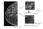
- merged image 100appears to include multiple clusters of calcifications 102 a and 102 b , which are potential predictors of cancer.
- the respective clusters of calcifications 102 a and 102 bwhich appear proximate when evaluating only in the x-y axis may actually be spread apart throughout the breast.
- the present inventionincludes a histogram 104 , 106 , which pictorially illustrates the depth of the clusters of calcifications 102 a and 102 b within the respective magnified regions. Histogram 104 illustrates that the cluster of calcifications 102 a are actually a real calcification cluster, while Histogram 106 illustrates that the visualized cluster of calcifications 102 b are actually distributed throughout the breast.
- FIG. 7is schematic illustration of an x-ray image acquisition, processing and display system 108 .
- the system 108includes an x-ray source 110 that projects x-rays 112 at a detector 116 in order to acquire x-ray images of breast tissue under the control of an acquisition control 118 .
- the acquired imagesare transmitted to a processing unit 120 , which generates a reconstructed 3D image set 122 , a 2D synthesized image 124 , and a synthesized merged image 126 , as described herein.
- These constructed/generated imagesmay be stored in a storage memory 130 coupled to the processor 120 and/or displayed on a viewing interface 152 of a display unit 150 .
- the merged imagemay combine regions of any combination of 2D and 3D image data, including an acquired mammogram, a synthesized mammogram, or a tomosynthesis projection image and a reconstructed 3D data set, thereby allowing the radiologist to quickly view a large number of regions of interest within a breast while referencing only a single 2D image and increasing the performance and efficiency of breast cancer screening and diagnosis.
Landscapes
- Engineering & Computer Science (AREA)
- Physics & Mathematics (AREA)
- General Physics & Mathematics (AREA)
- Theoretical Computer Science (AREA)
- Computer Vision & Pattern Recognition (AREA)
- Mathematical Physics (AREA)
- Mathematical Analysis (AREA)
- Mathematical Optimization (AREA)
- Algebra (AREA)
- Pure & Applied Mathematics (AREA)
- Computer Hardware Design (AREA)
- Apparatus For Radiation Diagnosis (AREA)
- Health & Medical Sciences (AREA)
- General Health & Medical Sciences (AREA)
- Medical Informatics (AREA)
- Nuclear Medicine, Radiotherapy & Molecular Imaging (AREA)
- Radiology & Medical Imaging (AREA)
- Quality & Reliability (AREA)
Abstract
Description
Claims (13)
Priority Applications (1)
| Application Number | Priority Date | Filing Date | Title |
|---|---|---|---|
| US14/360,389US10008184B2 (en) | 2005-11-10 | 2012-11-26 | System and method for generating a 2D image using mammography and/or tomosynthesis image data |
Applications Claiming Priority (8)
| Application Number | Priority Date | Filing Date | Title |
|---|---|---|---|
| US11/271,050US7577282B2 (en) | 2002-11-27 | 2005-11-10 | Image handling and display in X-ray mammography and tomosynthesis |
| US60406906A | 2006-11-24 | 2006-11-24 | |
| US11/827,909US7616801B2 (en) | 2002-11-27 | 2007-07-13 | Image handling and display in x-ray mammography and tomosynthesis |
| US12/276,006US7760924B2 (en) | 2002-11-27 | 2008-11-21 | System and method for generating a 2D image from a tomosynthesis data set |
| US12/471,981US8571289B2 (en) | 2002-11-27 | 2009-05-26 | System and method for generating a 2D image from a tomosynthesis data set |
| US201161563785P | 2011-11-27 | 2011-11-27 | |
| PCT/US2012/066526WO2013078476A1 (en) | 2011-11-27 | 2012-11-26 | System and method for generating a 2d image using mammography and/or tomosynthesis image data |
| US14/360,389US10008184B2 (en) | 2005-11-10 | 2012-11-26 | System and method for generating a 2D image using mammography and/or tomosynthesis image data |
Related Parent Applications (2)
| Application Number | Title | Priority Date | Filing Date |
|---|---|---|---|
| US12/471,981Continuation-In-PartUS8571289B2 (en) | 2002-11-27 | 2009-05-26 | System and method for generating a 2D image from a tomosynthesis data set |
| PCT/US2012/066526A-371-Of-InternationalWO2013078476A1 (en) | 2005-11-10 | 2012-11-26 | System and method for generating a 2d image using mammography and/or tomosynthesis image data |
Related Child Applications (1)
| Application Number | Title | Priority Date | Filing Date |
|---|---|---|---|
| US16/013,701ContinuationUS10573276B2 (en) | 2011-11-27 | 2018-06-20 | System and method for generating a 2D image using mammography and/or tomosynthesis image data |
Publications (2)
| Publication Number | Publication Date |
|---|---|
| US20140327702A1 US20140327702A1 (en) | 2014-11-06 |
| US10008184B2true US10008184B2 (en) | 2018-06-26 |
Family
ID=51845058
Family Applications (1)
| Application Number | Title | Priority Date | Filing Date |
|---|---|---|---|
| US14/360,389Active2034-01-23US10008184B2 (en) | 2005-11-10 | 2012-11-26 | System and method for generating a 2D image using mammography and/or tomosynthesis image data |
Country Status (1)
| Country | Link |
|---|---|
| US (1) | US10008184B2 (en) |
Cited By (20)
| Publication number | Priority date | Publication date | Assignee | Title |
|---|---|---|---|---|
| US20190043456A1 (en)* | 2011-11-27 | 2019-02-07 | Hologic, Inc. | System and method for generating a 2d image using mammography and/or tomosynthesis image data |
| US10977863B2 (en) | 2012-02-13 | 2021-04-13 | Hologic, Inc. | System and method for navigating a tomosynthesis stack using synthesized image data |
| US11403483B2 (en) | 2017-06-20 | 2022-08-02 | Hologic, Inc. | Dynamic self-learning medical image method and system |
| US11406332B2 (en) | 2011-03-08 | 2022-08-09 | Hologic, Inc. | System and method for dual energy and/or contrast enhanced breast imaging for screening, diagnosis and biopsy |
| US11419565B2 (en) | 2014-02-28 | 2022-08-23 | IIologic, Inc. | System and method for generating and displaying tomosynthesis image slabs |
| US11445993B2 (en) | 2017-03-30 | 2022-09-20 | Hologic, Inc. | System and method for targeted object enhancement to generate synthetic breast tissue images |
| US11452486B2 (en) | 2006-02-15 | 2022-09-27 | Hologic, Inc. | Breast biopsy and needle localization using tomosynthesis systems |
| US11455754B2 (en) | 2017-03-30 | 2022-09-27 | Hologic, Inc. | System and method for synthesizing low-dimensional image data from high-dimensional image data using an object grid enhancement |
| US11589944B2 (en) | 2013-03-15 | 2023-02-28 | Hologic, Inc. | Tomosynthesis-guided biopsy apparatus and method |
| US11701199B2 (en) | 2009-10-08 | 2023-07-18 | Hologic, Inc. | Needle breast biopsy system and method of use |
| US11775156B2 (en) | 2010-11-26 | 2023-10-03 | Hologic, Inc. | User interface for medical image review workstation |
| US11783476B2 (en) | 2019-10-25 | 2023-10-10 | DeepHealth, Inc. | System and method for analyzing three-dimensional image data |
| US20240096477A1 (en)* | 2021-09-18 | 2024-03-21 | GE Precision Healthcare LLC | C-arm imaging system and medical image processing method |
| US11957497B2 (en) | 2017-03-30 | 2024-04-16 | Hologic, Inc | System and method for hierarchical multi-level feature image synthesis and representation |
| US12029602B2 (en) | 2013-10-24 | 2024-07-09 | Hologic, Inc. | System and method for navigating x-ray guided breast biopsy |
| US12211608B2 (en) | 2013-03-15 | 2025-01-28 | Hologic, Inc. | System and method for navigating a tomosynthesis stack including automatic focusing |
| US12236582B2 (en) | 2018-09-24 | 2025-02-25 | Hologic, Inc. | Breast mapping and abnormality localization |
| US12236597B2 (en) | 2021-11-29 | 2025-02-25 | Hologic, Inc. | Systems and methods for correlating objects of interest |
| US12254586B2 (en) | 2021-10-25 | 2025-03-18 | Hologic, Inc. | Auto-focus tool for multimodality image review |
| US12367574B2 (en) | 2019-12-23 | 2025-07-22 | DeepHealth, Inc. | Systems and methods for analyzing two-dimensional and three-dimensional image data |
Families Citing this family (19)
| Publication number | Priority date | Publication date | Assignee | Title |
|---|---|---|---|---|
| US8571289B2 (en) | 2002-11-27 | 2013-10-29 | Hologic, Inc. | System and method for generating a 2D image from a tomosynthesis data set |
| US10008184B2 (en) | 2005-11-10 | 2018-06-26 | Hologic, Inc. | System and method for generating a 2D image using mammography and/or tomosynthesis image data |
| US10238354B2 (en)* | 2014-02-04 | 2019-03-26 | Koninklijke Philips N.V | Generating a breast parameter map |
| EP3221847B1 (en)* | 2014-11-20 | 2021-01-06 | Koninklijke Philips N.V. | Method for generation of synthetic mammograms from tomosynthesis data |
| JP6502509B2 (en) | 2015-09-10 | 2019-04-17 | 富士フイルム株式会社 | Image processing apparatus, radiographic imaging system, image processing method, and image processing program |
| JP6631360B2 (en)* | 2016-03-29 | 2020-01-15 | ブラザー工業株式会社 | Image processing apparatus and image processing method |
| AU2017204494B2 (en)* | 2016-09-01 | 2019-06-13 | Casio Computer Co., Ltd. | Diagnosis assisting device, image processing method in diagnosis assisting device, and non-transitory storage medium having stored therein program |
| EP3518182B1 (en)* | 2018-01-26 | 2022-05-18 | Siemens Healthcare GmbH | Tilted slices in dbt |
| US11205265B2 (en)* | 2018-11-23 | 2021-12-21 | Icad, Inc. | System and method for assessing breast cancer risk using imagery |
| EP3949862A4 (en) | 2019-03-29 | 2022-05-25 | FUJIFILM Corporation | IMAGE PROCESSING DEVICE, METHOD, AND PROGRAM |
| JP7209599B2 (en) | 2019-07-29 | 2023-01-20 | 富士フイルム株式会社 | Image processing device, method and program |
| JP7203705B2 (en)* | 2019-09-17 | 2023-01-13 | 富士フイルム株式会社 | Image processing device, method and program, and image display device, method and program |
| EP4101386A4 (en) | 2020-02-04 | 2023-07-12 | FUJIFILM Corporation | IMAGE ADJUSTMENT DEVICE, METHOD AND PROGRAM |
| EP4119055B1 (en) | 2020-03-13 | 2024-10-30 | FUJIFILM Corporation | Image generation device and program, learning device and program, and image processing device and program |
| JP7446410B2 (en)* | 2020-03-18 | 2024-03-08 | 富士フイルム株式会社 | Image processing device, method and program |
| CN115297778B (en)* | 2020-03-18 | 2025-08-08 | 富士胶片株式会社 | Image processing device, method, and recording medium |
| CN111603194B (en)* | 2020-06-02 | 2023-09-19 | 上海联影医疗科技股份有限公司 | Breast tomographic image display method, system and computer-readable storage medium |
| CN112365432B (en)* | 2020-10-30 | 2022-04-26 | 武汉联影医疗科技有限公司 | Fusion image display method and device and medical image system |
| US11881301B2 (en)* | 2021-02-26 | 2024-01-23 | GE Precision Healthcare LLC | Methods and systems for utilizing histogram views for improved visualization of three-dimensional (3D) medical images |
Citations (118)
| Publication number | Priority date | Publication date | Assignee | Title |
|---|---|---|---|---|
| US4160906A (en) | 1977-06-23 | 1979-07-10 | General Electric Company | Anatomically coordinated user dominated programmer for diagnostic x-ray apparatus |
| US4744099A (en) | 1983-11-03 | 1988-05-10 | Siemens Aktiengesellschaft | X-ray diagnostic apparatus comprising radiation filters |
| US4773086A (en) | 1983-12-16 | 1988-09-20 | Yokogawa Medical Systems, Limited | Operator console for X-ray tomographs |
| US4907156A (en) | 1987-06-30 | 1990-03-06 | University Of Chicago | Method and system for enhancement and detection of abnormal anatomic regions in a digital image |
| US4969174A (en) | 1989-09-06 | 1990-11-06 | General Electric Company | Scanning mammography system with reduced scatter radiation |
| USRE33634E (en) | 1986-09-23 | 1991-07-09 | Method and structure for optimizing radiographic quality by controlling X-ray tube voltage, current focal spot size and exposure time | |
| US5133020A (en) | 1989-07-21 | 1992-07-21 | Arch Development Corporation | Automated method and system for the detection and classification of abnormal lesions and parenchymal distortions in digital medical images |
| US5163075A (en) | 1991-08-08 | 1992-11-10 | Eastman Kodak Company | Contrast enhancement of electrographic imaging |
| US5343390A (en) | 1992-02-28 | 1994-08-30 | Arch Development Corporation | Method and system for automated selection of regions of interest and detection of septal lines in digital chest radiographs |
| US5365562A (en) | 1993-09-20 | 1994-11-15 | Fischer Imaging Corporation | Digital imaging apparatus |
| US5452367A (en) | 1993-11-29 | 1995-09-19 | Arch Development Corporation | Automated method and system for the segmentation of medical images |
| US5491627A (en) | 1993-05-13 | 1996-02-13 | Arch Development Corporation | Method and system for the detection of microcalcifications in digital mammograms |
| US5526394A (en) | 1993-11-26 | 1996-06-11 | Fischer Imaging Corporation | Digital scan mammography apparatus |
| US5553111A (en) | 1994-10-26 | 1996-09-03 | The General Hospital Corporation | Apparatus and method for improved tissue imaging |
| US5592562A (en) | 1994-01-19 | 1997-01-07 | International Business Machines Corporation | Inspection system for cross-sectional imaging |
| US5594769A (en) | 1991-11-27 | 1997-01-14 | Thermotrex Corporation | Method and apparatus for obtaining stereotactic mammographic guided needle breast biopsies |
| US5596200A (en) | 1992-10-14 | 1997-01-21 | Primex | Low dose mammography system |
| US5598454A (en) | 1994-04-26 | 1997-01-28 | Siemens Aktiengesellschaft | X-ray diagnostics installation |
| US5668889A (en) | 1990-04-19 | 1997-09-16 | Fuji Photo Film Co., Ltd. | Apparatus for determining an image position, and method for adjusting read-out conditions and/or image processing conditions for a radiation image |
| WO1998016903A1 (en) | 1996-10-16 | 1998-04-23 | Vital Images, Inc. | Advanced diagnostic viewer |
| US5828722A (en) | 1996-05-17 | 1998-10-27 | Sirona Dental Systems Gmbh & Co., Kg | X-ray diagnostic apparatus for tomosynthesis having a detector that detects positional relationships |
| US5872828A (en) | 1996-07-23 | 1999-02-16 | The General Hospital Corporation | Tomosynthesis system for breast imaging |
| US5878104A (en) | 1996-05-17 | 1999-03-02 | Sirona Dental Systems Gmbh & Co. Kg | Method for producing tomosynthesis exposures employing a reference object formed by a region of the examination subject |
| US5878746A (en) | 1993-08-25 | 1999-03-09 | Lemelson; Jerome H. | Computerized medical diagnostic system |
| US5896437A (en) | 1996-05-17 | 1999-04-20 | Sirona Dental Systems Gmbh & Co. Kg | X-ray diagnostics apparatus for tomosynthesis having a reference object in fixed relationship to a radiation emitter |
| US5941832A (en) | 1991-09-27 | 1999-08-24 | Tumey; David M. | Method and apparatus for detection of cancerous and precancerous conditions in a breast |
| US6005907A (en) | 1996-05-17 | 1999-12-21 | Sirona Dental Systems Gmbh & Co. Kg | Method and apparatus for producing tomosynthesis exposures employing a reference object composed of a number of sub-objects |
| EP0982001A1 (en) | 1998-08-25 | 2000-03-01 | General Electric Company | Protocol driven image reconstruction, display, and processing in a multislice imaging system |
| US6091841A (en) | 1997-09-04 | 2000-07-18 | Qualia Computing, Inc. | Method and system for segmenting desired regions in digital mammograms |
| US6137527A (en) | 1996-12-23 | 2000-10-24 | General Electric Company | System and method for prompt-radiology image screening service via satellite |
| US6175117B1 (en) | 1998-01-23 | 2001-01-16 | Quanta Vision, Inc. | Tissue analysis apparatus |
| US6196715B1 (en) | 1959-04-28 | 2001-03-06 | Kabushiki Kaisha Toshiba | X-ray diagnostic system preferable to two dimensional x-ray detection |
| US6216540B1 (en) | 1995-06-06 | 2001-04-17 | Robert S. Nelson | High resolution device and method for imaging concealed objects within an obscuring medium |
| US6233473B1 (en) | 1999-02-16 | 2001-05-15 | Hologic, Inc. | Determining body composition using fan beam dual-energy x-ray absorptiometry |
| US6243441B1 (en) | 1999-07-13 | 2001-06-05 | Edge Medical Devices | Active matrix detector for X-ray imaging |
| US6256370B1 (en) | 2000-01-24 | 2001-07-03 | General Electric Company | Method and apparatus for performing tomosynthesis |
| US6272207B1 (en) | 1999-02-18 | 2001-08-07 | Creatv Microtech, Inc. | Method and apparatus for obtaining high-resolution digital X-ray and gamma ray images |
| US6292530B1 (en) | 1999-04-29 | 2001-09-18 | General Electric Company | Method and apparatus for reconstructing image data acquired by a tomosynthesis x-ray imaging system |
| US6341156B1 (en) | 1999-05-14 | 2002-01-22 | Siemens Aktiengesellschaft | X-ray diagnostic apparatus with relatively moved x-ray source and detector |
| US6375352B1 (en) | 1999-10-01 | 2002-04-23 | General Electric Company | Apparatus and method for obtaining x-ray tomosynthesis data for mammography |
| US20020050986A1 (en) | 2000-08-11 | 2002-05-02 | Hitoshi Inoue | Image display apparatus and method, and storage medium |
| US6389104B1 (en) | 2000-06-30 | 2002-05-14 | Siemens Corporate Research, Inc. | Fluoroscopy based 3-D neural navigation based on 3-D angiography reconstruction data |
| US6411836B1 (en) | 1999-12-30 | 2002-06-25 | General Electric Company | Method and apparatus for user preferences configuring in an image handling system |
| US6415015B2 (en) | 1999-12-28 | 2002-07-02 | Ge Medical Systems Sa | Method and system of compensation of thickness of an organ |
| US6442288B1 (en) | 1997-12-17 | 2002-08-27 | Siemens Aktiengesellschaft | Method for reconstructing a three-dimensional image of an object scanned in the context of a tomosynthesis, and apparatus for tomosynthesis |
| US6463181B2 (en) | 2000-12-22 | 2002-10-08 | The United States Of America As Represented By The Secretary Of The Navy | Method for optimizing visual display of enhanced digital images |
| US20030007598A1 (en) | 2000-11-24 | 2003-01-09 | U-Systems, Inc. | Breast cancer screening with adjunctive ultrasound mammography |
| US20030026386A1 (en) | 2001-02-01 | 2003-02-06 | Cha-Mei Tang | Anti-scatter grids and collimator designs, and their motion, fabrication and assembly |
| US6556655B1 (en) | 1998-11-27 | 2003-04-29 | Ge Medical Systems Sa | Method for automatic detection of glandular tissue |
| US20030095624A1 (en) | 2001-11-21 | 2003-05-22 | Eberhard Jeffrey Wayne | Dose management system for mammographic tomosynthesis |
| US6574304B1 (en) | 2002-09-13 | 2003-06-03 | Ge Medical Systems Global Technology Company, Llc | Computer aided acquisition of medical images |
| JP2003189179A (en) | 2001-12-14 | 2003-07-04 | Konica Corp | Abnormal shade detector and image output device |
| US20030128893A1 (en)* | 2001-11-19 | 2003-07-10 | Alfio Castorina | Method for merging digital images to obtain a high dynamic range digital image |
| US6597762B1 (en) | 2002-11-27 | 2003-07-22 | Ge Medical Systems Global Technology Co., Llc | Method and apparatus of lesion detection and validation based on multiple reviews of a CT image |
| US20030169847A1 (en) | 2001-11-21 | 2003-09-11 | University Of Massachusetts Medical Center | System and method for x-ray fluoroscopic imaging |
| US6633674B1 (en) | 1999-11-24 | 2003-10-14 | General Electric Company | Picture archiving and communication system employing improved data compression |
| US20030194121A1 (en) | 2002-04-15 | 2003-10-16 | General Electric Company | Computer aided detection (CAD) for 3D digital mammography |
| US20030194050A1 (en) | 2002-04-15 | 2003-10-16 | General Electric Company | Multi modality X-ray and nuclear medicine mammography imaging system and method |
| US6647092B2 (en) | 2002-01-18 | 2003-11-11 | General Electric Company | Radiation imaging system and method of collimation |
| US20030210254A1 (en) | 2002-05-13 | 2003-11-13 | Doan William D. | Method, system and computer product for displaying axial images |
| US20030212327A1 (en) | 2000-11-24 | 2003-11-13 | U-Systems Inc. | Adjunctive ultrasound processing and display for breast cancer screening |
| US20030215120A1 (en) | 2002-05-15 | 2003-11-20 | Renuka Uppaluri | Computer aided diagnosis of an image set |
| US20040008901A1 (en) | 2002-07-11 | 2004-01-15 | Avinash Gopal B. | Interpolated image filtering method and apparatus |
| US20040047518A1 (en)* | 2002-08-28 | 2004-03-11 | Carlo Tiana | Image fusion system and method |
| US20040052328A1 (en) | 2002-09-13 | 2004-03-18 | Sabol John M. | Computer assisted analysis of tomographic mammography data |
| US20040070582A1 (en) | 2002-10-11 | 2004-04-15 | Matthew Warren Smith To Sonocine, Inc. | 3D modeling system |
| US20040094167A1 (en) | 2000-03-17 | 2004-05-20 | Brady John Michael | Three-dimensional reconstructions of a breast from two x-ray mammographics |
| US6744848B2 (en) | 2000-02-11 | 2004-06-01 | Brandeis University | Method and system for low-dose three-dimensional imaging of a scene |
| US6813334B2 (en) | 2000-10-20 | 2004-11-02 | Koninklijke Philips Electronics N.V. | Tomosynthesis in a limited angular range |
| US6882700B2 (en) | 2002-04-15 | 2005-04-19 | General Electric Company | Tomosynthesis X-ray mammogram system and method with automatic drive system |
| US6885724B2 (en) | 2003-08-22 | 2005-04-26 | Ge Medical Systems Global Technology Company, Llc | Radiographic tomosynthesis image acquisition utilizing asymmetric geometry |
| US20050113681A1 (en) | 2002-11-27 | 2005-05-26 | Defreitas Kenneth F. | X-ray mammography with tomosynthesis |
| WO2005051197A2 (en) | 2003-11-26 | 2005-06-09 | Koninklijke Philips Electronics, N.V. | Workflow optimization for high throughput imaging environment |
| US20050135664A1 (en) | 2003-12-23 | 2005-06-23 | Kaufhold John P. | Methods and apparatus for reconstruction of volume data from projection data |
| US20050135555A1 (en) | 2003-12-23 | 2005-06-23 | Claus Bernhard Erich H. | Method and system for simultaneously viewing rendered volumes |
| US6940943B2 (en) | 2002-10-07 | 2005-09-06 | General Electric Company | Continuous scan tomosynthesis system and method |
| US20050226375A1 (en) | 2004-03-31 | 2005-10-13 | Eberhard Jeffrey W | Enhanced X-ray imaging system and method |
| WO2005110230A1 (en) | 2004-05-14 | 2005-11-24 | Philips Intellectual Property & Standards Gmbh | System and method for diagnosing breast cancer |
| US20060018526A1 (en) | 2004-07-23 | 2006-01-26 | Avinash Gopal B | Methods and apparatus for noise reduction filtering of images |
| US6999554B2 (en) | 2003-11-17 | 2006-02-14 | Siemens Aktiengesellschaft | X-ray diagnostic apparatus for mammography examinations |
| US20060098855A1 (en) | 2002-11-27 | 2006-05-11 | Gkanatsios Nikolaos A | Image handling and display in X-ray mammography and tomosynthesis |
| US7110490B2 (en) | 2002-12-10 | 2006-09-19 | General Electric Company | Full field digital tomosynthesis method and apparatus |
| US7127091B2 (en) | 2000-12-22 | 2006-10-24 | Koninklijke Philips Electronics, N.V. | Method and apparatus for visualizing a limited part of a 3D medical image-point-related data set, through basing a rendered image on an intermediate region between first and second clipping planes, and including spectroscopic viewing of such region |
| US20070052700A1 (en) | 2005-09-07 | 2007-03-08 | Wheeler Frederick W | System and method for 3D CAD using projection images |
| US7323692B2 (en) | 2004-08-10 | 2008-01-29 | Research Foundation Of State University Of New York | Flat-panel detector with avalanche gain |
| US7346381B2 (en) | 2002-11-01 | 2008-03-18 | Ge Medical Systems Global Technology Company Llc | Method and apparatus for medical intervention procedure planning |
| WO2008047270A1 (en) | 2006-10-17 | 2008-04-24 | Koninklijke Philips Electronics N.V. | Visualization of 3d images in combination with 2d projection images |
| US20080130979A1 (en) | 2004-11-15 | 2008-06-05 | Baorui Ren | Matching Geometry Generation and Display of Mammograms and Tomosynthesis Images |
| US20080187095A1 (en) | 2005-05-03 | 2008-08-07 | The Regents Of The University Of California | Biopsy Systems For Breast Computed Tomography |
| US20090005668A1 (en) | 2007-06-30 | 2009-01-01 | West Jay B | Non-invasive method for using 2D angiographic images for radiosurgical target definition |
| US20090034684A1 (en) | 2007-08-02 | 2009-02-05 | Sylvain Bernard | Method and system for displaying tomosynthesis images |
| US20090080765A1 (en) | 2007-09-20 | 2009-03-26 | General Electric Company | System and method to generate a selected visualization of a radiological image of an imaged subject |
| US20090123052A1 (en) | 2002-11-27 | 2009-05-14 | Chris Ruth | System and Method for Generating a 2D Image from a Tomosynthesis Data Set |
| JP2009207545A (en) | 2008-02-29 | 2009-09-17 | Fujifilm Corp | Image display method and apparatus |
| US20090238424A1 (en) | 2008-03-19 | 2009-09-24 | Kazumasa Arakita | Image processing apparatus and image processing method |
| US7606801B2 (en) | 2005-06-07 | 2009-10-20 | Varonis Inc. | Automatic management of storage access control |
| US20090268865A1 (en) | 2003-11-26 | 2009-10-29 | Baorui Ren | X-ray imaging with X-ray markers that provide adjunct information but preserve image quality |
| US7616801B2 (en) | 2002-11-27 | 2009-11-10 | Hologic, Inc. | Image handling and display in x-ray mammography and tomosynthesis |
| US7630533B2 (en) | 2007-09-20 | 2009-12-08 | Hologic, Inc. | Breast tomosynthesis with display of highlighted suspected calcifications |
| US20090304147A1 (en) | 2005-08-15 | 2009-12-10 | Hologic, Inc. | X-ray mammography/tomosynthesis of patient's breast |
| US20100135558A1 (en) | 2002-11-27 | 2010-06-03 | Chris Ruth | System and Method for Generating a 2D Image from a Tomosynthesis Data Set |
| US20100259645A1 (en) | 2009-04-13 | 2010-10-14 | Pure Digital Technologies | Method and system for still image capture from video footage |
| WO2011008239A1 (en)* | 2009-06-29 | 2011-01-20 | Thomson Licensing | Contrast enhancement |
| US20110069906A1 (en)* | 2009-09-23 | 2011-03-24 | Samsung Electronics Co., Ltd. | Method and apparatus for blending images |
| EP2301432A1 (en) | 2008-06-03 | 2011-03-30 | FUJIFILM Corporation | Projection image creation device, method, and program |
| US20110109650A1 (en) | 2009-10-07 | 2011-05-12 | Hologic, Inc. | Displaying Computer-Aided Detection Information With Associated Breast Tomosynthesis Image Information |
| WO2011065950A1 (en) | 2009-11-27 | 2011-06-03 | Qview Medical, Inc. | Interactive display of computer aided detection radiological screening results combined with quantitative prompts |
| WO2011073864A2 (en) | 2009-12-17 | 2011-06-23 | Koninklijke Philips Electronics N.V. | Reconstructing an object of interest |
| US20110178389A1 (en) | 2008-05-02 | 2011-07-21 | Eigen, Inc. | Fused image moldalities guidance |
| WO2011091300A2 (en) | 2010-01-24 | 2011-07-28 | Mistretta Medical, Llc | System and method for implementation of 4d time-energy subtraction computed tomography |
| US20110234630A1 (en) | 2008-11-28 | 2011-09-29 | Fujifilm Medical Systems USA, Inc | Active Overlay System and Method for Accessing and Manipulating Imaging Displays |
| US20110242092A1 (en) | 2010-03-30 | 2011-10-06 | Fujifilm Corporation | Image display system |
| US8044972B2 (en) | 2006-12-21 | 2011-10-25 | Sectra Mamea Ab | Synchronized viewing of tomosynthesis and/or mammograms |
| US8051386B2 (en) | 2006-12-21 | 2011-11-01 | Sectra Ab | CAD-based navigation of views of medical image data stacks or volumes |
| WO2012063653A1 (en) | 2010-11-12 | 2012-05-18 | 株式会社 日立メディコ | Medical image display device and medical image display method |
| US20120293511A1 (en) | 2010-02-25 | 2012-11-22 | Siemens Aktiengesellschaft | Method for displaying an area to be medically examined and/or treated |
| US20140327702A1 (en) | 2005-11-10 | 2014-11-06 | Hologic, Inc | System and method for generating a 2d image using mammography and/or tomosynthesis image data |
| US8983156B2 (en) | 2012-11-23 | 2015-03-17 | Icad, Inc. | System and method for improving workflow efficiences in reading tomosynthesis medical image data |
- 2012
- 2012-11-26USUS14/360,389patent/US10008184B2/enactiveActive
Patent Citations (143)
| Publication number | Priority date | Publication date | Assignee | Title |
|---|---|---|---|---|
| US6196715B1 (en) | 1959-04-28 | 2001-03-06 | Kabushiki Kaisha Toshiba | X-ray diagnostic system preferable to two dimensional x-ray detection |
| US4160906A (en) | 1977-06-23 | 1979-07-10 | General Electric Company | Anatomically coordinated user dominated programmer for diagnostic x-ray apparatus |
| US4744099A (en) | 1983-11-03 | 1988-05-10 | Siemens Aktiengesellschaft | X-ray diagnostic apparatus comprising radiation filters |
| US4773086A (en) | 1983-12-16 | 1988-09-20 | Yokogawa Medical Systems, Limited | Operator console for X-ray tomographs |
| USRE33634E (en) | 1986-09-23 | 1991-07-09 | Method and structure for optimizing radiographic quality by controlling X-ray tube voltage, current focal spot size and exposure time | |
| US4907156A (en) | 1987-06-30 | 1990-03-06 | University Of Chicago | Method and system for enhancement and detection of abnormal anatomic regions in a digital image |
| US5133020A (en) | 1989-07-21 | 1992-07-21 | Arch Development Corporation | Automated method and system for the detection and classification of abnormal lesions and parenchymal distortions in digital medical images |
| US4969174A (en) | 1989-09-06 | 1990-11-06 | General Electric Company | Scanning mammography system with reduced scatter radiation |
| US5668889A (en) | 1990-04-19 | 1997-09-16 | Fuji Photo Film Co., Ltd. | Apparatus for determining an image position, and method for adjusting read-out conditions and/or image processing conditions for a radiation image |
| US5163075A (en) | 1991-08-08 | 1992-11-10 | Eastman Kodak Company | Contrast enhancement of electrographic imaging |
| US5941832A (en) | 1991-09-27 | 1999-08-24 | Tumey; David M. | Method and apparatus for detection of cancerous and precancerous conditions in a breast |
| US5594769A (en) | 1991-11-27 | 1997-01-14 | Thermotrex Corporation | Method and apparatus for obtaining stereotactic mammographic guided needle breast biopsies |
| US5343390A (en) | 1992-02-28 | 1994-08-30 | Arch Development Corporation | Method and system for automated selection of regions of interest and detection of septal lines in digital chest radiographs |
| US5596200A (en) | 1992-10-14 | 1997-01-21 | Primex | Low dose mammography system |
| US5491627A (en) | 1993-05-13 | 1996-02-13 | Arch Development Corporation | Method and system for the detection of microcalcifications in digital mammograms |
| US5878746A (en) | 1993-08-25 | 1999-03-09 | Lemelson; Jerome H. | Computerized medical diagnostic system |
| US5365562A (en) | 1993-09-20 | 1994-11-15 | Fischer Imaging Corporation | Digital imaging apparatus |
| US5526394A (en) | 1993-11-26 | 1996-06-11 | Fischer Imaging Corporation | Digital scan mammography apparatus |
| US5452367A (en) | 1993-11-29 | 1995-09-19 | Arch Development Corporation | Automated method and system for the segmentation of medical images |
| US5592562A (en) | 1994-01-19 | 1997-01-07 | International Business Machines Corporation | Inspection system for cross-sectional imaging |
| US5719952A (en) | 1994-01-19 | 1998-02-17 | International Business Machines Corporation | Inspection system for cross-sectional imaging |
| US5598454A (en) | 1994-04-26 | 1997-01-28 | Siemens Aktiengesellschaft | X-ray diagnostics installation |
| US5553111A (en) | 1994-10-26 | 1996-09-03 | The General Hospital Corporation | Apparatus and method for improved tissue imaging |
| US6216540B1 (en) | 1995-06-06 | 2001-04-17 | Robert S. Nelson | High resolution device and method for imaging concealed objects within an obscuring medium |
| US5878104A (en) | 1996-05-17 | 1999-03-02 | Sirona Dental Systems Gmbh & Co. Kg | Method for producing tomosynthesis exposures employing a reference object formed by a region of the examination subject |
| US6005907A (en) | 1996-05-17 | 1999-12-21 | Sirona Dental Systems Gmbh & Co. Kg | Method and apparatus for producing tomosynthesis exposures employing a reference object composed of a number of sub-objects |
| US5896437A (en) | 1996-05-17 | 1999-04-20 | Sirona Dental Systems Gmbh & Co. Kg | X-ray diagnostics apparatus for tomosynthesis having a reference object in fixed relationship to a radiation emitter |
| US5828722A (en) | 1996-05-17 | 1998-10-27 | Sirona Dental Systems Gmbh & Co., Kg | X-ray diagnostic apparatus for tomosynthesis having a detector that detects positional relationships |
| US5872828A (en) | 1996-07-23 | 1999-02-16 | The General Hospital Corporation | Tomosynthesis system for breast imaging |
| US6219059B1 (en) | 1996-10-16 | 2001-04-17 | Vital Images, Inc. | Interactive control of voxel attributes using selectable characteristics |
| US5986662A (en) | 1996-10-16 | 1999-11-16 | Vital Images, Inc. | Advanced diagnostic viewer employing automated protocol selection for volume-rendered imaging |
| WO1998016903A1 (en) | 1996-10-16 | 1998-04-23 | Vital Images, Inc. | Advanced diagnostic viewer |
| US6137527A (en) | 1996-12-23 | 2000-10-24 | General Electric Company | System and method for prompt-radiology image screening service via satellite |
| US6091841A (en) | 1997-09-04 | 2000-07-18 | Qualia Computing, Inc. | Method and system for segmenting desired regions in digital mammograms |
| US6442288B1 (en) | 1997-12-17 | 2002-08-27 | Siemens Aktiengesellschaft | Method for reconstructing a three-dimensional image of an object scanned in the context of a tomosynthesis, and apparatus for tomosynthesis |
| US6175117B1 (en) | 1998-01-23 | 2001-01-16 | Quanta Vision, Inc. | Tissue analysis apparatus |
| EP0982001A1 (en) | 1998-08-25 | 2000-03-01 | General Electric Company | Protocol driven image reconstruction, display, and processing in a multislice imaging system |
| US6141398A (en) | 1998-08-25 | 2000-10-31 | General Electric Company | Protocol driven image reconstruction, display, and processing in a multislice imaging system |
| US6556655B1 (en) | 1998-11-27 | 2003-04-29 | Ge Medical Systems Sa | Method for automatic detection of glandular tissue |
| US6233473B1 (en) | 1999-02-16 | 2001-05-15 | Hologic, Inc. | Determining body composition using fan beam dual-energy x-ray absorptiometry |
| US6272207B1 (en) | 1999-02-18 | 2001-08-07 | Creatv Microtech, Inc. | Method and apparatus for obtaining high-resolution digital X-ray and gamma ray images |
| US6292530B1 (en) | 1999-04-29 | 2001-09-18 | General Electric Company | Method and apparatus for reconstructing image data acquired by a tomosynthesis x-ray imaging system |
| US6341156B1 (en) | 1999-05-14 | 2002-01-22 | Siemens Aktiengesellschaft | X-ray diagnostic apparatus with relatively moved x-ray source and detector |
| US6243441B1 (en) | 1999-07-13 | 2001-06-05 | Edge Medical Devices | Active matrix detector for X-ray imaging |
| US6375352B1 (en) | 1999-10-01 | 2002-04-23 | General Electric Company | Apparatus and method for obtaining x-ray tomosynthesis data for mammography |
| US6912319B1 (en) | 1999-11-24 | 2005-06-28 | Ge Medical Systems Information Technologies, Inc. | Method and system for lossless wavelet decomposition, compression and decompression of data |
| US6633674B1 (en) | 1999-11-24 | 2003-10-14 | General Electric Company | Picture archiving and communication system employing improved data compression |
| US6415015B2 (en) | 1999-12-28 | 2002-07-02 | Ge Medical Systems Sa | Method and system of compensation of thickness of an organ |
| US6411836B1 (en) | 1999-12-30 | 2002-06-25 | General Electric Company | Method and apparatus for user preferences configuring in an image handling system |
| US6256370B1 (en) | 2000-01-24 | 2001-07-03 | General Electric Company | Method and apparatus for performing tomosynthesis |
| US6744848B2 (en) | 2000-02-11 | 2004-06-01 | Brandeis University | Method and system for low-dose three-dimensional imaging of a scene |
| US20040094167A1 (en) | 2000-03-17 | 2004-05-20 | Brady John Michael | Three-dimensional reconstructions of a breast from two x-ray mammographics |
| US6389104B1 (en) | 2000-06-30 | 2002-05-14 | Siemens Corporate Research, Inc. | Fluoroscopy based 3-D neural navigation based on 3-D angiography reconstruction data |
| US20020050986A1 (en) | 2000-08-11 | 2002-05-02 | Hitoshi Inoue | Image display apparatus and method, and storage medium |
| US6813334B2 (en) | 2000-10-20 | 2004-11-02 | Koninklijke Philips Electronics N.V. | Tomosynthesis in a limited angular range |
| US20030212327A1 (en) | 2000-11-24 | 2003-11-13 | U-Systems Inc. | Adjunctive ultrasound processing and display for breast cancer screening |
| US20030007598A1 (en) | 2000-11-24 | 2003-01-09 | U-Systems, Inc. | Breast cancer screening with adjunctive ultrasound mammography |
| US7127091B2 (en) | 2000-12-22 | 2006-10-24 | Koninklijke Philips Electronics, N.V. | Method and apparatus for visualizing a limited part of a 3D medical image-point-related data set, through basing a rendered image on an intermediate region between first and second clipping planes, and including spectroscopic viewing of such region |
| US6463181B2 (en) | 2000-12-22 | 2002-10-08 | The United States Of America As Represented By The Secretary Of The Navy | Method for optimizing visual display of enhanced digital images |
| US20030026386A1 (en) | 2001-02-01 | 2003-02-06 | Cha-Mei Tang | Anti-scatter grids and collimator designs, and their motion, fabrication and assembly |
| US20030128893A1 (en)* | 2001-11-19 | 2003-07-10 | Alfio Castorina | Method for merging digital images to obtain a high dynamic range digital image |
| US20030169847A1 (en) | 2001-11-21 | 2003-09-11 | University Of Massachusetts Medical Center | System and method for x-ray fluoroscopic imaging |
| US6751285B2 (en) | 2001-11-21 | 2004-06-15 | General Electric Company | Dose management system for mammographic tomosynthesis |
| US20030095624A1 (en) | 2001-11-21 | 2003-05-22 | Eberhard Jeffrey Wayne | Dose management system for mammographic tomosynthesis |
| JP2003189179A (en) | 2001-12-14 | 2003-07-04 | Konica Corp | Abnormal shade detector and image output device |
| US6647092B2 (en) | 2002-01-18 | 2003-11-11 | General Electric Company | Radiation imaging system and method of collimation |
| US20030194050A1 (en) | 2002-04-15 | 2003-10-16 | General Electric Company | Multi modality X-ray and nuclear medicine mammography imaging system and method |
| US20030194121A1 (en) | 2002-04-15 | 2003-10-16 | General Electric Company | Computer aided detection (CAD) for 3D digital mammography |
| US6882700B2 (en) | 2002-04-15 | 2005-04-19 | General Electric Company | Tomosynthesis X-ray mammogram system and method with automatic drive system |
| US20030210254A1 (en) | 2002-05-13 | 2003-11-13 | Doan William D. | Method, system and computer product for displaying axial images |
| US20030215120A1 (en) | 2002-05-15 | 2003-11-20 | Renuka Uppaluri | Computer aided diagnosis of an image set |
| US20040008901A1 (en) | 2002-07-11 | 2004-01-15 | Avinash Gopal B. | Interpolated image filtering method and apparatus |
| US20040047518A1 (en)* | 2002-08-28 | 2004-03-11 | Carlo Tiana | Image fusion system and method |
| US20040052328A1 (en) | 2002-09-13 | 2004-03-18 | Sabol John M. | Computer assisted analysis of tomographic mammography data |
| US6748044B2 (en) | 2002-09-13 | 2004-06-08 | Ge Medical Systems Global Technology Company, Llc | Computer assisted analysis of tomographic mammography data |
| US6574304B1 (en) | 2002-09-13 | 2003-06-03 | Ge Medical Systems Global Technology Company, Llc | Computer aided acquisition of medical images |
| US6940943B2 (en) | 2002-10-07 | 2005-09-06 | General Electric Company | Continuous scan tomosynthesis system and method |
| US20040070582A1 (en) | 2002-10-11 | 2004-04-15 | Matthew Warren Smith To Sonocine, Inc. | 3D modeling system |
| US7346381B2 (en) | 2002-11-01 | 2008-03-18 | Ge Medical Systems Global Technology Company Llc | Method and apparatus for medical intervention procedure planning |
| US7760924B2 (en) | 2002-11-27 | 2010-07-20 | Hologic, Inc. | System and method for generating a 2D image from a tomosynthesis data set |
| US9456797B2 (en) | 2002-11-27 | 2016-10-04 | Hologic, Inc. | System and method for generating a 2D image from a tomosynthesis data set |
| US6597762B1 (en) | 2002-11-27 | 2003-07-22 | Ge Medical Systems Global Technology Co., Llc | Method and apparatus of lesion detection and validation based on multiple reviews of a CT image |
| US20100135558A1 (en) | 2002-11-27 | 2010-06-03 | Chris Ruth | System and Method for Generating a 2D Image from a Tomosynthesis Data Set |
| US7616801B2 (en) | 2002-11-27 | 2009-11-10 | Hologic, Inc. | Image handling and display in x-ray mammography and tomosynthesis |
| US20090123052A1 (en) | 2002-11-27 | 2009-05-14 | Chris Ruth | System and Method for Generating a 2D Image from a Tomosynthesis Data Set |
| US7831296B2 (en) | 2002-11-27 | 2010-11-09 | Hologic, Inc. | X-ray mammography with tomosynthesis |
| US7577282B2 (en) | 2002-11-27 | 2009-08-18 | Hologic, Inc. | Image handling and display in X-ray mammography and tomosynthesis |
| US20060098855A1 (en) | 2002-11-27 | 2006-05-11 | Gkanatsios Nikolaos A | Image handling and display in X-ray mammography and tomosynthesis |
| US20050113681A1 (en) | 2002-11-27 | 2005-05-26 | Defreitas Kenneth F. | X-ray mammography with tomosynthesis |
| US8897535B2 (en) | 2002-11-27 | 2014-11-25 | Hologic, Inc. | System and method for generating a 2D image from a tomosynthesis data set |
| US8571289B2 (en) | 2002-11-27 | 2013-10-29 | Hologic, Inc. | System and method for generating a 2D image from a tomosynthesis data set |
| US7110490B2 (en) | 2002-12-10 | 2006-09-19 | General Electric Company | Full field digital tomosynthesis method and apparatus |
| US6885724B2 (en) | 2003-08-22 | 2005-04-26 | Ge Medical Systems Global Technology Company, Llc | Radiographic tomosynthesis image acquisition utilizing asymmetric geometry |
| US6999554B2 (en) | 2003-11-17 | 2006-02-14 | Siemens Aktiengesellschaft | X-ray diagnostic apparatus for mammography examinations |
| US20090268865A1 (en) | 2003-11-26 | 2009-10-29 | Baorui Ren | X-ray imaging with X-ray markers that provide adjunct information but preserve image quality |
| WO2005051197A2 (en) | 2003-11-26 | 2005-06-09 | Koninklijke Philips Electronics, N.V. | Workflow optimization for high throughput imaging environment |
| US20050135664A1 (en) | 2003-12-23 | 2005-06-23 | Kaufhold John P. | Methods and apparatus for reconstruction of volume data from projection data |
| US20050135555A1 (en) | 2003-12-23 | 2005-06-23 | Claus Bernhard Erich H. | Method and system for simultaneously viewing rendered volumes |
| US7142633B2 (en) | 2004-03-31 | 2006-11-28 | General Electric Company | Enhanced X-ray imaging system and method |
| US20050226375A1 (en) | 2004-03-31 | 2005-10-13 | Eberhard Jeffrey W | Enhanced X-ray imaging system and method |
| WO2005110230A1 (en) | 2004-05-14 | 2005-11-24 | Philips Intellectual Property & Standards Gmbh | System and method for diagnosing breast cancer |
| JP2007536968A (en) | 2004-05-14 | 2007-12-20 | コーニンクレッカ フィリップス エレクトロニクス エヌ ヴィ | Breast cancer diagnosis system and method |
| US20060018526A1 (en) | 2004-07-23 | 2006-01-26 | Avinash Gopal B | Methods and apparatus for noise reduction filtering of images |
| US7323692B2 (en) | 2004-08-10 | 2008-01-29 | Research Foundation Of State University Of New York | Flat-panel detector with avalanche gain |
| US8712127B2 (en) | 2004-11-15 | 2014-04-29 | Hologic, Inc. | Matching geometry generation and display of mammograms and tomosynthesis images |
| US8155421B2 (en) | 2004-11-15 | 2012-04-10 | Hologic, Inc. | Matching geometry generation and display of mammograms and tomosynthesis images |
| US20080130979A1 (en) | 2004-11-15 | 2008-06-05 | Baorui Ren | Matching Geometry Generation and Display of Mammograms and Tomosynthesis Images |
| US20150317538A1 (en) | 2004-11-15 | 2015-11-05 | Hologic, Inc. | Matching geometry generation and display of mammograpms and tomosynthesis |
| US9084579B2 (en) | 2004-11-15 | 2015-07-21 | Hologic, Inc. | Matching geometry generation and display of mammograms and tomosynthesis |
| US7702142B2 (en) | 2004-11-15 | 2010-04-20 | Hologic, Inc. | Matching geometry generation and display of mammograms and tomosynthesis images |
| US20080187095A1 (en) | 2005-05-03 | 2008-08-07 | The Regents Of The University Of California | Biopsy Systems For Breast Computed Tomography |
| US7606801B2 (en) | 2005-06-07 | 2009-10-20 | Varonis Inc. | Automatic management of storage access control |
| US20090304147A1 (en) | 2005-08-15 | 2009-12-10 | Hologic, Inc. | X-ray mammography/tomosynthesis of patient's breast |
| US20070052700A1 (en) | 2005-09-07 | 2007-03-08 | Wheeler Frederick W | System and method for 3D CAD using projection images |
| US20140327702A1 (en) | 2005-11-10 | 2014-11-06 | Hologic, Inc | System and method for generating a 2d image using mammography and/or tomosynthesis image data |
| WO2008047270A1 (en) | 2006-10-17 | 2008-04-24 | Koninklijke Philips Electronics N.V. | Visualization of 3d images in combination with 2d projection images |
| US8044972B2 (en) | 2006-12-21 | 2011-10-25 | Sectra Mamea Ab | Synchronized viewing of tomosynthesis and/or mammograms |
| US8051386B2 (en) | 2006-12-21 | 2011-11-01 | Sectra Ab | CAD-based navigation of views of medical image data stacks or volumes |
| US20090005668A1 (en) | 2007-06-30 | 2009-01-01 | West Jay B | Non-invasive method for using 2D angiographic images for radiosurgical target definition |
| US20090034684A1 (en) | 2007-08-02 | 2009-02-05 | Sylvain Bernard | Method and system for displaying tomosynthesis images |
| US8165365B2 (en) | 2007-08-02 | 2012-04-24 | General Electric Company | Method and system for displaying tomosynthesis images |
| US7630533B2 (en) | 2007-09-20 | 2009-12-08 | Hologic, Inc. | Breast tomosynthesis with display of highlighted suspected calcifications |
| US20090080765A1 (en) | 2007-09-20 | 2009-03-26 | General Electric Company | System and method to generate a selected visualization of a radiological image of an imaged subject |
| US8126226B2 (en) | 2007-09-20 | 2012-02-28 | General Electric Company | System and method to generate a selected visualization of a radiological image of an imaged subject |
| JP2009207545A (en) | 2008-02-29 | 2009-09-17 | Fujifilm Corp | Image display method and apparatus |
| US20090238424A1 (en) | 2008-03-19 | 2009-09-24 | Kazumasa Arakita | Image processing apparatus and image processing method |
| US20110178389A1 (en) | 2008-05-02 | 2011-07-21 | Eigen, Inc. | Fused image moldalities guidance |
| EP2301432A1 (en) | 2008-06-03 | 2011-03-30 | FUJIFILM Corporation | Projection image creation device, method, and program |
| WO2010059920A2 (en) | 2008-11-21 | 2010-05-27 | Hologic, Inc. | System and method for generating a 2d image from a tomosynthesis data set |
| EP2215600A2 (en) | 2008-11-21 | 2010-08-11 | Hologic Inc. | System and method for generating a 2d image from a tomosynthesis data set |
| US20110234630A1 (en) | 2008-11-28 | 2011-09-29 | Fujifilm Medical Systems USA, Inc | Active Overlay System and Method for Accessing and Manipulating Imaging Displays |
| US20100259645A1 (en) | 2009-04-13 | 2010-10-14 | Pure Digital Technologies | Method and system for still image capture from video footage |
| WO2011008239A1 (en)* | 2009-06-29 | 2011-01-20 | Thomson Licensing | Contrast enhancement |
| US20110069906A1 (en)* | 2009-09-23 | 2011-03-24 | Samsung Electronics Co., Ltd. | Method and apparatus for blending images |
| US20110110576A1 (en) | 2009-10-07 | 2011-05-12 | Hologic, Inc. | Selective Display Of Computer-Aided Detection Findings With Associated Breast X-Ray Mammogram and/or Tomosynthesis Image Information |
| US20110109650A1 (en) | 2009-10-07 | 2011-05-12 | Hologic, Inc. | Displaying Computer-Aided Detection Information With Associated Breast Tomosynthesis Image Information |
| WO2011065950A1 (en) | 2009-11-27 | 2011-06-03 | Qview Medical, Inc. | Interactive display of computer aided detection radiological screening results combined with quantitative prompts |
| WO2011073864A2 (en) | 2009-12-17 | 2011-06-23 | Koninklijke Philips Electronics N.V. | Reconstructing an object of interest |
| WO2011091300A2 (en) | 2010-01-24 | 2011-07-28 | Mistretta Medical, Llc | System and method for implementation of 4d time-energy subtraction computed tomography |
| US20120293511A1 (en) | 2010-02-25 | 2012-11-22 | Siemens Aktiengesellschaft | Method for displaying an area to be medically examined and/or treated |
| US20110242092A1 (en) | 2010-03-30 | 2011-10-06 | Fujifilm Corporation | Image display system |
| WO2012063653A1 (en) | 2010-11-12 | 2012-05-18 | 株式会社 日立メディコ | Medical image display device and medical image display method |
| US8983156B2 (en) | 2012-11-23 | 2015-03-17 | Icad, Inc. | System and method for improving workflow efficiences in reading tomosynthesis medical image data |
Non-Patent Citations (44)
| Title |
|---|
| "Biomechanical 3-D Finite Element Modeling of the Human Breast Using MRI Data" by A. Samani et al. IEEE Transactions on Medical Imaing, vol. 20, No. 4, pp. 271-279. 2001. |
| "ImageParser: a tool for finite element generation from three-dimensional medical images" by H. M. Yin et al., BioMedical Engineering OnLine. 3:31, pp. 1-9, Oct. 1, 2004. |
| "Mammogram synthesis using a 3D simulation. I. breast tissue model and image acquisition simulation" by Bakic et al., Medical Physics. 29, pp. 2131-2139 (2002). |
| "Predicting tumour location by simulating large deformations of the breast using a 3D finite element model and nonlinear elasticity" by P. Pathmanathan et al., Medical Image Computing and Computer-Assisted Intervention, pp. 217-224, vol. 3217 (2004). |
| Amendment Resonse after Final Office Action for U.S. Appl. No. 12/471,981 dated Apr. 3, 2013 (6 pages). |
| Amendment Response to Final Office Action for U.S. Appl. No. 12/276,006 dated Mar. 24, 2010 (6 pages). |
| Amendment Response to Non-Final Office Action for U.S. Appl. No. 12/276,006 dated Sep. 28, 2009 (8 pages). |
| Amendment Response to Non-Final Office Action for U.S. Appl. No. 12/471,981 dated Dec. 10, 2012 (6 pages). |
| Amendment Response to Non-Final Office Action for U.S. Appl. No. 14/044,959 dated May 13, 2014 (8 pages). |
| Computer generated translation of Foreign Patent Reference JP 2003-189179 A, published Jul. 4, 2003, 16 pages. |
| Extended EP Search Report for EP Application No. 17176956.5 dated Apr. 3, 2018, 7 pages. |
| Extended European Search Report for European Application No. 12851085.6, Applicant Hologic, Inc., EPO Forms 1507S, 1503, P0459, and 1703 dated Jan. 6, 2015 (6 pages). |
| Final Office Action dated Jan. 20, 2010 for U.S. Appl. No. 12/276,006. |
| Foreign Office Action for CN Application No. 200980101409.X dated Jun. 26, 2014. |
| Foreign Office Action for EP Patent Application No. 09796173.4 dated Apr. 11, 2014. |
| Foreign Office Action for JP Patent Application No. 2011-537644 dated Jul. 29, 2013. |
| Foreign Office Action for JP Patent Application No. 2014-047021 dated Jan. 21, 2015. |
| Giger et al. "Development of a smart workstation for use in mammography", in Proceedings of SPIE, vol. 1445 (1991), 101 103; 4 pages. |
| Giger et al., "An Intelligent Workstation for Computer-aided Diagnosis", in RadioGraphics, May 1993, 13:3 pp. 647 656; 10 pages. |
| Heang-Ping, Roc "Study of the effect of stereoscopic imaging on assessment of breast lesions," Medical Physics, vol. 32, No. 4, Apr. 2005. |
| International Preliminary Report on Patentability for International Publication No. PCT/US2009/065288 dated Feb. 18, 2014. |
| International Preliminary Report on Patentability for International Publication No. PCT/US2012/066526 dated May 27, 2014. |
| International Preliminary Report on Patentability for International Publication No. PCT/US2013/025993 dated Aug. 19, 2014. |
| International Search Reort for International Publication No. PCT/US2009/065288 dated Jan. 22, 2014. |
| International Search Report and Written Opinion for International Application No. PCT/US2013025993. Applicant Hologic, Inc., EPO Forms 1507S, 1503, P0459, and 1703 dated Oct. 10, 2015 (7 pages). |
| International Search Report dated Jan. 29, 2014 for PCT/US09/065288. |
| International Search Report for International Publication No. PCT/US2012/066526 dated Jan. 15, 2013. |
| International Search Report for International Publication No. PCT/US2013/025993 dated Apr. 1, 2013. |
| Mikko Lilja, "Fast and accurate voxel projection technique in free-form cone-beam geometry with application to algebraic reconstruction," Applies Sciences on Biomedical and Communication Technologies, 2008, Isabel '08, first international symposium on, IEEE, Piscataway NJ, Oct. 25, 2008. |
| Non-Final Office Action dated Aug. 10, 2012 for U.S. Appl. No. 12/471,981. |
| Non-Final Office Action dated Feb. 13, 2013 for U.S. Appl. No. 12/471,981. |
| Non-Final Office Action dated Feb. 13, 2014 for U.S. Appl. No. 14/044,959. |
| Non-Final Office Action dated Jun. 25, 2009 for U.S. Appl. No. 12/276,006. |
| Non-Final Office Action dated Mar. 9, 2017 for U.S. Appl. No. 15/088,844. |
| Notice of Allowance for U.S. Appl. No. 15/088,844 dated Jun. 29, 2017. |
| Notice of Allowance for U.S. Appl. No. 15/088,844 dated Mar. 28 2017. |
| Office action dated Feb. 1, 2018 for U.S. Appl. No. 15/802,225. |
| Office Action dated Feb. 19, 2018 for EP Application 12851085.6, Applicant Hologic, Inc. 5 pp. |
| Office Action dated Jan. 11, 2017 for Japanese Patent Application No. 2014556824, Applicant Hologic, Inc., including English Translation provided by Japanese associate, 12 pages. |
| Office Action dated Mar. 10, 2017 for Canadian Application No. 2,702,782, Owner Hologic, Inc., based on PCT/US2009/065288, 3 pages. |
| Office Action dated Nov. 20, 2015 for U.S. Appl. No. 14/549,604. |
| Office Action for Related Japanese Appln. No. 2014-543604, dated Oct. 4, 2016, with translation provided by foreign associate(11 pages). |
| PCT Notification of International Search Report and Written Opinion for PCT/US2012/066526, Applicant Hologic, Inc., dated Feb. 6, 2013 (7 pages). |
| Pediconi, "Color-coded automated signal intensity-curve for detection and characterization of breast lesions: Preliminary evaluation of new software for MR-based breast imaging," International Congress Series 1281 (2005) 1081-1086. |
Cited By (38)
| Publication number | Priority date | Publication date | Assignee | Title |
|---|---|---|---|---|
| US11452486B2 (en) | 2006-02-15 | 2022-09-27 | Hologic, Inc. | Breast biopsy and needle localization using tomosynthesis systems |
| US11918389B2 (en) | 2006-02-15 | 2024-03-05 | Hologic, Inc. | Breast biopsy and needle localization using tomosynthesis systems |
| US12193853B2 (en) | 2006-02-15 | 2025-01-14 | Hologic, Inc. | Breast biopsy and needle localization using tomosynthesis systems |
| US11701199B2 (en) | 2009-10-08 | 2023-07-18 | Hologic, Inc. | Needle breast biopsy system and method of use |
| US12193886B2 (en) | 2009-10-08 | 2025-01-14 | Hologic, Inc. | Needle breast biopsy system and method of use |
| US11775156B2 (en) | 2010-11-26 | 2023-10-03 | Hologic, Inc. | User interface for medical image review workstation |
| US12239471B2 (en) | 2011-03-08 | 2025-03-04 | Hologic, Inc. | System and method for dual energy and/or contrast enhanced breast imaging for screening, diagnosis and biopsy |
| US11406332B2 (en) | 2011-03-08 | 2022-08-09 | Hologic, Inc. | System and method for dual energy and/or contrast enhanced breast imaging for screening, diagnosis and biopsy |
| US12183309B2 (en) | 2011-11-27 | 2024-12-31 | Hologic, Inc. | System and method for generating a 2D image using mammography and/or tomosynthesis image data |
| US20190043456A1 (en)* | 2011-11-27 | 2019-02-07 | Hologic, Inc. | System and method for generating a 2d image using mammography and/or tomosynthesis image data |
| US11508340B2 (en) | 2011-11-27 | 2022-11-22 | Hologic, Inc. | System and method for generating a 2D image using mammography and/or tomosynthesis image data |
| US11837197B2 (en) | 2011-11-27 | 2023-12-05 | Hologic, Inc. | System and method for generating a 2D image using mammography and/or tomosynthesis image data |
| US10978026B2 (en) | 2011-11-27 | 2021-04-13 | Hologic, Inc. | System and method for generating a 2D image using mammography and/or tomosynthesis image data |
| US10573276B2 (en)* | 2011-11-27 | 2020-02-25 | Hologic, Inc. | System and method for generating a 2D image using mammography and/or tomosynthesis image data |
| US12307604B2 (en) | 2012-02-13 | 2025-05-20 | Hologic, Inc. | System and method for navigating a tomosynthesis stack using synthesized image data |
| US11663780B2 (en) | 2012-02-13 | 2023-05-30 | Hologic Inc. | System and method for navigating a tomosynthesis stack using synthesized image data |
| US10977863B2 (en) | 2012-02-13 | 2021-04-13 | Hologic, Inc. | System and method for navigating a tomosynthesis stack using synthesized image data |
| US12211608B2 (en) | 2013-03-15 | 2025-01-28 | Hologic, Inc. | System and method for navigating a tomosynthesis stack including automatic focusing |
| US12064291B2 (en) | 2013-03-15 | 2024-08-20 | Hologic, Inc. | Tomosynthesis-guided biopsy in prone |
| US11589944B2 (en) | 2013-03-15 | 2023-02-28 | Hologic, Inc. | Tomosynthesis-guided biopsy apparatus and method |
| US12324707B2 (en) | 2013-03-15 | 2025-06-10 | Hologic, Inc. | Tomosynthesis-guided biopsy in prone |
| US12029602B2 (en) | 2013-10-24 | 2024-07-09 | Hologic, Inc. | System and method for navigating x-ray guided breast biopsy |
| US11801025B2 (en) | 2014-02-28 | 2023-10-31 | Hologic, Inc. | System and method for generating and displaying tomosynthesis image slabs |
| US11419565B2 (en) | 2014-02-28 | 2022-08-23 | IIologic, Inc. | System and method for generating and displaying tomosynthesis image slabs |
| US11455754B2 (en) | 2017-03-30 | 2022-09-27 | Hologic, Inc. | System and method for synthesizing low-dimensional image data from high-dimensional image data using an object grid enhancement |
| US11445993B2 (en) | 2017-03-30 | 2022-09-20 | Hologic, Inc. | System and method for targeted object enhancement to generate synthetic breast tissue images |
| US11983799B2 (en) | 2017-03-30 | 2024-05-14 | Hologic, Inc. | System and method for synthesizing low-dimensional image data from high-dimensional image data using an object grid enhancement |
| US11957497B2 (en) | 2017-03-30 | 2024-04-16 | Hologic, Inc | System and method for hierarchical multi-level feature image synthesis and representation |
| US12070349B2 (en) | 2017-03-30 | 2024-08-27 | Hologic, Inc. | System and method for targeted object enhancement to generate synthetic breast tissue images |
| US12211124B2 (en) | 2017-03-30 | 2025-01-28 | Hologic, Inc. | System and method for synthesizing low-dimensional image data from high-dimensional image data using an object grid enhancement |
| US11850021B2 (en) | 2017-06-20 | 2023-12-26 | Hologic, Inc. | Dynamic self-learning medical image method and system |
| US11403483B2 (en) | 2017-06-20 | 2022-08-02 | Hologic, Inc. | Dynamic self-learning medical image method and system |
| US12236582B2 (en) | 2018-09-24 | 2025-02-25 | Hologic, Inc. | Breast mapping and abnormality localization |
| US11783476B2 (en) | 2019-10-25 | 2023-10-10 | DeepHealth, Inc. | System and method for analyzing three-dimensional image data |
| US12367574B2 (en) | 2019-12-23 | 2025-07-22 | DeepHealth, Inc. | Systems and methods for analyzing two-dimensional and three-dimensional image data |
| US20240096477A1 (en)* | 2021-09-18 | 2024-03-21 | GE Precision Healthcare LLC | C-arm imaging system and medical image processing method |
| US12254586B2 (en) | 2021-10-25 | 2025-03-18 | Hologic, Inc. | Auto-focus tool for multimodality image review |
| US12236597B2 (en) | 2021-11-29 | 2025-02-25 | Hologic, Inc. | Systems and methods for correlating objects of interest |
Also Published As
| Publication number | Publication date |
|---|---|
| US20140327702A1 (en) | 2014-11-06 |
Similar Documents
| Publication | Publication Date | Title |
|---|---|---|
| US12183309B2 (en) | System and method for generating a 2D image using mammography and/or tomosynthesis image data | |
| US10008184B2 (en) | System and method for generating a 2D image using mammography and/or tomosynthesis image data | |
| US11801025B2 (en) | System and method for generating and displaying tomosynthesis image slabs | |
| US11663780B2 (en) | System and method for navigating a tomosynthesis stack using synthesized image data |
Legal Events
| Date | Code | Title | Description |
|---|---|---|---|
| AS | Assignment | Owner name:HOLOGIC, INC., MASSACHUSETTS Free format text:ASSIGNMENT OF ASSIGNORS INTEREST;ASSIGNORS:KREEGER, KEVIN;SMITH, ANDREW P.;KSHIRSAGAR, ASHWINI;AND OTHERS;SIGNING DATES FROM 20120629 TO 20120726;REEL/FRAME:032956/0740 | |
| AS | Assignment | Owner name:GOLDMAN SACHS BANK USA, NEW JERSEY Free format text:SECURITY INTEREST;ASSIGNORS:HOLOGIC, INC.;BIOLUCENT, LLC;CYTYC CORPORATION;AND OTHERS;REEL/FRAME:033471/0102 Effective date:20140707 | |
| AS | Assignment | Owner name:DIRECT RADIOGRAPHY CORP., MASSACHUSETTS Free format text:SECURITY INTEREST RELEASE REEL FRAME 033471 0102;ASSIGNOR:GOLDMAN SACHS BANK USA, AS COLLATERAL AGENT;REEL/FRAME:036126/0683 Effective date:20150529 Owner name:CYTYC CORPORATION, MASSACHUSETTS Free format text:SECURITY INTEREST RELEASE REEL FRAME 033471 0102;ASSIGNOR:GOLDMAN SACHS BANK USA, AS COLLATERAL AGENT;REEL/FRAME:036126/0683 Effective date:20150529 Owner name:CYTYC SURGICAL PRODUCTS, LIMITED PARTNERSHIP, MASS Free format text:SECURITY INTEREST RELEASE REEL FRAME 033471 0102;ASSIGNOR:GOLDMAN SACHS BANK USA, AS COLLATERAL AGENT;REEL/FRAME:036126/0683 Effective date:20150529 Owner name:SUROS SURGICAL SYSTEMS, INC., MASSACHUSETTS Free format text:SECURITY INTEREST RELEASE REEL FRAME 033471 0102;ASSIGNOR:GOLDMAN SACHS BANK USA, AS COLLATERAL AGENT;REEL/FRAME:036126/0683 Effective date:20150529 Owner name:GEN-PROBE INCORPORATED, MASSACHUSETTS Free format text:SECURITY INTEREST RELEASE REEL FRAME 033471 0102;ASSIGNOR:GOLDMAN SACHS BANK USA, AS COLLATERAL AGENT;REEL/FRAME:036126/0683 Effective date:20150529 Owner name:BIOLUCENT, LLC, MASSACHUSETTS Free format text:SECURITY INTEREST RELEASE REEL FRAME 033471 0102;ASSIGNOR:GOLDMAN SACHS BANK USA, AS COLLATERAL AGENT;REEL/FRAME:036126/0683 Effective date:20150529 Owner name:THIRD WAVE TECHNOLOGIES, INC., MASSACHUSETTS Free format text:SECURITY INTEREST RELEASE REEL FRAME 033471 0102;ASSIGNOR:GOLDMAN SACHS BANK USA, AS COLLATERAL AGENT;REEL/FRAME:036126/0683 Effective date:20150529 Owner name:HOLOGIC, INC., MASSACHUSETTS Free format text:SECURITY INTEREST RELEASE REEL FRAME 033471 0102;ASSIGNOR:GOLDMAN SACHS BANK USA, AS COLLATERAL AGENT;REEL/FRAME:036126/0683 Effective date:20150529 | |
| AS | Assignment | Owner name:BANK OF AMERICA, N.A., AS COLLATERAL AGENT, NORTH CAROLINA Free format text:SECURITY AGREEMENT;ASSIGNORS:HOLOGIC, INC.;BIOLUCENT, LLC;CYTYC CORPORATION;AND OTHERS;REEL/FRAME:036307/0199 Effective date:20150529 Owner name:BANK OF AMERICA, N.A., AS COLLATERAL AGENT, NORTH Free format text:SECURITY AGREEMENT;ASSIGNORS:HOLOGIC, INC.;BIOLUCENT, LLC;CYTYC CORPORATION;AND OTHERS;REEL/FRAME:036307/0199 Effective date:20150529 | |
| STCF | Information on status: patent grant | Free format text:PATENTED CASE | |
| MAFP | Maintenance fee payment | Free format text:PAYMENT OF MAINTENANCE FEE, 4TH YEAR, LARGE ENTITY (ORIGINAL EVENT CODE: M1551); ENTITY STATUS OF PATENT OWNER: LARGE ENTITY Year of fee payment:4 |