US10004650B2 - Dynamic patient positioning system - Google Patents
Dynamic patient positioning systemDownload PDFInfo
- Publication number
- US10004650B2 US10004650B2US11/985,814US98581407AUS10004650B2US 10004650 B2US10004650 B2US 10004650B2US 98581407 AUS98581407 AUS 98581407AUS 10004650 B2US10004650 B2US 10004650B2
- Authority
- US
- United States
- Prior art keywords
- patient
- treatment
- transport
- area
- automated
- Prior art date
- Legal status (The legal status is an assumption and is not a legal conclusion. Google has not performed a legal analysis and makes no representation as to the accuracy of the status listed.)
- Expired - Fee Related, expires
Links
Images


Classifications
- A—HUMAN NECESSITIES
- A61—MEDICAL OR VETERINARY SCIENCE; HYGIENE
- A61G—TRANSPORT, PERSONAL CONVEYANCES, OR ACCOMMODATION SPECIALLY ADAPTED FOR PATIENTS OR DISABLED PERSONS; OPERATING TABLES OR CHAIRS; CHAIRS FOR DENTISTRY; FUNERAL DEVICES
- A61G1/00—Stretchers
- A61G1/017—Stretchers convertible into chairs
- A—HUMAN NECESSITIES
- A61—MEDICAL OR VETERINARY SCIENCE; HYGIENE
- A61B—DIAGNOSIS; SURGERY; IDENTIFICATION
- A61B34/00—Computer-aided surgery; Manipulators or robots specially adapted for use in surgery
- A61B34/20—Surgical navigation systems; Devices for tracking or guiding surgical instruments, e.g. for frameless stereotaxis
- A—HUMAN NECESSITIES
- A61—MEDICAL OR VETERINARY SCIENCE; HYGIENE
- A61B—DIAGNOSIS; SURGERY; IDENTIFICATION
- A61B6/00—Apparatus or devices for radiation diagnosis; Apparatus or devices for radiation diagnosis combined with radiation therapy equipment
- A61B6/04—Positioning of patients; Tiltable beds or the like
- A61B6/0407—Supports, e.g. tables or beds, for the body or parts of the body
- A—HUMAN NECESSITIES
- A61—MEDICAL OR VETERINARY SCIENCE; HYGIENE
- A61B—DIAGNOSIS; SURGERY; IDENTIFICATION
- A61B6/00—Apparatus or devices for radiation diagnosis; Apparatus or devices for radiation diagnosis combined with radiation therapy equipment
- A61B6/04—Positioning of patients; Tiltable beds or the like
- A61B6/0478—Chairs
- A—HUMAN NECESSITIES
- A61—MEDICAL OR VETERINARY SCIENCE; HYGIENE
- A61B—DIAGNOSIS; SURGERY; IDENTIFICATION
- A61B6/00—Apparatus or devices for radiation diagnosis; Apparatus or devices for radiation diagnosis combined with radiation therapy equipment
- A61B6/04—Positioning of patients; Tiltable beds or the like
- A61B6/0492—Positioning of patients; Tiltable beds or the like using markers or indicia for aiding patient positioning
- A—HUMAN NECESSITIES
- A61—MEDICAL OR VETERINARY SCIENCE; HYGIENE
- A61B—DIAGNOSIS; SURGERY; IDENTIFICATION
- A61B6/00—Apparatus or devices for radiation diagnosis; Apparatus or devices for radiation diagnosis combined with radiation therapy equipment
- A61B6/08—Auxiliary means for directing the radiation beam to a particular spot, e.g. using light beams
- A—HUMAN NECESSITIES
- A61—MEDICAL OR VETERINARY SCIENCE; HYGIENE
- A61B—DIAGNOSIS; SURGERY; IDENTIFICATION
- A61B6/00—Apparatus or devices for radiation diagnosis; Apparatus or devices for radiation diagnosis combined with radiation therapy equipment
- A61B6/44—Constructional features of apparatus for radiation diagnosis
- A61B6/4411—Constructional features of apparatus for radiation diagnosis the apparatus being modular
- A—HUMAN NECESSITIES
- A61—MEDICAL OR VETERINARY SCIENCE; HYGIENE
- A61B—DIAGNOSIS; SURGERY; IDENTIFICATION
- A61B6/00—Apparatus or devices for radiation diagnosis; Apparatus or devices for radiation diagnosis combined with radiation therapy equipment
- A61B6/54—Control of apparatus or devices for radiation diagnosis
- A61B6/547—Control of apparatus or devices for radiation diagnosis involving tracking of position of the device or parts of the device
- A—HUMAN NECESSITIES
- A61—MEDICAL OR VETERINARY SCIENCE; HYGIENE
- A61B—DIAGNOSIS; SURGERY; IDENTIFICATION
- A61B6/00—Apparatus or devices for radiation diagnosis; Apparatus or devices for radiation diagnosis combined with radiation therapy equipment
- A61B6/58—Testing, adjusting or calibrating thereof
- A61B6/582—Calibration
- A—HUMAN NECESSITIES
- A61—MEDICAL OR VETERINARY SCIENCE; HYGIENE
- A61B—DIAGNOSIS; SURGERY; IDENTIFICATION
- A61B90/00—Instruments, implements or accessories specially adapted for surgery or diagnosis and not covered by any of the groups A61B1/00 - A61B50/00, e.g. for luxation treatment or for protecting wound edges
- A61B90/39—Markers, e.g. radio-opaque or breast lesions markers
- A—HUMAN NECESSITIES
- A61—MEDICAL OR VETERINARY SCIENCE; HYGIENE
- A61G—TRANSPORT, PERSONAL CONVEYANCES, OR ACCOMMODATION SPECIALLY ADAPTED FOR PATIENTS OR DISABLED PERSONS; OPERATING TABLES OR CHAIRS; CHAIRS FOR DENTISTRY; FUNERAL DEVICES
- A61G1/00—Stretchers
- A61G1/02—Stretchers with wheels
- A61G1/0206—Stretchers with wheels characterised by the number of supporting wheels if stretcher is extended
- A61G1/0212—2 pairs having wheels within a pair on the same position in longitudinal direction, e.g. on the same axis
- A—HUMAN NECESSITIES
- A61—MEDICAL OR VETERINARY SCIENCE; HYGIENE
- A61G—TRANSPORT, PERSONAL CONVEYANCES, OR ACCOMMODATION SPECIALLY ADAPTED FOR PATIENTS OR DISABLED PERSONS; OPERATING TABLES OR CHAIRS; CHAIRS FOR DENTISTRY; FUNERAL DEVICES
- A61G1/00—Stretchers
- A61G1/02—Stretchers with wheels
- A61G1/0231—Stretchers with wheels having only fixed wheels
- A—HUMAN NECESSITIES
- A61—MEDICAL OR VETERINARY SCIENCE; HYGIENE
- A61G—TRANSPORT, PERSONAL CONVEYANCES, OR ACCOMMODATION SPECIALLY ADAPTED FOR PATIENTS OR DISABLED PERSONS; OPERATING TABLES OR CHAIRS; CHAIRS FOR DENTISTRY; FUNERAL DEVICES
- A61G1/00—Stretchers
- A61G1/02—Stretchers with wheels
- A61G1/0275—Stretchers with wheels having driven wheels, e.g. motorised
- A—HUMAN NECESSITIES
- A61—MEDICAL OR VETERINARY SCIENCE; HYGIENE
- A61G—TRANSPORT, PERSONAL CONVEYANCES, OR ACCOMMODATION SPECIALLY ADAPTED FOR PATIENTS OR DISABLED PERSONS; OPERATING TABLES OR CHAIRS; CHAIRS FOR DENTISTRY; FUNERAL DEVICES
- A61G1/00—Stretchers
- A61G1/02—Stretchers with wheels
- A61G1/0293—Stretchers with wheels stretcher supports with wheels, e.g. used for stretchers without wheels
- A—HUMAN NECESSITIES
- A61—MEDICAL OR VETERINARY SCIENCE; HYGIENE
- A61G—TRANSPORT, PERSONAL CONVEYANCES, OR ACCOMMODATION SPECIALLY ADAPTED FOR PATIENTS OR DISABLED PERSONS; OPERATING TABLES OR CHAIRS; CHAIRS FOR DENTISTRY; FUNERAL DEVICES
- A61G5/00—Chairs or personal conveyances specially adapted for patients or disabled persons, e.g. wheelchairs
- A61G5/04—Chairs or personal conveyances specially adapted for patients or disabled persons, e.g. wheelchairs motor-driven
- A61G5/041—Chairs or personal conveyances specially adapted for patients or disabled persons, e.g. wheelchairs motor-driven having a specific drive-type
- A61G5/046—Chairs or personal conveyances specially adapted for patients or disabled persons, e.g. wheelchairs motor-driven having a specific drive-type at least three driven wheels
- A—HUMAN NECESSITIES
- A61—MEDICAL OR VETERINARY SCIENCE; HYGIENE
- A61N—ELECTROTHERAPY; MAGNETOTHERAPY; RADIATION THERAPY; ULTRASOUND THERAPY
- A61N5/00—Radiation therapy
- A61N5/10—X-ray therapy; Gamma-ray therapy; Particle-irradiation therapy
- A61N5/1048—Monitoring, verifying, controlling systems and methods
- A61N5/1049—Monitoring, verifying, controlling systems and methods for verifying the position of the patient with respect to the radiation beam
- A—HUMAN NECESSITIES
- A61—MEDICAL OR VETERINARY SCIENCE; HYGIENE
- A61N—ELECTROTHERAPY; MAGNETOTHERAPY; RADIATION THERAPY; ULTRASOUND THERAPY
- A61N5/00—Radiation therapy
- A61N5/10—X-ray therapy; Gamma-ray therapy; Particle-irradiation therapy
- A61N5/1048—Monitoring, verifying, controlling systems and methods
- A61N5/1075—Monitoring, verifying, controlling systems and methods for testing, calibrating, or quality assurance of the radiation treatment apparatus
- A—HUMAN NECESSITIES
- A61—MEDICAL OR VETERINARY SCIENCE; HYGIENE
- A61N—ELECTROTHERAPY; MAGNETOTHERAPY; RADIATION THERAPY; ULTRASOUND THERAPY
- A61N5/00—Radiation therapy
- A61N5/10—X-ray therapy; Gamma-ray therapy; Particle-irradiation therapy
- A61N5/1077—Beam delivery systems
- A61N5/1079—Sharing a beam by multiple treatment stations
- G06F19/3481—
- G—PHYSICS
- G06—COMPUTING OR CALCULATING; COUNTING
- G06Q—INFORMATION AND COMMUNICATION TECHNOLOGY [ICT] SPECIALLY ADAPTED FOR ADMINISTRATIVE, COMMERCIAL, FINANCIAL, MANAGERIAL OR SUPERVISORY PURPOSES; SYSTEMS OR METHODS SPECIALLY ADAPTED FOR ADMINISTRATIVE, COMMERCIAL, FINANCIAL, MANAGERIAL OR SUPERVISORY PURPOSES, NOT OTHERWISE PROVIDED FOR
- G06Q50/00—Information and communication technology [ICT] specially adapted for implementation of business processes of specific business sectors, e.g. utilities or tourism
- G06Q50/10—Services
- G06Q50/22—Social work or social welfare, e.g. community support activities or counselling services
- G—PHYSICS
- G16—INFORMATION AND COMMUNICATION TECHNOLOGY [ICT] SPECIALLY ADAPTED FOR SPECIFIC APPLICATION FIELDS
- G16H—HEALTHCARE INFORMATICS, i.e. INFORMATION AND COMMUNICATION TECHNOLOGY [ICT] SPECIALLY ADAPTED FOR THE HANDLING OR PROCESSING OF MEDICAL OR HEALTHCARE DATA
- G16H20/00—ICT specially adapted for therapies or health-improving plans, e.g. for handling prescriptions, for steering therapy or for monitoring patient compliance
- G16H20/40—ICT specially adapted for therapies or health-improving plans, e.g. for handling prescriptions, for steering therapy or for monitoring patient compliance relating to mechanical, radiation or invasive therapies, e.g. surgery, laser therapy, dialysis or acupuncture
- G—PHYSICS
- G16—INFORMATION AND COMMUNICATION TECHNOLOGY [ICT] SPECIALLY ADAPTED FOR SPECIFIC APPLICATION FIELDS
- G16H—HEALTHCARE INFORMATICS, i.e. INFORMATION AND COMMUNICATION TECHNOLOGY [ICT] SPECIALLY ADAPTED FOR THE HANDLING OR PROCESSING OF MEDICAL OR HEALTHCARE DATA
- G16H30/00—ICT specially adapted for the handling or processing of medical images
- G16H30/20—ICT specially adapted for the handling or processing of medical images for handling medical images, e.g. DICOM, HL7 or PACS
- A—HUMAN NECESSITIES
- A61—MEDICAL OR VETERINARY SCIENCE; HYGIENE
- A61B—DIAGNOSIS; SURGERY; IDENTIFICATION
- A61B34/00—Computer-aided surgery; Manipulators or robots specially adapted for use in surgery
- A61B34/20—Surgical navigation systems; Devices for tracking or guiding surgical instruments, e.g. for frameless stereotaxis
- A61B2034/2046—Tracking techniques
- A—HUMAN NECESSITIES
- A61—MEDICAL OR VETERINARY SCIENCE; HYGIENE
- A61B—DIAGNOSIS; SURGERY; IDENTIFICATION
- A61B90/00—Instruments, implements or accessories specially adapted for surgery or diagnosis and not covered by any of the groups A61B1/00 - A61B50/00, e.g. for luxation treatment or for protecting wound edges
- A61B90/36—Image-producing devices or illumination devices not otherwise provided for
- A61B2090/363—Use of fiducial points
- A—HUMAN NECESSITIES
- A61—MEDICAL OR VETERINARY SCIENCE; HYGIENE
- A61B—DIAGNOSIS; SURGERY; IDENTIFICATION
- A61B90/00—Instruments, implements or accessories specially adapted for surgery or diagnosis and not covered by any of the groups A61B1/00 - A61B50/00, e.g. for luxation treatment or for protecting wound edges
- A61B90/36—Image-producing devices or illumination devices not otherwise provided for
- A61B2090/364—Correlation of different images or relation of image positions in respect to the body
- A—HUMAN NECESSITIES
- A61—MEDICAL OR VETERINARY SCIENCE; HYGIENE
- A61B—DIAGNOSIS; SURGERY; IDENTIFICATION
- A61B90/00—Instruments, implements or accessories specially adapted for surgery or diagnosis and not covered by any of the groups A61B1/00 - A61B50/00, e.g. for luxation treatment or for protecting wound edges
- A61B90/39—Markers, e.g. radio-opaque or breast lesions markers
- A61B2090/3904—Markers, e.g. radio-opaque or breast lesions markers specially adapted for marking specified tissue
- A—HUMAN NECESSITIES
- A61—MEDICAL OR VETERINARY SCIENCE; HYGIENE
- A61B—DIAGNOSIS; SURGERY; IDENTIFICATION
- A61B90/00—Instruments, implements or accessories specially adapted for surgery or diagnosis and not covered by any of the groups A61B1/00 - A61B50/00, e.g. for luxation treatment or for protecting wound edges
- A61B90/39—Markers, e.g. radio-opaque or breast lesions markers
- A61B2090/392—Radioactive markers
- A—HUMAN NECESSITIES
- A61—MEDICAL OR VETERINARY SCIENCE; HYGIENE
- A61B—DIAGNOSIS; SURGERY; IDENTIFICATION
- A61B90/00—Instruments, implements or accessories specially adapted for surgery or diagnosis and not covered by any of the groups A61B1/00 - A61B50/00, e.g. for luxation treatment or for protecting wound edges
- A61B90/39—Markers, e.g. radio-opaque or breast lesions markers
- A61B2090/3925—Markers, e.g. radio-opaque or breast lesions markers ultrasonic
- A61B2090/3929—Active markers
- A—HUMAN NECESSITIES
- A61—MEDICAL OR VETERINARY SCIENCE; HYGIENE
- A61B—DIAGNOSIS; SURGERY; IDENTIFICATION
- A61B90/00—Instruments, implements or accessories specially adapted for surgery or diagnosis and not covered by any of the groups A61B1/00 - A61B50/00, e.g. for luxation treatment or for protecting wound edges
- A61B90/39—Markers, e.g. radio-opaque or breast lesions markers
- A61B2090/3937—Visible markers
- A—HUMAN NECESSITIES
- A61—MEDICAL OR VETERINARY SCIENCE; HYGIENE
- A61B—DIAGNOSIS; SURGERY; IDENTIFICATION
- A61B90/00—Instruments, implements or accessories specially adapted for surgery or diagnosis and not covered by any of the groups A61B1/00 - A61B50/00, e.g. for luxation treatment or for protecting wound edges
- A61B90/39—Markers, e.g. radio-opaque or breast lesions markers
- A61B2090/3966—Radiopaque markers visible in an X-ray image
- A—HUMAN NECESSITIES
- A61—MEDICAL OR VETERINARY SCIENCE; HYGIENE
- A61B—DIAGNOSIS; SURGERY; IDENTIFICATION
- A61B90/00—Instruments, implements or accessories specially adapted for surgery or diagnosis and not covered by any of the groups A61B1/00 - A61B50/00, e.g. for luxation treatment or for protecting wound edges
- A61B90/39—Markers, e.g. radio-opaque or breast lesions markers
- A61B2090/397—Markers, e.g. radio-opaque or breast lesions markers electromagnetic other than visible, e.g. microwave
- A61B2090/3975—Markers, e.g. radio-opaque or breast lesions markers electromagnetic other than visible, e.g. microwave active
- A61B2090/3979—Markers, e.g. radio-opaque or breast lesions markers electromagnetic other than visible, e.g. microwave active infrared
- A—HUMAN NECESSITIES
- A61—MEDICAL OR VETERINARY SCIENCE; HYGIENE
- A61B—DIAGNOSIS; SURGERY; IDENTIFICATION
- A61B90/00—Instruments, implements or accessories specially adapted for surgery or diagnosis and not covered by any of the groups A61B1/00 - A61B50/00, e.g. for luxation treatment or for protecting wound edges
- A61B90/39—Markers, e.g. radio-opaque or breast lesions markers
- A61B2090/3983—Reference marker arrangements for use with image guided surgery
- A—HUMAN NECESSITIES
- A61—MEDICAL OR VETERINARY SCIENCE; HYGIENE
- A61B—DIAGNOSIS; SURGERY; IDENTIFICATION
- A61B90/00—Instruments, implements or accessories specially adapted for surgery or diagnosis and not covered by any of the groups A61B1/00 - A61B50/00, e.g. for luxation treatment or for protecting wound edges
- A61B90/39—Markers, e.g. radio-opaque or breast lesions markers
- A61B2090/3995—Multi-modality markers
- A—HUMAN NECESSITIES
- A61—MEDICAL OR VETERINARY SCIENCE; HYGIENE
- A61B—DIAGNOSIS; SURGERY; IDENTIFICATION
- A61B2560/00—Constructional details of operational features of apparatus; Accessories for medical measuring apparatus
- A61B2560/02—Operational features
- A61B2560/0223—Operational features of calibration, e.g. protocols for calibrating sensors
- A61B2560/0228—Operational features of calibration, e.g. protocols for calibrating sensors using calibration standards
- A—HUMAN NECESSITIES
- A61—MEDICAL OR VETERINARY SCIENCE; HYGIENE
- A61B—DIAGNOSIS; SURGERY; IDENTIFICATION
- A61B6/00—Apparatus or devices for radiation diagnosis; Apparatus or devices for radiation diagnosis combined with radiation therapy equipment
- A61B6/04—Positioning of patients; Tiltable beds or the like
- A—HUMAN NECESSITIES
- A61—MEDICAL OR VETERINARY SCIENCE; HYGIENE
- A61G—TRANSPORT, PERSONAL CONVEYANCES, OR ACCOMMODATION SPECIALLY ADAPTED FOR PATIENTS OR DISABLED PERSONS; OPERATING TABLES OR CHAIRS; CHAIRS FOR DENTISTRY; FUNERAL DEVICES
- A61G2200/00—Information related to the kind of patient or his position
- A61G2200/30—Specific positions of the patient
- A61G2200/32—Specific positions of the patient lying
- A61G2200/325—Specific positions of the patient lying prone
- A—HUMAN NECESSITIES
- A61—MEDICAL OR VETERINARY SCIENCE; HYGIENE
- A61G—TRANSPORT, PERSONAL CONVEYANCES, OR ACCOMMODATION SPECIALLY ADAPTED FOR PATIENTS OR DISABLED PERSONS; OPERATING TABLES OR CHAIRS; CHAIRS FOR DENTISTRY; FUNERAL DEVICES
- A61G2210/00—Devices for specific treatment or diagnosis
- A61G2210/50—Devices for specific treatment or diagnosis for radiography
- A—HUMAN NECESSITIES
- A61—MEDICAL OR VETERINARY SCIENCE; HYGIENE
- A61G—TRANSPORT, PERSONAL CONVEYANCES, OR ACCOMMODATION SPECIALLY ADAPTED FOR PATIENTS OR DISABLED PERSONS; OPERATING TABLES OR CHAIRS; CHAIRS FOR DENTISTRY; FUNERAL DEVICES
- A61G5/00—Chairs or personal conveyances specially adapted for patients or disabled persons, e.g. wheelchairs
- A61G5/006—Chairs or personal conveyances specially adapted for patients or disabled persons, e.g. wheelchairs convertible to stretchers or beds
- A—HUMAN NECESSITIES
- A61—MEDICAL OR VETERINARY SCIENCE; HYGIENE
- A61N—ELECTROTHERAPY; MAGNETOTHERAPY; RADIATION THERAPY; ULTRASOUND THERAPY
- A61N5/00—Radiation therapy
- A61N5/10—X-ray therapy; Gamma-ray therapy; Particle-irradiation therapy
- A61N5/1048—Monitoring, verifying, controlling systems and methods
- A61N5/1049—Monitoring, verifying, controlling systems and methods for verifying the position of the patient with respect to the radiation beam
- A61N2005/1051—Monitoring, verifying, controlling systems and methods for verifying the position of the patient with respect to the radiation beam using an active marker
- A—HUMAN NECESSITIES
- A61—MEDICAL OR VETERINARY SCIENCE; HYGIENE
- A61N—ELECTROTHERAPY; MAGNETOTHERAPY; RADIATION THERAPY; ULTRASOUND THERAPY
- A61N5/00—Radiation therapy
- A61N5/10—X-ray therapy; Gamma-ray therapy; Particle-irradiation therapy
- A61N5/1048—Monitoring, verifying, controlling systems and methods
- A61N5/1049—Monitoring, verifying, controlling systems and methods for verifying the position of the patient with respect to the radiation beam
- A61N2005/1059—Monitoring, verifying, controlling systems and methods for verifying the position of the patient with respect to the radiation beam using cameras imaging the patient
- A—HUMAN NECESSITIES
- A61—MEDICAL OR VETERINARY SCIENCE; HYGIENE
- A61N—ELECTROTHERAPY; MAGNETOTHERAPY; RADIATION THERAPY; ULTRASOUND THERAPY
- A61N5/00—Radiation therapy
- A61N5/10—X-ray therapy; Gamma-ray therapy; Particle-irradiation therapy
- A61N5/1048—Monitoring, verifying, controlling systems and methods
- A61N5/1049—Monitoring, verifying, controlling systems and methods for verifying the position of the patient with respect to the radiation beam
- A61N2005/1061—Monitoring, verifying, controlling systems and methods for verifying the position of the patient with respect to the radiation beam using an x-ray imaging system having a separate imaging source
- A—HUMAN NECESSITIES
- A61—MEDICAL OR VETERINARY SCIENCE; HYGIENE
- A61N—ELECTROTHERAPY; MAGNETOTHERAPY; RADIATION THERAPY; ULTRASOUND THERAPY
- A61N5/00—Radiation therapy
- A61N5/10—X-ray therapy; Gamma-ray therapy; Particle-irradiation therapy
- A61N5/1048—Monitoring, verifying, controlling systems and methods
- A61N5/1049—Monitoring, verifying, controlling systems and methods for verifying the position of the patient with respect to the radiation beam
- A61N2005/1063—Monitoring, verifying, controlling systems and methods for verifying the position of the patient with respect to the radiation beam maintaining the position when the patient is moved from an imaging to a therapy system
- G06F19/327—
- G—PHYSICS
- G16—INFORMATION AND COMMUNICATION TECHNOLOGY [ICT] SPECIALLY ADAPTED FOR SPECIFIC APPLICATION FIELDS
- G16H—HEALTHCARE INFORMATICS, i.e. INFORMATION AND COMMUNICATION TECHNOLOGY [ICT] SPECIALLY ADAPTED FOR THE HANDLING OR PROCESSING OF MEDICAL OR HEALTHCARE DATA
- G16H40/00—ICT specially adapted for the management or administration of healthcare resources or facilities; ICT specially adapted for the management or operation of medical equipment or devices
- G16H40/20—ICT specially adapted for the management or administration of healthcare resources or facilities; ICT specially adapted for the management or operation of medical equipment or devices for the management or administration of healthcare resources or facilities, e.g. managing hospital staff or surgery rooms
Definitions
- the inventionis in the field of radiation therapy and more specifically in the field of positioning a patient relative to a therapeutic beam.
- Mechanical and motor powered systems for transporting patients for radiation therapyare known in the art. These systems are typically configured to transport a patient from a preparatory location to a predetermined treatment location. At the treatment location, a second device may then used to position a patient relative to a treatment beam. Precise and accurate positioning of the patient relative to the treatment beam is important because treatment outcome depends, in part, on overlap between a part of a patient to be exposed to the beneficial effects of the radiation, referred to herein as a treatment volume, and the treatment beam. Patient positioning is typically performed under manual control and, thus, can require significant time and effort. This time and effort may significantly limit the number of patients that can be treated by a treatment system.
- Various embodiments of the inventioninclude systems and methods of moving one or more patients from a preparation area to one or more treatment areas.
- One or more devices configured to transport patientsis optionally further configured to position patients relative to one or more treatment beams after arrival at a treatment area.
- These systemsare optionally further configured to position patients in a variety of different positions.
- a transport systemmay be alternatively used to position a patient in a supine or sitting position.
- control systemconfigured to manage transport and/or positioning of patients.
- This control systemmay, for example, be configured to transport a patient along a variety of alternative transport paths to a variety of alternative treatment stations. Operation of the control system is optionally responsive to information regarding identity of a patient and a treatment plan associated with the patient.
- Various embodimentsinclude a treatment system comprising a particle beam source configured to generate a therapeutic beam of particles, a particle beam nozzle to direct the therapeutic beam of particles in a first treatment area, and an automated patient transport configured to transport a patient from a preparation area to the first treatment area and to position a treatment volume within the patient relative to the therapeutic beam of particles, the automated patient transport including a position sensor configured for use in determining the relative positions of the treatment volume and the therapeutic beam of particles.
- Various embodimentsinclude a treatment system comprising a particle beam source configured to generate a beam of particles, a particle beam nozzle to direct the beam of particles in a first treatment area, and a patient transport system configured for moving a plurality of patients from a preparation area to the first treatment area, the patient transport system including a first automated patient transporter configured to transport a first of the plurality of patients in a sitting position and a second automated patient transporter configured to transport a second of the plurality of patients in a supine position.
- Various embodimentsinclude a treatment system comprising a particle beam source configured to generate a beam of particles, a plurality of particle beam paths configured to direct the beam of particles to a plurality of treatment areas, an automated patient transport configured to transport a first patient to a first of the plurality of treatment areas along a first transport path and to transport a second patient to a second of the plurality of treatment areas along a second transport path, and logic configured to select between the first transport path and the second transport path.
- Various embodimentsinclude a treatment system comprising a patient database configured to store a patient identity, a particle beam source configured to generate a beam of particles, a plurality of particle beam paths configured to alternatively direct the particles to a plurality of treatment areas, a patient transporter configured to transport a patient from a preparation area to a selected one of the plurality of treatment areas, for treatment using the particles, and a control system configured to receive the patient identity, access a patient treatment plan responsive to the patient identity, direct the patient transporter to move the patient to the selected one of the plurality of treatment areas responsive to the patient treatment plan, and position a treatment volume within the patient relative to the particles according to the treatment plan.
- a patient transportercomprising a transport section including a drive system configured to propel the patient transporter, a first patient support configured to hold a patient in a sitting position while the patient transporter is propelled using the transport section, a second patient support configured to hold a patient in a supine position while the patient transporter is propelled using the transport section, and a mount configured to alternatively couple the first patient support or the second patient support to the transport section.
- Various embodimentsinclude a system comprising logic configured to receive an identity of a patient, logic configured to access a patient treatment plan from a patient database responsive to the identity of the patient, logic configured to direct a patient transporter to transport the patient to a selected one of a plurality of alternative treatment areas responsive to the patient treatment plan, logic configured to position the patient relative to a beam of therapeutic particles within the selected one of a plurality of alternative treatment areas, and logic configured to use a particle beam source to deliver the beam of therapeutic particles to a treatment volume within the patient according to the patient treatment plan.
- Various embodimentsinclude a method comprising generating position information by detecting a position of a treatment volume of a patient relative to a position sensor of an automated patient transporter, transporting the patient from a preparation area to a treatment area using the automated patient transporter, the preparation area being protected from radiation at the treatment area, determining a position of the automated patient transporter within the treatment area using the position sensor, and positioning the treatment volume relative to a treatment beam disposed within the treatment area, using the automated patient transporter, a position of the treatment volume relative to the treatment beam being determined using the position information.
- Various embodimentsinclude a treatment system comprising a radiation source configured to generate a therapeutic beam of radiation, a treatment head to direct the therapeutic beam of radiation in a first treatment area, and an automated patient transport configured to transport a patient from a preparation area to the first treatment area and to position a treatment volume within the patient relative to the therapeutic beam of radiation, the automated patient transport including a position sensor configured for use in determining the relative positions of the treatment volume and the therapeutic beam of radiation.
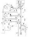
- FIG. 1illustrates various embodiments of a treatment system
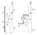
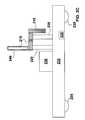
- FIGS. 2A-2Cillustrate various embodiments of a multi-position automated patient transporter
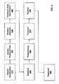
- FIG. 3illustrates various embodiments of a control system
- FIG. 4illustrates various embodiments of a method of transporting a patient to a treatment area and treating the patient.
- a patient treatment systemincludes an automated patient transporter configured to transport a patient from a preparation area to a treatment area.
- the automated patient transporteris configured to both transport the patient to the treatment area and to finely position the patient relative to a treatment beam after arriving at the treatment area.
- the preparation areais separate and/or shielded from the treatment area such that a first patient can be prepared in the preparation area while a second patient receives treatment in the treatment area.
- the automated patient transporteris configured to transport a patient to one of a number of alternative treatment areas.
- an automated patient transportercan be programmed to transport the patient to a specific treatment area responsive to a patient treatment plan or a treatment schedule.
- FIG. 1illustrates a Treatment System, generally designated 100 .
- Treatment System 100includes one, two or more Treatment Areas 105 (individually designated 105 A- 105 C) and a Preparation Area 110 .
- Preparation Area 110is configured for preparing one or more patients for treatment.
- a patientmay be positioned and stabilized on one of several Automated Patient Transporters 115 , (individually designated 115 A- 115 D). As is further described herein, this positioning typically includes determining a position of a desired treatment volume within the patient relative to the Automated Patient Transporter 115 A- 115 D.
- Various embodimentsinclude one, two, three or more of Automated Patient Transporter 115 .
- Treatment System 100optionally further includes one or more alternative Transport Paths 120 (individually designated 120 A- 120 F).
- Transport Paths 120are configured for use by Automated Patient Transporters 115 in order to travel between Preparation Area 110 , Treatment Areas 105 , and an optional Storage Area 125 .
- Transport Paths 120include one or more Switches 130 and one, two or more rails on which Automated Patient Transporters 115 move.
- Transport Paths 120include magnetic, electrical, or electromagnetic markers, or preprogrammed data representative of a route.
- Transport Paths 120may include an under-floor wire configured to emit a signal that Automated Patient Transporters 115 can follow.
- Transport Paths 120may include data stored on computer readable media within Automated Patient Transporters 115 and configured to reproducibly guide Automated Patient Transporters 115 along selected Transport Paths 120 .
- Preparation Area 110can include one, two or more different Transport Paths 120 . More than one of these Transport Paths 120 within Preparation Area 110 may be used for the preparation of more than one patient at a time or preparation of one patient on one of Automated Patient Transporters 115 while a second patient is being removed from a second of Automated Patient Transporters 115 .
- Storage Area 125is configured to store one or more Automated Patient Transporters 115 when the one or more Automated Patient Transporters 115 is not in use.
- Storage Area 125optionally includes a charging station (not shown) configured for charging a power source within Automated Patient Transporters 115 .
- Treatment System 100further includes a radiation source configured to generate one or more treatment beams configured for therapeutic use.
- the radiation sourceincludes a Particle Beam Generator 140 configured to generate a beam of high energy particles, e.g., particles with energies greater than 5, 10, 20, 50, 70, 100, 250 or 500 MeV. These particles may include hydrogen, carbon, neutrons, electrons, ions, neutrals, and/or the like, and are directed along one or more Beam Paths 145 to Particle Beam Sources 150 .
- Particle Beam Generator 140 , Beam Paths 145 and/or Particle Beam Sources 150include systems described in U.S. patent application Ser. No. 11/447,532.
- each of Treatment Areas 150alternatively or further includes other types of radiation sources such as x-ray sources.
- Treatment Areas 105optionally include more than one Particle Beam Source 150 .
- Treatment Area 105 Aincludes two Particle Beam Sources 150 .
- Automated Patient Transporters 115are optionally configured to position a patient relative to either and/or both of these two Particle Beam Sources 150 .
- Some Treatment Areas 105optionally include both Particle Beam Source 150 and an X-ray Source 155 .
- X-ray Source 155optionally includes systems described in U.S. patent application Ser. No. 11/415,974. X-ray Source 155 is configured for imaging and/or treatment of a patient.
- X-ray Source 155is configured for imaging a treatment volume within a patient and Particle Beam Source 150 is configured for generation of a particle beam for radiation of the treatment volume.
- Automated Patient Transporter 115may be configured to adjust the position of the patient in response to data generated using X-ray source 115 such that the treatment volume is intersected in a desirable manner by the particle beam.
- Treatment Area 105further includes one or more Area Position Sensors 160 .
- Area Position Sensor 160is configured for use in detecting positions of Automated Patient Transporter 115 within Treatment Area 105 .
- Area Position Sensor 160includes an encoder configured to read a marking on Automated Patient Transporter 115 C.
- Area Position Sensor 160may include a marking configured to be read by an encoder on Automated Patient Transporter 115 C.
- Area Position Sensor 160includes a docking device configured to mechanically couple with Automated Patient Transporter 115 .
- Area Position Sensor 160may include a locator pin, clamp, pin receiver, three point kinamatic coupling, rail, slot, electrical contact, or the like, configured to engage and precisely establish a position of one or more Automated Patient Transporters 115 .
- Area Position Sensor 160includes radiofrequency or optical devices configured for position detection.
- Area Position Sensor 160may include a radio frequency identification tag or tag sensor, part of an optical interferometer, a radio frequency positioning system element, or the like.
- Area Position Sensor 160is configured such that the position of one of Automated Patient Transporters 115 can be precisely detected relative to a radiation source such as one of Particle Beam Sources 150 , X-ray Source 155 , or an x-ray or particle beam generated thereby.
- the precision of this detectionis less than or equal to +/ ⁇ 3, 2, 1.5, 1, 0.5, 0.25, or 0.1 millimeters.
- the detected positioncan be used in combination with information relating the relative positions of a treatment volume and an Automated Patient Transporter 115 to determine a positional relationship between the treatment volume and treatment beam.
- One or more Treatment Areas 105optionally include a Turntable 165 configured to rotate Automated Patient Transporters 115 around one or more pivot points.
- Turntable 165may be configured to alternatively receive Automated Patient Transporter 115 A and/or 115 B, via Transport Path 120 C, and then rotate the received Automated Patient Transporter 115 around a vertical and/or horizontal axis.
- Turntable 165is configured to rotate Automated Patient Transporter 115 such that a patient is positioned relative to different radiation sources.
- These different radiation sourcescan include a particle beam source and an x-ray source, two particle beam sources, and/or two x-ray sources.
- Turntable 165may be configured such that a patient is first positioned relative to X-ray Source 155 , where imaging information can be generated. Turntable 165 is then used to rotate the Automated Patient Transporter 115 such that the patient is positioned relative to the Particle Beam Source 150 within Treatment Area 105 C. As such, the patient can receive therapeutic radiation at the same relative angle as the imaging information was obtained.
- Turntable 165is optionally used to rotate Automated Patient Transporter 115 configured to transport a patient in a sitting, standing, and/or supine position.
- Turntable 165is configured to rotate an embodiment of Automated Patient Transporter 115 configured to support a patient in a sitting position.
- Turntable 165is configured to rotate an embodiment of Automated Patient Transporter 115 configured to support a patient in a supine position.
- Turntable 165is configured to rotate an embodiment of Automated Patient Transporter 115 that is configured to support a patient in a variety of alternative positions.
- Turntable 165is configured such that a patient can be positioned in a variety of positions relative to a particular Particle Beam Source 150 .
- a Particle Beam Source 150configured to deliver a particle beam to a treatment volume through a horizontal plane may arrive at the treatment volume from a variety of different angles relative to a vertical axis by rotating the patient on Turntable 165 .
- this featureeliminates a need to have multiple Particle Beam Sources 150 at the same angle relative to the horizontal plane.
- only one Particle Beam Source 150is required at 45 degrees from the horizontal plane to direct a particle beam at a treatment volume from at position within a cone at 45 degrees rotated around an axis of rotation of Turntable 165 .
- only one Particle Beam Source 150is required in the horizontal plane to direct a particle beam at the treatment volume from any angle within the horizontal plane.
- Preparation Area 110is typically protected from radiation at Treatment Areas 105 . This protection can be achieved by distance or by one or more Barriers 170 including shielding. One or more Barriers 170 are also optionally configured to separate Treatment Areas 105 . For example, Barriers 170 may be used to prevent radiation in Treatment Area 105 A from reaching Treatment Area 105 B in significant quantities.
- Preparation Area 110may also include all or part of a Control System 175 .
- Control System 175is configured for controlling which Transport Paths 120 are used by which Automated Patient Transporters 115 , controlling operation of radiation beams, identifying patients, accessing treatment plans, and/or the like.
- All or part of Control System 175is optionally located somewhere other than Preparation Area 110 .
- Preparation Area 110may comprise one or more physically or visually separate areas or rooms made separate via any known means such as curtains, dividers, partitions, walls, floors, etc.
- FIGS. 2A-2Cillustrate further details of Automated Patient Transporter 115 , according to various embodiments.
- Automated Patient Transporter 115includes a Transport Section 210 and a Patient Support 215 , optionally separated by one or more Braces 220 .
- Patient Support 215is optionally configured to support a patient in one or more alternative positions.
- FIG. 2Aillustrates an embodiment of Patient Support 215 configured for supporting a patient in a supine position.
- the term supineis intended to include lying on a side or stomach, in addition to lying on the back.
- FIG. 2Billustrates an embodiment of Patient Support 215 configured for supporting a patient in a sitting position.
- Patient Support 215is optionally configured to bend or otherwise move in one or more locations such that the same Patient Support 215 can be adjusted into both of the configurations illustrated in FIGS. 2A and 2B .
- Patient Support 215is removable from Transport Section 210 .
- the embodiment of Patient Support 215 illustrated in FIGS. 2A and 2Bmay be removed from Transport Section 210 and replaced with the embodiment of Patient Support illustrated in FIG. 2C .
- Transport Section 210may be used to transport patients in a variety of different positions by attaching different embodiments of Patient Support 215 .
- Patient Support 215optionally includes patient positioning indicators (not shown) configured to identify the position of a patient relative to Patient Support 215 and/or Transport Section 210 .
- Patient Support 210may include surface markings indicative of patient positions.
- Patient Support 215includes an x-ray imaging detector.
- Patient Support 215may include one or more Receptacles 240 configured for receiving a removable X-ray Imaging Detector 245 configured for imaging a treatment volume within a patient.
- Patient Support 215typically includes elements configured to stabilize a patient. These elements may include a strap, clamp, pad, brace, collar, splint, and/or the like.
- Transport Section 210is configured to transport a patient, supported by Patient Support 215 , between Preparation Area 110 and one or more of Treatment Areas 105 . This transportation typically occurs along one or more predetermined Transport Paths 120 .
- Transport Section 210optionally includes Wheels 235 and may be configured to travel along one, two or more rails.
- Driver/Logic 230can include a motor, power source, microprocessor, memory, input/output interface, and/or the like.
- Driver/Logic 230includes batteries, an electric motor configured to drive Wheels 235 , memory configured to store characteristics of a selected member of Transport Paths 120 , and a processor configured to control the electric motor and Wheels 235 such that Transport Section 210 travels along the selected member of Transport Paths 120 .
- Transport Section 210 and/or Patient Support 215further include one or more Transporter Position Sensors 225 .
- Transporter Position Sensor 225is configured to determine the position of Automated Patient Transporter 115 along Transport Paths 120 . For example, if Transport Path 120 is marked using a guide wire embedded in a floor, then Transporter Position Sensor 225 may be configured to detect the guide wire. If Transport Path 120 includes a series of positions determined using radio frequency location signals, then Transporter Position Sensor 225 may be configured to detect these radio frequency location signals.
- Transporter Position Sensor 225is configured to detect the position of Automated Patient Transporter 115 relative to Area Position Sensor 160 .
- Transporter Position Sensor 225may include an encoder, an encoder pattern, an optical element, a wireless element, clamp, pin, pin receiver, mechanical device, and/or the like configured to interact with Area Position Sensor 160 .
- the two functions of detecting the position of an Automated Patient Transporter 115 along a Transport Path 120 and detecting the position of the Automated Patient Transporter 115 relative to Area Position Sensor 160are optionally performed by separate parts of Transporter Position Sensor 225 .
- the position of a treatment volume within a patient relative to a therapeutic radiation beamis detected by detecting the relative positions of the treatment volume and Transporter Position Sensor 225 , the relative positions of Transporter Position Sensor 225 and Area Position Sensor 160 , and the relative positions of Area Position Sensor 160 and the therapeutic radiation beam.
- the relative positions of the treatment volume and the Transporter Position Sensor 225are optionally determined using X-ray Imaging Detector 245 inserted in Receptacle 240 .
- the relative positions of Receptacle 240 and Transporter Position Sensor 225may be predetermined and an image received by X-ray Imaging Detector 245 may be used to detect the position of the treatment volume relative to the Receptacle 240 .
- X-ray Source 155is additionally or alternatively included in Preparation Area 110 .
- X-ray Source 155may be used to image the treatment volume and detect the position of the treatment volume relative to the Transporter Position Sensor 225 after the patient has been secured on Patient Support 215 but prior to transporting the patient to one of Treatment Areas 105 .
- X-ray Source 155included in one of Treatment Areas 105 , is additionally or alternatively used to detect the position of the treatment volume.
- X-ray Imaging Detector 245may be removed from Receptacle 240 prior to treatment using a therapeutic particle beam.
- detector apparatusfor detecting patient position may likewise be utilized including ultrasound imagers, radiofrequency detectors (e.g., for detecting radio signals affected by passive transmitters implanted in a patient, magnetic field detectors (e.g., for detecting small magnets implanted in a patient), etc.
- radiofrequency detectorse.g., for detecting radio signals affected by passive transmitters implanted in a patient
- magnetic field detectorse.g., for detecting small magnets implanted in a patient
- FIG. 3illustrates various embodiments of Control System 175 .
- These embodimentsinclude a Transport Control Logic 310 configured to control the movement of Automated Patient Transporters 115 between Treatment Areas 105 , Preparation Area 110 and/or Storage Area 125 .
- Transport Control Logic 310is typically configured to identify a starting point and an ending point for patient transport. This identification may include determining a preferred path between these points.
- Transport Control Logic 310may further be configured to control the fine positioning of a patient relative to a therapeutic radiation beam after the patient has been transported to one of Treatment Areas 105 .
- Transport Control Logic 310is configured to operate one or more of Switches 130 in Transport Paths 120 , to send signals through a guide wire to guide one of Automated Patient Transporters 115 along one of Transport Paths 120 , to start and stop movement of one of Automated Patient Transporters 115 , and/or the like.
- Transport Control Logic 310may also be configured for providing transport instructions to Drive/Logic 230 , controlling movement of Turntable 165 , controlling speeds of Automated Patient Transporters 115 , and/or the like.
- Transport Control Logic 310is configured to control movement of a plurality of Automated Patient Transporters 115 over a plurality of alternative Transport Paths 120 .
- Transport Control Logic 310is optionally configured for both controlling patient transport between Preparation Area 110 and one or more of Treatment Areas 105 , as well as positioning of a treatment volume relative to a therapeutic radiation beam.
- Transport Control Logic 310is optionally distributed among a number of devices, some of which may be included in Automated Patient Transporters 115 , Preparation Area 110 and/or Treatment Areas 105 .
- Transport Control Logic 310includes hardware, firmware, and/or software on a computer readable medium.
- Control System 175optionally further includes an Identity Input 315 , Treatment Plan Storage 320 , Treatment Plan Logic 325 , Position Detection Logic 330 , Position Storage 335 , and/or Scheduling Logic 340 .
- Identity Input 315is configured for receiving an identity of a patient.
- Identity Input 315may include a keypad, graphical user interface, a barcode reader, a radio frequency identification tag reader, a smartcard reader, and/or the like.
- a patient identity received using Identity Input 315may be used to access a patient treatment plan characterizing radiation treatment to be received by the patient.
- This patient treatment planmay include, for example, a location of a treatment volume, a dosage, spatial distribution, depth distribution, and/or type of radiation.
- the patient treatment planmay also include use of a particular Treatment Area 105 , Particle Beam Source 150 , X-ray Source 155 , and/or type of Patient Support 215 (e.g., a type to support the patient in a sitting or supine position).
- a particular Treatment Area 105Particle Beam Source 150 , X-ray Source 155 , and/or type of Patient Support 215 (e.g., a type to support the patient in a sitting or supine position).
- the patient treatment planis optionally stored in a Treatment Plan Storage 320 including a computer readable medium.
- Treatment Plan Storage 320may include computer memory, magnetic storage, optical storage, and/or a processor configured for executing a database application.
- Treatment Plan Storage 320is accessed using a patient database and includes further information about patients in addition to a particular treatment plan.
- Treatment Plan Storage 320may, thus, be part of an information system located external to Preparation Area 110 or Treatment Areas 105 .
- the patient treatment planmay be entered via Identity Input 315 .
- a patient treatment planis used by Treatment Plan Logic 20 to determine which Patient Support 215 and which of Automated Patient Transporters 115 should be used to transport a patient, as well as which of a plurality of alternative Treatment Areas 105 the patient should be transported to.
- the patient treatment planmay also be used to control operation of Particle Beam Generator 140 , Particle Beam Sources 150 , particle beam nozzles (not shown) associated with the Particle Beam Sources 150 , and/or X-ray Source 155 .
- Position Detection Logic 330is configured to detect a relative position between a treatment volume and Transporter Position Sensor 225 .
- Position Detection Logic 330is configured to receive image data from an x-ray imaging detector and an indication of a treatment volume within the image data from a user, and to determine the position of the treatment volume relative to Transporter Position Sensor 225 using this information.
- Position Detection Logic 330is configured to receive information regarding a patient's position according to markings on Patient Support 215 . For example, a technician may stabilize a patient on Patient Support 215 and then enter information about the patient's position based on markings on the Patient Support 215 .
- Position Detection Logic 330is configured to detect markers located within or on the patient, e.g. an RFID tag, and determine the position of the treatment volume relative to Transporter Position Sensor 225 based on the location of these markers.
- Position Storage 335typically includes a computer readable medium such as a hard drive, integrated circuit based memory cells, an optical drive, a magnetic drive, and/or the like. In some embodiments, Position Storage 335 is disposed external to Control System 175 , for example in an external database or in Drive/Logic 230 .
- Scheduling Logic 340is configured to schedule the use of Automated Patient Transporters 115 , Treatment Areas 105 , Preparation Area 110 , Particle Beam Sources 150 , particle beam nozzles, and/or the like. For example, in some embodiments, Scheduling Logic 340 is configured to optimize the use of each Treatment Area 105 by indicating the order and/or timing of each patient preparation in Preparation Area 110 , directing Transport Control Logic 310 to transport patients to Treatment Areas 105 , and/or activating Particle Beam Generator 140 as necessary for patient treatment. Scheduling Logic 340 is optionally configured to direct the preparation of one or more patients while one or more other patients are receiving radiation therapy.
- FIG. 4illustrates a method of transporting a patient to a Treatment Area 105 and treating the patient.
- a patientis identified, information about their planned treatment is accessed, and they are transported to a Treatment Area 105 where they receive treatment using therapeutic radiation.
- Identity Input 315is used to identify a patient to receive therapeutic radiation.
- the patientmay be identified using, for example, a barcode reader, an RFID tag reader, entering a name and/or identification number of the patient into a user interface, and/or the like.
- the identity of a patient identified in Patient Identification step 410is used to access a patient treatment plan optionally stored in Treatment Plan Storage 325 .
- the identity of the patientmay include an identification number, name, birth date, social security number, and/or the like, and may be used in a database query to access the patient treatment plan.
- a Position Detection Step 430the position of a treatment volume within the patient is detected relative to a part of an Automated Patient Transporter 115 , such as Transporter Position Sensor 225 .
- This detectionis accomplished using a two dimensional x-ray imager, a three dimensional x-ray imager, an ultrasonic imager, markers on or within a patient, markings on Patient Support 215 , and or the like.
- the detected positionis optionally stored in Position Storage 330 .
- Treatment Area Identification Step 440one of Treatment Areas 105 is selected for treating the patient. This selection may be based on availability of the treatment area, a particular Particle Beam Source 150 , a particle beam position or orientation within a transport area (e.g., horizontally or vertically oriented), and/or the like. Treatment Area Identification Step 440 is optional in embodiments including only one Treatment Area 105 .
- Schedule Step 450the treatment of the patient is scheduled using Scheduling Logic 360 .
- the schedulemay include a time of day for preparation and treatment, as well as characteristics of the treatment such as dosage, particle energy, particle beam nozzle, patient position, Patient Support 215 , Automated Patient Transporter 115 , Transport Path 120 , order of treatment for different patients, and/or the like.
- Schedule Step 450is optionally performed in combination with Treatment Area Identification Step 440 .
- one of Automated Patient Transporters 115is programmed to move the patient to the Treatment Area 105 identified in Treatment Area Identification Step 440 .
- This programmingmay include communication of data and or commands to Drive/Logic 230 .
- Switches 130 , guide signals, or one of the other mechanisms described herein for directing one of Automated Patient Transporters 115 along a particular Transport Path 120may be configured in Programming Step 450 .
- a Transport Step 470the patient is transported from Preparation Area 110 along one of Transport Paths 120 to one of Treatment Areas 105 , using Automated Patient Transporter 115 .
- Automated Patient Transporter 115is typically automated in that it travels along a predetermined member of Transport Paths 120 without requiring real-time human intervention.
- Position Detection Step 430is optionally performed and/or repeated following and/or as part of Transport Step 470 .
- a Position Step 480the treatment volume of the patient is positioned within one of Treatment Areas 105 relative to a therapeutic radiation beam. This positioning is typically performed using Automated Patient Transporter 115 and without removing the patient from Automated Patient Transporter 115 .
- the positioning of the patient relative to the therapeutic radiation beamis accomplished using Driver/Logic 230 and/or Wheels 235 .
- Patient Support 215is configured to move relative to Transport Section 210 in order to position the patient. This relative movement may be accomplished such that the position of the treatment volume relative to Transporter Position Sensor 225 remains known. For example, if Patient Support 215 is raised 5 millimeters relative to Transport Section 210 , a new position of the treatment volume relative to Transporter Position Sensor 225 can be calculated using this 5 millimeter displacement.
- the relative movement of Patient Support 215 and Transport Section 210is accomplished under the control of Driver/Logic 230 and/or by a mechanical system, e.g., adjustable embodiments of Braces 220 , included in Automated Patient Transporters 115 .
- This mechanical systemmay include hydraulics, levers, motors, drive systems, encoders, and/or the like. Further details of how Patient Support 215 may be moved relative to Transport Section 210 are discussed in U.S.
- Particle Beam Generator 140or some other radiation source, is used to generate a therapeutic radiation beam that is directed along one of Beam Paths 145 to the patient.
- This radiation beammay include high energy particles, x-rays, or any other therapeutic radiation.
- Automated Patient Transporter 115is optionally configured to return the patient to Preparation Area 110 along one of Transport Paths 120 , where the patient may be removed from Patient Support 215 .
Landscapes
- Health & Medical Sciences (AREA)
- Life Sciences & Earth Sciences (AREA)
- Engineering & Computer Science (AREA)
- General Health & Medical Sciences (AREA)
- Public Health (AREA)
- Animal Behavior & Ethology (AREA)
- Veterinary Medicine (AREA)
- Medical Informatics (AREA)
- Biomedical Technology (AREA)
- Nuclear Medicine, Radiotherapy & Molecular Imaging (AREA)
- Radiology & Medical Imaging (AREA)
- Pathology (AREA)
- Surgery (AREA)
- Molecular Biology (AREA)
- Heart & Thoracic Surgery (AREA)
- Physics & Mathematics (AREA)
- High Energy & Nuclear Physics (AREA)
- Optics & Photonics (AREA)
- Biophysics (AREA)
- Primary Health Care (AREA)
- Epidemiology (AREA)
- Urology & Nephrology (AREA)
- Oral & Maxillofacial Surgery (AREA)
- Robotics (AREA)
- Radiation-Therapy Devices (AREA)
- Business, Economics & Management (AREA)
- Tourism & Hospitality (AREA)
- Theoretical Computer Science (AREA)
- Child & Adolescent Psychology (AREA)
- Economics (AREA)
- Human Resources & Organizations (AREA)
- Marketing (AREA)
- Strategic Management (AREA)
- General Business, Economics & Management (AREA)
- General Physics & Mathematics (AREA)
- Pulmonology (AREA)
- Electromagnetism (AREA)
Abstract
Description
Claims (29)
Priority Applications (2)
| Application Number | Priority Date | Filing Date | Title |
|---|---|---|---|
| US11/985,814US10004650B2 (en) | 2005-04-29 | 2007-11-16 | Dynamic patient positioning system |
| US15/241,385US10881878B2 (en) | 2005-04-29 | 2016-08-19 | Dynamic patient positioning system |
Applications Claiming Priority (5)
| Application Number | Priority Date | Filing Date | Title |
|---|---|---|---|
| US67613805P | 2005-04-29 | 2005-04-29 | |
| US11/415,974US7640607B2 (en) | 2005-04-29 | 2006-05-01 | Patient support systems |
| US11/447,532US7547901B2 (en) | 2006-06-05 | 2006-06-05 | Multiple beam path particle source |
| US85967506P | 2006-11-17 | 2006-11-17 | |
| US11/985,814US10004650B2 (en) | 2005-04-29 | 2007-11-16 | Dynamic patient positioning system |
Related Parent Applications (2)
| Application Number | Title | Priority Date | Filing Date |
|---|---|---|---|
| US11/415,974Continuation-In-PartUS7640607B2 (en) | 2005-04-29 | 2006-05-01 | Patient support systems |
| US11/447,532Continuation-In-PartUS7547901B2 (en) | 2005-04-29 | 2006-06-05 | Multiple beam path particle source |
Related Child Applications (1)
| Application Number | Title | Priority Date | Filing Date |
|---|---|---|---|
| US15/241,385ContinuationUS10881878B2 (en) | 2005-04-29 | 2016-08-19 | Dynamic patient positioning system |
Publications (2)
| Publication Number | Publication Date |
|---|---|
| US20080071420A1 US20080071420A1 (en) | 2008-03-20 |
| US10004650B2true US10004650B2 (en) | 2018-06-26 |
Family
ID=39430341
Family Applications (2)
| Application Number | Title | Priority Date | Filing Date |
|---|---|---|---|
| US11/985,814Expired - Fee RelatedUS10004650B2 (en) | 2005-04-29 | 2007-11-16 | Dynamic patient positioning system |
| US15/241,385Active2028-08-14US10881878B2 (en) | 2005-04-29 | 2016-08-19 | Dynamic patient positioning system |
Family Applications After (1)
| Application Number | Title | Priority Date | Filing Date |
|---|---|---|---|
| US15/241,385Active2028-08-14US10881878B2 (en) | 2005-04-29 | 2016-08-19 | Dynamic patient positioning system |
Country Status (3)
| Country | Link |
|---|---|
| US (2) | US10004650B2 (en) |
| EP (1) | EP2088925B8 (en) |
| WO (1) | WO2008063573A2 (en) |
Cited By (3)
| Publication number | Priority date | Publication date | Assignee | Title |
|---|---|---|---|---|
| US11054534B1 (en) | 2020-04-24 | 2021-07-06 | Ronald Nutt | Time-resolved positron emission tomography encoder system for producing real-time, high resolution, three dimensional positron emission tomographic image without the necessity of performing image reconstruction |
| US11300695B2 (en) | 2020-04-24 | 2022-04-12 | Ronald Nutt | Time-resolved positron emission tomography encoder system for producing event-by-event, real-time, high resolution, three-dimensional positron emission tomographic image without the necessity of performing image reconstruction |
| US11684320B1 (en) | 2022-09-12 | 2023-06-27 | Izotropic Corporation | Linear motor assembly for X-ray computed tomography system |
Families Citing this family (13)
| Publication number | Priority date | Publication date | Assignee | Title |
|---|---|---|---|---|
| DE102008057145A1 (en)* | 2008-11-13 | 2010-05-27 | Siemens Aktiengesellschaft | Patient transport unit and method for transporting a patient |
| FR2945724B1 (en)* | 2009-05-22 | 2012-11-16 | Gen Electric | X-RAY APPARATUS |
| US20110154569A1 (en)* | 2009-12-28 | 2011-06-30 | Varian Medical Systems, Inc. | Mobile patient support system |
| US8934605B2 (en) | 2010-02-24 | 2015-01-13 | Accuray Incorporated | Gantry image guided radiotherapy system and related treatment delivery methods |
| US9687200B2 (en) | 2010-06-08 | 2017-06-27 | Accuray Incorporated | Radiation treatment delivery system with translatable ring gantry |
| JP5848005B2 (en)* | 2010-04-23 | 2016-01-27 | ゼネラル・エレクトリック・カンパニイ | A system for automatic support of object positioning in moving image acquisition |
| US8989846B2 (en) | 2010-08-08 | 2015-03-24 | Accuray Incorporated | Radiation treatment delivery system with outwardly movable radiation treatment head extending from ring gantry |
| US8536547B2 (en) | 2011-01-20 | 2013-09-17 | Accuray Incorporated | Ring gantry radiation treatment delivery system with dynamically controllable inward extension of treatment head |
| US8944318B2 (en)* | 2012-09-27 | 2015-02-03 | Elekta Ab | Workflow management system |
| DE102013213213A1 (en)* | 2013-07-05 | 2015-01-08 | Siemens Aktiengesellschaft | Patient Transport System |
| DE102015202327A1 (en)* | 2015-02-10 | 2016-08-11 | Kuka Roboter Gmbh | Diagnostic and / or therapy device |
| US20230210478A1 (en)* | 2020-05-29 | 2023-07-06 | Brainlab Ag | Moiré marker for x-ray imaging |
| EP4302694A1 (en)* | 2022-07-08 | 2024-01-10 | Koninklijke Philips N.V. | System and method for parallel preparation of multiple patients |
Citations (200)
| Publication number | Priority date | Publication date | Assignee | Title |
|---|---|---|---|---|
| US3133227A (en) | 1958-06-25 | 1964-05-12 | Varian Associates | Linear particle accelerator apparatus for high energy particle beams provided with pulsing means for the control electrode |
| US3144552A (en) | 1960-08-24 | 1964-08-11 | Varian Associates | Apparatus for the iradiation of materials with a pulsed strip beam of electrons |
| US3193717A (en) | 1959-03-09 | 1965-07-06 | Varian Associates | Beam scanning method and apparatus |
| GB1328033A (en) | 1970-11-06 | 1973-08-22 | Philips Electronic Associated | Apparatus for measuring the surface configuration of at least part of a body |
| JPS5057028A (en) | 1973-09-21 | 1975-05-19 | ||
| FR2269745A1 (en) | 1972-08-17 | 1975-11-28 | Lescrenier Charles | Position control of operating fable for radiation therapy - arrangement and method for holding a position reference between an emitter and a receiver object |
| US3987281A (en) | 1974-07-29 | 1976-10-19 | The United States Of America As Represented By The Department Of Health, Education And Welfare | Method of radiation therapy treatment planning |
| US4149247A (en) | 1975-12-23 | 1979-04-10 | Varian Associates, Inc. | Tomographic apparatus and method for reconstructing planar slices from non-absorbed and non-scattered radiation |
| US4149248A (en) | 1975-12-23 | 1979-04-10 | Varian Associates, Inc. | Apparatus and method for reconstructing data |
| US4208675A (en) | 1978-03-20 | 1980-06-17 | Agence Nationale De Valorization De La Recherche (Anvar) | Method and apparatus for positioning an object |
| US4209706A (en) | 1976-11-26 | 1980-06-24 | Varian Associates, Inc. | Fluoroscopic apparatus mounting fixture |
| EP0062941A1 (en) | 1981-04-08 | 1982-10-20 | Koninklijke Philips Electronics N.V. | Contour recording device |
| JPS5894835A (en) | 1981-11-30 | 1983-06-06 | 株式会社島津製作所 | Radioactive diagnostic and treating apparatus |
| JPS5976A (en) | 1982-06-22 | 1984-01-05 | 日本電気株式会社 | High energy ct for radiation treatment |
| FR2551664A1 (en) | 1982-09-13 | 1985-03-15 | Varian Associates | Thin mirror for illuminating an area for a medical electron accelerator |
| US4521808A (en) | 1979-03-22 | 1985-06-04 | University Of Texas System | Electrostatic imaging apparatus |
| WO1985003212A1 (en) | 1984-01-18 | 1985-08-01 | Lescrenier, Charles | Improved means for visually indicating an x-ray field |
| US4547892A (en) | 1977-04-01 | 1985-10-15 | Technicare Corporation | Cardiac imaging with CT scanner |
| US4593967A (en) | 1984-11-01 | 1986-06-10 | Honeywell Inc. | 3-D active vision sensor |
| US4628523A (en) | 1985-05-13 | 1986-12-09 | B.V. Optische Industrie De Oude Delft | Direction control for radiographic therapy apparatus |
| EP0205720A1 (en) | 1985-06-28 | 1986-12-30 | Instrument Ab Scanditronix | CT scanner for radiation theraphy planning |
| US4675731A (en) | 1983-02-01 | 1987-06-23 | Tokyo Shibaura Denki Kabushiki Kaisha | Diagnostic apparatus |
| US4679076A (en) | 1983-06-08 | 1987-07-07 | Vikterloef Karl Johan | Means for registering coordinates |
| US4726046A (en) | 1985-11-05 | 1988-02-16 | Varian Associates, Inc. | X-ray and electron radiotherapy clinical treatment machine |
| US4741621A (en) | 1986-08-18 | 1988-05-03 | Westinghouse Electric Corp. | Geometric surface inspection system with dual overlap light stripe generator |
| JPS63294839A (en) | 1987-05-27 | 1988-12-01 | Nec Corp | Ct simulator for radiotherapy |
| JPS6440069A (en) | 1987-08-05 | 1989-02-10 | Nec Corp | Radiotherapic apparatus |
| DE3828639A1 (en) | 1987-08-24 | 1989-03-16 | Mitsubishi Electric Corp | THERAPY DEVICE |
| JPH0162682U (en) | 1987-10-14 | 1989-04-21 | ||
| US4825393A (en) | 1986-04-23 | 1989-04-25 | Hitachi, Ltd. | Position measuring method |
| US4853777A (en) | 1987-07-07 | 1989-08-01 | Ashland Oil, Inc. | Method for evaluating smooth surfaces |
| US4868843A (en) | 1986-09-10 | 1989-09-19 | Varian Associates, Inc. | Multileaf collimator and compensator for radiotherapy machines |
| US4949408A (en)* | 1989-09-29 | 1990-08-21 | Trkla Theodore A | All purpose wheelchair |
| WO1990014129A1 (en) | 1989-05-18 | 1990-11-29 | University Of Florida | Dosimetric technique for stereotactic radiosurgery |
| US5001344A (en) | 1988-08-26 | 1991-03-19 | Hitachi, Ltd. | Scanning electron microscope and method of processing the same |
| US5014292A (en) | 1990-01-29 | 1991-05-07 | Siczek Bernard W | Tiltable x-ray table integrated with carriage for x-ray source and receptor |
| WO1992000567A1 (en) | 1990-07-02 | 1992-01-09 | Varian Associates, Inc. | Computed tomography apparatus using image intensifier detector |
| US5080100A (en) | 1988-10-04 | 1992-01-14 | Cgr Mev | System and method for measuring and/or checking the position of a patient in a radio-therapy machine |
| WO1992002277A1 (en) | 1990-08-03 | 1992-02-20 | Siemens Medical Laboratories, Inc. | Portal imaging device |
| US5099505A (en) | 1990-07-02 | 1992-03-24 | Varian Associates | Method for increasing the accuracy of a radiation therapy apparatus |
| EP0480035A1 (en) | 1989-06-30 | 1992-04-15 | Yokogawa Medical Systems, Ltd | Radiotherapeutic system |
| US5117445A (en) | 1990-07-02 | 1992-05-26 | Varian Associates, Inc. | Electronically enhanced x-ray detector apparatus |
| US5117829A (en)* | 1989-03-31 | 1992-06-02 | Loma Linda University Medical Center | Patient alignment system and procedure for radiation treatment |
| US5157707A (en) | 1989-02-20 | 1992-10-20 | Ao Medical Products Ab | Method and a cassette holder for performing x-ray examination |
| US5161546A (en)* | 1986-09-24 | 1992-11-10 | Bronn Donald G | System for intraoperative electron beam radiotherapy using remotely located beam generator |
| WO1992020202A1 (en) | 1991-05-06 | 1992-11-12 | Moore Robert M | Radiation image generating system and method |
| US5168532A (en) | 1990-07-02 | 1992-12-01 | Varian Associates, Inc. | Method for improving the dynamic range of an imaging system |
| JPH05976A (en) | 1991-06-25 | 1993-01-08 | Tokuyama Soda Co Ltd | Production of aromatic halide |
| JPH0557028A (en) | 1991-09-05 | 1993-03-09 | Nec Corp | Radiation treatment device |
| US5262649A (en) | 1989-09-06 | 1993-11-16 | The Regents Of The University Of Michigan | Thin-film, flat panel, pixelated detector array for real-time digital imaging and dosimetry of ionizing radiation |
| DE4223488A1 (en) | 1992-07-17 | 1994-01-20 | Despina Dr Med Katsohi | Restitutable compensation device for radiation treatment |
| JPH0679006A (en) | 1992-09-01 | 1994-03-22 | Hitachi Medical Corp | Stereotactic radiotherapy device |
| US5332908A (en) | 1992-03-31 | 1994-07-26 | Siemens Medical Laboratories, Inc. | Method for dynamic beam profile generation |
| US5335255A (en) | 1992-03-24 | 1994-08-02 | Seppi Edward J | X-ray scanner with a source emitting plurality of fan beams |
| JPH06339541A (en) | 1993-04-23 | 1994-12-13 | Gijutsu Kenkyu Kumiai Iryo Fukushi Kiki Kenkyusho | Display method for radiation treatment system |
| WO1995000204A1 (en) | 1993-06-18 | 1995-01-05 | Wisconsin Alumni Research Foundation | Method for radiation therapy planning |
| US5379468A (en) | 1993-04-26 | 1995-01-10 | Cassidy; Joseph P. | Patient-handling apparatus |
| US5394452A (en) | 1992-03-19 | 1995-02-28 | Wisconsin Alumni Research Foundation | Verification system for radiation therapy |
| US5400255A (en) | 1994-02-14 | 1995-03-21 | General Electric Company | Reconstruction of images from cone beam data |
| US5411026A (en) | 1993-10-08 | 1995-05-02 | Nomos Corporation | Method and apparatus for lesion position verification |
| US5438991A (en) | 1993-10-18 | 1995-08-08 | William Beaumont Hospital | Method and apparatus for controlling a radiation treatment field |
| JPH07255717A (en) | 1994-03-25 | 1995-10-09 | Toshiba Corp | Radiation therapy system |
| US5471516A (en) | 1994-10-06 | 1995-11-28 | Varian Associates, Inc. | Radiotherapy apparatus equipped with low dose localizing and portal imaging X-ray source |
| US5471546A (en) | 1993-12-29 | 1995-11-28 | Abb Research Ltd. | Fiber-optic transmission sensor with modulator |
| US5509042A (en) | 1991-02-13 | 1996-04-16 | Lunar Corporation | Automated determination and analysis of bone morphology |
| US5521957A (en) | 1994-03-15 | 1996-05-28 | Hansen; Steven J. | X-ray imaging system |
| EP0713677A1 (en) | 1990-08-14 | 1996-05-29 | Picker International, Inc. | Imaging apparatus and methods |
| US5537452A (en) | 1994-05-10 | 1996-07-16 | Shepherd; Joseph S. | Radiation therapy and radiation surgery treatment system and methods of use of same |
| US5591983A (en) | 1995-06-30 | 1997-01-07 | Siemens Medical Systems, Inc. | Multiple layer multileaf collimator |
| WO1997013552A1 (en) | 1995-10-07 | 1997-04-17 | Philips Electronics N.V. | Radiotherapy apparatus for treating a patient |
| US5622187A (en)* | 1994-09-30 | 1997-04-22 | Nomos Corporation | Method and apparatus for patient positioning for radiation therapy |
| US5647663A (en) | 1996-01-05 | 1997-07-15 | Wisconsin Alumni Research Foundation | Radiation treatment planning method and apparatus |
| US5661773A (en) | 1992-03-19 | 1997-08-26 | Wisconsin Alumni Research Foundation | Interface for radiation therapy machine |
| US5663999A (en) | 1996-06-28 | 1997-09-02 | Systems Medical Systems, Inc. | Optimization of an intensity modulated field |
| US5663995A (en) | 1996-06-06 | 1997-09-02 | General Electric Company | Systems and methods for reconstructing an image in a CT system performing a cone beam helical scan |
| JPH09239044A (en) | 1996-03-01 | 1997-09-16 | Philips Electron Nv | Intensity modulating arc medical treatment by dynamic multileaf collimation |
| US5673300A (en) | 1996-06-11 | 1997-09-30 | Wisconsin Alumni Research Foundation | Method of registering a radiation treatment plan to a patient |
| US5675625A (en) | 1994-06-17 | 1997-10-07 | Lap Gmbh Laser Applikationen | Apparatus for positioning and marking a patient at a diagnostic apparatus |
| DE19614643A1 (en) | 1996-04-13 | 1997-10-16 | Werner Dipl Phys Brenneisen | Stereotaxial targetted irradiation process for brain tumours |
| WO1997042522A1 (en) | 1996-05-07 | 1997-11-13 | The Regents Of The University Of California | Radiation therapy dose calculation engine |
| JPH09327453A (en) | 1996-06-12 | 1997-12-22 | Hitachi Medical Corp | X-ray diagnostic apparatus |
| US5719914A (en) | 1995-11-13 | 1998-02-17 | Imatron, Inc. | Method for correcting spherical aberration of the electron beam in a scanning electron beam computed tomography system |
| US5724400A (en) | 1992-03-19 | 1998-03-03 | Wisconsin Alumni Research Foundation | Radiation therapy system with constrained rotational freedom |
| US5748703A (en) | 1994-03-22 | 1998-05-05 | Cosman; Eric R. | Dynamic collimator for a linear accelerator |
| US5748907A (en)* | 1993-10-25 | 1998-05-05 | Crane; Harold E. | Medical facility and business: automatic interactive dynamic real-time management |
| JPH10113400A (en) | 1996-10-11 | 1998-05-06 | Hitachi Medical Corp | Radiotherapy system |
| US5757881A (en) | 1997-01-06 | 1998-05-26 | Siemens Business Communication Systems, Inc. | Redundant field-defining arrays for a radiation system |
| US5802136A (en) | 1994-05-17 | 1998-09-01 | Nomos Corporation | Method and apparatus for conformal radiation therapy |
| US5818902A (en) | 1996-03-01 | 1998-10-06 | Elekta Ab | Intensity modulated arc therapy with dynamic multi-leaf collimation |
| US5835558A (en) | 1996-07-09 | 1998-11-10 | Siemens Aktiengesellschaft | Mobile x-ray exposure apparatus |
| WO1998052635A1 (en) | 1997-05-23 | 1998-11-26 | William Beaumont Hospital | Method and apparatus for delivering radiation therapy during suspended ventilation |
| US5842987A (en) | 1997-05-20 | 1998-12-01 | Sahadevan; Velayudhan | Simulated patient setup for medical imaging with increased patient throughput |
| US5848126A (en) | 1993-11-26 | 1998-12-08 | Kabushiki Kaisha Toshiba | Radiation computed tomography apparatus |
| JPH10328318A (en) | 1997-05-29 | 1998-12-15 | Hitachi Medical Corp | Radiotherapy system |
| US5851182A (en) | 1996-09-11 | 1998-12-22 | Sahadevan; Velayudhan | Megavoltage radiation therapy machine combined to diagnostic imaging devices for cost efficient conventional and 3D conformal radiation therapy with on-line Isodose port and diagnostic radiology |
| US5858891A (en) | 1993-12-16 | 1999-01-12 | France Telecom | Glass-ceramic materials especially for lasers and optical amplifiers, doped with rare earths |
| WO1999003397A1 (en) | 1997-07-17 | 1999-01-28 | Medlennium Technologies, Inc. | Method and apparatus for radiation and hyperthermia therapy of tumors |
| US5877501A (en) | 1996-11-26 | 1999-03-02 | Picker International, Inc. | Digital panel for x-ray image acquisition |
| JPH1162682A (en) | 1997-08-21 | 1999-03-05 | Toyota Motor Corp | Accumulator type fuel injection device |
| JPH1199148A (en) | 1997-09-25 | 1999-04-13 | Masahiro Fukushi | Transmission CT device |
| US5912943A (en) | 1997-11-26 | 1999-06-15 | Picker International, Inc. | Cooling system for a sealed housing positioned in a sterile environment |
| EP0922943A2 (en) | 1997-11-28 | 1999-06-16 | Canon Kabushiki Kaisha | Radiation detecting device and radiation detecting method |
| US5926521A (en) | 1998-03-31 | 1999-07-20 | Siemens Corporate Research, Inc. | Exact region of interest cone beam imaging using 3D backprojection |
| US5929449A (en) | 1995-07-31 | 1999-07-27 | 1294339 Ontario, Inc. | Flat panel detector for radiation imaging with reduced electronic noise |
| US5949811A (en) | 1996-10-08 | 1999-09-07 | Hitachi Medical Corporation | X-ray apparatus |
| US5956382A (en) | 1997-09-25 | 1999-09-21 | Eliezer Wiener-Avnear, Doing Business As Laser Electro Optic Application Technology Comp. | X-ray imaging array detector and laser micro-milling method for fabricating array |
| US5960055A (en) | 1997-12-19 | 1999-09-28 | Siemens Corporate Research, Inc. | Fast cone beam image reconstruction using a detector weight list |
| WO1999048558A1 (en) | 1998-03-20 | 1999-09-30 | Elekta Ab (Publ) | Controlling delivery of radiotherapy |
| EP0948930A1 (en) | 1998-04-06 | 1999-10-13 | Picker International, Inc. | Acquiring volumetric image data |
| US5999587A (en) | 1997-07-03 | 1999-12-07 | University Of Rochester | Method of and system for cone-beam tomography reconstruction |
| US6031888A (en) | 1997-11-26 | 2000-02-29 | Picker International, Inc. | Fluoro-assist feature for a diagnostic imaging device |
| US6038283A (en) | 1996-10-24 | 2000-03-14 | Nomos Corporation | Planning method and apparatus for radiation dosimetry |
| WO2000015299A1 (en) | 1998-09-10 | 2000-03-23 | The Regents Of The University Of California | Falcon: automated optimization method for arbitrary assessment criteria |
| JP2000116638A (en) | 1998-10-15 | 2000-04-25 | Shimadzu Corp | Transmission CT system |
| JP2000140137A (en) | 1998-08-31 | 2000-05-23 | Sumitomo Heavy Ind Ltd | Method and device for positioning patient of radiotherapy |
| JP2000152927A (en) | 1998-11-19 | 2000-06-06 | Fuji Photo Film Co Ltd | Radiography device |
| US6075836A (en) | 1997-07-03 | 2000-06-13 | University Of Rochester | Method of and system for intravenous volume tomographic digital angiography imaging |
| US6078638A (en) | 1998-09-30 | 2000-06-20 | Siemens Corporate Research, Inc. | Pixel grouping for filtering cone beam detector data during 3D image reconstruction |
| US6104778A (en) | 1997-10-16 | 2000-08-15 | Varian Systems, Inc. | X-ray treatment method and apparatus |
| US6104780A (en) | 1997-11-24 | 2000-08-15 | Oec Medical Systems, Inc. | Mobile bi-planar fluoroscopic imaging apparatus |
| US6108400A (en) | 1998-08-10 | 2000-08-22 | Siemens Medical Systems, Inc. | System and method for using precalculated strips in calculating scatter radiation |
| US6113264A (en) | 1997-06-04 | 2000-09-05 | Kabushiki Kaisha Toshiba | X-ray diagnostic apparatus with C-shaped arms |
| US6134296A (en) | 1999-01-20 | 2000-10-17 | Siemens Medical Systems, Inc. | Microgradient intensity modulating multi-leaf collimator |
| US6142925A (en) | 1999-01-20 | 2000-11-07 | Siemens Medical Systems, Inc. | Method and system for increasing resolution in a radiotherapy system |
| US6144875A (en) | 1999-03-16 | 2000-11-07 | Accuray Incorporated | Apparatus and method for compensating for respiratory and patient motion during treatment |
| US6148058A (en) | 1998-10-23 | 2000-11-14 | Analogic Corporation | System and method for real time measurement of detector offset in rotating-patient CT scanner |
| JP2000317000A (en) | 1999-05-13 | 2000-11-21 | Mitsubishi Electric Corp | Control device of radiation irradiation device for radiation therapy |
| US6152598A (en) | 1997-09-02 | 2000-11-28 | Kabushiki Kaisha Toshiba | R/F and chest radiography compatible X-ray imaging table |
| JP2001029491A (en) | 1999-07-15 | 2001-02-06 | Mitsubishi Electric Corp | Irradiation dose value calculation device, irradiation dose value calculation method and recording medium |
| JP2001029489A (en) | 1999-07-15 | 2001-02-06 | Mitsubishi Electric Corp | Radiation irradiation device and method |
| US6200024B1 (en) | 1998-11-27 | 2001-03-13 | Picker International, Inc. | Virtual C-arm robotic positioning system for use in radiographic imaging equipment |
| JP2001095793A (en) | 1999-10-04 | 2001-04-10 | Hitachi Medical Corp | X-ray ct apparatus |
| US6219441B1 (en) | 1993-06-22 | 2001-04-17 | General Electric Company | Reconstruction of images from three-dimensional cone beam data |
| US6219403B1 (en) | 1999-02-17 | 2001-04-17 | Mitsubishi Denki Kabushiki Kaisha | Radiation therapy method and system |
| US6222901B1 (en) | 1995-12-18 | 2001-04-24 | U.S. Philips Corporation | X-ray examination apparatus including an image sensor matrix with a correction unit |
| EP1095628A2 (en) | 1999-10-29 | 2001-05-02 | Marconi Medical Systems, Inc. | Planning minimally invasive procedures for in - vivo placement of objects |
| JP2001120528A (en) | 1999-10-29 | 2001-05-08 | Konica Corp | Medical imaging method and medical imaging apparatus |
| US20010001807A1 (en) | 1997-12-19 | 2001-05-24 | Varian, Inc. | Radiotherapy machine including magnetic resonance imaging system |
| US6240161B1 (en) | 1997-09-25 | 2001-05-29 | Siemens Medical Systems, Inc. | Multi-leaf collimator constrained optimization of intensity modulated treatments |
| US20010008271A1 (en) | 2000-01-12 | 2001-07-19 | Kabushiki Kaisha Toshiba | Planar X-ray detector |
| US6269143B1 (en) | 1998-08-31 | 2001-07-31 | Shimadzu Corporation | Radiotherapy planning system |
| US6269141B1 (en) | 1998-08-05 | 2001-07-31 | U.S. Philips Corporation | Computer tomography apparatus with a conical radiation beam and a helical scanning trajectory |
| US6278766B1 (en) | 1996-10-25 | 2001-08-21 | Sherwood Services Ag | Jaw and circular collimator |
| WO2001060236A2 (en) | 2000-02-18 | 2001-08-23 | William Beaumont Hospital | Cone-beam computerized tomography with a flat-panel imager |
| US6285739B1 (en) | 1999-02-19 | 2001-09-04 | The Research Foundation Of State University Of New York | Radiographic imaging apparatus and method for vascular interventions |
| EP0965104B1 (en) | 1997-03-07 | 2001-09-05 | Computerized Medical Systems, Inc. | Autosegmentation/autocontouring methods for use with three-dimensional radiation therapy treatment planning |
| US6292526B1 (en) | 1999-10-27 | 2001-09-18 | General Electric Company | Methods and apparatus for preprocessing volumetric computed tomography data |
| US6307914B1 (en) | 1998-03-12 | 2001-10-23 | Mitsubishi Denki Kabushiki Kaisha | Moving body pursuit irradiating device and positioning method using this device |
| US6314159B1 (en) | 1999-12-08 | 2001-11-06 | Siemens Medical Systems, Inc. | System and method for optimizing radiation treatment with an intensity modulating multi-leaf collimator |
| US6318892B1 (en) | 1998-10-28 | 2001-11-20 | Hitachi Medical Corporation | Radiography apparatus with rotatably supported cylindrical ring carrying image pickup unit |
| US6325537B1 (en) | 1998-10-16 | 2001-12-04 | Kabushiki Kaisha Toshiba | X-ray diagnosis apparatus |
| US6325758B1 (en) | 1997-10-27 | 2001-12-04 | Nomos Corporation | Method and apparatus for target position verification |
| US6330300B1 (en) | 2000-08-23 | 2001-12-11 | Siemens Medical Solutions Usa, Inc. | High definition intensity modulating radiation therapy system and method |
| US6335961B1 (en) | 1998-10-06 | 2002-01-01 | Siemens Medical Systems, Inc. | Integrated high definition intensity multileaf collimator system which provides improved conformal radiation therapy while minimizing leakage |
| US20020006182A1 (en) | 2000-05-19 | 2002-01-17 | Siyong Kim | Multi-source intensity-modulated radiation beam delivery system and method |
| US6345114B1 (en) | 1995-06-14 | 2002-02-05 | Wisconsin Alumni Research Foundation | Method and apparatus for calibration of radiation therapy equipment and verification of radiation treatment |
| US6349129B1 (en) | 1999-12-08 | 2002-02-19 | Siemens Medical Solutions Usa, Inc. | System and method for defining radiation treatment intensity maps |
| WO2002013907A1 (en) | 2000-08-16 | 2002-02-21 | Elekta Ab (Publ) | Radiotherapy simulation apparatus |
| US6353222B1 (en) | 1998-09-03 | 2002-03-05 | Applied Materials, Inc. | Determining defect depth and contour information in wafer structures using multiple SEM images |
| WO2002024277A1 (en) | 2000-09-22 | 2002-03-28 | Radiological Imaging Technology, Inc. | Automated calibration for radiation dosimetry using fixed or moving beams and detectors |
| US6370421B1 (en) | 2000-06-30 | 2002-04-09 | Siemens Corporate Research, Inc. | Density modulated catheter for use in fluoroscopy based 3-D neural navigation |
| US6381302B1 (en) | 2000-07-05 | 2002-04-30 | Canon Kabushiki Kaisha | Computer assisted 2D adjustment of stereo X-ray images |
| US6385286B1 (en) | 1998-08-06 | 2002-05-07 | Wisconsin Alumni Research Foundation | Delivery modification system for radiation therapy |
| US6385288B1 (en) | 2001-01-19 | 2002-05-07 | Mitsubishi Denki Kabushiki Kaisha | Radiotherapy apparatus with independent rotation mechanisms |
| US6385477B1 (en) | 1997-06-19 | 2002-05-07 | Elektra Ab | Method for automatized dose planning |
| US6393096B1 (en) | 1998-05-27 | 2002-05-21 | Nomos Corporation | Planning method and apparatus for radiation dosimetry |
| US20020066860A1 (en) | 2000-12-04 | 2002-06-06 | General Electric Company | Imaging array minimizing leakage currents |
| US6405072B1 (en) | 1991-01-28 | 2002-06-11 | Sherwood Services Ag | Apparatus and method for determining a location of an anatomical target with reference to a medical apparatus |
| US20020077749A1 (en)* | 2000-12-15 | 2002-06-20 | Miwako Doi | Walker navigation system, walker navigation method, guidance data collection apparatus and guidance data collection method |
| US6411675B1 (en) | 2000-11-13 | 2002-06-25 | Jorge Llacer | Stochastic method for optimization of radiation therapy planning |
| US6429578B1 (en) | 1999-01-26 | 2002-08-06 | Mats Danielsson | Diagnostic and therapeutic detector system for imaging with low and high energy X-ray and electrons |
| WO2002061680A2 (en) | 2001-01-31 | 2002-08-08 | 3Q Technologies Ltd | Surface imaging |
| US6438202B1 (en) | 1998-08-06 | 2002-08-20 | Wisconsin Alumni Research Foundation | Method using post-patient radiation monitor to verify entrance radiation and dose in a radiation therapy machine |
| US6435715B1 (en) | 1998-11-30 | 2002-08-20 | Siemens Aktiengesellschaft | Radiography device |
| US6445766B1 (en) | 2000-10-18 | 2002-09-03 | Siemens Medical Solutions Usa, Inc. | System and method for improved diagnostic imaging in a radiation treatment system |
| US20020120986A1 (en)* | 2001-03-05 | 2002-09-05 | Stephan Erbel | Patient positioning system for radiotherapy |
| US6463122B1 (en) | 2000-08-21 | 2002-10-08 | Bio-Imaging Resource, Inc. | Mammography of computer tomography for imaging and therapy |
| US6473490B1 (en) | 2001-09-28 | 2002-10-29 | Siemens Medical Solutions Usa, Inc. | Intensity map reconstruction for radiation therapy with a modulating multi-leaf collimator |
| US6480565B1 (en) | 1999-11-18 | 2002-11-12 | University Of Rochester | Apparatus and method for cone beam volume computed tomography breast imaging |
| US6490476B1 (en) | 1999-10-14 | 2002-12-03 | Cti Pet Systems, Inc. | Combined PET and X-ray CT tomograph and method for using same |
| US20020179812A1 (en) | 2001-03-06 | 2002-12-05 | Topcon Corporation | Electron beam device and method for stereoscopic measurements |
| US6504892B1 (en) | 2000-10-13 | 2003-01-07 | University Of Rochester | System and method for cone beam volume computed tomography using circle-plus-multiple-arc orbit |
| US6502261B1 (en) | 1996-05-15 | 2003-01-07 | Elekta Ab | Patient support |
| US6508586B2 (en) | 2000-09-29 | 2003-01-21 | Kabushiki Kaisha Toshiba | IVR-CT apparatus |
| US6560311B1 (en) | 1998-08-06 | 2003-05-06 | Wisconsin Alumni Research Foundation | Method for preparing a radiation therapy plan |
| US6590953B2 (en) | 2000-09-12 | 2003-07-08 | Hitachi Medical Corporation | X-ray CT scanner |
| US6611700B1 (en)* | 1999-12-30 | 2003-08-26 | Brainlab Ag | Method and apparatus for positioning a body for radiation using a position sensor |
| US6618467B1 (en) | 1999-04-02 | 2003-09-09 | Wisconsin Alumni Research Foundation | Megavoltage computed tomography during radiotherapy |
| US6621889B1 (en) | 1998-10-23 | 2003-09-16 | Varian Medical Systems, Inc. | Method and system for predictive physiological gating of radiation therapy |
| US6640364B1 (en)* | 2001-11-30 | 2003-11-04 | Ge Medical Systems Global Technololgy Company, Llc | Pedestal for use with patient transport system for multiple imaging systems |
| US6661870B2 (en) | 2001-03-09 | 2003-12-09 | Tomotherapy Incorporated | Fluence adjustment for improving delivery to voxels without reoptimization |
| US6744848B2 (en) | 2000-02-11 | 2004-06-01 | Brandeis University | Method and system for low-dose three-dimensional imaging of a scene |
| US20040220467A1 (en)* | 1991-12-04 | 2004-11-04 | Bonutti Peter M. | Patient support apparatus |
| US6865254B2 (en) | 2002-07-02 | 2005-03-08 | Pencilbeam Technologies Ab | Radiation system with inner and outer gantry parts |
| US6888919B2 (en) | 2001-11-02 | 2005-05-03 | Varian Medical Systems, Inc. | Radiotherapy apparatus equipped with an articulable gantry for positioning an imaging unit |
| US6914959B2 (en) | 2001-08-09 | 2005-07-05 | Analogic Corporation | Combined radiation therapy and imaging system and method |
| US20060118736A1 (en) | 2003-03-07 | 2006-06-08 | Kunio Moriyama | Particle beam therapy system |
| US7397044B2 (en)* | 2005-07-21 | 2008-07-08 | Siemens Medical Solutions Usa, Inc. | Imaging mode for linear accelerators |
| US7640607B2 (en)* | 2005-04-29 | 2010-01-05 | Varian Medical Systems, Inc. | Patient support systems |
| US7945021B2 (en) | 2002-12-18 | 2011-05-17 | Varian Medical Systems, Inc. | Multi-mode cone beam CT radiotherapy simulator and treatment machine with a flat panel imager |
Family Cites Families (8)
| Publication number | Priority date | Publication date | Assignee | Title |
|---|---|---|---|---|
| DE10033063A1 (en)* | 2000-07-07 | 2002-01-24 | Brainlab Ag | Respiration compensated radiation treatment tracks target volume using markers and switches beam |
| ES2193030T3 (en)* | 2000-11-22 | 2003-11-01 | Brainlab Ag | METHOD FOR DETERMINING THE VOLUME OF LUNGS. |
| US20020193685A1 (en)* | 2001-06-08 | 2002-12-19 | Calypso Medical, Inc. | Guided Radiation Therapy System |
| EP1709994A1 (en)* | 2005-04-04 | 2006-10-11 | Ion Beam Applications S.A. | Patient positioning imaging device and method |
| US20070053491A1 (en)* | 2005-09-07 | 2007-03-08 | Eastman Kodak Company | Adaptive radiation therapy method with target detection |
| DE102005050344A1 (en)* | 2005-10-20 | 2007-05-03 | Siemens Ag | Cryocatheter for medical investigation and treatment equipment for e.g. diagnosis and treatment of heart infarcts, has image capture device that maps region of vessel around balloon arranged near catheter tip |
| JP5472757B2 (en)* | 2009-11-16 | 2014-04-16 | 三菱重工業株式会社 | Radiotherapy apparatus control apparatus, specific site position measurement method, and radiotherapy apparatus control apparatus operation method |
| JP6004464B2 (en)* | 2012-03-19 | 2016-10-05 | 国立大学法人北海道大学 | Radiotherapy control apparatus and radiotherapy control program |
- 2007
- 2007-11-16EPEP07862095.2Apatent/EP2088925B8/ennot_activeNot-in-force
- 2007-11-16USUS11/985,814patent/US10004650B2/ennot_activeExpired - Fee Related
- 2007-11-16WOPCT/US2007/024103patent/WO2008063573A2/enactiveApplication Filing
- 2016
- 2016-08-19USUS15/241,385patent/US10881878B2/enactiveActive
Patent Citations (222)
| Publication number | Priority date | Publication date | Assignee | Title |
|---|---|---|---|---|
| US3133227A (en) | 1958-06-25 | 1964-05-12 | Varian Associates | Linear particle accelerator apparatus for high energy particle beams provided with pulsing means for the control electrode |
| US3193717A (en) | 1959-03-09 | 1965-07-06 | Varian Associates | Beam scanning method and apparatus |
| US3144552A (en) | 1960-08-24 | 1964-08-11 | Varian Associates | Apparatus for the iradiation of materials with a pulsed strip beam of electrons |
| GB1328033A (en) | 1970-11-06 | 1973-08-22 | Philips Electronic Associated | Apparatus for measuring the surface configuration of at least part of a body |
| FR2269745A1 (en) | 1972-08-17 | 1975-11-28 | Lescrenier Charles | Position control of operating fable for radiation therapy - arrangement and method for holding a position reference between an emitter and a receiver object |
| JPS5057028A (en) | 1973-09-21 | 1975-05-19 | ||
| US3987281A (en) | 1974-07-29 | 1976-10-19 | The United States Of America As Represented By The Department Of Health, Education And Welfare | Method of radiation therapy treatment planning |
| US4149247A (en) | 1975-12-23 | 1979-04-10 | Varian Associates, Inc. | Tomographic apparatus and method for reconstructing planar slices from non-absorbed and non-scattered radiation |
| US4149248A (en) | 1975-12-23 | 1979-04-10 | Varian Associates, Inc. | Apparatus and method for reconstructing data |
| US4209706A (en) | 1976-11-26 | 1980-06-24 | Varian Associates, Inc. | Fluoroscopic apparatus mounting fixture |
| US4547892A (en) | 1977-04-01 | 1985-10-15 | Technicare Corporation | Cardiac imaging with CT scanner |
| US4208675A (en) | 1978-03-20 | 1980-06-17 | Agence Nationale De Valorization De La Recherche (Anvar) | Method and apparatus for positioning an object |
| US4521808A (en) | 1979-03-22 | 1985-06-04 | University Of Texas System | Electrostatic imaging apparatus |
| EP0062941A1 (en) | 1981-04-08 | 1982-10-20 | Koninklijke Philips Electronics N.V. | Contour recording device |
| EP0062941B1 (en) | 1981-04-08 | 1984-09-26 | Koninklijke Philips Electronics N.V. | Contour recording device |
| JPS5894835A (en) | 1981-11-30 | 1983-06-06 | 株式会社島津製作所 | Radioactive diagnostic and treating apparatus |
| JPS5976A (en) | 1982-06-22 | 1984-01-05 | 日本電気株式会社 | High energy ct for radiation treatment |
| FR2551664A1 (en) | 1982-09-13 | 1985-03-15 | Varian Associates | Thin mirror for illuminating an area for a medical electron accelerator |
| US4675731A (en) | 1983-02-01 | 1987-06-23 | Tokyo Shibaura Denki Kabushiki Kaisha | Diagnostic apparatus |
| US4679076A (en) | 1983-06-08 | 1987-07-07 | Vikterloef Karl Johan | Means for registering coordinates |
| WO1985003212A1 (en) | 1984-01-18 | 1985-08-01 | Lescrenier, Charles | Improved means for visually indicating an x-ray field |
| US4593967A (en) | 1984-11-01 | 1986-06-10 | Honeywell Inc. | 3-D active vision sensor |
| US4628523A (en) | 1985-05-13 | 1986-12-09 | B.V. Optische Industrie De Oude Delft | Direction control for radiographic therapy apparatus |
| EP0205720A1 (en) | 1985-06-28 | 1986-12-30 | Instrument Ab Scanditronix | CT scanner for radiation theraphy planning |
| US4726046A (en) | 1985-11-05 | 1988-02-16 | Varian Associates, Inc. | X-ray and electron radiotherapy clinical treatment machine |
| US4825393A (en) | 1986-04-23 | 1989-04-25 | Hitachi, Ltd. | Position measuring method |
| US4741621A (en) | 1986-08-18 | 1988-05-03 | Westinghouse Electric Corp. | Geometric surface inspection system with dual overlap light stripe generator |
| US4868843A (en) | 1986-09-10 | 1989-09-19 | Varian Associates, Inc. | Multileaf collimator and compensator for radiotherapy machines |
| US4868844A (en) | 1986-09-10 | 1989-09-19 | Varian Associates, Inc. | Mutileaf collimator for radiotherapy machines |
| US5161546A (en)* | 1986-09-24 | 1992-11-10 | Bronn Donald G | System for intraoperative electron beam radiotherapy using remotely located beam generator |
| JPS63294839A (en) | 1987-05-27 | 1988-12-01 | Nec Corp | Ct simulator for radiotherapy |
| US4853777A (en) | 1987-07-07 | 1989-08-01 | Ashland Oil, Inc. | Method for evaluating smooth surfaces |
| JPS6440069A (en) | 1987-08-05 | 1989-02-10 | Nec Corp | Radiotherapic apparatus |
| DE3828639A1 (en) | 1987-08-24 | 1989-03-16 | Mitsubishi Electric Corp | THERAPY DEVICE |
| US5039867A (en) | 1987-08-24 | 1991-08-13 | Mitsubishi Denki Kabushiki Kaisha | Therapeutic apparatus |
| JPH0162682U (en) | 1987-10-14 | 1989-04-21 | ||
| US5027818A (en) | 1987-12-03 | 1991-07-02 | University Of Florida | Dosimetric technique for stereotactic radiosurgery same |
| US5001344A (en) | 1988-08-26 | 1991-03-19 | Hitachi, Ltd. | Scanning electron microscope and method of processing the same |
| US5080100A (en) | 1988-10-04 | 1992-01-14 | Cgr Mev | System and method for measuring and/or checking the position of a patient in a radio-therapy machine |
| US5247555A (en) | 1988-10-28 | 1993-09-21 | Nucletron Manufacturing Corp. | Radiation image generating system and method |
| US5157707A (en) | 1989-02-20 | 1992-10-20 | Ao Medical Products Ab | Method and a cassette holder for performing x-ray examination |
| US5117829A (en)* | 1989-03-31 | 1992-06-02 | Loma Linda University Medical Center | Patient alignment system and procedure for radiation treatment |
| WO1990014129A1 (en) | 1989-05-18 | 1990-11-29 | University Of Florida | Dosimetric technique for stereotactic radiosurgery |
| EP0480035A1 (en) | 1989-06-30 | 1992-04-15 | Yokogawa Medical Systems, Ltd | Radiotherapeutic system |
| EP0480035B1 (en) | 1989-06-30 | 1994-11-09 | Yokogawa Medical Systems, Ltd | Radiotherapeutic system |
| US5262649A (en) | 1989-09-06 | 1993-11-16 | The Regents Of The University Of Michigan | Thin-film, flat panel, pixelated detector array for real-time digital imaging and dosimetry of ionizing radiation |
| US4949408A (en)* | 1989-09-29 | 1990-08-21 | Trkla Theodore A | All purpose wheelchair |
| US5014292A (en) | 1990-01-29 | 1991-05-07 | Siczek Bernard W | Tiltable x-ray table integrated with carriage for x-ray source and receptor |
| US5692507A (en) | 1990-07-02 | 1997-12-02 | Varian Associates, Inc. | Computer tomography apparatus using image intensifier detector |
| WO1992000567A1 (en) | 1990-07-02 | 1992-01-09 | Varian Associates, Inc. | Computed tomography apparatus using image intensifier detector |
| US5168532A (en) | 1990-07-02 | 1992-12-01 | Varian Associates, Inc. | Method for improving the dynamic range of an imaging system |
| US5099505A (en) | 1990-07-02 | 1992-03-24 | Varian Associates | Method for increasing the accuracy of a radiation therapy apparatus |
| US5117445A (en) | 1990-07-02 | 1992-05-26 | Varian Associates, Inc. | Electronically enhanced x-ray detector apparatus |
| WO1992002277A1 (en) | 1990-08-03 | 1992-02-20 | Siemens Medical Laboratories, Inc. | Portal imaging device |
| EP0713677A1 (en) | 1990-08-14 | 1996-05-29 | Picker International, Inc. | Imaging apparatus and methods |
| EP0471455B2 (en) | 1990-08-14 | 2002-04-24 | Picker International, Inc. | Imaging apparatus and methods |
| US6405072B1 (en) | 1991-01-28 | 2002-06-11 | Sherwood Services Ag | Apparatus and method for determining a location of an anatomical target with reference to a medical apparatus |
| US5509042A (en) | 1991-02-13 | 1996-04-16 | Lunar Corporation | Automated determination and analysis of bone morphology |
| WO1992020202A1 (en) | 1991-05-06 | 1992-11-12 | Moore Robert M | Radiation image generating system and method |
| JPH05976A (en) | 1991-06-25 | 1993-01-08 | Tokuyama Soda Co Ltd | Production of aromatic halide |
| JPH0557028A (en) | 1991-09-05 | 1993-03-09 | Nec Corp | Radiation treatment device |
| US20040220467A1 (en)* | 1991-12-04 | 2004-11-04 | Bonutti Peter M. | Patient support apparatus |
| US5661773A (en) | 1992-03-19 | 1997-08-26 | Wisconsin Alumni Research Foundation | Interface for radiation therapy machine |
| US5724400A (en) | 1992-03-19 | 1998-03-03 | Wisconsin Alumni Research Foundation | Radiation therapy system with constrained rotational freedom |
| US5394452A (en) | 1992-03-19 | 1995-02-28 | Wisconsin Alumni Research Foundation | Verification system for radiation therapy |
| US5335255A (en) | 1992-03-24 | 1994-08-02 | Seppi Edward J | X-ray scanner with a source emitting plurality of fan beams |
| US5332908A (en) | 1992-03-31 | 1994-07-26 | Siemens Medical Laboratories, Inc. | Method for dynamic beam profile generation |
| DE4223488A1 (en) | 1992-07-17 | 1994-01-20 | Despina Dr Med Katsohi | Restitutable compensation device for radiation treatment |
| JPH0679006A (en) | 1992-09-01 | 1994-03-22 | Hitachi Medical Corp | Stereotactic radiotherapy device |
| JPH06339541A (en) | 1993-04-23 | 1994-12-13 | Gijutsu Kenkyu Kumiai Iryo Fukushi Kiki Kenkyusho | Display method for radiation treatment system |
| US5379468A (en) | 1993-04-26 | 1995-01-10 | Cassidy; Joseph P. | Patient-handling apparatus |
| EP0810006B1 (en) | 1993-06-09 | 2000-08-30 | Wisconsin Alumni Research Foundation | Radiation therapy system |
| WO1995000204A1 (en) | 1993-06-18 | 1995-01-05 | Wisconsin Alumni Research Foundation | Method for radiation therapy planning |
| EP0656797B1 (en) | 1993-06-18 | 1998-09-23 | Wisconsin Alumni Research Foundation | Apparatus and method for radiation therapy planning |
| US6219441B1 (en) | 1993-06-22 | 2001-04-17 | General Electric Company | Reconstruction of images from three-dimensional cone beam data |
| US5411026A (en) | 1993-10-08 | 1995-05-02 | Nomos Corporation | Method and apparatus for lesion position verification |
| US5438991A (en) | 1993-10-18 | 1995-08-08 | William Beaumont Hospital | Method and apparatus for controlling a radiation treatment field |
| US5748907A (en)* | 1993-10-25 | 1998-05-05 | Crane; Harold E. | Medical facility and business: automatic interactive dynamic real-time management |
| US5848126A (en) | 1993-11-26 | 1998-12-08 | Kabushiki Kaisha Toshiba | Radiation computed tomography apparatus |
| US5858891A (en) | 1993-12-16 | 1999-01-12 | France Telecom | Glass-ceramic materials especially for lasers and optical amplifiers, doped with rare earths |
| US5471546A (en) | 1993-12-29 | 1995-11-28 | Abb Research Ltd. | Fiber-optic transmission sensor with modulator |
| US5400255A (en) | 1994-02-14 | 1995-03-21 | General Electric Company | Reconstruction of images from cone beam data |
| US5521957A (en) | 1994-03-15 | 1996-05-28 | Hansen; Steven J. | X-ray imaging system |
| US5748703A (en) | 1994-03-22 | 1998-05-05 | Cosman; Eric R. | Dynamic collimator for a linear accelerator |
| JPH07255717A (en) | 1994-03-25 | 1995-10-09 | Toshiba Corp | Radiation therapy system |
| US5537452A (en) | 1994-05-10 | 1996-07-16 | Shepherd; Joseph S. | Radiation therapy and radiation surgery treatment system and methods of use of same |
| US5748700A (en) | 1994-05-10 | 1998-05-05 | Shepherd; Joseph S. | Radiation therapy and radiation surgery treatment system and methods of use of same |
| US5802136A (en) | 1994-05-17 | 1998-09-01 | Nomos Corporation | Method and apparatus for conformal radiation therapy |
| US5675625A (en) | 1994-06-17 | 1997-10-07 | Lap Gmbh Laser Applikationen | Apparatus for positioning and marking a patient at a diagnostic apparatus |
| US5622187A (en)* | 1994-09-30 | 1997-04-22 | Nomos Corporation | Method and apparatus for patient positioning for radiation therapy |
| US5471516A (en) | 1994-10-06 | 1995-11-28 | Varian Associates, Inc. | Radiotherapy apparatus equipped with low dose localizing and portal imaging X-ray source |
| US6345114B1 (en) | 1995-06-14 | 2002-02-05 | Wisconsin Alumni Research Foundation | Method and apparatus for calibration of radiation therapy equipment and verification of radiation treatment |
| US5591983A (en) | 1995-06-30 | 1997-01-07 | Siemens Medical Systems, Inc. | Multiple layer multileaf collimator |
| US5929449A (en) | 1995-07-31 | 1999-07-27 | 1294339 Ontario, Inc. | Flat panel detector for radiation imaging with reduced electronic noise |
| US5751781A (en) | 1995-10-07 | 1998-05-12 | Elekta Ab | Apparatus for treating a patient |
| WO1997013552A1 (en) | 1995-10-07 | 1997-04-17 | Philips Electronics N.V. | Radiotherapy apparatus for treating a patient |
| JPH10511595A (en) | 1995-10-07 | 1998-11-10 | フィリップス エレクトロニクス エヌ ベー | Radiation therapy equipment for patient treatment |
| US5719914A (en) | 1995-11-13 | 1998-02-17 | Imatron, Inc. | Method for correcting spherical aberration of the electron beam in a scanning electron beam computed tomography system |
| US6222901B1 (en) | 1995-12-18 | 2001-04-24 | U.S. Philips Corporation | X-ray examination apparatus including an image sensor matrix with a correction unit |
| US5647663A (en) | 1996-01-05 | 1997-07-15 | Wisconsin Alumni Research Foundation | Radiation treatment planning method and apparatus |
| JPH09239044A (en) | 1996-03-01 | 1997-09-16 | Philips Electron Nv | Intensity modulating arc medical treatment by dynamic multileaf collimation |
| US5818902A (en) | 1996-03-01 | 1998-10-06 | Elekta Ab | Intensity modulated arc therapy with dynamic multi-leaf collimation |
| US6260005B1 (en) | 1996-03-05 | 2001-07-10 | The Regents Of The University Of California | Falcon: automated optimization method for arbitrary assessment criteria |
| DE19614643A1 (en) | 1996-04-13 | 1997-10-16 | Werner Dipl Phys Brenneisen | Stereotaxial targetted irradiation process for brain tumours |
| WO1997042522A1 (en) | 1996-05-07 | 1997-11-13 | The Regents Of The University Of California | Radiation therapy dose calculation engine |
| US6502261B1 (en) | 1996-05-15 | 2003-01-07 | Elekta Ab | Patient support |
| US5663995A (en) | 1996-06-06 | 1997-09-02 | General Electric Company | Systems and methods for reconstructing an image in a CT system performing a cone beam helical scan |
| US5673300A (en) | 1996-06-11 | 1997-09-30 | Wisconsin Alumni Research Foundation | Method of registering a radiation treatment plan to a patient |
| JPH09327453A (en) | 1996-06-12 | 1997-12-22 | Hitachi Medical Corp | X-ray diagnostic apparatus |
| US5663999A (en) | 1996-06-28 | 1997-09-02 | Systems Medical Systems, Inc. | Optimization of an intensity modulated field |
| US5835558A (en) | 1996-07-09 | 1998-11-10 | Siemens Aktiengesellschaft | Mobile x-ray exposure apparatus |
| US5851182A (en) | 1996-09-11 | 1998-12-22 | Sahadevan; Velayudhan | Megavoltage radiation therapy machine combined to diagnostic imaging devices for cost efficient conventional and 3D conformal radiation therapy with on-line Isodose port and diagnostic radiology |
| US5949811A (en) | 1996-10-08 | 1999-09-07 | Hitachi Medical Corporation | X-ray apparatus |
| JPH10113400A (en) | 1996-10-11 | 1998-05-06 | Hitachi Medical Corp | Radiotherapy system |
| US6038283A (en) | 1996-10-24 | 2000-03-14 | Nomos Corporation | Planning method and apparatus for radiation dosimetry |
| US6278766B1 (en) | 1996-10-25 | 2001-08-21 | Sherwood Services Ag | Jaw and circular collimator |
| US5877501A (en) | 1996-11-26 | 1999-03-02 | Picker International, Inc. | Digital panel for x-ray image acquisition |
| US5757881A (en) | 1997-01-06 | 1998-05-26 | Siemens Business Communication Systems, Inc. | Redundant field-defining arrays for a radiation system |
| EP0965104B1 (en) | 1997-03-07 | 2001-09-05 | Computerized Medical Systems, Inc. | Autosegmentation/autocontouring methods for use with three-dimensional radiation therapy treatment planning |
| US5842987A (en) | 1997-05-20 | 1998-12-01 | Sahadevan; Velayudhan | Simulated patient setup for medical imaging with increased patient throughput |
| WO1998052635A1 (en) | 1997-05-23 | 1998-11-26 | William Beaumont Hospital | Method and apparatus for delivering radiation therapy during suspended ventilation |
| JPH10328318A (en) | 1997-05-29 | 1998-12-15 | Hitachi Medical Corp | Radiotherapy system |
| US6113264A (en) | 1997-06-04 | 2000-09-05 | Kabushiki Kaisha Toshiba | X-ray diagnostic apparatus with C-shaped arms |
| US6385477B1 (en) | 1997-06-19 | 2002-05-07 | Elektra Ab | Method for automatized dose planning |
| US5999587A (en) | 1997-07-03 | 1999-12-07 | University Of Rochester | Method of and system for cone-beam tomography reconstruction |
| US6075836A (en) | 1997-07-03 | 2000-06-13 | University Of Rochester | Method of and system for intravenous volume tomographic digital angiography imaging |
| WO1999003397A1 (en) | 1997-07-17 | 1999-01-28 | Medlennium Technologies, Inc. | Method and apparatus for radiation and hyperthermia therapy of tumors |
| JPH1162682A (en) | 1997-08-21 | 1999-03-05 | Toyota Motor Corp | Accumulator type fuel injection device |
| US6152598A (en) | 1997-09-02 | 2000-11-28 | Kabushiki Kaisha Toshiba | R/F and chest radiography compatible X-ray imaging table |
| JPH1199148A (en) | 1997-09-25 | 1999-04-13 | Masahiro Fukushi | Transmission CT device |
| US5956382A (en) | 1997-09-25 | 1999-09-21 | Eliezer Wiener-Avnear, Doing Business As Laser Electro Optic Application Technology Comp. | X-ray imaging array detector and laser micro-milling method for fabricating array |
| US6240161B1 (en) | 1997-09-25 | 2001-05-29 | Siemens Medical Systems, Inc. | Multi-leaf collimator constrained optimization of intensity modulated treatments |
| US6104778A (en) | 1997-10-16 | 2000-08-15 | Varian Systems, Inc. | X-ray treatment method and apparatus |
| US6325758B1 (en) | 1997-10-27 | 2001-12-04 | Nomos Corporation | Method and apparatus for target position verification |
| US6104780A (en) | 1997-11-24 | 2000-08-15 | Oec Medical Systems, Inc. | Mobile bi-planar fluoroscopic imaging apparatus |
| US6031888A (en) | 1997-11-26 | 2000-02-29 | Picker International, Inc. | Fluoro-assist feature for a diagnostic imaging device |
| US5912943A (en) | 1997-11-26 | 1999-06-15 | Picker International, Inc. | Cooling system for a sealed housing positioned in a sterile environment |
| EP0922943A2 (en) | 1997-11-28 | 1999-06-16 | Canon Kabushiki Kaisha | Radiation detecting device and radiation detecting method |
| JPH11160440A (en) | 1997-11-28 | 1999-06-18 | Canon Inc | Radiation detection device and radiation detection method |
| US5960055A (en) | 1997-12-19 | 1999-09-28 | Siemens Corporate Research, Inc. | Fast cone beam image reconstruction using a detector weight list |
| US20010001807A1 (en) | 1997-12-19 | 2001-05-24 | Varian, Inc. | Radiotherapy machine including magnetic resonance imaging system |
| US6307914B1 (en) | 1998-03-12 | 2001-10-23 | Mitsubishi Denki Kabushiki Kaisha | Moving body pursuit irradiating device and positioning method using this device |
| WO1999048558A1 (en) | 1998-03-20 | 1999-09-30 | Elekta Ab (Publ) | Controlling delivery of radiotherapy |
| US5926521A (en) | 1998-03-31 | 1999-07-20 | Siemens Corporate Research, Inc. | Exact region of interest cone beam imaging using 3D backprojection |
| US6041097A (en) | 1998-04-06 | 2000-03-21 | Picker International, Inc. | Method and apparatus for acquiring volumetric image data using flat panel matrix image receptor |
| EP0948930A1 (en) | 1998-04-06 | 1999-10-13 | Picker International, Inc. | Acquiring volumetric image data |
| US6393096B1 (en) | 1998-05-27 | 2002-05-21 | Nomos Corporation | Planning method and apparatus for radiation dosimetry |
| US6269141B1 (en) | 1998-08-05 | 2001-07-31 | U.S. Philips Corporation | Computer tomography apparatus with a conical radiation beam and a helical scanning trajectory |
| US6385286B1 (en) | 1998-08-06 | 2002-05-07 | Wisconsin Alumni Research Foundation | Delivery modification system for radiation therapy |
| AU746987B2 (en) | 1998-08-06 | 2002-05-09 | Wisconsin Alumni Research Foundation | Delivery modification system for radiation therapy |
| US6438202B1 (en) | 1998-08-06 | 2002-08-20 | Wisconsin Alumni Research Foundation | Method using post-patient radiation monitor to verify entrance radiation and dose in a radiation therapy machine |
| US6560311B1 (en) | 1998-08-06 | 2003-05-06 | Wisconsin Alumni Research Foundation | Method for preparing a radiation therapy plan |
| US6108400A (en) | 1998-08-10 | 2000-08-22 | Siemens Medical Systems, Inc. | System and method for using precalculated strips in calculating scatter radiation |
| JP2000140137A (en) | 1998-08-31 | 2000-05-23 | Sumitomo Heavy Ind Ltd | Method and device for positioning patient of radiotherapy |
| US6269143B1 (en) | 1998-08-31 | 2001-07-31 | Shimadzu Corporation | Radiotherapy planning system |
| US6353222B1 (en) | 1998-09-03 | 2002-03-05 | Applied Materials, Inc. | Determining defect depth and contour information in wafer structures using multiple SEM images |
| WO2000015299A1 (en) | 1998-09-10 | 2000-03-23 | The Regents Of The University Of California | Falcon: automated optimization method for arbitrary assessment criteria |
| US6078638A (en) | 1998-09-30 | 2000-06-20 | Siemens Corporate Research, Inc. | Pixel grouping for filtering cone beam detector data during 3D image reconstruction |
| US6335961B1 (en) | 1998-10-06 | 2002-01-01 | Siemens Medical Systems, Inc. | Integrated high definition intensity multileaf collimator system which provides improved conformal radiation therapy while minimizing leakage |
| JP2000116638A (en) | 1998-10-15 | 2000-04-25 | Shimadzu Corp | Transmission CT system |
| US6325537B1 (en) | 1998-10-16 | 2001-12-04 | Kabushiki Kaisha Toshiba | X-ray diagnosis apparatus |
| US6148058A (en) | 1998-10-23 | 2000-11-14 | Analogic Corporation | System and method for real time measurement of detector offset in rotating-patient CT scanner |
| US6621889B1 (en) | 1998-10-23 | 2003-09-16 | Varian Medical Systems, Inc. | Method and system for predictive physiological gating of radiation therapy |
| US6318892B1 (en) | 1998-10-28 | 2001-11-20 | Hitachi Medical Corporation | Radiography apparatus with rotatably supported cylindrical ring carrying image pickup unit |
| JP2000152927A (en) | 1998-11-19 | 2000-06-06 | Fuji Photo Film Co Ltd | Radiography device |
| US6200024B1 (en) | 1998-11-27 | 2001-03-13 | Picker International, Inc. | Virtual C-arm robotic positioning system for use in radiographic imaging equipment |
| US6435715B1 (en) | 1998-11-30 | 2002-08-20 | Siemens Aktiengesellschaft | Radiography device |
| US6134296A (en) | 1999-01-20 | 2000-10-17 | Siemens Medical Systems, Inc. | Microgradient intensity modulating multi-leaf collimator |
| US6142925A (en) | 1999-01-20 | 2000-11-07 | Siemens Medical Systems, Inc. | Method and system for increasing resolution in a radiotherapy system |
| US6429578B1 (en) | 1999-01-26 | 2002-08-06 | Mats Danielsson | Diagnostic and therapeutic detector system for imaging with low and high energy X-ray and electrons |
| US6219403B1 (en) | 1999-02-17 | 2001-04-17 | Mitsubishi Denki Kabushiki Kaisha | Radiation therapy method and system |
| US6285739B1 (en) | 1999-02-19 | 2001-09-04 | The Research Foundation Of State University Of New York | Radiographic imaging apparatus and method for vascular interventions |
| US6144875A (en) | 1999-03-16 | 2000-11-07 | Accuray Incorporated | Apparatus and method for compensating for respiratory and patient motion during treatment |
| US6618467B1 (en) | 1999-04-02 | 2003-09-09 | Wisconsin Alumni Research Foundation | Megavoltage computed tomography during radiotherapy |
| JP2000317000A (en) | 1999-05-13 | 2000-11-21 | Mitsubishi Electric Corp | Control device of radiation irradiation device for radiation therapy |
| JP2001029491A (en) | 1999-07-15 | 2001-02-06 | Mitsubishi Electric Corp | Irradiation dose value calculation device, irradiation dose value calculation method and recording medium |
| JP2001029489A (en) | 1999-07-15 | 2001-02-06 | Mitsubishi Electric Corp | Radiation irradiation device and method |
| JP2001095793A (en) | 1999-10-04 | 2001-04-10 | Hitachi Medical Corp | X-ray ct apparatus |
| US6490476B1 (en) | 1999-10-14 | 2002-12-03 | Cti Pet Systems, Inc. | Combined PET and X-ray CT tomograph and method for using same |
| US6292526B1 (en) | 1999-10-27 | 2001-09-18 | General Electric Company | Methods and apparatus for preprocessing volumetric computed tomography data |
| JP2001120528A (en) | 1999-10-29 | 2001-05-08 | Konica Corp | Medical imaging method and medical imaging apparatus |
| EP1095628A2 (en) | 1999-10-29 | 2001-05-02 | Marconi Medical Systems, Inc. | Planning minimally invasive procedures for in - vivo placement of objects |
| US6480565B1 (en) | 1999-11-18 | 2002-11-12 | University Of Rochester | Apparatus and method for cone beam volume computed tomography breast imaging |
| US6314159B1 (en) | 1999-12-08 | 2001-11-06 | Siemens Medical Systems, Inc. | System and method for optimizing radiation treatment with an intensity modulating multi-leaf collimator |
| US6349129B1 (en) | 1999-12-08 | 2002-02-19 | Siemens Medical Solutions Usa, Inc. | System and method for defining radiation treatment intensity maps |
| US6611700B1 (en)* | 1999-12-30 | 2003-08-26 | Brainlab Ag | Method and apparatus for positioning a body for radiation using a position sensor |
| US20010008271A1 (en) | 2000-01-12 | 2001-07-19 | Kabushiki Kaisha Toshiba | Planar X-ray detector |
| US6744848B2 (en) | 2000-02-11 | 2004-06-01 | Brandeis University | Method and system for low-dose three-dimensional imaging of a scene |
| US20030007601A1 (en) | 2000-02-18 | 2003-01-09 | Jaffray David A. | Cone-beam computerized tomography with a flat-panel imager |
| US6842502B2 (en) | 2000-02-18 | 2005-01-11 | Dilliam Beaumont Hospital | Cone beam computed tomography with a flat panel imager |
| WO2001060236A2 (en) | 2000-02-18 | 2001-08-23 | William Beaumont Hospital | Cone-beam computerized tomography with a flat-panel imager |
| US20020006182A1 (en) | 2000-05-19 | 2002-01-17 | Siyong Kim | Multi-source intensity-modulated radiation beam delivery system and method |
| US6370421B1 (en) | 2000-06-30 | 2002-04-09 | Siemens Corporate Research, Inc. | Density modulated catheter for use in fluoroscopy based 3-D neural navigation |
| US6381302B1 (en) | 2000-07-05 | 2002-04-30 | Canon Kabushiki Kaisha | Computer assisted 2D adjustment of stereo X-ray images |
| WO2002013907A1 (en) | 2000-08-16 | 2002-02-21 | Elekta Ab (Publ) | Radiotherapy simulation apparatus |
| US6463122B1 (en) | 2000-08-21 | 2002-10-08 | Bio-Imaging Resource, Inc. | Mammography of computer tomography for imaging and therapy |
| US6330300B1 (en) | 2000-08-23 | 2001-12-11 | Siemens Medical Solutions Usa, Inc. | High definition intensity modulating radiation therapy system and method |
| US6590953B2 (en) | 2000-09-12 | 2003-07-08 | Hitachi Medical Corporation | X-ray CT scanner |
| WO2002024277A1 (en) | 2000-09-22 | 2002-03-28 | Radiological Imaging Technology, Inc. | Automated calibration for radiation dosimetry using fixed or moving beams and detectors |
| US6508586B2 (en) | 2000-09-29 | 2003-01-21 | Kabushiki Kaisha Toshiba | IVR-CT apparatus |
| AU2002215340B2 (en) | 2000-10-13 | 2005-04-14 | University Of Rochester | System and method for cone beam volume computed tomography using circle-plus-multiple-ARC orbit |
| US6504892B1 (en) | 2000-10-13 | 2003-01-07 | University Of Rochester | System and method for cone beam volume computed tomography using circle-plus-multiple-arc orbit |
| US6445766B1 (en) | 2000-10-18 | 2002-09-03 | Siemens Medical Solutions Usa, Inc. | System and method for improved diagnostic imaging in a radiation treatment system |
| US6411675B1 (en) | 2000-11-13 | 2002-06-25 | Jorge Llacer | Stochastic method for optimization of radiation therapy planning |
| US20020066860A1 (en) | 2000-12-04 | 2002-06-06 | General Electric Company | Imaging array minimizing leakage currents |
| US20020077749A1 (en)* | 2000-12-15 | 2002-06-20 | Miwako Doi | Walker navigation system, walker navigation method, guidance data collection apparatus and guidance data collection method |
| US6385288B1 (en) | 2001-01-19 | 2002-05-07 | Mitsubishi Denki Kabushiki Kaisha | Radiotherapy apparatus with independent rotation mechanisms |
| WO2002061680A2 (en) | 2001-01-31 | 2002-08-08 | 3Q Technologies Ltd | Surface imaging |
| US20050138732A1 (en) | 2001-03-05 | 2005-06-30 | Stephan Erbel | Patient positioning system for radiotherapy |
| US20020120986A1 (en)* | 2001-03-05 | 2002-09-05 | Stephan Erbel | Patient positioning system for radiotherapy |
| US20020179812A1 (en) | 2001-03-06 | 2002-12-05 | Topcon Corporation | Electron beam device and method for stereoscopic measurements |
| US6661870B2 (en) | 2001-03-09 | 2003-12-09 | Tomotherapy Incorporated | Fluence adjustment for improving delivery to voxels without reoptimization |
| US6914959B2 (en) | 2001-08-09 | 2005-07-05 | Analogic Corporation | Combined radiation therapy and imaging system and method |
| US6473490B1 (en) | 2001-09-28 | 2002-10-29 | Siemens Medical Solutions Usa, Inc. | Intensity map reconstruction for radiation therapy with a modulating multi-leaf collimator |
| US6888919B2 (en) | 2001-11-02 | 2005-05-03 | Varian Medical Systems, Inc. | Radiotherapy apparatus equipped with an articulable gantry for positioning an imaging unit |
| US6640364B1 (en)* | 2001-11-30 | 2003-11-04 | Ge Medical Systems Global Technololgy Company, Llc | Pedestal for use with patient transport system for multiple imaging systems |
| US6865254B2 (en) | 2002-07-02 | 2005-03-08 | Pencilbeam Technologies Ab | Radiation system with inner and outer gantry parts |
| US7945021B2 (en) | 2002-12-18 | 2011-05-17 | Varian Medical Systems, Inc. | Multi-mode cone beam CT radiotherapy simulator and treatment machine with a flat panel imager |
| US8116430B1 (en) | 2002-12-18 | 2012-02-14 | Varian Medical Systems, Inc. | Multi-mode cone beam CT radiotherapy simulator and treatment machine with a flat panel imager |
| US20060118736A1 (en) | 2003-03-07 | 2006-06-08 | Kunio Moriyama | Particle beam therapy system |
| US7640607B2 (en)* | 2005-04-29 | 2010-01-05 | Varian Medical Systems, Inc. | Patient support systems |
| US7397044B2 (en)* | 2005-07-21 | 2008-07-08 | Siemens Medical Solutions Usa, Inc. | Imaging mode for linear accelerators |
Non-Patent Citations (193)
| Title |
|---|
| "Advanced Workstation for Irregular Field Simulation and Image Matching," Copyright 1999, MDS Nordion, 7 pages. |
| Agostinelli, S. et al., A prototype 3D CT extension for radiotherapy simulators, 2001, pp. 11-21. |
| Akanuma, A., et al., New Patient Set Up in Linac-CT Radiotherapy System—First Mention of a Hybrid CT-Linac System, 1984, pp. 465-467. |
| Anderson, R., Software system for automatic parameter logging on Philips SL20 linear accelerator, 1995, pp. 220-222. |
| Andrew, J. W. et al., "A video-based patient contou acquisition system for the design radiotherapy compensators," Abstract, Med Phys, May-Jun. 1989, vol. 16 (3), pp. 425-430. |
| Antonuk L. et al., Strategies to improve the signal and noise performance of active matrix, flat-panel imagers for diagnostic x-ray applications, Feb. 2000, pp. 289-306. |
| Balter, J M., et al., "Daily Targeting of intrahepatic tumors for Radiotherapy", Int. J. Radiation Oncology Biol. Phys., vol. 52, No. 1, (2002), 266-271. |
| Bassett P., An Interactive Computer System for Studying Human Mucociliary Clearance, 1979, pp. 97-105. |
| Berkeshev, O.S. et. al., Practical realization of a method of digital x-ray diagnostics in a scanning-type device[3], 2001, pp. 36-37. |
| Bissonnette J. et al., Optimal radiographic magnification for portal imaging, Sep. 1994, pp. 1435-1445. |
| Bissonnette, J-P, et, al., An Alternative X-Ray Detector for Portal Imaging: High Density Glass Scintillator, 1993, pp. 36-37. |
| Bissonnette, J-P, et. al., Physical characterization and optimal magnification of a portal imaging system, 1992, pp. 182-188. |
| Bortfeld et al., Clinically relevant intensity modulation optimization using physical criteria, 1997, pp. 1-4. |
| Boyer A. et al., Intensity-Modulated Radiotherapy: Current Status and Issues of Interest, 2001, pp. 880-914. |
| Brewster et al., Automatic generation of beam apertures, 1993, pp. 1337-1342. |
| Brown A. et al., Three-Dimensional Photon Treatment Planning for Hodgkin's Disease, May 1992, pp. 205-215. |
| Budgell, Temporal resolution requirements for intensity modulated radiation therapy delivered by multileaf collimators, 1999, pp. 1581-1596. |
| Carey, G. F., Computational Grids: Generations, Adaptation & Solution Strategies, 1997. |
| Cheng A. et al., Systematic verification of a three-dimensional electron beam dose calculation algorithm, 1996, pp. 685-693. |
| Chin et al., Dose Optimization with Computer-Controlled Gantry Rotation, Collimator Motion and Dose-Rate Variation, May 1983, pp. 723-729. |
| Cho et al., Digital radiotherapy simulator, 1998, pp. 1-7. |
| Cho, Paul S. et al., Cone-Beam CT for Radiotherapy Applications, 1995, pp. 1863-1883. |
| Cho, Y. et al., Thermal Modelling of a Kilovoltage X-Ray Source for Portal Imaging, Jul. 2000, pp. 1856-1860. |
| Chui, C.S. et al., Dose calculation for photon beams with intensity modulation generated by dynamic jaw or multileaf collimations, 1994, pp. 1237-1244. |
| Colbeth, R. et al., 40 x 30 cm Flat Panel Imager for Angiography, R&F, and Cone-Beam CT Applications, Feb. pp. 94-102. |
| Colbeth, R. et al., A Multi-mode X-ray Imager for Medical and Industrial Applications, 1998, pp. VI-629-VI-632. |
| Colbeth, R. et al., Characterization of a third generation, multi-mode sensor panel, Feb. 1999, pp. 491-500. |
| Colbeth, R. et al., Characterization of an Amorphous Silicon Fluoroscopic Imager, 1997, pp. 42-51. |
| Colbeth, R. et al., Flat panel imaging system for fluoroscopy applications, Feb. 1998, pp. 376-387. |
| Cotrutz et al., Intensity modulated arc therapy (IMAT) with centrally blocked rotational fields, 2000, pp. 2185-2206. |
| Crooks et al, Linear algebraic methods applied to intensity modulated radiation therapy, 2001, pp. 2587-2606. |
| Cullity B., Elements of X-Ray Diffraction, 1978, pp. 6-12. |
| Dieu L. et al., Ion Beam Sputter-Deposited SiN/TiN Attenuating Phase-Shift Photoblanks, 2001, pp. 810-817. |
| Digital Imaging and Communications in Medicine (DICOM) Supplement 11 Radiotherapy Objects, 1997, pp. 1-103. |
| Du M. et al., A Multileaf Collimator Field Prescription Preparation System for Conventional Radiotherapy, 1994, pp. 707-714. |
| Du M. et al., A Multileaf Collimator Field Prescription Preparation System for Conventional Radiotherapy, 1995, pp. 513-520. |
| Ebert, M. et al., 3D image guidance in radiotherapy: a feasibility study, 2001, pp. 1807-1816. |
| Elliot, et al., "Interactive Imagine Segmentation for Radiation Treatment Planning", Abstract, IBM Systems Journal, vol. 31, No. 4, (1992), 620-634. |
| El-Mohri Y. et al., Relative dosimetry using active matrix flat-panel imager (AMFPI) technology, 1999, pp. 1530-1541. |
| EPO, European Search Report in European Application No. 07 86 2095, Dec. 2, 2013, 11 pages. |
| Fahrig, R , et al., "Three-Dimensional Computed Tomographic Reconstruction Using a C-Arm Mounted XRII: Image Based Correction of Gantry Motion Nonidealities", Med. Phys., vol. 27, No. 1, (Jan. 2000), 30-38. |
| Feldkamp, L A., et al., "Practical Cone-Beam Algorithm", J. Opt. Soc. Am. A, vol. 1, No. 6, (Jun. 1984), 612-619. |
| Ford, E.C. et al., Cone-beam CT with megavoltage beams and an amorphous silicon electronic portal imaging device: Potential for verification of radiotherapy of lung cancer, 2002, pp. 2913-2924. |
| Frazier A. et al., Dosimetric Evaluation of the Conformation of the Multileaf Collimator to Irregularly Shaped Fields, 1995, pp. 1229-1238. |
| Frazier A. et al., Effects of Treatment Setup Variation on Beam's Eye View Dosimetry for Radiation Therapy Using the Multileaf Collimator vs. the Cerrobend Block, 1995, pp. 1247-1256. |
| Gadermann, et al., "Three-dimensional radiation planning. Studies on clinical integration," Abstract, Strahlenther Onkol, 1993, vol. 169 (3), pp. 159-167. |
| Gilblom, D. et al., A real-time, high-resolution camera with an amorphous silicon large-area sensor, 1998, pp. 29-38. |
| Gilblom, D. et al., Real-time x-ray imaging with flat panels, 1998, pp. 213-223. |
| Graham M. et al., A Method to Analyze 2-Dimensional Daily Radiotherapy Portal Images from an On-Line Fiber-Optic Imaging System, 1991, pp. 613-619. |
| Groh, B A., et al., "A Performance Comparison of Flat-Panel Imager-Based MV and kV Conebeam CT", Med. Phys., vol. 29, No. 6, (Jun. 2002), 967-975. |
| Halverson K. et al., Study of Treatment Variation in the Radiotherapy of Head and Neck Tumors Using a Fiber-Optic On-Line Radiotherapy Imaging System, 1991, pp. 1327-1336. |
| Hara et al., "Radiotherapeutic System," 00480035/EP-B1, Citation from World Patent, 1994, 1 page. |
| Harms W. et al., A software tool for the quatitative evaluation of 3D dose calculation algorithms, 1998, pp. 1830-1836. |
| Hartson, M. et al., Comparison of CT numbers determined by a simulator CT & a diagnostic scanner, 1995, pp. 37-45. |
| Hunt, P. et al., Development of an IMRT quality assurance program using an amorphous silicon electronic portal imaging device, 2000, 1 page. |
| I.M.R.T.C.W. Group, Intensity-modulated radiotherapy: Current status and issues of interest, 2001, pp. 880-914. |
| Intensiteettimuokattu sädehoito—uusi tekniikka parantanee hoitotuloksia, 2001, Heikki Joensuu, Mauri Kouri, Mikko Tenhunen, pp. 389-394. |
| Jaffray D. et al., Activity distribution of a cobalt-60 teletherapy source, 1991, pp. 288-291. |
| Jaffray D. et al., Conebeam Tomographic Guidance of Radiation Field Placement for Radiotherapy of the Prostate, 1998, pp. 1-32. |
| Jaffray D. et al., X-ray scatter in megavoltage transmission radiography: Physical characteristics and influence on image quality, Jan. 1994, pp. 45-60. |
| Jaffray D. et al., X-ray sources of medical linear accelerators: Focal and extra-focal radiation, 1993, pp. 1417-1427. |
| Jaffray, D. et al., Medical linear accelerator x-ray sources: Variation with make, model, and time, 1992, pp. 174-181. |
| Jaffray, et al., Cone-beam computed tomography on a medical linear accelerator using a flat-panel imager, 2000, pp. 558-560. |
| Jinho LimOptimization in radiation treatment planning, 2002. |
| Johan Löt Development of a general framework for optimization of radiation therapy, 2000, pp. 1-140. |
| Johnsen, S. et al., Improved Clinac Electron Beam Quality, 1983, pp. 737. |
| Jyrki Alakuijala, Algorithms for modeling anatomic and target volumes in image-guided neurosurgery and radiotherapy, 2001, pp. 1-121. |
| Karzmark, C.J., A Primer on Theory and Operation of Linear Accelerators in Radiation Therapy, Dec. 1981, pp. 1-61. |
| Kestin L. et al., Improving the Dosimetric Coverage of Interstitial High-Dose-Rate Breast Implants, 2000, pp. 35-43. |
| Kestin L. et al., Intensity Modulation to Improve Dose Uniformity With Tangential Breast Radiotherapy: Initial Clinical Experience, 2000, pp. 1559-1568. |
| Keys, D. et al., A CCTV-Microcomputer Biostereometric System for Use in Radiation Therapy (Topography, Medical Physics, Tissue Compensators), 1984, pp. 3857. |
| Kini V. et al., Use of Three-Dimensional Radiation Therapy Planning Tools and Intraoperative Ultrasound to Evaluate High Dose Rate Prostate Brachytherapy Implants, 1999, pp. 571-578. |
| Kirkpatrick, S. et al., Optimization by simulated annealing, 1983, pp. 671-680. |
| Klausmeier-Brown, M.E. et al., Real-Time Image Processing in a Flat-Panel, Solid-State, Medical Fluoroscopic Imaging System, Jan. 1998, pp. 2-7. |
| Kubo, H., Potential and role of a prototype amorphous silicon array electronic portal imaging device in breathing synchronized radiotherapy, Nov. 1999, pp. 2410-2414. |
| Kudo et al., Feasible Cone Beam Scanning Methods for Exact Reconstruction in Three-Dimensional Tomography, 1990, pp. 2169. |
| Kuhn, M.H., AIM Project A2003: Computer Vision in RAdiology (COVIRA), Oct. 1994, pp. 17-31. |
| Kushima T. et al., New development of integrated CT simulation system for radiation therapy planning, 1993, pp. 197-213. |
| Kushima, GJ , et al., "New Development of Integrated CT Simulation System for Radiation Therapy Planning", Abstract. Kobe J. Med. Sci., vol. 9, No. 5-6, (Dec. 1993), 17-213. |
| Kutcher, et al., "Three dimensional radiation treatment planning," Abstract (1998), Dosimetry in radiotherapy, vol. 2, Proceedings of an international symposium held in Vienna, Austria, Aug.-Sep. 1987. |
| Kutcher, G. J. et al., Three dimensional radiation treatment planning, 1987. |
| L. Antonuk, Electronic portal imaging devices: a review and historical perspective of contemporary technologies and research, Mar. 1, 2002, pp. R31-R65. |
| Laughlin J. et al., Evaluation of High Energy Photon External Beam Treatment Planning: Project Summary, 1991, pp. 3-8. |
| Lockman et al., Estimating the dose variation in a volume of interest with explicit consideration of patient geometric variation, Sep. 2000, pp. 2100-2108. |
| MacKenzie, M. and, Robinson, D., Intensity modulated arc deliveries approximated by a large number of fixed gantry position sliding window dynamic multileaf collimator fields, 2002, pp. 2359-2365. |
| Mallik, R. et al., Simulator Based CT: 4 Years of Experience at the Royal North Shore Hospital, Sydney, Australia, Apr. 1993, pp. 177-185. |
| Maria Korteila, Varianin avulla säde tappaa kasvaimen tarkasti, 2000, pp. 1-8. |
| Martinez A. et al., Improvement in Dose Escalation Using the Process of Adaptive Radiotherapy Combined with Three-Dimensional Conformal or Intensity-Modulated Beams for Prostate Cancer, 2001, pp. 1226-1234. |
| Masahiro et al., "Patient Beam Positioning System Using CT Images", Phys. Med. Biol., 1982, vol. 27, No. 2, pp. 301-305, printed in Great Britain. |
| Masahiro, et al., Patient Beam Positioning System Using CT Images, 1982, pp. 301-305. |
| Masterson M. et al., Interinstitutional Experience in Verification of External Photon Dose Calculations, 1991, pp. 37-58. |
| MDS Nordion, Advanced Workstation for Irregular Field Simulation and Image Matching, 1999, 7 pages. |
| Michalski J. et al, Prospective Clinical Evaluation of an Electronic Portal Imaging Device, 1996, pp. 943-951. |
| Michalski J. et al., An Evaluation of Two Methods of Anatomical Alignment of Radiotherapy Portal Images, 1993, pp. 1199-1206. |
| Michalski J. et al., The Use of On-line Image Verification to Estimate the Variation in Radiation Therapy Dose Delivery, 1993, pp. 707-716. |
| Midgley, S. et al., A Feasibility Study for Megavoltage Cone Beam CT Using a Commercial EPID, 1998, pp. 155-169. |
| Milliken B. et al., Verification of the omni wedge technique, 1998, pp. 1419-1423. |
| Mohan, R, et al., Intersection of shaped radiation beams with arbitrary image sections, Jun. 1987, pp. 161-168. |
| Mohan, R., Three Dimensional Dose Calculations for Radiation Treatment Planning, 1991, pp. 25-36. |
| Mueller, K. et al., Cone-Beam Computed Tomography (CT) for a Megavoltage Linear Accelerator (LINAC) Using an Electronic Portal Imaging Device (EPID) and the Algebraic Reconstruction Technique (ART), Jul. 2000, pp. 2875-2878 |
| Munro, P. et al., A Digital Fluoroscopic Imaging Device for Radiotherapy Localization, 1990, pp. 641-649. |
| Munro, P. et al., Megavoltage Cone-Beam Computed Tomography Using a High Quantum Efficiency Image Receptor, 2002, pp. 1340. |
| Munro, P. et al., Therapy imaging: limitations of imaging with high energy x-ray beams, 1987, pp. 178-184. |
| Munro, P., On Line Portal Imaging, 1997, pp. 114. |
| Nag, S. et al., Intraoperative Planning and Evaluation of Permanent Prostate Brachytherapy: Report of the American Brachytherapy Society, 2001, pp. 1422-1430. |
| Nakagawa T. et al., Accuracy improvement of irradiation position and new trend, 2001, pp. 102-105. |
| Nakagawa, Keiichi, M.D. et al., Megavoltage CT-Assisted Stereotactic Radiosurgery for Thoracic Tumors: Original Research in the Treatment of Thoracic Neoplasms, 2000, pp. 449-457. |
| Niemierko, A. et al., Random sampling for evaluation treatment plans, 1990, pp. 753-762. |
| Ning et al., Flat Panel Detector-Based Cone-Beam Volume CT Angiography Imaging: System Evaluation, Sep. 2000, pp. 949-963. |
| Ning et al., Image Intensifier-Based Volume Tomographic Angiography Imaging System: System Evaluation, 1995, pp. 280-290. |
| Ning, R. et al., Real Time Flat Panel Detector-Based Volume Tomographic Angiography Imaging: Detector Evaluation, Feb. 2000, pp. 396-407. |
| Nishiki M., X-ray detector in IT era-FPD: Flat Panel Detector, 2001, pp. 1-2. |
| Oldham M. et al., Practical aspects of in situ 16O (y,n) 15O activation using a conventional medical accelerator for the purpose of perfusion imaging, Aug. 2001, pp. 1669-1678. |
| PCT. International Search Report and Written Opinion in PCT Application PCT/US07/24103, May 5, 2008, 8 pages. |
| Perera H. et al., Rapid Two-Dimensional Dose Measurement in Brachytherapy Using Plastic Scintillator Sheet: Linearity, Signal-to-Noise Ratio, and Energy Response Characteristics, 1992, pp. 1059-1069. |
| Podgorsak et al, Dynamic Stereotactic Radiosurgery, 1988, pp. 115-126. |
| Purdy J. et al., State of the Art of High Energy Photon Treatment Planning, 1987, pp. 4-24. |
| Ragan, "Correction for Distortion in a Beam Outline Transfer Device in Radiotherapy CT-Based Simulation," Med. Phys. 20 (1), Jan./Feb. 1993, pp. 179-185. |
| Redpath, et al., "Use of a simulator and treatment planning computer as a CT scanner for radiotherapy planning," Abstract, Proceedings—Eighth International Conference on the Use of Computers in Radiation Therapy held in Toronto, Canada, 1984, pp. 281-287. IEEE, New York, NY. |
| Reynolds, et al., "An algorithm for three-dimensional visualization of radiation therapy beams," Abstract, Jan.-Feb. 1988, vol. 15 (1), pp. 24-28. |
| Rizo et al., "Comparison of two three-dimensional x-ray cone-beam-reconstruction algorithms with circular source trajectories," J. Opt. Soc. Am. A, 10, 1639 (1991). |
| Rostkowska, J. et al., Physical and Dosimetric Aspects of QualityAssurance in Sterotactic Radiotherapy, 2001, pp. 53-54. |
| Rowbottom, C. et al., Simultaneous optimization of beam orientations and beam weights in conformal radiotherapy, 2001, pp. 1696-1702. |
| Ruchala, K J., et al., "Megavoltage CT Tomography System", Phy. Med. Biol., vol. 44, (1999), 2597-2621. |
| Schewe, J. E. et al., A room-based diagnostic imaging system for measurement of patient setup, Dec. 1998, pp. 2385-2387. |
| Sharpe M. et al., Compensation of x-ray beam penumbra in conformal radiotherapy, Aug. 2000, pp. 1739-1745. |
| Sharpe M. et al., Monitor unit settings for intensity modulated beams delivered using a step- and-shoot approach, Dec. 2000, pp. 2719-2725. |
| Shirazi et al., A cone-beam megavoltage CT scanner for treatment verification in conformal radiotherapy, 1998, pp. 319-328. |
| Shiu A et al., Verification data for electron beam dose algorithms, 1992, pp. 623-636. |
| Sidhu, K. et al., Optimization of Conformal Thoracic Radiotherapy Plance While Using Cone—Beam CT Imaging for Treatment Verification, 2001, pp. 175-176. |
| Simo Muinonen, Säadehoiden valmistelun optimointi fysiikan keinoin, 1995, pp. 1-166. |
| Smith, R. et al., Development of cone beam CT for radiotherapy treatment planning, 2001, pp. S115. |
| Sontag M. et al., State-of-the-Art of External Photon Beam Radiation Treatment Planning, 1991, pp. 9-23. |
| Spirou et al., A Gradient Inverse Planning Algorithm with Dose-Volume Contraints, 1998, pp. 321-333. |
| Spirou et al., Generation of Arbitrary Intensity Profiles by Dynamic Jaws or Multileaf Collimators, 1994, pp. 1031-1041. |
| Stromberg J. et al., Active Breathing Control (ABC) for Hodgkin's Disease: Reduction in Normal Tissue Irradiation with Deep Inspiration and Implications for Treatment, 2000, pp. 797-806. |
| Studholme et al., Automated three-dimensional registration of magnetic resonance and positron emission tomography brain images by multiresolution optimization of voxel similarity measures, 1997, pp. 25-35. |
| Swindell, W, et al., "Computed Tomography with a Linear Accelerator with Radiotherapy Applications", Med. Phys., vol. 10, No. 4, (Jul./Aug. 1983), 416-420. |
| Teicher B. et al., Allosteric effectors of hemoglobin as modulators of chemotherapy and radiation therapy in vitro and in vivo, 1998, pp. 24-30. |
| Tepper J. et al., Three-Dimensional Display in Planning Radiation Therapy: A Clinical Perspective, 1991, pp. 79-89. |
| Tervo et al., A Model for the Control of a Multileaf Collimator in Radiation Therapy Treatment Planning, 2000, pp. 1875-1895. |
| The Stanford medical linear accelerator. II. Installation and physical measurements, 1959, Weissbluth, M., C. J., Karzmark et al., 242-253. |
| Tiina Seppälä, FiR 1 epithermal neutron beam model and dose calculation for treatment planning in neutron capture therapy, 2002, pp. 1-46. |
| Uematsu, M, et al., "A Dual Computed Tomography Linear Accelerator Unit for Stereotactic Radiation Therapy: A New Approach Without Cranially Fixated Stereotactic Frames", Int. J. Radiation Oncology Biol. Phys., vol. 35, No. 3, (1996), pp. 587-592. |
| Uematsu, M, et al., "Infractional Tumor Position Stability During Computed Tomography (CT)—Guided Frameless Seterotactic Radiation Therapy for Lung or Liver cancers with a Fusion of CT and Linear Accelerator (FOCAL) Unit", Int. J. Radiation Oncology Biol. Phys, vol. 48, No. 2, (2000), 443-448. |
| Uematsu, M. et al., A Dual Computed Tomography Linear Accelerator Unit for Stereotactic RadiationTherapy: A New Approach WithoutCranially Fixated Stereotactic Frames, 1996, pp. 587-592. |
| Uematsu, M. et al., Daily Positioning Accuracy of Frameless Stereotactic Radiation Therapy with a Fusion of Computed Tomography and Linear Accelerator (FOCAL) Unit: Evaluation of Z-axis with a Z-marker, Mar. 1999, pp. 337-339. |
| Urie M. et al., The Role of Uncertainty Analysis in Treatment Planning, 1991, pp. 91-107. |
| van Herk et al., Automatic three-dimensional correlation of CT-CT, CT-MRI, and CT-SPECT using chamfer matching, Medical Physics 21(7), Jul. 1994, pp. 1163-1178. |
| Verfaillie, G. et al., Russian Doll Search for Solving Constraints Optimization Problems, 1996, pp. 181-187. |
| Vicini F. et al., Dose-Volume Analysis for Quality Assurance of Interstitial Brachytherapy for Breast Cancer, 1999, pp. 803-810. |
| Vicini F. et al., Implementation of 3D-Virtual Brachytherapy in the Management of Breast Cancer: A Description of a New Method of Interstitial Brachytherapy, 1998, pp. 629-635. |
| Vicini F. et al., Low-Dose-Rate Brachytherapy as the Sole Radiation Modality in the Management of Patients with Early-Stage Breast Cancer Treated with Breast-Conserving Therapy: Preliminary Results of a Pilot Trial, 1997, pp. 301-310. |
| W. De Gersem et al., Leaf position optimization for step-and-shoot IMRT, 2001, pp. 1371-1388. |
| Watanabe, Y., Development of corn beam X-ray CT system, Oct. 2002, pp. 778-783. |
| Webb et al, Inverse planning with constraints to generate smoothed intensity-modulated beams, 1998, pp. 2785-2794. |
| Williamson J. et al., One-dimensional scatter-subtraction method for brachytherapy dose calculation near bounded heterogeneities, 1993, pp. 233-244. |
| Wong J. et al., A new approach to CT pixel-based photon dose calculations in heterogeneous media, 1983, pp. 199-208. |
| Wong J. et al., Conservative management of osteoradionecrosis, Jul. 1997, pp. 16-21. |
| Wong J. et al., Development of a Second-Generation Fiber-Optic On-Line Image Verification System Effect of small Inhomogeneities on dose in a cobalt-60 beam, 1993, pp. 311-320. |
| Wong J. et al., Effect of small Inhomogeneities on does in a cobalt-60 beam, 1981, pp. 783-791. |
| Wong J. et al., On methods of inhomogeneity corrections for photon transport, 1990, pp. 807-814. |
| Wong J. et al., On-line image verification in radiation therapy: an early USA experience, 1993, pp. 43-54. |
| Wong J. et al., On-line Readiotherapy Imaging with an Array of Fiber-Optic Image Reducers, 1990, pp. 1477-1484. |
| Wong J. et al., Portal Dose Images I: Quantitative Treatment Plan Verification, 1990, pp. 1455-1463. |
| Wong J. et al., Reconsideration of the power-law (Batho) equation for inhomogeneity corrections, 1982, pp. 521-530. |
| Wong J. et al., Role of Inhomogeneity Corrections in Three-Dimensional Photon Treatment Planning, 1991, pp. 59-69. |
| Wong J. et al., Second scatter contribution to dose in a cobalt-60 beam, 1981, pp. 775-782. |
| Wong J. et al., The Cumulative Verification Image Analysis Tool for Offline Evaluation of Portal Images, 1995, pp. 1301-1310. |
| Wong J. et al., The Use of Active Breathing Control (ABC) to Reduce Margin for Breathing Motion, 1999, pp. 911-919. |
| Wong J. et al., Treatment Verifications and Patient Dose Estimations Using Portal Dose Imaging, 1988, pp. 213-225. |
| Wong, J. et al., Behandlung des Lungenkarzinoms mittels stereotaktischer Strahlentherapie unter Verwednung des weltweit ersten PRIMATOM Systems—eine Fallstudie[4], 2001, pp. 133-136. |
| Woods et al., MRI-PET Registration with Automated Algorithm, Journal of Computer Assisted Tomography 17(4), Jul./ Aug. 1993, pp. 536-546. |
| Wright, M. et al., Amorphous silicon dual mode medical imaging system, Feb. 1998, pp. 505-514. |
| Wu et al., Algorithm and Functionality of an Intensity Modulated Radiotherapy Optimization System, 2000, pp. 701-711. |
| Wu Y. et al., Implementing multiple static field delivery for intensity modulated beams, Nov. 2001, pp. 2188-2197. |
| Wu, Q. et al., Dynamic splitting of large intensity-modulated fields, Jul. 2000, pp. 1731-1740. |
| Xia et al., Multileaf Collimator Leaf Sequencing Algorithm for Intensity Modulated Beams with Multiple Static Segments, 1998, pp. 1424-1434. |
| Xing et al, Dosimetric verification of a commercial inverse treatment planning system, 1999, pp. 463-478. |
| Xing et al, Iterative methods for inverse treatment planning, 1996, pp. 2107-2123. |
| Yan D. et al., A Model to Accumulate Fractionated Dose in a Deforming Organ, 1999, pp. 665-675. |
| Yan D. et al., A New Model for "Accept or Reject" Strategies in Off-Line and On-Line Megavoltage Treatment Evaluation, 1995, pp. 943-952. |
| Yan D. et al., The influence of Interpatient and Intrapatient Rectum Variation on External Beam Treatment of Prostate Cancer, 2001, pp. 1111-1119. |
| Yan et al., "Derivation and analysis of a filtered backprojection algorithm for cone beam projection," IEEE Trans. Medical Imaging, 10, pp. 462-472 (1991). |
| Ying X. et al., A multiray model for calculating electron pencil beam distribution, 1988, pp. 662-671. |
| Ying X. et al., Photon dose perturbations due to small inhomogeneities, 1987, pp. 78-83. |
| Ying X. et al., Portal Dose Images II: Patient Dose Estimation, 1990, pp. 1465-1475. |
| Yu C. et al., A method for implementing dynamic photon beam intensity modulation using independent jaws and a multileaf collimator, 1995, pp. 769-787. |
| Yu C. et al., Photon does calculation incorporating explicit electron transport, Jul. 1995, pp. 1157-1166. |
| Yu C. X., Intensity-modulated arc therapy with dynamic multileaf collimation: An alternative to tomotherapy[1], 1995, pp. 1435-1449. |
| Yu, C. Intensity-modulated arc therapy with dynamic multileaf collimation: an alternative to tomotherapy, 1995, pp. 1435-1449. |
| Yu, C., Intensity Modulated Arc Therapy: Technology and Clinical Implementation, Sep. 1995, pp. 1-14. |
| Zellars, R.C. et al., Prostate position late in the course of external beam therapy: Patterns and predictors, 2000, pp. 655-660. |
Cited By (3)
| Publication number | Priority date | Publication date | Assignee | Title |
|---|---|---|---|---|
| US11054534B1 (en) | 2020-04-24 | 2021-07-06 | Ronald Nutt | Time-resolved positron emission tomography encoder system for producing real-time, high resolution, three dimensional positron emission tomographic image without the necessity of performing image reconstruction |
| US11300695B2 (en) | 2020-04-24 | 2022-04-12 | Ronald Nutt | Time-resolved positron emission tomography encoder system for producing event-by-event, real-time, high resolution, three-dimensional positron emission tomographic image without the necessity of performing image reconstruction |
| US11684320B1 (en) | 2022-09-12 | 2023-06-27 | Izotropic Corporation | Linear motor assembly for X-ray computed tomography system |
Also Published As
| Publication number | Publication date |
|---|---|
| WO2008063573A3 (en) | 2008-08-21 |
| US10881878B2 (en) | 2021-01-05 |
| EP2088925B1 (en) | 2015-04-22 |
| EP2088925A4 (en) | 2014-01-01 |
| WO2008063573A2 (en) | 2008-05-29 |
| EP2088925B8 (en) | 2015-06-17 |
| EP2088925A2 (en) | 2009-08-19 |
| US20080071420A1 (en) | 2008-03-20 |
| US20160354615A1 (en) | 2016-12-08 |
Similar Documents
| Publication | Publication Date | Title |
|---|---|---|
| US10881878B2 (en) | Dynamic patient positioning system | |
| EP2865419B1 (en) | Radiotherapy beam alignment | |
| EP1392397B1 (en) | Guided radiation therapy system | |
| US6260999B1 (en) | Isocenter localization using electronic portal imaging | |
| US8437449B2 (en) | Dynamic/adaptive treatment planning for radiation therapy | |
| EP2865418B1 (en) | ancillary devices/accessories in radiotherapy systems | |
| JP6899172B1 (en) | Patient transport trolley and particle beam irradiation system | |
| CN103945901A (en) | Charged particle beam irradiation system and charged particle beam irradiation planning method | |
| EP3870056B1 (en) | Detector module system and medical apparatus for positron emission tomography | |
| Miyabe et al. | Positioning accuracy of a new image‐guided radiotherapy system | |
| CN108883303A (en) | Particle beams dose evaluation system, plan making device, particle beam irradiation system and dose evaluation method | |
| US20150343237A1 (en) | Method and position determination system for determining a position of a target region of a patient to be irradiated in an irradiation device | |
| CN103028195A (en) | Combined imaging modalities for radiation treatment planning | |
| CN109806508B (en) | Method and system for checking alignment of radiation therapy system components | |
| CN105101901A (en) | Gamma source tracking system | |
| EP3821944B1 (en) | System for triggering an imaging process | |
| US20200269067A1 (en) | Brachytherapy quality assurance using invivo dosimeter |
Legal Events
| Date | Code | Title | Description |
|---|---|---|---|
| AS | Assignment | Owner name:VARIAN MEDICAL SYSTEMS TECHNOLOGIES, INC., CALIFOR Free format text:ASSIGNMENT OF ASSIGNORS INTEREST;ASSIGNORS:GUERTIN, TIMOTHY E.;MARC, MARCEL R.;REEL/FRAME:020164/0128 Effective date:20071115 | |
| AS | Assignment | Owner name:VARIAN MEDICAL SYSTEMS, INC., CALIFORNIA Free format text:MERGER;ASSIGNOR:VARIAN MEDICAL SYSTEMS TECHNOLOGIES, INC.;REEL/FRAME:021637/0193 Effective date:20080926 | |
| STCF | Information on status: patent grant | Free format text:PATENTED CASE | |
| FEPP | Fee payment procedure | Free format text:MAINTENANCE FEE REMINDER MAILED (ORIGINAL EVENT CODE: REM.); ENTITY STATUS OF PATENT OWNER: LARGE ENTITY | |
| LAPS | Lapse for failure to pay maintenance fees | Free format text:PATENT EXPIRED FOR FAILURE TO PAY MAINTENANCE FEES (ORIGINAL EVENT CODE: EXP.); ENTITY STATUS OF PATENT OWNER: LARGE ENTITY | |
| STCH | Information on status: patent discontinuation | Free format text:PATENT EXPIRED DUE TO NONPAYMENT OF MAINTENANCE FEES UNDER 37 CFR 1.362 | |
| FP | Lapsed due to failure to pay maintenance fee | Effective date:20220626 |