TWI744239B - Device for detecting misfolded proteins and methods of use thereof - Google Patents
Device for detecting misfolded proteins and methods of use thereofDownload PDFInfo
- Publication number
- TWI744239B TWI744239BTW105122413ATW105122413ATWI744239BTW I744239 BTWI744239 BTW I744239BTW 105122413 ATW105122413 ATW 105122413ATW 105122413 ATW105122413 ATW 105122413ATW I744239 BTWI744239 BTW I744239B
- Authority
- TW
- Taiwan
- Prior art keywords
- protein
- sample
- detection reagent
- item
- patent application
- Prior art date
Links
Images


Classifications
- G—PHYSICS
- G01—MEASURING; TESTING
- G01N—INVESTIGATING OR ANALYSING MATERIALS BY DETERMINING THEIR CHEMICAL OR PHYSICAL PROPERTIES
- G01N33/00—Investigating or analysing materials by specific methods not covered by groups G01N1/00 - G01N31/00
- G01N33/48—Biological material, e.g. blood, urine; Haemocytometers
- G01N33/50—Chemical analysis of biological material, e.g. blood, urine; Testing involving biospecific ligand binding methods; Immunological testing
- G01N33/53—Immunoassay; Biospecific binding assay; Materials therefor
- G01N33/543—Immunoassay; Biospecific binding assay; Materials therefor with an insoluble carrier for immobilising immunochemicals
- G01N33/54366—Apparatus specially adapted for solid-phase testing
- G—PHYSICS
- G01—MEASURING; TESTING
- G01N—INVESTIGATING OR ANALYSING MATERIALS BY DETERMINING THEIR CHEMICAL OR PHYSICAL PROPERTIES
- G01N33/00—Investigating or analysing materials by specific methods not covered by groups G01N1/00 - G01N31/00
- G01N33/48—Biological material, e.g. blood, urine; Haemocytometers
- G01N33/50—Chemical analysis of biological material, e.g. blood, urine; Testing involving biospecific ligand binding methods; Immunological testing
- G01N33/68—Chemical analysis of biological material, e.g. blood, urine; Testing involving biospecific ligand binding methods; Immunological testing involving proteins, peptides or amino acids
- G01N33/689—Chemical analysis of biological material, e.g. blood, urine; Testing involving biospecific ligand binding methods; Immunological testing involving proteins, peptides or amino acids related to pregnancy or the gonads
- G—PHYSICS
- G01—MEASURING; TESTING
- G01N—INVESTIGATING OR ANALYSING MATERIALS BY DETERMINING THEIR CHEMICAL OR PHYSICAL PROPERTIES
- G01N33/00—Investigating or analysing materials by specific methods not covered by groups G01N1/00 - G01N31/00
- G01N33/48—Biological material, e.g. blood, urine; Haemocytometers
- G01N33/50—Chemical analysis of biological material, e.g. blood, urine; Testing involving biospecific ligand binding methods; Immunological testing
- G01N33/68—Chemical analysis of biological material, e.g. blood, urine; Testing involving biospecific ligand binding methods; Immunological testing involving proteins, peptides or amino acids
- G01N33/6893—Chemical analysis of biological material, e.g. blood, urine; Testing involving biospecific ligand binding methods; Immunological testing involving proteins, peptides or amino acids related to diseases not provided for elsewhere
- G01N33/6896—Neurological disorders, e.g. Alzheimer's disease
- G—PHYSICS
- G01—MEASURING; TESTING
- G01N—INVESTIGATING OR ANALYSING MATERIALS BY DETERMINING THEIR CHEMICAL OR PHYSICAL PROPERTIES
- G01N2800/00—Detection or diagnosis of diseases
- G01N2800/28—Neurological disorders
- G01N2800/2814—Dementia; Cognitive disorders
- G01N2800/2828—Prion diseases
- G—PHYSICS
- G01—MEASURING; TESTING
- G01N—INVESTIGATING OR ANALYSING MATERIALS BY DETERMINING THEIR CHEMICAL OR PHYSICAL PROPERTIES
- G01N2800/00—Detection or diagnosis of diseases
- G01N2800/28—Neurological disorders
- G01N2800/2835—Movement disorders, e.g. Parkinson, Huntington, Tourette
Landscapes
- Health & Medical Sciences (AREA)
- Life Sciences & Earth Sciences (AREA)
- Engineering & Computer Science (AREA)
- Immunology (AREA)
- Biomedical Technology (AREA)
- Urology & Nephrology (AREA)
- Hematology (AREA)
- Chemical & Material Sciences (AREA)
- Molecular Biology (AREA)
- General Health & Medical Sciences (AREA)
- General Physics & Mathematics (AREA)
- Pathology (AREA)
- Microbiology (AREA)
- Biotechnology (AREA)
- Cell Biology (AREA)
- Biochemistry (AREA)
- Food Science & Technology (AREA)
- Medicinal Chemistry (AREA)
- Physics & Mathematics (AREA)
- Analytical Chemistry (AREA)
- Proteomics, Peptides & Aminoacids (AREA)
- Neurology (AREA)
- Neurosurgery (AREA)
- Reproductive Health (AREA)
- Gynecology & Obstetrics (AREA)
- Pregnancy & Childbirth (AREA)
- Investigating Or Analysing Biological Materials (AREA)
- Medicines That Contain Protein Lipid Enzymes And Other Medicines (AREA)
- Peptides Or Proteins (AREA)
Abstract
Description
Translated fromChinese本發明係關於一種用於檢測蛋白質之裝置及方法,更具體而言,係關於一種用於檢測哺乳動物動物的折疊錯誤蛋白質之裝置及其使用方法。The present invention relates to a device and method for detecting proteins, and more specifically, to a device and method for detecting misfolded proteins in mammals.
根據美國婦產科醫生學會(American College of Obstetricians and Gynecologists,ACOG),包含子癇前症(preeclampsia)併發的妊娠高血壓疾病涵蓋了全球約10%的孕婦且為母體與胎兒罹病及死亡之主因[參考資料:懷孕中高血壓,美國婦產科醫生學會專案組於懷孕中高血壓的報告,婦產科122,122卷,第5號,2013年11月(ACOG 2013準則)(Hypertension in Pregnancy,Report of the American College of Obstetricians and Gynecologists’ Task Force on Hypertension in Pregnancy,Obstetrics and Gynecology 122 VOL.122,NO.5,NOVEMBER 2013(the ACOG 2013 guildelines))]。此外,以上症狀為早產及相關併發產期死亡之主因[參考文獻:Ananth CV,Vintzileos AM.J Matern Fetal Neonatal Med.2006:19(12):773-82]。妊娠高血壓可被分類成1)子癇前症-子癇症(preeclampsia-eclampsia)、2)慢性高血壓、3)慢性高血壓併發子癇前症(chronic hypertension with superimposed preeclampsia)或4)妊娠高血壓(gestational hypertension)。According to the American College of Obstetricians and Gynecologists (ACOG), hypertension in pregnancy including preeclampsia covers about 10% of pregnant women worldwide and is the main cause of maternal and fetal illness and death[ Reference: Hypertension in Pregnancy, Report of the Task Force of the American College of Obstetricians and Gynecologists on Hypertension in Pregnancy, Obstetrics and Gynecology 122, Volume 122, No. 5, November 2013 (ACOG 2013 Guidelines) (Hypertension in Pregnancy, Report of the American College of Obstetricians and Gynecologists' Task Force on Hypertension in Pregnancy, Obstetrics and Gynecology 122 VOL.122, NO.5, NOVEMBER 2013(the ACOG 2013 guildelines))]. In addition, the above symptoms are the main causes of premature delivery and related deaths during delivery [Reference: Ananth CV, Vintzileos AM.J Matern Fetal Neonatal Med. 2006:19(12):773-82]. Pregnancy hypertension can be classified into 1) preeclampsia-eclampsia (preeclampsia-eclampsia), 2) chronic hypertension, 3) chronicHypertension is complicated by preeclampsia (chronic hypertension with superimposed preeclampsia) or 4) gestational hypertension.
子癇前症-子癇症為一種已知甚少之懷孕相關症狀,其為母體死亡、早產、以及用於生育的健保花費提升之主因。在全球,有76,000位準母親死於子癇前症。子癇前症在美國是與懷孕相關死亡的五分之一的原因,且其症狀可導致痙攣、器官衰竭及死亡。該病症大多發生於懷孕約20週之後,且婦女直到產後期仍有風險。該病症可導致稱為子癇症的痙攣或抽搐。子癇前症可被分類為輕度、重度、較不嚴重、更重度或無重度特徵的子癇前症、或有重度特徵的子癇前症。子癇前症亞型的溶血性肝臟酵素升高低血小板(hemodialysis-elevated liver enzyme-low platelet count,HELLP)症候群的特徵為病人罹有溶血症狀、肝臟酵素升高及低血小板量的症狀。病人具有不正常併發症狀之子癇前症稱為非典型子癇前症(atypical preeclampsia)Pre-eclampsia-eclampsia is a known pregnancy-related symptom, which is the main cause of maternal death, premature birth, and increased health insurance costs for childbirth. Globally, 76,000 expectant mothers have died of preeclampsia. Preeclampsia is the cause of one-fifth of pregnancy-related deaths in the United States, and its symptoms can lead to cramps, organ failure, and death. The disease mostly occurs after about 20 weeks of pregnancy, and women are still at risk until the postpartum period. This condition can cause cramps or convulsions called eclampsia. Preeclampsia can be classified as mild, severe, less severe, more severe, or preeclampsia without severe features, or preeclampsia with severe features. The hemodialysis-elevated liver enzyme-low platelet count (HELLP) syndrome of the preeclampsia subtype is characterized by symptoms of hemolysis, elevated liver enzymes, and low platelet count. Preeclampsia in which the patient has abnormal complication is called atypical preeclampsia (atypical preeclampsia)
其唯一已知的治療方式為分娩嬰兒,因此子癇前症為醫學上指示的早產之主因,估計為所有早產的17%。美國健保系統對於子癇前症的母親及嬰兒之分娩及照護的花費估計超過130億美金。現今,子癇前症在診斷上仍為挑戰,因其只能藉由其症狀來確認:通常為高血壓及尿蛋白的存在。對於改善子癇前症診斷的研究通常為尋找被調升或調降之血液中已知生物標記,但幾乎沒有發現已量產的全球性有效的診斷產品。The only known treatment is to deliver the baby, so pre-eclampsia is the main cause of preterm birth indicated by medicine, and it is estimated to be 17% of all preterm births. The US health insurance system is estimated to cost more than 13 billion U.S. dollars for the delivery and care of mothers and babies with preeclampsia. Today, the diagnosis of preeclampsia is still a challenge because it can only be confirmed by its symptoms: usually high blood pressure and the presence of urine protein. The research to improve the diagnosis of preeclampsia is usually to find the known biomarkers in the blood that have been adjusted up or down, but almost no globally effective diagnostic products that have been mass-produced have been found.
利用需要因子癇前症診斷的醫學上指示分娩(medically indicated delivery due to a diagnosis of preeclampsia,MIDPE)之重度子癇前症的婦女尿液樣本及無偏差質譜蛋白質圖譜法(unbiased mass spectrometry protein profiling approach)進行研究並找出SERPINA-1及白蛋白(albumin)的唯一非隨機裂解產物。傾向SERPINA-1斷片(fragment)折疊錯誤並形成超分子聚集體(supramolecular aggregates)的資訊而提出子癇前症可能為折疊錯誤疾病,而與阿茲海默症(Alzheimer’s disease)不同[參照美國專利第8,263,342號及Buhimsch等人,Am J Obstet Gynecol.2008 November;199(5):551.e1-551.16.doi:10.1016/j.ajog.2008.07.006.]。此外,基於蛋白質與剛果紅(Congo Red,CR)染劑(嗜剛果紅蛋白質,congophilla)結合之折疊錯誤蛋白質從患有子癇前症之婦女尿液內發現。與構形狀態相依抗類澱粉聚集體抗體(conformational state-dependent anti-amyloid aggregate antibodies)結合的一或多個的摺疊錯誤蛋白質或「超分子聚集體」與高度活化類澱粉前驅蛋白[amyloid precursor protein,APP)作用途徑相關且類澱粉樣蛋白沉積在子癇前症婦女的胎盤(參照Buhimschi等人,Sci.Transl.Med.6,245ra92(2014)]。在清洗之後,親和性點漬法(dot blot affinity assay)測量CR留存之比例(因與折疊錯誤蛋白質結合)(如原CR之%)且結果以剛果紅留存(Congo Red Retention,CRR)%呈現。Urine samples of women with severe preeclampsia and unbiased mass spectrometry protein profiling approach using medically indicated delivery due to a diagnosis of preeclampsia (MIDPE) Research and find the only non-random cleavage product of SERPINA-1 and albumin. The information that tends to misfold the fragments of SERPINA-1 and form supramolecular aggregates suggests that preeclampsia may be a misfolding disease, which is different from Alzheimer's disease [see U.S. Patent No. No. 8,263,342 and Buhimsch et al.,Am J Obstet Gynecol. 2008 November;199(5):551.e1-551.16.doi:10.1016/j.ajog.2008.07.006.]. In addition, misfolded proteins based on protein binding to Congo Red (Congo Red, CR) dye (congophilla) were found in the urine of women suffering from preeclampsia. One or more misfolded proteins or ``supramolecular aggregates'' combined with conformational state-dependent anti-amyloid aggregate antibodies and highly activated amyloid precursor protein (amyloid precursor protein) , APP) pathway of action is related and amyloid-like protein is deposited in the placenta of women with preeclampsia (refer to Buhimschi et al., Sci. Transl. Med. 6, 245ra92 (2014)]. After washing, the affinity dot method (dot The blot affinity assay measures the percentage of CR retained (because of binding to the misfolded protein) (such as the% of the original CR) and the results are presented as Congo Red Retention (CRR)%.
在80位婦女(其中40位需要醫學上指示的分娩及40位為「控制組」的健康孕婦)之可行性研究中,剛果紅留存%在重度子癇前症之尿液中顯著較高(P<0.001),該結果具100%靈敏度及特異性。在582位婦女(為橫向及縱向世代)的效度研究中,其中患有重度子癇前症及子癇前症合併存在高血壓或蛋白尿症(proteinuria)的婦女具有比其他所有臨床分類高的剛果紅留存%(P<0.001)。此外,75%診斷患有輕度子癇前症、89%診斷患有重度子癇前症及91%診斷患有合併子癇前症之婦女具有高於其他組別之剛果紅留存結果(P<0.05)。整體而言,在子癇前症需要MIDPE的預測中,在效度世代中僅剛果紅留存具有85.9%靈敏度及85.00%特異性,95%的陽性概度比(positive likelihood ratio)與95%的陰性概度比(negative likelihood ratio)。剛果紅留存優於目前用於子癇前症的臨床上篩選方法(相較於血壓或尿蛋白量測桿P<0.001;相較於美國婦產科醫生學會(ACOG)建議臨界值之合併血壓與尿蛋白的結合P=0.004)(Buhimschi等人,Sci.Transl.Med.6,245ra92(2014)及美國專利第9,229,009號)。In a feasibility study of 80 women (40 of whom required medically indicated delivery and 40 healthy pregnant women in the "control group"), the percentage of Congo red retention was significantly higher in the urine of severe preeclampsia (P <0.001), the result has 100% sensitivity and specificity. In the validity study of 582 women (horizontal and longitudinal generations), women with severe preeclampsia and preeclampsia with hypertension or proteinuria (proteinuria) had a higher Congolese classification than all other clinical classifications. Red retention% (P<0.001). In addition, 75% of women diagnosed with mild pre-eclampsia, 89% diagnosed with severe pre-eclampsia, and 91% diagnosed with pre-eclampsia had a higher retention of Congo red than other groups (P<0.05) . On the whole, in the prediction of preeclampsia requiring MIDPE, only Congo red remains with 85.9% sensitivity and 85.00% specificity in the validity generation, 95% positive likelihood ratio and 95% negative Negative likelihood ratio. Congo red retention is better than the current clinical screening methods used for preeclampsia (compared to blood pressure or urine protein measuring rod P<0.001; compared to the combined blood pressure and the threshold value recommended by the American College of Obstetricians and Gynecologists (ACOG) Urine protein binding P=0.004) (Buhimschi et al.,Sci. Transl. Med. 6,245ra92 (2014) and US Patent No. 9,229,009).
剛果紅亦與纖維素結合,其用以建立基於分析的簡單試紙。將應用於試紙上以產生染劑與試紙內的纖維素結合之一般的尿液-染劑混合物顯現成紅色緊實中心點。反之,具有嗜剛果紅尿蛋白之婦女尿液由於染劑已與該蛋白結合而不再與纖維素結合,因此替代地以擴散形式分散而顯示成暈圈(hole)(參照美國專利申請公報第20150293115號)。在參照生產及分娩篩檢中心進行排除子癇前症之346位婦女的臨床實驗中,病患尿液利用CR簡易試紙分析(CRD)進行測試。CRD測試根據ACOG 2013準則定義對子癇前症的診斷證實有79%靈敏度、89%特異性、91%的陰性預測值、74%的陽性預測值[參考文獻:Rood等人2016 AJOG,214卷,第1期,附錄,S24-S25頁(Rood et al 2016 AJOG Volume 214,Issue 1,Supplement,Pages S24-S25)]。CRD測試在將尿液運用於試紙前,需要混合尿液及染劑之步驟,且其結果可能在讀取及解釋上具有挑戰。Congo red is also combined with cellulose, which is used to create a simple test paper based on analysis. A general urine-dye mixture that is applied to the test paper to produce a combination of the dye and the cellulose in the test paper appears as a red compact center point. Conversely, the urine of women with Congo red urine protein is no longer bound to cellulose because the dye has been bound to the protein, so instead it is dispersed in a diffused form and displayed as a hole (refer to US Patent Application Publication No. 20150293115). In a clinical trial of 346 women who ruled out preeclampsia with reference to the birth and childbirth screening center, the patient's urine was tested using CR simple test paper analysis (CRD). CRD test according to the definition of ACOG 2013 guidelines for the diagnosis of preeclampsia confirmed 79% sensitivity, 89% specificity, 91% negative predictive value, 74% positive predictive value [Reference: Rood et al. 2016 AJOG, volume 214, Issue 1, Appendix, pages S24-S25 (Rood et al 2016 AJOG Volume 214, Issue 1, Supplement, Pages S24-S25)]. The CRD test requires the steps of mixing urine and dye before applying urine to the test paper, and the results may be challenging to read and interpret.
如上所述,可使用在照護點以檢測懷孕哺乳動物且特指婦女之子癇前症可能性之簡易診斷裝置仍具有高度意義及未滿足的需求性。該裝置藉由提供關於婦女是否具有罹患子癇前症之風險或已罹患子癇前症之早期資訊,並藉此因接收立即的治療性介入,因而可具有拯救成千上萬位懷孕婦女及其未出生之嬰兒之潛力。As mentioned above, a simple diagnostic device that can be used at the point of care to detect the possibility of preeclampsia in pregnant mammals, especially in women, still has high significance and unmet needs. This device borrowsBy providing early information about whether a woman is at risk of suffering from pre-eclampsia or has suffered from pre-eclampsia, and by receiving immediate therapeutic intervention, it can save thousands of pregnant women and their unborn babies The potential.
本文所提及的所有專利及出版物其全部內容於此併入作為參考。The entire contents of all patents and publications mentioned in this article are hereby incorporated by reference.
本發明包括用於檢測哺乳動物之生物樣本內至少一蛋白質之診斷裝置。該裝置包含a)樣本接收材料,其中樣本接收材料可接收生物樣本;b)檢測試劑,其中該檢測試劑可與存在於生物樣本中之至少一蛋白質反應(即結合);c)分離器(trap),其中分離器與樣本接收材料接觸且可與生物樣本中至少一蛋白質結合之檢測試劑與沒有與生物樣本中至少一蛋白質結合之檢測試劑分離,因而與生物樣本中至少一蛋白質結合之檢測試劑可流經分離器,且因而未與生物樣本中至少一蛋白質結合之檢測試劑藉由分離器捕捉;d)毛細管床,其中毛細管床與分離器接觸,且其中該毛細管床配置成含有在生物樣本流經分離器後含有生物樣本。若在生物樣本檢測到至少一蛋白質時,該毛細管床顯示結合的檢測試劑。樣本接收材料、分離器以及該毛細管床配置成依序接觸。該裝置之樣本接收材料可包含例如檢測試劑。並且,檢測試劑可位於樣本接收材料之上或其之內。The present invention includes a diagnostic device for detecting at least one protein in a biological sample of a mammal. The device includes a) a sample receiving material, wherein the sample receiving material can receive a biological sample; b) a detection reagent, wherein the detection reagent can react with (ie, bind) to at least one protein present in the biological sample; c) a trap ), wherein the separator is in contact with the sample receiving material and can bind to at least one protein in the biological sample. The detection reagent is separated from the detection reagent that does not bind to at least one protein in the biological sample, thereby binding to at least one protein in the biological sample. The detection reagent that can flow through the separator and is therefore not bound to at least one protein in the biological sample is captured by the separator; d) a capillary bed, wherein the capillary bed is in contact with the separator, and wherein the capillary bed is configured to contain the biological sample After passing through the separator, it contains biological samples. If at least one protein is detected in the biological sample, the capillary bed displays the bound detection reagent. The sample receiving material, the separator, and the capillary bed are configured to contact sequentially. The sample receiving material of the device may include, for example, detection reagents. Also, the detection reagent can be located on or in the sample receiving material.
再者,該裝置可裝入(encased)於外殼或片匣。外殼或片匣可包含用於施用生物樣本之槽(或其他入口)且亦可包含用於讀取所獲取的結果之窗口。該裝置可使用在各種臨床或非臨床環境或於臨床實驗室之照護點。Furthermore, the device can be encased in a housing or a cassette. The housing or cassette may include a slot (or other inlet) for the application of the biological sample and may also include a window for reading the obtained results. The device can be used in various clinical or non-clinical environments or care points in clinical laboratories.
如前述,檢測試劑與生物樣本中至少一蛋白質結合。該至少一蛋白質可例如為:折疊錯誤蛋白質、蛋白質聚集體(aggregated protein)、超分子蛋白質聚集體(supramolecular aggregated protein)及其混合物及各蛋白質之斷片。該至少一蛋白質可包含β-褶板結構(beta sheet structure)。此外,該至少一蛋白質可為嗜剛果紅性(congophilic)。折疊錯誤蛋白質可包含,例如:α1-抗胰蛋白酶(alpha-1 antitrypsin)(SerpinA1)、血漿銅藍蛋白(ceruloplasmin)、重鏈IgG、輕鏈IgG、可誘導干擾素蛋白質6-16(interferon-inducible protein)(IF-16-6,G1P3)、白蛋白(albumin),以及其混合物及其斷片、或各蛋白的斷片。然而,折疊錯誤蛋白質不限於此些蛋白質。例如,折疊錯誤蛋白質可為任何蛋白質(或蛋白質組合),其導致折疊錯誤疾病或與其相關。As mentioned above, the detection reagent binds to at least one protein in the biological sample. The at least one protein may be, for example, misfolded proteins, aggregated proteins, supramolecular aggregated proteins, mixtures thereof, and fragments of each protein. The at least one protein may comprise a beta sheet structure. In addition, the at least one protein may be congophilic. Misfolding proteins can include, for example: α1-antitrypsin (alpha-1 antitrypsin) (SerpinA1), ceruloplasmin, heavy chain IgG, light chain IgG, inducible interferon protein 6-16 (interferon- inducible protein) (IF-16-6, G1P3), albumin, and mixtures and fragments thereof, or fragments of each protein. However, misfolding proteins are not limited to these proteins. For example, the misfolded protein can be any protein (or combination of proteins) that causes or is related to misfolding diseases.
本檢測試劑可為例如:偶氮染劑(azo dye)、硫代黃素T(Thioflavin T)以及偶氮染劑之相似物。可與本發明結合使用之偶氮染劑之一例為剛果紅(即,二鈉4-氨基-3-[4-[4-(1-氨基-4-磺酸根基-萘-2-基)二氮烯基苯基]苯基]二氮烯基-萘-1-磺酸(disodium 4-amino-3-[4-[4(1-amino-4-sulfonato-naphthalen-2-yl)diazenylphenyl]phenyl]diazenyl-naphthalene-1-sulfonate))(亦可使用剛果紅之類似物,例如:化學式C32H22N6Na2O6S2之二級重氮染劑(secondary diazo dye)使用作為與本發明之裝置結合使用之檢測試劑)。剛紅果可預載入至前述之樣本接收材料上。The detection reagent can be, for example, azo dye, Thioflavin T and the analog of azo dye. An example of an azo dye that can be used in combination with the present invention is Congo Red (ie, disodium 4-amino-3-[4-[4-(1-amino-4-sulfonate-naphthalene-2-yl) Diazenylphenyl]phenyl]diazenyl-naphthalene-1-sulfonic acid (disodium 4-amino-3-[4-[4(1-amino-4-sulfonato-naphthalen-2-yl)diazenylphenyl ]phenyl]diazenyl-naphthalene-1-sulfonate) (also use Congo red analogues, for example: chemical formula C32 H22 N6 Na2 O6 S2 secondary diazo dye (secondary diazo dye) use As a detection reagent used in combination with the device of the present invention). Ganghongguo can be pre-loaded on the aforementioned sample receiving material.
本發明所述之裝置之該樣本接收材料可包含例如:硝化纖維素(nitrocellulose)、纖維素(cellulose)、玻璃纖維、棉花、編織篩網、不織布材料、多孔性塑膠、聚合物及/或聚酯類(polyester)。該聚酯類可為例如:聚乙烯(polyethylene)。The sample receiving material of the device of the present invention may include, for example, nitrocellulose, cellulose, glass fiber, cotton, woven mesh,Non-woven materials, porous plastics, polymers and/or polyesters. The polyester may be, for example, polyethylene.
本發明所述之裝置之分離器可包含例如:硝化纖維素、纖維素、玻璃纖維、棉花/玻璃纖維、編織篩網、不織布材料、聚合物及/或聚碸類(polysulfone)。The separator of the device of the present invention may include, for example, nitrocellulose, cellulose, glass fiber, cotton/glass fiber, woven mesh, non-woven material, polymer and/or polysulfone.
本發明之裝置之毛細管床可包含例如:硝化纖維、層析紙、聚碸類及/或纖維素等。The capillary bed of the device of the present invention may include, for example, nitrocellulose, chromatography paper, polymer, and/or cellulose.
尤其值得提及的是,本發明之裝置可在約10分鐘或10分鐘內以下,較佳為約5分鐘或5分鐘內,更佳為約3分鐘或3分鐘內,且最佳為約1分鐘或1分鐘內提供測試結果。再者,其一可藉由可視化提供定性或半定量。此外,如有需要,其一亦可獲得半定量結果或定量結果,以測量至少一蛋白質之量。In particular, it is worth mentioning that the device of the present invention can take about 10 minutes or less, preferably about 5 minutes or 5 minutes, more preferably about 3 minutes or 3 minutes, and most preferably about 1 Provide test results within minutes or 1 minute. Furthermore, one can provide qualitative or semi-quantitative visualization through visualization. In addition, if necessary, one can also obtain a semi-quantitative result or a quantitative result to measure the amount of at least one protein.
本發明亦包括前述之診斷裝置,其用於檢測生物樣本中之折疊錯誤蛋白質,用於檢測生物樣本內之蛋白質聚集體及/或用於檢測生物樣本內之超分子蛋白質聚集體,其中該樣本自哺乳動物取得,例如:人類、靈長類以及基因工程哺乳動物。在部分範例中,該哺乳動物可為懷孕個體。如前述之裝置可用於檢測子癇前症,當檢測試劑與生物樣本所含之折疊錯誤蛋白質、蛋白質聚集體及/或超分子蛋白質聚集體(即與懷孕哺乳動物之子癇前症相關之蛋白質)反應(即結合)時,則可診斷為子癇前症。裝置之分離器可配置成與裝置之檢測試劑進行競爭性(competitively)結合。該裝置可配置側流裝置(lateral flow device)或包含樣本接收材料、分離器及毛細管床之測試片。The present invention also includes the aforementioned diagnostic device for detecting misfolded proteins in biological samples, for detecting protein aggregates in biological samples, and/or for detecting supramolecular protein aggregates in biological samples, wherein the sample Obtained from mammals, such as humans, primates, and genetically engineered mammals. In some examples, the mammal may be a pregnant individual. The aforementioned device can be used to detect preeclampsia, when the detection reagent reacts with misfolded proteins, protein aggregates and/or supramolecular protein aggregates (that is, proteins related to preeclampsia in pregnant mammals) contained in biological samples (Ie combined), it can be diagnosed as preeclampsia. The separator of the device can be configured to competitively combine with the detection reagent of the device. The device can be equipped with a lateral flow device or a test piece containing sample receiving material, separator and capillary bed.
此外,本發明包含檢測哺乳動物之生物樣本中之至少一蛋白質之方法,該方法包含以下步驟:a)在足以允許至少一蛋白質與檢測試劑結合的情況下,將哺乳動物的生物樣本施加至本發明之診斷裝置的樣本接收材料一段時間;及b)檢測於毛細管床上之檢測試劑的存在,其中毛細管床上之檢測試劑的存在表示至少一蛋白質存在於生物樣本。In addition, the present invention includes a method for detecting at least one protein in a biological sample of a mammal, the method comprising the following steps: a) applying the biological sample of the mammal to the subject under conditions sufficient to allow the binding of at least one protein to the detection reagent The sample receiving material of the diagnostic device of the invention for a period of time; and b) detecting the presence of the detection reagent on the capillary bed, wherein the presence of the detection reagent on the capillary bed indicates that at least one protein is present in the biological sample.
該方法可用於檢測患有蛋白質折疊錯誤疾病(misfolded-protein disorders)或處於患有蛋白質折疊錯誤疾病風險之哺乳動物之生物樣本內至少一蛋白質。該方法包含以下步驟:(a)在足以允許至少一蛋白質與檢測試劑結合的時間及條件下,將哺乳動物的生物樣本施加至上述診斷裝置的樣本接收材料一段時間及(b)檢測於毛細管床上結合之檢測試劑的存在,其中毛細管床上之檢測試劑的存在表示至少一蛋白質存在於生物樣本,且代表該哺乳動物患有蛋白質折疊錯誤疾病。再者,該至少一蛋白質可為例如:折疊錯誤蛋白質、蛋白質聚集體、超分子蛋白質聚集體、或其混合物、或具有β-褶板結構之蛋白質,例如嗜剛果紅蛋白質。該至少一蛋白質可為嗜剛果紅性及/或具有β-褶板結構。更具體而言,折疊錯誤蛋白質可包含,例如:α1-抗胰蛋白酶(SerpinA1)、血漿銅藍蛋白、重鏈IgG、輕鏈IgG、可誘導干擾素-蛋白質6-16(IF-16-6,G1P3)、白蛋白、其混合物及其斷片或上述各蛋白質之斷片。蛋白質折疊錯誤疾病可為例如:子癇前症、阿茲海默症(Alzheimer’s disease)、傳染性蛋白顆粒疾病(prion disease)以及帕金森氏症(Parkinson’s disease)。在其他經確認為蛋白質折疊錯誤疾病之疾病或症狀中發現之折疊錯誤蛋白質亦可藉本發明之裝置檢測。關於使用本發明之裝置診斷子癇前症,其可診斷不同型態之子癇前症,其包括例如:輕度子癇前症(mild preeclampsia)、重度子癇前症(severe preeclampsia)、非典型子癇前症(atypical preeclampsia)、溶血性肝臟酵素升高低血小板(hemodialysis-elevated liver enzyme-low platelet count,HELLP)症候群及子癇症(eclampsia)。此外,病患可患有妊娠高血壓疾病,因此,本方法可用於識別性診斷部分妊娠高血壓疾病,例如辨別子癇前症與高血壓症狀,如慢性高血壓、妊娠高血壓、其他原因造成之高血壓,或辨別前述子癇前症之型態。The method can be used to detect at least one protein in a biological sample of mammals suffering from misfolded-protein disorders or at risk of protein folding disorders. The method includes the following steps: (a) applying a mammalian biological sample to the sample receiving material of the diagnostic device for a period of time and (b) testing on a capillary bed under time and conditions sufficient to allow at least one protein to bind to the detection reagent The presence of the combined detection reagent, wherein the presence of the detection reagent on the capillary bed indicates that at least one protein is present in the biological sample, and that the mammal has a protein folding error disease. Furthermore, the at least one protein may be, for example, a misfolded protein, a protein aggregate, a supramolecular protein aggregate, or a mixture thereof, or a protein having a β-pleated structure, such as a Congo red protein. The at least one protein may be Congo red and/or have a β-pleated structure. More specifically, the misfolding protein may include, for example: α1-antitrypsin (SerpinA1), ceruloplasmin, heavy chain IgG, light chain IgG, inducible interferon-protein 6-16 (IF-16-6 , G1P3), albumin, its mixture and its fragments or fragments of the above-mentioned proteins. The protein folding error disease can be, for example, preeclampsia, Alzheimer's disease, infectious protein particle disease (prion disease), and Parkinson's disease (Parkinson's disease). In other diseases that have been identified as protein folding errorsThe misfolded protein found in the disease or symptom of the disease can also be detected by the device of the present invention. Regarding the diagnosis of preeclampsia using the device of the present invention, it can diagnose different types of preeclampsia, including, for example, mild preeclampsia, severe preeclampsia, and atypical preeclampsia (atypical preeclampsia), hemodialysis-elevated liver enzyme-low platelet count (HELLP) syndrome and eclampsia. In addition, the patient may suffer from pregnancy-induced hypertension. Therefore, this method can be used to identify and diagnose some pregnancy-induced hypertension, such as distinguishing preeclampsia and hypertension symptoms, such as chronic hypertension, pregnancy-induced hypertension, and other causes. Hypertension, or identify the type of preeclampsia mentioned above.
前述方法使用且施加至樣本接收墊之生物樣本可為例如:尿液(乾淨或自然獲取)、血液、唾液、組織、組織液、血清、血漿、腦脊髓液、羊水以及萃取物質(例如:鼻腔分泌、耳垢、排泄物及組織之萃取物)。該方法可針對與哺乳動物相關之生物樣本使用,例如:人類、靈長類及基因工程哺乳動物。哺乳動物可為懷孕個體。在人類的情況下,該方法使用於約懷孕8至42週之孕婦(即,胎齡),較佳為懷孕約18至41週,更佳為懷孕20至41週或20週以分娩。然而本發明之方法亦可使用於產後哺乳動物。尤其值得提及的是,使用該裝置、利用該方法所檢測之至少一蛋白質可藉由視覺化檢測以取得定性或半定量結果,或藉由測量檢測以取得半定量或定量結果。視覺化後,可測量至少一蛋白質以取得半定量或定量結果。The biological samples used in the foregoing methods and applied to the sample receiving pad can be, for example, urine (clean or naturally obtained), blood, saliva, tissue, tissue fluid, serum, plasma, cerebrospinal fluid, amniotic fluid, and extracts (e.g., nasal secretions). , Earwax, excreta and tissue extracts). This method can be used for biological samples related to mammals, such as humans, primates, and genetically engineered mammals. The mammal can be a pregnant individual. In the case of humans, this method is used for pregnant women about 8 to 42 weeks of pregnancy (ie, gestational age), preferably about 18 to 41 weeks of pregnancy, and more preferably 20 to 41 weeks of pregnancy or 20 weeks for delivery. However, the method of the present invention can also be used in postpartum mammals. In particular, it is worth mentioning that at least one protein detected by the method can be visually detected to obtain qualitative or semi-quantitative results, or measured and detected to obtain semi-quantitative or quantitative results. After visualization, at least one protein can be measured to obtain semi-quantitative or quantitative results.
此外,本發明包括包含前述裝置之套件。該套件亦可包含校準器或控制組試劑以及該裝置之使用說明書。並且,該套件亦可包含樣本施用器。In addition, the present invention includes a kit including the aforementioned devices. The kit can also include the calibrator or control kit reagents and instructions for use of the device. In addition, the kit can also include a sample applicator.
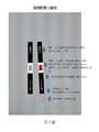
第1圖呈現位於片匣或外殼中的本發明之測試片(test strip)裝置之照片。第1圖的左側描繪在與生物樣本接觸之前外殼中的裝置,其具有樣本槽、結果窗口及控制線。第1圖的右側呈現該裝置測試已知與子癇前症相關之折疊錯誤蛋白質為陰性、弱陽性及陽性之尿液樣本的結果。Figure 1 shows a photo of the test strip device of the present invention in a cassette or housing. The left side of Figure 1 depicts the device in the housing before contact with the biological sample, which has a sample tank, a result window, and a control line. The right side of Figure 1 presents the results of the device testing urine samples that are known to be negative, weakly positive, and positive for misfolded proteins known to be associated with preeclampsia.
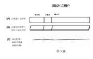
第2圖呈現本發明裝置之測試片實施例之範例。該測試片包含控制組染劑、顯示片(或毛細管床)、分離器(例如,三片材料)、第二樣本墊(或樣本接收材料)、第一樣本墊(或樣本接收材料)及於樣本墊上乾燥之染劑。Figure 2 shows an example of the test piece embodiment of the device of the present invention. The test piece includes a control group dye, a display piece (or capillary bed), a separator (for example, three pieces of material), a second sample pad (or sample receiving material), a first sample pad (or sample receiving material), and Dried dye on the sample pad.
第3圖呈現本發明之診斷裝置之實施例。第3圖A部分呈現具有樣本墊、分離器、及顯示片之俯視圖。第3圖B部分呈現從側面觀看第3圖A部分之相同實施例。該樣本墊、分離器及顯示片可彼此對接(butted)。第3圖C部分呈現彼此重疊的樣本墊、分離器及顯示片之實施例(側面圖)。Figure 3 presents an embodiment of the diagnostic device of the present invention. Part A of Figure 3 presents a top view of the sample pad, separator, and display sheet. Part B of Fig. 3 presents the same embodiment of part A of Fig. 3 when viewed from the side. The sample pad, separator and display sheet can be butted with each other. Part C of FIG. 3 shows an embodiment of the sample pad, separator and display sheet overlapping each other (side view).
第4圖顯示於測試片內之不同材料配置。第4圖A部分呈現具有兩個樣本墊、分離器及顯示片(毛細管床)的本發明之替代實施例,其各片依序具有與相鄰片的重疊。第4圖B部分呈現具有依序重疊的兩個樣本墊、三重分離器及顯示片之本發明的另一替代實施例,其各片具有與相鄰片的重疊。Figure 4 shows the different material configurations in the test piece. Part A of Figure 4 presents an alternative embodiment of the present invention with two sample pads, a separator, and a display sheet (capillary bed), each sheet having an overlap with the adjacent sheet in sequence. Part B of FIG. 4 shows another alternative embodiment of the present invention with two sample pads, triple separators, and display sheets overlapping sequentially, each sheet having an overlap with the adjacent sheet.
第5圖呈現本發明的測試片的量桿配置之範例。此實施例於測試片之頂部上配置有覆蓋帶。在測試片之頂部,覆蓋帶為不透明顏色且可用於支持測試片。於中間部,覆蓋帶為透明以提供用於結果的觀測窗。覆蓋帶可被配置成使透明觀測窗於例如分離器之上的約10mm。於觀測窗的覆蓋帶可由顏色(例如,白色)所環繞,以易於觀測結果。於分離器之下,覆蓋帶為不透明顏色以覆蓋分離器。於測試片之底部,覆蓋帶可顯示以指示利用何端浸入測試樣本中的箭頭,且可顯示以指示測試片浸入樣本之最大程度的線。於測試片的尾端可沒有覆蓋帶,以便於浸入時測試樣本的吸附(wicking)。Figure 5 shows an example of the measuring rod configuration of the test piece of the present invention. In this embodiment, a cover tape is arranged on the top of the test piece. On the top of the test piece, the cover tape is opaque and can be used to supportHold the test piece. In the middle part, the cover tape is transparent to provide an observation window for the results. The cover tape may be configured such that the transparent observation window is about 10 mm above the separator, for example. The cover band on the observation window can be surrounded by a color (for example, white) to make it easier to observe the results. Below the separator, the cover tape is an opaque color to cover the separator. At the bottom of the test piece, the cover tape can be displayed to indicate which end is used to immerse the arrow in the test sample, and it can be displayed to indicate the maximum degree of immersion of the test piece into the sample. There may be no covering tape at the end of the test piece to facilitate wicking of the test sample during immersion.
本發明為裝置以及該裝置之使用方法。更具體而言,該裝置為可用於數種臨床及非臨床環境的快速測流層析測試(lateral flow chromatographic rapid test),以檢測生物樣本中關注的蛋白質。該裝置可用於檢測哺乳動物的蛋白質折疊錯誤疾病。該哺乳動物可為懷疑患有一或多個此疾病或處於其風險中。The present invention is a device and a method of using the device. More specifically, the device is a lateral flow chromatographic rapid test that can be used in several clinical and non-clinical environments to detect proteins of interest in biological samples. The device can be used to detect protein folding errors in mammals. The mammal may be suspected of having one or more of these diseases or at risk.
本文將描述具有數種不同之實施例的裝置。基本上,其包含用於檢測生物樣本內之一或多個目的蛋白(protein of interest)之測試片(參照例如第1圖)。該檢測藉由一連串反應的手段執行。該測試片包含具有毛細作用的一段側流分析或層析材料,且具有層析溶劑運輸起始的第一端。其亦具有層析溶劑運輸終點的第二端。測試片包含位在第一端與第二端之間的複數個區域或區(參照例如第2圖及第3圖)。該區域包含由檢測試劑,例如染劑浸染之第一區域。該檢測試劑與該生物樣本內一或多個目的蛋白進行特異性結合。第一區域亦接收生物樣本。相較之下,為第一區域下游之第二區域留存未與生物樣本中一或多個目的蛋白結合之檢測試劑,並且與該生物樣本內之一或多個目的蛋白結合之檢測試劑將被轉移至第三區域。位於該第二區域之下游的測試片的第三區域接收通過第二區域後之樣本。若目的蛋白存在於樣本時,則第三區域將顯示檢測試劑。其亦包含用於檢測結合蛋白質的檢測試劑以作為測量生物樣本內一或多個蛋白質的手段。在一實施例中,該裝置確認了取自病患之生物樣本中的折疊錯誤蛋白質的存在,且可確定病患是否具有蛋白質折疊錯誤疾病。一或多個蛋白質的存在可經由視覺定義的定性或半定量,或可藉由使用可存在於裝置內之測量元件進行半定量或定量。在生物樣本施加於第一區域後,第一區域於樣本中釋出檢測試劑,且該第二區域分離與一或多個蛋白質結合之檢測試劑及未結合之檢測試劑,並僅容許結合之檢測試劑轉移至第三區域,且接著顯示用於觀察及測量的結合之檢測試劑。第一區域可為樣本接收材料。第二區域可為分離器。第三區域可為毛細管床或顯示片。This article will describe devices with several different embodiments. Basically, it contains a test strip for detecting one or more proteins of interest in a biological sample (see, for example, Figure 1). The detection is performed by means of a series of reactions. The test piece contains a section of lateral flow analysis or chromatography material with capillary action, and has a first end where the transportation of the chromatography solvent starts. It also has the second end of the chromatographic solvent transport endpoint. The test piece includes a plurality of regions or regions located between the first end and the second end (see, for example, Figures 2 and 3). This area contains the first area impregnated with a detection reagent, such as a dye. The detection reagent specifically binds to one or more target proteins in the biological sample. The first area also receives biological samples. In contrast, it is the second zone downstream of the first zoneThe domain retains detection reagents that are not bound to one or more target proteins in the biological sample, and the detection reagents that bind to one or more target proteins in the biological sample will be transferred to the third area. The third area of the test strip located downstream of the second area receives the sample after passing through the second area. If the target protein is present in the sample, the third area will display the detection reagent. It also includes detection reagents for detecting bound proteins as a means of measuring one or more proteins in a biological sample. In one embodiment, the device confirms the presence of a misfolded protein in a biological sample taken from a patient, and can determine whether the patient has a protein misfolding disease. The presence of one or more proteins can be qualitatively or semi-quantitatively defined visually, or can be semi-quantitative or quantitative by using measurement elements that can be present in the device. After the biological sample is applied to the first area, the first area releases the detection reagent in the sample, and the second area separates the detection reagent that binds to one or more proteins and the unbound detection reagent, and only allows the detection of the binding The reagent is transferred to the third area, and then the bound detection reagent for observation and measurement is displayed. The first area may be the sample receiving material. The second zone can be a separator. The third area can be a capillary bed or a display sheet.
該裝置之特定元件或構件及該元件的特性或性質將如下文詳細地描述:The specific elements or components of the device and the characteristics or properties of the elements will be described in detail as follows:
樣本接收材料、分離器及毛細管床可由相同或不同材料所製。該材料為側流裝置及層析的技術領域中通常已知[參照參考文獻:EMD Millipore Rapid Lateral Flow Test Strips Considerations for Product Development,available from EMD Millipore,Billerica,MA]。薄膜是基於其物理及化學性質所選擇,其影響毛細管流且因此影響試劑沉澱及分析表現。該材料包括例如但不限於:硝化纖維素、濾紙、層析紙、纖維素、塑膠聚合物、非對稱聚碸類膜(asymmetric polysulfone membrane)、棉、棉絨及/或玻璃纖維、聚酯類、聚乙烯及聚碸類。該薄膜可由聚合物製成,該聚合物包括例如:硝化纖維素、聚偏二氟乙烯(polyvinylidene fluoride)、尼龍(nylon)及聚醚碸(polyethersulfone)。該墊片材料通常使用作為樣本接收材料,以提供控制及均勻地接收樣本且助於其流至裝置中相鄰的條狀材料。墊片材料為多孔性的,通常由以下材料製成:纖維素(即濾紙)、玻璃纖維、編織網及合成的不織布材料或聚酯類。過濾基質特別在需要將包含於樣品中的外部材料與待分析之樣本部分分離時(例如,將細胞物質與流體分離出來時),其可用於樣本接收。過濾基質可例如為:纖維素、非對稱聚碸類膜(包括但不限於VividTM血漿分離膜(VividTM Plasma separation membrane)及非對稱性亞微米(BTS)聚碸類膜(asymmetric sub-micron(BTS)polysulfone membrane))。吸收墊可使用作為例如在裝置條之末端的芯片(wick),以將樣本拉過側流片,且可增加分析的樣本量並增強分析靈敏度。吸收墊通常為纖維素或棉花絨且可基於床體積(bed volumn)的厚度、可壓縮性及均勻性來最佳地選擇。測試片整體可安裝於背襯卡上,其通常為塑膠背襯且黏合劑的卡片。如本文所述,雖然各種材料通常在側流裝置具有特定目的,但各材料可考慮本發明的接收材料、分離器及顯示片的目的而具有適當地特性。詳見材料的進一步討論之例子。The sample receiving material, separator and capillary bed can be made of the same or different materials. This material is generally known in the technical field of lateral flow devices and chromatography [refer to reference: EMD Millipore Rapid Lateral Flow Test Strips Considerations for Product Development, available from EMD Millipore, Billerica, MA]. The membrane is selected based on its physical and chemical properties, which affect the capillary flow and therefore the reagent precipitation and analytical performance. The material includes, for example, but not limited to: nitrocellulose, filter paper, chromatography paper, cellulose, plastic polymer, asymmetric polysulfone membrane, cotton, cotton lint and/or glass fiber, polyester , Polyethylene and Polyethylene. The film can be made of polymers, including, for example, nitrocellulose, polyvinylidene fluoride, nylon, and polyethersulfone. The gasket material is usually used as a sample receiving material to provide control and evenly receive the sample and help it flow to the adjacent strip of material in the device. The gasket material is porous and is usually made of the following materials: cellulose (ie filter paper), glass fiber, woven mesh and synthetic non-woven material or polyester. The filter matrix can be used for sample reception especially when it is necessary to separate the external material contained in the sample from the sample part to be analyzed (for example, when separating the cellular material from the fluid). The filter matrix may be, for example, cellulose, asymmetric poly-tissue membrane (including but not limited to VividTM plasma separation membrane (VividTM Plasma separation membrane), and asymmetric sub-micron (BTS) poly-tissue membrane (asymmetric sub-micron membrane). (BTS)polysulfone membrane)). The absorbent pad can be used, for example, as a wick at the end of the device strip to pull the sample through the side flow sheet, and can increase the amount of sample analyzed and enhance the sensitivity of the analysis. Absorbent pads are usually cellulose or cotton wool and can be optimally selected based on the thickness, compressibility and uniformity of the bed volumn. The whole test piece can be installed on a backing card, which is usually a plastic backed and adhesive card. As described herein, although various materials generally have a specific purpose in the lateral flow device, each material may have appropriate characteristics in consideration of the purpose of the receiving material, separator, and display sheet of the present invention. See the material for further discussion examples.
用於測試片配置之其他材料,尤其是顯示片為側流技術及層析技術的技術領域中已知之材料。Other materials used for the configuration of the test piece, especially the display piece is a material known in the technical field of lateral flow technology and chromatography technology.
裝置之第一元件(以下略稱為樣本接收材料或樣本墊)作為海綿並保持用以測試的超量的樣本流體。接收材料吸收樣本,但亦容許其流動或吸附至下一個相鄰的材料。其通常對於存在於樣本中之目的蛋白以及檢測試劑(例如,染劑)為惰性,且因此不與其反應,以使蛋白質及檢測試劑經由材料而流動或吸附至側流裝置中的相鄰材料。The first element of the device (hereinafter abbreviated as sample receiving material or sample pad) acts as a sponge and holds the excess sample fluid for testing. The receiving material absorbs the sample, but also allows it to flow or adsorb to the next adjacent material. It is generally inert to the target protein and detection reagents (for example, stains) present in the sample, and therefore does not react with them, so that the protein and detection reagents flow through the material or are adsorbed to adjacent materials in the lateral flow device.
樣本接收材料可浸入自哺乳動物或病患樣本中,或替代地,該樣本可間接或直接施用於樣本接收材料。樣本可藉由,例如:具刻度之滴管、吸量管、移液管、或可重複配給病患樣本之吸量管而施用於樣本接收材料。若樣本接收材料配置以浸入生物樣本(例如,參照第5圖),則樣本接收材料可為相對較長(例如但不限於:約10mm)。若該樣本接收材料配置以接收藉由例如滴管或吸量管所施用的樣本時,則樣本接收材料可為相對較短(在約5mm至約10mm之間,但不限於此)。一般而言,該寬度可為約2mm至10mm,且更常見為2.5mm至5mm(±0.5mm)。樣本接收材料的長度及寬度的變化是可能的,且依據如片匣或外殼之大小以及生物樣本與檢測試劑充分混合之能力的因素。The sample receiving material can be immersed in a sample from a mammal or patient, or alternatively, the sample can be applied to the sample receiving material indirectly or directly. The sample can be applied to the sample receiving material by, for example, a graduated dropper, pipette, pipette, or a pipette that can be repeatedly dispensed to the patient's sample. If the sample receiving material is configured to be immersed in the biological sample (for example, refer to Figure 5), the sample receiving material may be relatively long (for example, but not limited to: about 10 mm). If the sample receiving material is configured to receive a sample applied by, for example, a dropper or a pipette, the sample receiving material may be relatively short (between about 5 mm and about 10 mm, but not limited to this). Generally speaking, the width may be about 2 mm to 10 mm, and more commonly 2.5 mm to 5 mm (±0.5 mm). Variations in the length and width of the sample receiving material are possible and depend on factors such as the size of the cassette or housing and the ability of the biological sample to fully mix with the detection reagent.
特別是,樣本接收墊作為毛細基質,其中生物樣本及檢測試劑(例如,染劑)可自由地混合。樣本墊亦可具有以乾燥型式之檢測試劑,其可適用於在分析物(例如,生物樣本內檢測之目的蛋白)與檢測試劑之間的最佳化化學反應。檢測試劑可預先載入於樣本接收材料上。在一實施例中,當加至樣本接收墊的生物樣本(例如,尿液)溶解檢測試劑時,接著樣本及檢測試劑染劑之混合物藉由流經測試墊至例如分離器的相鄰材料之方式通過該裝置。In particular, the sample receiving pad serves as a capillary matrix in which biological samples and detection reagents (for example, dyes) can be freely mixed. The sample pad may also have a detection reagent in a dry form, which is suitable for the optimized chemical reaction between the analyte (for example, the target protein to be detected in a biological sample) and the detection reagent. The detection reagent can be pre-loaded on the sample receiving material. In one embodiment, when the biological sample (for example, urine) added to the sample receiving pad dissolves the detection reagent, the sample and the detection testThe mixture of dyestuffs is passed through the device by flowing through the test pad to adjacent materials such as a separator.
另外,樣本墊包含一系列之兩個或更多的樣本墊(參照例如第4圖)。例如,第一墊可接收樣本且第二墊可含有檢測試劑,因而生物樣本由第一元件(第一墊)遷移至含有乾燥型態之檢測試劑的第二元件(第二墊),其可適用於目的的分析物與檢測試劑之間的最佳化化學反應。若使用兩個或更多的樣本接收材料,任其一可含有該檢測試劑。較佳地,第一樣本接收材料包含檢測試劑且第二樣本接收材料不含有檢測試劑。In addition, the sample pad includes a series of two or more sample pads (see, for example, Figure 4). For example, the first pad can receive the sample and the second pad can contain the detection reagent, so the biological sample migrates from the first element (first pad) to the second element (second pad) containing the detection reagent in a dry form, which can It is suitable for the optimized chemical reaction between the target analyte and the detection reagent. If two or more sample receiving materials are used, any one of them can contain the detection reagent. Preferably, the first sample receiving material contains detection reagents and the second sample receiving material does not contain detection reagents.
在另一實施例中,第一樣本墊接收生物樣本且被設計成分離出或留存可存在於該樣本中之不溶性材料。過濾的樣本其後流經第一樣本墊或至第二樣本墊且檢測材料已併入於第一樣本墊或第二樣本墊,且允許檢測試劑及樣本適當地混合。第二墊可具有與第一墊相同或不同的組成。In another embodiment, the first sample pad receives a biological sample and is designed to separate or retain insoluble materials that may be present in the sample. The filtered sample then flows through the first sample pad or to the second sample pad and the detection material has been incorporated into the first sample pad or the second sample pad, and the detection reagents and the sample are allowed to mix properly. The second pad may have the same or different composition as the first pad.
再者,在另一實施例中,該第一樣本墊接收樣本且亦含有檢測試劑,而第二樣本墊用於在進入測試片的下一相鄰材料(例如,分離器)之前提供檢測試劑與關注分析物混合的額外時間。Furthermore, in another embodiment, the first sample pad receives the sample and also contains detection reagents, and the second sample pad is used to provide detection before entering the next adjacent material (eg, separator) of the test piece The extra time for the reagent to mix with the analyte of interest.
在一額外實施例中,樣本接收材料包含用於檢測試劑之基材且乾燥留存於基材上。檢測試劑可在樣本接收材料上或其之內。一但病患樣本被加入,則釋放檢測材料。該基材不吸收病患樣本或不與其反應,當施用時,其移動經由基質移動並至相鄰材料之上,例如,分離器。In an additional embodiment, the sample receiving material includes a substrate for detecting reagents and is dried on the substrate. The detection reagent can be on or in the sample receiving material. Once the patient sample is added, the test material is released. The substrate does not absorb or react with the patient sample, and when applied, it moves through the substrate and onto the adjacent material, such as a separator.
應值得注意的是,該樣本可例如經由樣本槽或用於接收樣本之裝置其他物件施用或放置進片匣或外殼。當測試片安裝或封裝於片匣或外殼內側時,樣本槽或物件可位於例如測試片的樣本接收材料之上(參照第1圖)。It should be noted that the sample can be applied or placed in a cassette or housing, for example, via a sample tank or other object of a device for receiving the sample. When the test piece is installed or encapsulated inside the cassette or housing, the sample slot or object can be located on the sample receiving material of the test piece, for example (refer to Figure 1).
使用作為樣本接收材料或樣本墊之材料為側流裝置及層析的技術領域中已知之材料[見參考文獻:EMD Millipore Rapid Lateral Flow Test Strips Considerations for Product Development,available from EMD Millipore,Billerica,MA]且基於影響樣本接收、控制及均勻毛細管流及樣本過濾之物理性質及化學性質進行選擇。此外,若樣本接收材料亦含有檢測試劑,理論上,該材料為用於保持檢測試劑且當測試樣本加入時最佳化釋放的適當基質。該墊材料為多孔性的,通常由纖維素(例如:濾紙)、玻璃纖維、編織篩網及合成不織布材料或多孔性塑膠(例如:聚酯類)所製成。可使用作為樣本接收材料之其他材料例如為:非對稱聚碸類膜(polysulfone asymmetric membranes)、棉花/玻璃纖維材料如Ahlstrom® 8950、塑膠聚合物膜,例如聚乙烯(例如,高密度聚乙烯)、聚四氟乙烯(polytetrafluoroethylene)以及多孔性玻璃纖維膜(參照例如Porex,Fairburn,GA)。材料的進一步描述參照範例(即,範例2及範例6)。The material used as the sample receiving material or sample pad is a material known in the technical field of lateral flow devices and chromatography [see reference: EMD Millipore Rapid Lateral Flow Test Strips Considerations for Product Development, available from EMD Millipore, Billerica, MA] And based on the physical and chemical properties that affect sample reception, control and uniform capillary flow and sample filtration. In addition, if the sample receiving material also contains the detection reagent, in theory, the material is an appropriate matrix for holding the detection reagent and optimizing the release when the test sample is added. The pad material is porous and is usually made of cellulose (for example: filter paper), glass fiber, woven mesh and synthetic non-woven material or porous plastic (for example: polyester). Other materials that can be used as sample receiving materials are, for example: asymmetric polysulfone asymmetric membranes, cotton/glass fiber materials such as Ahlstrom® 8950, plastic polymer membranes, such as polyethylene (for example, high-density polyethylene) , Polytetrafluoroethylene (polytetrafluoroethylene) and porous glass fiber membranes (see, for example, Porex, Fairburn, GA). For further description of materials, refer to the examples (ie, Example 2 and Example 6).
應值得注意的是,當檢測試劑為具有對纖維素的親和性的染劑時,用於本發明之裝置的樣本接收材料可為存在足以與生物樣本中一或多個修飾目的蛋白質結合的檢測試劑之纖維素。更具體而言,該纖維素不可被容許與針對結合至例如生物樣本中的一或多個折疊錯誤蛋白質之關注蛋白質的檢測試劑進行「外部競爭(out compete)」。替代地,若纖維素存在於與檢測試劑之反應性低於一或多個修飾的目的蛋白質之基質時,則纖維素使可用於樣本接收材料(本文中提到之纖維素定義為具有(C6H10O5)n化學式之有機化合物,且特別為由數百至數千的β(1->4)鏈結D-葡萄糖單元(β linked D-glucose units)之直鏈所組成之多醣類)。It should be noted that when the detection reagent is a dye with affinity for cellulose, the sample receiving material used in the device of the present invention may be a detection material that is sufficient to bind to one or more modified target proteins in the biological sample. Reagent of cellulose. More specifically, the cellulose cannot be allowed to "out compete" with detection reagents for the protein of interest that bind to, for example, one or more misfolded proteins in a biological sample. Alternatively, if cellulose is present in a matrix whose reactivity with the detection reagent is lower than that of one or more modified proteins of interest, then the cellulose can be used as a sample receiving material (the cellulose mentioned herein is defined as having (CAn organic compound of 6 H10 O5 )n chemical formula, especially consisting of hundreds to thousands of straight chains of β(1->4)-linked D-glucose units (β linked D-glucose units) carbohydrate).
接著,該流體(例如,樣本或與檢測試劑混合的樣本)自樣本分析材料或樣本墊流經過濾器(以下略稱為「分離器」),其設計成可留存任何未結合之檢測試劑。特別是,分離器適用於分離側流裝置中的游離(free)檢測試劑(例如,染劑)與蛋白質結合之檢測試劑。具體而言,分離器材料容許樣本流流至下一個相鄰材料,但若生物樣本中不存在一或多個目的蛋白,則留存、阻礙未結合的檢測試劑的流動或與未結合的檢測試劑結合。Then, the fluid (for example, the sample or the sample mixed with the detection reagent) flows from the sample analysis material or sample pad through a filter (hereinafter referred to as a "separator"), which is designed to retain any unbound detection reagent. In particular, the separator is suitable for separating free detection reagents (for example, dyes) and detection reagents bound to proteins in the lateral flow device. Specifically, the separator material allows the sample stream to flow to the next adjacent material, but if one or more target proteins are not present in the biological sample, it will remain, hinder the flow of unbound detection reagents, or interact with unbound detection reagents. Combine.
分離器鄰接樣本墊,但較佳為與樣本墊重疊,其中樣本墊含有檢測試劑(例如,染料)或一系列之樣本墊元件(參照例如第3圖)。分離器之功能為分離以下兩者:與一或多個測試樣本蛋白或目的蛋白(例如,一或多個折疊錯誤蛋白質)結合之檢測試劑以及未與一或多個測試樣本蛋白質結合之檢測試劑,從而容許結合之檢測試劑流過分離器並留存未結合之檢測試劑於分離器。另外,分離器可為相同或不同材料的一系列的一或多個過濾器。The separator is adjacent to the sample pad, but preferably overlaps the sample pad, where the sample pad contains a detection reagent (for example, dye) or a series of sample pad elements (see, for example, Figure 3). The function of the separator is to separate the following two: detection reagents that are bound to one or more test sample proteins or target proteins (for example, one or more misfolded proteins) and detection reagents that are not bound to one or more test sample proteins , Which allows the bound detection reagent to flow through the separator and retains the unbound detection reagent in the separator. In addition, the separator can be a series of one or more filters of the same or different materials.
不受理論束縛,分離器材料可含有用與檢測試劑的基材,以使未結合檢測試劑與分離器材料結合且無法流至下一材料。已與蛋白質結合之檢測試劑不與分離器中之基材結合且流至測試片之下一材料。基材可為分離器材料(例如,纖維素)或可被化學修飾或外加至分離器材料。此外,分離器材料組成之結構特性提供未結合之檢測試劑的留存。Without being bound by theory, the separator material may contain a substrate used with the detection reagent, so that the unbound detection reagent is combined with the separator material and cannot flow to the next material. The detection reagent that has been bound to the protein is not bound to the substrate in the separator and flows to a material under the test piece. The substrate may be a separator material (e.g., cellulose) or may be chemically modified or added to the separator material. In addition, the structural characteristics of the separator material composition provide retention of unbound detection reagents.
此外,分離器亦可由重疊或連續的複數片材料所組成,以最佳化未結合檢測試劑之留存(見第4圖)。複數片可由相同或不同材料製成。可用於分離器的過濾器基質例如為纖維素、熱塑性聚合物,如非對稱聚碸淚膜(包括但不限於VividTM血漿分離膜及非對稱性亞微米(BTS)聚碸類膜(asymmetric sub-micron(BTS)polysulfone membrane))。分離器材料之進一步描述請參照範例(例如,範例2、範例4、範例5、範例8、範例13、範例14及範例16)。In addition, the separator can also be composed of overlapping or continuous multiple pieces of material to optimize the retention of unbound detection reagents (see Figure 4). Plural pieces can be made of the same or different materials. The filter matrix that can be used in the separator is, for example, cellulose, thermoplastic polymer, such as asymmetric polytear film (including but not limited to VividTM plasma separation membrane and asymmetric sub-micron (BTS) poly-tear film (asymmetric sub-micron (BTS)). -micron(BTS)polysulfone membrane)). For further description of separator materials, please refer to the examples (for example, Example 2, Example 4, Example 5, Example 8, Example 13, Example 14 and Example 16).
實際上,出乎意料且非預料中的結果為,許多硝化纖維素材料作為分離器材料確實運作情況良好且容許尿液及與染劑結合蛋白質流經該裝置。例如:Whatman® AE99硝化纖維素膜運作情形十分良好(見表1)。亦有運作狀況大致良好之纖維素材料(例如:Ahlstrom® 601、319、247、Whatman® CF1、CF3、CF4;EMI11513、5475、5493),但亦有部分纖維素材料表現不佳(例如:Ahlstrom® 270)。意外地,在允許蛋白結合CR染劑流動並留存未結合染劑方面表現良好的材料為VividTM血漿分離材料(Pall Corporation)及非對稱性亞微米(BTS)聚碸類膜(Pall Corporation)(參照表1及表2)。In fact, the unexpected and unexpected result is that many nitrocellulose materials do work well as separator materials and allow urine and dye-bound proteins to flow through the device. For example: Whatman® AE99 nitrocellulose membrane works very well (see Table 1). There are also cellulosic materials with generally good working conditions (for example: Ahlstrom® 601, 319, 247, Whatman® CF1, CF3, CF4; EMI11513, 5475, 5493), but there are also some cellulosic materials that perform poorly (for example: Ahlstrom® 270). Unexpectedly, materials that perform well in allowing protein-bound CR stains to flow and retain unboundstains are Vivid TM plasma separation material (Pall Corporation) and asymmetric submicron (BTS) polymeric membranes (Pall Corporation) ( Refer to Table 1 and Table 2).
當檢測試劑為如剛果紅的染劑時,分離器材料可為例如:濾紙、纖維素類型及/或如EMI 11513、EMI 5475、EMI 5493、1281、642、標準17(Standard 17)、C048、LF1、LF1、VF2、CF1、CF3、Ahlstrom 319的材料。分離器可為例如約5至10mm的長度,或其可為一系列的片,其中每個長度為5至10mm。When the detection reagent is a dye such as Congo red, the separator material can be, for example: filter paper, cellulose type and/or such as EMI 11513, EMI 5475, EMI 5493, 1281, 642, Standard 17 (Standard 17), C048, LF1, LF1, VF2, CF1, CF3, Ahlstrom 319 materials. The separator may be, for example, about 5 to 10 mm in length, or it may be a series of pieces, each of which is 5 to 10 mm in length.
在一實施例中,分離器留存游離的檢測試劑(例如,染劑),但容許結合蛋白質染劑流過。在一特定實施例中,分離器包含纖維素且檢測試劑為剛果紅。In one embodiment, the separator retains free detection reagents (e.g., stains), but allows bound protein stains to flow through. In a specific embodiment, the separator contains cellulose and the detection reagent is Congo Red.
檢測試劑為與樣本中一或多個目的蛋白反應的物質。例如,檢測試劑可為與存在於哺乳動物之生物樣本(例如,病患之樣本)之一或多個折疊錯誤蛋白質(例如,嗜剛果紅性蛋白質)、聚集蛋白質及/或超分子聚集蛋白質反應或具有與其之結合親和性的物質。檢測試劑可被預載入於檢測墊上(例如,施用於試劑墊上,或試劑墊浸於檢測試劑或染劑)。試劑墊可為樣本接收材料或樣本接收墊。The detection reagent is a substance that reacts with one or more target proteins in the sample. For example, the detection reagent may react with one or more misfolded proteins (for example, Congo red protein), aggregated proteins and/or supramolecular aggregated proteins present in a biological sample of a mammal (for example, a patient’s sample) Or a substance with binding affinity to it. The detection reagent may be preloaded on the detection pad (for example, applied to the reagent pad, or the reagent pad is immersed in the detection reagent or dye). The reagent pad may be a sample receiving material or a sample receiving pad.
在一態樣中,檢測試劑為染色生物樣本中若存在的目的蛋白之子集合之染劑。在一實施例中,檢測試劑可與折疊錯誤的蛋白質反應。例如,染劑可為偶氮染劑(azo dye),例如剛果紅(CR),或緩衝或非緩衝的其類似物。再者,只要其他染劑具有對生物樣本或病患樣本內目的的折疊錯誤蛋白質、聚集蛋白質及/或一或多個超分子蛋白質的親和性(及可與其結合或反應)時,亦可使用作為檢測試劑。該染劑的例子包括但不限於剛果紅類似物,例如於下列出版物所述:Sellarajah S等人,剛果紅類似物的合成及其抗病原性蛋白顆粒活性的評價,J Med Chem.2004 Oct 21;47(22):5515-34(Sellarajah S et al,Synthesis of analogues of Congo red and evaluation of their anti-prion activity,J Med Chem.2004 Oct 21;47(22):5515-34);以及Hélène Rudyk等人,篩選剛果紅及其類似物的預防瘙癢病感染細胞中PrP-res形成的能力,Journal of General Virology(2000),81,1155-1164(Hélène Rudyk et al.,Screening Congo Red and its analogues for their ability to prevent the formation of PrP-res in scrapie-infected cells,Journal of General Virology(2000),81,1155-1164)。該用於檢測一或多個折疊錯誤蛋白質之檢測試劑亦可例如為硫代黃素T(Thioflavin T)。In one aspect, the detection reagent is a dye that stains a subset of the target protein if present in the biological sample. In one embodiment, the detection reagent can react with the misfolded protein. For example, the dye may be an azo dye, such as Congo red (CR), or buffered or unbuffered analogs thereof. Furthermore, as long as other dyes have affinity for (and can bind or react with) the misfolded protein, aggregated protein, and/or one or more supramolecular proteins of interest in biological samples or patient samples, they can also be used As a detection reagent. Examples of such dyes include, but are not limited to, Congo red analogs, such as those described in the following publications: Sellarajah S et al. Synthesis of Congo red analogs and evaluation of their anti-pathogenic protein particle activity, J Med Chem. 2004 Oct 21; 47(22): 5515-34 (Sellarajah S et al,Synthesis of analogues of Congo red and evaluation of their anti-prion activity , J Med Chem. 2004 Oct 21; 47(22): 5515-34); And Hélène Rudyk et al. Screening Congo Red and its analogues to prevent the formation of PrP-res in scrapie-infected cells, Journal of General Virology (2000), 81, 1155-1164 (Hélène Rudyk et al.,Screening Congo Red and its analogues for their ability to prevent the formation of PrP-res in scrapie-infected cells , Journal of General Virology (2000), 81, 1155-1164). The detection reagent for detecting one or more misfolded proteins can also be Thioflavin T (Thioflavin T), for example.
此外,在本發明之一態樣中,檢測試劑以乾燥型態存在於裝置中,但亦可以其他型態存在。該檢測試劑之型態適於當施用且允許與目的蛋白結合時,與樣本進行混合之最佳化。檢測試劑之型態為適於長期穩定度或裝置儲存壽命之型態。在一實施例中,乾燥檢測試劑為染劑。再者,染劑可為剛果紅且可為例如:0.1μg至800μg,較佳為0.2μg至480μg,更佳為1μg至400μg,再更佳為2.5μg至120μg的量存在於裝置中。In addition, in one aspect of the present invention, the detection reagent is present in the device in a dry form, but it can also be present in other forms. The type of the detection reagent is suitable for the optimization of mixing with the sample when it is administered and allowed to bind to the target protein. The type of test reagent is suitable for long-term stability or storage life of the device. In one embodiment, the dry detection reagent is a dye. Furthermore, the dye may be Congo Red and may be, for example, 0.1 μg to 800 μg, preferably 0.2 μg to 480 μg, more preferably 1 μg to 400 μg, and even more preferably 2.5 μg to 120 μg in the device.
剛果紅(CR)(例如,緩衝及未緩衝)可預載入至樣本接收材料。例如,於套件生產時,剛果紅溶液可施用於材料並在其組裝及包裝前進行乾燥(參照第2圖及範例6)。Congo Red (CR) (for example, buffered and unbuffered) can be pre-loaded into the sample receiving material. For example, during kit production, Congo red solution can be applied to the material and dried before assembly and packaging (refer to Figure 2 and Example 6).
檢測試劑為可檢測的,即由裸眼可視或可藉由例如視覺檢驗及/或機械或電子讀取器檢查的其他方式檢測。The detection reagent is detectable, that is, visible by the naked eye or can be detected by other means such as visual inspection and/or inspection by a mechanical or electronic reader.
本發明提供例如,於裝置生產或組裝期間,併入測試裝置的檢測試劑。其為對於檢測折疊錯誤蛋白質之習知裝置,需在加入測試前先混合染劑及生物樣本的改良。在一較佳實施例中,測試片包含含有檢測試劑的樣本接收材料。當生物樣本加至測試裝置的樣本接收材料時,其與樣本接收材料中的檢測試劑混合且生物樣本-接收試劑混合物流經裝置。The present invention provides, for example, detection reagents that are incorporated into the test device during device production or assembly. It is a conventional device for detecting misfolded proteins. It is necessary to mix dye and biological samples before adding it to the test. In a preferred embodiment, the test strip includes a sample receiving material containing a detection reagent. When the biological sample is added to the sample receiving material of the test device, it is mixed with the detection reagent in the sample receiving material and the biological sample-receiving reagent mixture flows through the device.
具有或沒有蛋白質結合檢測試劑(protein-bound-detection reagent)之樣本(例如,尿液)流經分離器並至毛細管床上(以下略稱為「顯示片」)並進行累積。因此,顯示片容許樣本向上流至測試片並顯示檢測試劑之存在與否。具體而言,顯示片容許樣本向上流至測試片並顯示檢測試劑結合分析物的存在與否。檢測試劑或結合試劑其後可藉由人類或機械手段視覺化(以獲得定性結果)及/或進行測量(即,半定量或定量)。當目的蛋白存在時,顯示片最佳地提供整體樣本之均勻流動及檢測試劑的相對均相顯示(homogeneous display)。在部分實施例中,與顯示片上的檢測試劑的強度或濃度可對應至該樣本中目的蛋白的量。在另一實施例中,檢測試劑向上流至顯示片之距離可指示生物樣本內目的蛋白量。此外,檢測試劑的強度及流上顯示片上的距離兩者可指示樣本內目的蛋白的濃度。再者,在檢測折疊錯誤蛋白質聚集體或超分子聚集體的情況下,檢測試劑的強度及流上顯示片的距離兩者可指示樣本內存在之蛋白質聚集體的大小。適當的毛細管床的材料包括例如硝化纖維或層析紙。再者,非對稱聚碸類膜亦可提供適當的顯示片。其他適當的材料包括CytoSep®膜,如CytoSep® 1660及MN-260。如下所述,側流片的毛細管床對準側流片之片匣或外殼之結果觀測窗。材料的進一步細節參照範例(例如,範例2、範例3及範例9)。若蛋白質結合檢測試劑存在,則檢測試劑於結果觀測窗內可視化(見第1圖)。The sample (for example, urine) with or without protein-bound-detection reagent flows through the separator and onto the capillary bed (hereinafter abbreviated as "display piece") and is accumulated. Therefore, the indicator sheet allows the sample to flow up to the test sheet and indicates the presence or absence of the detection reagent. Specifically, the display sheet allows the sample to flow up to the test sheet and displays the presence or absence of the detection reagent bound to the analyte. The detection reagent or binding reagent can then be visualized (to obtain qualitative results) and/or measured (ie, semi-quantitative or quantitative) by human or mechanical means. When the target protein is present, the display sheet best provides a uniform flow of the whole sample and a relatively homogeneous display of the detection reagent. In some embodiments, the strength or concentration of the detection reagent on the display chip can correspond to the amount of the target protein in the sample. In another embodiment, the distance that the detection reagent flows upward to the display sheet can indicate the amount of the target protein in the biological sample. In addition, both the strength of the detection reagent and the distance on the display chip can indicate the concentration of the target protein in the sample. Furthermore, in the case of detecting misfolded protein aggregates or supramolecular aggregates, both the strength of the detection reagent and the distance of the flow-through display sheet can indicate the size of the protein aggregates present in the sample. Suitable materials for the capillary bed include, for example, nitrocellulose or chromatography paper. In addition, asymmetric poly-pure films can also provide suitable display sheets. Other suitable materials include CytoSep® membranes such as CytoSep® 1660 and MN-260. As described below, the capillary bed of the side streamer is aligned with the result observation window of the cassette or housing of the side streamer. For further details of the materials, refer to the examples (for example, Example 2, Example 3, and Example 9). If the protein binding detection reagent is present, the detection reagent is visualized in the result observation window (see Figure 1).
可選擇地,該裝置含有芯片。該芯片可位於本發明之測試片或裝置中的第三區域或顯示片之後。當其使用時,施用至該裝置之生物樣本(例如,流體)持續自顯示片遷移至最終多孔性吸收材料,「芯片」,其作為樣本累積器且亦有將樣本沿測試片拉動之功能。吸收墊可使用作為例如在該裝置條末端之芯片,以拉動樣本拉經過側流片,且可增加分析的樣本量以及增強分析靈敏度。吸收墊通常為纖維素或棉花絨且可基於床體積的厚度、可壓縮性及均勻性來最佳地選擇。Optionally, the device contains a chip. The chip may be located behind the third area or display sheet in the test sheet or device of the present invention. When it is used, the biological sample (for example, fluid) applied to the device continues to migrate from the display sheet to the final porous absorbent material, the "chip", which acts as a sample accumulator and also has the function of pulling the sample along the test sheet. The absorbent pad can be used, for example, as a chip at the end of the device strip to pull the sample through the side flow sheet, and can increase the sample volume for analysis.And enhance the analysis sensitivity. Absorbent pads are usually cellulose or cotton wool and can be optimally selected based on the thickness, compressibility and uniformity of the bed volume.
該裝置可具有背襯卡。測試片整體可安裝於背襯卡(例如:可自Lohmann,Orange,VA購得)上,其通常為塑膠背襯卡及黏合劑。The device can have a backing card. The whole test piece can be installed on a backing card (for example, available from Lohmann, Orange, VA), which is usually a plastic backing card and adhesive.
在本發明中之一實施例,該診斷裝置被容納、裝入(encase)或封裝於外殼或片匣。該裝置可進一步包含外殼或片匣,其包括但不限於配置成包裝該裝置為目的之匣狀物、塑膠裝置或擠製塑膠片。數種通常的片匣外殼為商業上可購得(例如:從Kanani Biologicals、Gujarat、India或EASE-Medtrend Biotech LTD,Shanghai,中國)或可依目的以現有材料客製(參照例如,美國設計專利申請序號No.29/533,647.)。該裝置可配置成具有樣本槽之外殼或片匣,以用於接收樣本,其中當樣本槽安裝/容納於片匣內時,樣本槽位於測試片之樣本接收材料之上。此外,該裝置可被配置以安裝於外殼或片匣中時,測試片的顯示片或毛細管床位於外殼之結果觀測窗之下。In an embodiment of the present invention, the diagnostic device is housed, encased or packaged in a housing or cassette. The device may further include a housing or a cassette, which includes, but is not limited to, a cassette, a plastic device, or an extruded plastic sheet configured to package the device. Several common cassette housings are commercially available (for example: from Kanani Biologicals, Gujarat, India or EASE-Medtrend Biotech LTD, Shanghai, China) or can be customized with existing materials according to the purpose (refer to, for example, the US design patent Application serial number No.29/533,647.). The device can be configured as a housing or a cassette with a sample slot for receiving samples, wherein when the sample slot is installed/contained in the cassette, the sample slot is located on the sample receiving material of the test piece. In addition, the device can be configured so that when the device is installed in a housing or a cassette, the display piece or capillary bed of the test piece is located under the result observation window of the housing.
在一實施例中,本發明之裝置亦包含可定量結果,例如毛細管床上(即,在結果視窗中)之檢測試劑的強度(例如,染劑)之電子讀取器,及亦可進一步包含顯示結果的顯示螢幕(例如,LED螢幕)。該讀取器可為外殼的一部份,或與外殼一體成形之元件,例如,在該結果顯示窗中。此讀取器例如在Venkatraman,Biosensors and Bioelectronics Volume 74,15 December 2015,Pages 150-155、PCT申請號WO2013083686 A1、PCT申請號WO2004010143 A2以及PCT申請號WO2006010072 A2中所描述。In one embodiment, the device of the present invention also includes an electronic reader that can quantify the results, such as the strength of the detection reagent (for example, dye) on the capillary bed (ie, in the result window), and may further include a display The display screen of the result (for example, an LED screen). The reader may be a part of the housing, or a component integrally formed with the housing, for example, in the result display window. This reader exampleAs described in Venkatraman, Biosensors and Bioelectronics Volume 74, 15 December 2015, Pages 150-155, PCT application number WO2013083686 A1, PCT application number WO2004010143 A2, and PCT application number WO2006010072 A2.
具有或沒有外殼之裝置可更進一步包含蓋體,如保護性膠帶(例如,可自Lohmann,Orange,VA購得)或可保護該裝置不受損壞且可提供正常讀取測試結果之其他材料。例如,該裝置可配置成作為測桿(dipstick)使用(例如,尿液測桿)。在本實施例中,側流片可以保護性膠帶覆蓋(參照第5圖及範例11)。The device with or without a housing may further include a cover, such as protective tape (for example, available from Lohmann, Orange, VA) or other materials that can protect the device from damage and provide normal read test results. For example, the device may be configured to be used as a dipstick (e.g., a urine dipstick). In this embodiment, the side flow sheet can be covered with protective tape (refer to Figure 5 and Example 11).
在一較佳實施例,在該裝置使用前,控制組試劑可存在於毛細管床上且可視於觀測窗(參照第1圖及第2圖、範例12)。當該生物樣本(例如,流體)流經毛細管床,控制組試劑溶解,控制組試劑線散布及/或帶走控制試劑至顯示片,以使結果觀測窗內不會看到控制組試劑,或其為模糊、或為可看到或測量的其他部份變化。在觀測窗內之控制組試劑之變化指示測試樣本(例如,流體)已被加入且已正常的通過該裝置,例如作為執行控制組(參照範例12)。控制組試劑為例如藉由裸眼或其他檢測或測量方式(例如,藉由機械檢驗及/或電子讀取器)而可視覺上檢測。在一實施例中,控制組試劑為酒石黃(tartazine)。作為執行控制組試劑使用之其他染劑包括食用藍色1號-亮藍色FCF,E133(藍色色澤)(FD&C Blue No.1-Brilliant Blue FCF,E133(blue shade))、食用藍色2號-靛藍,E(靛藍色色澤)(FD&C Blue No.2-Indigotine,E(indigo shade))、食用綠色3號-固綠FCF,E143-(綠松石色澤)(FD&C Green No.3-Fast Green FCF,E143(turquoise shade))、食用紅色7號-赤蘚紅,E127(粉紅色澤)(FD&C Red No.3-Erythrosine,E127(pink shade))、食用紅色40號-誘惑紅AC,E129(紅色色澤)(FD&C Red No.40-Allura Red AC,E129(red shade))、食用黃色4號-檸檬黃,E102(黃色色澤)(FD&C Yellow No.5-Tartrazine,E102(yellow shade))及食用黃色5號-日落黃FCF,E110(橙色色澤)(FD&C Yellow No.6-Sunset Yellow FCF,E110(orange shade))。In a preferred embodiment, before the device is used, the control group reagents can be present on the capillary bed and can be seen in the observation window (refer to Figures 1 and 2, Example 12). When the biological sample (for example, fluid) flows through the capillary bed, the control group reagent is dissolved, the control group reagent line is spread and/or the control reagent is taken to the display sheet, so that the control group reagent will not be seen in the result observation window, or It is fuzzy, or other changes that can be seen or measured. The change in the reagents of the control group in the observation window indicates that the test sample (for example, fluid) has been added and passed through the device normally, for example as an execution control group (refer to Example 12). The control reagents are visually detectable, for example, by naked eyes or other detection or measurement methods (for example, by mechanical inspection and/or electronic reader). In one embodiment, the control reagent is tartazine. Other dyes used as reagents in the executive control group include edible blue No. 1-Brilliant Blue FCF, E133 (FD&C Blue No.1-Brilliant Blue FCF, E133)(blue shade)), edible blue No. 2-indigo, E (indigo shade) (FD&C Blue No.2-Indigotine, E (indigo shade)), edible green No. 3-solid green FCF, E143- (turquoise) Color) (FD&C Green No.3-Fast Green FCF, E143 (turquoise shade)), edible red No. 7-erythrosine, E127 (pink shade) (FD&C Red No.3-Erythrosine, E127 (pink shade)), Edible Red No. 40-Allura Red AC, E129 (red shade) (FD&C Red No.40-Allura Red AC, E129 (red shade)), Edible Yellow No. 4-Lemon Yellow, E102 (yellow shade) (FD&C Yellow No. 5-Tartrazine, E102 (yellow shade)) and edible yellow No. 5-sunset yellow FCF, E110 (orange shade) (FD&C Yellow No. 6-Sunset Yellow FCF, E110 (orange shade)).
如前述,本發明為關於一種裝置及利用該裝置檢測關注蛋白質,更具體而言為折疊錯誤蛋白質、聚集的蛋白質及/或超分子蛋白質聚集體之方法。α螺旋(alpha helix)已知為天然構形之功能蛋白質的突出結構性模體(prominent structural motif)。反之,蛋白質的構形改變可能導致β褶板結構模體(即,β褶板結構)或折疊錯誤蛋白質其後傾向於導致蛋白質聚集與毒性。折疊錯誤蛋白質可因此成為蛋白質聚集體或超分子聚集體的形式且可與折疊錯誤蛋白質疾病相關,如子癇前症、阿茲海默症、傳染性蛋白顆粒疾病(prion disease)及帕金森氏症。As mentioned above, the present invention relates to a device and a method for using the device to detect proteins of interest, more specifically, misfolded proteins, aggregated proteins, and/or supramolecular protein aggregates. Alpha helix is known as a prominent structural motif of functional proteins in natural configuration. Conversely, the conformational change of the protein may lead to a β-pleated structure motif (ie, a β-pleated structure) or a misfolded protein, which tends to cause protein aggregation and toxicity thereafter. Misfolding proteins can therefore become protein aggregates or supramolecular aggregates and can be associated with misfolding protein diseases, such as preeclampsia, Alzheimer's disease, infectious protein particle disease (prion disease) and Parkinson's disease .
具體上,與子癇前症相關且藉由本發明的裝置及方法所檢測之折疊錯誤蛋白質、蛋白質聚集體及/或超分子聚集體可包括但不限於例如:α1-抗胰蛋白酶(alpha-1 antitrypsin)(SerpinA1)、血漿銅藍蛋白(ceruloplasmin)、重鏈IgG、輕鏈IgG、可誘導干擾素蛋白質6-16(interferon-inducible protein)(IF-16-6,G1P3)、白蛋白(albumin),以及各蛋白質之斷片、其混合物,以及該混合物之斷片。該蛋白質具有與本發明裝置使用之檢測試劑的結合親和性(如下文所述)。例如,折疊錯誤蛋白質為嗜剛果紅性,具有對稱為剛果紅等的染劑的親和性。Specifically, the misfolded proteins, protein aggregates, and/or supramolecular aggregates that are related to preeclampsia and detected by the device and method of the present invention may include, but are not limited to, for example, alpha-1 antitrypsin (alpha-1 antitrypsin). ) (SerpinA1), ceruloplasmin(ceruloplasmin), heavy chain IgG, light chain IgG, inducible interferon protein 6-16 (interferon-inducible protein) (IF-16-6, G1P3), albumin, and fragments of each protein, and mixtures thereof , And fragments of the mixture. The protein has binding affinity to the detection reagent used in the device of the present invention (as described below). For example, the misfolding protein is Congo red-philic and has affinity for dyes such as Congo red.
本發明之裝置亦可用於檢測子癇前症以外的蛋白折疊錯誤疾病。例如,該裝置可用於檢測折疊錯誤蛋白質疾病及症狀之折疊錯誤蛋白質,該折疊錯誤蛋白質疾病及症狀例如為阿茲海默症、大腦β類澱粉血管病變(Cerebral beta-amyloid angiopathy)、青光眼之視網膜神經節細胞退化(Retinal ganglion cell degeneration in glaucoma)、傳染性蛋白顆粒疾病、帕金森氏症及其他突觸核蛋白病(synucleinopathies)、tau蛋白病變(Tauopathies)、額顳葉退化症(Frontotemporal lobar degeneration,FTLD)、FTLD-FUS、肌肉萎縮性脊髓側索硬化症(Amyotrophic lateral sclerosis,ALS)、亨丁頓舞蹈症(Huntington’s disease)及其他三重、重複疾病、失智症(Dementia)(家族性英國及丹麥失智症(familial British and Danish))、遺傳性腦出血合併類澱粉病變(Hereditary cerebral hemorrhage with amyloidosis)、體顯性腦動脈血管病變合併皮質下腦梗塞及腦白質病變(CADASIL)、亞歷山大氏症(Alexander disease)、各種類澱粉病變(amyloidosis)、Serinopathies、第二型糖尿病(Type II diabetes)、包涵體肌炎/肌肉病變(Inclusion body myositis/myopathy)、白內障(cararacts)、色素性視網膜炎合併視紫紅質突變(Retinitis pigmentosa with rhodopsin mutations)、甲狀腺髓質癌(Medullary thyroid carcinoma)、垂體催乳素腺瘤(Pituitary prolactinoma)、遺傳性格子狀角膜營養不良(Hereditary lattice corneal dystrophy)、麥洛利氏小體(Mallory bodies)、肺泡蛋白質沉著症(Pulmonary alveolar proteinosis)、牙源性類澱粉瘤(Odontogenic tumor amyloid)、囊性纖維變性(Cystic fibrosis)、鐮刀型貧血症(Sickle cell disease)以及重症肌肉病變(Critical illness myopathy)。The device of the present invention can also be used to detect protein folding errors other than preeclampsia. For example, the device can be used to detect misfolded protein diseases and symptoms of misfolded protein, such as Alzheimer's disease, Cerebral beta-amyloid angiopathy, and glaucoma retina Retinal ganglion cell degeneration in glaucoma, infectious protein particle disease, Parkinson's disease and other synucleinopathies, tauopathies, frontotemporal lobar degeneration , FTLD), FTLD-FUS, Amyotrophic lateral sclerosis (Amyotrophic lateral sclerosis, ALS), Huntington's disease (Huntington's disease) and other triple, repetitive diseases, dementia (Dementia) (family British And Danish dementia (familial British and Danish), hereditary cerebral hemorrhage with amyloidosis (Hereditary cerebral hemorrhage with amyloidosis), somatic cerebral arterial vascular disease with subcortical cerebral infarction and white matter disease (CADASIL), Alexander Alexander disease, various amyloidosis, Serinopathies, Type II diabetes, Inclusion body myositis/myopathy, cararacts, pigmented retina Inflammation with rhodopsin mutations (Retinitis pigmentosa with rhodopsin mutations), Medullary thyroid carcinoma (Medullary thyroid carcinoma), Pituitary prolactinoma (Pituitary prolactinoma), hereditary lattice keratopathy (Hereditarylattice corneal dystrophy, Mallory bodies, Pulmonary alveolar proteinosis, Odontogenic tumor amyloid, Cystic fibrosis, sickle anemia Sickle cell disease and Critical illness myopathy.
待檢測之一或多個目的蛋白可自哺乳動物之生物樣本發現。生物樣本可例如為自乾淨或自然蒐集所取得之尿液、腦脊髓液、羊水或可能包含一或多個目的蛋白之任何體液樣本(例如,血液、唾液、羊水、腦脊髓液、血漿或血清)。該樣本亦可為自病人之分泌物之萃取物,例如:從鼻腔分泌物、排泄物、或耳垢、或以適當的溶液中萃取之組織試樣並施用至該裝置。目的蛋白可從例如懷孕或產後哺乳動物之生物樣本中發現。One or more target proteins to be detected can be found from biological samples of mammals. Biological samples can be, for example, urine, cerebrospinal fluid, amniotic fluid obtained from clean or natural collection, or any body fluid sample that may contain one or more proteins of interest (e.g., blood, saliva, amniotic fluid, cerebrospinal fluid, plasma or serum) ). The sample can also be an extract from the patient's secretions, for example: a tissue sample extracted from nasal secretions, excrement, or earwax, or an appropriate solution, and applied to the device. The protein of interest can be found in, for example, biological samples of pregnant or postpartum mammals.
該病患可為哺乳動物。此外,哺乳動物可為懷孕個體,例如:懷孕婦女、懷孕靈長類動物或例如利用實驗研究而設置成具有子癇前症的身體症狀及病徵的基因工程改造動物模式(例如,高血壓及尿蛋白)。較佳地,為了診斷子癇前症,該病人可為任何懷孕婦女。該懷孕婦女可為被懷疑患有子癇前症或處於罹患子癇前症之風險。例如,該懷疑可基於以下理由:(1)出現如美國婦產科醫生學會(ACOG)所訂立之子癇前症之病徵與症狀(懷孕中高血壓,美國婦產科醫生學會專案組於懷孕中高血壓的報告,婦產科122,122卷,第5號,2013年11月),具體而言,例如表E-1(ACOG 2013準則)及/或(2)具有一或多個子癇前症之風險因子(例如,過去懷孕時患有子癇前症之婦女、多胞胎身孕之婦女、患有心血管或腎臟異常之婦女或患有自體免疫疾病(如紅斑性狼瘡)的婦女)。The patient may be a mammal. In addition, the mammal may be a pregnant individual, such as a pregnant woman, a pregnant primate, or a genetically engineered animal model set to have physical symptoms and symptoms of preeclampsia (e.g., hypertension and urine protein using experimental research). ). Preferably, in order to diagnose preeclampsia, the patient can be any pregnant woman. The pregnant woman may be suspected of suffering from pre-eclampsia or at risk of suffering from pre-eclampsia. For example, the suspicion can be based on the following reasons: (1) The signs and symptoms of preeclampsia as defined by the American College of Obstetricians and Gynecologists (ACOG) (hypertension in pregnancy, the task force of the American College of Obstetricians and Gynecologists in pregnancy Report, Obstetrics and Gynecology 122, Volume 122, No. 5, November 2013), specifically, such as Table E-1 (ACOG 2013 Guidelines) and/Or (2) Have one or more risk factors for preeclampsia (e.g., women with preeclampsia during past pregnancy, women with multiple births, women with cardiovascular or renal abnormalities, or autoimmune Diseases (such as women with lupus erythematosus).
本發明亦包含用於檢測樣本的目的蛋白之套件。在一實施例中,該套件包含檢測與子癇前症相關的折疊錯誤蛋白質之裝置,該蛋白質存在於懷孕哺乳動物之樣本。該套件包含上述任何替代實施例中的本發明之裝置。該套件亦可包含將病患樣本施用至樣本接收材料之手段,例如:吸量管(例如,自Genesee Scientific,San Diego,CA可購得之細尖移液管)或滴管、控制組及該裝置的使用說明書。因此,該裝置不僅可以單獨實體(stand-alone entity)使用,其亦可以套件形式使用,其在部分臨床或非臨床環境上可具有更多優點。該套件可被包裝在箔袋或聚酯膠膜袋。此外,套件袋可為單獨包裝,或各包裝為多f個,例如2、5、10、15、25、50或100組套件。The present invention also includes a kit for detecting the target protein of the sample. In one embodiment, the kit includes a device for detecting a misfolded protein associated with preeclampsia, the protein being present in a sample of a pregnant mammal. The kit contains the device of the invention in any of the alternative embodiments described above. The kit may also include a means for applying patient samples to the sample receiving material, such as: pipette (for example, a fine-tip pipette available from Genesee Scientific, San Diego, CA) or dropper, control group, and Instructions for use of the device. Therefore, the device can be used not only as a stand-alone entity, but also as a kit, which can have more advantages in some clinical or non-clinical environments. The kit can be packaged in a foil bag or a polyester film bag. In addition, the kit bags may be individually packaged, or there may be more than f in each package, for example, 2, 5, 10, 15, 25, 50, or 100 sets of kits.
本發明之裝置可用於例如臨床實驗室(於醫院內的環境或醫院外的環境)、立即性照護環境、醫師室實驗室(physician office laboratories)、急診部門(例如,醫院內)或近病人端檢驗(near-patient testing)或由醫務人員、非醫務人員甚至是病患為人類時由病人本身的之定點照護(point-of-care)裝置。此外,該裝置可與其他診斷分析組合使用(例如,檢測與子癇前症相關之蛋白質(即生物標記)如sFlt-1、PlGF、PP-A、PP13、正五聚蛋白(pentraxin)、抑制素-A(inhibin-A)及可溶之內皮因子(endoglin)之免疫分析)及/或通常使用於診斷子癇前症之方法或觀測(例如:血壓讀數、用於診斷子癇前症之臨床測試包括血小板計數、血清肌酸酐濃度、血清丙胺酸轉胺酶(alaninine aminotransferase,ALT)及天冬胺酸轉胺酶(aspartate aminotransferase,AST)),及其他病徵或症狀,如體重增加、暈眩、頭痛、視線模糊等。The device of the present invention can be used, for example, in a clinical laboratory (in a hospital environment or an environment outside the hospital), immediate care environment, physician office laboratories, emergency department (for example, in a hospital), or near patient Near-patient testing or point-of-care devices used by medical staff, non-medical staff, or even when the patient is a human. In addition, the device can be used in combination with other diagnostic assays (for example, detection of pre-eclampsia related(Immune analysis of proteins (i.e. biomarkers) such as sFlt-1, PlGF, PP-A, PP13, pentraxin, inhibin-A and soluble endothelial factor (endoglin) And/or methods or observations commonly used to diagnose pre-eclampsia (eg blood pressure readings, clinical tests used to diagnose pre-eclampsia include platelet count, serum creatinine concentration, serum alanine aminotransferase (ALT) And aspartate aminotransferase (AST), and other signs or symptoms, such as weight gain, dizziness, headache, blurred vision, etc.
舉例而言,本發明包括診斷子癇前症之方法或在患有妊娠高血壓疾病或被懷疑可能罹有子癇前症的病人進行鑑別診斷之方法,該方法包含下列步驟:a)測定懷孕病患的血壓,其中血壓高於140/90mm/Hg之懷孕病患可指示該懷孕病患之子癇前症及b)施用來自病患的生物樣本至本發明之裝置,因而病患的生物樣本中至少一目的蛋白的檢測提供或支持病患的子癇前症診斷或作為鑑別診斷。For example, the present invention includes a method for diagnosing preeclampsia or a method for differential diagnosis in a patient suffering from pregnancy-induced hypertension or suspected of having preeclampsia. The method includes the following steps: a) Determination of pregnant patients The blood pressure of a pregnant patient with a blood pressure higher than 140/90mm/Hg can indicate preeclampsia of the pregnant patient and b) apply a biological sample from the patient to the device of the present invention, so that the patient’s biological sample is at least The detection of a target protein provides or supports the diagnosis of preeclampsia in patients or serves as a differential diagnosis.
再者,本發明亦包含治療懷疑患有子癇前症之懷孕哺乳動物的方法,其包含下列步驟:a)施用由哺乳動物之生物樣本至本發明之裝置,以測定至少一蛋白質的存在,該至少一蛋白質指示懷孕哺乳動物患有子癇前症;以及b)使懷孕哺乳動物進行分娩以治療子癇前症。此外,該裝置亦可用於分娩後,以測定該病患之生物樣本內是否表現一或多個折疊錯誤蛋白質。若一或多個折疊錯誤蛋白質存在,則病患可需更進一步的管理或治療性介入(例如,硫酸鎂(magnesium sulfate)及其他抗高血壓劑的施予)以治療子癇前症,或病患可利用本發明之裝置監控,以指示何時生物樣本內不再檢測到該蛋白質。另外,本發明之裝置可於治療上介入後使用,以測定治療是否成功或測量存在於生物樣本內的目的蛋白的量或存在的變化(例如,減少、移除或增加)。因此,本發明之裝置可用於病患的治療前及治療後,以測定是否需要進一步治療性介入、測定治療性介入是否有效及/或是否需施予治療性介入的替代形式或增加治療劑之劑量,以解決子癇前症或其他蛋白質折疊錯誤疾病。Furthermore, the present invention also includes a method for treating a pregnant mammal suspected of having preeclampsia, which comprises the following steps: a) applying a biological sample from the mammal to the device of the present invention to determine the presence of at least one protein, the At least one protein indicates that the pregnant mammal has preeclampsia; and b) allowing the pregnant mammal to give birth to treat the preeclampsia. In addition, the device can also be used after delivery to determine whether one or more misfolded proteins are present in the patient's biological sample. If one or more misfolded proteins are present, the patient may need further management or therapeutic intervention (for example, administration of magnesium sulfate and other antihypertensive agents) to treat preeclampsia, or disease The patient can be monitored by the device of the present invention to indicate when the protein is no longer detected in the biological sample. In addition, the device of the present invention can be used after therapeutic intervention to determine whether the treatment is successful or to measure the amount or change of the target protein present in the biological sample (e.g.For example, reduce, remove or increase). Therefore, the device of the present invention can be used before and after treatment of patients to determine whether further therapeutic intervention is required, whether the therapeutic intervention is effective, and/or whether alternative forms of therapeutic intervention or the addition of therapeutic agents are required. Dosage to solve preeclampsia or other protein folding errors.
本發明之裝置至少因以下理由優於習知裝置:1)該裝置提供較少步驟的簡易測試流程;2)該裝置提供用於最佳化生產的標準測試材料的使用;3)該裝置提供測試套件內之檢測試劑的改善穩定性,以提供更長的保存期限;4)結果更簡單且更容易判讀;5)保持相對低的成本;以及6)提供適用於定點照護使用或用於臨床實驗室環境的快速結果。The device of the present invention is superior to the conventional device for at least the following reasons: 1) the device provides a simple test process with fewer steps; 2) the device provides the use of standard test materials for optimized production; 3) the device provides Improve the stability of the test reagents in the test kit to provide a longer shelf life; 4) The results are simpler and easier to interpret; 5) The cost is kept relatively low; and 6) It is suitable for point-of-care use or clinical use Quick results in a laboratory environment.
具體上,提供的裝置及方法遠優於習知用於檢測可能的子癇前症的試紙套件(參照例如,美國專利申請公開號No.20150291115),亦即提供的裝置對於使用者需要更少的步驟,結果更易於判讀且其比試紙套件更為穩定並擁有至少6個月的長期保存期限,較佳為1年,更佳為2年,再更佳為3年,再更佳為4年,及再更佳為5年。本發明之裝置亦提供快速結果(即,由施用生物樣本(如尿液)至該裝置的3分鐘以內,較佳為2分鐘以內,更佳為1分鐘以內或更短時間內),以適合訓練最少的定點照護用戶使用。Specifically, the provided device and method are far superior to conventional test paper kits for detecting possible preeclampsia (see, for example, US Patent Application Publication No. 20150291115), that is, the provided device requires less for the user Steps, the result is easier to interpret and it is more stable than the test strip kit and has a long-term storage period of at least 6 months, preferably 1 year, more preferably 2 years, more preferably 3 years, and still more preferably 4 years , And preferably 5 years. The device of the present invention also provides quick results (that is, within 3 minutes from the application of a biological sample (such as urine) to the device, preferably within 2 minutes, more preferably within 1 minute or less), which is suitable for Used by the least trained fixed-point care users.
本發明可藉由下列的非限定範例進行說明:The present invention can be illustrated by the following non-limiting examples:
第1圖呈現位於片匣或外殼中的本發明之裝置測試片之照片。第1圖的左側描繪在與生物樣本接觸之前外殼中的裝置。第1圖的右側呈現該裝置測已知與子癇前症相關之折疊錯誤蛋白質為陰性、弱陽性及陽性之尿液樣本的結果。Figure 1 presents a photo of the test piece of the device of the present invention in a cassette or housing. The left side of Figure 1 depicts the device in the housing before contact with the biological sample. The right side of Figure 1 shows the results of urine samples tested by the device as negative, weakly positive, and positive for misfolded proteins known to be associated with preeclampsia.
第2圖呈現本發明之裝置之測試片實施例之範例。測試片包含控制組染劑、顯示片、分離器(例如,三片材料)、第二樣本墊、第一樣本墊及於樣本墊上乾燥之染劑。Figure 2 shows an example of the test piece embodiment of the device of the present invention. The test piece includes a control group dye, a display piece, a separator (for example, three pieces of material), a second sample pad, a first sample pad, and a dye dried on the sample pad.
第3圖呈現本發明之診斷裝置之實施例。第3圖A部分呈現俯視圖。第3圖B部分呈現從側面觀看第3圖A部分之相同實施例。該樣本墊、分離器及顯示片可為彼此對接。第3圖C部分呈現彼此重疊的樣本墊、分離器及顯示片之實施例(側面圖)。Figure 3 presents an embodiment of the diagnostic device of the present invention. Part A of Figure 3 presents a top view. Part B of Fig. 3 presents the same embodiment of part A of Fig. 3 when viewed from the side. The sample pad, separator, and display sheet can be docked with each other. Part C of FIG. 3 shows an embodiment of the sample pad, separator and display sheet overlapping each other (side view).
第4圖顯示於測試片內之不同材料配置。第4圖A部分呈現依序具有兩個樣本墊、分離器及顯示片(毛細管床)的本發明之替代實施例。各片具有與相鄰片的重疊。第4圖B部分呈現具有依序重疊的兩個樣本墊、三重分離器及顯示片之本發明的另一替代實施例,其各片具有與相鄰片的重疊。Figure 4 shows the different material configurations in the test piece. Part A of Figure 4 presents an alternative embodiment of the present invention with two sample pads, a separator, and a display sheet (capillary bed) in sequence. Each piece has an overlap with adjacent pieces. Part B of FIG. 4 shows another alternative embodiment of the present invention with two sample pads, triple separators, and display sheets overlapping sequentially, each sheet having an overlap with the adjacent sheet.
第5圖呈現本發明的測試片的量桿配置之範例。此實施例於測試片之頂部上配置有覆蓋帶。在測試片之頂部,覆蓋帶為不透明顏色且可用於支持測試片。於該測試片之中間部,覆蓋帶為透明以提供用於結果的觀測窗。覆蓋帶可被配置使透明觀測窗之位置於例如分離器之上的約10mm。該觀測窗之覆蓋帶可由顏色(例如,白色)所環繞,以易於觀測結果。於分離器之下,覆蓋帶為不透明顏色以覆蓋分離器。於該測試片之底部,覆蓋帶可顯示以指示利用何端浸入測試樣本中的箭頭,且可顯示以指示測試片浸入樣本時之最大程度的線。於測試片的尾端可沒有覆蓋帶,以便於浸入時測試樣本的吸附。Figure 5 shows an example of the measuring rod configuration of the test piece of the present invention. In this embodiment, a cover tape is arranged on the top of the test piece. On the top of the test piece, the cover tape has an opaque color and can be used to support the test piece. In the middle of the test piece, the cover tape is transparent to provide an observation window for the results. The cover tape can be configured such that the position of the transparent observation window is, for example, about 10 mm above the separator. The cover band of the observation window can be surrounded by colors (for example, white) to facilitate observation of the results. Below the separator, the cover tape is an opaque color to cover the separator. At the bottom of the test piece, the cover tape can showShows an arrow indicating the end to be immersed in the test sample, and can be displayed to indicate the maximum degree of immersion of the test piece into the sample. There may be no covering tape at the end of the test piece to facilitate the adsorption of the test sample during immersion.
各種類似紙材料用於測試自懷孕婦女的尿液樣本辨別罹患或未罹患子癇前症的能力。剛果紅(CR)染劑(Sigma.St.Louis,MO)加入至尿液樣本且將一滴加入至材料,以評估適合用於樣本接收、分離器及顯示片的特性。結果點漬在約3分鐘後可視化,並於子癇前症患者及正常控制組孕婦之尿液之間的視覺差異(表1)。結果「優良(excellent)」代表材料適合提供子癇前症患者的CR尿液與正常孕婦病患之CR尿液之間的可視覺觀察差異,因而材料可使用於診斷測試裝置。結果「不佳(poor)」代表材料不適合提供CR-子癇前症陽性與CR-控制組尿液之間的可視覺觀察差異。在結果評分表上,結果「不佳(poor)」為最不適合之材料,「微妙(subtle)」為稍佳但不理想,「可(okey)」為較佳,「良好(good)」為更佳,「佳(better)」為更提升且「優良」為最適當之材料。非對稱聚碸類膜被認為提供最佳結果。部分棉花材料如CF3及CF4效果良好,但其他棉花材料,如Ahlstrom 270則效果不佳。硝化纖維及玻璃纖維材料普遍在此用途下效果不佳。Various paper-like materials are used to test the ability of urine samples from pregnant women to distinguish pre-eclampsia from or without pre-eclampsia. Congo red (CR) stain (Sigma. St. Louis, MO) was added to the urine sample and one drop was added to the material to evaluate the characteristics of the sample receiving, separator, and display sheet. The results were visualized after about 3 minutes, and the visual difference between the urine of preeclampsia patients and pregnant women in the normal control group (Table 1). Results "excellent" means that the material is suitable for providing visually observable differences between CR urine of preeclampsia patients and CR urine of normal pregnant patients, so the material can be used in diagnostic testing devices. The result "poor" means that the material is not suitable for providing a visually observable difference between CR-preeclampsia positive and CR-control group urine. On the result score sheet, the result "poor" is the most unsuitable material, "subtle" is slightly better but not ideal, "okey" is better, and "good" is Better, "better" is more improved and "good" is the most appropriate material. The asymmetric polymer membrane is believed to provide the best results. Some cotton materials such as CF3 and CF4 work well, but other cotton materials such as Ahlstrom 270 do not work well. Nitrocellulose and glass fiber materials generally do not work well for this purpose.


由於BTS及Vivid在提供子癇前症患者之CR尿液與控制組孕婦病患之CR尿液之間的可視覺觀察差異呈現優良結果,因而測試片由Ahlstrom® 8950(棉花/玻璃纖維組成物,8950)樣本墊及BTS或VividTM執行片或毛吸管床所組成。剛果紅加入至尿液樣本,並將樣本渦流震盪,其後將樣本施用於測試片之8950端。於BTS或VividTM膜執行片上觀察結果。罹患子癇前症之婦女尿液施用至含有測試片的VividTM膜而產生非常清晰訊號(剛果紅染色的粉紅/紅)之結果,且陰性尿液則無訊號。該訊號為依據體積,隨著體積減少則訊號亦減少。在陽性及陰性尿液在較大樣本體積下皆產生陽性染色,而使BTS片顯示其之間無差異。隨著減少施用的樣本體積,而同樣地減少對於陽性或陰性尿液的訊號。Since BTS and Vivid provided excellent results for the visually observable difference between CR urine of preeclampsia patients and CR urine of pregnant patients in the control group, the test piece was made of Ahlstrom® 8950 (cotton/glass fiber composition, 8950) It is composed of sample pad and BTS or VividTM executive sheet or straw bed. Congo red was added to the urine sample, and the sample was vortexed, and then the sample was applied to the 8950 end of the test piece. Perform on-chip observation results on BTS or VividTM film. The urine of women suffering from preeclampsia is applied to the VividTM film containing the test piece and a very clear signal (pink/red stained with Congo red) is produced, and there is no signal in the negative urine. The signal is based on the volume, and the signal decreases as the volume decreases. Both positive and negative urine produced positive staining under a larger sample volume, and the BTS film showed no difference between them. As the volume of sample administered is reduced, the signal for positive or negative urine is similarly reduced.

VividTM及BTS膜被評估作為位於樣本墊與執行片(即毛細管床或顯示片)之間的分離器材料,以測定當施用陰性尿液(即,由未罹患子癇前症婦女之不含目的蛋白之尿液)時是否有助於CR染劑之留存,且允許具有子癇前症陽性尿液之CR流經至顯示片。測試片組裝以包含作為樣本墊的8950,其具有VividTM膜或BTS膜,隨後為CytoSep® 1660執行片。加入CR的尿液施用於8950且於CytoSep® 1660執行片上觀察結果。VividTM and BTS membranes are evaluated as separator materials located between the sample pad and the performance sheet (ie capillary bed or display sheet) to determine when a negative urine is administered (ie, no purpose for women without preeclampsia) Whether protein urine) helps the retention of CR dye and allows CR with preeclampsia-positive urine to flow through to the display. The test piece was assembled to contain 8950 as a sample pad with VividTM film or BTS film, followed by a CytoSep® 1660 performance piece. Urine added with CR was applied to 8950 and the results were observed on-chip onCytoSep ® 1660.
當施用陽性尿液(即,由罹患子癇前症婦女之含有目的蛋白的尿液)-剛果紅染劑時,於所有條片上觀察到染色。當施用陰性尿液-剛果紅時,由VividTM膜分離器所製之條片為陰性,且由BST膜分離器所製之條片呈現部分染劑染色。VividTM材料為有效的分離器,以在陰性尿液進行測試時,其可留存CR染劑,而在陽性尿液進行測試則可通過CR染劑。BTS為效果不佳的分離器。When positive urine (ie, urine containing the protein of interest from a woman suffering from preeclampsia)-Congo red stain was administered, staining was observed on all the strips. When the negative urine-Congo red was applied,the strips made by the Vivid TM membrane separator were negative, and the strips made by the BST membrane separator showed partial dye staining. The VividTM material is an effective separator, so that it can retain the CR stain when the negative urine is tested, and it can pass the CR stain when the positive urine is tested. BTS is a poorly effective separator.
剛果紅染劑施用於樣本墊材料並使其乾燥。施用陽性尿液並觀察染劑自樣本墊釋放之結果。Congo red stain is applied to the sample pad material and allowed to dry. Use positive urine and observe the results of the release of the dye from the sample pad.


該檢測試劑(即,此例子的染劑)理想上為當生物樣本(即,尿液)施用時,自樣本墊材料中釋放。部分材料包括POREX® 4894及Porex® X-4897提供良好的染劑釋放且因此其高度適用於診斷測試裝置。此外,部分材料亦提供染劑釋放且可適用,例如Fusion材料(玻璃微纖維)及Whatman® 33。The detection reagent (ie, the dye in this example) is ideally released from the sample pad material when the biological sample (ie, urine) is applied. Some materials including POREX® 4894 and Porex® X-4897 provide good dye release and are therefore highly suitable for diagnostic test devices. In addition, some materials also provide dye release and are applicable, such as Fusion material (glass microfiber) and Whatman® 33.
包括浸漬有剛果紅染劑之Porex® X4897及POREX® 4894之樣本墊材料併入至測試片,該測試片具有VividTM X分離器及MN-260執行或顯示片。尿液加入至各條片之樣本墊且MN-260顯示片上觀察結果。Sample pad materials including Porex ® X4897 and POREX® 4894 impregnated with Congo red dye are incorporated into the test piece, which has a VividTM X separator and an MN-260 execution or display piece. Urine was added to the sample pad of each strip and MN-260 showed the results of the on-chip observation.
結果:當加入尿液時,樣本墊材料釋放染劑。加入子癇前症陽性尿液產生MN260材料的陽性染色。在大多數的情形,加入子癇前症陰性尿液產生MN260材料的無染色結果;然而在部分情形,於MN-260片的起始仍有部分染色。以上結果顯示Porex®、VividTM X及MN260材料之配置可提供用於辨別子癇前症陽性尿液及子癇前症陰性尿液,以提供有用的診斷測試裝置。Result: When urine was added, the sample pad material released the dye. The addition of preeclampsia-positive urine produces positive staining of MN260 material. In most cases, adding preeclampsia-negative urine produces a stain-free result of the MN260 material; however, in some cases, there is still some staining at the beginning of the MN-260 slice. The above results show that the configuration of Porex® , VividTM X and MN260 materials can provide a useful diagnostic test device for distinguishing between pre-eclampsia-positive urine and pre-eclampsia-negative urine.
條片係由置於Fusion 5之上的浸有染劑POR-4899樣本墊,隨後為作為分離器的VividTM X膜,接著為MN-260顯示片所組成。該條片置於通用片匣內,並且樣本墊直接位於片匣之樣本注入口下且MN260芯片可見於片匣的結果觀測窗。75μL的尿液被加入至樣本注入口且結果可於結果觀測窗觀察。當加入陽性尿液時,可於結果觀測窗觀察到紅色染色,而當加入陰性尿液時,則不會觀察到染色。該配置提供用於辨別子癇前症陽性及子癇前症陰性之尿液之有效的診斷裝置。The strips consisted of a sample pad impregnated with dye POR-4899 placed on Fusion 5, followed by VividTM X film as a separator, and then a display of MN-260. The strip is placed in the universal cassette, and the sample pad is directly under the sample injection port of the cassette and the MN260 chip can be seen in the result observation window of the cassette. 75μL of urine is added to the sample injection port and the result can be observed in the result observation window. When positive urine is added, red staining can be observed in the result observation window, and when negative urine is added, no staining can be observed. This configuration provides an effective diagnostic device for distinguishing pre-eclampsia-positive urine and pre-eclampsia-negative urine.
有時,在條片上測試之陰性尿液在結果片上造成部分條紋,特別是時間過長時。因此評估額外的分離器配置,以觀察是否可實現陽性樣本與陰性樣本的較佳分離,及是否可排除陰性尿液的條紋。條片係由置於Fusion 5之上的浸有染劑POR-4899樣本墊、兩個錯置但重疊層之VividTM X分離器以及MN260顯示片所組成。當其施用陰性尿液進行測試時,雙層分離器避免染劑洩漏及條紋,因而提供用以辨別子癇前症陽性及子癇前症陰性之尿液之改善的診斷測試裝置。Sometimes, a negative urine test on the strip will cause some streaks on the result sheet, especially when the time is too long. Therefore, the additional separator configuration is evaluated to see whether a better separation of positive and negative samples can be achieved, and whether negative urine streaks can be eliminated. The strip consists of a sample pad impregnated with dye POR-4899 placed on Fusion 5, two staggered but overlapping VividTM X separators, and MN260 display film. When it uses negative urine for testing, the double-layer separator prevents dye leakage and streaks, thereby providing an improved diagnostic test device for distinguishing between pre-eclampsia-positive urine and pre-eclampsia-negative urine.
POR-4899使用運動分配機(Kinematic dispensing machine)分配6μl/cm之剛果紅染料進行條紋化。POR-4588則以4μl/cm進行條紋化。墊片於37℃乾燥1小時並保存於防潮箱。測試片由條紋化POR材料、一層VividTM X分離器及MN-260所組成。POR-4899片於進行尿液測試時效果良好;然而,當含有POR-4588之條片進行陰性尿液測試時,於顯示片上產生染劑洩漏。當POR-4588以4μl/cm條紋化時進一步的評估,則產生乾淨的陰性尿液運行。其後,POR-4588以6μl/cm或8μl/cm條紋化並與雙層VividTM X分離器組成來進行評估。與6μl/cm染劑片組成之條片於陰性尿液測試時得到最佳結果,且於陰性尿液測試時具有最低量的染劑洩漏及條紋。進一步地,具有3層VividTM X分離器且以8μl/cm條紋化之POR-4588的評估得到良好結果及較強的陽性訊號。POR-4899 uses a Kinematic dispensing machine (Kinematic dispensing machine) to dispense 6μl/cm of Congo red dye for striping. POR-4588 is striped at 4μl/cm. The gasket was dried at 37°C for 1 hour and stored in a moisture-proof box. The test piece is composed of striped POR material, a layer of VividTM X separator and MN-260. The POR-4899 tablet works well in the urine test; however, when the POR-4588-containing strip undergoes a negative urine test, the dye leaks on the display film. When POR-4588 is striped at 4μl/cm, further evaluation results in a clean negative urine run. Thereafter, POR-4588 was striped at 6 μl/cm or 8 μl/cm and combined with a double-layer VividTM X separator for evaluation. Strips composed of 6μl/cm stain tablets get the best results in a negative urine test, and have the lowest amount of stain leakage and streaks in a negative urine test. Furthermore, the evaluation of POR-4588 with a 3-layer VividTM X separator and striped at 8 μl/cm yielded good results and a strong positive signal.

以具有施用8μl/cm之剛果紅的POR-4588及雙層Vivid分離器所配置之裝置於測試子癇前症陰性尿液時呈現乾淨的陰性結果,且於測試子癇前症陽性尿液時呈現強陽性結果,因此其為較佳的診斷測試裝置配置。The device configured with POR-4588 with 8μl/cm Congo red and double-layer Vivid separator showed clean negative results when testing pre-eclampsia-negative urine, and showed strong results when testing pre-eclampsia-positive urine A positive result, so it is a better configuration for a diagnostic test device.
測試片是利用在6μl/cm、8μl/cm及10μl/cm條紋化之POR-4588與三層VividTM X分離器及MN-260所組成。條片浸入至60μL之尿液。該尿液成功地被吸附上該條片,且以8μl/cm條片的方式提供最佳的陽性測試結果及乾淨的陰性測試結果。因此,診斷測試裝置之適當測桿形式可以此方式配置且其可將子癇前症陽性及子癇前症陰性之尿液的辨別使用於診斷目的。The test piece is composed of POR-4588 striped at 6μl/cm, 8μl/cm and 10μl/cm, three-layer VividTM X separator and MN-260. The strips were immersed in 60 μL of urine. The urine was successfully adsorbed on the strips, and the best positive test results and clean negative test results were provided in the form of 8μl/cm strips. Therefore, the appropriate measuring rod form of the diagnostic test device can be configured in this way and it can distinguish between pre-eclampsia-positive and pre-eclampsia-negative urine for diagnostic purposes.
由兩個Porex®樣本墊,其一有染劑條紋;三層VividTM X分離器以及MN260所製成的測試片於片匣/外殼中進行測試且作為用於測試尿液的測桿。該片匣外殼可能因控制樣本於樣本墊的中心點上的流動、良好的染劑吸收、向上流至條片的中心、及執行片染色結果更均質而改善了結果。5個陰性尿液皆測試為陰性;5個陽性尿液樣本得到2個弱陽性、3個陽性結果。作為測桿型,測試片置入於測試管內100μl尿液中。在3分鐘時,所有陽性及陰性尿液的結果陰性。然而,在給予更多時間後,至5分鐘時的陽性尿液結果為2個弱陽性、3個陽性,而陰性尿液仍為陰性。因此,此測試配置皆適用於子癇前症診斷測試裝置。Two Porex® sample pads, one of which has dye stripes; a three-layer VividTM X separator and a test piece made of MN260 are tested in the cassette/case and used as a test rod for urine testing. The cassette shell may improve the results by controlling the flow of the sample on the center point of the sample pad, good dye absorption, flow up to the center of the strip, and performing more uniform staining results. All 5 negative urine samples tested negative; 5 positive urine samples got 2 weak positive and 3 positive results. As a measuring rod type, the test piece is placed in 100μl of urine in the test tube. At 3 minutes, all positive and negative urine results were negative. However, after more time was given, the positive urine results at 5 minutes were 2 weakly positive and 3 positive, while the negative urine was still negative. Therefore, this test configuration is suitable for pre-eclampsia diagnostic test devices.
本實驗之目的為探討指示何時樣本成功地跑上(run up)測試片之控制組,即「執行控制組(run control)」。當陰性生物樣本進行測試且於顯示片或結果片上沒有觀察到染色時,是難以告知其為陰性結果或測試沒有正確地執行(即,發生沒有確實加入樣本、加入的樣本不足或吸附上條片的樣本不足)。當水溶性著色的條紋噴佈於執行片材料上,並定位以在組裝於外殼時,其將可見於外匣結果觀測窗之頂部。利用樣本經由樣本注入口施加至樣本墊並可經由結果觀測窗進行觀察。當樣本到達控制組染劑條紋時,溶解該條紋並與樣本一起進行。執行控制組條紋的消失提供簡易的視覺指標,以指示該樣本已成功流過條片。控制組不干擾尿液測試的判讀結果。控制組條紋染劑之一例為酒石黃。可作為執行控制組之其他染劑包括:食用藍色1號-亮藍色FCF,E133(藍色色澤)(FD&C Blue No.1-Brilliant Blue FCF,E133(blue shade))、食用藍色2號-靛藍,E132(靛藍色色澤)(FD&C Blue No.2-Indigotine,E132(indigo shade))、食用綠色3號-固綠FCF,E143-(綠松石色澤)(FD&C Green No.3-Fast Green FCF,E143(turquoise shade))、食用紅色7號-赤蘚紅,E127(粉紅色澤)(FD&C Red No.3-Erythrosine,E127(pink shade))、食用紅色40號-誘惑紅AC,E129(紅色色澤)(FD&C Red No.40-Allura Red AC,E129(red shade))、食用黃色4號-檸檬黃,E102(黃色色澤)(FD&C Yellow No.5-Tartrazine,E102(yellow shade))及食用黃色5號-日落黃FCF,E110(橙色色澤)(FD&C Yellow No.6-Sunset Yellow FCF,E110(orange shade))。The purpose of this experiment is to explore the control group that indicates when the sample successfully run up the test piece, that is, the "run control". When a negative biological sample is tested and no staining is observed on the display or result film, it is difficult to tell that it is a negative result or the test is not performed correctly (that is, it occurs that the sample is not added, insufficient sample is added, or the strip is adsorbed. Insufficient samples). When the water-soluble colored stripes are sprayed on the actuator material and positioned to be assembled on the housing, they will be visible on the top of the outer box result observation window. The sample is applied to the sample pad through the sample injection port and can be observed through the result observation window. When the sample reaches the control dye streak, dissolve the streak and proceed with the sample. The disappearance of stripes in the executive control group provides a simple visual indicator to indicate that the sample has successfully flowed through the strip. The control group did not interfere with the interpretation results of the urine test. An example of streak dye in the control group is tartrazine. Other dyes that can be used as the executive control group include: Edible Blue No. 1-Brilliant Blue FCF, E133 (FD&C Blue No.1-Brilliant Blue FCF, E133 (blue shade)), Edible Blue 2 No.-Indigo, E132 (FD&C Blue No.2-Indigotine, E132 (indigo shade)), Edible Green No. 3-Fast Green FCF, E143- (Turquoise Color) (FD&C Green No.3-Fast Green FCF, E143 (turquoise shade), Edible Red No. 7-Erythrosine, E127 (Pink Shade) (FD&C Red No.3-Erythrosine, E127 (pink shade)), Edible Red No. 40-Temptation Red AC, E129 (Red color) (FD&C Red No.40-Allura Red AC, E129 (red shade)), edible yellow No. 4-lemon yellow, E102 (yellow shade) (FD&C Yellow No.5-Tartrazine, E102 (yellow shade)) And edible yellow No. 5-sunset yellow FCF, E110 (orange shade) (FD&C Yellow No. 6-Sunset Yellow FCF, E110 (orange shade)).
測試片由兩個Porex®樣本墊,其第一樣本墊以剛果紅(CR)染劑條紋化;如表5所示的各種替代分離器材料;以及MN-260執行片所組成。陰性尿液樣本於組合測試片上進行測試且評估各分離器之以下能力:1)留存樣本內不存在目的蛋白時的CR染劑的能力以及2)不留存結合CR的蛋白質之能力。The test piece consists of two Porex® sample pads, the first sample pad of which is striped with Congo red (CR) dye; various alternative separator materials as shown in Table 5; and an MN-260 executive piece. Negative urine samples are tested on a combination test piece and the following capabilities of each separator are evaluated: 1) the ability to retain CR stains when the target protein is not present in the sample, and 2) the ability to not retain CR-binding protein.

結果:大部分上述之分離器材料顯示允許嗜剛果紅蛋白質-陽性生物樣本(嗜剛果紅蛋白質陽性尿液)與嗜剛果紅蛋白質-陰性生物樣本(嗜剛果紅蛋白質陰性尿液)的識別。Result: Most of the above separator materials show that it allows the identification of Congo red protein-positive biological samples (Congo red protein positive urine) and Congo red protein-negative biological samples (Congo red protein negative urine).
為更進一步改善陰性樣本測試時的檢測試劑(CR)之留存,測試片接著利用表6中描述之選擇之分離器材料以三層製成。In order to further improve the retention of the detection reagent (CR) during the negative sample test, the test piece is then made of three layers using the selected separator material described in Table 6.

結果:三層分離器有效地留存未結合之CR檢測試劑而不使其進入顯示片。然而,由EMI 11513及C048製成之三層分離器配置亦降低測試陽性樣本之結果訊號強度。由642製成之三層分離器在測試的分離器之中,提供陽性樣本及陰性樣本兩者的最佳結果。Result: The three-layer separator effectively retains the unbound CR detection reagent without allowing it to enter the display sheet. However, the three-layer separator configuration made of EMI 11513 and C048 also reduces the signal strength of the test positive samples. The three-layer separator made of 642 provides the best results for both positive and negative samples among the tested separators.
為更進一步探討使用作為本發明之診斷測試裝置內之分離器的適當材料,測試片由兩個Porex®樣本墊,其中第一樣本墊以CR染劑(8μl/cm)條紋化;如表7所述之分離器;以及MN260執行片所組成。各配置之測試片皆以嗜剛果紅陰性尿液樣本、兩個弱陽性嗜剛果紅蛋白樣本及兩個強陽性嗜剛果紅蛋白樣本進行測試。In order to further explore the use of suitable materials as the separator in the diagnostic test device of the present invention, the test piece consists of two Porex® sample pads, of which the first sample pad is striped with CR dye (8μl/cm); as shown in the table 7 said separator; and MN260 execution chip composition. The test strips of each configuration are tested with a negative urine sample of Congo red, two weakly positive Congo globin samples and two strong positive Congo globin samples.

意外的,5493結果較5475之結果弱,其中5493相較於5475具有約4倍的吸收性及2倍快的毛細上升特性。此結果顯示吸收性低且毛細上升較慢之材料對於穩定化訊號為較佳的。同樣地,CF3較具有兩倍吸收力的CF1弱。材料238具有較237弱之訊號,且具有兩倍的吸附速度,且319在此列表中呈現最強的顏色結果,其具有最慢的吸附速度。運行較快的材料之MN640具有比MN-615弱的訊號。Surprisingly, the result of 5493 is weaker than the result of 5475. Compared with 5475, 5493 has about 4 times the absorbency and 2 times faster capillary rise characteristics. This result shows low absorbency and increased capillarySlower materials are better for stabilizing the signal. Similarly, CF3 is weaker than CF1, which has twice the absorption power. Material 238 has a weaker signal than 237, and has twice the adsorption speed, and 319 has the strongest color result in this list, which has the slowest adsorption speed. The faster-running material MN640 has a weaker signal than MN-615.
結果顯示具有較弱的吸收性、較慢的毛細上升或吸附速度之材料對於更穩定化訊號可為較佳材料。The results show that materials with weaker absorption, slower capillary rise or adsorption speed can be better materials for more stable signals.
組成完整的測試片,其包含:置於Fusion 5(由GE生產的專屬單層基質膜)之頂部上的浸有染劑之Porex®樣本墊、VividTM膜片分離器以及MN260毛細管床膜。該測試片容納於通用LFA片匣殼。尿液樣本加入至圓形樣本槽,且於三分鐘時於矩形觀測窗觀察結果。Compose a complete test piece, which includes: Porex ® sample pad impregnated with dye on top of Fusion 5 (exclusive single-layer substrate film produced by GE), Vivid TM membrane separator and MN260 capillary bed membrane. The test piece is contained in a universal LFA cassette case. The urine sample is added to the circular sample tank, and the result is observed in the rectangular observation window at three minutes.
結果:陰性尿液樣本於觀測窗無觀測到染劑,弱陽性尿液樣本觀測到淡粉紅色,強陽性尿液樣本觀測到深粉紅色。上述結果表示此為用於檢測樣本內嗜剛果紅蛋白的診斷測試裝置用以診斷子癇前症的適當配置。Results: No dye was observed in the observation window for negative urine samples, light pink was observed in weakly positive urine samples, and dark pink was observed in strongly positive urine samples. The above results indicate that this is an appropriate configuration for the diagnostic test device for detecting Congoglobin in the sample for diagnosing preeclampsia.
組成完整的測試片,其包含:浸有染劑之Porex®樣本墊,第二Porex®樣本墊(不含染劑)、11513、5475、319、247或CF1的單層或雙層分離器、具有Whatman 470芯片之MN260膜毛細管床。MN260含有酒石黃染劑之執行控制組條紋。該測試片被容納於類似LFA片匣,以使測試樣本添加至浸有染劑之Porex®墊上、以及使觀測窗居中於MN260膜上最佳化,以在執行測試之前可觀察到酒石黃染劑,而執行測試之後觀測不到。尿液樣本加入至圓形樣本槽且結果在三分鐘後可於該矩形觀測窗觀測結果。Compose a complete test piece, which includes: Porex® sample pad impregnated with dye, the second Porex® sample pad (without dye), 11513, 5475, 319, 247 or CF1 single-layer or double-layer separator, MN260 membrane capillary bed with Whatman 470 chip. MN260 contains the streaks of the executive control group of tartrazine dye. The test piece is contained in a similar LFA cassette so that the test sample is added to the Porex® pad impregnated with dye and the observation window is centered on the MN260 film to optimize the observation of tartrazine before performing the test. Stains, and cannot be observed after performing the test. The urine sample is added to the circular sample tank and the result can be observed in the rectangular observation window after three minutes.
在三分鐘後,陰性尿液樣本於觀測窗沒有觀測到染劑,弱陽性尿液樣本觀測到淡粉紅色,且強陽性尿液樣本觀測到深粉紅色/紅色。此外,在加入樣本之前明顯之黃色酒石黃染劑條紋可觀察到隨著樣本吸附而移動,且三分鐘後幾乎不可視或完全消失。上述結果表示此為用於檢測樣本內嗜剛果紅蛋白的診斷測試裝置用以診斷子癇前症的適當配置。After three minutes, no dye was observed in the observation window in the negative urine sample, light pink was observed in the weakly positive urine sample, and dark pink/red in the strongly positive urine sample. In addition, the obvious yellow tartrazine streaks before adding the sample can be observed to move along with the adsorption of the sample, and are almost invisible or completely disappeared after three minutes. The above results indicate that this is an appropriate configuration for the diagnostic test device for detecting Congoglobin in the sample for diagnosing preeclampsia.
本發明之測試裝置藉由測試自疑似罹患子癇前症婦女的剛果紅點狀(CRD)測試之研究的105個臨床尿液樣本進行評估(Rood等人,2016 AJOG,214(1)s24-s25)。該樣本進行選擇而提供27個強陽性樣本、28個陰性樣本及50個弱陽性樣本。該樣本之陰性/陽性狀態由%CRR定義(Irina A.Buhimschi等人,Sci.Transl.Med.6,245ra92(2014))。該樣本皆被加至檢測裝置且結果皆於三分鐘後進行視覺評分:陰性(-或0)、弱陽性(+或1)或強陽性(++或2)。The test device of the present invention is evaluated by testing 105 clinical urine samples from the study of the Congo red dot (CRD) test of women suspected of having preeclampsia (Rood et al., 2016 AJOG, 214(1)s24-s25 ). The sample was selected to provide 27 strong positive samples, 28 negative samples, and 50 weakly positive samples. The negative/positive status of the sample is defined by %CRR (Irina A. Buhimschi et al.,Sci. Transl. Med. 6, 245ra92 (2014)). The samples are all added to the testing device and the results are visually scored after three minutes: negative (- or 0), weakly positive (+ or 1) or strong positive (++ or 2).
結果顯示測試裝置結果與CRD結果為103/105個(98%)樣本一致。當與子癇前症的判定診斷(基於ACOG準則:Hypertension in Pregnancy Report of the American College of Obstetricians and Gynecologists’ Task Force on Hypertension in Pregnancy,2013 Obstetrics & Gynecology 122(5):1122-1131))進行比較時,本發明之裝置之測試結果顯示具有91%之靈敏度;64%之特異性,82%的準確率,其中陽性預測值為83%及陰性預測值為79%。The results show that the test device results are consistent with the CRD results of 103/105 (98%) samples. When it is diagnosed with preeclampsia (based on ACOG criteria: Hypertension in Pregnancy Reportof the American College of Obstetricians and Gynecologists' Task Force on Hypertension in Pregnancy, 2013 Obstetrics & Gynecology 122(5): 1122-1131)) When compared, the test result of the device of the present invention shows a sensitivity of 91%; 64% The specificity, 82% accuracy, of which the positive predictive value is 83% and the negative predictive value is 79%.
當測試樣本加入該測試之樣本槽上,於30秒後可觀察到液體前端移上測試片。於約1分鐘時該前端觸及黃色染劑控制條紋並於約1.5分鐘時移至該觀測窗至末端。隨著液體前端移至結果觀測窗的30秒及之後可清楚見到強陽性結果。測視窗的完整結果著色約1.5分鐘。由於液體前端通常在加入樣本後的約1至3分鐘以於結果觀測窗顯色而顯示陰性,因此弱陽性結果較強陽性結果需更長的時間進行解析。第1圖呈現結果的範例。When the test sample is added to the sample tank of the test, the tip of the liquid can be observed to move onto the test piece after 30 seconds. The front end touched the yellow dye control streak at about 1 minute and moved to the observation window to the end at about 1.5 minutes. As the liquid front moves to the result observation window for 30 seconds and later, a strong positive result can be clearly seen. The complete result of the measuring window is colored for about 1.5 minutes. Since the liquid tip usually develops color in the result observation window about 1 to 3 minutes after adding the sample and shows negative, it takes longer for the analysis of weak positive results and strong positive results. Figure 1 shows an example of the results.
組成的裝置測試片包含:浸有120ug/裝置染劑的Porex® 4588樣本墊、第二Porex®樣本墊(不含染劑)、Ahlstrom 319之雙層分離器、具有Whatman 470芯片之MN260膜毛細管床。MN260含有酒石黃染劑之執行控制組條紋。該測試片被容納於類似LFA片匣,以使測試樣本添加至浸有染劑之Porex®墊上、以及使觀測窗居中於MN260膜上最佳化,以在執行測試之前可觀察到酒石黃染劑,而執行測試之後觀測不到。The device test piece consists of: Porex® 4588 sample pad impregnated with 120ug/device dye, the second Porex® sample pad (without dye), Ahlstrom 319 double-layer separator, MN260 membrane capillary with Whatman 470 chip bed. MN260 contains the streaks of the executive control group of tartrazine dye. The test piece is contained in a similar LFA cassette so that the test sample is added to the Porex® pad impregnated with dye and the observation window is centered on the MN260 film to optimize the observation of tartrazine before performing the test. Stains, and cannot be observed after performing the test.
自確認罹患子癇前症的5位病患之尿液樣本匯集並建立陽性分析控制組。嗜剛果紅蛋白之存在藉由剛果紅點漬測試(Buhimschi 2014)進行確認。磷酸鹽緩衝生理鹽水(phosphate buffered saline)(10mM磷酸鹽(phosphate),pH 7.4,150mM NaCl)使用作為陰性控制組且用於系列稀釋陽性分析控制組。製備以下比例之稀釋溶液(樣本對緩衝液總體積之比例):未稀釋、1:4、1:8、1:15及1:40。各系列稀釋之尿液樣本於裝置中以二重複測試。Urine samples from 5 patients confirmed to have preeclampsia were collected and a positive analysis control group was established. The presence of Congoglobin was confirmed by the Congo red spot test (Buhimschi 2014). Phosphate buffered saline (10mM phosphate(Phosphate, pH 7.4, 150mM NaCl) was used as the negative control group and used for the serial dilution positive analysis control group. Prepare the following diluted solutions (the ratio of the sample to the total volume of the buffer): undiluted, 1:4, 1:8, 1:15, and 1:40. Each serially diluted urine sample is tested in two repeats in the device.
結果:對於特定稀釋的各尿液樣本之各重複在該裝置上呈現相同染色密度。該測試結果呈現於下表。Result: Each replicate of each urine sample of a specific dilution showed the same staining density on the device. The test results are presented in the table below.

上列數據證實該診斷裝置可檢測嗜剛果紅性蛋白質,且該效應為可滴定的(titratable)。該數據亦表示,該裝置對應於嗜剛果紅性蛋白質的濃度增加而呈現紅色的漸進式視覺量表(progressive visual scale)。The above data confirms that the diagnostic device can detect Congo red protein and the effect is titratable. The data also indicates that the device corresponds to a progressive visual scale that exhibits a red color in response to an increase in the concentration of Congo red protein.
Claims (33)
Translated fromChineseApplications Claiming Priority (2)
| Application Number | Priority Date | Filing Date | Title |
|---|---|---|---|
| US201562192962P | 2015-07-15 | 2015-07-15 | |
| US62/192,962 | 2015-07-15 |
Publications (2)
| Publication Number | Publication Date |
|---|---|
| TW201716778A TW201716778A (en) | 2017-05-16 |
| TWI744239Btrue TWI744239B (en) | 2021-11-01 |
Family
ID=57758210
Family Applications (3)
| Application Number | Title | Priority Date | Filing Date |
|---|---|---|---|
| TW105122413ATWI744239B (en) | 2015-07-15 | 2016-07-15 | Device for detecting misfolded proteins and methods of use thereof |
| TW110135659ATWI800009B (en) | 2015-07-15 | 2016-07-15 | Device for detecting misfolded proteins and methods of use thereof |
| TW112109527ATWI856560B (en) | 2015-07-15 | 2016-07-15 | Device for detecting misfolded proteins and methods of use thereof |
Family Applications After (2)
| Application Number | Title | Priority Date | Filing Date |
|---|---|---|---|
| TW110135659ATWI800009B (en) | 2015-07-15 | 2016-07-15 | Device for detecting misfolded proteins and methods of use thereof |
| TW112109527ATWI856560B (en) | 2015-07-15 | 2016-07-15 | Device for detecting misfolded proteins and methods of use thereof |
Country Status (14)
| Country | Link |
|---|---|
| US (5) | US10564153B2 (en) |
| EP (2) | EP4036574A1 (en) |
| JP (3) | JP6885608B2 (en) |
| CN (2) | CN111965343B (en) |
| AR (1) | AR105379A1 (en) |
| AU (2) | AU2016293547C1 (en) |
| CA (1) | CA2989444A1 (en) |
| DK (1) | DK3322985T3 (en) |
| ES (1) | ES2903034T3 (en) |
| MX (2) | MX395376B (en) |
| PL (1) | PL3322985T3 (en) |
| PT (1) | PT3322985T (en) |
| TW (3) | TWI744239B (en) |
| WO (1) | WO2017011727A1 (en) |
Families Citing this family (7)
| Publication number | Priority date | Publication date | Assignee | Title |
|---|---|---|---|---|
| DE112016004413T5 (en)* | 2015-09-29 | 2018-08-02 | Hamamatsu Photonics K.K. | PROCESS FOR CLEANING PURE THIOFLAVIN T, METHOD FOR PRODUCING PURE THIOFLAVIN T, COMPOSITION CONTAINING THIOFLAVIN T, AND AMYLOID PROOF PROCESS |
| TWI698641B (en)* | 2017-12-28 | 2020-07-11 | 大陸商浙江數問生物技術有限公司 | Device, kit and method for detecting misfolded protein |
| EP3734282A4 (en)* | 2017-12-28 | 2021-10-13 | Shuwen Biotech Co., Ltd. | DEVICE, REAGENT KIT, AND METHOD FOR DETECTING MISFOLDED PROTEIN |
| US12436155B2 (en) | 2020-04-16 | 2025-10-07 | Kalia Health, Inc. | Preeclampsia screening device and methods measuring activin and/or inhibin A |
| WO2021222594A1 (en)* | 2020-04-30 | 2021-11-04 | Daxor Corp. | Blood volume measurement with fluorescent dye |
| MX2023006026A (en)* | 2020-11-21 | 2023-07-04 | Shuwen Biotech Co Ltd | Apparatus and method for detecting misfolded protein in biological sample. |
| WO2022193028A1 (en)* | 2021-03-19 | 2022-09-22 | Kenota Inc. | Assay membrane test region localization |
Citations (2)
| Publication number | Priority date | Publication date | Assignee | Title |
|---|---|---|---|---|
| US20110280863A1 (en)* | 2008-10-31 | 2011-11-17 | Regents Of The University Of California, The | Methods and compositions for the detection and treatment of preeclampsia |
| TWM494304U (en)* | 2014-10-31 | 2015-01-21 | Anatech Co Ltd | Nonenzymous glycohemoglobin inspection device |
Family Cites Families (26)
| Publication number | Priority date | Publication date | Assignee | Title |
|---|---|---|---|---|
| AU2684488A (en)* | 1988-06-27 | 1990-01-04 | Carter-Wallace, Inc. | Test device and method for colored particle immunoassay |
| US5252496A (en)* | 1989-12-18 | 1993-10-12 | Princeton Biomeditech Corporation | Carbon black immunochemical label |
| US5451504A (en)* | 1991-07-29 | 1995-09-19 | Serex, Inc. | Method and device for detecting the presence of analyte in a sample |
| US5804452A (en)* | 1995-04-27 | 1998-09-08 | Quidel Corporation | One step urine creatinine assays |
| US6800488B2 (en)* | 2000-12-13 | 2004-10-05 | Lifescan, Inc. | Methods of manufacturing reagent test strips |
| GB2391068A (en) | 2002-07-18 | 2004-01-28 | Sensor Tech Ltd | A lateral flow through device comprising an electrochemical sesor |
| DK1646862T3 (en)* | 2003-06-03 | 2018-07-23 | Siemens Healthcare Diagnostics Inc | Automatic identification of reagent test strips using reflection values |
| EP1651964A2 (en)* | 2003-07-31 | 2006-05-03 | Hadasit Medical Research Services & Development Ltd. | Methods and kits for the detection of prion diseases |
| CA2548164C (en)* | 2003-12-12 | 2013-10-08 | Inverness Medical Switzerland Gmbh | Assay kit for detecting analytes of interest in samples |
| US7763454B2 (en) | 2004-07-09 | 2010-07-27 | Church & Dwight Co., Inc. | Electronic analyte assaying device |
| ATE488251T1 (en) | 2004-09-24 | 2010-12-15 | Beth Israel Hospital | METHOD FOR DIAGNOSIS AND TREATMENT OF PREGNANCY COMPLICATIONS |
| WO2007051069A2 (en) | 2005-10-27 | 2007-05-03 | Yale University | Urinary proteomic biomarker patterns in preeclampsia |
| TW200801190A (en)* | 2005-11-21 | 2008-01-01 | Trustees Of The University Of Alabama For And On Behalf Of The University Of Board Of | Regulators of protein misfolding and aggregation and methods of using the same |
| WO2007149043A1 (en)* | 2006-06-20 | 2007-12-27 | Åmic AB | Assay device |
| US8399261B2 (en)* | 2007-06-27 | 2013-03-19 | Inbios International, Inc. | Lateral flow assay system and methods for its use |
| SG10201403405RA (en)* | 2009-03-19 | 2014-09-26 | Kaneka Corp | Method, kit and device for detecting nucleic acid |
| TWM384314U (en)* | 2010-01-29 | 2010-07-11 | Nat Univ Chin Yi Technology | Protein folding calculation device |
| JP2012068064A (en)* | 2010-09-22 | 2012-04-05 | Pilot Ink Co Ltd | Liquid sample inspection tool and liquid sampling part structure of liquid inspection tool |
| DE102011003944A1 (en)* | 2011-02-10 | 2012-08-16 | Oxprotect Gmbh | Detection and removal of misfolded proteins / peptides |
| EP2602620A1 (en) | 2011-12-07 | 2013-06-12 | Nxp B.V. | An electronic lateral flow test arrangement and method |
| AU2012351504C1 (en)* | 2011-12-15 | 2018-04-26 | Mycartis N.V. | Biomarkers and parameters for preeclampsia |
| EP3182126A3 (en) | 2012-06-15 | 2017-08-02 | Wayne State University | Biomarker test for prediction or early detection of preeclampsia and/or hellp syndrome |
| EA201591019A1 (en)* | 2012-12-07 | 2015-11-30 | Байоджен Интернэшнл Нейросайенз Гмбх | A METHOD FOR REDUCING THE NUMBER OF AMYLOID PLATES IN THE BRAIN WITH ANTI-Aβ ANTITAL |
| US9937182B2 (en)* | 2013-02-08 | 2018-04-10 | University Of Iowa Research Foundation | Therapeutic strategies for the treatment of preeclampsia |
| JP6182027B2 (en)* | 2013-09-10 | 2017-08-16 | デンカ生研株式会社 | Method for measuring influenza B virus |
| WO2015157704A2 (en) | 2014-04-10 | 2015-10-15 | Yale University | Methods and compositions for detecting misfolded proteins |
- 2016
- 2016-07-15PTPT168252351Tpatent/PT3322985T/enunknown
- 2016-07-15CACA2989444Apatent/CA2989444A1/enactivePending
- 2016-07-15TWTW105122413Apatent/TWI744239B/enactive
- 2016-07-15ESES16825235Tpatent/ES2903034T3/enactiveActive
- 2016-07-15EPEP21198376.2Apatent/EP4036574A1/enactivePending
- 2016-07-15CNCN202010812330.6Apatent/CN111965343B/enactiveActive
- 2016-07-15JPJP2018521486Apatent/JP6885608B2/enactiveActive
- 2016-07-15AUAU2016293547Apatent/AU2016293547C1/enactiveActive
- 2016-07-15TWTW110135659Apatent/TWI800009B/enactive
- 2016-07-15CNCN201680041285.0Apatent/CN108291905B/enactiveActive
- 2016-07-15DKDK16825235.1Tpatent/DK3322985T3/enactive
- 2016-07-15PLPL16825235Tpatent/PL3322985T3/enunknown
- 2016-07-15ARARP160102164Apatent/AR105379A1/enunknown
- 2016-07-15TWTW112109527Apatent/TWI856560B/enactive
- 2016-07-15MXMX2018000528Apatent/MX395376B/enunknown
- 2016-07-15USUS15/211,957patent/US10564153B2/enactiveActive
- 2016-07-15WOPCT/US2016/042426patent/WO2017011727A1/ennot_activeCeased
- 2016-07-15EPEP16825235.1Apatent/EP3322985B1/enactiveActive
- 2018
- 2018-01-12MXMX2022010954Apatent/MX2022010954A/enunknown
- 2019
- 2019-12-20USUS16/723,500patent/US11073516B2/enactiveActive
- 2020
- 2020-12-16JPJP2020208124Apatent/JP2021073434A/enactivePending
- 2021
- 2021-06-17USUS17/350,580patent/US20210318299A1/ennot_activeAbandoned
- 2022
- 2022-12-15JPJP2022199988Apatent/JP7648188B2/enactiveActive
- 2023
- 2023-03-17USUS18/123,213patent/US20230251251A1/enactivePending
- 2023-03-23AUAU2023201820Apatent/AU2023201820A1/enactivePending
- 2025
- 2025-06-12USUS19/236,260patent/US20250306019A1/enactivePending
Patent Citations (2)
| Publication number | Priority date | Publication date | Assignee | Title |
|---|---|---|---|---|
| US20110280863A1 (en)* | 2008-10-31 | 2011-11-17 | Regents Of The University Of California, The | Methods and compositions for the detection and treatment of preeclampsia |
| TWM494304U (en)* | 2014-10-31 | 2015-01-21 | Anatech Co Ltd | Nonenzymous glycohemoglobin inspection device |
Also Published As
Similar Documents
| Publication | Publication Date | Title |
|---|---|---|
| TWI744239B (en) | Device for detecting misfolded proteins and methods of use thereof | |
| CN101965515A (en) | Methods and device for the detection of occult blood | |
| CN106066401A (en) | Biomarker VWF and ADAMTS13 and the purposes in liver cirrhosis diagnosis reagent thereof | |
| CN110967488A (en) | Test strip, kit and method for measuring glycosylated hemoglobin | |
| US20090263905A1 (en) | Detection test assembly for detecting the presence of a substance in a sample | |
| EP4249914A1 (en) | Apparatus and method for detecting misfolded protein in biological sample | |
| HK40040707B (en) | Device for detecting misfolded proteins and methods of use therof | |
| HK40040707A (en) | Device for detecting misfolded proteins and methods of use therof | |
| HK1247282B (en) | Device for detecting misfolded proteins and methods of use thereof | |
| BR112017028627B1 (en) | DEVICE FOR DETECTING DEFORMED PROTEINS AND METHODS OF USE | |
| CN219935868U (en) | Kit for detecting IgG subtype antibody | |
| RU2782326C1 (en) | Method for diagnosing hodgkin lymphoma | |
| TW202409564A (en) | Device and system for detecting misfolded proteins in biological sample | |
| CN118130788A (en) | Thrombomodulin immunochromatography test strip, kit and application thereof in sepsis | |
| CN117434052A (en) | A protein sedimentation reagent, kit and detection method for rapidly detecting uric acid concentration in whole blood | |
| WO2013019531A1 (en) | Diagnostic system |