TWI705458B - A method for recognizing medical image and a system of same - Google Patents
A method for recognizing medical image and a system of sameDownload PDFInfo
- Publication number
- TWI705458B TWI705458BTW108133858ATW108133858ATWI705458BTW I705458 BTWI705458 BTW I705458BTW 108133858 ATW108133858 ATW 108133858ATW 108133858 ATW108133858 ATW 108133858ATW I705458 BTWI705458 BTW I705458B
- Authority
- TW
- Taiwan
- Prior art keywords
- image data
- medical image
- operation layer
- disease
- convolution operation
- Prior art date
Links
Images


Classifications
- G—PHYSICS
- G06—COMPUTING OR CALCULATING; COUNTING
- G06T—IMAGE DATA PROCESSING OR GENERATION, IN GENERAL
- G06T7/00—Image analysis
- G06T7/0002—Inspection of images, e.g. flaw detection
- G06T7/0012—Biomedical image inspection
- G06T7/0014—Biomedical image inspection using an image reference approach
- G—PHYSICS
- G16—INFORMATION AND COMMUNICATION TECHNOLOGY [ICT] SPECIALLY ADAPTED FOR SPECIFIC APPLICATION FIELDS
- G16H—HEALTHCARE INFORMATICS, i.e. INFORMATION AND COMMUNICATION TECHNOLOGY [ICT] SPECIALLY ADAPTED FOR THE HANDLING OR PROCESSING OF MEDICAL OR HEALTHCARE DATA
- G16H30/00—ICT specially adapted for the handling or processing of medical images
- G16H30/20—ICT specially adapted for the handling or processing of medical images for handling medical images, e.g. DICOM, HL7 or PACS
- G—PHYSICS
- G06—COMPUTING OR CALCULATING; COUNTING
- G06N—COMPUTING ARRANGEMENTS BASED ON SPECIFIC COMPUTATIONAL MODELS
- G06N3/00—Computing arrangements based on biological models
- G06N3/02—Neural networks
- G06N3/04—Architecture, e.g. interconnection topology
- G06N3/045—Combinations of networks
- G—PHYSICS
- G06—COMPUTING OR CALCULATING; COUNTING
- G06N—COMPUTING ARRANGEMENTS BASED ON SPECIFIC COMPUTATIONAL MODELS
- G06N3/00—Computing arrangements based on biological models
- G06N3/02—Neural networks
- G06N3/08—Learning methods
- G—PHYSICS
- G06—COMPUTING OR CALCULATING; COUNTING
- G06T—IMAGE DATA PROCESSING OR GENERATION, IN GENERAL
- G06T2207/00—Indexing scheme for image analysis or image enhancement
- G06T2207/10—Image acquisition modality
- G06T2207/10072—Tomographic images
- G06T2207/10081—Computed x-ray tomography [CT]
- G—PHYSICS
- G06—COMPUTING OR CALCULATING; COUNTING
- G06T—IMAGE DATA PROCESSING OR GENERATION, IN GENERAL
- G06T2207/00—Indexing scheme for image analysis or image enhancement
- G06T2207/10—Image acquisition modality
- G06T2207/10116—X-ray image
- G—PHYSICS
- G06—COMPUTING OR CALCULATING; COUNTING
- G06T—IMAGE DATA PROCESSING OR GENERATION, IN GENERAL
- G06T2207/00—Indexing scheme for image analysis or image enhancement
- G06T2207/10—Image acquisition modality
- G06T2207/10132—Ultrasound image
- G—PHYSICS
- G06—COMPUTING OR CALCULATING; COUNTING
- G06T—IMAGE DATA PROCESSING OR GENERATION, IN GENERAL
- G06T2207/00—Indexing scheme for image analysis or image enhancement
- G06T2207/20—Special algorithmic details
- G06T2207/20081—Training; Learning
- G—PHYSICS
- G06—COMPUTING OR CALCULATING; COUNTING
- G06T—IMAGE DATA PROCESSING OR GENERATION, IN GENERAL
- G06T2207/00—Indexing scheme for image analysis or image enhancement
- G06T2207/20—Special algorithmic details
- G06T2207/20084—Artificial neural networks [ANN]
- G—PHYSICS
- G06—COMPUTING OR CALCULATING; COUNTING
- G06T—IMAGE DATA PROCESSING OR GENERATION, IN GENERAL
- G06T2207/00—Indexing scheme for image analysis or image enhancement
- G06T2207/30—Subject of image; Context of image processing
- G06T2207/30004—Biomedical image processing
- G—PHYSICS
- G06—COMPUTING OR CALCULATING; COUNTING
- G06T—IMAGE DATA PROCESSING OR GENERATION, IN GENERAL
- G06T2207/00—Indexing scheme for image analysis or image enhancement
- G06T2207/30—Subject of image; Context of image processing
- G06T2207/30004—Biomedical image processing
- G06T2207/30041—Eye; Retina; Ophthalmic
Landscapes
- Engineering & Computer Science (AREA)
- Health & Medical Sciences (AREA)
- Physics & Mathematics (AREA)
- Theoretical Computer Science (AREA)
- General Health & Medical Sciences (AREA)
- General Physics & Mathematics (AREA)
- Medical Informatics (AREA)
- Nuclear Medicine, Radiotherapy & Molecular Imaging (AREA)
- Radiology & Medical Imaging (AREA)
- Computer Vision & Pattern Recognition (AREA)
- Quality & Reliability (AREA)
- Artificial Intelligence (AREA)
- Evolutionary Computation (AREA)
- General Engineering & Computer Science (AREA)
- Biomedical Technology (AREA)
- Mathematical Physics (AREA)
- Software Systems (AREA)
- Computational Linguistics (AREA)
- Molecular Biology (AREA)
- Biophysics (AREA)
- Data Mining & Analysis (AREA)
- Life Sciences & Earth Sciences (AREA)
- Computing Systems (AREA)
- Public Health (AREA)
- Primary Health Care (AREA)
- Epidemiology (AREA)
- Ultra Sonic Daignosis Equipment (AREA)
- Measuring And Recording Apparatus For Diagnosis (AREA)
Abstract
Description
Translated fromChinese本發明係關於醫療影像辨識的方法與系統,特別是關於可應用於X光(X-ray)、電腦斷層掃描(Computed Tomography, CT)、核磁共振造影(Magnetic Resonance Imaging, MRI)、超音波、病理切片攝影或眼底攝影的醫療影像辨識的方法與系統,該方法與系統係利用建立以醫療影像為主的預訓練模型,以進一步提昇醫療影像的影像分類、目標檢測、分割等應用的準確率。The present invention relates to a method and system for medical image recognition, in particular, it is applicable to X-ray (X-ray), Computed Tomography (CT), Magnetic Resonance Imaging (MRI), Ultrasound, A method and system for medical image recognition of pathological slice photography or fundus photography. The method and system use the establishment of a pre-training model based on medical images to further improve the accuracy of medical image classification, target detection, segmentation and other applications .
近年來,由於ImageNet提供了數百萬張以上的圖片資料庫,例如:貓、狗、飛機、自行車等不同種類的物件圖片,使人工智慧的圖片辨識技術得以建立以大數據資料驅動的深度學習類神經網路技術,並使目前人工智慧的深度學習技術在應用於自然界的大部份物件的辨識時,能具有與人類能力相近的辨識率。In recent years, because ImageNet has provided millions of image databases, such as cats, dogs, airplanes, bicycles and other different types of object images, artificial intelligence image recognition technology can establish deep learning driven by big data data. Similar to neural network technology, the current deep learning technology of artificial intelligence can have a recognition rate similar to that of human ability when applied to the recognition of most objects in the natural world.
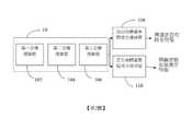
利用ImageNet所建立的類神經網路進行預訓練模型(Pre-Train Model)日益成熟,如第1圖所示,習知的預訓練模型係利用如ImageNet所提供的大量影像資料庫經由第一卷積運算層(Convolution Layer)42、第二卷積運算層44與第三卷積運算層46的卷積神經網路(Convolution Neural Network,CNN)進行運算分析後所建立的模型做為骨幹(Base Structure)40,將其接近最終辨識輸出結果的最後數層更改為適合的應用方面的全連接層(Full Connection Layer)48,可應用於相關物件影像辨識技術領域的影像分類、預測數值(回歸)、目標檢測、分割等應用。The pre-training model (Pre-Train Model) established by ImageNet is becoming more mature. As shown in Figure 1, the conventional pre-training model uses a large number of image databases provided by ImageNet through the first volume. The convolutional neural network (Convolution Neural Network, CNN) of the
然而,由於個人醫療資訊的保密性,ImageNet這類的公開資料庫缺乏大量的醫療影像可供醫療影像辨識技術使用,並且由於單一病例的影像數量有限,難以提高醫療影像預訓練模型的準確率。因此,建立以醫療影像為主的預訓練模型,以進一步提昇醫療影像的影像分類、目標檢測、分割等應用的準確率,就成為目前人工智慧技術領域的重要目標。However, due to the confidentiality of personal medical information, public databases such as ImageNet lack a large number of medical images for medical image recognition technology, and because the number of images in a single case is limited, it is difficult to improve the accuracy of medical image pre-training models. Therefore, the establishment of a pre-training model based on medical images to further improve the accuracy of image classification, target detection, and segmentation of medical images has become an important goal in the current artificial intelligence technology field.
本發明係關於醫療影像辨識的方法與系統,特別是關於可應用於X光(X-ray)、電腦斷層掃描(Computed Tomography, CT)、核磁共振造影(Magnetic Resonance Imaging, MRI)、超音波、病理切片攝影或眼底攝影的醫療影像辨識的方法與系統,該方法與系統係利用建立以醫療影像為主的預訓練模型,以進一步提昇醫療影像的影像分類、目標檢測、分割等應用的準確率。The present invention relates to a method and system for medical image recognition, in particular, it is applicable to X-ray (X-ray), Computed Tomography (CT), Magnetic Resonance Imaging (MRI), Ultrasound, A method and system for medical image recognition of pathological slice photography or fundus photography. The method and system use the establishment of a pre-training model based on medical images to further improve the accuracy of medical image classification, target detection, segmentation and other applications .
本發明提供一種醫療影像辨識的方法,包括步驟: 步驟一:輸入具有複數類疾病標記的複數個影像資料,每個影像資料至少具有一種疾病標記; 步驟二:建立一第一預訓練模型,並將該具有複數類疾病標記的複數個影像資料以混合方式同時對該第一預訓練模型進行訓練; 步驟三:將具有第一疾病標記的影像資料輸入至該第一預訓練模型; 步驟四:以該第一預訓練模型為骨幹,建立一第二預訓練模型;及 步驟五:使用該具有第一疾病標記的影像資料對該第二預訓練模型進行訓練與驗證。The present invention provides a method for medical image recognition, including the steps: Step 1: Input multiple image data with multiple disease markers, and each image data has at least one disease marker; Step 2: Establish a first pre-training model, and simultaneously train the first pre-training model on the plurality of image data with multiple disease markers in a mixed manner; Step 3: Input the image data with the first disease marker into the first pre-training model; Step 4: Use the first pre-training model as the backbone to establish a second pre-training model; and Step 5: Use the image data with the first disease marker to train and verify the second pre-training model.
本發明之醫療影像辨識的方法中,其中,該步驟四進一步包括步驟:凍結該第一預訓練模型至少一個卷積運算層的參數。In the medical image recognition method of the present invention, the step four further includes the step of freezing the parameters of at least one convolution operation layer of the first pre-training model.
本發明之醫療影像辨識的方法中,其中,在該步驟一之該複數類疾病標記至少包括三類疾病標記。In the method for medical image recognition of the present invention, wherein the plural types of disease markers in the step one include at least three types of disease markers.
本發明之醫療影像辨識的方法中,其中該複數個影像資料為相同格式的影像資料。In the medical image recognition method of the present invention, the plurality of image data are image data of the same format.
本發明之醫療影像辨識的方法中,其中該影像資料的格式為X光(X-ray)、電腦斷層掃描(Computed Tomography, CT)、核磁共振造影(Magnetic Resonance Imaging, MRI)、超音波、病理切片攝影或眼底攝影的其中一種。In the medical image recognition method of the present invention, the format of the image data is X-ray (X-ray), Computed Tomography (CT), Magnetic Resonance Imaging (MRI), Ultrasound, Pathology One of slice photography or fundus photography.
本發明另提供一種醫療影像辨識的系統,包括:一骨幹,該骨幹包括第一卷積運算層、第二卷積運算層與第三卷積運算層,該第一卷積運算層、該第二卷積運算層與該第三卷積運算層用以至少對具有第一疾病標記的影像資料與具有第一疾病標記的影像資料進行運算分析後建立一預訓練模型;一第四卷積運算層或全連接層用以對該骨幹所建立的該預訓練模型所輸出的數據資料訓練其辨識有無第一疾病特徵;以及一第五卷積運算層或全連接層用以對該骨幹所建立的該預訓練模型所輸出的數據資料訓練其辨識有無第二疾病特徵。The present invention also provides a medical image recognition system, including: a backbone, the backbone includes a first convolution operation layer, a second convolution operation layer, and a third convolution operation layer, the first convolution operation layer, the second convolution operation layer The second convolution operation layer and the third convolution operation layer are used to calculate and analyze at least the image data with the first disease marker and the image data with the first disease marker to establish a pre-training model; a fourth convolution operation A layer or a fully connected layer is used to train the data output from the pre-training model established on the backbone to recognize whether there is a first disease feature; and a fifth convolution operation layer or a fully connected layer is used to establish the backbone The data output by the pre-training model trains it to recognize whether there is a second disease feature.
本發明之醫療影像辨識的系統中,其中該系統另包括一第六卷積運算層或全連接層,該第六卷積運算層或全連接層用以在該預訓練模型經過該第一疾病標記的影像資料與該第二疾病標記的影像資料的預訓練後,對一具有第三疾病標記的影像資料進行辨識。In the medical image recognition system of the present invention, the system further includes a sixth convolution operation layer or fully connected layer, and the sixth convolution operation layer or fully connected layer is used to pass the first disease in the pre-trained model After the pre-training of the marked image data and the image data of the second disease marker, an image data with the third disease marker is identified.
本發明之醫療影像辨識的系統中,其中該系統在進行該具有第三疾病標記的影像資料的辨識時,凍結該該骨幹中至少一個卷積運算層的參數。In the medical image recognition system of the present invention, the system freezes the parameters of at least one convolutional operation layer in the backbone when recognizing the image data with the third disease marker.
本發明之醫療影像辨識的系統中,其中該第一疾病標記、該第二疾病標記與該第三疾病標記的影像資料為相同格式的影像資料。In the medical image recognition system of the present invention, the image data of the first disease marker, the second disease marker and the third disease marker are image data in the same format.
本發明之醫療影像辨識的系統中,其中該影像資料的格式為X光、電腦斷層掃描、核磁共振造影、超音波、病理切片攝影或眼底攝影的其中一種。In the medical image recognition system of the present invention, the format of the image data is one of X-ray, computed tomography, MRI, ultrasound, pathological slice photography or fundus photography.
本發明可適用在醫療影像辨識技術的領域,包括X-ray、CT、MRI、超音波、病理切片攝影或眼底攝影等的影像辨識技術領域,本發明也可以應用於醫療影像的分類、預測數值(回歸)、目標檢測、分割等需求。本發明利用不同疾病標記的影像資料進行運算分析建立預訓練模型,在醫療影像辨識技術領域常見的有效資料數量不足的情況下對於提昇影像辨識準確率有明顯助益。The present invention can be applied to the field of medical image recognition technology, including X-ray, CT, MRI, ultrasound, pathological slice photography or fundus photography and other image recognition technology fields. The present invention can also be applied to the classification and prediction of medical images. (Regression), target detection, segmentation and other requirements. The present invention uses image data of different disease markers to perform calculation analysis to establish a pre-training model, which is obviously helpful for improving the accuracy of image recognition when the number of effective data is insufficient in the field of medical image recognition technology.
在發展醫療影像的人工智慧辨識技術時,最常遇到的問題通常是在需要判斷某種疾病病徵的情況時,該疾病的可供參考的有效資料數量與其他自然界領域的有效資料數量相比非常少,在大數量資料驅動的模型下,有效資料數量是模型準確度重要的因素之一。而在醫療影像的人工智慧辨識技術中,也常用ImageNet這類通用的影像資料庫當作預訓練模型的骨幹(Base Structure),但醫療影像並不是通常可見的自然界的影像資料,其與通常可見的自然界的影像資料存在較大的差異。使用不同疾病所建立的多任務(Multi-task)的預訓練模型的辨識準確度會比僅使用ImageNet這類通用的影像資料庫當作預訓練模型來的更高。因此本發明係利用大量不同疾病但都是在相同的某種情境下(例如:X-ray、CT、MRI、超音波、病理切片攝影或眼底攝影等)的影像,以多任務(Multi-task)的方式建立一個預訓練模型,再將此預訓練模型當作骨幹應用於某種疾病下。根據本發明所揭示的具體實施例,請參閱第2圖所示的第一實施例的醫療影像辨識的系統,以北美放射科協會 (Radiological Society of North America, RSNA)與美國史丹佛大學(Leland Stanford Junior University in United States, 以下簡稱“史丹佛大學”)所提供的數萬張醫療影像資料庫為例,RSNA提供約3萬餘張胸腔X-ray的影像,其中皆有標示(Label)正常或肺炎之標記,史丹佛大學提供名為MURA(Musculoskeletal Radiographs)的1萬餘張的骨科X-ray影像資料庫,其中皆有標示正常或不正常之標記。我們將這兩個完全不同疾病和不同部位的X-ray的醫療影像資料,建立一個多任務(Multi-task)的訓練模型。第一實施例中的骨幹10係利用經由第一卷積運算層(Convolution Layer)102、第二卷積運算層104與第三卷積運算層106的卷積神經網路(Convolution Neural Network,CNN)對RSNA提供的3萬餘張胸腔X-ray影像與史丹佛大學的MURA提供的1萬餘張的骨科X-ray影像進行運算分析後所建立的模型,通過骨幹10運算分析後所輸出的數據資料分別通過第四卷積運算層或全連接層(Full Connection Layer)108與第五卷積運算層或全連接層110以分別訓練其辨識有無肺炎特徵或骨骼異常特徵的結果。本實施例中的卷積運算層數量可視實際應用需求進行調整。本實施例的實際應用時亦可視不同需求加入其他運算層,例如:非線性層(Rectified Linear Layer)、降採樣層(Pooling Layer)等。本發明可適用的影像辨識領域並不僅限於本實施例中所例示的肺炎或骨骼影像辨識領域。In the development of artificial intelligence recognition technology for medical images, the most common problem is usually when it is necessary to determine the symptoms of a certain disease, the number of valid data available for reference for the disease is compared with the number of valid data in other natural fields Very few, in a model driven by a large amount of data, the number of valid data is one of the important factors for the accuracy of the model. In the artificial intelligence identification technology of medical images, general image databases such as ImageNet are also commonly used as the backbone of the pre-training model (base structure), but medical images are not usually visible natural image data. There are big differences in the image data of nature. The recognition accuracy of the multi-task pre-training model established by using different diseases will be higher than that of using only a general image database such as ImageNet as the pre-training model. Therefore, the present invention utilizes images of a large number of different diseases but all in the same certain situation (for example: X-ray, CT, MRI, ultrasound, pathological slice photography or fundus photography, etc.), with multi-task (Multi-task) ) Method to establish a pre-training model, and then use this pre-training model as the backbone for a certain disease. According to the specific embodiment disclosed in the present invention, please refer to the medical image recognition system of the first embodiment shown in Figure 2. The Radiological Society of North America (RSNA) and the Stanford University (Leland Stanford Junior University in United States, hereinafter referred to as "Stanford University") provides tens of thousands of medical image databases as an example, RSNA provides about 30,000 chest X-ray images, all of which have the label (Label) normal Or for signs of pneumonia, Stanford University provides a database of more than 10,000 orthopedic X-ray images named MURA (Musculoskeletal Radiographs), all of which are marked normal or abnormal. We use these two completely different diseases and different parts of the X-ray medical imaging data to build a multi-task training model. The
在上述預訓練完成後,再將此預訓練模型的骨幹10部分應用於RSNA提供的骨齡預測的影像資料庫,建立可辨識骨齡的模型。請參閱第3圖所示的第二實施例的醫療影像辨識的系統,骨幹10對RSNA提供的骨齡預測的影像資料庫經由第一卷積運算層102、第二卷積運算層104與第三卷積運算層106進行運算分析後,所輸出的數據資料通過第六卷積運算層或全連接層202以進行辨識骨齡的結果。本實施例中的卷積運算層數量可視實際應用需求進行調整。本實施例的實際應用時亦可視不同需求加入其他運算層,例如:非線性層(Rectified Linear Layer)、降採樣層(Pooling Layer)等。本發明可適用的影像辨識領域並不僅限於本實施例中所例示的骨齡影像辨識領域。After the above-mentioned pre-training is completed, the
請參閱第4圖,經過實驗證實,在不同數量級的資料下,使用本發明的預訓練模型所得到的影像辨識準確率比使用ImageNet所建立的模型更高,尤其在資料比較少的情況下,改善的幅度更為明顯,在醫療影像辨識技術領域常見的有效資料數量不足的情況下對於提昇影像辨識準確率有明顯助益。Please refer to Figure 4. Experiments have confirmed that under different orders of magnitude of data, the image recognition accuracy rate obtained by using the pre-trained model of the present invention is higher than that of the model established using ImageNet, especially when the data is relatively small. The extent of improvement is more obvious, and it is obviously helpful to improve the accuracy of image recognition in the case of insufficient effective data in the field of medical image recognition technology.
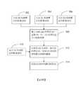
請參閱第5圖所示的第三實施例,第三實施例為建立醫學影像的預訓練模型的方法流程,其包括以下步驟: 步驟302:輸入具有D0疾病標記的影像資料; 步驟304:輸入具有D1疾病標記的影像資料; 步驟306:輸入具有D2疾病標記的影像資料; 步驟308:建立multi-task的模型M0,並將D0、D1、D2等疾病標記的影像資料以混合方式同時對模型M0進行訓練; 步驟310:將具有D3疾病標記的影像資料輸入至模型M0; 步驟312:以模型M0為骨幹,建立新的模型M1,使用具有D3疾病標記的影像資料對模型M1進行訓練與驗證; 步驟314:得到D3疾病特徵判讀的模型M1。Please refer to the third embodiment shown in Fig. 5. The third embodiment is a method flow of establishing a pre-training model of medical images, which includes the following steps: Step 302: Input image data with D0 disease markers; Step 304: Input image data with D1 disease markers; Step 306: Input image data with D2 disease markers; Step 308: Establish a multi-task model M0, and simultaneously train the model M0 with imaging data of disease markers such as D0, D1, D2, etc. in a mixed manner; Step 310: Input the image data with D3 disease markers into the model M0; Step 312: Using model M0 as the backbone, establish a new model M1, and use the image data with D3 disease markers to train and verify the model M1; Step 314: Obtain the model M1 for D3 disease feature interpretation.
上述第三實施例中的預訓練模型的訓練過程中,在步驟302、步驟304、步驟306所使用的不同疾病標記的種類總數至少為2類以上,每個影像資料至少具有一種疾病標記,可以根據實際應用的需求進行增加。In the training process of the pre-training model in the above third embodiment, the total number of different disease marker types used in
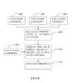
由於在相同的影像辨識應用情境下,預訓練模型在最前面幾層的特徵是類似的,因此訓練時可以凍結前面幾層的參數,以提高影像辨識的準確率。請參閱第6圖所示的第四實施例,第四實施例為建立醫學影像的預訓練模型的方法流程,其中凍結至少骨幹中一個卷積運算層的參數,第四實施例的方法流程包括以下步驟: 步驟402:輸入具有D0疾病標記的影像資料; 步驟404:輸入具有D1疾病標記的影像資料; 步驟406:輸入具有D2疾病標記的影像資料; 步驟408:建立multi-task的模型M0,並將D0、D1、D2等疾病標記的影像資料以混合方式同時對模型M0進行訓練; 步驟410:將具有D3疾病標記的影像資料輸入至模型M0; 步驟412:以模型M0為骨幹,建立新的模型M1,凍結M0至少一個卷積運算層的參數,使用具有D3疾病標記的影像資料對模型M1進行訓練與驗證; 步驟414:得到D3疾病特徵判讀的模型M1。Because in the same image recognition application scenario, the features of the first few layers of the pre-training model are similar, so the parameters of the first few layers can be frozen during training to improve the accuracy of image recognition. Please refer to the fourth embodiment shown in FIG. 6. The fourth embodiment is a method flow for establishing a pre-training model of medical images, wherein the parameters of at least one convolution operation layer in the backbone are frozen, and the method flow of the fourth embodiment includes The following steps: Step 402: Input image data with D0 disease markers; Step 404: Input image data with D1 disease marker; Step 406: Input image data with D2 disease markers; Step 408: Establish a multi-task model M0, and simultaneously train the model M0 with imaging data of disease markers such as D0, D1, D2, etc. in a mixed manner; Step 410: Input the image data with D3 disease markers into the model M0; Step 412: Using the model M0 as the backbone, a new model M1 is established, the parameters of at least one convolution operation layer of M0 are frozen, and the image data with D3 disease markers are used to train and verify the model M1; Step 414: Obtain a model M1 for D3 disease feature interpretation.
上述第四實施例中的預訓練模型的訓練過程中,在步驟402、步驟404、步驟406所使用的不同疾病標記的種類總數至少為2類以上,每個影像資料至少具有一種疾病標記,可以根據實際應用的需求進行增加。In the training process of the pre-training model in the foregoing fourth embodiment, the total number of types of different disease markers used in
請參閱第7圖,經過實驗證實,在相同的影像辨識應用情境下,進行不同數量級的實驗分析,凍結前面幾層的卷積運算層參數而只訓練之後的卷積運算層,所得到的預訓練模型可以增加預測的準確率,在資料比較少的情況下,改善的幅度更為明顯。使用本發明的凍結前面幾層的預訓練模型所得到的影像辨識準確率也比使用ImageNet所建立的模型更高,尤其在資料比較少的情況下,改善的幅度更為明顯,在醫療影像辨識技術領域常見的有效資料數量不足的情況下對於提昇影像辨識準確率有明顯助益。Please refer to Figure 7. Experiments have confirmed that in the same image recognition application context, different orders of magnitude of experimental analysis are performed. The parameters of the convolution operation layer of the previous layers are frozen and only the convolution operation layer after training is obtained. Training the model can increase the accuracy of the prediction. In the case of less data, the improvement is more obvious. The image recognition accuracy rate obtained by using the pre-trained model of freezing the first few layers of the present invention is also higher than that of the model established by ImageNet, especially in the case of less data, the improvement is more obvious. In medical image recognition In the case of insufficient effective data, which is common in the technical field, it is obviously helpful to improve the accuracy of image recognition.
上述實施例僅為例示本發明之實施方式,本發明可適用的領域不僅限於上述實施例所例示的特定醫療影像領域,本發明亦可使用其他疾病在相同的情境下(例如:X-ray、CT、MRI、超音波、病理切片攝影或眼底攝影等)的影像辨識技術領域,本發明也可以應用於醫療影像的分類、預測數值(回歸)、目標檢測、分割等需求。The above-mentioned examples are only examples of the embodiments of the present invention. The applicable field of the present invention is not limited to the specific medical imaging field exemplified in the above-mentioned examples. The present invention can also use other diseases in the same context (for example: X-ray, In the field of image recognition technology such as CT, MRI, ultrasound, pathological slice photography or fundus photography, etc., the present invention can also be applied to the needs of medical image classification, predictive value (regression), target detection, segmentation, etc.
10:骨幹102:第一卷積運算層104:第二卷積運算層106:第三卷積運算層108:第四卷積運算層或全連接層110:第五卷積運算層或全連接層202:第六卷積運算層或全連接層302:步驟304:步驟306:步驟308:步驟310:步驟312:步驟314:步驟402:步驟404:步驟406:步驟408:步驟410:步驟412:步驟414:步驟10: Backbone102: The first convolution operation layer104: The second convolution operation layer106: The third convolution operation layer108: The fourth convolution operation layer or fully connected layer110: The fifth convolutional operation layer or fully connected layer202: The sixth convolution operation layer or fully connected layer302: Steps304: Step306: Steps308: Step310: Step312: Step314: Step402: Step404: Step406: Step408: Step410: Step412: Step414: Step
第1圖係習知技術的示意圖。 第2圖係本發明之第一實施例的醫療影像辨識的系統示意圖。 第3圖係本發明之第二實施例的醫療影像辨識的系統示意圖。 第4圖係本發明之第二實施例的實驗準確率分析圖。 第5圖係本發明之第三實施例的方法流程圖。 第6圖係本發明之第四實施例的方法流程圖。 第7圖係本發明之第四實施例的實驗準確率分析圖。Figure 1 is a schematic diagram of the conventional technology. Figure 2 is a schematic diagram of a medical image recognition system according to the first embodiment of the present invention. Figure 3 is a schematic diagram of a medical image recognition system according to the second embodiment of the present invention. Figure 4 is an analysis diagram of the experimental accuracy of the second embodiment of the present invention. Figure 5 is a flow chart of the method of the third embodiment of the present invention. Figure 6 is a flowchart of the method of the fourth embodiment of the present invention. Figure 7 is an analysis diagram of the experimental accuracy of the fourth embodiment of the present invention.
302:步驟302: Step
304:步驟304: Step
306:步驟306: Step
308:步驟308: step
310:步驟310: Step
312:步驟312: Step
314:步驟314: Step
Claims (10)
Translated fromChinesePriority Applications (3)
| Application Number | Priority Date | Filing Date | Title |
|---|---|---|---|
| TW108133858ATWI705458B (en) | 2019-09-19 | 2019-09-19 | A method for recognizing medical image and a system of same |
| CN202010013881.6ACN112530548A (en) | 2019-09-19 | 2020-01-02 | Medical image identification method and system |
| US17/022,126US11282200B2 (en) | 2019-09-19 | 2020-09-16 | Method for recognizing medical image and system of same |
Applications Claiming Priority (1)
| Application Number | Priority Date | Filing Date | Title |
|---|---|---|---|
| TW108133858ATWI705458B (en) | 2019-09-19 | 2019-09-19 | A method for recognizing medical image and a system of same |
Publications (2)
| Publication Number | Publication Date |
|---|---|
| TWI705458Btrue TWI705458B (en) | 2020-09-21 |
| TW202113868A TW202113868A (en) | 2021-04-01 |
Family
ID=74091530
Family Applications (1)
| Application Number | Title | Priority Date | Filing Date |
|---|---|---|---|
| TW108133858ATWI705458B (en) | 2019-09-19 | 2019-09-19 | A method for recognizing medical image and a system of same |
Country Status (3)
| Country | Link |
|---|---|
| US (1) | US11282200B2 (en) |
| CN (1) | CN112530548A (en) |
| TW (1) | TWI705458B (en) |
Citations (3)
| Publication number | Priority date | Publication date | Assignee | Title |
|---|---|---|---|---|
| TW201839634A (en)* | 2017-04-24 | 2018-11-01 | 太豪生醫股份有限公司 | Cloud medical image analysis system and method |
| CN109670530A (en)* | 2018-11-15 | 2019-04-23 | 首都医科大学附属北京友谊医院 | A kind of construction method of atrophic gastritis image recognition model and its application |
| CN110223289A (en)* | 2019-06-17 | 2019-09-10 | 上海联影医疗科技有限公司 | A kind of image processing method and system |
Family Cites Families (8)
| Publication number | Priority date | Publication date | Assignee | Title |
|---|---|---|---|---|
| US10417788B2 (en)* | 2016-09-21 | 2019-09-17 | Realize, Inc. | Anomaly detection in volumetric medical images using sequential convolutional and recurrent neural networks |
| WO2019024568A1 (en)* | 2017-08-02 | 2019-02-07 | 上海市第六人民医院 | Ocular fundus image processing method and apparatus, computer device, and storage medium |
| US10565708B2 (en)* | 2017-09-06 | 2020-02-18 | International Business Machines Corporation | Disease detection algorithms trainable with small number of positive samples |
| US20210042916A1 (en)* | 2018-02-07 | 2021-02-11 | Ai Technologies Inc. | Deep learning-based diagnosis and referral of diseases and disorders |
| CN108389614B (en)* | 2018-03-02 | 2021-01-19 | 西安交通大学 | A method for constructing medical image atlas based on image segmentation and convolutional neural network |
| CN111656357B (en)* | 2018-04-17 | 2024-05-10 | 深圳华大生命科学研究院 | Modeling method, device and system for ophthalmic disease classification model |
| CN109389587B (en)* | 2018-09-26 | 2021-07-16 | 上海联影智能医疗科技有限公司 | Medical image analysis system, device and storage medium |
| CN109427060A (en)* | 2018-10-30 | 2019-03-05 | 腾讯科技(深圳)有限公司 | A kind of method, apparatus, terminal device and the medical system of image identification |
- 2019
- 2019-09-19TWTW108133858Apatent/TWI705458B/enactive
- 2020
- 2020-01-02CNCN202010013881.6Apatent/CN112530548A/enactivePending
- 2020-09-16USUS17/022,126patent/US11282200B2/enactiveActive
Patent Citations (3)
| Publication number | Priority date | Publication date | Assignee | Title |
|---|---|---|---|---|
| TW201839634A (en)* | 2017-04-24 | 2018-11-01 | 太豪生醫股份有限公司 | Cloud medical image analysis system and method |
| CN109670530A (en)* | 2018-11-15 | 2019-04-23 | 首都医科大学附属北京友谊医院 | A kind of construction method of atrophic gastritis image recognition model and its application |
| CN110223289A (en)* | 2019-06-17 | 2019-09-10 | 上海联影医疗科技有限公司 | A kind of image processing method and system |
Also Published As
| Publication number | Publication date |
|---|---|
| US20210090258A1 (en) | 2021-03-25 |
| CN112530548A (en) | 2021-03-19 |
| US11282200B2 (en) | 2022-03-22 |
| TW202113868A (en) | 2021-04-01 |
Similar Documents
| Publication | Publication Date | Title |
|---|---|---|
| Gozes et al. | Coronavirus detection and analysis on chest ct with deep learning | |
| Hennessey et al. | Artificial intelligence in veterinary diagnostic imaging: A literature review | |
| US8126242B2 (en) | Computer program products and methods for detection and tracking of rheumatoid arthritis | |
| Yoon et al. | Medical image analysis using artificial intelligence | |
| US12182733B2 (en) | Label inference system | |
| Zhang et al. | CdcSegNet: Automatic COVID-19 infection segmentation from CT images | |
| Feng et al. | Deep learning for chest radiology: a review | |
| CN114119516A (en) | Virus focus segmentation method based on transfer learning and cascade adaptive hole convolution | |
| Fan et al. | Graph reasoning module for Alzheimer’s disease diagnosis: A plug-and-play method | |
| CN116612885A (en) | Prediction device for acute exacerbation of chronic obstructive pulmonary disease based on multiple modes | |
| CN114787816A (en) | Data Augmentation for Machine Learning Methods | |
| Patel et al. | PTXNet: An extended UNet model based segmentation of pneumothorax from chest radiography images | |
| Chaisangmongkon et al. | External validation of deep learning algorithms for cardiothoracic ratio measurement | |
| CN115409812A (en) | CT image automatic classification method based on fusion time attention mechanism | |
| CN117393100B (en) | Diagnostic report generation method, model training method, system, device and medium | |
| TWI705458B (en) | A method for recognizing medical image and a system of same | |
| Kurasova et al. | Semi-supervised learning with pseudo-labeling for pancreatic cancer detection on ct scans | |
| JP7718915B2 (en) | Learning device, method, and program, and information processing device, method, and program | |
| CN118414631A (en) | Selecting training data for annotation | |
| WO2023276432A1 (en) | Image retrieval device, method, and program | |
| Huang et al. | Semantics Guided Disentangled GAN for Chest X-Ray Image Rib Segmentation | |
| Sahin et al. | Segmentation of covid-19 infected lung area in ct scans with deep algorithms | |
| Arya et al. | COVID-19 infection segmentation using deep learning techniques | |
| CN115330733B (en) | Intelligent disease identification method and system based on fine-grained domain knowledge | |
| Kasturi et al. | BioVLM-T: A temporal framework for radiology report generation using pre-trained vision language foundational models |