TW202302046A - System and method for processing black bone mri data - Google Patents
System and method for processing black bone mri dataDownload PDFInfo
- Publication number
- TW202302046A TW202302046ATW111104607ATW111104607ATW202302046ATW 202302046 ATW202302046 ATW 202302046ATW 111104607 ATW111104607 ATW 111104607ATW 111104607 ATW111104607 ATW 111104607ATW 202302046 ATW202302046 ATW 202302046A
- Authority
- TW
- Taiwan
- Prior art keywords
- bone
- black
- mri
- black bone
- patient
- Prior art date
Links
Images


Classifications
- G—PHYSICS
- G01—MEASURING; TESTING
- G01R—MEASURING ELECTRIC VARIABLES; MEASURING MAGNETIC VARIABLES
- G01R33/00—Arrangements or instruments for measuring magnetic variables
- G01R33/20—Arrangements or instruments for measuring magnetic variables involving magnetic resonance
- G01R33/44—Arrangements or instruments for measuring magnetic variables involving magnetic resonance using nuclear magnetic resonance [NMR]
- G01R33/48—NMR imaging systems
- G01R33/54—Signal processing systems, e.g. using pulse sequences ; Generation or control of pulse sequences; Operator console
- G01R33/56—Image enhancement or correction, e.g. subtraction or averaging techniques, e.g. improvement of signal-to-noise ratio and resolution
- G01R33/5608—Data processing and visualization specially adapted for MR, e.g. for feature analysis and pattern recognition on the basis of measured MR data, segmentation of measured MR data, edge contour detection on the basis of measured MR data, for enhancing measured MR data in terms of signal-to-noise ratio by means of noise filtering or apodization, for enhancing measured MR data in terms of resolution by means for deblurring, windowing, zero filling, or generation of gray-scaled images, colour-coded images or images displaying vectors instead of pixels
- A—HUMAN NECESSITIES
- A61—MEDICAL OR VETERINARY SCIENCE; HYGIENE
- A61B—DIAGNOSIS; SURGERY; IDENTIFICATION
- A61B34/00—Computer-aided surgery; Manipulators or robots specially adapted for use in surgery
- A61B34/10—Computer-aided planning, simulation or modelling of surgical operations
- A—HUMAN NECESSITIES
- A61—MEDICAL OR VETERINARY SCIENCE; HYGIENE
- A61B—DIAGNOSIS; SURGERY; IDENTIFICATION
- A61B90/00—Instruments, implements or accessories specially adapted for surgery or diagnosis and not covered by any of the groups A61B1/00 - A61B50/00, e.g. for luxation treatment or for protecting wound edges
- A61B90/36—Image-producing devices or illumination devices not otherwise provided for
- A61B90/37—Surgical systems with images on a monitor during operation
- G—PHYSICS
- G01—MEASURING; TESTING
- G01R—MEASURING ELECTRIC VARIABLES; MEASURING MAGNETIC VARIABLES
- G01R33/00—Arrangements or instruments for measuring magnetic variables
- G01R33/20—Arrangements or instruments for measuring magnetic variables involving magnetic resonance
- G01R33/44—Arrangements or instruments for measuring magnetic variables involving magnetic resonance using nuclear magnetic resonance [NMR]
- G01R33/48—NMR imaging systems
- G01R33/54—Signal processing systems, e.g. using pulse sequences ; Generation or control of pulse sequences; Operator console
- G01R33/56—Image enhancement or correction, e.g. subtraction or averaging techniques, e.g. improvement of signal-to-noise ratio and resolution
- G01R33/561—Image enhancement or correction, e.g. subtraction or averaging techniques, e.g. improvement of signal-to-noise ratio and resolution by reduction of the scanning time, i.e. fast acquiring systems, e.g. using echo-planar pulse sequences
- G01R33/5613—Generating steady state signals, e.g. low flip angle sequences [FLASH]
- G—PHYSICS
- G02—OPTICS
- G02B—OPTICAL ELEMENTS, SYSTEMS OR APPARATUS
- G02B27/00—Optical systems or apparatus not provided for by any of the groups G02B1/00 - G02B26/00, G02B30/00
- G02B27/01—Head-up displays
- G02B27/017—Head mounted
- G—PHYSICS
- G06—COMPUTING OR CALCULATING; COUNTING
- G06T—IMAGE DATA PROCESSING OR GENERATION, IN GENERAL
- G06T7/00—Image analysis
- G06T7/0002—Inspection of images, e.g. flaw detection
- G06T7/0012—Biomedical image inspection
- A—HUMAN NECESSITIES
- A61—MEDICAL OR VETERINARY SCIENCE; HYGIENE
- A61B—DIAGNOSIS; SURGERY; IDENTIFICATION
- A61B34/00—Computer-aided surgery; Manipulators or robots specially adapted for use in surgery
- A61B34/10—Computer-aided planning, simulation or modelling of surgical operations
- A61B2034/101—Computer-aided simulation of surgical operations
- A61B2034/105—Modelling of the patient, e.g. for ligaments or bones
- A—HUMAN NECESSITIES
- A61—MEDICAL OR VETERINARY SCIENCE; HYGIENE
- A61B—DIAGNOSIS; SURGERY; IDENTIFICATION
- A61B90/00—Instruments, implements or accessories specially adapted for surgery or diagnosis and not covered by any of the groups A61B1/00 - A61B50/00, e.g. for luxation treatment or for protecting wound edges
- A61B90/36—Image-producing devices or illumination devices not otherwise provided for
- A61B2090/364—Correlation of different images or relation of image positions in respect to the body
- A61B2090/365—Correlation of different images or relation of image positions in respect to the body augmented reality, i.e. correlating a live optical image with another image
- A—HUMAN NECESSITIES
- A61—MEDICAL OR VETERINARY SCIENCE; HYGIENE
- A61B—DIAGNOSIS; SURGERY; IDENTIFICATION
- A61B90/00—Instruments, implements or accessories specially adapted for surgery or diagnosis and not covered by any of the groups A61B1/00 - A61B50/00, e.g. for luxation treatment or for protecting wound edges
- A61B90/36—Image-producing devices or illumination devices not otherwise provided for
- A61B90/37—Surgical systems with images on a monitor during operation
- A61B2090/374—NMR or MRI
- G—PHYSICS
- G02—OPTICS
- G02B—OPTICAL ELEMENTS, SYSTEMS OR APPARATUS
- G02B27/00—Optical systems or apparatus not provided for by any of the groups G02B1/00 - G02B26/00, G02B30/00
- G02B27/01—Head-up displays
- G02B27/0101—Head-up displays characterised by optical features
- G02B2027/014—Head-up displays characterised by optical features comprising information/image processing systems
- G—PHYSICS
- G06—COMPUTING OR CALCULATING; COUNTING
- G06T—IMAGE DATA PROCESSING OR GENERATION, IN GENERAL
- G06T2207/00—Indexing scheme for image analysis or image enhancement
- G06T2207/10—Image acquisition modality
- G06T2207/10072—Tomographic images
- G06T2207/10088—Magnetic resonance imaging [MRI]
- G—PHYSICS
- G06—COMPUTING OR CALCULATING; COUNTING
- G06T—IMAGE DATA PROCESSING OR GENERATION, IN GENERAL
- G06T2207/00—Indexing scheme for image analysis or image enhancement
- G06T2207/30—Subject of image; Context of image processing
- G06T2207/30004—Biomedical image processing
- G06T2207/30008—Bone
Landscapes
- Engineering & Computer Science (AREA)
- Health & Medical Sciences (AREA)
- Physics & Mathematics (AREA)
- Nuclear Medicine, Radiotherapy & Molecular Imaging (AREA)
- General Health & Medical Sciences (AREA)
- General Physics & Mathematics (AREA)
- Surgery (AREA)
- Life Sciences & Earth Sciences (AREA)
- Radiology & Medical Imaging (AREA)
- Medical Informatics (AREA)
- Computer Vision & Pattern Recognition (AREA)
- Heart & Thoracic Surgery (AREA)
- Public Health (AREA)
- Veterinary Medicine (AREA)
- Animal Behavior & Ethology (AREA)
- Condensed Matter Physics & Semiconductors (AREA)
- Molecular Biology (AREA)
- Biomedical Technology (AREA)
- High Energy & Nuclear Physics (AREA)
- Signal Processing (AREA)
- Artificial Intelligence (AREA)
- Theoretical Computer Science (AREA)
- Robotics (AREA)
- Quality & Reliability (AREA)
- Optics & Photonics (AREA)
- Gynecology & Obstetrics (AREA)
- Oral & Maxillofacial Surgery (AREA)
- Pathology (AREA)
- Magnetic Resonance Imaging Apparatus (AREA)
Abstract
Description
Translated fromChinese相關申請案之交叉參考Cross References to Related Applications
本申請案主張於2021年2月8日申請之美國臨時申請案第63/147,200號之權益,該美國臨時申請案係以引用方式併入本文中。This application claims the benefit of U.S. Provisional Application No. 63/147,200, filed February 8, 2021, which is incorporated herein by reference.
本發明係有關於用於處理黑骨MRI資料的系統及方法。The present invention relates to a system and method for processing black bone MRI data.
外科程序可能經常為複雜且時間敏感的,且範圍因患者而異。舉例而言,在骨外科手術之情況下,修復點可視確切位置、大小等而在條件或程序要求上可有所不同。需要患者骨頭之準確視圖以確保提供適當的治療。程序之準確性及效率非常關鍵,且基於外科手術執行所在的區域之患者特定局部幾何形狀及物理性質的詳細規劃為基礎。為了達成新的術前準備水平,越來越多地利用3D CT及MRI影像。但骨成像特別困難,此係因為已證實MRI資料係有缺陷的,需要依賴不足以展示重要及理想的特徵之CT掃描及x射線。Surgical procedures can often be complex and time sensitive, and the scope varies from patient to patient. For example, in the case of orthopedic surgery, the repair point may vary in terms of conditions or procedural requirements depending on exact location, size, etc. An accurate view of the patient's bones is needed to ensure proper treatment is provided. Accuracy and efficiency of the procedure are critical and are based on detailed planning of the patient-specific local geometry and physical properties of the area in which the surgery is performed. To achieve new levels of preoperative readiness, 3D CT and MRI images are increasingly being used. Bone imaging is particularly difficult, however, because MRI data have proven to be flawed, relying on CT scans and x-rays that do not adequately reveal important and desirable features.
電腦斷層掃描(CT)為用於對神經外科、脊柱及骨科中之骨病成像的當前黃金標準,因為它的獲取時間短,對骨縫及骨折之骨描繪出色,成本較低,且具有可用性。雖然標準磁共振成像(MRI)為非游離成像方法,但其長獲取時間及差的骨解析度使其成為對骨病成像之次最佳候選者。標準MRI上的低骨解析度由硬組織中之低質子含量引起。此等挑戰為硬組織之質子含量低及橫向弛豫時間短的結果。Computed tomography (CT) is the current gold standard for imaging bone disease in neurosurgery, spine, and orthopedics because of its short acquisition time, excellent delineation of sutures and fractured bone, low cost, and availability . Although standard magnetic resonance imaging (MRI) is a non-ionogenic imaging modality, its long acquisition time and poor bone resolution make it a second-best candidate for imaging bone disease. Low bone resolution on standard MRI is caused by low proton content in hard tissue. These challenges are a result of the low proton content and short transverse relaxation times of hard tissue.
最大的擔憂之一為游離輻射曝露,特別在兒童中。通常,由於游離輻射之風險,無法獲得術後成像,從而限制術後VR交互。骨組織的術後成像可提高ST產品在患者參與方面之利用率。需要一種利用骨組織之MRI掃描之益處的方法。One of the greatest concerns is exposure to ionizing radiation, especially in children. Often, postoperative imaging is not available due to the risk of ionizing radiation, limiting postoperative VR interaction. Post-operative imaging of bone tissue increases utilization of ST products in terms of patient engagement. What is needed is a method that takes advantage of the benefits of MRI scanning of bone tissue.
一種用於將一黑骨MRI資料集處理成一虛擬模型之方法,該方法包含以下步驟: 對一特定患者之一骨及組織執行一黑骨MRI; 自該黑骨MRI獲得該患者之一黑骨資料集; 處理該黑骨資料集;及 根據該經處理的黑骨資料集生成該患者之該骨及組織之一動態虛擬模型。A method for processing a black bone MRI data set into a virtual model, the method comprising the following steps: performing a black bone MRI on the bone and tissue of a particular patient; obtaining one of the patient's black bone datasets from the black bone MRI; process the black bone data set; and A dynamic virtual model of the bone and tissue of the patient is generated according to the processed black bone data set.
亦提供一種用於將一黑骨MRI資料集處理成一虛擬模型之方法,該方法包含以下步驟: 對一特定患者之一骨及組織執行一黑骨MRI,其中該黑骨MRI利用具有一低翻轉角梯度之一回波序列,從而提供骨與組織之間的高對比度; 自該黑骨MRI獲得該患者之一黑骨資料集; 利用一自動偵測演算法處理該黑骨資料集,該自動偵測演算法自該黑骨資料集偵測強度範圍與骨之強度範圍類似的像素;及 根據該經處理的黑骨資料集生成該患者之該骨及組織之一動態虛擬模型,其中該虛擬模型經組態以突出顯示該患者之骨結構。Also provided is a method for processing a black bone MRI data set into a virtual model, the method comprising the steps of: performing a black bone MRI on bone and tissue of a particular patient, wherein the black bone MRI utilizes an echo sequence with a low flip angle gradient, thereby providing high contrast between bone and tissue; obtaining one of the patient's black bone datasets from the black bone MRI; processing the black bone dataset with an auto-detection algorithm that detects from the black bone dataset pixels with an intensity range similar to that of bone; and A dynamic virtual model of the bone and tissue of the patient is generated based on the processed black bone data set, wherein the virtual model is configured to highlight the bone structure of the patient.
進一步提供一種用於將一黑骨MRI資料集處理成一虛擬模型之方法,該方法包含以下步驟: 對一特定患者之一骨及組織執行一黑骨MRI,其中 該黑骨MRI利用具有一低翻轉角梯度之一回波序列,從而提供骨與組織之間的高對比度,且其中 該黑骨MRI利用在一5度翻轉角下4.2 ms且一重複時間為8.6 ms之一回波時間(TE)序列; 自該黑骨MRI獲得該患者之一黑骨資料集; 藉由將該黑骨資料集反轉及利用一自動偵測演算法來處理該黑骨資料集,該自動偵測演算法自該黑骨資料集偵測強度範圍與骨之強度範圍類似的像素;及 根據該經處理的黑骨資料集生成該患者之該骨及組織之一動態虛擬模型,其中該虛擬模型為經組態以突出顯示該患者之骨結構之一3D 360VR模型。Further provided is a method for processing a black bone MRI data set into a virtual model, the method comprising the following steps: performing a black bone MRI on the bone and tissue of a particular patient, wherein The black bone MRI utilizes an echo sequence with a low flip angle gradient, thereby providing high contrast between bone and tissue, and wherein The black bone MRI utilizes an echo time (TE) sequence of 4.2 ms at a 5 degree flip angle and a repetition time of 8.6 ms; obtaining one of the patient's black bone datasets from the black bone MRI; Processing the black bone dataset by inverting the black bone dataset and utilizing an auto-detection algorithm that detects pixels with an intensity range similar to that of bone from the black bone dataset ;and A dynamic virtual model of the bone and tissue of the patient is generated based on the processed black bone data set, wherein the virtual model is a 3D 360VR model configured to highlight the bone structure of the patient.
又進一步提供一種系統,該系統包括用於執行上述方法中之任一者之一電腦系統。Still further provided is a system comprising a computer system for performing any one of the above methods.
亦提供額外的實例實施例,在下文更詳細地描述該等實例實施例中之一些而非全部。Additional example embodiments are also provided, some but not all of which are described in greater detail below.
以下縮寫詞及定義將幫助理解實施方式:The following abbreviations and definitions will help to understand the embodiments:
AR-擴增實境-物理真實世界環境之實時視圖,該環境之要素已藉由電腦生成的感官要素(諸如聲音、視訊或圖形)增強。AR - Augmented Reality - A real-time view of a physical real-world environment, elements of which have been augmented with computer-generated sensory elements such as sound, video or graphics.
VR-虛擬實境-電腦生成的-3維環境,人們可不同程度地對該環境進行探索且與其交互。VR - Virtual Reality - A computer generated - 3-dimensional environment that people can explore and interact with to varying degrees.
HMD-頭戴式顯示器係指可在AR或VR環境中使用之耳機。HMD可為有線或無線的。HMD亦可包括一或多個附加件,諸如頭戴式耳機、麥克風、HD攝影機、紅外線攝影機、手部追蹤器、位置追蹤器等。HMD - Head Mounted Display refers to a headset that can be used in an AR or VR environment. HMDs can be wired or wireless. The HMD may also include one or more add-ons, such as headphones, microphones, HD cameras, infrared cameras, hand trackers, position trackers, and the like.
控制器-包括按鈕及方向控制器之裝置。控制器可為有線或無線的。此裝置之實例為Xbox遊戲台、PlayStation遊戲台、Oculus touch等。Controller - A device that includes buttons and directional controls. Controllers can be wired or wireless. Examples of such devices are Xbox gaming consoles, PlayStation gaming consoles, Oculus touch, etc.
SNAP模型-SNAP病例(case)係指使用患者之一或多次掃描(CT、MR、fMR、DTI等)以DICOM檔案格式創建之3D紋理或3D物件。SNAP模型亦包括用於過濾特定範圍且為3D紋理中之其他範圍著色的不同分割預設。SNAP模型亦可包括置放於場景中的3D物件,包括用以標記感興趣之特定點或解剖結構之3D形狀、3D標籤、3D量測標記、用於引導之3D箭頭及3D手術工具。手術工具及裝置已經模型化以用於教育及患者特之預演,特別係用於適當調整動脈瘤夾之大小。SNAPModel - A SNAP case refers to a 3D texture or 3D object created in DICOM file format using one or more scans (CT, MR, fMR, DTI, etc.) of a patient. The SNAP model also includes different segmentation presets for filtering specific areas and coloring other areas in the 3D texture. SNAP models can also include 3D objects placed in the scene, including 3D shapes to mark specific points of interest or anatomical structures, 3D labels, 3D measurement markers, 3D arrows for guidance, and 3D surgical tools. Surgical tools and devices have been modeled for educational and patient-specific rehearsals, particularly for proper sizing of aneurysm clips.
化身- 化身代表虛擬環境中之使用者。Avatar - An avatar represents a user in a virtual environment.
MD6DM-多維全球面虛擬實境6自由度模型。該模型提供圖形模擬環境,該圖形模擬環境使得醫師能夠在全球面虛擬實境環境中體驗、規劃、執行及導航介入。MD6DM - 6 DOF model for multidimensional spherical virtual reality. The model provides a graphical simulation environment that enables physicians to experience, plan, execute and navigate interventions in a global virtual reality environment.
先前在以引用方式併入本申請案中之美國專利申請案第8,311,791號中描述的手術預演及準備工具已開發用於基於預建之SNAP模型將靜態CT及MRI醫學影像轉換為動態且交互之多維全球面虛擬實境六(6)自由度模型(「MD6DM」),醫師可使用該模型即時地模擬醫療程序。MD6DM提供圖形模擬環境,該圖形模擬環境使得醫師能夠在全球面虛擬實境環境中體驗、規劃、執行及導航介入。特別地,MD6DM賦予外科醫師使用自傳統二維患者醫學掃描建構之唯一多維模型進行導航的能力,該唯一多維模型在整個體積球面虛擬實境模型中賦予球面虛擬實境6個自由度(即,線性;x、y、z及角度、偏航、俯仰、滾轉)。Surgical rehearsal and preparation tools previously described in U.S. Patent Application No. 8,311,791, which is incorporated herein by reference, have been developed to convert static CT and MRI medical images into dynamic and interactive ones based on pre-built SNAP models. A multidimensional global virtual reality six (6) degrees of freedom model ("MD6DM") that physicians can use to simulate medical procedures in real time. MD6DM provides a graphical simulation environment that enables physicians to experience, plan, execute and navigate interventions in a global virtual reality environment. In particular, MD6DM empowers surgeons to navigate using a unique multidimensional model constructed from traditional 2D patient medical scans that gives spherical VR six degrees of freedom (i.e., linear; x, y, z and angle, yaw, pitch, roll).
MD6DM係使用自患者自身之醫學影像(包括CT、MRI、DTI等)之資料集建構之SNAP模型藉由影像產生器即時地渲染且係患者特定的,諸如先前在以引用方式併入本文中之美國專利第8,311,791號中描述的SNAP電腦。可整合諸如Atlas資料之代表性腦模型以創建部分患者特定之模型,若外科醫師期望如此。該模型提供來自MD6DM上之任何點的360°球面視圖。使用MD6DM,觀看者虛擬地定位在解剖結構內部且可查看及觀察解剖結構及病理結構兩者,就好像他站在患者體內一樣。觀看者可向上看、向下看、環顧四周等,且將看到彼此相關之原生結構,完全如該等結構在患者體內發現的那樣。內部結構之間的空間關係得以保留且可使用MD6DM來了解。MD6DM is a SNAP model constructed from a dataset of the patient's own medical images (including CT, MRI, DTI, etc.) rendered in real-time by an image generator and is patient-specific, such as previously incorporated herein by reference The SNAP computer described in US Patent No. 8,311,791. Representative brain models such as Atlas data can be integrated to create partially patient-specific models if desired by the surgeon. The model provides a 360° spherical view from any point on the MD6DM. Using MD6DM, the viewer is virtually positioned inside the anatomy and can view and observe both anatomy and pathology as if he were standing inside the patient. The viewer can look up, look down, look around, etc., and will see the native structures in relation to each other, exactly as they are found in the patient's body. Spatial relationships between internal structures are preserved and can be understood using MD6DM.
由影像產生器渲染的MD6DM之演算法獲取醫學影像資訊且將其建構成球面模型,該球面模型為當在解剖結構內部「飛行」時可自任何角度觀看之全連續即時模型。特別地,在CT、MRI等拍攝到真實有機體且將其解構成由數千個點建構的數百個切片之後,MD6DM藉由代表此等點中之每一者的360°視圖自內部及外部兩者而將真實有機體還原為3D模型。The algorithm of MD6DM rendered by the image generator takes medical image information and constructs it into a spherical model, which is a full continuous real-time model that can be viewed from any angle while "flying" inside the anatomy. In particular, after CT, MRI, etc. have taken a real organism and decomposed it into hundreds of slices constructed from thousands of points, MD6DM has a 360° view representing each of these points from inside and outside Both restore real organisms to 3D models.
本文中描述一種成像系統,其利用影像產生器及MD6DM模型以用於利用黑骨MRI資料來創建受試者之同步擴增實境視圖以用於創建模型。特別地,該成像系統能夠在相應的實體模型或即時患者影像之上擴增且疊加MD6DM模型。此外,該成像系統將MD6DM模型錨定至實體模型或患者且使該兩者同步,使得根據模型周圍之運動在實體模型之上創建且疊加新的影像。此係藉由將影像產生器直接流式傳輸至HMD、追踪HMD之位置及定位及基於追踪之移動調整影像產生器來實現。因此,在虛擬模型與實體模型之間創建相依性。Described herein is an imaging system utilizing an image generator and an MD6DM model for creating a simultaneous augmented reality view of a subject using black bone MRI data for creating the model. In particular, the imaging system is capable of augmenting and superimposing MD6DM models on top of corresponding mockups or live patient images. In addition, the imaging system anchors the MD6DM model to the mockup or the patient and synchronizes the two so that new images are created and superimposed on top of the mockup based on motion around the model. This is accomplished by streaming the image generator directly to the HMD, tracking the position and positioning of the HMD, and adjusting the image generator based on the tracked movement. Therefore, a dependency is created between the virtual model and the physical model.
藉由創建此種相依性且將虛擬模型綁定或錨定至實體模型或患者,然後基於相對於實體模型之移動調整疊加在實體模型之上的影像,HMD能夠接收實體模型之同步擴增實境視圖,無論HDM之使用者相對於實體模型定位在何處,從而為使用者提供實體模型的經改良視角。作為將虛擬模型錨定至實體模型的結果,視覺模型沒有與實體模型分離。換言之,若HMD之使用者轉過頭且將視線離開實體模型,則使用者亦將不再看到虛擬模型。只有當使用者將焦點返回至實體模型時,使用者才會再次看到虛擬模型,該虛擬模型視需要進行疊加及同步。因此,可向使用者呈現主要實體物件之擴增視圖,同時仍為使用者提供在主要實體物件附近操縱且與次要實體物件交互而不干擾使用者的視圖或與次要物件之交互的自由度及靈活性。By creating this dependency and binding or anchoring the virtual model to the physical model or patient, and then adjusting the image superimposed on the physical model based on movement relative to the physical model, the HMD can receive simultaneous augmentation of the physical model. The context view provides the user with an improved perspective of the mockup regardless of where the user of the HDM is positioned relative to the mockup. As a result of anchoring the virtual model to the physical model, the visual model is not separated from the physical model. In other words, if the user of the HMD turns his head and takes his eyes off the physical model, the user will no longer see the virtual model. Only when the user returns focus to the physical model does the user see the virtual model again, which is superimposed and synchronized as necessary. Thus, the user can be presented with an augmented view of the primary physical object while still providing the user with the freedom to maneuver around the primary physical object and interact with secondary physical objects without interfering with the user's view or interaction with the secondary objects degree and flexibility.
應了解,雖然提到將虛擬模型錨定或綁定至實體模型,但虛擬模型可錨定至實體位置,而非錨定至實體物件,且將理解,實體物件之位置在實體物件的擴增實境查看期間並未移動。It should be understood that although reference is made to anchoring or binding a virtual model to a physical model, the virtual model may be anchored to a physical location rather than to a physical object, and it will be understood that the location of a physical object is an extension of the physical object Did not move during reality check.
應了解,雖然本文中描述之實例通常可係關於醫療應用且確切地係關於患者之解剖結構的虛擬模型或影像,出於執行脊柱手術之目的,該等虛擬模型或影像經擴增且與相應患者之身體同步,但成像系統可類似地用於將任何虛擬物件之虛擬模型或影像與相應的實體物件同步且擴增。It should be appreciated that while the examples described herein may relate generally to medical applications and specifically to virtual models or images of a patient's anatomy, for the purpose of performing spinal surgery, these virtual models or images are augmented and aligned with the corresponding The patient's body is synchronized, but the imaging system can similarly be used to synchronize and augment a virtual model or image of any virtual object with the corresponding physical object.
本申請案中揭示之方法涉及利用黑骨MRI成像來構建所需的虛擬模型以供生成複雜的動態模型及擴增實境視圖,以規劃及執行關於特定患者之醫療程序。此方法解決使用黑骨MRI自MRI掃描渲染骨組織之問題。此黑骨方法為CT成像提供替代的成像技術,其可減少輻射曝露及額外的重要MRI資料集。The method disclosed in this application involves the use of black bone MRI imaging to build the virtual models needed for generating complex dynamic models and augmented reality views to plan and execute medical procedures with respect to a specific patient. This method addresses the problem of rendering bone tissue from MRI scans using black bone MRI. This black-bone approach provides an alternative imaging technique to CT imaging that reduces radiation exposure and additional important MRI data sets.
黑骨MRI為獨特的MRI獲取術,在最近的研究中展示出作為頭部CT之可靠替代方案以用於骨成像的前景。黑骨MRI利用具有低翻轉角梯度之回波序列,該回波序列最大限度地減少周圍軟組織,從而提供骨與組織之間的高對比度。此成像技術涉及在使用1.5或3.0 T磁體之情況下在5度翻轉角下4.2 ms/8.6重複時間(8.6 ms)之短回波時間(TE)序列。Black bone MRI is a unique MRI acquisition procedure that has shown promise in recent studies as a reliable alternative to head CT for bone imaging. Black bone MRI utilizes echo sequences with low flip angle gradients that minimize surrounding soft tissue, thereby providing high contrast between bone and tissue. This imaging technique involves a short echo time (TE) sequence of 4.2 ms/8.6 repetition time (8.6 ms) at a 5 degree flip angle using a 1.5 or 3.0 T magnet.
回波時間(TE)係指施加射頻激勵脈衝與線圈中感應之信號的峰值之間的時間。回波時間以毫秒為單位來量測。T2鬆弛量由TE來控制Echo time (TE) is the time between the application of an RF excitation pulse and the peak value of the signal induced in the coil. Echo time is measured in milliseconds. T2 relaxation is controlled by TE
來自牛津大學出版品之「黑骨MRI」協定序列適用於本揭示之程序。使用此協定開發出一種後處理軟體以將黑骨MRI資料集處理為360VR模型。此模型突出顯示骨結構。該後處理軟體首先將該資料集反轉,然後利用一自動偵測演算法,該自動偵測演算法偵測強度範圍與骨之強度範圍類似的像素。開發出額外的工具以幫助進一步清理模型,該等額外的工具諸如在區域之指定界限內移除在強度範圍外之像素的抹除工具。The "Black Bone MRI" protocol sequence from Oxford University Publications was adapted for use in the procedures of this disclosure. A post-processing software was developed using this protocol to process the black bone MRI dataset into a 360VR model. This model highlights bone structure. The post-processing software first inverts the data set and then utilizes an auto-detection algorithm that detects pixels with an intensity range similar to that of bone. Additional tools were developed to help further clean up the model, such as a wipe tool that removes pixels outside the intensity range within specified boundaries of an area.
「黑骨」MRI序列(bbMRI)之新的後處理演算法為用於視覺化及診斷骨病之輻射節省工具,具有與傳統頭部CT相同之靈敏度及特異性。A new post-processing algorithm for "black bone" MRI sequences (bbMRI) is a radiation-saving tool for visualizing and diagnosing bone disease with the same sensitivity and specificity as traditional head CT.
MRI引導方法依賴於資料輸入來確保骨近似之準確性。由於獲得之掃描使用產生高對比度骨及組織之成像序列,因此吾人之軟體不需要額外資料或關於整個資料集之機器學習序列。MRI-guided methods rely on data input to ensure the accuracy of bone approximation. Since the scans obtained use imaging sequences that produce high-contrast bone and tissue, our software does not require additional data or machine learning sequences on the entire dataset.
圖1圖解用於擴增虛擬模型102且將該虛擬模型與實體模型104同步之系統100。特別地,系統100使得使用者106 (諸如醫師)能夠自實體模型104之任何透視角度查看實體模型104之擴增實境視圖108。換言之,使用者106可在實體模型104周圍走動且自任何側面、角度或透視角度查看實體模型104,且使虛擬模型102之同步對應視圖疊加在實體模型104上,以便形成擴增實境視圖108。而且,若使用者106遠離實體模型104,使得實體模型104不再處於當前視圖或視線內,則虛擬模型102類似地亦自當前視圖或視線消除。FIG. 1 illustrates a
虛擬模型102可提供額外的生物特徵以添加至實體模型104,諸如藉由向例如骨架之實體模型提供內部器官及/或肌肉組織之虛擬模型。虛擬模型102及實體模型104中之一者或兩者可為通用模型,或藉由各種成像掃描技術判定的基於實際患者之實體生物特性之模型。虛擬模型102可替代地或另外地包括各種工具、植入物或其他物理實體之模型。The
系統100包括用於為使用者106提供擴增實境視圖108之擴增實境頭戴式顯示器(「HMD」) 110,該擴增實境視圖包括與諸如虛擬模型102之額外整合內容結合的實體模型104之實時實際視覺。舉例而言,系統100包括AR同步與影像處理電腦112,其用於自MRI成像系統118存取黑骨MRI資料及影像120,用於處理資料,自虛擬模型資料庫114擷取諸如SNAP模型之虛擬模型102,用於自虛擬模型102顯現虛擬影像116,以及用於將虛擬影像116提供至HMD 110。在一個實例中,AR同步電腦112包括用於自虛擬模型102顯現虛擬影像116之影像產生器(未示出)。在另一實例中,影像產生器特定於虛擬模型102且包括在自虛擬模型資料庫114擷取之虛擬模型102中。
應了解,雖然AR同步電腦112係描述為在HMD 110外,但在一個實例中,AR同步電腦112可併入至HMD 110中。此提供用於接收及處理虛擬模型102之單一整合解決方案,使得HMD 110可為使用者提供所描述的擴增實境視圖108。在此種實例中,虛擬模型102或虛擬模型102之影像產生器被直接流式傳輸至HMD 110。It should be appreciated that while
與HMD 110結合之AR同步電腦112經組態以將虛擬模型102綁定或錨定至實體模型104,且將虛擬模型102與實體模型104同步且將虛擬模型疊加在實體模型之實時實際視覺上,以便經由HMD 110創建實體模型104之擴增實境視圖108。為了便於生成骨影像,AR同步電腦112經組態以與黑骨MRI系統118通信。特別地,AR同步電腦112經組態以自MRI系統118接收黑骨成像資料及影像120且使用黑骨成像資料及影像120及其他儲存的模型影像102來生成模型影像116。AR同步電腦112然後可根據成像資料120及虛擬模型102生成3D模型,從而將該等模型綁定至實體模型/患者104。一旦錨定,AR同步電腦112即能夠視經由導航系統118追踪的HMD 110之移動及資料MRI 120來生成適當的虛擬影像116。The
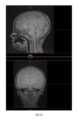
圖2圖解在處理之前自黑骨MRI成像獲得之實例影像。圖3展示處理後之此等影像。圖4展示具有用於生成實例影像之掃描參數的表。Figure 2 illustrates example images obtained from black bone MRI imaging prior to processing. Figure 3 shows these images after processing. Figure 4 shows a table with scan parameters used to generate an example image.
圖6提供用於利用黑骨MRI資料生成擴增實境(AR)視圖以供為特定患者規劃或執行醫療程序使用之實例程序600。對患者執行黑骨MRI 602。自MRI獲得一黑骨資料集604。然後處理此資料集606,以供生成一虛擬模型608。然後可使用該虛擬模型以提供一AR模型610,該模型可用於規劃或執行醫療程序612。FIG. 6 provides an
可對黑骨MRI資料執行各種類型的處理,包括:將黑骨資料集反轉;利用自動偵測演算法,該自動偵測演算法自反轉之黑骨資料集偵測強度範圍與骨之強度範圍類似的像素;及/或利用具有低翻轉角梯度之回波序列,從而提供骨與組織之間的高對比度。舉例而言,該處理可包括使用抹除工具在區域之指定界限內移除在強度範圍外之像素。Various types of processing can be performed on black bone MRI data, including: inverting the black bone data set; using an auto-detection algorithm that detects the intensity range and bone distance from the inverted black bone data set pixels with similar intensity ranges; and/or utilize echo sequences with low flip angle gradients, thereby providing high contrast between bone and tissue. For example, the processing may include removing pixels outside the intensity range within the specified boundaries of the region using a wipe tool.
在一較佳實施例中,黑骨MRI利用在使用1.5T或3.0T磁鐵之情況下在5度翻轉角下4.2 ms且重複時間為8.6 ms之回波時間(TE)序列。此外,MRI可利用具有低翻轉角梯度之回波序列,從而提供骨與組織之間的高對比度。In a preferred embodiment, black bone MRI utilizes an echo time (TE) sequence of 4.2 ms at a 5 degree flip angle with a repetition time of 8.6 ms using a 1.5T or 3.0T magnet. In addition, MRI can utilize echo sequences with low flip angle gradients, thereby providing high contrast between bone and tissue.
在一較佳實施例中,虛擬模型為經組態以突出患者之骨結構的3D 360VR模型。該模型之一實例用途為用於視覺化及診斷骨病。In a preferred embodiment, the virtual model is a 3D 360VR model configured to highlight the patient's bone structure. An example use of the model is for visualization and diagnosis of bone disease.
在一個實例中,如圖5所示,提供虛擬模型300,其展示基於掃描資料之影像,包括工具影像302。舉例而言,可自資料庫擷取資料,諸如2D或3D模型及各種工具之渲染的資料。In one example, as shown in FIG. 5 , a
如可了解,本文中描述之系統為使用者或醫師提供眾多益處。舉例而言,將擴增實境系統用於骨手術或植入物置放或用於任何其他外科程序允許外科醫師更好地為手術做準備且以更安全的方式執行手術。此之所以成為可能,係因為向外科醫師呈現的獨特而新穎之視圖允許外科醫師查看骨與解剖結構之組合,包括軟組織、神經、脊柱、血管、肺等,且允許查看解剖結構,即使該解剖結構被其他組織遮擋。As can be appreciated, the systems described herein provide numerous benefits to the user or physician. For example, using an augmented reality system for bone surgery or implant placement or for any other surgical procedure allows a surgeon to better prepare for surgery and perform it in a safer manner. This is possible because the unique and novel view presented to the surgeon allows the surgeon to view a combination of bone and anatomical structures, including soft tissue, nerves, spine, blood vessels, lungs, etc. The structure is obscured by other tissues.
圖7為用於實施圖1之AR同步電腦112之實例電腦的示意圖。實例電腦700意欲表示各種形式的數位電腦,包括膝上型電腦、桌上型電腦、手持式電腦、平板電腦、智慧型電話、伺服器及其他類似類型的計算裝置。電腦700包括藉由介面710經由匯流排712可操作地連接的處理器702、記憶體704、儲存裝置706及通信埠708。FIG. 7 is a schematic diagram of an example computer for implementing the
處理器702经由記憶體704處理指令以供在電腦600內執行。在一實例實施例中,可使用多個處理器連同多個記憶體。The
記憶體704可為揮發性記憶體或非揮發性記憶體。記憶體704可為電腦可讀媒體,諸如磁碟或光碟。儲存裝置706可為電腦可讀媒體,諸如軟碟裝置、硬碟裝置、光碟裝置、磁帶裝置、快閃記憶體、相變記憶體或其他類似的固態記憶體裝置或裝置之陣列,該陣列包括處於其他組態之儲存區域網路中之裝置。電腦程式產品可有形地體現在諸如記憶體704或儲存裝置706之電腦可讀媒體中。The
電腦700可耦接至一或多個輸入及輸出裝置,諸如顯示器714、印表機716、掃描器718、滑鼠720及HMD 724。
如熟習此項技術者將了解,實例實施例可實現為一方法、系統、電腦程式產品或前述各者之一組合,或可通常利用該方法、系統、電腦程式產品或該組合。因此,實施例中之任一者可採用儲存於儲存裝置中用於在電腦硬體上執行的可執行指令之專用軟體的形式,其中該軟體可儲存於具有具體化於媒體中之電腦可用程式碼的電腦可用儲存媒體上。As those skilled in the art will appreciate, example embodiments may be implemented as, or may generally utilize, a method, system, computer program product, or combination of the foregoing. Accordingly, any of the embodiments may take the form of special purpose software stored in a storage device as executable instructions for execution on computer hardware, where the software may be stored in a computer usable program embodied in a medium codes on computer-usable storage media.
資料庫可使用可購得之電腦應用程式來實現,該等電腦應用程式諸如:開源解決方案,諸如MySQL,或如Microsoft SQL之封閉解決方案,該等封閉解決方案可在所揭示之伺服器或額外電腦伺服器上運行。資料庫可利用關係或面向物件之範例來儲存用於以上揭示之實例實施例之資料、模型及模型參數。此等資料庫可使用已知的資料庫程式設計技術來定制以實現本文中揭示之專門適用性。The database can be implemented using commercially available computer applications such as: open source solutions such as MySQL, or closed solutions such as Microsoft SQL that can be hosted on the disclosed server or Run on an additional computer server. A database may utilize a relational or object-oriented paradigm to store data, models and model parameters for the example embodiments disclosed above. These databases can be customized for a particular suitability disclosed herein using known database programming techniques.
可利用任何合適之電腦可用(電腦可讀)媒體來儲存包含可執行指令之軟體。電腦可用或電腦可讀媒體可為例如但不限於電子、磁性、光學、電磁、紅外線或半導體系統、設備、裝置或傳播媒體。電腦可讀媒體之更具體實例(非詳盡列表)可將包括以下各者:具有一根或多根電線之電連接;有形媒體,諸如攜帶型電腦軟盤、硬碟、隨機存取記憶體(RAM)、唯讀記憶體(ROM)、可抹除可程式化唯讀記憶體(EPROM或快閃記憶體)、光碟唯讀記憶體(CDROM)或其他有形的光學或磁性儲存裝置;或傳輸媒體,諸如支援網際網路或內部網路之傳輸媒體。Software containing executable instructions may be stored on any suitable computer-usable (computer-readable) medium. A computer usable or computer readable medium may be, for example but not limited to, an electronic, magnetic, optical, electromagnetic, infrared or semiconductor system, apparatus, device or propagation medium. More specific examples (not an exhaustive list) of computer readable media would include the following: electrical connections having one or more wires; tangible media such as portable computer floppy disks, hard disks, random access memory (RAM) ), read-only memory (ROM), erasable programmable read-only memory (EPROM or flash memory), compact disc read-only memory (CDROM), or other tangible optical or magnetic storage devices; or transmission media , such as a transmission medium that supports the Internet or an intranet.
在本文件之上下文中,電腦可用或電腦可讀媒體可為能夠含有、儲存、傳達、傳播或傳輸程式指令以供指令執行系統、平台、設備或裝置使用或結合執行系統、平台、設備或裝置使用之任何媒體,執行系統、平台、設備或裝置可包括任何合適的電腦(或電腦系統),包含一或多個可程式化或專用處理器/控制器。電腦可用媒體可包括在基帶中或作為載波之一部分的帶有電腦可用程式碼之傳播資料信號。電腦可用程式碼可使用任何合適之媒體傳輸,媒體傳輸包括但不限於網際網路、有線、光纖電纜、區域通信匯流排、射頻(RF)或其他方式。In the context of this document, a computer-usable or computer-readable medium is any medium that is capable of containing, storing, conveying, distributing, or transporting program instructions for use by or in connection with an instruction execution system, platform, device, or device Any medium, execution system, platform, device or device used may include any suitable computer (or computer system), including one or more programmable or dedicated processors/controllers. A computer usable medium may include a propagated data signal with computer usable code in baseband or as part of a carrier wave. The computer usable code may be transmitted using any suitable medium, including but not limited to the Internet, wireline, fiber optic cable, local communication bus, radio frequency (RF), or other means.
具有用於實行實例實施例之操作之可執行指令的電腦程式碼可藉由使用任何電腦語言之習知方式來編寫,電腦語言包括但不限於:解譯或事件驅動之語言,諸如BASIC、Lisp、VBA或VBScript,或GUI實施例,諸如visual basic;編譯程式設計語言,諸如FORTRAN、COBOL或Pascal;面向物件、腳本化或非腳本化程式設計語言,諸如Java、JavaScript、Perl、Smalltalk、C++、C#、Object Pascal或類似者;人工智慧語言,諸如Prolog;即時嵌入式語言,諸如Ada;或甚至使用梯形邏輯之更直接或簡化程式設計,組譯器語言,或使用適當機器語言之直接程式設計。Computer program code having executable instructions for carrying out operations of the example embodiments may be written in a conventional manner using any computer language including, but not limited to, interpreted or event-driven languages such as BASIC, Lisp , VBA or VBScript, or GUI implementations such as visual basic; compiled programming languages such as FORTRAN, COBOL or Pascal; object-oriented, scripted or non-scripted programming languages such as Java, JavaScript, Perl, Smalltalk, C++, C#, Object Pascal, or similar; artificial intelligence languages, such as Prolog; real-time embedded languages, such as Ada; or even more direct or simplified programming using ladder logic, assembler languages, or direct programming using appropriate machine languages .
就在說明書或申請專利範圍中使用之術語「包括」而言,該術語意圖以類似於術語「包含」在其用作技術方案中之過渡詞時所解譯之方式而為包括性的。此外,在使用術語「或」(例如,A或B)之某種程度上,該術語意圖指「A或B或兩者」。當申請者意圖指示「僅A或B,而非兩者」時,則將採用術語「僅A或B,而非兩者」。因此,術語「或」在本文中之使用為包括性的,而非排他性使用。參見Bryan A. Garner的《現代法律用法詞典624》(第2版,1995年)。同樣,在術語「在……中(in)」或「至……中(into)」在說明書或申請專利範圍中使用之某种程度上,該等術語意圖另外意指「在……上(on)」或「至……上(onto)」。此外,在術語「連接」在說明書或申請專利範圍中使用之某種程度上,該術語意圖不僅「直接連接至」,而且「間接連接至」,諸如經由另一或多個組件連接。As far as the term "comprising" is used in the specification or claims, the term is intended to be inclusive in a manner similar to how the term "comprising" is interpreted when it is used as a transition word in a technical solution. Also, to the extent the term "or" is used (eg, A or B), that term is intended to mean "A or B or both." When the applicant intends to indicate "only A or B, but not both", then the term "only A or B, but not both" will be used. Accordingly, use of the term "or" herein is inclusive, not exclusive. See Bryan A. Garner, Dictionary of Modern Legal Usage 624 (2nd ed., 1995). Likewise, to the extent that the terms "in" or "into" are used in the specification or claim, these terms are intended to mean otherwise "on ( on)" or "to... on (onto)". Furthermore, to the extent the term "connected" is used in the specification or claim, the term is intended to be not only "directly connected to", but also "indirectly connected to", such as via another component or components.
儘管本申請案已藉由對其實施例之描述加以說明且儘管該等實施例已經賦予相當詳細地描述,但申請人之意圖並非將所附申請專利範圍之範疇約束或以任何方式限制於此細節。熟習此項技術者將容易地明白額外的優點及修改。因此,本申請案在其更廣泛態樣中不限於所展示及描述之特定細節、代表性設備及方法及說明性實例。因此,在不脫離申請人之一般發明概念之精神及範疇的情況下,可脫離此等細節。Although the application has been illustrated by the description of its embodiments and although the embodiments have been described in considerable detail, it is the applicant's intention not to restrict or in any way limit the scope of the appended application claims thereto detail. Additional advantages and modifications will be readily apparent to those skilled in the art. Therefore, the application in its broader aspects is not limited to the specific details, the representative apparatus and method, and illustrative examples shown and described. Accordingly, departures may be made from such details without departing from the spirit and scope of applicant's general inventive concept.
100:系統 102:虛擬模型 104:實體模型 106:使用者 108:擴增實境視圖 110:擴增實境頭戴式顯示器 112:AR同步與影像處理電腦 114:虛擬模型資料庫 116:虛擬影像 118:MRI成像系統 120:黑骨MRI資料及影像/黑骨成像資料及影像 300:虛擬模型 302:工具影像 600:實例程序 700:實例電腦 702:處理器 704:記憶體 706:儲存裝置 708:通信埠 710:介面 712:匯流排 714:顯示器 716:印表機 718:掃描器 720:滑鼠 724:HMD100: system 102:Virtual model 104: Solid model 106: user 108: Augmented Reality View 110: Augmented Reality Head Mounted Display 112: AR synchronization and image processing computer 114:Virtual model database 116:Virtual image 118:MRI Imaging System 120:Black bone MRI data and images/black bone imaging data and images 300: virtual model 302:Tool image 600: example program 700: instance computer 702: Processor 704: memory 706: storage device 708: communication port 710: interface 712: busbar 714: display 716:Printer 718:Scanner 720: mouse 724:HMD
在附圖中,圖解多個結構,該等結構與在下文提供的實施方式一起描述所主張之發明的例示性實施例。相同元件用相同元件符號來標識。應理解,展示為單一組件之元件可用多個組件替換,且展示為多個組件之元件可用單一組件替換。圖式未按比例繪製,且出於說明目的,某些元件之比例可被夸示。 圖1圖解使用黑骨MRI資料進行擴增實境模擬之一實例係統。 圖2圖解在處理前的利用黑骨MRI資料之實例影像。 圖3圖解在處理後的利用黑骨MRI資料之實例影像。 圖4展示黑骨MRI成像程序之實例設定的表; 圖5圖解擴增實境模擬之一實例模型影像。 圖6圖解用於處理黑骨MRI資料之一實例方法。 圖7圖解實施圖1之實例系統的一實例電腦。In the drawings, a number of structures are illustrated which, together with the description provided hereinafter, describe illustrative embodiments of the claimed invention. Like elements are identified with like element symbols. It should be understood that elements shown as a single component may be replaced by multiple components and that elements shown as multiple components may be replaced by a single component. The drawings are not drawn to scale and the proportions of some elements may have been exaggerated for illustrative purposes. Figure 1 illustrates an example system for an augmented reality simulation using black bone MRI data. Figure 2 illustrates an example image using black bone MRI data before processing. Figure 3 illustrates an example image using black bone MRI data after processing. Figure 4 shows a table of example settings for a black bone MRI imaging program; FIG. 5 illustrates an example model image of an augmented reality simulation. Figure 6 illustrates one example method for processing black bone MRI data. FIG. 7 illustrates an example computer implementing the example system of FIG. 1 .
100:系統100: system
102:虛擬模型102:Virtual model
104:實體模型104: Solid model
106:使用者106: user
108:擴增實境視圖108: Augmented Reality View
110:擴增實境頭戴式顯示器110: Augmented Reality Head Mounted Display
112:AR同步與影像處理電腦112: AR synchronization and image processing computer
114:虛擬模型資料庫114:Virtual model database
116:虛擬影像116:Virtual image
118:MRI成像系統118:MRI Imaging System
120:黑骨MRI資料及影像/黑骨成像資料及影像120:Black bone MRI data and images/black bone imaging data and images
Claims (21)
Translated fromChineseApplications Claiming Priority (2)
| Application Number | Priority Date | Filing Date | Title |
|---|---|---|---|
| US202163147200P | 2021-02-08 | 2021-02-08 | |
| US63/147,200 | 2021-02-08 |
Publications (1)
| Publication Number | Publication Date |
|---|---|
| TW202302046Atrue TW202302046A (en) | 2023-01-16 |
Family
ID=82704311
Family Applications (1)
| Application Number | Title | Priority Date | Filing Date |
|---|---|---|---|
| TW111104607ATW202302046A (en) | 2021-02-08 | 2022-02-08 | System and method for processing black bone mri data |
Country Status (3)
| Country | Link |
|---|---|
| US (1) | US20220249170A1 (en) |
| TW (1) | TW202302046A (en) |
| WO (1) | WO2022170264A1 (en) |
Family Cites Families (4)
| Publication number | Priority date | Publication date | Assignee | Title |
|---|---|---|---|---|
| US6560477B1 (en)* | 2000-03-17 | 2003-05-06 | The Regents Of The University Of California | Joint imaging system utilizing magnetic resonance imaging and associated methods |
| US20160000326A1 (en)* | 2013-02-21 | 2016-01-07 | Isis Innovation Ltd. | Method of imaging bone |
| US20210223341A1 (en)* | 2018-06-01 | 2021-07-22 | The Trustees Of The University Of Pennsylvania | Solid-state mri as a noninvasive alternative to computed tomography (ct) |
| WO2019245865A1 (en)* | 2018-06-19 | 2019-12-26 | Tornier, Inc. | Mixed reality indication of points at which 3d bone and implant models collide |
- 2022
- 2022-02-08TWTW111104607Apatent/TW202302046A/enunknown
- 2022-02-08WOPCT/US2022/015668patent/WO2022170264A1/ennot_activeCeased
- 2022-02-08USUS17/667,261patent/US20220249170A1/enactivePending
Also Published As
| Publication number | Publication date |
|---|---|
| WO2022170264A1 (en) | 2022-08-11 |
| US20220249170A1 (en) | 2022-08-11 |
Similar Documents
| Publication | Publication Date | Title |
|---|---|---|
| US11983824B2 (en) | System and method for augmenting and synchronizing a virtual model with a physical model | |
| US12086988B2 (en) | Augmented reality patient positioning using an atlas | |
| McJunkin et al. | Development of a mixed reality platform for lateral skull base anatomy | |
| JP2023175709A (en) | Registration for spatial tracking system and augmented reality display | |
| JP2013521971A (en) | System and method for computerized simulation of medical procedures | |
| TW202207242A (en) | System and method for augmented reality spine surgery | |
| Karmonik et al. | Augmented reality with virtual cerebral aneurysms: a feasibility study | |
| CN114173692A (en) | System and method for recommending parameters for surgical procedures | |
| KR102213412B1 (en) | Method, apparatus and program for generating a pneumoperitoneum model | |
| US8781188B2 (en) | Method and device for displaying changes in medical image data | |
| Schenkenfelder et al. | Elastic registration of abdominal MRI scans and RGB-D images to improve surgical planning of breast reconstruction | |
| Nimura et al. | Pneumoperitoneum simulation based on mass-spring-damper models for laparoscopic surgical planning | |
| TW202302046A (en) | System and method for processing black bone mri data | |
| US11393111B2 (en) | System and method for optical tracking | |
| Cotin et al. | Augmented Reality for Computer-Guided Interventions | |
| Palomar et al. | Mr in video guided liver surgery | |
| TW202131875A (en) | System and method for augmenting and synchronizing a virtual model with a physical model | |
| US20250078419A1 (en) | Spine level determination using augmented reality | |
| Kalavakonda | Isosurface Visualization Using Augmented Reality for Improving Tumor Resection Outcomes | |
| Xiang et al. | MRMSim: A Framework for Mixed Reality based Microsurgery Simulation | |
| CN116091517A (en) | Medical image processing method, medical image processing device, storage medium and computer program product |