TW202113062A - Manufacturing method of renal stromal cells - Google Patents
Manufacturing method of renal stromal cellsDownload PDFInfo
- Publication number
- TW202113062A TW202113062ATW109119500ATW109119500ATW202113062ATW 202113062 ATW202113062 ATW 202113062ATW 109119500 ATW109119500 ATW 109119500ATW 109119500 ATW109119500 ATW 109119500ATW 202113062 ATW202113062 ATW 202113062A
- Authority
- TW
- Taiwan
- Prior art keywords
- cells
- renal interstitial
- renal
- medium
- cell
- Prior art date
Links
Images


Classifications
- A—HUMAN NECESSITIES
- A61—MEDICAL OR VETERINARY SCIENCE; HYGIENE
- A61K—PREPARATIONS FOR MEDICAL, DENTAL OR TOILETRY PURPOSES
- A61K35/00—Medicinal preparations containing materials or reaction products thereof with undetermined constitution
- A61K35/12—Materials from mammals; Compositions comprising non-specified tissues or cells; Compositions comprising non-embryonic stem cells; Genetically modified cells
- A61K35/22—Urine; Urinary tract, e.g. kidney or bladder; Intraglomerular mesangial cells; Renal mesenchymal cells; Adrenal gland
- C—CHEMISTRY; METALLURGY
- C12—BIOCHEMISTRY; BEER; SPIRITS; WINE; VINEGAR; MICROBIOLOGY; ENZYMOLOGY; MUTATION OR GENETIC ENGINEERING
- C12N—MICROORGANISMS OR ENZYMES; COMPOSITIONS THEREOF; PROPAGATING, PRESERVING, OR MAINTAINING MICROORGANISMS; MUTATION OR GENETIC ENGINEERING; CULTURE MEDIA
- C12N5/00—Undifferentiated human, animal or plant cells, e.g. cell lines; Tissues; Cultivation or maintenance thereof; Culture media therefor
- C12N5/06—Animal cells or tissues; Human cells or tissues
- C12N5/0602—Vertebrate cells
- C12N5/0618—Cells of the nervous system
- C12N5/0623—Stem cells
- C—CHEMISTRY; METALLURGY
- C12—BIOCHEMISTRY; BEER; SPIRITS; WINE; VINEGAR; MICROBIOLOGY; ENZYMOLOGY; MUTATION OR GENETIC ENGINEERING
- C12N—MICROORGANISMS OR ENZYMES; COMPOSITIONS THEREOF; PROPAGATING, PRESERVING, OR MAINTAINING MICROORGANISMS; MUTATION OR GENETIC ENGINEERING; CULTURE MEDIA
- C12N5/00—Undifferentiated human, animal or plant cells, e.g. cell lines; Tissues; Cultivation or maintenance thereof; Culture media therefor
- C12N5/06—Animal cells or tissues; Human cells or tissues
- C12N5/0602—Vertebrate cells
- C12N5/0684—Cells of the urinary tract or kidneys
- C12N5/0687—Renal stem cells; Renal progenitors
- C—CHEMISTRY; METALLURGY
- C12—BIOCHEMISTRY; BEER; SPIRITS; WINE; VINEGAR; MICROBIOLOGY; ENZYMOLOGY; MUTATION OR GENETIC ENGINEERING
- C12N—MICROORGANISMS OR ENZYMES; COMPOSITIONS THEREOF; PROPAGATING, PRESERVING, OR MAINTAINING MICROORGANISMS; MUTATION OR GENETIC ENGINEERING; CULTURE MEDIA
- C12N2501/00—Active agents used in cell culture processes, e.g. differentation
- C12N2501/10—Growth factors
- C12N2501/115—Basic fibroblast growth factor (bFGF, FGF-2)
- C—CHEMISTRY; METALLURGY
- C12—BIOCHEMISTRY; BEER; SPIRITS; WINE; VINEGAR; MICROBIOLOGY; ENZYMOLOGY; MUTATION OR GENETIC ENGINEERING
- C12N—MICROORGANISMS OR ENZYMES; COMPOSITIONS THEREOF; PROPAGATING, PRESERVING, OR MAINTAINING MICROORGANISMS; MUTATION OR GENETIC ENGINEERING; CULTURE MEDIA
- C12N2501/00—Active agents used in cell culture processes, e.g. differentation
- C12N2501/10—Growth factors
- C12N2501/119—Other fibroblast growth factors, e.g. FGF-4, FGF-8, FGF-10
- C—CHEMISTRY; METALLURGY
- C12—BIOCHEMISTRY; BEER; SPIRITS; WINE; VINEGAR; MICROBIOLOGY; ENZYMOLOGY; MUTATION OR GENETIC ENGINEERING
- C12N—MICROORGANISMS OR ENZYMES; COMPOSITIONS THEREOF; PROPAGATING, PRESERVING, OR MAINTAINING MICROORGANISMS; MUTATION OR GENETIC ENGINEERING; CULTURE MEDIA
- C12N2501/00—Active agents used in cell culture processes, e.g. differentation
- C12N2501/10—Growth factors
- C12N2501/135—Platelet-derived growth factor [PDGF]
- C—CHEMISTRY; METALLURGY
- C12—BIOCHEMISTRY; BEER; SPIRITS; WINE; VINEGAR; MICROBIOLOGY; ENZYMOLOGY; MUTATION OR GENETIC ENGINEERING
- C12N—MICROORGANISMS OR ENZYMES; COMPOSITIONS THEREOF; PROPAGATING, PRESERVING, OR MAINTAINING MICROORGANISMS; MUTATION OR GENETIC ENGINEERING; CULTURE MEDIA
- C12N2501/00—Active agents used in cell culture processes, e.g. differentation
- C12N2501/10—Growth factors
- C12N2501/15—Transforming growth factor beta (TGF-β)
- C—CHEMISTRY; METALLURGY
- C12—BIOCHEMISTRY; BEER; SPIRITS; WINE; VINEGAR; MICROBIOLOGY; ENZYMOLOGY; MUTATION OR GENETIC ENGINEERING
- C12N—MICROORGANISMS OR ENZYMES; COMPOSITIONS THEREOF; PROPAGATING, PRESERVING, OR MAINTAINING MICROORGANISMS; MUTATION OR GENETIC ENGINEERING; CULTURE MEDIA
- C12N2501/00—Active agents used in cell culture processes, e.g. differentation
- C12N2501/30—Hormones
- C12N2501/38—Hormones with nuclear receptors
- C12N2501/385—Hormones with nuclear receptors of the family of the retinoic acid recptor, e.g. RAR, RXR; Peroxisome proliferator-activated receptor [PPAR]
- C—CHEMISTRY; METALLURGY
- C12—BIOCHEMISTRY; BEER; SPIRITS; WINE; VINEGAR; MICROBIOLOGY; ENZYMOLOGY; MUTATION OR GENETIC ENGINEERING
- C12N—MICROORGANISMS OR ENZYMES; COMPOSITIONS THEREOF; PROPAGATING, PRESERVING, OR MAINTAINING MICROORGANISMS; MUTATION OR GENETIC ENGINEERING; CULTURE MEDIA
- C12N2501/00—Active agents used in cell culture processes, e.g. differentation
- C12N2501/40—Regulators of development
- C12N2501/415—Wnt; Frizzeled
- C—CHEMISTRY; METALLURGY
- C12—BIOCHEMISTRY; BEER; SPIRITS; WINE; VINEGAR; MICROBIOLOGY; ENZYMOLOGY; MUTATION OR GENETIC ENGINEERING
- C12N—MICROORGANISMS OR ENZYMES; COMPOSITIONS THEREOF; PROPAGATING, PRESERVING, OR MAINTAINING MICROORGANISMS; MUTATION OR GENETIC ENGINEERING; CULTURE MEDIA
- C12N2501/00—Active agents used in cell culture processes, e.g. differentation
- C12N2501/70—Enzymes
- C12N2501/72—Transferases [EC 2.]
- C12N2501/727—Kinases (EC 2.7.)
- C—CHEMISTRY; METALLURGY
- C12—BIOCHEMISTRY; BEER; SPIRITS; WINE; VINEGAR; MICROBIOLOGY; ENZYMOLOGY; MUTATION OR GENETIC ENGINEERING
- C12N—MICROORGANISMS OR ENZYMES; COMPOSITIONS THEREOF; PROPAGATING, PRESERVING, OR MAINTAINING MICROORGANISMS; MUTATION OR GENETIC ENGINEERING; CULTURE MEDIA
- C12N2502/00—Coculture with; Conditioned medium produced by
- C12N2502/99—Coculture with; Conditioned medium produced by genetically modified cells
- C—CHEMISTRY; METALLURGY
- C12—BIOCHEMISTRY; BEER; SPIRITS; WINE; VINEGAR; MICROBIOLOGY; ENZYMOLOGY; MUTATION OR GENETIC ENGINEERING
- C12N—MICROORGANISMS OR ENZYMES; COMPOSITIONS THEREOF; PROPAGATING, PRESERVING, OR MAINTAINING MICROORGANISMS; MUTATION OR GENETIC ENGINEERING; CULTURE MEDIA
- C12N2503/00—Use of cells in diagnostics
- C12N2503/02—Drug screening
- C—CHEMISTRY; METALLURGY
- C12—BIOCHEMISTRY; BEER; SPIRITS; WINE; VINEGAR; MICROBIOLOGY; ENZYMOLOGY; MUTATION OR GENETIC ENGINEERING
- C12N—MICROORGANISMS OR ENZYMES; COMPOSITIONS THEREOF; PROPAGATING, PRESERVING, OR MAINTAINING MICROORGANISMS; MUTATION OR GENETIC ENGINEERING; CULTURE MEDIA
- C12N2506/00—Differentiation of animal cells from one lineage to another; Differentiation of pluripotent cells
- C12N2506/45—Differentiation of animal cells from one lineage to another; Differentiation of pluripotent cells from artificially induced pluripotent stem cells
- C—CHEMISTRY; METALLURGY
- C12—BIOCHEMISTRY; BEER; SPIRITS; WINE; VINEGAR; MICROBIOLOGY; ENZYMOLOGY; MUTATION OR GENETIC ENGINEERING
- C12N—MICROORGANISMS OR ENZYMES; COMPOSITIONS THEREOF; PROPAGATING, PRESERVING, OR MAINTAINING MICROORGANISMS; MUTATION OR GENETIC ENGINEERING; CULTURE MEDIA
- C12N2513/00—3D culture
- C—CHEMISTRY; METALLURGY
- C12—BIOCHEMISTRY; BEER; SPIRITS; WINE; VINEGAR; MICROBIOLOGY; ENZYMOLOGY; MUTATION OR GENETIC ENGINEERING
- C12N—MICROORGANISMS OR ENZYMES; COMPOSITIONS THEREOF; PROPAGATING, PRESERVING, OR MAINTAINING MICROORGANISMS; MUTATION OR GENETIC ENGINEERING; CULTURE MEDIA
- C12N2533/00—Supports or coatings for cell culture, characterised by material
- C12N2533/50—Proteins
- C12N2533/52—Fibronectin; Laminin
- C—CHEMISTRY; METALLURGY
- C12—BIOCHEMISTRY; BEER; SPIRITS; WINE; VINEGAR; MICROBIOLOGY; ENZYMOLOGY; MUTATION OR GENETIC ENGINEERING
- C12P—FERMENTATION OR ENZYME-USING PROCESSES TO SYNTHESISE A DESIRED CHEMICAL COMPOUND OR COMPOSITION OR TO SEPARATE OPTICAL ISOMERS FROM A RACEMIC MIXTURE
- C12P21/00—Preparation of peptides or proteins
- C12P21/02—Preparation of peptides or proteins having a known sequence of two or more amino acids, e.g. glutathione
Landscapes
- Health & Medical Sciences (AREA)
- Engineering & Computer Science (AREA)
- Life Sciences & Earth Sciences (AREA)
- Biomedical Technology (AREA)
- Zoology (AREA)
- Biotechnology (AREA)
- Chemical & Material Sciences (AREA)
- Organic Chemistry (AREA)
- Genetics & Genomics (AREA)
- Wood Science & Technology (AREA)
- Bioinformatics & Cheminformatics (AREA)
- Cell Biology (AREA)
- General Health & Medical Sciences (AREA)
- Urology & Nephrology (AREA)
- Microbiology (AREA)
- Biochemistry (AREA)
- General Engineering & Computer Science (AREA)
- Developmental Biology & Embryology (AREA)
- Immunology (AREA)
- Animal Behavior & Ethology (AREA)
- Neurology (AREA)
- Virology (AREA)
- Medicinal Chemistry (AREA)
- Pharmacology & Pharmacy (AREA)
- Epidemiology (AREA)
- Neurosurgery (AREA)
- Public Health (AREA)
- Veterinary Medicine (AREA)
- Micro-Organisms Or Cultivation Processes Thereof (AREA)
- Medicines Containing Material From Animals Or Micro-Organisms (AREA)
- Medicines That Contain Protein Lipid Enzymes And Other Medicines (AREA)
- Measuring Or Testing Involving Enzymes Or Micro-Organisms (AREA)
- Preparation Of Compounds By Using Micro-Organisms (AREA)
Abstract
Description
Translated fromChinese本發明係關於用於製造腎間質細胞的方法及培養基,以及使用腎間質細胞製造腎臟類器官(organoid)的方法。The present invention relates to a method and culture medium for producing renal interstitial cells, and a method of using renal interstitial cells to produce kidney organoids.
由慢性腎臟病及急性腎疾病等所引起的腎纖維化,伴隨腎功能之明顯降低,以致對於患者,導入透析或腎移植成為必要。近年,紅血球生成素(EPO)產生細胞及纖維母細胞等腎間質細胞(Renal stromal cell:RSC)參與腎纖維化變得明確。使用腎間質細胞的試驗系統被認為在解明腎纖維化之機制,及確立預防方法及治療方法上有用。Renal fibrosis caused by chronic kidney disease and acute kidney disease is accompanied by a significant decrease in renal function, so that it is necessary for patients to introduce dialysis or kidney transplantation. In recent years, it has become clear that Renal stromal cells (RSC) such as erythropoietin (EPO)-producing cells and fibroblasts are involved in renal fibrosis. The test system using renal interstitial cells is believed to be useful in understanding the mechanism of renal fibrosis and establishing prevention and treatment methods.
在非專利文獻1中,記載從人工多能性幹細胞(induced pluripotent stem cell:iPSC)誘導EPO產生細胞。其誘導方法包含於含有激活素A(activin A)、CHIR99021(GSK3β阻礙劑)及Y-27632(ROCK阻礙劑)之培養基中培養iPSC的步驟等。藉由非專利文獻1記載之方法所得到的EPO產生細胞,為肝細胞系列(Hepatic lineage),與腎間質細胞相異。Non-Patent
又,非專利文獻2記載從iPSC誘導腎前驅細胞(Renal progenitors)。其誘導方法包含於含有激活素A及CHIR99021(GSK3β阻礙劑)之培養基中培養iPSC的步驟等。In addition, Non-Patent
在試管中(in vitro),從iPSC等多能性幹細胞誘導腎間質細胞的技術迄今尚未報導。In vitro, the technique of inducing renal interstitial cells from pluripotent stem cells such as iPSC has not been reported so far.
[先前技術文獻][Prior Technical Literature]
[非專利文獻][Non-Patent Literature]
[非專利文獻1]"源自人類多能性幹細胞之紅血球生成素產生細胞改善小鼠的腎性貧血(Human pluripotent stem cell-derived erythropoietin-producing cells ameliorate renal anemia in mice)", Science Translational Medicine, 2017, Vol. 9, Issue 409,eaaj2300[Non-Patent Document 1] "Human pluripotent stem cell-derived erythropoietin-producing cells ameliorate renal anemia in mice", Science Translational Medicine, 2017, Vol. 9, Issue 409,eaaj2300
[非專利文獻2]"使用源自人工多能性幹細胞之腎前驅細胞的細胞療法減輕小鼠之腎臟損傷(Cell Therapy Using Human Induced Pluripotent Stem Cell-Derived Renal Progenitors Ameliorates Acute Kidney Injury in Mice)", STEM CELLS TRANSLATIONAL MEDICINE 2015;4:980-992[Non-Patent Document 2] "Cell Therapy Using Human Induced Pluripotent Stem Cell-Derived Renal Progenitors Ameliorates Acute Kidney Injury in Mice", STEM CELLS TRANSLATIONAL MEDICINE 2015;4:980-992
[非專利文獻3]"誘導人類ENS細胞系列以赫普隆氏症的細胞治療及藥物開發(Deriving human ENS lineages for cell therapy and drug discovery in Hirschsprung disease)", Nature, 2016, 531, 105-109[Non-Patent Document 3] "Deriving human ENS lineages for cell therapy and drug discovery in Hirschsprung disease", Nature, 2016, 531, 105-109
[非專利文獻4]"經由使用小分子化合物及限定之培養基的神經脊細胞系列,從多能性幹細胞誘導間質基質細胞(Derivation of mesenchymal stromal cells from pluripotent stem cells through a neural crest lineage using small molecule compounds with defined media)", PLoS One, 2014, 9, 12[Non-Patent Document 4] "Derivation of mesenchymal stromal cells from pluripotent stem cells are induced from a series of neural spinal cells using small molecule compounds and a limited culture medium.pluripotent stem cells through a neural crest lineage using small molecule compounds with defined media)", PLoS One, 2014, 9, 12
[非專利文獻5]”用於誘導人工多能性幹細胞的新穎有效無飼養層培養基系統(A novel efficient feeder-free culture system for the derivation of human induced pluripotent stem cells)”, Scientific Reports, 2014, 4, 3594[Non-Patent Document 5] "A novel efficient feeder-free culture system for the derivation of human induced pluripotent stem cells", Scientific Reports, 2014, 4 , 3594
[非專利文獻6]"從人類多能性幹細胞產生腎臟類器官(Generation of kidney organoids from human pluripotent stem cells)", Nature Protocols volume 2016, 11, 1681-1692[Non-Patent Document 6] "Generation of kidney organoids from human pluripotent stem cells", Nature Protocols volume 2016, 11, 1681-1692
[非專利文獻7]"使用人類多能性幹細胞之定向分化以製造腎臟類器官(Making a Kidney Organoid Using the Directed Differentiation of Human Pluripotent Stem Cells)", Methods in Molecular Biology, 2017, 1597, 195-206[Non-Patent Document 7] "Making a Kidney Organoid Using the Directed Differentiation of Human Pluripotent Stem Cells", Methods in Molecular Biology, 2017, 1597, 195-206
為了使用腎間質細胞之試驗系統的構築等,尋求供給腎間質細胞的技術。In order to construct a test system using renal interstitial cells, etc., technologies for supplying renal interstitial cells are sought.
為解決上述問題,本發明提供以下之[1]至[31]。To solve the above problems, the present invention provides the following [1] to [31].
[1]一種腎間質細胞之製造方法,其包含在含有源自血小板之成長因子受體促效劑的培養基中培養腎間質前驅細胞,得到腎間質細胞的步驟(3)。[1] A method for producing renal interstitial cells, which includes the step (3) of culturing renal interstitial precursor cells in a medium containing a platelet-derived growth factor receptor agonist to obtain renal interstitial cells.
[2]如[1]之製造方法,其中前述培養基進一步含有鹼性纖維母細胞成長因子及/或纖維母細胞成長因子9。[2] The production method according to [1], wherein the medium further contains basic fibroblast growth factor and/or
[3]如[1]或[2]之製造方法,其進一步包含從神經脊(neural crest)細胞誘導腎間質前驅細胞的步驟(2)。[3] The manufacturing method of [1] or [2], which further comprises the step (2) of inducing renal interstitial precursor cells from neural crest cells.
[4]如[1]至[3]中任一項之製造方法,其進一步包含在含有GSK3β阻礙劑、TGFβ阻礙劑、以及視黃酸及/或其衍生物的培養基中培養多能性幹細胞,誘導神經脊細胞的步驟(1)。[4] The production method according to any one of [1] to [3], which further comprisesculturing multipotent in a medium containing GSK3 β inhibitor, TGFβ inhibitor, and retinoic acid and/or derivatives thereof Sex stem cells, steps to induce neural crest cells (1).
[4a]如[4]之製造方法,其中前述多能性幹細胞為人工多能性幹細胞。[4a] The manufacturing method according to [4], wherein the aforementioned pluripotent stem cells are artificial pluripotent stem cells.
[4b]如[4]或[4a]之製造方法,其中GSK3β阻礙劑為CHIR98014(N6-[2-[[4-(2,4-二氯苯基)-5-(1H-咪唑-1-基)-2-嘧啶基]胺基]乙基]-3-硝基-2,6-吡啶二胺)。[4b] The manufacturing method as in [4] or [4a], wherein the GSK3β inhibitor is CHIR98014(N6-[2-[[4-(2,4-dichlorophenyl)-5-(1H-imidazole- 1-yl)-2-pyrimidinyl]amino]ethyl]-3-nitro-2,6-pyridinediamine).
[4c]如[4]至[4b]中任一項之製造方法,其中TGFβ阻礙劑為SB431542(4-[4-(1,3-苯并二氧雜環戊烯-5-基)-5-(2-吡啶基)-1H-咪唑-2-基]-苄醯胺)。[4c] The production method according to any one of [4] to [4b], wherein the TGFβ inhibitor is SB431542 (4-[4-(1,3-benzodioxol-5-yl) -5-(2-pyridyl)-1H-imidazol-2-yl]-benzamide).
[5]如[3]至[4c]中任一項之製造方法,其中前述神經脊細胞為後腦神經脊細胞。[5] The production method according to any one of [3] to [4c], wherein the aforementioned neural spinal cell is a posterior brain neural spinal cell.
[6]如[1]至[5]中任一項之製造方法,其中前述腎間質細胞於低氧條件下產生紅血球生成素(erythropoietin)。[6] The production method according to any one of [1] to [5], wherein the aforementioned renal interstitial cells produce erythropoietin under hypoxic conditions.
[7]如[1]至[6]中任一項之製造方法,其中前述源自血小板之成長因子受體促效劑為源自血小板之成長因子(PDGF)。[7] The production method according to any one of [1] to [6], wherein the aforementioned platelet-derived growth factor receptor agonist is platelet-derived growth factor (PDGF).
[8]一種腎間質細胞製造用培養基,其包含源自血小板之成長因子受體促效劑。[8] A medium for the production of renal interstitial cells, which contains a platelet-derived growth factor receptor agonist.
[9]如[8]之培養基,其進一步包含鹼性纖維母細胞成長因子及/或纖維母細胞成長因子9。[9] The medium of [8], which further contains basic fibroblast growth factor and/or
[10]如[8]或[9]之培養基,其中前述源自血小板之成長因子受體促效劑為源自血小板之成長因子(PDGF)。[10] The medium according to [8] or [9], wherein the aforementioned platelet-derived growth factor receptor agonist is platelet-derived growth factor (PDGF).
[11]一種腎臟類器官之製造方法,其包含將腎間質細胞或腎間質前驅細胞與中間中胚葉進行共培養的步驟。[11] A method for producing kidney organoids, which comprises the step of co-cultivating renal interstitial cells or renal interstitial precursor cells and intermediate mesoderm.
[12]如[11]之製造方法,其中腎間質細胞或腎間質前驅細胞為藉由包含步驟(3)的腎間質細胞之製造方法所得到者,其中該腎間質細胞之製造方法的步驟(3)包含在含有源自血小板之成長因子受體促效劑的培養基中培養腎間質前驅細胞而得到腎間質細胞,或腎間質細胞或腎間質前驅細胞為藉由從神經脊細胞誘導腎間質前驅細胞的步驟(2)所得到者。[12] The manufacturing method of [11], wherein the renal interstitial cells or renal interstitial precursor cells are obtained by the method of manufacturing renal interstitial cells including step (3), wherein the manufacturing of the renal interstitial cells Step (3) of the method includes culturing renal interstitial precursor cells in a medium containing platelet-derived growth factor receptor agonists to obtain renal interstitial cells, or renal interstitial cells or renal interstitial precursor cells are obtained by Obtained from step (2) of inducing renal interstitial precursor cells from neural crest cells.
[13]一種篩選腎纖維症之預防或治療用物質的方法,其包含:[13] A method for screening substances for the prevention or treatment of renal fibrosis, which comprises:
將藉由[1]至[7]中任一項之製造方法所得到的腎間質細胞,於誘導該細胞之纖維化的物質存在下培養,提供包含纖維化腎間質細胞之細胞集團的程序;The renal interstitial cells obtained by the manufacturing method of any one of [1] to [7] are cultured in the presence of a substance that induces fibrosis of the cells to provide a cell group containing fibrotic renal interstitial cells program;
於受驗物質存在下及不存在下培養前述細胞集團的程序;The procedure of cultivating the aforementioned cell group in the presence and absence of the test substance;
測定各前述細胞集團中的細胞之纖維化程度的程序;及Procedures for determining the degree of fibrosis of cells in each of the aforementioned cell groups; and
將於前述受驗物質存在下所培養的細胞集團中,與在前述受驗物質不存在下所培養的細胞集團相比,使細胞之纖維化程度降低之受驗物質予以選擇的程序。A procedure for selecting a test substance that reduces the degree of fibrosis of the cells from the cell group cultured in the presence of the test substance compared with the cell group cultured in the absence of the test substance.
[13a]如[13]之方法,其中誘導纖維化之物質為TGFβ(轉化生長因子β)。[13a] The method as in [13], wherein the fibrosis-inducing substance is TGFβ (transforming growth factorβ ).
[14]一種方法,其為決定腎纖維症之生物標記的方法,其包含[14] A method, which is a method for determining a biomarker of renal fibrosis, comprising
將藉由[1]至[7]中任一項之製造方法所得到的腎間質細胞,於誘導該細胞之纖維化的物質存在下培養,提供包含纖維化腎間質細胞之細胞集團的程序;The renal interstitial cells obtained by the manufacturing method of any one of [1] to [7] are cultured in the presence of a substance that induces fibrosis of the cells to provide a cell group containing fibrotic renal interstitial cells program;
鑑定包含於前述細胞集團之培養液中之物質的程序;及Procedures to identify the substances contained in the culture medium of the aforementioned cell group; and
將所鑑定之物質與罹患腎纖維症之哺乳動物的體液中所含之物質做比較,特定出一致之物質的程序。A procedure for comparing the identified substances with the substances contained in the body fluids of mammals suffering from renal fibrosis to identify the same substances.
[14a]如[14]之方法,其中誘導纖維化之物質為TGFβ。[14a] The method as in [14], wherein the fibrosis-inducing substance is TGFβ .
[15]一種製造方法,其為含有腎間質前驅細胞之腎障礙之預防或治療用醫藥的製造方法,其包含從神經脊細胞誘導腎間質前驅細胞的步驟(2)。[15] A manufacturing method, which is a method for manufacturing a medicine for the prevention or treatment of renal disorders containing renal interstitial precursor cells, which includes the step (2) of inducing renal interstitial precursor cells from neural spinal cells.
[16]一種製造方法,其為含有腎間質細胞之腎障礙之預防或治療用醫藥的製造方法,其包含在含有源自血小板之成長因子受體促效劑的培養基中,培養腎間質前驅細胞,得到腎間質細胞的步驟(3)。[16] A manufacturing method, which is a method for manufacturing a medicine for the prevention or treatment of renal disorders containing renal interstitial cells, which comprises culturing the renal interstitium in a medium containing a platelet-derived growth factor receptor agonist Precursor cells to obtain renal interstitial cells (3).
[17]如[15]或[16]之製造方法,其進一步包含在含有GSK3β阻礙劑、TGFβ阻礙劑、以及視黃酸及/或其衍生物的培養基中,培養多能性幹細胞,誘導神經脊細胞的步驟(1)。[17] The production method of [15] or [16], which further comprisesculturing pluripotent stem cells in a medium containing GSK3 β inhibitor, TGFβ inhibitor, and retinoic acid and/or derivatives thereof, Steps to induce neural spinal cells (1).
[18]一種醫藥,其包含源自多能性幹細胞的腎間質前驅細胞及/或源自多能性幹細胞的腎間質細胞。[18] A medicine comprising renal interstitial precursor cells derived from pluripotent stem cells and/or renal interstitial cells derived from pluripotent stem cells.
[18a]如[18]之醫藥,其係用於腎障礙之預防或治療。[18a] The medicine as in [18], which is used for the prevention or treatment of renal disorders.
[19]一種腎障礙之預防劑或治療劑,其包含源自多能性幹細胞的腎間質前驅細胞及/或源自多能性幹細胞的腎間質細胞。[19] A preventive or therapeutic agent for renal disorders, which comprises renal interstitial precursor cells derived from pluripotent stem cells and/or renal interstitial cells derived from pluripotent stem cells.
[20]一種腎障礙之預防或治療方法,其包含將源自多能性幹細胞之腎間質前驅細胞及/或源自多能性幹細胞之腎間質細胞投與至對象的程序。[20] A method for preventing or treating renal disorders, which comprises a procedure of administering renal interstitial precursor cells derived from pluripotent stem cells and/or renal interstitial cells derived from pluripotent stem cells to a subject.
[21]一種源自多能性幹細胞的腎間質前驅細胞及/或源自多能性幹細胞的腎間質細胞,其係用於腎障礙之預防或治療。[21] A renal interstitial precursor cells derived from pluripotent stem cells and/or renal interstitial cells derived from pluripotent stem cells, which are used for the prevention or treatment of renal disorders.
[22]一種源自多能性幹細胞的腎間質前驅細胞及/或源自多能性幹細胞的腎間質細胞之用途,其係用於腎障礙之預防或治療用醫藥的製造。[22] A use of renal interstitial precursor cells derived from pluripotent stem cells and/or renal interstitial cells derived from pluripotent stem cells in the manufacture of medicines for the prevention or treatment of renal disorders.
[23]一種腎間質細胞,其係在包含源自血小板之成長因子受體促效劑的培養基中,培養腎間質前驅細胞而得到。[23] A renal interstitial cell obtained by culturing renal interstitial precursor cells in a medium containing a platelet-derived growth factor receptor agonist.
[23a]如[23]之腎間質細胞,其中前述源自血小板之成長因子受體促效劑為源自血小板之成長因子(PDGF)。[23a] The renal interstitial cell of [23], wherein the aforementioned platelet-derived growth factor receptor agonist is platelet-derived growth factor (PDGF).
[23b]如[23]或[23a]之腎間質細胞,其中前述培養基進一步包含鹼性纖維母細胞成長因子及/或纖維母細胞成長因子9。[23b] The renal interstitial cell of [23] or [23a], wherein the aforementioned medium further comprises basic fibroblast growth factor and/or
[23c]如[23]至[23b]中任一項之腎間質細胞,其係表現CD73及PDGFRβ,於低氧條件下產生紅血球生成素(erythropoietin)。[23c] The renal interstitial cells of any one of [23] to [23b], which express CD73 and PDGFRβ, and produce erythropoietin under hypoxic conditions.
[23d]如[23c]之腎間質細胞,其進一步表現肌間線蛋白(desmin)及波形蛋白(vimentin)。[23d] Renal interstitial cells such as [23c], which further express desmin and vimentin.
[24]一種腎間質前驅細胞,其係在包含StemPro(註冊商標)MSC SFM Xenofree的培養基中,培養神經脊細胞而得到。[24] A renal interstitial precursor cell obtained by culturing neural spinal cells in a medium containing StemPro (registered trademark) MSC SFM Xenofree.
[24a]如[24]之腎間質前驅細胞,其表現FOXD1、CD73及PDGFRβ,不產生紅血球生成素。[24a] Renal interstitial precursor cells such as [24], which express FOXD1, CD73 and PDGFRβ, and do not produce erythropoietin.
[25]一種腎臟類器官,其包含如[23]至[24a]中任一項之腎間質細胞及/或腎間質前驅細胞。[25] A renal organoid comprising the renal interstitial cells and/or renal interstitial precursor cells of any one of [23] to [24a].
[26]一種腎間質細胞分化誘導劑,其包含源自血小板之成長因子受體促效劑。[26] A renal mesenchymal cell differentiation inducer comprising a platelet-derived growth factor receptor agonist.
[26a]如[26]之腎間質細胞分化誘導劑,其中前述源自血小板之成長因子受體促效劑為源自血小板之成長因子(PDGF)。[26a] The renal interstitial cell differentiation inducer of [26], wherein the aforementioned platelet-derived growth factor receptor agonist is platelet-derived growth factor (PDGF).
[27]一種源自人工多能性幹細胞之腎間質前驅細胞。[27] A renal interstitial precursor cell derived from artificial pluripotent stem cells.
[28]一種腎間質前驅細胞之製造方法,其包含從神經脊細胞誘導腎間質前驅細胞的步驟(2)。[28] A method for producing renal interstitial precursor cells, which comprises the step (2) of inducing renal interstitial precursor cells from neural spinal cells.
[29]一種源自人工多能性幹細胞之腎間質細胞。[29] A renal interstitial cell derived from artificial pluripotent stem cells.
[30]一種腎間質細胞分化誘導劑,其包含STEMdiff APEL2培養基(STEMCELL Technologies,ST-05275)。[30] A renal mesenchymal cell differentiation inducer comprising STEMdiff APEL2 medium (STEMCELL Technologies, ST-05275).
[31]一種腎間質細胞之製造方法,其包含在STEMdiff APEL2培養基(STEMCELL Technologies,ST-05275)中培養腎間質前驅細胞的步驟。[31] A method for producing renal interstitial cells, which includes the step of culturing renal interstitial precursor cells in STEMdiff APEL2 medium (STEMCELL Technologies, ST-05275).
[定義][definition]
在本發明中,「源自血小板之成長因子受體促效劑」意指結合於源自血小板之成長因子受體(Platelet derived growth factor receptor(PDGFR)),可媒介以該受體之磷酸化為起點之信號傳達的物質。In the present invention, "platelet-derived growth factor receptor agonist" means that it binds to platelet-derived growth factor receptor (Platelet derived growth factor receptor (PDGFR)) and can mediate the phosphorylation of the receptor The substance conveyed by the signal of the starting point.
「源自血小板之成長因子受體(Platelet derived growth factor receptor (PDGFR)」中,存在PDGFRα及PDGFRβ之二種亞型。PDGFR藉由與源自血小板之成長因子(Platelet derived growth factor(PDGF))等配體之結合,形成二聚體,該二聚體中,存在PDGFR-α α、PDGFR-α β、PDGFR-β β之三種組合。藉由配體與PDGFR結合,PDGFR之酪胺酸殘基接受自身磷酸化,成為與具有SH2結構域之信號傳達分子(PLC-γ、Grb2、PI3K等)的結合部位,向下游傳達信號。In "Platelet derived growth factor receptor (PDGFR)", there aretwo subtypes of PDGFR α and PDGFRβ . PDGFR is combined with platelet derived growth factor (PDGFR). )) and other binding ligands, the formation of a dimer, the dimer, the presence of PDGFR- αα, three combinations PDGFR- αβ, PDGFR- β β's. by ligand binding to PDGFR, PDGFR of tyramine The acid residue undergoes autophosphorylation and becomes a binding site with SH2 domain-containing signal transmission molecules (PLC-γ , Grb2, PI3K, etc.), and transmits signals downstream.
源自血小板之成長因子受體促效劑,可為抗體、肽、低分子化合物等,較佳為PDGF。The growth factor receptor agonist derived from platelets may be antibodies, peptides, low-molecular compounds, etc., preferably PDGF.
「腎間質細胞(Renal Stromal Cell:RSC)」意指CD73陽性且PDFGRβ陽性之細胞,可產生紅血球生成素(以下,在本說明書中稱為「EPO」;若未特別限定,「EPO」意指紅血球生成素蛋白質)。"Renal Stromal Cell (RSC)" means CD73-positive and PDFGRβ- positive cells that can produce erythropoietin (hereinafter, referred to as "EPO" in this specification; if not specifically limited, "EPO" Means erythropoietin protein).
腎間質細胞之「纖維化」,意指非α SMA陽性之腎間質細胞成為α SMA陽性之細胞。又,腎間質細胞成為α SMA陽性,意指彼等分化為肌纖維母細胞(myofibroblast)。該分化可藉由腎間質細胞之TGFβ 1刺激而誘導。該分化有伴隨細胞之肥大化的情況。肌纖維母細胞為α SMA陽性。The "fibrosis"of renal interstitial cells means that non-α SMA-positive renal interstitial cells becomeα SMA-positive cells. In addition, renal interstitial cells becomeα SMA positive, which means that they differentiate into myofibroblasts. This differentiation can be induced by
「腎間質前驅細胞(Renal Stromal Precursor:RSP)」意指FOXD1陽性、CD73陽性且PDGFRβ陽性之細胞,不產生EPO。"Renal Stromal Precursor (RSP)" means FOXD1-positive, CD73-positive, and PDGFRβ- positive cells that do not produce EPO.
「神經脊細胞(Neural Crest Cell:NCC)」,意指在發育初期,從神經板形成神經管時,於神經外胚葉與表皮外胚葉之間產生的細胞,具有分化為神經細胞、神經膠細胞、間葉系間質細胞、骨細胞、軟骨細胞、角膜細胞及色素細胞等多種細胞的多潛能性(multipotency)及自身增殖能力。神經脊細胞為SOX10陽性。"Neural Crest Cell (NCC)" refers to the cells produced between the neuroectodermal and epidermal ectoderm when the neural tube is formed from the neural plate at the early stage of development. It has differentiation into nerve cells and glial cells. , Mesenchymal cells, bone cells, chondrocytes, corneal cells and pigment cells and other cells multipotency (multipotency) and self-proliferation ability. Nerve spinal cells are SOX10 positive.
「後腦神經脊細胞(hindbrain NCC:hNCC)」意指後方化之NCC,具有為神經脊細胞標記的SOX10陽性、NGFR陽性、HOXB4陽性、紅血球生成素(EPO)陽性。"Hindbrain NCC: hNCC" means posteriorized NCC, which has SOX10 positive, NGFR positive, HOXB4 positive, and erythropoietin (EPO) positive as a marker for neural spinal cells.
「後方化」為將發育期的體軸之頭尾(Anterior-Posterior Axis)偏向較尾側(Posterior),在本發明中,意指將從多能性幹細胞分化之中腦神經脊細胞(midbrain NCC)誘導成後腦神經脊細胞(hindbrain NCC)。"Posteriorization" refers to shifting the head and tail of the developmental body axis (Anterior-Posterior Axis) to the more caudal side (Posterior). In the present invention, it means that the midbrain neurospinal cells (midbrain cells) are differentiated from pluripotent stem cells. NCC) is induced into hindbrain NCC.
「中間中胚葉(intermediate mesoderm)」意指在個體之發育中,從中胚葉發育的胚之一種,為可分化成前腎、中腎、中腎管、後腎、副腎皮質及生殖腺的細胞,為OSR1(odd-skipped related 1)陽性。"Intermediate mesoderm" means a kind of embryo that develops from mesoderm during the development of an individual. It is a cell that can differentiate into pronephric, mesoderm, mesoderm, metanephric, adrenal cortex, and gonads. OSR1 (odd-skipped related 1) was positive.
「腎臟類器官」意指包含構成活體內之腎臟組織的至少1種以上之細胞集團的3次元構造體。"Kidney organoid" means a three-dimensional structure containing at least one type of cell group that constitutes kidney tissue in the living body.
「TGFβ阻礙劑」意指對TGFβ(轉化生長因子β)具有阻礙活性的物質。TGFβ為結合於2種絲胺酸/蘇胺酸蛋白質激酶型受體的細胞激素,經由以Smad(R-Smad)之活化為主的信號傳達,控制細胞增殖、細胞分化及細胞死亡等。就具有TGFβ阻礙活性之物質而言,可列舉阻礙TGFβ與其受體之結合的物質,或阻礙TGFβ與受體結合後之下游信號的物質。就該下游信號而言,可例示由TGFβ II型受體所導致的TGFβ I型受體之磷酸化、由磷酸化TGFβ I型受體所導致的Smad之磷酸化等。本發明中所用之「TGFβ阻礙劑」,只要具有TGFβ阻礙活性,無特別限定。"TGFβ inhibitor" means a substance that has inhibitory activityon TGF β (transforming growth factorβ). TGFβ is a cytokine that binds to two serine/threonine protein kinase type receptors, and controls cell proliferation, cell differentiation, and cell death through signal transmission based on the activation of Smad (R-Smad). Examples of substances having TGFβ blocking activity include substances that blockthe binding of TGF β to its receptor, or substances that blockdownstream signals after TGF β binds to the receptor. In respect of downstream signals, can be exemplified by phosphorylation ofTGF β type I receptor type II receptor ofTGF β caused, phosphorylation, etc. of Smad phosphorylation by theTGF β type I receptor resulting.The "TGF β inhibitor" used in the present invention is not particularly limited as long as it has TGFβ inhibitory activity.
「GSK3β阻礙劑」意指對GSK3β(糖原合成酶激酶3β)具有阻礙活性的物質。GSK3(糖原合成酶激酶3)為絲胺酸/蘇胺酸蛋白質激酶之一種,參與糖原之產生、細胞凋亡、幹細胞之維持等相關的多個信號途徑。GSK3存在α及β之2個同功型(isoform)。本發明中所用的「GSK3β阻礙劑」,只要具有GSK3β阻礙活性,無特別限定,亦可為兼具GSK3β阻礙活性合併GSK3α阻礙活性的物質。"GSK3β inhibitor" means a substance that has inhibitory activityon GSK3 β (
「培養」意指使細胞於試管(in vitro)環境中維持,並使其增殖(成長)、及/或分化。「進行培養」意指在組織外或體外,例如,於細胞培養皿或燒瓶中維持細胞、使其增殖(成長)、及/或分化。"Culturing" means maintaining cells in an in vitro environment and allowing them to proliferate (grow) and/or differentiate. "Culturing" means to maintain, proliferate (grow), and/or differentiate cells outside of tissue or in vitro, for example, in a cell culture dish or flask.
「細胞集團」意指相同種類或相異種類之2個以上細胞。「細胞集團(population)」亦意指相同種類或相異種類之細胞的一塊(mass)。"Cell group" means two or more cells of the same type or different types. "Population" also means a mass of cells of the same type or different types.
「附著培養」意指將細胞以附著於容器之狀態,例如於適當培養基存在下,將細胞以附著於滅菌塑膠(或塗覆之塑膠)之細胞培養皿或燒瓶的狀態進行培養。"Attached culture" means that cells are attached to a container, for example, in the presence of a suitable medium, the cells are cultured in a state of attaching to a sterile plastic (or coated plastic) cell culture dish or flask.
「懸浮培養」意指將細胞以不附著於容器,以分散於適當培養基中的狀態進行培養。"Suspension culture" means that cells are cultured in a state dispersed in an appropriate medium without attaching to a container.
「維持培養(Sustain)」意指將期望之細胞集團維持彼等之數目同時進行培養。細胞數目之維持,可為藉由細胞在不增殖下生存而達到者,亦可為藉由細胞之增殖所造成的增數與死滅所造成的減數互相抵消而達到者。細胞數目之維持,未必需要使細胞之數目維持完全相同,只要依照本發明之目的,使細胞之數目維持實質上相同即可。"Sustain" means to maintain the number of desired cell groups while culturing them. The maintenance of the cell number can be achieved by the survival of the cells without proliferation, or by the increase in cell proliferation and the decrease in death caused by offsetting each other. The maintenance of the number of cells does not necessarily require the number of cells to be kept exactly the same, as long as the number of cells is kept substantially the same in accordance with the purpose of the present invention.
「擴大培養(Expand)」意指以使期望之細胞集團增殖,使細胞數增加為目的而進行培養。細胞數之增加,只要藉由細胞之增殖所造成的增數超過死滅所造成的減數而達成者即可,未必需要細胞集團之全部細胞均增殖。細胞數目之增加,與擴大培養之開始前相比,可為1.1倍、1.2倍、1.5倍、2倍、3倍、4倍、5倍、6倍、7倍、8倍、9倍、10倍、15倍、20倍、30倍以上。"Expanded culture (Expand)" refers to culture for the purpose of proliferating a desired cell group and increasing the number of cells. The increase in the number of cells can be achieved as long as the increase caused by cell proliferation exceeds the decrease caused by death, and it does not necessarily require that all cells of the cell group proliferate. The increase in cell number can be 1.1 times, 1.2 times, 1.5 times, 2 times, 3 times, 4 times, 5 times, 6 times, 7 times, 8 times, 9 times, 10 times, compared with before the start of expansion culture. Times, 15 times, 20 times, 30 times or more.
「多能性(pluripotency)」意指可分化成具有各種相異之型態或功能的組織或細胞,亦能分化成3胚葉之任何系統之細胞的能力。「多能性(pluripotency)」,無法分化成胚盤,因此從「無法形成個體之能力」的觀點而言,與可分化成身體之所有組織-包含胚盤-的「全能性(totipotency)」有所區別。"Pluripotency" refers to the ability to differentiate into tissues or cells with various types or functions, and to differentiate into cells of any system of three embryonic leaves. "Pluripotency" cannot differentiate into the blastoderm, so from the point of view of "the ability to form an individual," it is related to the "totipotency" that can differentiate into all tissues of the body including the blastoderm. There is a difference.
「多潛能性(multipotency)」意指可分化成複數個限定數目之系統之細胞的能力。例如,間葉系幹細胞、造血幹細胞、神經幹細胞為多潛能性(multipotency),而非多能性(pluripotency)。ENP具有分化成神經細胞及神經膠細胞的多潛能性(multipotency)。"Multipotency" means the ability to differentiate into a plurality of cells of a limited number of systems. For example, mesenchymal stem cells, hematopoietic stem cells, and neural stem cells are multipotency rather than pluripotency. ENP has the multipotency to differentiate into nerve cells and glial cells.
「標記」意指為「標記蛋白質」或「標記基因」,在設定之細胞類型中,於細胞表面、細胞質內及/或核內等特異地表現的蛋白質或其基因。標記可為陽性選擇標記或陰性選擇標記。較佳之標記為細胞表面標記,尤其若藉由細胞表面陽性選擇標記,實施生存細胞之濃縮、單離、及/或檢測將成為可能。"Marker" means "marker protein" or "marker gene". In the set cell type, a protein or its gene that is specifically expressed on the cell surface, in the cytoplasm and/or in the nucleus, etc. The marker can be a positive selection marker or a negative selection marker. The preferred markers are cell surface markers, especially if positive selection markers on the cell surface make it possible to concentrate, isolate, and/or detect viable cells.
標記蛋白質之檢測,可採用使用該標記蛋白質特異性抗體的免疫學檢定,例如,ELISA、免疫染色、流式細胞術(flow cytometry)等而進行。就標記蛋白質特異性抗體而言,可使用與標記蛋白質中特定之胺基酸序列或結合於標記蛋白質之特定糖鏈等結合的抗體。又,在細胞內表現,而未出現於細胞表面之標記蛋白質(例如轉錄因子或其亞單元(subunit)等)的情況,使報告蛋白質(reporter protein)與該標記蛋白質一起表現,藉由檢測該報告蛋白質,可檢測為對象的標記蛋白質(例如,非專利文獻4)。此方法較佳用於適當的細胞表面標記尚未被認定的情況。標記基因之檢測,可利用該領域中周知之核酸擴增方法及/或核酸檢測方法,例如,RT-PCR、微陣列(microarray)、生物晶片及RNAseq等而進行。The detection of the labeled protein can be performed using immunological assays using specific antibodies for the labeled protein, for example, ELISA, immunostaining, flow cytometry, and the like. As for the labeled protein-specific antibody, an antibody that binds to a specific amino acid sequence in the labeled protein or a specific sugar chain bound to the labeled protein can be used. In addition, if the marker protein (such as transcription factor or its subunit, etc.) is expressed in the cell but does not appear on the cell surface, the reporter protein is expressed together with the marker protein, by detecting the Reporter protein is a marker protein that can be detected as a target (for example, Non-Patent Document 4). This method is preferably used in situations where appropriate cell surface markers have not yet been identified. The detection of marker genes can be carried out by using well-known nucleic acid amplification methods and/or nucleic acid detection methods in the field, such as RT-PCR, microarray, biochip, and RNAseq.
「表現(expression)」可被定義為由細胞內之啟動子(promotor)所驅動的特定核苷酸序列之轉錄及/或轉譯。"Expression" can be defined as the transcription and/or translation of a specific nucleotide sequence driven by a promoter (promotor) in the cell.
「陽性(positive)」或「表現」,意指蛋白質或基因藉由該領域中周知之手法,以可檢測之量表現。蛋白質之檢測可採用使用抗體之免疫學檢定,例如,ELISA、免疫染色、流式細胞術而進行。又,在細胞內表現,而於細胞表面未表現之蛋白質(例如轉錄因子或其亞單元等)的情況,使報告蛋白質與該蛋白質一起表現,藉由檢測該報告蛋白質,可檢測為對象之蛋白質。基因之檢測,例如,可利用RT-PCR、微陣列、生物晶片及RNAseq等之核酸擴增方法及/或核酸檢測方法而進行。"Positive (positive)" or "performance" means that proteins or genes are expressed in detectable quantities by methods well known in the field. Protein detection can be performed by immunological assays using antibodies, for example, ELISA, immunostaining, and flow cytometry. Also, the protein that is expressed in the cell but is not expressed on the surface of the cellIn the case of quality (for example, transcription factor or its subunits, etc.), the reporter protein is expressed together with the protein. By detecting the reporter protein, the target protein can be detected. Gene detection, for example, can be performed by nucleic acid amplification methods and/or nucleic acid detection methods such as RT-PCR, microarray, biochip, and RNAseq.
「陰性(negative)」或「無表現」意指蛋白質或基因之表現量,藉由如上述之周知手法之全部或任一種,均未達檢測下限值。蛋白質或基因之表現的檢測下限值,隨各種手法而異。"Negative" or "no expression" means that the expression level of protein or gene, by all or any of the above-mentioned well-known methods, does not reach the lower limit of detection. The detection limit of protein or gene expression varies with various methods.
「紅血球生成素(Erythropoietin)」(以下,在本說明書中,稱為「EPO」;又,若未特別限定,「EPO」意指紅血球生成素蛋白質。)為促進紅血球之產生的造血因子之一,由165個胺基酸構成。分子量為約34000。EPO在胎兒及新生兒時期,係從肝臟細胞分泌,然而從妊娠後期至成人時期,係從腎間質細胞分泌(非專利文獻1)。"Erythropoietin" (hereinafter, referred to as "EPO" in this specification; if not specifically limited, "EPO" means erythropoietin protein.) is one of hematopoietic factors that promote the production of red blood cells , Consists of 165 amino acids. The molecular weight is about 34,000. EPO is secreted from liver cells during the fetus and newborn period, but from the renal interstitial cells from late pregnancy to adulthood (Non-Patent Document 1).
「CD73」亦稱為5’-核苷酸酶(5’-NT),為藉由NT5E基因編碼的膜蛋白質。70kDa之亞單元形成二聚體,錨定(anchored)在GPI,存在於細胞表面。CD73主要表現於間葉系幹細胞,亦可見表現於T細胞、B細胞等免疫系細胞,或上皮細胞及內皮細胞之一部分。具有5'-核苷酸酶之酵素活性,催化嘌呤及嘧啶之核糖、去氧核糖核苷一磷酸之形成對應核苷的脫磷酸化。"CD73" is also known as 5'-nucleotidase (5'-NT), which is a membrane protein encoded by the NT5E gene. The 70kDa subunit forms a dimer, anchored in GPI, and exists on the cell surface. CD73 is mainly expressed in mesenchymal stem cells, but also in T cells, B cells and other immune cells, or part of epithelial cells and endothelial cells. It has the enzyme activity of 5'-nucleotidase and catalyzes the formation of purine and pyrimidine ribose and deoxyribonucleoside monophosphate corresponding to the dephosphorylation of nucleosides.
「肌間線蛋白(desmin)」為屬於細胞質內之蛋白質性纖維系統之一的中間絲蛋白III類之蛋白質。在肌細胞等中為構造完整性及生存所必需的因子。"Desmin" is a protein of intermediate filament protein III, which is one of the proteinaceous fibrous systems in the cytoplasm. It is a factor necessary for structural integrity and survival in muscle cells.
「波形蛋白(vimentin)」為細胞質內之蛋白質性纖維系統之一的中間絲蛋白III類之蛋白質,表現於纖維母細胞、血管內皮細胞、平滑肌細胞、橫紋肌細胞、骨/軟骨細胞、神經鞘細胞等間葉系細胞。"Vimentin" is a protein of intermediate filaggrin III, which is one of the proteinaceous fibrous systems in the cytoplasm. It is expressed in fibroblasts, vascular endothelial cells, smooth muscle cells, striated muscle cells, bone/chondrocytes, nerve sheath cells And other mesenchymal cells.
「Forkhead box D:FOXD1」為重新編程(reprogramming)控制因子之一,係在已進行重新編程(reprogramming)之細胞中表現上升時期的特異性標記。在多能性幹細胞中則不表現。"Forkhead box D: FOXD1" is one of the reprogramming control factors, which is a specific marker of the rising period in cells that have undergone reprogramming. It does not appear in pluripotent stem cells.
「SOX10」,於全部神經脊細胞中皆可見到表現。另一方面,SOX10在腎間質前驅細胞及腎間質細胞中不表現。"SOX10" can be seen in all nerve spinal cells. On the other hand, SOX10 is not expressed in renal interstitial precursor cells and renal interstitial cells.
「神經成長因子受體(Nerve growth factor receptor:NGFR)」為結合神經成長因子(Nerve growth factor:NGF)的受體。"Nerve growth factor receptor (Nerve growth factor receptor: NGFR)" is a receptor that binds to nerve growth factor (Nerve growth factor: NGF).
「HOXB4」為Antp同源匣(homeobox)蛋白質之一,由同源匣B基因簇內之HOXB4基因編碼。HOXB4蛋白質具有同源匣DNA結合區域,在個體發育時有作為特異轉錄因子的功能。"HOXB4" is one of the Antp homeobox proteins, which is encoded by the HOXB4 gene in the homeobox B gene cluster. The HOXB4 protein has a homeobox DNA binding region and functions as a specific transcription factor during ontogeny.
「類神經EGFL因子2(Neural EGFL Like 2:NELL2)」為具有類表皮生長因子(epidermal growth factor)(EGF)重複單元的蛋白質激酶C結合蛋白,於身體內,在中樞神經之表現高,亦表現在包含腎間質細胞的各種間質細胞之細胞質內。"Neural EGFL Like 2: NELL2" is a protein kinase C binding protein with epidermal growth factor (EGF) repeating units. It has a high performance in the central nervous system in the body. It is expressed in the cytoplasm of various interstitial cells including renal interstitial cells.
「配對盒蛋白-7(Paired box protein-7:PAX7)」為神經脊之發育及分化所必要的轉錄因子,與該神經脊之發育及分化所必要的PAX3形成雜二聚體,並與DNA結合。在身體中,亦表現在肌衛星細胞或腎間質細胞。"Paired box protein-7 (PAX7)" is a transcription factor necessary for the development and differentiation of the neural ridge, and forms a heterodimer with PAX3 necessary for the development and differentiation of the neural ridge, and combines with DNA Combine. In the body, it is also manifested in muscle satellite cells or renal interstitial cells.
「Nik相關激酶(Nik Related Kinase:NRK)」為絲胺酸/蘇胺酸激酶之一,在TNF-JNK信號傳遞途徑(signaling pathway)之磷酸化具有功能。基因存在於X染色體,在身體中,亦表現在睪丸或卵巢等生殖器或腎間質細胞。"Nik Related Kinase (Nik Related Kinase: NRK)" is one of serine/threonine kinases, which has a function in the phosphorylation of the TNF-JNK signaling pathway. Genes exist on the X chromosome, and in the body, they are also expressed in genital or renal interstitial cells such as testicles or ovaries.
「包含~(comprise(s)或comprising)」意指雖表示包含該語句後續之要素,然而並非限定於此等。因此,雖暗示包含該語句後續之要素,然而並非暗示排除其他任何要素。"Containing ~ (comprise(s) or comprising)" means that although it includes the elements following the sentence, it is not limited to these. Therefore, although the inclusion of the elements subsequent to the sentence is implied, it does not imply the exclusion of any other elements.
「約」或「大概」,表示相對於基準值,分別為正或負至30%、25%、20%、15%、10%、8%、6%、5%、4%、3%、2%或1%而變動的值。較佳而言,「約」或「大概」之術語,表示相對於基準值,分別為正或負15%、10%、5%、或1%之範圍。"About" or "approximately" means that relative to the benchmark value, it is positive or negative to 30%, 25%, 20%, 15%, 10%, 8%, 6%, 5%, 4%, 3%, The value varies by 2% or 1%. Preferably, the term "about" or "approximately" means a range of plus or minus 15%, 10%, 5%, or 1%, respectively, relative to the reference value.
依照本發明,可提供用來供給腎間質細胞的技術。According to the present invention, a technique for supplying renal interstitial cells can be provided.
圖1係展示在從人工多能性幹細胞誘導的神經脊細胞中,EPO及SOX10之表現的免疫組織化學染色圖。Figure 1 is an immunohistochemical staining diagram showing the expression of EPO and SOX10 in neural spinal cells induced from artificial pluripotent stem cells.
圖2係展示在將RA濃度調成0μM(-)、1μM(低)及3μM(高)之情況,於各培養日數測定從神經脊細胞分泌至培養基中之EPO蓄積量之結果的圖。縱軸表示EPO濃度(mIU/ml)。橫軸表示培養日數(Day6-21),「iPSC」表示作為陰性對照之人工多能性幹細胞的測定值。Figure 2 shows that when the concentration of RA is adjusted to 0μ M (-), 1μ M (low), and 3μ M (high), the accumulation of EPO secreted from neural spinal cells into the culture medium is measured on each culture day Figure of the result of the measurement. The vertical axis represents EPO concentration (mIU/ml). The horizontal axis represents the number of culture days (Day 6-21), and "iPSC" represents the measured value of artificial pluripotent stem cells as a negative control.
圖3係展示低氧刺激(FG2216或FG4592)後方化之神經脊細胞,測定EPO分泌量之結果的圖。縱軸表示EPO濃度(mIU/ml)。Figure 3 is a graph showing the results of measuring the amount of EPO secreted by neural spinal cells after hypoxia stimulation (FG2216 or FG4592). The vertical axis represents EPO concentration (mIU/ml).
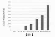
圖4係展示測定在從人工多能性幹細胞誘導之後腦神經脊細胞中之HOXB4表現量之結果的圖。縱軸係將後腦神經脊細胞中的HOXB4之表現量,該表現量以人工多能性幹細胞株(「iPSC」)之表現量當作1時的相對值來表示。橫軸表示視黃酸之濃度。Figure 4 is a graph showing the results of measuring the expression level of HOXB4 in cranial nerve spinal cells after induction from artificial pluripotent stem cells. On the vertical axis, the expression level of HOXB4 in the posterior cranial nerve spinal cells is expressed as a relative value when the expression level of an artificial pluripotent stem cell line ("iPSC") is regarded as 1. The horizontal axis represents the concentration of retinoic acid.
圖5係展示在將RA濃度調成0-10μM之情況,測定從後腦神經脊細胞分泌至培養基中之EPO蓄積量之結果的圖。縱軸表示EPO濃度(mIU/ml)。橫軸表示視黃酸之濃度。「iPSC」表示作為陰性對照之人工多能性幹細胞的測定值。Figure 5 is displayed in the case of RA to adjust the concentration of0-10 μ M, measured after secretion from the cells to the brain spinal result accumulation amount of EPO in the medium of FIG. The vertical axis represents EPO concentration (mIU/ml). The horizontal axis represents the concentration of retinoic acid. "IPSC" represents the measured value of artificial pluripotent stem cells as a negative control.
圖6係展示測定敲入(knock-in)EPO-emGFP基因之人類人工多能性幹細胞株(「iPSC」)、揀選(sorting)前之後腦神經脊細胞(「not sort」)、GFP陰性後腦神經脊細胞(「GFP-sort」)及GFP陽性後腦神經脊細胞(「GFP+sort」)中之EPO mRNA表現量之結果的圖。縱軸係將EPO mRNA表現量以敲入(knock-in)EPO-emGFP基因之人類人工多能性幹細胞株(「iPSC」)之表現量當作1時的相對值來表示。Figure 6 shows the human artificial pluripotent stem cell line ("iPSC") with knock-in EPO-emGFP gene determination, the cranial nerve spinal cells before and after sorting ("not sort"), and the GFP-negative hindbrain A graph showing the results of the expression level of EPO mRNA in neural spinal cells ("GFP-sort") and GFP-positive brain neural spinal cells ("GFP+sort"). On the vertical axis, the expression level of EPO mRNA is expressed as a relative value when the expression level of the human artificial pluripotent stem cell line ("iPSC") with knock-in EPO-emGFP gene is regarded as 1.
圖7係展示測定後腦神經脊細胞(「NCC D18」)、腎間質前驅細胞(「RSP D39」)、腎間質細胞(「APEL D15」)及經低氧刺激之腎間質細胞(「+FG4592」)之EPO mRNA表現量之結果的圖。縱軸係將EPO mRNA表現量以敲入EPO-emGFP基因之人類人工多能性幹細胞株(「iPSC」)之表現量當作1時的相對值來表示。Figure 7 shows the brain neural spinal cells ("NCC D18"), renal interstitial precursor cells ("RSP D39"), renal interstitial cells ("APEL D15") and hypoxia-stimulated renal interstitial cells (" +FG4592”) shows the results of the expression level of EPO mRNA. On the vertical axis, the expression level of EPO mRNA is expressed as a relative value when the expression level of the human artificial pluripotent stem cell line ("iPSC") knocked into the EPO-emGFP gene is taken as 1.
圖8係展示從腎間質前驅細胞誘導之腎間質細胞中之低氧回應性EPO蛋白質之表現的免疫組織化學染色圖。紅色表示EPO蛋白質,藍色表示細胞核。Figure 8 is an immunohistochemical staining diagram showing the expression of hypoxia-responsive EPO protein in renal interstitial cells induced from renal interstitial precursor cells. Red represents EPO protein, blue represents cell nucleus.
圖9係展示於bFGF、FGF9及PDGF-BB存在下,誘導之腎間質細胞的細胞增殖(A)及EPO分泌量(B)之測定結果圖。Figure 9 is a graph showing the measurement results of the cell proliferation (A) and EPO secretion (B) of renal interstitial cells induced in the presence of bFGF, FGF9 and PDGF-BB.
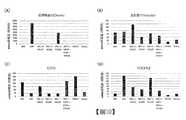
圖10係展示測定低氧刺激之腎間質細胞之腎間質細胞標記(肌間線蛋白(desmin)、波形蛋白(vimentin)、CD73、PDGFRβ)之表現量之結果的圖。縱軸係將mRNA表現量以人工多能性幹細胞株之表現量當作1時的相對值來表示。Figure 10 is a graph showing the results of measuring the expression levels of renal interstitial cell markers (desmin, vimentin, CD73, PDGFRβ) of renal interstitial cells stimulated by hypoxia. On the vertical axis, the expression level of mRNA is expressed as a relative value when the expression level of the artificial pluripotent stem cell line is regarded as 1.
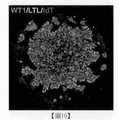
圖11係展示藉由TGFβ 1刺激,從腎間質細胞分化之肌纖維母細胞中之α SMA及FOXD1表現的免疫組織化學染色圖。Figure 11 shows the immunohistochemical stainingof α SMA and FOXD1 in myofibroblasts differentiated from renal interstitial cells stimulatedby
圖12係展示經TGFβ 1刺激之腎間質細胞分化為肌纖維母細胞之比例的圖。縱軸表示tdTomato染色細胞之面積相對於培養細胞之全體面積的比例(%)。Figure 12 is a graph showing theratio of renal interstitial cells that are stimulated by
圖13將培養第4日(D4)之RSC,以TGFβ 1(1ng/ml)處置9日後(D13 TGF),以含有TGFβ 1之培養基(D25 TGF+)或不含TGFβ 1之培養基(D25 TGF-)進一步培養12日。表示D13及D25的tdTomato染色細胞之面積相對於培養細胞之全體面積的比例(%)。「D25 Cont」表示陰性對照(無TGFβ 1處置)。Figure 13 The RSC on the fourth day of culture (D4) wastreated with TGF β 1 (1ng/ml) for 9 days (D13 TGF), and then with amedium containing TGF β 1 (D25 TGF+) or amedium without TGF β 1 ( D25 TGF-) was further cultured for 12 days. It represents the ratio (%) of the area of tdTomato stained cells of D13 and D25 to the total area of cultured cells. "D25 Cont" represents a negative control (no
圖14將培養第4日(D4)之RSC,以TGFβ 1(10ng/ml)處置9日後(D13 TGF),以含有TGFβ 1之培養基(D25 TGF+)或不含之培養基(D25 TGF-)進一步培養12日。表示D13及D25的tdTomato染色細胞之面積相對於培養細胞之全體面積的比例(%)。「D25 Cont」表示陰性對照(無TGFβ 1處置)。Figure 14 The RSC on the fourth day of culture (D4) wastreated with TGF β 1 (10ng/ml) for 9 days (D13 TGF), and then treated with amedium containing TGF β 1 (D25 TGF+) or a medium without (D25 TGF- ) Further training for 12 days. It represents the ratio (%) of the area of tdTomato stained cells of D13 and D25 to the total area of cultured cells. "D25 Cont" represents a negative control (no
圖15將培養第4日(D4)之RSC,以TGFβ 1(10ng/ml)處置9日後(D13),以不含TGFβ 1之培養基進一步培養2日(D15)。在D15之細胞之培養基中添加SB431542(3、10、30μM),進一步培養9日(D24)。表示D15、D17、D20、D22及D24的tdTomato染色細胞之面積。「Cont」表示陰性對照(不添加SB)。Fig. 15 The RSC on the fourth day of culture (D4) wastreated with TGF β 1 (10 ng/ml) for 9 days (D13), andfurther cultured in a medium without
圖16為腎臟類器官之顯微鏡像。Figure 16 is a microscope image of kidney organs.
圖17為表示測定人工多能性幹細胞株(「iPSC」)、中間中胚葉(「IM(D7)」)、腎臟類器官(Mini-kidney:「IM+RSP(D25)」)、不含腎間質前驅細胞之腎臟類器官(「KiO(D25)」)及低氧條件下之腎臟類器官(Mini-kidney:「IM+RSP(D25)+FG4592」)的EPO mRNA表現量之結果的圖。縱軸係將EPO mRNA表現量以人工多能性幹細胞株之表現量當作時1的相對值來表示。Figure 17 shows the measurement of artificial pluripotent stem cell lines ("iPSC"), intermediate mesoderm ("IM(D7)"), kidney organoids (Mini-kidney: "IM+RSP(D25)"), without kidney Figure of the results of the expression level of EPO mRNA in the kidney organoids of mesenchymal precursor cells ("KiO(D25)") and the kidney organoids under hypoxic conditions (Mini-kidney: "IM+RSP(D25)+FG4592") . On the vertical axis, the expression level of EPO mRNA is expressed by the expression level of the artificial pluripotent stem cell line as the relative value of
圖18係展示測定人工多能性幹細胞株(「iPSC」)、中間中胚葉(「IM(D7)」)、腎臟類器官(Mini-kidney:「IM+RSP(D25)」)、不含腎間質前驅細胞之腎臟類器官(「KiO(D25)」)及低氧條件下之腎臟類器官(Mini-kidney:「IM+RSP(D25)+FG4592」)中的腎間質細胞標記(肌間線蛋白(desmin)、波形蛋白(vimentin)、CD73、PDGFRβ)之表現量之結果的圖。縱軸係將mRNA表現量藉由以人工多能性幹細胞株之表現量當作1時的相對值來表示。Figure 18 shows the measurement of artificial pluripotent stem cell lines ("iPSC"), intermediate mesoderm ("IM(D7)"), kidney organoids (Mini-kidney: "IM+RSP(D25)"), without kidney Interstitial precursor cells in kidney organoids ("KiO(D25)") and kidney organoids under hypoxic conditions (Mini-kidney: "IM+RSP(D25)+FG4592") in renal interstitial cell markers (muscle Graph showing the results of expression levels of desmin, vimentin, CD73, PDGFRβ ). The vertical axis represents the expression level of mRNA by taking the expression level of the artificial pluripotent stem cell line as a relative value.
圖19係展示腎臟類器官(Mini-kidney)中的絲球體細胞標記(WT1)及腎小管細胞標記(LTL)之表現的免疫組織化學染色圖。Figure 19 is an immunohistochemical staining diagram showing the performance of the silk spheroid cell marker (WT1) and the renal tubular cell marker (LTL) in the kidney organoids (Mini-kidney).
圖20係展示包含RSP之腎臟類器官(IM+RSP(D25)、右)及不含RSP之腎臟類器官(KiO(D25)、左)之細胞集團的T-SNE plot。Figure 20 shows a T-SNE plot of the cell group of kidney organoids containing RSP (IM+RSP (D25), right) and kidney organoids without RSP (KiO (D25), left).
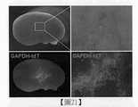
圖21係將源自EPO-emGFP/GAPDH-tdTomato(GAPDH-tdT)雙基因敲入(dual knock-in)人類iPS細胞株之RSP細胞塊移植至免疫不全NOG小鼠之腎後,摘出之腎臟之實體顯微鏡下的螢光像。其展示藉由GAPDH-tdT之螢光觀察結果,細胞從移植之細胞塊游走,於腎內生著。Figure 21 is the transplantation of the RSP cell block derived from the EPO-emGFP/GAPDH-tdTomato (GAPDH-tdT) dual knock-in human iPS cell line into the kidney of immunodeficiency NOG mice, and the kidney removed Fluorescent image under a physical microscope. It showed that by the fluorescent observation of GAPDH-tdT, cells migrated from the transplanted cell mass and grew in the kidney.
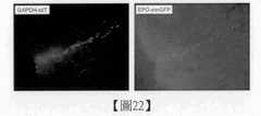
圖22係展示將細胞塊於腎移植後,摘出的包含腎臟之RSP(GAPDH-tdTomato陽性細胞)生著部位之組織片的螢光像,及在移植之RSP之一部分細胞中,檢測藉由EPO-emGFP之表現所產生的螢光。Figure 22 shows the fluorescence image of the tissue slice containing the growing part of RSP (GAPDH-tdTomato positive cells) of the kidney after the cell block was transplanted in the kidney, and a part of the transplanted RSP cells were detected by EPO -Fluorescence produced by the performance of emGFP.
以下,說明實施本發明之較佳型態。再者,以下所說明之實施型態,係表示本發明之代表性實施型態之一例,不能藉此狹隘地解釋本發明之範圍。Hereinafter, the preferred mode for implementing the present invention will be described. In addition, the embodiments described below are examples of representative embodiments of the present invention, and the scope of the present invention cannot be interpreted narrowly.
1.腎間質細胞之製造方法1. Manufacturing method of renal interstitial cells
根據本發明之腎間質細胞的製造方法,包含以下之步驟(3),並任意地包含步驟(2)及/或步驟(1)。The method for producing renal interstitial cells according to the present invention includes the following step (3), and optionally includes step (2) and/or step (1).
步驟(3):在包含源自血小板之成長因子受體促效劑的培養基培養腎間質前驅細胞,得到腎間質細胞的步驟。Step (3): a step of culturing renal interstitial precursor cells in a medium containing platelet-derived growth factor receptor agonists to obtain renal interstitial cells.
步驟(2):從神經脊細胞誘導腎間質前驅細胞的步驟。Step (2): a step of inducing renal interstitial precursor cells from neural crest cells.
步驟(1):在包含GSK3β阻礙劑、TGFβ阻礙劑、及視黃酸及/或其衍生物之培養基培養多能性幹細胞,誘導神經脊細胞的步驟。Step (1): a stepof culturing pluripotent stem cells in a medium containing GSK3 β inhibitor, TGFβ inhibitor, and retinoic acid and/or its derivatives to induce neural spinal cells.
1-1.步驟(3)1-1. Step (3)
本步驟中,於包含源自血小板之成長因子受體促效劑的培養基,培養腎間質前驅細胞,得到腎間質細胞。源自血小板之成長因子受體促效劑,可見到具有將腎間質前驅細胞分化誘導成腎間質細胞的活性,而有作為腎間質細胞誘導劑之功能。就源自血小板之成長因子受體促效劑而言,以源自血小板之成長因子(PDGF)為較佳。In this step, renal interstitial precursor cells are cultured in a medium containing platelet-derived growth factor receptor agonists to obtain renal interstitial cells. Growth factor receptor agonists derived from platelets can be seen to have the activity of inducing the differentiation of renal interstitial precursor cells into renal interstitial cells, and have the function of acting as an inducer of renal interstitial cells. As for the growth factor receptor agonist derived from platelet, the growth factor derived from platelet (PDGF) is preferred.
腎間質前驅細胞,可為藉步驟(2)得到者,又,亦可為商業上購入之細胞,亦可為從活體內分離的離體(ex vivo)細胞。The renal interstitial precursor cells can be obtained by step (2), or they can be commercially purchased cells, or they can be ex vivo cells isolated from the living body.
在使用從人工多能性幹細胞誘導之腎間質前驅細胞的情況,可於本步驟中製造源自人工多能性幹細胞之腎間質細胞。In the case of using renal interstitial precursor cells induced from artificial pluripotent stem cells, renal interstitial cells derived from artificial pluripotent stem cells can be produced in this step.
培養基(腎間質細胞製造用培養基),包含源自血小板之成長因子受體促效劑,較佳進一步包含鹼性纖維母細胞成長因子(bFGF)或纖維母細胞成長因子9(FGF9),更佳包含bFGF及FGF9。The culture medium (medium for the production of renal interstitial cells) contains a growth factor receptor agonist derived from platelets, preferably further contains basic fibroblast growth factor (bFGF) or fibroblast growth factor 9 (FGF9), and more It preferably contains bFGF and FGF9.
就基礎培養基而言,無特別限定,例如較佳可使用STEMdiff APEL2培養基(STEMCELL Technologies,ST-05275)、TeSR1培養基以及化學合成培養基(Chemically Defined Medium(CDM))。此外,亦可使用BME培養基、BGJb培養基、CMRL 1066培養基、Glasgow MEM培養基、改良MEM(IMEM)培養基、改良MDM(IMDM)培養基、Medium 199培養基、Eagle MEM培養基、α MEM培養基、DMEM培養基(高葡萄糖、低葡萄糖)、DMEM/F12培養基、Ham’s培養基、RPMI 1640培養基、Fischer's培養基、及此等之混合培養基等。The basic medium is not particularly limited. For example, STEMdiff APEL2 medium (STEMCELL Technologies, ST-05275), TeSR1 medium, and Chemically Defined Medium (CDM) can be preferably used. In addition, BME medium, BGJb medium, CMRL 1066 medium, Glasgow MEM medium, modified MEM (IMEM) medium, modified MDM (IMDM) medium, Medium 199 medium, Eagle MEM medium,α MEM medium, DMEM medium (high glucose , Low glucose), DMEM/F12 medium, Ham's medium, RPMI 1640 medium, Fischer's medium, and mixed medium of these.
就CDM培養基而言,無特別限定,例如,可使用從伊斯科夫改良型杜氏培養基(Iscove’s modified Dulbecco’s medium(GE Healthcare公司製))調製的培養基。The CDM medium is not particularly limited. For example, a medium prepared from Iscove's modified Dulbecco's medium (manufactured by GE Healthcare) can be used.
在基礎培養基中,可添加Ham’s F-12營養混合物、人類血清白蛋白等白蛋白、聚乙烯醇(PVA)、脫離子BSA、亞麻油酸、次亞麻油酸、膽固醇、胰島素、原轉鐵蛋白(apotransferrin)、硒、乙醇胺、單硫代甘油、無蛋白質融合瘤混合物II(Protein-free hybridoma mixture II)(PFHMII)、抗壞血酸、L-丙胺醯基-L-麩醯胺酸及/或抗生素等通常使用於細胞培養的物質。In the basic medium, you can add Ham's F-12 nutrient mixture, human serum albumin and other albumin, polyvinyl alcohol (PVA), deionized BSA, linoleic acid, hypolinoleic acid, cholesterol, insulin, pro-transferrin (apotransferrin), selenium, ethanolamine, monothioglycerol, Protein-free hybridoma mixture II (PFHMII), ascorbic acid, L-propylamino-L-glutamic acid and/or antibiotics, etc. Usually used in cell culture substances.
FGF及FGF9,只要依據所培養之腎間質前驅細胞之來源動物種類,選用適當的來源動物種類即可,較佳使用源自與腎間質前驅細胞之來源動物種類相同之動物的因子。For FGF and FGF9, it is only necessary to select an appropriate source animal species according to the source animal species of the cultured renal interstitial precursor cells, and preferably use factors derived from the same animal species as the source animal species of the renal interstitial precursor cells.
PDGF為參與纖維母細胞等間葉系細胞之游走及增殖等之調節的增殖因子之一。PDGF,存在4種:PDGF-A、PDGF-B、PDGF-C及PDGF-D。其中,A鏈及B鏈藉由形成二硫鍵而成為均或雜二聚體構造,因此具有PDGF-AA、PDGF-AB及PDGF-BB三種同功型。C鏈及D鏈分別形成均二聚體(PDGF-CC及PDGF-DD)。在本發明中,以PDGF-BB為較佳。就PDGF-BB而言,無特別限定,例如,可使用PEPROTECH公司製#100-14B等。PDGF is one of the proliferation factors involved in the regulation of the migration and proliferation of fibroblasts and other mesenchymal cells. There are 4 types of PDGF: PDGF-A, PDGF-B, PDGF-C and PDGF-D. Among them, the A chain and the B chain become a homo-or heterodimer structure by forming a disulfide bond, and therefore have three isoforms of PDGF-AA, PDGF-AB and PDGF-BB. The C chain and D chain form homodimers (PDGF-CC and PDGF-DD) respectively. In the present invention, PDGF-BB is preferred. PDGF-BB is not particularly limited, and for example, #100-14B manufactured by PEPROTECH, etc. can be used.
就PDGF、bFGF及FGF9而言,可使用全長肽或其受體結合片段。For PDGF, bFGF, and FGF9, full-length peptides or receptor-binding fragments thereof can be used.
PDGF、bFGF及FGF9可從市售者購入而使用。就bFGF及FGF9而言,無特別限定,例如,可分別使用富士薄膜和光純藥股份有限公司製品編號#068-04544及製品編號#AF-100-23等。PDGF, bFGF, and FGF9 can be purchased from commercial vendors and used. There are no particular limitations on bFGF and FGF9. For example, Fuji Film Wako Pure Chemical Industries, Ltd. product number #068-04544 and product number #AF-100-23 can be used, respectively.
PDGF於培養基中之添加濃度,可依據所用的PDGF之亞型而適宜調整,例如,為0.05ng/ml至1μg/ml。較佳為0.1至500ng/ml,更佳為1至100ng/ml。在使用PDGF-BB之情況,例如為0.1ng/ml至1μg/ml。較佳為1至300ng/ml,更佳為3至100ng/ml,特佳為約10ng/ml。PDGF was added to the concentration in the medium may be appropriately adjusted according to the subtypes of PDGF used, e.g., to 0.05ng / ml to1 μ g / ml. Preferably it is 0.1 to 500 ng/ml, more preferably 1 to 100 ng/ml. Usage of PDGF-BB, for example, 0.1ng / ml to1 μ g / ml. It is preferably 1 to 300 ng/ml, more preferably 3 to 100 ng/ml, and particularly preferably about 10 ng/ml.
bFGF於培養基中之添加濃度,例如為0.1ng/ml至1μg/ml。較佳為1至500ng/ml,更佳為10至300ng/ml,特佳為約40ng/mL。bFGF was added in the medium at a concentration of, for example, 0.1ng / ml to1 μ g / ml. It is preferably 1 to 500 ng/ml, more preferably 10 to 300 ng/ml, particularly preferably about 40 ng/mL.
FGF9於培養基中之添加濃度,例如為0.1ng/ml至1μg/ml。較佳為1至500ng/ml,更佳為10至300ng/ml,特佳為約200ng/mL。FGF9 addition concentration in the culture medium, for example, 0.1ng / ml to1 μ g / ml. It is preferably 1 to 500 ng/ml, more preferably 10 to 300 ng/ml, particularly preferably about 200 ng/mL.
在將源自血小板之成長因子受體促效劑,作為使腎間質前驅細胞分化成腎間質細胞之誘導劑使用的情況,該誘導劑除源自血小板之成長因子受體促效劑外,亦可包含bFGF、FGF9、視黃酸(RA)等。When a platelet-derived growth factor receptor agonist is used as an inducer for the differentiation of renal interstitial precursor cells into renal interstitial cells, the inducer is in addition to the platelet-derived growth factor receptor agonist , Can also contain bFGF, FGF9, retinoic acid (RA) and so on.
本步驟之培養期間,只要為足以使腎間質前驅細胞分化成腎間質細胞的期間即可,無特別限定,例如為7至24日,較佳為10至20日,更佳為12至18日,特佳為約15日。The culture period in this step is not particularly limited as long as it is a period sufficient to differentiate renal interstitial precursor cells into renal interstitial cells. For example, it is 7 to 24 days, preferably 10 to 20 days, and more preferably 12 to 12 days. On the 18th, the best is about 15 days.
本步驟以藉由附著培養為較佳,然而亦可為懸浮培養。In this step, attachment culture is preferred, but suspension culture may also be used.
附著培養方面,可使用培養皿、燒瓶、微培養盤、及OptiCell(製品名)(Nunc公司)等細胞培養片等培養容器。For attachment culture, culture vessels such as petri dishes, flasks, microplates, and cell culture sheets such as OptiCell (product name) (Nunc) can be used.
附著培養所用的容器,可進行使與細胞之接著性(親水性)提高用的表面處理,或以膠原、明膠、聚-L-離胺酸、聚-D-離胺酸、層黏連蛋白(laminin)、纖連蛋白(fibronectin)、基質膠(matrigel)、玻連蛋白(vitronectin)等細胞接著用基質塗覆,不過以使用不進行此等表面處理或塗覆的容器為更佳。The container used for attachment culture can be surface treated to improve the adhesion (hydrophilicity) to cells, or use collagen, gelatin, poly-L-lysine, poly-D-lysine, and laminin (laminin), fibrinCells such as fibronectin, matrigel, and vitronectin are then coated with a matrix, but it is better to use a container that is not subjected to such surface treatment or coating.
懸浮培養方面,係使細胞分散於培養基中,藉由攪拌或振盪,使培養基成分及培養基內氧濃度均勻化,同時形成細胞凝集塊。較佳之攪拌速度,雖隨細胞密度及培養容器之大小而適宜設定,然而過度之攪拌或振盪對細胞賦予物理性應力,阻礙細胞凝集塊形成。因此,以能將培養基成分及培養基內氧濃度均勻化,且不阻礙細胞凝集塊形成的方式,控制攪拌或振盪速度。亦可不進行攪拌或振盪,而以靜置方式進行懸浮培養。For suspension culture, cells are dispersed in a culture medium, and the composition of the culture medium and the oxygen concentration in the culture medium are homogenized by stirring or shaking, and at the same time, cell aggregates are formed. The preferred stirring speed is appropriately set according to the cell density and the size of the culture vessel, but excessive stirring or shaking imparts physical stress to the cells and prevents the formation of cell aggregates. Therefore, the stirring or shaking speed can be controlled in a way that the medium components and the oxygen concentration in the medium can be made uniform without hindering the formation of agglomerates of cells. It is also possible to carry out suspension culture in a static manner without stirring or shaking.
懸浮培養方面,以使用Prime surface(製品名,住友電木公司)等低接著塗層之容器為較佳。For suspension culture, it is better to use a container with a low adhesion coating such as Prime surface (product name, Sumitomo Bakelite Co., Ltd.).
培養溫度無特別限定,於30至40℃(例如,37℃)進行。又,培養容器中之二氧化碳濃度為例如約5%。The culture temperature is not particularly limited, and it is performed at 30 to 40°C (e.g., 37°C). In addition, the concentration of carbon dioxide in the culture container is, for example, about 5%.
腎間質細胞之生成的確認,例如,可列舉測定標記蛋白質或標記基因之表現的方法。若所得到之細胞表現EPO基因及/或蛋白質、CD73及PDGFRβ,較佳為進一步表現肌間線蛋白(desmin)及波形蛋白(vimentin),可判斷該細胞為腎間質細胞。To confirm the production of renal interstitial cells, for example, a method of measuring the expression of marker proteins or marker genes can be cited. If the obtained cell expresses EPO gene and/or protein, CD73 and PDGFRβ , and preferably further expresses desmin and vimentin, it can be judged that the cell is a renal interstitial cell.
標記蛋白質之檢測,可利用使用對該標記蛋白質特異之抗體的免疫學檢定,例如,ELISA、免疫染色、流式細胞術等而進行。標記基因之檢測,可利用該領域中周知之核酸擴增方法及/或核酸檢測方法,例如,RT-PCR、微陣列、生物晶片等而進行。The detection of the labeled protein can be performed by immunological assays using antibodies specific to the labeled protein, for example, ELISA, immunostaining, flow cytometry, and the like. The detection of marker genes can be carried out by using nucleic acid amplification methods and/or nucleic acid detection methods known in the art, for example, RT-PCR, microarrays, biochips, etc.
本步驟中所得到之腎間質細胞,尤其具備於低氧條件下產生EPO的功能(低氧回應性)。低氧回應性可藉由以使HIF信號活化之化合物對細胞處置,檢測EPO之mRNA量(level)或蛋白質量之表現上升而確認。就使HIF信號活化之化合物而言,可使用例如FG-2216(Selleck,#18382)及FG-4592(Selleck,#S1007)。The renal interstitial cells obtained in this step especially have the function of producing EPO under hypoxic conditions (hypoxic responsiveness). Hypoxia responsiveness can be achieved by compounds that activate HIF signaling on the cell.Set, detect the increase of the mRNA level or protein quality of EPO to confirm. As for the compound that activates the HIF signal, for example, FG-2216 (Selleck, #18382) and FG-4592 (Selleck, #S1007) can be used.
低氧回應性亦可藉由將為對象之細胞於低氧濃度之條件下培養而確認。低氧濃度之條件下的氧濃度,可調成可於活體內環境中產生之低氧狀態的氧濃度。就氧濃度而言,例如,可調成20%以下,即可調成20至10%、10至5%、5至1%。Hypoxia responsiveness can also be confirmed by culturing the cells of the subject under conditions of low oxygen concentration. The oxygen concentration under the condition of low oxygen concentration can be adjusted to the low oxygen concentration that can be generated in the living body environment. As far as the oxygen concentration is concerned, for example, if it can be adjusted to 20% or less, it can be adjusted to 20 to 10%, 10 to 5%, or 5 to 1%.
由於有報導指出腎纖維症係因具有EPO產生能力之腎間質細胞纖維化所致,因此具有EPO產生能力的腎間質細胞,在開發腎纖維症治療藥,作為模擬活體之腎纖維化模型之構築用材料上有用。又,具有EPO產生能力之腎間質細胞,亦特別可用於以腎性貧血之治療或預防為目的之對腎障礙的細胞醫療。Since it has been reported that renal fibrosis is caused by the fibrosis of renal interstitial cells with EPO-producing ability, renal interstitial cells with EPO-producing ability are developing therapeutic drugs for renal fibrosis as a model of renal fibrosis that mimics the living body. The construction materials are useful. In addition, renal interstitial cells with the ability to produce EPO are particularly useful for cell therapy of renal disorders for the purpose of treatment or prevention of renal anemia.
本發明亦提供包含依照本步驟所得到之腎間質細胞的凍結儲備液。The present invention also provides a frozen stock solution containing renal interstitial cells obtained according to this step.
凍結儲備液可藉由將所得到之腎間質細胞離心,從培養基分離,懸浮於凍結保存液中、凍結而製造。凍結保存液方面,只要使用先前凍結保存細胞用的試藥即可。例如,市售之Cryostem Freezing Medium(商品名)及CELLBANKER(註冊商標)等。The frozen stock solution can be produced by centrifuging the obtained renal interstitial cells, separating them from the culture medium, suspending in the freezing storage solution, and freezing. As for the freezing preservation solution, only the reagent used to freeze the preservation of the cells can be used. For example, Cryostem Freezing Medium (trade name) and CELLBANKER (registered trademark) are commercially available.
凍結儲備液可被用於製作,例如,以腎間質細胞作為構成要素的組織模型(腎臟類器官)。The frozen stock solution can be used to make, for example, a tissue model (kidney organoid) with renal interstitial cells as a constituent element.
1-2.步驟(2)1-2. Step (2)
在本步驟中,從神經脊細胞誘導腎間質前驅細胞。In this step, renal interstitial precursor cells are induced from neural spinal cells.
本發明提供從神經脊細胞製造腎間質前驅細胞的方法。The present invention provides a method for producing renal interstitial precursor cells from neural spinal cells.
神經脊細胞可為步驟(1)中所得到者,又,亦可為從商業上購入之細胞,亦可為從活體內分離的離體(ex vivo)細胞。The neural crest cells may be those obtained in step (1), or they may be commercially purchased cells, or they may be ex vivo cells isolated from the living body.
就市售之神經脊細胞而言,例如,可列舉人類毛囊外毛根鞘細胞(Cosmo Bio公司製)、O9-1小鼠顱骨神經脊細胞株(Merck Millipore公司製)等。Commercially available neural spinal cells include, for example, human hair follicle outer hair root sheath cells (manufactured by Cosmo Bio), O9-1 mouse cranial nerve spinal cell strain (manufactured by Merck Millipore), and the like.
又,據報導神經脊細胞存在於受精後30日前後之人類胚的神經管、胎生第9日前後之小鼠胚的神經管、人類、豬及齧齒類之成體的皮膚等(Betters et al.,Developmental biology,2010,344(2):578-592、Jiang et al.,Development,2000,127(8):1607-1616、Dupin et al.,Developmental biology,2012,366(1):83-95、Nagoshi et al.,Cell Stem Cell 2,April 2008,392-403)。亦可使用周知之方法(例如,Motohashi et al.,Biology open,2016,5:311-322、Pfaltzgraffet al.,Journal of Visualized Experiments,2012,64:4134),採集此種神經脊細胞,在適宜後方化後,提供於本步驟。In addition, it is reported that neural spinal cells exist in the neural tube of human embryos around 30 days after fertilization, the neural tube of mouse embryos around the 9th day of viviparous birth, and the adult skin of humans, pigs, and rodents (Betters et al. .,Developmental biology,2010,344(2):578-592, Jiang et al.,Development,2000,127(8):1607-1616, Dupin et al.,Developmental biology,2012,366(1):83 -95, Nagoshi et al.,
神經脊細胞方面,以使用後腦神經脊細胞(hindbrain NCC:hNCC)為較佳。In terms of neural spinal cells, hindbrain NCC (hNCC) is preferably used.
在使用從人工多能性幹細胞誘導之神經脊細胞的情況,於本步驟中製造源自人工多能性幹細胞之腎間質前驅細胞。In the case of using neural crest cells induced from artificial pluripotent stem cells, in this step, renal interstitial precursor cells derived from artificial pluripotent stem cells are produced.
藉由將神經脊細胞於間葉系間質細胞誘導用培養基中培養,得到腎間質前驅細胞。在間葉系間質細胞誘導用培養基方面,例如,可使用StemPro MSC無異種成分(xeno-free)培養基(ThermoFisher:A1067501)。The renal interstitial precursor cells are obtained by culturing the neural crest cells in a medium for inducing mesenchymal mesenchymal cells. Regarding the medium for inducing mesenchymal mesenchymal cells, for example, StemPro MSC xeno-free medium (ThermoFisher: A1067501) can be used.
本步驟之培養期間,只要為神經脊細胞足以分化成腎間質前驅細胞之期間即可,無特別限定,例如為10至90日,較佳為20至50日,更佳為30至40日,特佳為30至35日。The culture period in this step is not particularly limited as long as the neural crest cells are sufficient to differentiate into renal interstitial precursor cells, and it is, for example, 10 to 90 days, preferably 20 to 50 days, and more preferably 30 to 40 days. , Especially good for 30 to 35 days.
本步驟以藉由附著培養為較佳,亦可藉由懸浮培養。In this step, attachment culture is preferred, and suspension culture may also be used.
附著培養方面,可使用培養皿、燒瓶、微培養盤、及OptiCell(製品名)(Nunc公司)等細胞培養片等培養容器。For attachment culture, culture vessels such as petri dishes, flasks, microplates, and cell culture sheets such as OptiCell (product name) (Nunc) can be used.
附著培養所用的容器,可進行使與細胞之接著性(親水性)提高用的表面處理,或以膠原、明膠、聚-L-離胺酸、聚-D-離胺酸、層黏連蛋白、纖連蛋白、基質膠、玻連蛋白等細胞接著用基質塗覆,而以使用不進行此等表面處理或塗覆的容器為更佳。The container used for attachment culture can be surface treated to improve the adhesion (hydrophilicity) to cells, or use collagen, gelatin, poly-L-lysine, poly-D-lysine, and laminin , Fibronectin, matrigel, vitronectin and other cells are then coated with a matrix, and it is better to use a container without such surface treatment or coating.
懸浮培養方面,係使細胞分散於培養基中,藉由攪拌或振盪,使培養基成分及培養基內氧濃度均勻化,同時形成細胞凝集塊。較佳之攪拌速度,雖隨細胞密度及培養容器之大小而適宜設定,然而過度之攪拌或振盪對細胞賦予物理性應力,阻礙細胞凝集塊形成。因此,以能將培養基成分及培養基內氧濃度均勻化,且不阻礙細胞凝集塊形成的方式,控制攪拌或振盪速度。亦可不進行攪拌或振盪,在靜置下進行懸浮培養。For suspension culture, cells are dispersed in a culture medium, and the composition of the culture medium and the oxygen concentration in the culture medium are homogenized by stirring or shaking, and at the same time, cell aggregates are formed. The preferred stirring speed is appropriately set according to the cell density and the size of the culture vessel, but excessive stirring or shaking imparts physical stress to the cells and prevents the formation of cell aggregates. Therefore, the stirring or shaking speed can be controlled in a way that the medium components and the oxygen concentration in the medium can be made uniform without hindering the formation of agglomerates of cells. It is also possible to carry out suspension culture under standing without stirring or shaking.
懸浮培養方面,以使用Prime surface(製品名,住友電木公司)等低接著塗層的容器為較佳。For suspension culture, it is better to use a container with a low adhesion coating such as Prime surface (product name, Sumitomo Bakelite Co., Ltd.).
培養溫度無特別限定,於30至40℃(例如,37℃)進行。又,培養容器中之二氧化碳濃度為例如約5%。The culture temperature is not particularly limited, and it is performed at 30 to 40°C (e.g., 37°C). In addition, the concentration of carbon dioxide in the culture container is, for example, about 5%.
以下,在本說明書中,將細胞凝集塊簡稱為「細胞塊」。Hereinafter, in this specification, the cell aggregate is abbreviated as "cell mass".
腎間質前驅細胞之生成的確認,例如,可藉由確認細胞之分化誘導成腎間質細胞之能力而進行。The confirmation of the production of renal interstitial precursor cells can be performed, for example, by confirming the ability of the cells to differentiate into renal interstitial cells.
又,腎間質前驅細胞之生成的確認,例如,可列舉測定標記蛋白質或標記基因之表現的方法。若所得到之細胞表現FOXD1、CD73及PDGFRβ,而不表現紅血球生成素,可判斷該細胞為腎間質前驅細胞。In addition, for the confirmation of the production of renal interstitial precursor cells, for example, a method of measuring the expression of marker proteins or marker genes can be cited. If the obtained cells express FOXD1, CD73 and PDGFRβ , but do not express erythropoietin, the cells can be judged to be renal interstitial precursor cells.
本發明亦提供包含依照本步驟所得到之包含腎間質前驅細胞的凍結儲備液。The present invention also provides a frozen stock solution containing renal interstitial precursor cells obtained according to this step.
凍結儲備液可藉由將所得到之腎間質前驅細胞藉由離心,從培養基分離,懸浮於凍結保存液中、凍結而製造。凍結保存液方面,只要使用先前細胞之凍結保存所用的試藥即可。例如,市售之Cryostem Freezing Medium(商品名)及CELLBANKER(註冊商標)等。The frozen stock solution can be prepared by separating the obtained renal interstitial precursor cells from the culture medium by centrifugation, suspending in the freezing preservation solution, and freezing. As for the freezing preservation solution, only the reagent used for the freezing preservation of the previous cells can be used. For example, Cryostem Freezing Medium (trade name) and CELLBANKER (registered trademark) are commercially available.
凍結儲備液可被利用以作為例如從腎間質前驅細胞,得到腎間質細胞的起始材料。又,凍結儲備液可被用於製作以腎間質前驅細胞作為構成要素的組織模型(腎臟類器官)。The frozen stock solution can be used as a starting material for obtaining renal interstitial cells, for example, from renal interstitial precursor cells. In addition, the frozen stock solution can be used to make a tissue model (kidney organoid) with renal interstitial precursor cells as a constituent element.
1-3.步驟(1)1-3. Step (1)
本步驟係於包含GSK3β阻礙劑、TGFβ阻礙劑、及視黃酸及/或其衍生物之培養基中,培養多能性幹細胞(尤其,人工多能性幹細胞),誘導神經脊細胞(較佳為後腦神經脊細胞)。This step is tocultivate pluripotent stem cells (especially artificial pluripotent stem cells) in a medium containing GSK3 β inhibitor, TGFβ inhibitor, and retinoic acid and/or its derivatives, and induce neural spinal cells (more Preferably, the posterior cranial nerve spinal cell).
從多能性幹細胞分化誘導成各種神經脊細胞,可依照文獻周知(例如,非專利文獻3、4)之方法進行。在從人類人工多能性幹細胞分化誘導成神經脊細胞的情況,將人工多能性幹細胞播種於培養皿等,進行附著培養後,於包含TGFβ阻礙劑及GSK3β阻礙劑之培養基進行附著培養,然後在進一步添加視黃酸及/或其衍生物的培養基進行附著培養,使其分化成神經脊細胞。Differentiation from pluripotent stem cells into various neural crest cells can be performed according to methods known in the literature (for example,
在本發明中可使用之「多能性幹細胞(pluripotent stem cell)」,意指可分化成活體之具有各種相異型態或功能的組織或細胞,亦具有能分化成3胚葉(內胚葉、中胚葉、外胚葉)之任何系統之細胞之能力的幹細胞。在此方面,無特別限定,例如,可列舉胚性幹細胞(ESC)、藉由核移植所得到的源自純株胚之胚性幹細胞、精子幹細胞、胚性生殖細胞、人工多能性幹細胞(本說明書中,亦稱為「iPSC」)等。又,在本發明中可使用之「多潛能性幹細胞(multipotent stem cell)」,意指具有能分化成複數個限定數目之系統之細胞的能力的幹細胞。就本發明中可使用之「多潛能性幹細胞(multipotent stem cell)」而言,例如,可列舉齒髓幹細胞、源自口腔黏膜之幹細胞、毛囊幹細胞、源自培養纖維母細胞或骨髓幹細胞之體性幹細胞等。較佳之多能性幹細胞(pluripotent stem cell)為ESC及iPSC。The "pluripotent stem cell" that can be used in the present invention refers to tissues or cells with various different types or functions that can be differentiated into living organisms, and they also have the ability to differentiate into three embryonic leaves (endodermal, Mesodermal, ectodermal) stem cells with the ability of cells of any system. In this respect, there is no particular limitation. For example, embryonic stem cells (ESC), derived from pure plant embryos obtained by nuclear transfer can be mentioned.Embryonic stem cells, sperm stem cells, embryonic germ cells, artificial pluripotent stem cells (also referred to as "iPSC" in this specification), etc. In addition, the "multipotent stem cell" that can be used in the present invention refers to a stem cell that has the ability to differentiate into a plurality of cells of a limited number of systems. Regarding the "multipotent stem cells" that can be used in the present invention, for example, dental pulp stem cells, stem cells derived from oral mucosa, hair follicle stem cells, bodies derived from cultured fibroblasts or bone marrow stem cells can be cited Sexual stem cells and so on. Preferred pluripotent stem cells are ESC and iPSC.
就「ESC」而言,若為小鼠ESC,可利用inGenious targeting laboratory公司理研(理化學研究所)等所建立的各種小鼠ESC株,若為人類ESC,可利用威斯康辛大學、NIH、理研、京都大學、國立成育醫療研究中心及Cellartis公司等所建立的各種人類ESC株。例如,就人類ESC株而言,可利用ESI Bio公司分讓之CHB-1至CHB-12株、RUES1株、RUES2株、HUES1至HUES28株等,WiCell Research分讓之H1株、H9株等,理研分讓之KhES-1株、KhES-2株、KhES-3株、KhES-4株、KhES-5株、SSES1株、SSES2株、SSES3株等。For "ESC", if it is a mouse ESC, you can use various mouse ESC strains established by inGenious targeting laboratory company Riken (Institute of Physics and Chemistry), etc. If it is a human ESC, you can use the University of Wisconsin, NIH, Riken, Various human ESC strains established by Kyoto University, National Institute of Adult Education and Medical Research, and Cellartis. For example, for human ESC strains, CHB-1 to CHB-12 strains, RUES1 strains, RUES2 strains, HUES1 to HUES28 strains distributed by ESI Bio, and H1 strains and H9 strains distributed by WiCell Research can be used. Riken assigns KhES-1 strain, KhES-2 strain, KhES-3 strain, KhES-4 strain, KhES-5 strain, SSES1 strain, SSES2 strain, SSES3 strain, etc.
「人工多能性幹細胞」意指藉由將特定之因子(核初期化因子)導入哺乳動物體細胞或未分化幹細胞,再重新編程所得到的細胞。現今,「人工多能性幹細胞」方面有各式各樣者,除了依照山中氏等,藉由將4因子:Oct3/4、Sox2、Klf4、c-Myc導入小鼠纖維母細胞所建立的iPSC(Takahashi K,Yamanaka S.,Cell,(2006)126:663-676)之外,亦可使用:將同樣之4因子導入人類纖維母細胞所建立的源自人類細胞之iPSC(Takahashi K,Yamanaka S.,et al.Cell,(2007)131:861-872);上述4因子導入後,以Nanog之表現作為指標篩選所建立的Nanog-iPSC(Okita,K.,Ichisaka,T.,and Yamanaka,S.(2007).Nature 448,313-317);以不含c-Myc之方法所製作的iPSC(Nakagawa M,Yamanaka S.,et al.Nature Biotechnology,(2008)26,101-106);以無病毒法導入6因子所建立的iPSC(Okita K et al.Nat.Methods 2011 May;8(5):409-12、Okita K et al.Stem Cells.31(3):458-66)等。又,亦可使用依照Thomson氏等製作之導入4因子:OCT3/4、SOX2、NANOG、LIN28所建立的人工多能性幹細胞(Yu J.,Thomson JA.et al.,Science(2007)318:1917-1920)、依照Daley氏等所製作的人工多能性幹細胞(Park IH,Daley GQ.et al.,Nature(2007)451:141-146)、依照櫻田氏所製作的人工多能性幹細胞(日本特開2008-307007號)等。"Artificial pluripotent stem cells" refer to cells obtained by introducing specific factors (nuclear priming factors) into mammalian somatic cells or undifferentiated stem cells, and then reprogramming them. Nowadays, there are all kinds of "artificial pluripotent stem cells". In addition to iPSC established by introducing four factors: Oct3/4, Sox2, Klf4, and c-Myc into mouse fibroblasts, according to Yamanaka, etc. (Takahashi K, Yamanaka S., Cell, (2006) 126: 663-676) can also be used: iPSC derived from human cells established by introducing the same 4 factors into human fibroblasts (Takahashi K, Yamanaka S., et al. Cell, (2007) 131: 861-872); after the introduction of the above 4 factors, the Nanog-iPSC (Okita, K., Ichisaka, T., and Yamanaka) was used as an index to screen the established Nanog-iPSC (Okita, K., Ichisaka, T., and Yamanaka). , S. (2007). Nature 448, 313-317); iPSC made by a method without c-Myc (Nakagawa M, Yamanaka S., et al. NatureBiotechnology, (2008) 26, 101-106); iPSC established by virus-free introduction of 6 factors (Okita K et al. Nat. Methods 2011 May; 8(5): 409-12, Okita K et al. Stem Cells. 31(3): 458-66) and so on. In addition, artificial pluripotent stem cells established by introducing 4 factors: OCT3/4, SOX2, NANOG, LIN28 according to Thomson's, etc. (Yu J., Thomson JA. et al., Science (2007) 318: 1917-1920), according to the artificial pluripotent stem cells produced by Daley et al. (Park IH, Daley GQ.et al., Nature (2007) 451:141-146), according to the artificial pluripotent stem cells produced by Sakurada (Japanese Patent Publication No. 2008-307007) and so on.
此外,亦可使用公開之所有論文(例如,ShiY.,Ding S.,et al.,Cell Stem Cell,(2008)Vol3,Issue 5,568-574;Kim JB.,Scholer HR.,et al.,Nature,(2008)454,646-650;Huangfu D.,Melton,DA.,et al.,Nature Biotechnology,(2008)26,No 7,795-797)、或專利(例如,日本特開2008-307007號、特開2008-283972號、US2008-2336610、US2009-047263、WO2007-069666、WO2008-118220、WO2008-124133、WO2008-151058、WO2009-006930、WO2009-006997、WO2009-007852)中所記載的該領域中周知之人工多能性幹細胞之任一種。In addition, all published papers (for example, ShiY., Ding S., et al., Cell Stem Cell, (2008) Vol3,
就人工多能性幹細胞株而言,可利用NIH、理研、京都大學等所建立的各種iPSC株。例如,若為人類iPSC株,可列舉理研之HiPS-RIKEN-1A株、HiPS-RIKEN-2A株、HiPS-RIKEN-12A株、Nips-B2株等,京都大學之253G1株、201B7株、409B2株、454E2株、606A1株、610B1株、648A1株、1231A3株、Ff-I14s04株、QHJ I14s04株等,以1231A3株為較佳。For artificial pluripotent stem cell lines, various iPSC lines established by NIH, Riken, Kyoto University, etc. can be used. For example, if it is a human iPSC strain, Riken's HiPS-RIKEN-1A strain, HiPS-RIKEN-2A strain, HiPS-RIKEN-12A strain, Nips-B2 strain, etc., Kyoto University's 253G1 strain, 201B7 strain, and 409B2 strain , 454E2 strain, 606A1 strain, 610B1 strain, 648A1 strain, 1231A3 strain, Ff-I14s04 strain, QHJ I14s04 strain, etc., with 1231A3 strain being preferred.
培養基包含GSK3β阻礙劑、TGFβ阻礙劑、及視黃酸及/或其衍生物。就視黃酸之衍生物而言,可使用視黃醇、視黃醛、維甲酸、異維甲酸、阿利維甲酸(alitretinoin)、阿維A酯(atretinate)、阿曲汀(acitretin)、他札羅汀(tazarotene)、貝沙羅汀(bexarotene)、阿達帕林(adapalene)。此等亦可將2種以上組合而使用。以下,將「視黃酸及/或其衍生物」簡稱為「視黃酸等」。The medium contains GSK3β inhibitor, TGFβ inhibitor, and retinoic acid and/or derivatives thereof. For derivatives of retinoic acid, retinol, retinal, retinoic acid, isotretinoin, alitretinoin, atretinate, acitretin, other Tazarotene (tazarotene), bexarotene (bexarotene), adapalene (adapalene). These can also be used in combination of 2 or more types. Hereinafter, "retinoic acid and/or its derivatives" are simply referred to as "retinoic acid, etc.".
基礎培養基無特別限定,例如較佳可使用將StemFit AK03之A液、B液及C液混合者、TeSR1培養基以及Chemically Defined Medium(CDM)培養基。此外,亦可使用BME培養基、BGJb培養基、CMRL 1066培養基、Glasgow MEM培養基、改良MEM(IMEM)培養基、改良MDM(IMDM)培養基、Medium 199培養基、Eagle MEM培養基、α MEM培養基、DMEM培養基(高葡萄糖、低葡萄糖)、DMEM/F12培養基、Ham’s培養基、RPMI 1640培養基、Fischer's培養基、及此等之混合培養基等。The basic medium is not particularly limited. For example, a mixture of StemFit AK03 liquid A, liquid B, and liquid C, TeSR1 medium, and Chemically Defined Medium (CDM) medium can be preferably used. In addition, BME medium, BGJb medium, CMRL 1066 medium, Glasgow MEM medium, modified MEM (IMEM) medium, modified MDM (IMDM) medium, Medium 199 medium, Eagle MEM medium,α MEM medium, DMEM medium (high glucose , Low glucose), DMEM/F12 medium, Ham's medium, RPMI 1640 medium, Fischer's medium, and mixed medium of these.
就CDM培養基而言,無特別限定,例如,可使用從伊斯科夫改良型杜氏培養基(GE Healthcare公司製)調製的培養基。The CDM medium is not particularly limited. For example, a medium prepared from Iskoff's Modified Dulbecco's Medium (manufactured by GE Healthcare) can be used.
基礎培養基方面,可添加原轉鐵蛋白(apotransferrin)、單硫代甘油、牛血清白蛋白(BSA)、胰島素及/或抗生素等通常細胞培養所用的物質。In terms of basal medium, apotransferrin, monothioglycerol, bovine serum albumin (BSA), insulin and/or antibiotics and other substances commonly used for cell culture can be added.
就TGFβ阻礙劑而言,可列舉SB431542(4-[4-(1,3-苯并二氧雜環戊烯-5-基)-5-(2-吡啶基)-1H-咪唑-2-基]-苄醯胺)、A83-01(3-(6-甲基-2-吡啶基)-N-苯基-4-(4-喹啉基)-1H-吡唑-1-甲硫醯胺)、LDN193189(4-[6-[4-(1-哌
TGFβ阻礙劑於培養基中之添加濃度,可依據所添加之TGFβ阻礙劑的種類而適宜調整,例如為0.1至50μM,較佳為1至20μM。Theconcentration of the TGF β inhibitor added to the medium can beappropriately adjusted according to the type of TGF β inhibitor added, for example, 0.1 to 50μM , preferably 1 to 20μM .
在使用SB431542(4-[4-(1,3-苯并二氧雜環戊烯-5-基)-5-(2-吡啶基)-1H-咪唑-2-基]-苄醯胺)的情況,添加濃度為例如1至100μM,較佳為5至20μM,特佳可為約10μM。Using SB431542 (4-[4-(1,3-benzodioxol-5-yl)-5-(2-pyridyl)-1H-imidazol-2-yl]-benzamide) case, added at a concentration of for example 1 to 100μ M, preferably 5 to 20μ M, particularly preferred may be from about10 μ M.
就GSK3β阻礙劑而言,可列舉CHIR98014(N6-[2-[[4-(2,4-二氯苯基)-5-(1H-咪唑-2-基)-2-嘧啶基]胺基]乙基]-3-硝基-2,6-吡啶二胺)、CHIR99021(6-{2-[4-(2,4-二氯-苯基)-5-(5-甲基-1H-咪唑-2-基)-嘧啶-2-基胺基]-乙基胺基}-菸鹼甲腈)、CP21R7(CP21R7)、LY2090314(3-[9-氟-1,2,3,4-四氫-2-(1-哌啶基羰基)吡咯并[3,2,1-jk][1,4]苯并二氮呯-7-基]-4-咪唑并[1,2-a]吡啶-3-基-1H-吡咯-2,5-二酮)、TDZD-8(2-甲基-4-(苯基甲基)-1,2,4-噻二唑啶-3,5-二酮)、SB216763(3-(2,4-二氯苯基)-4-(1-甲基-1H-吲哚-3-基)-1H-吡咯-2,5-二酮)、TWS-119(3-[[6-(3-胺基苯基)-7H-吡咯并[2,3-d]嘧啶-4-基]氧基]酚)、肯帕羅酮(Kenpaullone)、1-氮雜肯帕羅酮(1-azakenpaullone)、SB415286([3-[(3-氯-4-羥基苯基)胺基]-4-(2-硝基苯基)-1H-吡咯-2,5-二酮])及AR-A0144-18(1-[(4-甲氧基苯基)甲基]-3-(5-硝基-1,3-噻唑-2-基)脲)、CT99021、CT20026、BIO((2'Z,3'E)-6-溴靛玉紅-3'-肟)、BIO-丙酮肟、吡啶并咔唑-環戊二烯基釕複合體、OTDZT、α-4-二溴乙醯苯、鋰等。此等,亦可將2種以上組合使用。For GSK3β inhibitors, CHIR98014(N6-[2-[[4-(2,4-dichlorophenyl)-5-(1H-imidazol-2-yl)-2-pyrimidinyl]amine Yl]ethyl]-3-nitro-2,6-pyridinediamine), CHIR99021(6-{2-[4-(2,4-dichloro-phenyl)-5-(5-methyl- 1H-imidazol-2-yl)-pyrimidin-2-ylamino]-ethylamino}-nicotine carbonitrile), CP21R7(CP21R7), LY2090314(3-[9-Fluoro-1,2,3, 4-Tetrahydro-2-(1-piperidinylcarbonyl)pyrrolo[3,2,1-jk][1,4]benzodiazepine-7-yl]-4-imidazo[1,2 -a]Pyridin-3-yl-1H-pyrrole-2,5-dione), TDZD-8 (2-methyl-4-(phenylmethyl)-1,2,4-thiadiazolidine- 3,5-dione), SB216763 (3-(2,4-dichlorophenyl)-4-(1-methyl-1H-indol-3-yl)-1H-pyrrole-2,5-di Ketone), TWS-119 (3-[[6-(3-aminophenyl)-7H-pyrrolo[2,3-d]pyrimidin-4-yl]oxy]phenol), Kenparone ( Kenpaullone), 1-azakenpaullone, SB415286 ([3-[(3-chloro-4-hydroxyphenyl)amino]-4-(2-nitrophenyl)-1H -Pyrrole-2,5-dione]) and AR-A0144-18(1-[(4-methoxyphenyl)methyl]-3-(5-nitro-1,3-thiazole-2- Yl)urea), CT99021, CT20026, BIO ((2'Z,3'E)-6-bromoindirubin-3'-oxime), BIO-acetone oxime, pyridocarbazole-cyclopentadienyl ruthenium Complex, OTDZT,α -4-dibromoacetonitrile, lithium, etc. These can also be used in combination of two or more types.
GSK3β阻礙劑不以此等為限,亦可使用針對GSK3β之mRNA的反義寡核苷酸或siRNA、與GSK3β結合之抗體、顯性負性(dominant negative)GSK3β變異體等作為GSK3β阻礙劑,此等可從商業上購入,或依照周知之方法合成。GSK3β inhibitors are not limited to this, and can also be used as antisense oligonucleotides or siRNAagainst GSK3 βmRNA, antibodies that bind to GSK3 β , dominant negative GSK3β variants, etc. GSK3β inhibitors, these can be purchased commercially or synthesized according to well-known methods.
GSK3β阻礙劑於培養基中之添加濃度,隨所添加之GSK3β阻礙劑之種類而適宜調整,例如為0.1至10μM,較佳為0.5至2μM。Theconcentration of GSK3 β inhibitor added to the culture medium isappropriately adjusted according to the type of GSK3 β inhibitor added, for example, 0.1 to 10μM , preferably 0.5 to 2μM .
在使用CHIR99021之情況,添加濃度可為例如0.1至10μM,較佳為0.5至2μM,特佳為約1μM。In the case of using CHIR99021, may be added at a concentration example, 0.1 to 10μ M, preferably 0.5 to 2μ M, particularly preferably from about1 μ M.
視黃酸及/或其衍生物於培養基之添加濃度,隨所添加之化合物之種類而適宜調整,例如為0.001至50μM,較佳為0.1至10μM。Retinoic acid and / or its derivatives added to the culture medium concentration, with the type of compound added the appropriately adjusted, for example, 0.001 to 50μ M, preferably 0.1 to10 μ M.
在使用視黃酸誘導後腦神經脊細胞(hNCC)的情況,添加濃度可為例如0.1至10μM,較佳為1-5μM,特佳為約3μM。After using retinoic acid ridge where brain cells (hNCC), and may be added at a concentration of from about3 μ example, 0.1 to 10μ M, preferably 1-5μ M, particularly preferably M.
於包含TGFβ阻礙劑及GSK3β阻礙劑之培養基的培養期間,為例如0至24日,特佳為約18日。於進一步添加視黃酸等之培養基的培養期間,為例如3-24日,特佳為約12日。The culture period of the medium containing the TGFβ inhibitor and GSK3β inhibitor is, for example, 0 to 24 days, and particularly preferably about 18 days. The culture period in which a medium such as retinoic acid is further added is, for example, 3-24 days, and particularly preferably about 12 days.
本步驟以藉由附著培養為較佳,然而亦可為懸浮培養。In this step, attachment culture is preferred, but suspension culture may also be used.
附著培養方面,可使用培養皿、燒瓶、微培養盤、及OptiCell(製品名)(Nunc公司)等細胞培養片等培養容器。For attachment culture, culture vessels such as petri dishes, flasks, microplates, and cell culture sheets such as OptiCell (product name) (Nunc) can be used.
附著培養所用的容器,可進行使與細胞之接著性(親水性)提高用的表面處理,或以膠原、明膠、聚-L-離胺酸、聚-D-離胺酸、層黏連蛋白、纖連蛋白、基質膠、玻連蛋白等細胞接著用基質塗覆,而以使用不進行此等表面處理或塗覆的容器為更佳。The container used for attachment culture can be surface treated to improve the adhesion (hydrophilicity) to cells, or use collagen, gelatin, poly-L-lysine, poly-D-lysine, and laminin , Fibronectin, matrigel, vitronectin and other cells are then coated with a matrix, and it is better to use a container without such surface treatment or coating.
懸浮培養方面,係使細胞分散於培養基中,藉由攪拌或振盪,使培養基成分及培養基內氧濃度均勻化,同時形成細胞凝集塊。較佳之攪拌速度,雖隨細胞密度及培養容器之大小而適宜設定,然而過度之攪拌或振盪對細胞賦予物理性應力,阻礙細胞凝集塊形成。因此,以能將培養基成分及培養基內氧濃度均勻化,且不阻礙細胞凝集塊形成的方式,控制攪拌或振盪速度。亦可不進行攪拌或振盪,在靜置下進行懸浮培養。For suspension culture, cells are dispersed in a culture medium, and the composition of the culture medium and the oxygen concentration in the culture medium are homogenized by stirring or shaking, and at the same time, cell aggregates are formed. The preferred stirring speed is appropriately set according to the cell density and the size of the culture vessel, but excessive stirring or shaking imparts physical stress to the cells and prevents the formation of cell aggregates. Therefore, the stirring or shaking speed can be controlled in a way that the medium components and the oxygen concentration in the medium can be made uniform without hindering the formation of agglomerates of cells. It is also possible to carry out suspension culture under standing without stirring or shaking.
懸浮培養方面,以使用Opticel(製品名,住友電木公司)等低接著塗層的容器為較佳。For suspension culture, it is preferable to use a container with a low adhesion coating such as Opticel (product name, Sumitomo Bakelite Co., Ltd.).
培養溫度無特別限定,於30至40℃(例如,37℃)進行。又,培養容器中之二氧化碳濃度為例如約5%。The culture temperature is not particularly limited, and it is performed at 30 to 40°C (e.g., 37°C). In addition, the concentration of carbon dioxide in the culture container is, for example, about 5%.
本步驟中所得到之神經脊細胞,可藉由例如磁活性細胞分選(FACS)或磁細胞分選(MACS)等周知手段分離。藉由從包含神經脊細胞之細胞集團,將設定之表面標記(例如,CD271、CD49D、HNK-1)的表現陽性細胞分離,可於細胞集團中富化神經脊細胞。The neural crest cells obtained in this step can be separated by well-known means such as magnetic active cell sorting (FACS) or magnetic cell sorting (MACS). By separating cells that are positive for the set surface markers (for example, CD271, CD49D, HNK-1) from the cell group containing neural ridge cells, the neural ridge cells can be enriched in the cell group.
本步驟所得到之神經脊細胞及/或富化之神經脊細胞,於供給步驟(2)之前,可進行一定期間之維持培養及/或擴大培養(以下,在本說明書中,稱為「步驟(1B)」)。The neural spinal cells and/or enriched neural spinal cells obtained in this step can be maintained and/or expanded for a certain period of time before being supplied to step (2) (hereinafter, in this specification, referred to as "step (1B)”).
1-4.步驟(1B)1-4. Step (1B)
神經脊細胞可藉由在包含GSK3β阻礙劑、TGFβ阻礙劑、EGF及bFGF之培養基進行懸浮培養而維持及擴大。Nerve spinal cells can be maintained and expanded by suspension culture in a mediumcontaining GSK3 β inhibitor, TGFβ inhibitor, EGF, and bFGF.
基礎培養基無特別限定,較佳可使用例如將StemFit AK03之A液及B液混合者、TeSR1培養基以及化學合成培養基(Chemically Defined Medium(CDM))培養基。此外,亦可使用BME培養基、BGJb培養基、CMRL 1066培養基、Glasgow MEM培養基、改良MEM(IMEM)培養基、改良MDM(IMDM)培養基、Medium 199培養基、Eagle MEM培養基、α MEM培養基、DMEM培養基(高葡萄糖、低葡萄糖)、DMEM/F12培養基、Ham’s培養基、RPMI 1640培養基、Fischer's培養基、及此等之混合培養基等。The basal medium is not particularly limited, and it is preferable to use, for example, a mixture of StemFit AK03 liquid A and liquid B, TeSR1 medium, and Chemically Defined Medium (CDM) medium. In addition, BME medium, BGJb medium, CMRL 1066 medium, Glasgow MEM medium, modified MEM (IMEM) medium, modified MDM (IMDM) medium, Medium 199 medium, Eagle MEM medium,α MEM medium, DMEM medium (high glucose , Low glucose), DMEM/F12 medium, Ham's medium, RPMI 1640 medium, Fischer's medium, and mixed medium of these.
就CDM培養基而言,無特別限定,例如,可使用從伊斯科夫改良型杜氏培養基[Iscove’s modified Dulbecco’s medium(GE Healthcare公司製)]調製的培養基。The CDM medium is not particularly limited. For example, a medium prepared from Iscove's modified Dulbecco's medium (manufactured by GE Healthcare) can be used.
基礎培養基方面,可添加原轉鐵蛋白(apotransferrin)、單硫代甘油、牛血清白蛋白(BSA)、胰島素及/或抗生素等通常使用於細胞培養的物質。Regarding the basic medium, apotransferrin, monothioglycerol, bovine serum albumin (BSA), insulin, and/or antibiotics, which are commonly used in cell culture, can be added.
TGFβ阻礙劑於培養基中之添加濃度,隨所添加的TGFβ阻礙劑之種類而適宜調整,而為例如0.1至50μM,較佳為1至20μM。Theconcentration of the TGF β inhibitor added to the medium isappropriately adjusted according to the type of TGF β inhibitor added, and is, for example, 0.1 to 50μM , preferably 1 to 20μM .
在使用SB431542(4-[4-(1,3-苯并二氧雜環戊烯-5-基)-5-(2-吡啶基)-1H-咪唑-2-基]-苄醯胺)的情況,添加濃度為例如1至100μM,較佳為5至20μM,特佳可為約10μM。Using SB431542 (4-[4-(1,3-benzodioxol-5-yl)-5-(2-pyridyl)-1H-imidazol-2-yl]-benzamide) case, added at a concentration of for example 1 to 100μ M, preferably 5 to 20μ M, particularly preferred may be from about10 μ M.
GSK3β阻礙劑於培養基中之添加濃度,隨所添加之GSK3β阻礙劑的種類而適宜調整,例如為0.1至10μM,較佳為0.5至5μM。Theconcentration of GSK3 β inhibitor added to the culture medium isappropriately adjusted according to the type of GSK3 β inhibitor added, for example, 0.1 to 10μM , preferably 0.5 to 5μM .
在使用CHIR99021的情況,添加濃度可為例如0.1至10μM,較佳為0.5至5μM,特佳為約3μM。In the case of using CHIR99021, may be added at a concentration example, 0.1 to 10μ M, preferably 0.5 to 5μ M, particularly preferably from about3 μ M.
bFGF之添加濃度無特別限定,例如為10至200ng/ml,較佳為20至40ng/ml。The concentration of bFGF added is not particularly limited, and is, for example, 10 to 200 ng/ml, preferably 20 to 40 ng/ml.
EGF之添加濃度無特別限定,例如為5至100ng/ml,較佳為20至40ng/ml。The concentration of EGF added is not particularly limited, and is, for example, 5 to 100 ng/ml, preferably 20 to 40 ng/ml.
懸浮培養方面,係使細胞分散於培養基中,藉由攪拌或振盪,使培養基成分及培養基內氧濃度均勻化,同時形成細胞凝集塊。較佳之攪拌速度,雖隨細胞密度及培養容器之大小而適宜設定,然而過度之攪拌或振盪對細胞賦予物理性應力,阻礙細胞凝集塊形成。因此,以能將培養基成分及培養基內氧濃度均勻化,且不阻礙細胞凝集塊形成的方式,控制攪拌或振盪速度。亦可不進行攪拌或振盪,在靜置下進行懸浮培養。For suspension culture, cells are dispersed in a culture medium, and the composition of the culture medium and the oxygen concentration in the culture medium are homogenized by stirring or shaking, and at the same time, cell aggregates are formed. The preferred stirring speed is appropriately set according to the cell density and the size of the culture vessel, but excessive stirring or shaking imparts physical stress to the cells and prevents the formation of cell aggregates. Therefore, the stirring or shaking speed can be controlled in a way that the medium components and the oxygen concentration in the medium can be made uniform without hindering the formation of agglomerates of cells. It is also possible to carry out suspension culture under standing without stirring or shaking.
培養溫度無特別限定,於30至40℃(例如,37℃)進行。又,培養容器中之二氧化碳濃度為例如約5%。The culture temperature is not particularly limited, and it is performed at 30 to 40°C (e.g., 37°C). In addition, the concentration of carbon dioxide in the culture container is, for example, about 5%.
本步驟中的培養期間,只要足以形成細胞塊的期間即可,可適宜調整。較佳為24小時至7日,更佳為3至5日,特佳為約3日。The culturing period in this step is sufficient as long as the period is sufficient for the formation of cell clumps, and can be appropriately adjusted. It is preferably 24 hours to 7 days, more preferably 3 to 5 days, and particularly preferably about 3 days.
其間,可進行適宜之細胞繼代。繼代例如於播種後每5至8日進行。繼代間隔,以係足以使細胞凝集塊擴大的期間,但比細胞凝集塊變得過大,氧或養分變得難以到達細胞凝集塊內部細胞之期間短的期間為較佳。In the meantime, suitable cell subcultures can be carried out. Substitution is carried out, for example, every 5 to 8 days after sowing. The subculture interval is preferably a period sufficient to expand the cell aggregate, but is preferably shorter than the period during which the cell aggregate becomes too large and it becomes difficult for oxygen or nutrients to reach the cells inside the cell aggregate.
2.腎臟類器官之製造方法2. Manufacturing method of kidney organs
根據本發明的腎臟類器官之製造方法,包含將腎間質細胞或腎間質前驅細胞與中間中胚葉進行共培養的步驟。The method for producing kidney organoids according to the present invention includes the step of co-cultivating renal interstitial cells or renal interstitial precursor cells and intermediate mesoderm.
腎間質細胞可為步驟(3)中所得到者,又,亦可為從商業上購入之細胞,亦可為從活體內分離的離體(ex vivo)細胞。The renal interstitial cells may be those obtained in step (3), or they may be commercially purchased cells, or they may be ex vivo cells isolated from the living body.
腎間質前驅細胞可為步驟(2)所得到者,又,亦可為從商業上購入之細胞,亦可為從活體內分離的離體(ex vivo)細胞。The renal interstitial precursor cells may be those obtained in step (2), or they may be commercially purchased cells, or they may be ex vivo cells isolated from the living body.
中間中胚葉可為從多能性幹細胞(例如,人工多能性幹細胞)誘導者。The intermediate mesoderm may be induced from pluripotent stem cells (for example, artificial pluripotent stem cells).
從多能性幹細胞誘導成中間中胚葉,及從中間中胚葉及腎間質細胞或腎間質前驅細胞誘導成腎臟類器官,可根據文獻周知之手法(非專利文獻6及非專利文獻7)而進行。The induction of pluripotent stem cells into intermediate mesoderm, and induction of intermediate mesoderm and renal interstitial cells or renal interstitial precursor cells into kidney organoids can be based on well-known methods in the literature (
具體而言,首先,藉由將人類人工多能性幹細胞於包含GSK3β阻礙劑之培養基培養後,於包含FGF9及肝素的培養基培養,得到中間中胚葉。Specifically, first, human artificial pluripotent stem cells are cultured in amedium containing GSK3 β inhibitor, and then cultured in a medium containing FGF9 and heparin to obtain intermediate mesoderm.
GSK3β阻礙劑於培養基中之添加濃度,為例如0.1至10μM,較佳為1至10μM,更佳為3至9μM,特佳為約8μM。GSK3 β inhibitor comprising adding to the concentration in the medium for example, 0.1 to 10μ M, preferably 1 to 10μ M, more preferably 3 to 9μ M, particularly preferably from about8 μ M.
FGF9於培養基中之添加濃度,為例如0.1ng/ml至1μg/ml,較佳為1至500ng/ml,更佳為10至300ng/ml,特佳為約200ng/ml。FGF9 concentration added to the medium, for example, 0.1ng / ml to1 μ g / ml, preferably from 1 to 500ng / ml, more preferably from 10 to 300ng / ml, particularly preferably from about 200ng / ml.
肝素於培養基中之添加濃度,為例如0.01至100μg/ml,較佳為0.1至10μg/ml,特佳為約1μg/ml。Add the concentration of heparin in the culture medium, for example, 0.01 to100 μ g / ml, preferably 0.1 to10 μ g / ml, particularly preferably from about1 μ g / ml.
在包含GSK3β阻礙劑之培養基的培養期間,為例如2至6日,特佳為約4日。The culture period of the medium containing the GSK3β inhibitor is, for example, 2 to 6 days, and particularly preferably about 4 days.
在包含FGF9及肝素之培養基的培養期間,為例如2至6日,特佳為約3日。The culture period of the medium containing FGF9 and heparin is, for example, 2 to 6 days, and particularly preferably about 3 days.
繼而,使中間中胚葉分散,與腎間質細胞或腎間質前驅細胞混合,製成細胞塊(pellet)。Then, the middle mesoderm is dispersed and mixed with renal interstitial cells or renal interstitial precursor cells to form a pellet.
從中間中胚葉分散之細胞與腎間質細胞或腎間質前驅細胞的混合比率,相對於前者1,後者為例如1至0.002,較佳為0.02至0.6,更佳為0.2-0.4。The mixing ratio of cells dispersed from the middle mesoderm to renal interstitial cells or renal interstitial precursor cells is, for example, 1 to 0.002, preferably 0.02 to 0.6, and more preferably 0.2-0.4 relative to the former 1.
藉由將細胞塊(pellet)以包含GSK3β阻礙劑及肝素之培養基處理後,於包含FGF9及肝素的培養基培養,進一步於包含肝素的培養基培養,得到腎臟類器官。The cell pellet istreated with a medium containing GSK3 β inhibitor and heparin, then cultured in a medium containing FGF9 and heparin, and further cultured in a medium containing heparin to obtain kidney organoids.
GSK3β阻礙劑於培養基中之添加濃度,為例如0.1至10μM,較佳為1至10μM,更佳為3至7μM,特佳為約5μM。GSK3 β inhibitor comprising adding to the concentration in the medium for example, 0.1 to 10μ M, preferably 1 to 10μ M, more preferably 3 to 7μ M, particularly preferably from about5 μ M.
FGF9於培養基中之添加濃度,為例如0.1ng/ml至1μg/ml,較佳為1至500ng/ml,更佳為10至300ng/ml,特佳為約200ng/ml。FGF9 concentration added to the medium, for example, 0.1ng / ml to1 μ g / ml, preferably from 1 to 500ng / ml, more preferably from 10 to 300ng / ml, particularly preferably from about 200ng / ml.
肝素於培養基中之添加濃度,為例如0.01至100μg/ml,較佳為0.1至10μg/ml,特佳為約1μg/ml。Add the concentration of heparin in the culture medium, for example, 0.01 to100 μ g / ml, preferably 0.1 to10 μ g / ml, particularly preferably from about1 μ g / ml.
包含GSK3β阻礙劑及肝素之培養基的處理時間,為例如0.5至2小時,特佳為約1小時。The treatment time of the medium containing the GSK3β inhibitor and heparin is, for example, 0.5 to 2 hours, and particularly preferably about 1 hour.
在包含FGF9及肝素之培養基的培養期間,為例如3至7日,特佳為約5日。The culture period of the medium containing FGF9 and heparin is, for example, 3 to 7 days, and particularly preferably about 5 days.
在包含肝素之培養基的培養期間,為例如7至30日,特佳為約21日。The culture period of the medium containing heparin is, for example, 7 to 30 days, and particularly preferably about 21 days.
就基礎培養基而言,無特別限定,例如較佳可使用STEMdiff APEL2培養基(STEMCELL Technologies,ST-05275)、TeSR1培養基以及化學合成培養基(CDM)。此外,亦可使用BME培養基、BGJb培養基、CMRL 1066培養基、Glasgow MEM培養基、改良MEM(IMEM)培養基、改良MDM(IMDM)培養基、培養基199、Eagle MEM培養基、α MEM培養基、DMEM培養基(高葡萄糖、低葡萄糖)、DMEM/F12培養基、Ham’s培養基、RPMI 1640培養基、Fischer's培養基、及此等之混合培養基等。The basic medium is not particularly limited. For example, STEMdiff APEL2 medium (STEMCELL Technologies, ST-05275), TeSR1 medium and chemical synthesis medium (CDM) can be preferably used. In addition, BME medium, BGJb medium, CMRL 1066 medium, Glasgow MEM medium, modified MEM (IMEM) medium, modified MDM (IMDM) medium, medium 199, Eagle MEM medium,α MEM medium, DMEM medium (high glucose, Low glucose), DMEM/F12 medium, Ham's medium, RPMI 1640 medium, Fischer's medium, and mixed medium of these.
就CDM培養基而言,無特別限定,例如,可使用從伊斯科夫改良型杜氏培養基(Iscove’s modified Dulbecco’s medium(GE Healthcare公司製))調製的培養基。The CDM medium is not particularly limited. For example, a medium prepared from Iscove's modified Dulbecco's medium (manufactured by GE Healthcare) can be used.
在基礎培養基中,可添加Ham’s F-12營養混合物、人類血清白蛋白等白蛋白、聚乙烯醇(PVA)、脫離子BSA、亞麻油酸、次亞麻油酸、膽固醇、胰島素、原轉鐵蛋白(apotransferrin)、硒、乙醇胺、單硫甘油、無蛋白質融合瘤混合物II(Protein-free hybridoma mixture II)(PFHMII)、抗壞血酸、L-丙胺醯基-L-麩醯胺酸及/或抗生素等通常使用於細胞培養的物質。In the basic medium, you can add Ham's F-12 nutrient mixture, human serum albumin and other albumin, polyvinyl alcohol (PVA), deionized BSA, linoleic acid, hypolinoleic acid, cholesterol, insulin, pro-transferrin (apotransferrin), selenium, ethanolamine, monothioglycerol, protein-free fusion tumor mixture II(Protein-free hybridoma mixture II) (PFHMII), ascorbic acid, L-propylamino-L-glutamic acid, and/or antibiotics and other substances commonly used in cell culture.
本步驟以藉由附著培養為較佳,然而亦可為懸浮培養。In this step, attachment culture is preferred, but suspension culture may also be used.
附著培養方面,可使用培養皿、燒瓶、微培養盤、及OptiCell(製品名)(Nunc公司)等細胞培養片等培養容器。For attachment culture, culture vessels such as petri dishes, flasks, microplates, and cell culture sheets such as OptiCell (product name) (Nunc) can be used.
附著培養所用的容器,可進行使與細胞之接著性(親水性)提高用的表面處理,或藉由膠原、明膠、聚-L-離胺酸、聚-D-離胺酸、層黏連蛋白、纖連蛋白、基質膠、玻連蛋白等細胞接著用基質塗覆,而以使用不進行此等表面處理或塗覆的容器為更佳。The container used for attachment culture can be surface treated to improve the adhesion (hydrophilicity) to cells, or by collagen, gelatin, poly-L-lysine, poly-D-lysine, and laminar adhesion Cells such as protein, fibronectin, matrigel, and vitronectin are then coated with a matrix, and it is better to use a container without such surface treatment or coating.
懸浮培養方面,係使細胞分散於培養基中,藉由攪拌或振盪,使培養基成分及培養基內氧濃度均勻化,同時形成細胞凝集塊。較佳之攪拌速度,雖隨細胞密度及培養容器之大小而適宜設定,然而過度之攪拌或振盪對細胞賦予物理性應力,阻礙細胞凝集塊形成。因此,以能將培養基成分及培養基內氧濃度均勻化,且不阻礙細胞凝集塊形成的方式,控制攪拌或振盪速度。亦可不進行攪拌或振盪,於靜置下進行懸浮培養。For suspension culture, cells are dispersed in a culture medium, and the composition of the culture medium and the oxygen concentration in the culture medium are homogenized by stirring or shaking, and at the same time, cell aggregates are formed. The preferred stirring speed is appropriately set according to the cell density and the size of the culture vessel, but excessive stirring or shaking imparts physical stress to the cells and prevents the formation of cell aggregates. Therefore, the stirring or shaking speed can be controlled in a way that the medium components and the oxygen concentration in the medium can be made uniform without hindering the formation of agglomerates of cells. It is also possible to carry out suspension culture under standing without stirring or shaking.
懸浮培養方面,以使用Prime surface(製品名,住友電木公司)等低接著塗層的容器為較佳。For suspension culture, it is better to use a container with a low adhesion coating such as Prime surface (product name, Sumitomo Bakelite Co., Ltd.).
培養溫度無特別限定,於30至40℃(例如,37℃)進行。又,培養容器中之二氧化碳濃度為例如約5%。The culture temperature is not particularly limited, and it is performed at 30 to 40°C (e.g., 37°C). In addition, the concentration of carbon dioxide in the culture container is, for example, about 5%.
中間中胚葉及腎臟類器官之生成的確認,例如,可列舉測定標記蛋白質或標記基因之表現的方法。For confirmation of the production of intermediate mesodermal leaves and kidney organoids, for example, methods of measuring the expression of marker proteins or marker genes can be cited.
若所得到之細胞塊表現OSR1,可判斷該細胞塊為中間中胚葉。If the obtained cell mass expresses OSR1, it can be judged that the cell mass is an intermediate mesoderm.
若所得到之細胞塊具有於低氧條件下之EPO產生能力,能表現腎間質、絲球體及腎小管之標記,可判斷為腎臟類器官。就腎間質細胞標記而言,如FOXD1、PDGFRβ、CD73。就絲球體標記而言,如WT1、NPHS1。就腎小管標記而言,如LTL、CUBN、E-鈣黏蛋白(E-cadherin)。If the obtained cell mass has the ability to produce EPO under hypoxic conditions, it can show the marks of renal interstitium, silk spheres and renal tubules, and it can be judged as a kidney organoid. In terms of renal interstitial cell markers, such as FOXD1, PDGFRβ and CD73. In terms of silk sphere markers, such as WT1, NPHS1. In terms of renal tubular markers, such as LTL, CUBN, E-cadherin (E-cadherin).
又,中間中胚葉之生成的確認,例如,亦可藉由確認細胞分化誘導成絲球體或腎小管的能力而進行。In addition, the confirmation of the production of the intermediate mesoderm can also be carried out, for example, by confirming the ability of the cells to induce differentiation into filaments or tubules.
3.應用3. Application
依照本發明之製造方法所得到的腎間質細胞及腎間質前驅細胞以及腎臟類器官,可使用於腎疾病之病態解析,又,亦可適用於腎疾病之新藥篩選或腎毒性之評價試驗等。The renal interstitial cells, renal interstitial precursor cells, and renal organoids obtained according to the manufacturing method of the present invention can be used for pathological analysis of renal diseases, and also suitable for screening new drugs for renal diseases or evaluation tests of nephrotoxicity Wait.
3-1.腎纖維症之預防或治療用物質的篩選方法3-1. Screening methods of substances for the prevention or treatment of renal fibrosis
本發明亦提供腎纖維症之預防或治療用物質的篩選方法,其包含以下之程序。The present invention also provides a method for screening substances for the prevention or treatment of renal fibrosis, which includes the following procedures.
(1)將藉由上述之製造方法所得到的腎間質細胞,於誘導該細胞之纖維化的物質存在下培養,提供包含纖維化腎間質細胞之細胞集團的程序。(1) The renal interstitial cells obtained by the above-mentioned manufacturing method are cultured in the presence of a substance that induces fibrosis of the cells to provide a process of providing a cell group including fibrotic renal interstitial cells.
(2A)於受驗物質存在下及不存在下,培養前述細胞集團的程序。(2A) The procedure for cultivating the aforementioned cell group in the presence and absence of the test substance.
(3A)測定各前述細胞集團中的細胞之纖維化程度的程序。(3A) A procedure for measuring the degree of fibrosis of the cells in each of the aforementioned cell groups.
(4A)將在前述受驗物質存在下培養之細胞集團中,與在前述受驗物質不存在下培養之細胞集團相比,使細胞之纖維化程度降低之受驗物質予以選擇的程序。(4A) A procedure for selecting a test substance that reduces the degree of cell fibrosis among the cell group cultured in the presence of the test substance, compared with the cell group cultured in the absence of the test substance.
程序(1)中,藉由將上述之腎間質細胞的製造方法之步驟(3)所得到的腎間質細胞,於培養基中添加纖維化誘導物質進一步培養,可得到包含纖維化腎間質細胞的細胞集團。In the procedure (1), the renal interstitial cells obtained in step (3) of the above-mentioned renal interstitial cell manufacturing method are further cultured by adding a fibrosis-inducing substance to the culture medium, and the renal interstitium containing fibrosis can be obtained. Cell group of cells.
使用於篩選之腎間質細胞之細胞集團的細胞密度無特別限定,在藉由附著培養之情況,例如,可為5,000至500,000細胞/cm2,較佳為10,000至200,000細胞/cm2,更佳為20,000至100,000細胞/cm2,特佳為50,000細胞/cm2。在藉由懸浮培養之情況,例如,可為5至500細胞/μL,較佳為20至200細胞/μL,特佳為50至100細胞/μL。The cell density of the cell group of renal interstitial cells used for screening is not particularly limited. In the case of culture by attachment, for example, it can be 5,000 to 500,000 cells/cm2 , preferably 10,000 to 200,000 cells/cm2 , and more It is preferably 20,000 to 100,000 cells/cm2 , particularly preferably 50,000 cells/cm2 . In the case of culturing and suspended by, for example, may be 5 to 500 cells/ μ L, preferably 20 to 200 cells/ μ L, particularly preferably 50 to 100 cells/ μ L.
誘導腎間質細胞之纖維化的物質(纖維化誘導物質),例如可為TGFβ家族,較佳為TGFβ,其中特佳之同功型為TGFβ 1。The substance that induces fibrosis of renal interstitial cells (fibrosis inducing substance) may be, for example, the TGFβ family, preferably TGFβ , and a particularly preferred isoform is
纖維化誘導物質之濃度及培養期間,可隨所用之物質而適宜設定,而在例如使用TGFβ 1的情況,濃度為0.1至100ng/ml,較佳為1至30ng/ml,特佳為約10ng/ml,培養期間為2至20日,較佳為4至14日,特佳為約9日。The concentration of the fibrosis-inducing substance and the culture period can be appropriately set according to the substance used. For example, when
再者,在本發明中,TGFβ 1若以1ng/ml以下濃度於纖維化誘導後從培養基中除去,則纖維化之腎間質細胞回復無纖維化之狀態,變得明顯。由於藉由以10ng/ml以上濃度的TGFβ 1誘導纖維化,即使TGFβ 1從培養基中除去後,亦能維持腎間質細胞之纖維化,故可進行改善持續性纖維化狀態之物質的篩選。Furthermore, in the present invention,if
程序(2A)中所用的受驗物質及其濃度,可適宜選擇。本程序中,可使用能使腎間質細胞之纖維化程度降低的周知物質作為陽性對照。在陽性對照方面,可使用例如TGF受體阻礙劑(SB431542等)。The test substance and its concentration used in the procedure (2A) can be selected appropriately. In this procedure, a known substance that can reduce the degree of fibrosis of renal interstitial cells can be used as a positive control. For the positive control, for example, a TGF receptor blocker (SB431542, etc.) can be used.
程序(3A)中之細胞纖維化程度的測定,例如,可藉由在2次元或3次元之一定區域中,決定纖維化之細胞所佔的面積或體積之比例而進行。The determination of the degree of cell fibrosis in the procedure (3A) can be carried out, for example, by determining the area or volume ratio of fibrotic cells in a certain area of 2 or 3 dimensions.
在受驗物質存在下所培養的細胞集團中,與受驗物質不存在下所培養之細胞集團相比,細胞之纖維化程度受到抑制的情況,該受驗物質可能對於改善腎纖維化為有用者。In a cell group cultured in the presence of a test substance, the degree of cell fibrosis is suppressed compared to a cell group cultured in the absence of the test substance. The test substance may be useful for improving renal fibrosis By.
纖維化之細胞之比例的決定,例如,可藉由在管家(Housekeeping)基因(例如,GAPDH)之啟動子控制下,使報告蛋白(例如,tdTomato)表現,將細胞全體進行螢光標識,藉由螢光之檢測,測定細胞之面積或體積而進行。已知為腎臟之間質的纖維母細胞,藉由纖維化促進增殖,一個個細胞之面積及體積亦肥大化。腎間質細胞之纖維化程度,可基於細胞之面積或體積的增加而測定。The ratio of fibrotic cells can be determined, for example, by making the reporter protein (for example, tdTomato) expressed under the control of the promoter of the housekeeping gene (for example, GAPDH), and fluorescently label the whole cell, by Fluorescence detection is performed by measuring the area or volume of the cell. Fibroblasts, known as the interstitium of the kidney, promote proliferation through fibrosis, and the area and volume of each cell are also enlarged. The degree of fibrosis of renal interstitial cells can be determined based on the increase in cell area or volume.
螢光標識之細胞之面積及體積的測定,可依照周知之方法進行。例如,藉由將螢光攝影之該細胞圖像變換為8bit,並進行2值化,可測定細胞面積。螢光攝影之圖像的解析,例如,可使用圖像解析軟體Image J(NIH)等。藉由決定為測定對象的一定面積中之報告蛋白陽性細胞之面積的比例,可評價纖維化之程度。The area and volume of fluorescently labeled cells can be determined according to well-known methods. For example, the cell area can be measured by converting the cell image of fluoroscopy to 8bit and performing binarization. For the analysis of fluorescent photography images, for example, image analysis software Image J (NIH) can be used. By determining the ratio of the area of reporter protein-positive cells in a certain area of the measurement object, the degree of fibrosis can be evaluated.
纖維化之程度,例如,可藉由在孔內以一定數目播種腎間質細胞,添加受驗物質進行培養後,將該孔內之細胞面積,與未添加受驗物質之孔內的細胞面積比較而測定。例如,若添加受驗物質之孔的細胞面積比未添加受驗物質之孔的細胞面積小,可評價為該受驗物質使纖維化程度降低。又,若添加受驗物質之孔的細胞面積比未添加受驗物質之孔的細胞面積大,可評價為該受驗物質使纖維化之程度上升。細胞面積之縮小率及增大率,可藉由「(受驗物質存在下之細胞面積)/(受驗物質不存在下之細胞面積)×100(%)」算出。The degree of fibrosis, for example, can be achieved by seeding a certain number of renal interstitial cells in the wells, adding a test substance and culturing, and then the cell area in the well and the cell area in the well without adding the test substance Determined by comparison. For example, if the cell area of the well to which the test substance is added is smaller than the cell area of the well to which the test substance is not added, it can be evaluated that the test substance reduces the degree of fibrosis. Furthermore, if the cell area of the well to which the test substance is added is larger than the cell area of the well to which the test substance is not added, it can be evaluated that the test substance has increased the degree of fibrosis. The reduction rate and increase rate of the cell area can be calculated by "(cell area in the presence of the test substance)/(cell area in the absence of the test substance) x 100 (%)".
又,藉由使用針對α SMA之抗體,進行免疫染色,決定細胞集團中的α SMA陽性細胞之比例,亦可測定細胞之纖維化程度。α SMA陽性細胞之比例可依照周知之方法進行。In addition, by using anantibody against α SMA to perform immunostaining to determinethe proportion of α SMA positive cells in the cell group, the degree of cell fibrosis can also be measured.The ratio of α SMA positive cells can be performed according to well-known methods.
3-2.腎纖維症之生物標記的決定方法3-2. Methods of determining biomarkers of renal fibrosis
本發明亦提供決定腎纖維症之生物標記的方法,其包含以下之程序。The present invention also provides a method for determining biomarkers of renal fibrosis, which includes the following procedures.
(1)將依照上述之製造方法所得到的腎間質細胞,於誘導該細胞之纖維化的物質存在下培養,提供包含纖維化腎間質細胞之細胞集團的程序。(1) The renal interstitial cells obtained according to the above-mentioned manufacturing method are cultured in the presence of a substance that induces fibrosis of the cells to provide a procedure for providing a cell group including fibrotic renal interstitial cells.
(2B)鑑定前述細胞集團之培養液中所含之物質的程序。(2B) Procedures for identifying the substances contained in the culture medium of the aforementioned cell group.
(3B)將所鑑定之物質,與罹患腎纖維症之哺乳動物之體液中所含的物質做比較,特定一致之物質的程序。(3B) The procedure of comparing the identified substance with the substance contained in the body fluid of a mammal suffering from renal fibrosis, and specifying the same substance.
程序(1)與前項之篩選方法的程序(1)相同。The procedure (1) is the same as the procedure (1) of the screening method in the preceding paragraph.
程序(2B)中之培養液中所含的物質之鑑定,例如可藉由將上清液之蛋白質成分藉由丙酮沉澱等回收,用胰蛋白酶等蛋白酶處理,決定所得到之肽片段的胺基酸序列而進行。肽片段之胺基酸序列,可藉由液體層析質量分析法而決定。上清液之蛋白質成分,以在丙酮沉澱前,除去主要蛋白質成分(白蛋白、IgG、IgA等)為較佳。The identification of the substances contained in the culture medium in the procedure (2B) can be done by, for example, recovering the protein components of the supernatant by acetone precipitation, etc., and treating them with trypsin and other proteases to determine the amine groups of the peptide fragments obtained Acid sequence. The amino acid sequence of the peptide fragment can be determined by liquid chromatography mass analysis. For the protein components of the supernatant, it is preferable to remove the main protein components (albumin, IgG, IgA, etc.) before acetone precipitation.
用於程序(3B)之罹患腎纖維症的哺乳動物(疾病個體)之體液中所含之物質相關的資料,可從日本腎臟學會(JSN)及日本醫療資料學會(JAMI)提供的綜合慢性腎臟病數據庫(J-CKD-DB)等數據庫獲得。就體液而言,可使用尿、血液、血清、血漿、唾液、汗、淋巴液、組織液、細胞外液、細胞內液等。又,該資料亦可藉由病患個體之尿、生檢(biopsy)(活體組織採樣檢查)、血液、血清或血漿等之蛋白質組(proteome)解析而獲得。Information about substances contained in the body fluids of mammals suffering from renal fibrosis (disease individuals) used in the procedure (3B) can be obtained from the comprehensive chronic kidney diseases provided by the Japanese Society of Nephrology (JSN) and the Japanese Society of Medical Data (JAMI) The disease database (J-CKD-DB) and other databases are available. As for body fluids, urine, blood, serum, plasma, saliva, sweat, lymph, tissue fluid, extracellular fluid, intracellular fluid, etc. can be used. In addition, the data can also be obtained by proteome analysis of the patient's urine, biopsy (biopsy), blood, serum, or plasma.
被分泌於纖維化腎間質細胞之培養液中,且病患個體之體液中亦含有的物質,可利用作為腎纖維症之生物標記。Substances secreted in the culture medium of fibrotic renal interstitial cells and also contained in the body fluids of patients can be used as biomarkers of renal fibrosis.
3-3.醫藥3-3. Medicine
再者,依照本發明之製造方法所得到的腎間質細胞及腎間質前驅細胞以及腎臟類器官,可被製成用於腎疾病之預防或治療的細胞製劑。Furthermore, the renal interstitial cells, renal interstitial precursor cells and renal organoids obtained according to the manufacturing method of the present invention can be made into cell preparations for the prevention or treatment of renal diseases.
亦即,本發明亦提供包含源自多能性幹細胞之腎間質前驅細胞及/或源自多能性幹細胞之腎間質細胞及/或腎臟類器官的醫藥及其製造方法。That is, the present invention also provides medicines and methods for manufacturing the renal interstitial precursor cells derived from pluripotent stem cells and/or renal interstitial cells and/or renal organoids derived from pluripotent stem cells.
根據本發明的醫藥之製造方法,可包含本發明之腎間質細胞之製造方法之步驟(1)至(3)。The method for manufacturing a medicine according to the present invention may include steps (1) to (3) of the method for manufacturing renal interstitial cells of the present invention.
就腎疾病而言,可列舉急性絲球體腎炎、慢性絲球體腎炎、急速進行性絲球體腎炎、糖尿病性腎症、腎性貧血等。Renal diseases include acute filament nephritis, chronic filament nephritis, rapidly progressive filament nephritis, diabetic nephropathy, renal anemia, and the like.
醫藥含有之腎間質細胞、腎間質前驅細胞或腎臟類器官,例如可為將培養中之細胞剝離而回收的細胞,亦可為在凍結保存液中凍結的細胞。The renal interstitial cells, renal interstitial precursor cells, or renal organoids contained in the medicine may be, for example, cells recovered by detaching cells in culture, or cells frozen in a cryopreservation solution.
醫藥可隨用途或型態,依照常法,含有藥學上容許之載劑或添加物等其他成分。就載劑或添加物而言,例如,可列舉等張化劑、增黏劑、糖類、糖醇類、防腐劑(保存劑)、殺菌劑或抗菌劑、pH調節劑、安定化劑、螯合劑、油性基劑、凝膠基劑、界面活性劑、懸浮化劑、流動化劑、分散劑、緩衝劑、抗氧化劑等。The medicine may contain other ingredients such as pharmaceutically acceptable carriers or additives according to the use or type according to common law. With regard to carriers or additives, for example, isotonic agents, thickeners, sugars, sugar alcohols, preservatives (preservatives), bactericides or antibacterial agents, pH adjusters, stabilizers, chelating agents, etc. Mixtures, oily bases, gel bases, surfactants, suspending agents, fluidizing agents, dispersing agents, buffers, antioxidants, etc.
藉由醫藥,提供一種腎疾病之預防或治療方法,其包含將該醫藥之治療有效量投與至對象。With medicine, a method for preventing or treating kidney disease is provided, which comprises administering a therapeutically effective amount of the medicine to a subject.
治療有效量意指在將醫藥對患者投與之情況,與未投與之對照組比較,可對上述疾病得到治療效果的量。就具體之治療有效量而言,可依據醫藥之投與型態、投與方法、使用目的及患者之年齡、體重、症狀等而適宜設定。人類(例如成人)每1次之治療的有效量,例如以細胞數計算,為200,000至1,00,000,000個/公斤體重。此等細胞可為以單一細胞之狀態分散者,亦可成為複數個細胞集合的細胞塊(sphere),亦可為此等混合者。The therapeutically effective amount refers to the amount that can obtain a therapeutic effect on the above-mentioned diseases when the medicine is administered to the patient, compared with the non-administered control group. As far as the specific therapeutically effective dose is concerned, it can be appropriately set according to the type of administration, method of administration, purpose of use of the medicine, and the age, weight, symptoms, etc. of the patient. The effective dose per treatment for humans (such as adults) is 200,000 to 1,00,000,000 cells per kilogram of body weight, for example, in terms of the number of cells. These cells may be dispersed in the state of a single cell, may become a sphere of a plurality of cells, or may be a mixture of these cells.
就醫藥之投與方法而言,例如,可列舉腹腔內注射、藉由開腹對腎之直接注射、使含有細胞之片狀裝置附著於腎臟周圍等。Regarding the method of administration of medicine, for example, intraperitoneal injection, direct injection into the kidney by laparotomy, and attachment of a sheet-like device containing cells to the periphery of the kidney, etc. can be cited.
本發明亦提供一種包含上述之製造方法所得到的腎間質細胞、腎間質前驅細胞或腎臟類器官的凍結儲備液。The present invention also provides a frozen stock solution containing renal interstitial cells, renal interstitial precursor cells or renal organoids obtained by the above-mentioned manufacturing method.
凍結儲備液可藉由將所得到之腎間質細胞、腎間質前驅細胞或腎臟類器官藉由離心而從培養基分離,再懸浮於凍結保存液中,凍結而製造。凍結保存液方面,只要使用先前細胞之凍結保存所用的試藥即可。例如,市售之Cryostem Freezing Medium(商品名)及Stemcell Banker GMP Grade(日本全藥工業股份有限公司)等。The frozen stock solution can be produced by separating the obtained renal interstitial cells, renal interstitial precursor cells or kidney organoids from the culture medium by centrifugation, resuspending in the freezing storage solution, and freezing. As for the freezing preservation solution, only the reagent used for the freezing preservation of the previous cells can be used. For example, the commercially available Cryostem Freezing Medium (trade name) and Stemcell Banker GMP Grade (Japan General Pharmaceutical Co., Ltd.) and so on.
腎間質前驅細胞之凍結儲備液,可被利用作為用於使腎間質前驅細胞分化,得到腎間質細胞及腎臟類器官的起始材料。又,腎間質細胞及腎臟類器官之凍結儲備液,可利用於以腎間質細胞及腎臟類器官作為構成要素的組織模型之製作。The frozen stock solution of renal interstitial precursor cells can be used as a starting material for the differentiation of renal interstitial precursor cells to obtain renal interstitial cells and kidney organoids. In addition, the frozen stock solution of renal interstitial cells and renal organoids can be used to make tissue models with renal interstitial cells and renal organoids as constituent elements.
[實施例][Example]
[實施例1:從iPSC向hNCC之誘導][Example 1: Induction from iPSC to hNCC]
實施例1-1:從人工多能性幹細胞(iPSC)向神經脊細胞(NCC)之誘導Example 1-1: Induction from artificial pluripotent stem cells (iPSC) to neural spinal cells (NCC)
從人類iPSC向NCC之誘導,依照非專利文獻4記載之方法,於無異種成分(xeno-free)之條件下進行。The induction from human iPSC to NCC was carried out under xeno-free conditions according to the method described in
人類iPSC方面,使用1231A3株(參照非專利文獻5)。For human iPSC, the 1231A3 strain was used (see Non-Patent Document 5).
在塗覆有層黏連蛋白-511(Laminin-511(iMatrix-511 Silk,Nippi股份有限公司,#387-10131))(0.5g/cm2)之10cm培養皿中,將人類iPSC以2×105細胞/培養皿播種,並於添加10μM Y-27632(富士薄膜和光純藥股份有限公司,#034-24024)之StemFit AK03N(A+B+C)培養基(味之素股份有限公司)進行24小時預備培養。In a 10cm Petri dish coated with Laminin-511 (Laminin-511 (iMatrix-511 Silk, Nippi Co., Ltd., #387-10131)) (0.5g/cm2 ), the human iPSC was divided at 2× 10⁵ cells / dish seeded, and added to10 μ M Y-27632 (Fuji film Wako pure Chemical Industries, Ltd., # 034-24024) of StemFit AK03N (A + B + C ) medium (Ajinomoto Co., Ltd. ) Preliminary culture for 24 hours.
將培養基交換成不含Y-27632之培養基,培養人類iPSC4日,形成群體(colony)(第-4至0日)。The medium was exchanged to a medium without Y-27632, and human iPSCs were cultured for 4 days to form a colony (day -4 to day 0).
將形成群體(colony)之人類iPSC,於添加10μM SB431542(富士薄膜和光純藥股份有限公司,#035-24294)及1μM CHIR99021(Axon Medchem,#1386)的StemFit AK03N(A+B)培養基進行10日培養(Day 0至10),得到SOX10陽性之NCC。Forming groups (Colony) of human iPSCs, was added to10 μ M SB431542 (Fuji film Wako Pure Chemical Industries, Ltd., # 035-24294) and1 μ M CHIR99021 (Axon Medchem, # 1386) of StemFit AK03N (A + B ) Culture medium for 10 days (
實施例1-2:藉由視黃酸(RA)的NCC之後方化(hNCC之誘導)Example 1-2: Post-NCC by retinoic acid (RA) (induction of hNCC)
於實施例1-1之培養的第6日,為了NCC之後方化,將1μM RA添加於培養基中,進行培養,至第14日(第0至14日)。在第14日,藉由mRNA量(level)及蛋白質量確認紅血球生成素(EPO)之表現。On the 6th day of the culture of Example 1-1, 1μM RA was added to the culture medium for post-NCC formation, and the culture was carried out until the 14th day (
從mRNA量及蛋白質量二者,均確認EPO之表現。將EPO及SOX10之免疫組織化學染色圖示於圖1。Both the amount of mRNA and the quality of protein confirm the performance of EPO. Figure 1 shows the immunohistochemical staining of EPO and SOX10.
實施例1-3:分化誘導期間之檢討Example 1-3: Review during differentiation induction
除將添加於培養基的RA濃度調成0、1或3μM外,依照與實施例1-1及1-2同樣之方法,進行朝向神經脊細胞之誘導及後方化。定量於各RA濃度(0、1或3μM)之條件下,於誘導期間EPO分泌至培養基中的量(EPO ELISA Kit,Sigma,#1693417)。Except that the concentration of RA is added to the culture medium into a 0,1 or3 μ M, but in accordance with the same manner as in Example 1-1 and 1-2 embodiment, and the rear of the induced orientation of the neural crest cells. Each RA concentration at the quantification (0,1 or3 μ M) of the condition, during the induction secreted into the medium an amount of EPO (EPO ELISA Kit, Sigma, # 1693417).
將分化誘導開始後培養基中EPO之蓄積量隨時間的變化示於圖2。EPO分泌至培養基中,係從第9日(Day 9)起被確認,蓄積量於第18日(Day 18)達到最大。雖未觀察到該傾向係依存於RA濃度,然而在RA濃度為3μM之條件下,EPO之分泌量最多。Fig. 2 shows the change over time of the accumulation of EPO in the medium after the induction of differentiation. The secretion of EPO into the medium was confirmed from the 9th day (Day 9), and the accumulation reached the maximum on the 18th day (Day 18). Although not observed such a tendency based RA concentration dependent, but in the condition of RA concentration of3 μ M, the most secretion of EPO.
實施例1-4:經後方化之NCC(hNCC)之低氧回應性的確認Example 1-4: Confirmation of Hypoxic Responsiveness of NCC (hNCC) after Posteriorization
已知EPO產生細胞於低氧環境下回應HIF信號,進行EPO之產生及分泌。使用使HIF信號活化的2種化合物(FG-2216:Selleck #18382及FG-4592:Selleck #S1007,各100μM),確認於實施例1-3製作的第18日之hNCC的低氧回應性。定量FG-2216或FG-4592添加後第1日於培養基中之EPO的分泌量,將其結果示於圖3。回應低氧刺激,於培養基中的EPO分泌為增加。It is known that EPO-producing cells respond to HIF signals in a hypoxic environment to produce and secrete EPO. Making use of the two compounds (FG-2216: Selleck # 18382 and FG-4592: Selleck # S1007, each100 μ M) HIF activation signal, hypoxia
實施例1-5:RA濃度之檢討Example 1-5: Review of RA concentration
除了將添加於培養基之RA濃度條件變更為0至10μM之範圍,及將培養期間從第10日延長至第21日,依照與實施例1-1及1-2同樣之方法,進行從iPSC向NCC之誘導及後方化。將為後腦之標記的HOXB4之表現,藉由mRNA濃度確認。又,測定於培養基中的EPO分泌量。In addition to adding to the concentrations of RA medium was changed to a range of 0 to 10μ M, and the prolonged incubation period of from 10 to 21, 1-1, and in accordance with the same manner as in Example 1-2, from Induction and rearwardization of iPSC to NCC. The expression of HOXB4, which will be the mark of the hindbrain, is confirmed by mRNA concentration. In addition, the amount of EPO secreted in the medium was measured.
將第10日的HOXB4之表現量之測定結果示於圖4。HOXB4之表現量以依存於RA濃度之方式增加。將第18日的EPO分泌量之測定結果示於圖5。EPO之分泌量在RA濃度為3μM之條件為最多。The measurement results of the expression level of HOXB4 on the 10th day are shown in FIG. 4. The expression level of HOXB4 increases in a manner dependent on the concentration of RA. The measurement results of the EPO secretion amount on the 18th day are shown in FIG. 5. EPO secretion of RA in a concentration of3 μ M of conditions most.
[實施例2:hNCC向RSP之誘導][Example 2: Induction of hNCC to RSP]
實施例2-1:EPO-emGFP基因敲入(knock-in)型人類iPS細胞株之建立Example 2-1: Establishment of EPO-emGFP gene knock-in (knock-in) human iPS cell line
在EPO啟動子之控制下,製作以雙順反子方式(cistronically)表現EPO及GFP(emGFP)的EPO-emGFP基因敲入(knock-in)型人類iPS細胞株。Under the control of the EPO promoter, an EPO-emGFP gene knock-in (knock-in) human iPS cell line expressing EPO and GFP (emGFP) cistronically was produced.
將EPO基因之終止密碼子部位置換成F2A-emGFP的供者載體(donor vector)、標的序列之sgRNA表現質體及SpCas9 D10A切口酶表現質體(富士薄膜和光純藥股份有限公司),使用Neon轉染系統(Life Technologies)共導入1231A3株中。Replace the stop codon of the EPO gene with the donor vector of F2A-emGFP, the sgRNA expression plastid of the target sequence, and the SpCas9 D10A nickase expression plastid (Fuji FilmWako Pure Chemical Industries, Ltd.), used Neon Transfection System (Life Technologies) to co-introduce the 1231A3 strain.
藉由使用遺傳黴素(Geneticin)(Invitrogen:#10131035)之藥劑選擇及PCR,選擇目標基因被導入染色體的EPO-emGFP株。By using Geneticin (Invitrogen: #10131035) for drug selection and PCR, the EPO-emGFP strain in which the target gene was introduced into the chromosome was selected.
再者,使用Excision only piggyBAC transposase(Transposagen:#PBx-m),除去EPO-emGFP株之藥劑選擇部分的序列,得到EPO-emGFP基因敲入(knock-in)型人類iPS細胞株。Furthermore, using Excision only piggyBAC transposase (Transposagen: #PBx-m) to remove the sequence of the drug selection part of the EPO-emGFP strain, the EPO-emGFP gene knock-in (knock-in) human iPS cell line was obtained.
實施例2-2:使用EPO-emGFP基因敲入(knock-in)型人類iPS細胞株揀選產生EPO的hNCCExample 2-2: Using EPO-emGFP gene knock-in (knock-in) human iPS cell line to select hNCC producing EPO
使用實施例2-1所建立的EPO-emGFP基因敲入(knock-in)型人類iPS細胞株,依照實施例1記載之方法,誘導hNCC。使用第18日之hNCC,利用細胞分選儀(cell sorter)(BD,FACSAria II)分離(揀選)出EPO表現細胞。具體而言,將單細胞化之hNCC懸浮於包含2% BSA(SIGMA,A8806-5G)之HBSS溶液(富士薄膜和光純藥股份有限公司)中,分離DAPI(Invitrogen:D1306)陰性(表示活細胞)且emGFP之螢光陽性的細胞。圖6中,展示對於PO-emGFP基因敲入(knock-in)型人類iPS細胞株(誘導前)、揀選前之hNCC(圖中「not sort」)、GFP陰性hNCC(「GFP-sort」)及GFP陽性hNCC(「GFP+sort」),測定EPO mRNA表現量的結果。圖中,縱軸係將EPO mRNA表現量以EPO-emGFP基因敲入(knock-in)型人類iPS細胞株(誘導前)之表現量當作1時的相對值來表示。在GFP陽性hNCC中,可確認EPO mRNA之高表現。Using the EPO-emGFP gene knock-in type human iPS cell line established in Example 2-1, according to the method described in Example 1, hNCC was induced. Using hNCC on the 18th day, the EPO expressing cells were separated (sorted) using a cell sorter (BD, FACSAria II). Specifically, single-celled hNCC was suspended in an HBSS solution (Fuji Film Wako Pure Chemical Industries, Ltd.) containing 2% BSA (SIGMA, A8806-5G), and DAPI (Invitrogen: D1306) was isolated negative (indicating live cells). ) And emGFP fluorescence-positive cells. Figure 6 shows the PO-emGFP gene knock-in (knock-in) human iPS cell line (before induction), hNCC before sorting ("not sort" in the figure), and GFP-negative hNCC ("GFP-sort") And GFP-positive hNCC ("GFP+sort"), the result of measuring the expression level of EPO mRNA. In the figure, on the vertical axis, the expression level of EPO mRNA is expressed as a relative value when the expression level of the EPO-emGFP gene knock-in (knock-in) human iPS cell line (before induction) is regarded as 1. In GFP-positive hNCC, the high expression of EPO mRNA can be confirmed.
實施例2-3:hNCC向腎間質前驅細胞(RSP)之誘導Example 2-3: Induction of hNCC into renal interstitial precursor cells (RSP)
將實施例2-2中所得到之hNCC(GFP陽性細胞),於PrimeSurface培養盤96V(住友電木股份有限公司,MS-9096V)中,以10,000細胞/孔之量播種,並於添加10μM SB431542、3μM CHIR99021、40ng/ml bFGF(富士薄膜和光純藥股份有限公司,#068-04544)、40ng/ml EGF(富士薄膜和光純藥股份有限公司,#053-07871)的StemFit AK03N(A+B)培養基中,懸浮培養3日(第18日至第21日)。The hNCC (GFP-positive cells) obtained in Example 2-2 was seeded in PrimeSurface 96V (Sumitomo Bakelite Co., Ltd., MS-9096V) at an amount of 10,000 cells/well, and added 10μM SB431542,3 μ M CHIR99021,40ng / ml bFGF ( Fuji film Wako pure Chemical Co., Ltd., # 068-04544), 40ng / ml EGF ( Fuji film Wako pure Chemical Industries, Ltd., # 053-07871) of StemFit AK03N (A+B) Suspension culture in the medium for 3 days (
將所得到之細胞塊,以1細胞塊/孔之量播種於塗覆有纖連蛋白(fibronectin)(MERCK,FC010-10MG)之6孔培養盤中,並於StemPro MSC SFM XenoFree培養基(ThermoFisher,#A1067501)培養34日(第21日至第55日),得到CD73/PDGFRβ/FOXD1陽性之RSP。The obtained cell mass was sown in a 6-well culture dish coated with fibronectin (MERCK, FC010-10MG) at a rate of 1 cell mass/well, and placed in StemPro MSC SFM XenoFree medium (ThermoFisher, #A1067501) Cultured for 34 days (day 21 to day 55), RSP positive forCD73/PDGFR β /FOXD1 was obtained.
[實施例3:RSP向腎間質細胞(RSC)之誘導][Example 3: Induction of RSP to renal interstitial cells (RSC)]
實施例3-1:向RSC之分化誘導Example 3-1: Induction of differentiation into RSC
將實施例2中所得到之RSP,以2×10⁵細胞/孔之量播種在塗覆有纖連蛋白(fibronectin)之6孔培養盤中,並於STEMdiff APEL2培養基(STEMCELL Technologies,ST-05275)中培養15日,得到RSC(第55日至第70日)。The RSP obtained in Example 2 wassown in a 6-well culture dish coated with fibronectin at an amount of 2×10 5 cells/well, and placed in STEMdiff APEL2 medium (STEMCELL Technologies, ST-05275 ) Cultured for 15 days to obtain RSC (from day 55 to day 70).
對於所得到之RSC,確認低氧回應性之EPO產生能力。For the obtained RSC, confirm the EPO production ability of hypoxia responsiveness.
已知EPO產生細胞於低氧環境下回應HIF信號,進行EPO之產生及分泌。以使HIF信號活化之化合物(FG-4592、Selleck、#S1007、100μM)處置RSC。It is known that EPO-producing cells respond to HIF signals in a hypoxic environment to produce and secrete EPO. In the compound of the HIF activation signal (FG-4592, Selleck, # S1007,100 μ M) disposal RSC.
圖7中,展示對於第18日之細胞(hNCC,圖中「NCC D18」))、第39日之細胞(RSP,「RSPD39」)、於STEMdiff APEL2培養基中培養第15日(Day 70)之細胞(RSC,「APEL2 D15」)及低氧刺激之RSC(「+FG4592」),測定EPO mRNA表現量的結果。圖中,縱軸係將EPO mRNA表現量以EPO-emGFP基因敲入(knock-in)型人類iPS細胞株(誘導前)之表現量當作1時的相對值來表示。又,將低氧條件下之RSC的EPO之免疫組織化學染色圖示於圖8。可見到回應低氧刺激,EPO之mRNA濃度(level)及蛋白質濃度的表現。In Fig. 7, it is shown that the cells on the 18th day (hNCC, "NCC D18" in the figure)), the cells on the 39th day (RSP, "RSPD39"), cultured in STEMdiff APEL2 medium on the 15th day (Day 70) Cells (RSC, "APEL2 D15") and RSC stimulated by hypoxia ("+FG4592"), the results of measuring the expression level of EPO mRNA. In the figure, on the vertical axis, the expression level of EPO mRNA is expressed as a relative value when the expression level of the EPO-emGFP gene knock-in (knock-in) human iPS cell line (before induction) is regarded as 1. And willThe immunohistochemical staining diagram of EPO of RSC under hypoxic conditions is shown in FIG. 8. It can be seen that in response to hypoxic stimulation, the level of mRNA and protein concentration of EPO can be seen.
實施例3-2:bFGF、FGF9、PDGF-BB對於朝向RSC之分化的影響Example 3-2: The influence of bFGF, FGF9, PDGF-BB on the differentiation towards RSC
在實施例3-1之誘導條件下,將40ng/ml bFGF、200ng/ml FGF9(富士薄膜和光純藥股份有限公司,#AF-100-23)、10ng/ml PDGF-BB(PEPROTECH,#100-14B)分別以單獨或組合之方式添加於培養基,測定在此等情況中所得到之細胞數目及培養基中之EPO分泌量。培養基中之EPO係藉由ELISA測定。Under the induction conditions of Example 3-1, 40ng/ml bFGF, 200ng/ml FGF9 (Fuji Film Wako Pure Chemical Industries, Ltd., #AF-100-23), 10ng/ml PDGF-BB (PEPROTECH, #100 -14B) was added to the culture medium separately or in combination, and the number of cells obtained in these cases and the amount of EPO secretion in the culture medium were measured. The EPO in the culture medium was determined by ELISA.
將結果示於圖9。在添加PDGF-BB之情況,EPO分泌量增加,在添加bFGF之情況,促進細胞之增殖。再者,藉由使此等因子任一種包含於培養基中,可見細胞之增殖及EPO分泌量之增加。The results are shown in Figure 9. When PDGF-BB is added, EPO secretion increases, and when bFGF is added, it promotes cell proliferation. Furthermore, by including any of these factors in the culture medium, cell proliferation and increased EPO secretion can be seen.
實施例3-3:腎間質細胞標記表現之評價Example 3-3: Evaluation of Renal Interstitial Cell Marking Performance
將實施例3-2中所得到之RSC進行低氧刺激(100μM,FG-4592),藉由mRNA量確認腎間質細胞標記(肌間線蛋白(desmin)、波形蛋白(vimentin)、CD73、PDGFRβ)之表現。可見到任一者之表現(參照圖10)。圖中,「NHMC」表示在市售之正常人類腎系膜細胞(Normal human mesangial cells)中的表現量,「Kindey」表示在市售之腎cDNA庫(library)中的表現量。The RSC obtained in Example 3-2 was subjected to hypoxic stimulation (100μM , FG-4592), and the renal interstitial cell markers (desmin, vimentin, vimentin, etc.) were confirmed by the amount of mRNA. CD73, PDGFRβ ) performance. The performance of either one can be seen (refer to Figure 10). In the figure, "NHMC" represents the expression level in commercially available normal human mesangial cells (Normal human mesangial cells), and "Kindey" represents the expression level in the commercially available kidney cDNA library (library).
實施例3-4:TGFβ 1回應性之評價Example 3-4: Evaluation of
已知活體內之腎間質細胞回應TGFβ 1刺激,引起朝向α SMA陽性之肌纖維母細胞(myofibroblast)之分化及肥大化(所謂纖維化(fibrosis))。從後述之實施例4-2中所得到的EPO-emGFP/GAPDH-tdTomato(GAPDH-tdT)雙基因敲入(dual knock-in)型人類iPS細胞株,依照實施例1、實施例2及實施例3-1記載之方法,得到RSC。在實施例1記載之方法中,RA濃度成為3μM。將培養第4日(D4)之RSC,藉由TGFβ 1(0.01至10ng/ml)處置9日(D13),使用圖像解析軟體Image J(NIH),決定tdTomato陽性細胞之比例,評價纖維化之程度。具體而言,將螢光攝影之圖像變換為8bit,並進行2值化,將全體之面積分為「細胞之面積」及「其他之面積(不存在細胞的面積)」。比例係以「細胞之面積」/「全體之面積」算出。It is known that renal interstitial cells in vivo respond to
將結果示於圖11及圖12。藉由TGFβ 1刺激,可見到分化成α SMA陽性肌纖維母細胞及細胞之肥大化。又,細胞之肥大化係以TGFβ 1濃度依賴性之方式引起,藉由TGF受體阻礙劑(SB431542)之共處置而予以抑制(參照圖12)。The results are shown in Fig. 11 and Fig. 12. Stimulated by
實施例3-5:腎纖維症之預防或治療藥之篩選Example 3-5: Screening of preventive or therapeutic drugs for renal fibrosis
於實施例3-4中,顯示RSC回應TGFβ 1而纖維化。再者,藉由1ng/ml之TGFβ 1而誘導纖維化的RSC,藉由將TGFβ 1從培養基除去,回復為無纖維化之RSC(圖13)。與此相對地,以10ng/ml之TGFβ 1誘導纖維化的RSC,即使將TGFβ 1從培養基除去,仍維持纖維化狀態(圖14)。利用此RSC之纖維化的性質,構築改善持續纖維化狀態之化合物的篩選系統。In Example 3-4, it was shown that RSC fibrosis inresponse to TGF β1.Furthermore, the fibrotic RSC induced by
將RSP以3000細胞/孔之量播種於384-孔培養盤,在STEMdiff APEL2培養基(STEMCELL Technologies,ST-05275)啟動向RSC之誘導。於培養第4日(D4)添加10ng/ml TGFβ 1,誘導纖維化。培養期間中,每3日交換包含TGFβ 1的培養液。培養第13日(D13)從培養基除去TGFβ1,進一步繼續培養2日(D15)。在D15之細胞培養基中,添加為TGF受體阻礙劑的SB431542(SB),作為受驗物質,進一步培養9日後(D24),決定tdTomato陽性細胞之面積。RSP was seeded on a 384-well culture plate at 3000 cells/well, and induction of RSC was initiated in STEMdiff APEL2 medium (STEMCELL Technologies, ST-05275). On the fourth day of culture (D4), 10ng/
將結果示於圖15。在SB不添加群(Cont)中,可見D15以後纖維化之增強。另一方面,在SB添加群(SB+)中,可確認纖維化受到顯著抑制。可確認本篩選系統在篩選具有改善腎纖維化之活性的化合物上有用。The results are shown in Fig. 15. In the SB non-additive group (Cont), the enhancement of fibrosis after D15 can be seen. On the other hand, in the SB-added group (SB+), it was confirmed that fibrosis was significantly suppressed. It can be confirmed that this screening system is useful for screening compounds with the activity of improving renal fibrosis.
實施例3-6:關於腎纖維症之生物標記探索法Example 3-6: Biomarker Exploration Method for Renal Fibrosis
藉由分析經誘導纖維化之RSC之培養上清液的成分,探索關於腎纖維症之生物標記。Explore the biomarkers of renal fibrosis by analyzing the composition of the culture supernatant of the induced fibrosis of RSC.
將RSP以2×10⁶細胞/培養皿之量播種於10cm培養皿,在STEMdiff APEL2培養基(STEMCELL Technologies,ST-05275)開始朝向RSC之誘導。於培養第7日(D7)添加10ng/ml TGFβ 1,進一步培養9日,誘導纖維化。The RSP was sown on a 10 cm petri dish at an amount of 2×10⁶ cells per petri dish, and the induction of RSC was started in STEMdiff APEL2 medium (STEMCELL Technologies, ST-05275). On the 7th day of culture (D7), 10ng/
回收培養上清液,除去白蛋白、IgG、IgA後,進行丙酮沉澱及胰蛋白酶消化,得到肽片段。藉由LC/MS分析,決定肽片段之胺基酸序列。The culture supernatant was recovered, albumin, IgG, and IgA were removed, and then subjected to acetone precipitation and trypsin digestion to obtain peptide fragments. Determine the amino acid sequence of the peptide fragment by LC/MS analysis.
結果,周知為纖維化標記之纖連蛋白(Fibronectin)(FN1)的肽片段被檢測出,在進一步進行TGFβ 1處置之細胞的培養上清中,與無處置之細胞之培養上清相比,驗出約10倍之FN1。As a result, a peptide fragment of Fibronectin (FN1), which is known as a fibrosis marker, was detected and compared with the culture supernatant of cells without treatment in the culture supernatant of cellsthat were further treated with
[實施例4:腎臟類器官(Mini-kidney)之製作][Example 4: Production of Mini-kidney]
實施例4-1:從iPSC向中間中胚葉(intermediate mesoderm:IM)之分化誘導Example 4-1: Induction of differentiation from iPSC to intermediate mesoderm (IM)
從iPSC分化誘導成IM,及從IM與實施例2中所得到之RSP分化誘導成腎臟類器官,係參考非專利文獻6及非專利文獻7而進行。The differentiation from iPSC to IM and the differentiation from IM and RSP obtained in Example 2 to kidney organoids were performed with reference to
人類iPSC方面,係使用1231A3株。For human iPSC, the 1231A3 strain is used.
將人類iPSC以5.76×10⁵細胞/孔之量播種於塗覆有層黏連蛋白-511(Laminin-511)的6孔培養盤,在添加10μM Y-27632的StemFit AK03N(A+B+C)培養基進行24小時預備培養。The amount of human iPSC cells / well of 5.76 × 10⁵ were sown in laminin coated -511 (Laminin-511) in a 6-well plate, add10 μ M Y-27632 is StemFit AK03N (A + B +C) The medium is pre-cultured for 24 hours.
藉由在添加5% PFHM-II(Thermo Fisher,#1204007)之APEL2培養基(以下,亦稱為「IM誘導基本培養基」)添加8μM CHIR99021的培養基,培養4日。繼而,以在IM誘導基本培養基添加200ng/ml FGF9及1μg/ml肝素(heparin)(富士薄膜和光純藥股份有限公司,#081-00136)的培養基培養3日,得到IM(第7日)。With the addition of 5% PFHM-II (Thermo Fisher , # 1204007) of APEL2 medium (hereinafter also referred to as "IM-induced basal medium") added8 μ M CHIR99021 medium, and cultured for four days. Then, the basic medium was added to 200ng / ml FGF9 and1 μ g / ml heparin (heparin) induced in the IM (Fuji film Wako Pure Chemical Industries, Ltd., # 081-00136) medium for 3 days to give IM (day 7 ).
實施例4-2:EPO-emGFP/GAPDH-tdTomato(GAPDH-tdT)雙基因敲入(dual knock-in)型人類iPS細胞株之建立Example 4-2: Establishment of EPO-emGFP/GAPDH-tdTomato (GAPDH-tdT) dual knock-in human iPS cell line
在實施例2-1所建立的EPO-emGFP基因敲入(knock-in)人類iPS細胞株中,進一步將於GAPDH啟動子之控制下表現的GAPDH及tdTomato之融合蛋白質(GAPDH-tdT)進行基因敲入(knock-in)。In the EPO-emGFP gene knock-in (knock-in) human iPS cell line established in Example 2-1, the GAPDH and tdTomato fusion protein (GAPDH-tdT) expressed under the control of the GAPDH promoter was further used for gene Knock-in.
在EPO-emGFP基因敲入(knock-in)型人類iPS細胞株中,使用Neon轉染系統(Life Technologies)共導入GAPDH基因之終止密碼子部位置換成F2A-tdT的供者載體(donor vector)、標的序列之sgRNA(Integrated DNA Technologies)及SpCas9 D10A切口酶蛋白質(Integrated DNA Technologies,#1081062)的複合體。In the EPO-emGFP gene knock-in (knock-in) human iPS cell line, the stop codon of the GAPDH gene was co-introduced with the Neon transfection system (Life Technologies) and replaced with a donor vector of F2A-tdT. ), a complex of target sequence sgRNA (Integrated DNA Technologies) and SpCas9 D10A nickase protein (Integrated DNA Technologies, #1081062).
藉由tdT之螢光進行細胞之篩選,藉由PCR,得到目標基因被插入染色體之細胞,即EPO-emGFP/GAPDH-tdT雙基因敲入(dual knock-in)型人類iPS細胞株。The cells were screened by the fluorescence of tdT, and by PCR, the cells in which the target gene was inserted into the chromosome were obtained, namely the EPO-emGFP/GAPDH-tdT dual knock-in human iPS cell line.
實施例4-3:RSP與IM之共培養Example 4-3: Co-cultivation of RSP and IM
將實施例4-1中第7日之IM藉由Accutase(細胞消化液)(Innovative Cell Technologies,AT104)分離。又,依照實施例1及實施例2記載之方法,從實施例4-2中製作的EPO-emGFP/GAPDH-tdT雙基因敲入(dual knock-in)型人類iPS細胞株誘導RSP。The IM on
將IM與RSP(tdT-labeled)混合,製成細胞塊(pellet),播種在Transwell(Corning,#3450)上,進行共培養。相對於5×10⁵細胞/團之IM,將1/5量(1×105細胞/團)或1/25量(2×104細胞/團)之RSP混合。IM and RSP (tdT-labeled) were mixed to form a pellet, which was sown on Transwell (Corning, #3450) for co-cultivation. Relative to the IM of 5×10⁵ cells/cluster, 1/5 amount (1×10⁵ cells/cluster) or 1/25 amount (2×10⁴ cells/cluster) of RSP is mixed.
將細胞塊(pellet)在添加5μM CHIR99021及1μg/ml肝素(heparin)之IM誘導基本培養基處理1小時後,於添加200ng/ml FGF9及1μg/ml肝素(heparin)的IM誘導基本培養基培養5日。然後,將培養基以添加1μg/ml肝素(heparin)的IM誘導基本培養基交換,進一步進行約3週之培養,得到腎臟類器官(Mini-kidney)。腎臟類器官之顯微鏡照片示於圖16。After IM Induction of cell mass (a pellet) adding5 μ M CHIR99021 and1 μ g / ml heparin (heparin) of minimal medium for 1 hour, to the addition of 200ng / ml FGF9 and1 μ g / ml heparin (heparin) in IM Induction of minimal medium culture for 5 days. Then, the medium was added to induce
實施例4-4:Mini-kidney於低氧條件下之EPO產生能力的評價Example 4-4: Evaluation of Mini-kidney's EPO production ability under hypoxic conditions
從實施例4-3所得到的共培養開始第25日之腎臟類器官(Mini-kidney,IM+RSP(D25))及不含RSP之腎臟類器官(KiO(D25))萃取RNA,並藉由Ion AmpliSeqTM轉錄組人類基因表現套組(Ion AmpliSeqTM Transcriptome Human Gene Expression Kit(ThermoFisher,#A26326)),製作轉錄組(Transcriptome)解析用數據庫。將所製作之數據庫,使用Ion 54TM Chip Kit(ThermoFisher,#A27765),藉由序列解析(Ion S5 XL定序儀(sequencer),ThermoFisher)定量EPO mRNA之表現量。The kidney organoids (Mini-kidney, IM+RSP (D25)) and kidney organoids without RSP (KiO (D25)) were extracted from the kidney organoids (Mini-kidney, IM+RSP (D25)) from the co-culture obtained in Example 4-3 on the 25th day, and borrowed Ion AmpliSeqTM transcriptome of human gene expression kit(Ion AmpliSeq TM transcriptome human gene expression Kit (ThermoFisher, # A26326)), making transcriptome (transcriptome) for analyzing the database. The prepared database was used Ion 54TM Chip Kit (ThermoFisher, #A27765) to quantify the expression level of EPO mRNA by sequence analysis (Ion S5 XL sequencer, ThermoFisher).
將結果示於圖17。不含iPS細胞株、IM、及RSP之腎臟類器官(KiO(D25))中,任一者均未能見到EPO mRNA之顯著的表現。與此相對地,Mini-kidney(IM+RSP(D25))方面,可確認EPO mRNA之顯著表現。又,依照與實施例1-4同樣之方法,確認Mini-kidney之低氧回應性時,藉由低氧刺激(100μM FG-4592),EPO表現量增加(IM+RSP(D25)+FG4592)。The results are shown in Figure 17. In the kidney organoids (KiO(D25)) without iPS cell lines, IM, and RSP, no significant expression of EPO mRNA was seen in any of them. In contrast, Mini-kidney (IM+RSP(D25)) can confirm the significant expression of EPO mRNA. In addition, according to the same method as in Example 1-4, when confirming the hypoxic responsiveness of Mini-kidney, with hypoxic stimulation (100μM FG-4592), EPO expression increased (IM+RSP(D25)+ FG4592).
實施例4-5:Mini-kidney之腎間質細胞標記的評價Example 4-5: Evaluation of Mini-kidney's Renal Interstitial Cell Marker
藉由實施例4-4中製成的轉錄組用數據庫之序列解析,定量腎間質細胞標記(肌間線蛋白(desmin)、波形蛋白(vimentin)、PDGFRβ、CD73)的mRNA表現量。Based on the sequence analysis of the transcriptome database prepared in Example 4-4,the mRNA expression levels of renal interstitial cell markers (desmin, vimentin, PDGFR β , CD73) were quantified.
將結果示於圖18。在Mini-kidney(IM+RSP(D25))中,任一腎間質細胞標記均被確認表現。又,依照與實施例1-4同樣之方法,確認Mini-kidney(IM+RSP(D25))之低氧回應性時,藉由低氧刺激(100μM FG-4592),肌間線蛋白(desmin)及CD73之表現量減少(IM+RSP(D25)+FG4592)。The results are shown in Figure 18. In Mini-kidney (IM+RSP(D25)), any renal interstitial cell markers have been confirmed. In addition, according to the same method as in Example 1-4, when confirming the hypoxic responsiveness of Mini-kidney (IM+RSP(D25)), by hypoxic stimulation (100μM FG-4592), the myogenin (desmin) and decreased expression of CD73 (IM+RSP(D25)+FG4592).
實施例4-6:腎臟類器官(Mini-kidney)之絲球體標記及腎小管標記的評價Example 4-6: Evaluation of silk sphere labeling and renal tubule labeling of kidney organoids (Mini-kidney)
將為絲球體細胞標記的WT1及為腎小管細胞標記的LTL之表現,藉由免疫染色檢測。確認WT1陽性細胞及LTL陽性細胞彼此相鄰而存在(參照圖19)。The expressions of WT1 labeled with silk spheroid cells and LTL labeled with renal tubular cells are detected by immunostaining. It was confirmed that WT1-positive cells and LTL-positive cells were adjacent to each other (see Fig. 19).
實施例4-7:Mini-kidney之細胞群體(cell population)的評價Example 4-7: Evaluation of the cell population of Mini-kidney
將實施例4-3中所得到之從共培養開始第25日的腎臟類器官(Mini-kidney,IM+RSP(D25))及不含RSP之腎臟類器官(KiO(D25))分離成單細胞。利用單細胞解析平台(Chromium Controller:10X Genomics:#1000202),進行cDNA數據庫之製作、鹼基序列之決定及T-SNE plot之生成。The kidney organoids (Mini-kidney, IM+RSP(D25)) and the kidney organoids (KiO(D25)) without RSP obtained from the co-culture on the 25th day obtained in Example 4-3 were separated into single cell. Using a single cell analysis platform (Chromium Controller: 10X Genomics: #1000202), cDNA database creation, base sequence determination, and T-SNE plot creation were performed.
將T-SNE plot示於圖20。包含RSP之腎臟類器官(IM+RSP(D25),圖右),與不含RSP之腎臟類器官(KiO(D25),圖左)相比,除RSP之群體外,RSC之群體亦增加,表示藉由與IM之共培養,促進從RSP分化成RSC。再者,RSC之群體方面,可確認NELL2、PAX7、NGFR及NRK之表現上升。The T-SNE plot is shown in Fig. 20. Renal organs containing RSP (IM+RSP (D25), right in the figure), compared with kidney organs without RSP (KiO (D25), left in the figure), in addition to the RSP population, the RSC population also increased. It means that through co-cultivation with IM, it promotes the differentiation from RSP to RSC. Furthermore, the RSC group can confirm the increase in the performance of NELL2, PAX7, NGFR and NRK.
[實施例5:RSP之活體內(in vivo)移植實驗][Example 5: In vivo transplantation experiment of RSP]
為確認RSP於活體內之功能,將RSP移植至免疫不全NOG小鼠(In-Vivo Science)之腎皮膜下。In order to confirm the function of RSP in vivo, RSP was transplanted under the renal epithelium of immunodeficiency NOG mice (In-Vivo Science).
使用實施例4-2中所建立的EPO-emGFP/GAPDH-tdTomato(GAPDH-tdT)雙基因敲入(dual knock-in)型人類iPS細胞株,依照實施例1、2記載之方法,誘導RSP。藉由將RSP以30,000細胞/孔之量播種於PrimeSurface 96-孔V底培養盤(住友電木#MS-9096V),進行3日培養,使細胞塊形成。將細胞塊移植至NOG小鼠之左腎皮膜下。35日後,將移植有細胞塊之腎臟取出,藉由實體顯微鏡(Leica,MZ10F)進行GAPDH-tdT之螢光觀察。Using the EPO-emGFP/GAPDH-tdTomato (GAPDH-tdT) dual knock-in human iPS cell line established in Example 4-2, according to the methods described in Examples 1 and 2 to induce RSP . The RSP was seeded on PrimeSurface 96-well V-bottom culture plate (Sumitomo Bakelite #MS-9096V) at 30,000 cells/well, and cultured for 3 days to form cell masses. The cell mass was transplanted under the skin of the left kidney of NOG mice. After 35 days, the kidney transplanted with the cell mass was taken out, and the GAPDH-tdT fluorescence observation was performed with a stereo microscope (Leica, MZ10F).
結果,確認細胞從移植之細胞塊游走,生著於腎內(參照圖21)。再者,藉由使用螢光顯微鏡(Keyence,BZ-700)的螢光觀察,移植之RSP(GAPDH-tdT陽性細胞)的一部分,確認EPO-emGFP之螢光(參照圖22)。As a result, it was confirmed that the cells migrated from the transplanted cell mass and were born in the kidney (see Fig. 21). Furthermore, by fluorescence observation using a fluorescent microscope (Keyence, BZ-700), a part of the transplanted RSP (GAPDH-tdT positive cells) was confirmed for the fluorescence of EPO-emGFP (see Figure 22).
Claims (22)
Translated fromChineseApplications Claiming Priority (2)
| Application Number | Priority Date | Filing Date | Title |
|---|---|---|---|
| JP2019108342 | 2019-06-11 | ||
| JP2019-108342 | 2019-06-11 |
Publications (1)
| Publication Number | Publication Date |
|---|---|
| TW202113062Atrue TW202113062A (en) | 2021-04-01 |
Family
ID=73780891
Family Applications (1)
| Application Number | Title | Priority Date | Filing Date |
|---|---|---|---|
| TW109119500ATW202113062A (en) | 2019-06-11 | 2020-06-10 | Manufacturing method of renal stromal cells |
Country Status (5)
| Country | Link |
|---|---|
| US (1) | US20220313733A1 (en) |
| EP (1) | EP3985104A4 (en) |
| JP (1) | JP7541700B2 (en) |
| TW (1) | TW202113062A (en) |
| WO (1) | WO2020250913A1 (en) |
Families Citing this family (2)
| Publication number | Priority date | Publication date | Assignee | Title |
|---|---|---|---|---|
| US20240318142A1 (en)* | 2021-08-11 | 2024-09-26 | Kyoto University | Method for producing renal interstitial progenitor cells, erythropoietin-producing cells, and method for producing renin-producing cells |
| CN117362441B (en)* | 2023-12-08 | 2024-02-13 | 再少年(北京)生物科技有限公司 | Method for inducing directional differentiation of iPS into kidney-like organ and application |
Family Cites Families (14)
| Publication number | Priority date | Publication date | Assignee | Title |
|---|---|---|---|---|
| ES2270388T3 (en) | 2004-04-10 | 2007-04-01 | Henkel Kommanditgesellschaft Auf Aktien | CURLER. |
| JP2006006103A (en)* | 2004-06-22 | 2006-01-12 | Taisho Pharmaceut Co Ltd | A novel animal model of renal interstitial fibrosis induced by TGF-β1 |
| DE602006017071D1 (en)* | 2005-01-25 | 2010-11-04 | Five Prime Therapeutics Inc | COMPOSITIONS AND METHODS OF TREATING HEART DISEASES |
| PT1970446E (en) | 2005-12-13 | 2011-09-01 | Univ Kyoto | Nuclear reprogramming factor |
| US8278104B2 (en) | 2005-12-13 | 2012-10-02 | Kyoto University | Induced pluripotent stem cells produced with Oct3/4, Klf4 and Sox2 |
| JP2008104401A (en)* | 2006-10-25 | 2008-05-08 | Kyoto Univ | Method for in vitro proliferation of spermatogonial stem cells |
| US7661738B2 (en) | 2006-11-28 | 2010-02-16 | Veritainer Corporation | Radiation detection unit for mounting a radiation sensor to a container crane |
| MX348010B (en) | 2007-04-07 | 2017-05-23 | Whitehead Inst Biomedical Res | Reprogramming of somatic cells. |
| EP2164951A2 (en) | 2007-05-30 | 2010-03-24 | The General Hospital Corporation | Methods of generating pluripotent cells from somatic cells |
| JP2008307007A (en) | 2007-06-15 | 2008-12-25 | Bayer Schering Pharma Ag | Human pluripotent stem cell induced from human tissue-originated undifferentiated stem cell after birth |
| USRE46364E1 (en)* | 2008-02-28 | 2017-04-11 | Nippon Shinyaku Co., Ltd. | Fibrosis inhibitor |
| JP2013116854A (en)* | 2010-06-10 | 2013-06-13 | Kyoto Univ | Method for producing erythropoietin, and method for isolating erythropoietin-producing cells |
| WO2013094771A1 (en)* | 2011-12-19 | 2013-06-27 | Kyoto University | Method for inducing differentiation of human pluripotent stem cells into intermediate mesoderm cells |
| CA2877223C (en)* | 2012-08-06 | 2019-08-27 | Brainstorm Cell Therapeutics Ltd. | Methods of generating mesenchymal stem cells which secrete neurotrophic factors |
- 2020
- 2020-06-10TWTW109119500Apatent/TW202113062A/enunknown
- 2020-06-10JPJP2021526104Apatent/JP7541700B2/enactiveActive
- 2020-06-10USUS17/618,080patent/US20220313733A1/enactivePending
- 2020-06-10EPEP20822392.5Apatent/EP3985104A4/enactivePending
- 2020-06-10WOPCT/JP2020/022769patent/WO2020250913A1/ennot_activeCeased
Also Published As
| Publication number | Publication date |
|---|---|
| EP3985104A4 (en) | 2023-04-12 |
| US20220313733A1 (en) | 2022-10-06 |
| JP7541700B2 (en) | 2024-08-29 |
| JPWO2020250913A1 (en) | 2020-12-17 |
| EP3985104A1 (en) | 2022-04-20 |
| WO2020250913A1 (en) | 2020-12-17 |
Similar Documents
| Publication | Publication Date | Title |
|---|---|---|
| US11980641B2 (en) | Methods for generation of podocytes from pluripotent stem cells and cells produced by the same | |
| US20220033773A1 (en) | Derivation of human microglia from pluripotent stem cells | |
| KR102679619B1 (en) | Method of in vitro differentiation of midbrain dopamine (mda) neurons | |
| CN112585262B (en) | Method for producing enteric nerve precursor cell | |
| KR20190140451A (en) | Personalized 3D Neural Cultivation System for Human Rare Glial Cell Production and In Vitro Myelin Research | |
| CN111492052A (en) | cell culture method | |
| JP7541700B2 (en) | Method for producing renal interstitial cells | |
| JP2023139077A (en) | Microfluidic systems and methods of their use | |
| US20170240861A1 (en) | Human pluripotent stem cell derived endocardial endothelium | |
| JP7533896B2 (en) | Screening methods and toxicity evaluation methods | |
| WO2021201175A1 (en) | Method for separating pituitary hormone-producing cells and progenitor cells thereof | |
| JP6105854B2 (en) | Cell culture substrate, cell culture method using the same, and differentiation induction method for pluripotent stem cells | |
| EP4019549A1 (en) | Method for enriching cardiac myocytes | |
| CN111566206A (en) | Derivation of somatotropin cells from stem cells and uses thereof | |
| CN114650836B (en) | Method for producing and separating midbrain dopamine neurons | |
| WO2023230382A1 (en) | Brain endothelial cells and methods of making | |
| WO2024029617A1 (en) | Method for producing cardiac muscles | |
| WO2025114599A1 (en) | Improved kidney organoid culture method by modulating pka | |
| CN120359301A (en) | Methods used to evaluate nephrotoxicity | |
| HK1178568B (en) | Differentiated pluripotent stem cell progeny depleted of extraneous phenotypes | |
| HK1178568A1 (en) | Differentiated pluripotent stem cell progeny depleted of extraneous phenotypes |