RU2770992C1 - Surgical tube for protection of soft tissues during endoscopic endonasal surgery of skull base - Google Patents
Surgical tube for protection of soft tissues during endoscopic endonasal surgery of skull baseDownload PDFInfo
- Publication number
- RU2770992C1 RU2770992C1RU2021135648ARU2021135648ARU2770992C1RU 2770992 C1RU2770992 C1RU 2770992C1RU 2021135648 ARU2021135648 ARU 2021135648ARU 2021135648 ARU2021135648 ARU 2021135648ARU 2770992 C1RU2770992 C1RU 2770992C1
- Authority
- RU
- Russia
- Prior art keywords
- tube
- surgical
- proximal end
- nasal cavity
- soft tissues
- Prior art date
Links
- 238000001356surgical procedureMethods0.000titleclaimsabstractdescription27
- 210000004872soft tissueAnatomy0.000titleclaimsabstractdescription25
- 210000001154skull baseAnatomy0.000titleclaimsabstractdescription13
- 210000003928nasal cavityAnatomy0.000claimsabstractdescription22
- 238000005452bendingMethods0.000claimsabstractdescription15
- 229920001169thermoplasticPolymers0.000claimsabstractdescription9
- 239000002861polymer materialSubstances0.000claimsabstractdescription4
- 238000003780insertionMethods0.000claimsabstract2
- 230000037431insertionEffects0.000claimsabstract2
- 239000000463materialSubstances0.000abstractdescription26
- 208000014674injuryDiseases0.000abstractdescription15
- 230000006378damageEffects0.000abstractdescription10
- 208000027418Wounds and injuryDiseases0.000abstractdescription8
- 230000003749cleanlinessEffects0.000abstractdescription8
- 239000004952PolyamideSubstances0.000abstractdescription5
- 229920002647polyamidePolymers0.000abstractdescription5
- 210000001331noseAnatomy0.000abstractdescription4
- 239000003814drugSubstances0.000abstractdescription3
- 229920000642polymerPolymers0.000abstractdescription3
- 238000011161developmentMethods0.000abstractdescription2
- 210000004400mucous membraneAnatomy0.000abstractdescription2
- 239000000126substanceSubstances0.000abstract1
- 238000001125extrusionMethods0.000description8
- 238000005516engineering processMethods0.000description7
- 238000000034methodMethods0.000description7
- 238000007639printingMethods0.000description7
- 230000008733traumaEffects0.000description7
- 239000000654additiveSubstances0.000description5
- 230000000996additive effectEffects0.000description5
- 239000004416thermosoftening plasticSubstances0.000description5
- 210000001519tissueAnatomy0.000description5
- 238000013461designMethods0.000description4
- 229920001971elastomerPolymers0.000description4
- 239000005060rubberSubstances0.000description4
- -1styrene-ethylene-butylene-styreneChemical class0.000description4
- 239000004033plasticSubstances0.000description3
- 229920003023plasticPolymers0.000description3
- 230000001681protective effectEffects0.000description3
- 2380000101463D printingMethods0.000description2
- 210000004556brainAnatomy0.000description2
- 210000003128headAnatomy0.000description2
- 238000010438heat treatmentMethods0.000description2
- 238000004519manufacturing processMethods0.000description2
- 238000011084recoveryMethods0.000description2
- 238000010561standard procedureMethods0.000description2
- 229920006344thermoplastic copolyesterPolymers0.000description2
- 239000012815thermoplastic materialSubstances0.000description2
- 229920001634CopolyesterPolymers0.000description1
- 239000004743PolypropyleneSubstances0.000description1
- 238000013459approachMethods0.000description1
- 230000015572biosynthetic processEffects0.000description1
- 238000011960computer-aided designMethods0.000description1
- 238000006073displacement reactionMethods0.000description1
- 238000002674endoscopic surgeryMethods0.000description1
- 239000004744fabricSubstances0.000description1
- 238000005286illuminationMethods0.000description1
- 238000007654immersionMethods0.000description1
- 229920000126latexPolymers0.000description1
- 210000004086maxillary sinusAnatomy0.000description1
- 238000005259measurementMethods0.000description1
- 239000000155meltSubstances0.000description1
- 238000002844meltingMethods0.000description1
- 230000008018meltingEffects0.000description1
- 210000002850nasal mucosaAnatomy0.000description1
- 210000000492nasalseptumAnatomy0.000description1
- 210000001989nasopharynxAnatomy0.000description1
- 210000003695paranasal sinusAnatomy0.000description1
- 230000037361pathwayEffects0.000description1
- 229920001155polypropylenePolymers0.000description1
- 238000012545processingMethods0.000description1
- 239000000047productSubstances0.000description1
- 230000035807sensationEffects0.000description1
- 229920002379silicone rubberPolymers0.000description1
- 239000004945silicone rubberSubstances0.000description1
- 239000007787solidSubstances0.000description1
- 239000012265solid productSubstances0.000description1
- 239000002195soluble materialSubstances0.000description1
- 230000007704transitionEffects0.000description1
- 210000001944turbinateAnatomy0.000description1
- 238000011179visual inspectionMethods0.000description1
- 238000012800visualizationMethods0.000description1
- 230000029663wound healingEffects0.000description1
Images


Classifications
- A—HUMAN NECESSITIES
- A61—MEDICAL OR VETERINARY SCIENCE; HYGIENE
- A61B—DIAGNOSIS; SURGERY; IDENTIFICATION
- A61B1/00—Instruments for performing medical examinations of the interior of cavities or tubes of the body by visual or photographical inspection, e.g. endoscopes; Illuminating arrangements therefor
- A61B1/32—Devices for opening or enlarging the visual field, e.g. of a tube of the body
- A—HUMAN NECESSITIES
- A61—MEDICAL OR VETERINARY SCIENCE; HYGIENE
- A61B—DIAGNOSIS; SURGERY; IDENTIFICATION
- A61B17/00—Surgical instruments, devices or methods
- A61B17/00234—Surgical instruments, devices or methods for minimally invasive surgery
Landscapes
- Health & Medical Sciences (AREA)
- Life Sciences & Earth Sciences (AREA)
- Surgery (AREA)
- Nuclear Medicine, Radiotherapy & Molecular Imaging (AREA)
- Molecular Biology (AREA)
- Veterinary Medicine (AREA)
- Public Health (AREA)
- General Health & Medical Sciences (AREA)
- Animal Behavior & Ethology (AREA)
- Engineering & Computer Science (AREA)
- Biomedical Technology (AREA)
- Heart & Thoracic Surgery (AREA)
- Medical Informatics (AREA)
- Physics & Mathematics (AREA)
- Biophysics (AREA)
- Radiology & Medical Imaging (AREA)
- Pathology (AREA)
- Optics & Photonics (AREA)
- Materials For Medical Uses (AREA)
Abstract
Description
Translated fromRussianПредлагаемое изобретение относится к области медицины, а именно - к инструментам для проведения эндоскопических операций. Использование заявленного технического решения обеспечивает защиту слизистой оболочки, мягких тканей, например, носовой полости, и поддержание чистоты операционного поля. Изобретение применимо для эндоскопической эндоназальной хирургии основания черепа.The present invention relates to the field of medicine, namely to instruments for endoscopic operations. The use of the claimed technical solution provides protection of the mucous membrane, soft tissues, such as the nasal cavity, and maintains the cleanliness of the surgical field. The invention is applicable to endoscopic endonasal skull base surgery.
Эндоскопическая хирургия головного мозга, основания черепа и носоглотки обладает значительными преимуществами. Она позволяет избежать больших черепных разрезов и костных отверстий, которые требуют более обширного воздействия, ретракции мозга и заживления ран. Также эндоскопический доступ обеспечивает улучшенное освещение и визуализацию тканей-мишеней, поскольку камера эндоскопа подводится непосредственно к операционному полю. Доступ к операционному полю обеспечивается трансназальным или сублабиальным путем, а также трансорбитальным путем. Однако, при использовании данных типов хирургического вмешательства, как правило, возникают некоторые локальные травмы слизистой оболочки носа, носовых раковин, носовой перегородки и клиновидной (лобной) верхнечелюстной пазух, а также, в случае трансорбитальных доступов, глазничной и периорбитальной ткани. Эта травма хирургического вмешательства может усугубить травму процедуры и продлить время восстановления пациента.Endoscopic surgery of the brain, skull base and nasopharynx has significant advantages. It avoids large cranial incisions and bony openings that require more extensive exposure, brain retraction, and wound healing. Also, endoscopic access provides improved illumination and visualization of target tissues, since the endoscope camera is brought directly to the surgical field. Access to the surgical field is provided by the transnasal or sublabial route, as well as the transorbital route. However, when using these types of surgery, as a rule, there are some local injuries of the nasal mucosa, turbinates, nasal septum and sphenoid (frontal) maxillary sinuses, as well as, in the case of transorbital approaches, orbital and periorbital tissue. This surgical trauma can exacerbate the trauma of the procedure and prolong the patient's recovery time.
Выявленные заявителем из исследованного уровня техники технические решения не решают эту проблему в полной мере.The technical solutions identified by the applicant from the studied prior art do not fully solve this problem.
Так, из исследованного уровня техники выявлено изобретение по патенту US20130204092A1 «Soft tissue shield for trans-orbital surgery» «Защита мягких тканей при трансорбитальной хирургии»[1], сущностью является хирургический экран для защиты мягких тканей во время трансорбитальной хирургии головы, содержащий: первую секцию, соединенную со второй секцией, при этом первая секция сужается наружу к проксимальному концу экрана; основной канал, полностью проходящий через первую и вторую секции; а также первая секция и вторая секция состоят из гибкого материала.So, from the studied state of the art, an invention was revealed according to the patent US20130204092A1 “Soft tissue shield for trans-orbital surgery” “Protection of soft tissues during transorbital surgery” [1], the essence is a surgical screen for protecting soft tissues during transorbital head surgery, containing: the first a section connected to the second section, the first section tapering outward towards the proximal end of the screen; the main channel, completely passing through the first and second sections; and also the first section and the second section consist of a flexible material.
Таким образом, известный хирургический экран защищает мягкие ткани коллатераля от повреждений во время хирургической операции в голове пациента. Экран выполнен в виде удлиненной гибкой оболочки. Экран имеет одну или несколько тонких гибких боковых стенок, которые могут прилегать к ткани вокруг экрана или опираться на него. Другие области или боковые стенки экрана могут быть толще, чтобы лучше противостоять перфорации хирургическими инструментами и/или для лучшего сохранения просвета пути к операционному полю.Thus, the known surgical screen protects the soft tissues of the collateral from damage during a surgical operation in the patient's head. The screen is made in the form of an elongated flexible shell. The screen has one or more thin, flexible sidewalls that may be adjacent to or supported by fabric around the screen. Other areas or sidewalls of the screen may be thicker to better resist perforation by surgical instruments and/or to better maintain a clear path to the operative field.
Недостатком известного технического решения является то, что во время этого типа хирургического вмешательства с использованием трансорбитального доступа, как правило, возникает некоторая локальная травма орбитальной и периорбитальной ткани. Эта травма хирургического пути может усугубить травму процедуры и продлить время восстановления пациента.The disadvantage of the known technical solution is that during this type of surgery using transorbital access, as a rule, there is some local trauma to the orbital and periorbital tissue. This trauma to the surgical pathway can exacerbate the trauma of the procedure and prolong the patient's recovery time.
Из исследованного уровня техники выявлено изобретение по патенту US20170042409A1 «Surgical tissue protection sheath» «Хирургическая защита тканей» [2], сущностью является хирургический чехол, содержащий: конический участок, имеющий первый конец большего конического участка, сужающийся к второму концу меньшего конического участка, причем первый конец большего конического участка образует проксимальное отверстие на самом проксимальном конце хирургического футляра; угловую секцию, имеющую первую, вторую, третью и четвертую стороны, причем первая сторона противоположна третьей стороне, а вторая сторона - противоположна четвертой стороне, второй конец меньшей конической секции примыкает ко второй стороне, а первая сторона и третья сторона, пересекающаяся и оканчивающаяся на второй стороне; секцию корпуса, имеющую первый конец большей части корпуса, сужающуюся ко второму концу меньшей секции корпуса, с четвертой стороной угловой секции, присоединенной ко второму концу секции корпуса, и с центральной осью секции корпуса, перпендикулярной четвертой стороне угловая секция, а третья сторона не параллельна корпусной секции; причем секция корпуса имеет первую длину, а коническая секция имеет вторую длину, а первая длина больше второй длины, а коническая секция, угловая секция и секция корпуса содержат гибкий материал, позволяющий оболочке соответствовать внутренним стенкам ноздри пациента.From the studied state of the art, an invention was revealed according to the patent US20170042409A1 "Surgical tissue protection sheath" [2], the essence is a surgical sheath containing: a conical section having the first end of a larger conical section tapering to the second end of the smaller conical section, and the first end of the larger conical portion defines a proximal opening at the most proximal end of the surgical sheath; a corner section having first, second, third and fourth sides, wherein the first side is opposite the third side and the second side is opposite the fourth side, the second end of the smaller conical section is adjacent to the second side, and the first side and the third side intersecting and terminating on the second side; body section having a first end of the larger body section tapering towards the second end of the smaller body section, with the fourth side of the corner section attached to the second end of the body section, and with the central axis of the body section perpendicular to the fourth side of the corner section, and the third side not parallel to the body section sections; moreover, the body section has a first length, and the conical section has a second length, and the first length is greater than the second length, and the conical section, the angular section and the body section contain a flexible material that allows the shell to fit the inner walls of the patient's nostril.
Известное техническое решение представляет собой хирургический чехол для использования в эндоскопической трансназальной или внутриглазной хирургии, состоящий из оболочки, имеющей основную, угловую и расширяющуюся или коническую секцию. Оболочка может быть отформована из резины или пластика как единое целое с корпусной, угловой и расширяющейся частью.The known technical solution is a surgical cover for use in endoscopic transnasal or intraocular surgery, consisting of a shell having a main, angular and expanding or conical section. The shell can be molded from rubber or plastic as a single unit with the body, corner and expanding part.
Недостатком известного технического решения является твердая оболочка корпусной части. Твердая оболочка тубуса доставляет дискомфорт пациенту во время хирургических манипуляций, а также ограничивает свободу движений эндоскопических инструментов в операционном поле.The disadvantage of the known technical solution is the hard shell of the hull. The hard shell of the tube causes discomfort to the patient during surgical procedures, and also limits the freedom of movement of endoscopic instruments in the operating field.
Из исследованного заявителем уровня техники выявлен источник [http://spiway.com/the-spiway.html] «Водонепроницаемый растягиваемый стент анатомической формы nasal access guide (NAG)» [3], выбранный заявителем в качестве прототипа, сущностью которого является защитный тубус для эндоскопической эндоназальной хирургии основания черепа, разработанный для уменьшения травм слизистой оболочки и поддержания чистоты операционного поля. Тубус представляет собой гибкую трубку, имеющую проксимальный конец и дистальный конец. Проксимальный конец имеет первое отверстие, а дальний конец - второе отверстие. Также тубус дополнительно содержит боковую стенку между ближним и дальним концами. Боковая стенка предназначена для защиты от перфорации хирургическими инструментами, а также для определения и поддержания пути доступа к операционному полю.From the prior art studied by the applicant, the source [http://spiway.com/the-spiway.html] "Anatomically shaped waterproof nasal access guide (NAG) stent" [3], chosen by the applicant as a prototype, was identified, the essence of which is a protective tube for endoscopic endonasal skull base surgery, designed to reduce mucosal trauma and keep the operating field clean. The tube is a flexible tube having a proximal end and a distal end. The proximal end has a first hole and the distal end has a second hole. Also, the tube additionally contains a side wall between the proximal and distal ends. The side wall is designed to protect against perforation by surgical instruments and to define and maintain an access path to the surgical site.
Недостатками прототипа является:The disadvantages of the prototype are:
- недостаточно устойчивое расположение в полости носа вследствие того, что тубус изготовлен из гибких материалов - латексного каучука, силиконового каучука или других гибких полимерных материалов, что привносит неудобство хирургу при проведении манипуляций. А именно может происходить смещение тубуса вглубь полости носа и тем самым мешать правильному проведению хирургических манипуляций.- insufficiently stable location in the nasal cavity due to the fact that the tube is made of flexible materials - latex rubber, silicone rubber or other flexible polymeric materials, which makes the surgeon uncomfortable during manipulations. Namely, there may be a displacement of the tube deep into the nasal cavity and thereby interfere with the correct conduct of surgical procedures.
- возможность травмирования мягких тканей носа при введении эндоскопических инструментов в носовое отверстие, так как наружная часть тубуса имеет малую жесткость, которая недостаточна для защиты мягких тканей от механического воздействия хирургического инструмента.- the possibility of injury to the soft tissues of the nose during the introduction of endoscopic instruments into the nasal opening, since the outer part of the tube has a low rigidity, which is insufficient to protect the soft tissues from the mechanical impact of the surgical instrument.
Техническим результатом заявленного технического решения является разработка хирургического тубуса для защиты мягких тканей, устраняющего недостатки прототипа, а именно:The technical result of the claimed technical solution is the development of a surgical tube for protecting soft tissues, eliminating the disadvantages of the prototype, namely:
- обеспечивающего устойчивое расположение в полости носа вследствие того, что тубус имеет градиентную структуру жесткости материала, которая внутри носовой полости мягкая, а снаружи (вне носа)- providing a stable location in the nasal cavity due to the fact that the tube has a gradient structure of material stiffness, which is soft inside the nasal cavity, and outside (outside the nose)
- жесткая, и изготовлен из полимерных (полиамид) материалов, что приводит к комфортным ощущениям пациента во время проведения хирургических манипуляций, а также позволяет хирургу комфортно проводить требуемые манипуляции;- rigid, and made of polymer (polyamide) materials, which leads to comfortable feelings of the patient during surgical procedures, and also allows the surgeon to comfortably carry out the required manipulations;
- возможность проведения хирургических манипуляций с минимальным риском возникновения травм слизистых и мягких тканей хирургическим инструментом, а также обеспечивающих чистоту операционного поля.- the possibility of performing surgical manipulations with a minimal risk of trauma to the mucous and soft tissues with a surgical instrument, as well as ensuring the cleanliness of the surgical field.
При этом заявленный хирургический тубус для защиты мягких тканей обладает одновременно комплексом более высоких потребительских свойств при использовании по назначению, а именно наличием градиента механических свойств в конструкции. Которая заключается в высокой жесткости заднего конца и в мягкости дистального конца.At the same time, the claimed surgical tube for protecting soft tissues simultaneously has a complex of higher consumer properties when used for its intended purpose, namely, the presence of a gradient of mechanical properties in the structure. Which lies in the high rigidity of the posterior end and the softness of the distal end.
Сущностью заявленного технического решения является хирургический тубус для защиты мягких тканей при эндоскопической эндоназальной хирургии основания черепа, содержащий трубку, имеющую проксимальный конец и дистальный конец, отличающийся тем, что выполнен из термопластичных полимерных материалов, проксимальный конец имеет жесткую структуру c прочностью на изгиб более 10 МПа, а дистальный конец имеет гибкую структуру прочностью на изгиб менее 10 МПа и принимает анатомическую форму, при этом проксимальный конец выполнен в виде кольца с изогнутым "ушком" и имеет наружный диаметр 30 мм и внутренний 10-25 мм, а дистальный конец выполнен в форме конуса и имеет длину 60-110 мм, ширину 10-25 мм с переднего конца и 35-40 мм с заднего конца с возможностью плотного прилегания к полости носа и принятия его анатомической формы, при этом толщина стенки хирургического тубуса равна 0.3 мм.The essence of the claimed technical solution is a surgical tube for protecting soft tissues during endoscopic endonasal surgery of the skull base, containing a tube having a proximal end and a distal end, characterized in that it is made of thermoplastic polymer materials, the proximal end has a rigid structure with a bending strength of more than 10 MPa , and the distal end has a flexible structure with a bending strength of less than 10 MPa and takes an anatomical shape, while the proximal end is made in the form of a ring with a curved "eye" and has an outer diameter of 30 mm and an inner diameter of 10-25 mm, and the distal end is made in the form cone and has a length of 60-110 mm, a width of 10-25 mm from the anterior end and 35-40 mm from the posterior end with the possibility of a snug fit to the nasal cavity and taking its anatomical shape, while the wall thickness of the surgical tube is 0.3 mm.
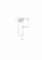
Заявленное техническое решение иллюстрируется Фиг. 1 - Фиг. 5.The claimed technical solution is illustrated in Fig. 1 - Fig. 5.
На Фиг. 1 представлен изометрический вид заявленного хирургического тубуса.On FIG. 1 is an isometric view of the claimed surgical sheath.
На Фиг. 2 представлен вид заявленного тубуса спереди.On FIG. 2 shows a front view of the claimed tube.
На Фиг. 3 представлен вид заявленного тубуса сбоку.On FIG. 3 shows a side view of the claimed tube.
На Фиг. 4 представлен вид заявленного тубуса сверху.On FIG. 4 shows a view of the claimed tube from above.
На Фиг. 5 представлен вид заявленного тубуса снизу.On FIG. 5 shows the bottom view of the claimed tube.
Позиции на Фигурах обозначают:The positions in the Figures denote:
1 - дистальный конец,1 - distal end,
2 - переход от дистального конца к проксимальному концу,2 - transition from the distal end to the proximal end,
3 - кольцо проксимального конца,3 - ring of the proximal end,
4 - изогнутое «ушко» кольца проксимального конца,4 - curved "eye" of the ring of the proximal end,
L1 - общая длина заявленного тубуса, равная 70-120 мм,L1 - the total length of the declared tube, equal to 70-120 mm,
L2 - длина дистального конца, равная 60-110 мм,L2 - length of the distal end, equal to 60-110 mm,
В1 - ширина основания с заднего конца, равная 35-40 мм,B1 - the width of the base from the rear end, equal to 35-40 mm,
В2 - ширина с переднего конца, равная 10-25 мм,B2 - width from the front end, equal to 10-25 mm,
∅1 - наружный диаметр проксимального конца, равный 30 мм,∅1 - outer diameter of the proximal end, equal to 30 mm,
R - наружный радиус проксимального конца, равный 15 мм,R is the outer radius of the proximal end, equal to 15 mm,
∅2 - внутренний диаметр проксимального конца, равный 10-25 мм,∅2 - inner diameter of the proximal end, equal to 10-25 mm,
b - толщина стенки заявленного тубуса, равная 0,1 - 0.3 мм.b - wall thickness of the declared tube, equal to 0.1 - 0.3 mm.
Далее заявителем приведено описание заявленного технического решения.Further, the applicant provides a description of the claimed technical solution.
Поставленные цели и заявленный технический результат достигается тем, что с помощью аддитивных технологий производства создают конструкцию хирургического тубуса для защиты мягких тканей для эндоскопической эндоназальной хирургии основания черепа. В целом заявленный тубус имеет конфигурацию трубки, имеющей проксимальный конец и дистальный конец. Проксимальный конец жесткий и имеет форму кольца, дистальный конец гибкий и имеет коническую форму, при этом «ушко» кольца проксимального конца выполнено с некоторым изгибом, для облегчения введения тубуса в носовую полость.The set goals and the claimed technical result are achieved by using additive manufacturing technologies to create the design of a surgical tube to protect soft tissues for endoscopic endonasal surgery of the skull base. In General, the claimed tube has the configuration of a tube having a proximal end and a distal end. The proximal end is rigid and has the shape of a ring, the distal end is flexible and has a conical shape, while the "eye" of the ring of the proximal end is made with some bending to facilitate the introduction of the tube into the nasal cavity.
Заявленный хирургический тубус создают методом экструзионной 3D-печати, например, на 3D принтере picaso 3D designer X PRO.The claimed surgical tube is created by extrusion 3D printing, for example, on a picaso 3D designer X PRO 3D printer.
В качестве материала хирургического защитного тубуса используют термопластичные полимерные материалы, например термопластичный сополиэфир марки FLEX REC или стирол-этилен-бутилен-стирол марки RUBBER REC или полиамид.As the material of the surgical protective sheath, thermoplastic polymeric materials are used, for example, thermoplastic copolyester of the FLEX REC brand or styrene-ethylene-butylene-styrene of the RUBBER REC brand or polyamide.
Жесткость проксимального конца придает конструкции заявленного тубуса устойчивость, устраняя возникновение повреждений оболочки тубуса при введении эндоскопических инструментов, тем самым исключает недостатки прототипа и снижает возможность возникновения травм при проведении хирургических манипуляций.The rigidity of the proximal end gives stability to the design of the claimed tube, eliminating the occurrence of damage to the shell of the tube during the introduction of endoscopic instruments, thereby eliminating the disadvantages of the prototype and reducing the possibility of injury during surgical procedures.
Далее заявителем приведено описание конструкции заявленного хирургического тубуса (Фиг. 1 - Фиг. 5).Further, the applicant describes the design of the claimed surgical tube (Fig. 1 - Fig. 5).
Общий изометрический вид заявленного тубуса представлен на Фиг. 1.A general isometric view of the claimed tube is shown in Fig. one.
В целом заявленный хирургический тубус представляет собой трубку, состоящую из проксимального конца в виде кольца, представляющего собой первое отверстие, и дистального конца в виде конуса, представляющего собой второе отверстие. Проксимальный конец имеет жесткую структуру c прочностью на изгиб более 10 МПа, а дистальный конец имеет гибкую структуру прочностью на изгиб менее 10 МПа и принимает анатомическую форму полости носовой пазухи.In general, the claimed surgical sheath is a tube consisting of a proximal end in the form of a ring, which is the first hole, and a distal end in the form of a cone, which is the second hole. The proximal end has a rigid structure with a bending strength of more than 10 MPa, and the distal end has a flexible structure with a bending strength of less than 10 MPa and takes the anatomical shape of the sinus cavity.
При этом хирургический тубус выполнен из одного вида полимерного материала для 3D-печати, характеризующегося градиентной степенью жесткости по всему периметру, а именно - проксимальный конец имеет жесткую структуру, а дистальный конец имеет гибкую структуру, за счет чего принимает анатомическую форму полости носовой пазухи, при этом хирургический тубус не имеет грубых границ раздела между участками с различной степенью жесткости.At the same time, the surgical tube is made of one type of polymeric material for 3D printing, characterized by a gradient degree of rigidity around the entire perimeter, namely, the proximal end has a rigid structure, and the distal end has a flexible structure, due to which it takes the anatomical shape of the nasal sinus cavity, while In this case, the surgical sheath does not have rough interfaces between areas with varying degrees of rigidity.
Заявленный хирургический тубус представляет собой трубку общей длиной L1=70-120 мм (Фиг. 2).The claimed surgical tube is a tube with a total length L1=70-120 mm (Fig. 2).
Тубус схематично разделен на 2 части, а именно - проксимальный конец, который имеет жесткую структуру, а дистальный конец, который имеет гибкую структуру, при этом границей раздела служит разность структуры материала - твердого и гибкого (А-А на Фиг.2), при этом:The tube is schematically divided into 2 parts, namely the proximal end, which has a rigid structure, and the distal end, which has a flexible structure, while the interface is the difference in the structure of the material - solid and flexible (A-A in Fig.2), with this:
1) погружная часть (дистальный конец) выполнена в форме конуса, имеет размеры (Фиг.2, Фиг.3):1) the immersion part (distal end) is made in the form of a cone, has dimensions (Figure 2, Figure 3):
- длину L2=60-110 мм,- length L2=60-110 mm,
- ширину основания с заднего конца В1=35-40 мм,- the width of the base from the rear end B1 = 35-40 mm,
- ширину с переднего конца В2=10-25 мм.- width from the front end B2=10-25 mm.
При виде снизу дистальный конец имеет форму эллипса (Фиг.5). Толщина стенки дистального конца не менее 0,15 мм. за счет чего и достигается требуемая эластичность с одновременной надлежащей атравматичностью.When viewed from below, the distal end has the shape of an ellipse (Figure 5). The wall thickness of the distal end is not less than 0.15 mm. due to which the required elasticity is achieved with at the same time proper atraumaticity.
2) наружная часть (проксимальный конец) (вид сверху на Фиг.4) выполнена в виде кольца с изогнутым "ушком" и имеет размеры:2) the outer part (proximal end) (top view in Fig.4) is made in the form of a ring with a curved "eye" and has the following dimensions:
- наружный диаметр ∅1=30 мм (R=15 мм),- outer diameter ∅1=30 mm (R=15 mm),
- внутренний диаметр ∅2=10-25 мм.- inner diameter ∅2=10-25 mm.
Толщина стенки заявленного тубуса (проксимальный конец) b=0.3 мм.The wall thickness of the claimed tube (proximal end) b=0.3 mm.
Заявленный хирургический тубус изготавливают следующим образом.The claimed surgical tube is made as follows.
Конструкцию заявленного хирургического тубуса создают с помощью аддитивных технологий.The design of the claimed surgical tube is created using additive technologies.
Заявленный хирургический тубус создают методом послойного наплавления на 3D принтере picaso 3D designer X PRO. Процесс послойного наплавления заключается выдавливанием («экструзией») и нанесением микрокапель расплавленного термопластика с формированием последовательных слоев, застывающих сразу после экструдирования. Данная технология позволяет с помощью программного обеспечения последовательно экструдировать различный термопластичный материал с разной прочностью, пластичностью и жесткостью, при этом получают цельное изделие из одного вида материала, характеризующееся градиентной степенью жесткости по всему периметру и не имеющее грубых границ раздела. Таким образом, разность структуры материала по периметру тубуса обеспечивается последовательной наплавкой в 3D-принтере термопластичных материалов разной степенью жесткости. В качестве материала хирургического защитного тубуса используют термопластичные полимерные материалы, например термопластичный сополиэфир марки FLEX REC или стирол-этилен-бутилен-стирол марки RUBBER REC или полиамид..The claimed surgical tube is created by layer-by-layer fusing on a picaso 3D designer X PRO 3D printer. The process of layer-by-layer fusing consists of extrusion ("extrusion") and the application of microdroplets of molten thermoplastic with the formation of successive layers that solidify immediately after extrusion. This technology allows using the software to sequentially extrude various thermoplastic materials with different strengths, ductility and stiffness, while obtaining a solid product from one type of material, characterized by a gradient degree of rigidity around the entire perimeter and not having rough interfaces. Thus, the difference in the structure of the material along the perimeter of the tube is ensured by successive surfacing in a 3D printer of thermoplastic materials of varying degrees of rigidity. Thermoplastic polymeric materials are used as the material of the surgical protective sheath, for example thermoplastic copolyester of the FLEX REC brand or styrene-ethylene-butylene-styrene of the RUBBER REC brand or polyamide.
Создают трехмерную модель заявленного хирургического тубуса с помощью известного программного комплекса САПР, задавая необходимые существенные признаки - параметры (размеры), включенные в формулу изобретения, а именно: проксимальный конец с наружным диаметром 30 мм, внутренним диаметром 10-25 мм, дистальный конец длиной 60-110 мм, шириной 10-25 мм с переднего конца и 35-40 мм с заднего конца. Далее трехмерную модель преобразуют в формат файла, необходимый для печати на 3D принтере picaso 3D designer X PRO. Преобразованный файл отправляют на 3D принтер и запускают процесс печати.A three-dimensional model of the claimed surgical tube is created using a well-known CAD software package, setting the necessary essential features - parameters (dimensions) included in the claims, namely: the proximal end with an outer diameter of 30 mm, an inner diameter of 10-25 mm, a distal end with a length of 60 -110 mm, width 10-25 mm from the front end and 35-40 mm from the rear end. Next, the 3D model is converted into the file format required for printing on the picaso 3D designer X PRO 3D printer. The converted file is sent to a 3D printer and the printing process is started.
Процесс печати начинается с обработки трехмерной цифровой модели. Модель в формате STL делится на слои. Процесс подготовки модели для печати называется «slicing», в результате которого генерируется G-code. В нем закладываются все параметры печати, перемещения экструдера, при необходимости генерируются поддерживающие структуры, необходимые для печати нависающих элементов. Некоторые устройства позволяют использовать разные материалы во время одного производственного цикла. Например, возможна печать модели из одного материала с печатью опор из другого, легкорастворимого материала, что позволяет с легкостью удалять поддерживающие структуры после завершения процесса печати. Изделие, или «модель», производится выдавливанием («экструзией») и нанесением микрокапель расплавленного термопластика с формированием последовательных слоев, застывающих сразу после экструдирования. Пластиковая нить разматывается с катушки и подается в экструдер - устройство, оснащенное механическим приводом для подачи нити, нагревательным элементом для плавки материала и соплом, через которое осуществляется непосредственно экструзия. Нагревательный элемент служит для нагревания сопла, которое в свою очередь плавит пластиковую нить и подает расплавленный материал на строящуюся модель. Как правило, верхняя часть сопла наоборот охлаждается с помощью вентилятора для создания резкого градиента температур, необходимого для обеспечения плавной подачи материала. Экструдер перемещается в горизонтальной и вертикальной плоскостях под контролем алгоритмов, аналогичных используемым в станках с числовым программным управлением. Сопло перемещается по траектории, заданной системой автоматизированного проектирования. Модель строится слой за слоем, снизу вверх. Как правило, экструдер приводится в движение шаговыми двигателями или сервоприводами. Наиболее популярной системой координат, применяемой в FDM, является прямоугольная, с осями X, Y и Z.The printing process begins with the processing of a three-dimensional digital model. The STL model is divided into layers. The process of preparing a model for printing is called "slicing", as a result of which a G-code is generated. It contains all the printing parameters, extruder movements, if necessary, the supporting structures necessary for printing overhanging elements are generated. Some devices allow the use of different materials during one production run. For example, it is possible to print a model from one material with supports printed from another, easily soluble material, which makes it easy to remove support structures after the printing process is completed. The product, or “model”, is produced by extrusion (“extrusion”) and the application of microdroplets of molten thermoplastic to form successive layers that solidify immediately after extrusion. The plastic thread is unwound from the reel and fed into the extruder - a device equipped with a mechanical drive for feeding the thread, a heating element for melting the material and a nozzle through which the extrusion is carried out directly. The heating element serves to heat the nozzle, which in turn melts the plastic filament and supplies the melted material to the model being built. Typically, the top of the nozzle is instead cooled by a fan to create the sharp temperature gradient needed to ensure smooth material flow. The extruder moves in horizontal and vertical planes under the control of algorithms similar to those used in machine tools with numerical control. The nozzle moves along the trajectory specified by the computer-aided design system. The model is built layer by layer, from bottom to top. Typically, the extruder is driven by stepper motors or servo drives. The most popular coordinate system used in FDM is rectangular, with X, Y, and Z axes.
Далее готовый тубус снимают со столика. Получают заявленный хирургический тубус.Next, the finished tube is removed from the table. Get the declared surgical tube.
Далее проводят проверку готового тубуса на соответствие заданным характеристикам, например, путем визуального осмотра, обмера.Next, the finished tube is checked for compliance with the specified characteristics, for example, by visual inspection, measurement.
Далее заявителем приведены примеры использования заявленного хирургического тубуса.Further, the applicant provides examples of the use of the claimed surgical tube.
Пример 1. Использование заявленного хирургического тубуса с размерами: общая длина 70 мм, проксимальный конец с наружным диаметром 30 мм, внутренним диаметром 10 мм, дистальный конец длиной 60 мм, шириной 10 мм с переднего конца и 35 мм с заднего конца.Example 1. Use of the claimed surgical sheath with dimensions: total length 70 mm, proximal end with an outer diameter of 30 mm, an inner diameter of 10 mm, a distal end with a length of 60 mm, a width of 10 mm from the anterior end and 35 mm from the posterior end.
Изготавливают по описанной выше аддитивной технологии заявленный хирургический тубус из термопластичного полимерного материала сополиэфир марки FLEX REC с размерами: общей длиной 70 мм, наружным диаметром 30 мм, внутренним диаметром 10 мм, дистальным концом длиной 60 мм, шириной 10 мм с переднего конца и 35 мм с заднего конца. Общая толщина стенки тубуса 0.3 мм. Проксимальный конец имеет жесткую структуру c прочностью на изгиб более 10 МПа, а дистальный конец имеет гибкую структуру прочностью на изгиб менее 10 МПа и принимает анатомическую форму.The claimed surgical tube is made according to the additive technology described above from a thermoplastic polymeric material copolyester brand FLEX REC with dimensions: total length 70 mm, outer diameter 30 mm, inner diameter 10 mm, distal end 60 mm long, 10 mm wide from the front end and 35 mm from the back end. The total wall thickness of the tube is 0.3 mm. The proximal end has a rigid structure with a bending strength of more than 10 MPa, and the distal end has a flexible structure with a bending strength of less than 10 MPa and takes an anatomical shape.
Проведена эндоскопическая эндоназальная хирургия основания черепа у пациента А., 10 лет.Endoscopic endonasal skull base surgery was performed in patient A., 10 years old.
Берут заявленный хирургический тубус с указанными размерами и вводят его в полость носа через ноздри для доступа к операционному полю, проводят операцию эндоскопической эндоназальной хирургии основания черепа.The claimed surgical tube with the specified dimensions is taken and inserted into the nasal cavity through the nostrils to access the surgical field, the operation of endoscopic endonasal surgery of the skull base is performed.
По результатам операции можно сделать вывод, что:Based on the results of the operation, it can be concluded that:
- обеспечено устойчивое и плотное прилегание к полости носа, пациент чувствовал себя комфортно во время проведения хирургических манипуляций;- a stable and tight fit to the nasal cavity was provided, the patient felt comfortable during surgical procedures;
- отсутствовали травмы слизистых и мягких тканей, обеспечена чистота операционного поля.- there were no injuries of mucous and soft tissues, the cleanliness of the surgical field was ensured.
Указанное свидетельствует о достижении заявленного технического результата.This indicates the achievement of the claimed technical result.
Пример 2. Использование заявленного хирургического тубуса с размерами: общая длина 120 мм, проксимальный конец с наружным диаметром 30 мм, внутренним диаметром 25 мм, дистальный конец длиной 110 мм, шириной 25 мм с переднего конца и 40 мм с заднего конца.Example 2. Use of the claimed surgical sheath with dimensions: total length 120 mm, proximal end with an outer diameter of 30 mm, an inner diameter of 25 mm, a distal end with a length of 110 mm, a width of 25 mm from the anterior end and 40 mm from the posterior end.
Изготавливают по описанной выше аддитивной технологии заявленный хирургический тубус из фотополимерного материала, например, полипропиленовго флекса марки Dental Ortho Flex с размерами: общей длиной 120 мм, наружным диаметром 30 мм, внутренним диаметром 25 мм, дистальным концом длиной 110 мм, шириной 25 мм с переднего конца и 40 мм с заднего конца. Общая толщина стенки тубуса 0.3 мм. Проксимальный конец имеет жесткую структуру c прочностью на изгиб более 10 МПа, а дистальный конец имеет гибкую структуру прочностью на изгиб менее 10 МПа и принимает анатомическую форму.The claimed surgical tube is made according to the additive technology described above from a photopolymer material, for example, Dental Ortho Flex brand polypropylene flex with dimensions: total length 120 mm, outer diameter 30 mm, inner diameter 25 mm, distal end 110 mm long, width 25 mm from the front end and 40 mm from the rear end. The total wall thickness of the tube is 0.3 mm. The proximal end has a rigid structure with a bending strength of more than 10 MPa, and the distal end has a flexible structure with a bending strength of less than 10 MPa and takes an anatomical shape.
Проведена эндоскопическая эндоназальная хирургия основания черепа у пациента Б., 45 лет.Endoscopic endonasal skull base surgery was performed in patient B., 45 years old.
Берут заявленный хирургический тубус с указанными размерами и вводят его в полость носа через ноздри для доступа к операционному полю. Проводят операцию по стандартной методике.Take the declared surgical tube with the specified dimensions and introduce it into the nasal cavity through the nostrils to access the surgical field. The operation is carried out according to the standard method.
По результатам операции можно сделать вывод, что:Based on the results of the operation, it can be concluded that:
- обеспечено устойчивое и плотное прилегание к полости носа, пациент чувствовал себя комфортно во время проведения хирургических манипуляций;- a stable and tight fit to the nasal cavity was provided, the patient felt comfortable during surgical procedures;
- отсутствовали травмы слизистых и мягких тканей, обеспечена чистота операционного поля.- there were no injuries of mucous and soft tissues, the cleanliness of the surgical field was ensured.
Указанное свидетельствует о достижении заявленного технического результата.This indicates the achievement of the claimed technical result.
Пример 3. Использование заявленного хирургического тубуса с размерами: общая длина 100 мм, проксимальный конец с наружным диаметром 30 мм, внутренним диаметром 15 мм, дистальный конец длиной 90 мм, шириной 15 мм с переднего конца и 37 мм с заднего конца.Example 3. Use of the claimed surgical sheath with dimensions: total length 100 mm, proximal end with an outer diameter of 30 mm, an inner diameter of 15 mm, a distal end with a length of 90 mm, a width of 15 mm from the anterior end and 37 mm from the posterior end.
Изготавливают по описанной выше аддитивной технологии заявленный хирургический тубус из термопластичного полимерного материала, например, или стирол-этилен-бутилен-стирол марки RUBBER REC с размерами: общей длиной 100 мм, наружным диаметром 30 мм, внутренним диаметром 15 мм, дистальным концом длиной 90 мм, шириной 15 мм с переднего конца и 37 мм с заднего конца. Общая толщина стенки тубуса 0.3 мм. Проксимальный конец имеет жесткую структуру c прочностью на изгиб более 10 МПа, а дистальный конец имеет гибкую структуру прочностью на изгиб менее 10 МПа и принимает анатомическую форму.The claimed surgical tube is made using the additive technology described above from a thermoplastic polymer material, for example, or styrene-ethylene-butylene-styrene of the RUBBER REC brand with dimensions: total length 100 mm, outer diameter 30 mm, inner diameter 15 mm, distal end 90 mm long , 15 mm wide at the front end and 37 mm at the rear end. The total wall thickness of the tube is 0.3 mm. The proximal end has a rigid structure with a bending strength of more than 10 MPa, and the distal end has a flexible structure with a bending strength of less than 10 MPa and takes an anatomical shape.
Проведена эндоскопическая эндоназальная хирургия основания черепа у пациента В., 31 год.Endoscopic endonasal skull base surgery was performed in patient V., 31 years old.
Берут заявленный хирургический тубус с указанными размерами и вводят его в полость носа через ноздри для доступа к операционному полю. Проводят операцию по стандартной методике.Take the declared surgical tube with the specified dimensions and introduce it into the nasal cavity through the nostrils to access the surgical field. The operation is carried out according to the standard method.
По результатам операции можно сделать вывод, что:Based on the results of the operation, it can be concluded that:
- обеспечено устойчивое и плотное прилегание к полости носа, пациент чувствовал себя комфортно во время проведения хирургических манипуляций;- a stable and tight fit to the nasal cavity was provided, the patient felt comfortable during surgical procedures;
- отсутствовали травмы слизистых и мягких тканей, обеспечена чистота операционного поля.- there were no injuries of mucous and soft tissues, the cleanliness of the surgical field was ensured.
Указанное свидетельствует о достижении заявленного технического результата.This indicates the achievement of the claimed technical result.
Таким образом, из описанного выше можно сделать вывод, что заявителем достигнут заявленный технический результат, а именно - разработан хирургический тубус для защиты мягких тканей, устраняющий недостатки прототипа, а именно:Thus, from the above, we can conclude that the applicant has achieved the claimed technical result, namely, a surgical tube has been developed to protect soft tissues, eliminating the disadvantages of the prototype, namely:
- обеспечивающего устойчивое расположение в полости носа вследствие того, что тубус имеет градиентную структуру жесткости материала, которая внутри носовой полости мягкая, а снаружи (вне носа) - жесткая, и изготовлен из полимерных (полиамид) материалов, что приводит к комфортным ощущениям пациента во время проведения хирургических манипуляций, а также позволяет хирургу комфортно проводить требуемые манипуляции;- providing a stable location in the nasal cavity due to the fact that the tube has a gradient structure of material stiffness, which is soft inside the nasal cavity, and hard outside (outside the nose), and is made of polymer (polyamide) materials, which leads to comfortable sensations for the patient during carrying out surgical procedures, and also allows the surgeon to comfortably carry out the required manipulations;
- возможность проведения хирургических манипуляций с минимальным риском возникновения травм слизистых и мягких тканей, а также обеспечивающих чистоту операционного поля.- the possibility of performing surgical manipulations with a minimal risk of injury to the mucous and soft tissues, as well as ensuring the cleanliness of the surgical field.
При этом заявленный хирургический тубус для защиты мягких тканей обладает одновременно комплексом более высоких потребительских свойств при использовании по назначению, а именно наличие градиента механических свойств в конструкции. Которая заключается в высокой жесткости заднего конца и в мягкости дистального конца.At the same time, the claimed surgical tube for protecting soft tissues simultaneously has a complex of higher consumer properties when used for its intended purpose, namely, the presence of a gradient of mechanical properties in the structure. Which lies in the high rigidity of the posterior end and the softness of the distal end.
Заявленное техническое решение соответствует условию патентоспособности «новизна», предъявляемому к изобретениям, так как из исследованного заявителем уровня техники не выявлена совокупность признаков, приведенная в независимом пункте формулы изобретения.The claimed technical solution complies with the "novelty" patentability condition for inventions, since the set of features given in the independent claim of the invention has not been identified from the prior art studied by the applicant.
Заявленное техническое решение соответствует условию патентоспособности «изобретательский уровень», предъявляемому к изобретениям, так как из исследованного заявителем уровня техники не выявлена совокупность приведенных в независимом пункте формулы изобретения признаков и совокупность полученных технических результатов.The claimed technical solution complies with the "inventive step" patentability condition for inventions, since the totality of the features listed in the independent claim and the totality of the technical results obtained have not been identified from the prior art studied by the applicant.
Заявленное техническое решение соответствует условию патентоспособности «промышленная применимость», предъявляемому к изобретениям, так как заявленное техническое решение возможно реализовать в промышленности посредством применения известных из уровня техники материалов, оборудование и технологий.The claimed technical solution complies with the "industrial applicability" patentability condition for inventions, since the claimed technical solution can be implemented in industry through the use of materials, equipment and technologies known from the prior art.
Источники информацииInformation sources
1. Патент US20130204092A1 кл. A61B 1/32 Soft tissue shield for trans-orbital surgery / Blake Hannaford, Randall A. Bly. - 21.04. 2015. - URL: https://patents.google.com/patent/US20130204092.1. Patent US20130204092A1 cl.
2. Патент US20170042409A1 кл. A61B 90/00 Surgical tissue protection sheath / Blake Hannaford, Randall A. Bly. - 16.02.2017 - Blake Hannaford, Randall A. Bly, Eugene Chen, Aylin Kim, Cang Lam, Ciporen, Kristen Moe, Louis Kim, Laligam Sekhar. - URL: https://patents.google.com/patent/US20170042409A1/en.2. Patent US20170042409A1 cl. A61B 90/00 Surgical tissue protection sheath / Blake Hannaford, Randall A. Bly. - 02/16/2017 - Blake Hannaford, Randall A. Bly, Eugene Chen, Aylin Kim, Cang Lam, Ciporen, Kristen Moe, Louis Kim, Laligam Sekhar. - URL: https://patents.google.com/patent/US20170042409A1/en.
3. SPIWay Endonasal Access Guide. [Электронный ресурс]. - URL: http://spiway.com/the-spiway.html.3. SPIWay Endonasal Access Guide. [Electronic resource]. - URL: http://spiway.com/the-spiway.html.
Claims (1)
Translated fromRussianPriority Applications (1)
| Application Number | Priority Date | Filing Date | Title |
|---|---|---|---|
| RU2021135648ARU2770992C1 (en) | 2021-12-03 | 2021-12-03 | Surgical tube for protection of soft tissues during endoscopic endonasal surgery of skull base |
Applications Claiming Priority (1)
| Application Number | Priority Date | Filing Date | Title |
|---|---|---|---|
| RU2021135648ARU2770992C1 (en) | 2021-12-03 | 2021-12-03 | Surgical tube for protection of soft tissues during endoscopic endonasal surgery of skull base |
Publications (1)
| Publication Number | Publication Date |
|---|---|
| RU2770992C1true RU2770992C1 (en) | 2022-04-25 |
Family
ID=81306393
Family Applications (1)
| Application Number | Title | Priority Date | Filing Date |
|---|---|---|---|
| RU2021135648ARU2770992C1 (en) | 2021-12-03 | 2021-12-03 | Surgical tube for protection of soft tissues during endoscopic endonasal surgery of skull base |
Country Status (1)
| Country | Link |
|---|---|
| RU (1) | RU2770992C1 (en) |
Cited By (1)
| Publication number | Priority date | Publication date | Assignee | Title |
|---|---|---|---|---|
| RU2787293C1 (en)* | 2022-06-06 | 2023-01-09 | федеральное государственное автономное образовательное учреждение высшего образования "Казанский (Приволжский) федеральный университет" (ФГАОУ ВО КФУ) | Method for manufacturing medical products from plastic using sla technology with gradient illumination |
Citations (5)
| Publication number | Priority date | Publication date | Assignee | Title |
|---|---|---|---|---|
| US20100331777A1 (en)* | 2008-01-29 | 2010-12-30 | Goeran ALMGREN | Nose device |
| US20110118551A1 (en)* | 2009-11-14 | 2011-05-19 | SPI Surgical, Inc. | Collateral soft tissue protection surgical device |
| DE202012100028U1 (en)* | 2012-01-04 | 2012-04-25 | Hubert Hipp | nasal tube |
| US20170042409A1 (en)* | 2009-11-14 | 2017-02-16 | Spiway Llc | Surgical tissue protection sheath |
| US20190274525A1 (en)* | 2013-03-13 | 2019-09-12 | Spiway Llc | Surgical tissue protection sheath |
- 2021
- 2021-12-03RURU2021135648Apatent/RU2770992C1/enactive
Patent Citations (5)
| Publication number | Priority date | Publication date | Assignee | Title |
|---|---|---|---|---|
| US20100331777A1 (en)* | 2008-01-29 | 2010-12-30 | Goeran ALMGREN | Nose device |
| US20110118551A1 (en)* | 2009-11-14 | 2011-05-19 | SPI Surgical, Inc. | Collateral soft tissue protection surgical device |
| US20170042409A1 (en)* | 2009-11-14 | 2017-02-16 | Spiway Llc | Surgical tissue protection sheath |
| DE202012100028U1 (en)* | 2012-01-04 | 2012-04-25 | Hubert Hipp | nasal tube |
| US20190274525A1 (en)* | 2013-03-13 | 2019-09-12 | Spiway Llc | Surgical tissue protection sheath |
Cited By (1)
| Publication number | Priority date | Publication date | Assignee | Title |
|---|---|---|---|---|
| RU2787293C1 (en)* | 2022-06-06 | 2023-01-09 | федеральное государственное автономное образовательное учреждение высшего образования "Казанский (Приволжский) федеральный университет" (ФГАОУ ВО КФУ) | Method for manufacturing medical products from plastic using sla technology with gradient illumination |
Similar Documents
| Publication | Publication Date | Title |
|---|---|---|
| EP2491973B1 (en) | Systems useable for treating sinusitis | |
| RU2572745C2 (en) | Removing tissue from paranasal sinus and nasal cavity | |
| EP3142851B1 (en) | System and method for fabricating custom medical implant devices | |
| JP5952452B1 (en) | Training device for transnasal endoscopic skull base surgery | |
| US12426902B2 (en) | Surgical cutting guides designed for anatomical landmarks | |
| JP6933828B2 (en) | Surgical instrument for acetabular rotation osteotomy | |
| JP2008178654A (en) | Protector for nasal cavity | |
| JP6855074B2 (en) | Holding mechanism for long medical devices | |
| WO2015127371A1 (en) | Custom reduction splint for edentulous patients | |
| Ruiters et al. | Applications of three-dimensional printing in orbital diseases and disorders | |
| RU2770992C1 (en) | Surgical tube for protection of soft tissues during endoscopic endonasal surgery of skull base | |
| Stokken et al. | The emerging role of 3-dimensional printing in rhinology | |
| JP7532401B2 (en) | Eustachian tube dilation catheter with depth indication | |
| KR101994956B1 (en) | Guide for mandible reduction surgery | |
| CN108056811B (en) | Tongue body ablation surgery navigation and positioning device and its supporting surgical instruments | |
| US20120232525A1 (en) | Suction device | |
| Arndt et al. | Management of cochlear implantation in patients with malformations | |
| KR101778790B1 (en) | Surgical Guide for Zygomatic Bone, Manufacturing Method Thereof, Recording Medium Therefor, Manufacturing Apparatus Therefor | |
| KR102463670B1 (en) | External and internal layered cranial remolding helmet and manufacturing method thereof | |
| CN206518594U (en) | Tongue body ablative surgery navigation positional device and the operating theater instruments supporting with it | |
| CN205548622U (en) | Lower jaw operation guide plate | |
| RU2523352C1 (en) | Method for surgical approach to periapical tissues of jaw | |
| Kamizono et al. | Safe and rapid contouring of fibro-osseous lesions in the orbital area using navigation with minimally invasive cranial bone registration | |
| MacKenty | Three new plastic operations on the nose and throat | |
| Nam et al. | Application and limitations of facial computed tomography and three-dimensional scanner images for patient-specific three-dimensional printing of a nose mask |