RU2759564C1 - Method for performing laparoscopic longitudinal resection of stomach in morbid obesity - Google Patents
Method for performing laparoscopic longitudinal resection of stomach in morbid obesityDownload PDFInfo
- Publication number
- RU2759564C1 RU2759564C1RU2020143224ARU2020143224ARU2759564C1RU 2759564 C1RU2759564 C1RU 2759564C1RU 2020143224 ARU2020143224 ARU 2020143224ARU 2020143224 ARU2020143224 ARU 2020143224ARU 2759564 C1RU2759564 C1RU 2759564C1
- Authority
- RU
- Russia
- Prior art keywords
- stomach
- trocar
- along
- trocars
- stapler
- Prior art date
Links
- 210000002784stomachAnatomy0.000titleclaimsabstractdescription76
- 238000000034methodMethods0.000titleclaimsabstractdescription26
- 208000008589ObesityDiseases0.000titleclaimsabstractdescription10
- 208000012696congenital leptin deficiencyDiseases0.000titleclaimsabstractdescription10
- 208000001022morbid obesityDiseases0.000titleclaimsabstractdescription10
- 238000002271resectionMethods0.000titleclaimsdescription17
- 210000004185liverAnatomy0.000claimsabstractdescription12
- QTCANKDTWWSCMR-UHFFFAOYSA-Ncostic aldehydeNatural productsC1CCC(=C)C2CC(C(=C)C=O)CCC21CQTCANKDTWWSCMR-UHFFFAOYSA-N0.000claimsabstractdescription8
- ISTFUJWTQAMRGA-UHFFFAOYSA-Niso-beta-costalNatural productsC1C(C(=C)C=O)CCC2(C)CCCC(C)=C21ISTFUJWTQAMRGA-UHFFFAOYSA-N0.000claimsabstractdescription8
- 210000001187pylorusAnatomy0.000claimsabstractdescription8
- 210000002417xiphoid boneAnatomy0.000claimsabstractdescription8
- 206010052428WoundDiseases0.000claimsabstractdescription5
- 208000027418Wounds and injuryDiseases0.000claimsabstractdescription5
- 239000000523sampleSubstances0.000claimsdescription13
- 210000000496pancreasAnatomy0.000claimsdescription7
- 230000000007visual effectEffects0.000claimsdescription6
- 238000009434installationMethods0.000claimsdescription5
- 230000023597hemostasisEffects0.000claimsdescription4
- 239000002184metalSubstances0.000claimsdescription4
- BOMUADPKDXMXIH-UHFFFAOYSA-M1,3-bis(1-methylquinolin-1-ium-6-yl)urea;methyl sulfateChemical groupCOS([O-])(=O)=O.COS([O-])(=O)=O.C[N+]1=CC=CC2=CC(NC(=O)NC=3C=C4C=CC=[N+](C4=CC=3)C)=CC=C21BOMUADPKDXMXIH-UHFFFAOYSA-M0.000claimsdescription3
- 206010019909HerniaDiseases0.000claimsdescription3
- 210000000566lesser sacAnatomy0.000claimsdescription3
- 238000012360testing methodMethods0.000claimsdescription3
- 208000002193PainDiseases0.000abstractdescription10
- 208000014674injuryDiseases0.000abstractdescription6
- 230000008733traumaEffects0.000abstractdescription6
- 230000002980postoperative effectEffects0.000abstractdescription4
- 239000003814drugSubstances0.000abstractdescription3
- 208000011580syndromic diseaseDiseases0.000abstractdescription3
- 208000001145Metabolic SyndromeDiseases0.000abstractdescription2
- 201000000690abdominal obesity-metabolic syndromeDiseases0.000abstractdescription2
- 238000007681bariatric surgeryMethods0.000abstractdescription2
- 238000005452bendingMethods0.000abstract1
- 230000000694effectsEffects0.000abstract1
- 238000007373indentationMethods0.000abstract1
- 239000000126substanceSubstances0.000abstract1
- 238000001356surgical procedureMethods0.000description12
- 210000000683abdominal cavityAnatomy0.000description6
- 230000007774longtermEffects0.000description5
- 208000007882GastritisDiseases0.000description3
- 208000032843HemorrhageDiseases0.000description3
- 208000034158bleedingDiseases0.000description3
- 230000000740bleeding effectEffects0.000description3
- 238000003745diagnosisMethods0.000description3
- 201000010099diseaseDiseases0.000description3
- 208000037265diseases, disorders, signs and symptomsDiseases0.000description3
- 238000002604ultrasonographyMethods0.000description3
- 208000035965Postoperative ComplicationsDiseases0.000description2
- 210000003484anatomyAnatomy0.000description2
- 230000006835compressionEffects0.000description2
- 238000007906compressionMethods0.000description2
- 238000002224dissectionMethods0.000description2
- 238000013110gastrectomyMethods0.000description2
- 230000002496gastric effectEffects0.000description2
- 210000003041ligamentAnatomy0.000description2
- 210000001699lower legAnatomy0.000description2
- 210000004977neurovascular bundleAnatomy0.000description2
- 238000010561standard procedureMethods0.000description2
- 230000004580weight lossEffects0.000description2
- 102000012004GhrelinHuman genes0.000description1
- 101800001586GhrelinProteins0.000description1
- 206010019842HepatomegalyDiseases0.000description1
- 206010020710HyperphagiaDiseases0.000description1
- 208000032984Intraoperative ComplicationsDiseases0.000description1
- 206010057765Procedural complicationDiseases0.000description1
- 230000003187abdominal effectEffects0.000description1
- 230000000151anti-reflux effectEffects0.000description1
- 210000001367arteryAnatomy0.000description1
- 230000015572biosynthetic processEffects0.000description1
- 230000037396body weightEffects0.000description1
- 210000002318cardiaAnatomy0.000description1
- 238000012937correctionMethods0.000description1
- 238000013461designMethods0.000description1
- 238000011161developmentMethods0.000description1
- 238000010586diagramMethods0.000description1
- 201000006549dyspepsiaDiseases0.000description1
- 210000003238esophagusAnatomy0.000description1
- GNKDKYIHGQKHHM-RJKLHVOGSA-NghrelinChemical compoundC([C@H](NC(=O)[C@@H](NC(=O)[C@H](CO)NC(=O)CN)COC(=O)CCCCCCC)C(=O)N[C@@H](CC(C)C)C(=O)N[C@@H](CO)C(=O)N1[C@@H](CCC1)C(=O)N[C@@H](CCC(O)=O)C(=O)N[C@@H](CC=1N=CNC=1)C(=O)N[C@@H](CCC(N)=O)C(=O)N[C@@H](CCCNC(N)=N)C(=O)N[C@@H](C(C)C)C(=O)N[C@@H](CCC(N)=O)C(=O)N[C@@H](CCC(N)=O)C(=O)N[C@@H](CCCNC(N)=N)C(=O)N[C@@H](CCCCN)C(=O)N[C@@H](CCC(O)=O)C(=O)N[C@@H](CO)C(=O)N[C@@H](CCCCN)C(=O)N[C@@H](CCCCN)C(=O)N1[C@@H](CCC1)C(=O)N1[C@@H](CCC1)C(=O)N[C@@H](C)C(=O)N[C@@H](CCCCN)C(=O)N[C@@H](CC(C)C)C(=O)N[C@@H](CCC(N)=O)C(=O)N1[C@@H](CCC1)C(=O)N[C@@H](CCCNC(N)=N)C(O)=O)C1=CC=CC=C1GNKDKYIHGQKHHM-RJKLHVOGSA-N0.000description1
- 208000024798heartburnDiseases0.000description1
- 230000003054hormonal effectEffects0.000description1
- 230000001771impaired effectEffects0.000description1
- 210000003127kneeAnatomy0.000description1
- 210000000629knee jointAnatomy0.000description1
- 238000009533lab testMethods0.000description1
- 239000004816latexSubstances0.000description1
- 229920000126latexPolymers0.000description1
- 210000002414legAnatomy0.000description1
- 239000007788liquidSubstances0.000description1
- 210000000510mesogastriumAnatomy0.000description1
- 235000020830overeatingNutrition0.000description1
- 238000004321preservationMethods0.000description1
- 230000002265preventionEffects0.000description1
- 230000003014reinforcing effectEffects0.000description1
- 238000011160researchMethods0.000description1
- 230000003393splenic effectEffects0.000description1
- 238000011179visual inspectionMethods0.000description1
- 238000012800visualizationMethods0.000description1
Images


Classifications
- A—HUMAN NECESSITIES
- A61—MEDICAL OR VETERINARY SCIENCE; HYGIENE
- A61B—DIAGNOSIS; SURGERY; IDENTIFICATION
- A61B17/00—Surgical instruments, devices or methods
- A—HUMAN NECESSITIES
- A61—MEDICAL OR VETERINARY SCIENCE; HYGIENE
- A61B—DIAGNOSIS; SURGERY; IDENTIFICATION
- A61B17/00—Surgical instruments, devices or methods
- A61B17/00234—Surgical instruments, devices or methods for minimally invasive surgery
- A—HUMAN NECESSITIES
- A61—MEDICAL OR VETERINARY SCIENCE; HYGIENE
- A61B—DIAGNOSIS; SURGERY; IDENTIFICATION
- A61B17/00—Surgical instruments, devices or methods
- A61B17/068—Surgical staplers, e.g. containing multiple staples or clamps
- A61B17/072—Surgical staplers, e.g. containing multiple staples or clamps for applying a row of staples in a single action, e.g. the staples being applied simultaneously
- A61B17/07207—Surgical staplers, e.g. containing multiple staples or clamps for applying a row of staples in a single action, e.g. the staples being applied simultaneously the staples being applied sequentially
- A—HUMAN NECESSITIES
- A61—MEDICAL OR VETERINARY SCIENCE; HYGIENE
- A61B—DIAGNOSIS; SURGERY; IDENTIFICATION
- A61B17/00—Surgical instruments, devices or methods
- A61B17/12—Surgical instruments, devices or methods for ligaturing or otherwise compressing tubular parts of the body, e.g. blood vessels or umbilical cord
- A61B17/122—Clamps or clips, e.g. for the umbilical cord
- A—HUMAN NECESSITIES
- A61—MEDICAL OR VETERINARY SCIENCE; HYGIENE
- A61B—DIAGNOSIS; SURGERY; IDENTIFICATION
- A61B17/00—Surgical instruments, devices or methods
- A61B17/34—Trocars; Puncturing needles
- A—HUMAN NECESSITIES
- A61—MEDICAL OR VETERINARY SCIENCE; HYGIENE
- A61M—DEVICES FOR INTRODUCING MEDIA INTO, OR ONTO, THE BODY; DEVICES FOR TRANSDUCING BODY MEDIA OR FOR TAKING MEDIA FROM THE BODY; DEVICES FOR PRODUCING OR ENDING SLEEP OR STUPOR
- A61M25/00—Catheters; Hollow probes
Landscapes
- Health & Medical Sciences (AREA)
- Life Sciences & Earth Sciences (AREA)
- Surgery (AREA)
- Veterinary Medicine (AREA)
- Public Health (AREA)
- General Health & Medical Sciences (AREA)
- Engineering & Computer Science (AREA)
- Biomedical Technology (AREA)
- Heart & Thoracic Surgery (AREA)
- Animal Behavior & Ethology (AREA)
- Molecular Biology (AREA)
- Medical Informatics (AREA)
- Nuclear Medicine, Radiotherapy & Molecular Imaging (AREA)
- Vascular Medicine (AREA)
- Reproductive Health (AREA)
- Pathology (AREA)
- Biophysics (AREA)
- Pulmonology (AREA)
- Anesthesiology (AREA)
- Hematology (AREA)
- Surgical Instruments (AREA)
Abstract
Description
Translated fromRussianИзобретение относится к медицине, а именно к бариатрической хирургии, и может быть использовано для лечения морбидного ожирения, в том числе при сопутствующем метаболическом синдроме.The invention relates to medicine, namely to bariatric surgery, and can be used to treat morbid obesity, including with concomitant metabolic syndrome.
Продольная резекция желудка (ПРЖ) стремительно набирает популярность в мире и занимает сегодня лидирующие позиции по частоте выполнения в России.Longitudinal gastric resection (LRG) is rapidly gaining popularity in the world and today occupies a leading position in terms of frequency of execution in Russia.
ПРЖ — рестриктивная операция, при которой формируется малый желудок объемом 100 - 150 мл. Механизм снижения веса обусловлен рестриктивным компонентом, а также гормональным механизмом, связанным с резким снижением содержания грелина. Качество выполнения операции зависит в первую очередь от навыков и знаний хирурга, но не менее важны качество инструментов и понимание последовательности этапов операции.PRG is a restrictive operation, in which a small stomach is formed with a volume of 100 - 150 ml. The mechanism of weight loss is due to a restrictive component, as well as a hormonal mechanism associated with a sharp decrease in ghrelin content. The quality of the operation depends primarily on the skills and knowledge of the surgeon, but the quality of the instruments and understanding of the sequence of stages of the operation are no less important.
Известен способ выполнения бариатрических лапароскопических операций (RU2564144, опубликовано: 2015.09.27), включающий хирургический доступ к желудку и формирование маленького желудочка со стороны малой кривизны желудка, отличающийся тем, что при резекции сохраняют 2-4 см медиальной поверхности дна желудка и формируют антирефлюксный клапан, накладывая по меньшей мере один шов, захватывающий проксимальную часть сохраненной медиальной поверхности дна желудка, левую ножку диафрагмы и абдоминальную часть пищевода.There is a known method of performing bariatric laparoscopic operations (RU2564144, published: 2015.09.27), including surgical access to the stomach and the formation of a small ventricle from the side of the lesser curvature of the stomach, characterized in that during resection 2-4 cm of the medial surface of the fundus of the stomach are preserved and an antireflux valve is formed , applying at least one suture that captures the proximal part of the preserved medial surface of the fundus of the stomach, the left crus of the diaphragm and the abdominal part of the esophagus.
В этой методике не указано количество троакаров и схема их, что предполагает стандартный метод выполнения продольной резекции желудка– 5 троакаров, что требует к привлечению операции нескольких ассистентов.This technique does not indicate the number of trocars and their scheme, which suggests a standard method for performing longitudinal resection of the stomach - 5 trocars, which requires several assistants to engage in the operation.
Стандартная схема предполагает пять троакаров. Первый, 10 мм троакар для введения лапароскопа, устанавливается на 5–10 см выше пупочного кольца. Для введения эндоретрактора субксифоидально устанавливается 5 или 10 мм троакар. В правом подреберье по среднеключичной линии на середине расстояния между реберной дугой и уровнем пупочного кольца – 12 мм троакар для введения эндозажима и сшивающего аппарата. Через этот доступ, расширенный до 3 см, удаляем резецированную часть желудка. В левом подреберье по среднеключичной линии на середине расстояния между реберной дугой и уровнем пупочного кольца устанавливаются 5 или 10 мм троакар для введения эндодиссектора, эндоножниц, иглодержателя и электрохирургических или ультразвуковых инструментов. В левом подреберье на 2 см ниже реберной дуги по передней подмышечной линии – 5 мм троакар для введения эндозажима.The standard design assumes five trocars. The first, 10 mm trocar for the introduction of the laparoscope, is placed 5–10 cm above the umbilical ring. For the introduction of the endoretractor, a 5 or 10 mm trocar is inserted subxiphoidally. In the right hypochondrium along the midclavicular line, in the middle of the distance between the costal arch and the level of the umbilical ring, there is a 12 mm trocar for the introduction of an endoclamp and a stapler. Through this access, expanded to 3 cm, we remove the resected part of the stomach. In the left hypochondrium along the midclavicular line in the middle of the distance between the costal arch and the level of the umbilical ring, a 5 or 10 mm trocar is installed for the introduction of an endodissector, endoscissors, a needle holder and electrosurgical or ultrasonic instruments. In the left hypochondrium, 2 cm below the costal arch along the anterior axillary line, there is a 5 mm trocar for the introduction of an endo-clamp.
Указанный аналог характерен травматичностью и выраженным болевым синдромом в послеоперационном периоде.This analogue is characterized by traumatism and severe pain syndrome in the postoperative period.
Наиболее близким аналогом является способ хирургического лечения морбидного ожирения путем лапароскопического создания нескольких камер желудка (RU2701222, опубликовано: 25.09.2019), отличающийся тем, что выполняют лапароскопическую рукавную гастропластику с отсечением большой кривизны желудка линейным сшивающим аппаратом на зонде диаметром 36 Fr, с последующим наложением обвивного непрерывного армирующего эндошва монофиламентной нерассасывающейся нитью 2.0 вдоль всей степлерной линии, и формируют трехкамерный желудок-рукав, состоящий из верхней постэзофагеальной камеры 6 см и объемом 10 мл, камеры тела желудка длиной до 7 см и объемом до 20 мл и препилорической камеры длиной 5 см и объемом до 50 мл; осуществляют калибровку объема созданных камер желудка и их коррекцию на зонде с латексным баллончиком, наполняемых требуемым объемом жидкости, путем уменьшения камер желудка дополнительными вворачивающими одиночными эндошвами.The closest analogue is a method of surgical treatment of morbid obesity by laparoscopic creation of several stomach chambers (RU2701222, published: 09/25/2019), characterized in that laparoscopic sleeve gastroplasty is performed with cutting off the greater curvature of the stomach with a linear stapler on a probe with a diameter of 36 Fr, followed by imposition a twisted continuous reinforcing endosut with a monofilament non-absorbable suture 2.0 along the entire stapler line, and a three-chambered stomach-sleeve is formed, consisting of an upper post-esophageal chamber of 6 cm and a volume of 10 ml, a chamber of the stomach body up to 7 cm in length and up to 20 ml in volume, and a
Способ характерен установкой 5 троакаров - по средней линии на 6 см выше пупка, эпигастрии, левом и правом подреберье и правой парастернальной линии в мезогастрии, выполнением полной мобилизации желудка по большой кривизне начиная на 2 см выше пилорического жома, для создания большой мобильности антрального отдела желудка пересекают правую желудочно-поджелудочную (пилорическо-поджелудочную) связку. Краниально вверх большую кривизну мобилизуют до полного пересечения всех коротких сосудов желудка с пересечением желудочно-поджелудочной, селезеночной, диафрагмальной и пищеводной связок с выделением левой ножки диафрагмы.The method is characterized by the installation of 5 trocars - in the
В прототипе технической проблемой является неоптимальная расстановка троакаров, обеспечивающая сложность отсечения желудка по большой кривизне только 4 троакарами, что требует установки дополнительного пятого троакара, а это ведет необходимости в привлечении к операции нескольких ассистентов.In the prototype, a technical problem is the suboptimal placement of trocars, which provides the complexity of cutting off the stomach along the greater curvature with only 4 trocars, which requires the installation of an additional fifth trocar, and this leads to the need to involve several assistants in the operation.
Кроме того, отсечение большой кривизны осуществляют линейным эндостеплером 5-6 кассетами на зонде диаметром 36 Fr., что требует специальных инструментов. Это ставит процесс осуществления хирургической операции в зависимости от наличия данных инструментов.In addition, the large curvature is cut off with a linear endostepler with 5-6 cassettes on a 36 Fr. probe, which requires special tools. This puts the process of performing a surgical operation depending on the availability of these instruments.
Задачей изобретения является устранение указанных выше технических проблем, характерных для известных методов.The object of the invention is to eliminate the above technical problems typical of the known methods.
Техническим результатом способа является возможность выполнения лапароскопической продольной резекции желудка при морбидном ожирении с использованием 4 стандартных троакаров, при котором отсутствует зависимость возможности осуществления данной хирургической операции от наличия сложных дорогостоящих инструментов.The technical result of the method is the possibility of performing a laparoscopic longitudinal resection of the stomach in morbid obesity using 4 standard trocars, in which there is no dependence of the possibility of performing this surgical operation on the availability of complex expensive instruments.
Кроме того, способ характерен простотой и доступностью при полной безопасности для пациента и малой травматичностью.In addition, the method is characterized by simplicity and accessibility with complete safety for the patient and low trauma.
В целом способ обеспечивает: меньшую операционную травму и менее выраженный болевой синдром в послеоперационном периоде; привлечение к операции всего одного ассистента; комфортную работу хирурга вследствие отсутствия конфликта между инструментами; удобство работы на желудке сшивающим аппаратом с учетом угла сгибания.In general, the method provides: less surgical trauma and less pronounced pain in the postoperative period; involvement of only one assistant in the operation; comfortable work of the surgeon due to the absence of conflict between instruments; convenience of working on the stomach with a stapler, taking into account the angle of flexion.
Указанный технический результат достигается за счет того, что заявлен способ выполнения лапароскопической продольной резекции желудка при морбидном ожирении, включающий отсечение большой кривизны желудка линейным сшивающим аппаратом на зонде, установку троакаров по средней линии, левом и правом подреберье, отличающийся тем, что ставят 4 троакара, где первый троакар ставят на 15 см ниже мечевидного отростка по срединной линии, левый троакар ставят по среднеподмышечной линии в правом подреберье в области привратника и атрального отдела желудка, правый и левый троакары ставят со смещением относительно условной оси, перпендикулярной срединной линии, причем правый троакар ставят под визуальным контролем по заднеподмышечной линии ниже реберной дуги на уровне большой кривизны желудка в левом подреберье, верхний троакар ставят по срединной линии в области мечевидного отростка, с выколом в сторону левой доли печени; троакары ставят с наклоном оси на тупой угол; после установки троакаров проводят мобилизацию желудка по большой кривизне ультразвуковым скальпелем и лапароскопическим зажимом, причем мобилизация начинается с отсечения ультразвуковым скальпелем Harmonic желудка по большой кривизне с отступом от привратника 5 см; после осуществления доступа в сальниковую сумку осуществляется ее ревизия и рассечение спаек между желудком и поджелудочной железой; далее проводят мобилизацию в зоне угла Гисса и прицельный осмотр для исключения грыжи пищеводного отверстия диафрагмы; через правый троакар заводят сшивающий аппараты с зеленой кассетой, при помощи ассистента отводят левую долю печени при помощи нераскрытого металлического веерного ретрактора введенного в верхний троакар; изменяют кривизну дистальной части сшивающего аппарата с учетом направления резекции для формирования равномерного диаметра рукава; с помощью ассистента через левый троакар вводят ретрактор для тракции левой доли печени; тракцию желудка проводят граспером; после зажима бранш сшивающего аппарата дополнительно контролируют нет ли избытка задней стенки желудка, после чего прошивают желудок; окончательный гемостаз обеспечивается при перитонизации аппаратного шва; калибровочный зонд удаляется после проверки герметичности; желудок захватывают граспером, извлекают через правый или надпупочный доступ, в зависимости от необходимости постановки дренажа; на заключительном этапе осуществляют ушивание троакарных ран.The specified technical result is achieved due to the fact that the claimed method for performing laparoscopic longitudinal resection of the stomach in morbid obesity, including cutting off the greater curvature of the stomach with a linear stapler on the probe, installing trocars along the midline, left and right hypochondrium, characterized in that they put 4 trocars, where the first trocar is placed 15 cm below the xiphoid process along the midline, the left trocar is placed along the mid-axillary line in the right hypochondrium in the area of the pylorus and atral part of the stomach, the right and left trocars are placed with an offset relative to the conditional axis, perpendicular to the midline, and the right trocar is placed under visual control along the posterior axillary line below the costal arch at the level of the greater curvature of the stomach in the left hypochondrium, the upper trocar is placed along the midline in the region of the xiphoid process, with a puncture towards the left lobe of the liver; trocars are placed with the axis tilted at an obtuse angle; after the installation of trocars, the stomach is mobilized along the greater curvature with an ultrasonic scalpel and a laparoscopic clamp, and mobilization begins with cutting off the stomach with a Harmonic ultrasonic scalpel along the greater curvature with an indent from the pylorus of 5 cm; after access to the omental bursa, it is revised and the adhesions between the stomach and pancreas are dissected; then mobilization is carried out in the area of the Giss angle and a targeted examination to exclude a hernia of the esophageal opening of the diaphragm; stitching devices with a green cassette are inserted through the right trocar, with the help of an assistant, the left lobe of the liver is withdrawn using an unopened metal fan retractor inserted into the upper trocar; the curvature of the distal part of the stapler is changed taking into account the direction of resection to form a uniform diameter of the sleeve; with the help of an assistant, a retractor is inserted through the left trocar for traction of the left lobe of the liver; traction of the stomach is carried out with a grasper; after clamping the branch of the stapler, they additionally control whether there is an excess of the posterior wall of the stomach, after which the stomach is stitched; the final hemostasis is provided with peritonization of the hardware suture; the calibration probe is removed after a tightness test; the stomach is captured with a grasper, removed through the right or supra-umbilical access, depending on the need for drainage; at the final stage, trocar wounds are sutured.
Краткое описание чертежейBrief Description of Drawings
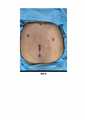
На Фиг.1 показана схема установки троакаров при выполнении лапароскопической продольной резекции желудка.Figure 1 shows a diagram of the installation of trocars when performing a laparoscopic longitudinal resection of the stomach.
На Фиг.2 показана установка троакаров.Figure 2 shows the placement of trocars.
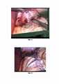
На Фиг.3 показана мобилизация желудка по большой кривизне.Figure 3 shows the mobilization of the stomach along the greater curvature.
На Фиг.4 показан этап отсечения резецированной части желудка сшивающими аппаратами на калибровочном зонде.Figure 4 shows the stage of cutting off the resected part of the stomach with staplers on the calibration probe.
На Фиг.5 показана перитонезация аппаратного шва непрерывным швом на калибровочном зонде.Figure 5 shows the peritoneisation of a hardware suture with a continuous suture on a calibration probe.
На Фиг.6 показан заключительный этап ушивания троакарных ран.Figure 6 shows the final stage of suturing trocar wounds.
На чертежах: 1 - первый троакар, 2 - правый троакар, 3 - левый троакар, 4 - верхний троакар, 5 - правая и левая среднеподмышечные линии, 6 - границы реберных дуг, 7 - заднеподмышечные линии, 8 - срединная линия, 9 - условная линия, показывающая, что правый и левый троакары находятся немного на разном уровне.In the drawings: 1 - first trocar, 2 - right trocar, 3 - left trocar, 4 - superior trocar, 5 - right and left mid-axillary lines, 6 - costal arches borders, 7 - posterior axillary lines, 8 - midline, 9 - conditional a line showing that the right and left trocars are slightly at different levels.
Осуществление изобретенияImplementation of the invention
Техника ПРЖ связана с соблюдением основных факторов:The PRG technique is associated with the observance of the main factors:
- положение пациента и хирурга;- the position of the patient and the surgeon;
- положение троакаров и инструментов;- position of trocars and instruments;
- знание особенностей лапароскопической анатомии брюшной полости;- knowledge of the features of the laparoscopic anatomy of the abdominal cavity;
- владение навыками выполнения интракорпоральных узлов.- possession of the skills of performing intracorporeal nodes.
Положение пациента на функциональном операционном столе с плотно зафиксированными ногами. Под колени установлены мягкие валики для предотвращения пережатия сосудисто-нервного пучка. При сверхожирении (более 200 кг) после фиксации пациента и подъёме головного конца операционного стола, под стопу пациента устанавливались опорные конструкции, для снижения нагрузки на коленные суставы, уменьшения риска сдавления сосудисто-нервных пучков. Что технически не мешало проведению операции.The position of the patient on a functional operating table with tightly fixed legs. Soft rollers are installed under the knees to prevent compression of the neurovascular bundle. In the case of overgrowing (more than 200 kg), after fixing the patient and raising the head end of the operating table, support structures were installed under the patient's foot to reduce the load on the knee joints, and to reduce the risk of compression of the neurovascular bundles. That did not technically interfere with the operation.
Операция выполняется с одним ассистентом, что не удлиняет ее, не вносит технические сложности.The operation is performed with one assistant, which does not lengthen it, does not introduce technical difficulties.
Этапы операции:Operation stages:
1) мобилизация желудка по большой кривизне1) mobilization of the stomach along the greater curvature
2) при необходимости рассечение спаек между желудком с поджелудочной железой2) if necessary, dissection of adhesions between the stomach and the pancreas
3) отсечение резецированной части желудка сшивающими аппаратами на калибровочном зонде3) cutting off the resected part of the stomach with staplers on the calibration probe
4) перитонезация аппаратного шва непрерывным швом на калибровочном зонде4) peritoneisation of the hardware suture with a continuous suture on the calibration probe
5) извлечение резецированного желудка из брюшной полости.5) removal of the resected stomach from the abdominal cavity.
Во время операции используются следующие инструменты: одноразовый сшивающий аппарат 60 мм и кассеты для него зеленые и синие; ультразвуковой диссектор Harmonic, мягкий зажим, диссектор, зубатый зажим типа граспера, 1 троакар 10 мм, 1 троакар 5 мм, 2 троакара по 12 мм.During the operation, the following instruments are used: a 60 mm disposable stapler and green and blue cassettes for it; ultrasonic dissector Harmonic, soft forceps, dissector, toothed forceps such as grasper, 1 trocar 10 mm, 1
Способ проведения операции заключается в следующем.The method of performing the operation is as follows.
Первый троакар 1 ставится на 15 см ниже мечевидного отростка по срединной линии 8, что соответствует при стандартной анатомии уровню ниже привратника (см. Фиг.1).The first trocar 1 is placed 15 cm below the xiphoid process along the
Параллельно заднеподмышечным линиям 7 расположены правая и левая среднеподмышечные линии 5 (см. Фиг.1).Parallel to the posterior
Левый троакар 3 ставится по среднеподмышечной линии 5 ближе к срединной линии 8 в правом подреберье на уровне привратника, атрального отдела желудка.The
Правый троакар 2 ставится под визуальным контролем по среднезаднеподмышечной линии 5 ниже реберной дуги на уровне большой кривизны желудка в левом подреберье.The
Верхний троакар 4 ставится по срединной линии 8 в области мечевидного отростка, с выколом в сторону левой доли печени.The
Ось троакаров должна иметь тупой угол. Данное расположение позволяет без особых усилий отсечь желудок по большой кривизне без смены троакаров, без установки дополнительных троакаров, так как обеспечен удобный угол работы лапароскопическими инструментами, и нет конфликта между инструментами. Так, на Фиг.1 поз. 9 можно видеть условную линию, показывающую, что правый и левый троакары находятся немного на разном уровне, следовательно меньше конфликт лапароскопических инструментов.The trocar axis should have an obtuse angle. This arrangement allows you to effortlessly cut off the stomach along the greater curvature without changing trocars, without installing additional trocars, since a convenient angle of operation with laparoscopic instruments is provided, and there is no conflict between instruments. So, in Fig. 1 pos. 9 you can see a conditional line showing that the right and left trocars are at a slightly different level, therefore there is less conflict between laparoscopic instruments.
Также, тупой угол наклона оси троакаров обеспечивает достаточный обзор при использовании косой (негибкой) оптики. Кроме того, все сшивающие аппараты последних поколений имеют угол сгибания не более 45 градусов. При таком расположении троакаров с учетом угла сгибания сшивающего аппарата работать на желудке удобно, возможный объем движений инструментами максимальный.Also, the obtuse angle of the trocar axis provides sufficient visibility when using oblique (inflexible) optics. In addition, all latest generation staplers have a 45 degree flexion angle. With such an arrangement of trocars, taking into account the angle of flexion of the stapler, it is convenient to work on the stomach, the possible range of movements of the instruments is maximum.
После установки троакаров как показано на Фиг.2 мобилизация желудка по большой кривизне осуществляется ультразвуковым скальпелем и мягким лапароскопическим зажимом.After placing the trocars as shown in Fig. 2, the mobilization of the stomach along the greater curvature is carried out with an ultrasonic scalpel and a soft laparoscopic forceps.
Мобилизация начинается с отсечения ультразвуковым скальпелем Harmonic желудка по большой кривизне (см. Фиг.3). Отступ от привратника 5 см. Ориентиром может служить бранша лапароскопического диссектора и известным расстоянием надписи на нем.Mobilization begins with cutting off the stomach along the greater curvature with an ultrasonic scalpel Harmonic (see Fig. 3). The distance from the gatekeeper is 5 cm. The branch of the laparoscopic dissector and the known distance of the inscription on it can serve as a reference point.
Хирург вводит ультразвуковой скальпель в левый троакар, в правый - мягкий зажим. Начинает мобилизацию желудка по большой кривизне в зоне привратника, далее переставляет зажим в верхний троакар, в правый ставится ретрактор, продолжает мобилизацию После осуществления доступа в сальниковую сумку осуществляется ее ревизия и рассечение спаек между желудком и поджелудочной железой.The surgeon inserts an ultrasonic scalpel into the left trocar, and a soft forceps into the right. Begins mobilization of the stomach along the greater curvature in the pylorus zone, then rearranges the clamp into the upper trocar, a retractor is placed in the right one, continues mobilization.After access to the omental bursa, its revision and dissection of adhesions between the stomach and pancreas are performed.
Далее тщательная мобилизация в зоне угла Гисса, прицельный осмотр для исключения грыжи пищеводного отверстия диафрагмы. Работа в данной зоне осложнена нарушением визуализации, риском кровотечения при пересечении коротких артерий. Далее через правый троакар заводится сшивающий аппарат с зеленой кассетой, ассистент левой рукой отводит левую долю печени при помощи нераскрытого металлического веерного ретрактора введенного в верхний троакар. Хирург изменяет кривизну дистальной части сшивающего аппарата с учетом направления резекции для формирования равномерного диаметра рукава (см. Фиг.4). Тракция большой кривизны желудка осуществляется хирургом при помощи граспера введенного в правый троакар. После зажима бранш сшивающего аппарата хирург дополнительно контролирует нет ли избытка задней стенки желудка. Желудок прошивается. Дополнительный визуальный контроль линии аппаратного шва. При появлении кровотечения из линии аппаратного шва методом остановки является прижатие салфеткой, реже клипирование кровоточащей зоны. Окончательный гемостаз обеспечивается при перитонизации аппаратного шва (см. Фиг.5). Сшивающий аппарат со второй зеленой кассетой вводится также через левый троакар.Further, careful mobilization in the area of the Hiss angle, targeted examination to exclude a hernia of the esophageal opening of the diaphragm. Work in this area is complicated by impaired visualization, the risk of bleeding when crossing short arteries. Then, through the right trocar, a stapler with a green cassette is inserted, the assistant with his left hand retracts the left lobe of the liver using an unopened metal fan-shaped retractor inserted into the upper trocar. The surgeon changes the curvature of the distal part of the stapler, taking into account the direction of resection to form a uniform diameter of the sleeve (see Fig. 4). Traction of the greater curvature of the stomach is carried out by the surgeon using a grasper inserted into the right trocar. After clamping the branch of the stapler, the surgeon additionally checks for excess of the posterior stomach wall. The stomach is stitched. Additional visual inspection of the hardware seam line. When bleeding occurs from the line of the hardware suture, the method of stopping is pressing with a napkin, less often clipping of the bleeding zone. The final hemostasis is provided by peritonization of the hardware suture (see Fig. 5). The stapler with the second green cassette is also inserted through the left trocar.
Если технически невозможно выполнить его равномерное наложение без избытка задней стенки и перекрута желудка хирург переворачивает сшивающий аппарат верхней браншей вниз, что облегчает в большинстве случаев фиксацию кассеты без формирования избытка задней стенки или перекрута желудка.If it is technically impossible to perform its uniform application without excess of the posterior wall and torsion of the stomach, the surgeon turns the stapler of the upper jaws downward, which in most cases facilitates fixation of the cassette without forming an excess of the posterior wall or torsion of the stomach.
Ассистент через левый троакар вводит ретрактор для тракции левой доли печени.The assistant inserts the retractor through the left trocar to traction the left lobe of the liver.
Клинически выявлено снижение травматизации печени при выполнении ее тракции нераскрытым металлическим веерным ретрактором.Clinically, a decrease in liver trauma was revealed when traction was performed with an unopened metal fan retractor.
Сшивающий аппарат вводится в брюшную полость через правый 12 мм троакар. Третья и последующие используемые кассеты с меньшей высотой скрепок. Хирург осуществляет тракцию желудка граспером установленный через верхний троакар левой рукой. Последняя кассета заводится с учетом сохранения пищеводно-желудочного угла.The stapler is inserted into the abdominal cavity through the right 12 mm trocar. The third and subsequent used cassettes with a lower staple height. The surgeon performs traction of the stomach with a grasper inserted through the upper trocar with his left hand. The last cassette is started taking into account the preservation of the esophageal-gastric angle.
Перитонизация аппаратного шва проводится на калибровочном зонде непрерывный швом рассасывающейся нитью с насечками с целью гемостаза и профилактики несостоятельности аппаратного шва. Калибровочный зонд удаляется после проверки герметичности.Peritonization of the hardware suture is carried out on a calibration probe with a continuous suture with an absorbable suture with notches for the purpose of hemostasis and prevention of inconsistency of the hardware suture. The calibration probe is removed after a tightness test.
Желудок захватывается граспером, извлекается через правый доступ 12 мм, реже через надпупочный , в зависимости от необходимости постановки дренажа.The stomach is captured with a grasper, it is removed through the 12 mm right access, less often through the supra-umbilical, depending on the need for drainage.
Ушивание троакарных ран проводится скорняжной иглой (см. Фиг.6).Suturing of trocar wounds is carried out with a furrier needle (see Fig. 6).
Способ иллюстрируется следующими клиническими примерами.The method is illustrated by the following clinical examples.
Пример 1. Больная А., 40 лет, поступила в клинику по поводу морбидного ожирения для оперативного лечения. Кроме жалоб, связанных с основным заболеванием, больная предъявляла жалобы на изжогу в эпигастральной области после переедания. Максимальный вес пациентки составил 153 кг, индекс массы тела (ИМТ) 51 кг/м2.Example 1. Patient A., 40 years old, was admitted to the clinic for morbid obesity for surgical treatment. In addition to complaints related to the underlying disease, the patient complained of heartburn in the epigastric region after overeating. The patient's maximum weight was 153 kg, body mass index (BMI) 51 kg / m2 .
По данным фиброгастродуоденоскопии (ФГДС) установлен диагноз: Поверхностный гастрит. Умеренно активный бульбит. По УЗИ брюшной полости: диффузные изменения поджелудочной железы.According to fibrogastroduodenoscopy (FGDS), the diagnosis was made: Superficial gastritis. Moderately active bulbite. Ultrasound of the abdominal cavity: diffuse changes in the pancreas.
Больная оперирована по заявленному способу, выполнена продольная резекция желудка из 4 троакаров. Операция была выполнена за 100 минут, без интраоперационных сложностей - отсутствие конфликта между инструментами, при этом комфортная работа на желудке сшивающим аппаратом с учетом угла сгибания. Через 2 часа после операции по визуально-аналоговой шкале (VAS) пациентка оценила интенсивность боли в 3 балла, через 24 часа после операции в 2 балла. Через 48 часов после операции выписана домой в удовлетворительном состоянии без развития осложнений. С первых часов после операции и через в течении года после на момент осмотра у пациентки отсутствуют жалобы, при этом получен желаемый результат (Вес 82, при ИМТ= 27, 5 кг/м2). На протяжении 3 лет наблюдения результат сохранен, отдаленных осложнений нет.The patient was operated on according to the claimed method, performed a longitudinal resection of the stomach from 4 trocars. The operation was performed in 100 minutes, without intraoperative difficulties - there was no conflict between instruments, while comfortable work on the stomach with a stapler taking into account the flexion angle. 2 hours after surgery, the patient rated the pain intensity at 3 points on the visual analogue scale (VAS), and 2 points at 24 hours after surgery. She was discharged home 48 hours after the operation in a satisfactory condition without any complications. From the first hours after the operation and within a year after at the time of examination, the patient had no complaints, while the desired result was obtained (Weight 82, with BMI = 27, 5 kg / m2 ). During 3 years of follow-up, the result was preserved, there were no long-term complications.
Пример 2. Больной Д., 47 лет, поступил в клинику по поводу морбидного ожирения 3 степени для оперативного лечения. Предьявлял жалобы связанные с основным заболеванием. Максимальный вес пациента составлял 178 кг, при ИМТ-55 кг/м2.Example 2. Patient D., 47 years old, was admitted to the clinic for
По данным фиброгастродуоденоскопии (ФГДС) установлен диагноз: Недостаточность кардии. Поверхностный гастрит. По УЗИ брюшной полости: гепатомегалия, диффузные изменения поджелудочной железы.According to fibrogastroduodenoscopy (FGDS), the diagnosis was made: Insufficiency of the cardia. Superficial gastritis. Ultrasound of the abdominal cavity: hepatomegaly, diffuse changes in the pancreas.
Больной оперирован по заявленному способу, выполнена продольная резекция желудка из 4 троакаров. Операция была выполнена за 110 минут, без интраоперационных сложностей, при этом отсутствовал конфликт между инструментами, комфортно было работать на желудке сшивающим аппаратом с учетом угла сгибания. Через 2 часа после операции по визуально-аналоговой шкале (VAS) пациент оценил интенсивность боли в 4 балла, через 24 часа после операции в 1 балл. Через 48 часов после операции выписан домой в удовлетворительном состоянии без развития осложнений. С первых часов после операции и через в течении года после на момент осмотра у пациента отсутствуют жалобы, при этом получен желаемый результат (Вес 95, при ИМТ= 29,5 кг/м2). На протяжении 3 лет наблюдения результат сохранен, отдаленных осложнений нет.The patient was operated on according to the claimed method, performed a longitudinal resection of the stomach from 4 trocars. The operation was performed in 110 minutes, without intraoperative complications, while there was no conflict between instruments, it was comfortable to work on the stomach with a stapler taking into account the flexion angle. 2 hours after surgery, the patient rated the pain intensity at 4 points on the visual analogue scale (VAS), and 1 point at 24 hours after surgery. 48 hours after the operation, he was discharged home in a satisfactory condition without the development of complications. From the first hours after the operation and within a year after at the time of examination, the patient has no complaints, while the desired result is obtained (Weight 95, with BMI = 29.5 kg / m2 ). During 3 years of follow-up, the result was preserved, there were no long-term complications.
Пример 3. Больная Д., 32 года, поступила в клинику по поводу морбидного ожирения для оперативного лечения. Жалобы пациентки связаны с основным заболеванием. Максимальный вес пациентки составил 103 кг, индекс массы тела(ИМТ) 40 кг/м2.Example 3. Patient D., 32 years old, was admitted to the hospital for morbid obesity for surgical treatment. The patient's complaints are related to the underlying disease. The patient's maximum weight was 103 kg, body mass index (BMI) 40 kg / m2 .
По данным фиброгастродуоденоскопии (ФГДС) установлен диагноз: Поверхностный гастрит. По УЗИ брюшной полости: диффузные изменения печени, поджелудочной железы.According to fibrogastroduodenoscopy (FGDS), the diagnosis was made: Superficial gastritis. By ultrasound of the abdominal cavity: diffuse changes in the liver, pancreas.
Больная оперирована по заявленному способу, выполнена продольная резекция желудка из 4 троакаров. Операция была выполнена за 110 минут, без интраоперационных сложностей - отсутствие конфликта между инструментами, при этом комфортная работа на желудке сшивающим аппаратом с учетом угла сгибания. Через 2 часа после операции по визуально-аналоговой шкале (VAS) пациентка оценила интенсивность боли в 3 балла, через 24 часа после операции в 1 балла. Через 48 часов после операции выписана домой в удовлетворительном состоянии без развития осложнений. С первых часов после операции и через в течении года после на момент осмотра у пациентки отсутствуют жалобы, при этом получен желаемый результат (Вес 67, при ИМТ= 26 кг/м2). На протяжении 3 лет наблюдения результат сохранен, отдаленных осложнений нет.The patient was operated on according to the claimed method, performed a longitudinal resection of the stomach from 4 trocars. The operation was performed in 110 minutes, without intraoperative difficulties - there was no conflict between instruments, while comfortable work on the stomach with a stapler taking into account the flexion angle. 2 hours after surgery, the patient rated the pain intensity at 3 points on the visual analogue scale (VAS), and 1 point at 24 hours after surgery. She was discharged home 48 hours after the operation in a satisfactory condition without any complications. From the first hours after the operation and within a year after at the time of examination, the patient had no complaints, while the desired result was obtained (Weight 67, with BMI = 26 kg / m2 ). During 3 years of follow-up, the result was preserved, there were no long-term complications.
Эффективность заявленного способа оценивали по результатам лабораторных исследований (рутинные методы обследования, принятые для пациентов, подлежащих хирургическому лечению), клинической картине, результатам инструментальных методов исследования. Отдаленные результаты прослежены на протяжении 2 лет.The effectiveness of the claimed method was assessed by the results of laboratory tests (routine examination methods adopted for patients subject to surgical treatment), the clinical picture, and the results of instrumental research methods. Long-term results were followed for 2 years.
По заявленному способу были проведены продольные резекции 110 больным. Послеоперационные осложнения отсутствовали, все пациенты добились желаемого результата по снижению массы тела.According to the claimed method, longitudinal resections were performed in 110 patients. There were no postoperative complications, all patients achieved the desired result in weight loss.
Для сравнения были исследованы 27 больных, которым произведена продольная резекция желудка по стандартной методике. Для оценки полученных результатов сформированы 2 группы больных:For comparison, we studied 27 patients who underwent longitudinal gastrectomy according to the standard technique. To assess the results obtained, 2 groups of patients were formed:
1-я: 110 пациентов, которым выполнена лапароскопическая продольная резекция желудка из 4 троакаров.1st: 110 patients who underwent laparoscopic longitudinal gastrectomy using 4 trocars.
2-я: 27 пациентов, которым выполнена стандартная лапароскопическая продольная резекция желудка.2nd: 27 patients underwent standard laparoscopic longitudinal gastric resection.
В 1-й группе с первых часов после операций у пациентов больных болевой синдром был минимальный (в среднем 2, 5 бала по шкале VAS). Во 2-й группе болевой синдром в среднем составлял 4, 3 балла по шкале VAS.In group 1, from the first hours after surgery, the patients had minimal pain (on average 2.5 points on the VAS scale). In
Время выполнения операции в 1-й и 2-й группе пациентов сопоставимо и составило в 1-й группе в среднем - 105 минут, во 2-й - 115 минут. Операции пациентам в 1-й группе выполнялись с привлечением всего одного ассистента. Послеоперационных осложнений в группах не было как в 1-й так и 2-й группах. В отдаленном периоде пациенты как 1-й так и 2-й групп добились удобоваримых результатов по снижению массы тела.The operation time in the 1st and 2nd groups of patients was comparable and averaged 105 minutes in the 1st group, and 115 minutes in the 2nd. Operations for patients in group 1 were performed with the involvement of only one assistant. There were no postoperative complications in the groups, both in the 1st and 2nd groups. In the long-term period, patients of both
Полученные результаты лечения больных в 1-й группах позволяют утверждать, что применение продольной резекции с помощью 4 троакаров снижает болевой синдром, при полной безопасности для пациента и характеризуется малой травматичностью. The results obtained in the treatment of patients in the 1st group suggest that the use of longitudinal resection with 4 trocars reduces pain, with complete safety for the patient and is characterized by low trauma.
В целом способ обеспечивает: меньшую операционную травму и менее выраженный болевой синдром в послеоперационном периоде; привлечение к операции всего одного ассистента; комфортную работу хирурга вследствие отсутствия конфликта между инструментами; удобство работы на желудке сшивающим аппаратом с учетом угла сгибания.In general, the method provides: less surgical trauma and less pronounced pain in the postoperative period; involvement of only one assistant in the operation; comfortable work of the surgeon due to the absence of conflict between instruments; convenience of working on the stomach with a stapler, taking into account the angle of flexion.
Заявленный способ является патогенетически обоснованным при выполнении бариатрических лапароскопических операций.The claimed method is pathogenetically justified when performing bariatric laparoscopic operations.
Claims (1)
Translated fromRussianPriority Applications (1)
| Application Number | Priority Date | Filing Date | Title |
|---|---|---|---|
| RU2020143224ARU2759564C1 (en) | 2020-12-27 | 2020-12-27 | Method for performing laparoscopic longitudinal resection of stomach in morbid obesity |
Applications Claiming Priority (1)
| Application Number | Priority Date | Filing Date | Title |
|---|---|---|---|
| RU2020143224ARU2759564C1 (en) | 2020-12-27 | 2020-12-27 | Method for performing laparoscopic longitudinal resection of stomach in morbid obesity |
Publications (1)
| Publication Number | Publication Date |
|---|---|
| RU2759564C1true RU2759564C1 (en) | 2021-11-15 |
Family
ID=78607320
Family Applications (1)
| Application Number | Title | Priority Date | Filing Date |
|---|---|---|---|
| RU2020143224ARU2759564C1 (en) | 2020-12-27 | 2020-12-27 | Method for performing laparoscopic longitudinal resection of stomach in morbid obesity |
Country Status (1)
| Country | Link |
|---|---|
| RU (1) | RU2759564C1 (en) |
Cited By (1)
| Publication number | Priority date | Publication date | Assignee | Title |
|---|---|---|---|---|
| RU2793928C1 (en)* | 2022-05-23 | 2023-04-10 | Станислав Петрович Мужиков | Method of single-port longitudinal resection of the gastroma by standard laparoscopic instrumentation |
Citations (3)
| Publication number | Priority date | Publication date | Assignee | Title |
|---|---|---|---|---|
| US20100081863A1 (en)* | 2008-09-30 | 2010-04-01 | Ethicon Endo-Surgery, Inc. | Methods and devices for performing gastrectomies and gastroplasties |
| US20140107698A1 (en)* | 2012-10-04 | 2014-04-17 | Children's Hospital Medical Center | Gastric traction device and method |
| RU2640783C1 (en)* | 2016-08-19 | 2018-01-11 | Александр Георгиевич Хитарьян | Method of surgical treatment of morbid obesity |
- 2020
- 2020-12-27RURU2020143224Apatent/RU2759564C1/enactive
Patent Citations (3)
| Publication number | Priority date | Publication date | Assignee | Title |
|---|---|---|---|---|
| US20100081863A1 (en)* | 2008-09-30 | 2010-04-01 | Ethicon Endo-Surgery, Inc. | Methods and devices for performing gastrectomies and gastroplasties |
| US20140107698A1 (en)* | 2012-10-04 | 2014-04-17 | Children's Hospital Medical Center | Gastric traction device and method |
| RU2640783C1 (en)* | 2016-08-19 | 2018-01-11 | Александр Георгиевич Хитарьян | Method of surgical treatment of morbid obesity |
Non-Patent Citations (2)
| Title |
|---|
| RAMOS ALMINO CARDOSO et al. "Technical aspects of laparoscopic sleeve gastrectomy" Arquivos brasileiros de cirurgia digestive, 2015, vol. 28 Suppl 1, 65-68.* |
| МУЖИКОВ С.П. и др. Клинический опыт адаптации принципов FAST TRACK у пациентов после выполнения лапароскопической продольной резекции желудка по поводу морбидного ожирения, Московский хирургический журнал, 2019, 6 (70), с. 41-44.* |
Cited By (4)
| Publication number | Priority date | Publication date | Assignee | Title |
|---|---|---|---|---|
| RU2793928C1 (en)* | 2022-05-23 | 2023-04-10 | Станислав Петрович Мужиков | Method of single-port longitudinal resection of the gastroma by standard laparoscopic instrumentation |
| RU2813035C1 (en)* | 2023-05-12 | 2024-02-06 | Станислав Петрович Мужиков | Method of surgical treatment of morbid obesity in patients with type 2 diabetes mellitus |
| RU2820732C1 (en)* | 2023-05-12 | 2024-06-07 | Станислав Петрович Мужиков | Method for surgical management of morbid obesity in patients with type 2 diabetes mellitus |
| RU2840124C1 (en)* | 2024-09-05 | 2025-05-19 | Марина Алиевна Биджиева | Method for performing longitudinal gastrectomy |
Similar Documents
| Publication | Publication Date | Title |
|---|---|---|
| Shope et al. | Early results after laparoscopic gastric bypass: EEA vs GIA stapled gastrojejunal anastomosis | |
| Nguyen et al. | Laparoscopic Roux-en-Y gastric bypass for super/super obesity | |
| RU2759564C1 (en) | Method for performing laparoscopic longitudinal resection of stomach in morbid obesity | |
| Orringer et al. | An improved technique for the combined Collis-Belsey approach to dilatable esophageal strictures | |
| Ikramuddin et al. | Open and laparoscopic Roux-en-Y gastric bypass: our techniques | |
| RU2751289C1 (en) | Method for performing reduction gastroplasty | |
| RU2286742C2 (en) | Method of selection of points for introducing trocars ar laparoscopic combined operation | |
| RU2200475C2 (en) | Method for treating the cases of esophageal opening hernia | |
| Grill et al. | Experiences with the chevron (V) osteotomy on adolescent hallux valgus | |
| RU2132159C1 (en) | Method for treating the cases of reflux esophagitis | |
| Uyama et al. | Laparoscopic and minilaparotomy Billroth I gastrectomy for gastric ulcer using an abdominal wall-lifting method | |
| RU2718309C1 (en) | Method for videolaparotranshiatal access to lower third of thoracic esophagus | |
| RU2840124C1 (en) | Method for performing longitudinal gastrectomy | |
| Angrisani et al. | Laparoscopic adjustable silicone gastric banding: preliminary results of the University of Naples experience | |
| RU2827848C1 (en) | Method for surgical management of morbid obesity | |
| RU2305501C1 (en) | Mini-access method for carrying out cholecystectomy | |
| RU2813035C1 (en) | Method of surgical treatment of morbid obesity in patients with type 2 diabetes mellitus | |
| RU2210330C1 (en) | Method for surgical treating complete prolapse of uterus and vaginal walls | |
| RU2846665C1 (en) | Method of manual oesophagoenteroanastomosis formation in laparoscopic gastrectomy | |
| RU2328236C1 (en) | Method of platypodia foot arch correction | |
| RU2820732C1 (en) | Method for surgical management of morbid obesity in patients with type 2 diabetes mellitus | |
| RU2189179C2 (en) | Method for treating the cases of duodenal peptic ulcer | |
| RU2115380C1 (en) | Method for treatment of cross-spread deformation of foot with valgus deviation of great toe | |
| RU2838874C1 (en) | Method for prevention of trocar hernias in patients with diastasis rectus abdominis muscle and connective tissue dysplasia | |
| RU2743882C1 (en) | Method of single-stem decompression ileostoma application in patients with adiposity 3-4 degrees |