RU2748905C1 - Method for individualization of dental implants using laser ablation and diagnostics of 5 types of bone density - Google Patents
Method for individualization of dental implants using laser ablation and diagnostics of 5 types of bone densityDownload PDFInfo
- Publication number
- RU2748905C1 RU2748905C1RU2020136643ARU2020136643ARU2748905C1RU 2748905 C1RU2748905 C1RU 2748905C1RU 2020136643 ARU2020136643 ARU 2020136643ARU 2020136643 ARU2020136643 ARU 2020136643ARU 2748905 C1RU2748905 C1RU 2748905C1
- Authority
- RU
- Russia
- Prior art keywords
- dental
- dental implant
- laser
- implant
- types
- Prior art date
Links
Images

Classifications
- A—HUMAN NECESSITIES
- A61—MEDICAL OR VETERINARY SCIENCE; HYGIENE
- A61B—DIAGNOSIS; SURGERY; IDENTIFICATION
- A61B5/00—Measuring for diagnostic purposes; Identification of persons
Landscapes
- Life Sciences & Earth Sciences (AREA)
- Health & Medical Sciences (AREA)
- Medical Informatics (AREA)
- Biophysics (AREA)
- Pathology (AREA)
- Engineering & Computer Science (AREA)
- Biomedical Technology (AREA)
- Heart & Thoracic Surgery (AREA)
- Physics & Mathematics (AREA)
- Molecular Biology (AREA)
- Surgery (AREA)
- Animal Behavior & Ethology (AREA)
- General Health & Medical Sciences (AREA)
- Public Health (AREA)
- Veterinary Medicine (AREA)
- Dental Tools And Instruments Or Auxiliary Dental Instruments (AREA)
Abstract
Description
Translated fromRussianИзобретение относятся к медицине, а именно к стоматологии и челюстно-лицевой хирургии, и может использоваться для индивидуализации дентальных имплантатов и производства индивидуальных медицинских изделий под определенную зону имплантации в области отсутствующего зуба для определения оптимальной конструкции индивидуального медицинского изделия.The invention relates to medicine, namely to dentistry and maxillofacial surgery, and can be used for the individualization of dental implants and the production of individual medical products for a specific implantation zone in the area of a missing tooth to determine the optimal design of an individual medical product.
Известен способ измерения плотности костной такни денситометрия, где оценивается оптическая плотность костной ткани по отношению к излучению рентгена, измеряется в единицах Хаунсфилда (HU) (см. Ю.А. Барабаш, Н.В. Тишков, А.П. Барабаш, В.Д. Балаян, К.А. Гражданов «Динамика минерализации большеберцовой кости по шкале Хаунсфилда при лечении ложных суставов методом чрезкостного компрессионного остеосинтеза с использованием биологической стимуляции регенерации», Бюллетень Восточно-Сибирского научного центра Сибирского отделения Российской академии медицинских наук, 2011 г., №4-1).A known method for measuring the density of bone tissue densitometry, where the optical density of bone tissue is estimated in relation to X-ray radiation, is measured in Hounsfield units (HU) (see Yu.A. Barabash, N.V. Tishkov, A.P. Barabash, V. D. Balayan, K.A. Grazhdanov "Dynamics of tibia mineralization according to the Hounsfield scale in the treatment of pseudoarthrosis by the method of transosseous compression osteosynthesis using biological stimulation of regeneration", Bulletin of the East Siberian Scientific Center of the Siberian Branch of the Russian Academy of Medical Sciences, 2011, no. 4-1).
Недостатком данного способа является высокая степень ионизирующего облучения и высокая стоимость данной процедуры.The disadvantage of this method is the high degree of ionizing radiation and the high cost of this procedure.
Известен способ применения модифицированных дентальных имплантатов на основе нетканого титанового материала (Щербовских А.Е. «ОБОСНОВАНИЕ ПРИМЕНЕНИЯ МОДИФИЦИРОВАННЫХ ДЕНТАЛЬНЫХ ИМПЛАНТАТОВ НА ОСНОВЕ НЕТКАНОГО ТИТАНОВОГО МАТЕРИАЛА СО СКВОЗНОЙ ПОРИСТОСТЬЮ» [Текст]: дис. кан. мед. наук / Щербовских Алексей Евгеньевич. - Самара, 2017., 155 с.)A known method of using modified dental implants based on non-woven titanium material (Shcherbovskikh AE "RATIONALE FOR THE APPLICATION OF MODIFIED DENTAL IMPLANTS BASED ON NON-WOVEN TITANIUM MATERIAL WITH THROUGH POROUSITY" [Text] / sciences. , 2017., 155 p.)
Недостатком данного способа является отсутствие возможности индивидуализировать дизайн поверхности дентального имплантата под конкретный тип костной ткани.The disadvantage of this method is the inability to individualize the design of the surface of the dental implant for a specific type of bone tissue.
Наиболее близким аналогом заявляемого технического решения является способ диагностики состояния костной ткани и лечения пациентов с применением индивидуальных дентальных имплантатов (патент RU 2718321, МПК А61В 5/00, опубл. 01.04.2020), согласно которому получают на компьютерном томографе Sirona Galilleos номинальной мощностью 85 кВ компьютерную томограмму пациента. На компьютерной томограмме в сагиттальной плоскости зоны планируемой дентальной имплантации определяют величину оптической плотности костной ткани от анатомического образования, в области которого планируют расположение апикальной части дентального имплантата, до зоны края альвеолярного отростка. Затем осуществляют выбор профиля резьбовой поверхности дентального имплантата согласно таблице 1, содержащейся в описании. Изготавливают индивидуальный дентальный имплантат и устанавливают пациенту.The closest analogue of the claimed technical solution is a method for diagnosing the state of bone tissue and treating patients using individual dental implants (patent RU 2718321, IPC А61В 5/00, publ. 01.04.2020), according to which a Sirona Galilleos computed tomograph with a nominal power of 85 kV is obtained computed tomography of the patient. On a computed tomogram in the sagittal plane of the zone of the planned dental implantation, the value of the optical density of the bone tissue is determined from the anatomical formation in the area of which the location of the apical part of the dental implant is planned to the zone of the edge of the alveolar process. Then carry out the selection of the profile of the threaded surface of the dental implant according to table 1 contained in the description. An individual dental implant is made and placed in the patient.
Недостатком данного технического решения является то, что способ охватывает не достаточное количество типов резьб, наиболее часто используемых при изготовлении дентальных имплантатов.The disadvantage of this technical solution is that the method does not cover a sufficient number of types of threads, most often used in the manufacture of dental implants.
Техническим результатом предлагаемого изобретения является повышение эффективности производства индивидуальных медицинских изделий и доступности метода дентальной имплантации, повышение качества протезирования за счет создания индивидуальных медицинских изделий в виде индивидуальных дентальных имплантатов, конструкция которых повторяет и конгруэнтно сочетается с пятью типами плотности костной ткани челюстной кости.The technical result of the proposed invention is to increase the efficiency of the production of individual medical devices and the availability of the method of dental implantation, improve the quality of prosthetics by creating individual medical devices in the form of individual dental implants, the design of which repeats and is congruently combined with five types of bone density of the jaw bone.
Технический результат достигается за счет того, что согласно способу, включающему получение с помощью компьютерного томографа, с номинальной мощностью 57-90 кВ, компьютерной томограммы пациента, анализ компьютерной томограммы с определением оптической плотности костной ткани в сагиттальной плоскости зоны планируемой дентальной имплантации от анатомического образования, в области которого планируют расположение апикальной части дентального имплантата, до зоны края альвеолярного отростка и подбор резьбовой поверхности дентального имплантата, выбор профиля резьбовой поверхности дентального имплантата осуществляют согласно 5 типам плотности костной ткани, указанным в таблице 1, содержащейся в описании, далее выбирают длину и диаметр будущего индивидуального дентального имплантата, затем изготавливают заготовку имплантата и при помощи иттербиевой лазерной установки наносят геометрию поверхности, используя лазерную абляцию в атмосферном аргоне, при этом лазер имеет длину волны 1064 нм с непрерывной мощностью 20 Вт, скоростью сканирования 8,7 м/с и размером лазерного пятна 0,1 мм.The technical result is achieved due to the fact that according to the method, including obtaining a computer tomogram of the patient using a computed tomograph with a rated power of 57-90 kV, analyzing a computed tomogram with determining the optical density of bone tissue in the sagittal plane of the zone of the planned dental implantation from an anatomical formation, in the area of which the location of the apical part of the dental implant is planned, to the zone of the edge of the alveolar process and the selection of the threaded surface of the dental implant, the choice of the profile of the threaded surface of the dental implant is carried out according to 5 types of bone density indicated in table 1 contained in the description, then the length and diameter are selected future individual dental implant, then an implant blank is made and, using an ytterbium laser device, the surface geometry is applied using laser ablation in atmospheric argon, while the laser has a wavelength of 1064 nm with a continuous With a burst power of 20 W, a scanning speed of 8.7 m / s and a laser spot size of 0.1 mm.
Применение индивидуального подхода к изготовлению дентальных имплантатов с помощью лазерной абляции, выполненной на иттербиевой лазерной установке в атмосферном аргоне с определенными показателями, увеличивает контакт поверхности костной ткани с поверхностью имплантата, что повышает эффективность производства индивидуальных медицинских изделий и доступность метода дентальной имплантации.The use of an individual approach to the manufacture of dental implants using laser ablation performed on an ytterbium laser device in atmospheric argon with certain parameters increases the contact of the bone tissue surface with the implant surface, which increases the efficiency of manufacturing individual medical devices and the availability of the dental implantation method.
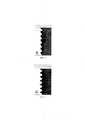
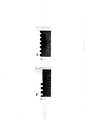
Реализация способа характеризуется следующими графическими материалами:The implementation of the method is characterized by the following graphic materials:
На фиг. 1 изображена геометрия поверхности, подходящая типу D1 больше 1250HUFIG. 1 shows surface geometry suitable for type D1 over 1250HU
На фиг. 2 изображена геометрия поверхности, подходящая типу D2 от 850HU до 1250HUFIG. 2 shows surface geometry suitable for type D2 850HU to 1250HU
На фиг. 3 изображена геометрия поверхности, подходящая типу D3 от 350HU до 850HUFIG. 3 shows surface geometry suitable for type D3 from 350HU to 850HU
На фиг. 4 изображена геометрия поверхности, подходящая типу D4 от 150HU до 350HUFIG. 4 shows surface geometry suitable for type D4 from 150HU to 350HU
На фиг. 5 изображена геометрия поверхности, подходящая типу D5 меньше 150HUFIG. 5 shows surface geometry suitable for type D5 less than 150HU
Исходя из данных (Malmo University Faculty of Odontology Doctoral Dissertations 2014 ON EFFICACY OF IMPLANTTHREAD DESIGN FOR BONE) были исследованы типы геометрии профиля резьбы и их взаимоотношения с различными типами плотности костной ткани и выявлено наиболее благоприятное взаимодействие 5 типов геометрии профиля резьбы дентального имплантата с пятью типами плотности костной ткани. Установлено что геометрия поверхности (Е) создает лучшее взаимодействие с костной тканью плотностью большей 1250 Hu и соответствует типу плотности D1 что способствует улучшению остеоинтеграции и снижению вероятности возникновения пост операционных осложнений. Для костной ткани плотности от 1250 Hu до 850 Hu геометрия поверхности (G) лучше сочетается с типом плотности D2 и способствует лучшей интеграции индивидуальных дентальных имплантатов. Для плотности костной ткани от 850HU до 350HU соответствует классу плотности D3 при котором профиль поверхности соответствует (А). Для костной ткани плотностью от 350HU до 150HU поверхность соответствует типу (F) качество кости D4, при плотности ниже 150HU тип геометрии будет (D) при качестве кости D5.Based on the data (Malmo University Faculty of Odontology Doctoral Dissertations 2014 ON EFFICACY OF IMPLANTTHREAD DESIGN FOR BONE), the types of thread geometry and their relationship with various types of bone density were investigated and the most favorable interaction of 5 types of thread geometry of the dental implant with five types was revealed. bone density. It was found that the surface geometry (E) creates a better interaction with bone tissue with a density greater than 1250 Hu and corresponds to the D1 density type, which improves osseointegration and reduces the likelihood of postoperative complications. For bone grades from 1250 Hu to 850 Hu, the surface geometry (G) matches the density type D2 better and facilitates better integration of individual dental implants. For bone density between 850HU and 350HU, it corresponds to density class D3, where the surface profile corresponds to (A). For bone tissue with a density from 350HU to 150HU, the surface corresponds to type (F) bone quality D4, with a density below 150HU, the geometry type will be (D) with bone quality D5.
Преимущество данного способа заключается в персонифицированном подходе к геометрии поверхности при определенной плотности костной ткани по 5 типам. Индивидуальные дентальные имплантаты обеспечивают равномерное не превышающих допустимых нагрузок на костную ткань давление от имплантата.The advantage of this method lies in a personalized approach to the surface geometry at a certain bone density in 5 types. Individual dental implants provide uniform pressure from the implant that does not exceed the permissible load on the bone tissue.
Способ иллюстрирован клиническим примером.The method is illustrated by a clinical example.
Больной Е обратился в Центр Индивидуализации Дентальной имплантации с жалобой на боль в области центрального зуба на верхней челюсти. При осмотре полости рта, зуб 1.1 обладал 2 степенью подвижности, и слизистая оболочка вокруг него была гиперемирована.Patient E applied to the Center for Individualization of Dental Implantation with a complaint of pain in the area of the central tooth in the upper jaw. On examination of the oral cavity, tooth 1.1 had a 2nd degree of mobility, and the mucous membrane around it was hyperemic.
Предварительный диагноз: обострение хронического периодонтита зуба 1.1Preliminary diagnosis: exacerbation of chronic periodontitis of the tooth 1.1
По результатам компьютерной томографии, полученной спустя 2 месяца после удаления зуба с помощью томографа KaVo optopantomograph OP3DPro, были получены данные о плотности костной ткани в области удаленного зуба 1.1, в сагиттальной плоскости зоны планируемой дентальной имплантации от анатомического образования, в области которого планировали расположение апикальной части дентального имплантата, до зоны края альвеолярного отростка, которые соответствовали геометрии поверхности F и А согласно таблице 1. После чего выбрали длину и диаметр будущего индивидуального дентального имплантата. Изготовили заготовку имплантата при помощи иттербиевой лазерной установки (Мини Маркер 2.IPG Photonics, Россия). Лазером наносили геометрию поверхности, используя лазерную абляцию в атмосферном аргоне, при этом лазер имеет рабочий объем 100×100×250 мм и длину волны 1064 нм с непрерывной мощностью 20 Вт, скоростью сканирования 8,7 м/с и размером лазерного пятна 0,1 мм. Затем произвели установку индивидуального дентального имплантата. Через 2 месяца после оперативного вмешательства была выполнена компьютерная томография, на которой отмечалось остеоинтеграция индивидуального дентального имплантата и увеличение плотности костной ткани окружающий индивидуальный дентальный имплантат.According to the results of computed tomography, obtained 2 months after tooth extraction using the KaVo optopantomograph OP3DPro tomograph, data on the bone density in the area of the extracted tooth 1.1 were obtained, in the sagittal plane of the zone of the planned dental implantation from the anatomical formation, in the area of which the location of the apical part was planned dental implant, to the zone of the edge of the alveolar process, which corresponded to the geometry of the surface F and A according to table 1. After that, the length and diameter of the future individual dental implant were chosen. An implant blank was made using an ytterbium laser device (Mini Marker 2.IPG Photonics, Russia). The surface geometry was applied with a laser using laser ablation in atmospheric argon, while the laser has a working volume of 100 × 100 × 250 mm and a wavelength of 1064 nm with a continuous power of 20 W, a scanning speed of 8.7 m / s and a laser spot size of 0.1 mm. Then an individual dental implant was installed. 2 months after surgery, computed tomography was performed, which showed osseointegration of the individual dental implant and an increase in the bone density of the surrounding individual dental implant.

Способ индивидуализации дентальной имплантации рекомендован для использования в стоматологии и челюстно-лицевой хирургии для лечения пациентов с частичным и полным отсутствием зубов.The method of individualization of dental implantation is recommended for use in dentistry and maxillofacial surgery for the treatment of patients with partial and complete absence of teeth.
Claims (1)
Translated fromRussianPriority Applications (1)
| Application Number | Priority Date | Filing Date | Title |
|---|---|---|---|
| RU2020136643ARU2748905C1 (en) | 2020-11-06 | 2020-11-06 | Method for individualization of dental implants using laser ablation and diagnostics of 5 types of bone density |
Applications Claiming Priority (1)
| Application Number | Priority Date | Filing Date | Title |
|---|---|---|---|
| RU2020136643ARU2748905C1 (en) | 2020-11-06 | 2020-11-06 | Method for individualization of dental implants using laser ablation and diagnostics of 5 types of bone density |
Publications (1)
| Publication Number | Publication Date |
|---|---|
| RU2748905C1true RU2748905C1 (en) | 2021-06-01 |
Family
ID=76301269
Family Applications (1)
| Application Number | Title | Priority Date | Filing Date |
|---|---|---|---|
| RU2020136643ARU2748905C1 (en) | 2020-11-06 | 2020-11-06 | Method for individualization of dental implants using laser ablation and diagnostics of 5 types of bone density |
Country Status (1)
| Country | Link |
|---|---|
| RU (1) | RU2748905C1 (en) |
Citations (5)
| Publication number | Priority date | Publication date | Assignee | Title |
|---|---|---|---|---|
| EP3320877A1 (en)* | 2016-11-14 | 2018-05-16 | Andreas Schwitalla | Implant made from fibre-reinforced plastic |
| RU2667306C1 (en)* | 2017-06-08 | 2018-09-18 | Александр Анатольевич Марков | Method for predicting the high efficiency of titanium implants using the x-ray microtomography method |
| WO2019049062A1 (en)* | 2017-09-07 | 2019-03-14 | Ossio Ltd. | Fiber reinforced biocomposite threaded implants |
| RU2716460C1 (en)* | 2019-05-29 | 2020-03-11 | Общество с ограниченной ответственностью "Практика доктора Купряхина" | Method of producing adapted dental implants |
| RU2718321C1 (en)* | 2019-05-29 | 2020-04-01 | Общество с ограниченной ответственностью "КЛИНИКА АКАДЕМИКА БОГАТОВА" | Method for diagnosing bone tissue and treating patients with using individual dental implants |
- 2020
- 2020-11-06RURU2020136643Apatent/RU2748905C1/enactive
Patent Citations (5)
| Publication number | Priority date | Publication date | Assignee | Title |
|---|---|---|---|---|
| EP3320877A1 (en)* | 2016-11-14 | 2018-05-16 | Andreas Schwitalla | Implant made from fibre-reinforced plastic |
| RU2667306C1 (en)* | 2017-06-08 | 2018-09-18 | Александр Анатольевич Марков | Method for predicting the high efficiency of titanium implants using the x-ray microtomography method |
| WO2019049062A1 (en)* | 2017-09-07 | 2019-03-14 | Ossio Ltd. | Fiber reinforced biocomposite threaded implants |
| RU2716460C1 (en)* | 2019-05-29 | 2020-03-11 | Общество с ограниченной ответственностью "Практика доктора Купряхина" | Method of producing adapted dental implants |
| RU2718321C1 (en)* | 2019-05-29 | 2020-04-01 | Общество с ограниченной ответственностью "КЛИНИКА АКАДЕМИКА БОГАТОВА" | Method for diagnosing bone tissue and treating patients with using individual dental implants |
Similar Documents
| Publication | Publication Date | Title |
|---|---|---|
| US8398714B2 (en) | Dental bone implant, methods for implanting the dental bone implant and methods and systems for manufacturing dental bone implants | |
| Lambert et al. | A methodological approach to assessing alveolar ridge preservation procedures in humans: hard tissue profile | |
| Mangano et al. | Immediate restoration of fixed partial prostheses supported by one-piece narrow-diameter selective laser sintering implants: a 2-year prospective study in the posterior jaws of 16 patients | |
| Trinh et al. | Indirect sinus augmentation with and without the addition of a biomaterial: a randomized controlled clinical trial | |
| Mangano et al. | Custom-made computer-aided-design/computer-assisted-manufacturing (CAD/CAM) synthetic bone grafts for alveolar ridge augmentation: A retrospective clinical study with 3 years of follow-up | |
| Dominiak et al. | Three‐Dimensional Bone Block Planning for Mandibular Sagittal Bone Defect Reconstruction | |
| RU2748905C1 (en) | Method for individualization of dental implants using laser ablation and diagnostics of 5 types of bone density | |
| RU2734053C1 (en) | Upper jaw extension method | |
| Parize et al. | Three-dimensional (3D) facially driven workflow for anterior ridge defect evaluation: A treatment concept | |
| Li et al. | A feasibility study of applying cone-beam computed tomography to observe dimensional changes in human alveolar bone | |
| Pantus | Clinical evaluation of the fiber matrix application effectiveness during the guided bone regeneration of periodontal intraosseous jaw defects. DentscherWissenschaltsherold | |
| RU2691566C1 (en) | Method for eliminating soft tissue failure around an installed implant on the upper jaw | |
| RU2718321C1 (en) | Method for diagnosing bone tissue and treating patients with using individual dental implants | |
| Karaś et al. | Two-Step Endodontic and Surgical Treatment of Large Periapical Lesions in the Maxilla: A Case Report | |
| Shaker et al. | Comparison of Khoury’s Bone Shell Technique vs Titanium-reinforced Polytetrafluoroethylene Membrane for 3D-bone Augmentation in Atrophic Posterior Mandible: A Randomized Clinical Trial | |
| RU2840696C1 (en) | Method of treating jaw cyst | |
| Alves et al. | Inferior Alveolar Nerve Skeletization with Simultaneous Implants Placement–Buccal Cortical Plate Reposition Technique | |
| Sleeth | In Vitro Assessment of Dynamic Guidance in Endodontic Microsurgical Osteotomy & Root-End Resection | |
| Bahgat et al. | Comparative Evaluation Of Osseodensification Vs. Traditional Technique For Dental Implants Placement In Posterior Maxilla Using Resonance Frequency Analysis And Bone Density Measurement | |
| Epistatu et al. | A radiological study method of vertical alveolar resorptions using immediate dental implants | |
| Hashem et al. | Osseodensification versus Piezosurgery in crestal sinus lifting with simultaneous implant placement | |
| Othman et al. | Utilization of a 3D-Printed Mandibular Jaw for Ridge Reconstruction in Periodontics: A Case Report | |
| Takiguchi et al. | Clinical report of the immediate placement implants in patients aged 80 and over: Five cases and a short review | |
| RU2597145C1 (en) | Method for recovery of gingiva within the exposed surface of the implant and device for its implementation | |
| Gabr et al. | Stability and Osteointegration of Short Implants Versus Long Ones in Poor Residual Bone Height in Posterior Maxilla with and without Sinus Elevation |