RU2745651C1 - Method for the prevention of the "no-reflow" phenomenon during percutaneous coronary interventions in patients with acute myocardial infarction with st segment evalution - Google Patents
Method for the prevention of the "no-reflow" phenomenon during percutaneous coronary interventions in patients with acute myocardial infarction with st segment evalutionDownload PDFInfo
- Publication number
- RU2745651C1 RU2745651C1RU2020104242ARU2020104242ARU2745651C1RU 2745651 C1RU2745651 C1RU 2745651C1RU 2020104242 ARU2020104242 ARU 2020104242ARU 2020104242 ARU2020104242 ARU 2020104242ARU 2745651 C1RU2745651 C1RU 2745651C1
- Authority
- RU
- Russia
- Prior art keywords
- coronary
- artery
- distal
- site
- patients
- Prior art date
Links
- 238000000034methodMethods0.000titleclaimsabstractdescription26
- 238000013146percutaneous coronary interventionMethods0.000titleclaimsabstractdescription23
- 206010000891acute myocardial infarctionDiseases0.000titleclaimsdescription21
- 230000002265preventionEffects0.000titleclaimsdescription5
- 206010061216InfarctionDiseases0.000claimsabstractdescription31
- 230000007574infarctionEffects0.000claimsabstractdescription31
- 210000001367arteryAnatomy0.000claimsabstractdescription28
- 230000001419dependent effectEffects0.000claimsabstractdescription25
- 230000003902lesionEffects0.000claimsabstractdescription21
- 239000004020conductorSubstances0.000claimsabstractdescription12
- 238000002583angiographyMethods0.000claimsabstractdescription9
- 210000004351coronary vesselAnatomy0.000abstractdescription16
- 238000011161developmentMethods0.000abstractdescription10
- 230000000694effectsEffects0.000abstractdescription5
- 230000002008hemorrhagic effectEffects0.000abstractdescription2
- 239000000126substanceSubstances0.000abstract1
- 230000017531blood circulationEffects0.000description21
- 230000001732thrombotic effectEffects0.000description18
- 208000007536ThrombosisDiseases0.000description11
- 238000002586coronary angiographyMethods0.000description7
- 230000006378damageEffects0.000description6
- 208000002193PainDiseases0.000description5
- 230000001154acute effectEffects0.000description5
- 230000036407painEffects0.000description5
- 208000004476Acute Coronary SyndromeDiseases0.000description4
- 208000010125myocardial infarctionDiseases0.000description4
- 238000002560therapeutic procedureMethods0.000description4
- 208000037260Atherosclerotic PlaqueDiseases0.000description3
- 108010056764EptifibatideProteins0.000description3
- 208000032843HemorrhageDiseases0.000description3
- 208000034158bleedingDiseases0.000description3
- 230000000740bleeding effectEffects0.000description3
- 208000029078coronary artery diseaseDiseases0.000description3
- 239000003814drugSubstances0.000description3
- 230000010102embolizationEffects0.000description3
- CZKPOZZJODAYPZ-LROMGURASA-NeptifibatideChemical compoundN1C(=O)[C@H](CC(O)=O)NC(=O)CNC(=O)[C@H](CCCCNC(=N)N)NC(=O)CCSSC[C@@H](C(N)=O)NC(=O)[C@@H]2CCCN2C(=O)[C@@H]1CC1=CNC2=CC=CC=C12CZKPOZZJODAYPZ-LROMGURASA-N0.000description3
- 230000002107myocardial effectEffects0.000description3
- 210000004165myocardiumAnatomy0.000description3
- 230000010412perfusionEffects0.000description3
- NNJVILVZKWQKPM-UHFFFAOYSA-NLidocaineChemical compoundCCN(CC)CC(=O)NC1=C(C)C=CC=C1CNNJVILVZKWQKPM-UHFFFAOYSA-N0.000description2
- 206010030124Oedema peripheralDiseases0.000description2
- 208000031481Pathologic ConstrictionDiseases0.000description2
- 208000006117ST-elevation myocardial infarctionDiseases0.000description2
- 229960000446abciximabDrugs0.000description2
- 210000001015abdomenAnatomy0.000description2
- OIRCOABEOLEUMC-GEJPAHFPSA-NbivalirudinChemical compoundC([C@@H](C(=O)N[C@@H](CCC(O)=O)C(=O)N[C@@H](CCC(O)=O)C(=O)N[C@@H]([C@@H](C)CC)C(=O)N1[C@@H](CCC1)C(=O)N[C@@H](CCC(O)=O)C(=O)N[C@@H](CCC(O)=O)C(=O)N[C@@H](CC=1C=CC(O)=CC=1)C(=O)N[C@@H](CC(C)C)C(O)=O)NC(=O)[C@H](CC(O)=O)NC(=O)CNC(=O)[C@H](CC(N)=O)NC(=O)CNC(=O)CNC(=O)CNC(=O)CNC(=O)[C@H]1N(CCC1)C(=O)[C@H](CCCNC(N)=N)NC(=O)[C@H]1N(CCC1)C(=O)[C@H](N)CC=1C=CC=CC=1)C1=CC=CC=C1OIRCOABEOLEUMC-GEJPAHFPSA-N0.000description2
- 229960001500bivalirudinDrugs0.000description2
- 108010055460bivalirudinProteins0.000description2
- 230000036772blood pressureEffects0.000description2
- 229960004468eptifibatideDrugs0.000description2
- 210000005240left ventricleAnatomy0.000description2
- 229960004194lidocaineDrugs0.000description2
- 238000002690local anesthesiaMethods0.000description2
- 238000007726management methodMethods0.000description2
- 239000002184metalSubstances0.000description2
- 208000031225myocardial ischemiaDiseases0.000description2
- 230000002980postoperative effectEffects0.000description2
- 210000002321radial arteryAnatomy0.000description2
- 230000029058respiratory gaseous exchangeEffects0.000description2
- 230000001020rhythmical effectEffects0.000description2
- 230000036262stenosisEffects0.000description2
- 208000037804stenosisDiseases0.000description2
- 208000011580syndromic diseaseDiseases0.000description2
- 229960003425tirofibanDrugs0.000description2
- COKMIXFXJJXBQG-NRFANRHFSA-NtirofibanChemical compoundC1=CC(C[C@H](NS(=O)(=O)CCCC)C(O)=O)=CC=C1OCCCCC1CCNCC1COKMIXFXJJXBQG-NRFANRHFSA-N0.000description2
- 206010002329AneurysmDiseases0.000description1
- 206010002383Angina PectorisDiseases0.000description1
- 208000009042Anterior Wall Myocardial InfarctionDiseases0.000description1
- BSYNRYMUTXBXSQ-UHFFFAOYSA-NAspirinChemical compoundCC(=O)OC1=CC=CC=C1C(O)=OBSYNRYMUTXBXSQ-UHFFFAOYSA-N0.000description1
- 239000005552B01AC04 - ClopidogrelSubstances0.000description1
- 208000024172Cardiovascular diseaseDiseases0.000description1
- 206010008479Chest PainDiseases0.000description1
- 206010008469Chest discomfortDiseases0.000description1
- 206010011086Coronary artery occlusionDiseases0.000description1
- 206010011703CyanosisDiseases0.000description1
- 206010019280Heart failuresDiseases0.000description1
- HTTJABKRGRZYRN-UHFFFAOYSA-NHeparinChemical compoundOC1C(NC(=O)C)C(O)OC(COS(O)(=O)=O)C1OC1C(OS(O)(=O)=O)C(O)C(OC2C(C(OS(O)(=O)=O)C(OC3C(C(O)C(O)C(O3)C(O)=O)OS(O)(=O)=O)C(CO)O2)NS(O)(=O)=O)C(C(O)=O)O1HTTJABKRGRZYRN-UHFFFAOYSA-N0.000description1
- 206010028851NecrosisDiseases0.000description1
- 206010033546PallorDiseases0.000description1
- 206010063837Reperfusion injuryDiseases0.000description1
- 206010071436Systolic dysfunctionDiseases0.000description1
- 208000007814Unstable AnginaDiseases0.000description1
- 208000027418Wounds and injuryDiseases0.000description1
- 229960001138acetylsalicylic acidDrugs0.000description1
- 238000002399angioplastyMethods0.000description1
- 201000009658anterolateral myocardial infarctionDiseases0.000description1
- 230000002785anti-thrombosisEffects0.000description1
- 239000003146anticoagulant agentSubstances0.000description1
- 229940127218antiplatelet drugDrugs0.000description1
- 238000013176antiplatelet therapyMethods0.000description1
- 238000013459approachMethods0.000description1
- 230000003143atherosclerotic effectEffects0.000description1
- 230000015572biosynthetic processEffects0.000description1
- 239000008280bloodSubstances0.000description1
- 210000004369bloodAnatomy0.000description1
- GKTWGGQPFAXNFI-HNNXBMFYSA-NclopidogrelChemical compoundC1([C@H](N2CC=3C=CSC=3CC2)C(=O)OC)=CC=CC=C1ClGKTWGGQPFAXNFI-HNNXBMFYSA-N0.000description1
- 229960003009clopidogrelDrugs0.000description1
- 230000003111delayed effectEffects0.000description1
- 238000003745diagnosisMethods0.000description1
- 229940079593drugDrugs0.000description1
- 230000002526effect on cardiovascular systemEffects0.000description1
- 238000011156evaluationMethods0.000description1
- 229960002897heparinDrugs0.000description1
- 229920000669heparinPolymers0.000description1
- 208000014674injuryDiseases0.000description1
- 238000003780insertionMethods0.000description1
- 230000037431insertionEffects0.000description1
- 229940056984integrilinDrugs0.000description1
- 238000001990intravenous administrationMethods0.000description1
- 208000028867ischemiaDiseases0.000description1
- 230000007774longtermEffects0.000description1
- 238000013508migrationMethods0.000description1
- 230000005012migrationEffects0.000description1
- 230000017074necrotic cell deathEffects0.000description1
- 150000002823nitratesChemical class0.000description1
- 230000037311normal skinEffects0.000description1
- 239000002674ointmentSubstances0.000description1
- 230000001936parietal effectEffects0.000description1
- 230000001575pathological effectEffects0.000description1
- 230000007170pathologyEffects0.000description1
- 230000001991pathophysiological effectEffects0.000description1
- 230000002085persistent effectEffects0.000description1
- 239000000106platelet aggregation inhibitorSubstances0.000description1
- 238000011240pooled analysisMethods0.000description1
- 230000000306recurrent effectEffects0.000description1
- 230000000250revascularizationEffects0.000description1
- 238000012552reviewMethods0.000description1
- 210000001562sternumAnatomy0.000description1
- 238000001356surgical procedureMethods0.000description1
- 230000035900sweatingEffects0.000description1
- 230000001225therapeutic effectEffects0.000description1
- 210000001519tissueAnatomy0.000description1
- 229940124549vasodilatorDrugs0.000description1
- 239000003071vasodilator agentSubstances0.000description1
Images


Classifications
- A—HUMAN NECESSITIES
- A61—MEDICAL OR VETERINARY SCIENCE; HYGIENE
- A61B—DIAGNOSIS; SURGERY; IDENTIFICATION
- A61B5/00—Measuring for diagnostic purposes; Identification of persons
- A61B5/02—Detecting, measuring or recording for evaluating the cardiovascular system, e.g. pulse, heart rate, blood pressure or blood flow
- A—HUMAN NECESSITIES
- A61—MEDICAL OR VETERINARY SCIENCE; HYGIENE
- A61B—DIAGNOSIS; SURGERY; IDENTIFICATION
- A61B6/00—Apparatus or devices for radiation diagnosis; Apparatus or devices for radiation diagnosis combined with radiation therapy equipment
Landscapes
- Health & Medical Sciences (AREA)
- Life Sciences & Earth Sciences (AREA)
- Medical Informatics (AREA)
- Engineering & Computer Science (AREA)
- Biophysics (AREA)
- Physics & Mathematics (AREA)
- Animal Behavior & Ethology (AREA)
- Veterinary Medicine (AREA)
- Public Health (AREA)
- Pathology (AREA)
- General Health & Medical Sciences (AREA)
- Biomedical Technology (AREA)
- Heart & Thoracic Surgery (AREA)
- Molecular Biology (AREA)
- Surgery (AREA)
- High Energy & Nuclear Physics (AREA)
- Radiology & Medical Imaging (AREA)
- Optics & Photonics (AREA)
- Nuclear Medicine, Radiotherapy & Molecular Imaging (AREA)
- Cardiology (AREA)
- Physiology (AREA)
- Media Introduction/Drainage Providing Device (AREA)
Abstract
Description
Translated fromRussianИзобретение относится к разделу медицины, кардиологии, конкретно к способам профилактики феномена «nо-reflow» при проведении чрескожных коронарных вмешательств (ЧКВ) у пациентов с острым инфарктом миокарда с подъемом сегмента ST (ОИМП ST).The invention relates to the field of medicine, cardiology, specifically to methods for the prevention of the "no-reflow" phenomenon during percutaneous coronary interventions (PCI) in patients with acute ST-segment elevation myocardial infarction (STEMI).
Сердечно-сосудистая патология традиционно занимает лидирующие позиции в структуре причин общей смертности на территории, как развивающихся, так и развитых государств [Шальнова С.А. 2012]. Большую долю в группе сердечно сосудистых заболеваний занимает ишемическая болезнь сердца (ИБС). Причиной развития ИБС является атеросклеротическое поражение коронарных артерий. Своего рода кульминацией течения ишемической болезни сердца, ее критическим моментом является острый коронарный синдром (ОКС), в конечном счете, ведущий к острому инфаркту миокарда (ОИМ) (с подъемом сегмента ST (OИMпST) или без подъема сегмента ST (OИMбпST)) либо к нестабильной стенокардии (Чазов Е.И., 2009). Рассматривая вопрос лечения пациентов с ОКС в историческом контексте, следует отметить, что существовало несколько подходов к лечению. Однако в настоящее время восстановление магистрального кровотока в инфаркт-зависимой коронарной артерии с помощью чрескожного коронарного вмешательства (ЧКВ) является ведущей стратегией лечения больных с ОИMП ST и характеризуется значительным снижением смертности в сравнении с консервативной терапией [Ibanez В, James S, Agewall S, et al. ESC guidelines for the management of acute myocardial infarction in patients presenting with ST-segment elevation: The task force for the management of acute myocardial infarction in patients presenting with ST-segment elevation of the European Society of Cardiology (ESC). Eur Heart J. 2017;00:1-8].Cardiovascular pathology traditionally occupies a leading position in the structure of causes of general mortality in the territory of both developing and developed countries [Shalnova S.A. 2012]. Ischemic heart disease (CHD) occupies a large share in the group of cardiovascular diseases. The cause of the development of coronary artery disease is atherosclerotic lesions of the coronary arteries. A kind of culmination of the course of coronary heart disease, its critical moment is acute coronary syndrome (ACS), ultimately leading to acute myocardial infarction (AMI) (with ST segment elevation (STEMI) or without ST segment elevation (STEMI)) or to unstable angina pectoris (Chazov E.I., 2009). Considering the issue of treatment of patients with ACS in a historical context, it should be noted that there were several approaches to treatment. However, at present, restoration of the main blood flow in the infarction-dependent coronary artery using percutaneous coronary intervention (PCI) is the leading strategy for the treatment of patients with STEMI and is characterized by a significant reduction in mortality compared with conservative therapy [Ibanez B, James S, Agewall S, et al. ESC guidelines for the management of acute myocardial infarction in patients presenting with ST-segment elevation: The task force for the management of acute myocardial infarction in patients presenting with ST-segment elevation of the European Society of Cardiology (ESC). Eur Heart J. 2017; 00: 1-8].
Своевременно выполненное ЧКВ является залогом восстановления адекватного кровотока и предупреждения некроза миокарда. Однако в 5-30% случаев, несмотря на полное восстановление проходимости коронарной артерии, у пациентов развивается феномен «no-reflow», или «феномен невосстановленного кровотока», заключающийся в отсутствии адекватного кровотока на уровне тканей (миокардиальной перфузии). По причине развития феномена «no-reflow» лечение инфаркта миокарда может осложниться систолической дисфункцией левого желудочка, формированием аневризмы левого желудочка, либо оказаться полностью неэффективным [Коваль, М. Феномен «no-reflow» - ложка дегтя в бочке меда реваскуляризаци. Medicine Review. 2008; 5(5), 32-36.]. Феномен «no-reflow» является предиктором смертности на госпитальном этапе, а так же в отдаленном периоде. Кроме того, при развитии «no-reflow» увеличивается риск повторного инфаркта миокарда и прогрессирования сердечной недостаточности [Brosh D, Assali AR, Mager A, et al. Effect of no-reflow during primary percutaneous coronary intervention for acute myocardial infarction on six-month mortality. Am J Cardiol. 2007;99: 442-445.]. Причина феномена «no-reflow» до сих пор не имеет точного патофизиологического объяснения и содержит множество компонентов. Наиболее часто встречается «ангиографический феномен «no-reflow», развивающийся вследствие реперфузионного повреждения при проведении первичных ЧКВ. Кроме того, большой вклад в развитие феномена «no-reflow» вносит тромботическая обструкция микроциркуляторного русла, возникающая вследствие разрушения тромба при проведении ЧКВ. Этим обуславливается риск развития «no-reflow» у пациентов, исходно имеющих полную тромботическую окклюзию инфаркт-зависимой коронарной артерии [Valgimigli М., Campo G., Malagutti P., et al. Persistent coronary no flow after wire insertion is an early and readily available mortality risk factor despite successful mechanical intervention in acute myocardial infarction: A pooled analysis from the STRATEGY (Single High-Dose Bolus Tirofiban and Sirolimus-Eluting Stent Versus Abciximab and Bare-Metal Stent in Acute Myocardial Infarction) and MULTISTRATEGY (Multicenter Evaluation of Single High-Dose Bolus Tirofiban Versus Abciximab With Sirolimus-Eluting Stent or Bare-Metal Stent in Acute Myocardial Infarction Study) trials. JACC Cardiovasc Interv 2011; 4: 51-62.].Timely PCI is the key to restoring adequate blood flow and preventing myocardial necrosis. However, in 5-30% of cases, despite the complete restoration of the patency of the coronary artery, patients develop the phenomenon of "no-reflow", or "the phenomenon of unrecovered blood flow", which consists in the absence of adequate blood flow at the tissue level (myocardial perfusion). Due to the development of the phenomenon of "no-reflow" treatment of myocardial infarction may be complicated by systolic dysfunction of the left ventricle, the formation of an aneurysm of the left ventricle, or be completely ineffective [Koval, M. Phenomenon "no-reflow" - a fly in the ointment revascularization. Medicine Review. 2008; 5 (5), 32-36.]. The phenomenon of "no-reflow" is a predictor of mortality at the hospital stage, as well as in the long-term period. In addition, with the development of "no-reflow" increases the risk of recurrent myocardial infarction and progression of heart failure [Brosh D, Assali AR, Mager A, et al. Effect of no-reflow during primary percutaneous coronary intervention for acute myocardial infarction on six-month mortality. Am J Cardiol. 2007; 99: 442-445.]. The reason for the "no-reflow" phenomenon still does not have an exact pathophysiological explanation and contains many components. The most common “no-reflow” angiographic phenomenon that develops as a result of reperfusion injury during primary PCI. In addition, thrombotic obstruction of the microvasculature, resulting from the destruction of a thrombus during PCI, makes a great contribution to the development of the "no-reflow" phenomenon. This explains the risk of developing "no-reflow" in patients who initially have complete thrombotic occlusion of the infarct-dependent coronary artery [Valgimigli M., Campo G., Malagutti P., et al. Persistent coronary no flow after wire insertion is an early and readily available mortality risk factor despite successful mechanical intervention in acute myocardial infarction: A pooled analysis from the STRATEGY (Single High-Dose Bolus Tirofiban and Sirolimus-Eluting Stent Versus Abciximab and Bare-Metal Stent in Acute Myocardial Infarction) and MULTISTRATEGY (Multicenter Evaluation of Single High-Dose Bolus Tirofiban Versus Abciximab With Sirolimus-Eluting Stent or Bare-Metal Stent in Acute Myocardial Infarction Study) trials. JACC Cardiovasc Interv 2011; 4: 51-62.].
Известен способ эндоваскулярной профилактики феномена «no-reflow» у пациентов с острым коронарным синдромом с подъемом сегмента ST при проведении первичного чрескожного коронарного вмешательства инфаркт-зависимой артерии (Загидуллин Б.И., Загидуллин Н.Ш., Хафизов Т.Н., Хафизов P.P., Шарипов И.И., Дунаева А.Р. Патент RU 2574123 опубл.: 10.02.2016).Выполняют коронароангиографию, выявляют острую тромботическую окклюзию или стеноз, оценивают диаметр артерии, дистальный кровоток по градации степени восстановления коронарного кровотока и перфузии миокарда. Проводят коронарный проводник 0,014 дюйма в дистальные отделы инфаркт - зависимой артерии(ИЗА), после чего по проводнику устройством для аспирации тромба выполняют тромбаспирацию из ИЗА. Аспирационный катетер подсоединяют к Y-коннектору и по первому коронарному проводнику подводят к тромбу ИЗА, через основную шахту проводят второй коронарный проводник, по которому проводят коронарный баллон диаметром и длиной не более 1,5×10 мм и длиной доставки не менее 1500 мм за тромботическую окклюзию, раздувают его номинальным давлением в дистальной части ИЗА. Выполняют аспирацию тромба через дополнительный канал Y-коннектора. Коронарный проводник удаляют из основной шахты коронарного баллона и через нее вводят раствор вазодилататора в сочетании с внутривенным болюсным введением раствора эптифибатида в дозе 180 мкг/кг. После восстановления кровотока и перфузии миокарда выполняют стентирование ИЗА. Способ позволяет повысить лечебный эффект операции, обеспечить защиту дистальных ветвей от миграции остаточных тромботических масс, а также осуществить возможность одномоментной баллонной ангиопластики дестабилизированной атероматозной бляшки или пристеночных мелких тромботических массA known method of endovascular prevention of the phenomenon of "no-reflow" in patients with acute coronary syndrome with ST segment elevation during primary percutaneous coronary intervention of an infarct-dependent artery (Zagidullin B.I., Zagidullin N.Sh., Khafizov T.N., Khafizov PP, Sharipov I.I., Dunaeva A.R. Patent RU 2574123 publ .: 02/10/2016). Coronaroangiography is performed, acute thrombotic occlusion or stenosis is detected, the diameter of the artery, distal blood flow is assessed according to the gradation of the degree of restoration of coronary blood flow and myocardial perfusion. A 0.014-inch coronary guidewire is inserted into the distal infarction-dependent artery (ISA), after which thrombus aspiration from the IPA is performed along the guidewire with a thrombus aspiration device. The aspiration catheter is connected to the Y-connector and through the first coronary conductor is brought to the IZA thrombus, the second coronary conductor is passed through the main shaft, through which a coronary balloon with a diameter and length of not more than 1.5 × 10 mm and a delivery length of at least 1500 mm for thrombotic occlusion, inflate it with the nominal pressure in the distal part of the IPA. Thrombus is aspirated through the additional channel of the Y-connector. The coronary conductor is removed from the main shaft of the coronary balloon and a vasodilator solution is injected through it in combination with an intravenous bolus of eptifibatide solution at a dose of 180 μg / kg. After restoration of blood flow and myocardial perfusion, IPA stenting is performed. The method makes it possible to increase the therapeutic effect of the operation, to protect the distal branches from the migration of residual thrombotic masses, and also to implement the possibility of simultaneous balloon angioplasty of a destabilized atheromatous plaque or parietal small thrombotic masses
Недостатком данного способа является необходимость рутинного использования аспирационного катетера, что ассоциируется с увеличением времени облучения, продолжительности самого вмешательства и значительными экономическими затратами. Введение раствора эптифибатида является дополнительным фактором риска развития серьезных кровотечений у больных с ОИМ. При этом кровотечения, которые требуют гемотрансфузии, у этой когорты больных сопровождаются более высокой частотой госпитальной смертности.The disadvantage of this method is the need for routine use of an aspiration catheter, which is associated with an increase in the exposure time, the duration of the intervention itself and significant economic costs. Administration of eptifibatide solution is an additional risk factor for the development of serious bleeding in patients with AMI. At the same time, bleeding that requires blood transfusion in this cohort of patients is accompanied by a higher rate of hospital mortality.
В ряде проведенных исследований было изучено влияние прямого стентирования при проведении первичных ЧКВ на результаты лечения больных с ОИМПSТ. При проведении прямого стентирования происходит «прижатие» разорвавшейся или эрозированной покрышки атеросклеротической бляшки к стенке артерии. При этом практически полностью сокращается возможность дистальной эмболизации артерии компонентами атеросклеротической бляшки и сформировавшимися тромботическими массами [Бессонов И.С., Кузнецов В.А., Зырянов И.П., Сапожников С.С. Прямое стентирование в сравнении со стентированием после предилатации или мануальной тромбоаспирации у пациентов с острым инфарктом миокарда с подъемом сегмента ST и полной тромботической окклюзией инфаркт-связанной коронарной артерии. Эндоваскулярная хирургия. 2018; 5 (4): 410-7]. Эти данные позволяют предполагать, что именно «минимальное» воздействие на инфаркт-зависимое поражение при проведении первичного ЧКВ (без предварительной баллонной дилатации) препятствует разрушению сформировавшихся тромботических масс в области целевого сегмента и в последствии сокращает возможность дистальной эмболизации коронарной артерии и развитие феномена «no-reflow», соответственно.A number of studies have examined the effect of direct stenting during primary PCI on the results of treatment of patients with STEMI. During direct stenting, the ruptured or eroded lining of the atherosclerotic plaque is "pressed" against the artery wall. At the same time, the possibility of distal embolization of an artery by components of atherosclerotic plaque and formed thrombotic masses is almost completely reduced [Bessonov I.S., Kuznetsov V.A., Zyryanov I.P., Sapozhnikov S.S. Direct stenting versus stenting after predilation or manual thromboaspiration in patients with acute ST-segment elevation myocardial infarction and complete thrombotic occlusion of an infarct-associated coronary artery. Endovascular surgery. 2018; 5 (4): 410-7]. These data suggest that it is the “minimal” effect on the infarct-dependent lesion during primary PCI (without preliminary balloon dilatation) that prevents the destruction of the formed thrombotic masses in the target segment and subsequently reduces the possibility of distal embolization of the coronary artery and the development of the “no- reflow ", respectively.
Недостатком известного способа является то, что, пациенты с ОИМ нередко имеют полную тромботическую окклюзию инфаркт зависимой артерии и проведение проводника не всегда способствует восстановлению коронарного кровотока. Это делает невозможным выполнить прямое стентирование у этих больных.The disadvantage of this method is that patients with AMI often have complete thrombotic occlusion of the dependent artery infarction and conduction of the conductor does not always contribute to the restoration of coronary blood flow. This makes it impossible to perform direct stenting in these patients.
Наиболее близким по технической сущности и достигаемому результату к предлагаемому является способ лечения больных острым инфарктом миокарда с массивным тромбозом инфаркт-связанной коронарной артерии [Вышлов Е.В., Крылов А.Л., Белокопытова Н.В., Демьянов С.В., Баев А.Е., Рябов В.В., Алексеева Я.В. Способ лечения больных острым инфарктом миокарда с массивным тромбозом инфаркт- связанной коронарной артерии. Патент RU 2 649 572 опубл.: 03.04.2018], включает проведение антитромботической терапии и чрескожного коронарного вмешательства (ЧКВ), причем, проводят экстренную коронарную ангиографию и при выявлении острой окклюзии коронарной артерии и наличия длины тромба более 3 диаметров артерии осуществляют антитромбоцитарную терапию, включающую итегрилин с гепарином или бивалирудин, затем через 1 сутки проводят контрольную ангиографию и осуществляют стентирование гемодинамически значимого остаточного стеноза. Также, при обнаружении на экстренной коронарной ангиографии отсутствия кровотока TIMI-2-3 в инфаркт-связанной коронарной артерии производят частичное разрушение тромба проводником с нераскрытым коронарным баллоном, восстанавливают кровоток до TIMI-2-3.The closest in technical essence and the achieved result to the proposed is a method of treating patients with acute myocardial infarction with massive thrombosis of an infarction-related coronary artery [Vyshlov E.V., Krylov A.L., Belokopytova N.V., Demyanov S.V., Baev A.E., Ryabov V.V., Alekseeva Ya.V. A method of treating patients with acute myocardial infarction with massive thrombosis of an infarct-associated coronary artery. Patent RU 2 649 572 publ .: 04/03/2018], includes antithrombotic therapy and percutaneous coronary intervention (PCI), moreover, emergency coronary angiography is performed, and if acute coronary artery occlusion and the presence of a thrombus length of more than 3 artery diameters are detected, antiplatelet therapy is performed, including ittegrilin with heparin or bivalirudin, then after 1 day control angiography is performed and stenting of hemodynamically significant residual stenosis is performed. Also, if an emergency coronary angiography detects the absence of TIMI-2-3 blood flow in the infarct-associated coronary artery, the thrombus is partially destroyed with a guidewire with an unopened coronary balloon, and blood flow is restored to TIMI-2-3.
Недостатком этого способа является проведение отсроченного стентирования, что при сохранении окклюзии инфаркт-зависимой артерии после частичного разрушения тромба может увеличить общее время ишемии гибернирующего миокарда, что приведет к некрозу большей массы сердечной мышцы в целом. Кроме того, феномен «no-reflow» может развиваться у пациентов без массивного тромбоза инфаркт- зависимой коронарной артерии, что ограничивает применение способа. Кроме того, введение дополнительных антитромбоцитарных препаратов таких как: интегрилин и бивалирудин, может способствовать развитию массивных кровотечений у больных с ОИМ.The disadvantage of this method is delayed stenting, which, while maintaining the occlusion of the infarct-dependent artery after partial destruction of the thrombus, can increase the total time of ischemia of the hibernating myocardium, which will lead to necrosis of a greater mass of the heart muscle as a whole. In addition, the phenomenon of "no-reflow" can develop in patients without massive thrombosis of the infarction-dependent coronary artery, which limits the application of the method. In addition, the introduction of additional antiplatelet drugs, such as integrilin and bivalirudin, can contribute to the development of massive bleeding in patients with AMI.
Новый технический результат - расширение области применения способа, снижение риска развития геморрагических осложнений, при упрощении способа и снижении затрат на его осуществление.The new technical result is to expand the scope of the method, reduce the risk of developing hemorrhagic complications, while simplifying the method and reducing the cost of its implementation.
Для достижения нового технического результата в способе профилактики феномена «no-reflow» при проведении чрескожных коронарных вмешательств у пациентов с острым инфарктом миокарда с подъемом сегмента ST, включающем проведение коронарного проводника 0.014 дюйма до дистальных отделов инфаркт-зависимой артерии и последующей контрольной ангиографией, при контрастировании участка артерии, расположенного дистальнее места ее целевого поражения выполняют прямое стентирование, при отсутствии контрастирования инфаркт-зависимой артерии дистальнее места целевого поражения, по коронарному проводнику к месту начала окклюзии подводят баллонный катетер, затем не раздувая его выполняют трехкратное возвратно-поступательное перемещение нераскрытого баллонного катетера через место окклюзии на расстояние 30 мм дистальнее исходного положения и обратно, после этого выполняют контрольное контрастирование, а затем прямое стентирование целевого поражения инфаркт -зависимой артерии.To achieve a new technical result in the method of preventing the "no-reflow" phenomenon during percutaneous coronary interventions in patients with acute myocardial infarction with ST segment elevation, including conducting a coronary conductor 0.014 inches to the distal parts of the infarct-dependent artery and subsequent control angiography, with contrast of the artery located distal to the site of its target lesion, direct stenting is performed, in the absence of contrasting of the infarct-dependent artery distal to the site of the target lesion, a balloon catheter is brought along the coronary conductor to the site of occlusion, then, without inflating it, a three-fold reciprocating movement of the unopened balloon catheter is performed through the occlusion site at a distance of 30 mm distal to the initial position and back, after which control contrasting is performed, and then direct stenting of the target lesion of the infarct-dependent artery.
Способ осуществляют следующим образом. Сущность способа профилактики феномена «no-reflow» при проведении чрескожных коронарных вмешательств у пациентов с острым инфарктом миокарда с подъемом сегмента ST заключается в следующем: при выявлении инфаркт-зависимого поражения у больного с ОИМ во время ЧКВ выполняют проведение коронарного проводника 0,014 дюйма через целевое поражение, после чего оценивают коронарный кровоток по инфаркт-зависимой артерии. При контрастировании участка артерии, расположенного дистальнее места ее целевого поражения, выполняют прямое стентирование. При отсутствии контрастирования инфаркт-зависимой артерии (дистальнее места целевого поражения), по коронарному проводнику к месту начала окклюзии подводят баллонный катетер размером 2,0 мм - 15 мм. Затем, не раздувая, проводят баллонный катетер через место окклюзии на расстояние 30 мм дистальнее от исходного положения, а после этого перемещают баллонный катетер возвращая обратно в исходное положение. Возвратно-поступательное перемещение баллонного катетера повторяют троекратно. После этого выполняют контрольное контрастирование, а затем прямое стентирование целевого поражения инфаркт-зависимой артерии.The method is carried out as follows. The essence of the method for the prevention of the "no-reflow" phenomenon during percutaneous coronary interventions in patients with acute myocardial infarction with ST-segment elevation is as follows: when an infarction-dependent lesion is detected in a patient with AMI during PCI, a 0.014-inch coronary conductor is inserted through the target lesion , after which the coronary blood flow is assessed along the infarction-dependent artery. When contrasting the area of the artery located distal to the site of its target lesion, direct stenting is performed. In the absence of contrasting of the infarct-dependent artery (distal to the site of the target lesion), a balloon catheter measuring 2.0 mm - 15 mm is brought along the coronary conductor to the site of occlusion onset. Then, without inflating, a balloon catheter is passed through the occlusion site at a distance of 30 mm distal from the initial position, and then the balloon catheter is moved back to its original position. Reciprocating movement of the balloon catheter is repeated three times. After that, control contrasting is performed, and then direct stenting of the target lesion of the infarct-dependent artery.
Заявленный способ обеспечивает минимальное воздействие на инфаркт-зависимое поражение при проведении первичного ЧКВ, препятствует разрушению сформировавшихся тромботических масс в области целевого сегмента, сокращает возможность дистальной эмболизации коронарной артерии и развития феномена «no-reflow» соответственно. В соответствии с этим, заявляемый способ может быть использован у всех больных с ОИМ.The claimed method provides a minimal effect on the infarction-dependent lesion during primary PCI, prevents the destruction of the formed thrombotic masses in the target segment, reduces the possibility of distal embolization of the coronary artery and the development of the "no-reflow" phenomenon, respectively. Accordingly, the claimed method can be used in all patients with AMI.
Существенные признаки, характеризующие изобретение и отличающие заявляемое техническое решение от прототипа, проявили в заявляемой совокупности новые свойства, явным образом не вытекающие из уровня техники и не являющиеся очевидными для специалиста.The essential features that characterize the invention and distinguish the claimed technical solution from the prototype, have shown in the claimed aggregate new properties that are not clearly derived from the prior art and are not obvious to a specialist.
Идентичной совокупности признаков не обнаружено в патентной и научно- медицинской литературе данной и смежных областей медицины.An identical set of features was not found in the patent and scientific-medical literature of this and related fields of medicine.
Предлагаемый способ использован в клинической практике для профилактики феномена «no-reflow» при проведении ЧКВ у больных с острым инфарктом миокарда для повышения качества лечения.The proposed method is used in clinical practice to prevent the phenomenon of "no-reflow" during PCI in patients with acute myocardial infarction to improve the quality of treatment.
Таким образом, предлагаемый в качестве изобретения способ соответствует условиям патентоспособности: «новизна», «изобретательский уровень», «промышленная применимость».Thus, the method proposed as an invention meets the conditions of patentability: "novelty", "inventive step", "industrial applicability".
Для лучшего понимания сущности способа ниже приведены примеры его конкретного осуществления.For a better understanding of the essence of the method, below are examples of its specific implementation.
Клинический пример 1.Clinical example 1.
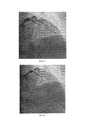
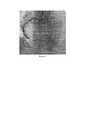
Больной А., возраст 74 года, поступил в приемное отделение Тюменского кардиологического научного центра с жалобами на общую слабость, интенсивные давящие боли в прекардиальной области. Настоящее ухудшение около четырех часов назад, когда упал на улице, со слов, «на руки», с этого же момента отмечает появление давящих болей за грудиной, в локтях, которые связал с травмой. В дальнейшем боли сохранялись, появился «шум в голове», наросла слабость. Вызвал бригаду скорой медицинской помощи, по ЭКГ зарегистрированы признаки очагового повреждения миокарда переднебоковой стенки ЛЖ. После оказания помощи (аспирин, клопидогрель 600 мг, нитраты) транспортирован в приемное отделение. При поступлении: состояние тяжелое, кожные покровы бледные, АД 140/80 мм рт. ст.Легкий цианоз губ. Дыхание жесткое, чистое. ЧДД 16 в 1 мин. Сердечные тоны приглушены, ритмичны 73 уд в 1 мин. Живот мягкий, б/б. Периферических отеков нет. Выставлен диагноз: ИБС. Трансмуральный переднебоковой инфаркт миокарда. Пациента отправили в катетерную лабораторию для проведения экстренного первичного ЧКВ. Пациенту было проведено лечение согласно предлагаемому способу. Под местной анестезией 2% раствором лидокаина, выполнили пункцию правой лучевой артерии. На коронарной ангиографии была выявлена острая тромботическая окклюзия передней межжелудочковой артерии (ПМЖА) (Фиг. 1). После этого коронарный проводник 0,014 дюйма провели до дистальных отделов ПМЖА. После проведения коронарного проводника при введении контраста коронарная артерия не визуализировалась дистальнее места полной тромботической окклюзии (Фиг. 2). Далее по коронарному проводнику к месту полной окклюзии был подведен баллонный катетер размером 2.0 мм - 15 мм. Затем, нераскрытый Возвратно-поступательное перемещение баллонного катетера повторили троекратно. В дальнейшем на контрольной коронарной ангиографии был визуализирован кровоток по ПМЖА TIMI I, а также границы инфаркт-зависимого поражения (Фиг. 3). Далее по коронарному проводнику подвели и имплантировали стент размером 3.0 мм-28 мм (Фиг. 4). На контрольной ангиографии кровоток восстановлен TIMI III ст. (Фиг. 5) Послеоперационный период протекал без осложнений, пациент был выписан на амбулаторное наблюдение со стандартными рекомендациями по терапии на 10 сутки.Patient A., age 74, was admitted to the admission department of the Tyumen Cardiological Research Center with complaints of general weakness, intense pressing pain in the precordial region. The present aggravation about four hours ago, when he fell on the street, according to the words, "on his hands", from the same moment marks the appearance of pressing pains behind the sternum, in the elbows, which he associated with the injury. In the future, the pain persisted, there was a "noise in the head", increased weakness. I called the ambulance team, the ECG showed signs of focal damage to the myocardium of the anterolateral wall of the LV. After providing assistance (aspirin, clopidogrel 600 mg, nitrates), he was transported to the emergency department. On admission: a serious condition, pale skin, blood pressure 140/80 mm Hg. Mild cyanosis of the lips. Breathing hard, clean. NPV 16 in 1 min. Heart sounds are muffled, rhythmic 73 beats per minute. The abdomen is soft, b / w. No peripheral edema. Diagnosed with ischemic heart disease. Transmural anterolateral myocardial infarction. The patient was sent to the catheter laboratory for emergency primary PCI. The patient was treated according to the proposed method. Under local anesthesia with 2% lidocaine solution, puncture of the right radial artery was performed. Coronary angiography revealed acute thrombotic occlusion of the anterior interventricular artery (LAD) (Fig. 1). Thereafter, a 0.014 inch coronary guidewire was passed to the distal LAD. After conducting the coronary guidewire with the introduction of contrast, the coronary artery was not visualized distal to the site of complete thrombotic occlusion (Fig. 2). Further, a balloon catheter with a size of 2.0 mm - 15 mm was brought to the site of complete occlusion along the coronary guidewire. Then, the unopened Reciprocating movement of the balloon catheter was repeated three times. Subsequently, the control coronary angiography showed the blood flow according to the TIMI I LAD, as well as the boundaries of the infarction-dependent lesion (Fig. 3). Next, a 3.0 mm-28 mm stent was brought along the coronary guidewire and implanted (Fig. 4). On control angiography, blood flow was restored TIMI III grade. (Fig. 5) The postoperative period was uneventful, the patient was discharged for outpatient observation with standard recommendations for therapy on day 10.
Клинически пример 2.Clinical example 2.
Больной Н., возраст 42 года, был доставлен бригадой скорой медицинской помощи в приемное отделение Тюменского кардиологического научного центра. На момент поступления жалобы на дискомфорт в грудной клетке и общую слабость. Из анамнеза известно, что 4 дня назад у больного внезапно начался приступ загрудинных болей с иррадиацией в левую руку. Болевой синдром сопровождался обильным потоотделением и слабостью и купировался самостоятельно через 1 час. Впоследствии болевой синдром рецидивировал за 2 часа до текущего поступления в клинику. Диагноз острого нижнего ОИМпST верифицирован на основе типичного ангинозного приступа, зарегистрированной на догоспитальном этапе электрокардиограмме: подъем сегмента ST в отведениях II, III, AVF до 1,5-2 мм, реципрокные изменения в виде депрессии сегмента ST в отведениях I, AVL до 1,5 мм, в V1-V6 до 4-5 мм, патологический зубец qR в III, AVF. При поступлении: состояние тяжелое, кожные покровы нормальной окраски, АД 130/80 мм рт. ст.Дыхание жесткое, чистое. ЧДД 16 в 1 мин. Сердечные тоны приглушены, ритмичны 78 уд в 1 мин. Живот мягкий, б/б. Периферических отеков нет. Затем больной был экстренно доставлен в рентгеноперационную для выполнения первичной коронароангиографии. Под местной анестезией 2% раствором лидокаина, выполнили пункцию правой лучевой артерии. Пациенту было проведено лечение согласно предлагаемому способу. На коронарной ангиографии была выявлена острая тромботическая окклюзия правой коронарной артерии (ПКА) (Фиг. 6). После этого коронарный проводник 0,014 дюйма был проведен до дистальных отделов ПКА. После того, как коронарный проводник был проведен через место тромботической окклюзии, выполнили контрольную ангиографию артерии. При введении контраста коронарная артерия не визуализировалась дистальнее места полной тромботической окклюзии (Фиг. 7). Затем по коронарному проводнику к месту полной окклюзии подвели баллонный катетер размером 2.0 мм-15 мм., а после этого, не раздувая, его провели через место окклюзии на расстояние 30 мм дистальнее от исходного положения (Фиг. 8), после чего переместили его в исходное положение. Описанное выше возвратно-поступательное перемещение баллонного катетера повторили троекратно. В дальнейшем на контрольной коронарной ангиографии визуализировали кровоток по ПКА TIMI II, а также границы инфаркт-зависимого поражения (Фиг. 9). Далее по коронарному проводнику в область инфаркт-зависимого поражения подвели и имплантировали стент размером 3.5 мм-33 мм (Фиг. 10). Результаты контрольной ангиографии подтвердили то, что кровоток восстановлен TIMI III ст.(Фиг. 11) Послеоперационный период протекал спокойно, пациент был выписан на амбулаторное наблюдение со стандартными рекомендациями по терапии на 7 сутки.Patient N., age 42 years old, was taken by an ambulance team to the admission department of the Tyumen Cardiological Scientific Center. At the time of receipt of the complaint of chest discomfort and general weakness. From the anamnesis it is known that 4 days ago the patient suddenly began an attack of chest pain radiating to the left arm. The pain syndrome was accompanied by profuse sweating and weakness and stopped on its own after 1 hour. Subsequently, the pain syndrome recurred 2 hours before the current admission to the clinic. The diagnosis of acute lower STEMI was verified on the basis of a typical anginal attack recorded at the prehospital stage by an electrocardiogram: elevation of the ST segment in leads II, III, AVF up to 1.5-2 mm, reciprocal changes in the form of depression of the ST segment in leads I, AVL up to 1, 5 mm, in V1-V6 up to 4-5 mm, pathological qR wave in III, AVF. On admission: a serious condition, normal skin color, blood pressure 130/80 mm Hg. Breathing hard, clean. NPV 16 in 1 min. Heart sounds are muffled, rhythmic 78 beats per minute. The abdomen is soft, b / w. No peripheral edema. Then the patient was urgently taken to the X-ray operating room for primary coronary angiography. Under local anesthesia with 2% lidocaine solution, puncture of the right radial artery was performed. The patient was treated according to the proposed method. Coronary angiography revealed acute thrombotic occlusion of the right coronary artery (RCA) (Fig. 6). Thereafter, a 0.014-inch coronary guidewire was passed to the distal RCA. After the coronary guidewire was passed through the thrombotic occlusion site, control angiography of the artery was performed. With the introduction of contrast, the coronary artery was not visualized distal to the site of complete thrombotic occlusion (Fig. 7). Then a balloon catheter with a size of 2.0 mm-15 mm was brought along the coronary guidewire to the place of complete occlusion, and then, without inflating, it was passed through the occlusion site at a distance of 30 mm distal from the initial position (Fig. 8), after which it was moved to initial position. The above-described reciprocating movement of the balloon catheter was repeated three times. Subsequently, the control coronary angiography visualized the blood flow according to the TIMI II RCA, as well as the boundaries of the infarction-dependent lesion (Fig. 9). Further, a stent 3.5 mm-33 mm in size was brought and implanted along the coronary conductor in the area of the infarct-dependent lesion (Fig. 10). The results of the control angiography confirmed that the blood flow was restored TIMI III grade (Fig. 11) The postoperative period was calm, the patient was discharged for outpatient observation with standard recommendations for therapy on day 7.
Предлагаемый способ апробирован на 20 больных. У всех больных при его выполнении отсутствовал феномен «no-reflow». Таким образом, этот способ позволяет предотвратить развитие феномена «no-reflow» инфаркт-связанной коронарной артерии у пациентов ОИМ при выполнении ЧКВ.The proposed method has been tested on 20 patients. All patients had no “no-reflow” phenomenon during its implementation. Thus, this method makes it possible to prevent the development of the phenomenon of "no-reflow" infarction-associated coronary artery in AMI patients during PCI.
ПРИЛОЖЕНИЕATTACHMENT
Фиг. 1 - Полная тромботическая окклюзия ПМЖА по данным ангиографии.FIG. 1 - Complete thrombotic occlusion of the LAD according to angiography.
Фиг. 2 - Отсутствие коронарного кровотока по ПМЖА после проведения коронарногоFIG. 2 - Lack of coronary blood flow through LAD after coronary
проводника до дистальных отделов артерии.guide to the distal artery.
Фиг. 3-Частичное восстановление коронарного кровотока после трехкратного возвратно-поступательного перемещения баллонного катетера на 30 мм дистальнее от места начала полной окклюзии, а затем обратно (без дилатации).FIG. 3-Partial restoration of coronary blood flow after three-fold reciprocating movement of the balloon catheter 30 mm distal from the site of the onset of complete occlusion, and then back (without dilatation).
Фиг. 4 - Прямое стентирование (установка коронарного стента 3.0 мм - 28 мм) в ПМЖА.FIG. 4 - Direct stenting (placement of a coronary stent 3.0 mm - 28 mm) in the LAD.
Фиг. 5 - Финальный результат. Восстановлен адекватный антеградный кровоток.FIG. 5 - Final result. Adequate antegrade blood flow was restored.
Фиг. 6 - Полная тромботическая окклюзия ПКА по данным ангиографии.FIG. 6 - Complete thrombotic occlusion of RCA according to angiography.
Фиг. 7 - Отсутствие коронарного кровотока по ПКА после проведения коронарного проводника до дистальных отделов артерии.FIG. 7 - Lack of coronary blood flow through the RCA after the passage of the coronary guidewire to the distal artery.
Фиг. 8 - Перемещение баллонного катетера на расстояние 30 мм дистальнее от начала целевого поражения. Красным овалом отмечено дистальное положение баллонного катетера.FIG. 8 - Move the balloon catheter 30 mm distal from the start of the target lesion. The red oval marks the distal position of the balloon catheter.
Фиг. 9 - Частичное восстановление коронарного кровотока после трехкратного возвратно-поступательного перемещения баллонного катетера на расстояние 30 мм дистальнее от места начала полной окклюзии, а затем обратно (без дилатации).FIG. 9 - Partial restoration of coronary blood flow after three times reciprocating movement of the balloon catheter at a distance of 30 mm distal from the site of the onset of complete occlusion, and then back (without dilatation).
Фиг. 10 - Прямое стентирование (установка коронарного стента 3.5 мм - 33 мм) в ПКА.FIG. 10 - Direct stenting (placement of a coronary stent 3.5 mm - 33 mm) in the RCA.
Фиг. 11 - Финальный результат (кровоток TIMI III ст.).FIG. 11 - Final result (TIMI III blood flow).
Claims (1)
Translated fromRussianPriority Applications (1)
| Application Number | Priority Date | Filing Date | Title |
|---|---|---|---|
| RU2020104242ARU2745651C1 (en) | 2020-01-30 | 2020-01-30 | Method for the prevention of the "no-reflow" phenomenon during percutaneous coronary interventions in patients with acute myocardial infarction with st segment evalution |
Applications Claiming Priority (1)
| Application Number | Priority Date | Filing Date | Title |
|---|---|---|---|
| RU2020104242ARU2745651C1 (en) | 2020-01-30 | 2020-01-30 | Method for the prevention of the "no-reflow" phenomenon during percutaneous coronary interventions in patients with acute myocardial infarction with st segment evalution |
Publications (1)
| Publication Number | Publication Date |
|---|---|
| RU2745651C1true RU2745651C1 (en) | 2021-03-30 |
Family
ID=75353523
Family Applications (1)
| Application Number | Title | Priority Date | Filing Date |
|---|---|---|---|
| RU2020104242ARU2745651C1 (en) | 2020-01-30 | 2020-01-30 | Method for the prevention of the "no-reflow" phenomenon during percutaneous coronary interventions in patients with acute myocardial infarction with st segment evalution |
Country Status (1)
| Country | Link |
|---|---|
| RU (1) | RU2745651C1 (en) |
Cited By (1)
| Publication number | Priority date | Publication date | Assignee | Title |
|---|---|---|---|---|
| RU2786150C1 (en)* | 2021-10-27 | 2022-12-19 | Государственное бюджетное учреждение здравоохранения Московской области "Московский областной научно-исследовательский клинический институт им. М.Ф. Владимирского" (ГБУЗ МО МОНИКИ им. М.Ф. Владимирского) | Method for preventing the "no-reflow" phenomenon during coronary artery stenting in patients with acute myocardial infarction with st elevation due to massive thrombosis |
Citations (1)
| Publication number | Priority date | Publication date | Assignee | Title |
|---|---|---|---|---|
| RU2649572C1 (en)* | 2017-04-12 | 2018-04-03 | Федеральное государственное бюджетное научное учреждение "Томский национальный исследовательский медицинский центр Российской академии наук" (Томский НИМЦ) | Method of treatment of patients with acute myocardial infarction with massive thrombosis of the infarct-related coronary artery |
- 2020
- 2020-01-30RURU2020104242Apatent/RU2745651C1/enactive
Patent Citations (1)
| Publication number | Priority date | Publication date | Assignee | Title |
|---|---|---|---|---|
| RU2649572C1 (en)* | 2017-04-12 | 2018-04-03 | Федеральное государственное бюджетное научное учреждение "Томский национальный исследовательский медицинский центр Российской академии наук" (Томский НИМЦ) | Method of treatment of patients with acute myocardial infarction with massive thrombosis of the infarct-related coronary artery |
Non-Patent Citations (4)
| Title |
|---|
| Carrick D. et al. A Randomized Trial of Deferred Stenting versus Immediate Stenting to Prevent No- or Slow Reflow in Acute ST-Elevation Myocardial Infarction (DEFER-STEMI), J. Am. coll. cardiol. - 2014. - Vol. 63. - P. 2088—2098.* |
| Алексеева Я.В. и др. Отсроченное эндоваскулярное вмешательство при остром инфаркте миокарда с массивным тромбозом инфаркт-связанной коронарной артерии, Сибирский медицинский журнал. 2018; 33(2), 16-20.* |
| Алексеева Я.В. и др. Отсроченное эндоваскулярное вмешательство при остром инфаркте миокарда с массивным тромбозом инфаркт-связанной коронарной артерии, Сибирский медицинский журнал. 2018; 33(2), 16-20. Бессонов И.С., Кузнецов В.А., Зырянов И.П., Сапожников С.С. Прямое стентирование в сравнении со стентированием после предилатации или мануальной тромбоаспирации у пациентов с острым инфарктом миокарда с подъемом сегмента ST и полной тромботической окклюзией инфаркт-связанной коронарной артерии. Эндоваскулярная хирургия. 2018; 5 (4): 410-7. Carrick D. et al. A Randomized Trial of Deferred Stenting versus Immediate Stenting to Prevent No- or Slow Reflow in Acute ST-Elevation Myocardial Infarction (DEFER-STEMI), J. Am. coll. cardiol. - 2014. - Vol. 63. - P. 2088—2098.* |
| Бессонов И.С., Кузнецов В.А., Зырянов И.П., Сапожников С.С. Прямое стентирование в сравнении со стентированием после предилатации или мануальной тромбоаспирации у пациентов с острым инфарктом миокарда с подъемом сегмента ST и полной тромботической окклюзией инфаркт-связанной коронарной артерии. Эндоваскулярная хирургия. 2018; 5 (4): 410-7.* |
Cited By (2)
| Publication number | Priority date | Publication date | Assignee | Title |
|---|---|---|---|---|
| RU2786150C1 (en)* | 2021-10-27 | 2022-12-19 | Государственное бюджетное учреждение здравоохранения Московской области "Московский областной научно-исследовательский клинический институт им. М.Ф. Владимирского" (ГБУЗ МО МОНИКИ им. М.Ф. Владимирского) | Method for preventing the "no-reflow" phenomenon during coronary artery stenting in patients with acute myocardial infarction with st elevation due to massive thrombosis |
| RU2798161C1 (en)* | 2022-06-01 | 2023-06-16 | Федеральное государственное бюджетное научное учреждение Томский национальный исследовательский медицинский центр Российской академии наук (Томский НИМЦ) | Method of endovascular treatment of thrombotic occlusion of autovenous aortocoronary bypass grafts in patients with acute st segment elevation myocardial infarction |
Similar Documents
| Publication | Publication Date | Title |
|---|---|---|
| Perry | Complications of missed arterial injuries | |
| Molina et al. | Paget-Schroetter syndrome treated with thrombolytics and immediate surgery | |
| Quigley et al. | Percutaneous transluminal coronary angioplasty in unstable angina: comparison with stable angina. | |
| Shannon Jr et al. | Peripheral arterial missile embolization: a case report and 22-year literature review | |
| Holleman Jr et al. | Arterial surgery for arm ischemia. A survey of 136 patients | |
| Gergoudis et al. | Acute compartment syndrome as a complication of radial artery catheterization | |
| RU2745651C1 (en) | Method for the prevention of the "no-reflow" phenomenon during percutaneous coronary interventions in patients with acute myocardial infarction with st segment evalution | |
| Gschwind | The intravenous foreign body: a report of 2 cases | |
| Prakash et al. | Radial artery pseudoaneurysm following cardiac catheterization: a case report | |
| RU2463965C1 (en) | Method of selecting arterial access for performing x-ray endovascular intervention on coronary arteries | |
| Wylie et al. | Thromboendarterectomy, a clinical appraisal | |
| RU2649572C1 (en) | Method of treatment of patients with acute myocardial infarction with massive thrombosis of the infarct-related coronary artery | |
| Hasegawa et al. | A case of upper extremity deep vein thrombosis with long-term patency using pharmaco-mechanical catheter-directed thrombolysis in the acute phase | |
| Rencuzogullari et al. | Coronary thrombosis in three coronary arteries due to whey protein | |
| RU2798161C1 (en) | Method of endovascular treatment of thrombotic occlusion of autovenous aortocoronary bypass grafts in patients with acute st segment elevation myocardial infarction | |
| RU2811275C1 (en) | Method of treating acute occlusion of extra- and intracranial sections of internal carotid artery in acute period of ischemic stroke | |
| RU2799257C1 (en) | Method of the treatment of acute arterial thrombosis of the popliteal-ankle segment associated with covid-19 | |
| RU2786150C1 (en) | Method for preventing the "no-reflow" phenomenon during coronary artery stenting in patients with acute myocardial infarction with st elevation due to massive thrombosis | |
| Fukushima et al. | Management of intraaortic balloon entrapment | |
| Ponce et al. | Early detection and endovascular intervention to correct dialysis vascular access malfunction | |
| RU2791401C1 (en) | Method of surgical treatment of atherosclerotic lesions of bifurcation of the carotid artery | |
| Kaiser et al. | Procedural Complications | |
| Sebayang | Acute Limb Ischemia: An update on diagnosis and management | |
| Binayendu et al. | Radial Artery Pseudoaneurysm Following Cardiac Catheterization: A Case Report | |
| RU2716093C1 (en) | Method of endovascular treatment of a false radial artery aneurysm |