RU2667957C1 - Method of obtaining and certification of standard sample of antigen virus of tick-borne encephalitis - Google Patents
Method of obtaining and certification of standard sample of antigen virus of tick-borne encephalitisDownload PDFInfo
- Publication number
- RU2667957C1 RU2667957C1RU2017133181ARU2017133181ARU2667957C1RU 2667957 C1RU2667957 C1RU 2667957C1RU 2017133181 ARU2017133181 ARU 2017133181ARU 2017133181 ARU2017133181 ARU 2017133181ARU 2667957 C1RU2667957 C1RU 2667957C1
- Authority
- RU
- Russia
- Prior art keywords
- tick
- concentration
- glycoprotein
- borne encephalitis
- standard sample
- Prior art date
Links
- 238000000034methodMethods0.000titleclaimsabstractdescription22
- 241000700605VirusesSpecies0.000titleclaimsabstractdescription11
- 208000004006Tick-borne encephalitisDiseases0.000titleclaimsabstractdescription9
- 239000000427antigenSubstances0.000titledescription14
- 102000036639antigensHuman genes0.000titledescription14
- 108091007433antigensProteins0.000titledescription14
- 241000710771Tick-borne encephalitis virusSpecies0.000claimsabstractdescription30
- 102000004169proteins and genesHuman genes0.000claimsabstractdescription21
- 108090000623proteins and genesProteins0.000claimsabstractdescription21
- 101000807236Human cytomegalovirus (strain AD169) Membrane glycoprotein US3Proteins0.000claimsabstractdescription13
- 238000004364calculation methodMethods0.000claimsabstractdescription7
- 238000000108ultra-filtrationMethods0.000claimsabstractdescription5
- 238000001914filtrationMethods0.000claimsabstractdescription4
- 238000002264polyacrylamide gel electrophoresisMethods0.000claimsabstractdescription4
- 238000001556precipitationMethods0.000claimsabstractdescription4
- 210000003527eukaryotic cellAnatomy0.000claimsabstract2
- 238000003119immunoblotMethods0.000claimsabstract2
- 238000002798spectrophotometry methodMethods0.000claimsabstract2
- 238000004519manufacturing processMethods0.000claimsdescription10
- 229960005486vaccineDrugs0.000claimsdescription7
- 238000004587chromatography analysisMethods0.000claimsdescription4
- 238000000746purificationMethods0.000claimsdescription3
- 230000002779inactivationEffects0.000claimsdescription2
- WSFSSNUMVMOOMR-NJFSPNSNSA-NmethanoneChemical compoundO=[14CH2]WSFSSNUMVMOOMR-NJFSPNSNSA-N0.000claimsdescription2
- 238000002360preparation methodMethods0.000claimsdescription2
- 108010075717Flavivirus glycoprotein EProteins0.000claims1
- 238000009825accumulationMethods0.000claims1
- 230000000694effectsEffects0.000abstractdescription4
- 229960003239encephalitis vaccineDrugs0.000abstractdescription2
- 239000000126substanceSubstances0.000abstractdescription2
- WSFSSNUMVMOOMR-UHFFFAOYSA-NFormaldehydeChemical compoundO=CWSFSSNUMVMOOMR-UHFFFAOYSA-N0.000abstract3
- 238000005227gel permeation chromatographyMethods0.000abstract1
- 239000000523sampleSubstances0.000description15
- 239000012141concentrateSubstances0.000description12
- 238000002965ELISAMethods0.000description6
- 238000001962electrophoresisMethods0.000description6
- 230000000890antigenic effectEffects0.000description5
- 230000003612virological effectEffects0.000description5
- 239000000243solutionSubstances0.000description4
- 238000001262western blotMethods0.000description4
- 101710204837Envelope small membrane proteinProteins0.000description3
- 101710088839Replication initiation proteinProteins0.000description3
- 238000004128high performance liquid chromatographyMethods0.000description3
- 239000000463materialSubstances0.000description3
- 238000004458analytical methodMethods0.000description2
- 210000004027cellAnatomy0.000description2
- 239000012531culture fluidSubstances0.000description2
- 238000010828elutionMethods0.000description2
- 230000002163immunogenEffects0.000description2
- 239000007788liquidSubstances0.000description2
- 235000015097nutrientsNutrition0.000description2
- 239000012466permeateSubstances0.000description2
- 238000001542size-exclusion chromatographyMethods0.000description2
- 238000002415sodium dodecyl sulfate polyacrylamide gel electrophoresisMethods0.000description2
- 238000003860storageMethods0.000description2
- 238000012360testing methodMethods0.000description2
- 210000002845virionAnatomy0.000description2
- 108010088751AlbuminsProteins0.000description1
- 102000009027AlbuminsHuman genes0.000description1
- 101800001603Capsid protein CProteins0.000description1
- 101710094648Coat proteinProteins0.000description1
- 101800001847Core protein precursorProteins0.000description1
- 241000287828Gallus gallusSpecies0.000description1
- 108010001336Horseradish PeroxidaseProteins0.000description1
- 102000008100Human Serum AlbuminHuman genes0.000description1
- 108091006905Human Serum AlbuminProteins0.000description1
- 108010052285Membrane ProteinsProteins0.000description1
- 102000018697Membrane ProteinsHuman genes0.000description1
- 108010058846OvalbuminProteins0.000description1
- 239000002033PVDF binderSubstances0.000description1
- 102000035195PeptidasesHuman genes0.000description1
- 108091005804PeptidasesProteins0.000description1
- GLUUGHFHXGJENI-UHFFFAOYSA-NPiperazineChemical compoundC1CNCCN1GLUUGHFHXGJENI-UHFFFAOYSA-N0.000description1
- 102000007327ProtaminesHuman genes0.000description1
- 108010007568ProtaminesProteins0.000description1
- 239000004365ProteaseSubstances0.000description1
- 229920002684SepharosePolymers0.000description1
- 108010067390Viral ProteinsProteins0.000description1
- SXEHKFHPFVVDIR-UHFFFAOYSA-N[4-(4-hydrazinylphenyl)phenyl]hydrazineChemical compoundC1=CC(NN)=CC=C1C1=CC=C(NN)C=C1SXEHKFHPFVVDIR-UHFFFAOYSA-N0.000description1
- 238000010521absorption reactionMethods0.000description1
- 230000009830antibody antigen interactionEffects0.000description1
- 238000011088calibration curveMethods0.000description1
- 238000012512characterization methodMethods0.000description1
- 238000006243chemical reactionMethods0.000description1
- 239000003153chemical reaction reagentSubstances0.000description1
- 238000011097chromatography purificationMethods0.000description1
- 238000004140cleaningMethods0.000description1
- 238000000326densiometryMethods0.000description1
- 238000001514detection methodMethods0.000description1
- 230000007717exclusionEffects0.000description1
- 210000002950fibroblastAnatomy0.000description1
- 238000011010flushing procedureMethods0.000description1
- 238000004108freeze dryingMethods0.000description1
- 238000001502gel electrophoresisMethods0.000description1
- 230000008105immune reactionEffects0.000description1
- 230000005847immunogenicityEffects0.000description1
- 230000003993interactionEffects0.000description1
- 210000001161mammalian embryoAnatomy0.000description1
- 239000003550markerSubstances0.000description1
- 238000005259measurementMethods0.000description1
- 239000012528membraneSubstances0.000description1
- 239000000203mixtureSubstances0.000description1
- 238000012544monitoring processMethods0.000description1
- 229940126619mouse monoclonal antibodyDrugs0.000description1
- 229940092253ovalbuminDrugs0.000description1
- 239000008055phosphate buffer solutionSubstances0.000description1
- 238000000053physical methodMethods0.000description1
- 229960005141piperazineDrugs0.000description1
- 229920003023plasticPolymers0.000description1
- 229920001184polypeptidePolymers0.000description1
- 229920002981polyvinylidene fluoridePolymers0.000description1
- 108090000765processed proteins & peptidesProteins0.000description1
- 102000004196processed proteins & peptidesHuman genes0.000description1
- 238000012545processingMethods0.000description1
- 229950008679protamine sulfateDrugs0.000description1
- 239000011265semifinished productSubstances0.000description1
- 238000001228spectrumMethods0.000description1
- 230000000087stabilizing effectEffects0.000description1
- 239000000758substrateSubstances0.000description1
- 239000000725suspensionSubstances0.000description1
- 239000004546suspension concentrateSubstances0.000description1
- 238000010257thawingMethods0.000description1
- 238000012546transferMethods0.000description1
- 229960004854viral vaccineDrugs0.000description1
Images


Classifications
- G—PHYSICS
- G01—MEASURING; TESTING
- G01N—INVESTIGATING OR ANALYSING MATERIALS BY DETERMINING THEIR CHEMICAL OR PHYSICAL PROPERTIES
- G01N33/00—Investigating or analysing materials by specific methods not covered by groups G01N1/00 - G01N31/00
- G01N33/48—Biological material, e.g. blood, urine; Haemocytometers
- G01N33/50—Chemical analysis of biological material, e.g. blood, urine; Testing involving biospecific ligand binding methods; Immunological testing
- G01N33/52—Use of compounds or compositions for colorimetric, spectrophotometric or fluorometric investigation, e.g. use of reagent paper and including single- and multilayer analytical elements
- G01N33/521—Single-layer analytical elements
- G01N33/523—Single-layer analytical elements the element being adapted for a specific analyte
Landscapes
- Health & Medical Sciences (AREA)
- Life Sciences & Earth Sciences (AREA)
- Hematology (AREA)
- Immunology (AREA)
- Engineering & Computer Science (AREA)
- Urology & Nephrology (AREA)
- Molecular Biology (AREA)
- Chemical & Material Sciences (AREA)
- Biomedical Technology (AREA)
- Food Science & Technology (AREA)
- Microbiology (AREA)
- Cell Biology (AREA)
- Biotechnology (AREA)
- Medicinal Chemistry (AREA)
- Physics & Mathematics (AREA)
- Analytical Chemistry (AREA)
- Biochemistry (AREA)
- General Health & Medical Sciences (AREA)
- General Physics & Mathematics (AREA)
- Pathology (AREA)
- Peptides Or Proteins (AREA)
Abstract
Description
Translated fromRussianИзобретение относится к области медицинской биотехнологии, производству и контролю вирусных вакцин и касается способа получения и аттестации стандартного образца (СО) гликопротеина Е (gpE) вируса клещевого энцефалита (ВКЭ). Изобретение может быть использовано для решения практических задач медицинской биотехнологии -аттестации стандартного образца предприятия (СОП) для контроля специфической активности вакцины и определения количественного содержания гликопротеина Е в полуфабрикатах и готовом препарате вакцины КЭ.The invention relates to the field of medical biotechnology, production and control of viral vaccines, and relates to a method for producing and certification of a standard sample (CO) of glycoprotein E (gpE) of tick-borne encephalitis virus (TBE). The invention can be used to solve practical problems of medical biotechnology - certification of a standard enterprise sample (SOP) for monitoring the specific activity of the vaccine and determining the quantitative content of glycoprotein E in the semi-finished products and the finished preparation of the CE vaccine.
Определение содержания антигена ВКЭ в вакцинах проводится с помощью метода иммуноферментного анализа (ИФА). При этом для количественного определения вирусного антигена методом ИФА необходимо использовать аттестованный СОП вакцины клещевого энцефалита с известной концентрацией белка Е.Determination of the content of TBEV antigen in vaccines is carried out using the method of enzyme-linked immunosorbent assay (ELISA). Moreover, for the quantitative determination of viral antigen by ELISA, it is necessary to use a certified SOP of a tick-borne encephalitis vaccine with a known concentration of protein E.
Из-за вариабельности антигенных свойств разных штаммов ВКЭ, проявляющейся в индивидуальных особенностях взаимодействия вирусных антигенов каждого штамма с панелями моноклональных антител, наиболее достоверным вариантом является использование для изготовления и аттестации СО, СОП производственного штамма вируса, использующегося при производстве вакцины.Due to the variability of the antigenic properties of different TBEV strains, which is manifested in the individual characteristics of the interaction of the viral antigens of each strain with panels of monoclonal antibodies, the most reliable option is to use for the production and certification of CO, SOP of the production strain of the virus used in the production of the vaccine.
Другая методологическая проблема состоит в том, что белки ВКЭ (и в первую очередь основной иммуноген - gpE) нестабильны и чувствительны к воздействию рН и протеаз. Срок их хранения в жидком виде ограничен, а при замораживании-оттаивании (в том числе лиофилизации) происходят изменения структуры, ведущие к снижению аффинности в иммунологических реакциях.Another methodological problem is that TBEV proteins (and primarily the main immunogen, gpE) are unstable and sensitive to the effects of pH and proteases. Their shelf life in liquid form is limited, and upon freezing-thawing (including lyophilization), structural changes occur, leading to a decrease in affinity in immunological reactions.
При аттестации контрольных образцов необходимо применять методы, отличные по физико-химическим принципам от тех, в которых эти образцы в дальнейшем будут выступать стандартами. Наиболее подходящими являются прямые физические методы, например, гравиметрический. Поскольку аттестуемый СОП вакцины будет в дальнейшем использоваться в ИФА и отсутствуют международные или отраслевые стандарты белка Е, то нельзя применять методы, основанные на феномене взаимодействия антиген-антитело, так как результат этих методов будет зависеть от свойств применяемых антител.In the certification of control samples, it is necessary to apply methods that are different in physical and chemical principles from those in which these samples will continue to act as standards. The most suitable are direct physical methods, for example, gravimetric. Since the certified SOP of the vaccine will be further used in ELISA and there are no international or industry standards for protein E, methods based on the antigen-antibody interaction phenomenon cannot be used, since the result of these methods will depend on the properties of the antibodies used.
Учитывая, что количество gpE в материале для изготовления стандарта невелико, а его очистка сопряжена с опасностью изменений структуры, то оптимальным вариантом является выделение цельновирионной фракции из культуральной жидкости. В составе вирионов gpE наиболее стабилен и сохраняет антигенную активность и иммуногенность. Между тем в таком виде нельзя определить его концентрацию прямыми методами, так как он находится в смеси с другими вирионными белками, белками клеток-продуцентов и компонентами питательной среды.Given that the amount of gpE in the material for the production of the standard is small, and its purification is fraught with the danger of structural changes, the best option is to isolate the whole-virion fraction from the culture fluid. In the composition of virions, gpE is most stable and retains antigenic activity and immunogenicity. Meanwhile, in this form it is impossible to determine its concentration by direct methods, since it is mixed with other virion proteins, proteins of producer cells and components of the nutrient medium.
В связи с этим, в целях аттестации СО нами предложено применять опосредованные вычисления. После определения концентрации общего белка в образце по методу Лоури или Несслера, с помощью электрофореза в полиакриамидном геле (ПААГ) устанавливают долю содержания gpE в образце. Зная два этих показателя, вычисляют абсолютную концентрацию gpE.In this regard, in order to certify WITH, we proposed to use indirect calculations. After determining the concentration of total protein in the sample by the method of Lowry or Nessler, using a polyacryamide gel electrophoresis (SDS page) establish the proportion of gpE in the sample. Knowing these two indicators, the absolute concentration of gpE is calculated.
Стабильность такого стандартного образца (СО) вирусных белков ограничена. Из-за того, что СО вирусного антигена необходимо хранить в жидком виде, максимальный срок хранения составляет 1 месяц при температуре 2-8°С. Однако, этого времени достаточно для того, чтобы провести аттестацию лиофилизированного СОП вакцины КЭ.The stability of such a standard sample (CO) of viral proteins is limited. Due to the fact that the CO of the viral antigen must be stored in liquid form, the maximum shelf life is 1 month at a temperature of 2-8 ° C. However, this time is enough to carry out the certification of the lyophilized SOP of the CE vaccine.
Задачей, решаемой данным изобретением, является получение и аттестация стандартного образца гликопротеина Е вируса КЭ.The problem solved by this invention is the receipt and certification of a standard sample of glycoprotein E virus CE.
Поставленная задача решается следующим образом:The problem is solved as follows:
Для изготовления СО антигена ВКЭ используют производственный штамм ВКЭ 205, вирус репродуцируют в культуре первично-трипсинизированных фибробластов куриных эмбрионов. Полученную культуральную жидкость инактивируют формальдегидом, очищают с помощью осаждения протаминсульфатом, фильтрации. Затем концентрируют с помощью ультрафильтрации в тангенциальном потоке. Затем проводят тонкую очистку с помощью эксклюзионной хроматографии и финишное концентрирование с помощью центрифужных концентраторов.For the production of CO antigen of TBEV, the production strain of TBEV 205 is used; the virus is reproduced in the culture of primary trypsinized chicken embryo fibroblasts. The resulting culture fluid is inactivated with formaldehyde, purified by precipitation with protamine sulfate, and filtration. It is then concentrated by ultrafiltration in a tangential flow. Then carry out a fine cleaning using size exclusion chromatography and final concentration using centrifuge concentrators.
Полученный концентрат СО антигена ВКЭ контролируют по следующим показателям:The obtained concentrate WITH antigen BCE is controlled by the following indicators:
- содержание общего белка;- total protein content;
- чистота высокомолекулярной фракции (ВЭЖХ);- purity of high molecular weight fraction (HPLC);
- подлинность (идентификация электрофоретических полос, соответствующих gpE, в Western-blot);- authenticity (identification of electrophoretic bands corresponding to gpE, in Western-blot);
- определение относительной концентрации gpE (электрофорез в ПААГ).- determination of the relative concentration of gpE (electrophoresis in SDS page).
После этого расчетно определяют абсолютное содержание gpE в концентрате СО антигена ВКЭ, разводят до необходимой концентрации и упаковывают для хранения.After that, the absolute content of gpE in the CO concentrate of the TBEV antigen is calculated, bred to the required concentration and packaged for storage.
Новым в предлагаемом способе является использование комплекса методов очистки и характеризации гликопротеина Е в составе стандартного образца антигена вируса КЭ (электрофорез в ПААГ, Western-blot и определение концентрации общего белка), позволяющих напрямую рассчитать содержание основного иммуногена ВКЭ.New in the proposed method is the use of a complex of methods for purification and characterization of glycoprotein E as part of a standard sample of KE virus antigen (PAGE, Western-blot electrophoresis and determination of the concentration of total protein), allowing direct calculation of the content of the main TBEV immunogen.
Пример 1.Example 1
В качестве материала для выделения антигена вируса КЭ использовали:The following materials were used as a material for isolating the TBE antigen:
концентрат вирусной взвеси (с.57) объемом 1,10 л.viral suspension concentrate (p. 57) with a volume of 1.10 l.
Основные показатели качества:Key performance indicators:
Общий белок 2,33 мг/млTotal protein 2.33 mg / ml
Антигенная активность 31,96 мкг/млAntigenic activity 31.96 mcg / ml
Овальбумин 0,080 мкг/мл (80,38 нг/мл)Ovalbumin 0.080 mcg / ml (80.38 ng / ml)
Концентрат был получен после инактивации, осветляющей фильтрации и концентрирования методом ультрафильтрации в тангенциальном потоке.The concentrate was obtained after inactivation, clarifying filtration and concentration by the method of ultrafiltration in a tangential flow.
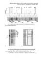
Хроматографическая очистка проводилась эксклюзионной хроматографией с использованием хроматографа АКТА Explorer 100 (Amersham bioscience, Sweden), колонны XK-50-70 (GE Healthcare, Sweden), Sepharose 6FF (GE HealthCare, Sweden), 0,05 M фосфатный буферный раствор, рН 7,5-7,7 (Фиг. 1).Chromatographic purification was performed by size exclusion chromatography using an AKTA Explorer 100 chromatograph (Amersham bioscience, Sweden), XK-50-70 columns (GE Healthcare, Sweden), Sepharose 6FF (GE HealthCare, Sweden), 0.05 M phosphate buffer solution,
Контроль чистоты цельновирионной фракции проводили с помощью ВЭЖХ (колонна ProteinPac 300 SW).The purity control of the whole virion fraction was carried out using HPLC (ProteinPac 300 SW column).
Контроль концентрации общего белка проводили по методу Лоури.The concentration of total protein was monitored by the Lowry method.
Контроль антигенной активности проводили в ИФА с помощью коммерческой тест-системы.Control of antigenic activity was carried out in ELISA using a commercial test system.
Условия хроматографии:Chromatography conditions:
Анализ методом ВЭЖХ показал, что содержание высокомолекулярной фракции в осветленном концентрате вирусной взвеси составило 9,3%, в очищенном концентрате - 98,8% (Фиг. 2).Analysis by HPLC showed that the content of the high molecular weight fraction in the clarified concentrate of viral suspension was 9.3%, in the purified concentrate - 98.8% (Fig. 2).
Концентрирование антигена ВКЭ проводили с помощью центрифуги Thermo Multifuge 4KR (ThermoScientific, USA; Heraeus, Germany) с баккет-ротором, центрифужных концентраторов MilliporeAmicon Ultra-15 с порогом отсечения 10,0 кДа (Millipore, France).TBEV antigen was concentrated using a Thermo Multifuge 4KR centrifuge (ThermoScientific, USA; Heraeus, Germany) with a bucket rotor, MilliporeAmicon Ultra-15 centrifuge concentrators with a cut-off threshold of 10.0 kDa (Millipore, France).
В каждый концентратор вносили 15 мл очищенного концентрат. Центрифугировали при 4000g, 15°С в течение 20-40,0 мин до объема 1,0 мл раствора в каждом концентраторе. После остановки центрифуги удаляли пермеат и доливали очищенный концентрат. Затем повторяли процесс.15 ml of purified concentrate was added to each concentrator. It was centrifuged at 4000g, 15 ° C for 20-40.0 min to a volume of 1.0 ml of solution in each concentrator. After centrifuge stopped, permeate was removed and refined concentrate was added. Then the process was repeated.
Контроль концентрации общего белка проводили по методу Лоури.The concentration of total protein was monitored by the Lowry method.
Контроль антигенной активности проводили в ИФА.Control of antigenic activity was carried out in ELISA.
В процессе концентрирования всего было получено 30,0 мл центрифужного концентрата и около 1,1 л пермеата. Таким образом, было проведено концентрирование по объему в 37,2 раза (табл. 1).In the process of concentration, a total of 30.0 ml of a centrifuge concentrate and about 1.1 l of permeate were obtained. Thus, concentration by volume was 37.2 times (Table 1).

В ходе аттестации СО антигена ВКЭ проводили контроль концентрации общего белка по методу Лоури без осаждения (ГФ XII, ч. 2). В качестве стандарта использовали ОСО ФГБУ ГНЦ МЗ РФ с содержанием альбумина 10,1%. Измерение ОП растворов проводили на спектрофотометрах Shimazu 1800-UV и при длине волны 750 нм. Расчет концентраций белка в контрольном и исследуемом образцах осуществляли по калибровочной кривой.During the attestation of CO of the TBEV antigen, the total protein concentration was monitored using the Lowry method without precipitation (GP XII, part 2). The OSO FSBI SSC of the Ministry of Health of the Russian Federation with an albumin content of 10.1% was used as a standard. Measurement of the OD of the solutions was carried out on Shimazu 1800-UV spectrophotometers and at a wavelength of 750 nm. Calculation of protein concentrations in the control and test samples was carried out according to a calibration curve.
Было проведено 5 серий исследований центрифужного концентрата. Среднее значение содержания общего белка составило 1,65 мг/мл.5 series of studies of a centrifuge concentrate were conducted. The average total protein content was 1.65 mg / ml.
Определение относительного содержания gpE в СО проводили с помощью электрофореза в полиакриламидном геле, подтверждение подлинности gpE на электрофореграмме проводили с помощью Western-blot (Фиг. 3).The relative content of gpE in CO was determined using polyacrylamide gel electrophoresis, gpE authenticity on the electrophoregram was carried out using Western-blot (Fig. 3).
Для анализа использовали маркеры молекулярных масс Spectra Multicolor High Rang Protein Ladder 315-42 кДа и Unstained Protein WW Marker 116-14,4 кДа (ThermoFisher), Coomassi blue, Диаминобензидин («Sigma»), ECL detection reagent (Amersham), пленка Hyperfilm ECL (Amersham), PVDF-мембрану, первичные мышиные моноклональные антитела к белку Е линии 14D51 (ЗАО «Биосан»), вторичные моноклональные антитела к мышиным IgG, коньюгированные с пероксидазой хрена, камеру для электрофореза mini-protean 3 (BIO-RAD), камеру для полусухого переноса (BIO-RAD). Всего было проведено 11 серий исследований.For analysis, molecular weight markers were used: Spectra Multicolor High Rang Protein Ladder 315-42 kDa and Unstained Protein WW Marker 116-14,4,4 kDa (ThermoFisher), Coomassi blue, Diaminobenzidine (Sigma), ECL detection reagent (Amersham), Hyperfilm film ECL (Amersham), PVDF membrane, primary mouse monoclonal antibodies to protein E line 14D51 (Biosan CJSC), secondary monoclonal antibodies to mouse IgG conjugated with horseradish peroxidase, mini-protean 3 electrophoresis chamber (BIO-RAD), semi-dry transfer chamber (BIO-RAD). A total of 11 series of studies were conducted.
33 электрофорезные дорожки были подвергнуты денситометрии (Фиг. 4).33 electrophoresis tracks were densitometry (Fig. 4).
По денситограммам были определены молекулярные массы выявленных белков и их относительные концентрации (доля в суммарной площади пиков).Densitograms were used to determine the molecular weights of the detected proteins and their relative concentrations (fraction in the total peak area).
По электрофоретической подвижности можно идентифицировать сывороточный альбумин человека (66 кДа), гликопротеин Е вируса КЭ (53,68 кДа - полипептид, 55 кДа -гликозилированная форма), капсидный белок С ВКЭ (12,1 кДа) и мембранный белок М ВКЭ (8,2 кДа). Остальные полосы на электрофорезе соответствуют различным белкам субстратных клеток и компонентов питательной среды.Electrophoretic mobility can be used to identify human serum albumin (66 kDa), KE virus glycoprotein E (53.68 kDa - polypeptide, 55 kDa glycosylated form), TBEV capsid protein C (12.1 kDa) and M TBE membrane protein (8, 2 kDa). The remaining bands on electrophoresis correspond to various proteins of substrate cells and components of the nutrient medium.
В проведенных исследованиях подвижность gpE варьировала от 52,72 до 56,03 кДа (средняя 54,17 кДа, табл. 2), что согласуется с литературными данными (Фиг. 4).In the studies performed, gpE mobility ranged from 52.72 to 56.03 kDa (average 54.17 kDa, Table 2), which is consistent with published data (Fig. 4).
Принадлежность белка с электрофоретической подвижностью, соответствующей 54,17 кДа к gpE ВКЭ была подтверждена специфической реакцией с моноклональными антителами против gpE ВКЭ в Western-blot (Фиг. 5).The affinity of the protein with electrophoretic mobility corresponding to 54.17 kDa to gpE BCE was confirmed by a specific reaction with monoclonal antibodies against gpE BCE in Western-blot (Fig. 5).
Относительную концентрацию целевого белка (gpE) вычисляли, как долю площади пика среди общей площади денситограммы. Она составляла от 18,6 до 32,4% (табл. 2). Однако два результата были исключены из дальнейшей обработки, так как отклонение их значений от среднего арифметического было больше двух стандартных отклонений (табл. 2, №14, 15 помечено серым).The relative concentration of the target protein (gpE) was calculated as the fraction of the peak area among the total area of the densitogram. It ranged from 18.6 to 32.4% (Table 2). However, two results were excluded from further processing, since the deviation of their values from the arithmetic mean was more than two standard deviations (Table 2, No. 14, 15 are marked in gray).

Таким образом, после исключения двух наблюдений, доля пика денситограммы, соответствующего gpE ВКЭ (относительная концентрация) составила 28,18±2,06%.Thus, after the exclusion of two observations, the fraction of the densitogram peak corresponding to gpE TBEV (relative concentration) was 28.18 ± 2.06%.
Расчет концентрации gpE вируса КЭ в центрифужном концентрате проводили по формуле:The calculation of the concentration of gpE of TBE virus in a centrifuge concentrate was carried out according to the formula:
CgpE=Ctotal*Pband/l 00%,CgpE= Ctotal * Pband / l 00%,
гдеWhere
CgpE - концентрация gpE ВКЭ в пробеCgpE - concentration of gpE TBEV in the sample
Ctotal - концентрация общего белка в пробеCtotal - total protein concentration in the sample
Pband - доля площади пика gpE ВКЭ (относительная концентрация)Pband is the fraction of the peak area of gpE TBE (relative concentration)
CgpE=l.65 г/л*28.18%/100%=0.465 г/лCgpE = l.65 g / l * 28.18% / 100% = 0.465 g / l
Расчет стандартного отклонения среднего концентрации gpE проводили по формуле:The calculation of the standard deviation of the average concentration of gpE was carried out according to the formula:
σCgpE=CgpE*SQRT((σCtotal/Ctotal)2+(σPband/Pband)2),σCgpE = CgpE * SQRT ((σCtotal / Ctotal )2 + (σPband / Pband )2 ),
гдеWhere
CgpE - концентрация gpE ВКЭ в пробеCgpE - concentration of gpE TBEV in the sample
Ctotal - концентрация общего белка в пробеCtotal - total protein concentration in the sample
Pband - доля площади пика gpE ВКЭ (относительная концентрация)Pband is the fraction of the peak area of gpE TBE (relative concentration)
σ - стандартные отклонения соответствующих величинσ - standard deviations of the corresponding quantities
σCgpE=0,465*SQRT((0,13/1,65)2+(2,06/28,18)2)=0,050σCgpE = 0.465 * SQRT ((0.13 / 1.65)2 + (2.06 / 28.18)2 ) = 0.050
Таким образом, количественное содержание gpE ВКЭ в центрифужном концентрате составила 0.465±0,050 г/л.Thus, the quantitative content of gpE TBEV in the centrifuge concentrate was 0.465 ± 0.050 g / l.
Для приготовления стандартного образца антигена ВКЭ разводили центрифужный концентрат в стабилизирующем растворе в 8,4 раза. Получили СО антигена вируса КЭ (стандарт 1 уровня), содержащий 55,36±5,95 мкг/мл gpE ВКЭ. Полученный стандартный образец разлили в пластиковые пробирки с завинчивающимися крышками по 1 мл.To prepare a standard sample of TBEV antigen, a centrifuge concentrate in a stabilizing solution was diluted 8.4 times. Received CO virus antigen CE (standard level 1) containing 55.36 ± 5.95 μg / ml gpE TBEV. The resulting standard sample was poured into 1 ml plastic tubes with screw caps.
Стабильность СО в течение 1 месяца хранения при температуре 2-8°СStability of СО during 1 month of storage at a temperature of 2-8 ° С

Claims (3)
Translated fromRussianPriority Applications (1)
| Application Number | Priority Date | Filing Date | Title |
|---|---|---|---|
| RU2017133181ARU2667957C1 (en) | 2017-09-25 | 2017-09-25 | Method of obtaining and certification of standard sample of antigen virus of tick-borne encephalitis |
Applications Claiming Priority (1)
| Application Number | Priority Date | Filing Date | Title |
|---|---|---|---|
| RU2017133181ARU2667957C1 (en) | 2017-09-25 | 2017-09-25 | Method of obtaining and certification of standard sample of antigen virus of tick-borne encephalitis |
Publications (1)
| Publication Number | Publication Date |
|---|---|
| RU2667957C1true RU2667957C1 (en) | 2018-09-25 |
Family
ID=63668929
Family Applications (1)
| Application Number | Title | Priority Date | Filing Date |
|---|---|---|---|
| RU2017133181ARU2667957C1 (en) | 2017-09-25 | 2017-09-25 | Method of obtaining and certification of standard sample of antigen virus of tick-borne encephalitis |
Country Status (1)
| Country | Link |
|---|---|
| RU (1) | RU2667957C1 (en) |
Cited By (1)
| Publication number | Priority date | Publication date | Assignee | Title |
|---|---|---|---|---|
| CN114354526A (en)* | 2021-12-28 | 2022-04-15 | 国药集团动物保健股份有限公司 | Quantitative detection method for porcine circovirus type3 pure protein |
Citations (2)
| Publication number | Priority date | Publication date | Assignee | Title |
|---|---|---|---|---|
| RU2250465C1 (en)* | 2004-06-07 | 2005-04-20 | Государственное образовательное учреждение высшего профессионального образования "Тверская государственная медицинская академия Министерства здравоохранения Российской Федерации" (ГОУ ВПО Тверская ГМА Минздрава России) | Method for quantitative detection of mucin |
| RU2402606C1 (en)* | 2009-02-05 | 2010-10-27 | Учреждение Российской академии медицинских наук Институт полиомиелита и вирусных энцефалитов имени М.П.Чумакова РАМН | Method of obtaining virion antigen of tick-borne encephalitis virus |
- 2017
- 2017-09-25RURU2017133181Apatent/RU2667957C1/enactive
Patent Citations (2)
| Publication number | Priority date | Publication date | Assignee | Title |
|---|---|---|---|---|
| RU2250465C1 (en)* | 2004-06-07 | 2005-04-20 | Государственное образовательное учреждение высшего профессионального образования "Тверская государственная медицинская академия Министерства здравоохранения Российской Федерации" (ГОУ ВПО Тверская ГМА Минздрава России) | Method for quantitative detection of mucin |
| RU2402606C1 (en)* | 2009-02-05 | 2010-10-27 | Учреждение Российской академии медицинских наук Институт полиомиелита и вирусных энцефалитов имени М.П.Чумакова РАМН | Method of obtaining virion antigen of tick-borne encephalitis virus |
Non-Patent Citations (4)
| Title |
|---|
| HAVLIK M et al. Comprehensive size-determination of whole virus vaccine particles using gas-phase electrophoretic mobility macromolecular analyzer, atomic force microscopy, and transmission electron microscopy, Analytical chemistry, 2015, 87, 17, pp. 8657-8664, найдено в Интернете 23.07.2018 [on-line] на сайте https://pubs.acs.org/doi/10.1021/acs.analchem.* |
| ПРИШЕДЬКО Д.В. Количественное определение гликопротеина Е в стандартном образце предприятия вакцины клещевого энцефалита ЭНЦЕВИР. Сибирский медицинский журнал, 2011, 26, 2, стр. 107-111, найдено в Интернете 23.07.2018 [on-line] на сайте https://cyberleninka.ru/article/v/kolichestvennoe-opredelenie-glikoproteina-e-v-standartnom-obraztse-predpriyatiya-vaktsiny-kleschevogo-entsefalita-entsevir.* |
| ФС 3.3.1.0031.15 Вакцина клещевого энцефалита культуральная очищенная концентрированная инактивированная жидкая сорбированная или сухая в растворителем МЗ РФ, XIII, том III, М., 2015, стр. 1048-1060, найдено в Интернете 23.07.2018 [on-line] на сайте http://193.232.7.120/feml/clinical_ref/pharmacopoeia_3_html/HTML/#1048.* |
| ФС 3.3.1.0031.15 Вакцина клещевого энцефалита культуральная очищенная концентрированная инактивированная жидкая сорбированная или сухая в растворителем МЗ РФ, XIII, том III, М., 2015, стр. 1048-1060, найдено в Интернете 23.07.2018 [on-line] на сайте http://193.232.7.120/feml/clinical_ref/pharmacopoeia_3_html/HTML/#1048. ПРИШЕДЬКО Д.В. Количественное определение гликопротеина Е в стандартном образце предприятия вакцины клещевого энцефалита ЭНЦЕВИР. Сибирский медицинский журнал, 2011, 26, 2, стр. 107-111, найдено в Интернете 23.07.2018 [on-line] на сайте https://cyberleninka.ru/article/v/kolichestvennoe-opredelenie-glikoproteina-e-v-standartnom-obraztse-predpriyatiya-vaktsiny-kleschevogo-entsefalita-entsevir. HAVLIK M et al. Comprehensive size-determination of whole virus vaccine particles using gas-phase electrophoretic mobility macromolecular analyzer, atomic force microscopy, and transmission electron microscopy, Analytical chemistry, 2015, 87, 17, pp. 8657-8664, найдено в Интернете 2* |
Cited By (1)
| Publication number | Priority date | Publication date | Assignee | Title |
|---|---|---|---|---|
| CN114354526A (en)* | 2021-12-28 | 2022-04-15 | 国药集团动物保健股份有限公司 | Quantitative detection method for porcine circovirus type3 pure protein |
Similar Documents
| Publication | Publication Date | Title |
|---|---|---|
| Chen et al. | Optimization of the production process and characterization of the yeast-expressed SARS-CoV recombinant receptor-binding domain (RBD219-N1), a SARS vaccine candidate | |
| Pato et al. | Purification of yellow fever virus produced in Vero cells for inactivated vaccine manufacture | |
| Kröber et al. | Continuous purification of influenza virus using simulated moving bed chromatography | |
| Jacinto et al. | Optimization and miniaturization of aqueous two phase systems for the purification of recombinant human immunodeficiency virus-like particles from a CHO cell supernatant | |
| Trabelsi et al. | Purification of rabies virus produced in Vero cells grown in serum free medium | |
| Racevskis et al. | Synthesis and processing of precursor polypeptides to murine mammary tumor virus structural proteins | |
| Weigel et al. | A membrane-based purification process for cell culture-derived influenza A virus | |
| BR112014030797A2 (en) | isolated peptide epitope, recombinant protein, isolated nucleic acid molecule, vector, host cell, method for preparing peptide or mutant epitope, protein vaccine, use of peptide or mutant epitope, gene vaccine, use nucleic acid molecule, pharmaceutical composition, monoclonal antibody and antigen-binding fragment thereof, hybridoma cell line, kit, method for detecting the presence or level of hbsag protein in a sample and use of the monoclonal antibody or fragment of antigen binding | |
| CN105158480A (en) | Kit for detecting peste des petits ruminants virus hemagglutinin protein antibody and application method of kit | |
| McMillen et al. | Immunological reactivity of antisera to sodium dodecyl sulfate-derived polypeptides of polyoma virions | |
| CN102520169A (en) | ELISA (Enzyme-Linked Immuno Sorbent Assay) detection kit of animal rabies neutralizing antibody and application thereof | |
| RU2667957C1 (en) | Method of obtaining and certification of standard sample of antigen virus of tick-borne encephalitis | |
| TW201416373A (en) | Method for purifying transgenic Factor VII | |
| He et al. | Downstream processing of Vero cell-derived human influenza A virus (H1N1) grown in serum-free medium | |
| Bauer et al. | Oncornavirus‐like particles in HeLa cells. II. Immunological characterization of the virus | |
| Salmanowicz et al. | Charge-based characterisation of high-molecular-weight glutenin subunits from common wheat by capillary isoelectric focusing | |
| CN105085676A (en) | Rabbit anti-bovine lactoferrin polyclonal antibody and preparation method and application thereof | |
| NL2022538B1 (en) | Methods for providing purified viral particles of Semliki Forest Virus (SFV), preparations obtainable thereby, and uses thereof. | |
| Lothert et al. | Chromatographic Purification of Viruses: State of the Art and Current Trends | |
| Kommareddy et al. | Preparation of highly concentrated influenza vaccine for use in novel delivery approaches | |
| WO2022146174A1 (en) | Method of obtaining betulin as an adjuvant in a vaccine against coronavirus sars-cov-2 | |
| Kumarasamy et al. | Galactose-rich glycoproteins are on the cell surface of herpes virus-infected cells: 1. Surface labeling and serial lectin binding studies of Asn-linked oligosaccharides of glycoprotein gC | |
| Nestola | Improving downstream processing for viral vectors and viral vaccines | |
| Welling et al. | Comparison of detergents for extraction and ion-exchange high-performance liquid chromatography of Sendai virus membrane proteins | |
| Pose et al. | Characterisation of a new molecule based on two E2 sequences from bovine viral diarrhoea-mucosal disease virus fused to the human immunoglobulin Fc fragment |
Legal Events
| Date | Code | Title | Description |
|---|---|---|---|
| PD4A | Correction of name of patent owner |