RU2633333C1 - Tibial guide for determination of channel center during single-beam anatomic plasty of anterior cross-shaped ligament of knee joint - Google Patents
Tibial guide for determination of channel center during single-beam anatomic plasty of anterior cross-shaped ligament of knee jointDownload PDFInfo
- Publication number
- RU2633333C1 RU2633333C1RU2016151548ARU2016151548ARU2633333C1RU 2633333 C1RU2633333 C1RU 2633333C1RU 2016151548 ARU2016151548 ARU 2016151548ARU 2016151548 ARU2016151548 ARU 2016151548ARU 2633333 C1RU2633333 C1RU 2633333C1
- Authority
- RU
- Russia
- Prior art keywords
- tibia
- center
- guide
- knee joint
- tooth
- Prior art date
Links
- 210000000629knee jointAnatomy0.000titleclaimsdescription38
- 210000003041ligamentAnatomy0.000titleabstractdescription7
- 210000001264anterior cruciate ligamentAnatomy0.000claimsdescription33
- 229920003023plasticPolymers0.000claimsdescription12
- 239000004033plasticSubstances0.000claimsdescription12
- 230000007704transitionEffects0.000claimsdescription6
- 238000007598dipping methodMethods0.000claims1
- 210000002303tibiaAnatomy0.000abstractdescription31
- 210000000689upper legAnatomy0.000abstractdescription9
- 239000003814drugSubstances0.000abstractdescription2
- 230000008407joint functionEffects0.000abstractdescription2
- 238000004321preservationMethods0.000abstractdescription2
- 238000002360preparation methodMethods0.000abstract1
- 239000000126substanceSubstances0.000abstract1
- 238000002054transplantationMethods0.000abstract1
- 238000002316cosmetic surgeryMethods0.000description6
- 230000005499meniscusEffects0.000description6
- 208000027418Wounds and injuryDiseases0.000description5
- 230000000399orthopedic effectEffects0.000description5
- 230000002421anti-septic effectEffects0.000description4
- 210000000988bone and boneAnatomy0.000description4
- 230000036407painEffects0.000description4
- 238000013459approachMethods0.000description3
- 230000006378damageEffects0.000description3
- 210000001699lower legAnatomy0.000description3
- 238000002271resectionMethods0.000description3
- 238000001356surgical procedureMethods0.000description3
- 229940064004antiseptic throat preparationsDrugs0.000description2
- 238000004500asepsisMethods0.000description2
- 239000008280bloodSubstances0.000description2
- 210000004369bloodAnatomy0.000description2
- 238000006073displacement reactionMethods0.000description2
- 210000003414extremityAnatomy0.000description2
- 208000014674injuryDiseases0.000description2
- 238000002595magnetic resonance imagingMethods0.000description2
- 238000000034methodMethods0.000description2
- 210000003205muscleAnatomy0.000description2
- 238000002693spinal anesthesiaMethods0.000description2
- 208000024891symptomDiseases0.000description2
- 210000002435tendonAnatomy0.000description2
- ZKMNUMMKYBVTFN-HNNXBMFYSA-N(S)-ropivacaineChemical compoundCCCN1CCCC[C@H]1C(=O)NC1=C(C)C=CC=C1CZKMNUMMKYBVTFN-HNNXBMFYSA-N0.000description1
- 206010002091AnaesthesiaDiseases0.000description1
- LFQSCWFLJHTTHZ-UHFFFAOYSA-NEthanolChemical compoundCCOLFQSCWFLJHTTHZ-UHFFFAOYSA-N0.000description1
- KFZMGEQAYNKOFK-UHFFFAOYSA-NIsopropanolChemical compoundCC(C)OKFZMGEQAYNKOFK-UHFFFAOYSA-N0.000description1
- 230000002411adverseEffects0.000description1
- 230000037005anaesthesiaEffects0.000description1
- 229960000686benzalkonium chlorideDrugs0.000description1
- CADWTSSKOVRVJC-UHFFFAOYSA-Nbenzyl(dimethyl)azanium;chlorideChemical compound[Cl-].C[NH+](C)CC1=CC=CC=C1CADWTSSKOVRVJC-UHFFFAOYSA-N0.000description1
- 230000009693chronic damageEffects0.000description1
- 230000001684chronic effectEffects0.000description1
- 230000005495cold plasmaEffects0.000description1
- 238000013461designMethods0.000description1
- 238000003745diagnosisMethods0.000description1
- 201000010099diseaseDiseases0.000description1
- 208000037265diseases, disorders, signs and symptomsDiseases0.000description1
- 230000004064dysfunctionEffects0.000description1
- 230000002497edematous effectEffects0.000description1
- 230000006870functionEffects0.000description1
- 229910052602gypsumInorganic materials0.000description1
- 239000010440gypsumSubstances0.000description1
- 230000002008hemorrhagic effectEffects0.000description1
- 210000003127kneeAnatomy0.000description1
- 210000003141lower extremityAnatomy0.000description1
- 230000007246mechanismEffects0.000description1
- 230000004660morphological changeEffects0.000description1
- 229940029571naropinDrugs0.000description1
- 238000002559palpationMethods0.000description1
- 230000007170pathologyEffects0.000description1
- 238000000554physical therapyMethods0.000description1
- 239000011505plasterSubstances0.000description1
- 230000008961swellingEffects0.000description1
- 238000012360testing methodMethods0.000description1
- 230000008719thickeningEffects0.000description1
- 238000012549trainingMethods0.000description1
- 230000008733traumaEffects0.000description1
- 230000003313weakening effectEffects0.000description1
Images
Classifications
- A—HUMAN NECESSITIES
- A61—MEDICAL OR VETERINARY SCIENCE; HYGIENE
- A61B—DIAGNOSIS; SURGERY; IDENTIFICATION
- A61B17/00—Surgical instruments, devices or methods
- A61B17/16—Instruments for performing osteoclasis; Drills or chisels for bones; Trepans
- A61B17/17—Guides or aligning means for drills, mills, pins or wires
- A61B17/1714—Guides or aligning means for drills, mills, pins or wires for applying tendons or ligaments
- A—HUMAN NECESSITIES
- A61—MEDICAL OR VETERINARY SCIENCE; HYGIENE
- A61B—DIAGNOSIS; SURGERY; IDENTIFICATION
- A61B17/00—Surgical instruments, devices or methods
- A61B17/16—Instruments for performing osteoclasis; Drills or chisels for bones; Trepans
- A61B17/17—Guides or aligning means for drills, mills, pins or wires
- A61B17/1739—Guides or aligning means for drills, mills, pins or wires specially adapted for particular parts of the body
- A61B17/1764—Guides or aligning means for drills, mills, pins or wires specially adapted for particular parts of the body for the knee
Landscapes
- Prostheses (AREA)
Abstract
Description
Translated fromRussianИзобретение относится к медицине, а именно к травматологии и ортопедии, и может быть использовано для определения центра канала при однопучковой анатомической пластике передней крестообразной связки коленного сустава.The invention relates to medicine, namely to traumatology and orthopedics, and can be used to determine the center of the channel with single-beam anatomical plastic of the anterior cruciate ligament of the knee joint.
Известен большеберцовый направитель для определения центра канала при однопучкой анатомической пластике передней крестообразной связки коленного сустава (The Smith & Nephew ACUFEX™ Director Application Anatomic Guide), содержащий рукоятку, выполненную из двух размещенных под углом не более 90° составляющих, одна из которых снабжена градусной угловой шкалой с центром в точке перехода конца другой составляющей рукоятки под тупым углом в рабочую часть направителя, снабженную на конце заостренным выступающим зубом. При выполнении однопучковой анатомической пластики передней крестообразной связки коленного сустава известный направитель размещают в одном из доступов, например передне-внутреннем или передне-наружном, а в другом доступе размещают артроскоп, под контролем которого определяют на плато большеберцовой кости местоположение центра зоны прикрепления передней крестообразной связки, соответствующего центру костного канала при однопучковой анатомической пластике передней крестообразной связки коленного сустава. Однако для определения центра зоны прикрепления передней крестообразной связки на плато большеберцовой кости сначала визуализируют внутренний бугорок межмыщелкового возвышения, затем отступают от него на глаз на расстояние около 5,0 мм кнаружи и около 10 мм кпереди и устанавливают заостренный выступающий зуб рабочей части инструмента на полученный искомый центр.Known tibial guide for determining the center of the channel with a single-beam anatomical plastic of the anterior cruciate ligament of the knee joint (The Smith & Nephew ACUFEX ™ Director Application Anatomic Guide), containing a handle made of two components placed at an angle of no more than 90 °, one of which is equipped with a degree angular a scale centered at the transition point of the end of the other component of the handle at an obtuse angle to the working part of the guide, equipped at the end with a pointed protruding tooth. When performing single-beam anatomical plastic surgery of the anterior cruciate ligament of the knee joint, the known guide is placed in one of the accesses, for example, the anteroposterior or anteroposterior, and the arthroscope is placed in another access, under the control of which the location of the center of the anterior cruciate ligament attachment zone is determined, corresponding to the center of the bone channel with a single-beam anatomical plastic of the anterior cruciate ligament of the knee joint. However, to determine the center of the area of attachment of the anterior cruciate ligament on the tibial plateau, the inner tubercle of the intercondylar eminence is first visualized, then it is retreated from the eye by a distance of about 5.0 mm outwards and about 10 mm anteriorly and the pointed protruding tooth of the working part of the instrument is placed on the desired center.
Несмотря на кажущуюся простоту способа определения центра зоны прикрепления передней крестообразной связки при помощи известного направителя, необходимо отметить недостатки методики использования известного направителя, которые не позволяют провести эффективную и качественную анатомическую пластику передней крестообразной связки коленного сустава. В первую очередь, это неточность определения центра зоны прикрепления передней крестообразной связки за счет того, что ориентирование относительно внутреннего бугорка межмыщелкового возвышения с помощью известного направителя осуществляют на глазок, путем смещения в двух направлениях. Это может привести к получению центра канала в большеберцовой кости со смещением относительно анатомического центра зоны прикрепления передней крестообразной связки, т.е. можно получить неанатомическое расположение центра костного канала. Следствием этого может служить ограничение движений в суставе или это может привести к ослаблению фиксации сустава, а, возможно, и к разрыву трансплантата в процессе реабилитации пациента. В конечном итоге велика вероятность неблагоприятного исхода оперативного лечения.Despite the apparent simplicity of the method for determining the center of the anterior cruciate ligament attachment zone using a known guide, it is necessary to note the drawbacks of using the known guide, which do not allow effective and high-quality anatomical plastic surgery of the anterior cruciate ligament of the knee joint. First of all, this is an inaccuracy in determining the center of the anterior cruciate ligament attachment zone due to the fact that the orientation relative to the inner tubercle of the intercondylar eminence using a known guide is carried out by eye, by biasing in two directions. This can lead to a canal center in the tibia with a displacement relative to the anatomical center of the anterior cruciate ligament attachment zone, i.e. you can get a non-anatomical location of the center of the bone canal. The consequence of this may be a restriction of movement in the joint, or it may lead to a weakening of the fixation of the joint, and, possibly, to rupture of the graft during the rehabilitation of the patient. Ultimately, there is a high probability of an adverse outcome of surgical treatment.
Предлагаемое изобретение решает задачу определения центра канала при однопучковой анатомической пластике передней крестообразной связки коленного сустава, соответствующего центру зоны прикрепления передней крестообразной связки с помощью предлагаемого направителя во время выполнения анатомической пластики. Конструкция предлагаемого направителя обеспечивает достижение нового технического результата при выполнении анатомической пластики передней крестообразной связки коленного сустава аутотрансплантатом, не достигаемого ни одним из известных способов, состоящего в повышении эффективности проводимой операции за счет более точного определения центра канала в большеберцовой кости при артроскопической анатомической пластике передней крестообразной связки коленного сустава, соответствующего анатомическому центру зоны прикрепления передней крестообразной связки. При этом центр канала определяют не на глазок, а в месте размещения острия дополнительного заостренного выступающего зуба рабочей части инструмента, расположенного на расстоянии не более 13 мм от острия зуба на конце рабочей части, который устанавливают на заднем крае межмыщелкового возвышения большеберцовой кости, т.е. в конкретное место для конкретного пациента. Это позволяет обеспечить точное анатомическое размещение трансплантата и, соответственно, сохранение функций сустава. Кроме того, следствием анатомического расположения трансплантата является создание лучших условий для обеспечения естественного функционирования трансплантата передней крестообразной связки коленного сустава, меняющего функциональное натяжение каждого из элементов пучка передней крестообразной связки коленного сустава при изменении амплитуды движения сустава, в результате чего возможна ранняя активная реабилитация пациентов.The present invention solves the problem of determining the center of the channel with a single-beam anatomical plastic of the anterior cruciate ligament of the knee joint, corresponding to the center of the attachment zone of the anterior cruciate ligament using the proposed guide during the execution of anatomical plastic surgery. The design of the proposed guide ensures a new technical result when performing anatomical plastic surgery of the anterior cruciate ligament of the knee joint with an autograft, not achieved by any of the known methods, which consists in increasing the efficiency of the operation due to a more accurate determination of the center of the canal in the tibia with arthroscopic anatomical plastic of the anterior cruciate ligament the knee joint corresponding to the anatomical center of the area of attachment of ne anterior cruciate ligament. In this case, the center of the canal is determined not by eye, but at the location of the point of the additional pointed protruding tooth of the working part of the tool, located at a distance of not more than 13 mm from the tip of the tooth at the end of the working part, which is installed on the posterior edge of the intercondylar elevation of the tibia, i.e. . to a specific place for a particular patient. This allows for accurate anatomical placement of the graft and, accordingly, the preservation of joint functions. In addition, the consequence of the anatomical location of the graft is to create better conditions for the natural functioning of the transplant of the anterior cruciate ligament of the knee joint, which changes the functional tension of each element of the bundle of the anterior cruciate ligament of the knee joint when the amplitude of movement of the joint is changed, as a result of which early active rehabilitation of patients is possible.
Указанный технический результат достигается тем, что в большеберцовом направителе для определения центра канала при однопучковой анатомической пластике передней крестообразной связки коленного сустава, содержащем рукоятку, выполненную из двух размещенных под углом не более 90° составляющих, одна из которых снабжена градусной угловой шкалой с центром в точке перехода конца другой составляющей рукоятки под тупым углом в рабочую часть направителя, снабженную на конце заостренным выступающим зубом, на рабочей части направителя выполнен дополнительный заостренный выступающий зуб на расстоянии не более 13 мм от острия зуба на конце рабочей части, высота которого вдвое превышает высоту острия дополнительного зуба.The indicated technical result is achieved in that in the tibial guide for determining the center of the canal with single-beam anatomical plastic of the anterior cruciate ligament of the knee joint, containing a handle made of two components placed at an angle of no more than 90 °, one of which is equipped with a degree angle scale with a center at a point the transition of the end of the other component of the handle at an obtuse angle to the working part of the guide, equipped at the end with a pointed protruding tooth, is made on the working part of the guide an additional pointed protruding tooth at a distance of not more than 13 mm from the tip of the tooth at the end of the working part, the height of which is twice the height of the tip of the additional tooth.
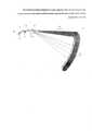
Изобретение поясняется чертежом, на котором схематично изображен большеберцовый направитель для определения центра канала при однопучковой анатомической пластике передней крестообразной связки коленного сустава.The invention is illustrated in the drawing, which schematically shows the tibial guide for determining the center of the channel with single-beam anatomical plastic of the anterior cruciate ligament of the knee joint.
Большеберцовый направитель для определения центра канала при однопучковой анатомической пластике передней крестообразной связки коленного сустава, содержит рукоятку 1 и рабочую часть 2. Рукоятка 1 выполнена из двух размещенных под углом не более 90° составляющих 3 и 4. Одна из составляющих 3 снабжена градусной угловой шкалой с центром 5 в точке перехода конца другой составляющей 4 рукоятки 1 под тупым углом в рабочую часть 2 направителя. На конце рабочей части 2 направителя размещен заостренный выступающий зуб 6 и на расстоянии не более 13 мм от него на рабочей части 2 направителя расположен дополнительный заостренный выступающий зуб 7. Высота острия зуба 6 на конце рабочей части 2 направителя вдвое превышает высоту острия дополнительного зуба 7.The tibial guide for determining the center of the channel with a single-beam anatomical plastic of the anterior cruciate ligament of the knee joint contains a handle 1 and a working
Большеберцовый направитель для определения центра канала при однопучковой анатомической пластике передней крестообразной связки коленного сустава используют следующим образом.The tibial guide to determine the center of the channel with a single-beam anatomical plastic of the anterior cruciate ligament of the knee joint is used as follows.
Пациента укладывают в положение на спине, голень пациента свободно свисает с ортопедического стола, при этом коленный сустав находится в положении сгибания под углом 90°. После выполнения спинномозговой анестезии или эндотрахеального наркоза на верхнюю треть бедра оперируемой конечности накладывают пневматическую манжету для предотвращения возможной кровопотери. Осуществляют обработку операционного поля растворами антисептиков. Подготавливают трансплантат. Выполняют передне-наружный и передне-внутренний доступы. Во время проведения операции выполняют стандартное артроскопическое пособие: резекцию мениска, удаление свободных тел и другие манипуляции при необходимости. Далее выполняют каналы в большеберцовой и бедренной костях. Для выполнения канала в большеберцовой кости сначала определяют анатомический центр зоны прикрепления передней крестообразной связки к большеберцовой кости, соответствующий центру канала в большеберцовой кости. Для этого в одном из доступов, например, в передне-наружном, размещают заявляемый большеберцовый направитель, удерживая его за рукоятку, выполненную из двух размещенных под углом не более 90° составляющих 3 и 4, одна из которых 3 снабжена градусной угловой шкалой с центром 5 в точке перехода конца другой составляющей 4 рукоятки 1 под тупым углом в рабочую часть 2 направителя. В другом передне-внутреннем доступе размещают артроскоп. С помощью артроскопа визуализируют внутренний бугорок межмыщелкового возвышения и фиксируют направитель, устанавливая заостренный выступающий зуб 6, размещенный на конце рабочей части 2 направителя, на заднем крае межмыщелкового возвышения большеберцовой кости. Другой дополнительный заостренный выступающий зуб 7, выполненный на рабочей части 2 направителя на расстоянии не более 13 мм от острия зуба 6 на конце рабочей части 2 направителя, определит на плато большеберцовой кости центр зоны прикрепления передней крестообразной связки к большеберцовой кости. За счет разницы высот острия зуба 6 на конце рабочей части 2 и дополнительного зуба 7, высота острия которого вдвое ниже высоты острия зуба 6 на конце рабочей части 2, острие дополнительного зуба 7 четко определяет центр канала в большеберцовой кости, соответствующий анатомическому центру зоны прикрепления передней крестообразной связки к большеберцовой кости. Через полученный центр канала в большеберцовой кости формируют канал в этой кости. После выполнения каналов в большеберцовой и бедренной костях последовательно размещают трансплантат в этих каналах и фиксируют его к стенкам каналов после натяжения соединяющей их между собой связки. Трансплантат фиксируют, движения в суставе без ограничений и препятствий. Рану послойно зашивают с учетом всех правил асептики и антисептики. Накладывают асептическую повязку. Нижнюю конечность иммобилизируют брейсом для коленного сустава. Начинают ранние пассивные движения в коленном суставе.The patient is placed in a supine position, the patient’s lower leg freely hanging from the orthopedic table, while the knee joint is in the flexion position at an angle of 90 °. After performing spinal anesthesia or endotracheal anesthesia, an air cuff is applied to the upper third of the thigh of the operated limb to prevent possible blood loss. The surgical field is treated with antiseptic solutions. Prepare the graft. Perform front-outer and front-inner accesses. During the operation, a standard arthroscopic aid is performed: meniscus resection, removal of free bodies and other manipulations, if necessary. Then, channels are made in the tibia and femur. To perform the canal in the tibia, first determine the anatomical center of the area of attachment of the anterior cruciate ligament to the tibia, corresponding to the center of the canal in the tibia. To do this, in one of the accesses, for example, in the front-outer, place the claimed tibial guide, holding it by a handle made of two
Клинический примерClinical example
Пациент К. 25 лет обратился за помощью в травматолого-ортопедическое отделение ГУЗ №12 в плановом порядке для консультации по поводу нарушения функции правого коленного сустава. Жалобы на болезненность при движении, нестабильность, ограничение функции правого коленного сустава.A 25-year-old patient K. asked for help at the traumatology and orthopedic department of the State Healthcare Institution No. 12 in a planned manner for consultation about a dysfunction of the right knee joint. Complaints of pain during movement, instability, limitation of the function of the right knee joint.
Из анамнеза заболевания известно, что пациент К. получил травму в результате падения при игре в волейбол около 2 лет назад, механизм травмы ротационно-сгибательный. После травмы пациент обратился за помощью в травматологический пункт, где ему была выполнена рентгенография правого коленного сустава в 2-х проекциях, на которых костно-травматической патологии не выявлено. Кроме того, была выполнена пункция коленного сустава, аспирировано около 70 мл геморрагического содержимого без жировых включений, после чего была наложена иммобилизация задней гипсовой лонгетой на срок 3 недели. После снятия гипсовой иммобилизации пациент прошел курс реабилитации, включающей лечебную физкультуру, в результате чего объем движений в коленном суставе полностью был восстановлен. Спустя некоторое время при возобновлении тренировок появилась болезненность в правом коленном суставе при физических нагрузках, невозможность выполнения привычной нагрузки, возникли эпизоды нестабильности в правом коленном суставе, сопровождающиеся резкой болью и отеком сустава. В результате выполненной магнитно-резонансной томографии правого коленного сустава было выявлено повреждение наружного мениска и передней крестообразной связки правого коленного сустава. При физикальном исследовании правый коленный сустав умеренно отечный, при пальпации определялась локальная болезненность в проекции наружного мениска, положительные симптомы Чаклина, Перельмана, Штейнмана-Бухарда, симптом «переднего выдвижного ящика» положительный, лахман-тест +++, баллотации надколенника нет, осевая нагрузка незначительно болезнена, амплитуда движений в правом коленном суставе 0-0-120 градусов. Объективное исследование на артрометре КТ-1000 показало переднее смещение голени - 8 мм (при максимально допустимой границе до 4 мм).From the medical history of the disease, it is known that patient K. was injured as a result of a fall when playing volleyball about 2 years ago, the mechanism of injury is rotational-flexion. After the injury, the patient turned to the trauma center for help, where he underwent X-ray of the right knee joint in 2 projections, on which no bone-traumatic pathology was revealed. In addition, a puncture of the knee joint was performed, about 70 ml of hemorrhagic contents without fatty inclusions were aspirated, after which the immobilization of the posterior plaster cast was imposed for a period of 3 weeks. After removing the gypsum immobilization, the patient underwent a rehabilitation course, including physiotherapy exercises, as a result of which the range of movements in the knee joint was completely restored. After some time, with the resumption of training, pain appeared in the right knee joint during physical exertion, the inability to perform the usual load, there were episodes of instability in the right knee joint, accompanied by severe pain and swelling of the joint. As a result of magnetic resonance imaging of the right knee joint, damage to the external meniscus and the anterior cruciate ligament of the right knee joint was revealed. During a physical examination, the right knee joint is moderately edematous, palpation determined local pain in the projection of the external meniscus, positive symptoms of Chaklin, Perelman, Steinman-Buchard, the symptom of the “front drawer” is positive, Lahman test +++, no patellar ballot, axial load slightly painful, the range of motion in the right knee joint is 0-0-120 degrees. An objective study on a KT-1000 arthrometer showed an anterior displacement of the lower leg of 8 mm (with a maximum permissible border of up to 4 mm).
На основании жалоб пациента, анамнеза, клинической картины, данных рентгенографии, заключения магнитно-резонансной томографии, артрометрии коленного сустава был поставлен диагноз: застарелое повреждение наружного мениска и передней крестообразной связки правого коленного сустава, хроническая передняя нестабильность правого коленного сустава.Based on the patient's complaints, medical history, clinical picture, X-ray data, magnetic resonance imaging, knee arthrometry, the diagnosis was made: chronic damage to the external meniscus and anterior cruciate ligament of the right knee joint, chronic anterior instability of the right knee joint.
Пациенту было предложено оперативное лечение. После получения согласия пациент был госпитализирован в травматолого-ортопедическое отделение в плановом порядке для оперативного лечения.The patient was offered surgical treatment. After obtaining consent, the patient was hospitalized in the traumatology and orthopedic department in a planned manner for surgical treatment.
Пациенту была выполнена частичная резекция наружного мениска правого коленного сустава и анатомическая однопучковая пластика передней крестообразной связки аутотрансплантатом из полусухожильной мышцы бедра. Для пластики передней крестообразной связки коленного сустава пациента уложили в положение на спине, голень пациента свободно свисала с ортопедического стола, при этом коленный сустав находился в положении сгибания под углом 90°. После выполнения спинномозговой анестезии 0,5% раствором Наропина в дозе 3,5 мл на верхнюю треть бедра оперируемой конечности была наложена пневматическая манжета для предотвращения возможной кровопотери. Провели обработку операционного поля раствором антисептика Кутасепт Ф (пропанол-2 (спирт) - 63,0%, бензалконий хлорид (ЧАС) - 0,025%). Подготовили трансплантат. Для этого осуществили забор аутотрансплантата, который содержал сухожилие полусухожильной мышцы бедра длиной 24 см. Каждый сегмент сухожилия складывали, формируя четырехпучковый трансплантат. Выполнили передне-наружный и передне-внутренний доступы. Во время проведения операции выполнили стандартное артроскопическое пособие: ревизию коленного сустава, резекцию мениска, оценку морфологических изменений передней крестообразной связки. В области бедренного и большеберцового прикреплений определили утолщения передней крестообразной связки, которые удалили. Выполнили экономные пластики межмыщелковой ямки бедренной кости (notch-пластика) и области прикрепления передней крестообразной связки к плато большеберцовой кости холодноплазменным аблятором Quantum 2 System.The patient underwent partial resection of the external meniscus of the right knee joint and anatomical single-beam plastic surgery of the anterior cruciate ligament with an autograft from the semitendinosus muscle of the thigh. For plastic surgery of the anterior cruciate ligament of the knee joint, the patient was placed in a supine position, the patient’s lower leg freely hanging from the orthopedic table, while the knee joint was in the flexion position at an angle of 90 °. After performing spinal anesthesia with a 0.5% solution of Naropin in a dose of 3.5 ml, an air cuff was applied to the upper third of the thigh of the operated limb to prevent possible blood loss. The surgical field was treated with a solution of the antiseptic Kutasept F (propanol-2 (alcohol) - 63.0%, benzalkonium chloride (HOUR) - 0.025%). Prepared a transplant. For this, an autograft was taken that contained a tendon of a semi-tendon of the femoral muscle 24 cm long. Each segment of the tendon was folded to form a four-beam graft. They performed anteroposterior and anteroposterior approaches. During the operation, a standard arthroscopic aid was performed: revision of the knee joint, meniscus resection, assessment of morphological changes in the anterior cruciate ligament. In the area of the femoral and tibial attachments, thickenings of the anterior cruciate ligament were determined, which were removed. Sparing plastics of the intercondylar fossa of the femur (notch plastic) and the areas of attachment of the anterior cruciate ligament to the tibial plateau with the
Далее выполнили каналы в большеберцовой и бедренной костях. Для выполнения канала в большеберцовой кости сначала определили анатомический центр зоны прикрепления передней крестообразной связки к большеберцовой кости, соответствующий центру канала в большеберцовой кости. Для этого в одном из доступов - передне-наружном разместили заявляемый большеберцовый направитель, удерживая его за рукоятку 1, выполненную из двух размещенных под углом не более 90° составляющих 3 и 4. Одна из составляющих 3 снабжена градусной угловой шкалой с центром 5 в точке перехода конца другой составляющей 4 рукоятки 1 под тупым углом в рабочую часть 2 направителя. В другом передне-внутреннем доступе разместили артроскоп. С помощью артроскопа визуализировали внутренний бугорок межмыщелкового возвышения и зафиксировали направитель. Для этого установили заостренный выступающий зуб 6, размещенный на конце рабочей части 2 направителя, на заднем крае межмыщелкового возвышения большеберцовой кости. Другой дополнительный заостренный выступающий зуб 7, выполненный на рабочей части 2 направителя на расстоянии не более 13 мм от острия зуба 6 на конце рабочей части 2 направителя, определил на плато большеберцовой кости центр зоны прикрепления передней крестообразной связки к большеберцовой кости. За счет разницы высот острия зуба 6 на конце рабочей части 2 и дополнительного зуба 7, высота острия которого вдвое ниже высоты острия зуба 6 на конце рабочей части 2, острие дополнительного зуба 7 четко опредило центр канала в большеберцовой кости, соответствующий анатомическому центру зоны прикрепления передней крестообразной связки к большеберцовой кости. Через полученный центр канала в большеберцовой кости сформировали канал в этой кости. После выполнения каналов в большеберцовой и бедренной костях последовательно разместили трансплантат в этих каналах и зафиксировали его к стенкам каналов после натяжения соединяющей их между собой связки. Трансплантат был зафиксирован, движения в суставе без ограничений и препятствий. Рану послойно зашили с учетом всех правил асептики и антисептики. Наложили асептическую повязку. Осуществили иммобилизацию брейсом для коленного сустава, начали ранние пассивные движения в левом коленном суставе, по заживлению раны и снятии швов на 10-е сутки пациента выписали из клиники на амбулаторное лечение. При наблюдении пациента через 3, 6 и 12 месяцев состояние стабильное, все движения в суставе сохранены.Then the canals were performed in the tibia and femur. To perform a canal in the tibia, the anatomical center of the area of attachment of the anterior cruciate ligament to the tibia was first determined, corresponding to the center of the canal in the tibia. To do this, in one of the accesses - the front-outer placed the claimed tibial guide, holding it by the handle 1, made of two
Claims (1)
Translated fromRussianPriority Applications (1)
| Application Number | Priority Date | Filing Date | Title |
|---|---|---|---|
| RU2016151548ARU2633333C1 (en) | 2016-12-26 | 2016-12-26 | Tibial guide for determination of channel center during single-beam anatomic plasty of anterior cross-shaped ligament of knee joint |
Applications Claiming Priority (1)
| Application Number | Priority Date | Filing Date | Title |
|---|---|---|---|
| RU2016151548ARU2633333C1 (en) | 2016-12-26 | 2016-12-26 | Tibial guide for determination of channel center during single-beam anatomic plasty of anterior cross-shaped ligament of knee joint |
Publications (1)
| Publication Number | Publication Date |
|---|---|
| RU2633333C1true RU2633333C1 (en) | 2017-10-11 |
Family
ID=60129317
Family Applications (1)
| Application Number | Title | Priority Date | Filing Date |
|---|---|---|---|
| RU2016151548ARU2633333C1 (en) | 2016-12-26 | 2016-12-26 | Tibial guide for determination of channel center during single-beam anatomic plasty of anterior cross-shaped ligament of knee joint |
Country Status (1)
| Country | Link |
|---|---|
| RU (1) | RU2633333C1 (en) |
Cited By (1)
| Publication number | Priority date | Publication date | Assignee | Title |
|---|---|---|---|---|
| RU2844266C1 (en)* | 2024-12-03 | 2025-07-28 | Федеральное государственное бюджетное военное образовательное учреждение высшего образования "Военно-медицинская академия имени С.М. Кирова" Министерства обороны Российской Федерации (ВМедА) | Device for precision drilling of bone tunnel in femur |
Citations (4)
| Publication number | Priority date | Publication date | Assignee | Title |
|---|---|---|---|---|
| EP0162027A2 (en)* | 1984-03-14 | 1985-11-21 | Magnus Odensten | A drill guiding and aligning device and a drill rod and a milling device to be used in connection therewith |
| SU1219057A1 (en)* | 1984-10-10 | 1986-03-23 | Научно-производственное объединение "Мединструмент" | Apparatus for osteotomy of bone articular end |
| RU2125844C1 (en)* | 1998-06-04 | 1999-02-10 | Малыгина Марина Александровна | Guide for formation of canals for transplants of knee joint cruciate ligaments |
| RU2583369C2 (en)* | 2009-05-29 | 2016-05-10 | Смит Энд Нефью, Инк. | Methods and devices for knee arthroplasty |
- 2016
- 2016-12-26RURU2016151548Apatent/RU2633333C1/ennot_activeIP Right Cessation
Patent Citations (4)
| Publication number | Priority date | Publication date | Assignee | Title |
|---|---|---|---|---|
| EP0162027A2 (en)* | 1984-03-14 | 1985-11-21 | Magnus Odensten | A drill guiding and aligning device and a drill rod and a milling device to be used in connection therewith |
| SU1219057A1 (en)* | 1984-10-10 | 1986-03-23 | Научно-производственное объединение "Мединструмент" | Apparatus for osteotomy of bone articular end |
| RU2125844C1 (en)* | 1998-06-04 | 1999-02-10 | Малыгина Марина Александровна | Guide for formation of canals for transplants of knee joint cruciate ligaments |
| RU2583369C2 (en)* | 2009-05-29 | 2016-05-10 | Смит Энд Нефью, Инк. | Methods and devices for knee arthroplasty |
Cited By (1)
| Publication number | Priority date | Publication date | Assignee | Title |
|---|---|---|---|---|
| RU2844266C1 (en)* | 2024-12-03 | 2025-07-28 | Федеральное государственное бюджетное военное образовательное учреждение высшего образования "Военно-медицинская академия имени С.М. Кирова" Министерства обороны Российской Федерации (ВМедА) | Device for precision drilling of bone tunnel in femur |
Similar Documents
| Publication | Publication Date | Title |
|---|---|---|
| Pederzini et al. | Elbow arthroscopy in stiff elbow | |
| Jerosch et al. | Arthroscopic treatment of patients with moderate arthrofibrosis after total knee replacement | |
| Steadman et al. | Microfracture: surgical technique and rehabilitation to treat chondral defects. | |
| Amendola | Unicompartmental osteoarthritis in the active patient: the role of high tibial osteotomy | |
| Pisani et al. | Sinus tarsi syndrome and subtalar joint instability | |
| Zhang et al. | Superior alignment but no difference in clinical outcome after minimally invasive computer-assisted unicompartmental knee arthroplasty (MICA-UKA) | |
| Guido et al. | Treatment of patella baja by a modified Z-plasty | |
| Civinini et al. | Total knee arthroplasty after complex tibial plateau fractures | |
| Bonadio et al. | Combined reconstruction of the posterior cruciate ligament and medial collateral ligament using a single femoral tunnel | |
| Sha et al. | A novel patient-specific navigational template for anatomical reconstruction of the lateral ankle ligaments | |
| Dabboussi et al. | Minimally invasive total knee arthroplasty: a comparative study to the standard approach | |
| Barth et al. | Surgical treatment of iatrogenic patella baja | |
| Parisien et al. | Diagnostic and Operative Arthroscopy of the Ankle: An Experimental Approach. | |
| Ren et al. | Clinical and radiologic outcomes after a modified bone plug technique with anatomical meniscal root reinsertion for meniscal allograft transplantation and a minimum 18-month follow-up | |
| W-Dahl et al. | Introducing prospective national registration of knee osteotomies. A report from the first year in Sweden | |
| RU2701776C2 (en) | Method of simultaneous plasty of anterior cruciate and lateral anterolateral ligament of knee joint in patients with combined instability of knee joint | |
| Chou et al. | Clinical comparison of valgus and varus deformities in primary total knee arthroplasty following midvastus approach | |
| RU2432135C1 (en) | Method of plastic surgery on anterior cruciate ligament of knee joint and related graft for implementation thereof | |
| Kim et al. | Short-term results of primary total knee arthroplasties performed with a mini-incision or a standard incision | |
| Sim et al. | Clinical and radiologic evaluation of medial epicondylar osteotomy for varus total knee arthroplasty | |
| RU2633333C1 (en) | Tibial guide for determination of channel center during single-beam anatomic plasty of anterior cross-shaped ligament of knee joint | |
| Morattel et al. | Unusual apical femoral head deformity treated by hip arthroscopy and tunnel drilling through femoral head: a case report | |
| RU2633277C2 (en) | Method for anatomic single-beam plasty of anterior cross-shaped ligament by patellar ligament transplant | |
| Noh et al. | One-year serial follow-up magnetic resonance imaging study of rigidfix for femoral fixation in anterior cruciate ligament reconstruction | |
| RU2611958C2 (en) | Method for determination of channel center during tibia anatomical single-beam plastic of anterior cruciate knee ligament |
Legal Events
| Date | Code | Title | Description |
|---|---|---|---|
| MM4A | The patent is invalid due to non-payment of fees | Effective date:20181227 |