RU2602221C1 - Method of treating keratoconus and tool for its implementation - Google Patents
Method of treating keratoconus and tool for its implementationDownload PDFInfo
- Publication number
- RU2602221C1 RU2602221C1RU2015123553/14ARU2015123553ARU2602221C1RU 2602221 C1RU2602221 C1RU 2602221C1RU 2015123553/14 ARU2015123553/14 ARU 2015123553/14ARU 2015123553 ARU2015123553 ARU 2015123553ARU 2602221 C1RU2602221 C1RU 2602221C1
- Authority
- RU
- Russia
- Prior art keywords
- cornea
- tool
- keratoconus
- excimer laser
- epithelial layer
- Prior art date
Links
- 201000002287KeratoconusDiseases0.000titleclaimsabstractdescription30
- 238000000034methodMethods0.000titleclaimsdescription20
- 210000004087corneaAnatomy0.000claimsabstractdescription48
- 210000005081epithelial layerAnatomy0.000claimsabstractdescription22
- 238000000608laser ablationMethods0.000claimsabstractdescription18
- 210000005252bulbus oculiAnatomy0.000claimsabstractdescription6
- 238000009434installationMethods0.000claimsabstractdescription6
- AUNGANRZJHBGPY-SCRDCRAPSA-NRiboflavinChemical compoundOC[C@@H](O)[C@@H](O)[C@@H](O)CN1C=2C=C(C)C(C)=CC=2N=C2C1=NC(=O)NC2=OAUNGANRZJHBGPY-SCRDCRAPSA-N0.000claimsdescription26
- AUNGANRZJHBGPY-UHFFFAOYSA-ND-LyxoflavinNatural productsOCC(O)C(O)C(O)CN1C=2C=C(C)C(C)=CC=2N=C2C1=NC(=O)NC2=OAUNGANRZJHBGPY-UHFFFAOYSA-N0.000claimsdescription13
- 229960002477riboflavinDrugs0.000claimsdescription13
- 235000019192riboflavinNutrition0.000claimsdescription13
- 239000002151riboflavinSubstances0.000claimsdescription13
- 210000004045bowman membraneAnatomy0.000claimsdescription5
- 206010002091AnaesthesiaDiseases0.000abstractdescription3
- 230000037005anaesthesiaEffects0.000abstractdescription3
- 239000003814drugSubstances0.000abstractdescription3
- 230000000694effectsEffects0.000abstractdescription3
- 230000004807localizationEffects0.000abstractdescription3
- 238000005259measurementMethods0.000abstractdescription3
- 239000000126substanceSubstances0.000abstract1
- 210000003560epithelium cornealAnatomy0.000description3
- 238000012544monitoring processMethods0.000description3
- 230000003287optical effectEffects0.000description3
- 238000009738saturatingMethods0.000description3
- 238000001356surgical procedureMethods0.000description3
- 238000013459approachMethods0.000description2
- 210000003683corneal stromaAnatomy0.000description2
- 238000006073displacement reactionMethods0.000description2
- 210000002889endothelial cellAnatomy0.000description2
- 210000000981epitheliumAnatomy0.000description2
- 210000001508eyeAnatomy0.000description2
- 238000010186stainingMethods0.000description2
- 230000001954sterilising effectEffects0.000description2
- 238000004659sterilization and disinfectionMethods0.000description2
- 102000008186CollagenHuman genes0.000description1
- 108010035532CollagenProteins0.000description1
- ZVKAMDSUUSMZES-NZQWGLPYSA-NOS IINatural productsCC(=O)N[C@H]1[C@H](OC[C@@H](O)[C@@H](O)[C@@H](O)CO)O[C@H](CO)[C@H](O[C@H]2O[C@H](CO)[C@@H](O)[C@H](O)[C@H]2O)[C@@H]1O[C@@H]3O[C@H](CO)[C@@H](O)[C@H](O)[C@H]3OZVKAMDSUUSMZES-NZQWGLPYSA-N0.000description1
- 238000002679ablationMethods0.000description1
- 238000003759clinical diagnosisMethods0.000description1
- 229920001436collagenPolymers0.000description1
- 210000000399corneal endothelial cellAnatomy0.000description1
- 238000012937correctionMethods0.000description1
- 238000004132cross linkingMethods0.000description1
- 201000000255cycloplegiaDiseases0.000description1
- 230000004452decreased visionEffects0.000description1
- 230000007257malfunctionEffects0.000description1
- 230000002980postoperative effectEffects0.000description1
- 230000000750progressive effectEffects0.000description1
- 229920006395saturated elastomerPolymers0.000description1
- NLVFBUXFDBBNBW-PBSUHMDJSA-NtobramycinChemical compoundN[C@@H]1C[C@H](O)[C@@H](CN)O[C@@H]1O[C@H]1[C@H](O)[C@@H](O[C@@H]2[C@@H]([C@@H](N)[C@H](O)[C@@H](CO)O2)O)[C@H](N)C[C@@H]1NNLVFBUXFDBBNBW-PBSUHMDJSA-N0.000description1
- 229940035275tobrexDrugs0.000description1
- 230000000472traumatic effectEffects0.000description1
Images
Classifications
- A—HUMAN NECESSITIES
- A61—MEDICAL OR VETERINARY SCIENCE; HYGIENE
- A61F—FILTERS IMPLANTABLE INTO BLOOD VESSELS; PROSTHESES; DEVICES PROVIDING PATENCY TO, OR PREVENTING COLLAPSING OF, TUBULAR STRUCTURES OF THE BODY, e.g. STENTS; ORTHOPAEDIC, NURSING OR CONTRACEPTIVE DEVICES; FOMENTATION; TREATMENT OR PROTECTION OF EYES OR EARS; BANDAGES, DRESSINGS OR ABSORBENT PADS; FIRST-AID KITS
- A61F9/00—Methods or devices for treatment of the eyes; Devices for putting in contact-lenses; Devices to correct squinting; Apparatus to guide the blind; Protective devices for the eyes, carried on the body or in the hand
- A61F9/007—Methods or devices for eye surgery
- A61F9/008—Methods or devices for eye surgery using laser
Landscapes
- Health & Medical Sciences (AREA)
- Ophthalmology & Optometry (AREA)
- Heart & Thoracic Surgery (AREA)
- Vascular Medicine (AREA)
- Veterinary Medicine (AREA)
- Surgery (AREA)
- Engineering & Computer Science (AREA)
- Biomedical Technology (AREA)
- Public Health (AREA)
- Nuclear Medicine, Radiotherapy & Molecular Imaging (AREA)
- Life Sciences & Earth Sciences (AREA)
- Animal Behavior & Ethology (AREA)
- General Health & Medical Sciences (AREA)
- Physics & Mathematics (AREA)
- Optics & Photonics (AREA)
- Radiation-Therapy Devices (AREA)
Abstract
Description
Translated fromRussianИзобретение относится к медицине, а именно к офтальмологии, и может быть использовано для лечения кератоконуса.The invention relates to medicine, namely to ophthalmology, and can be used to treat keratoconus.
Известен способ лечения кератоконуса и инструмент для точечной деэпителизации роговицы (см. описание изобретения к патенту РФ №2531471 на изобретение «Инструмент для дозирования скарификации эпителия роговицы» авторов Малюгина Б.Э., Измайлова С.Б., Латыпова И.А., Мерзлова Д.Е. от 04.07.2013), заключающийся в удалении эпителиального слоя путем скарификации, для чего на поверхность глазного яблока перилимбально накладывают кольцо-держатель с направляющими полозьями, на которое устанавливают инструмент для точечной деэпителизации путем проведения скарификации, центрируя относительно вершины кератоконуса, и воздействии на роговицу путем насыщения ее многократными инсталляциями 0,1% раствором рибофлавина с последующим ультрафиолетовым облучением. При осуществлении этого способа используют инструмент для точечной деэпителизации роговицы, на рабочей поверхности которого выполнены шипы длиной 50-80 мкм в количестве 15-20 на см2. Причем возможно использовать 3 варианта инструмента, у одного из которых шипы размещены по всей рабочей поверхности, у другого - с участком без шипов в форме полукольца, у третьего - с участком без шипов в форме кольца (для применения при наличии одного или двух интрастромальных сегментов).There is a known method of treating keratoconus and a tool for punctually corneal de-epithelization (see the description of the invention to RF patent No. 2531471 for the invention “Instrument for dosing the scarification of corneal epithelium” by B. Malyugin, SB Izmailov, IA Latypova, Merzlova D.E. from 04.07.2013), which consists in the removal of the epithelial layer by scarification, for which a ring-holder with guiding runners is perilimbally imposed on the surface of the eyeball, onto which a tool for point de-epithelization is installed by scarification centering relative to the top of keratoconus, and effects on the cornea by saturating its multiple installations riboflavin 0.1% solution, followed by ultraviolet irradiation. In the implementation of this method, a tool is used for the targeted de-epithelization of the cornea, on the working surface of which spikes are made with a length of 50-80 microns in an amount of 15-20 per cm2 . Moreover, it is possible to use 3 versions of the tool, one of which has spikes placed over the entire working surface, the other with a section without spikes in the shape of a half-ring, and the third with a section without spikes in the shape of a ring (for use with one or two intrastromal segments) .
Недостатком известного способа и инструмента, используемого при выполнении известного способа, является недостаточная эффективность способа за счет отсутствия индивидуального подхода к лечению каждого пациента. Это в первую очередь определяет наличие шипов одинаковой длины, расположенных по внутренней рабочей поверхности инструмента, применяемого в этом известном способе, и использование только 3-х вариантов исполнения рабочей поверхности этого известного инструмента для точечной деэпителизации роговицы. Этот способ не учитывает или учитывает не в полной мере индивидуальные особенности роговицы пациента с кератоконусом, вследствие чего может быть получен недостаточный или избыточный эффект скарификации эпителия роговицы. Выполнение рабочей поверхности используемого инструмента с расположенными на ней шипами вогнутой формы, повторяющей форму роговицы, соответствует какому-то среднему значению кривизны роговицы, а не конкретного пациента, что не позволяет проводить равномерную точечную деэпителизацию роговицы данного пациента, особенно в тех случаях, когда кривизна роговицы, а также толщина эпителиального слоя у пациента в центре и на периферии значительно отличаются друг от друга. Известный инструмент не обеспечивает выполнение точной скарификации на толщину эпителия роговицы с учетом индивидуальных ее особенностей: толщины эпителиального слоя и кривизны роговицы. Кроме того, необходимость ориентирования рабочей части инструмента относительно вершины кератоконуса, что предполагает ее смещение, не позволит спрогнозировать предполагаемый результат лечения не только от величины кривизны роговицы пациента и толщины эпителиального слоя, но и от степени усилия, прикладываемого к подпружиненной кнопке инструмента. Управление выдвижением шипов инструмента в руках разных пользователей не может обеспечить идентичность воздействия рабочей части на роговицу в виду возможных вариаций пространственной ориентации при установке инструмента на поверхность роговицы и различного усилия, прилагаемого хирургом при нажатии на кнопку. В данном случае присутствует субъективный фактор, что всегда ведет к непредсказуемости результата. К тому же, инструмент как операционное оборудование требует регулярной предстерилизационной обработки и стерилизации, в результате чего подвижные элементы подобных механических устройств часто заклинивают и, как следствие, дают сбой и отказ в работе, что влечет за собой снижение качества проводимых операций. Следует также отметить более травматичное воздействие на эпителиальный слой в известном способе точечной деэпителизации роговицы по сравнению с предлагаемым нами эксимерлазерным воздействием.The disadvantage of this method and the tool used when performing the known method is the lack of effectiveness of the method due to the lack of an individual approach to the treatment of each patient. This primarily determines the presence of spikes of the same length located on the inner working surface of the instrument used in this known method, and the use of only 3 variants of the working surface of this known instrument for the targeted de-epithelization of the cornea. This method does not take into account or does not fully take into account the individual characteristics of the cornea of a patient with keratoconus, as a result of which an insufficient or excessive effect of scarification of the corneal epithelium can be obtained. The implementation of the working surface of the used tool with concave spikes located on it, repeating the shape of the cornea, corresponds to some average value of the curvature of the cornea, and not of a particular patient, which does not allow for uniform point de-epithelization of the cornea of this patient, especially in cases where the curvature of the cornea as well as the thickness of the epithelial layer in the patient in the center and on the periphery are significantly different from each other. The known tool does not provide accurate scarification for the thickness of the corneal epithelium, taking into account its individual characteristics: the thickness of the epithelial layer and the curvature of the cornea. In addition, the need to orient the working part of the instrument relative to the apex of the keratoconus, which suggests its displacement, will not allow predicting the expected treatment result not only from the curvature of the patient’s cornea and thickness of the epithelial layer, but also from the degree of effort applied to the spring-loaded button of the instrument. Controlling the extension of the tool spikes in the hands of different users cannot ensure that the impact of the working part on the cornea is identical in view of the possible variations in spatial orientation when installing the tool on the surface of the cornea and the various efforts made by the surgeon when the button is pressed. In this case, there is a subjective factor, which always leads to unpredictability of the result. In addition, the tool as an operating equipment requires regular pre-sterilization treatment and sterilization, as a result of which the movable elements of such mechanical devices often jam and, as a result, give a malfunction and failure in operation, which entails a decrease in the quality of the operations performed. It should also be noted a more traumatic effect on the epithelial layer in the known method of point de-epithelialization of the cornea in comparison with the excimer laser action we offer.
Предлагаемое изобретение решает задачу разработки нового способа лечения кератоконуса. Получаемый при этом технический результат состоит в повышении эффективности способа лечения кератоконуса прежде всего за счет индивидуализированного подхода к лечению каждого конкретного пациента, к получению предсказуемых и прогнозируемых результатов лечения. Разработанные нами способ и инструмент для точечной деэпителизации роговицы, на рабочей поверхности которого размещены отверстия, выполняющие роль диафрагмы, колибрующие лазерный луч, обеспечивают дозированную эксимерлазерную абляцию эпителиального слоя роговицы, не зависящую от индивидуальных кератометрических параметров роговицы, а также индивидуальных качеств пользователя, что обеспечивает отсутствие влияния субъективных факторов на дозированную точечную деэпителизацию роговицы. Заявляемый нами способ в сочетании с предлагаемым инструментом, рабочая часть которого не контактирует с поверхностью роговицы, и который может быть свободно отцентрирован на роговице относительно вершины кератоконуса, позволяет выполнить точечную деэпителизацию роговицы на всю ее глубину до боуменовой мембраны дозированно в соответствии с локализацией кератоконуса и толщиной эпителиального слоя роговицы программируемым, управляемым эксимерлазерным лучом за одно воздействие. Способ прост в исполнении и не представляет сложности для специалистов при его использовании. Таким образом, обеспечивается получение нового положительного результата, не обеспечиваемого ни одним из известных способов для решения той же задачи.The present invention solves the problem of developing a new method of treating keratoconus. The technical result obtained in this case consists in increasing the effectiveness of the method of treating keratoconus primarily due to the individualized approach to the treatment of each specific patient, to obtain predictable and predicted treatment results. We have developed a method and tool for the targeted de-epithelialization of the cornea, on the working surface of which there are holes that act as a diaphragm, oscillating the laser beam, provide dosed excimer laser ablation of the corneal epithelial layer, which does not depend on individual keratometric parameters of the cornea, as well as individual qualities of the user, which ensures the absence of the influence of subjective factors on the dosed point de-epithelization of the cornea. The inventive method in combination with the proposed tool, the working part of which does not come in contact with the surface of the cornea, and which can be freely centered on the cornea relative to the top of the keratoconus, allows to carry out point de-epithelization of the cornea to its entire depth to the bowman membrane in a dosage in accordance with the localization of the keratoconus and thickness a corneal epithelial layer with a programmable, controlled excimer laser beam in one exposure. The method is simple to implement and is not difficult for specialists when using it. Thus, it provides a new positive result that is not provided by any of the known methods for solving the same problem.
Указанный технический результат достигается тем, что в способе лечения кератоконуса, заключающемся в удалении эпителиального слоя путем точечной деэпителизации, для чего на поверхность глазного яблока перилимбально накладывают кольцо-держатель с направляющими полозьями, на которое устанавливают инструмент для проведения точечной деэпителизации, центрируя относительно вершины кератоконуса, и воздействии на роговицу путем насыщения ее многократными инсталляциями 0,1% раствором рибофлавина с последующим ультрафиолетовым облучением, отличающийся тем, что проведение точечной деэпителизации проводят путем эксимерлазерной абляции эпителиального слоя на всю его глубину до боуменовой мембраны через отверстия, выполненные в неконтактирующей с роговицей рабочей пластине инструмента для точечной деэпителизации, размещенного в направляющих полозьях кольца-держателя.The specified technical result is achieved by the fact that in the method of treating keratoconus, which consists in removing the epithelial layer by point de-epithelialization, for this purpose, a holder ring with guiding runners is perilimbally imposed on the surface of the eyeball, onto which a tool for carrying out point de-epithelization is mounted, centering relative to the top of the keratoconus, and exposure to the cornea by saturating it with multiple installations with a 0.1% riboflavin solution followed by ultraviolet irradiation, characterized in that the point de-epithelialization is carried out by excimer laser ablation of the epithelial layer to its entire depth to the bowman’s membrane through openings made in the working plate of the point de-epithelialization instrument non-contacting with the cornea, located in the guide rails of the holder ring.
Указанный технический результат достигается также тем, что в инструменте для точечной деэпителизации, выполненном в виде скобы с рабочей частью, рабочая часть выполнена в виде горизонтальной пластины, на которой равномерно нанесены сквозные отверстия диаметром и расстоянием между ними, соответствующими диаметру «летающего» лазерного пятна при эксимерлазерной абляции, причем отверстия выполнены в пределах площади эксимерлазерной абляции и последующего ультрафиолетового облучения, а скоба по бокам снабжена направляющими.The specified technical result is also achieved by the fact that in the tool for point de-epithelization, made in the form of a bracket with a working part, the working part is made in the form of a horizontal plate on which through holes with a diameter and distance between them, corresponding to the diameter of the “flying” laser spot, are uniformly applied at excimer laser ablation, and the holes are made within the area of excimer laser ablation and subsequent ultraviolet irradiation, and the bracket on the sides is provided with guides.
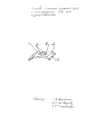
Изобретение поясняется чертежом, на котором схематично в аксонометрической проекции изображен предлагаемый инструмент.The invention is illustrated in the drawing, which schematically in axonometric projection shows the proposed tool.
Инструмент для точечной деэпителизации выполнен в виде скобы 1 с рабочей частью 2, которая представляет собой горизонтальную пластину 3. На горизонтальной пластине 3 равномерно нанесены сквозные отверстия 4, выполненные в пределах площади эксимерлазерной абляции и последующего ультрафиолетового облучения. Отверстия 4 выполнены диаметром и расстоянием между ними, соответствующими диаметру «летающего» лазерного пятна при эксимерлазерной абляции. Скоба 1 по бокам снабжена направляющими 5.The tool for point de-epithelialization is made in the form of a bracket 1 with a working part 2, which is a
Способ лечения кератоконуса с использованием предлагаемого инструмента осуществляют следующим образом.A method of treating keratoconus using the proposed tool is as follows.
Предварительно проводят полное предоперационное обследование, включающее в том числе ОКТ роговицы с измерением толщины эпителиального слоя, а также кератотопографическое обследование, при котором определяют точную локализацию вершины кератоконуса, кератометрические значения на вершине кератоконуса и в его основании. По полученным параметрам программируют объем эксимерлазерной абляции с помощью эксимерного лазера, работающего по принципу «летающего пятна», включающий диаметр общей зоны и глубину эксимерлазерного воздействия и величину смещения зоны воздействия в соответствии с кератотопографией кератоконуса. В условиях операционной под инсталляционной анестезией на поверхность глазного яблока перилимбально накладывают кольцо-держатель с направляющими полозьями, в которых размещают направляющие 5 скобы 1 инструмента, устанавливая таким образом инструмент для точечной деэпителизации до касания упоров на торце кольца-держателя, что определяет фиксацию и центровку рабочей части скобы относительно отцентрованного на роговице кольца-держателя. Проводят центрирование рабочей части 2 инструмента для точечной деэпителизации вместе с кольцом-держателем относительно вершины кератоконуса за счет перемещения в собранном виде кольца-держателя с установленным в нем инструментом. Далее по заданным параметрам проводят эксимерлазерную абляцию эпителиального слоя роговицы, используя эксимерный лазер, работающий по принципу «летающего пятна». Эксимерлазерную абляцию эпителиального слоя проводят через равномерно нанесенные на неконтактирующей с роговицей горизонтальной пластине 3 рабочей части 2 инструмента для точечной деэпителизации роговицы сквозные отверстия 4, выполненные в пределах площади эксимерлазерной абляции и последующего ультрафиолетового облучения, на всю глубину эпителиального слоя роговицы до боуменовой мембраны. По окончанию эксимерлазерного воздействия инструмент для точечной деэпителизации в собранном виде убирают. Воздействуют на роговицу путем насыщения ее многократными инстилляциями 0,1% раствором рибофлавина каждые 2-3 минуты до наступления достаточной степени насыщения роговицы рибофлавином. Биомикроскопический контроль насыщения роговицы 0,1% раствором рибофлавина проводят через 15 минут от начала данного этапа операции. О достаточном уровне насыщения роговицы раствором рибофлавина говорит диффузное желтое прокрашивание стромы роговицы. По достижению необходимого уровня насыщения роговицы раствором рибофлавина проводят ультрафиолетовое облучение роговицы длиной волны 365 нм мощностью 3 мВт/см2 в течение 30 мин, которое сопровождают дополнительными инстилляциями 0,1% раствором рибофлавина на роговицу каждые 3-4 минуты для поддержания его концентрации. Ведение раннего послеоперационного периода осуществляют так же, как и при выполнении стандартной методики кросслинкинга роговичного коллагена.A preliminary preoperative examination is preliminarily carried out, including corneal OCT with measurement of the thickness of the epithelial layer, as well as a keratotopographic examination, in which the exact location of the keratoconus vertex, keratometric values at the top of the keratoconus and at its base are determined. According to the obtained parameters, the volume of excimer laser ablation is programmed using an excimer laser operating on the basis of the “flying spot” principle, including the diameter of the common zone and the depth of excimer laser exposure and the magnitude of the displacement of the exposure zone in accordance with keratotopography of keratoconus. Under operating conditions under installation anesthesia, a holder ring with guide rails is perilimbally applied to the surface of the eyeball, in which guide 5 brackets 1 of the instrument are placed, thus installing a tool for point de-epithelization until the stops touch the end of the holder ring, which determines the fixation and centering of the working parts of the bracket relative to the holder ring centered on the cornea. The centering of the working part 2 of the tool for point de-epithelization is carried out together with the holder ring relative to the top of the keratoconus due to the movement in the assembled form of the holder ring with the tool installed in it. Then, according to the given parameters, excimer laser ablation of the corneal epithelial layer is carried out using an excimer laser operating on the basis of the “flying spot” principle. Excimer laser ablation of the epithelial layer is carried out through the
Клинический пример.Clinical example.
Пациент А., 27 лет. Жалобы на прогрессивное сниженное зрение на левом глазу (OS). Оптические средства коррекции пациент не использует. Было проведено полное офтальмологическое обследование, включающее визометрию, рефрактометрию в обычных условиях и в условиях циклоплегии, многоточечную пахиметрию, измерение диаметра роговицы, определение плотности эндотелиальных клеток роговицы, кератотопографию, осмотр глазного дна с линзой Гольдмана, ОКТ роговицы с определением толщины эпителиального слоя. Результаты обследования: VOD = 1,0, ROD = sph -0,25 Дптр cyl -0,5 Дптр ax 10, VOS = 0,1 cyl -6,0 Дптр ax 130 = 0,3, ROS = sph -1,75 Дптр cyl -9,0 Дптр ax 137, пахиметрия в центральной оптической зоне OD = 481 мкм, пахиметрия в центральной оптической зоне OS = 422 мкм. Плотность эндотелиальных клеток OD = 2902 CD/мм2, плотность эндотелиальных клеток OS = 2750 CD/мм2, диаметр роговицы OD = 10,5 мм, OS = 11,0 мм. Кератотопографически на OS картина центрального кератоконуса. Кератометрия на вершине кератоконуса 56,5 дптр. Вершина кератоконуса смещена книзу и кнутри на 0,2 мм от центра роговицы. Толщина эпителия на вершине кератоконуса 47 мкм.Patient A., 27 years old. Complaints of progressive decreased vision in the left eye (OS). The patient does not use optical correction tools. A complete ophthalmological examination was carried out, including visometry, refractometry under ordinary conditions and in conditions of cycloplegia, multipoint pachymetry, measurement of the diameter of the cornea, determination of the density of corneal endothelial cells, keratotopography, examination of the fundus with a Goldman lens, corneal OCT with determination of the thickness of the epithelium. Survey results: VOD = 1.0, ROD = sph -0.25 Dptr cyl -0.5 Dptr ax 10, VOS = 0.1 cyl -6.0 Dptr ax 130 = 0.3, ROS = sph -1, 75 Dptr cyl -9.0 Dptr ax 137, pachymetry in the central optical zone OD = 481 microns, pachymetry in the central optical zone OS = 422 microns. Endothelial cell density OD = 2902 CD / mm2 , endothelial cell density OS = 2750 CD / mm2 , corneal diameter OD = 10.5 mm, OS = 11.0 mm Keratotopographically on OS a picture of a central keratoconus. Keratometry at the top of keratoconus 56.5 diopters. The top of the keratoconus is shifted down and inward by 0.2 mm from the center of the cornea. The thickness of the epithelium at the top of the keratoconus is 47 microns.
Клинический диагноз: «Кератоконус OS II степени».Clinical diagnosis: "Keratoconus OS II degree."
Пациенту провели операцию по предлагаемому способу лечения кератоконуса. В условиях операционной под инсталляционной анестезией на поверхность глазного яблока перилимбально было наложено кольцо-держатель с направляющими полозьями, в которых разместили направляющие 5 скобы 1 инструмента, устанавливая таким образом инструмент для точечной деэпителизации до касания упоров на торце кольца-держателя, что определило фиксацию и центровку рабочей части скобы относительно отцентрованного на роговице кольца-держателя. Провели центрирование рабочей части 2 инструмента для точечной деэпителизации вместе с кольцом-держателем относительно вершины кератоконуса за счет перемещения в собранном виде кольца-держателя с установленным в нем инструментом. Далее по заданным параметрам провели эксимерлазерную абляцию эпителиального слоя роговицы, используя эксимерлазерную установку Швинд Амарис, работающую по принципу «летающего пятна». Эксимерлазерную абляцию эпителиального слоя провели через сквозные отверстия 4, равномерно нанесенные на неконтактирующей с роговицей горизонтальной пластине 3 рабочей части 2 инструмента для точечной деэпителизации роговицы, на всю глубину 47 мкм эпителиального слоя роговицы до боуменовой мембраны. Диаметр зоны абляции составил 7,0 мм, и сквозные отверстия 4 были выполнены в пределах этого диаметра зоны эксимерлазерной абляции и последующего ультрафиолетового облучения. Диаметр точки воздействия - 0,5 мм. По окончанию эксимерлазерного воздействия инструмент для точечной деэпителизации в собранном виде убрали. Инстилляционно капельно с интервалом 3 минуты в течение 25 минут проведено насыщение роговицы 0,1% раствором рибофлавина. Биомикроскопический контроль насыщения роговицы 0,1% раствором рибофлавина провели через 25 минут от начала данного этапа операции. О достаточном уровне насыщения роговицы раствором рибофлавина говорит диффузное желтое прокрашивание стромы роговицы. Пахиметрический контроль роговицы провели непосредственно перед ультрафиолетовым облучением - 422 мкм. Провели ультрафиолетовое облучение роговицы длиной волны 365 нм мощностью 3,0 мВт/см2 с расстояния 50 мм в течение 30 мин, которое сопровождали дополнительными инстилляциями 0,1% раствором рибофлавина на роговицу каждые 4 минуты для поддержания его концентрации. После операции пациенту в оперированный глаз был назначен Тобрекс по 1 капле 6 раз в день и Корнерегель - 3-4 раза в день. Полная эпителизация была достигнута через 42 часа после операции. На 10-й день после операции VOS = 0,3 с cyl -6,0 ах 130 = 0,6-0,8.The patient underwent surgery according to the proposed method for the treatment of keratoconus. Under operating conditions under installation anesthesia, a holder ring with guide rails was perilimbally applied to the surface of the eyeball, in which guide 5 brackets of 1 instrument were placed, thus installing a tool for point de-epithelization until the stops touch the end of the holder ring, which determined fixation and centering the working part of the bracket relative to the holder ring centered on the cornea. The centering of the working part 2 of the tool for point de-epithelization was carried out together with the holder ring relative to the top of the keratoconus due to the movement in the assembled form of the holder ring with the tool installed in it. Then, according to the set parameters, excimer laser ablation of the corneal epithelial layer was performed using the Shvind Amaris excimer laser device operating on the basis of the “flying spot” principle. Excimer laser ablation of the epithelial layer was carried out through the through
Claims (2)
Translated fromRussianPriority Applications (1)
| Application Number | Priority Date | Filing Date | Title |
|---|---|---|---|
| RU2015123553/14ARU2602221C1 (en) | 2015-06-18 | 2015-06-18 | Method of treating keratoconus and tool for its implementation |
Applications Claiming Priority (1)
| Application Number | Priority Date | Filing Date | Title |
|---|---|---|---|
| RU2015123553/14ARU2602221C1 (en) | 2015-06-18 | 2015-06-18 | Method of treating keratoconus and tool for its implementation |
Publications (1)
| Publication Number | Publication Date |
|---|---|
| RU2602221C1true RU2602221C1 (en) | 2016-11-10 |
Family
ID=57278006
Family Applications (1)
| Application Number | Title | Priority Date | Filing Date |
|---|---|---|---|
| RU2015123553/14ARU2602221C1 (en) | 2015-06-18 | 2015-06-18 | Method of treating keratoconus and tool for its implementation |
Country Status (1)
| Country | Link |
|---|---|
| RU (1) | RU2602221C1 (en) |
Cited By (2)
| Publication number | Priority date | Publication date | Assignee | Title |
|---|---|---|---|---|
| RU2684472C1 (en)* | 2018-02-01 | 2019-04-09 | Федеральное государственное автономное учреждение "Межотраслевой научно-технический комплекс "Микрохирургия глаза" имени академика С.Н. Федорова" Министерства здравоохранения Российской Федерации | Method of treating keratoconus |
| RU2760482C1 (en)* | 2021-02-19 | 2021-11-25 | федеральное государственное автономное учреждение "Национальный медицинский исследовательский центр "Межотраслевой научно-технический комплекс "Микрохирургия глаза" имени академика С.Н. Федорова" Министерства здравоохранения Российской Федерации | Method for treatment of progressive keratoconus |
Citations (2)
| Publication number | Priority date | Publication date | Assignee | Title |
|---|---|---|---|---|
| WO1991008711A1 (en)* | 1989-12-14 | 1991-06-27 | Corneal Contouring, Inc. | Method and apparatus for re-profiling the cornea |
| RU2531471C1 (en)* | 2013-07-04 | 2014-10-20 | федеральное государственное бюджетное учреждение "Межотраслевой научно-технический комплекс "Микрохирургия глаза" имени академика С.Н. Федорова" Министерства здравоохранения Российской Федерации | Instrument for graduated scarification of corneal epithelium |
- 2015
- 2015-06-18RURU2015123553/14Apatent/RU2602221C1/enactive
Patent Citations (2)
| Publication number | Priority date | Publication date | Assignee | Title |
|---|---|---|---|---|
| WO1991008711A1 (en)* | 1989-12-14 | 1991-06-27 | Corneal Contouring, Inc. | Method and apparatus for re-profiling the cornea |
| RU2531471C1 (en)* | 2013-07-04 | 2014-10-20 | федеральное государственное бюджетное учреждение "Межотраслевой научно-технический комплекс "Микрохирургия глаза" имени академика С.Н. Федорова" Министерства здравоохранения Российской Федерации | Instrument for graduated scarification of corneal epithelium |
Non-Patent Citations (1)
| Title |
|---|
| СОЛОДКОВА Е.Г. и др., Сравнительный анализ способов лечения кератоконуса, Актуальные проблемы офтальмологии, 2011, с. 197. Roibeard O'hEineachain. CROss-linkinG. epithelium-conserving collagen cross-linking technique. better for patients' recovery. EUROTIMES, Volume 17, Issue 9, p.39.* |
Cited By (2)
| Publication number | Priority date | Publication date | Assignee | Title |
|---|---|---|---|---|
| RU2684472C1 (en)* | 2018-02-01 | 2019-04-09 | Федеральное государственное автономное учреждение "Межотраслевой научно-технический комплекс "Микрохирургия глаза" имени академика С.Н. Федорова" Министерства здравоохранения Российской Федерации | Method of treating keratoconus |
| RU2760482C1 (en)* | 2021-02-19 | 2021-11-25 | федеральное государственное автономное учреждение "Национальный медицинский исследовательский центр "Межотраслевой научно-технический комплекс "Микрохирургия глаза" имени академика С.Н. Федорова" Министерства здравоохранения Российской Федерации | Method for treatment of progressive keratoconus |
Similar Documents
| Publication | Publication Date | Title |
|---|---|---|
| Kymionis et al. | Femtosecond laser technology in corneal refractive surgery: a review | |
| Ratkay-Traub et al. | First clinical results with the femtosecond neodynium-glass laser in refractive surgery | |
| Gyldenkerne et al. | Comparison of corneal shape changes and aberrations induced by FS-LASIK and SMILE for myopia | |
| Nordan et al. | Femtosecond laser flap creation for laser in situ keratomileusis: six-month follow-up of initial US clinical series | |
| Vestergaard | Past and present of corneal refractive surgery: A retrospective study of long‐term results after photorefractive keratectomy and a prospective study of refractive lenticule extraction | |
| Vestergaard et al. | Femtosecond (FS) laser vision correction procedure for moderate to high myopia: a prospective study of ReLEx® flex and comparison with a retrospective study of FS‐laser in situ keratomileusis | |
| CN107095735B (en) | System and method for correcting astigmatism by utilizing multiple pairs of arc laser cornea incisions | |
| Ji et al. | Lower laser energy levels lead to better visual recovery after small-incision lenticule extraction: prospective randomized clinical trial | |
| Stahl et al. | Anterior segment OCT analysis of thin IntraLase femtosecond flaps | |
| Kampik et al. | Influence of corneal collagen crosslinking with riboflavin and ultraviolet-a irradiation on excimer laser surgery | |
| Rocha et al. | Epithelial and stromal remodeling after corneal collagen cross-linking evaluated by spectral-domain OCT | |
| CN105682620B (en) | Cross-linking control | |
| Kovács et al. | The effect of femtosecond laser capsulotomy on the development of posterior capsule opacification | |
| Vryghem et al. | Efficacy, safety, and flap dimensions of a new femtosecond laser for laser in situ keratomileusis | |
| Romani et al. | Pachymetry-based accelerated crosslinking: the “M Nomogram” for standardized treatment of all-thickness progressive ectatic corneas | |
| RU2434616C1 (en) | Method of treating keratoconus | |
| RU2466699C1 (en) | Method of treating corneal keratoconus | |
| RU2602221C1 (en) | Method of treating keratoconus and tool for its implementation | |
| Tamayo | Predictable visual outcomes with accelerated corneal cross-linking concurrent with laser in situ keratomileusis | |
| RU2684472C1 (en) | Method of treating keratoconus | |
| RU2531471C1 (en) | Instrument for graduated scarification of corneal epithelium | |
| Chen et al. | Medium-to long-term results of corneal cross-linking for keratoconus using phototherapeutic keratectomy for epithelial removal and partial stromal ablation | |
| Yip et al. | Randomized, contralateral eye study to evaluate the effect of standard and inverted side‐cut angle on corneal biomechanical properties during femtosecond laser‐assisted in situ keratomileusis | |
| RU2626690C2 (en) | Method for open-angle glaucoma treatment | |
| RU2487691C1 (en) | Method of treating keratoconus |