RU2598642C2 - Method and device for estimating mineral density of a bone - Google Patents
Method and device for estimating mineral density of a boneDownload PDFInfo
- Publication number
- RU2598642C2 RU2598642C2RU2013114988/14ARU2013114988ARU2598642C2RU 2598642 C2RU2598642 C2RU 2598642C2RU 2013114988/14 ARU2013114988/14 ARU 2013114988/14ARU 2013114988 ARU2013114988 ARU 2013114988ARU 2598642 C2RU2598642 C2RU 2598642C2
- Authority
- RU
- Russia
- Prior art keywords
- bone
- patient
- mineral density
- parameter
- cortical layer
- Prior art date
Links
- 210000000988bone and boneAnatomy0.000titleclaimsabstractdescription101
- 229910052500inorganic mineralInorganic materials0.000titleclaimsabstractdescription53
- 238000000034methodMethods0.000titleclaimsabstractdescription53
- 239000011707mineralSubstances0.000titleclaimsabstractdescription53
- 230000005855radiationEffects0.000claimsabstractdescription31
- 230000008859changeEffects0.000claimsabstractdescription30
- 230000001054cortical effectEffects0.000claimsdescription38
- 210000002436femur neckAnatomy0.000claimsdescription32
- 208000001132OsteoporosisDiseases0.000claimsdescription19
- 238000002604ultrasonographyMethods0.000claimsdescription14
- 210000000689upper legAnatomy0.000claimsdescription14
- 210000004872soft tissueAnatomy0.000claimsdescription12
- 230000003993interactionEffects0.000claimsdescription11
- 208000010392Bone FracturesDiseases0.000claimsdescription10
- 210000002303tibiaAnatomy0.000claimsdescription10
- 238000012417linear regressionMethods0.000claimsdescription7
- 210000000623ulnaAnatomy0.000claimsdescription7
- 238000004590computer programMethods0.000claimsdescription6
- 238000000611regression analysisMethods0.000claimsdescription5
- 210000000707wristAnatomy0.000claimsdescription5
- 210000003811fingerAnatomy0.000claimsdescription4
- 230000003054hormonal effectEffects0.000claimsdescription4
- 238000005259measurementMethods0.000abstractdescription30
- 230000000694effectsEffects0.000abstractdescription5
- 239000000126substanceSubstances0.000abstract1
- 238000003745diagnosisMethods0.000description8
- 230000008569processEffects0.000description8
- 239000000203mixtureSubstances0.000description4
- 230000035515penetrationEffects0.000description4
- 210000002320radiusAnatomy0.000description4
- 238000004364calculation methodMethods0.000description3
- 239000003814drugSubstances0.000description3
- 229940079593drugDrugs0.000description3
- 230000000149penetrating effectEffects0.000description3
- 230000005180public healthEffects0.000description3
- 230000008901benefitEffects0.000description2
- 230000005540biological transmissionEffects0.000description2
- 210000000459calcaneusAnatomy0.000description2
- 238000012937correctionMethods0.000description2
- 239000013078crystalSubstances0.000description2
- 230000006378damageEffects0.000description2
- 238000009547dual-energy X-ray absorptiometryMethods0.000description2
- 210000004392genitaliaAnatomy0.000description2
- 210000000474heelAnatomy0.000description2
- 230000001771impaired effectEffects0.000description2
- 230000005865ionizing radiationEffects0.000description2
- 210000004705lumbosacral regionAnatomy0.000description2
- 210000003205muscleAnatomy0.000description2
- 230000011164ossificationEffects0.000description2
- 210000001835visceraAnatomy0.000description2
- 208000027418Wounds and injuryDiseases0.000description1
- 210000003484anatomyAnatomy0.000description1
- 230000000903blocking effectEffects0.000description1
- 230000037396body weightEffects0.000description1
- 210000001185bone marrowAnatomy0.000description1
- 230000037118bone strengthEffects0.000description1
- 230000001684chronic effectEffects0.000description1
- 239000012141concentrateSubstances0.000description1
- 230000007423decreaseEffects0.000description1
- 230000001419dependent effectEffects0.000description1
- 238000011161developmentMethods0.000description1
- 230000018109developmental processEffects0.000description1
- 201000010099diseaseDiseases0.000description1
- 208000037265diseases, disorders, signs and symptomsDiseases0.000description1
- 238000005516engineering processMethods0.000description1
- 229940011871estrogenDrugs0.000description1
- 239000000262estrogenSubstances0.000description1
- 210000003414extremityAnatomy0.000description1
- 230000002349favourable effectEffects0.000description1
- PCHJSUWPFVWCPO-UHFFFAOYSA-NgoldChemical compound[Au]PCHJSUWPFVWCPO-UHFFFAOYSA-N0.000description1
- 230000036541healthEffects0.000description1
- 208000014674injuryDiseases0.000description1
- 230000007774longtermEffects0.000description1
- 239000000463materialSubstances0.000description1
- 230000009245menopauseEffects0.000description1
- 239000002184metalSubstances0.000description1
- 230000004048modificationEffects0.000description1
- 238000012986modificationMethods0.000description1
- 210000002346musculoskeletal systemAnatomy0.000description1
- 210000000056organAnatomy0.000description1
- 230000002093peripheral effectEffects0.000description1
- 238000012545processingMethods0.000description1
- 230000004044responseEffects0.000description1
- 230000008054signal transmissionEffects0.000description1
- 238000012360testing methodMethods0.000description1
- 238000002560therapeutic procedureMethods0.000description1
- 210000001519tissueAnatomy0.000description1
- 238000012285ultrasound imagingMethods0.000description1
Images

Classifications
- A—HUMAN NECESSITIES
- A61—MEDICAL OR VETERINARY SCIENCE; HYGIENE
- A61B—DIAGNOSIS; SURGERY; IDENTIFICATION
- A61B8/00—Diagnosis using ultrasonic, sonic or infrasonic waves
- A61B8/08—Clinical applications
- A61B8/0875—Clinical applications for diagnosis of bone
- A—HUMAN NECESSITIES
- A61—MEDICAL OR VETERINARY SCIENCE; HYGIENE
- A61B—DIAGNOSIS; SURGERY; IDENTIFICATION
- A61B5/00—Measuring for diagnostic purposes; Identification of persons
- A61B5/45—For evaluating or diagnosing the musculoskeletal system or teeth
- A61B5/4504—Bones
- A61B5/4509—Bone density determination
- A—HUMAN NECESSITIES
- A61—MEDICAL OR VETERINARY SCIENCE; HYGIENE
- A61B—DIAGNOSIS; SURGERY; IDENTIFICATION
- A61B5/00—Measuring for diagnostic purposes; Identification of persons
- A61B5/72—Signal processing specially adapted for physiological signals or for diagnostic purposes
- A61B5/7235—Details of waveform analysis
- A61B5/7246—Details of waveform analysis using correlation, e.g. template matching or determination of similarity
- A—HUMAN NECESSITIES
- A61—MEDICAL OR VETERINARY SCIENCE; HYGIENE
- A61B—DIAGNOSIS; SURGERY; IDENTIFICATION
- A61B6/00—Apparatus or devices for radiation diagnosis; Apparatus or devices for radiation diagnosis combined with radiation therapy equipment
- A—HUMAN NECESSITIES
- A61—MEDICAL OR VETERINARY SCIENCE; HYGIENE
- A61B—DIAGNOSIS; SURGERY; IDENTIFICATION
- A61B8/00—Diagnosis using ultrasonic, sonic or infrasonic waves
- A61B8/52—Devices using data or image processing specially adapted for diagnosis using ultrasonic, sonic or infrasonic waves
- A61B8/5215—Devices using data or image processing specially adapted for diagnosis using ultrasonic, sonic or infrasonic waves involving processing of medical diagnostic data
- A61B8/5223—Devices using data or image processing specially adapted for diagnosis using ultrasonic, sonic or infrasonic waves involving processing of medical diagnostic data for extracting a diagnostic or physiological parameter from medical diagnostic data
- A—HUMAN NECESSITIES
- A61—MEDICAL OR VETERINARY SCIENCE; HYGIENE
- A61B—DIAGNOSIS; SURGERY; IDENTIFICATION
- A61B8/00—Diagnosis using ultrasonic, sonic or infrasonic waves
- A61B8/52—Devices using data or image processing specially adapted for diagnosis using ultrasonic, sonic or infrasonic waves
- A61B8/5292—Devices using data or image processing specially adapted for diagnosis using ultrasonic, sonic or infrasonic waves using additional data, e.g. patient information, image labeling, acquisition parameters
- G—PHYSICS
- G16—INFORMATION AND COMMUNICATION TECHNOLOGY [ICT] SPECIALLY ADAPTED FOR SPECIFIC APPLICATION FIELDS
- G16H—HEALTHCARE INFORMATICS, i.e. INFORMATION AND COMMUNICATION TECHNOLOGY [ICT] SPECIALLY ADAPTED FOR THE HANDLING OR PROCESSING OF MEDICAL OR HEALTHCARE DATA
- G16H50/00—ICT specially adapted for medical diagnosis, medical simulation or medical data mining; ICT specially adapted for detecting, monitoring or modelling epidemics or pandemics
- G16H50/30—ICT specially adapted for medical diagnosis, medical simulation or medical data mining; ICT specially adapted for detecting, monitoring or modelling epidemics or pandemics for calculating health indices; for individual health risk assessment
- A—HUMAN NECESSITIES
- A61—MEDICAL OR VETERINARY SCIENCE; HYGIENE
- A61B—DIAGNOSIS; SURGERY; IDENTIFICATION
- A61B6/00—Apparatus or devices for radiation diagnosis; Apparatus or devices for radiation diagnosis combined with radiation therapy equipment
- A61B6/50—Apparatus or devices for radiation diagnosis; Apparatus or devices for radiation diagnosis combined with radiation therapy equipment specially adapted for specific body parts; specially adapted for specific clinical applications
- A61B6/505—Apparatus or devices for radiation diagnosis; Apparatus or devices for radiation diagnosis combined with radiation therapy equipment specially adapted for specific body parts; specially adapted for specific clinical applications for diagnosis of bone
- A—HUMAN NECESSITIES
- A61—MEDICAL OR VETERINARY SCIENCE; HYGIENE
- A61B—DIAGNOSIS; SURGERY; IDENTIFICATION
- A61B8/00—Diagnosis using ultrasonic, sonic or infrasonic waves
- A61B8/08—Clinical applications
- A61B8/0858—Clinical applications involving measuring tissue layers, e.g. skin, interfaces
Landscapes
- Health & Medical Sciences (AREA)
- Life Sciences & Earth Sciences (AREA)
- Engineering & Computer Science (AREA)
- Medical Informatics (AREA)
- Public Health (AREA)
- General Health & Medical Sciences (AREA)
- Pathology (AREA)
- Biomedical Technology (AREA)
- Biophysics (AREA)
- Physics & Mathematics (AREA)
- Heart & Thoracic Surgery (AREA)
- Veterinary Medicine (AREA)
- Molecular Biology (AREA)
- Surgery (AREA)
- Animal Behavior & Ethology (AREA)
- Radiology & Medical Imaging (AREA)
- Nuclear Medicine, Radiotherapy & Molecular Imaging (AREA)
- Computer Vision & Pattern Recognition (AREA)
- Orthopedic Medicine & Surgery (AREA)
- Rheumatology (AREA)
- Physiology (AREA)
- Dentistry (AREA)
- Oral & Maxillofacial Surgery (AREA)
- Optics & Photonics (AREA)
- High Energy & Nuclear Physics (AREA)
- Data Mining & Analysis (AREA)
- Databases & Information Systems (AREA)
- Epidemiology (AREA)
- Primary Health Care (AREA)
- Artificial Intelligence (AREA)
- Psychiatry (AREA)
- Signal Processing (AREA)
- Ultra Sonic Daignosis Equipment (AREA)
- Measurement Of The Respiration, Hearing Ability, Form, And Blood Characteristics Of Living Organisms (AREA)
- Apparatus For Radiation Diagnosis (AREA)
Abstract
Description
Translated fromRussianОбласть техникиTechnical field
Изобретение относится к способу и устройству для получения оценки (оценивания) минеральной плотности кости (bone mineral density, BMD) и, конкретно, таких костей пациента, как головка бедренной кости, шейка бедренной кости и/или поясничный сегмент позвоночника.The invention relates to a method and apparatus for obtaining an assessment (estimation) of bone mineral density (BMD) and, in particular, of such bones of a patient as the head of the femur, the neck of the femur and / or lumbar segment of the spine.
Уровень техникиState of the art
Повреждения мышечно-скелетной системы занимают ведущее место в перечне заболеваний и во всем мире являются самым распространенным источником хронической длительной боли и недееспособности. К числу таких повреждений, количество которых в национальном масштабе очень быстро увеличивается, относится остеопороз (разрежение кости). В некоторых прогнозах предсказывается дальнейший рост количества переломов костей, что для общества выразится в увеличении соответствующих затрат.Damage to the musculoskeletal system occupies a leading position in the list of diseases and is the most common source of chronic long-term pain and disability worldwide. Osteoporosis (rarefaction of the bone) is among such injuries, the number of which is growing very rapidly nationwide. Some forecasts predict a further increase in the number of bone fractures, which for society will translate into an increase in related costs.
У большинства пациентов наличие остеопороза не идентифицируется до тех пор, пока не произойдет несколько переломов, вызванных слабыми воздействиями. Из уровня техники известно несколько способов диагностики минеральной плотности кости (т.е. остеопороза), включая, например, централизованную двухэнергетическую рентгеновскую абсорбциометрию (ДРА), признанную так называемым золотым стандартом диагностик остеопороза. В клиническом плане эта измерительная методология позволяет выявлять остеопороз, измеряя значения минеральной плотности в шейке бедренной кости или в поясничном сегменте позвоночника. На практике диагноз устанавливают путем сопоставления результатов измерений пациента и параметров, нормальных для молодой женщины. Если полученный результат меньше среднего значения на 1-2,5 стандартных отклонений, у пациента нарушен остеогенез. Еще более низкие значения свидетельствуют в пользу диагноза, подтверждающего для пациента наличие остеопороза.In most patients, the presence of osteoporosis is not identified until several fractures occur due to mild exposure. Several methods for diagnosing bone mineral density (i.e., osteoporosis) are known in the art, including, for example, centralized dual-energy x-ray absorptiometry (DXA), recognized by the so-called gold standard for the diagnosis of osteoporosis. Clinically, this measurement methodology can detect osteoporosis by measuring the mineral density in the neck of the femur or in the lumbar segment of the spine. In practice, the diagnosis is made by comparing the patient’s measurements with the parameters normal for a young woman. If the result is less than the average value by 1-2.5 standard deviations, the patient has osteogenesis impaired. Even lower values are in favor of a diagnosis confirming the presence of osteoporosis for the patient.
Однако решения, известные из уровня техники, сталкиваются с некоторыми проблемами. Прежде всего, это весьма ограниченная доступность рентгеновского оборудования, пригодного для такого исследования. Во-вторых, это оборудование имеет высокую стоимость (в типичном случае порядка примерно 50000-100000 евро), что не позволяет применять его в общедоступном здравоохранении. В добавление к сказанному, из-за больших габаритов такого оборудования для его размещения требуется отдельное помещение, а вследствие наличия рентгеновского излучения пациент во время измерений неизбежно получает дозу радиации, причем, в частности, во время измерения верхнего сегмента бедренной кости доза радиации подается в зону, непосредственно примыкающую к чувствительным внутренним органам и генитальным клеткам. Применение рентгеновской техники требует также, чтобы оператор аппаратуры знал особенности источника ионизирующего излучения и имел опыт работы с таким источником, при этом сам оператор получает повышенную дозу радиации. Поэтому возможность использования способа ДРА в масштабном обследовании, проводимом на уровне здравоохранения общего типа для людей, входящих в группы риска по отношению к остеопорозу, представляется крайне сложной, а в некоторых случаях просто невозможной.However, prior art solutions encounter some problems. First of all, this is the very limited availability of X-ray equipment suitable for such a study. Secondly, this equipment has a high cost (in the typical case of about 50,000-100,000 euros), which does not allow its use in public health care. In addition, because of the large dimensions of such equipment, a separate room is required for its placement, and due to the presence of x-ray radiation, the patient inevitably receives a radiation dose during measurements, and, in particular, during the measurement of the upper segment of the femur, the radiation dose is supplied to the area directly adjacent to sensitive internal organs and genital cells. The use of x-ray technology also requires that the operator of the equipment knows the features of the ionizing radiation source and has experience working with such a source, while the operator himself receives an increased dose of radiation. Therefore, the possibility of using the DRA method in a large-scale examination conducted at the general level of public health for people at risk for osteoporosis seems extremely difficult, and in some cases simply impossible.
Из уровня техники известны также периферийная модификация ДРА и ультразвуковая методология, полезные для измерения конечностей (например, пяток) и потенциально пригодные для применения на уровне здравоохранения общего типа. Однако обеспечиваемое таким оборудованием прогнозирование, например, плотности верхнего сегмента бедренной кости все же неудовлетворительное или, в лучшем случае, посредственное (коэффициент линейной корреляции r=0,2-0,6), т.е. эти способы не могут обеспечить достаточную надежность при диагностиках остеопороза и планировании мероприятий по охране здоровья. Фактически было установлено, что для подтверждения диагноза "остеопороз" примерно 40-60% пациентов, обследованных с помощью существующих методологий, приходится отправлять на централизованное исследование с применением ДРА.The peripheral modification of DRA and the ultrasound methodology are also known from the prior art, useful for measuring limbs (for example, heels) and potentially suitable for use at the general level of public health. However, the prediction provided by such equipment, for example, of the density of the upper segment of the femur, is still unsatisfactory or, at best, mediocre (linear correlation coefficient r = 0.2-0.6), i.e. these methods cannot provide sufficient reliability for the diagnosis of osteoporosis and the planning of health measures. In fact, it was found that to confirm the diagnosis of osteoporosis, approximately 40-60% of patients examined using existing methodologies have to be sent for a central study using DXA.
Раскрытие изобретенияDisclosure of invention
Одна из задач, на решение которой направлено изобретение, состоит в устранении некоторых недостатков, свойственных уровню техники. Согласно одному из вариантов изобретение направлено на то, чтобы повысить качество прогнозирования минеральной плотности головки бедренной кости, шейки бедренной кости и/или поясничного сегмента позвоночника и, в то же время, устранить или свести к минимуму дозу радиации, получаемую пациентом. Другой задачей изобретения является получение параметра или оценки, позволяющего (позволяющей) прогнозировать вероятность перелома кости пациента.One of the tasks to which the invention is directed is to eliminate some of the disadvantages inherent in the prior art. In one embodiment, the invention aims to improve the quality of predicting the mineral density of the femoral head, neck of the femur and / or lumbar spine and, at the same time, eliminate or minimize the radiation dose received by the patient. Another objective of the invention is to obtain a parameter or score that allows (allowing) to predict the likelihood of a patient’s bone fracture.
Часть задач, поставленных перед изобретением, решается с помощью способа, раскрытого в п.1 прилагаемой формулы.Part of the tasks posed before the invention is solved using the method disclosed in
Способ, устройство и компьютерный программный продукт по изобретению характеризуются признаками, включенными соответственно в п.1, п.10 и п.16 формулы.The method, device and computer program product according to the invention are characterized by features included respectively in
Изобретение включает получение оценки минеральной плотности первой кости пациента, под которой подразумевается в особенности головка бедренной кости, шейка бедренной кости и/или поясничный сегмент позвоночника. Согласно одному из вариантов осуществления для получения данной оценки применяют способ на основе ультразвуковой эхографии (ультразвукового эхоимпульсного метода), посредством которой (которого) определяют первый параметр, связанный с изменением свойств ультразвукового измерительного сигнала, поданного в сторону второй кости, не совпадающей с первой костью, и находившегося во взаимодействии со второй костью. Второй костью могут быть, например, пяточная кость, большеберцовая кость, палец, а также лучевая и/или локтевая кости и, в особенности, кортикальный слой трубчатой кости. По сравнению, в частности, с зоной головки бедренной кости они гораздо легче, а результаты их измерений точнее, поскольку в данной зоне область измерения покрыта мягкими тканями, влияющими на процесс измерения и провоцирующими ошибочные результаты. В добавление к сказанному, геометрия головки бедренной кости создает проблемы для измерения.The invention includes obtaining an estimate of the mineral density of the first bone of the patient, which means in particular the head of the femur, the neck of the femur and / or lumbar segment of the spine. According to one embodiment, a method based on ultrasonic ultrasound imaging (ultrasonic echo pulse method) is used to obtain this estimate, by which (which) a first parameter is determined associated with a change in the properties of the ultrasonic measuring signal applied to a second bone that does not coincide with the first bone, and interacted with the second bone. The second bone can be, for example, the calcaneus, tibia, finger, as well as the radius and / or ulna and, in particular, the cortical layer of the tubular bone. Compared, in particular, with the area of the femoral head, they are much lighter, and the results of their measurements are more accurate, since in this area the measurement area is covered with soft tissues that affect the measurement process and provoke erroneous results. In addition to this, the geometry of the femoral head creates problems for measurement.
Согласно одному из вариантов осуществления в качестве посылаемого измерительного сигнала выбран ультразвуковой сигнал, который подается, например, от любого ультразвукового передающего устройства, известного из уровня техники. Наиболее предпочтителен способ на основе ультразвукового эхоимпульсного метода. Согласно второму варианту осуществления измерительный сигнал, поданный в сторону второй кости, может содержать рентгеновское излучение. Тем не менее, следует отметить, что, даже если поданное рентгеновское излучение будет само по себе ионизирующим, при его подаче в сторону какой-то из перечисленных вторых костей поражающая доза излучения, получаемая пациентом, фактически не окажется существенной, поскольку эти зоны, в отличие, в частности, от зоны головки бедренной кости, не содержат анатомически чувствительных органов. Однако способ, основанный на использовании только ультразвукового излучения, обеспечивает существенное преимущество, т.к. в этом случае пациент вообще не получает вредную дозу излучения. Кроме того, по сравнению, например, с рентгеновским оборудованием ультразвуковые установки привлекательны по цене и компактны по размеру.According to one embodiment, an ultrasonic signal is selected as the measurement signal to be sent, which is supplied, for example, from any ultrasonic transmission device known in the art. Most preferred is a method based on an ultrasonic echo pulse method. According to a second embodiment, the measurement signal applied to the side of the second bone may comprise x-rays. Nevertheless, it should be noted that, even if the applied X-ray radiation is itself ionizing, when it is applied to one of the listed second bones, the damaging dose of radiation received by the patient will not actually be significant, since these zones, unlike , in particular, from the area of the femoral head, do not contain anatomically sensitive organs. However, a method based on the use of only ultrasonic radiation provides a significant advantage, because in this case, the patient does not receive a harmful dose of radiation at all. In addition, in comparison, for example, with x-ray equipment, ultrasonic units are attractive in price and compact in size.
Из упомянутых изменяемых свойств измерительного сигнала, меняющегося во время его взаимодействия с указанной второй костью, предпочтительны, например, затухание или проникновение рентгеновской радиации или, что более предпочтительно, затухание, изменение скорости, проникновение, отражение и/или рассеяние ультразвукового излучения. В особо благоприятном варианте данное изменение свойств измерительного сигнала связано с отражением ультразвукового излучения от первой и второй кромок кортикального слоя трубчатой кости, позволяющим определить, например, толщину этого слоя. Изменение свойств может соответствовать временной задержке, детектируемой в случае, когда измерительный сигнал взаимодействует со второй костью по сравнению с ситуацией, в которой этот сигнал с данной костью не взаимодействует. Согласно одному из вариантов указанные изменения свойств измерительного сигнала позволяют определить, например, толщину всей кости, в частности толщину пяточной кости, или, например, толщину кортикального слоя локтевой, лучевой или большеберцовой кости. Следует отметить, что, в частности, толщина кортикального слоя большеберцовой кости обеспечивает только довольно грубое прогнозирование минеральной плотности, например, шейки бедренной кости по сравнению с определением этого параметра посредством, в частности, методологии ДРА.Of these variable properties of the measuring signal, which changes during its interaction with the specified second bone, it is preferable, for example, the attenuation or penetration of x-ray radiation or, more preferably, the attenuation, change in speed, penetration, reflection and / or scattering of ultrasonic radiation. In a particularly favorable embodiment, this change in the properties of the measuring signal is associated with the reflection of ultrasonic radiation from the first and second edges of the cortical layer of the tubular bone, which makes it possible to determine, for example, the thickness of this layer. A change in properties may correspond to a time delay detected in the case when the measuring signal interacts with the second bone compared to a situation in which this signal does not interact with this bone. According to one of the options, the indicated changes in the properties of the measuring signal make it possible to determine, for example, the thickness of the entire bone, in particular the thickness of the calcaneus, or, for example, the thickness of the cortical layer of the ulna, radius or tibia. It should be noted that, in particular, the thickness of the tibial cortical layer provides only a rather rough prediction of the mineral density, for example, of the femoral neck compared to the determination of this parameter by, in particular, the DRA methodology.
При наличии остеопороза толщина кортикального слоя уменьшается, т.е. измерение толщины само по себе приобретает значение диагноза. При измерении с проникновением сигнала, например, со стороны пятки на измеряемый сигнал влияют свойства как губчатой кости, так и кортикального слоя, что делает традиционное измерение такого рода неэффективным. При сквозных измерениях пропускания изменения свойств (состав/структура) различных компонентов кости (губчатая кость/кортикальный слой) могут привести к дополнительным изменениям измерительного сигнала, воздействующим противоположным образом. Кроме того, на измерительный сигнал независимо друг от друга оказывают воздействие составляющие (желтая/красная) костного мозга. Тем не менее, все перечисленные проблемы можно решить посредством способа по изобретению, использующего измерение посредством ультразвукового эхоимпульсного метода.In the presence of osteoporosis, the thickness of the cortical layer decreases, i.e. thickness measurement in itself takes on the significance of a diagnosis. When measuring with signal penetration, for example, from the heel side, the measured signal is affected by the properties of both the cancellous bone and the cortical layer, which makes traditional measurement of this kind ineffective. With end-to-end transmission measurements, changes in the properties (composition / structure) of various bone components (spongy bone / cortical layer) can lead to additional changes in the measurement signal, acting in the opposite way. In addition, the measuring signal is independently influenced by the components (yellow / red) of the bone marrow. However, all of these problems can be solved by the method according to the invention, using measurement by ultrasonic echo pulse method.
Следует отметить также, что для измерения с проникающим сигналом требуются по меньшей мере два датчика, в то время как измерение параметров трубчатой кости можно провести, используя только один датчик. Кроме того, появляется возможность легче измерить несколько частей скелета, поскольку применение одного датчика в частях, имеющих разную анатомию, существенно проще, чем, например, применение двух датчиков, которые всегда приходится позиционировать на определенном минимальном расстоянии друг от друга и/или под определенным углом друг к другу. На измерение с проникающим сигналом неизбежно влияет также слой мягкой ткани, покрывающий кость сверху, в то время как при измерении толщины трубчатой кости толщина/состав этого слоя никакого воздействия не оказывают. Таким образом, способ по изобретению обеспечивает гораздо более высокую точность и реализуется проще, чем способы, основанные на измерениях с проникающим сигналом.It should also be noted that for measurements with a penetrating signal, at least two sensors are required, while the measurement of the parameters of the tubular bone can be carried out using only one sensor. In addition, it becomes easier to measure several parts of the skeleton, since the use of one sensor in parts having different anatomy is much simpler than, for example, the use of two sensors, which always have to be positioned at a certain minimum distance from each other and / or at a certain angle to each other. A measurement with a penetrating signal is inevitably affected by a layer of soft tissue covering the bone from above, while when measuring the thickness of a tubular bone, the thickness / composition of this layer does not have any effect. Thus, the method according to the invention provides much higher accuracy and is easier to implement than methods based on measurements with a penetrating signal.
Согласно одному из предпочтительных вариантов осуществления, чтобы получить оценку минеральной плотности, определяют также набор вторых параметров, в который входят, в частности, возраст и масса конкретного пациента. Сами по себе эти данные в качестве прогностических факторов по отношению к минеральным плотностям участков, выбираемых на головке бедренной кости, эффективны довольно слабо или умеренно. Однако в связи с одним из вариантов осуществления было показано, что комбинация определенных параметров обеспечивает очень хорошую корреляцию для минеральной плотности выбранных участков (например, шейки), связанных с головкой бедренной кости. Согласно одному из иллюстративных вариантов в число таких параметров входят по меньшей мере следующие факторы:According to one of the preferred embodiments, in order to obtain an estimate of the mineral density, a set of second parameters is also determined, which includes, in particular, the age and weight of a particular patient. By themselves, these data as prognostic factors with respect to the mineral densities of the sites selected on the femoral head are rather weak or moderate effective. However, in connection with one embodiment, it was shown that a combination of certain parameters provides a very good correlation for the mineral density of selected areas (eg, neck) associated with the femoral head. According to one illustrative embodiment, such parameters include at least the following factors:
- параметр, связанный с изменением свойств измерительного сигнала после взаимодействия этого сигнала и второй кости или с каким-то другим параметром, полученным из данного параметра, таким как толщина кортикального слоя, и- a parameter associated with a change in the properties of the measuring signal after the interaction of this signal and the second bone or with some other parameter obtained from this parameter, such as the thickness of the cortical layer, and
- возраст и масса пациента или другой подобный второй параметр.- age and weight of the patient or other similar second parameter.
Согласно одному из вариантов осуществления оценку минеральной плотности первой кости пациента фактически проводят, используя указанный первый параметр, однако, добавляя к нему в другом варианте осуществления также по меньшей мере один из вторых параметров, указанных в данном описании.According to one embodiment, the mineral density of the first bone of the patient is actually estimated using said first parameter, however, adding to it in another embodiment also at least one of the second parameters specified in this description.
В частности, согласно одному из вариантов осуществления первый параметр связан с толщиной кортикального слоя, измеренной для одной или более его точек, например для передней части, средней секции и/или нижней части. Наиболее предпочтительно определять толщину кортикального слоя, используя ультразвуковую аппаратуру. Согласно одному из других вариантов предусмотрена возможность измерить эту толщину посредством способа, основанного на применении рентгеновской аппаратуры.In particular, according to one embodiment, the first parameter is associated with the thickness of the cortical layer, measured for one or more of its points, for example for the front, middle section and / or lower part. It is most preferable to determine the thickness of the cortical layer using ultrasound equipment. According to one of the other options, it is possible to measure this thickness by a method based on the use of x-ray equipment.
Согласно одному из вариантов осуществления оценку минеральной плотности проводят, дополнительно применяя набор вторых параметров, в который входит по меньшей мере один из следующих факторов, характеризующих пациента: возраст, масса, рост, индекс массы тела (ИМТ), гормональный статус (фактор, подобный таким давно известным понятиям, как менопауза и эстрогенный уровень), геометрический параметр бедренной кости (например, площадь или диаметр поперечного сечения), измеренный по меньшей мере у одной точки, например у стержня и/или шейки бедренной кости, и сила кистевого хвата пациента. Согласно одному из вариантов осуществления в перечень вторых параметров может входить (в добавление к перечисленным параметрам или само по себе) изменение свойств ультразвукового сигнала, поданного в сторону первой кости и находившегося во взаимодействии с ней. Такому изменению может отвечать, например, параметр "воспринимаемое интегрированное обратное рассеяние" (Apparent Integrated Backscatter, AIB), определяемый в децибелах (дБ) по измерению обратного рассеяния ультразвукового излучения на головке и/или шейке бедренной кости. Конкретно, как показано авторами изобретения, любой параметр из данного набора вторых параметров сам по себе обладает лишь умеренной способностью предсказывать минеральную плотность в зоне головки бедренной кости; однако, когда эти параметры, обладающие низкой или умеренной способностью предсказывать минеральную плотность в выбранных точках этой зоны, скомбинированы с упомянутыми первым и/или вторыми параметрами, полученный результат предпочтительно соответствует очень высокому уровню прогнозирования минеральной плотности костей в зоне головки бедренной кости.According to one embodiment, the mineral density is evaluated using an additional set of second parameters, which includes at least one of the following factors characterizing the patient: age, weight, height, body mass index (BMI), hormonal status (a factor similar to such long-known concepts such as menopause and estrogen level), the geometric parameter of the femur (for example, the area or diameter of the cross section), measured at least at one point, for example, the rod and / or neck of the femur Second bone strength and carpal patient enough. According to one embodiment, the list of second parameters may include (in addition to the listed parameters or in itself) a change in the properties of the ultrasonic signal applied to the side of the first bone and interacting with it. Such a change may correspond, for example, to the Apparent Integrated Backscatter (AIB) parameter, determined in decibels (dB) by measuring the backscattering of ultrasonic radiation on the head and / or neck of the femur. Specifically, as shown by the inventors, any parameter from this set of second parameters alone has only a moderate ability to predict mineral density in the area of the femoral head; however, when these parameters, with low or moderate ability to predict mineral density at selected points in this zone, are combined with the aforementioned first and / or second parameters, the result obtained preferably corresponds to a very high level of prediction of bone mineral density in the area of the femoral head.
Следует отметить, что в процессе измерения интенсивности (выраженной в дБ) обратного рассеяния ультразвукового излучения на головке и/или шейке бедренной кости (с получением параметра AIB, измеряемого в дБ) при измерении импульсов, отраженных от комбинации, в которую входит компонент из мягкой ткани, влияние состава и количества этой ткани может привести к ошибке, превышающей даже 100%. Согласно одному из вариантов осуществления воздействие мягкой ткани на измерение можно скорректировать, например, с помощью методологии ДЧУ (двухчастотное ультразвуковое излучение, а конкретно - мультичастотное измерение, например, на частотах 2,25 МГц и 5,0 МГц) и/или, учитывая затухание сигнала, происходящее в кортикальном слое кости. Затухание ультразвука в жировой и мышечной тканях является свойством, зависящим от частоты. Поэтому два коэффициента отражения (определенные для поверхности кости на двух различающихся частотах) и сигнал времени можно использовать для вычисления количеств жира и мышц в мягкой ткани, расположенной поверх кости, а также, тем самым, для определения суммарной толщины мягкой ткани. Степень влияния мягкой ткани можно определить, в частности, сопоставляя свойства ультразвукового сигнала, отраженного от кости или рассеянного на ней, со свойствами сигнала, отраженного, например, от границы вода-металл.It should be noted that in the process of measuring the intensity (expressed in dB) of the backscattering of ultrasonic radiation on the head and / or neck of the femur (with obtaining the AIB parameter, measured in dB) when measuring pulses reflected from the combination, which includes a component of soft tissue , the effect of the composition and quantity of this tissue can lead to errors exceeding even 100%. According to one embodiment, the effect of soft tissue on the measurement can be adjusted, for example, using the methodology of differential frequency control (two-frequency ultrasonic radiation, and specifically, multi-frequency measurement, for example, at 2.25 MHz and 5.0 MHz) and / or, taking into account the attenuation signal occurring in the cortical layer of the bone. Ultrasound attenuation in adipose and muscle tissue is a frequency-dependent property. Therefore, two reflection coefficients (determined for the surface of the bone at two different frequencies) and a time signal can be used to calculate the amounts of fat and muscles in the soft tissue located on top of the bone, and thereby to determine the total thickness of the soft tissue. The degree of influence of soft tissue can be determined, in particular, by comparing the properties of the ultrasonic signal reflected from the bone or scattered on it with the properties of the signal reflected, for example, from the water-metal boundary.
Согласно одному из предпочтительных вариантов осуществления оценку минеральной плотности выполняют, комбинируя указанные выше параметры (по меньшей мере первый параметр и по меньшей мере один параметр из набора вторых параметров) с помощью, например, регрессионного анализа, в частности, посредством линейной регрессии.According to one of the preferred embodiments, the mineral density assessment is performed by combining the above parameters (at least the first parameter and at least one parameter from the set of second parameters) using, for example, regression analysis, in particular by linear regression.
В добавление к сказанному, согласно одному из вариантов осуществления параметр, представляющий степень остеопороза у пациента, можно определить, используя значение минеральной плотности, полученное для первой кости пациента, и сопоставляя это значение со справочными значениями, считающимися нормальными. Например, если полученный результат меньше среднего значения на 1-2,5 стандартных отклонений, у пациента нарушен остеогенез. Еще более низкие значения свидетельствуют в пользу диагноза, подтверждающего для пациента наличие остеопороза.In addition, according to one embodiment, a parameter representing the degree of osteoporosis in a patient can be determined using the mineral density value obtained for the first bone of the patient, and comparing this value with reference values considered normal. For example, if the result obtained is less than the average value by 1-2.5 standard deviations, the patient has osteogenesis impaired. Even lower values are in favor of a diagnosis confirming the presence of osteoporosis for the patient.
По сравнению с системами, описанными выше, изобретение обеспечивает наличие очевидных преимуществ. В частности, оценивание минеральной плотности с последующей диагностикой остеопороза можно провести в общедоступных клиниках, не прибегая к отправлению пациента в специализированные больницы. В добавление к сказанному, поскольку появляется возможность протестировать всех пациентов, посещающих такие клиники и по отношению к остеопорозу попавших в группу риска, медикаментозное лечение можно также начать с пациентов, для которых это необходимо, тем самым блокируя или по меньшей мере замедляя у них развитие остеопороза. Таким образом, затраты на лечение переломов костей можно также свести к минимуму. Кроме того, изобретение позволяет минимизировать дозу ионизирующего излучения, получаемую пациентом, и сконцентрировать ее в зонах, не имеющих чувствительных внутренних органов или генитальных клеток. В добавление к сказанному, использование параметра, характеризующего прогнозируемую согласно изобретению минеральную плотность шейки бедренной кости, например, в программе расчета оценки вероятности перелома, позволяет определить вероятность перелома кости, например, на следующие 10 лет, причем эти данные можно получить даже в случае применения измерительной аппаратуры, используемой на уровне общего медицинского обеспечения, но достаточно надежной и недорогой. Далее, исходя из полученных результатов, можно назначить возможный курс упреждающей активности или терапии.Compared to the systems described above, the invention provides obvious advantages. In particular, the assessment of mineral density with subsequent diagnosis of osteoporosis can be carried out in public clinics without resorting to sending the patient to specialized hospitals. In addition, since it becomes possible to test all patients visiting such clinics and who are at risk for osteoporosis, drug treatment can also be started with patients for whom this is necessary, thereby blocking or at least slowing down the development of osteoporosis . Thus, the cost of treating bone fractures can also be minimized. In addition, the invention allows to minimize the dose of ionizing radiation received by the patient, and to concentrate it in areas that do not have sensitive internal organs or genital cells. In addition, the use of a parameter characterizing the mineral density of the femoral neck predicted according to the invention, for example, in a program for calculating the probability of fracture, allows you to determine the probability of a bone fracture, for example, over the next 10 years, and these data can be obtained even if the measurement equipment used at the level of general medical support, but rather reliable and inexpensive. Further, based on the results obtained, a possible course of proactive activity or therapy can be prescribed.
Краткое описание чертежейBrief Description of the Drawings
В следующем разделе предпочтительные варианты осуществления изобретения будут описаны более подробно, со ссылками на прилагаемые чертежи, из которыхIn the next section, preferred embodiments of the invention will be described in more detail with reference to the accompanying drawings, of which
фиг.1 иллюстрирует, в качестве примера, способ оценивания минеральной плотности кости согласно одному из предпочтительных вариантов изобретения,1 illustrates, by way of example, a method for estimating bone mineral density according to one preferred embodiment of the invention,
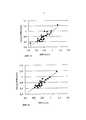
фиг.2a иллюстрирует эффективность прогнозирования минеральной плотности кости, проведенного согласно одному из вариантов осуществления на основе толщины кортикального слоя (дистальный и проксимальный концы), возраста пациента и массы его тела,figa illustrates the effectiveness of predicting bone mineral density, carried out according to one of the embodiments based on the thickness of the cortical layer (distal and proximal ends), the age of the patient and his body weight,
фиг.2b иллюстрирует эффективность прогнозирования минеральной плотности кости, проведенного согласно одному из вариантов осуществления на основе комбинации всех установленных параметров,fig.2b illustrates the effectiveness of predicting bone mineral density, carried out according to one of the embodiments based on a combination of all set parameters,
на фиг.3 представлен пример устройства, предназначенного для оценивания минеральной плотности кости согласно одному из предпочтительных вариантов изобретения.figure 3 presents an example of a device for evaluating the mineral density of the bone according to one of the preferred variants of the invention.
Осуществление изобретенияThe implementation of the invention
На фиг.1 под цифровым обозначением 100 представлен один из иллюстративных способов оценки плотности первой кости пациента, выполняемый согласно одному из предпочтительных вариантов осуществления изобретения и в качестве первой кости использующий, например, головку бедренной кости, шейку бедренной кости и/или поясничный сегмент позвоночника. На шаге 102 определяют первый параметр, который связан с изменением свойств измерительного сигнала, поданного в сторону второй кости пациента, не совпадающей с первой, и находившегося во взаимодействии со второй костью. Первый параметр может представлять собой, например, величину соответствующего изменения измерительного сигнала, в частности степень затухания ультразвукового излучения, степень изменения скорости, интенсивность отражения или рассеяния, величину изменения интенсивности или паузу между сигналами, отраженными от кости, конкретно, паузу между сигналами, отраженными от поверхностей первой и второй кости, измеренную в направлении измерительного сигнала. В качестве первого параметра можно также выбрать новый количественный параметр, определенный на основе измеренных свойств измерительного сигнала, например толщину второй кости или толщину кортикального слоя локтевой/лучевой или большеберцовой кости, измеренную в одной или более точках.1, with
На шаге 104 определяют набор вторых параметров, таких, например, как возраст и масса пациента или какой-то другой второй параметр, упомянутый в данном описании.At
Предусмотрена возможность дополнительно уточнить, на шаге 106, набор вторых параметров, включающий по меньшей мере один из следующих параметров пациента: возраст, массу, индекс массы тела, геометрический параметр бедренной кости (например, площадь или диаметр поперечного сечения), относящийся, например, к ее стержню и/или шейке, и силу кистевого хвата пациента. Согласно одному из вариантов осуществления в набор вторых параметров может входить также (в дополнение к перечисленным или само по себе) изменение свойств ультразвукового сигнала, поданного в сторону первой кости и находившегося во взаимодействии с ней. Такому изменению может отвечать, например, параметр AIB, измеряемый в дБ и определяемый на основе обратного рассеяния ультразвукового излучения на головке и/или шейке бедренной кости. Однако следует отметить, что шаг 106 необязателен. Затем на шаге 108 можно провести корректировки возможных параметров, такие, например, как корректировка изменения свойств ультразвукового сигнала или корректировка ДЧУ, относящаяся к измерению интенсивности (в дБ) обратного рассеяния ультразвукового излучения на головке и/или шейке бедренной кости. Корректировки параметров могут касаться ошибок, вызванных, например, составом или количеством мягкой ткани. Шаг 108 также необязателен.It is possible to further clarify, at
На шаге 110 проводят оценку минеральной плотности первой кости пациента, комбинируя перечисленные параметры (по меньшей мере первый параметр и какие-то параметры из набора вторых параметров). Такую комбинацию можно получить с помощью, например, регрессионного анализа, в частности посредством линейной регрессии (см. фиг.2a и 2b).At
Далее на шаге 112 полученное значение минеральной плотности можно использовать для определения параметра, характеризующего степень остеопороза у пациента, а также параметра, прогнозирующего опасность (например, вероятность) перелома. Для этого, например, измеренную минеральную плотность сопоставляют со справочными значениями, считающимися нормальными.Next, at
Согласно одному из иллюстративных вариантов осуществления значения толщины кортикального слоя для нижнего и верхнего сегментов большеберцовой кости (соответственно CTh_ds и CTh_pr) и обратное рассеяние (AIB) ультразвукового излучения на шейке бедренной кости определяли посредством построения эхографической картины (см. далее Таблицу 1). AIB определяли для 25 женщин, а полученные результаты корректировали с помощью методологии ДЧУ, учитывая затухание, вызванное наличием мягкой ткани, расположенной поверх кости. Остальные параметры определяли для 29 женщин.According to one illustrative embodiment, the cortical layer thickness values for the lower and upper tibia segments (CTh_ds and CTh_pr, respectively) and the backscattering (AIB) of ultrasound radiation on the femoral neck were determined by constructing an echographic picture (see Table 1 below). AIB was determined for 25 women, and the results were corrected using the DLC methodology, taking into account the attenuation caused by the presence of soft tissue located on top of the bone. The remaining parameters were determined for 29 women.
В добавление к сказанному, как видно из Таблицы 2, обратное рассеяние (AIB) ультразвукового излучения, измеренное на шейке бедренной кости, толщина кортикального слоя, измеренная на большеберцовой кости у дистального конца (CTh_ds) и у проксимального конца (Cth_pr), и исходная информация о пациенте, конкретно, возраст, масса и индекс массы тела (ИМТ) сами по себе (как об этом можно судить по их коэффициентам r линейной корреляции) обладают только слабыми или умеренными способностями предсказывать значение минеральной плотности (BMD) шейки бедренной костиIn addition, as can be seen from Table 2, the backscattering (AIB) of ultrasonic radiation, measured on the neck of the femur, the thickness of the cortical layer, measured on the tibia at the distal end (CTh_ds) and at the proximal end (Cth_pr), and background information about a patient, specifically, age, weight and body mass index (BMI) per se (as can be judged by their linear correlation coefficients r) have only weak or moderate abilities to predict the mineral density (BMD) of the femoral neck tee
Чтобы обеспечить высокую прогнозирующую способность по отношению к минеральной плотности кости, параметры, полезные по меньшей мере в одном варианте осуществления, можно обработать, например, посредством методов линейной регрессии. Вариант, который осуществляют, используя только толщину кортикального слоя, возраст и массу, позволяет получить оценку минеральной плотности шейки бедренной кости (см. фиг.2а; r=0,87, n=29) в следующем виде:In order to provide a high predictive ability with respect to bone mineral density, parameters useful in at least one embodiment can be processed, for example, by linear regression methods. The option, which is carried out using only the thickness of the cortical layer, age and weight, allows you to get an estimate of the mineral density of the neck of the femur (see figa; r = 0.87, n = 29) in the following form:
BMD_estimate_1=0,912-0,014×возраст+0,092×CTh_ds+0.006×масса+0,098×CTh_pr,BMD_estimate_1 = 0.912-0.014 × age + 0.092 × CTh_ds + 0.006 × mass + 0.098 × CTh_pr,
где estimate = оценка.where estimate = rating.
Вариант, который осуществляют, используя все имеющиеся параметры, позволяет получить оценку минеральной плотности шейки бедренной кости (см. фиг.2b; r=0,90, n=25) в следующем виде:The option, which is carried out using all available parameters, allows to obtain an estimate of the mineral density of the femoral neck (see fig.2b; r = 0.90, n = 25) in the following form:
BMD_estimate_2=3,624-0,015×возраст+0,070×CTh_pr+0,064×CTh_ds-0,061×ИМТ-0,005×AIB-0,016×рост+0,027×масса.BMD_estimate_2 = 3.624-0.015 × age + 0.070 × CTh_pr + 0.064 × CTh_ds-0.061 × BMI-0.005 × AIB-0.016 × height + 0.027 × mass.
Однако следует отметить, что в разных вариантах осуществления параметры, применяемые для этих оценок, могут иметь различные коэффициенты, что связано, в частности, со свойствами использованного измеряемого материала.However, it should be noted that in different embodiments, the parameters used for these estimates may have different coefficients, which is associated, in particular, with the properties of the used measured material.
Фиг.2a и 2b иллюстрируют уровень эффективности расчета, выполненного согласно одному из вариантов осуществления и оценивающего минеральную плотность шейки бедренной кости пациента. В первом случае (фиг.2a, r=0,87; n=29) в основу расчета положены толщина кортикального слоя кости (у дистального и проксимального концов), возраст и масса. Во втором случае (фиг.2b, r=0,90; n=25) в основу расчета положена комбинация всех собранных параметров, скомбинированных посредством линейной регрессии.Figa and 2b illustrate the level of effectiveness of the calculation performed according to one of the embodiments and evaluating the mineral density of the neck of the femoral neck of the patient. In the first case (Fig. 2a, r = 0.87; n = 29), the calculation is based on the thickness of the cortical layer of the bone (at the distal and proximal ends), age and weight. In the second case (fig.2b, r = 0.90; n = 25), the calculation is based on a combination of all the collected parameters combined by linear regression.
На фиг.3 представлен пример устройства 300, предназначенного для оценивания минеральной плотности кости и функционирующего согласно одному из предпочтительных вариантов изобретения. Устройство 300 содержит передающее средство 302 для посылки измерительного сигнала (предпочтительно прибор ультразвуковой эхографии). Средство 302 предназначено для посылки измерительного сигнала в сторону второй кости 301 пациента, не совпадающей с его первой костью. Кроме того, предусмотрено наличие приемного средства 304, которое принимает измерительный сигнал после его взаимодействия со второй костью 301, причем в предпочтительном варианте это - ультразвуковой сигнал, отраженный, например, от кортикального слоя. Взаимодействовавшим сигналом может быть, в частности, ультразвуковой сигнал, а его свойствами, подлежащими измерению, могут быть, например, отражение, рассеяние (например, интенсивность рассеяния или угол рассеяния), изменение скорости или затухание. В порядке альтернативы, данный сигнал может представлять собой, например, пучок рентгеновского излучения, а его свойствами, подлежащими измерению, могут быть, в частности, глубина проникновения или затухание (см. штриховую стрелку на фиг.3).FIG. 3 shows an example of a device 300 for evaluating bone mineral density and functioning in accordance with one preferred embodiment of the invention. The device 300 includes transmitting means 302 for sending a measurement signal (preferably an ultrasound ultrasound device). The
Следует отметить, что согласно одному из вариантов осуществления предусмотрена возможность размещать приемное средство 304 в различных точках измерительной схемы. Например, для процесса определения измерительного сигнала после его прохождения через измеряемый объект (например, в процессе измерения затухания измерительного сигнала) приемное средство 304 желательно установить на стороне измеряемого объекта, противоположной передающему устройству 302. Соответственно, для процесса измерения измерительного сигнала после его отражения или рассеяния измеряемым объектом приемное средство 304 предпочтительно установить относительно объекта измерения на той же стороне измеряемого объекта, что и передающее устройство 302. Следует отметить также, что согласно одному из вариантов осуществления передающее устройство 302 может быть одновременно и приемным устройством 304, причем такой передатчик/приемник 302/304 может содержать физически общий кристалл, который сначала генерирует ультразвуковой сигнал, а затем принимает его после взаимодействия сигнала с объектом измерения. Особо следует отметить, что единый приемопередающий датчик 302/304, содержащийся в ультразвуковой измерительной схеме, а конкретно - в эхоимпульсном (эхографическом) устройстве, - очень удобен для использования в процессе измерения у нескольких различных точек.It should be noted that according to one embodiment, it is possible to place the receiving means 304 at various points of the measuring circuit. For example, for the process of determining the measuring signal after it passes through the measured object (for example, in the process of measuring the attenuation of the measuring signal), it is desirable to install the receiving means 304 on the side of the measured object opposite to the transmitting
Кроме того, предусмотрена возможность снабдить передающее и приемное средства 302, 304 какими-то управляющими электронными схемами 302a, 304a. Конкретно, согласно одному из вариантов осуществления эти схемы выполнены с возможностью управления передатчиком 302 и приемником 304 таким образом, чтобы передающая функция передатчика прерывалась на время приема сигнала приемником. В результате сводятся к минимуму интерференционные взаимодействия, причем в особенности в варианте осуществления, применяющем один и тот же кристалл для генерирования ультразвукового сигнала и для приема сигнала, отраженного от объекта. В добавление к сказанному, желательно, чтобы устройство содержало элементы 306, предназначенные для ввода в него указанных вторых параметров. Такими элементами могут быть клавиатура или средство ввода графических данных. Посредством элементов 306 в устройство могут вводиться по меньшей мере некоторые из вторых параметров пациента (рост, индекс массы тела, гормональный статус, геометрический параметр бедренной кости, относящийся к ее стержню и/или шейке, например, площадь или диаметр поперечного сечения, а также сила кистевого хвата пациента). В добавление к сказанному, предусмотрена возможность посредством элементов 306 вводить в устройство параметр, характеризующий изменение свойств ультразвукового сигнала, поданного в сторону первой кости и находившегося во взаимодействии с ней. Имеется в виду, например, параметр AIB, измеряемый в дБ и определяемый на основе обратного рассеяния ультразвукового излучения на головке и/или шейке бедренной кости.In addition, it is possible to provide the transmitting and receiving means 302, 304 with some kind of control
В предпочтительном варианте устройство содержит также управляющие компоненты 308, предназначенные для управления средством передачи измерительного сигнала и приемным средством, в частности для управления работой ультразвукового передатчика 302 и приемника 304 (или источника 302 и приемника 304 рентгеновского излучения), например, для синхронизации функционирования передающего средства и приемного средства. Предпочтительно, чтобы компоненты 308 могли управлять работой передающего средства 302 и/или приемного средства 304 через сопряженные с ними управляющие электронные схемы 302a, 304a.In a preferred embodiment, the device also contains
Кроме того, устройство содержит процессорные компоненты 310, выполненные с возможностью находить оценку минеральной плотности первой кости пациента, используя по меньшей мере первый и вторые параметры. Согласно одному из вариантов осуществления компоненты 310 можно также адаптировать для того, чтобы в процессе получения оценки использовался по меньшей мере один из вторых параметров. Оценку предпочтительно выполнять посредством каких-то методов регрессионного анализа, таких, например, как линейная регрессия.In addition, the device contains
Согласно одному из вариантов осуществления процессорные компоненты 310 выполнены с возможностью определять первый параметр, который связан с изменением свойств измерительного сигнала, поданного в сторону какой-то второй кости пациента, которая не совпадает с первой, и находившегося во взаимодействии со второй костью. Согласно одному из вариантов осуществления компоненты 310 выполнены с возможностью, исходя из изменения свойств измерительного сигнала, определять какой-то другой параметр или количественный фактор, например толщину кортикального слоя большеберцовой или локтевой/лучевой кости в одной или нескольких его точках, и использовать эти данные в качестве первого параметра.According to one embodiment, the
В добавление к сказанному, согласно одному из вариантов процессорные компоненты 310 устройства можно выполнить с возможностью проведения корректировки параметров, например, такой как корректировка изменения свойств измерительного сигнала после его взаимодействия с объектом измерения или корректировки интенсивности (в дБ) обратного рассеяния ультразвукового излучения на головке и/или шейке бедренной кости с учетом влияния мягкой ткани. В основу корректировки могут быть положены, например, методология ДЧУ и/или учет затухания, происходящего в кортикальном слое кости.In addition, according to one embodiment, the
Далее, согласно одному из вариантов осуществления процессорные компоненты 310 устройства можно выполнить с возможностью определения параметра, характеризующего степень остеопороза у пациента, используя для этого, как это было описано выше, оценку минеральной плотности первой кости пациента и сопоставление этой оценки со справочными данными, считающимися нормальными.Further, according to one embodiment, the
Согласно одному из вариантов устройство может содержать также элементы 312, предназначенные для определения геометрического параметра (например, площади или диаметра поперечного сечения) стержня и/или шейки бедренной кости, причем данный параметр используется в качестве одного из вторых параметров. Такими элементами могут быть, например, приемопередатчики ультразвукового излучения и программное обеспечение для интерпретации принятого ультразвукового сигнала и для вычисления площади поверхности. Кроме того, устройство можно снабдить элементами 314, определяющими изменение свойств ультразвукового сигнала, поданного в сторону первой кости и находившегося во взаимодействии с ней. Такому изменению может отвечать, например, параметр AIB, измеряемый в дБ и определяемый на основе обратного рассеяния ультразвукового излучения на головке и/или шейке бедренной кости. Далее, устройство может содержать элементы 316, предназначенные для определения силы кистевого хвата пациента с использованием этой информации в качестве одного из вторых параметров.According to one embodiment, the device may also include
Согласно одному из вариантов осуществления по меньшей мере некоторые элементы устройства и, в особенности, некоторые функциональные свойства его процессорных компонентов 310 можно запрограммировать посредством, например, компьютерного программного продукта, который выполнен с возможностью приводить в действие процессор, обрабатывающий используемые в устройстве данные. В добавление к сказанному, согласно одному из вариантов осуществления предусмотрена возможность адаптировать данный программный продукт для получения оценки вероятности переломов кости, которые могут произойти в течение следующих 10 лет, причем, например, курсы лечения остеопороза можно составить без труда, быстро и точно, не прибегая к дорогому отдельному измерению с использованием, например, аппаратуры ДРА.According to one embodiment, at least some elements of the device and, in particular, some functional properties of its
Согласно одному из вариантов осуществления предусмотрена возможность приложения программного компьютерного обеспечения, в частности, к некоторым проблемам, касающимся факторов вероятности перелома, с получением на выходе простых ответов типа "да" или "нет". Затем программное обеспечение адаптируют для определения вероятности перелома и, в одном варианте осуществления, для составления лечебного предписания, например, такого типа: "никакого медикаментозного лечения", "измерить минеральную плотность" или "медикаментозное лечение".According to one embodiment, it is possible to apply computer software, in particular, to some problems relating to the probability factors of a fracture, to obtain simple “yes” or “no” answers. The software is then adapted to determine the likelihood of a fracture and, in one embodiment, to formulate a treatment prescription, for example, of the type: “no drug treatment”, “measure mineral density”, or “drug treatment”.
Согласно одному из вариантов осуществления компьютерный программный продукт для оценивания минеральной плотности первой кости пациента, в качестве которой выбрана головка бедренной кости, шейка бедренной кости и/или поясничный сегмент позвоночника, обеспечивает возможности:According to one embodiment, a computer program product for evaluating the mineral density of the first bone of a patient, which is selected as the head of the femur, the neck of the femur and / or the lumbar segment of the spine, provides:
- определять первый параметр, характеризующий изменение свойств измерительного сигнала, поданного в сторону второй кости пациента, не совпадающей с его первой костью, и находившегося во взаимодействии с данной второй костью, и- determine the first parameter characterizing the change in the properties of the measuring signal applied to the side of the second bone of the patient that does not coincide with his first bone, and which was in interaction with this second bone, and
- получать оценку минеральной плотности первой кости пациента, используя первый параметр и применяя компьютерный программный продукт, взаимодействующий с процессором, обрабатывающим используемые данные.- obtain an estimate of the mineral density of the first bone of the patient using the first parameter and using a computer program product that interacts with a processor that processes the data used.
В добавление к сказанному, согласно одному из вариантов осуществления компьютерный программный продукт можно выполнить с возможностью получения такой оценки с использованием также набора вторых параметров, содержащего по меньшей мере один компонент группы, в которую входят такие параметры пациента, как возраст, масса, рост, индекс массы тела, геометрический параметр бедренной кости, такой, например, как площадь или диаметр поперечного сечения, измеренный для ее стержня и/или шейки, изменение свойств ультразвукового сигнала, поданного в сторону первой кости пациента и находившегося во взаимодействии с ней, характеризуемое, например, параметром AIB, измеряемым в дБ и определяемым на основе обратного рассеяния на головке и/или шейке бедренной кости, а также сила кистевого хвата пациента.In addition, according to one embodiment, a computer program product can be configured to obtain such an assessment using a set of second parameters, comprising at least one component of a group that includes patient parameters such as age, weight, height, index body mass, geometric parameter of the femur, such as, for example, the area or diameter of the cross section measured for its shaft and / or neck, changes in the properties of the ultrasonic signal applied to the side of the first bone of the patient and in interaction with it, characterized, for example, by the AIB parameter, measured in dB and determined on the basis of backscattering on the head and / or neck of the femur, as well as the strength of the patient’s wrist grip.
В данном описании представлены только некоторые варианты решения проблем, поставленных перед изобретением. Предусмотрена возможность, не выходя за границы объема охраны, определяемые прилагаемой формулой, модифицировать заложенные в изобретении принципы, относящиеся, в частности, к используемым деталям и различным аспектам применения.In this description, only some of the solutions to the problems posed by the invention are presented. It is possible, without going beyond the scope of protection defined by the attached formula, to modify the principles embodied in the invention, relating, in particular, to the parts used and various aspects of application.
Claims (15)
Translated fromRussian- посредством эхоимпульсного метода определяют первый параметр, который связан с изменением свойств ультразвукового измерительного сигнала, посланного в сторону кортикального слоя трубчатой кости пациента и находившегося во взаимодействии с указанным кортикальным слоем, представляющим собой кортикальный слой второй кости, которая является большеберцовой костью, пальцем или локтевой/лучевой костью,
- определяют набор вторых параметров, в который входят возраст и масса указанного пациента,
- используя первый параметр и набор вторых параметров, получают оценку минеральной плотности первой кости пациента.1. The method of assessing the mineral density of the first bone of the patient, the first bone corresponding to the head of the femur, the neck of the femur or the lumbar segment of the spine, characterized in that it includes the following steps:
- using the echo pulse method, the first parameter is determined, which is associated with a change in the properties of the ultrasonic measuring signal sent to the side of the cortical layer of the patient’s tubular bone and interacting with the specified cortical layer, which is the cortical layer of the second bone, which is the tibia, finger or ulna / radius bone
- determine the set of second parameters, which includes the age and weight of the specified patient,
- using the first parameter and a set of second parameters, an estimate of the mineral density of the first bone of the patient is obtained.
отличающееся тем, что выполнено с возможностью:
- в ходе применения эхоимпульсного метода передавать ультразвуковой измерительный сигнал в сторону кортикального слоя трубчатой кости пациента, а также принимать ультразвуковой измерительный сигнал после его взаимодействия с кортикальным слоем трубчатой кости, представляющим собой кортикальный слой второй кости, которая является большеберцовой костью, пальцем или локтевой/лучевой костью,
- определять первый параметр, который связан с изменением свойств ультразвукового измерительного сигнала после его взаимодействия с кортикальным слоем трубчатой кости,
- определять набор вторых параметров, в который входят возраст и масса указанного пациента, и
- используя первый параметр и набор вторых параметров, получать оценку минеральной плотности первой кости пациента.9. A device for assessing the mineral density of the first bone of a patient, the first bone corresponding to the head of the femur, the neck of the femur or the lumbar segment of the spine, containing an ultrasound transmitter and an ultrasound receiver for generating, transmitting and receiving an ultrasonic measuring signal during the application of the echo pulse method,
characterized in that it is configured to:
- during the application of the echo pulse method, transmit the ultrasonic measuring signal towards the cortical layer of the patient’s tubular bone, and also receive the ultrasonic measuring signal after its interaction with the cortical layer of the tubular bone, which is the cortical layer of the second bone, which is the tibia, finger or ulna / radius bone
- determine the first parameter, which is associated with a change in the properties of the ultrasonic measuring signal after its interaction with the cortical layer of the tubular bone,
- determine a set of second parameters, which includes the age and weight of the specified patient, and
- using the first parameter and a set of second parameters, obtain an estimate of the mineral density of the first bone of the patient.
- определять, посредством эхоимпульсного метода, первый параметр, который связан с изменением свойств ультразвукового измерительного сигнала, поданного в сторону кортикального слоя трубчатой кости пациента и находившегося во взаимодействии с кортикальным слоем трубчатой кости, представляющим собой кортикальный слой второй кости, которая является большеберцовой костью, пальцем или локтевой/лучевой костью,
- определять набор вторых параметров, в который входят возраст и масса указанного пациента, и
- получать, используя первый параметр и набор вторых параметров, оценку минеральной плотности первой кости пациента,
при этом процессор представляет собой процессор обработки данных.15. A processor for estimating the mineral density of a patient’s bone, programmed by means of a computer program product for evaluating the mineral density of a patient’s first bone, which corresponds to a femoral head, a femoral neck, or a lumbar segment of the spine, characterized in that the computer-based software product at startup enables:
- determine, using the echo pulse method, the first parameter, which is associated with a change in the properties of the ultrasonic measuring signal applied to the cortical layer of the patient’s tubular bone and interacting with the cortical layer of the tubular bone, which is the cortical layer of the second bone, which is the tibia, finger or ulna / radius
- determine a set of second parameters, which includes the age and weight of the specified patient, and
- receive, using the first parameter and a set of second parameters, an estimate of the mineral density of the first bone of the patient,
wherein the processor is a data processor.
Applications Claiming Priority (3)
| Application Number | Priority Date | Filing Date | Title |
|---|---|---|---|
| FI20105936 | 2010-09-09 | ||
| FI20105936AFI126104B (en) | 2010-09-09 | 2010-09-09 | Procedure and system for bone mineral density assessment |
| PCT/FI2011/050772WO2012032225A1 (en) | 2010-09-09 | 2011-09-09 | Method and arrangement for estimating mineral density of a bone |
Publications (2)
| Publication Number | Publication Date |
|---|---|
| RU2013114988A RU2013114988A (en) | 2014-10-20 |
| RU2598642C2true RU2598642C2 (en) | 2016-09-27 |
Family
ID=42829672
Family Applications (1)
| Application Number | Title | Priority Date | Filing Date |
|---|---|---|---|
| RU2013114988/14ARU2598642C2 (en) | 2010-09-09 | 2011-09-09 | Method and device for estimating mineral density of a bone |
Country Status (9)
| Country | Link |
|---|---|
| US (1) | US9526472B2 (en) |
| EP (1) | EP2613705B1 (en) |
| JP (1) | JP5960699B2 (en) |
| CN (1) | CN103237501B (en) |
| ES (1) | ES3008695T3 (en) |
| FI (1) | FI126104B (en) |
| PL (1) | PL2613705T3 (en) |
| RU (1) | RU2598642C2 (en) |
| WO (1) | WO2012032225A1 (en) |
Cited By (1)
| Publication number | Priority date | Publication date | Assignee | Title |
|---|---|---|---|---|
| RU2750976C1 (en)* | 2020-10-16 | 2021-07-07 | Федеральное государственное автономное образовательное учреждение высшего образования "Новосибирский национальный исследовательский государственный университет" (Новосибирский государственный университет, НГУ) | Method for determining density of bone tissue based on standing wave emission from peripheral skeleton microseisms |
Families Citing this family (5)
| Publication number | Priority date | Publication date | Assignee | Title |
|---|---|---|---|---|
| CN103598875B (en)* | 2013-11-16 | 2015-01-28 | 沈阳医学院 | Human body bone mineral density predicting device for predicting risk of osteoporosis |
| EP3364864B1 (en)* | 2015-11-13 | 2022-09-28 | Orthoforge | Medical devices for monitoring and stimulating osteogenesis |
| WO2017106485A1 (en)* | 2015-12-16 | 2017-06-22 | Hologic, Inc. | Systems and methods for presenting complex medical condition diagnoses |
| KR101840349B1 (en)* | 2016-11-15 | 2018-03-21 | 강원대학교산학협력단 | Apparatus and method for estimating bone mineral density using ultrasonic sum frequency component |
| US12076181B2 (en)* | 2017-06-21 | 2024-09-03 | Charité —Universitätsmedizin Berlin | System, method, and computer program product for determining cortical bone characteristics |
Citations (6)
| Publication number | Priority date | Publication date | Assignee | Title |
|---|---|---|---|---|
| US4941474A (en)* | 1988-07-01 | 1990-07-17 | Massachusetts Institute Of Technology | Multivariable analysis of bone condition |
| US5218963A (en)* | 1991-10-15 | 1993-06-15 | Lunar Corporation | Ultrasonic bone analysis device and method |
| US5817020A (en)* | 1995-11-29 | 1998-10-06 | Sekisui Kagaku Kogyo Kabushiki Kaisya | Apparatus and method for diagnosing osteoporosis |
| US20050004457A1 (en)* | 2001-11-30 | 2005-01-06 | Petro Moilanen | Method and device for the non-invasive assessement of bones |
| US20050015002A1 (en)* | 2003-07-18 | 2005-01-20 | Dixon Gary S. | Integrated protocol for diagnosis, treatment, and prevention of bone mass degradation |
| RU2305491C2 (en)* | 2003-12-05 | 2007-09-10 | Российская медицинская академия последипломного образования Министерства здравоохранения Российской Федерации (РМАПО МЗ РФ) | Method for studying and diagnosing pathologic changes in bone tissue in diabetes mellitus cases |
Family Cites Families (18)
| Publication number | Priority date | Publication date | Assignee | Title |
|---|---|---|---|---|
| US4913157A (en)* | 1986-06-03 | 1990-04-03 | Analog Devices, Inc. | Ultrasound method and apparatus for evaluating, in vivo, bone conditions |
| US5840029A (en)* | 1988-05-11 | 1998-11-24 | Lunar Corporation | Imaging ultrasonic densitometer |
| US6213934B1 (en)* | 1995-06-01 | 2001-04-10 | Hyper3D Corp. | Electromagnetic bone-assessment and treatment: apparatus and method |
| US20020006181A1 (en) | 1998-01-23 | 2002-01-17 | Mackenzie Innes K. | Method and device for estimating bone mineral content of the calcaneus |
| AU2001244004A1 (en) | 2000-03-28 | 2001-10-08 | Signalgene Inc. | Method for determining osteoporosis susceptibility and/or low bone density and reagents therefor |
| JP2002238904A (en)* | 2001-02-19 | 2002-08-27 | Tanita Corp | Bone density estimation method and bone density estimation device |
| US6468215B1 (en)* | 2001-07-16 | 2002-10-22 | Artann Laboratories | Method and device for multi-parametric ultrasonic assessment of bone conditions |
| US7386337B2 (en) | 2002-05-06 | 2008-06-10 | University Of Rochester | Correction of dual energy X-ray absorptiometry measurements based on body lead levels |
| EP1393680B1 (en) | 2002-08-30 | 2007-08-01 | L'acn L'accessorio Nucleare Srl | Apparatus for bone mineral density measurement |
| US8202219B2 (en)* | 2004-02-23 | 2012-06-19 | Cyberlogic, Inc. | Ultrasonic bone assessment apparatus and method |
| CA2580726A1 (en)* | 2004-09-16 | 2006-03-30 | Imaging Therapeutics, Inc. | System and method of predicting future fractures |
| WO2008038159A2 (en)* | 2006-09-29 | 2008-04-03 | Odetect As | Ultrasound measurement techniques for bone analysis |
| CN101199429B (en)* | 2006-12-13 | 2012-06-06 | 计算机逻辑公司 | Ultrasonic bone assessment apparatus and method |
| US7862510B2 (en)* | 2007-02-09 | 2011-01-04 | Cyberlogic, Inc. | Ultrasonic bone assessment apparatus and method |
| JP5280647B2 (en) | 2007-05-29 | 2013-09-04 | 古野電気株式会社 | Bone strength diagnostic device using ultrasound and method of operating bone strength diagnostic device using ultrasound |
| US8679019B2 (en)* | 2007-12-03 | 2014-03-25 | Bone Index Finland Oy | Method for measuring of thicknesses of materials using an ultrasound technique |
| CN102186422B (en) | 2009-02-13 | 2013-06-12 | 计算机逻辑公司 | Ultrasonic bone assessment apparatus and method |
| US8880143B2 (en)* | 2012-01-17 | 2014-11-04 | Sectra Imtec Ab | Apparatus and method for estimating the bone mineral density to asses bone fractures risk |
- 2010
- 2010-09-09FIFI20105936Apatent/FI126104B/enactiveIP Right Grant
- 2011
- 2011-09-09JPJP2013527654Apatent/JP5960699B2/enactiveActive
- 2011-09-09RURU2013114988/14Apatent/RU2598642C2/enactive
- 2011-09-09ESES11823123Tpatent/ES3008695T3/enactiveActive
- 2011-09-09WOPCT/FI2011/050772patent/WO2012032225A1/enactiveApplication Filing
- 2011-09-09EPEP11823123.2Apatent/EP2613705B1/enactiveActive
- 2011-09-09USUS13/821,883patent/US9526472B2/enactiveActive
- 2011-09-09PLPL11823123.2Tpatent/PL2613705T3/enunknown
- 2011-09-09CNCN201180054044.7Apatent/CN103237501B/enactiveActive
Patent Citations (6)
| Publication number | Priority date | Publication date | Assignee | Title |
|---|---|---|---|---|
| US4941474A (en)* | 1988-07-01 | 1990-07-17 | Massachusetts Institute Of Technology | Multivariable analysis of bone condition |
| US5218963A (en)* | 1991-10-15 | 1993-06-15 | Lunar Corporation | Ultrasonic bone analysis device and method |
| US5817020A (en)* | 1995-11-29 | 1998-10-06 | Sekisui Kagaku Kogyo Kabushiki Kaisya | Apparatus and method for diagnosing osteoporosis |
| US20050004457A1 (en)* | 2001-11-30 | 2005-01-06 | Petro Moilanen | Method and device for the non-invasive assessement of bones |
| US20050015002A1 (en)* | 2003-07-18 | 2005-01-20 | Dixon Gary S. | Integrated protocol for diagnosis, treatment, and prevention of bone mass degradation |
| RU2305491C2 (en)* | 2003-12-05 | 2007-09-10 | Российская медицинская академия последипломного образования Министерства здравоохранения Российской Федерации (РМАПО МЗ РФ) | Method for studying and diagnosing pathologic changes in bone tissue in diabetes mellitus cases |
Non-Patent Citations (1)
| Title |
|---|
| Л.Я. Рожинская, СИСТЕМНЫЙ ОСТЕОПОРОЗ, Практическое руководство для врачей, Издание 2-е, М., Издатель Мокеев, 2000.* |
Cited By (1)
| Publication number | Priority date | Publication date | Assignee | Title |
|---|---|---|---|---|
| RU2750976C1 (en)* | 2020-10-16 | 2021-07-07 | Федеральное государственное автономное образовательное учреждение высшего образования "Новосибирский национальный исследовательский государственный университет" (Новосибирский государственный университет, НГУ) | Method for determining density of bone tissue based on standing wave emission from peripheral skeleton microseisms |
Also Published As
| Publication number | Publication date |
|---|---|
| WO2012032225A1 (en) | 2012-03-15 |
| JP5960699B2 (en) | 2016-08-02 |
| US9526472B2 (en) | 2016-12-27 |
| FI20105936L (en) | 2012-03-10 |
| EP2613705B1 (en) | 2024-12-11 |
| EP2613705A1 (en) | 2013-07-17 |
| FI126104B (en) | 2016-06-30 |
| CN103237501B (en) | 2015-09-30 |
| FI20105936A0 (en) | 2010-09-09 |
| CN103237501A (en) | 2013-08-07 |
| US20130245443A1 (en) | 2013-09-19 |
| EP2613705C0 (en) | 2024-12-11 |
| JP2013537055A (en) | 2013-09-30 |
| RU2013114988A (en) | 2014-10-20 |
| ES3008695T3 (en) | 2025-03-24 |
| PL2613705T3 (en) | 2025-04-28 |
| FI20105936A7 (en) | 2012-03-10 |
| EP2613705A4 (en) | 2018-01-17 |
Similar Documents
| Publication | Publication Date | Title |
|---|---|---|
| US11911175B2 (en) | Ultrasound apparatus for assessing the quality of a patient's bone tissue | |
| Baran et al. | Diagnosis and management of osteoporosis: guidelines for the utilization of bone densitometry | |
| RU2598642C2 (en) | Method and device for estimating mineral density of a bone | |
| Tromp et al. | Quantitative ultrasound measurements of the tibia and calcaneus in comparison with DXA measurements at various skeletal sites | |
| Brooke-Wavell et al. | Ultrasound and dual X-ray absorptiometry measurement of the calcaneus: influence of region of interest location | |
| Wüster et al. | Quantitative ultrasonometry (QUS) for the evaluation of osteoporosis risk: reference data for various measurement sites, limitations and application possibilities | |
| Avcı et al. | The comparison of bedside point-of-care ultrasound and computed tomography in elbow injuries | |
| Rohde et al. | Influence of porosity, pore size, and cortical thickness on the propagation of ultrasonic waves guided through the femoral neck cortex: a simulation study | |
| Egorov et al. | Osteoporosis detection in postmenopausal women using axial transmission multi-frequency bone ultrasonometer: Clinical findings | |
| Damilakis et al. | Ultrasound velocity through the cortex of phalanges, radius, and tibia in normal and osteoporotic postmenopausal women using a new multisite quantitative ultrasound device | |
| Gonnelli et al. | The use of ultrasound in the assessment of bone status | |
| Rosenthall et al. | Correlation of ultrasound velocity in the tibial cortex, calcaneal ultrasonography, and bone mineral densitometry of the spine and femur | |
| Wang et al. | Monitoring bone growth using quantitative ultrasound in comparison with DXA and pQCT | |
| Laugier | Quantitative ultrasound instrumentation for bone in vivo characterization | |
| Maricic et al. | Bone densitometry | |
| Robertson et al. | Bone quantification | |
| Glinkowski et al. | Clinical experiences with ultrasonometric measurement of fracture healing | |
| Karjalainen | Novel pulse-echo ultrasound methods for diagnostics of osteoporosis | |
| Njeh et al. | Evaluation of a gel‐coupled quantitative ultrasound device for bone status assessment. | |
| JP2014151175A (en) | Bone strength evaluation by measuring intraosseous ultrasonic propagation velocity | |
| Alhawkash et al. | The Importance of Biomedical Engineering In medical interventions in medical devices | |
| Arpinar et al. | Correlation between mechanical vibration analysis and dual energy X-ray absorptiometry (DXA) in the measurement of in vivo human tibial bone strength | |
| JP3182558B2 (en) | Evaluation of bone mineral density by ultrasonic measurement | |
| Grondin et al. | 22 Sensitivity of Ultrasound to Indices of Femoral Neck Strength Investigated With Simulation of Wave Propagation in Cortical Bone | |
| Glüer et al. | Assessment of bone status by quantitative ultrasound |