RU2584810C1 - Method for tool fixation of at least part of thoracic and/or lumbar spine to pelvis in various diseases - Google Patents
Method for tool fixation of at least part of thoracic and/or lumbar spine to pelvis in various diseasesDownload PDFInfo
- Publication number
- RU2584810C1 RU2584810C1RU2014148506/14ARU2014148506ARU2584810C1RU 2584810 C1RU2584810 C1RU 2584810C1RU 2014148506/14 ARU2014148506/14 ARU 2014148506/14ARU 2014148506 ARU2014148506 ARU 2014148506ARU 2584810 C1RU2584810 C1RU 2584810C1
- Authority
- RU
- Russia
- Prior art keywords
- pelvis
- fixing
- assembly
- fixing element
- spine
- Prior art date
Links
- 210000004197pelvisAnatomy0.000titleclaimsabstractdescription45
- 238000000034methodMethods0.000titleclaimsabstractdescription35
- 210000004705lumbosacral regionAnatomy0.000titleclaimsabstractdescription19
- 210000000115thoracic cavityAnatomy0.000titleclaimsabstractdescription13
- 201000010099diseaseDiseases0.000titleclaimsabstractdescription6
- 208000037265diseases, disorders, signs and symptomsDiseases0.000titleclaimsabstractdescription6
- 210000000988bone and boneAnatomy0.000claimsabstractdescription17
- 210000003692iliumAnatomy0.000claimsabstractdescription11
- 210000001621ilium boneAnatomy0.000claimsabstractdescription7
- 230000007170pathologyEffects0.000claimsabstractdescription5
- 230000008569processEffects0.000claimsabstractdescription4
- 230000008878couplingEffects0.000claimsdescription13
- 238000010168coupling processMethods0.000claimsdescription13
- 238000005859coupling reactionMethods0.000claimsdescription13
- 230000023597hemostasisEffects0.000claimsdescription3
- 210000000038chestAnatomy0.000claimsdescription2
- 239000000463materialSubstances0.000claimsdescription2
- 238000005406washingMethods0.000claimsdescription2
- 239000003814drugSubstances0.000abstractdescription2
- 239000000126substanceSubstances0.000abstract1
- 238000001356surgical procedureMethods0.000description12
- 208000032170Congenital AbnormalitiesDiseases0.000description8
- 206010058907Spinal deformityDiseases0.000description7
- 238000002591computed tomographyMethods0.000description6
- 238000009434installationMethods0.000description6
- 239000002184metalSubstances0.000description6
- 206010061619DeformityDiseases0.000description5
- 230000008901benefitEffects0.000description5
- 238000012937correctionMethods0.000description5
- 230000001272neurogenic effectEffects0.000description5
- 206010039722scoliosisDiseases0.000description5
- 238000013461designMethods0.000description4
- 238000011161developmentMethods0.000description4
- 206010028980NeoplasmDiseases0.000description3
- 208000009905NeurofibromatosesDiseases0.000description3
- 208000002193PainDiseases0.000description3
- 230000007547defectEffects0.000description3
- 238000003745diagnosisMethods0.000description3
- 201000004931neurofibromatosisDiseases0.000description3
- 230000000399orthopedic effectEffects0.000description3
- 230000036407painEffects0.000description3
- 230000001769paralizing effectEffects0.000description3
- 210000003049pelvic boneAnatomy0.000description3
- 206010010356Congenital anomalyDiseases0.000description2
- 206010033892ParaplegiaDiseases0.000description2
- 206010061335Pelvic deformityDiseases0.000description2
- 208000027418Wounds and injuryDiseases0.000description2
- 230000000903blocking effectEffects0.000description2
- 238000006073displacement reactionMethods0.000description2
- 230000007774longtermEffects0.000description2
- 238000012986modificationMethods0.000description2
- 230000004048modificationEffects0.000description2
- 210000003205muscleAnatomy0.000description2
- 230000002232neuromuscularEffects0.000description2
- 231100001060spine abnormalityToxicity0.000description2
- 238000012549trainingMethods0.000description2
- 206010002091AnaesthesiaDiseases0.000description1
- 208000006386Bone ResorptionDiseases0.000description1
- 206010050694Congenital scoliosisDiseases0.000description1
- 206010017577Gait disturbanceDiseases0.000description1
- 208000008930Low Back PainDiseases0.000description1
- 201000004404NeurofibromaDiseases0.000description1
- 208000037273Pathologic ProcessesDiseases0.000description1
- 230000003187abdominal effectEffects0.000description1
- 230000005856abnormalityEffects0.000description1
- 230000037005anaesthesiaEffects0.000description1
- 210000003484anatomyAnatomy0.000description1
- 230000024279bone resorptionEffects0.000description1
- 239000003795chemical substances by applicationSubstances0.000description1
- 230000006835compressionEffects0.000description1
- 238000007906compressionMethods0.000description1
- 230000006378damageEffects0.000description1
- 210000002249digestive systemAnatomy0.000description1
- 230000008030eliminationEffects0.000description1
- 238000003379elimination reactionMethods0.000description1
- 210000003195fasciaAnatomy0.000description1
- 239000000834fixativeSubstances0.000description1
- 238000011010flushing procedureMethods0.000description1
- 230000036541healthEffects0.000description1
- 230000001771impaired effectEffects0.000description1
- 230000006872improvementEffects0.000description1
- 230000002757inflammatory effectEffects0.000description1
- 208000014674injuryDiseases0.000description1
- 230000036244malformationEffects0.000description1
- 238000004519manufacturing processMethods0.000description1
- 208000018360neuromuscular diseaseDiseases0.000description1
- 210000000056organAnatomy0.000description1
- 230000001575pathological effectEffects0.000description1
- 230000009054pathological processEffects0.000description1
- 230000000737periodic effectEffects0.000description1
- 239000002861polymer materialSubstances0.000description1
- 238000002360preparation methodMethods0.000description1
- 238000012545processingMethods0.000description1
- 230000029058respiratory gaseous exchangeEffects0.000description1
- 208000002320spinal muscular atrophyDiseases0.000description1
- 210000004003subcutaneous fatAnatomy0.000description1
- 238000011477surgical interventionMethods0.000description1
- 230000008733traumaEffects0.000description1
Images


Classifications
- A—HUMAN NECESSITIES
- A61—MEDICAL OR VETERINARY SCIENCE; HYGIENE
- A61B—DIAGNOSIS; SURGERY; IDENTIFICATION
- A61B17/00—Surgical instruments, devices or methods
- A61B17/56—Surgical instruments or methods for treatment of bones or joints; Devices specially adapted therefor
- A61B17/58—Surgical instruments or methods for treatment of bones or joints; Devices specially adapted therefor for osteosynthesis, e.g. bone plates, screws or setting implements
Landscapes
- Health & Medical Sciences (AREA)
- Life Sciences & Earth Sciences (AREA)
- Orthopedic Medicine & Surgery (AREA)
- Surgery (AREA)
- Medical Informatics (AREA)
- Engineering & Computer Science (AREA)
- Biomedical Technology (AREA)
- Heart & Thoracic Surgery (AREA)
- Nuclear Medicine, Radiotherapy & Molecular Imaging (AREA)
- Molecular Biology (AREA)
- Animal Behavior & Ethology (AREA)
- General Health & Medical Sciences (AREA)
- Public Health (AREA)
- Veterinary Medicine (AREA)
- Surgical Instruments (AREA)
- Orthopedics, Nursing, And Contraception (AREA)
Abstract
Description
Translated fromRussianИзобретение относится к медицине и может быть использовано в ортопедии, нейрохирургии, в том числе нейроортопедии, травматологии и вертебрологии.The invention relates to medicine and can be used in orthopedics, neurosurgery, including neuroorthopedics, traumatology and vertebrology.
Оно предназначено для операций, производимых из дорсального (заднего) доступа, преимущественно для выполнения позвоночно-тазовой фиксации при условии максимальной безопасности и обеспечения максимальной стабильности позвоночно-тазовой фиксации. Использование показано, в первую очередь, при нарушении позвоночно-тазовой стабильности, опорной функции таза, в том числе при нейрогенных сколиозах, деформациях при нейрофиброматозе, врожденных сколиозах, опухолях и аномалиях развития позвоночника и таза.It is intended for operations performed from the dorsal (posterior) access, mainly for performing spinal-pelvic fixation under the condition of maximum safety and ensuring maximum stability of the spinal-pelvic fixation. The use is shown, first of all, in violation of spinal-pelvic stability, pelvic support, including neurogenic scoliosis, deformities in neurofibromatosis, congenital scoliosis, tumors and abnormalities of the spine and pelvis.
Известно два основных способа выполнения позвоночно-тазовой фиксации.Two main methods of performing spinal pelvic fixation are known.
Первый способ, при котором установленные на позвоночнике фиксирующие элементы (крючки, проволока, винты) фиксируют к крыльям таза при помощи специально изогнутых контурированных стержней, которые могут как внедряться в крылья таза, так и упираться на них сверху (Khaled М. Kebaish Sacropelvic Fixation Techniques and Complications // Spine, 2010, Vol. 35, N 25, pp. 2245-2251; Michael W. Peelle, Lawrence G. Lenke, Keith H. Bridwell, Brenda Sides Comparison of Pelvic Fixation Techniques in Neuromuscular Spinal Deformity Correction: Galveston Rod Versus Iliac and Lumbosacral Screws // Spine, 2006, Vol. 31, N 20, pp. 2392-2398).The first method, in which the fixing elements installed on the spine (hooks, wire, screws) are fixed to the wings of the pelvis using specially curved contoured rods that can either be inserted into the wings of the pelvis or rest on top of them (Khaled M. Kebaish Sacropelvic Fixation Techniques and Complications // Spine, 2010, Vol. 35,
Второй способ, при котором в кости таза устанавливают винты, которые соединяются через стержни с установленными на позвоночнике фиксирующими элементами (крючки, проволока, винты) (Khaled М. Kebaish Sacropelvic Fixation Techniques and Complications // Spine 2010 Vol 35, N 25, pp 2245-2251; Michael W. Peelle, Lawrence G. Lenke, Keith H. Bridwell, Brenda Sides Comparison of Pelvic Fixation Techniques in Neuromuscular Spinal Deformity Correction: Galveston Rod Versus Iliac and Lumbosacral Screws // Spine, 2006, Vol. 31, N 20, pp. 2392-2398; патент РФ №2271165, приоритет 12.05.2003).The second method, in which screws are inserted into the pelvic bone that connect through the rods to the fixing elements (hooks, wire, screws) mounted on the spine (Khaled M. Kebaish Sacropelvic Fixation Techniques and Complications // Spine 2010
Недостатком первого способа фиксации является отсутствие достаточной стабильности фиксации к тазу, микроподвижность. В обоих способах фиксации высок риск развития несостоятельности костной ткани в месте упора устройства в связи с малой площадью контакта и большой нагрузкой (он его может просто продавить), в связи с чем высокий риск смещения фиксирующих элементов, недостаточность осевой фиксации. Кроме этого, возможны переломы стержня вследствие большой прилагаемой к нему нагрузки.The disadvantage of the first method of fixation is the lack of sufficient stability of fixation to the pelvis, micromobility. In both methods of fixation, there is a high risk of developing bone failure at the point of abutment of the device due to the small contact area and large load (it can simply push it), which is why there is a high risk of displacement of the fixing elements and insufficient axial fixation. In addition, rod fractures are possible due to the large load applied to it.
Ближайшим аналогом является способ инструментальной фиксации, по меньшей мере, части грудного и/или поясничного отдела позвоночника к тазу (Allen В. Jr., Ferguson R. The Galveston Technique of Pelvic Fixation with L-Rod Instrumentation of The Spine // Spine, 1984, Vol. 9, N 4, pp. 388-394).The closest analogue is a method of instrumental fixation of at least part of the thoracic and / or lumbar spine to the pelvis (Allen B. Jr., Ferguson R. The Galveston Technique of Pelvic Fixation with L-Rod Instrumentation of The Spine // Spine, 1984 Vol. 9,
Преимуществом способа является относительная простота установки, использование стандартных фиксирующих элементов, широко используемых в современной хирургии позвоночника.The advantage of the method is the relative ease of installation, the use of standard fixing elements, widely used in modern spinal surgery.
Недостатком его является отсутствие достаточной стабильности фиксации к тазу, микроподвижность, возможность развития несостоятельности костной ткани в месте упора устройства в связи с малой площадью контакта и большой нагрузкой (он его может просто продавить), высокий риск смещения фиксирующих элементов, недостаточность осевой фиксации. Кроме этого, возможны переломы стержня вследствие большой прилагаемой к нему нагрузки.Its disadvantage is the lack of sufficient stability of fixation to the pelvis, micromotion, the possibility of development of bone tissue failure at the point of abutment of the device due to the small contact area and heavy load (it can simply push it), high risk of displacement of the fixing elements, and insufficient axial fixation. In addition, rod fractures are possible due to the large load applied to it.
Технической задачей заявленного изобретения является повышение эффективности выполнения позвоночно-тазовой фиксации при различных патологических состояниях (декомпенсация баланса туловища при нейрогенных деформациях позвоночника с перекосом таза и позвоночно-тазовая нестабильность, нарушение опорной функции крестца и таза при различных врожденных аномалиях, опухолевых и опухолево-подобных заболеваниях или после различных оперативных вмешательств). При врожденных аномалиях развития позвоночника и таза, нейрогенных сколиозах, нейрофиброматозе и других заболеваниях (патологических процессах) кости таза зачастую истончены и не позволяют проводить фиксацию винтами, стержнями или другими способами.The technical task of the claimed invention is to increase the efficiency of performing spinal-pelvic fixation in various pathological conditions (decompensation of the balance of the trunk with neurogenic deformities of the spine with a skew of the pelvis and spinal-pelvic instability, violation of the support function of the sacrum and pelvis in various congenital anomalies, tumor and tumor-like diseases or after various surgical interventions). With congenital malformations of the spine and pelvis, neurogenic scoliosis, neurofibromatosis and other diseases (pathological processes), the pelvic bones are often thinned and do not allow fixation with screws, rods or other methods.
Технический результат достигается тем, что способ инструментальной фиксации, по меньшей мере, части грудного и/или поясничного отдела позвоночника к тазу при различных заболеваниях позвоночника осуществляется путем использования устройства для фиксации, по меньшей мере, части грудного и/или поясничного отделов позвоночника человека к тазу, содержащего сборку из множества фиксирующих элементов, расположенных один над другим, и, по меньшей мере, один стержень или проволоку, соединяющие эти фиксирующие элементы, а также приспособление (3) для прикрепления сборки к тазу, причем приспособление (3) для прикрепления сборки к тазу включает пару опорных фиксирующих элементов (4), каждый из которых представляет собой протяженное тело (5), имеющее углубление (6), протяженное вдоль длины протяженного тела (5), причем контактная поверхность (14) углубления (6) предназначена для соприкосновения с, по меньшей мере, частью наружной и внутренней поверхностей крыла подвздошной кости и, по меньшей мере, с частью гребня подвздошной кости, причем каждый опорный фиксирующий элемент (4) снабжен, по меньшей мере, одним фиксирующим средством, в фиксирующей позиции распространяющимся в пространство, образованное контактной поверхностью (14) углубления (6), при этом фиксирующее средство содержит винт (34) с головкой (35) и выполненное в опорном фиксирующем элементе (4) сквозное отверстие (15) под него, причем пара опорных фиксирующих элементов (16) и (17) соединены между собой с возможностью поворота и сдвига, а на каждом опорном фиксирующем элементе имеются стяжные хомуты (19), причем протяженный элемент (18) представляет собой цилиндр (20), концы (21) которого смонтированы в отверстиях стяжных хомутов (19) с возможностью затяга, при этом центральная часть (23) посредством дополнительных соединительных деталей (24) соединена со сборкой (2), а каждая дополнительная соединительная деталь (24) содержит хомут (25) с возможностью затяга, отверстие (26) которого охватывает цилиндр (20), и углубление (27) для установки конца стержня или проволоки (28) сборки (2), поперек которой в дополнительной соединительной детали (24) выполнено отверстие (29) с резьбой (30), в которое ввинчен винт (31) с возможностью упора торцом (32) в стержень или проволоку (28) сборки (2), при этом опорные фиксирующие элементы (4) изготовлены на основе предварительно выполненной трехмерной модели таза и пояснично-крестцового отдела позвоночника на основании компьютерной томографии, и включает следующие операционные этапы:The technical result is achieved in that the method of instrumental fixation of at least part of the thoracic and / or lumbar spine to the pelvis for various diseases of the spine is carried out by using a device for fixing at least part of the thoracic and / or lumbar spine of the person to the pelvis comprising an assembly of a plurality of fixing elements located one above the other, and at least one rod or wire connecting these fixing elements, as well as a device (3) for attaching the assembly to the pelvis, and the device (3) for attaching the assembly to the pelvis includes a pair of supporting fixing elements (4), each of which is an extended body (5) having a recess (6), extended along the length of the extended body (5) moreover, the contact surface (14) of the recess (6) is intended for contact with at least part of the outer and inner surfaces of the iliac wing and at least part of the ilium crest, each supporting fixing element (4) provided at least at least one fixing means, in a fixing position extending into the space formed by the contact surface (14) of the recess (6), while the fixing means comprises a screw (34) with a head (35) and a through hole made in the supporting fixing element (4) (15) under it, and a pair of supporting fixing elements (16) and (17) are interconnected with the possibility of rotation and shear, and on each supporting fixing element there are coupling clamps (19), and the extended element (18) is a cylinder ( 20), ends (21) of which о are mounted in the holes of the coupling clamps (19) with a possibility of tightening, while the central part (23) is connected to the assembly (2) by means of additional connecting parts (24), and each additional connecting part (24) contains a clamp (25) with a possibility of tightening , the hole (26) of which covers the cylinder (20), and the recess (27) for installing the end of the rod or wire (28) of the assembly (2), across which a hole (29) with thread (30) is made in the additional connecting part (24) into which a screw (31) is screwed with the possibility of abutment with the end face (32) in Zhen or wire (28) assembly (2), wherein the support fixing elements (4) are made on the basis of pre-formed three-dimensional model of the pelvis and lumbar spine at computed tomography based and includes the following operating steps:
- разрез кожи по линии остистых отростков, при этом верхнюю точку разреза определяют в зависимости от выявленной патологии, а нижнюю точку разреза осуществляют до уровня второго или ниже крестцовых позвонков;- a skin incision along the line of the spinous processes, while the upper point of the incision is determined depending on the revealed pathology, and the lower point of the incision is carried out to the level of the second or lower sacral vertebrae;
- обнажают костную ткань задних отделов позвонков;- expose the bone tissue of the posterior vertebrae;
- на позвонки с обеих сторон устанавливают фиксирующие элементы, такие как винты, крючки, проволоку, ленту из полимерных материалов и другие;- fixing elements, such as screws, hooks, wire, tape made of polymeric materials and others, are installed on the vertebrae on both sides;
- обнажают костную ткань задней и средней трети гребней подвздошных костей с обеих сторон;- expose the bone tissue of the posterior and middle third of the iliac crests on both sides;
- частично обнажают наружную и внутреннюю поверхности крыльев подвздошных костей;- partially expose the outer and inner surfaces of the wings of the ilium;
- осуществляют сборку устройства (1) для фиксации, по меньшей мере, части грудного и/или поясничного отделов позвоночника человека к тазу, для чего- carry out the assembly of the device (1) for fixing at least part of the chest and / or lumbar spine of the person to the pelvis, for which
- на цилиндр 20 насаживают отверстиями 26 две дополнительные соединительные детали 24, сдвигая их к центру цилиндра 20,- two additional connecting
- левый и правый концы 21 цилиндра 20 вставляют в отверстия 22 стяжного хомута 19 соответственно левого 16 и правого 17 опорного фиксирующего элемента,- the left and
- после этого на гребни левой и правой подвздошных костей устанавливают левый опорный фиксирующий элемент 16 и правый опорный фиксирующий элемент 17 соответственно,- after that, on the ridges of the left and right iliac bones, a left
- в сквозные отверстия на левом опорном фиксирующем элементе 16 и правом опорном фиксирующем элементе 17 устанавливают и ввинчивают винты 34 для закрепления их на подвздошной кости,- through the holes on the left
- на установленные на позвонках фиксирующие элементы с обеих сторон укладывают и фиксируют отмоделированные стержни (проволоки) соответствующей длины, причем нижние концы (противоположные от головы) стержней 28 укладывают в углубления 27 дополнительных соединительных деталей 24,- on the vertebrae mounted fixing elements on both sides are laid and fixed the modeled rods (wires) of the corresponding length, and the lower ends (opposite from the head) of the
- после этого стержни (проволоки) фиксируют в углублениях 27 дополнительных соединительных деталей 24 при помощи винтов 31,- after this, the rods (wire) are fixed in the
- производят затяжку стяжных хомутов 19 и хомутов 25 дополнительных соединительных деталей 24 винтами 33,- tighten the
- после установки устройства (1) для фиксации, по меньшей мере, части грудного и/или поясничного отделов позвоночника человека к тазу, осуществляют промывку, гемостаз, дренирование и ушивание раны.- after installing the device (1) for fixing at least part of the thoracic and / or lumbar spine of the person to the pelvis, washing, hemostasis, drainage and suturing of the wound are performed.
Данная совокупность общих существенных признаков представляет собой сущность заявляемого изобретения. Она необходима и достаточна во всех случаях его реализации.This set of common essential features is the essence of the claimed invention. It is necessary and sufficient in all cases of its implementation.
Такое техническое решение имеет следующие преимущества по сравнению с прототипом:Such a technical solution has the following advantages compared to the prototype:
- высокая стабильность фиксации к тазу,- high stability of fixation to the pelvis,
- большая площадь опоры на кости таза и, как следствие, возможность выдерживать большие нагрузки без развития нестабильности и разрушения костной ткани;- a large area of support on the pelvic bones and, as a result, the ability to withstand large loads without the development of instability and destruction of bone tissue;
- возможность использования при различных аномалиях таза и пояснично-крестцового отдела;- the ability to use for various anomalies of the pelvis and lumbosacral;
- возможность использования при несостоятельности ранее проведенной хирургической позвоночно-тазовой фиксации.- the possibility of using in case of insolvency of previously performed surgical spinal-pelvic fixation.
Данный способ характеризуется также относительной простотой проведения операции.This method is also characterized by the relative simplicity of the operation.
Опорные фиксирующие элементы могут быть изготовлены на основе предварительно выполненной трехмерной модели таза и пояснично-крестцового отдела позвоночника на основании компьютерной томографии. Это дает возможность изготовления наиболее точных анатомически сопоставимых опорных элементов и устройства в целом, что, в свою очередь, позволяет достигать максимальной степени стабильности фиксации, проводить предоперационное планирование, облегчить ход операции, уменьшить время и травматичность операции.Supporting fixing elements can be made on the basis of a pre-made three-dimensional model of the pelvis and lumbosacral spine based on computed tomography. This makes it possible to manufacture the most accurate anatomically comparable support elements and the device as a whole, which, in turn, allows to achieve the maximum degree of fixation stability, preoperative planning, facilitate the course of the operation, reduce the time and invasiveness of the operation.
В заключение данного раздела описания следует отметить, что в целом преимущество настоящего изобретения заключается в расширении возможности осуществления хирургической помощи пациентам с тяжелыми деформациями позвоночника и позвоночно-тазовой нестабильности, в том числе на фоне нейромышечных заболеваний, аномалий развития позвоночника и таза, слабости костной ткани и наличия дефектов после ранее проведенных операций.To conclude this section of the description, it should be noted that, in general, the advantage of the present invention is to expand the possibility of surgical care for patients with severe spinal deformities and spinal-pelvic instability, including against the background of neuromuscular diseases, abnormalities of the spine and pelvis, bone tissue weakness defects after previous operations.
Способ по изобретению позволяет выполнять позвоночно-тазовую фиксацию путем комбинирования со стандартными общепринятыми современными металлоконструкциями для фиксации позвоночника.The method according to the invention allows for spinal-pelvic fixation by combining with standard generally accepted modern metal structures for fixing the spine.
Важным преимуществом изобретения является также то, что способ может применяться травматологами-ортопедами и нейрохирургами без длительного специального обучения и с использованием стандартного оборудования в операционных, предназначенных для выполнения операций на позвоночнике. В результате использования данного способа благодаря достижению стабильной позвоночно-тазовой фиксации возможно исключение необходимости длительного постельного периода и проведение ранней реабилитации пациентов.An important advantage of the invention is also that the method can be used by orthopedic trauma surgeons and neurosurgeons without long special training and using standard equipment in operating rooms designed to perform operations on the spine. As a result of using this method, due to the achievement of stable spinal-pelvic fixation, it is possible to exclude the need for a long bed period and conduct early rehabilitation of patients.
Помимо приведенного варианта способа возможны и другие многочисленные его модификации, охватываемые приведенной формулой изобретения.In addition to the above variant of the method, numerous other modifications are possible, covered by the claims.
Изобретение поясняется чертежами.The invention is illustrated by drawings.
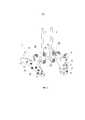
На фиг. 1 изображено заявленное реализующее заявленный способ устройство в сборе с вариантом установки на тазе, аксонометрия.In FIG. 1 shows the claimed device implementing the claimed method assembly with the installation option on the pelvis, axonometry.
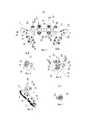
На фиг. 2 изображено реализующее заявленный способ устройство в сборе, аксонометрия.In FIG. 2 shows an assembly of a device implementing the claimed method, a perspective view.
На фиг. 3 изображено реализующее заявленный способ устройство в сборе на виде сзади.In FIG. 3 depicts a device assembly implementing the claimed method in a rear view.
На фиг. 4 - то же, сечение по А-А на фиг. 3, в сборке со стяжным хомутом, цилиндром и винтом.In FIG. 4 is the same section along AA in FIG. 3, assembled with a coupling collar, cylinder and screw.
На фиг. 5 - то же, сечение по Б-Б на фиг. 3, в сборке с дополнительной соединительной деталью, винтом хомута, винтом, фиксирующим стержень или проволоку сборки, и стержень или проволока сборки.In FIG. 5 is the same section along BB in FIG. 3, in an assembly with an additional connecting part, a clamp screw, a screw securing the assembly rod or wire, and the assembly rod or wire.
На фиг. 6 - то же, вид сбоку по стрелке В на фиг. 3, аксонометрия.In FIG. 6 is the same side view along arrow B in FIG. 3, axonometry.
На фиг. 7 - то же, сечение по Г-Г на фиг. 3, в сборке с дополнительной соединительной деталью, винтом и стержнем или проволокой сборки.In FIG. 7 is the same, section along G-D in FIG. 3, in an assembly with an additional connecting part, a screw and a rod, or an assembly wire.
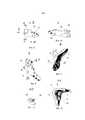
На фиг. 8 изображен левый опорный фиксирующий элемент, вид сверху, аксонометрия.In FIG. 8 shows a left support fixing element, a top view, a perspective view.
На фиг. 9 - то же, вид по стрелке Д на фиг. 8, аксонометрия.In FIG. 9 is the same, view along arrow D in FIG. 8, axonometry.
На фиг. 10 - то же, вид по стрелке Е на фиг. 8, аксонометрия.In FIG. 10 is the same, view along arrow E in FIG. 8, axonometry.
На фиг. 11 - то же, вид спереди по стрелке Ж на фиг. 8, аксонометрия.In FIG. 11 is the same, front view along arrow G in FIG. 8, axonometry.
На фиг. 12 - то же, сечение по И-И на фиг. 8.In FIG. 12 is the same, section along II in FIG. 8.
На фиг. 13 - то же, сечение по К-К на фиг. 9.In FIG. 13 is the same, section along KK in FIG. 9.
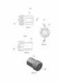
На фиг. 14 изображен цилиндр, продольный вид.In FIG. 14 shows a cylinder, a longitudinal view.
На фиг. 15 - то же, вид сбоку по стрелке Л на фиг. 14.In FIG. 15 is the same side view along arrow L in FIG. fourteen.
На фиг. 16 - изображен цилиндр, аксонометрия.In FIG. 16 - shows a cylinder, a perspective view.
На фиг. 17 изображена дополнительная соединительная деталь, вид сбоку.In FIG. 17 shows an additional connecting part, side view.
На фиг. 18 - то же, вид по стрелке М на фиг. 17.In FIG. 18 is the same, view along arrow M in FIG. 17.
На фиг. 19- то же, сечение по Н-Н на фиг. 17.In FIG. 19 is the same, a cross-section along H-H in FIG. 17.
На фиг. 20 изображен винт для фиксации стержня или проволоки сборки.In FIG. 20 depicts a screw for fixing a rod or wire assembly.
На фиг. 21 изображена дополнительная соединительная деталь, вид сбоку.In FIG. 21 shows an additional connecting part, side view.
На фиг. 22 - то же, вид по стрелке О.In FIG. 22 - same, view along arrow O.
На фиг. 23 - то же, сечение по П-П на фиг.21.In FIG. 23 is the same, a section along PP in FIG.
На фиг. 24 изображен винт для фиксации стержня или проволоки сборки, аксонометрия;In FIG. 24 shows a screw for fixing the rod or wire assembly, a perspective view;
На фиг. 25 изображен винт хомута.In FIG. 25 shows a clamp screw.
На фиг. 26 - то же, вид по стрелке Р на фиг. 25.In FIG. 26 is the same, view along arrow P in FIG. 25.
На фиг. 27 изображен винт хомута сбоку с частичным вырывом по сечению С-СIn FIG. 27 shows the clamp screw on the side with a partial tear along the cross section CC
на фиг. 26. in FIG. 26.
На фиг. 28 изображен винт хомута, аксонометрия.In FIG. 28 shows a clamp screw, a perspective view.
На фиг. 29 изображен винт, аксонометрия;In FIG. 29 shows a screw, a perspective view;
На фиг. 30 изображен установленный на гребне опорный фиксирующий элемент с фиксирующим винтом (поперечное сечение).In FIG. 30 shows a support fixing element mounted on a ridge with a fixing screw (cross section).
Реализующее заявленный способ устройство 1 для фиксации, по меньшей мере, части грудного и/или поясничного отделов позвоночника человека к тазу включает сборку из множества фиксирующих элементов, расположенных один над другим (на фигурах не показана, т.к. не является предметом изобретения), и, по меньшей мере, один стержень 2 или проволока, соединяющие эти фиксирующие элементы (фиг. 1 - фиг. 6). Кроме того, заявленное устройство включает приспособление 3 для прикрепления сборки к тазу. Приспособление 3 для прикрепления сборки к тазу содержит опорные фиксирующие элементы 4 (фиг. 1 - фиг. 3, фиг. 6, фиг. 8 - фиг. 11). Каждый опорный фиксирующий элемент 4 представляет собой протяженное тело 5, имеющее одно углубление 6, в поперечном сечении протяженное на всю длину тела 5 (фиг. 6). Контактная поверхность 7 углубления 6 предназначена для соприкосновения с, по меньшей мере, частью наружной 8, внутренней 9 и верхней 10 поверхностей гребня 11 крыла 12 подвздошной кости 13 (фиг. 1).The
Опорный фиксирующий элемент 4 снабжен, по меньшей мере, одним фиксирующим средством, в фиксирующей позиции распространяющимся в пространство 14, образованное контактной поверхностью углубления (фиг. 6).The supporting
Заявитель разработал множество таких конструкций. Наиболее оптимальная конструкция фиксирующего средства содержит винт 34 (фиг. 29) и выполненное в опорном фиксирующем элементе 4 сквозное отверстие 15 под него (фиг. 1, фиг. 6). Для дополнительной фиксации опорного фиксирующего элемента 4 на подвздошной кости достаточно винт вставить в отверстие 15 и ввинтить в кость.The applicant has developed many such designs. The most optimal design of the fixing means comprises a screw 34 (Fig. 29) and a through
Как отмечено выше, заявитель иллюстрирует изобретение на примере варианта использования пары опорных фиксирующих элементов 4 (см. следующий абзац и далее).As noted above, the applicant illustrates the invention using an example of the use of a pair of support locking elements 4 (see the following paragraph and below).
Устройство, изображенное на фигурах, содержит пару опорных фиксирующих элементов 4, а именно левый опорный фиксирующий элемент 16 и правый опорный фиксирующий элемент 17, соединенные между собой посредством протяженного элемента 18. Протяженных элементов может быть два и более двух в зависимости от конструкции приспособления для прикрепления сборки к тазу.The device depicted in the figures contains a pair of supporting
Пара опорных фиксирующих элементов 16 и 17 соединены между собой с возможностью поворота и сдвига. Заявитель разработал несколько вариантов такой конструкции.A pair of supporting
Наиболее оптимальным, по мнению заявителя, является следующий.The most optimal, according to the applicant, is the following.
На каждом опорном фиксирующем элементе 4 имеются стяжные хомуты 19, а протяженный элемент 18 представляет собой цилиндр 20, концы 21 (фиг. 16) которого смонтированы в отверстиях 22 (фиг. 10) стяжных хомутов 19 (фиг. 4) с возможностью затяга, а центральная часть 23 (фиг. 14) посредством дополнительных соединительных деталей 24 соединена со сборкой 2.On each supporting fixing
Каждая дополнительная соединительная деталь 24 содержит хомут 25 (фиг. 5, фиг. 18) с возможностью затяга, отверстие 26 которого охватывает цилиндр 20, и углубление 27 для установки конца стержня или проволоки 28 сборки 2, поперек которой в дополнительной соединительной детали 24 выполнено отверстие 29 с резьбой 30, в которое ввинчен винт 31 с возможностью упора торцом 32 в проволоку или стержень сборки (фиг. 7, фиг. 24).Each additional connecting
Окончательная фиксация заявленного устройства 1 для фиксации после ее установки осуществляется при помощи винтов 33 (фиг. 1, фиг. 3, фиг. 28), при помощи которых затягиваются стяжные хомуты 19 и хомуты 25 дополнительных соединительных деталей 24.The final fixation of the claimed
Заявитель считает необходимым более подробно остановиться на фиксирующем средстве (фиг. 29, фиг. 30). Как отмечено выше, оно содержит винт 34 с головкой 35 и выполненное в опорном фиксирующем элементе 4 сквозное отверстие 15 под него. Сквозное отверстие 15 выполнено с резьбой 36, а винт 34 имеет дополнительную резьбу 37 под головкой 35. В опорном фиксирующем элементе 4 с противоположной стороны от сквозного отверстия 15 выполнено дополнительное сквозное отверстие 38, и в фиксирующей позиции винт 34 проходит через оба отверстия 15 и 38. При окончательной фиксации винт 34 оказывается соединенным с опорным фиксирующим элементом посредством резьбы 37 под головкой винта и резьбы 36 в сквозном отверстии 15. Так же при установке винт 34 проходит насквозь через гребень подвздошной кости и соединяется с костной тканью основной резьбовой частью 39.The applicant considers it necessary to dwell in more detail on the fixing means (Fig. 29, Fig. 30). As noted above, it comprises a
Опорные фиксирующие элементы могут быть изготовлены на основе предварительно выполненной трехмерной модели таза и пояснично-крестцового отдела позвоночника на основании компьютерной томографии.Supporting fixing elements can be made on the basis of a pre-made three-dimensional model of the pelvis and lumbosacral spine based on computed tomography.
Предложенное техническое решение задачи иллюстрируется на примере использования его для проведения операции: «Коррекция деформации позвоночника при помощи металлоконструкции и выполнения позвоночно-тазовой фиксации».The proposed technical solution to the problem is illustrated by the example of using it for the operation: "Correction of spinal deformity with the help of metal structures and performing spinal-pelvic fixation."
1. При подготовке к операции пациенту выполняют компьютерную томографию (далее - КТ) пояснично-крестцового отделов позвоночника и таза.1. In preparation for surgery, the patient performs computed tomography (hereinafter referred to as CT) of the lumbosacral spine and pelvis.
2. По данным, полученным при КТ, выполняется трехмерная модель пояснично-крестцового отдела позвоночника и таза пациента.2. According to CT data, a three-dimensional model of the lumbosacral spine and pelvis of the patient is performed.
3. На основании этой модели моделируется (проектируется) и изготовляется устройство 1 для фиксации, по меньшей мере, части грудного и/или поясничного отделов позвоночника человека к тазу.3. Based on this model, a
4. При проведении операции пациенту под наркозом придают положение лежа на животе.4. During the operation, the patient under anesthesia is given a supine position.
5. После обработки кожных покровов производят продольный разрез кожи по линии остистых отростков на необходимую длину.5. After processing the skin, a longitudinal skin incision is made along the line of the spinous processes to the required length.
6. Верхнюю точку со стороны головы начала разреза определяют в зависимости от патологии, а нижняя часть разреза производится до уровня второго или ниже крестцовых позвонков.6. The upper point from the side of the head of the beginning of the incision is determined depending on the pathology, and the lower part of the incision is made to the level of the second or lower sacral vertebrae.
7. Рассекают последовательно подкожно-жировую клетчатку, фасцию, мышцы.7. Dissect sequentially subcutaneous fat, fascia, muscles.
8. Обнажают (скелетируют) костную ткань задних отделов позвонков.8. Bone tissue of the posterior vertebrae is exposed (skeletonized).
9. На позвонки устанавливаются фиксирующие элементы (винты, крючки, проволока, ленты из полимерных материалов и т.д.) (на фигурах не показано, поскольку общеизвестно и не является предметом изобретения).9. Fixing elements (screws, hooks, wire, tapes made of polymer materials, etc.) are installed on the vertebrae (not shown in the figures, since it is well known and is not the subject of the invention).
10. Фиксирующие элементы устанавливают с обеих сторон.10. The locking elements are installed on both sides.
11. Уровень установки фиксирующих элементов от головы, их количество и порядок установки зависят от патологии.11. The installation level of the fixing elements from the head, their number and installation order depend on the pathology.
12. Обнажаются (скелетируются) костная ткань задней поверхности крестца до уровня второго крестцового позвонка (при необходимости и ниже).12. Bone tissue of the posterior sacral surface is exposed (skeletonized) to the level of the second sacral vertebra (and, if necessary, lower).
13. Обнажают (скелетируют) костную ткань задней и средней трети гребней подвздошных костей с обеих сторон, а также частично скелетируют наружную и внутреннюю поверхности крыльев подвздошных костей на данном протяжении.13. Bone tissue of the posterior and middle third of the iliac crests on both sides is exposed (skeletonized), and the outer and inner surfaces of the iliac wings are partially skeletonized over this length.
14. Производят предварительную сборку устройства 1 для фиксации, по меньшей мере, части грудного и/или поясничного отделов позвоночника человека к тазу.14. Pre-assemble the
15. Для этого на цилиндр 20 насаживают отверстиями 26 две дополнительные соединительные детали 24, сдвигая их к центру цилиндра 20. Левый и правый концы 21 цилиндра 20 вставляют в отверстия 22 стяжного хомута 19 соответственно левого 16 и правого 17 опорного фиксирующего элемента.15. For this purpose, two additional connecting
16. После этого на гребни левой и правой подвздошных костей устанавливают левый опорный фиксирующий элемент 16 и правый опорный фиксирующий элемент 17 соответственно.16. After that, the left supporting fixing
17. В сквозные отверстия на левом опорном фиксирующем элементе 16 и правом опорном фиксирующем элементе 17 устанавливаются и ввинчиваются винты 34 для закрепления их на подвздошной кости. Количество и положение винтов могут быть различными и определяются в зависимости от анатомии и особенностей подвздошных костей.17.
18. На установленные на позвонках фиксирующие элементы с обеих сторон укладываются и фиксируются отмоделированные стержни (проволоки) соответствующей длины, причем нижние концы (противоположные от головы) стержней 28 укладывают в углубления 27 дополнительных соединительных деталей 24.18. On the vertebral fixing elements installed on both sides, the modeled rods (wires) of the corresponding length are laid and fixed, and the lower ends (opposite from the head) of the
19. После этого стержни (проволока) фиксируются в углублениях 27 дополнительных соединительных деталей 24 при помощи винтов 31.19. After this, the rods (wire) are fixed in the
20. Производится затяжка стяжных хомутов 19 и хомутов 25 дополнительных соединительных деталей 24 винтами 33.20. The tightening of the coupling clamps 19 and the
21. Далее производятся завершающие этапы операции (промывка, гемостаз, дренирование и ушивание раны).21. Next, the final stages of the operation are performed (flushing, hemostasis, drainage and suturing of the wound).
Сущность изобретения поясняется клиническими примерами.The invention is illustrated by clinical examples.
Пример 1Example 1
Больная Ф., 16 лет, поступила с диагнозом: нейрогенный паралитический левосторонний грудопоясничный сколиоз IV степени, декомпенсированный. Нижняя параплегия. Состояние после оперативного лечения.Patient F., 16 years old, was admitted with a diagnosis of neurogenic paralytic left-sided thoracolumbar scoliosis of the fourth degree, decompensated. Lower paraplegia. Condition after surgical treatment.
Ранее пациентка по поводу паралитического сколиоза для коррекции деформации и устранения позвоночно-тазовой декомпенсации была оперирована.Previously, the patient was operated on for paralytic scoliosis to correct deformity and eliminate spinal pelvic decompensation.
Была выполнена коррекция деформации металлоконструкцией с выполнением позвоночно-тазовой фиксации посредством установки винтов в подвздошные кости и соединением их с вышележащей металлоконструкцией.The deformation correction was performed by the metal structure with the implementation of the spinal-pelvic fixation by installing screws in the iliac bones and connecting them to the overlying metal structure.
Через 1 год после операции пациентка отметила боли в области поясницы и таза. При обследовании на рентгенограммах и компьютерной томографии отмечена нестабильность позвоночно-тазовой фиксации, расшатывание винтов в подвздошных костях с выраженной костной резорбцией вокруг них.1 year after surgery, the patient noted pain in the lower back and pelvis. Examination on radiographs and computed tomography revealed instability of the spinal-pelvic fixation, loosening of the screws in the iliac bones with pronounced bone resorption around them.
В связи с нестабильностью фиксации, болевыми ощущениями пациентки и риском развития воспалительных изменений пациентке было показано проведение оперативного лечения.In connection with the instability of fixation, pain of the patient and the risk of developing inflammatory changes, the patient was shown surgical treatment.
Учитывая слабость костной ткани подвздошных костей и наличие дефектов в связи с установленными ранее винтами, единственным вариантом выполнения позвоночно-тазовой фиксации являлось применение заявленного устройства.Given the weakness of the bone tissue of the ilium and the presence of defects due to previously installed screws, the only option for performing spinal-pelvic fixation was to use the claimed device.
Во время операции пациентке были удалены нестабильные винты из подвздошных костей, установлены опорные фиксирующие элементы согласно изобретению, соединенные с вышележащей металлоконструкцией.During the operation, the patient was removed unstable screws from the iliac bones, installed support retaining elements according to the invention, connected to an overlying metal structure.
В результате операции удалось достичь стабильную позвоночно-тазовую фиксацию с купированием болевого синдрома.As a result of the operation, it was possible to achieve stable spinal-pelvic fixation with relief of pain.
Отдаленный срок наблюдения за пациенткой после операции составляет один год, позвоночно-тазовая фиксация стабильна, пациентка операцией довольна, особых жалоб не предъявляет.The long-term follow-up of the patient after surgery is one year, the spinal-pelvic fixation is stable, the patient is satisfied with the operation, does not present any special complaints.
Данное лечение иллюстрирует пример использования заявленного изобретения при отсутствии других вариантов выполнения позвоночно-тазовой фиксации.This treatment illustrates an example of the use of the claimed invention in the absence of other options for the implementation of spinal-pelvic fixation.
Пример 2Example 2
Больная Ч., 20 лет, поступила с диагнозом: Спинальная амиотрофия. Нейрогенный паралитический левосторонний грудопоясничный сколиоз IV степени, декомпенсированный.Patient Ch., 20 years old, was admitted with a diagnosis of Spinal amyotrophy. Neurogenic paralytic left-side thoracolumbar scoliosis of the IV degree, decompensated.
Пациентка в связи с нижней параплегией передвигается только на инвалидной коляске.The patient in connection with lower paraplegia moves only in a wheelchair.
Нахождение пациентки в положении сидя связано с большими сложностями в связи со слабостью мышц туловища.Finding a patient in a sitting position is associated with great difficulties due to weakness of the muscles of the trunk.
При этом происходит значительное усиление деформации позвоночника со сдавлением органов брюшной полости и с выраженным перекосом таза.In this case, there is a significant increase in spinal deformity with compression of the abdominal organs and with pronounced skewness of the pelvis.
Применяемый для компенсации этих явлений корсет не является достаточно эффективным и вызывает целый ряд неудобств.The corset used to compensate for these phenomena is not effective enough and causes a number of inconveniences.
При обследовании на рентгенограммах выявлена тяжелая сколиотическая деформация позвоночника с выраженным перекосом таза, отмечается значительное усугубление деформации на рентгенограмме в положении сидя по сравнению с рентгенограммами, выполненными в положении лежа.Examination on radiographs revealed severe scoliotic deformity of the spine with pronounced distortion of the pelvis, there is a significant aggravation of deformation on the radiograph in the sitting position compared with radiographs performed in the supine position.
Все выше сказанное послужило показаниями для выполнения оперативного лечения.All of the above served as indications for the implementation of surgical treatment.
Целью запланированного оперативного лечения является коррекция деформации позвоночника и предоставление пациентке возможности находиться в положении сидя без усугубления позвоночно-тазовой деформации и «заваливания».The purpose of the planned surgical treatment is to correct spinal deformity and provide the patient with the opportunity to be in a sitting position without aggravating spinal-pelvic deformity and “blocking”.
Для достижения поставленной цели была выполнена операция с использованием заявленного устройства. В результате операции были достигнуты коррекция деформации позвоночника, значительное устранение перекоса таза, стабильная позвоночно-тазовая фиксация. В результате лечения пациентка может без внешних фиксирующих средств находиться длительное время в положении сидя, при этом она не отмечает «заваливания», отмечает улучшение функции внешнего дыхания, работы пищеварительной системы.To achieve this goal, an operation was performed using the claimed device. As a result of the operation, correction of spinal deformity, significant elimination of pelvic distortion, and stable spinal-pelvic fixation were achieved. As a result of treatment, the patient may be sitting for a long time without external fixative agents, while she does not notice “blocking”, notes improvement in the function of external respiration, and the digestive system.
Отдаленный срок наблюдения за пациенткой после операции составляет один год, позвоночно-тазовая фиксация стабильна, пациентка операцией довольна, жалоб не предъявляет.The long-term follow-up of the patient after surgery is one year, the spinal-pelvic fixation is stable, the patient is satisfied with the operation, and has no complaints.
Данное лечение иллюстрирует наиболее типичный пример оперативного лечения.This treatment illustrates the most typical example of surgical treatment.
Пример 3Example 3
Больной О., 8 лет, поступил с диагнозом: Нейрофиброматоз, вторичная деформация крестца.Patient O., 8 years old, was admitted with a diagnosis of Neurofibromatosis, secondary deformity of the sacrum.
У пациента родители отметили деформацию таза, нарушение походки, периодические жалобы на боли в пояснице.In the patient, the parents noted pelvic deformity, gait disturbance, periodic complaints of lower back pain.
При обследовании по данным рентгенографии и компьютерной томографии выявлена выраженная деформация крестца с частичным его недоразвитием, вторичного генеза на фоне давления нейрофибром.Examination according to x-ray and computed tomography revealed a pronounced deformity of the sacrum with its partial underdevelopment, secondary genesis against the background of pressure of neurofibromas.
В данном случае имелся крайне высокий риск развития перелома крестца и нарушения опорной функции таза, усугубления перекоса таза и сколиотической деформации.In this case, there was an extremely high risk of developing a sacral fracture and impaired pelvic support, aggravated pelvic distortion and scoliotic deformity.
Пациенту была выполнена операция с использованием заявленного изобретения. В результате был устранен перекос таза и предотвращена угроза развития перелома крестца и позвоночно-тазовой нестабильности.The patient underwent surgery using the claimed invention. As a result, the distortion of the pelvis was eliminated and the threat of the development of a fracture of the sacrum and vertebral-pelvic instability was prevented.
Наблюдение ребенка в течение 1 года после операции показало стабильную фиксацию.Observation of the child for 1 year after surgery showed stable fixation.
В общей сложности в группе вертебрологии ФГБУ «ЦИТО им. Н.Н. Приорова» Минздрава РФ оперировано 6 больных с использованием вышеописанного изобретения с хорошим результатом лечения.In total, in the vertebrology group of FSBI “CITO named after N.N. Priorova "of the Ministry of Health of the
Использование заявленного способа позволило выполнить стабильную позвоночно-тазовую фиксацию у пациентов с крайне тяжелыми деформациями, в том числе когда выполнение других видов позвоночно-тазовой фиксации не представлялось возможным.Using the claimed method made it possible to perform stable spinal-pelvic fixation in patients with extremely severe deformities, including when other types of spinal-pelvic fixation were not possible.
Таким образом, использование заявленного изобретения позволяет осуществить стабильную позвоночно-тазовую фиксацию, даже в случае наличия аномалий развития крестца и таза, слабости костной ткани и наличия дефектов после ранее проведенных операций.Thus, the use of the claimed invention allows for stable vertebral-pelvic fixation, even in the case of abnormalities in the development of the sacrum and pelvis, bone weakness and the presence of defects after previous operations.
Способ по изобретению позволяет выполнять позвоночно-тазовую фиксацию путем комбинирования со стандартными общепринятыми современными металлоконструкциями для фиксации позвоночника.The method according to the invention allows for spinal-pelvic fixation by combining with standard generally accepted modern metal structures for fixing the spine.
Важным преимуществом изобретения является также то, что способ может применяться травматологами-ортопедами и нейрохирургами без дополнительного длительного специального обучения и с использованием стандартного оборудования в операционных, предназначенных для выполнения операций на позвоночнике. В результате использования данного способа благодаря достижению стабильной позвоночно-тазовой фиксации возможно исключение необходимости длительного постельного периода и проведение ранней реабилитации пациентов.An important advantage of the invention is also that the method can be used by orthopedic traumatologists and neurosurgeons without additional long special training and using standard equipment in operating rooms designed to perform operations on the spine. As a result of using this method, due to the achievement of stable spinal-pelvic fixation, it is possible to exclude the need for a long bed period and conduct early rehabilitation of patients.
Помимо приведенного варианта способа возможны и другие многочисленные его модификации.In addition to the above variant of the method, numerous other modifications are possible.
Эти и многочисленные другие варианты способа охватываются приведенной далее заявителем формулой изобретения.These and numerous other variations of the method are covered by the following claims.
Claims (1)
Translated fromRussianпри этом фиксирующее средство содержит винт (34) с головкой (35) и выполненное в опорном фиксирующем элементе (4) сквозное отверстие (15) под него, причем пара опорных фиксирующих элементов (16) и (17) соединены между собой с возможностью поворота и сдвига, а на каждом опорном фиксирующем элементе имеются стяжные хомуты (19), причем протяженный элемент (18) представляет собой цилиндр (20), концы (21) которого смонтированы в отверстиях стяжных хомутов (19) с возможностью затяга, при этом центральная часть (23) посредством дополнительных соединительных деталей (24) соединена со сборкой (2), а каждая дополнительная соединительная деталь (24) содержит хомут (25) с возможностью затяга, отверстие (26) которого охватывает цилиндр (20), и углубление (27) для установки конца стержня или проволоки (28) сборки (2), поперек которой в дополнительной соединительной детали (24) выполнено отверстие (29) с резьбой (30), в которое ввинчен винт (31) с возможностью упора торцом (32) в стержень или проволоку (28) сборки (2), при этом опорные фиксирующие элементы (4) изготовлены на основе предварительно выполненной трехмерной модели таза и пояснично-крестцового отдела позвоночника на основании компьютерной томографии,
и включающий:
- разрез кожи по линии остистых отростков, при этом верхнюю точку разреза определяют в зависимости от выявленной патологии, а нижнюю точку разреза осуществляют до уровня второго или ниже крестцовых позвонков;
- обнажают костную ткань задних отделов позвонков;
- на позвонки с обеих сторон устанавливают фиксирующие элементы, такие как винты, крючки, проволока, лента из полимерных материалов и другие;
- обнажают костную ткань задней и средней трети гребней подвздошных костей с обеих сторон;
- частично обнажают наружную и внутреннюю поверхности крыльев подвздошных костей;
- осуществляют сборку устройства (1) для фиксации, по меньшей мере, части грудного и/или поясничного отделов позвоночника человека к тазу, для чего
- на цилиндр 20 насаживают отверстиями 26 две дополнительные соединительные детали 24, сдвигая их к центру цилиндра 20,
- левый и правый концы 21 цилиндра 20 вставляют в отверстия 22 стяжного хомута 19 соответственно левого 16 и правого 17 опорного фиксирующего элемента,
- после этого на гребни левой и правой подвздошных костей устанавливают левый опорный фиксирующий элемент 16 и правый опорный фиксирующий элемент 17 соответственно,
- в сквозные отверстия на левом опорном фиксирующем элементе 16 и правом опорном фиксирующем элементе 17 устанавливают и ввинчивают винты 34 для закрепления их на подвздошной кости,
- на установленные на позвонках фиксирующие элементы с обеих сторон укладывают и фиксируют отмоделированные стержни или проволоки соответствующей длины, причем нижние концы (противоположные от головы) стержней 28 укладывают в углубления 27 дополнительных соединительных деталей 24,
- после этого стержни или проволоки фиксируют в углублениях 27 дополнительных соединительных деталей 24 при помощи винтов 31,
- производят затяжку стяжных хомутов 19 и хомутов 25 дополнительных соединительных деталей 24 винтами 33,
- после установки устройства (1) для фиксации, по меньшей мере, части грудного и/или поясничного отделов позвоночника человека к тазу осуществляют промывку, гемостаз, дренирование и ушивание раны.A method of instrumental fixation of at least part of the thoracic and / or lumbar spine to the pelvis for various diseases of the spine by using a device (1) for fixing at least part of the human thoracic and / or lumbar spine to the pelvis containing the assembly ( 2) from a plurality of fixing elements located one above the other, and at least one rod or wire (28) connecting these fixing elements, as well as a device (3) for attaching the assembly to the pelvis, and it is adapted e (3) for attaching the assembly to the pelvis includes a pair of supporting fixing elements (4), each of which is an extended body (5), having a recess (6), extended along the length of the extended body (5), and the contact surface (14) the recesses (6) are intended for contact with at least part of the external and internal surfaces of the iliac wing and at least part of the iliac crest, each supporting fixing element (4) provided with at least one fixing means in fixing positions extending into the space formed by the contact surface (14) of the recess (6),
wherein the fixing means comprises a screw (34) with a head (35) and a through hole (15) made in the supporting fixing element (4), a pair of supporting fixing elements (16) and (17) are rotatably connected to each other and shear, and on each supporting fixing element there are coupling clamps (19), and the extended element (18) is a cylinder (20), the ends (21) of which are mounted in the holes of the coupling clamps (19) with the possibility of tightening, while the central part ( 23) through additional fittings (24) is connected to the assembly (2), and each additional connecting part (24) contains a clamp (25) that can be tightened, the hole (26) of which covers the cylinder (20), and a recess (27) for installing the end of the rod or wire ( 28) assembly (2), across which a hole (29) with a thread (30) is made in the additional connecting part (24), into which a screw (31) is screwed with the possibility of abutment with the end face (32) in the rod or wire (28) of the assembly ( 2), while the supporting fixing elements (4) are made on the basis of a previously executed three-dimensional model of the pelvis and girdle -border-sacral spine based on CT,
and including:
- a skin incision along the line of the spinous processes, while the upper point of the incision is determined depending on the revealed pathology, and the lower point of the incision is carried out to the level of the second or lower sacral vertebrae;
- expose the bone tissue of the posterior vertebrae;
- fixing elements, such as screws, hooks, wire, tape made of polymeric materials and others, are installed on the vertebrae on both sides;
- expose the bone tissue of the posterior and middle third of the iliac crests on both sides;
- partially expose the outer and inner surfaces of the wings of the ilium;
- carry out the assembly of the device (1) for fixing at least part of the chest and / or lumbar spine of the person to the pelvis, for which
- two additional connecting parts 24 are mounted on the cylinder 20 with holes 26, shifting them to the center of the cylinder 20,
- the left and right ends 21 of the cylinder 20 are inserted into the holes 22 of the coupling clamp 19, respectively, of the left 16 and right 17 of the supporting fixing element,
- after that, on the ridges of the left and right iliac bones, a left support fixing element 16 and a right support fixing element 17 are installed, respectively,
- through the holes on the left support fixing element 16 and the right support fixing element 17, screws 34 are installed and screwed in to fix them on the ilium,
- mounted on the vertebrae of the fixing elements on both sides lay and fix the modeled rods or wires of the corresponding length, and the lower ends (opposite from the head) of the rods 28 are laid in the recesses 27 of the additional connecting parts 24,
- after that, the rods or wires are fixed in the recesses 27 of the additional connecting parts 24 using screws 31
- tighten the coupling clamps 19 and the clamps 25 additional connecting parts 24 screws 33,
- after installing the device (1) for fixing at least part of the human thoracic and / or lumbar spine to the pelvis, washing, hemostasis, drainage and suturing of the wound are performed.
Priority Applications (1)
| Application Number | Priority Date | Filing Date | Title |
|---|---|---|---|
| RU2014148506/14ARU2584810C1 (en) | 2014-12-02 | 2014-12-02 | Method for tool fixation of at least part of thoracic and/or lumbar spine to pelvis in various diseases |
Applications Claiming Priority (1)
| Application Number | Priority Date | Filing Date | Title |
|---|---|---|---|
| RU2014148506/14ARU2584810C1 (en) | 2014-12-02 | 2014-12-02 | Method for tool fixation of at least part of thoracic and/or lumbar spine to pelvis in various diseases |
Publications (1)
| Publication Number | Publication Date |
|---|---|
| RU2584810C1true RU2584810C1 (en) | 2016-05-20 |
Family
ID=56012322
Family Applications (1)
| Application Number | Title | Priority Date | Filing Date |
|---|---|---|---|
| RU2014148506/14ARU2584810C1 (en) | 2014-12-02 | 2014-12-02 | Method for tool fixation of at least part of thoracic and/or lumbar spine to pelvis in various diseases |
Country Status (1)
| Country | Link |
|---|---|
| RU (1) | RU2584810C1 (en) |
Cited By (2)
| Publication number | Priority date | Publication date | Assignee | Title |
|---|---|---|---|---|
| RU2669028C2 (en)* | 2017-03-01 | 2018-10-05 | Федеральное государственное бюджетное образовательное учреждение высшего образования "Кубанский государственный медицинский университет" Министерства здравоохранения Российской Федерации (ФГБОУ ВО КубГМУ Минздрава России) | Method of prevention of fractures of related vertebrae in transpedicular fixation on the background of osteoporosis |
| RU2804846C1 (en)* | 2022-10-10 | 2023-10-06 | Федеральное государственное автономное учреждение "Национальный медицинский исследовательский центр здоровья детей" Министерства здравоохранения Российской Федерации (ФГАУ "НМИЦ здоровья детей" Минздрава России) | Method for surgical correction of pelvic tilt in children with neuro-muscular and syndromal scoliose |
Citations (2)
| Publication number | Priority date | Publication date | Assignee | Title |
|---|---|---|---|---|
| RU2341218C1 (en)* | 2007-03-23 | 2008-12-20 | Федеральное государственное учреждение науки "Российский научный центр "Восстановительная травматология и ортопедия" имени академика Г.П.Илизарова Федерального агентства по здравоохранению и социальному развитию" | Method of correction of s-shaped deformation of backbone |
| EP2662037A2 (en)* | 2012-05-09 | 2013-11-13 | CoLigne AG | Iliac connector, connector head, spinal fixation system and method of stabilizing a spine |
- 2014
- 2014-12-02RURU2014148506/14Apatent/RU2584810C1/enactive
Patent Citations (2)
| Publication number | Priority date | Publication date | Assignee | Title |
|---|---|---|---|---|
| RU2341218C1 (en)* | 2007-03-23 | 2008-12-20 | Федеральное государственное учреждение науки "Российский научный центр "Восстановительная травматология и ортопедия" имени академика Г.П.Илизарова Федерального агентства по здравоохранению и социальному развитию" | Method of correction of s-shaped deformation of backbone |
| EP2662037A2 (en)* | 2012-05-09 | 2013-11-13 | CoLigne AG | Iliac connector, connector head, spinal fixation system and method of stabilizing a spine |
Non-Patent Citations (1)
| Title |
|---|
| ДОНЧЕНКО С.В. и др. Применение позвоночно-тазовой транспедикулярной фиксации при лечении нестабильных повреждений тазового кольца. Травматология и ортопедия России, 2013, N 4 (70), с.67-74. Universal Spinal System (USS) Polyaxial and Iliosacral Spine Fixation. A versatile system for posterior stabilization of spinal segments. 2009, 11, pp.29,30.* |
Cited By (2)
| Publication number | Priority date | Publication date | Assignee | Title |
|---|---|---|---|---|
| RU2669028C2 (en)* | 2017-03-01 | 2018-10-05 | Федеральное государственное бюджетное образовательное учреждение высшего образования "Кубанский государственный медицинский университет" Министерства здравоохранения Российской Федерации (ФГБОУ ВО КубГМУ Минздрава России) | Method of prevention of fractures of related vertebrae in transpedicular fixation on the background of osteoporosis |
| RU2804846C1 (en)* | 2022-10-10 | 2023-10-06 | Федеральное государственное автономное учреждение "Национальный медицинский исследовательский центр здоровья детей" Министерства здравоохранения Российской Федерации (ФГАУ "НМИЦ здоровья детей" Минздрава России) | Method for surgical correction of pelvic tilt in children with neuro-muscular and syndromal scoliose |
Similar Documents
| Publication | Publication Date | Title |
|---|---|---|
| US9872711B2 (en) | Spinal implant system and method | |
| Odent et al. | Fusionless surgery in early-onset scoliosis | |
| RU2585733C1 (en) | Device for fixation of at least part of thoracic and/or lumbar of person to pelvis | |
| US20150190178A1 (en) | Spinal correction system and method | |
| US9615859B2 (en) | Posted translation system and method | |
| Altiok et al. | Kyphectomy in children with myelomeningocele | |
| US20140066990A1 (en) | Multi-planar axial spinal rod connector | |
| US9427265B2 (en) | Surgical instrument system and method | |
| US11284924B1 (en) | Adjustable spinal implant, system and method | |
| EP3958766B1 (en) | Spinal implant system | |
| RU2584810C1 (en) | Method for tool fixation of at least part of thoracic and/or lumbar spine to pelvis in various diseases | |
| RU2625986C2 (en) | Method for pelvic bones reconstruction | |
| ES2982036T3 (en) | U-BLOQ Chest Wall System | |
| RU2693821C1 (en) | Method for correcting spinal deformity with a lumbar artery | |
| US20240148422A1 (en) | Pelvic internal fixation device and methods of using the same | |
| Márquez et al. | Growing rods in early-onset scoliosis. Do they really help to control the deformity and spinal and thoracic growth? | |
| RU2538797C2 (en) | Method for surgical management of compression vertebral fracture accompanying osteoporosis | |
| Szabó et al. | Percutaneous spine fusion combined with whole-body traction in the acute surgical treatment of AO A-and C-type fractures: A technical note | |
| RU2197912C2 (en) | Surgical and device method for treating spondylolisthesis | |
| Cigliano et al. | A new instrumentation system for the reduction and posterior stabilization of unstable thoracolumbar fractures | |
| RU2778776C1 (en) | Plate for stabilising the chest in children and method for application thereof for treating rigid forms of pectus carinatum | |
| RU2804846C1 (en) | Method for surgical correction of pelvic tilt in children with neuro-muscular and syndromal scoliose | |
| Palmiere et al. | Fatal hemorrhage following sacroiliac joint fusion surgery: a case report | |
| RU2820917C1 (en) | Schanz rods transosseous entrenchment device | |
| RU2726047C1 (en) | Method for intraoperative correction of spine scoliosis |